Note: Descriptions are shown in the official language in which they were submitted.
CA 02593429 2007-05-10
WO 2006/058900 PCT/EP2005/056373
1
THE LUPAC BIFUNCTIONAL MARKER
AND ITS USE IN PROTEIN PRODUCTION
FIELD OF THE INVENTION
This invention relates to industrial production of proteins. More
specifically,
the invention relates to surrogate markers corresponding to a fusion between
luciferase and the puromycin N-acetyl transferase. The invention further
relates to
the use of these surrogate for screening cells for high expression of a
protein of
interest.
BACKGROUND
Introducing heterologous genes into animal host cells and screening for
expression
of the added genes is a lengthy and complicated process. Typically a number of
hurdles have to be overcome: (i) the construction of large expression vectors;
(ii) the
transfection and selection of clones with stable long-term expression,
eventually in
the absence of selective pressure; and (iii) screening for high expression
rates of the
heterologous protein of interest.
1. Selection of clones expressing the heterologous gene
Selection of the clones having integrated the gene of interest is performed
using a
selection marker conferring resistance to a selective pressure. Most of the
selection
markers confer resistance to an antibiotic such as, e.g., neomycin, kanamycin,
hygromycin, gentamycin, chloramphenicol, puromycin, zeocin or bleomycin.
When generating cell clones expressing a gene of interest from expression
vectors,
host cells are typically transfected with a plasmid DNA vector encoding both
the
protein of interest and the selection marker on the same vector. Quite often
the
capacity of a plasmid is limited and the selection marker has to be expressed
from a
second plasmid, which is co-transfected with the plasmid comprising the gene
of
interest.
Stable transfection leads to random integration of the expression vector in
the
genome of the host cell. Use of selective pressure, e.g. by administrating an
antibiotic to the media, will eliminate all cells that did not integrate the
vector
containing the selection marker providing resistance to the respective
antibiotic or
selective pressure. If this selection marker is on the same vector as the gene
of
CA 02593429 2007-05-10
WO 2006/058900 PCT/EP2005/056373
2
interest or, if this selection marker is on a second vector and vector
comprising the
gene of interest was co-integrated, the cells will express both the selection
marker
and the gene of interest. It is frequently observed, however, that the
expression level
of the gene of interest is highly variable depending on the site of
integration.
Furthermore, when removing selective pressure, expression becomes quite often
very unstable or even extinguished. Only a small number of initial
transfectants are
thus providing high and stable long-term expression and it is extremely
tedious to
identify these clones in a large population of candidates. Typically, high
expressing
candidates are isolated and then cultivated in absence of selective pressure.
Under
these conditions a large proportion of initially selected candidates are
eliminated due
to their loss of gene of interest expression upon removal of selective
pressure. It
would thus be advantageous to cultivate the candidates, following an initial
period of
selection for stable transfection, in absence of selective pressure and only
then
screen for gene of interest expression.
2. Screening for high expressing clones
Screening for high-expressing clones for a protein of interest is often done
by
methods directly revealing the presence of high amounts of the protein.
Typically
immunologic methods, such as ELISA or immunohistochemical staining, are
applied
to detect the product either intracellularly or in cell culture supernatants.
These
methods are tedious, expensive, time-consuming, and often not amenable to high
throughput screenings (HTS). In addition, an antibody reactive to the
expressed
protein must be available.
Attempts to quantify the protein amounts by Fluorescence-Activated Cell
Sorting
(FACS) have also been made, but only with a limited success, especially in the
case
of secreted proteins (Borth et al., 2000)
One approach for the screening of high expression rates of the protein of
interest
would be the use of an easily measurable surrogate marker, expressed from the
same vector as the gene of interest (Chesnut et aL, 1996) . The idea
underlying the
use of a measurable surrogate marker is that there is a correlation between
the
expression of the gene of interest and the surrogate marker gene due to the
physical
link of the two genes on the same vector.
Numerous easily measurable markers are available in the art. They usually
correspond to enzymes which act on a chromogenic or luminogenic substrate such
as, e.g., the R-glucuronidase, the chloramphenicol acetyltransferase, the
nopaline
synthase, the R-galactosidase, secreted alkaline phosphatase (SEAP) and the
CA 02593429 2007-05-10
WO 2006/058900 PCT/EP2005/056373
3
luciferase. The green fluorescent protein (GFP) may also be used as a
measurable
marker in FACS. The activity of all these proteins can be measured by standard
assays that may be used in HTS.
The drawback of this approach is the use of yet another expression cassette
for the
surrogate marker gene. This renders the expression vector rather bulky,
hosting
expression units comprising a promoter, a cDNA and polyadanylation signals for
at
least three proteins (i.e., the gene of interest, the selection marker and the
surrogate
marker). For multi-chain proteins the situation becomes even more complex.
Alternatively, individual plasmid vectors expressing the three genes, which
encode
the protein of interest, the selection marker and the surrogate marker
respectively,
could be co-transfected. However, it is likely that the vectors would be
either
integrated at different loci, or exhibit varying and uncorrelated expression.
A promising approach for overcoming the above limitations consists in the use
of a
chimeric marker that combines the functional properties of a selection marker
and of
a measurable marker. Such an approach has been described by, e.g., Bennett et
al.
(1998). This article discloses the GFP-ZeoR marker, which confers resistance
to the
Zeocin antibiotic, and the expression of which can be monitored by
fluorescence
microscopy.
EP 1 262 553 discloses chimeric markers and their use either in a method for
trapping unknown genes, or in a method of selecting cells in which a genetic
element
has been targeted into a predefined locus by homologous recombination.
However,
EP 1 262 553 does not teach the use of chimeric markers for screening for
clones
expressing high levels of a recombinant protein. Furthermore, the experimental
data
relates to a chimeric marker corresponding to a fusion protein between
luciferase and
the protein conferring resistance to hygromycin.
The potential use of chimeric markers for screening for high-expressing clones
remains a poorly explored field, and the efficiency of such chimeric markers
for
screening for high-expressing clones needs to be further investigated. The
finding of
a novel, alternative and powerful chimeric surrogate marker would be extremely
useful in the field of industrial production of therapeutic proteins.
SUMMARY OF THE INVENTION
The present invention stems from the construction and characterization of a
novel
bifunctional chimeric marker, Lupac. Lupac corresponds to a fusion protein
between
luciferase and a protein conferring resistance to puromycin, the puromycin N-
acetyl
CA 02593429 2007-05-10
WO 2006/058900 PCT/EP2005/056373
4
transferase (pac). It has been demonstrated that Lupac combines the functional
properties of both luciferase and pac. Lupac's usefulness for the isolation of
high-
expressing clones for a therapeutic protein has further been demonstrated.
Therefore, a first aspect of the invention relates to a Lupac polypeptide
comprising a
fragment of a luciferase fused to a fragment of a puromycin N-acetyl
transferase
(pac), wherein said Lupac polypeptide exhibits (i) luciferase activity; and
(ii)
puromycin N-acetyl transferase activity.
A second aspect relates to a nucleic acid encoding a Lupac polypeptide
according to
the invention.
A third aspect relates to a vector comprising a nucleic acid according to the
invention.
A fourth aspect relates to a cell comprising a nucleic acid according to the
invention.
A fifth aspect relates to the use of a cell comprising a nucleic acid
according to the
invention for producing a protein of interest.
A sixth aspect relates to the use of a polypeptide, a nucleic acid or a vector
according
to the invention for screening cells for expression of a protein of interest.
A seventh aspect relates to a method of screening cells for expression of a
protein of
interest, said method comprising the step of:
(i) transfecting cells by a an expression vector according to the
invention;
(ii) selecting cells being resistant to puromycin; and
(iii) assaying the luciferase activity of the cells selected in step (ii).
An eight aspect relates to a method of obtaining a cell line expressing a
protein of
interest, said method comprising the step of:
(i) screening cells according to a method of screening according to
the invention;
(ii) selecting the cell exhibiting the highest expression of said protein
of interest; and
(iii) establishing a cell line from said cell.
A ninth aspect relates to a method of producing a protein of interest, said
method
comprising the step of:
(i) culturing a cell line obtained according to the invention under
conditions which permit expression of said protein of interest; and
(ii) collecting said protein of interest.
CA 02593429 2007-05-10
WO 2006/058900 PCT/EP2005/056373
A tenth aspect relates to a method of producing a polypeptide according to
the invention comprising the step of:
(i) culturing a cell comprising a nucleic acid according to the invention
under conditions which permit expression of the Lupac
5 polypeptide; and
(ii) collecting the polypeptide according to the invention.
BRIEF DESCRIPTION OF THE FIGURES
Figure 1 shows an alignment between a Lupac polypeptide in accordance with
the invention (SEQ ID NO: 2), luciferase (SEQ ID NO: 8) and pac (SEQ ID NO:
9).
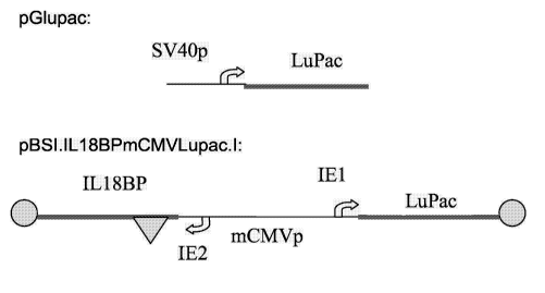
Figure 2 shows a scheme of the pGlupac and of the pBSI.IL18BPmCMVLupac.l
vectors. pGlupac contains the ORF for a Lupac polypeptide of SEQ ID NO: 1
expressed form an SV40 promoter with an SV40 enhancer at 3'. Plasmid
pBSI.IL18BPmCMVLupac.l contains a Lupac polypeptide of SEQ ID NO: 2
expressed from the IE1 promoter of the bi-directional mouse CMV immediate
early
region. The IE2 promoter of this vector is driving expression of IL18BP.
Figure 3 shows the luciferase activity of CHO cells transiently transfected
either
with pGlupac or with pGL3ctrl + pPur (vectors comprising luciferase and pac
respectively). Luciferase activity is normalized by cell density in
million/mi.
Figure 4 shows the positive correlation between expression of a Lupac
polypeptide of SEQ ID NO: 2 (RLU) and IL18BP (RU) in 24 clones transfected
with
the pBSI.IL18BPmCMVLupac.I vector.
Figure 5 is a scheme of the expression vector used in Example 4. This vector
contains a Lupac polypeptide of SEQ ID NO: 2 expressed from the IE1 promoter
of
the bi-directional mouse CMV immediate early region. The IE2 promoter of this
vector is driving expression of a recombinant protein referred to as "Serono
Protein
1" (r-SP1). The insulator is described in PCT/EP2004/052591.
Figures 6 and 7 show the r-SP1 titer and the viable cell density obtained when
culturing a CHO cell line expressing r-SP1 (CHO-r-SP1), which was selected
using
Lupac as a surrogate marker (see Example 4).
BRIEF DESCRIPTION OF THE SEQUENCES OF THE SEQUENCE LISTING
SEQ ID NO: 1 corresponds to the nucleic acid sequence of a Lupac polypeptide
in
accordance with the invention.
CA 02593429 2007-05-10
WO 2006/058900 PCT/EP2005/056373
6
SEQ ID NO: 2 corresponds to the protein sequence encoding a Lupac polypeptide
in
accordance with the invention.
SEQ ID Nos. 3 to 6 correspond to primers used for constructing a Lupac
polypeptide
in accordance with the invention.
SEQ ID NO: 7 corresponds to the fragment of the mouse CMV immediate early
region present in the pBSI.IL18BPmCMVLupac.I vector.
SEQ ID NO: 8 corresponds to the protein sequence of the photinus pyralis
luciferase.
SEQ ID NO: 9 corresponds to the protein sequence of the Streptomyces alboniger
pac.
DETAILED DESCRIPTION OF THE INVENTION
The present invention stems from the construction and characterization of a
novel
bifunctional chimeric marker referred to as Lupac. Lupac corresponds to a
fusion
protein between luciferase and a protein conferring resistance to puromycin,
the
puromycin N-acetyl transferase (pac).
It has been demonstrated that Lupac combines the functional properties of
luciferase
and of pac (Example 2). Accordingly, the Lupac marker can be used both as a
selectable marker in stable transfections due to its pac activity and as an
easily
measurable surrogate marker due to its luciferase activity.
Lupac's usefulness for the isolation of high-expressing clones for a
therapeutic
protein has further been demonstrated. In Example 3, a vector comprising Lupac
and
a gene of interest, expressed from two different promoters, has been
constructed. It
has been shown that there is a very good positive correlation between Lupac
expression levels and expression levels of the gene of interest.
Accordingly, the present invention provides a powerful marker, Lupac, which
can
both be used to provide selectivity in stable transfection and act as a
surrogate
marker for screening candidate clones for high expression of a gene of
interest.
Using Lupac in HTS allows keeping the same chance for selecting high-
expressing
clones as when the expression level of the gene of interest is measured
directly.
Moreover, using Lupac allows reducing time, cost and resources since (i)
standardized product-independent and simple analysis is performed; and (ii)
measuring luciferase activity is an inexpensive assay.
CA 02593429 2007-05-10
WO 2006/058900 PCT/EP2005/056373
7
1. Lupac polypeptides
A first aspect of the present invention relates to a polypeptide comprising a
fragment
of a luciferase fused to a fragment of a puromycin N-acetyl transferase (pac),
wherein said Lupac polypeptide exhibits (i) luciferase activity; and (ii)
puromycin N-
acetyl transferase activity. As further used herein, the term "a Lupac
polypeptide" or
"Lupac" refers to such a polypeptide.
As used herein, a polypeptide exhibits "luciferase activity" when said
polypeptide is
capable of oxidizing luciferin. Preferably, said polypeptide is capable of
catalyzing at
least one of the following reactions:
= Photinus luciferin + 02 + ATP => oxidized Photinus luciferin + CO2 + H20 +
AMP +
diphosphate + light.
= Renilla or Cypridina luciferin + 02 <=> oxidized Renilla luciferin + CO2 +
light
Photinus luciferin refers to (S)-4,5-dihydro-2-(6-hydroxy-2- benzothiazoloyl)-
4-
thiazolecarboxylic acid. Cypridina luciferin refers to [3-[3,7-dihydro-6-(1 H-
indol-3-yl)-
2-[(S)-1- methyl-6-propyl]-3-oxoimidazo-[1,2-a]pyrazin-8-yl]propyl]guanidine.
Renilla
luciferin refers to 8-benzyl-2-(4-hydroxybenzyl)-6-(4-hydroxyphenyl)imidazo-
[1,2-
A]pyrazin-3(7H)-one.
Measurement of the light emitted during the above reactions allows measurement
of
luciferase activity. The luciferase activity can for example be measured as
described
in Example 2.2.
As used herein, a polypeptide exhibits "puromycin N-acetyl transferase
activity" when
said polypeptide is capable of conferring resistance to puromycin to a cell.
The
puromycin N-acetyl transferase activity can for example be measured as
described in
Example 2.3.
In a preferred embodiment, the Lupac polypeptide comprises a fragment of a
luciferase coming from a firefly such as, e.g., photinus pyralis, Luciola
cruciata,
Luciola lateralis or Photuris pennsylvanica. Preferably, the Lupac polypeptide
comprises a fragment of the photinus pyralis luciferase. As used herein, the
term
"photinus pyralis luciferase" refers to a polypeptide of SEQ ID NO: 8, or to
an allelic
variant, a splice variant or a mutein thereof. Most preferably, said fragment
of a
photinus pyralis luciferase comprises amino acids 1 to 547 of SEQ ID NO: 8.
Alternatively, said fragment of a photinus pyralis luciferase can correspond
to a
fragment of at least 50, 75, 100, 125, 150, 175, 200, 225, 250, 275, 300, 325,
350,
CA 02593429 2007-05-10
WO 2006/058900 PCT/EP2005/056373
8
375, 400, 425, 450, 475, 500 or 525 amino acids of the full-length photinus
pyralis
luciferase, or to the full-length photinus pyralis luciferase.
In another preferred embodiment, the Lupac polypeptide comprises a fragment of
a
luciferase coming from Renilla reniformis (sea pansy) or from Vargula
hilgendorfii
(Sea firefly).
In another preferred embodiment, the Lupac polypeptide comprises a fragment of
a
pac coming from a Streptomyces species such as, e.g., Streptomyces alboniger
or
Streptomyces coelicolor. Preferably, the Lupac polypeptide comprises a
fragment of
a Streptomyces alboniger pac. As used herein, the term "Streptomyces alboniger
~ac" refers to a polypeptide of SEQ ID NO: 9 or to an allelic variant, a
splice variant
or a mutein thereof. More Preferably, the pac fragment comprises amino acids 2
to
199 of SEQ ID NO: 9. Alternatively, said fragment of a Streptomyces alboniger
pac
can correspond to a fragment of at least 50, 75, 100, 125, 150 or 175 amino
acids of
the full-length Streptomyces alboniger pac, or to the full-length Streptomyces
alboniger pac.
In a Lupac polypeptide, the luciferase fragment may be fused to the 5'
terminus of
the pac fragment, or the pac fragment may be fused to the 5' terminus of the
luciferase fragment. Preferably, the luciferase fragment is fused to the 5'
terminus of
the pac fragment.
In a most preferred embodiment, the Lupac polypeptide comprises or consists of
SEQ ID NO: 2.
In another most preferred embodiment, the Lupac polypeptide comprises or
consists
of an amino acid sequence at least 50% identical, more preferably at least 60%
identical, and still more preferably at least 70%, 75%, 80%, 85%, 90%, 95%,
96%,
97%, 98% or 99% identical to SEQ ID NO: 2.
As used herein, the term "mutein" refers to an analog of a naturally occurring
polypeptide, in which one or more of the amino acid residues of a naturally
occurring
polypeptide are replaced by different amino acid residues, or are deleted, or
one or
more amino acid residues are added to the naturally occurring sequence of the
polypeptide, without lowering considerably the activity of the resulting
products as
compared with the naturally occurring polypeptide. These muteins are prepared
by
known synthesis and/or by site-directed mutagenesis techniques, or any other
known
technique suitable therefore. Muteins of Streptomyces alboniger pac or of
photinus
pyralis luciferase that can be used in accordance with the present invention,
or
nucleic acids encoding the muteins, including a finite set of substantially
CA 02593429 2007-05-10
WO 2006/058900 PCT/EP2005/056373
9
corresponding sequences as substitution peptides or polynucleotides which can
be
routinely obtained by one of ordinary skill in the art, without undue
experimentation,
based on the teachings and guidance presented herein.
Muteins of Streptomyces alboniger pac or of photinus pyralis luciferase in
accordance with the present invention include proteins encoded by a nucleic
acid,
such as DNA or RNA, which hybridizes to DNA or RNA, which encodes pac or
luciferase, in accordance with the present invention, under moderately or
highly
stringent conditions. The term "stringent conditions" refers to hybridization
and
subsequent washing conditions, which those of ordinary skill in the art
conventionally
refer to as "stringent". See Ausubel et al., Current Protocols in Molecular
Biology,
supra, lnterscience, N.Y., 6.3 and 6.4 (1987, 1992), and Sambrook et al.
(Sambrook, J. C., Fritsch, E. F., and Maniatis, T. (1989) Molecular Cloning: A
Laboratory Manual, Cold Spring Harbor Laboratory Press, Cold Spring Harbor,
NY).
Without limitation, examples of stringent conditions include washing
conditions 12-20 C below the calculated Tm of the hybrid under study in, e.g.,
2 x
SSC and 0.5% SDS for 5 minutes, 2 x SSC and 0.1% SDS for 15 minutes; 0.1 x SSC
and 0.5% SDS at 37 C for 30-60 minutes and then, a 0.1 x SSC and 0.5% SDS at
68 C for 30-60 minutes. Those of ordinary skill in this art understand that
stringency
conditions also depend on the length of the DNA sequences, oligonucleotide
probes
(such as 10-40 bases) or mixed oligonucleotide probes. If mixed probes are
used, it
is preferable to use tetramethyl ammonium chloride (TMAC) instead of SSC.
Muteins of Streptomyces alboniger pac or of photinus pyralis luciferase
include polypeptides having an amino acid sequence at least 50% identical,
more
preferably at least 60% identical, and still more preferably at least 70%,
75%, 80%,
85%, 90%, 95%, 96%, 97%, 98% or 99% identical to the naturally occurring
polypeptide.
By a polypeptide having an amino acid sequence at least, for example, 95%
"identical" to a query amino acid sequence of the present invention, it is
intended that
the amino acid sequence of the subject polypeptide is identical to the query
sequence except that the subject polypeptide sequence may include up to five
amino
acid alterations per each 100 amino acids of the query amino acid sequence. In
other
words, to obtain a polypeptide having an amino acid sequence at least 95%
identical
to a query amino acid sequence, up to 5% (5 of 100) of the amino acid residues
in
the subject sequence may be inserted, deleted, or substituted with another
amino
acid.
CA 02593429 2007-05-10
WO 2006/058900 PCT/EP2005/056373
For sequences where there is not an exact correspondence, a "% identity" may
be
determined. In general, the two sequences to be compared are aligned to give a
maximum correlation between the sequences. This may include inserting "gaps"
in
either one or both sequences, to enhance the degree of alignment. A % identity
may
5 be determined over the whole length of each of the sequences being compared
(so-
called global alignment), that is particularly suitable for sequences of the
same or
very similar length, or over shorter, defined lengths (so-called local
alignment), that is
more suitable for sequences of unequal length.
Methods for comparing the identity and homology of two or more sequences are
well
10 known in the art. Thus for instance, programs available in the Wisconsin
Sequence
Analysis Package, version 9.1 (Devereux et aL, 1984), for example the programs
BESTFIT and GAP, may be used to determine the % identity between two
polynucleotides and the % identity and the % homology between two polypeptide
sequences. BESTFIT uses the "local homology" algorithm of (Smith and Waterman,
1981) and finds the best single region of similarity between two sequences.
Other
programs for determining identity and/or similarity between sequences are also
known in the art, for instance the BLAST family of programs (Altschul et aL,
1990),
accessible through the home page of the NCBI at world wide web site
ncbi.nlm.nih.gov) and FASTA (Pearson and Lipman, 1988; Pearson, 1990).
Preferred changes for muteins in accordance with the present invention are
what are
known as "conservative" substitutions. Conservative amino acid substitutions
of
Streptomyces alboniger pac or of photinus pyralis luciferase, may include
synonymous amino acids within a group which have sufficiently similar
physicochemical properties that substitution between members of the group will
preserve the biological function of the molecule (Grantham, 1974). It is clear
that
insertions and deletions of amino acids may also be made in the above-defined
sequences without altering their function, particularly if the insertions or
deletions
only involve a few amino acids, e.g. under thirty, and preferably under ten,
and do not
remove or displace amino acids which are critical to a functional
conformation, e.g.
cysteine residues. Proteins and muteins produced by such deletions and/or
insertions come within the purview of the present invention.
Preferably, the synonymous amino acid groups are those defined in Table I.
More
preferably, the synonymous amino acid groups are those defined in Table II;
and
most preferably the synonymous amino acid groups are those defined in Table
III.
CA 02593429 2007-05-10
WO 2006/058900 PCT/EP2005/056373
11
Table I
Preferred Groups of Synonymous Amino Acids
Amino Acid Synonymous Group
Ser Ser, Thr, Gly, Asn
Arg Arg, Gin, Lys, Glu, His
Leu lie, Phe, Tyr, Met, Val, Leu
Pro Gly, Ala, Thr, Pro
Thr Pro, Ser, Ala, Gly, His, Gin, Thr
Ala Gly, Thr, Pro, Ala
Val Met, Tyr, Phe, lie, Leu, Val
Gly Ala, Thr, Pro, Ser, Gly
lie Met, Tyr, Phe, Val, Leu, lie
Phe Trp, Met, Tyr, lie, Val, Leu, Phe
Tyr Trp, Met, Phe, lie, Val, Leu, Tyr
Cys Ser, Thr, Cys
His Glu, Lys, Gin, Thr, Arg, His
Gin Glu, Lys, Asn, His, Thr, Arg, Gin
Asn Gin, Asp, Ser, Asn
Lys Glu, Gin, His, Arg, Lys
Asp Glu, Asn, Asp
Glu Asp, Lys, Asn, Gin, His, Arg, Glu
Met Phe, lie, Val, Leu, Met
Trp Trp
Table II
More Preferred Groups of Synonymous Amino Acids
Amino Acid Synonymous Group
Ser Ser
Arg His, Lys, Arg
Leu Leu, lie, Phe, Met
Pro Ala, Pro
Thr Thr
Ala Pro, Ala
Val Val, Met, lie
Gly Gly
lie lie, Met, Phe, Val, Leu
Phe Met, Tyr, lie, Leu, Phe
Tyr Phe, Tyr
Cys Cys, Ser
His His, Gin, Arg
Gin Glu, Gin, His
Asn Asp, Asn
Lys Lys, Arg
Asp Asp, Asn
Glu Glu, Gin
Met Met, Phe, lie, Val, Leu
Trp Trp
CA 02593429 2007-05-10
WO 2006/058900 PCT/EP2005/056373
12
Table III
Most Preferred Groups of Synonymous Amino Acids
Amino Acid Synonymous Group
Ser Ser
Arg Arg
Leu Leu, lie, Met
Pro Pro
Thr Thr
Ala Ala
Val Val
Gly Gly
lie lie, Met, Leu
Phe Phe
Tyr Tyr
Cys Cys, Ser
His His
Gin Gin
Asn Asn
Lys Lys
Asp Asp
Glu Glu
Met Met, lie, Leu
Trp Trp
Examples of production of amino acid substitutions in proteins which can be
used for obtaining muteins of Streptomyces alboniger pac or of photinus
pyralis
luciferase for use in the present invention include any known method steps,
such as
presented in US patents 4,959,314, 4,588,585 and 4,737,462, to Mark et al;
5,116,943 to Koths et al., 4,965,195 to Namen et al; 4,879,111 to Chong et al;
and
5,017,691 to Lee et al; and lysine substituted proteins presented in US patent
No.
4,904,584 (Shaw et al).
Preferably, the muteins of the present invention exhibit substancially the
same biological activity as the naturally occurring polypeptide to which it
corresponds.
2. Lupac nucleic acids, and vectors and host cells comprising them
A second aspect of the present invention relates to a nucleic acid encoding a
Lupac polypeptide. As further used in this specification, the term "Lupac
nucleic acid"
refers to such a nucleic acid.
In a preferred embodiment, the Lupac nucleic acid comprises or consists of
SEQ ID NO: 1.
CA 02593429 2007-05-10
WO 2006/058900 PCT/EP2005/056373
13
Any procedures known in the art can be used to obtain Lupac nucleic acids of
the present invention. Lupac nucleic acids can for example be obtained as
described
in Example 1.
A third aspect of the present invention relates to a vector comprising a Lupac
nucleic acid. Such a vector is referred to as a "Lupac vector" within the
present
specification. Preferably, the Lupac vector is an expression vector. Such a
vector is
further referred to as a "Lupac expression vector". The term "Lupac vector"
encompasses the term "Lupac expression vector". The term "vector" is used
herein to
designate either a circular or a linear DNA or RNA compound, which is either
double-
stranded or single-stranded, and which comprise at least one polynucleotide of
the
present invention to be transferred in a cell host or in a unicellular or
multicellular
host organism. An "expression vector" comprises appropriate signals in the
vectors,
said signals including various regulatory elements, such as
enhancers/promoters
from viral, bacterial, plant, mammalian, and other eucaryotic sources that
drive
expression of the inserted polynucleotide in host cells.
In a most preferred embodiment, the Lupac expression vector further
comprises a nucleic acid encoding a protein of interest. As shown in example
3, such
vectors are particularly useful for screening cells for high expression of a
protein of
interest.
In accordance with the present invention, the protein of interest may be any
polypeptide for which production is desired. The protein of interest may find
use in
the field of pharmaceutics, agribusiness or furniture for research
laboratories.
Preferred proteins of interests find use in the field of pharmaceutics.
For example, the protein of interest may be, e.g., a naturally secreted
protein,
a normally cytoplasmic protein, a normally transmembrane protein, or a human
or a
humanized antibody. When the protein of interest is a normally cytoplasmic or
a
normally transmembrane protein, the protein has preferably been engineered in
order
to become soluble. The polypeptide of interest may be of any origin. Preferred
polypeptides of interest are of human origin.
In preferred embodiments, the protein of interest is selected from the group
consisting of chorionic gonadotropin, follicle-stimulating hormone, lutropin-
choriogonadotropic hormone, thyroid stimulating hormone, human growth hormone,
interferons (e.g., interferon beta-la, interferon beta-1 b), interferon
receptors (e.g.,
interferon gamma receptor), TNF receptors p55 and p75, interieukins (e.g.,
interieukin-2, interieukin-11), interieukin binding proteins (e.g.,
interieukin-18 binding
CA 02593429 2007-05-10
WO 2006/058900 PCT/EP2005/056373
14
protein), anti-CD11a antibodies, erythropoietin, granulocyte colony
stimulating factor,
granulocyte-macrophage colony-stimulating factor, pituitary peptide hormones,
menopausal gonadotropin, insulin-like growth factors (e.g., somatomedin-C),
keratinocyte growth factor, glial cell line-derived neurotrophic factor,
thrombomodulin,
basic fibroblast growth factor, insulin, Factor VIII, somatropin, bone
morphogenetic
protein-2, platelet-derived growth factor, hirudin, epoietin, recombinant LFA-
3/IgG1
fusion protein, glucocerebrosidase, and muteins, fragments, soluble forms,
functional
derivatives, fusion proteins thereof.
In a preferred embodiment, the Lupac expression vector is a nucleic acid
encoding a protein of interest and comprising at least two promoters, one
driving the
expression of the Lupac polypeptide, and the other one driving the expression
of the
protein of interest. Such a vector may further comprise enhancer regions,
and/or
expression promoting sequences such as insulators, boundary elements, LCRs
(e.g.
described by (Blackwood and Kadonaga, 1998) or matrix/scaffold attachment
regions
(e.g. described by (Li et aL, 1999). Internal ribosomal entry sites (IRES) may
also be
present between distinct ORFs present in the Lupac expression vector.
Alternatively, the Lupac expression vector comprises a promoter that drives
both the expression of the gene of interest and the expression of Lupac, the
ORF of
Lupac being separated from the ORF of the protein of interest by the presence
of an
IRES. The IRES may be derived from, e.g., a virus or a cellular gene.
The term "promoter" as used herein refers to a region of DNA that functions to
control the transcription of one or more DNA sequences, and that is
structurally
identified by the presence of a binding site for DNA-dependent RNA-polymerase
and
of other DNA sequences, which interact to regulate promoter function. A
functional
expression promoting fragment of a promoter is a shortened or truncated
promoter
sequence retaining the activity as a promoter. Promoter activity may be
measured in
any of the assays known in the art, e.g. in a reporter assay using Luciferase
as
reporter gene (Wood et al., 1984; SELIGER and McELROY, 1960; de Wet et aL,
1985), or commercially available from Promega ). An "enhancer region" refers
to a
region of DNA that functions to increase the transcription of one or more
genes. More
specifically, the term "enhancer", as used herein, is a DNA regulatory element
that
enhances, augments, improves, or ameliorates expression of a gene irrespective
of
its location and orientation vis-a-vis the gene to be expressed, and may be
enhancing, augmenting, improving, or ameliorating expression of more than one
promoter.
CA 02593429 2007-05-10
WO 2006/058900 PCT/EP2005/056373
In a preferred embodiment, the Lupac expression vector comprises at least
one promoter of the murine CMV immediate early region. The promoter may for
example be the promoter of the mCMV IE1 gene (the "IE1 promoter"), which is
known from, e.g., WO 87/03905. The promoter may also be the promoter of the
5 mCMV IE2 gene (the "IE2 promoter"), the mCMV IE2 gene itself being known
from,
e.g., Messerle et al. (1991). The IE2 promoter and the IE2 enhancer regions
are
described in details in PCT/EP2004/050280. Preferably, the Lupac expression
vector
comprises at least two promoters of the murine CMV immediate early region.
More
preferably, the two promoters are the IE1 and the IE2 promoters. Most
preferably, the
10 Lupac expression vector comprises SEQ ID NO: 7, which comprises the IE1
promoter, the IE2 promoter and an enhancer region.
In a preferred embodiment, the Lupac expression vector comprises at least
two promoters of the murine CMV immediate early region, wherein one of them
drives the expression of a Lupac polypeptide, and the other one drives the
15 expression of a protein of interest. This embodiment is exemplified by the
pBSI.IL18BPmCMVLupac.l vector shown on Figure 4, wherein the IE1 promoter
drives the expression of a Lupac polypeptide and the IE2 promoter drives the
expression of a protein of interest.
In another preferred embodiment, the promoters of the murine CMV
immediate early region drive the expression of genes encoding a protein of
interest,
and the Lupac polypeptide is expressed from an additional expression cassette
inserted in the vector backbone. The IE1 and IE2 promoters may drive the
expression either of two identical copies of the gene encoding the protein of
interest,
or of two subunits of a multimeric protein of interest such as antibodies or
peptide
hormones.
In another preferred embodiment, the Lupac expression vector comprises an
amplification marker. This amplification marker may be selected from the group
consisting of, e.g., adenosine deaminase (ADA), dihydrofolate reductase
(DHFR),
multiple drug resistance gene (MDR), ornithine decarboxylase (ODC) and N-
(phosphonacetyl) -L-aspartate resistance (CAD). Amplification of the gene
encoding
the protein of interest allows increasing the expression level of the protein
of interest
upon integration of the vector in a cell (Kaufman et al., 1985).
A fourth aspect of the invention relates to a cell transfected with a Lupac
vector. Many cells are suitable in accordance with the present invention, such
as
primary or established cell lines from a wide variety of eukaryotes including
plant
CA 02593429 2007-05-10
WO 2006/058900 PCT/EP2005/056373
16
and animal cells. Preferably, said cell is an eukaryotic cell. More
preferably, said
cell is a mammalian cell. Most preferably, said cell is a Chinese hamster cell
or a
human cell.
For example, suitable cells include NIH-3T3 cells, COS cells, MRC-5 cells,
BHK cells, VERO cells, CHO cells, rCHO-tPA cells, rCHO - Hep B Surface Antigen
cells, HEK 293 cells, rHEK 293 cells, rC127 - Hep B Surface Antigen cells, CV1
cells, mouse L cells, HT1080 cells, LM cells, YI cells, NSO and SP2/0 mouse
hybridoma cells and the like, RPMI-8226 cells, Vero cells, WI-38 cells, MRC-5
cells,
Normal Human fibroblast cells, Human stroma cells, Human hepatocyte cells,
human osteosarcoma cells, Namalwa cells, human retinoblast cells, PER.C6 cells
and other immortalized and/or transformed mammalian cells.
3. Methods of using Lupac
A fifth aspect relates to the use of a cell comprising a Lupac nucleic acid
for
producing a protein of interest. Preferably, said cell comprises a Lupac
vector.
As discussed in Examples 3.3.2., 3.3.3 and 4, using a Lupac polypeptide as a
selection and surrogate marker provides numerous advantages for screening
cells
for high expression of a protein of interest. Specifically, since the
expression of the
Lupac polypeptide is highly correlated with the expression of the protein of
interest, it
is advantageous to perform a primary screen for high Lupac expression. The
expression of the protein of interest is assayed in a secondary screen, which
is only
performed with the best producers isolated further to the primary screen for
high
Lupac expression.
Accordingly, a sixth aspect of the invention relates to the use of a Lupac
polypeptide, of a Lupac nucleic acid or of a Lupac expression vector for
screening
cells for expression or for high expression of a protein of interest. The
cells are first
screened for high expression of Lupac, and expression of Lupac is then
correlated to
that of a protein of interest by inference. This allows to rapidly eliminate
80 to 95% of
the tested cells based on low Lupac expression levels, and to retain the
remaining 5-
20% for analysis of expression of the gene of interest in a second step.
In the context of the uses and methods of the present invention, "high
expression" refers to an expression level in a cell that is screened that is
higher than
in other cells that are screened. "High expression" of a protein is a relative
value. For
example, final expression levels of recombinant proteins that are commercially
produced range from 1 to 2'000 mg.l-' depending on the protein, annual
quantities
CA 02593429 2007-05-10
WO 2006/058900 PCT/EP2005/056373
17
required and therapeutic dose. During a screening, the expression level of a
protein
of interest is lower than the final expression level.
A seventh aspect relates to a method of screening cells for expression or high
expression of a protein of interest, said method comprising the step of:
(i) transfecting cells by a Lupac expression vector;
(ii) selecting cells being resistant to puromycin; and
(iii) assaying the luciferase activity of the cells selected in step (ii).
In a preferred embodiment, the 20% of cells that exhibit highest luciferase
activity in step (iii) comprise the cell that exhibit highest expression of
said protein of
interest. Preferably, the 10% of cells that exhibit highest luciferase
activity in step (iii)
comprise the cell that exhibit highest expression of said protein of interest.
Most
preferably, the 1% or the 5% of cells that exhibit highest luciferase activity
in step (iii)
comprise the cell that exhibit highest expression of said protein of interest.
Preferably, luciferase activity is measured in step (iii) of the above method
by
the Bright-Glo luciferase assay on a Centro LB 960 luminometer during 5
seconds
acquisition time.
Any number of cells may be screened by such a method. Preferably, the
luciferase activity of at least 20, 50, 100, 500, 1'000, 5'000, 10'000,
50'000, 100'000,
500'000 or 1'000'000 cells is assayed in step (iii). Most preferably, a
population of
cells sufficient for obtaining at least 1'000 to 10'000'000 independant
transfectants
being resistant to puromycin is screened. Out of these, at least 10 to
1'000'000
candidate clones being resistant to puromycin can further be assayed for
luciferase
activity.
The cells obtained at the end of the above screening method may be ranked
relative to each other regarding Lupac expression. The cells exhibiting the
highest
luciferase activity may be selected at the end of any of the above methods of
screening. For example, individual cells exhibiting luciferase activity
corresponding to
the top 5-20% of Lupac expressors are selected for further analysis of
expression of
the gene of interest in a subsequent step.
In a preferred embodiment, the above screening method further comprises
the step of (iv) selecting about 1% to about 20% of the cells assayed in step
(iii),
wherein the selected cells are those exhibiting highest luciferase activity in
step (iii).
About 5% to about 20% of the cells assayed in step (iii) may be selected based
on
highest Lupac activity. Alternatively, about 1%, 1,5%, 2%, 3%, 4%, 5% to about
30%,
CA 02593429 2007-05-10
WO 2006/058900 PCT/EP2005/056373
18
40%, 50%, 60%, 70% or 80% of the cells assayed in step (iii) may be selected
based
on highest Lupac activity.
In another preferred embodiment, the above method of screening is
performed using multiwell microtiter plates or similar.
Upon selection of the cells exhibiting the highest luciferase activity, the
expression level of the protein of interest in said selected cells may further
be
assayed.
Thus an eight aspect relates to a method of obtaining a cell line expressing a
protein of interest, said method comprising the step of:
(i) screening cells according to any of the above methods of
screening;
(ii) selecting the cell exhibiting the highest expression of said protein
of interest; and
(iii) establishing a cell line from said cell.
As used herein, a "cell line" refers to one specific type of cell that can
grow in
a laboratory. A cell line can usually be grown in a permanently established
cell
culture, and will proliferate indefinitely given appropriate fresh medium and
space.
Methods of establishing cell lines from isolated cells are well-known by those
of skill
in the art.
A ninth aspect relates to a method of producing a protein of interest, said
method comprising the step of:
(i) culturing a cell line obtained as described above under conditions
which permit expression of said protein of interest; and
(ii) collecting said protein of interest.
Conditions which permit expression of the protein of interest can easily be
established by one of skill in the art by standard methods. For example, the
conditions disclosed in Example 3.3.1 may be used.
In a preferred embodiment, the above method of producing a protein of
interest further comprises the step of purifying said protein of interest. The
purification may be made by any technique well-known by those of skill in the
art. In
the case of a protein of interest for use in the field of pharmaceutics, the
protein of
interest is preferably formulated into a pharmaceutical composition.
CA 02593429 2007-05-10
WO 2006/058900 PCT/EP2005/056373
19
A tenth aspect relates to a method of producing a Lupac polypeptide
comprising the step of:
(i) culturing a cell comprising a Lupac nucleic acid under conditions
which permit expression of the Lupac polypeptide; and
(ii) collecting the Lupac polypeptide.
Such a method may for example be performed as described in Example 2.
Such a method may further comprise the step of purifying the Lupac polypeptide
according to any method known in the art.
Having now fully described this invention, it will be appreciated by those
skilled
in the art that the same can be performed within a wide range of equivalent
parameters,
concentrations and conditions without departing from the spirit and scope of
the
invention and without undue experimentation.
While this invention has been described in connection with specific
embodiments thereof, it will be understood that it is capable of further
modifications.
This application is intended to cover any variations, uses or adaptations of
the
invention following, in general, the principles of the invention and including
such
departures from the present disclosure as come within known or customary
practice
within the art to which the invention pertains and as may be applied to the
essential
features hereinbefore set forth as follows in the scope of the appended
claims.
All references cited herein, including journal articles or abstracts,
published or
unpublished U.S. or foreign patent application, issued U.S. or foreign patents
or any
other references, are entirely incorporated by reference herein, including all
data, tables,
figures and text presented in the cited references. Additionally, the entire
contents of the
references cited within the references cited herein are also entirely
incorporated by
reference.
Reference to known method steps, conventional methods steps, known
methods or conventional methods is not any way an admission that any aspect,
description or embodiment of the present invention is disclosed, taught or
suggested in
the relevant art.
The foregoing description of the specific embodiments will so fully reveal the
general nature of the invention that others can, by applying knowledge within
the skill of
the art (including the contents of the references cited herein), readily
modify and/or
adapt for various application such specific embodiments, without undue
experimentation, without departing from the general concept of the present
invention.
Therefore, such adaptations and modifications are intended to be within the
meaning
CA 02593429 2007-05-10
WO 2006/058900 PCT/EP2005/056373
and range of equivalents of the disclosed embodiments, based on the teaching
and
guidance presented herein. It is to be understood that the phraseology or
terminology
herein is for the purpose of description and not of limitation, such that the
terminology or
phraseology of the present specification is to be interpreted by the skilled
artisan in light
5 of the teachings and guidance presented herein, in combination with the
knowledge of
one of ordinary skill in the art.
EXAMPLES
Example 1: Construction of Lupac
10 1.1. Obtention of a Lupac nucleic acid by PCR
Lupac was constructed by fusing the open reading frames for firefly luciferase
and
PAC by PCR cloning.
The template DNA for Luciferase was the pGL3-ctrl plasmid (Promega, cat #
E1741)
and the template DNA for puromycine acetyl transferase was the pPUR plasmid
15 (Clontech, cat # 6156-1). Two couples of PCR primers (SEQ ID Nos. 3-6),
allowing
amplification and fusion of the 3' end of luciferase to the 5' end of
puromycin acetyl
transferase, were designed. The first one amplifies the Luciferase gene from
the
PpuMl site to the last amino acid excluding the stop codon, and the second one
fused the 3' end of luciferase to the 5'of puromycin acetyl transferase
amplification,
20 excluding the initial Methionin acting as translation start codon.
The PCR conditions were as follows:
= Amplification of Luciferase: 50 pmol of primers od SEQ ID Nos. 3 and 4, 20ng
of
pGL3-ctrl, 200 M each dNTPs, lx Thermopol Buffer, 2 units of Vent DNA
polymerase (New England Biolabs, cat # M0254S, contains lOx buffer), 5% DMSO,
total volume is 50 1.
= Amplification of pac: Same chemical condition as for luciferase except 50
pmol of
primers of SEQ ID Nos. 5 and 6, and 20 ng pPUR.
= Cycling:
- Denaturation: I cycle, 98 C, 5'
- Hybridation/ Polymerisation : 25 cycles at 98 C for 1' and at 60 C, 1'
- Final polymerization: 1 cycle, 72 C, 10'
A band of 474 base pairs was obtained with the PCR for amplifying luciferase,
whereas a band of 618 bp base pairs was obtained with the PCR for amplifying
pac.
CA 02593429 2007-05-10
WO 2006/058900 PCT/EP2005/056373
21
Each PCR reaction was purified using Nucleospin Extract kit from Macherey-
Nagel
following manufacturer's protocol, cat # 740 590 .50.
A 1 l of each purified PCR fragment was used for performing another PCR
reaction
as follows:
50 pmol of primers of SEQ ID Nos. 3 and 6 were used. The same mix as described
for the first step PCR was used, except that the Vent DNA polymerase was added
after the first denaturation step (Hot Start method). The cycling parameters
remained
essentially the same, except the Hybridisation/Polymerization temperature was
increased to 65 C.
This PCR reaction allows to fuse the two DNA fragments into one fragment of
1092
base pairs.
The PCR fragment was purified by cutting the band from an agarose gel, and
centrifuging it in a Corning filter tip (cat #4823) at 9600 rpm for 10' in an
Eppendorf
table centrifuge. The eluate was then precipitated by adding 2 volume of
Ethanol 100
% and centrifuged full speed.
1.2. Cloning of said nucleic acid
1.2.1. pBS-Lupac
The pellet was resuspended and treated with T4 polynucleotide kinase,
(Stratagene,
cat # 600103) according to the manufacurer's protocol. The recipient vector
was
pBluescript II SK(+) (cat # 212205-01,) cut by EcoRV, followed by Calf
Intestine
Alkaline Phosphatase treatment (Gibco 18009-027) according to the
manufacurer's
protocol. The plasmid pBS-Lupac was obtained by conventional ligation reaction
and
cloning in E. coli. The sequence of the plasmid was then verified by
sequencing.
1.2.2. pGLupac
The verified pBS-Lupac sequence was digested by PpuMl /Xbal, the 1.1 kb band
was purified using Corning tips and ethanol precipitation. The recipient
vector was
pGL3-ctrl digested by PpuMl/ Xbal. The 4.8kb band was purified by Corning tips
elution. The vector obtained after ligation, pGLupac, comprised the Lupac
sequence
of SEQ ID NO: 1, which codes for the Lupac polypeptide of SEQ ID NO: 2.
pGlupac is shown on Figure 2. pGlupac contains the ORF for Lupac expressed
from
a SV40 promoter with an SV40 enhancer at 3'.
CA 02593429 2007-05-10
WO 2006/058900 PCT/EP2005/056373
22
1.2.3. pGLupac-Basic
pGL3 Basic (Promega, cat # E1751) was cut by Ncol/Xbal. pGLupac is cut by
Ncol/Xbal, and the lupac insert of 2.3 kb was purified using Corning tips. The
vector
obtained after ligation was called pGLupac-Basic.
1.2.4. pBSI.IL18BPmCMVLupac.l
pGLupac-Basic was digested by Nhel/Clal/Pvul. The 2.6 kb Nhel/Clal fragment
was
purified. The recipient vector was obtained by digesting a vector comprising
the
mouse CMV immediate early region by Nhel and Clal. This recipient vector
comprises a DNA sequence of SEQ ID NO: 7 which includes the IE1 and the IE2
promoters as well as the IE1 and IE2 enhancers. A detailed description of the
function of a DNA sequence of SEQ ID NO: 7 is provided in PCT application No.
PCT/EP2004/050280. The recipient vector further comprised a DNA sequence
encoding IL18BP (SwissProt Accession No. 095998).
After ligation, a vector termed pBSI.IL18BPmCMVLupac.l was obtained.
pBSI.IL18BPmCMVLupac.l is shown on Figure 2. The expression of IL18BP is
driven
by the IE2 promoter, whilst the expression of Lupac is driven by the IE1
promoter.
In this construct, IL18BP corresponds to a protein of interest to be produced
and
Lupac to the surrogate marker.
Example 2: Characterization of Lupac's function
Assays were performed in order to determine whether Lupac confers a dual
function
to stably transfected cells, namely measurable luciferase activity and
resistance to
puromycin. This was tested by transfecting CHO cells with an expression vector
for
Lupac. As a control, CHO cells were co-transfected with an expression vector
for wild
type firefly luciferase and an expression vector for pac.
2.1. Transfection of CHO cells using Lipofectamine.
The CHO-S cells were purchased from Gibco/ Invitrogen ( Cat no:11619).
Format : 6 well plate
CHO-S in exponential growth phase were diluted to 0.75 x 10⁶ cells/mI twenty-
four
hours before transfection.
0.6 x 10⁶ cells were resuspended in 440 l ProCho5 medium (Cambrex, cat
#12766Q) supplemented with 4,5 mM Glutamine and lx Hypoxanthine/Thymidine
(HT). (100x HT, Invitrogen, Cat.#:11067-030, L-Glutamine 200 mM, Sigma, G-
7513)
CA 02593429 2007-05-10
WO 2006/058900 PCT/EP2005/056373
23
Two mixes were prepared:
= Mix A: Lipofectamine (Invitrogen, Cat No:18324-012): 8.8 l
ProCho5 Medium:211.2 l
Total volume: 220 l.
= Mix B: 1 g plasmid DNA for either control or lupac construct, respectively.
Control: 0.5 g of pGL3-Ctrl + 0.5 g of pPur.
Lupac: 0.5 g pGLupac+ 0.5 g of an irrelevant plasmid (pBluescript II
SK(+)).
ProCho5 Medium : complement to 220 l.
Mixes A and B were mixed together, and let at room temp for 30 min.
This A+B mix was added to the 440 l of medium containing 0.6 x 10⁶ cells. The
cell
suspension was placed in an incubator, 37 C, 5% CO2 for 3 hours, and 1.6 ml
ProCho5 supplemented with 1X HT and 4,5 mM L-Glutamine was then added. The
cell suspension was further incubated under the same conditions.
2.2. Luciferase Measurement :
2.2.1. Protocol
Luciferase activity was measured two days after transfection. The Bright-Glo
Luciferase assay system was purchased from Promega (Cat No : E2610). The assay
was performed according to the manufacturer's guidelines. Briefly, the cell
suspension was homogenized by pipetting up and down several times, and an
aliquot of 50 l was taken out and put it in a white 96 well plate (Nunc, Cat
no:236108). 50 l of reconstituted Bright-Glo Reagent was added directly, and
the
cell suspension was incubated for 5 min at room temp. Light emission was
measured
on a Centro LB 960 luminometer (Berthold Technologies) and acquisition time
was 5
sec. Light emission is measured in Relative Light Units (RLU). The results
were
normalized for cell number (by cell density in million /ml).
2.2.2. Results
The results are shown on Figure 3. Luciferase activity was measured both for
cells
transfected with pGlupac and for cells transfected with pG13-ctrl+pPur.
Accordingly,
Lupac exhibits luciferase activity.
CA 02593429 2007-05-10
WO 2006/058900 PCT/EP2005/056373
24
2.3. Selection for resistance to puromycin
2.3.1. Protocol
After Luc measurement, the remaining cells from the 6 well plate were
transferred to
a 15 ml Falcon tube, centrifuged, and the cell pellet was resuspended in 2 ml
medium containing 5% Fetal Bovine Serum (FBS) in a 6 well plate. Selection was
applied 48 hours post transfection, by exchanging the medium for
ProCho5/HT/Glutamine/5% FBS containing 10 g/ml of puromycin (Sigma, P-8833).
Every two days, a medium exchange was performed by discarding the old medium,
washing with lx PBS, and adding fresh selective medium. After 2 weeks of
selection,
the cells were trypsinised, counted, and a series of dilutions corresponding
to 1000,
500, 100, 50, 20, 10 cells/well of a 6-w format was performed. Ten days later,
the
colonies growing in all dilutions were counted, and all of them were picked to
allow
growth in suspension in the absence of serum for protocione analysis.
For the positive control co-transfection with pGL3-Ctrl and pPur, fifty seven
(57)
clones grew.
For the negative control, corresponding to untransfected CHO-S wild-type
cells, no
clones grew.
For the transfection with pGlupac and pBluescript II SK(+), forty eight (48)
clones
grew.
Thus a similar amount of clones growing in the selective medium was observed.
Accordingly, it can be concluded that the puromycin resistance conferred by
the
fusion protein is comparable to the puromycin resistance conferred by the wild-
type
puromycin resistance gene.
The luciferase activity of the resistant clones was then measured. Luciferase
measurement identified higher percentage of clones expressing both functions
upon
transfection with pGLupac than upon co-transfection with luciferase and the
puromycin resistance gene on separate vectors (Table IV). This confirms the
efficacy
of Lupac.
Table IV
Number of puromycin-resistant % of clones expressing Luciferase
clones analyzed
pGL3-ctrl + 57 82
SVp-Puro
pGLupac 48 98
CA 02593429 2007-05-10
WO 2006/058900 PCT/EP2005/056373
In conclusion, the Lupac fusion protein shows the combined activity and
function of
both luciferase and pac proteins.
Example 3: Use of Lupac as a surrogate marker
5 The dual function of the created Lupac fusion protein suggests that it
should also
have a dual impact. First, Lupac should allow the isolation of stably
transfected
clones by their resistance to puromycin, and secondly, Lupac expression levels
should reflect expression levels of a physically connected gene of interest by
measurement of luciferase activity. In order to test this hypothesis, a series
of clones
10 from pools of cells stably transfected with pBSI.IL18BPmCMVLupac.l were
generated. Lupac activity and IL18BP expression levels were measured.
3.1. Transfection of CHO cells using Lipofectamine.
The CHO-S cells were purchased from Gibco/ Invitrogen (Cat no:11619).
Format : T75 flasks.
15 CHO-S cells in exponential growth phase were passaged 24 h before
transfection.
They were diluted to 0.75 x 10⁶ cells/mI.
5 x 10⁶ cells were resuspended in 7 ml ProCho5 medium ( Cambrex Cat no:12766Q)
supplemented with 1X HT (Invitrogen, Cat.no:11067-030) and 4,5 mM L-Glutamine
(Sigma, Cat.no:G-7513) in a T75 flask.
20 Two mixes were prepared:
= Mix A: Lipofectamine ( Invitrogen, Cat No:18324-012): 52,1 l
ProCho5 Medium: 517,9 l
Total volume: 570 l.
= Mix B: DNA: 10 g of total DNA containing 5 g of
25 pBSI.IL18BPmCMVLupac.l, linearized with Xmnl, and 5 g of an
irrelevant plasmid (pBluescript II SK+)
ProCho5 Medium : complement to 570 l.
Mixes A and B were combined, and incubated for 30 min at room temperature.
This A+B mix was added to the 7 ml containing 5 x 10⁶ cells. This suspension
was
placed back in an incubator at 37 C, 5% CO2 for 3 hours. The culture was then
centrifuged at 800g for 3 minutes, and the cell pellet resuspended in 5 ml
ProCho5
supplemented with 1X HT and 4,5 mM L-Glutamine. 5 ml of ProCho5/HT/Glutamine
CA 02593429 2007-05-10
WO 2006/058900 PCT/EP2005/056373
26
were then added directly to the T75 flask, and added to the suspension. At the
end,
x 106 cells were in 10 ml ProCho5/ HT/ Glutamine medium.
3.2. Selection Procedure
The selection was applied 48 hours post transfection, by exchanging the medium
and
5 diluting to 0.5 x 10⁶ cells / ml in ProCho5 / HT/ Glutamine containing 10
g/ ml of
puromycine (Sigma, P-8833). Every two days, cells were counted, centrifuged,
and
resuspended in fresh selective medium at 0.5 x 10⁶ living cells / ml.
Viability is
checked at these points. After 21 to 35 days the selection was completed. The
viability, after the expected initial drop, reached more than 80 % again.
Once the stable pool established, cells were seeded in 384 well plates (Nunc,
cat.
No.: 164688) at a density of 1 cell per well (70 l/well) using a Multidrop
dispenser
(ThermoLabsystems, cat. 5840150), and randomly picked clones were re-arrayed
in
one 96 well plate two weeks later.
3.3. Analysis of expression
3.3.1. Protocol
In order to evaluate expression of both genes a high throughput format was
used (96
well plates).
= Day 1: A 50% dilution of the cells was performed in 100 l of ProCho5
culture
medium (serum free) + 100 l of fresh ProCho5 containing 5 % Fetal Bovine
Serum. Weekly passage of the maintenance plate was done in a 1/20 dilution
factor in ProCho5 medium devoid of serum.
= Day 2: Medium was discarded, washed once with 200 l lx PBS (Invitrogen,
Cat. No.: 10010-015). 75 l fresh ProCho5 containing 5% FBS was added, and
the suspension was incubated for a 24 h expression pulse.
= Day 3: 50 l of the supernatants were recovered, and 200 l Elisa buffer
added
(lx PBS, 0.1 % w/v BSA, 0.2 % v/v Tween 20). 100 l were analyzed by a
standard ELISA assay for IL18BP.
The wells were washed with 200 l lx PBS (discard) and 100 l Glo Lysis buffer
(Promega, E266a) were added. The wells were incubated for 30 min at room
temperature to ensure cell lysis.
Luciferase measurement was done using 30 l lysed cells transferred in a white
96
well plate (Nunc Cat 236108) + 30 l reconstituted Bright-Glo reagent
(Promega,
CA 02593429 2007-05-10
WO 2006/058900 PCT/EP2005/056373
27
E263a). Light emission was measured on a Centro LB960 luminometer during 5
seconds acquisition time.
In Figure 4, IL18BP expression level is measured by a Biacore instrument in
Response Units (RU, a measure given by the manufacturer) and luciferase
activity is
measured in Relative Light Units (RLU).
3.3.2. Results
Eighty-five (85) candidate clones were randomly selected and assayed for both
Lupac and IL18BP expression. Twenty-four (24) individual clones where selected
for
further studies: 8 clones each representing high, medium or low Lupac
expression
respectively. All 24 clones were then re-assayed for expression of both genes.
Figure 4 shows the correlation observed with these experiments. There is a
very
good correlation between Lupac and IL18BP expression, especially at both ends
of
the range. In other words, the highest expressors for Lupac also correspond to
the
highest expressors for IL18BP.
Lupac can thus be used as a selective and surrogate marker to establish and
screen
candidate clones with a vector expressing both Lupac and the protein of
interest. For
example, a primary screen can be done for high Lupac expression with a high
probability of selecting clones that also exhibit high gene of interest
expression.
Thus, very tedious, lengthy and costly screens using ELISA or other approaches
can
be avoided. Furthermore, screening for Lupac is independent from the specific
gene
of interest that is chosen, so the same approach can be used for a variety of
screening programs, which is a clear logistical advantage. Gene of interest
expression will only be assayed in a secondary screen of the best producers
from the
primary screen.
3.3.3. Conclusion
Using Lupac in HTS will allow keeping the same chance for selecting high
expressing
clones, and allow reducing time and resources. In a classical HTS clone
generation
approach, the best clones are typically chosen on the basis of high titers for
secreted
proteins upon screening of more than 2,000 clones. Using Lupac, a clone giving
a
similar productivity for IL18BP was obtained upon screening of only 85 clones.
The
reduction of the screening sample size to get similar productivities of a gene
of
interest may relate to the ease of use of the Lupac approach and the
associated
reduction of sampling errors and assay variance related to ELISA high
throughput
CA 02593429 2007-05-10
WO 2006/058900 PCT/EP2005/056373
28
screens. In addition, by selecting the 5 to 10 best clones per plate, the best
clone per
plate is expected to be selected. Thus, using Lupac for screening 1,000 clones
will
reduce the number of clones to be analyzed to 50 to 100, and thus allow the
avoidance of a second HTS.
In addition, it is important to note that the fusion of two individual enzymes
with so
different activities and origins surprisingly retains their function in Lupac
as it is
described here. The retained dual function clearly leads to a dual impact as
Lupac
can truly be used to provide selectivity in stable transfection and act as a
surrogate
marker for screening candidate clones for high expression of a gene of
interest.
Finally, it is also worth to note that in the present experiments, the
expression of
intracellular Lupac is highly correlated with the expression of the secreted
IL18BP.
Other high throughput techniques, such as cell sorting with a FACS, clearly
have
their limitations when screening for secreted proteins.
Example 4: Obtention of a high producer clone for r-SP1
The aim of this experiment was to develop a high producer CHO cell line for a
recombinant protein referred to as recombinant Serono Protein 1(r-SP1).
No direct high throughput screening method for the r-SP1 protein did exist.
The
commercially available Elisa assay for measuring the quantity of r-SP1 in a
sample
only allowed analyzing eight (8) samples per microtiter plaque due to its low
sensitivity. Such an Elisa assay is qualified as a "low throughput" Elisa
assay to the
contrary to "high throughput" Elisa assays that allow analyzing close to
ninety-six (96)
samples per microtiter plaque.
It would be an extremely tedious and time-consuming process to screen for high
producer clones for r-SP1 using the commercially available low throughput
Elisa
assay (if at all realistic). Instead of using such a lengthy process, it will
be shown
below that high producer clones were rapidly and successfully screened using
Lupac
as a surrogate marker.
4.1. Experimental approach for the screening
A flowchart of the experimental approach is shown in Table V.
The expression vector coding for the recombinant protein r-SP1 and combining
the
bi-directional mouse CMV IE1 and IE2 promoters with other elements necessary
for
efficient expression is shown in Figure 5. This vector co-expresses r-SP1 and
Lupac.
The pools of stably transfected cells were established in the presence of
puromycin.
CA 02593429 2007-05-10
WO 2006/058900 PCT/EP2005/056373
29
Following selection, titer and specific productivity (pcd) of r-SP1 were
evaluated at
the level of the pools, and the two best pools were subjected to clone
isolation.
700 clones for each selected pool were analysed in two rounds of high
throughput
luciferase assay. The clones expressing the highest levels of Lupac were
selected.
This allowed reducing the number of clones from 1400 to 19 in only two days.
These 19 clones were further analyzed in quantitative ELISA for titer and
specific
productivity of r-SP1 in order to select the clones expressing the highest
levels of r-
SP1. These clones underwent a further round of cloning in order to ensure the
obtention of independent clones. Specific productivity (pcd) was analyzed for
the
independent clones. A Master Cell Bank was established for the most promising
clone. The resulting cell line was referred to as CHO-r-SP1.
Table V
Construction of the expression vector
I
Transfection of the expression vector into CHO cells
= suspension culture in serum-free medium
I
Establishment of stable pools:
= selection in the presence of puromycin
I
Isolation of 1400 candidate clones:
= limiting dilution at 1 cell/well in 384 well plates
= re-arraying of candidate clones from 384 into 96 well plates
I
Screening in 96 well plates using a high throughput luciferase ELISA assay:
= 1400 candidate clones -> 19 candidate clones in only two days
I
Measurement of r-SPI expression using a low throughput r-SPI ELISA assay:
= 19 candidate clones -> 5 candidate clones
~
Re-cloning of the 5 candidate clones
~
Clone evaluation
~
Selection of the best producer clone
~
Construction of a Master Cell Bank
~
Process development and production of r-SPI
4.2. Production of r-SP1 from the CHO-r-SP1 cell line
The CHO-r-SP1 cell line was cultivated in suspension in a serum-free medium in
a
250 L bioreactor using a fed-batch process. As shown on Figures 6 and 7, the r-
SP1
titer reached levels as high as 401 mg/L after 22 days. The cell viability was
still very
good at the end of the run. The CHO-r-SP1 cell line growed to high cell
density (10
millions/mi) with an average specific productivity of 2 pcd.
CA 02593429 2007-05-10
WO 2006/058900 PCT/EP2005/056373
4.3. Conclusion
This experiment validates the use of Lupac in a real screening format. The use
of
Lupac as a surrogate marker allowed successfully selecting a producer clone
that
expressed high levels of a protein of interest for which no direct high
throughput
5 screening method was available.
REFERENCES
Altschul,S.F., Gish,W., Miller,W., Myers,E.W., and Lipman,D.J. (1990). Basic
local
10 alignment search tool. J. Mol. Biol. 215, 403-410.
Bennett,R.P., Cox,C.A., and HoefFler,J.P. (1998). Fusion of green fluorescent
protein
with the Zeocin-resistance marker allows visual screening and drug selection
of
transfected eukaryotic cells. Biotechniques 24, 478-482.
Blackwood,E.M. and Kadonaga,J.T. (1998). Going the distance: a current view of
15 enhancer action. Science 281, 61-63.
Borth,N., Zeyda,M., Kunert,R., and Katinger,H. (2000). Efficient selection of
high-
producing subclones during gene amplification of recombinant Chinese hamster
ovary cells by flow cytometry and cell sorting. Biotechnol. Bioeng. 71, 266-
273.
Chesnut,J.D., Baytan,A.R., Russell,M., Chang,M.P., Bernard,A., Maxwell,I.H.,
and
20 HoefFler,J.P. (1996). Selective isolation of transiently transfected cells
from a
mammalian cell population with vectors expressing a membrane anchored single-
chain antibody. J. Immunol. Methods 193, 17-27.
de Wet,J.R., Wood,K.V., Helinski,D.R., and DeLuca,M. (1985). Cloning of
firefly
luciferase cDNA and the expression of active luciferase in Escherichia coli.
Proc Natl
25 Acad Sci U S A 82, 7870-7873.
Devereux,J., Haeberli,P., and Smithies,O. (1984). A comprehensive set of
sequence
analysis programs for the VAX. Nucleic Acids Res. 12, 387-395.
Grantham,R. (1974). Amino acid difference formula to help explain protein
evolution.
Science 185, 862-864.
30 Kaufman,R.J., Wasley,L.C., Spiliotes,A.J., Gossels,S.D., Latt,S.A.,
Larsen,G.R., and
Kay,R.M. (1985). Coamplification and coexpression of human tissue-type
CA 02593429 2007-05-10
WO 2006/058900 PCT/EP2005/056373
31
plasminogen activator and murine dihydrofolate reductase sequences in Chinese
hamster ovary cells. Mol. Cell Biol. 5, 1750-1759.
Li,Q., Harju,S., and Peterson,K.R. (1999). Locus control regions: coming of
age at a
decade plus. Trends Genet. 15, 403-408.
Messerle,M., Keil,G.M., and Koszinowski,U.H. (1991). Structure and expression
of
murine cytomegalovirus immediate-early gene 2. J. Virol. 65, 1638-1643.
Pearson,W.R. (1990). Rapid and sensitive sequence comparison with FASTP and
FASTA. Methods Enzymol. 183, 63-98.
Pearson,W.R. and Lipman,D.J. (1988). Improved tools for biological sequence
comparison. Proc. Nati. Acad. Sci. U. S. A 85, 2444-2448.
SELIGER,H.H. and McELROY,W.D. (1960). Spectral emission and quantum yield of
firefly bioluminescence. Arch. Biochem. Biophys. 88, 136-141.
Smith,T.F. and Waterman,M.S. (1981). Identification of common molecular
subsequences. J. Mol. Biol. 147, 195-197.
Wood,K.V., de Wet,J.R., Dewji,N., and DeLuca,M. (1984). Synthesis of active
firefly
luciferase by in vitro translation of RNA obtained from adult lanterns.
Biochem
Biophys. Res. Commun. 124, 592-596.