Note: Descriptions are shown in the official language in which they were submitted.
CA 02593988 2007-07-18
WO 2006/076181 PCT/US2006/000060
GYNECOLOGICAL ABLATION PROCEDURE AND SYSTEM
RELATED APPLICATIONS
This application is a continuation-in-part of U.S. Patent Application No.
09/920,425, filed July 31, 2001 (U.S. Patent No. 6,840,935), which claims
priority to U.S.
Provisional Patent Application No. 60/224,191, filed August 9, 2000. This
application is
related to U.S. Patent Application No. 10/853,599, filed May 24, 2004. These
prior patent
applications are incorporated by reference herein for all purposes.
BACKGROUND OF THE INVENTION
1. Field of Invention
The present invention relates to a procedure and system for treating
gynecological
disorders. More particularly, the present invention relates to the treatment
of abdomino-
pelvic tumors.
2. Discussion of the Related Art
Benign and malignant tumors can occur in the abdomen and pelvis. For example,
uterine leiomyomata are muscle cell tumors that occur in 77% of women in the
reproductive years. Although uterine leiomyomata rarely (0.1%) progress to
cancer, these
tumors can cause excessive menstrual bleeding, irregular bleeding, pregnancy
loss,
infertility, urinary frequency and retention, increased abdominal girth, and
pelvic and/or
abdominal pressure or pain with sexual activity, menses, or daily activities.
Women with
uterine leiomyomata frequently incur surgical procedures (e.g., hysterectomy,
dilatation
and curettage, myomectomy, endometrial ablation, and hysteroscopy), medical
and
hormonal therapies, office visits, and a variety of radiologic procedures
(e.g., ultrasounds,
CAT scans, and MRIs), in an effort to 'treat these tumors. Uterine leiomyomata
account for
approximately 200,000 hysterectomies per year in the United States alone, at a
direct cost
of well over $2 billion. Hysterectomies carry a morbidity rate of 1%, with
2,000 deaths per
year and 240,000 complications per year in North America.
Uterine leiomyomata are most often multiple, and may be subserosal (i.e.,
bulging
externally from the uterus), intramural (i.e., growing within the wall of the
uterus),
-1-
CA 02593988 2007-07-18
WO 2006/076181 PCT/US2006/000060
submucosal (i.e., extending partially into or completely contained within the
uterine
cavity), or pedunculated (i.e., growing externally with a stalk-like base).
Because patients
may have multiple uterine leiomyomata at different locations, current
conservative
surgeries may involve both an abdominal and a vaginal (hysteroscopic)
approach, thereby
necessitating two procedures.
Investigators have utilized a laser or bipolar cautery to perform myolysis or
destruction of these tumors, although neither of these methods is performed in
significant
numbers today. These methods necessarily destroy normal overlying and
surrounding
tissue in order to treat the tumor. As a result, the integrity of the uterus
is compromised,
and harmful scar tissue (e.g., adhesions) may occur. Previous methods have
also treated
only those tumors visible on the external uterine surface. Thus, there is a
need for an
improved method of treating benign and malignant pelvic tumors that does not
damage the
overlying tissue. Such an improved method could be used on women who wish to
later
conceive and subsequently deliver. There is also a need for a single method
capable of
treating all sizes of subserosal, intramural, submucuosal, and pedunculated
tumors in all
locations. A single method, which would relieve most or all symptoms of
abdominal or
pelvic pain/pressure, increased abdominal girth, abnormal uterine bleeding,
urinary
frequency and retention, infertility, and miscarriage, is also needed. In
addition, it would
be desirable for the method and system to better preserve the uterus while
being less
invasive, less costly, safer, more cosmetic, and with a faster and less
painful recovery than
conventional methods of treating pelvic tumors.
SUMMARY
The present invention, also referred to as "the Halt procedure," is an
innovative,
outpatient procedure that utilizes electromagnetic energy to effectively
ablate pelvic
tumors. The invention employs an ablation device that uses radio-frequency
(RF) energy
to treat pelvic tumors, while sparing the surrounding normal tissue. Although
the ablation
device utilized in the present invention has FDA approval for ablation of soft
tissue tumors,
no known reports exist in the medical literature of the ablation device's
application to
uterine leiomyomata or other pelvic tumors. In addition, current results
indicate that,
compared to other conservative therapies, the present method is very
effective. Thus far,
-2-
CA 02593988 2007-07-18
WO 2006/076181 PCT/US2006/000060
the present invention has provided relief from most types of symptoms caused
by uterine
leiomyomata. Furthermore, the present invention is versatile, safe, and well-
accepted by
patients. Advantages of the present invention include a quick recovery time,
typically no
more than a week, and significant cost savings. More importantly, the present
invention
provides a practical and efficient way to achieve uterine conservation on an
out-patient
basis.
In accordance with one embodiment of the present invention, a method of
treating a
tumor includes inserting an ablation device into a pelvic region, the ablation
device
including an electrode; positioning the electrode within a tumor; delivering a
first amount
of energy through the electrode to pre-heat the tumor; exposing a larger area
of the
electrode within the tumor; and delivering a second amount of energy through
the electrode
to directly ablate the tumor.
In accordance with another embodiment of the present invention, a method of
treating pelvic tumors includes inserting an ablation device into a pelvic
region, the
ablation device including an electrode; positioning the electrode within a
pelvic tumor with
an imaging probe separate from the ablation device; and delivering energy
through the
electrode to directly ablate the pelvic tumor.
In accordance with yet another embodiment of the present invention, a surgical
system for ablating pelvic tumors includes an ablation device including an
electrode for
insertion into a pelvic tumor; an imaging probe separate from the ablation
device for
positioning the electrode within the pelvic tumor; and an energy source
coupled to the
ablation device for providing energy to the electrode to directly ablate the
pelvic tumor.
The present invention procedure may be performed by laparoscopy (i.e.,
placement
of a scope usually near the umbilicus), trans-abdominally with or without
laparoscopic
guidance, transvaginally, or hysteroscopically. The Halt procedure has most
often utilized
conventional laparoscopy with the additional placement of (1) a supra-pubic
port or sleeve
(10 mm) at the top of the uterus for insertion of an intra-abdominal
ultrasound probe and
(2) an ablation device, also usually in the lower abdominal region proximate
to the tumors
to be treated. The Halt procedure has also been performed by a trans-abdominal
technique,
utilizing conventional trans-abdominal ultrasound and placement of the
ablation device
-3-
CA 02593988 2007-07-18
WO 2006/076181 PCT/US2006/000060
trans-abdominally with laparoscopic confirmation, as well as by a trans-
vaginal and a
trans-cervical technique.
The scope of the invention is defined by the claims, which are incorporated
into this
section by reference. A more complete understanding of embodiments of the
present
invention will be afforded to those skilled in the art, as well as a
realization of additional
advantages thereof, by a consideration of the following detailed description
of one or more
embodiments. Reference will be made to the appended sheets of drawings that
will first be
described briefly.
BRIEF DESCRIPTION OF DRAWINGS
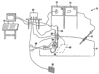
FIG. 1 is a perspective diagram of a surgical system for ablating pelvic
tumors in
accordance with an embodiment of the present invention.
FIG. 2 is a top plan view of the surgical system of FIG. 1, illustrating an
arrangement of certain equipment with respect to a patient lying on an
operating table.
FIG. 3 is a top plan view of a surgical system for ablating pelvic tumors in
accordance with another embodiment of the present invention.
FIG. 4 is a flowchart illustrating a method of ablating pelvic tumors in
accordance
with an embodiment of the present invention.
Use of the same reference symbols in different figures indicates similar or
identical
items. It is further noted that the drawings may not be drawn to scale.
DETAILED DESCRIPTION
Referring first to FIG. 1, a surgical system 10 for ablating pelvic tumors
includes a
laparoscope 12, a video monitor 14 associated with laparoscope 12, an imaging
device 16,
a video monitor 18 associated with imaging device 16, an energy source 20, and
an
ablation device 22.
Laparoscope 12, which is inserted into a patient P, is connected to a light
source
and to a video monitor 14 for displaying an image from laparoscope 12. As will
be
-4-
CA 02593988 2007-07-18
WO 2006/076181 PCT/US2006/000060
explained in greater detail below, laparoscope 12 enables a surgeon to view
the structures
in the pelvis and abdomen and the insertion and placement of ablation device
22 into a
pelvic or abdominal region of the patient. Standard recording devices such as
a VCR,
DVD, or CD recorders may be utilized to record and document laparoscopic
images.
Imaging device 16 is connected to a video monitor 18 to provide images of the
patient's pelvic region in one example. These images, which are displayed on
video
monitor 18, enable the surgeon to determine the presence and location of
pelvic tumors in
one example. In one embodiment, imaging device 16 shown in FIG. 1 includes an
imaging
probe 24, and in one example is an ultrasound machine including an intra-
abdominal
ultrasound probe. Recording devices such as a standard VCR, DVD, or CD
recorders may
be placed at various sites along the signal pathway for documentation and
storage purposes.
Instead of an intra-abdominal ultrasound probe, a transducer (not shown) may
be coupled
to the ultrasound machine for trans-abdominal ultrasound imaging. In addition,
other
imaging devices, such as an MRI machine or a CT device in conjunction with any
appropriate imaging probe, may also be used instead of an ultrasound machine.
Ablation device 22 is a sterile, electrosurgical device that includes at least
one
electrode and may include a plurality of retractable electrodes or arms 26.
FIG. 1 shows
arms 26 of ablation device 22 deployed in a pelvic tumor 28. Each arm 26 of
ablation
device 22 is a retractable curved electrode for delivering energy and has a
thermocouple
(not shown) located at the distal end. In one embodiment, a Doppler transducer
may also
be incorporated at the distal end of an electrode. Although FIG. 1 shows
ablation device 22
as including deployable arms, an ablation device without any arms may also be
used.
Alternatively, the ablation device or devices may include two or more needles
that may be
inserted into the tumor. Although the use of one ablation device is described
herein, the
use of a plurality of ablation devices is within the scope of the present
invention.
Examples of applicable ablation devices include, but are not limited to, the
Model
Electrosurgical Device and the RITA StarBurstTM XL, both available from RITA
Medical Systems, Inc. of Mountain View, California, the Cool-tipTM RF Ablation
System
electrode or cluster electrode from Valleylab of Boulder, Colorado, the LeVeen
Needle
-5-
CA 02593988 2007-07-18
WO 2006/076181 PCT/US2006/000060
Electrode from RadioTherapeutics Corporation of Sunnyvale, California, and the
R.F gel
electrode and the OPAL/OPALflex device from Prosurg Inc. of San Jose,
California.
Ablation device 22 is coupled to energy source 20, which supplies energy to
each of
the arms 26 of ablation device 22. Energy source 20 may be an RF generator in
one
example, including a standard radiofrequency generator commonly used in
surgical
procedures for cutting and coagulation. The supply of RF energy from energy
source 20 to
ablation device 22 and to a dispersive electrode 30 is controlled by an
operator control,
such as by a foot pedal 32. The application of RF energy causes an increase in
tumor
temperature. At sufficiently high temperatures, e.g., 65 degrees Celsius and
above, cell
death occurs, thereby destroying the tumor.
Examples of applicable energy sources include, but are not limited to, the
Model
500 Generator or the RITA Model 1500 RF Generator, both available from RITA
Medical Systems, Inc. of Mountain View, California, and the RF 3000 generator
from
RadioTherapeutics Corporation of Sunnyvale, California.
Energy source 20 may further include a mono-polar or bipolar energy source,
which
allows the ablation device 22 to utilize traditional mono-polar or bipolar
cautery to treat
very small, superficial tumors and to ablate the track formed during insertion
of ablation
device 22. Cauterizing the ablation device track reduces or prevents bleeding
upon
insertion or withdrawal of ablation device 22 from the organ.
As better illustrated in FIG. 2, in accordance with an embodiment of the
present
invention, the equipment of surgical system 10 is set up about the patient in
a non-
traditional arrangement. FIG. 2 illustrates the patient P lying in a dorsal
position on an
operating table 34.
A tower 36, which supports video monitor 14 for laparoscope 12 and video
monitor
18 for imaging device 16, is located proximate the patient's waist, rather
than at the foot of
operating table 34. Since the surgeon S is located on the other side of
operating table 34
across from tower 36, the surgeon S has a direct view of the monitors 14 and
18. Video
monitors 14 and 18 need not be provided on tower 36; they may be suspended
from the
ceiling and located on the other side of operating table 34 across from the
surgeon S.
-6-
CA 02593988 2007-07-18
WO 2006/076181 PCT/US2006/000060
Advantageously, during longer surgical procedures, the placement of video
monitors 14
and 18 directly across from the surgeon is more comfortable for the surgeon
and safer for
the patient, as the surgeon need not turn his/her head toward the foot of
operating table 34
to view monitors 14 and 18.
Although FIGS. 1 and 2 show separate video monitors 14 and 18 for laparoscope
12
and imaging device 16, respectively, a single monitor capable of
simultaneously displaying
multiple images from the laparoscope and the imaging device, such as a picture-
in-picture
monitor, may also be used. The single monitor would be located across the
table from the
surgeon S and may be mounted on a tower similar to tower 36, suspended from
the ceiling,
or otherwise located across the patient from the surgeon for easy viewing by
the surgeon.
Tower 36 may include additional equipment (not shown), such as an insufflation
machine, a printer, a light source, and a VCR. Tower 36 may be provided with
wheels so
that it may be easily moved about the operating room.
An additional monitor 37 for laparoscope 12 may also be provided across from a
surgical assistant A, who is seated or standing across the table from the
surgeon S, at
approximately the patient's chest level. Thus, additional monitor 37 would be
located
adjacent the surgeon S. Additional monitor 37 may be mounted on a movable
tower (not
shown), suspended from the ceiling, or otherwise appropriately located across
from
assistant A.
Imaging device 16, which is not located on tower 36, is positioned along
operating
table 34, across from the surgeon S, and toward the foot of operating table
34. For
example, imaging device 16 may be located proximate the patient's knees.
Referring now to FIG. 3, a top plan view of a surgical system 11 for ablating
pelvic
tumors is shown in accordance with another embodiment of the present
invention.
Equipment similar to that shown in system 10 of FIG. 2 is used and the same
reference
symbols indicate the same or similar items as in FIG. 2. However, in FIG. 3,
energy source
20 is provided on tower 36 and video monitor 18 for the imaging device is
provided on a
movable platform attached by an arm 17 to tower 36.
-7-
CA 02593988 2007-07-18
WO 2006/076181 PCT/US2006/000060
As shown in FIG. 3, tower 36, which includes video monitor 14, energy source
20,
an insufflation machine (not shown), a printer (not shown), a light source
(now shown),
and a VCR or digital recording device(s)(not shown), is placed proximate the
patient's
waist and across from the surgeon S. Video monitor 18 is placed on a moveable
platform
so that the surgeon S has an unobstructed view of the monitors without having
to
significantly turn the head. A surgical assistant Al is located across the
table from the
surgeon S at about the patient's chest level, with tower 36 located behind the
assistant Al
and further toward the foot of operating table 34.
A computer 19, operably coupled to energy source 20, is located next to tower
36
toward the foot of operating table 34. Computer 19 records data from the
ablation
procedure, including but not limited to temperature, power, impedance, and
length of time
of ablation. Software for recording the data is available from RITA Medical
Systems, Inc.
An assistant A2 may be proximate computer 19 to operate energy source 20
and/or
computer 19. Imaging device 16 is situated alongside operating table 34 on the
same side
as the assistant Al and toward the foot of operating table 34. The additional
monitor 37 is
positioned across from the surgical assistant Al at about the patient's chest
level. A table
21 for operative instruments may be located along the foot of operating table
34 as shown.
A method of treating pelvic tumors, in accordance with one embodiment of the
present invention, will now be described, with reference to the flow chart
illustrated in FIG.
4. This method 50 employs a laparoscopic technique for ablating pelvic tumors.
At step 51, a pre-operative evaluation is optionally performed prior to the
surgery.
In one embodiment, the pre-operative study may include physical examination of
the
uterus, ultrasound examination trans-abdominally and intra-vaginally, and/or
Doppler
imaging to analyze and to localize blood flow within the myometrium and the
tumors
themselves. Three-dimensional ultrasound mapping of the patient's pelvic area
may also
be used to image and map multiple tumors.
After the pre-operative study, the surgical procedure outlined below in
conjunction
with FIGS. 1-3 may include steps 52 through 94 although not necessarily all
the steps.
First, at step 52, the patient is prepared for laparoscopy by placing and
properly adhering
-8-
CA 02593988 2007-07-18
WO 2006/076181 PCT/US2006/000060
dispersive electrode 30 to the patient's thighs and/or lower back to allow
current from the
ablation device to be dissipated.
At step 54, the patient is placed under general anesthesia and then positioned
for
examination of the patient's pelvic region by the surgeon. Such an examination
allows the
surgeon to confirm the location of palpable tumors and portions of the
analysis from the
pre-operative study in step 51. A manipulator 38 (FIG. 1), such as a
tenaculum, is placed
on the patient's cervix, in one embodiment pinching the anterior and posterior
lips of the
cervix together so as to use the tenaculum as a uterine manipulator.
Advantageously,
manipulator 38 is not placed inside the uterus thereby reducing the likelihood
of infection
and/or complications from interaction with other instruments or energy
sources. A 14 Fr.
Foley catheter is inserted into the patient's bladder for emptying the bladder
during the
surgical procedure.
At step 56, the patient is placed in a dorsal position with her arms at her
sides,
rather than extended out as an airplane, and a blanket and a surgical drape
are placed over
the patient. This position provides the surgeon and surgical assistant with
more room to
move about. The dorsal position is also a safer position for the patient than
a frog-leg or
lithotomy position, as the dorsal position reduces the instance of nerve
injuries and
provides better circulation. In addition, the dorsal position does not require
the use of
custom drapes and stirrups. The surgical drape contains pouches for at least
one
laparoscopic cord. Serial compression devices (not shown) are placed on the
patient's legs
and activated to improve circulation throughout the surgical procedure and
reduce the
possibility of thrombosis. In addition, the patient may be placed in a bear
hugger system
(not shown) to assist in the maintenance of the patient's body temperature
while under
general anesthesia.
At step 58, in one embodiment of the present invention, the equipment is
advantageously arranged about operating table 34. FIGS. 2 and 3 illustrate two
embodiments of a surgical system for ablating pelvic tumors which system is
described
above with respect to FIGS. 2 and 3.
At step 60, the surgeon S then makes an infra-umbilical or sub-umbilical or
other
abdominal incision for safe and advantageous placement of a Verres needle and
-9-
CA 02593988 2007-07-18
WO 2006/076181 PCT/US2006/000060
laparoscopic trocar and sleeve. The Verres needle is then inserted through the
incision and
into the peritoneal cavity in the standard technique of laparoscopy. The
insufflation
machine is then used to insufflate the abdomen with carbon dioxide gas until
the abdominal
pressure is approximately 14-20 mm Hg, thereby providing the surgeon with a
window for
visualization.
Next, at step 62, a 3, 5, 7, or 10 mm trocar and sleeve are inserted through
the infra-
umbilical or sub-umbilical incision. The trocar is then removed and
laparoscope 12 is
inserted into the sleeve. Laparosope 12 and monitor 14 are then used to verify
correct
placement of laparoscope 12 within the peritoneal cavity and the absence of
any trauma.
The sleeve is attached to the carbon dioxide gas supply and includes a valve
for controlling
the abdominal pressure of the peritoneal cavity.
Steps 60 and 62 discussed above describe a closed laparoscopy procedure. For
those patients, for whom the surgeon feels an open laparoscopy would be
advantageous,
the surgeon would make an infra- or sub-umbilical incision and use a
combination of blunt
and sharp dissection through subcutaneous tissue. The surgeon would then
utilize
retractors for exposure. When the fascia is visualized, it is grasped with one
or more
clamps, elevated, and incised. This provides a view of the peritoneum below,
which may
be bluntly or sharply incised. An appropriate laparoscopic sleeve is then
placed, and the
abdomen is insufflated with carbon dioxide gas. The laparoscope is then
inserted into the
peritoneal cavity through the sleeve.
At step 64, the surgeon then uses laparoscope 12, while palpating a top of the
uterine fundus, to determine an optimal location for an intra-abdominal
ultrasound probe.
The optimal location is generally at the most cephalad extent of the uterus in
the midline,
rather than supra-pubic or lateral. This usually provides the best vantage
point from which
to visualize all surfaces of the uterus. Other locations may be utilized if
deemed
appropriate by the operating surgeon.
An incision is then made at this location and a 3, 5, 7, or 10 mm trocar and
sleeve
are inserted. The trocar is removed and imaging probe 24 is inserted into the
sleeve. By
way of example, the imaging probe 24 may be an Aloka model no. UST-5526L-7.5
MHz
probe for use with an Aloka model no. SSD 1400 ultrasound machine. Imaging
probe 24
-10-
CA 02593988 2007-07-18
WO 2006/076181 PCT/US2006/000060
transmits a signal to imaging device 16 which then displays an image of the
pelvic region
on video monitor 18 based upon the signal. Thus, the surgeon may
simultaneously view
the images on video monitors 14 and 18. As discussed above, a single monitor
that
simultaneously displays images from laparoscope 12 and imaging device 16 may
be used
instead of separate monitors 14 and 18.
At step 66, the surgeon examines the entire pelvis and abdomen to confirm the
presence or absence of any visible pathological conditions. The surgeon also
uses
laparoscope 12 and imaging probe 24 to visualize any tumors, such as uterine
leiomyomata. In particular, the surgeon takes note of the number, location,
and size of all
tumors, and compares that information with previously acquired data from the
pre-
operative evaluation in step 51 and the pelvic examination in step 54.
Advantageously,
imaging probe 24 allows for uterine manipulation and imaging in real-time.
At step 68, the surgeon formulates and/or modifies the existing ablation plan
and
determines an order for treating the tumors. This order is determined based on
the sizes
and locations of the various tumors, and whether or not the tumors are
accessible from a
single point of insertion of the ablation device or if multiple locations are
required. For
example, if two tumors are generally along the same track of ablation device
22, the
surgeon may first ablate the deeper tumor and, upon retraction of ablation
device 22, ablate
the remaining tumor. In addition, in larger tumors requiring multiple
overlapping
ablations, the surgeon may choose to ablate first a portion of the tumor that
is furthest away
from the vasculature of that tumor and work toward the vasculature, or vice
versa.
At step 70, the surgeon tests ablation device 22 to ensure that it is
operating
properly. Ablation device 22 is connected to energy source 20, and proper
feedback from
the thermocouples, if any, is observed. In particular, the surgeon operates
foot pedal 32, or
any other appropriate operator control, to activate the supply of RF energy
from energy
source 20 and notes an appropriate rise in temperature and any peaks. The
ablation device
22 may also be flushed with saline prior to use to keep the electrode cool and
to reduce
char formation.
At step 72, the surgeon makes an incision, approximately 2.5 to 3.0 mm long,
where suitable to ablate the first tumor, and inserts ablation device 22 into
the abdomen.
-11-
CA 02593988 2007-07-18
WO 2006/076181 PCT/US2006/000060
Entry of ablation device 22 is observed using laparoscope 12. The surgeon uses
imaging
probe 24 to visualize the size and location of the tumors with respect to
ablation device 22.
Whenever possible, the placement and use of ablation device 22 is correlated
with imaging
probe 24 such that the electrode of ablation device 22 and the transducer of
imaging probe
24 are substantially parallel to one another for the most effective placement
of the ablation
device 22. Imaging probe 24 may not be substantially parallel to ablation
device 22 in all
instances.
Next, at step 74, tenaculum 38 and imaging probe 24 are utilized to position
and
stabilize the uterus. In other embodiments, other uterine manipulators may be
used to
manipulate and stabilize the uterus. Attachment of a stabilization device
which punctures
or injures the serosal surface of the uterus is generally to be avoided.
Advantageously, the
position of the imaging probe 24, the ablation device 22, and the uterus are
controlled
without puncturing the uterus multiple times in accordance with the present
invention.
At step 76, after the surgeon has stabilized the uterus and located the
tumors, the
surgeon guides ablation device 22 into a wall of the uterus. In one
embodiment, the
surgeon may press or tap against the uterus with the ablation device without
penetration to
verify position of the entry point with the imaging device. The surgeon may
guide ablation
device 22 by changing the position of the uterus relative to ablation device
22 as noted
above. In addition, the surgeon may rotate the ablation device for better
penetration of the
uterine wall with less movement of the uterus. Rotation of the ablation device
is also
beneficial for penetration into and exiting from higher density tumors.
Insertion of the
ablation device may also be performed while applying energy to the needle to
coagulate the
track and to increase ease of insertion. Ablation device 22 has a plurality of
markings (not
shown) that enable the surgeon to note the depth of penetration of device 22.
Confirmation
of the location and placement of ablation device 22 are provided by
laparoscope 12 and
imaging probe 24. Imaging probe 24 is utilized in multiple planes (e.g.,
sagital and
transverse) to confirm the position of the electrode associated with ablation
device 22.
Next, at step 78, the surgeon advances the tip of ablation device 22 to an
appropriate depth for treating a tumor. In doing so, the needle makes only a
very small
puncture. For example, an ablation device having a needle of 16 gauge may
produce a
-12-
CA 02593988 2007-07-18
WO 2006/076181 PCT/US2006/000060
puncture site of approximately 1 mm to 2 mm in diameter. The appropriate depth
depends
upon the size of the tumor and characteristics of the ablation device 22. The
operator may
elect to position the ablation device 22 in such a manner as to most
effectively ablate the
blood supply of the tumor based on findings from pre-operative as well as
intra-operative
imaging studies such as Doppler ultrasound. When ablation device 22 has been
inserted to
the appropriate depth, arms 26 of ablation device 22 are deployed to the
appropriate extent
in the tumor 28, as illustrated in FIG. 1. Visualization via the imaging probe
24 and the
laparoscope 12 are used to ensure that all of the arms 26 remain within the
confines of the
tumor and do not extend outside of the organ. Arms 26 may effectively anchor
ablation
device 22 in tumor 28.
At step 80, the surgeon then records a baseline temperature of the tumor,
usually
39-42 degrees Celsius. The temperature of the tumor is obtained by the
thermocouples
located at the distal ends of arms 26 of ablation device 22.
At step 82, the surgeon then ablates the tumor by supplying RF energy from
generator 20 to ablation device 22. While generator 20 is activated, it is
important to
monitor the temperature or impedance at all parts of the ablation device. If
the temperature
or impedance at any part of ablation device 22 is abnormal, it could indicate
that that part
of the device is external to the organ.
RF energy is supplied to the tumor to raise the temperature of the tumor, such
that it
is in the range of between approximately 65 C and 100 C, for about 14
minutes in one
example. Cell death occurs at a temperature of about 65 C. However, a
preferred target
temperature range for ablating pelvic tumors is between 85 C and 100 C to
promote
conduction of heat throughout the ablation zone. For smaller tumors the target
time may be
between approximately 3 minutes and 10 minutes. One of ordinary skill in the
art,
however, will appreciate that ablation times of less than 3 minutes may also
be adequate.
In one embodiment, a particularly dense tumor may be pre-heated to condition
the
tumor for ease of penetration and achievement of full deployment of the
electrode resulting
in maximal ablation volume. To pre-heat the tumor, the ablation device
electrode may be
deployed until significant resistance is encountered to further deployment.
The generator is
then activated and the tumor pre-heated to a temperature below the target
temperature.
-13-
CA 02593988 2007-07-18
WO 2006/076181 PCT/US2006/000060
Once the tumor and ablation arms 26 are heated, the ablation device is further
deployed to
a greater extent as desired. Multiple stages of heating may be required in
extreme cases to
achieve optimal deployment. In one example, if the deployment length desired
is about 4
cm but the tumor is of a high density and deployment is difficult, the arms
may be
deployed to about 2 cm and the tumor area pre-heated to approximately 90
degrees Celsius
to soften the fibroid and heat the arms 26. The arms may then be more easily
and
effectively further deployed in the heated fibroid to the desired length for
full achievement
of the ablation volume of the ablation device. Conventional ablation has
disadvantageously deployed cold electrodes into an unheated target tumor for
ablation.
The ablation device 22 may also be inserted into the tumor or organ while
activated
to facilitate placement into a particularly dense tumor or organ. In another
embodiment,
the electrode may be deployed in conjunction with withdrawal of the shaft to
modify the
shape of the ablation volume from spherical to ovular to advantageously ablate
non-
spherical shaped tumors.
The temperature of the tumor at various sites, as provided by the
thermocouples, is
monitored and recorded. Thus, at least a baseline starting temperature, half-
time
temperature, and end-of-ablation-period temperature are recorded for each
ablation. While
RF energy is being delivered to the tumor, the surgeon observes monitors 14
and 18 to
ensure that none of the arms 26 of ablation device 22 extends beyond the
tumor. The
uterus may contract during ablation, causing arms 26 of ablation device 22 to
project from
the tumor and contact normal tissue, which may be damaged by the RF energy.
When the
tumor has been sufficiently ablated, energy from the energy source 20 is
discontinued.
Periodically between ablations, as shown by step 84, the uterus is irrigated
with
fluid such as normal saline or Lactated Ringers. The fluid keeps the serosa
moist and
prevents drying as a result of the carbon dioxide gas that is infused into the
abdomen
during laparoscopy. Irrigation also removes blood, thereby inhibiting
formation of fibrin
which could trigger adhesion formation.
If the tumor is larger than the ablation capacity of the given ablation
device, then at
step 86, the surgeon may need to reposition ablation device 22 within another
part of the
tumor and reapply RF energy, repeating steps 76 through 84. Thus, if the
tumors are
-14-
CA 02593988 2007-07-18
WO 2006/076181 PCT/US2006/000060
greater in size than the ablation capacity of ablation device 22, or if
suboptimal deployment
or placement of the ablation device occurs, multiple ablations which may
overlap may be
necessary to ablate the bulk of the tumor. For tumors less than 5 cm, however,
a single
application of the RF energy is usually sufficient. Ablation devices are
currently available
which can potentially achieve a 7 cm ablation with a single application of
energy.
The rate at which power is delivered to the ablation device 22 may be
controlled to
optimize ablation efficiency. Power may be delivered over time until a target
temperature is
reached in such a manner that charring or tissue dehydration proximate arms 26
is
inhibited. Charring around the arms 26 is undesirable as it inhibits
conduction of heat and
may result in incomplete or irregular ablations. With small tumors, power may
be
delivered more quickly via manual power control, thus achieving target
temperatures faster
thereby decreasing overall operative time. Subserosal fibroids may also be
treated without
deployment of electrodes but with an active tip of the ablation device shaft,
similar to a
track ablation mode. Accordingly, three methods of ablation may be used,
including but
not limited to using an algorithm for the application of power to the ablation
device,
heating with manual power control of the energy generator, and destruction of
tumors with
no deployment of the electrode.
An intra-abdominal Doppler study may be performed after the ablation to
confirm
effective cessation of blood flow to the tumor. At least one Doppler
transducer
incorporated at the distal end of ablation device 22 or imaging probe 24 may
be used to
perform the Doppler analysis. In one embodiment, the ultrasound probe can
transmit
ultrasound for imaging as well as for Doppler analysis.
In a further embodiment, based upon the Doppler analysis, imaging probe 24 may
then be used to occlude the uterine arteries by physical pressure against the
arteries of
interest where they insert into the uterus. Accordingly, blood flow through
the uterus may
be decreased, thereby reducing the cooling effect from blood circulation and
thus more
efficiently ablating the fibroid.
In yet another embodiment, once the arms of the ablation device are deployed
and
anchored in the fibroid, the uterus may be suspended or lifted by elevating
the ablation
device 22 to examine pelvic areas otherwise hidden from view.
-15-
CA 02593988 2007-07-18
WO 2006/076181 PCT/US2006/000060
At step 88, the surgeon then repositions ablation device 22 at the next tumor.
The
surgeon may leave ablation device 22 in the same track if the next tumor is
along the same
line of approach. The surgeon would retract arms 26, advance or withdraw
ablation device
22 as needed, and then insert ablation device 22 into the next tumor. The
surgeon would
then repeat the ablation sequence of step 78 through step 86 described above.
If the subsequent tumor is in a different location, the surgeon may retract
arms 26 of
ablation device 22 and withdraw ablation device 22 from the uterus, while
applying a
mono-polar cautery to reduce or prevent bleeding from the ablation device
track.
Alternatively, rather than completely withdrawing ablation device 22 and re-
inserting
ablation device 22 at a new point of entry, repeating steps 72 through 86, the
surgeon may
withdraw ablation device 22 until it is only 0.5 cm to 1 cm deep within the
uterus and
adjust the uterus until the desired angle of approach is obtained. The
ablation device 22
may then be inserted into the new tumor as previously described.
Advantageously,
imaging probe 24 and uterine manipulator 38 may be used to adjust the uterus.
In the case of multiple ablations, the ablation device may be removed entirely
from
the patient and cleaned with water and a soft brush to remove from the needle
shaft and
arms 26 adherent debris and tissue.
Small, superficial, subserosal fibroids (e.g., less than 1 cm) may be ablated
using
different techniques. In one example, a mono-polar cautery may be used at step
90.
Bipolar paddles may also be used if the fibroid extends from the wall of the
uterus.
Similarly, if the tumor is pedunculated, the surgeon may treat and/or incise
the stalk.
Mono-polar or bipolar cautery may be applied to subserosal, intramural, and
submucuos
leiomyomata. In addition, other pelvic pathologies are treated as appropriate.
After all of the tumors have been ablated, at step 92, the surgeon confirms
hemostasis, withdraws ablation device 22, and applies a mono-polar cautery
with ablation
device 22 to the puncture sites, if necessary. A small amount of irrigation
fluid may be left
in the pelvis.
Finally, at step 94, documentation, including videotapes, ultrasound
photographs,
and photographs from the laparoscope are obtained. The sleeves are opened to
allow the
-16-
CA 02593988 2007-07-18
WO 2006/076181 PCT/US2006/000060
escape of the carbon dioxide gas and a local anesthetic agent is injected into
the skin
incisions. The surgeon then repairs the fascia of the 10 mm incision using an
absorbable
suture and S-retractors to facilitate visualization. AlisTM clamps or other
atraumatic clamps
are used to grasp and elevate the fascial edges for suturing. The skin and
subcutaneous
tissue are closed in the standard fashion. The surgeon then removes the
dispersive
electrode 30 and the foley catheter and examines the surrounding skin.
The patient is transported to a recovery room, where she will remain until she
is
tolerating liquids, ambulating with assistance, and voiding adequately.
If the patient's uterus is very large (e.g., 16 weeks or greater), the above-
described
laparoscopic technique may be less effective. Accordingly, a direct trans-
abdominal
insertion of ablation device 22 may be performed with predominately
laparoscopic
confirmation (e.g., minimal intra-abdominal ultrasound confirmation). In this
method the
patient is prepared in the same manner as that described above at step 52. The
surgeon also
performs a pelvic examination, positions the patient, places a foley catheter
and serial
compression devices, arranges the equipment, makes an infra-umbilical
incision, insufflates
the patient's abdomen, and inserts laparoscope 12, as in step 54 through to
step 62 above.
Specifically, the surgeon inspects the abdomen and documents the presence or
absence of
bowel adhesions or other pathologic conditions that would render this method
inappropriate without surgical correction. Lysis of abdominal adhesions may be
performed
as needed to establish normal anatomy. Radiofrequency ablation may then be
performed as
follows.
Next, the surgeon releases the COa gas from the patient's abdomen, allowing
the
abdominal wall to contact an anterior portion of the uterus. A sterile cover
drape over a
transducer allows for trans-abdominal ultrasound imaging using a non-sterile
transducer
(not shown). The ultrasound is used to locate and measure the tumors.
The surgeon then makes an incision for ablation device 22 and inserts ablation
device 22 percutaneously and trans-abdominally, using trans-abdominal
ultrasonography to
guide its insertion.
-17-
CA 02593988 2007-07-18
WO 2006/076181 PCT/US2006/000060
Ablation device 22 is positioned at a tumor and arms 26 are deployed in the
tumor,
just as described above with respect to the laparoscopic method. Prior to
applying RF
energy to the tumor, the surgeon insufflates the abdomen and performs a
laparoscopy to
confirm that none of the arms 26 of ablation device 22 extend beyond the
uterine tissue.
The surgeon then applies RF energy to the tumor, in the same manner as
described
at step 80 through step 84 above, including recording the baseline, half-time,
and end-of-
ablation-period temperatures. The surgeon may use the same approach as
described above
to ablate multiple pelvic tumors. Upon withdrawal of the ablation device 22,
the surgeon
fulgurates the ablation device track with a mono-polar cautery under
visualization through
the laparoscope. Thus, remaining steps are the same as step 86 through step 94
described
above.
The above-described methods enable the surgeon to ablate substantially all of
a
tumor from a single, ablation device puncture site but multiple punctures may
be necessary
for larger tumors. In addition, depending on the location of the tumors,
multiple tumors
may be ablated from a single puncture site. The methods fiurther enable the
surgeon to treat
all sizes of tumors in any area of the pelvic and/or abdominal region.
The above-described embodiments of the present invention are merely meant to
be
illustrative and not limiting. Various changes and modifications may be made
without
departing from this invention in its broader aspects. For example, although
the present
invention has been described with respect to the treatment of uterine
leiomyomata, the
present invention may also be used to treat other pelvic tumors, such as those
present in the
ovaries. The present invention may be performed using a trans-cervical, a
hysteroscopic,
or a trans-vaginal technique, in addition to the laparoscopic and trans-
abdominal techniques
described above. Therefore, the appended claims encompass all such changes and
modifications as falling within the true spirit and scope of this invention.
-18-