Note: Descriptions are shown in the official language in which they were submitted.
CA 02594489 2007-07-09
WO 2006/078703 PCT/US2006/001717
1
METHOD AND APPARATUS FOR ARRHYTHMIA DETECTION IN A
MEDICAL DEVICE
FIELD OF THE INVENTION
The present invention relates generally to medical devices, and, more
particularly, to a method and apparatus for sensing and detecting arrhythmias
in a
medical device.
BACKGROUND OF THE INVENTION
Implantable medical devices (IMDs) have many functions including the
delivery of therapies to cardiac patients, neuro-stimulators, muscular
stimulators, and
others. For purposes of this application reference will be made only to
implantable
cardiac devices, it being understood that the principles herein may have
applicability to
other implantable medical devices as well.
An implantable cardiac device (ICD) may be a device commonly referred to as
a pacemaker, which is used to stimulate the heart into a contraction if the
sinus node of
the heart is not properly timing, or pacing, the contractions of the heart.
Modern cardiac
devices also perform many other functions beyond that of pacing. For example,
some
cardiac devices may also perform therapies such as defibrillation and
cardioversion as
well as providing several different pacing therapies, depending upon the needs
of the
user and the physiologic condition of the user's heart. For convenience, all
types of
implantable cardiac devices will be referred to herein as ICDs, it being
understood that
the term, unless otherwise indicated, is inclusive of an implantable device
capable of
administering any of a number of therapies to the heart of the user.
In typical use, an ICD is implanted in a convenient location usually under the
skin of the user and in the vicinity of the one or more major arteries or
veins. One or
more electrical leads connected to the pacemaker are inserted into or on the
heart of the
user, usually through a convenient vein or artery. The ends of the leads are
placed in
contact with the walls or surface of one or more chambers of the heart,
depending upon
the particular therapies deemed appropriate for the user.
CA 02594489 2007-07-09
WO 2006/078703 PCT/US2006/001717
2
One or more of the leads is adapted to carry a current from the pacemalcer to
the
heart tissue to stimulate the heart in one of several ways, again depending
upon the
particular therapy being delivered. The leads are simultaneously used for
sensing the
physiologic signals provided by the heart to determine when to deliver a
therapeutic
pulse to the heart, and the nature of the pulse, e.g., a pacing pulse or a
defibrillation
shock.
There has been recent interest in development of implantable defibrillators
that
may be inserted entirely subcutaneously or sub-muscularly, having no leads or
electrodes within the thoracic cavity. The elimination of transvenous or
epicardial
leads is believed likely to allow for implant of the devices by a wider range
of
physicians, in some cases at a lower cost than traditional ICDs. Absence of
transvene
or epicardial leads may reduce acute and long term complications. Such
devices, are
therefore believed to offer the opportunity for increased levels of use,
particularly for
prophylactic implant. US Application Publication Nos. 2002/0042634,
200200068958
and 2002/0035377 to Bardy et al., are exemplary of current thinking with
regard to
such subcutaneous ICDs. Additional subcutaneous ICDs are disclosed in US
Application Publication No. 20020082658 by Heinrich et al. and PCT publication
WO/04043919A2 by Olson. All of the above cited applications and publications
are
incorporated herein by reference in their entireties.
One potential problem associated with the sensing of the physiologic signal
from the heart in both the transvenous systems and the subcutaneous systems
relates to
what is often referred to as "false positive" and "false negative" detections.
The most
widely accepted detection algorithm is based on the rate of depolarizations of
the
ventricles, or simply on "heart rate". Such algorithms rely on detecting
events based
upon signals obtained between two electrodes positioned within or on the
heart. If the
number of detected events per a given time is greater than a preset value,
then the
device charges an energy storage capacitor and then shocks the heart;
otherwise no
shock is delivered.
CA 02594489 2007-07-09
WO 2006/078703 PCT/US2006/001717
3
BRIEF DESCRIPTION OF THE DRAWINGS
Aspects of the present invention will be readily appreciated as they become
better understood by reference to the following detailed description when
considered in
connection with the accompanying drawings, wherein:
FIG. 1 is a schematic diagram of an exemplary medical device according to the
present invention;
FIG. 2 is a schematic diagram of an exemplary medical device according to the
present invention;
FIG. 3 is a schematic diagram of an exemplary medical device according to the
present invention;
FIG. 4 is a schematic diagram of an exemplary medical device according to the
present invention;
FIG. 5 is a top cross sectional view illustrating the positioning of a medical
device according to an embodiment of the present invention;
FIG. 6 is a top cross sectional view illustrating the positioning of a medical
device according to an embodiment of the present invention;
FIG. 7 is a schematic view of a sensor of a medical device according the
present
invention;
FIG. 8 is a functional schematic diagram of an implantable
pacemaker/cardioverter/defibrillator (ICD) in which the present invention may
usefully
be practiced;
FIG. 9 is a schematic diagram of sensing of depolarization events utilizing a
medical device of the present invention;
FIGS. 10 and 11 are schematic diagrams of sensing of depolarization events
utilizing a medical device of the present invention;
FIG. 12 is a flowchart of a method for detecting arrhythmias in a medical
device
according to an embodiment of the present invention;
FIG. 13 is an exemplary illustration of determining change in duration for a
current rhythm according to an embodiment of the present invention; and
FIGS. 14A-14C are schematic diagrams of electrode configurations in an
exemplary medical device according to the present invention.
CA 02594489 2007-07-09
WO 2006/078703 PCT/US2006/001717
4
DETAILED DESCRIPTION OF THE INVENTION
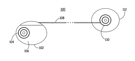
FIG. 1 is a schematic diagram of an exemplary medical device according to the
present invention. As illustrated in FIG. 1, a medical device 100 according to
an
embodiment of the present invention, which may be an implantable
cardioverter/defibrillator (ICD), for example, includes a housing 102 having
an
electrode 104 positioned along a side wall 106 of housing 102 that is intended
to be
directed inward towards a heart of a patient when housing is positioned
subcutaneously
within the patient, as will be described in detail below. Housing 102 is
coupled to a
subcutaneous lead 108 carrying conventional conductors (not shown) extending
therethrough to electrically couple circuitry located within housing 102 to an
electrode
110 positioned on an insulated patch 112 positioned subcutaneously within the
patient
so that electrode 110 is directed towards the patient's heart.
According to the present invention, electrodes 104 and 110 are formed using
Laplacian electrodes that are utilized both as sensors to sense cardiac
depolarization-
signals and as high voltage cardioversion/defibrillation electrodes to deliver
cardioversion/defibrillation therapy to the patient. Since the sensitivity of
Laplacian
sensors to events, especially to dipole layers corresponding to the
depolarization of the
heart, decreases with the inverse distance cube (1/r3 ), electrodes 104 and
110 sense
signals in a very localized and reduced area, resulting in larger cardiac
signal to noise
ratios than in conventional sensing methodologies. In addition, because of the
reduced
sensing area, noise due to body motion will only intermittently affect signal
quality
when local muscles are activated during the body motion.
FIG. 2 is a schematic diagram of an exemplary medical device according to the
present invention. As illustrated in FIG. 2, a medical device 200 according to
another
embodiment of the present invention includes a housing 202 having an electrode
204
positioned along a side wall 206 of housing 202 that is intended to be
directed inward
towards the heart of a patient when housing 202 is positioned subcutaneously
within
the patient. Housing 202 is coupled to two subcutaneous leads 208 and 209,
each
carrying conventional conductors (not shown) extending therethrough to
electrically
couple circuitry located within housing 202 to respective electrodes 210 and
211
positioned on associated insulated patches 212 and 213 that are to be
positioned
CA 02594489 2007-07-09
WO 2006/078703 PCT/US2006/001717
subcutaneously within the patient so that electrodes 210 and 211 are directed
towards
the patient's heart.
FIG. 3 is a schematic diagram of an exemplary medical device according to the
present invention. As illustrated in FIG. 3, a medical device 300 according to
the
5 present invention may include a housing 302 having two electrodes 304 and
305
positioned along a side wall 306 of housing 302 that is intended to be
directed inward
towards the heart of a patient when housing 302 is positioned subcutaneously
within
the patient. Housing 302 is coupled to a subcutaneous lead 208 carrying
conventional
conductors (not shown) extending therethrough to electrically couple circuitry
located
within housing 302 to an electrode 210 positioned on an insulated patch 212
that is
intended to be positioned subcutaneously within the patient so that electrode
210 is
directed towards the patient's heart.
FIG. 4 is a schematic diagram of an exemplary medical device according to the
present invention. As illustrated in FIG. 4, a medical device 400 according to
the
present invention may include a housing 402 having two electrodes 404 and 405
positioned along a side wa11406 of housing 402 that is intended to be directed
inward
towards the heart of a patient when housing 402 is positioned subcutaneously
within
the patient. Housing 402 is coupled to subcutaneous leads 408 and 409, each
carrying
conventional conductors (not shown) extending therethrough to electrically
couple
circuitry located within housing 402 to electrodes 410 and 411 positioned on a
second
housing 412 that is intended to be positioned subcutaneously within the
patient so that
electrodes 410 and 411 are directed towards the patient's heart. According to
yet
another embodiment, electrodes 410 and 411 are positioned on an insulated
patch 412
so that housing 402 is coupled to insulated patch 412 via leads 408 and 409.
FIG. 5 is a top cross sectional view illustrating the positioning of a medical
device according to an embodiment of the present invention. It is understood
that the
present invention is not intended to be limited to the exemplary electrode
configurations of FIGS. 1-4. Rather, any desired number of electrodes may be
located
on the housing and any number of insulated patches containing any number or
array of
electrodes may be coupled to the housing via corresponding leads. In addition,
the
electrodes may be utilized only for pacing and/or only for sensing without
departing
CA 02594489 2007-07-09
WO 2006/078703 PCT/US2006/001717
6
from the invention. Furthermore, placement of the housing and electrodes will
depend
upon the number of electrodes utilized.
For example, as illustrated in FIG. 5, in the three electrode embodiment of
the
present invention illustrated in FIG. 2, housing 202 is positioned along side
of costal
muscle 220 along the abdomen below the sternum so that electrode 204
positioned
along side wall 206 of housing 202 is directed inward towards the heart 215 of
a
patient. According to the present invention, housing 202 may or may not
include one
or more electrodes, as described above. In addition, one insulated patch 212
is
positioned in the anterior thorax, overlaying the heart, slightly left of the
sternum and
within the fourth intercostal space to be positioned at a location associated
the V4 lead
of the twelve-lead ECG position so that electrode 210 is directed inward
towards heart
215. The other insulated patch 213 is positioned laterally left of the sternum
from
insulated patch 212 to be located at the V6 lead location of the twelve-lead
ECG
position so that electrode 211 is directed inward towards heart 215. In this
way,
electrodes 210 and 211 are positioned so that a vector extending between
electrodes
210 and 211 extends through an appropriate portion of heart 215.
FIG. 6 is a top cross sectional view illustrating the positioning of a medical
device according to an embodiment of the present invention. As illustrated in
FIGS. 1
and 6, in the two electrode embodiment of the present invention, housing 102
is
positioned in the anterior thorax, overlaying the heart, slightly left of the
sternum and
within the fourth intercostal space to be positioned at a location associated
the V4 lead
of the twelve-lead ECG position so that electrode 104 is directed inward
towards heart
215. Insulated patch 112 is positioned laterally left of the sternum from
housing 102 to
be located at the V61ead location of the twelve-lead ECG position so that
electrode 110
is directed inward towards heart 215. In this way, electrodes 104 and 110 are
positioned so that a vector extending between electrodes 104 and 110 extends
through
an appropriate portion of heart 215.
According to the present invention, an insulated layer 225 may be included
along an outer portion of the insulated patches in order to reduce the effects
of the
current delivered from the electrodes on subcutaneous nerves along electrodes,
resulting in a reduction of pain that may be experienced by the patient during
delivery
of cardioversion/defibrillation therapy by the medical device.
CA 02594489 2007-07-09
WO 2006/078703 PCT/US2006/001717
7
FIG. 7 is a schematic view of a sensor of a medical device according the
present
invention. As illustrated in FIG. 7, an electrode 300 of the present
invention, such as
any of the electrodes described above, includes a disk-shaped, electrically
insulating
patch 302 formed of silicone rubber or other compliant, electrically
insulating material.
Insulating patch 302 defines a generally planar contact surface 304 into which
an
electrically conductive sub-assembly 307 is substantially flushly embedded.
Sub-
assembly 307 includes an electrically conductive inner pad 308 separated by a
first
insulating layer 310 from a continuous middle loop electrode 312. Loop
electrode 312
is surrounded by a second insulating layer 314, which, in turn, is surrounded
by a
continuous exterior loop electrode 316. In order to provide electrode 300 with
uniform
directionality of response in a plane during sensing, inner pad 308 is round
and circular
in shape and loop electrodes 312 and 316 are formed as circular rings located
concentrically with respect to pad 308 as well as with respect to one another.
In
applications where uniform directionality of response is not required or where
it is
desired to provide enhanced or reduced sensitivity in certain directions, pad
308 and/or
loop electrodes 312 and 316 can be formed in other shapes and/or located off
center
with respect to one another provided that at least some mutual spacing is
maintained
between pad 308 and continuous loop electrodes 312 and 316.
In order to render electrode 300 sensitive only to the electrical activity of
that
muscle tissue which substantially immediately underlies the skin surface which
sub-
assembly 307 is placed in contact with, inner pad 308 and electrode 316 are
electrically
coupled to one another, preferably by a short circuit. For example, this is
achieved by a
jumper wire 318 having one end connected to pad 308 and its opposing end
connected
to electrode 316. The portion of jumper wire 318 that crosses electrode 312 is
electrically insulated in order to electrically isolate electrode 312 from
both pad 308
and electrode 316. Electrode 300 is provided with a pair of insulated lead
wires 320,
322. A conductor 324 extending through lead wire 320 is connected directly to
electrode 312 while a conductor 326 extending through lead wire 322 is
connected
electrically in common with both inner pad 308 and electrode 316. This is
conveniently accomplished with a single electrical connection 328 by attaching
the
conductor 326 of lead wire 322 directly to jumper wire 318. To avoid
detachment of
lead wires 320 and 322 from sub-assembly 307, strain relief is preferably
provided by
CA 02594489 2007-07-09
WO 2006/078703 PCT/US2006/001717
8
anchoring wires 320 and 322 to patch 302. This may readily be achieved by
insert
molding or use of an adhesive. Concentricity of pad 308 and electrodes 312 and
316
may be assured by various means, molding or performing patch 302 with a recess
for
locating pad 308 and appropriately sized and centered channels for receiving
electrodes
312 and 316.
Although a single pad and two concentric rings are shown in FIG. 7, it is
understood that other configurations are intended to be included in
subcutaneous
sensing and detecting according to the present invention. For example, sensor
300 may
include inner pad 308 and a single concentric electrode 316, thus eliminating
the need
for electrode 312, insulating layer 314 and jumper wire 318. The benefit of
such a bi-
polar configuration is that it provides increased signal amplitudes, although
the signal
localization may be reduced.
As the distance between the electrodes 312 and 316 increases, or in the bi-
polar
configuration, as the distance between pad 308 and electrode 316 increases,
the
amplitude of the detected signal increases, and the sensor 300 becomes more
sensitive
to sources further away from the immediate vicinity of the electrode 300.
Although the
desired total radius of the sub-assembly 307 associated with the sensor
typically will be
dependent on the patient's anatomy, the inventors have found that a for a
person of
median anatomy, sub-assembly 307 should be approximately between 10 mm and 70
mm in diameter, for example. According to an embodiment of the present
invention,
sub-assembly 307 is approximately 35 mm in diameter. In one embodiment, a
distance
330 between electrodes 312 and 316, in which insulating layer 314 is located,
is
approximately equal to 2 mm, although distance 330 could have any desired
value,
depending upon the level of far-field sensitivity desired.
It is understood that while patch 302 is shown having a circular shape, the
present invention is not intended to be limited to the use of circular patches
and
electrodes. Rather, the patch may be formed in any shape, including oval,
square,
rectangular and so forth. In addition, while electrodes 312 and 316 are shown
as being
concentric and circular, they may have other desired shapes without departing
from the
present invention.
FIG. 8 is a functional schematic diagram of an implantable
pacemaker/cardioverter/defibrillator (ICD) in which the present invention may
usefully
CA 02594489 2007-07-09
WO 2006/078703 PCT/US2006/001717
9
be practiced. This diagram should be taken as exemplary of the type of device
in which
the invention may be embodied, and not as limiting, as it is believed that the
invention
may usefully be practiced in a wide variety of device implementations,
including
devices providing therapies for treating atrial arrhythmias instead of or in
addition to
ventricular arrhythmias, pacemakers which do not provide anti-tachycardia
pacing
therapies, anti-tachycardia pacers which do not provide cardioversion or
defibrillation,
and totally subcutaneous devices that deliver defibrillation and/or pacing
therapies or
subcutaneous monitoring-only devices that do not provide therapy. Most of the
components of the ICD as illustrated correspond to those used in prior art
Medtronic
implantable defibrillators. In particular, reference is made to US Patent
Publication No.
20020082658 to Heinrich et al. and PCT Publication No. WO/04043919A2 to Olson,
as
well as to US Patent Application Publication No. 20010034539 by Olson et al.,
all
incorporated herein by reference in their entireties. While the circuitry
described above
is based upon implantable device circuitry, similar circuitry would be used in
those
embodiments in which the invention is practiced as an external pacemaker or
defibrillator, coupled to a subcutaneous electrode array according to the
present
invention.
The device is provided with electrodes, which may be as described above.
Alternate lead systems embodying the invention may also be substituted. The
functions
of the illustrated electrodes are as follows: Electrode 311 is a first
defibrillation/cardioversion electrode and corresponds to electrodes 104, 204,
304, 404
and 408, located on the device housings, for example. Electrode 320 is a
second
cardioversion/defibrillation electrode and corresponds to the lead mounted
cardioversion/defibrillation electrodes 110, 210, 410, 411, for example.
Electrode 318
corresponds to the optional third defibrillation electrode referred to in
conjunction with
FIGS. 2-4. As such, there may be more or less than the three electrodes
illustrated,
which are intended to merely be exemplary.
Electrodes 311, 318 and 320 are coupled to high voltage output circuit 234 and
switch matrix 208, which under control of microprocessor 224 selectively
couples
electrodes 311, 318 and 320 to sensing circuit 204 and/or to pacing output
circuits 216
and 214. Sensing circuit 204 preferably takes the form of one or more
automatic gain
controlled amplifiers providing adjustable sensing threshold as a function of
the
CA 02594489 2007-07-09
WO 2006/078703 PCT/US2006/001717
measured depolarization wave amplitudes. A signal is provided to pacer timing
and
control circuitry 212 when a sensed signal or signals indicate occurrence of a
cardiac
depolarization. The general operation of the sensing circuit 204 may
correspond to that
disclosed in U.S. Patent No. 5,117,824, to Keimel et al., incorporated herein
by
5 reference in its entirety. Amplifier gain would have to be increased as
compared to
devices employing electrodes directly contacting the heart. Alternatively,
amplifiers
more closely resembling those discussed in the Heinrich et al. application
cited above
or in automatic external defibrillators might be substituted.
Signals from sensing circuit 204 may also be provided to multiplexer 220, and
10 thereafter converted to multi-bit digital signals by A/D converter 222, for
storage in
RAM/ROM 226 under control of direct memory access circuit 228. Microprocessor
224 may employ digital signal analysis techniques to characterize the
digitized signals
stored in random access memory 226 to recognize and classify the patient's
heart
rhythm employing any of the numerous signal processing methodologies known to
the
art.
Control of the ICD by the physician or by a patient is accomplished via
telemetry circuit 210. Externally generated programming signals are received
by
antenna 212, demodulated by telemetry circuitry 210 and passed through
multiplexer
220 to the microprocessor via bus 218. The telemetry circuitry may be any
conventional telemetry circuit employed in prior art implantable pacemakers
and
defibrillators and may correspond to that described in US Patent No. 5,752,977
issued
to Grevious, et al. or to US Patent No. 5,999,857 issued to Weijand, et al,
both of which
are included by reference in their entireties.
The remainder of the circuitry is dedicated to the provision of cardiac
pacing,
cardioversion and defibrillation therapies, and, for purposes of the present
invention
may correspond generally to circuitry known in the prior art. An exemplary
apparatus is
disclosed of accomplishing pacing, cardioversion and defibrillation functions
follows.
The pacer timing/control circuitry 212 includes programmable digital counters
which
control the basic rime intervals associated- with single chamber anti-
bradycardia
pacing, typically ventricular pacing.. Circuitry 212 also controls escape
intervals
associated with single chamber anti-tachyarrhythmia pacing, also typically
ventricular
pacing, employing any antitachyarrhythmia pacing therapies known to the art.
CA 02594489 2007-07-09
WO 2006/078703 PCT/US2006/001717
11
Alternative embodiments in which atrial cardioversion/defibrillation and/or
atrial anti-
tachycardia pacing are also believed to be within the scope of the invention..
Intervals defined by pacing circuitry 212 typically include ventricular pacing
escape intervals, the refractory periods during which sensed P-waves and R-
waves are
ineffective to restart timing of the escape intervals and the pulse widths of
the pacing
pulses. The durations of these intervals are determined by microprocessor 224,
in
response to stored data in memory 226 and are communicated to the pacing
circuitry
212 via address/data bus 218. Pacer circuitry 212 also determines the
amplitude of the
cardiac pacing pulses under control of microprocessor 224.
During pacing, the escape interval counters within pacer timing /control
circuitry 212 are typically reset upon sensing of R-waves as indicated by
signals on bus
206, and in accordance with the selected mode of pacing on timeout trigger
generation
of pacing pulses by pacer output circuits 214 and/or and 216, which are
coupled to
electrodes 311, 318 and 320. Output circuits 214 and 216 may correspond to
conventional cardiac pacing output circuits, with the exception that they
provide pulses
of higher amplitude, e.g. up to 20 volts or higher or up to 35 milliamps or
higher.
Alternatively, output circuits 214 and 216 may correspond generally to that
disclosed in
US Patent No. 4,349,030 issued to Belgard et al., which employs a long
duration pacing
pulse to reduce pain associated with transcutaneous pacing or to that
disclosed in US
Patent No. 5,018,522 issued to Mehra, which employs a ramped pacing pulse to
reduce
pain associated with transcutaneous pacing. Output circuits 214 and/or 216 may
also
provide pacing pulses of different amplitudes to different pairs or sets of
electrodes,
under control of microprocessor 224 in conjunction with other electrode
configurations
employing multiple electrode pairs.
The escape interval counters are also reset on generation of pacing pulses,
and
thereby control the basic timing of cardiac pacing functions, including anti-
tachyarrhythmia pacing. The durations of the intervals defined by the escape
interval
timers are determined by microprocessor 224, via data/address bus 218. The
value of
the count present in the escape interval counters when reset by sensed R-waves
and P-
waves may be used to measure the durations of R-R, which measurements are
stored in
memory 226 and used in conjunction with the present invention to diagnose the
occurrence of a variety of tachyarrhythmias
CA 02594489 2007-07-09
WO 2006/078703 PCT/US2006/001717
12
Microprocessor 224 operates as an interrupt driven device, and is responsive
to
interrupts from pacer timing/control circuitry 212 corresponding to the
occurrences of
sensed R-waves and corresponding to the generation of cardiac pacing pulses.
These
interrupts are provided via data/address bus 218. Any necessary mathematical
calculations to be performed by microprocessor 224 and any updating of the
values or
intervals controlled by pacer timing/control circuitry 212 take place
following such
interrupts. A portion of the memory 226 may be configured as a plurality of
recirculating buffers, capable of holding series of measured intervals, which
may be
analyzed in response to the occurrence of a pace or sense interrupt to
determine
whether the patient's heart is presently exhibiting ventricular
tachyarrhythmia.
In the event that a ventricular tachyarrhythmia is detected, and an anti-
tachyarrhythmia pacing regimen is desired, appropriate timing intervals for
controlling
generation or anti-tachyarrhythmia pacing therapies are loaded from
microprocessor
224 into the pacer timing and control circuitry 212, to control the operation
of the
escape interval counters therein and to define refractory periods during which
detection
of R-waves and P-waves is ineffective to restart the escape interval counters.
In the event that generation of a cardioversion or defibrillation pulse is
required,
microprocessor 224 employs the escape interval counter to'control timing of
such
cardioversion and defibrillation pulses, as well as associated refractory
periods. In
response to the detection of atrial or ventricular fibrillation or
tachyarrhythmia
requiring a cardioversion pulse, microprocessor 224 activates
cardioversion/defibrillation control circuitry 230, which initiates charging
of the high
voltage capacitors 246, 248 via charging circuit 236, under control of high
voltage
charging control line 240. The voltage on the high voltage capacitors is
monitored via
VCAP line 244, which is passed through multiplexer 220 and in response to
reaching a
predetermined value set by microprocessor 224, results in generation of a
logic signal
on Cap Full (CF) line 254, terminating charging. Thereafter, timing of the
delivery of
the defibrillation or cardioversion pulse is controlled by pacer
timing/control circuitry
212. Following delivery of the fibrillation or tachycardia therapy the
microprocessor
then returns the device to cardiac pacing and awaits the next successive
interrupt due to
pacing or the occurrence of a sensed atrial or ventricular depolarization.
CA 02594489 2007-07-09
WO 2006/078703 PCT/US2006/001717
13
One embodiment of an appropriate system for delivery and synchronization of
ventricular cardioversion and defibrillation pulses and for controlling the
timing
functions related to them is disclosed in more detail in commonly assigned
U.S. Patent
No. 5,188,105 to Keimel, incorporated herein by reference in its entirety.
However,
any known cardioversion or defibrillation pulse control circuitry is believed
usable in
conjunction with the present invention. In the illustrated device, delivery of
the
cardioversion or defibrillation pulses is accomplished by output circuit 234,
under
control of control circuitry 230 via control bus 238. Output circuit 234
determines
whether a monophasic or biphasic pulse is delivered, whether the housing
serves as
cathode or anode and which electrodes are involved in delivery of the pulse.
An
example of output circuitry for delivery of biphasic pulse regimens may be
found in
U.S. Patent No. 4,727,877 to Kallok, incorporated by reference in its
entirety.
An example of circuitry which may be used to control delivery of monophasic
pulses is set forth in commonly assigned U.S. Patent No. 5;163,427, by Keimel,
issued
November 17, 1992, also incorporated herein by reference in its entirety.
However,
output control circuitry as disclosed in U.S. Patent No. 4,953,551, issued to
Mehra et al.
on September 4, 1990 or U.S. Patent No. 4,800,883, issued to Winstrom on
January 31,
1989 both incorporated herein by reference in their entireties, may also be
used in
conjunction with a device embodying the present invention for delivery of
biphasic
pulses.
In modern implantable cardioverter/defibrillators, the particular therapies
are
programmed into the device ahead of time by the physician, and a menu of
therapies is
typically provided. For example, on initial detection of a tachycardia, an
anti-
tachycardia pacing therapy may be selected and delivered to the pacing
electrode array.
On redetection of tachycardia, a more aggressive anti-tachycardia pacing
therapy may
be scheduled. If repeated attempts at anti-tachycardia pacing therapies fail,
a higher
level cardioversion pulse may be selected thereafter. Therapies for
tachycardia
termination may also vary with the race of the detected tachycardia, with the
therapies
increasing in aggressiveness as the rate of the detected tachycardia
increases. For
example, fewer attempts at antitachycardia pacing may be undertaken prior to
delivery
of cardioversion pulses if the rate of the detected tachycardia is above a
preset
CA 02594489 2007-07-09
WO 2006/078703 PCT/US2006/001717
14
threshold. The references cited above in conjunction with descriptions of
prior art
tachycardia detection and treatment therapies are applicable here as well.
In the event that fibrillation is identified, the typical therapy will be
delivery of
a high amplitude defibrillation pulse, typically in excess of 5 joules. Lower
energy
levels may be employed for cardioversion. As in the case of currently
available
implantable pacemaker/cardioverter/defibrillators, and as discussed in the
above-cited
references, it is envisioned that the amplitude of the defibrillation pulse
may be
incremented in response to failure of an initial pulse or pulses to terminate
fibrillation.
Prior art patents illustrating such pre-set therapy menus or anti-
tachyarrhythmia
therapies include U.S. Patent No. 4,830,006, issued to Haluska et al., U.S.
Patent No.
4,727,380, issued to Vollmann et al. and U.S. Patent No. 4,587,970, issued to
Holley et
al., all also incorporated herein by reference in their entireties.
The device illustrated in FIG. 8 provides the full fiinctionality of a modern
ICD.
If the invention is to be practiced in an embodiment wherein no high voltage
cardioversion/defibrillation pulses are to be delivered, such in cases in
which the pacing
electrode array is coupled to an external or implantable pacemaker, the
structures in
FIG. 8 associated with delivery of cardioversion/defibrillation pulses can be
deleted.
Provisions for detection of tachyarrhythmias should be retained if the
pacemaker is to
provide anti-arrhythmia pacing.
FIG. 9 is a schematic diagram of sensing of depolarization events utilizing a
medical device of the present invention. As illustrated in FIG. 9, during
periods of
normal depolarization, i.e., periods in which there is proper atrioventricular
(AV)
conduction resulting in ventricular depolarizations with supraventricular
origin,
idealized depolarization signals 400-406 are generated as a result of
corresponding
electrical activity generated through the heart for each depolarization. In a
three
electrode embodiment of the present invention that includes electrodes 204,
210 and
211 described above, for example, where electrode 204 is positioned within
costal
muscle 220 along the abdomen below the sternum, electrode 210 is positioned in
the
anterior thorax, overlaying the heart, slightly left of the sternum and within
the fourth
intercostal space to be positioned at a location associated the V4 lead of the
twelve-lead
ECG position, and electrode 211 is positioned laterally left of the sternum
from
insulated patch 212 to be located at the V6 lead location of the twelve-lead
ECG
CA 02594489 2007-07-09
WO 2006/078703 PCT/US2006/001717
position, a corresponding localized signa1408-414 is sensed by electrodes 204,
210 and
211, respectively, for each depolarization. Each of the sensed localized
signals 408-
414 includes a respective moment of activation (MOA) of the localized
ventricular
muscle near the electrode, defined as the zero crossing of the signal.
Therefore,
5 localized depolarization 408 includes MOAs 416-420, which occur over
detection
duration 422, localized depolarization 410 includes MOAs424-428, which occur
over
detection duration 430, localized depolarization 412 includes MOAs 432-436,
which
occur over detection duration 438, and localized detection depolarization 414
includes
MOAs 440-444, which occur over detection duration 446.
10 In this way, as each of the depolarization signals 400-406 propagates
through
the heart, the propagation is sensed locally at electrodes 204, 210 and 211.
Depending
upon the electrical activity forming the depolarization event, i.e., whether
the
depolarization is the result of normal sinus rhythm, a supraventricular event,
or a
ventricular tachycardia event, and so forth, the sensed localized signals 408-
414 are
15 detected in a given sequence and duration that is determined to be
characteristic of that
event for the particular patient. For example, in the exemplary detection
result
illustrated in FIG. 9, assuming electrode 204 is identified as a first
electrode, electrode
210 is identified as a second electrode, and electrode 211 is identified as a
third
electrode, and if depolarizations 400-406 are first detected by electrode 204,
then by
electrode 211, followed by electrode 210, a 1-3-2 detection sequence is
generated
between electrodes 204, 210 and 211. The detection durations 422, 430, 438,
446, i.e.,
the duration between the moment of activation of the first electrode to detect
the
depolarization and the moment of activation of the last electrode to detect
the
depolarization, remains approximately the same for each local depolarization
408-414.
Such a detection sequence and duration may be determined to correspond to
normal sinus rhythm, for example. Since this detection sequence and duration
represents normal conduction through the patient's heart, it may also
represent the
patient's normal intrinsic rhythm during atrial fibrillation, or during
supraventricular
tachycardia such as sinus tachycardia or rapidly conducted atrial
fibrillation. Subtle
changes in durations 422, 430, 438 and 446 may result from accelerated heart
rates
during supraventricular tachycardia due to physiologic factors such as
increased
catecholemine levels, etc.
CA 02594489 2007-07-09
WO 2006/078703 PCT/US2006/001717
16
FIGS. 10 and 11 are schematic diagrams of sensing of depolarization events
utilizing a medical device of the present invention. As described previously,
the
detection sequence and duration during supraventricular tachycardia may be
similar to
the patient's intrinsic rhythm (i.e. normal sinus rhythm), but at a faster
heart rate. As
illustrated in Figure 10, a supraventricular tachycardia event may also have
conduction
aberrancy which results in a detection duration and sequence corresponding to
electrodes 204, 210 and 211 which differs from the duration and sequence
determine to
correspond to normal intrinsic rhythm for that patient. Localized
depolarization 508
includes MOAs 516-520, which occur over detection duration 522, localized
depolarization 510 includes MOAs 524-528, which occur over detection duration
530,
localized depolarization 512 includes MOAs 532-536, which occur over detection
duration 538, and localized detection depolarization 514 includes MOAs 540-
544,
which occur over detection duration 546. The detection durations 522, 530, 538
and
546 may or may not differ relative to the duration 420 associated with normal
sinus
rhythm, depending upon the patient, but remain approximately the same for each
depolarization 500-506.
In the same way, as illustrated in FIG. 11, during a ventricular tachycardia
event, depolarizations 600-606 are first detected by electrode 210, then by
electrode
204, followed by electrode 211, resulting in a 2-1-3 detection sequence.
Localized
depolarization 608 includes MOAs 616-620, which occur over detection duration
622,
localized depolarization 610 includes MOAs 624-628, which occur over detection
duration 630, localized depolarization 612 includes MOAs 632-636, which occur
over
detection duration 638, and localized detection depolarization 614 includes
MOAs 640-
644, which occur over detection duration 646. The detection durations 622,
630, 638
and 646 may or may not differ relative to duration 420 associated with normal
sinus
rhythm or durations 522, 530, 538 and 546 associated with supraventricular
tachycardia, depending upon the patient, but remains approximately the same
for each
depolarization 600-606. During a ventricular fibrillation event, the
synchronization of
the moment of activations is no longer present, and therefore two significant
and
relatively easily detectable changes occur with the onset of ventricular
fibrillation,
namely the sequence of moment of activations and the delays with respect to
the other
sensor sites change from cycle to cycle for any sensor site, and the event-to-
event time
CA 02594489 2007-07-09
WO 2006/078703 PCT/US2006/001717
17
or interval between consecutive events varies from beat to beat and from
sensor to
sensor. Thus, the present invention uses these factors to identify a
ventricular
fibrillation event.
FIG. 12 is a flowchart of a method for detecting arrhythmias in a medical
device
according to an embodiment of the present invention. As illustrated in FIG.
12, during
an initialization period subsequent to placement of device 200 within patient
using one
of the electrode configurations described above, for example, a determination
is made
whether normal sensing has been established for each of the electrodes, Step
700, such
as electrodes 204, 210 and 211 if the three electrode configuration is
utilized. In
addition, sensing of cardiac activity may occur between any pair of electrode
sub-
elements (308, 312, 316) from electrodes 204, 210, 211, or between any
electrode sub-
elements (308, 312, 316) and any metallic housing or defibrillation
electrodes. Such
sensing of cardiac activity from non-concentric electrodes provides a more
"global"
(non-localized) view of cardiac activity than the more localized concentric
sensing
from electrode 204, 210, and 211. Once normal sensing is established at each
electrode, noise levels are determined for each of the electrodes, and, based
on the
determined noise levels, one of the signals from electrodes 204, 210 and 211
or the
global sensing vector (described above) is chosen to be utilized for rate
detection, Step
702. As a result, local noise sensed at electrodes resulting from activation
of local
muscles during intermittent periods of body motion duringpatient activity can
be
reduced. Furthermore, since noise due to body motion only intermittently
affects the
signal quality at an electrode when local muscles are activated, having more
than one
electrode provides a "redundant" set of cardiac signals that are each affected
by muscle
noise only during the time when the local muscle is activated, resulting in
increased
accuracy in sensing cardiac signals.
Once the electrode that is to be utilized for rate detection has been
established,
the sequences and durations of the sensed signal at the electrodes for normal
rhythms,
such as normal sinus rhythm or supraventricular tachycardia, are determined,
step 704,
such as those described above in reference to figures 8 and 9. Using the
sequences
illustrated in Figs 9 and 10, for example, to determine the detection
sequences in Step
704, normal sinus rhythm and normal supraventricular tachycardia is identified
as being
associated with a 1-3-2 detection sequence, and aberrant supraventricular
tachycardia is
CA 02594489 2007-07-09
WO 2006/078703 PCT/US2006/001717
18
identified as being associated with a 2-3-1 detection sequence between the
electrodes
204, 210 and 211. At least one of these normal detection sequences and
durations is
stored as a "template" by the device for purposes of rhythm classification
during future
"unknown" fast rate rhythms. Methods for automatically collecting and updating
templates of cardiac activity have been previously described in the patent
literature, and
could be applied in this device to update the detection sequence and durations
associated with the patient's normal rhythm. For example, the template
generation
methods disclosed in commonly assigned U.S. Patent No. 6,745,068, issued to
Koyrakh
et al., U.S. Patent Application No. 10/826,618 to Cao et al., U.S. Patent
Application
No. 10/826,512 to Cao et al., and U.S. Patent Application No. 11/002,482 to
Cao et al.,
all incorporated by reference in their entireties, may be utilized.
After the template of normal rhythm is established, the device is ready to
apply
the template to the rhythm detection and classification process. Determination
of the
best signal for rate detection is made by continuously monitoring of all
cardiac signals.
The cardiac signal with best signal to noise ratio is selected for rate
determination
among the candidate cardiac signals. Noise levels and signal amplitudes are
monitored
continuously, and increased noise levels and/or reduced signal amplitudes in
the current
rate detection sensor are both reasons to potentially change the rate
detection signal
(step 708). The device continuously monitors the rate detected at the optimum
rate
detection electrode and determines whether the detected rate meets a
predetermined
rate detection criterion. The rate detection criterion may consistent of one
or more
thresholds, such as when the detected rate exceeds a predetermined rate
detection
threshold, if the detected rate is slower than a predetermined rate detection
threshold
(indicative of undersensing of the present rhythm or asystole), or if the
detected rate
becomes highly irregular (also indicative of undersensing of the current
rhythm), for
example, Step 710. The predetermined detection criterion is programmable, and
therefore can be set at any desired set of conditions. According to an
embodiment of
the present invention, the predetermined rate detection threshold is set to
200 beats per
minute, for example, so that an arrhythmia is detected when the detected rate
is greater
than or equal to 200 beats per minute. Similarly, undersensing may be
indicated if the
detected rate becomes less than 30 bpm or if the detected rate results in high
variability
which is indicated by beat-beat variations in detected cardiac intervals of
250 ms or
CA 02594489 2007-07-09
WO 2006/078703 PCT/US2006/001717
19
more, or more than some percentage of the patient average heart rate (i.e.
beat-to-beat
variability of more than 50% of the current heart rate).
If the rate detection criterion are not satisfied, e.g., the detected rate is
not
irregular, does not exceed the predetermined rate detection threshold, or is
not less than
the predetermined asystole rate detection threshold, NO in Step 710, a
determination is
again made as to which electrode is best suited to be chosen to be utilized as
the rate
detection electrode, Step 708, and the determination of whether the rate
detection
threshold has been satisfied, Step 710, is repeated using the current selected
rate
detection electrode.
Once the rate detection criteria are satisfied, Yes in Step 710, the sequence
and/or duration of the corresponding signals sensed by the electrodes is
determined,
Step 712, and, based on the determined sequence and/or duration, the origin of
the
rhythm is determined, Step 714. In particular, for example, if it is
determined that the
sequence and/or duration of the current rhythm that meets the rate detection
criteria
(determined in step 712) is different than the template of normal or aberrant
SVT
sequence/duration established in step 704, then the appropriate therapy is
delivered,
such as shock therapy, for example. Beat-to-beat variability of the sequence
of
activation and/or duration of the current rhythm may indicate a polymorphic
rhythm or
VF, also indicative of therapy. On the other hand, if it is determined that
the sequence
and duration established by electrodes 214, 210 and 211 in step 712 is the
same as
normal or aberrant SVT, then the fast rhythm may be classified as normal and
therapy
is withheld. Once it is decided to deliver a therapy, the duration and
sequences of the
events may be used to determine what type of therapy is delivered, such as a
pacing
therapy or a shock. For example, fibrillatory rhythms may require a shock and
will be
characterized by disappearance of the synchronization of the MOAs. This may be
indicated by the changes in the sequence of MOAs and the delays with respect
to the
other sensor sites from cycle to cycle for any sensor site and variability of
the event-to-
event time from beat to beat and from sensor to sensor. On the other hand,
rhythms
that may be terminated by antitachycardia pacing therapy will demonstrate
relative
beat-to-beat synchrony of the MOAs.
An additional confirmatory step, 716, may be optionally applied in order to
confirm the presence of an arrhythmic event. In particular, rhythms such as
fine
CA 02594489 2007-07-09
WO 2006/078703 PCT/US2006/001717
ventricular fibrillation may be difficult to distinguish from asystole or
normal intrinsic
rhythm during extended periods (a few cardiac cycles) of noise on one or more
of the
Laplacian sensors. In order to confirm or refute the presence of a treatable
ventricular
tachyarrhythmia, one or more pacing pulses could be delivered between two of
the
5 sensors, and the cardiac evoked response can be measured by the third
electrode/sensor.
In asystole, or during electrical noise, there would be a cardiac evoked
response
but during VF, there wouldn't be a cardiac evoked response. This confirmatory
step
may or may not be used for rhythms where the activation sequence or duration
is
consistent from beat-beat, since under these conditions it is more certain
that the true
10 rhythm is represented by the electrical events that are being detected, and
not corrupted
by noise or asystole.
According to an embodiment of the present invention, the change in duration is
determined in Step 712 by comparing durations associated with the current
rhythm with
the durations determined for the determined durations established in Step 704.
For
15 example, according to an embodiment of the present invention, the detection
duration
associated with the current rhythm is compared with the detection duration for
normal
sinus rhythm that was determined in Step 704, and if the amount that the
current
detection duration is greater than the normal sinus rhythm duration is less
than or equal
to a predetermined threshold, the current rhythm is likely a fast rhythm
occurring via
20 the normal conduction pattern, and therefore treatment is withheld. If the
amount that
the current detection duration is greater than the normal sinus rhythm
duration is
greater than the predetermined threshold, the current rhythm is likely a fast
rhythm
occurring somewhere other than the normal conduction pattern, and therefore
treatment
is delivered.
FIG. 13 is an exemplary illustration of determining change in duration for a
current rhythm according to an embodiment of the present invention. According
to an
embodiment of the present invention, the detection duration for normal sinus
rhythm is
classified by identifying the first electrode to detect the rhythm as a
reference electrode
and setting the reference electrode equal to zero milliseconds. The second and
third
electrodes are then defined relative to the reference electrode. In
particular, as
illustrated in FIG. 13, using the exemplary rhythms illustrated in FIGS. 9-11,
using the
detection sequence associated with electrodes 204, 210 and 211 for normal
sinus
CA 02594489 2007-07-09
WO 2006/078703 PCT/US2006/001717
21
rhythm as being determined to be a 1-3-2 sequence (FIG. 8), since the first
electrode to
detect a signal during norinal sinus rhythm is electrode 204, followed by
electrode 211
and then electrode 210, electrode 204 is set as the reference electrode and is
therefore
set equal to zero. Since the signal is detected by electrode 211 approximately
30
milliseconds after being detected by the reference electrode (electrode 204) a
detection
time value for electrode 211 is therefore set equal to 30 milliseconds. In the
same way,
since the signal is detected by electrode 210 approximately 80 milliseconds
after being
detected by the reference electrode, a detection time value for electrode 210
is set equal
to 80 milliseconds.
In the same way, timing values are assigned for electrodes 204, 210 and 211
during detection of the subsequently sensed rhythm in Step 712 so that the
first
electrode to sense the current rhythm is set as the reference electrode and
therefore set
equal to zero and the other two electrodes are then defined relative to the
reference
electrode. In particular, as illustrated in FIG. 13, since electrode 210 is
the first
electrode to detect the current rhythm, followed by electrode 204 and then by
electrode
211, electrode 210 is set as the reference electrode and is therefore set
equal to zero.
Since the current rhythm is detected by electrode 204 approximately 80
milliseconds
after being detected by the reference electrode (electrode 210), a detection
time value
for electrode 204 is set equal to 80 milliseconds. In the same way, since the
current
rhythm is detected by electrode 211 approximately 130 milliseconds after being
detected by the reference electrode, a detection time value for electrode 211
is set equal
to 130 milliseconds.
It is understood that while a reference point for defining the detection by
the
electrodes is described in terms of defining the first electrode to detect the
rhythm as
the reference electrode, other reference points may be.utilized. For example,
according
to an embodiment of the present invention, a peak of a far-field signal
detected between
two electrodes or between an electrode and the housing of the device, may be
utilized
as the reference so that the relative times associated with each of the
electrodes is
defined relative to the detected peak voltage of the far-field signal rather
than the first
electrode to detect the rhythm locally.
Once the values have been determined for the current rhythm, the sum of the
absolute differences of the relative detection time values associated with one
of the
CA 02594489 2007-07-09
WO 2006/078703 PCT/US2006/001717
22
rhythms determined in Step 704, such as normal sinus rhythm, for example, and
the
relative detection time values associated with the current rhythm is
determined in order
to generate a detection duration for the current rhythm. For example, the
absolute
difference between the relative detection time values associated with
electrode 204, i.e.,
between 0 milliseconds and 80 milliseconds, is determined to be 80
milliseconds, the
absolute difference between the relative detection time values associated with
electrode
210, i.e., between 80 milliseconds and 0 milliseconds, is determined to be 80
milliseconds, and the absolute difference between the relative detection time
values
associated with electrode 211, i.e., 30 milliseconds and 130 milliseconds, is
determined
to be 100 milliseconds, so that the detection duration is determined to be 260
milliseconds (80ms +80ms + lOOms), for example.
According to the present invention, the determined detection duration is used
to
discriminate between cardiac events and is utilized in determining whether to
provide
therapy and/or the type of therapy to be provided. For example, if the
detection
duration is determined to be less than or equal to a predetermined threshold,
such as 30
ms for example, it is likely that the conduction pattern of the fast rhythm
propagates via
the normal His-Purkinje system, and therefore no therapy is delivered.
However, once
the detection duration is substantial, i.e., greater than the predetermined
threshold, it is
likely that the rhythm is being propagated along a conduction path different
from the
normal His-Purkinje pathway, such as a cell-to cell conduction pathway that
originates
in the ventricles. Therefore, it is likely the rhythm is either ventricular
tachycardia or
supraventricular tachycardia with bundle branch block aberency, and therapy
should be
delivered.
It is understood that while multiple Laplacian electrodes are illustrated as
being
utilized above, the present invention could include the use of a single
Laplacian sensor
and a global sensing in order to provide adequate discrimination. Additional
Laplacian
electrodes would only serve to improve discrimination accuracy and may not be
required.
FIGS. 14A-14C are schematic diagrams of electrode configurations in an
exemplary medical device according to the present invention. For example, as
illustrated in FIG. 14A and 14B, a patch electrode according to an embodiment
of the
present invention may include two electrodes 902 and 904, only one of which is
CA 02594489 2007-07-09
WO 2006/078703 PCT/US2006/001717
23
utilized as both a sensing and defibrillation electrode. Defibrillation
therapy is
delivered using both electrodes. According to another embodiment illustrated
in FIG.
14C, a patch 906 is formed to integrate one or more Laplacian sensors 908, 910
that are
utilized both for sensing and for delivering therapy in combination with a
larger surface
electrode 912 utilized during defibrillation. For example, the patch could be
Y-shaped
with a Laplacian electrode at the top ends of the "Y", with the "Y" serving as
a cathode
and energy would be delivered to an active can which would serve as the anode
and
also contain a Laplacian electrode. The electrode patch could take any desired
shape
that enables the use of the larger defibrillation electrode with one or more
sensing and
defibrillation electrodes.
Some of the techniques described above may be embodied as a computer-
readable medium comprising instructions for a programmable processor such as a
microprocessor. The programmable processor may include one or more individual
processors, which may act independently or in concert. A "computer-readable
medium" includes but is not limited to any type of computer memory such as
floppy
disks, conventional hard disks, CR-ROMS, Flash ROMS, nonvolatile ROMS, RAM
and a magnetic or optical storage medium. The medium may include instructions
for
causing a processor to perform any of the features described above for
initiating a
session of the escape rate variation according to the present invention.
The preceding specific embodiments are illustrative of the practice of the
invention. It is to be understood, therefore, that other expedients known to
those of
skill in the art or disclosed herein may be employed without departing from
the
invention or the scope of the appended claim. It is therefore to be understood
that the
invention may be practiced otherwise than as specifically described, without
departing
from the scope of the present invention. As to every element, it may be
replaced by
any one of infinite equivalent alternatives, only some of which are disclosed
in the
specification.