Note: Descriptions are shown in the official language in which they were submitted.
CA 02594730 2007-07-12
WO 2006/075254 PCT/IB2006/000796
1
METHODS AND PRODUCTS FOR IN VITRO GENOTYPING
Related applications
This application is related to: Spanish patent application
P200500089 filed 13 January 2005; Spanish patent application
P200502423 filed 5 October 2005; U.S. Provisional Application
filed January 12, 2006 by Sim6n Buela et al title "METODOS Y
PRODUCTOS PARA GENOTIPADO IN VITRO" and having an attorney docket
number 620-411, Serial No. 60/ the contents of all
of which are incorporated herein by reference in their entirety.
Field of the invention
The invention relates to methods and products for in vitro
genotyping by analysis of biological samples. In particular the
invention relates to DNA-chips and the use of the chips to detect
genetic variations, e.g., polymorphisms or genetic mutations
associated with disease, or connected to genotyping of antigens of
interest, or associated with resistance to pharmaceutical
treatment. The invention further relates to methods for analysing
chip data and to computer software based on the methods.
Eackground to the invention
-NA-chips
In 2001, the Consortium for the Human Genome 'Project and the
private company Celera presented the first complete example of the
human genome with 30,000 genes. From this moment on, the
possibility of studying the complete genome or large scale (high-
throughput) studies began. So-called "DNA-chips", also named
"micro-arrays", "DNA-arrays" or "DNA bio-chips" are apparatus that
functional genomics can use for large scale studies. Functional
genomics' studies changes in the expression of genes due to
CA 02594730 2007-07-12
WO 2006/075254 PCT/IB2006/000796
2
environmental factors and to genetic characteristics of an
individual. Gene sequences present small interindividual
variations at one 'unique nucleotide called an SNP ("single
nucleotide polymorphism"), which in a srn.all percentage are
involved in changes in the expression and/or function of genes
that cause certain pathologies. The majority of studies which
apply DNA-chips study gene expression, although chips are also
used in the detection of SNPs.
The first DNA-chip was the "Southern blot" where labelled nucleic
acid molecules were used to examine nucleic acid molecules
attached to a solid support. The support was typically a nylon
membrane.
Two breakthroughs marked the definitive beginning of DNA-chip. The
use of a solid non-porous support, such as glass, enabled
miniaturisation of arrays thereby allowing a large number of
individual probe features to be incorporated onto the surface of
the support at a density of >1,000 probes per cmz. The adaptation
of semiconductor photolithographic techniques enabled the
production of DNA-chips containing more than 400,000 different
oligonucleotides in a region of approximately 20 m2, so-called
high density DNA-chips.
In general, a DNA-chip comprises'a solid support, which contains
hundreds of fragments of sequences of different genes represented
in the form of DNA, cDNA or fixed oligonucleotides, attached to
the solid surface in fixed positions. The supports are generally
glass slides for the microscope, nylon membranes or silicon
"chips". It is important that the nucleotide sequences or probes
are attached to the support in fixed positions as the robotized
localisation of each probe determines the gene whose expression is
being measured. DNA-chips can be classified as:
CA 02594730 2007-07-12
WO 2006/075254 PCT/IB2006/000796
3
- high density DNA-chips: the oligonucleotides found on the
surface of the support, e.g. glass slides, have been synthesized
"in situ", by a method called photolithography.
- low density DNA-chips: the oligonucleotides, cDNA or PCR
amplification fragments are deposited in the form of nanodrops on
the surface of the support, e.g. glass, by means of a robot that
prints those DNA sequences on the support. There are very few
examples of low density DNA-chips which exist: a DNA-chip to
detect 5 mutations in the tyrosinase gene; a DNA-chip to detect
mutations in p53 and k-ras; a DNA-chip to detect 12 mutations
which cause hypertrophic cardiomypathy; a DNA-chip for genotyping
of Escherichia coli strains; or DNA-chips to detect pathogens such
as Cryptosporidium parvum or rotavirus.
For genetic expression studies, probes deposited on the solid
surface, e.g. glass, are hybridized to cDNAs synthesized from
mRNAs extracted from a given sample. In general the cDNA has been
labelled with a fluorophore. The larger the number of cDNA
molecules joined to their complementary sequence in the DNA-chip,
the greater the intensity of the fluorescent signal detected,
typically measured with a laser. This measure is therefore a
reflection of the number of mRNA moleciules in the analyzed sample
and consequently, a reflection of the level of expression of each
gene represented in the DNA-chip.
Gene expression DNA-chips typically also contain probes for
detection of expression of control genes, often referred to as
"house-keeping genes", which allow experimental results to be
standardized and multiple experiments to be comparedr in a
quantitive manner. With the DNA-chip, the levels of expression of
hundreds or thousands of genes in one cell can be determined in
one single experiment. cDNA of a'test sample and that of a control
sample can be labelled with two different fluorophores so that the
same DNA-chip can be used to study differences in gene expression.
CA 02594730 2007-07-12
WO 2006/075254 PCT/IB2006/000796
4
DNA-chips for detection of genetic polymorphisms, changes or
mutations (in general, genetic variations) in the DNA sequence,
comprise a solid surface, typically glass, on which a high number
of genetic sequences are deposited (the probes), complementary to
the genetic variations to be studied. Using standard robotic
printers to apply probes to the array a high density of individual
probe features can be obtained, for example probe densities of.600
features per cm2 or more can be typically achieved. The
positioning of probes on an array is precisely controlled by the
printing device (robot, inkjet printer, photolithographic mask
etc) and probes are aligned in a grid. The organisation of probes
on the array facilitates the subsequent identification of specific
probe-target interactions. Additionally it is common, but not
necessary to divide the array features into smaller sectors, also
grid-shaped, that are subsequently referred to as sub-arrays.
Sub-arrays typically comprise 32 individual probe features
although lower (e.g. 16) or higher (e.g. 64 or more) features can
comprise each subarray.
One strategy used to detect genetic variations involves
hybridization to sequences which specifically recognize the normal
and the mutant allele in a fragment of DNA derived from a test
sample. Typically, the fragment has been amplified, e.g. by using
the polymerase chairn reaction (PCR), and labelled e.g. with a
fluorescent molecule. A laser can be used to detect bound
labelled fragments on the chip and thus an individual who is
homozygous for the normal allele can be specifically distinguished
from heterozygous individuals (in the case of autosomal dominant
conditions then these individuals are referred to as carriers) or
those who are homozygous for the mutant allele.
Another strategy to detect genetic variations comprises carrying
out an amplification reaction or extension reaction on the DNA-
chip itself.
CA 02594730 2007-07-12
WO 2006/075254 PCT/IB2006/000796
For differential hybridisation based methods there are a number of
methods for analysing hybridization data for genotyping:
-Increase in hybridization level: The hybridization level of
complementary probes to the normal and mutant alleles are
5 compared.
-Decrease in hybridization level: Differences in the
sequence between a control- sample and a test sample can be
identified by a fall in the hybridization level of the totally
complementary oligonucleotides with a reference sequence. A
complete loss is produced in mutant homozygous individuals while
there is only 50% loss in heterozygotes. In DNA-chips for
examining all the bases of a sequence of "n" nucleotides
("oligonucleotide") of length in both strands, a minimum of "2n"
oligonucleotides that overlap with the previous oligonucleotide in
all the sequence except in the nucleotide are necessary. Typically
the size of the oligonucleotides is about 25 nucleotides. The
increased number of oligonucleotides used to reconstruct the
sequence reduces errors derived 'from fluctuation of the
hybridization level. However, the exact change in sequence cannot
be identified with this method; sequencing is later necessary in
order to identify the mutation.
Where amplification or extension is carried out on the DNA-chip
itself, three methods are presented by way of example:
In the Minisequencing strategy, a mutation specific primer is
fixed on the slide and after an extension reaction with
fluorescent dideoxynucleotides, the image of the DNA-chip is
captured with a scanner.
In the Primer extension strategy, two oligonucleotides are
designed for detection of the wild type and mutant sequences
respectively. The extension reaction is subsequently carried out
with one fluorescently labelled nucleotide and the remaining
CA 02594730 2007-07-12
WO 2006/075254 PCT/IB2006/000796
6
nucleotides unlabelled. In either case the starting material can
be either an RNA sample or a DNA product amplified by PCR.
In the Tag arrays strategy, an extension reaction is carried out
in solution with specific primers, which carry a determined 5'
sequence or "tag". The use of DNA-chips with oligonucleotides
complementary to these sequences or "tags" allows the capture of
the resultant products of the extension. Examples of this include
the high density DNA-chip "Flex-flex" (Affymetrix).
For genetic diagnosis, simplicity must be taken into account. The
need for amplification and purification reactions presents
disadvantages for the on-chip extension/amplification methods
compared to the differential hybridization based methods.
Typically, DNA-chip analysis is carried out using differential
hybridization techniques. However, differential hybridization
does not produce as high specificity or sensitivity as methods
associated with amplification on glass slides. For this reason the
development of mathematical_ algorithms, which increase specificity
and sensitivity of the hybridization methodology, are needed
(Cutler DJ, Zwick ME, Carrasquillo MN, Yohn CT, Tobi KP, Kashuk C,
Mathews DJ, Shah N, Eichler EE, Warrington JA, Chakravarti A.
Geneome Research; 11:1913-1925 (2001).
The problems of existing DNA-chips in simultaneously detecting the
presence or absence of a high number of genetic variations in a
sensitive, specific and reproducible manner has prevented the
application of DNA-chips for routine use in clinical diagnosis. of
human disease. The inventors have developed a sequential method of
processing and interpreting the experimental data genera'ted by
genotyping DNA-chips based on an increase in hybridization signal.
The method produces high levels of specificity, sensitivity and
reproducibility, which allow the DNA-chips developed on the basis
CA 02594730 2007-07-12
WO 2006/075254 PCT/IB2006/000796
7
of this method to be used for example, for reliable clinical
genetic diagnosis.
Inflammatory Bowel Disease
Inflammatory Bowel Disease (IBD) is characterized by chronic
inflammation of the intestine. This pathology presents two
clinical forms, Crohns Disease (CD) and Ulcerative Colitis (UC).
CD can affect any area of the intestinal tract and is associated
with irregular internal injuries of the intestinal wall, while in
the case of UC the inflammation is limited to the rectum and
colonic mucosa and the injuries are continuous-and superficial.
The annual rate of UC and CD in Spain is from 4 to 5 and from 1.8
to 2.5 cases per 100,000 people, respectively. In the United
States the prevalence of these diseases can reach numbers of 200
to 300 in every 100,000. The disease has a severe effect on
quality of life, in particular given its' chronic progress,
evolution in outbreaks and frequent need for surgery. Patients of
both suffer inflammation of the skin, eyes and joints.
Treatments for IBD include immunosuppressants, anti-inflammatory
agents, such as antibodies targeted against tumour necrosis factor
a (TNF-a) and surgery. The molecular biology of the pathogenesis
of IBD is still not clear, but causative factors appear to include
bacterial infection in the intestinal wall and an imbalance in the
regulation of the bowel immune response.
CD and UC are classified as autoimmune diseases, both being more
prevalent in individuals who have previously had another
autoimmune condition. There is a predominance of CD in the female
population and of UC in the male, predominantly in the older age
bracket with distal proctitis or colitis.
Epidemiologic and genetic studies have provided evidence, of the
presence of genetic susceptibility factors for IBD, increasing
CA 02594730 2007-07-12
WO 2006/075254 PCT/IB2006/000796
8
expectations that the identification of genes related to IBD could
bring a better understanding of the pathogenesis, diagnosis,
location, and prognosis and appropriate treatment. Starting from
informal studies to evaluate the risk of contracting the disease,
such as segregati-on analysis, evidence has been provided of a
genetic origin. Between 10-20% of the relatives of patients
affected by CD or UC also suffered from these diseases. However,
the tendency to CD and UC is complex and includes various genes as
well as environmental factors. IBD is considered to be a complex
genetic disease in which inheritance is not considered to be a
simple Mendelian trait. Numerous studies of the association
between genome and disease susceptibility have recently identified
several genes in which one or more genetic variations results in a
higher or lower risk of contracting the disease, a better or worse
response to drugs or a better or worse prognosis.
For this reason, the clinical application of a DNA-chip to
characterize the genetic variations associated with IBD will
provide benefits for diagnosis and treatment. From a clinical
point of view, the early diagnosis, prognosis and location of the
disease would influence therapeutic decisions as to treatment of
IBD. At least two different groups would benefit from this
development:
- relatives of IBD patients who are interested in knowing
their likelihood of developing the disease; and
- patients who have IBD, in order to be able to choose a
personalised therapy, depending on the risk of inflammation
or fistulae. The higher the risk of contracting a severe
form of IBD, the greater the need for more aggressive
therapy.
Apart from the contribution to diagnosis and treatment of IBD and
the development of new therapeutic strategies, progress in the
physiopathology of the inflammatory reaction in IBD will also be
of interest in the study of a wide range of autoimmune diseases
CA 02594730 2007-07-12
WO 2006/075254 PCT/IB2006/000796
9
including several neurodegenerative diseases, rheumatoid arthritis
and dermatological conditions such as psoriasis.
A DNA-chip, which allows the simultaneous, sensitive, specific and
reproducible detection of genetic variations associated with IBD,
could be used clinically in diagnosing IBD.
Erythrocyte antigens
The blood of each person is so characteristic that it can serve as
ai" means of identification that is nearly as precise as
fingerprints; only identical twins have exactly the same blood
characteristics. Blood group determination is particularly useful
in medical fields such as blood transfusions, haemolytic diseases
in fetuses and the new born, medical-legal appl'ications and organ
transplantation.
The majority of transfusions can be considered safe. However,
sometimes they produce slight reactions or possibly a serious and
even fatal reaction. Temperature and allergic (hypersensitivity)
reactions, occur in, 1-2% of transfusions, but more serious
incompatibilities do exist which cause the destruction of red
cells, (a haemolytic intravascular reaction).
Foetal and new born haemolytic disease (HDNF) is a well known
immunological condition, in which the potential for survival of
the fetus or new born is compromised due to the action of maternal
antibodies that pass through the placenta and specifically target
antigens of paternal origin present in the red cells of the fetus
or new born. It has been determined that EHPN is not only due to
antibodies against the D antigen, but that antigens -of the RH
system, the ABO system and others are also involved.
CA 02594730 2007-07-12
WO 2006/075254 PCT/IB2006/000796
Correct genotyping of blood groups therefore has importance in
transfusions (including the detection of rare or infrequent
alleles).
5 Blood groups are composed of alloantigens present on the surface
of the erythrocyte membrane and red cells, which are transmitted
from parents to children according to the laws of Mendelian
genetics.
10 The International Society of Blood Transfusions has olassified
more than 26 different human blood groups. The majority have been
defined at a genetic level and include polymorphisms at one unique
nucleotide (SNPs), genetic deletions, conversions and other
events, which result in genetic variation. The blood group
antigens can be classified in two large groups:
A. Antigens determined by carbohydrates.
B. Antigens determined by proteins.
A. Antigens determined by carbohydrates
Group AB
This blood group is of clinical importance because it causes
the majority of incompatibility reactions in transfusions and
organ transplants. The biochemical basis of group ABO depends
on the activity of an N-acetylgalactosamine transferase in
individuals of blood group A and a aalactosvl transferase in
blood group B; whilst individuals belonging to group 0 lack an
active transferase enzyme. The genetic basis of the ABO
phenotypes is the substitution of amino acids in the ABO gene
of glycosyltransferase. This gene is 19,514 bases in size and
encodes a membrane bound enzyme that uses Ga1NAc or UDP-Gal as
a substrate. Four amino acid changes in exons 6 and 7 of the
ABO gene are responsible for substrate specificity of the
transferases A and B respectively, within them the changes
CA 02594730 2007-07-12
WO 2006/075254 PCT/IB2006/000796
11
Gly235Ser and Leu266Met are vital. The majority of individuals
of group 0 present deletion of one single nucleotide (A261G)
which gives rise to a change in the reading frame and results
in the production of an inactive transferase protein.
Nonetheless, a growing num.ber of 0 alleles (about 20) exist
that result in nonexpression ofthe transferases A or B. Rare
alleles of the subgroup ABO, like A3, Ax, Ael, B3Bx and Bel
have been descri.bed. These alleles have arisen from genetic
recombinations from different alleles of the ABO group.
B. Antigens determined by proteins.
B.1. Antigens dependent on expression of erythrocyte transferase
molecules.
Rh (RH)
Incompatibility of RH occurs in a large portion of transfusion
reactions and is the main cause of hemolytic disease in newborn
and fetuses (HDNF). The RH antigens come from two proteins (RH
CcEe and RH D) encoded by the RH locus (1p34-36.2) that contains
the genes RHD and RHCE (70 Kb). Possibly the positive D haplotypes
present a configuration of the genes RHD-RHCE of the same
orientation, while the negative D haplotypes present a reverse
orientation. The negative D phenotype, common in old European
populations, is caused by a deletion of the gene RHD. This seems
to have been generated by an unequal crossing over between the
genes RHCE and RHD. In the African population a pseudogene of RHD
is the predominant D negative allele but its frequency diminishes
amongst Afro-Americans and Afro-Caribbeans. Recombinations
between the genes RHCE and RHD cause rare hybrids that lead to a
partial expression of the D antigen. These uncommon antigens on
some occasions have been identified as clinically significant.
The proteins RH CcEe and RH D co-express themselves with an
equivalent glycoprotein (36% identity), the associated
CA 02594730 2007-07-12
WO 2006/075254 PCT/IB2006/000796
12
glycoprotein RH (RHAG). This erythrocyte specific complex is
possibly a hetero tetramer implicated in bidirectional ammonia
transport. The mutations in RHAG are the causes of RH null
syndrome, associated with defects in transport across the
erythrocyte membrane, deficiencies in CD47 and a total absence of
ICAM-4. Furthermore, genes related to RHAG, RHBG and RHCG have
been found in the regions 1q21.3 and 15q25 respectively. These
genes are expressed in different forms in different human tissue.
Kidd (JK)
The Kidd (JK) antigens occur in the urea transporter hUT-B1 of red
cells. The significance of the Kidd antigen has been known for two
decades when it was discovered that JK (a'b-) red cells were
resistant to lysis in 2M urea. The molecular basis of the
expression of the Kidd antigen is a SNP in nucleotide 838 (G-A)
causing a change Asp28OAsn (JK*A-JK*B). The Kidd null phenotype,
JK (a-b-) is due to mutations causing fame-shift mutations,
premature termination of translation, inappropriate gene splicing
and partial deletions in the gene SLC14A1.
Diego (DI)
The antigens of the blood group Diego (DI) are the most abundant
proteins on the surface of red cells (1.1 million copies per
cell), and are crucial for carrying C02 and acid-base homeostasis.
It is thought that Di antigens vary due to multiple SNPs present
in the gene-SLC4A1.
Colton (CO)
The CO antigens (COa, COb and C03) are expressed by the carrier
molecule AQP-1. The (COa- COb) antigens are produced by a SNP in
AQP-1 that produces a change in codon 45 from alanine to valine.
B.2 Antigens determined by expression of red cell membrane
enzymes.
CA 02594730 2007-07-12
WO 2006/075254 PCT/IB2006/000796
13
Kell (KEL)
The antigens of the KEL system are very important in transfusions;
the k antigen is the second main cause of haemolytic disease in
the new born. The glycoprotein KEL is a type II membrane protein.
The C-terminal catalytic regions process large endothelins that
are potent vasocontrictors. Cysteine 72 of the glycoprotein KEL
forms a disulphide bridge with the protein Kx, which might explain
why erythrocytes null for KEL (Ko) show activation of levels of
the Kx antigen. The antigen of this system with most clinical
importance, K(KEL1), is associated with a change Met193Thr that
allows Asn-X.ThrN-glycosylation to occur.
Dombrock (DO)
The variants DOa/DOb are due to an SNP in the gene DOK1, which
encodes an enzyme ADP ribosyltransferase, that affects codon 265
(Asn-Asp). The ADP ribosyltransferase of red cells could help
eliminate the NAD+ of serum, but it has been noted that it also
takes part in the post-transcriptional modification of other
proteins. The RGD motif and DOb take part in cellular adhesion.
Oddly the allelic variant DO*B is more common in African and Asian
populations and could be an evolutionary advaiitage against the
invasion of Plasmodium falciparum which expresses RGD proteins
during its infection process.
B.3. Antigens determined by expression of membrane receptors_of
red cells.
Duffy (FY)
The function of the glycoprotein FY as a cytokine receptor of red
cells is to accelerate proinflammatory cytokine signalling. The FY
glycoprotein is the erythrocyte receptor for the malarial parasite
Plasmodium vivax and as a consequence FY negative individuals (FY
a-b-) are very common in populations where this parasite is found
(Western Africa). Three main alleles of FY exist: FY*A, FY*B and
FY*A and B which differ due to an SNP which alters codon 42, while
CA 02594730 2007-07-12
WO 2006/075254 PCT/IB2006/000796
14
phenotype FY (a-b-) in Africans is caused by a SNP (C-T) in the FY
gene promoter that results in an absence of FY glycoprotein in the
erythrocytes.
MNSs (MNS)
The MNS antigens are generated against glycoporin A, while the Ss
antigens are against glycoporin B. The genes GYPA and GYPB line up
in tandem in the locus 4q28-31 but there is no relationship
between glycoporins C and D. Two amino acid changes in the N-
terminal region of GPA are responsible for the blood group M-N and
a change in amino acid in GPB determines the blood group S-s. A
large number of MNS alleles exist due to genetic recombinations,
genetic conversions or SNPs.
Human blood groups have been defined at a genetic level for the
majority of antigens with clinical significance. Nevertheless,
genotyping of red cells is still only performed rarely, mainly in
prenatal determination of blood groups in cases of haemolytic
diseases in newborns and fetuses.
The compatibility of blood transfusions between donors and
recipients is generally evaluated by serological techniques
(antibody-antigen reactions). The use of these techniques can give
incorrect results, which could lead to a potential adverse immune
reaction in the recipient (patient) No serological tests exist
for a high number of the so-called'weak' genes and on various
occasions the antibodies used have not been sufficiently specific.
The only process capable of preventing problems of this type is
that based on complete molecular genotyping of both the donor and
the recipient.
SNP genotyping will allow both these determinations to be carried
out on a large scale and also the genotyping of rare alleles in
blood groups that with existing techniques cannot be determined.
CA 02594730 2007-07-12
WO 2006/075254 PCT/IB2006/000796
The appearance of new alleles in certain blood groups (e.g. RH)
will continue and will therefore require technology capable of
progressing and being constantly monitored. The Human Genome
project has identified new SNPs in many proteins in the blood
5 groups concerned, although it still needs to be serologically
determined if these SNPs are in antigens related to blood groups.-
Nowadays genetic molecular analysis is common in transfusions. For
example, detection of viral contamination, such as the hepatitis C
10 virus (HCV), the human immunodeficiency virus (HIV) or the
hepatitis B virus (HBV), by PCR methodology from small volumes of
plasma has been common practice in the European Union (EU) since
1999. Diagnosis based on PCR has practically taken the place of
.serology in the determination of HLA (human leukocyte antigen);
15 and is routinely used in transfusion centres involved in bone
marrow transplants.
One of the discoveries of the Human Genome project was the high
frequency of polymorphisms in a single nucleotide (SNPs) found in
human DNA. Approximately one SNP was found for every kilobase.
This discovery has pushed forward the technical development of
rapid diagnosis of SNP genotyping, for example by using DNA-chips.
This new technology can be applied to developing a rapid method of
genotyping of blood groups.
Diverse methods of diagnosis for different blood groups have been
described. As an illustrative example, US patent no. 5804379
relates to a molecular method of diagnosis and a kit to determine
the genotypes of the blood group KEL. US patent no. 5723293
relates to a method and kit to determine the genotypes of the
blood group RH. Furthermore a serological diagnostic test to
classify blood groups from blood or serum has been described.
Likewise new genetic variations of the blood group Duffy have been
described as a method of genotyping this blood group.
CA 02594730 2007-07-12
WO 2006/075254 PCT/IB2006/000796
16
However, no method has been described based on DNA-chip technology
capable of being an open platform for genotyping of all the
allelic variants of the blood groups with major clinical relevance
(including rare variants) that can be used as a method of
diagnosis on a huge scale in the population.
A DNA-chip which allows the simultaneous, sensitive, specific and
reproducible detection of genetic variations associated with
determined erythrocyte antigens could be used clinically for
genotyping antigens of blood erythrocytes on a large scale in the
population and therefore for determining blood groups in humans.
Adverse reactions to medicine
Any medicine is developed with the intention of curing, relieving,
preventing or diagnosing an illness or disease but unfortunately
these can also produce adverse effects with a risk, which,
depending on the specific case, could range from minimal to
severe. Although difficult to calculate, the risk of the treatment
should not be ignored and the order of magnitude should be known
by the doctor and also the patient and accepted, with the
understanding that the potential benefit of the medicine
compensates any of these risks.
An adverse reaction is any harmful or unwanted effect that happens'
after the administration of the dose usually prescribed to a human
being for the prophylaxis, diagnosis or treatment of a disease.
Present consensus allows this definition, which was created by the
World Health Association in 1972, to be understood in the
following manner: "It is any unwanted effect that appears ori
administering a medicine of adequate dose, for the prophylaxis,
diagnosis or treatment of a disease or for the modification of a
physiological function."
CA 02594730 2007-07-12
WO 2006/075254 PCT/IB2006/000796
17
Developed countries count on systems of drug vigilance to
centralize the supervision of security and efficiency of drugs
used, which are responsible for collecting and analyzing details
of adverse reactions suspected of being produced by the drug used
on the market.
In Spain the first steps in creating a system of pharmacovigilance
were started in the 70s and in 1983, Spain incorporated the
International Programme of Pharmacovigilance of Health. In 1992 a
computerized database called FEDRA (Spanish Pharmacovigilance of
Data of Adverse Reactions) was created. The pharmaceutical
industry actively collaborates with this system, and moreover as
established by The 1986 Gerneral Health Act, and also The 1990
Medicine Act, all public health personnel, including doctors,
pharmacists, vets and nurses, are obliged to notify health
authorities of any suspicion of' adverse reactions to drugs known
to them and to collaborate with the Spanish system of
pharmacovigilance. Spain also collaborates with the European
Medical Evaluation Agency which came into operation in 1995. From
the information collected by FEDRA it appears that Spain is within
the group of countries with the highest rate of notification, with
an average similar to Germany and France although lower than
countries such as the USA, Ireland, Norway, New Zealand, The UK or
Sweden.
Nowadays, in countries like Spain, where the older population is
growing and more medicine is being administered, particularly to
this age group and also with increasing self-medication, it is
only to be expected that the problem of adverse reactions may be
important. The Centre for Drug Evaluation and Research of the FDA
(U.S. Food and Drug Administration), confirms that more than two
million adverse reactions occur annually in the USA, which cause
about 100,000 deaths a year, being the fourth cause of death ahead
of lung disease, diabetes, AIDS, pneumonia and traffic accidents.
The number of patients that die in England and Wales due to errors
CA 02594730 2007-07-12
WO 2006/075254 PCT/IB2006/000796
18
in prescription of medicines or adverse reactions_ is growing and
the difficulty is that the extent of the problem is not known. In
Spain, five out of every hundred casualty cases in public
hospitals_are due to adverse reactions to drugs and between 10-20%
of those hospitalized suffered this medical mishap on receiving
medication. Of those affected, 1% die as a consequence.
Until May 2000 about 80,000 notifications of adverse reactions to
registered drugs had been recorded in the database at the Centre
for Pharmaceutical Vigilance in Catalunya. Of these, two thirds
were spontaneous and came from primary care. Of those reactions
notified most were minor or moderate, whilst 12% were serious and
1% fatal. 50% of reactions were skin, digestive or neurological.
The majority of decisions to withdraw drugs are related to
hepatic/liver and haematological reactions. What causes concern is
that these types of reactions, which represent a small percentage
of the total, are those where the majority of drugs are withdrawn.
Antibiotics are the main cause of adverse effects, followed by
anti-rheumatic drugs and painkillers and drugs to prevent
cardiovascular- disease. The detection of adverse effects can
provoke not only the withdrawal but also the decision to change
the use of the drug, or the reformulation or introduction of new
directions for specific patients.
A DNA-chip, which allows the simultaneous, sensitive, specific and
reproducible detection of genetic variations associated with
adverse reactions to medicine, could be cl.inicallv useful to
prevent or reduce the aforementioned reactions in patients
receiving medical treatment.
Summary of the Invention
The present inventors have developed a sensitive,- specific and
reproducible method for simultaneously detecting and
characterising genetic, variations which is useful for the
CA 02594730 2007-07-12
WO 2006/075254 PCT/IB2006/000796
19
development of products for genotyping. The method is based on a
combination of an original trial design for genotyping DNA-chips
and the development of a sequential system (algorithm) for
processing and interpreting the trial data generated by the chips
(based on an increase in hybridization signal), which guarantees
high levels of- specificity, sensitivity and reproducibility of
results and in turn allows the chips to be used, for example, as
reliable apparatus in clinical genetic diagnosis.
Accordingly the invention provides an in vitro method for
genotyping genetic variations in an individual, the method
comprising:
(a) providing a sample containing nucleic acid which comprises the
genetic variations to be genotyped (the target DNA);
(b) providing, for each genetic variation to be genotyped, at
least 2 oligonucleotide probe pairs, wherein:
(i) one pair consists of probes 1 and 2, and the other pair
consists of probes 3 and 4;
(ii) one probe in each pair is capable of hybridising to genetic
variation A and the other probe in each pair is capable of
hybridising to genetic variation B;
(iii) each probe is provided in replicates; and
(iv) the probe replicates are deposited at positions on a solid
support according to a known uniform distribution;
(c) contacting the target DNA with the probes under conditions
which allow hybridisation to occur, thereby forming nucleic acid-
probe hybridisation complexes, wherein each complex is detectably
labelled;
(d) determining the intensity of detectable label at each probe
replica position, thereby obtaining a raw intensity value;
(e) optionally amending the raw intensity value to take account of
background noise, thereby obtaining a clean intensity value for
each replica; and
(e) applying a suitable algorithm to the intensity data from (d)
or (e), thereby determining the genotype with respect to each
CA 02594730 2007-07-12
WO 2006/075254 PCT/IB2006/000796
genetic variation, wherein application of the algorithm comprises
calculating an average intensity value from the intensity values
for each of the replicas of each probe and wherein the algorithm
uses three linear functions that characterise each of the three
5 possible genotypes AA, AB or BB for the genetic variation.
The invention additionally provides:
- a DNA chip comprising a plurality of probes deposited on a solid
support, the chip being suitable for use in a method of the
10 invention;
- a computational method for obtaining a genotype from DNA-chip
hybridisation intensity data wherein the method comprises
using ratios 1 and 2 in each of three linear functions which
characterise each of the three possible genotypes, AA, AB and BB,
15 for a genetic variation wherein:
Function 1 is the linear function that characterises'individuals
with the genotype AA and consists of a linear combination of
ratios 1 and 2;
Function 2 is the linear function that characterises individuals
with the-genotype AB and consists of a linear combination of
ratios 1 and 2; -
Function 3 is the linear function that characterises individuals
with the genotype BB and consists of a linear combination of
ratios 1 and 2; and
the linear functions are formed by coefficients which accompany
the variables ratio 1 and 2;
and wherein:
ratio 1= average intensity value for probe 1
average intensity value for probe 1
CA 02594730 2007-07-12
WO 2006/075254 PCT/IB2006/000796
21
+ average intensity value for probe 2
and
ratio 2 average intensity value for probe 3
average intensity value for probe 3
+ average intensity value for probe 4
and wherein:
-
probes 1 and 3 detect genetic variation A and probes 2 and 4
detect genetic variation B; and
the average intensity values are obtainable by a method of the
invention;
- a method of deriving linear functions for use in a method of the
invention, the method comprising, for each of n individuals having -
genotype AA, n individuals having genotype AB and n individuals
having genotype BB with respect to a genetic variation:
(a) providing a sample containing nucleic acid which comprises the
genetic variation (the target DNA);
(b) providing, for the genetic variation, at least 2
oligonucleotide probe pairs (probes 1 + 2, and probes 3 + 4),
wherein:
(i) one pair consists of probes 1 and 2 and the-other pair
consists of probes 3 and 4;
(ii) one probe in each pair is capable of hybridising to genetic
variation A and the other probe in each pair is capable of
hybridising to genetic variation B;
(iii) each probe is provided in replicates; and
(iv) the probes are deposited at positions on a solid support
which comprises additional deposited probes, and the probes are
deposited according to a known uniform distribution;
CA 02594730 2007-07-12
WO 2006/075254 PCT/IB2006/000796
22
(c) contacting the nucleic acid sample with the probes under
conditions which allow hybridisation to occur, thereby forming
nucleic acid-probe hybridisation complexes, wherein each complex
is detectably labelled;
(d) determining the intensity of detectable label at each probe
replica position thereby obtaining a raw intensity value;
(e) optionally amending the raw intensity value to take account of
background noise thereby obtaining a clean intensity value for
each replica;
(f) applying a suitable algorithm to the intensity data from (d)
or (e), wherein application of the algorithm comprises calculating
an average intensity value from the intensity values for each of
the replicas of each probe and wherein the algorithm uses three
linear functions intended to characterise each of the three
possible genotypes AA, AB or BB for the genetic variation; and
(g) deriving linear functions which maximise discrimination
between the three genotype groups AA, AB and BB in a
discriminatory analysis;
- a computational method of deriving linear functions for use
in a method of the invention using ratios 1 and 2 obtained for
each of n individuals having genotype AA,n individuals having
genotype AB and n individuals having genotype BB with respect to a
genetic variation, which comprises:
(a) applying a suitable algorithm, wherein the algorithm uses
three linear functions (Functions 1, 2 and 3) intended to
characterise each of the three possible genotypes AA, AB or BB for
the genetic variation and wherein:
Function 1 is the linear function that characterises individuals
witb the genotype AA and consists of a linear combination of
ratios 1 and 2;
Function 2 is the linear function that characterises individuals
with the genotype AB and consists of a linear combination of
ratios 1 and 2;
CA 02594730 2007-07-12
WO 2006/075254 PCT/IB2006/000796
23
Function 3 is the linear function that characterises individuals
with the genotype BB and consists of a linear combination of
ratios 1 and 2; and
the linear functions are formed by coefficients which accompany
the variables ratio 1 and 2;
and wherein:
ratio 1= average intensity value for probe 1
average intensity value for probe 1
+ average intensity value for probe 2
and
ratio 2 = average intensity value for probe 3
average intensity value for probe 3
+ average intensity value for probe 4
and wherein:
probes 1 and 3 detect genetic variation A and probes 2 and 4
detect genetic variation B;
and
(b) deriving linear functions which maximise discrimination
between the three genotype groups AA, AB and BB in a
discriminatory analysis, thereby obtaining coefficients for,each
of the three functions; _
wherein ratios 1 and 2 are obtainable by a method of the
invention;
CA 02594730 2007-07-12
WO 2006/075254 PCT/IB2006/000796
24
-a computer system comprising a processor and means for
controlling the processor to carry out a computational method of
the invention;
- a computer program comprising computer program code which when
run on a computer or computer network causes the computer or
computer network to carry out a computational method of the
invention;
- at least one oligonucleotide selected from:
- the oligonucleotides listed in SEQ ID NOS 255-630;
- the oligonucleotides listed in SEQ ID NOS 631-960 and 1429-
1652;
- the oligonucleotides listed in SEQ ID NOS 961-1316; and
- the oligonucleotides of SEQ ID NOs 1-254 and 1317-1428;
- a pair of PCR primers selected from the pairs of PCR primers in
SEQ ID NOs.1-254 and 1317-1428;
- a PCR amplification kit comprising at least one pair of primers
of the invention;
- - a pair of oligonucleotide probes for identification of a genetic
variation, the pair being selected from the probe pairs in SEQ ID
NOS 255-1316 and 1429-1652;
- a set of at least 4 oligonucleotide probes, comprising at least
2 pairs of,probes according to the invention wherein each pair is
for identification of the same genetic variation;
- a diagnostic kit comprising a DNA-chip of the invention;
- a method of diagnosing IBD or susceptibility to IBD in an
individual comprising genotyping an individual with respect to one
CA 02594730 2007-07-12
WO 2006/075254 PCT/IB2006/000796
or more genetic variations by a method of the invention wherein
the genetic variations are associated with IBD;
- a method of selecting a treatment for an individual having IBD
5 comprising:
(a) genotyping an individual with respect to one or more genetic
variations by a method of the invention wherein the genetic
variations are associated with IBD; and
(b) selecting a suitable treatment based on the genotype
10 determined in (a).
- a method of treating an individual having IBD comprising:
(a) genotyping an individual with respect to one or more genetic
variations by a method of the invention wherein the genetic
15 variations are associated with IBD; and
(b) selecting a suitable treatment based on the genotype
determined in (a); and
(c) administering said treatment to the individual.
20 - a method of determining blood group in an individual comprising
genotyping an individual with respect to one or more genetic
variations by a method of the invention wherein the genetic
variations are associated with erythrocyte antigens;
25 - a method of determining susceptibility to adverse reactions to
pharmaceuticals in an individual comprising genotyping an
individual with respect to one or more genetic variations by a
method of the invention wherein the genetic variations are
associated with adverse reactions to pharmaceuticals;
- a method of selecting a pharmaceutical treatment for an
individual comprising:
(a) genotyping an individual with respect to one or more genetic
variations by a method of the invention wherein the genetic
CA 02594730 2007-07-12
WO 2006/075254 PCT/IB2006/000796
26
variations are associated with adverse reactions to
pharmaceuticals; and
(b) selecting a suitable treatment based on.the genotype
determined in (a);
- a method of treating an individual with a pharmaceutical
comprising:
(a) genotyping an individual with respect to one or more genetic
variations by a method of the invention wherein the genetic
variations are associated with adverse reactions to
pharmaceuticals;
(b) selecting a suitable treatment based on the genotype
determined in (a); and
(c) administering said treatment to the individual;
- a method of identifying genetic variations predictive of a
particular IBD phenotype the method comprising:
(a) genotyping a plurality of individuals with respect to one or
more genetic variations by a method of the invention, wherein the
genetic variations are associated with IBD and wherein the IBD
phenotype of the individuals is known;
(b) comparing the genotypes of the individuals tested for one or
more genetic variations with the known phenotypes of the
individuals; and
(c) identifying any genetic variations for which there is a
statistically significant association between the genetic
variation and the phenotype;
- a method of predicting the likely development of the IBD
phenotype of an individual by determining the genotype of the
individual with respect to one more genetic variations which have
been identified as predictive of development of a particular IBD
phenotype by the method of the invention;
CA 02594730 2007-07-12
WO 2006/075254 PCT/IB2006/000796
27
- a nucleic acid selected from SEQ ID NOS: 1-1652 for use in
medicine.
Brief description of the Figures
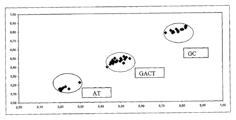
Figure 1 shows a representation of ratios 1 and 2 in a study of 15
blood donors, five of genotype 188G189C, five of genotype
188GA189CT and five of genotype 188A1189T (Example 2}.
Figure 2 shows a representation of ratios 1 and 2 in a study of 9
patients, 3 of genotype AA, 3 of genotype AT and 3 of genotype TT
at genetic variation A2033T in the CSFR1 gene (Example 4).
Figures 3-10 (Example 6) demonstrate the respective probabilities
associated with the development of determined phenotypes (disease
prognosis), based on genotypic data obtained with a DNA-chip
according to the invention, for each of the eight IBD phenotypes
analysed.
Figures 3-7 show probabilities for development of phenotypes
associated with Crohns disease and Figures 8-10 show probabilities
associated with the development of phenotypes associated with
ulcerative colitis. Figures 11-13 (Example 6) indicate the
probabilities assodiated with the risk of developing resistance to
corticosteroid treatment in individuals suffering from IBD.
Brief description of the sequences
SEQ ID NOS 1-124 and 1317-1428 are PCR primers suitable for
amplifying target DNA regions comprising genetic variations
associated with IBD.
SEQ ID NOS 125-254 are PCR primers suitable for amplifying target
DNA regions comprising genetic variations associated with adverse
reactions to pharmaceuticals.
CA 02594730 2007-07-12
WO 2006/075254 PCT/IB2006/000796
28
SEQ ID NOS 255-630 are probes suitable for detection of genetic
variations associated with known erythrocyte antigens, and useful
for genotyping for blood groups.
SEQ ID NOS 631- 960 and 1429-1652 are probes suitable for
detection of genetic variations associated with IBD.
SEQ ID NOS 961-1316 are probes suitable for detection of genetic
variations associated with adverse reactions to pharmaceuticals.
SEQ ID NO 1653 is an external control nucleic acid.
SEQ ID NOS 1654-1655 are probes suitable for detection of the
external control nucleic acid of SEQ ID NO: 1653.
Detailed description of the invention
The present invention relates to a method of genotyping genetic
variations in an individual, which is sufficiently sensitive,
specific and reproducible as to allow its use in a clinical
setting. The inventors have developed DNA-chips with specifically
designed probes for use in the method, and a computational method
or algorithm for interpreting and processing the data generated by
the chips.
Thus in one aspect, the invention comprises an in vitro method for
genotyping genetic variations in an individual. The in vitro,
extracorporeal method is for-simultaneous sensitive, specific and
reproducible genotyping of multiple human genetic variations
present in one or more genes of a subject. The method of the
invention allows identification of nucleotide changes, such as,
insertions, duplications and deletions and the determination of
the genotype of a subject for a given genetic variation.
CA 02594730 2007-07-12
WO 2006/075254 PCT/IB2006/000796
29
The terms "genetic variation" or "genetic variant", as they are
used in the present description include mutations, polymorphisms
and allelic variants. A variation or genetic variant is found
amongst individuals within the population and amongst populations
within the species.
The term "polymorphism" refers to a variation in the sequence of
nucleotides of nucleic acid where every possible sequence is
present in a proportion of equal to or greater than 1% of a
population; in a particular case, when the said variation occurs
in just one nucleotide (A, C, T or G) it is called a single
nucleotide polymorphism (SNP).
The term "genetic mutation" refers to a variation in the sequence
of nucleotides in a nucleic acid where every possible sequence is
present in less than 1% of a population
The terms "allelic variant" or "allele" are used without
distinction in the present description and refer to a polymorphism
that appears in the same locus in the same population.
Thus a genetic variation may comprise a deletion, substitution or
insertion of one or more nucleotides. In one aspect the genetic
variations to be genotyped according to the present methods
comprise SNPs.
A given gene may comprise one or more genetic variations. Thus
the present methods may be used for genotyping of one or more
genetic variations in one or more genes.
Typically the individual is a human.
Typically, for a given genetic variation there are three possible
genotypes:
CA 02594730 2007-07-12
WO 2006/075254 PCT/IB2006/000796
AA the individual is homozygous for genetic variation A (e.g
homozygous for a wild type allele)
BB the individual is homozygous for genetic variation B (e.g.
homozygous for a mutant allele)
5 AB the individual is heterozygous for genetic variations A and
B (e.g. one wild type and one mutant allele)
In one aspect the genetic variations, such as SNPs, to be analysed
according to the present methods, are associated with a particular
10 phenotype or disease condition. For example, the variations may
be associated with particular erythrocyte antigens (and thus often
a particular blood group); or with IBD; or with adverse reactions
to pharmaceuticals in an individual.
15 Examples of genetic variations associated with IBD which may be
assessed by the present methods include those in Table 1 below.
Ta7.Lfs 1 Genetic variations associated with !RD
20 The polymorphism G2677T/A/C Ala893Ser/Thr/Pro of the gene
Multidrug resistance protein l(MDR1);
The polymorphism C3435T of the gene Multidrug resistance
protein 1(MDR1);
The polymorphisms R702W, G908R, 1007insC in the gene Caspase
25 recruitment domain-containing protein 15 (CARD15);
The polymorphism T612C Y113H in the gene Microsomal epoxide
hydrolase (EPXH1);
The polymorphism (-2518)G/A of the gene Monocyte chemotactic
protein 1 (MCP1) ;
30 The polymorphisms (-1082) G/A and G43A (G15R) in the gene
Interleukin 10 (IL10);
The polymorphism (-295)T/C in the gene Interleukin 16
(IL16) ;
The polymorphism (-843)C/T in the gene Fas ligand;
CA 02594730 2007-07-12
WO 2006/075254 PCT/IB2006/000796
31
The polymorphisms 94delATTG and -263A/G in the gene Nuclear
factor kappa-B 1(NFKB1);
The polymorphism in 3'UTR (G/A) of the gene Nuclear factor
kappa-B inhibitor alpha (NFKBIA);
The polymorphism G2964A in the gene Signal transducer and
activator of transcription 6 (STAT6);
The polymorphism. TCA/TCC of codon 35 in the gene Interleukin
18 (IL18);
The polymorphisms E474E, Q476Q, D510D, P588P, -177A/G,
A165A, R202Q in the gene Mediterranean fever gene (MEFV);
The polymorphism 113G/A (R30Q) in the gene Discslarge,
Drosophila, homolog of, 5 (DLG5);
The polymorphism A2033T in the gene Colony stimulating
factor receptor 1 (CSFR1);
The polymorphism 1672C/T (L503F) in the gene Organic cation
transporter (OCTN1, SLC22A4);
The polymorphism (-207G/C) in the Organic cation transporter
(OCTN2, SLC22A5);
The polymorphisms Asp299Gly and Thr3991le_in the gene Toll-
like receptor 4 (TLR4);
The polymorphisms (-511)A/C and 3954 TaqI RFLP in the gene
Interleukin 1 beta (IL1R);
The polymorphism Alal6Val in the gene Superoxide dismutase
2 (SOD2);
The polymorphism Prol2Ala in the gene Peroxisome
proliferator-activated receptor gamma (PPARG);
The polymorphisms K469E, R241G in the gene Intercellular
adhesion molecule 1 (ICAM1);
The polymorphisms IGR2060a_1, IGR2198a_1, IGR3096a_1 in the
locus Inflammatory Bowel Disease 5(IBD5);
The polymorphism 1267A/G (Gln35lGln) in the gene Heat shock
protein 70 (HSP70-2);
The polymorphism 1237C/T in the gene Toll-like receptor 9
(TLR9);
CA 02594730 2007-07-12
WO 2006/075254 PCT/IB2006/000796
32
The polymorphism C677T (V222A) in the gene
Methylinetetrahydrofolate reductase (MTFHR);
The polymorphisms (-590)C/T, (-34)C/T in the gene
Interleukin 4 (IL4);
The polymorphisms Gly54Asp (A/G), Gly57Glu (A/G), Arg52Cys
(C/T) in the gene Mannose-binding lectin (MBL);
The polymorphism (-6) A/T in the gene Angiotensinogen
precursor (AGT);
The polymorphism 4G/5G in the gene Plasminogen activator
inhibitor (PAI);
The polymorphisms (_-857C/T), (-308G/A), (-238 G/A) in the
gene Tumor necrosis factor alpha (TNF-a);
The polymorphisms G238C, G460A, A719G in the gene TPMT;
The polymorphisms Trpl4Gly, Thr24Ala, Met129Val, Lys173Glu,
G1y175Ser of the gene Major histocompatibility complex class
I chain-realted-gene A (MICA) that discriminates the
alleles MICA*007 and MICA*008;
The polymorphism of the promoter region (-377 to -222)
characteristic of allele 7 of the gene Solute carrier family
11, member 1 (SLC11A1=NRAMP1);
The polymorphism (-159)T/C of the gene CD14;
The polymorphism G4985T (Vall58Phe) of the gene
CD16A=FCGR3A;
The polymorphism -25385C/T of the gene Nuclear receptor
subfamily 1, group I, member 2(NR1I2);
The polymorphism (T/A) (CyslOStop) of the gene Caspase
recruitment domain-containing protein 8
(TUCAN/CARD8/CARDINAL);
The polymorphism 738T/C (Cys224Arg) of the gene Inhibitor of
kappa light chain gene enhancer in B cells-like (IKBL);
The polymorphisms G593A and T620C of the gene Tumor necrosis
factor receptor subfamily, member 1B (TNFRSFIB =TNFR2);
The polymorphism Asp643Asn of the gene Mitogen-Activated
kinase kinase kinase 1 (MEKK1);
CA 02594730 2007-07-12
WO 2006/075254 PCT/IB2006/000796
33
The polymorphisms 159G/A/C and 282C/T of the gene Major
Histocompatibility complex, class II, DQ Alpha-1 (HLA-DQ)
for the identification of the alleles DQB1*0401 and
DQB1*0402;
The polymorphisms 109T/C, 119T/C/G/A, 122A/C/G/T, 129A/G,
161G/A/T, 175A/T/C/G, 184A/C/delA, 286C/A/T, 305C/G for the
identification of alleles DR2, DR9, DRB1*0103, DR4, DR7,
DRB3*0301 and DR3 of the gene Major histocompatibility
complex, class II, DR Beta-1 (HLA-DRB1);
The polymorphisms 2018T/C and 2073C/T of the gene
Interleukin 1 receptor antagonist (IL1RN);
The polymorphism 3954 C/T (TAQI) of the gene Interleukin 1
receptor, type II (IL1RB);
The polymorphism (-670) G/A of the gene Fas Antigen ;
The polymorphism 93 C/T of the gene Caspase 9 (CASP9);
The polymorphism G/C (R80T) of the gene Toll-like receptor 1
(TLR1);
The polymorphism A/G (R753G) of the gene Toll-like receptor
2 ( TLR2 ) ;
The polymorphism T/C (S249P) of the gene Toll-like receptor
6 (TLR6);
The polymorphism 5A/6A of the gene Matrix metalloproteinase
3 (MMP3);
The polymorphism indel +32656 of the gene NOD-1 protein
(NOD1=CARD4);
The polymorphism DLG5_e26 in the gene Discslarge,
.-Drosophila, homolog of, 5 (DLG5);
The polymorphism with rs20752817 of the gene NOD-1 protein
(NOD1=CARD4);
The polymorphism with rs2975632 of the gene NOD-1 protein
(NODI=CARD4);
The polymorphism with rs3020207 of the gene NOD-1 protein
(NODI=CARD4);
The polymorphism with rs2075818 of the gene NOD-1 protein
(NOD1=CARD4);
CA 02594730 2007-07-12
WO 2006/075254 PCT/IB2006/000796
34
The polymorphism with rs2235099 of the gene NOD-1 protein
(NODI=CARD4) ;
The polymorphism with rs2075821 of the gene NOD-1 protein
(NODI=CARD4);
The polymorphism with rs2075822 of the gene NOD-1 protein
(NODI=CARD4);
The polymorphism with rs2907748 of the gene NOD-1 protein
(NODI=CARD4);
The polymorphism with rs5743368 of the gene NOD-1 protein
(NODI=CARD4);
The polymorphism with rs2289311 of the gene NOD-1 protein
(NODI=CARD4);
The polymorphism A1298C in the gene
Methylinetetrahydrofolate reductase (MTFHR);
The polymorphism I1e114Thr in the gene N-Acetyl tranferase
2 (NAT2 ) ;
The polymorphism (A/G) Lys268Arg in the gene N-Acetyl
tranferase 2(NAT2);
The polymorphism with rs9340799 of the gene Estrogen
receptor 1 (ESR1);
The polymorphism with rs2234693 of the gene Estrogen
receptor 1 (ESR1);
The polymorphism C/T V726A in the gene Mediterranean fever
gene (MEFV);
The polymorphism with rs10735810 in the Vitamin D receptor
(VDR);
The polymorphism (C/G)E127Q in EGF-like module-contining,
mucin-like hormone receptor 3 (EMR3);
The polymorphism (G/T)Q496K in EGF-like module-contining,
mucin-like hormone receptor 1 (EMR3);
The polymorphism R653Q in the Methylenetetrahydrofate
dehydrogenase 1 (MTHFDI);
CA 02594730 2007-07-12
WO 2006/075254 PCT/IB2006/000796
The polymorphism 1420 (C/T) in the Serine
hydroxymethyltransferase (SHMT1);
The polymorphism G1y286G1u in the gene N-Acetyl tranferase
2(NAT2);
5 The polymorphism Arg197Gln in the gene N-Acetyl tranferase
2(NAT2);
The polymorphism 191 (G/A) in the gene N-Acetyl tranferase
2(NAT2);
The polymorphism Arg392Stop of the gene Toll-like receptor
10 5 (TLR5); The polymorphism A49G of the gene cytotoxic T lymphocyte-
associated 4 (CTLA4);
The polymorphism D132H of the gene MutL, E. coli, homolog
of, 1 (MLH1) ;
15 The polymorphism 66A/G of the gene Methionine synthase
reductase (MTRR);
The polymorphism 94C/A of the gene Inosine Triphosphatase
( ITPA) ;
The polymorphism E148Q in the gene Mediterranean fever gene
20 (MEFV);
The polymorphism R620W in the protein tyrosine phosphatase,
nonreceptor-type, 22 (PTPN22);
The polymorphism 3357 A/G in the Low density lipoprotein
receptor-related protein 5 (LRP-5);
25 The polymorphism C318T of the gene cytotoxic T lymphocyte-
associated 4 (CTLA4);
The polymorphism rs333 32bpdel of the gene chemokine, CC
motif, receptor 5(CCR5);
The polymorphism -174G/C of the gene interleukin-6(IL6);
30 The polymorphism with rs6190 of the gene glucocorticoid
receptor (GR ER22/23EK);
The polymorphism Arg72Pro of the gene p53;
The polymorphism P1371Q in the gene Discslarge, Drosophila,
homolog of, 5 (DLG5);
CA 02594730 2007-07-12
WO 2006/075254 PCT/IB2006/000796
36
The polymorphism with rs6189 of the gene glucocorticoid
receptor (GR ER22/23EK);
The polymorphism C135242T in the Low density lipoprotein
receptor-related protein 5 (LRP-5);
The polymorphism G121513A in the gene Low density
lipoprotein receptor-related protein 5 (LRP-5);
The polymorphism C141759T in the gene Low density
lipoprotein receptor-related protein 5 (LRP-5);
The polymorphism G138351A in the gene Low density
lipoprotein receptor-related protein 5 (LRP-5);
The polymorphism (-298) C/T in the gene Purinergic receptor
P2X, ligand-gated ion chanel, 7 (P2RX7);
The polymorphism (-838) G/T in the gene Purinergic receptor
P2X, ligand-gated ion chanel, 7 (P2RX7);
The polymorphism E1317Q in the gene Adenomatous polyposis
of the colon (APC);
And the polymorphism T64C in the gene CD97 (CD97);
Examples of genetic variations associate-d with particular
erythrocyte antigens which may be assessed by the present methods
i.nclude.those in Table 2 below.
Table 2 Genetic variations associated with erythrocyte antigens -
The polymorphism GG87_88insG (Genotype 04) (BC008) in exon 2
of the gene ABO,
The polymorphism G188A+C189T (Genotype 01v) (BC012) in exon
4 of the gene ABO,
The polymorphisms 261de1G (Genotype 01/01v) (BC001), C322T
(Genotype 05) (BC009) in exon 6 of the gene ABO,
The polymorphisms C467T (P156L) (Genotype A2) (BC014), G542A
(Genotype 08) (BC013), T646A (Genotype Ax/01v) (BC015),
G703A -(Genotype G235S) (B) (BC002), C796A -(Genotype L266M)
(B) (BC003), G802A (Genotype 02) (BC004), G803C (Genotype
G268A) (B, ci.sAB-1) (BC005), 798-804insG (Genotype 03, Ael)
(BC007), C893T (Genotype 06) (BC010), C927A (Genotype 07)
CA 02594730 2007-07-12
WO 2006/075254 PCT/IB2006/000796
37
(BC011), 1059-1061delC (D FS354+21aa) (Genotype A2) (BC006)
in.exon 7 of the gene ABO,
The polymorphisms C8G (S3C) (Genotype weak D type 3)
(BC040), G48A (W16X) (Genotype RHD W16X) (BC046), C121T
(Q41X) (Genotype RHD Q41X) (BC047) in exon 1 of the gene
RHD,
The polymorphisms A178C, G203A, T307C (exon scanning)
(BC016, BC017, BC018), T161C (L54P) (Genotype DMH) (BC033),
G270A (W90X) (Genotype RHD W90X) (BC047), T329C (L110P)
(Genotype DVII) (BC028) in exon 2 of the gene RHD,
The polymorphisms C340T (Genotype weak D type 17) (BC043),
C410T (Genotype DIIIiv) (BC059), C446A (A149D) (Genotype
weak D type 5) (BC041), A455C (Genotype DIIIa, DIIIiv, DIVa)
(BC060), IVS3+lG>A (Genotype negative allele) (BC049) in
exon 3 of the gene RHD,
The polymorphisms 488de14 negative genotype allele (BC050),
A497C (H166P) ' (Genotype DFW) (BC030), T509C (M170T)
(Genotype DOL) (BC027), A514T (Genotype DFRI) (BC065),
T544A, G577A, A594T (Genotype DVI-I weak D type 4) (exon
scanning), (BC019, BC020, BC021) in exon 4 of the gene RHD,
The polymorphisms G635T (G212V) (Genotype RHD G212V)
(BC051), T667G (Genotype DIIIa, weak D type 4, Dva, DAR,
DOL, DCS) (BC061), G676C (Genotype DCS, G686A (Genotype DHR)
(BC031), G697C (E233Q), (Genotype G712A (M238V) (DVI I,- weak
D type 4, DV, DCS) (BC022, BC023),A712G (genotype negative
allele) (BC023) in exon 5 of the gene RHD,
The polymorphisms T807G (Genotype pseudogene) (BC044), T809G
(Genotype weak D type 1) (BC038), G845A (G282D) (Genotype
weak D type 15, DIM) (BC037), C848T (T283I) (Genotype DHMI)
(BC029), G854A (C285Y) (Genotype DIM) (BC032), G885T (M295I)
(Genotype negative allele M295I) (BC053), 906insGGCT
(Genotype negative allele) (BC054), G916A, A932G (consensus
exon scanning) (BC062, BC063), IVS6+ldel4 (Gehotype allele
negative) (BC055) in exon 6 the gene RHD, polymorphisms
G941T (G314V) (Genotype negative allele) (BC056), C990G
CA 02594730 2007-07-12
WO 2006/075254 PCT/IB2006/000796
38
(Y330X) (Genotype negative allele) (BC057), G1016A (G339E)
(Genotype weak D type 7) (BC042), T1025C (1342T) (exon
scanning) (BC024), G1048C (Genotype DIVa, DIVb) (BC094),
G1057A (G353R) (Genotype DNU) (BC034), C1061A (A354N)
(Genotype DII) (BC036), G1063A (G355S) (Genotype =DNB)
(BC026), T1073C (Genotype DWI) (BC035) in exon 7 the gene
RHD,
The polymorphism IV8+1G>A (Genotype negative allele) (BC058)
in exon 8 of the gene RHD,
The polymorphisms G1154C (G385A) (Genotype weak D type 2)
(BC039), A1193T (Genotype DIVb) (BC064), G1227A (K409K)
(Genotype K409K) (BC045) in exon 9 of the gene RHD,
The polymorphisms G106A (A36T) (Genotype Cx) (BC068), A122G
(Q41R) (Genotype Cw) (BC067) in exon 1 of the gene RHCE,
The polymorphism T307C (S103P) (Genotype RHc) (BC066) in
exon 2 of the gene RHCE,
The polymorphism C410T (A137V) (BC059) in exon 3 of the gene
RHCE,
The polymorphisms C676G (P226A) (Genotype Ee) (BC025,
BC069), C733G (L245V) (Genotype VS) (BC070) in exon 5 of the
gene RHCE,
The polymorphism G1006T (G336C) (Genotype VS-/VS+) (BC071)
in exon 7 of the gene RHCE,
The polymorphisms A697T (Genotype Kk) (BC073), C698T (T193M)
(Genotype Kk) (BC072) in exon 6 of the gene KEL,
The polymorphisms T961C (R281W) (Genotype KpaKpb) (BC074),
G962A (R281Q) (Genotype KpbKpc) (BC075) in exon 8 of the
gene KEL,
The polymorphism G1208A (S363N) (Genotype Kmod-1) (BC077) in
exon 10 of the gene KEL,
The polymorphism C1910T (L597P) (Genotype JsaJsb) (BC076) in
exon 17 of the gene KEL,
The polymorphism I5AG>AA (Genotype Jknull) (BC079) in exon 6
of the gene SLC14A1 (blood group KIDD),
CA 02594730 2007-07-12
WO 2006/075254 PCT/IB2006/000796
39
The polymorphisms G838A (D280N) (Genotype JkaJkb) (BC078),
T871C (S291P) (Genotype Jknull) (BC080) in exon 9 of the
gene SLC14A1 (blood group KIDD),
The polymorphisms T-33C (Genotype FYGATA) (BC082), G125A
(D42G) (Genotype FYaFYb) (BC081), C265T (R89C) (Genotype
FYx) (BC083) in the gene DARC (blood group DUFFY),
The polymorphisms C59T, G71A, T72G (S20L, G42E, G42E)
(Genotype MN) (BC084, BC085) in exon 2 of the gene GYPA,
The polymorphism T143C (M48T) (Genotype Ss) (BC086) in exon
4 of the gene GYPB,
The polymorphisms C790A (Genotype GpMUR MiIII) (BC089),
C850G (Genotype GpMUR M:iIII) (BC090) in exon 3 of the gene
GYPE,
The polymorphisms C230T (Genotype U) (BC087), 15+5GT
(Genotype U) (BC088) in exon 5 of the gene GYPB,
The polymorphism T2561C (P854L) (Genotype DiaDib) (BC091) in
exon 19 of the gene SLC4A1 (blood group DIEGO),
The polymorphism A793G (Genotype DoaDob) (BC092) in exon 2
of the gene DOMBROCK,
The polymorphism C134T (A45V) (Genotype CoaCob) (BC093) in
exon 1 of the gene COLTON.
Examples of genetic variations associated with adverse reactions
to pharmaceuticals which may be assessed by the present methods
include those in Table 3 below.
Table 3 r__e*_,eti c variations associated with adverse reactions to
pharmaceuticals
The polymorphism Arg389Gly in the adrenergic beta 1 receptor
(ADRB1)
The polymorphisms Argl6Gly and Gln27Glu in the adrenergic
beta 2 receptor (ADRB2),
The polymorphism Ser9Gly of the dopamine receptor D3 (DRD3),
The polymorphisms Hi.s452Tyr and T102C of the serotonin
receptor 2A (HTR2A),
CA 02594730 2007-07-12
WO 2006/075254 PCT/IB2006/000796
The polymorphism Va1108Met of Catechol-O-methyltransferase
(COMT),
The polymorphism S1e105Va1 of Glutathione S transferase
class 1 (GSTP1),
5 The polymorphism Gly460Trp of Adducin 1 (ADD1),
The polymorphism Arg399G1n of the DNA repair protein XRCC1,
The polymorphism Ile462Va1 of the cytochrome P450 lAl
(CYP1A1),
The polymorphism A1166C of the angiotensin II, type 1
10 receptor (AGTR1),
The polymorphism C-58T of the receptor B2 of bradykinin
(BDKRB2),
The polymorphism Met235Thr of angiotensinogen (AGT),
The polymorphisms C430T, A1075C, 818de1A, T1076C and C1080G
15 of the cytochrome P450 2C9 (CYP2C9),
The polymorphisms H324P, V136V, V11M, C882G, C1038T, G4180C,
A1847G, C-1584G, C100T, 138insT, C1023T, G1659A, 1707T/del,
G1758A/T, 1863ins9bp, 1973insG, 2539de1AACT, 2549A/del,
2613delAGA, C2850T, G3183A, C3198G, T3277C, G4042A and
20 4125insGTGCCCACT of the cytochrome P450 2D6 (CYP2D6),
The polymorphisms A805T, G416A, A1196G and C792G of the
cytochrome P450 2C8 (CYP2C8),
The polymorphisms T341C, C481T, A803G, C282T, G590A, G857A
and G191A of N-acetyltransferase 2 (NAT2),
25 The polymorphisms G636A, G681A, C680T, A1G, IVS5+2T>A,
T358C, G431A and C1297T of the cytochrome P450 2C19
(CYP2C19),
The polymorphism C2664T of the glutamate receptor
ionotropic, N-methyl D-asparate (NMDA) 2B (GRIN2B),
30 The polymorphism C3435T of glycoprotein P(ABCB1),
The polymorphisms A719G and G238C of thiopurine S-
methyltransferase (TPMT),
The polymorphism C677T of 5,10-
methylenetetrahydrofolatereductase (MTHFR)
CA 02594730 2007-07-12
WO 2006/075254 PCT/IB2006/000796
41
The polymorphisms Asp70Gly and Ala539Thr of
butyrylcholinesterase (BCHE),
The polymorphism A-392G of the cytochrome P450 3A4 (CYP3A4),
The polymorphisms A-163C, A-3860G, G3534A and C558A of the
cytochrome P450 1A2 (CYP1A2),
The polymorphisms G14690A, C3699T, G19386A, T29753C and
G6986A of the cytochrome P450 3A5 (CYP3A5),
The polymorphism 44bp deletion of the promotor of the
serotonin transporter (SLC6A4),
The polymorphism delAGA (allele*B) of Glutathione S-
transferase M3 (GSTM3),
The polymorphism null allele of Glutathione S-transferase MI
(GSTM1),
The polymorphism null allele of Glutathione S-transferase nl
(GSTT1),
The polymorphisms Cysll2Arg and Arg158Cys of apolipoprotein
E (APOE),
The polymorphism G-308A of Tumor necrosis factor (TNF), and
The polymorphism G-1082A of Interleukin 10 (IL10)
The sequences of all the genes mentioned in Tables 1-3 are
known and recognized on the following websites: GeneBank (NCBI),
GeneCard (Weizmann Institute of Sciences) and Snpper.chip.org
(Innate Immunity PGA).
By permitting clinical genotyping of one or more of the above
genetic variations, the present method has use in for example,
diagnosing susceptibility to or the presence of IBD or adverse
reactions to pharmaceuticals. The methods also allow reliable
determination of erythrocyte antigens and are useful in blood
grouping or typing.
At least one genetic variation is analysed in the present methods.
The present methods allow simultaneous genotyping of multiple
variations in an individual and typically multiple variations are
CA 02594730 2007-07-12
WO 2006/075254 PCT/IB2006/000796
42
analysed, in general, at least 10, 12, 14, 16, 18 or 20 genetic
variations. For example, 30, 40, 50, 60, 70, 80 or 100 variations
or up to 200, 300, 400, 500, or 600 variations may be tested, such
as 250, 350 or 450 variations.
Thus the present methods may be used for genotyping an individual
with respect to all of the variations in any one of Tables 1 to 3,
or a selection of the variations in any one of the Tables, as
described herein. Thus the variations to be detected may comprise
or be selected from any one of Tables 1 to 3.
According to the present methods, a sample is provided, containing
nucleic acid which comprises at least one of the genetic
variations to be tested (the target DNA) . The nucleic acid
comprises one or more target regions comprising the genetic
variation(s) which are to be characterised.
The nucleic acid may be obtained from any appropriate biological
- sample which contains nucleic acid. The sample may be taken from
a fluid or tissue, secretion, cell or cell line derived from the
human body.
For example, samples may be taken from blood, including serum,
lymphocytes, lymphoblastoid cells, fibroblasts, platelets,
mononuclear cells or other blood cells, from saliva, liver,
kidney, pancreas or heart, urine or from any other tissue, fluid,
cell or cell line derived from the human body. For example, a
suitable sample may be a sample of cells from the buccal cavity.
Preferably nucleic acid is obtained from a blood sample.
In general, nucleic acid is extracted from the biological sample
using conventional techniques. The nucleic acid to be extracted
from the biological sample may be DNA, or RNA, typically total
RNA. Typically RNA is extracted if the genetic variation to be
studied is situated in the coding sequence of a gene. Where RNA
CA 02594730 2007-07-12
WO 2006/075254 PCT/IB2006/000796
43
is extracted from the biological sample, the methods further
comprise a step of obtaining cDNA from the RNA. This may be
carried out using conventional methods, such as reverse
transcription using suitable primers. Subsequent procedures are
then carried out on the extracted DNA or the cDNA obtained from
extracted RNA. The term DNA, as used herein, may include both DNA
and cDNA.
In general the genetic variations to be tested are known and
characterised, e.g. in terms of sequence. Therefore nucleic acid
regions comprising the genetic variations may be obtained using
methods known in the art.
In one aspect, DNA regions which contain the genetic variations to
be identified (target DNA regions) are subjected to an
amplification reaction in order to obtain amplification products
which contain the genetic variations to be identified. Any
suitable technique or method may be used for amplification. In
general, the technique allows the (simultaneous) amplification of
all the DNA sequences containing the genetic variations to be
identified. In other words, where multiple genetic variations are
to be analysed, it is preferable to simultaneously amplify all of
the corresponding target DNA regions (comprising the variations).
Carrying out the amplification in a single step (or as few steps
as possible) simplifies the method.
For example, multiplex PCR may be carried out, using appropriate
pairs of oligonucleotide PCR primers which are capable of
amplifying the target regions containing the genetic variations to
be identified. Any suitable pair of primers which allow specific
amplification of a target DNA region may be used. In one aspect,
the primers allow amplification in the least possible number of
PCR reactions. Thus, by using appropriate pairs of oligonucleotide
primers and appropriate conditions, all of the target DNA regions
necessary for genotyping the genetic variations can be amplified
CA 02594730 2007-07-12
WO 2006/075254 PCT/IB2006/000796
44
for genotyping (e.g. DNA-chip) analysis with the minimum number of
reactions. Suitable PCR primers for amplification of target DNA
regions comprising genetic variations associated with erythrocyte
antigens, IBD, or adverse reaction to pharmaceuticals, are
described herein. In particular, PCR primers for amplification of
target DNA regions comprising the genetic variations associated
with IBD in Table 1 are listed in SEQ ID NOS 1-124 and 1317-1428.
PCR primers for amplification of target DNA regions comprising the
genetic variations associated with adverse reaction to drugs in
Table 3 are listed in SEQ ID NOS 125-254. The present method may
comprise the use of one or more of these primers or one or more of
the listed primer pairs.
In one instance, the amplification products can be labelled during
the amplification reaction with a detectable label. The aim is to
be able to later detect hybridisation between the fragments of
target DNA containing the genetic variations being analysed and
probes fixed on a solid support. The greater the extent of
hybridisation of labelled target DNA to a probe, the greater the
intensity of detectable label at that probe position.
The amplification products may be labelled by conventional
methods. For example, a labelled nucleotide may be incorporated
during the amplification reaction or labelled primers may be used
for amplification.
Labelling may be direct using for example, fluorescent or
radioactive markers or any other marker known by persons skilled
in the art. Examples of fluorophores which can be used, include
for example, Cy3 or Cy5. Alternatively enzymes may be used for
sample labelling, for example alkaline phosphatase or peroxidase.
Examples of radioactive isotopes which can be used include for
example 33P, 125I, or any other marker known by persons skilled in
the art. In one instance, labelling of amplification products is
carried out using.a nucleotide which has been labelled directly or
CA 02594730 2007-07-12
WO 2006/075254 PCT/IB2006/000796
indirectly with one or more fluorophores. In another example,
labelling of amplification products is carried out using primers
labelled directly or indirectly with one or more fluorophores.
5 Labelling may also be indirect, using, for example, chemical or
enzymatic methods. For example, an amplification product may
incorporate one member of a specific binding pair, for example
avidin or streptavidin, conjugated with a fluorescent marker and
the probe to which it will hybridise may be joined to the other
10 member of the specific binding pair, for example biotin
(indicator), allowing the probe/target binding signal to be
measured by fluorimetry. In another example, an amplification
product may incorporate one member of a specific binding pair, for
example, an anti-dioxigenin antibody combined with an enzyme
15 (marker) and the probe to which it will hybridise may be joined to
the other member of the specific binding_pair, for example
dioxigenin (indicator). On hybridization of amplification product
to probe the enzyme substrate is converted into a luminous or
fluorescent product and the signal can be read by, for example,
20 chemi-luminescence or fluorometry.
The nucleic acid comprising the genetic variation(s) to be tested,
e.g. the (optionally labelled) amplification products, may further
undergo a fragmentation reaction, thereby obtaining some
25 fragmentation products which comprise or contain the genetic
variations to be identified or analysed. Typically fragmentation
increases the- efficiency of the hybridisation reaction.
Fragmentation may be carried out by any suitable method known in
the art, for example, by contacting the nucleic acid, e.g. the
30 amplification products with.a suitable enzyme such as a DNase.
If the nucleic acid has not been previously labelled, e.g. during
the amplification reaction, (and, typically, where no
posthybridisation amplification or ligation is carried out on the
35 solid support) then labelling with a detectable label may be
CA 02594730 2007-07-12
WO 2006/075254 PCT/IB2006/000796
46
carried out prehybridisation by labelling the fragmentation
products. Suitable labelling techniques are known in the art and
may be direct or indirect as described herein. Direct labelling
may comprise the use of, for example, fluorophores, enzymes or
radioactive isotopes. Indirect labelling may comprise the use of,
for, example, specific binding pairs that incorporate e.g.
fluorophores, enzymes, etc. For example, if amplification products
have not been labelled during the amplification reaction the
fragmentation products may undergo a direct or indirect labelling
with one or various markers, for example one or various
fluorophores, although other known markers can be used by those
skilled in the art.
According to the present methods the nucleic acid, e.g. the
amplification or fragmentation products, comprising the genetic
variation(s) to be detected (target DNA), is contacted with
oligonucleotide probes which are capable of detecting the
corresponding genetic variations by hybridisation under suitable
conditions.
Typically the hybridisation conditions allow specific
hybridisation between probes and corresponding target nucleic
acids to form specific probe/target hybridisation complexes while
ininimising hybridisation between probes carrying one or more
mismatches to the DNA. Such conditions may be determined
empirically, for example by varying the time and/or temperature of
hybridisation and/or the number and stringency of the array
washing steps that are performed following hybridisation and are
designed to eliminate all probe-DNA interactions that are
inspecific.
In the method, the probes are provided deposited on a solid
support or surface. The probes are deposited at positions on the
solid support according to a predetermined pattern, forming a
"DNA-chip". It has been found that the chips should comply with a
CA 02594730 2007-07-12
WO 2006/075254 PCT/IB2006/000796
47
number of requirements in order to be used in the present methods,
for example in terms of the design of the probes, the number of
probes provided for each genetic variation to be detected and the
distribution of probes on the support. These are described in
detail herein. The inventors have developed suitable genotyping
chips for use in the present methods and accordingly in one aspect
the invention provides a DNA-chip or (micro)array comprising a
plurality of probes deposited or immobilised on a solid support as
described herein.
In general the solid support or phase comprises oligonucleotide
probes suitable for detection of each genetic variation to be
tested. The number and type of genetic variations to be tested
using a chip may be selected as described herein.
Typically there will be at least one probe which is capable of
hybridising specifically to genetic variation A (e.g. a wildtype
or normal allele) (probe 1) and one probe which is.capable of
hybridising specifically to genetic variation B (e.g. a mutant
allele) (probe 2) under the selected hybridisation conditions.
These probes form a probe pair. Probe 1 is for detection of
genetic variation A and probe 2 for detection of genetic variation
B. Typically the probes can be used to discriminate between A and
B (e.g. the wildtype and mutant alleles).
The probes may examine either the sense or the antisense strand.
Typically, probes 1 and 2 examine the same nucleic acid strand
(e.g. the sense strand or antisense strand) although in some cases
the probes may examine different stran.ds. In one aspect probes 1
and 2 have the same sequence except for the site of the genetic
variation.
In one instance, the probes in a probe pair have the same length.
In some aspects, where two or more pairs of probes are provided
CA 02594730 2007-07-12
WO 2006/075254 PCT/IB2006/000796
48
for analysis of a genetic variation, the probes may all have the
same length.
Preferably more than one probe pair is provided for detection of
each genetic variation. Thus, at least 2, 3, 4, 5, 6, 7, 8, 9, 10
or more probe pairs may be provided per genetic variation. In one
aspect, (at least) 2 probe pairs are provided. The aim is to
reduce the rate of false positives and negatives in the present
methods.
For example, for a given genetic variation there may be:
Probe 1 which is capable of hybridising to genetic variation A
(e.g. a normal allele) 15 Probe 2 which is capable of hybridising to genetic
variation B
(e.g. a mutant allele)
Probe 3 which is capable of hybridising to genetic variation A
(e.g. a normal allele)
Probe 4 which is capable of hybridising to genetic variation B
(e.g. a mutant allele).
The probes may examine the same or different strands. Thus in one
embodiment, probes 3 and 4 are the complementary probes of probes
1 and 2 respectively and are designed to examine the complementary
strand. In one aspect it is preferred that, the probes provided for
detection of each genetic variation examine both strands.
More than 2 pairs of probes may be provided for analysis of a
genetic variation as above. For example, where a genetic
variation exists as any one of 4 bases in the same strand (e.g.
there are three mutant possibilities), at least one pair of probes
may be provided to detect each possibility. Preferably, at least
2 pairs of probes are provided for each possibility.
CA 02594730 2007-07-12
WO 2006/075254 PCT/IB2006/000796
49
Thus, for example, for the SNP G2677T/A/C, at least one pair of
probes may be provided for detection of G2677T, one pair for
detection of G2677/A, and one pair for detection of G2677C.
Preferably at least two pairs of probes are provided for each of
these substitutions.
A number of methods are known in the art for designing
oligonucleotide probes suitable for use in DNA-chips.
A "standard tiling" method may be used. In this method, 4
oligonucleotides are designed that are totally complementary to
the reference sequence except in the central position where,
typically the 4 possible nucleotides A, C, G and T are examined.
An illustrative example of this strategy is the DNA-chip for
genotyping of HIV-1 (Affymetrix).
In "alternative tiling" 5 oligonucleotides are designed, so that
the fifth examines a possible deletion in the sequence. An
example of this strategy is the DNA-chip to detect mutations in
p53 (Affymetrix).
In "block tiling" 4 oligonucleotides are designed that are totally
complementary to the normal sequence and another 4 totally
complementary to the mutant sequence. The nucleotide which
changes is placed in the central position, but a mismatch of one
of the 4 bases (A, C, T or G) is placed 2 nucleotides before or
after the nucleotide position that it is wished to interrogate. An
example of this strategy is the DNA-chip for the detection of
mutations in cytochrome p450 (Roche and Affymetrix).
A further example is "alternative block tiling" where the
"mismatch" is used to increase the specificity of the hybrid not
only in one position but also in the positions -4, -1, 0, +1 and
+4 to identify the change produced in the central position or 0.
An example is the DNA-chip to detect 1,500 SNPS (Affymetrix).
CA 02594730 2007-07-12
WO 2006/075254 PCT/IB2006/000796
Any one or more of these strategies may be used to design probes
for the present invention. Preferably standard tiling is used, in
particular with 2 pairs of probes e.g. 2 pairs of complementary
5 probes as above. Thus it is preferable that the oligonucleotide
sequence is complementary to the target DNA or sequence in the
regions flanking the variable nucleotide(s). However, in some
cases, one or more mismatches may be introduced, as described
above.
The oligonucleotide probes for use in the present invention
typically present the base to be examined (the site of the genetic
variation) at the centre of the oligonucleotide. This is
particularly the case where differential hybridisation methods are
used, as in general this allows the best discrimination between
matched and mismatched probes. In these methods, typically there
is formation of specific detectable hybridisation complexes
without post-hybridisation on-chip amplification. For example, for
precise (single base) mutations, the base which differs between
the normal and the mutant allele is typically placed in the
central position of,the probe. In the case of insertions,
deletions and duplications, the first nucleotide which differs
between the normal and the mutant sequence is placed in the
central position. It is believed that placing the mutation at the
centre of the probe maximises specificity.
Where post-hybridisation on-chip amplification (e.g. ligation or
primer extension methods) is employed, oligonucleotide probes
typically present the variable base(s) at the 3' end of the probe.
Where OLA methodology is used, oligonucleotides (labelled directly
or-indirectly) are also designed which hybridise to probe-target
complexes to allow ligation.
In general the probes for use in the present invention comprise or
in some embodiments consist (essentially) of 17 to 27 nucleotides,
CA 02594730 2007-07-12
WO 2006/075254 PCT/IB2006/000796
51
for example, 19, 21, 23, or 25 nucleotides or 18, 20, 22, 24 or 26
nucleotides.
Preferably the individual probes provided for detection of a
genetic variation are capable of hybridising specifically to the
normal and mutant alleles respectively under the selected
hybridisation conditions. For example, the melting temperature of
the probe/target complexes may occur at 75-85 degrees C and
hybridisation may be for one hour, although higher and lower
temperatures and longer or shorter hybridisations may also
suffice.
The probes provided for detection of each genetic variation (as
described above) are typically capable of discriminating between
genetic variation A and B (e.g. the normal and mutant alleles)
under the givenhybridisation conditions as above. Preferably the
discrimination capacity of the probes is substantially 100%. If
the discrimination capacity is not 100%, the probes are preferably
redesigned. Preferably the melting temperature of the
probe/target complexes occurs at 75-85 degrees C. Methods for
testing discrimination capacity are described herein.
In one example, the probes provided for detection of a genetic
variation examine both strands and have lengths ranging from 19-27
nucleotides. Preferably the probes have 100% discrimination
capacity and the melting temperature of probe/target complexes is
75-85 degrees C.
Typically in order to obtain probes for use in the present
methods, a number of probes are designed and tested experimentally
for, e.g. hybridisation specificity and ability to discriminate
between genetic variants (e.g. a normal and a mutant allele).
Candidate oligonucleotide probe sequences may be designed as
described above. These may vary for example in length, strand
specificity, position of the genetic variation and degree of
CA 02594730 2007-07-12
WO 2006/075254 PCT/IB2006/000796
52
complementarity to the sequence flanking the genetic variation in
the target DNA. Once probe pairs have been designed, these can be
tested for hybridisation specificity and discrimination capacity.
The capacity of specific probes to discriminate between the
genetic variations A and B (e.g. normal and mutant alleles)
depends on hybridisation conditions, the sequence flanking the
mutation and the secondary structure of the sequence in the region
of the mutation. By using stable hybridisation conditions,
appropriate parameters such as strand specificities and lengths
can be established in order to maximise discrimination.
Preferably, the genetic variation is maintained at the central
position in the tested probes.
Methods for testing discrimination capacity of probes are
described herein. Typically a number of candidate probe pairs are
provided and used in a training method as described below. In
general two pairs of probes (probes 1 and 2, and probes 3 and 4)
are tested in the method. For example, two pairs of probes
examining both strands (complementary to each other) may be
tested. If it is not possible to obtain 100% discrimination
between the three genotyping groups using the probes, the probes
are typically redesigned. Hybridisation conditions in the
training method are generally maintained stably. Typically the
melting temperature of probe/target complexes is 75-85 degrees C.
For example, starting from probes of 25 nucleotides which detect a
genetic variation (e.g. the normal allele) and another genetic
variation (e.g. a mutant allele) in both strands (sense and
antisense), in general an - average of 8 probes may be
experimentally tested to identify two definite pairs.
Probes are chosen to have maximum hybridisation specificity and
discrimination capacity between genetic variants (e.g. a normal
and a mutant allele) under suitable hybridisation conditions. For
example, the probes for detection of a given genetic variation,
CA 02594730 2007-07-12
WO 2006/075254 PCT/IB2006/000796
53
e.g. two probe pairs, typically have substantially 100%
discrimination capacity. Typically the melting temperature of
probe/target complexes is at 75-85-C.
Using the methods herein the inventors have developed
oligonucleotide probes suitable for detection of the IBD-
associated genetic variations in Table 1. These probes are
presented as SEQ ID NOS 631-960 and 1429-1652. The probes are
listed in probe sets (133 sets in total), each set being for
detection of a given genetic variation. At least two pairs of
probes are listed in each set.
The inventors have also developed oligonucleotide probes suitable
for detection of the erythrocyte antigen-associated genetic
variations in Table 2. These probes are presented as SEQ ID NOS
255-630. The probes are listed in probe sets (94 sets in total),
each set being for detection of a given genetic variation. At
least two pairs of probes are listed in each set.
The inventors have also developed oligonucleotide probes suitable
for detection of the genetic variations associated with adverse
reactions to drugs in Table 3. These probes are presented as SEQ
ID NOS 961-1316. The probes are listed in probe sets (89 sets in
total), each set being for detection of a given genetic variation.
At least two pairs of probes are listed in each set.
In one aspect the invention relates to any one or more of the
oligonucleotide probes, pairs of probes or sets of probes set out
in SEQ ID NOS 255-630, 631-960, 961-1316 or 1429-1652, and to
their use in the genotyping, diagnostic or therapeutic.,methods of
the invention. The invention further relates to any one or more
of the oligonucleotide probes, pairs of probes or sets of probes
set out in SEQ ID NOS 255-630,"631-960, 961-1316 or 1429-1652 for
use in medicine, for example in a diagnostic or therapeutic method
CA 02594730 2007-07-12
WO 2006/075254 PCT/IB2006/000796
54
described herein. A chip of the invention may comprise one or
more of the listed probe pairs or sets.
In general probes are provided on the support in replicate.
Typically, at least 4, 6, 8, 10, 12, 14, 16, 18 or 20 replicates
are provided of each probe, in particular, 6, 8 or 10 replicates.
Thus for example, the support (or DNA-chip) may comprise or
include 10 replicates for each of (at least) 4 probes used to
detect each genetic variation (i.e. 40 probes). Alternatively the
support (or DNA-chip) may comprise or include 8 replicates for
each of (at least) 4 probes used to detect each genetic variation
(i.e. 32 probes) . Still further the support (or DNA-chip) may
comprise or include 6 replicates for each of (at least) 4 probes
used to detect each genetic variation (i.e. 24 probes). Using
probe replicates helps to minimise distortions in data
interpretation from the chip and improves reliability of the
methods.
In general the support also comprises one or more control
oligonucleotide probes. These are also provided in replicate as
above. Thus the support (or DNA-chip) may additionally comprise
one or more oligonucleotides deposited on the support which are
useful as positive and/or negative controls of the hybridisation
reactions. If post-hybridisation amplification or ligation
reactions are carried out on the chip, there may also be one or
more positive or negative controls of these reactions.
Typically the chip or array will include positive control probes,
e.g., probes known to be complementary and hybridisable to
sequences in the target polynucleotide molecules, probes known to
hybridise to an external control DNA, and negative control probes,
e.g., probes known to not be complementary and hybridizable to
sequences in the target polynucleotide molecules. The chip may
have one or more controls specific for each target, for example,
CA 02594730 2007-07-12
WO 2006/075254 PCT/IB2006/000796
2, 3, or more controls. There may also be at least one control
for the array.
Positive controls may for example be synthesized along the
5 perimeter of the array or in diagonal stripes across the array.
The reverse complement for each probe may be synthesized next to
the position of the probe to serve as a negative control. In yet
another example, sequences from other species of organism may be
used as negative controls in order to help determine background
10 (non-specific) hybridisation.
As above, the support (or DNA-chip) may include some (one or more)
oligonucleotides deposited on the support which are useful as
positive and negative controls of the hybridization reactions. In
15 general, each one of the sub-arrays, for example 16, which
typically constitute a DNA-chip, is flanked by some external
hybridization controls, which serve as reference points _allowing
allow the points within the grid to be located more easily.
20 In one instance, the nucleotide sequence of an external control
DNA is the following (5'->3'):
CEH: GTCGTCAAGATGCTACCGTTCAGGAGTCGTCAAGATGCTACCGTTCAGGA -
SEQ ID NO: 1653
and the sequences of the oligonucleotides for its detection are
25 the following:
ON1: CTTGACGACTCCTGAACGG - SEQ ID NO: 1654
oN2: CTTGACGACACCTGAACGG - SEQ ID NO: 1655
Positive control probes are generally designed to hybridise
30 equally to all target DNA samples and provide a reference signal
intensity against which hybridisation of the target DNA (sample)
to the test probes can be compared. Negative controls comprise
either "blanks" where only solvent (DMSO) has been applied to the
support or control oligonucleotides that have been selected to
35 show no, or only minimal, hybridisation to the target, e.g.human,
CA 02594730 2007-07-12
WO 2006/075254 PCT/IB2006/000796
56
DNA (the test DNA). The intensity of any signal detected at either
blank or negative control oligonucleotide features is an
indication of non-specific interactions between the sample DNA and
the array and is thus a measure of the background signal against
which the signal from real probe-sample interactions must be
discriminated.
Desirably, the number of sequences in the array will be such that
where the number of nucleic acids suitable for detection of
genetic variations is n, the number of positive and negative
--control nucleic acids is n', where n' is typically from 0.01 to
0.4n.
In general, the support or chip is suitable for genotyping, in
particular, genotyping according to the present methods. The chip
typically comprises probes suitable for detection of at least one
but preferably multiple, genetic variation(s), typically at least
10, 12, 14, 16, 18 or 20 genetic variations. For example, 30, 40,
5Q, 60, 70, 80 or 100 variations or up to 200, 300, 400, 500, or
600 variations may be tested, such as 250, 350 or 450 variations.
The genetic variations may be those in any one of Tables 1 to 3.
Thus an array may comprise probes suitable for genotyping an
individual with respect to all of the variations in any one of
Tables 1 to 3, or a selection of the variations in any one of the
Tables, as described above.
The present DNA-chips can be used, in combination with the present
methods, to detect practically any human genetic variation of
interest, for example, human genetic variations associated with
diseases or antigens of interest. Suitable probes will be used for
those genetic variations to be detected. As genetic variations
associated with the diseases or antigens of interest are
identified, suitable probes for their detection can be
incorporated in the chips. Probes and DNA-chips for this purpose
CA 02594730 2007-07-12
WO 2006/075254 PCT/IB2006/000796
57
can be designed in accordarice with the teaching of the present
invention.
The inventors have designed, produced and validated the clinical
use of the invention in detection of genetic variations associated
with IBD, with known human erythrocyte antigens and with adverse
reactions to medicine by developing (designing and producing)
corresponding DNA-chips.
Therefore, in one particular embodiment, the invention relates to
a chip for genotyping of genetic variations associated with IBD
(an "IBD-chip"). Typically the DNA-chip allows simultaneous,
sensitive, specific and reproducible detection of genetic
variations associated with IBD. Non-limiting examples of such
variations are given in Table 1. Nevertheless, the number of
genetic variations contained in the Table can be increased as
other genetic variations are subsequently identified and are
associated with IBD. Thus the genetic variations detectable by
the chip may comprise, or consist (essentially) of those listed in
Table 1 or a selection of these. The chip will comprise probes
suitable for detection of these genetic variations as described
herein. In one aspect the chip comprises probes selected from
those in SEQ ID NOS 631-960 and 1429-1652. The probes are listed
in probe sets (133 sets in total), each set being for detection of
a given genetic variation. At least two pairs of probes are
provided in each set. A chip may comprise at least one probe pair
or at least one probe set, or a selection of the probe sets, for
example, at least 5, 10, 20, 30, 40, 50, 60, 70, 80, 90, 100, 110,
120, 130 or all 133 sets, according to the genetic variations
being tested. A chip may comprise other probes for detection of
variations in Table 1 or other variations associated with IBD
instead of or in addition to those specifically listed.
In another embodiment the chip is for genotyping of genetic
variations associated with erythrocyte antigens (the "blood
CA 02594730 2007-07-12
WO 2006/075254 PCT/IB2006/000796
58
chip"). Typically the DNA-chip allows the simultaneous,
sensitive, specific and reproducible detection of genetic
variations associated with determined erythrocyte antigens. Non-
limiting examples of such variations are given in Table 2.
Nonetheless the number of genetic variations contained in the
table can be increased as other genetic variations are
subsequently identified and are associated with erythrocyte
antigens. Thus the genetic variations detectable by the chip may
comprise, or consist (essentially) of those listed in Table 2 or a
selection of these. The chip will comprise probes suitable for
detection of these genetic variations as described herein. In one
aspect the chip comprises probes selected from those in SEQ ID NOS
255-630. The probes are listed in probe sets (94 sets in total),
each set being for detection of a given genetic variation. At
least two pairs of probes are provided in each set. A chip may
comprise at least one probe pair or at least one probe set, or a
selection of the probe sets, for example, at least 5, 10, 20, 30,
40, 50, 60, 70, 80, 90, or all 94 sets, according to the genetic
variations being tested. A chip may comprise other probes for
detection of variations in Table 2 or other variations associated
with erythrocyte antigens instead of or in addition to those
specifically listed.
In another embodiment the chip is for genotyping of genetic
variations associated with adverse reactions to pharmaceuticals
(the "drug chip"). Typically the chip allows the simultaneous,
sensitive, specific and reproducible detection of genetic
variations associated with adverse reactions to medicine. Non-
limiting examples are given in Table 3. Nevertheless, the number
of genetic variations contained in the table can be increased as
other genetic variations are subsequently identified and are
associated with these adverse reactions. Thus the genetic
variations detectable by the chip may comprise, or consist
(essentially) of those listed in Table 3 or a selection-of these.
The chip will comprise probes suitable ior detection of these
CA 02594730 2007-07-12
WO 2006/075254 PCT/IB2006/000796
59
genetic variations as described herein. In one aspect the chip
comprises probes selected from those in SEQ ID NOS 961-1316. The
probes are listed in probe sets (89 sets-in total), each set being
for detection of a given genetic variation. At least two pairs of
probes are provided in each set. A chip may comprise at least one
probe pair or at least one probe set, or a selection of the probe
sets, for example, at least 5, 10, 20, 30, 40, 50, 60, 70, 80, or
all 89 sets, according to the genetic variations being tested. A
chip may comprise other probes for detection of variations in
Table 3 or other variations associated with adverse reactions to
drugs instead of or in addition to those specifically listed.
An IBD chip, blood chip or drug chip may additionally comprise
oligonucleotide probes for detection of genetic variations not
associated with IBD, erythrocyte antigens or adverse reactions to
drugs respectively. For example, the chips may comprise probes
for detection of genetic variations such as SNPs associated with
another (related) condition or other (related) antigen(s).
Typically, in an IBD chip, blood chip or drug chip, the number of
nucleic acids suitable for detection of genetic variations
associated with IBD, erythrocyte antigens or adverse reactions to
drugs respectively (e.g. those in Tables 1, 2, or 3) represent at
least 50%, 60%, 70%, 75%, 80%, 85%, 90%, 95%, 97%, 98%, 99% or
more of the nucleic acids in the array.
In general the support or chip has from 300 to 40000 nucleic acids
(probes), for example, from 400 to 30000 or 400 to 20000. The
chip may have from 1000 to 20000 probes, such as 1000 to 15000 or
1000 to 10000, or 1000 to 5000. A suitable chip may have from
2000 to 20000, 2000 to 10000 or 2000 to 5000 probes. For example,
a chip may have 1000, 2000, 3000, 4000, 5000, 6000, 7000, 8000,
9000, 10000, 12000, 14000, 16000, 18000 or 20000 probes. Smaller
chips 400 to 1000 probes, such as 400, 500, 600, 700, 800, 900 or
950 probes are also envisaged.
CA 02594730 2007-07-12
WO 2006/075254 PCT/IB2006/000796
In general the array or chip of the invention comprises a support
or surface with an ordered array of binding (e.g. hybridisation)
sites or probes. Thus the arrangement of probes on the support is
predetermined. Each probe (i.e each probe replicate) is located at
5 a known predetermined position on the solid support such that the
identity (i.e. the sequence) of each probe can be determined from
its position in the array. Typically the probes are uniformly
distributed in a predetermined pattern.
10 Preferably, the probes deposited on the support, although they
maintain a predetermined arrangement, are not grouped by genetic
variation but have a random distribution. Typically they are also
not grouped within the same genetic variation. If desired, this
random distribution can be always the same. Therefore, typically
15 the probes are deposited on the solid support (in an array)
following a predetermined pattern so that they are uniformly
distributed, for example, between the two areas that may
constitute a DNA-chip, but not grouped according to the genetic
variation to be characterised. Distributing probe replicates
20 across the array in this way helps to reduce or eliminate any
distortion of signal and data interpretation, e.g. arising from a
non-uniform distribution of background noise across the array.
As explained above, probes may be arranged on the support in
25 subarrays.
The support, on which the plurality of probes is deposited, can be
any solid support to which oligonucleotides can be attached.
Practically any support, to which an oligonucleotide can be joined
30 or immobilized, and which may be used in the production of DNA-
chips, can be used in the invention. For example, the said support
can be of a non-porous material, for example, glass, silicone,
plastic, or a porous material such as a membrane or filter (for
example, nylon, nitrocelullose) or a gel. In one embodiment, the
35 said support is a glass support, such as a glass slide.
CA 02594730 2007-07-12
WO 2006/075254 PCT/IB2006/000796
61
Microarrays are in general prepared by selecting probes which
comprise a given polynucleotide sequence, and then immobilizing
such probes to a solid support or surface. Probes may be
designed, tested and selected as described herein. In general the
probes may comprise DNA sequences. In some embodiments the probes
may comprise RNA sequences, or copolymer sequences of DNA and RNA.
The polynucleotide sequences of the probes may also comprise DNA
and/or RNA analogues, or combinations thereof. For example, the
polynucleotide sequences of the probes may be full or partial
fragments of genomic DNA. The polynucleotide sequences of the
probes may also be synthesized nucleotide sequences, such as
synthetic oligonucleotide sequences. The probe sequences can be
synthesized either enzymatically in vivo, enzymatically in vitro
(e.g., by PCR), or non-enzymatically in vitro.
Microarrays or chips can be made in a number of ways. However
produced, microarrays typically share certain characteristics. The
arrays are reproducible, allowing multiple copies of a given array
to be produced and easily compared with each other. Preferably,
microarrays are made from materials that are stable under binding
(e.g., nucleic acid hybridization) conditions. The microarrays are
preferably small, e.g., between 0.25 to 25 or 0.5 to 20cm2, such
0.5 to 20cm2 or 0.5 to 15cm2, for example, 1 to 15cmz or 1 to
10cm2, such as 2, 4, 6 or 8cm2.
Probes may be attached to the present support using conventional
techniques for immobilization of oligonucleotides on the surface
of the supports. The techniques used depend, amongst other
factors, on the nature of the support used [porous (membranes,
micro-particles, etc.) or non-porous (glass, plastic, silicone,
etc.)] In general, the probes can be immobilized on the support
either by using non-covalent immobilization techniques or by using
immobilization techniques based on the covalent binding of the
probes to the support by chemical processes.
CA 02594730 2007-07-12
WO 2006/075254 PCT/IB2006/000796
62
Preparation of non-porous supports (e.g., glass, silicone,
plastic) requires, in general, either pre-treatment with reactive
groups (e.g., amino, aldehyde) or covering the surface of the
support with a member of a specific binding pair (e.g. avidin,
streptavidin). Likewise, in general, it is advisable to pre-
activate the probes to be immobilized by means of corresponding
groups such as thiol, amino or biotin, in order to achieve a
specific immobilization of the probes on the support.
The immobilization of the probes on the support can be carried out
by conventional methods, for example, by means of techniques based
on the synthesis in situ of probes on the support (e.g.,
photolithography, direct chemical synthesis, etc.) or by
techniques based on, for example, robotic arms which deposit the
corresponding pre-synthesized probe (e.g. printing without
contact, printing by contact).
In one embodiment, the support is a glass slide and in this case,
the probes, in the number of established replicates (for example,
6, 8 or 10) are printed on pre-treated glass slides, for example
coated with aminosilanes, using equipment for automated production
of DNA-chips by deposition of the oligonucleotides on the glass
slides ("micro-arrayer"). Deposition is carried out under
appropriate conditions, for example, by means of crosslinking with
ultraviolet radiation and heating (80 C), maintaining the humidity
and controlling the temperature during the process of deposition,
typically at a relative humidity of between 40-50% and typically
at a temperature of 20 C.
The replicate probes are distributed uniformly amongst the areas
or sectors (sub-arrays), which typically constitute a DNA-chip.
The number of replicas and their uniform distribution across the
DNA-chip minimizes the variability arising from the printing
process that can affect experimental results. Likewise, positive
CA 02594730 2007-07-12
WO 2006/075254 PCT/IB2006/000796
63
and negative hybridisation controls (as described herein) may be
printed.
To control the quality of the manufacturing process of the DNA-
chip, in terms of hybridization signal, background noise,
specificity, sensitivity and reproducibility of each replica as
well as differences caused by variations in the morphology of the
spotted probe features after printing, a commercial DNA can be
used. For example, as a quality control of the printing of the
DNA-chips, hybridization may be carried out with a commercial DNA
(e.g. k562 DNA High Molecular Weight, Promega)
In the first place, the morphology and size of the printed spots
are analyzed. In the hybridization with control DNA the
parameters described below for determining reliability of genotype
determination, are adhered to; specifically the relationship
between the signal intensity and background noise, average
specificity and sensitivity and reproducibility between replicated
copies of the same probe. This method allows the correct genotype
of the control DNA to be determined.
As above, in accordance with the present method, a nucleic acid
sample, e.g. amplification or fragmentation products, comprising
the genetic variation(s) to be detected (target DNA) is contacted
with a probe array as described herein, under conditions which
allow hybridisation to occur between target DNA and the
corresponding probes. Specific hybridisation complexes are thus
formed between target nucleic acid and corresponding probes.
The hybridization of e.g. fragmentation products, with probes
capable of detecting corresponding genetic variations deposited on
a support may be carried out using conventional methods and
devices. In one instance, hybridization is carried out using an
automated hybridisation station. For hybridization to occur, the
e.g. fragmentation products, are placed in contact with the probes
CA 02594730 2007-07-12
WO 2006/075254 PCT/IB2006/000796
64
under conditions which allow hybridization to take place. t7sin
stable hybridization conditions allows the length and sequence of
the probes to be optimised in order to maximize the discrimination
between genetic variations A and B, e.g. between wild type and
mutant sequences, as described herein.
In one instance, the method relies on differential hybridisation,
in particular an increase in hybridisation signal. The method
involves formation of specific hybridisation complexes between
target DNA and corresponding probes. Thus target DNA bearing the
wild type sequence will hybridise to the probes designed to detect
the wild type sequence, whereas target DNA bearing a mutant
sequence will hybridise to the probes designed to detect that
mutant sequence. The hybridisation complexes are detectably
labelled by means described herein (e.g. the target DNA is
directly labelled, or both target and probe are labelled in such a
way that the label is only detectable on hybridisation). By
detecting the intensity of detectable label (if any) at the
predetermined probe positions it is possible to determine the
nature of the target DNA in the sample. In this instance the
probes (also referred to as allele specific oligonucleotides,
ASOs) preferably have the variable nucleotide(s) at the central
position, as described herein.
In another instance, hybridisation of target DNA to probes on the
solid support (chip) may be followed by on-chip amplification, for
example, using primer extension or ligation, e.g. oligonucleotide
ligation assay (OLA) technologies (Eggerding FA, Iovannisci DM,
Brinson E., Grossman P., Winn-Deen E.S. 1995 Human Mutation,
5:153-65). In this case, the probes on the ,..support typically
comprise the variable nucleotide(s) at the 3' end of the probe.
Labelling can be carried out during post hybridisation
amplification. The labelling can be by direct labelling using, for
example, fluorophores, enzymes, radioactive isotopes, etc. or by
CA 02594730 2007-07-12
WO 2006/075254 PCT/IB2006/000796
indirect labelling using, for example, specific binding pairs
which incorporate fluorophores, enzymes etc., by using
conventional methods, such as those previously mentioned in
relation to labelling amplification or fragmentation products.
5
Post-hybridization amplification may be carried out, for example,
using the "primer extension" methodology. Typically, after
hybridization, an extension reaction of the hybrid
oligonucleotides is carried out on the support (e.g. a glass
10 slide). Extension may be carried out with directly or indirectly
labelled nucleotides and will only happen if the extreme 3' of the
oligonucleotide hybridizes perfectly with the amplification
product.
Primer extension is a known method for genotype discrimination
15 (Pastinen T, Raitio M, Lindroos K, Tainola P, Peltonen L, Syvanen
AC. 2000 Genome Research 10:1031-42.) and can be performed in a
number of different ways. In a commonly used approach a set of
allele specific oligonucleotide probes are designed to hybridise
to the target sequences. The probes differ from one another in
20 their extreme 3' nucleotide, which for each probe is designed to
complement one of the possible polymorphic nucleotides at a given
position.
When the 3' nucleotide of the probe complements the sequence under
test then the ensuing base pairing allows a DNA polymerase to
25 extend the oligonucleotide primer by incorporation of additional
nucleotides that can be directly or indirectly labelled thereby
allowing the subsequent identification. of those probes that have
been extended and those that have not. Probes that are
successfully extended carry the complementary nucleotide to the
30 SNP at their 3' end thus allowing the genotype of the test sample
- to be determined. Similar approaches, for example the
Amplification Refractory Mutation System (ARMS) have' also been
developed.
CA 02594730 2007-07-12
WO 2006/075254 PCT/IB2006/000796
66
Alternatively, a post hybridization ligation reaction may be
carried out, for example using OLA methodology. After
hybridization, a ligation reaction of the hybridised
oligonucleotides is carried out on the support (e.g. glass slide)
with labelled oligonucleotides. A ligation will only take place if
the extreme 3' end of the probe deposited on the -support
hybridizes perfectly with the target DNA (e.g. amplification
product).
The oligonucleotide ligation assay (OLA) is another method for
interrogating SNPs (Eggerding FA, Zovannisci DM, Brinson E.,
Grossman P., Winn-Deen E.S. 1995 Human Mutation, 5:153-65). OLA
uses a pair of oligonucleotide probes that hybridize to adjacent
segments of target DNA including the variable base. The probe
designed to hybridise to the 5' side of the polymorphic nucleotide
is an allele-specific oligonucleotide (ASO) to one of the target
alleles. The last base at the 3' end of this ASO is positioned at
the site of the target DNA's polymorphism; the ASO typically also
has a biotin molecule at its 5' end that functions as a "hook"
that can subsequently be used to recover the oligonucleotide by
virtue of the highly specific interaction that biotin undergoes
with streptavidin.
The oligomer on the 3' or right-hand side of the pair is the
common oligomer (the sequence is the same for the two or more
different alleles it is wished to test.) The common oligomer is
positioned at an invariable site next to the target DNA's
polymorphism and is fluorescently labelled at its 3' end.
If the. ASO is perfectly complementary to the target sequence the
ASO hybridizes completely when annealed and will lie flat against
that target allowing DNA ligase to covalently join the ASO to the
common oligomer. After the ligation reaction the biotin hook is
used to remove the ASO and the e.g. fluorescently labeled common
oligomer will also be removed, producing detectable fluorescence.
CA 02594730 2007-07-12
WO 2006/075254 PCT/IB2006/000796
67
When the ASO is not a perfect match to the target sequence
hybridization is incomplete and the 3' base of the oligomer will
not be base-paired to the target DNA thus preventing ligation.
Under these circumstances when the biotin hook is used to remove
the ASO, the common oligonucleotide will not be removed and
therefore there is no detectable label, e.g.fluorescence, in the
molecule removed.
To distinguish between two known alleles that differ by a single
base, three oligonucleotides are necessary: Two are allele-
specific oligonucleotides (ASOs) that differ from each other only
in the single 3' terminal base; the first is complementary to one
allele and the second is complementary to the second allele. The
third oligonucleotide is complementary to the invariable sequence
adjacent to the variant base.
Once hybridisation (and optionally post-hybridisation
amplification) has taken place, the intensity of detectable label
at each probe position (including control probes) can be
determined. The intensity of the signal (the raw intensity value)
is a measure of hybridisation at each probe.
The intensity of detectable label at each probe position (each
probe replica) may be determined using any suitable means. The
means chosen will depend upon the nature of the label. In general
an appropriate device, for example, a scanner, collects the image
of the hybridized and developed DNA-chip. An image is captured and
quantified.
In one instance, e.g. where fluorescent labelling is used, after
hybridization, (optionally after post-hybridization amplification
or ligation) the hybridized and developed DNA-chip is placed in a
scanner in order to quantify the intensity of. labelling at the
points where hybridization has taken place. Although practically
CA 02594730 2007-07-12
WO 2006/075254 PCT/IB2006/000796
68
any scanner can be used, in one embodiment a fluorescence confocal
scanner is used. In this case, the DNA-chip is placed in the said
apparatus and the signal emitted by the fluorpohore due to
excitation by a laser is scanned in order to quantify the signal
intensity at the points where hybridization has taken place. Non-
limiting examples of scanners which can be used according to the
present invention, include scanners marketed by the following
companies: Axon, Agilent, Perkin Elmer, etc.
Typically, in determining the intensity of detectable label at
each probe position (i.e for each probe replica), account is taken
of background noise, which is eliminated. Background noise arises
because of non-specific binding to the probe array and may be
determined by means of controls included in the array. Once the
intensity of the background signal has been determined, this can
be subtracted from the raw intensity value for each probe replica
in order to obtain a clean intensity value. Typically the local
background, based on the signal intensity detected in the vicinity
of each individual feature is subtracted from the raw signal
intensity value. This background is determined from the signal
intensity in a predetermined area surrounding each feature (e.g.
an area of X, Y or Zpm2 centred on the position of the probe).
The background signal is typically determined from the local
signal of "blank" controls (solvent only). In many iristances the
device, e.g. scanner, which is used to determine signal
intensities will provide means for determining background signal.
Thus, for example, where the label is a fluorescent label,
absolute fluorescence values (raw intensity values) may be
gathered for each probe replica and the background noise
associated with each probe replica can also be assessed in order
to produce "clean" values for signal intensity at each probe
position.
CA 02594730 2007-07-12
WO 2006/075254 PCT/IB2006/000796
69
Once the target DNA has-been hybridised to the chip and the
intensity of detectable label has been determined at the probe
replica positions on the chip (the raw intensity values), it is
necessary to provide a method (model) which can relate the
intensity data from the chip to the genotype of the individual.
The inventors have found that this can be done by applying a
suitable algorithm to the intensity data. The algorithm and
computer software developed by the inventors allows analysis of
the genetic variations with sufficient sensitivity and
reproducibility as to allow use in a clinical setting. The
algorithm uses three linear functions which characterise each of
the three genotypes AA, AB and BB for a given genetic variation.
The method generally involves collating the intensity values for
all of the replicas of each probe, to calculate an average
intensity value for each probe. Optionally, the raw intensity
values for each replica may be amended to take account of
background noise (to obtain a clean intensity value) before the
intensity values for each of the replicas are collated.
In general, for a given genetic variation, analysis and
interpretation of a chip comprises the following steps:
(a) providing the intensity of detectable label at each replica
for each of at least four probes (probes 1, 2, 3 and 4) provided
for detection of the genetic variation (the raw intensity value),
wherein:
- probe 1 detects (is capable of specifically hybridising to)
genetic variation A (e.g. a normal allele), and probe 2 detects
(is capable of specifically hybridising to) genetic variation B
(e.g. a mutant allele);
- probe 3 detects (is capable of specifically hybridising to)
genetic variation A (e.g. a normal allele)and probe 4 detects (is
CA 02594730 2007-07-12
WO 2006/075254 PCT/IB2006/000796
capable of specifically hybridising to) genetic variation B (e.g.
a mutant allele); and
- probes 1 and 2 form a first probe pair and probes 3 and 4 form a
second probe pair;
5
(b) optionally amending the raw intensity value for each replica
to take account of background noise, thus obtaining a clean
intensity value;
10 (c) collating the (optionally clean) intensity values for each of
the replicas of each probe and determining an average intensity
value for each probe;
(d) calculating ratios 1 and 2 wherein:
Ratio 1 = average intensity value for probe 1
average intensity value for probel
+ average intensity value for probe 2
and
Ratio 2= average intensity value for probe 3
average intensity value for probe 3
+ average intensity value for probe 4
(e) inputting ratios 1 and 2 into each of three linear functions
which characterise each of the three possible genotypes, AA, AB
and BB, wherein:
Function 1 is the linear function that characterises individuals
with the genotype AA and consists of a linear combination of
ratios 1 and 2;
CA 02594730 2007-07-12
WO 2006/075254 PCT/IB2006/000796
71
Function 2 is the linear function that characterises individuals
with the genotype AB and consists of a linear combination of
ratios 1 and 2;
Function 3 is the linear function that characterises individuals
with the genotype BB and consists of a linear combination of
ratios 1 and 2;
the linear functions are formed by coefficients which accompany
the variables ratio 1 and 2;
(f) determining which of the three linear functions has the
highest value; and
(g) thereby determining the genotype of the individual for the
genetic variation.
Thus the linear function corresponding to the genotype of that
individual will have the highest absolute value.
The inventors have found that the use of replicas and averages
calculated from replicas is important for reliable working of the
invention. Use of the functions speeds up analysis and allows
better discrimination.
Preferably the discrimination capacity between the three genotypes
is (approximately) 1000= If the discrimination is less than 100%
the probes are preferably redesigned.
The raw intensity value for each probe replica may be determined
according to the methods described above. Thus probe sequences
and replicas can be selected as described herein. In one example,
4 probes are used per genetic variation and 6, 8 or 10 replicas
are used per probe.
CA 02594730 2007-07-12
WO 2006/075254 PCT/IB2006/000796
72
Typically, amending the raw intensity value to obtain the clean
intensity value for each probe replica comprises subtracting
background noise from the raw value. Background noise is
typically determined using appropriate controls as described
herein.
Typically calculating the average intensity value comprises
eliminating extreme values or outliers. Thus, when the
(optionally clean) intensity values from each of the probe
replicas are collated, outlying values can be identified and
excluded from further consideration. In one embodiment outliers
make up between 10% and 50%, for example, 15, 20, 25, 30, 35, 40
or 45% of the values obtained. In one embodiment, 40% of values
are eliminated. In one embodiment, 4 probes are used with 6, 8 or
10 replicas per probe and extreme values or outliers make up
between 10% and 50% of the values obtained.
A number of suitable linear functions are known in the art. These
functions may be used in a linear discriminant analysis for the
purposes of the present invention.
In one aspect the invention thus relates to a computational method
or model (algorithm) for determining genotype with respect to a
given genetic variation using ratios 1 and 2 in the three linear
functions as defined above (steps e and f). The method can thus in
one embodiment produce an output of genotype (AA, AB or BB) from
an input of ratios 1 and 2. The method may also include
calculating one or both of ratios 1 and 2 (step d). In some
embodiments the method additionally comprises calculating an
average intensity value for each probe (step c) and/or calculating
a clean intensity value for each probe replica (step b). Thus the
input to the model may comprise one or more of the average
intensity values, clean replica intensity values or raw replica
intensity values. The method may additionally comprise
CA 02594730 2007-07-12
WO 2006/075254 PCT/IB2006/000796
73
determining the raw intensity value for each probe replica (step
a). The method may comprise one or more of the above steps.
In order to carry out the above methods, the coefficients for the
linear functions must first be determined in a training process
using data from control individuals whose genotype for the genetic
variation is already known. Methods for training are known in the
art. Typically in such methods, input data (in this case,
typically ratios 1 and 2) is used for which the output (in the
present case, genotype) is already known. Coefficients are
substituted in the three linear equations at random and the output
is calculated. Based on that output, one or more coefficients are
altered and the input data is entered again to produce another
output. The process is continued until coefficients are obtained
which optimise the desired output. These optimised coefficients
are then used in the linear functions when the method is applied
to test data (where the output is as yet unknown).
In order to train the present model, ratios 1 and 2 are obtained
for n control individuals having genotype AA (for example,
homozygous wild type), n control individuals having genotype AB
(heterozygous) and n control individuals having genotype BB (for
example, homozygous mutant). The ratios may be obtained using the
methods described above. The ratios are inputted as above and the
coefficients altered in a discriminatory analysis until three
linear functions are obtained which maximise discrimination
between the AA, AB and BB groups. These coefficients are then
used in the three functions when the model is used on unknown test
samples (where the genotype is not predetermined)
Thus in one aspect the invention provides a method of deriving
linear functions for use in the present genotyping methods. The
method typically comprises carrying out thesteps of the
genotyping methods as described, for n control individuals having
genotype AA (for example, homozygous wild type), n control
individuals having genotype AB (heterozygous) and n control
CA 02594730 2007-07-12
WO 2006/075254 PCT/IB2006/000796
74
individuals having genotype BB (for example, homozygous mutant)
with respect to a genetic variation. The intensity values
obtained for each of the probe replicas are gathered as described
and an algorithm is applied.
As described for the genotyping methods, application of the
algorithm comprises calculating an average intensity value for
each probe and the algorithm uses three linear functions intended
to characterise each of the three possible genotypes, AA, AB and
BB for the given genetic variation. Coefficients are inserted in
the functions in a repetitive way until functions are derived
which maximise discrimination between the genotypes in a
discriminatory analysis. This provides the coefficients for use
in the linear functions when the method or algorithm is in
operational use (i.e. to determine the genotype of test
individuals).
The algorithm or method which uses the three linear functions for
analysing the intensity data may be as described above.
In some cases, the training method allows feedback optimisation.
Thus, as intensity values and ratios are obtained for test
individuals and these are genotyped, the intensity data, e.g. the
ratios, and genotype are inputted and coefficients recalculated
for the linear functions.
In one aspect the invention relates to a computational method for
training. The method can be used to derive linear functions for
use in the present genotyping methods by using ratios 1 and 2
obtained for each of n individuals having genotype AA, n
individuals having genotype AB and n individuals having genotype
BB with respect to a genetic variation. The ratios can be
obtained by the methods described above. The method typically
comprises applying the algorithm which uses the three linear
functions (Functions 1, 2 and 3) intended to characterise each of
CA 02594730 2007-07-12
WO 2006/075254 PCT/IB2006/000796
the three possible genotypes AA, AB or BB for the genetic
variation such that:
Function 1 is the linear function that characterises individuals
with the genotype AA and consists of a linear combination of
5 ratios 1 and 2;
Function 2 is the linear function that characterises individuals
with the genotype AB and consists of a linear combination of
ratios 1 and 2;
Function 3 is the linear function that characterises individuals
with the genotype BB and consists of a linear combination of
ratios 1 and 2; and
the linear functions are formed by coefficients which accompany
the variables ratio 1 and 2;
and deriving linear functions which maximise discrimination
between the three genotype groups AA, AB and BB in a
discriminatory analysis, so as to obtain the coefficients which
can be used in the linear functions when the algorithm is used in
a test method (i.e. is in operational use for determining
genotype).
The algorithm or method which uses the three linear functions for
analysing the intensity data may be as described above.
The computational training method may additionally involve
calculating ratios 1 and 2 from average intensity value provided
for each of the probes, and/or collating intensity values from
probe replicas to determine an average intensity value for each
probe and/or amending a raw intensity value for a probe replica to
take account of background noise thereby obtaining clean intensity
values for the replica.
CA 02594730 2007-07-12
WO 2006/075254 PCT/IB2006/000796
76
In some aspects the computational method also allows a feedback
optimisation step as described.
Typically in training n is >- 3, for example, 3, 4, 5, 6, 7, 8, 9
or 10. In one aspect, n is > 5. In some-cases n may be from 10 to
50 or more, for example, 15 to 40, or 25 to 35, such as 20 or 30.
Probes and probe replicas for the training method are selected as
described herein. In one embodiment 4 probes are used for each
genetic variation, with 6, 8 or 10 replicas of each probe. Once
selected, the probes used in training are also used when the model
is in operational use (to determine unknown genotype). If the
probes are altered, typically the model must be retrained to
optimise discrimination with the new probes.
Preferably the coefficients are such that the discrimination
between the three genotype groups (both in training and in
operational use) is substantially 100%. If the discrimination is
not 1000, the probes are preferably redesigned.
As above, the model may also undergo feedback opti.misation when it
is in operational use. In that case, the model is first used to
determine the genotype of an individual (AA, AB or BB). The
ratios 1 and 2 for that individual are then inputted into the
model and the coefficients in the linear functions altered as
necessary in order to optimise discrimination between the three
genotype groups. In this way, the additional data gathered as the
model is in use can be used to optimise the discrimination
capacity of the linear functions.
There are a number of parameters which can be determined and
optimised in order to optimise performance and reliability of the
analytical model or method.
CA 02594730 2007-07-12
WO 2006/075254 PCT/IB2006/000796
77
(i) In one aspect ratios 1 and 2 determined for an individual fall
within the range of ratios 1 and 2 used to train the model (i.e.
to optimise the three linear functions). If desired this can thus
provide a double test for the genotype of an individual.
(ii) In one aspect the average fluorescence intensity of 4n
replicas (where "n" is the number of replicas for each probe, e.g.
6, 8 or 10), for example, 40 replicas, with regard to the
background noise is greater than 5.
(iii) In one aspect the variation between intensity values (raw or
clean) for replicas of the same probe is a minimum. For example,
the coefficient of variation between the intensity values for the
replicas of a given probe is preferably less than 0.25
(iv) In one aspect the ratio of the sum of the raw intensity
values for all probe replicas on a chip to the intensity of the
background noise is greater than 15 when a fluorescence scanner is
used.
(v) In one aspect the raw signal intensity value obtained for the
negative controls is <-3 times greater than the intensity value of
the background noise. For example, negative controls may include
the DMSO "blank" and the non-hybridising oligonucleotides referred
to above. The background noise is the signal derived from the
regions of the array where no probe has been spotted and may be
determined as above.
Preferably any one or more of (i) to (v) applies when intensity is
fluorescence intensity of a fluorescent label, in particular where
the intensity is determined by means of a confocal fluorescent
scanner.
Ensuring that the model meets one or more of the above helps to
provide reliability and reproducibility. Any one or more of (i) to
CA 02594730 2007-07-12
WO 2006/075254 PCT/IB2006/000796
'7 8
(v) may be true for the model. Preferably the model meets (i)
above. In one example, (i), (ii) and (iii) are true. In another
example, (iii), (iv), (v) are true. Preferably, all of the above
are true for the model. This applies both to training and to
operational use.
As above, the experimentally derived ratios obtained for a test
sample may be compared to the ratibs previously obtained for the
(n) control samples obtained from individuals of known genotype,
where n is as above, usually >5, or >10, or >20. The reference
ratios derived from analysis of the control samples permits a
genotype to be assigned to the test sample. This can therefore be
a double test.
In one instance the analytical method or algorithm of the
invention comprises a sequence of the following steps:
using 4 probes (2 pairs of probes) in replicate (6, 8 or 10
replicas), calculating the average intensity of each probe from
the collated intensities of the replicas; calculating ratios 1 and
2 as above for the 2 pairs of probes (to detect the genetic
variations A and B); substituting ratios 1 and 2 obtained in
three linear equations which have been derived in a discriminatory
analysis using ratios 1 and 2 calculated for "n" control patients
with genotype AA, "n" control patients with genotype AB and "n"
control patients with genotype BB (with respect to the genetic
variation) (in one experiment "n" is 5); and determining the
genotype of a patient for the genetic variation (for each genetic
variation included in the DNA-chip) based on which linear function
has the greatest absolute value. The test ratios may also be
compared to the ratios of the "n" control patients to determine
each genotype.
-In one aspect a genotyping method of the invention comprises:
-- extracting DNA from a biological sample provided by a
subject;
CA 02594730 2007-07-12
WO 2006/075254 PCT/IB2006/000796
79
- amplifying the regions of the said nucleic acid which
contain the genetic variations to be identified and as an
option, labelling these products during the reaction of
amplification in order to obtain several products of
amplification, optionally labelled, which contain the
genetic variations to be identified;
- fragmenting the products of amplification to obtain
several products of fragmentation which contain the genetic
variations and if the said products have not been previously
labelled during the amplification stage, labelling the
products of fragmentation which contain the genetic
variations to be identified;
- hybridising the fragmentation products which contain the
genetic variations to be identified with probes capable of
identifying the genetic variations under conditions which
allow hybridization to take place, wherein said probes are
deposited on a support and for every genetic variation to be
characterized, 4 probes are used following a determined
pattern so that they are uniformly distributed but not
grouped by genetic variation to be characterized, wherein of
the 4 probes, 2 detect one genetic variation and the other
two detect another and wherein the number of replicas of
each one of the probes is 10, 8 or 6;
- introducing the solid support into a scanner and
quantifying the intensity of the points where hybridisation
has occurred and;
- genotvping each one of the genetic variants from the
average of the collated intensities of the 10, 8 or 6
replicates of each one of the 4 probes, wherein extreme
values are eliminated, by an algorithm developed for such a
purpose that permits the detection of each one of the
mutations with a sensitivity, specificity and
reproducibility that permits this method to be used for
clinical applications, based on the fact that it leads to
CA 02594730 2007-07-12
WO 2006/075254 PCT/IB2006/000796
obtaining three linear functions which characterize each one
of the possible genotypes.
The analysis and interpretation above has been described with
5 respect to'one genetic variation. However, it is to be understood
that the present chip generally includes probes for detection of
multiple genetic variations which can be analysed at the same
time. Thus the present methods include analysis of multiple
genetic variations, as described herein, in parallel.
In a further aspect the invention relates to a computer system
comprising a processor and means for controlling the processor to
carry out a computational method of the invention.
The invention additionally relates to a computer program
comprising co,mputer program code which when run on a computer or
computer network causes the computer or computer network to carry
out a computational method of the invention. The computer program
may be stored on a computer readable medium.
In addition to the probes and chips described herein, the
inventors have also designed and validated oligonucleotide primers
which are capable of amplifying, e.g. by means of multiplex PCR,
the target DNA regions which contain human genetic variations
associated with IBD, or adverse reactions to drugs. These primers
are therefore useful in preparing nucleic acid for use in the
present genotyping, diagnostic and therapeutic methods.
Example 3 lists pairs of primers which amplify target DNA regions
that contain human genetic variations associated with IBD (SEQ ID
NOS 1-124 and 1317-1428) and the corresponding genetic variation.
In 'particular, these primers are useful for amplification of
target DNA regions containing the genetic variations in Table 1.
CA 02594730 2007-07-12
WO 2006/075254 PCT/IB2006/000796
81
Example 5 lists pairs of primers which amplify target DNA regions
that contain human genetic variations associated with adverse
reactions to drugs (SEQ ID NOS 125-254) and the corresponding
genetic variations. In particular, these primers are useful for
amplification of target DNA regions containing the genetic
variations in Table 3.
The listed oligonucleotide primers have the advantage of allowing
specific amplification of the said target DNA regions in a very
low number of PCR reactions. For example, in the case of detection
of genetic variations associated with IBD, the listed primers
allow, in a minimum number of multiplex PCR reactions,
amplification of all the fragments necessary for genotyping of the
genetic variations in Table 1, and which may be analyzed on an
IBD-chip as in Example 3. In the case of the detection of genetic
variations associated with adverse reactions to drugs the listed
primers allow, in only 4 multiplex PCR reactions, amplification of
65 fragments necessary for genotyping of the 89 genetic variations
in Table 3 which may be analyzed on a drug-chip as in Example 5.
In a further aspect, the present invention relates to each of the
PCR primers listed in Examples 3 and 5(SEg ID NOS 1-254 and 1317-
1428), and in particular to each of the listed pairs of PCR
primers and their use in PCR amplification, e.g. in a multiplex
PCR reaction, of a target DNA region containing the corresponding
genetic variation. The invention in one aspect provides any one
of these primers or pairs of primers for use in medicine, in
particular for use in' the present genotyping,. diagnostic or
therapeutic methods.
The invention further relates to a PCR amplification kit
comprising at least one pair of listed PCR primers. The kit may
additionally include, for example, a. (thermostable) polymerase,
dNTPs, a suitable buffer, additional primers, and/or instructions
for use, e.g. to amplify a target DNA region containing the
CA 02594730 2007-07-12
WO 2006/075254 PCT/IB2006/000796
82
corresponding genetic variation. The kit may be used for
amplification of target DNA regions from nucleic acid samples, for
use in the present methods.
In another aspect the present invention relates to a genotyping or
diagnostic (preferably in vitro) kit comprising a DNA-chip or
array according to the invention. The kit may additionally
comprise instructions for use, of the chip in a genotyping method
of the invention, for example instructions for use in the present
analytical method or algorithm. Further components of a kit may
include:
- computer software, a computer program or -a computer system
according to the invention;
- one or more PCR primers or pairs of PCR primers according to the
invention; and/or .
- a PCR amplification kit according to the invention.
The probes for the chip or PCR primers may be selected as above
depending on the genetic variations to be detected or the
diagnostic purpose of the kit.
The kit may contain one or more positive and/or negative controls
of the hybridisation reaction.
The kit may be used to detect and analyse genetic variations
associated with diseases or antigens of interest. Suitable probs
may.be designed accordingly.
In one aspect the kit is for detection or genotyping of genetic
variations associated with known erythrocyte antigens, such as
those described herein. The kit may therefore be useful in
determining blood group type of an individual.
In another aspect the kit is for detection or genotyping of
genetic variations associated with IBD, such as those described
CA 02594730 2007-07-12
WO 2006/075254 PCT/IB2006/000796
83
herein. The kit may therefore be useful in diagnosing IBD or
susceptibility to IBD as described herein.
In a further aspect the genotyping kit is for detection or
genotyping of genetic variations associated with adverse reactions
to pharmaceuticals, such as those described herein. The kit may
therefore be useful in diagnosing or predicting susceptibility to
adverse reactions as described herein.
The invention further relates to the use of the kit in a
genotyping, diagnostic or therapeutic method of the invention.
As described herein, the present methods are useful for diagnosing
IBD in a patient or susceptibility to IBD in a patient. The
present methods may be used to genotype an individual with respect
to one or more genetic variations associated with IBD (e.g. those
in Table 1). The results may be used to diagnose IBD or for
prognosis and may be useful in determining the appropriate
treatment for IBD (e.g. by predicting response to therapy).
IBD presents a number of phenotypes. For example, phenotypes _
observed in sufferers from Crohns disease include the development
of fistulae, perianal disease and clinically relevant
extraintestinal manifestations, in addition some sufferers require
surgical intervention (intestinal resection). Examples of disease
phenotypes observed in sufferers from ulcerative colitis include
pancol_itis and clinically relevant extraintestinal manifestations,
in addition surgical intervention may be required (colectomy).
Genetic data obtained from a Spanish trial of IBDchip (579
patients) has demonstrated a clear ability to predict the
probability (high, moderate, low or minimal) of developing the'
abovementioned disease phenotypes in individuals suffering from
Crohns disease and ulcerative colitis respectively based on their
specific genetic profiles (Figures 3-10 - Example 6).
CA 02594730 2007-07-12
WO 2006/075254 PCT/IB2006/000796
84
Because of the aggressive nature of IBD, successful treatment
often depends on individualising treatment regimens to fit each
person's needs. Treatment typically includes controlling the
active inflammation of the disease and maintaining remission
through medication. The IBDchip is a genotyping tool that allows
clinicians to evaluate the likely course of disease progression
based on the individual genetic profiles of their patients as well
as providing an indication of the most appropriate therapeutic
interventions. A genotype predictive of a rapidly progressing
and/or aggressive development of the disease will indicate the
need for earlier and more closely monitored treatment regimes as
well as indicating the probable need for surgical intervention.
Conversely a genotype predictive of less severe disease
progression may prevent the use of unnecessary treatment and/or
surgery.
A wide range of drugs are been used to treat IBD sufferers
including: aminosalysilates (e.g. sulfasalazine, olsalazine);
antimetabolites (e.g. mercaptopurine, methotrexate);
antirheumatics (e.g. azathioprine, 6-mercaptopurine) antibiotics
(ciprofloxacin), biologics (e.g. infliximab); as well as a wide
range of corticosteroid drugs. However, as discussed above the
response of individual patient to these treatments can vary
enormously and there is a clear clinical need for better methods
of selecting the best therapeutic approadh for IBD sufferers. Use
of genetic data obtained from the use of IBDchip allowed the
identification of individuals with varying probabilities (high,
moderate, low and minimal) of developing resistance to
corticosteroid treatment (Figures 11-13 - Example 6). The
genotyping methodology described herein can be used to determine
similar patterns relating to the genetic influence on drug
response in similar clinical trials.
CA 02594730 2007-07-12
WO 2006/075254 PCT/IB2006/000796
The present arrays and methods thus provide a means for clinicians
to predict the likely course of disease progression in individual
patients and also aid in the selection of the most suitable
treatment regime including the likelihood of the need for surgical
5 intervention. They are therefore useful prognostic tools.
Genotype information obtained according to the present invention
may aid in clinical decision making or diagnosis in cases where
symptoms (disease phenotype) are ambiguous. Genetic information
provided by IBDchip or other methods could also help in
10 determining.the likelihood of disease development in asymptomatic
individuals (e.g. immediate family members of IBD sufferers)
allowing for example guidanc'e on lifestyle and diet to be provided
and indicating the need for continued monitoring of individuals
who have a genetic constitution that indicates possible
15 susceptibility to disease development.
In one aspect the invention therefore relates to a method of
diagnosing IBD or susceptibility to IBD in an individual, or
determining the likely course of disease progression in an
20 individual as above. Preferably the method is in vitro. The
invention further relates to a-method of selecting a treatment,
e.g. determining the need for surgical intervention for an
individual having IBD, in some cases where the individual has been
diagnosed or tested according to the methods of the invention.
25 Still further the invention in some aspects relates to methods of
treating an individual suffering from IBD, wherein, after the
treatment is selected, the treatment is administered to the
individual.
30 Particular genetic variations associated with IBD may be
predictive of particular phenotypes or development of particular
phenotypes and hence disease progession. In other words, it may
be that there is a statistically significant association between
e.g. the mutant allele B, of a given genetic variation and the
35 occurrence/development of a particular phenotype.
CA 02594730 2007-07-12
WO 2006/075254 PCT/IB2006/000796
86
Since the present genotyping methods allow reliable genotyping of
multiple genetic variations in a clinical setting, these can be
used to genotype individuals of known IBD phenotype, and to thus
identify genetic variations predictive of particular IBD
phenotypes.
In one aspect the invention therefore relates to a method of
identifying genetic variations predictive of a particular IBD
phenotype, such as the phenotypes listed above. The method
involves genotyping a plurality of individuals with respect to one
or more genetic variations using a method of the invention, in
which the genetic variations are associated with'IBD. Typically
300-1000 individuals are genotyped, for example 400,500 or.600
individuals may be genotyped. The IBD phenotype of each
individual is already known. IBD phenotype may be determined by
any appropriate method, e.g. the Vienna Classification (Gasche C,
Scholmerich J, Brynskov J, et al. A simple classification
of Crohn's disease: report of the Working Party for the
World Congresses of Gastroenterology, Vienna 1998. Inflamm
Bowel Dis 2000; 6: 8-15) or the Montreal Classification
(Silverberg MS, Satsangi J, Ahmad T, Arnott ID, Bernstein CN,
Brant SR, Caprilli R, Colombel JF, Gasche C, Geboes K, Jewell DP,
Karban A, Loftus Jr EV, Pena AS, Riddell RH,-Sachar DB, Schreiber
S, Steinhart AH, Targan SR, Vermeire S, Warren BF. Toward an
integrated clinical, molecular and serological classification of
infla?nmatory bowel disease: Report of a Working Party of the 2005
Montreal World Congress of Gastroenterology. Can J Gastroenterol.
2005 Sep;19 Suppl A:5-36)
Once the genotypes are obtained, this data is compared with the
phenotype data and statistically significant associations between
particular genotypes and particular phenotypes are identified.
Methods for determining statistical significance are known in the
art.
CA 02594730 2007-07-12
WO 2006/075254 PCT/IB2006/000796
87
The genetic variations identified as predictive of particular
phenotypes/disease course can then be used to diagnose these
phenotypes/disease courses in test individuals, by genotyping the
individuals with respect to the predictive genetic variation(s).
Thus it is possible to determine the likely course of disease
progression in the individual. Genotyping can be done by any
appropriate method, depending on the number of variations to be
tested. For example, a genotyping method of the invention may be
used. Alternatively, sequence based or othe.r chip-based methods
may be appropriate.
Thus in one aspect the invention further relates to a method of
diagnosing IBD phenotype or predicting the likely course of
disease progression in an individual by determining the genotype
of the individual with respect to one or more genetic variations
which_have been identified as predictive (of the particular IBD
phenotype or disease course) by the methods described herein.
Once the prediction has been made, it will then be possible to
select the most suitable therapeutic approach, e.g. to determine
the need for surgical intervention.
The invention is also useful in determining the blood group of an
individual by determining genotype with respect to one or more
particular erythrocyte associated antigens (e.g. those in Table
2). Therefore in a further aspect the invention relates to a
method (in one aspect in vitro) of determining blood group or type
in an individual. Such methods may be useful in for example, blood
transfusions, organ transplantation, medical-legal applications ot
treatment of haemolytic disease of the fetus and new born.
The invention is further useful in determining the likelihood of
an adverse reaction to pharmaceuticals in an individual.
Therefore in a further aspect the invention relates to a method
CA 02594730 2007-07-12
WO 2006/075254 PCT/IB2006/000796
88
(in one aspect in vitro) of diagnosing or predicting
susceptibility to adverse reaction to pharmaceuticals in an
individual. The method comprises determining the genotype of an
individual with respect to one or more genetic variations
associated with adverse reaction to pharmaceuticals (e.g. those in
Table 3) by the present genotyping methods. The genotyping results
may be used to select a treatment for the individual which can
then be administered. Thus in some aspects the invention further
relates to methods of selecting a pharmaceutical treatment for an
individual, and methods of treating an individual with the
selected pharmaceutical.
The diagnostic, predictive and therapeutic methods comprise
carrying out a genotyping method of the invention as described
herein. Any of the methods may involve carrying out a trainirig
method of the invention as described herein in order to derive
linear functions for use in determining genotype. Further the
methods may comprise the use of a chip, computer system, computer
program, oligonucleotide probes or pair or set of probes,
oligonucleotide primer or pair of primers, PCR amplification kit
or diagnostic kit of the invention as described herein.
CA 02594730 2007-07-12
WO 2006/075254 PCT/IB2006/000796
89
Examples
Although in general, the techniques mentioned herein are well
known in the art, reference may be made in particular to Sambrook
et al, 1989, Molecular Cloning: a laboratory manual.
Example 1
Detection of human genetic variations associated with human
erythrocyte antigens, using a DNA-chip for the identification of
human blood groups
1.1 Design of the DNA-chip for genotyping blood groups
A DNA-chip was designed and produced to detect human genetic
variations associated with several erythrocyte antigens, which
permits the simultaneous, sensitive, specific and reproducible
detection of the genetic variations. Illustrative examples of the
variations which can be determined using the DNA-chip are listed
in Table 2.
In this case, the DNA-chip designed and produced consists of a
support (a glass slide), which comprises a plurality of probes on
its surface, which permit the detection of the genetic variations.
These probes are capable of hybridizing with (amplified) target
gene sequences that encode the erythrocyte antigens to be studied.
The DNA sequences of the probes used are listed below. In general,
the name of the gene, the mutation (nucleotide change, "ins":
.insertion "del": deletion), the genotype and the exon are
indicated.
ABO G261delG GENOTYPE: ABO O1/O1v [probes to detect the
polymorphism G261de1G (Genotype ABO 01/01v) in exon 6 of the ABO
gene]
EXON 6
1 BCOO10V01 CAGCCAAGGGGTCACCACGAGGACA 25 SEQ ID N0:255
CA 02594730 2007-07-12
WO 2006/075254 PCT/IB2006/000796
BC001OV02 CCAGCCAAGGGGTACCACGAGGACA 25 SEQ ID NO:256
BC001OV03 CCAGCCAAGGGGTCACCACGAGGACAT 27 SEQ ID NO:257
BC001OV04 GCCAGCCAAGGGGTACCACGAGGACAT 27 SEQ ID NO:258
ABO G703A GENOTYPE: ABO B
EXON 7
BC0020V01 ACCCTGCACCCCGGCTTCTACGGAA 25 SEQ ID NO:259
BC0020V02 ACCCTGCACCCCAGCTTCTACGGAA 25 SEQ ID NO:260
BC0020V03 CACCCTGCACCCCGGCTTCTACGGAAG 27 SEQ ID NO:261
BC0020V04 CACCCTGCACCCCAGCTTCTACGGAAG 27 SEQ ID NO:262
5 ABO C796A GENOTYPE: ABO B
EXON7
BC0030V01 AGAACCCCCCCAGGTAGTAGAAATC 25 SEQ ID NO:263
BC0030V02 AGAACCCCCCCATGTAGTAGAAATC 25 SEQ ID NO:264
BC0030V03 AAGAACCCCCCCAGGTAGTAGAA,.~TCG 27 SEQ ID NO:265
BC0030V04 AAGAACCCCCCCATGTAGTAGAAATCG 27 SEQ ID NO:266
ABO G802A GENOTYPE: ABO 02
EXON7
BC0040V01 CCCCGAAGAACCCCCCCAGGTAGTA 25 SEQ ID NO:267
BC0040V02 CCCCGAAGAACCTCCCCAGGTAGTA 25 SEQ ID NO:268
BC0040V03 CCCGAAGAACCCCCCCAGGTAGT 23 SEQ ID NO:269
BC0040V04 CCCGAAGAACCTCCCCAGGTAGT 23 SEQ ID NO:270
ABO G803C GENOTYPE: ABO B2,cisAB-1
EXON7
BC0050V01 CCCCCGAAGAACCCCCCCAGGTAGT 25 SEQ ID'N0:271
BC0050V02 CCCCCGAAGAACGCCCCCAGGTAGT 25 SEQ ID NO:272
BC0050V03 ACCCCCGAAGAACCCCCCCAGGTAGTA 27 SEQ ID NO:273
BC00501J04 ACCCCCGAAGAACGCCCCCAGGTAGTA 27 SEQ ID NO:274
ABO CCC1059-1061 GENOTYPE: ABO A2
EXON7
CA 02594730 2007-07-12
WO 2006/075254 PCT/IB2006/000796
91
BC0060V01 CGGTCCGGAACCCGTGAGCGGCTGC 25 SEQ ID NO:275
BC0060V02 CGGTCCGGAACCGTGAGCGGCTGCC 25 SEQ ID NO:276
BC0060V03 GCGGTCCGGAACCCGTGAGCGGCTGCC 27 SEQ ID NO:277
BC0060V04 GCGGTCCGGAACCGTGAGCGGCTGCCA 27 SEQ ID NO:278
ABO GGGGGGG G798 804insG GENOTYPE: ABO 03,Ael
EXON7
BC0070V01 CCCCGAAGAACCCCCCCAG 19 SEQ ID NO:279
BC0070V02. CCCGAAGAACCCCCCCCAG 19 SEQ ID NO:280
BC0070V03 CCCCCGAAGAACCCCCCCAGG 21 SEQ ID NO:281
BC0070V04 CCCCGAAGAACCCCCCCCAGG 21 SEQ ID NO:282
ABO GG87 88i.nsG GENOTYPE: ABO 04
EXON2
BCOO80VO1 TGCTTGTCTTGGTCTTGTTTGGGTA 25 SEQ ID NO:283
BC0080V02 TGCTTGTCTTGGGTCTTGTTTGGGT 25 SEQ ID NO:284
BC008OV03 GCTTGTCTTGGTCTTGTTTGGGT 23 SEQ ID NO:285
BC008OV04 GCTTGTCTTGGGTCTTGTTTGGG 23 SEQ ID NO:286
ABO C322T GENOTYPE: ABO 05
EXON6
BC0090V01 GGAGCCTGAACTGCTCGTTGAGGAT 25 SEQ ID NO:287
BC0090V02 GGAGCCTGAACTACTCGTTGAGGAT 25 SEQ ID NO:288
BC0090V03 TGGAGCCTGAACTGCTCGTTGAGGATG 27 SEQ ID NO:289
BC0090V04 TGGAGCCTGAACTACTCGTTGAGGATG 27 SEQ ID NO:290
ABO C893T GENOTYPE: ABO 06
EXON7
BC0100V01 TCGTGCCACACGGCCTCGATGCCGT 25 SEQ ID NO:291
BC0100V02 TCGTGCCACACGACCTCGATGCCGT 25 SEQ ID NO:292
BCO100V03 CGTGCCACACGGCCTCGATGCCG 23 SEQ ID NO:293
BCOl00V04 CGTGCCACACGACCTCGATGCCG 23 SEQ ID NO:294
CA 02594730 2007-07-12
WO 2006/075254 PCT/IB2006/000796
92
ABO C927A GENOTYPE: ABO 07
EXON7
BC0110V01 CCTGAACAAGTACCTGCTGCGCCAC 25 SEQ ID NO:295
BC0110V02 CCTGAACAAGTAACTGCTGCGCCAC 25 SEQ ID N0:296
BC0110V03 ACCTGAACAAGTACCTGCTGCGCCACA 27 SEQ ID NO:297
BC0110V04 ACCTGAACAAGTAACTGCTGCGCCACA 27 SEQ ID NO:298
ABO G188A/C189T GENOTYPE: ABO Olv
EXON4
BC0120V01 ACCATCTGCAGCGCGTCTCGTTGCC 25 SEQ ID NO:299
BC0120V02 ACCATCTGCAGCATGTCTCGTTGCC 25 SEQ ID NO:300
BC0120V03 CCATCTGCAGCGCGTCTCGTTGC 23 SEQ ID NO:301
BC0120V04 CCATCTGCAGCATGTCTCGTTGC 23 SEQ ID NO:302
ABO G542A GENOTYPE: ABO 08
EXON7
BC0130V01 GACACGTCCTGCCAGCGCTTGTAGG 25 SEQ ID NO:303
BC0130V02 GACACGTCCTGCTAGCGCTTGTAGG 25 SEQ ID NO:304
BC0130V03 ACACGTCCTGCCAGCGCTTGTAG 23 SEQ ID NO:305
BC0130V04 ACACGTCCTGCTAGCGCTTGTAG 23 SEQ TD N0:306
ABO C467T GENOTYPE: ABO A2
EXON7
BC0140V01 GGCACCGCGGCCGGCTGGTCGGTGA 25 SEQ ID NO:307
BC0140V02 GGCACCGCGGCCAGCTGGTCGGTGA 25 SEQ ID N0:308
BC0140V03 GGGCACCGCGGCCGGCTGGTCGGTGAA 27 SEQ ID NO:309
BC0140V04 GGGCACCGCGGCCAGCTGGTCGGTGAA j27 SEQ ID N0:310
ABO T646A GENOTYPE: ABO Ax /Olv
EXON7
BC0150V01 GTGGACATGGAGTTCCGCGACCACG 25 SEQ ID NO:311
BC015OV02 GTGGACATGGAGATCCGCGACCACG 25 SEQ ID NO:312
BC0150V03 CGTGGACATGGAGTTCCGCGACCACGT 27 SEQ ID NO:313
BC0150V04 CGTGGACATGGAGATCCGCGACCACGT 27 SEQ ID NO:314
CA 02594730 2007-07-12
WO 2006/075254 PCT/IB2006/000796
93
RHD A178C GENOTYPE: RHD DIIIb
EXON2
BC0160V01 GTGATGGCGGCCATTGGCTTGGGCT 25 SEQ ID NO:315
BC0160V02 GTGATGGCGGCCCTTGGCTTGGGCT 25 SEQ ID NO:316
BC0160V03 TGATGGCGGCCATTGGCTTGGGC 23 SEQ ID NO:317
BC0160V04 TGATGGCGGCCCTTGGCTTGGGC 23 SEQ ID NO:318
RHD G203A GENOTYPE: RHD DIIIb
EXON2
BC0170V01 TCCTCACCTCGAGTTTCCGGAGACA 25 SEQ ID NO:319
BC0170V02 TCCTCACCTCGAATTTCCGGAGACA 25 SEQ ID NO:320
BC0170V03 TTCCTCACCTCGAGTTTCCGGAGACAC 27 SEQ-ID NO:321
BC0170V04 TTCCTCACCTCGAATTTCCGGAGACAC 27 SEQ ID NO:322
RHD T307C GENOTYPE: RHD DIIIb
EXON2
BC0180V01 AGCCAGTTCCCTTCTGGGAAGGTGG 25 SEQ ID NO:323
BC0180V02 AGCCAGTTCCCTCCTGGGAAGGTGG 25 SEQ ID NO:324
BC0180V03 GAGCCAGTTCCCTTCTGGGAAGGTGGT 27 SEQ ID NO:325
BC0180V04 GAGCCAGTTCCCTCCTGGGAAGG'I'GGT 27 SEQ ID NO:326
RHD T544A - GENOTYPE: RHD EXON SCANNING
EXON4
BC0190V01 TATTTTGGGCTGTCTGTGGCCTGGT 25 SEQ ID NO:327
BC0190V02 TATTTTGGGCTGACTGTGGCCTGGT 25 SEQ ID NO:328
BC0190V03 TTTTGGGCTGTCTGTGGCCTG 21 SEQ ID NO:329
BC0190V04 TTTTGGGCTGACTGTGGCCTG 21 SEQ ID NO:330
RHD G577A GENOTYPE: RHD EXON SCANNING
EXON4
BC0200V01 AGCCTCTACCCGAGGGAACGGAG 23 SEQ ID NO:331
BC0200V02 AGCCTCTACCCAAGGGAACGGAG 23 SEQ ID NO:332
BC0200V03 GCCTCTACCCGAGGGAACGGA 21 SEQ ID NO:333
CA 02594730 2007-07-12
WO 2006/075254 PCT/IB2006/000796
94
BC0200V04 GCCTCTACCCAAGGGAACGGA 21 SEQ ID NO:334
RHD A594T GENOTYPE: REiD EXON SCANNING
EXON4
BC0210V01 ACGGAGGATAAAGATCAGACAGC 23 SEQ ID NO:335
BC0210V02 ACGGAGGATAAT GAT CAGACAGC 23 SEQ ID NO:336
BC021OV03 CGGAGGATAA.AGATCAGACAG 21 SEQ ID NO:337
BC0210V04 CGGAGGATAATGATCAGACAG 21 SEQ ID NO:338
RHD G697C GENOTYPE: RHD Dva(kou,to,yh,sm)
EXONS
BC0220V01 AGAAGTCCAATCGAAAGGAAGAATG 25 SEQ ID NO:339
BC0220V02 AGAAGTCCAATCCAAAGGAAGAATG 25 SEQ ID N0:340
BC0220V03 GA.AGTCCAATCGAAAGGAAGA.AT 23 SEQ ID NO:341
BC0220V04 GAAGTCCA.ATCCAAAGGAAGAAT 23 SEQ ID NO:342
RHD G712A GENOTYPE: RHD Dva(to,vh)
EXON5
BC0230V01 GGAAGAATGCCGTGTTCAACACC 23 SEQ ID NO:343
BC0230V02 GGAAGAATGCCATGTTCAACACC 23 SEQ ID NO:344
BC0230V03 GAAGAATGCCGTGTTCAACAC 21 SEQ ID NO:345
BC0230V04 GAAGAATGCCATGTTCAACAC 21 SEQ ID NO:346
RfiD T1025C GENOTYPE : RHD DAR (v;eakDtype4 . 2)
EXON7
BC0240V01 TGGAGAGATCATCTACATTGTGC 23 SEQ ID NO:347
BC0240V02 TGGAGAGATCACCTACATTGTGC 23 SEQ ID NO:348
BC0240V03 GGAGAGATCATCTACATTGTG 21 SEQ ID NO:349
BC0240V04 GGAGAGATCACCTACATTGTG 21 SEQ ID NO:350
RHD G676C GENOTYPE: RHD DCS, Dva(kou,yh)
EXON5
BC0250V01 AGTTTCAACTCTGCTCTGCTGAGAA 25 SEQ ID NO:351
BC025OV02 P_GTTTCAACTCTCCTCTGCTGAGAA 25 SEQ ID NO:352
CA 02594730 2007-07-12
WO 2006/075254 PCT/IB2006/000796
BC0250V03 AATTTTCAACTCTGCTCTGCTGAGAAG 27 SEQ ID N0:353
BC0250V04 AAGTTTCAACTCTCCTCTGCTGAGAAG 27 SEQ ID NO:354
F.HD G1063A GENOTYPE: RHD DNB
EXON7
BC0260V01 ACCGTCGGAGCCGGCAATGGCATGT 25 SEQ ID NO:355
BC0260V02 ACCGTCGGAGCCAGCAATGGCATGT 25 SEQ ID NO:356
BC0260V03 TACCGTCGGAGCCGGCAATGGCATGTG 27 SEQ ID NO:357
BC0260V04 TACCGTCGGAGCCAGCAATGGCATGTG 27 SEQ ID NO:358
5 RHD T509C GENOTYPE: RHD DFRI, DOL
EXON4
BC027OV01 ACATGAACATGATGCACATCTACGT 25 SEQ ID NO:359
BC0270V02 ACATGAACATGACGCACATCTACGT 25 SEQ ID NO:360
BC0270V03 CACATGAACATGATGCACATCTACGTG 27 SEQ ID NO:361
BC0270V04 CACATGAACATGACGCACATCTACGTG 27 SEQ ID NO:362
RED T329C GENOTYPE: RHD DVII
EXON2
BC0280V01 TGGTCATCACACTGTTCAGGTATTG 25 SEQ ID NO:363
BC028OV02 TGGTCATCACACCGTTCAGGTATTG 25 SEQ ID NO:364
BC0280V03 GGTCATCACACTGTTCAGGTATT 23 SEQ ID NO:365
BC0280V04 GGTCATCACACCGTTCAGGTATT 23 SEQ ID NO:366
RHD C848T GENOTYPE: RHD DHMi
EXON6
BC0290V01 GCTGTGGGTACCTCGTGTCAC 21 SEQ ID NO:367
BC0290V02 GCTGTGGGTATCTCGTGTCAC 21 SEQ ID NO:368
BC0290V03 GGCTGTGGGTACCTCGTGTCACC 23 SEQ ID NO:369
BC0290V04 GGCTGTGGGTATCTCGTGTCACC 23 SEQ ID NO:370
RHD A497C GENOTYPE: RHD DFW
EXON4
BC0300V01 AGACAGACTACCACATGAACATGAT 25 SEQ ID NO:371
CA 02594730 2007-07-12
WO 2006/075254 PCT/IB2006/000796
96
BC0300V02 AGACAGACTACCC CAT GAACAT GAT 25 SEQ ID NO:372
BC0300V03 GACAGACTACCACATGAACATGA 23 SEQ ID NO:373
BC0300V04 GACAGACTACCCCATGAACATGA 23 SEQ ID NO:374
RHD G686A GENOTYPE: RHD DHR
EXON5
BC0310V01 CTGCTCTGCTGAGAAGTCCAATCGA 25 SEQ ID NO:375
BC0310V02 CTGCTCTGCTGAA.AAGTCCAATCGA 25 SEQ ID NO:376
BC0310V03 TGCTCTGCTGAGAAGTCCAATCG 23 SEQ ID NO:377
BC031OV04 TGCTCTGCTGAAAAGTCCA.ATCG 23 SEQ ID NO:378
RHD G854A GENOTYPE: RHD DIM
.,EXON 6
BC0320V01 TGGGTACCTCGTGTCACCTGATCCC 25 SEQ ID NO:379
BC0320V02 TGGGTACCTCGTATCACCTGATCCC 25 SEQ ID NO:380
BC0320V03 GGGTACCTCGTGTCACCTGATCC 23 SEQ ID NO:381
BC0320V04 GGGTACCTCGTATCA.CCTGATCC 23 SEQ ID NO:382
RHD T161C GENOTYPE: RHD DMH
EXON2
BC0330V01 TTGGCCAAGATCTGACCGTGATGGC 25 SEQ ID NO:383
BC0330V02 TTGGCCAAGATCCGACCGTGATGGC 25 SEQ ID NO:384
BC0330V03 GTTGGCCAAGATCTGACCGTGATGGCG 27 SEQ ID NO:385
BC0330V04 GTTGGCCAAGATCCGACCGTGATGGCG 27 SEQ ID NO:386
RHD G1057A GENOTYPE: RHD DNU
EXON7
BC0340V01 CTTGATACCGTCGGAGCCGGCAATG 25 SEQ ID NO:387
BC0340V02 CTTGATACCGTCAGAGCCGGCAATG 25 SEQ ID NO:388
BC0340V03 GCTTGATACCGTCGGAGCCGGCAATGG 27 SEQ ID NO:389
BC0340V04 GCTTGATACCGTCAGAGCCGGCAATGG 27 SEQ ID NO:390
RHD T1073C GENOTYPE: RHD DWI
EXON7.
CA 02594730 2007-07-12
WO 2006/075254 PCT/IB2006/000796
97
BC0350V01 CCGGCAATGGCATGTGGGTCACTGG 25 SEQ ID NO:391
BC0350V02 CCGGCAATGGCACGTGGGTCACTGG 25 SEQ ID NO:392
BC0350V03 CGGCAATGGCATGTGGGTCACTG 23 SEQ ID NO:393
BC0350V04 CGGCAATGGCACGTGGGTCACTG 23 SEQ ID NO:394
RHD C1061A GENOTYPE: RHD DII, DIV IV
EXON7 -
BC0360V01 ATACCGTCGGAGCCGGCAATGGCAT 25 SEQ ID NO:395
BC0360V02 ATACCGTCGGAGACGGCAATGGCAT 25 SEQ ID NO:396
BC0360V03 GCCGGCAATGGCATGTGGGTCACTGGG 27 SEQ ID NO:397
BC0360V04 GCCGGCAATGGCACGTGGGTCACTGGG 27 SEQ ID NO:398
RHD G845A GENOTYPE: RHD.weak D type 15
EXON6
BC0370V01 GCGTGGCTGTGGGTACCTCGTGTCA 25 SEQ ID NO:399
BC0370V02 GCGTGGCTGTGGATACCTCGTGTCA 25 SEQ ID NO:400
BC0370V03 GATACCGTCGGAGCCGGCAATGGCATG 27 SEQ ID NO:401
BC0370V04 GATACCGTCGGAGACGGCAATGGCATG 27 SEQ ID NO:402
RHD T809G GENOTYPE: RHD weak D type 1, psi
EXON6
BC0380V01 TGCAGACTTATGTGCACAGTGCGGT 25 SEQ ID NO:403
BC0380V02 TGCAGACTTATGGGCACAGTGCGGT 25 SEQ ID NO:404
BC0380V03 GCAGACTTATGTGCACAGTGCGG 23 SEQ ID NO:405
BC0380V04 GCAGACTTATGGGCACAGTGCGG 23 SEQ ID NO:406
RHD G1154C GENOTYPE: RHD weak D type 2
EXON9
BC0390V01 GCATTTAAACAGGTTTGCTCCTAAA 25 SEQ ID NO: 407
BC0390V02 GCATTTAAACAGCTTTGCTCCTAAA 25 SEQ ID NO:408
BC0390V03 TGCATTTAAACAGGTTTGCTCCTAAAT 27 SEQ ID NO:409
BC0390V04 TGCATTTAAACAGCTTTGCTCCTAAAT 2.7 SEQ ID NO:410
CA 02594730 2007-07-12
WO 2006/075254 PCT/IB2006/000796
98
RHD C8G GENOTYPE: RHD weak D type 3
EXON1
BC0400V01 ACAGGATGAGCTCTAAGTACCCGCG 25 SEQ ID NO:411
BC0400V02 ACAGGATGAGCTGTAAGTACCCGCG 25 SEQ ID NO:412
BC0400V03 CACAGGATGAGCTCTAAGTACCCGCGG 27 SEQ ID NO:413
BC0400V04 CACAGGATGAGCTGTAAGTACCCGCGG 27 SEQ ID NO:414
RHD C446A GENOTYPE: RHD weak D type 5
EXON3
BC0410V01 TGGAGGTGACAGCTTTAGGCAACCT 25 SEQ ID NO:415
BC041OV02 TGGAGGTGACAGATTTAGGCAACCT 25 SEQ ID NO: 416
BC0410V03 GGAGGTGACAGCTTTAGGCAACC 23 SEQ ID N0:417
BC0410V04 GGAGGTGACAGATTTAGGCAACC 23 SEQ ID NO:418
RHD G1016A GENOTYPE: RHD weak D type 7
EXON7
BC0420V01 TGGGTCTGCTTGGAGAGATCATCTA 25 SEQ ID NO:419
BC0420V02 TGGGTCTGCTTGAAGAGATCATCTA 25 SEQ ID NO:420
BC0420V03 GGGTCTGCTTGGAGAGATCATCT 23 SEQ ID NO:421
BC0420V04 GGGTCTGCTTGAAGAGATCATCT 23 SEQ ID NO:422
RHD C340T GENOTYPE: RHD weak D type 17
EXON3
BC0430V01 TCCCCCAGTATTCGGCTGGCCACCA 25 SEQ ID NO:423
BC0430V02 TCCCCCAGTATTTGGCTGGCCACCA 25 SEQ ID NO:424
BC0430V03 CTCCCCCAGTATTCGGCTGGCCACCAT 27 SEQ ID NO:425
BC0430V04 CTCCCCCAGTATTTGGCTGGCCACCAT 27 SEQ ID NO:426
RHD T807G GENOTYPE: RHD PSI
EXON6
BC0440V01 TTT GCAGACT TAT GTGCACAGTGCG 25 SEQ ID NO:427
BC0440V02 TTTGCAGACTTAGGTGCACAGTGCG 25 SEQ ID N0:428'
BC0440V03 TTGCAGACTTATGTGCACAGTGC 23 SEQ ID NO:429
BC0440V04 TTGCAGACTTAGGTGCACAGTGC 23 SEQ ID N0:430
CA 02594730 2007-07-12
WO 2006/075254 PCT/IB2006/000796
99
RHD G1227A GENOTYPE: RHD K409K Dnull
EXON9
BC0450V01 AGTTTTCTGGAAGGTAAGATTTTTC 25 SEQ ID NO:431
BC0450V02 AGTTTTCTGGAAAGTAAGATTTTTC 25 SEQ ID NO:432
BC0450V03 AAGTTTTCTGGAAGGTAAGATTTTTCA 27 SEQ ID NO:433
BC0450V04 AAGTTTTCTGGAAAGTAAGATTTTTCA 27 SEQ ID NO:434
RHD G48A GENOTYPE: RHD W16X Dnull
EXON1.
BC0460V01 CCTGCCCCTCTGGGCCCTAACACTG 25 SEQ ID NO:435
BC0460V02 CCTGCCCCTCTGAGCCCTAACACTG 25 SEQ ID NO:436
BC0460V03 CTGCCCCTCTGGGCCCTAACACT 23 SEQ ID NO:437
BC0460V04 CTGCCCCTCTGAGCCCTAACACT 23 SEQ ID NO:438
RED C121T GENOTYPE: RiiD Q41X Dnull
EXONI
BC0470V01 TCCTTAGAGGATCAAAAGGGGCTCG 25 SEQ ID NO:439
BC0470V02 TCCTTAGAGGATTAAAAGGGGCTCG 25 SEQ ID NO:440
BC0470V03 CCTTAGAGGATCAAAAGGGGCTC 23 SEQ ID NO:441
BC0470V04 CCTTAGAGGATTAAAAGGGGCTC 23 SEQ ID NO:442
RHD G270A GENOTYPE: RHD W90X Dnull
EXON2
BC0480V01 TGGTGTGCAGTGGGCAATCCTGCTG 25 SEQ ID NO:443
BC0480V02 TGGTGTGCAGTGAGCAATCCTGCTG 25 SEQ ID N0:444
BC0480V03 GGTGTGCAGTGGGCAATCCTGCT 23. SEQ ID NO:445
BC0480V04 GGTGTGCAGTGAGCAATCCTGCT 23 SEQ ID NO:446
RHD IVS3+1G>A GENOTYPE: RHD IVS3+1G>A Dneg
EXON3
BC0490V01 AATATCTTCAACGTGAGTCATGGTG 25 SEQ ID NO:447
BC0490V02 AATATCTTCAACATGAGTCATGGTG 25 SEQ ID N0:448
BC0490V03 ATATCTTCAACGTGAGTCATGGT 23 SEQ ID NO:449
CA 02594730 2007-07-12
WO 2006/075254 PCT/IB2006/000796
100
BC0490V04 ATATCTTCAACATGAGTCATGGT 23 SEQ ID NO: 450
RHD 488de14 GENOTYPE: RHD 488de14 Dnull
EXON4
BC0500V01 TTTATTGCAGACAGACTACCACATG 25 SEQ ID NO:451
BC0500V02 TTTATTGCAGACTACCACATGAACA 25 SEQ ID NO:452
BC0500V03 T TAT T GCAGACAGACTACCACAT 23 SEQ ID NO:453
BC0500V04 TTATTGCAGACTACCACATGAAC 23 SEQ ID N0:454
RHD G635T GENOTYPE: RHD G212V Dnull
EXON5
BC0510V01 CTGGCCCCCAGGCGCCCTCTTCT 23 SEQ ID NO:455
BC051OV02 CTGGCCCCCAGTCGCCCTCTTCT 23 SEQ ID NO:456
BC051OV03 TGGCCCCCAGGCGCCCTCTTC 21 SEQ ID NO:457
BC0510V04 TGGCCCCCAGTCGCCCTCTTC 21 SEQ ID NO:458
'RHD del7ll GENOTYPE: RHD del7ll Dnull
EXON5
BC0520V01 AAGGAAGAATGCCGTGTTCAACACC 25 SEQ ID NO:459
BC0520V02 AAGGAAGAATGCGTGTTCAACACCT 25 SEQ ID NO:460
BC0520V03 AGGAAGAATGCCGTGTTCAACAC 23 SEQ ID NO:461
BC0520V04 AGGAAGAATGCGTGTTCAACACC 23 SEQ ID NO:462
RHD G885T GENOTYPE: RHD M2951 Dnuli, weak D typell
EXON5
BC0530V01 GCTTGCCATGGTGCTGGGT 19 SEQ ID NO:463
BC0530V02 GCTTGCCATTGTGCTGGGT 19 SEQ ID NO:464
BC0530V03 GGCTTGCCATGGTGCTGGGTC 21 SEQ ID NO:465
BC0530V04 GGCTTGCCATTGTGCTGGGTC 21 SEQ ID NO:466
RHD 906insTGGCT GENOTYPE: RHD 906insTGGCT Dnull
EXON6
BC0540V01 CTTGTGGCTGGGCTGATCTCCGTCG 25 SEQ ID NO:467
BC0540V02 CTTGTGGCTGGGGGCTCTGATCTCC 25 SEQ ID NO:468
CA 02594730 2007-07-12
WO 2006/075254 PCT/IB2006/000796
101
BC0540V03 TTGTGGCTGGGCTGATCTCCGTC 23 SEQ ID NO:469
BC0540V04 TTGTGGCTGGGGGCTCTGATCTC 23 SEQ ID N0:470
RHD T_VS6+Idel4 GENOTYPE: RHD IVS6+ldel4 Dnull
EXON6
BC0550V01 AGTACCTGCCGGTAAGAAACTAGAC 25 SEQ ID N0:471
BC0550V02 AGTACCTGCCGGAAACTAGACAACT 25 SEQ ID N0:472
BC0550V03 GTACCTGCCGGTAAGAAACTAGA 23 SEQ ID N0:473
BC0550V04 GTACCTGCCGGAAACTAGACAAC 23 SEQ ID NO:474
RHD G941T GENOTYPE: RHD G314V Dnull
EXON7
BC0560V01 CTTGTCCACAGGGGTGTTGTAACCG 25 SEQ ID NO:475
BC0560V02 CTTGTCCACAGGTGTGTTGTAACCG 25 SEQ ID NO:476
BC0560V03 TTGTCCACAGGGGTGTTGTAACC 23 SEQ ID NO:477
BC0560V04 TTGTCCACAGGTGTGTTGTAACC 23 SEQ ID NO:478
RHD C990G GENOTYPE: RFiD Y330X Dnull
EXON7
BC0570V01 CATCATGGGCTACAACTTCAGCTTG 25 SEQ ID NO:479
BC0570V02 CATCATGGGCTAGAACTTCAGCTTG 25 SEQ ID NO:480
BC0570V03 ATCATGGGCTACAACTTCAGCTT 23 SEQ ID NO:481
BC0570V04 ATCATGGGCTAGAACTTCAGCTT 23 SEQ ID NO:482
RHD IVS8+1G>A GENOTYPE: RHD IVS8+1G>A Dnull
EXON8
BC0580V01 GTCTCCTGACAGGTCAGTGTGAGGC 25 SEQ ID NO:483
BC0580V02 GTCTCCTGACAGATCAGTGTGAGGC 25 SEQ ID NO:484
BC0580V03 TCTCCTGACAGGTCAGTGTGAGG 23 SEQ ID NO:485
BC0580V04 TCTCCTGACAGATCAGTGTGAGG 23 SEQ ID NO:486
RHD C410T GENOTYPE: RHD DIII IV
EXON3
BC0590V01 GGTCAACTTGGCGCAGTTGGTGG 23 SEQ ID NO:487
CA 02594730 2007-07-12
WO 2006/075254 PCT/IB2006/000796
102
BC0590V02 GGTCAACTTGGTGCAGTTGGTGG 23 SEQ ID NO:488
BC0590V03 GTCAACTTGGCGCAGTTGGTG 21 SEQ ID NO:489
BC0590V04 GTCAACTTGGTGCAGTTGGTG 21 SEQ ID NO:490
RHD A455C GENOTYPE: RTiD DIIIa,DIIIc,DIII IV,DIVa
EXON3
BC0600V01. CAGCTTTAGGCAACCTGAGGATGGT 25 SEQ ID NO:491
BC0600V02 CAGCTTTAGGCACCCTGAGGATGGT 25 SEQ ID NO:492
BC0600V03 ACAGCTTTAGGCAACCTGAGGATGGTC 27 SEQ ID NO:493
BC0600V04 ACAGCTTTAGGCACCCTGAGGATGGTC 27 SEQ ID NO:494
RHD T667G GENOTYPE: RHD DIIIa,DVa(kou,yh), DCS, DAR (weak D
type 4.2), weak D type4, weak D type 4.1, weak D type 29, DIII V,
DOL
EXON5
BC061OV01 CTGGCCAAGTTTCAACTCTGC 21 SEQ ID NO:495
BC061OV02 CTGGCCAAGTGTCAACTCTGC 21 SEQ ID NO:496
BC0610V03 TGGCCAAGTTTCAACTCTG 19 SEQ ID N0:497
BC0610V04 TGGCCAAGTGTCAACTCTG 19 SEQ ID NO:498
RHD G916A RHD [consensus strand] exon scanning
EXON6
BC0620V01 GGCTGATCTCCGTCGGGGGAGCC 23 SEQ ID NO:499
BC0620V02 GGCTGATCTCCATCGGGGGAGCC 23 SEQ ID NO:500
BC0620V03 GCTGATCTCCGTCGGGGGAGC 21 SEQ ID NO:501
BC0620V04 GCTGATCTCCATCGGGGGAGC 21 SEQ ID NO:502
RHD A932G RED [consensus strand] exon scanning
EXON6
BC0630V01 GGGGAGCCAAGTACCTGCCGGTAAG 25 SEQ ID NO:503
BC0630V02 GGGGAGCCAAGTGCCTGCCGGTAAG 25 SEQ ID NO:504
BC0630V03 GGGAGCCAAGTACCTGCCGGTAA 23 SEQ ID NO:505
BC0630V04 GGGAGCCAAGTGCCTGCCGGTAA 23 SEQ ID NO:506
CA 02594730 2007-07-12
WO 2006/075254 PCT/IB2006/000796
103
RHD A1193T GENOTYPE: RHD DIVb
EXON9
BC0640V01 GCACCTCATGAGGCTAAATAT 21 SEQ ID NO:507
BC0640V02 GCACCTCATGTGGCTAAATAT 21 SEQ ID NO:508
BC0640V03 AGCACCTCATGAGGCTAAATATT 23 SEQ ID NO:509
BC0640V04 AGCACCTCATGTGGCTAAATATT 23 SEQ ID NO:510
RHD A514T GENOTYPE: RHD DFRI
EXON4
BC0650V01 AACATGATGCACATCTACGTGTTCG 25 SEQ ID NO:511
BC0650V02 AACCTGAGGCACTTCTACGTGTTCG 25 SEQ ID NO:512
BC0650V03 ACATGATGCACATCTACGTGTTC 23 SEQ ID NO:513
BC0650V04 ACCTGAGGCACTTCTACGTGTTC 23 SEQ ID NO:514
RHCE T307C GENOTYPE: RHCE RHc
EXON2
BC0660V01 AGCCAGTTCCCTTCTGGGAAGGTGG 25 SEQ ID NO:515
BC0660V02 AGCCAGTTCCCTCCTGGGAAGGTGG 25 SEQ ID NO:516
BC0660V03 GAGCCAGTTCCCTTCTGGGAAGGTGGT 27 SEQ ID NO:517
BC0660V04 GAGCCAGTTCCCTCCTGGGAAGGTGGT 27 SEQ ID NO:518
RHCE A122G GENOTYPE: RHCE Cw
EXON1
BC0670V01 CTTAGAGGATCAAAAGGGGCTCG 23 SEQ ID NO:519
BC0670V02 CTTAGAGGATCGAAAGGGGCTCG 23 SEQ ID NO:520
BC0670V03 TTAGAGGATCAAAAGGGGCTC 21 SEQ ID NO:521
BC0670V04 TTAGAGGATCGAAAGGGGCTC 21 JSEQ ID NO:522
RHCE G106A GENOTYPE: RHCE Cx
EXON1
BC0680V01 ACCCACTATGACGCTTCCTTAGAGG 25 SEQ ID NO:523
BC0680V02 ACCCACTATGACACTTCCTTAGAGG 25 SEQ ID NO:524
BC0680V03 TACCCACTATGACGCTTCCTTAGAGGA 27 SEQ ID NO:525
BC0680V04 TACCCACTATGACACTTCCTTAGAGGA 27 SEQ ID NO:526
CA 02594730 2007-07-12
WO 2006/075254 PCT/IB2006/000796
104
RHCE C676G GENOTYPE: RHCE E/e
EXON5
BC0690V01 AGTGTCAACTCTCCTCTGCTGAGAA 25 SEQ ID NO:527
BC0690V02 AGTGTCAACTCTGCTCTGCTGAGAA 25 SEQ ID NO:528
BC0690V03 AAGTGTCAACTCTCCTCTGCTGAGAAG 27 SEQ ID NO:529
BC0690V04 AAGTGTCAACTCTGCTCTGCTGAGAAG 27 SEQ ID NO:530
RHCE C733G GENOTYPE: RHCE VS
EXON5 BC0700V01 ACCTACTATGCTCTAGCAGTCAGTG 25 SEQ ID NO:531
BC0700V02 ACCTACTATGCTGTAGCAGTCAGTG 25 SEQ ID NO:532
BC0700V03 CACCTACTATGCTCTAGCAGTCAGTGT 27 SEQ ID NO:533
BC0700V04 CACCTACTATGCTGTAGCAGTCAGTGT 27 SEQ ID NO:534
RHCE G1006T GENOTYPE: RHCE VS/V-
EXON7
BC0710V01 TTCAGCTTGCTGGGTCTTGCTTGGA 25 SEQ ID NO:535
BC0710V02 TTCAGCTTGCTGTGTCTTGCTTGGA 25 SEQ ID NO:536
BC0710V03 CTTCAGCTTGCTGGGTCTTGCTTGGAG 27 SEQ ID NO:537
BC0710V04 CTTCAGCTTGCTGTGTCTTGCTTGGAG 27 SEQ ID NO:538
KEL T698C GENOTYPE: KEL K/k
EXON6
BC0720V01 AGAAGTCTCAGCATTCGGTTAAAGT 25 SEQ ID NO:539
BC0720V02 AGAAGTCTCAGCGTTCGGTTAAAGT 25 SEQ ID NO:540
BC0720V03 CP_GAAGTCTCAGCATTCGGTTAAAGTT 27 SEQ ID NO:541
BC0720V04 CAGAAGTCTCAGCGTTCGGTTAAAGTT 27 SEQ ID NO:542
KEL A697T GENOTYPE: KEL K
EXON6
BC0730V01 AACTTTAACCGAACGCTGAGACTTC 25 SEQ ID NO:543
BC0730V02 AACTTTAACCGATCGCTGAGACTTC 25 SEQ ID NO:544
BC0730V03 AAACTTTAACCGAACGCTGAGACTTCT 27 SEQ ID NO:545
CA 02594730 2007-07-12
WO 2006/075254 PCT/IB2006/000796
105
BC0730V04 AAACTTTAACCGATCGCTGAGACTTCT 27 SEQ ID NO:546
KEL T961C GENOTYPE: KEL Kpa/Kpb
EXON8
BC0740V01 ACTGGAACAGCCATGAAGTGATGGA 25 SEQ ID NO:547
BC0740V02 ACTGGAACAGCCGTGAAGTGATGGA 25 SEQ ID NO:548
BC0740V03 AACTGGAACAGCCATGAAGTGATGGAG 27 SEQ ID NO:549
BC0740V04 AACTGGAACAGCCGTGAAGTGATGGAG 27 SEQ ID NO:550
KEL G962A GENOTYPE: KEL Kpc
EXON8
BC0750V01 AACTGGAACAGCCGTGAAGTGATGG 25 SEQ ID NO: 551
BC0750V02 AACTGGAACAGCTGTGAAGTGATGG 25 SEQ ID NO:552
BC0750V03 AAACTGGAACAGCCGTGAAGTGATGGA 27 SEQ ID NO:553
BC075OV04 AAACTGGAA.CAGCTGTGAAGTGATGGA 27 SEQ ID NO:554
KEL C1910T GENOTYPE: KFL Jsa/Jsb
EXON17
BC0760V01 TGGGGGCTGCCCCGCCTGTGACA 23 SEQ ID NO:555
BC0760V02 TGGGGGCTGCCTCGCCTGTGACA 23 SEQ ID NO:556
BC0760V03 GGGGGCTGCCCCGCCTGTGAC 21 SEQ ID NO:557
BC0760V04 GGGGGCTGCCTCGCCTGTGAC 21 SEQ ID NO:558
KEL G1208A GENOTYPE: KEL Knod-1
EXONl0
BC0770V01 AAGATCATGTGGCTCTGCAGAAAGT f 25 SEQ ID NO:559
BC0770V02 AAGATCATGTGGTTCTGCAGAAAGT 25 SEQ ID N0:560
BC0770V03 TAAGATCATGTGGCTCTGCAGAAAGTC 27 SEQ ID NO:561
BC0770V04 TAAGATCATGTGGTTCTGCAGAAAGTC 27 SEQ ID.NO:562
KIDD G838A GENOTYPE: KIDD Jka/Jkb
EXON9
BC0780V01 GCCCCATTTGAGGACATCTACTTTG 25 SEQ ID NO:563
L0780v02 GCCCCATTTGAGAACATCTACTTTG 25 SEQ ID NO:564
CA 02594730 2007-07-12
WO 2006/075254 PCT/IB2006/000796
106
BC0780V03 CCCCATTTGAGGACATCTACTTT 23 SEQ ID N0:565
BC0780V04 CCCCATTTGAGAACATCTACTTT 23 SEQ ID NO:566
KIDD Irntron5C->A GENOTYPE : KIDD Jknull
EXON6
BC0790V01 TCTTGCCCCACAGGTCATTAATAGC 25 SEQ ID NO:567
BC0790V02 TCTTGCCCCACAAGTCATTAATAGC 25 SEQ ID NO:568
BC0790V03 GCTATTAATGACCTGTGGGGCAAGA 25 SEQ ID NO:569
BC0790V04 GCTATTAATGACTTGTGGGGCAAGA 25 SEQ ID NO:570
KIDD T871C GENOTYPE: KIDD Jknull
EXON9
BC0800V01 GGTTTCAACAGCTCTCTGGCCTGCA 25 SEQ ID NO:571
BC0800V02 GGTTTCAACAGCCCTCTGGCCTGCA 25 SEQ ID NO:572
BC0800V03 GGGTTTCAACAGCTCTCTGGCCTGCAT 27 SEQ ID NO:573
BC080OV04 GGGTTTCAACAGCCCTCTGGCCTGCAT 27 SEQ ID NO:574
DUFFY G125A GENOTYPE: DUFFY FYa/FYb
BC0810V01 ATGGAGACTATGGTGCCAACCTGGA 25 SEQ ID NO:575
BC0810V02 ATGGAGACTATGATGCCAACCTGGA 25 SEQ ID NO:576
BC0810V03 GATGGAGACTATGGTGCCAACCTGGAA 27 SEQ ID NO:577
BC0810V04 GATGGAGACTATGATGCCAACCTGGAA 27 SEQ ID NO:578
DUFFY T-33C GENOTYPE: DUFFY FYGATA-1
PROMOTER
BC0820V01 CCTTGGCTCTTATCTTGGAAGCACA 25 SEQ ID NO:579
BC0820V02 CCTTGGCTCTTACCTTGGAAGCACA 25 SEQ ID NO:580
BC0820V03 CTTGGCTCTTATCTTGGAAGCAC 23 SEQ ID NO:581
BC0820V04 CTTGGCTCTTACCTTGGAAGCAC 23 SEQ ID NO:582
DUFFY C265T GENOTYPE: DUFFY FYx
BC0830V01 CCTCTCTTCCGCTGGCAGC 19 SEQ ID NO:583
BC0830V02 CCTCTCTTCCGCTGGCAGC 19 SEQ ID NO:584
BC0830V03 ACCTCTCTTCCGCTGGCAGCT 21 SEQ ID NO:585
CA 02594730 2007-07-12
WO 2006/075254 PCT/IB2006/000796
107
1 BC0830V04 ACCTCTCTTCCGCTGGCAGCT 21 SEQ ID NO:586
MNS C59T GENOTYPE: MNS MN
EXON2GYPA
BC0840V01 GCATATCAGCATCAAGTACCACTGG 25 SEQ ID NO:587
BC0840V02 GCATATCAGCATTAAGTACCACTGA 25 SEQ ID NO:588
BC0840V03 CATATCAGCATCAAGTACCACTG 23 SEQ ID NO:589
BC0840V04 CATATCAGCATTAAGTACCACTG 23 SEQ ID NO:590
MS1S G71A T72G GENOTYPE: MNS MN
EXON2GYPA
BC0850V01 CAAGTACCACTGGTGTGGCAATGCA 25 SEQ ID NO:591
BC0850V02 TAAGTACCACTGAGGTGGCAATGCA 25 SEQ ID NO:592
BC0850V03 TCAAGTACCACTGGTGTGGCAATGCAC 27 SEQ ID NO:593
BC0850V04 TTAAGTACCACTGAGGTGGCAATGCAC 27 SEQ ID NO:594
MNS T143C GENOTYPE: MNS Sjs
EXON4GYPB
BC0860V01 TTATAGGAGAAATGGGACAACTTGT 25 SEQ ID NO:595
BC0860V02 TTATAGGAGAAACGGGACAACTTGT 25 SEQ ID NO:596
BC0860V03 TTTATAGGAGAAATGGGACAACTTGTC 27 SEQ ID NO:597
BC0860V04 TTTATAGGAGAAACGGGACAACTTGTC 27 SEQ ID NO:598
Is''S C230T GENOTYPE : MNS U
EXON5GYPB
BC0870V01 GTATTATTGGAACGATCCTCTTAAT 25 SEQ ID NO:599
BC0870V02 GTATTATTGGAATGATCCTCTTAAT 25 SEQ ID NO:600
BC0870V03 GGTATTATTGGAACGATCCTCTTAATT 27 SEQ ID NO:601
BC0870V04 GGTATTATTGGAATGATCCTCTTAATT 27 SEQ ID NO:602
MNS INTRON5f-5GT GENOTYPE: MNS U
EXON5GYPB
BC0880V01 TGATAAAGGTGAGAATTCAGTTTTT 25 SEQ ID NO: 603
BC0880V02 TGATAA.AGGTGATAATTCAGTTTTT 25 SEQ ID NO:604
CA 02594730 2007-07-12
WO 2006/075254 PCT/IB2006/000796
108
BC0880V03 AAAAACTGAATTCTCACCTTTATCA 25 SEQ ID N0:605
BC0880V04 AAAAACTGAATTATCACCTTTATCA 25 SEQ ID NO:606
MNS C790A GENOTYPE: MNS GP.Mur (Mi.III)
EXON3
BC0890V01 TATATGCAGATACGCACAAACGGGA 25 SEQ ID NO:607
BC0890V02 TATATGCAGATAAGCACAAACGGGA 25 SEQ ID NO:608
BC0890V03 TTATATGCAGATACGCACAAACGGGAC 27 SEQ ID N0:609
BC0890V04 TTATATGCAGATAAGCACAAACGGGAC 27 SEQ ID NO:610
M.N'S C850G GENOTYPE: MNS MNS GP.Mur (Mi.III)
EXON3
BC0900V01 GGGGAAACAGTTGTAACAGAAATTT 25 SEQ ID NO:611
BC0900V02 GGGCAAACAGTTCTAACAGAAATTT 25 SEQ ID NO:612
BC0900V03 AGGGGAAACAGTTGTAACAGAAATTTC 27 SEQ ID NO:613
BC0900V04 AGGGCAAACAGTTCTAACAGAAATTTC 27 SEQ ID NO:614
DIEGO T2561C GENOTYPE: DIEGO DIa/DIb
EXON19
BC0910V01 GCCAGGGAGGCCAGCGTGGACTTCA 25 SEQ ID NO:615
BC091OV02 GCCAGGGAGGCCGGCGTGGACTTCA 25 SEQ ID NO:616
BC091OV03 CCAGGGAGGCCAGCGTGGACTTC 23 SEQ ID NO:617
BC091OV04 CCAGGGAGGCCGGCGTGGACTTC 23 SEQ ID NO:618
DOMBROCK A793G GENOTYPE: DOMBROCK DOa/DOb
EXON2
BC0920V01 ACTGCAACCAGTTTCCTCTTGGGTG 25 SEQ ID NO: 619
BC0920V02 ACTGCAACCAGTCTCCTCTTGGGTG 25 SEQ ID NO:620
BC0920V03 AACTGCAACCAGTTTCCTCTTGGGTGG 27 SEQ ID NO:621
BC0920V04 AACTGCAACCAGTCTCCTCTTGGGTGG 27 SEQ ID NO:622
COLTON C134T GENOTYPE: COLTON COa/COb
EXON1
BC0930V01 TTGTCCTGGACCGCCGTCTGGTTGT 25 SEQ ID NO:623
CA 02594730 2007-07-12
WO 2006/075254 PCT/IB2006/000796
109
BC0930V02 TTGTCCTGGACCACCGTCTGGTTGT 25 SEQ ID NO:624
BC0930V03 TGTCCTGGACCGCCGTCTGGTTG 23 SEQ ID NO:625
BC0930V04 TGTCCTGGACCACCGTCTGGTTG 23 SEQ ID NO:626
RHD G1048C GENOTYPE: RHD DIVa/DZVb
EXON7
BC0940V01 GCTGGTGCTTGATACCGTCGG 21 SEQ ID NO:627
BC0940V02 GCTGGTGCTTCATACCGTCGG 21 SEQ ID NO:628
BC0940V03 TGCTGGTGCTTGATACCGTCGGA 23 SEQ ID NO:629
BC0940V04 TGCTGGTGCTTCATACCGTCGGA 23 SEQ ID NO:630
1.2 Producti.on of the DNA-chip for genotyping blood groups
Printing and processing of the glass slides
The probes capable of detecting the genetic variations
previously identified are printed onto aminosilane coated supports
(glass slides) using DMSO as a solvent. The printing is carried
out using a spotter or printer of oligonucleotides (probes) while
controlling the temperature and relative humidity.
The joining of the probes to the support (glass slides) is
carried out by means of crosslinking with ultraviolet radiation
and heating as described in the documentation provided by the
manufacturer (for example, Corning Lifesciences
http://www.corning.com). The relative humidity during the
deposition process is maintained between 40-50% and the
temperature around 20 C.
1.3 Validation of the clinical usefulness of the DNA-chip to
identify human blood groups: simultaneous, sensitive, specific
and reproducible detection of human genetic variations associated
with erythrocyte antigens
CA 02594730 2007-07-12
WO 2006/075254 PCT/IB2006/000796
110
1.3.1 Preparation of the sample to be hybridized
The DNA of the individual is extracted from a blood sample
by a standard protocol of filtration. (For example, commercial
kits from Macherey Nagel, Qiagene etc).
All the exons and introns of interest are amplified by
multiplex PCR using appropriate pairs of oligonucleotide primers.
Oligonucleotide primers useful for carrying out PCR multiplex for
the detection of genetic variations associated with human
erythrocyte antigens can be designed by those skilled in the art
using the corresponding gene sequences as described in GenBank
with, for example, the software: Primer 3
(http://frodo.wi.mit.edu/cgi-bin/primer3/primer3 www.cgi) or Web
Primer (http://seq.yeastgeneome.org/cgi-bin/web-primer).
Practically any pair of oligonucleotide primers can be used that
permit the specific amplification of genetic fragments where a
genetic variation to be detected may exist. Where possible, those
pairs of oligonucleotide primers which permit the said
amplifications to be performed in the least possible number of PCR
reactions are used.
In this case, primers were selected which permitted, in only
3 PCR reactions, amplification of the 36 fragments necessary for
genotyping the (94) genetic variations previously mentioned using
the DNA-chip for detection of genetic variations associated with
erythrocyte antigens.
The PCR multiplex reactions -are carried out simultaneously
under the same conditions of time and temperature which permit
specific amplification of the gene fragments in which the genetic
variations to be detected can exist. Once the PCR multiplex has
finished, agarose gel analysis is used to check that the
amplification reaction has taken place.
Next, the sample to be hybridized (products of
amplification) is subjected to fragmentation with a DNase and the
resulting fragmentation products subjected to indirect labelling.
A terminal transferase adds a nucleotide, covalently joined to one
CA 02594730 2007-07-12
WO 2006/075254 PCT/IB2006/000796
111
member -of a pair of molecules that show specific binding to one
another e.g. biotin, to the end of these small DNA fragments.
Before applying the sample to the DNA-chip, the sample is
denatured by heating to 95 C for 5 minutes and then, the "ChipMap
Kit Hybridization Buffer" (Ventana Medical System) is added.
1.3.2 Hybridization
Hybridization is carried out automatically in a
hybridisation station such as the Ventana Discovery (Ventana
Medical Systems) that has been specifically developed for such a
use. Alternatively hybridisation can be performed manually.
The prehybridization and blocking of the slides is carried
out with BSA. Next, the hybridization solution {ChipMap Kit
Hybridization Buffer, Ventana Medical System) is applied to the
surface of the DNA-chip which is maintained at 45 C for 1 hour
following the protocol of Ventana 9.0 Europe (Ventana Medical
System) . Finally the slides are subjected to different cleaning
solutions (ChipMap hybridisation Kit Buffers, Ventana Medical
System). Once the process of hybridization has finished, the final
cleaning and drying of the slides begins.
When hybridization has taken place, the DNA chip is
developed by incubation with a fluorescently labelled molecule
that is able to specifically bind to the molecule incorporated
into the amplification product by terminal transferase (e.g. in
the case of biotin incorporation a fluorophore coupled to
streptavidin such as streptavidin-Cy3 can be used) to label the
probe positions where hvbridization has occured.
1.3.3.,Scanning the slides
The slides are placed in a fluorescent confocal scanner, for
example Axon 41001, and the signal emitted by the fluorophore is
scanned when stimulated by the laser.
1.3.4 Quantification of the image
CA 02594730 2007-07-12
WO 2006/075254 PCT/IB2006/000796
112
The scanner's own software allows quantification of the
image obtained from the signal at the points where hybridization
has taken place.
1.3.5 Interpretation of the results: determination of the genotype
of the individual, regarding the human genetic variations
associated with human erythrocyte antigens and the identification
of the blood group of the individual.
From the signal obtained with the probes which detect the
different genetic variations, the genotype of the individual is
established. In the first instance the scanner software executes a
function to subtract the local background noise from the absolute
signal intensity value obtained for each probe. Next, the
replicates for each of the 4 probes that are used to characterize
each genetic variation are grouped. The average intensity value
for each of 4 probes is calculated using the average collated from
the replicates in order to identify abnormal values (outliers)
that can be excluded from further consideration. Once the average
intensity value for each of the probes is knowri then two ratios
are calculated (ratio 1 and ratio 2):
Average intensity for probe 1
Ratio 1 = ---------------------------------------------------
Average intensity for probe 1+ Average intensity
for probe 2
Average intensity for probe 3
Ratio 2 = ----------------------------- ---------------------
------
Average intensity for probe 3 + Average intensity
for probe 4
wherein probe 1 detects (is capable of specifically hybridising
to) genetic variation A (e.g. a normal allele), probe 2 detects
CA 02594730 2007-07-12
WO 2006/075254 PCT/IB2006/000796
113
(is capable of specifically hybridising to) genetic variation B
(e.g. a mutant allele), probe 3 detects (is capable of
specifically hybridising to) genetic variation A (e.g. a normal
allele) and probe 4 detects (is capable of specifically
hybridising to) genetic variation B (e.g. a mutant allele).
These ratios are substituted in three linear functions which
characterize each one of the three possible genotypes:
AA Function 1
AB Function 2
BB Function 3
The function which presents the highest absolute value
determines the genotype of the patient.
In this case, the linear functions are obtained by analyzing
5 subjects for each of the three possible genotypes of the genetic
variation (AA, AB, BB). With the results, ratios 1 and 2 are
calculated for the 15 subjects. These ratios are classification
variables for the three groups to create the linear functions,
with which the discriminatory capacity of the two pairs of
designed probes are evaluated. If the discriminatory capacity is
not 100%, the probes are redesigned. New subjects characterized
for each of the three genotypes make up new ratios 1 and 2 to
perfect the linear functions and in short, to improve the
discriminatory capacity of the algorithm based on these three
functions.
When using a confocal fluorescent scanner, to obtain
reliable results it is preferable that ratios-1 and 2 are within
the range of the ratios used to build the groups, the average
fluorescence intensity of the 4n (for example 40) replicates with
regard to background noise is greater than 5 and the coefficient
of variation of all of the DNA-chip replicates is below 0.25.
Again when a fzuorescent confocal scanner is used in the
experiment, for a complete hybridization to be considered reliable
CA 02594730 2007-07-12
WO 2006/075254 PCT/IB2006/000796
114
preferably the ratio of probe fluorescence intensity to background
noise of all the DNA-chip probes is above' 15. Likewise, the
average of all the ratios is preferably above 0.6 and the negative
control is preferably less than or equal to 3 times the background
noise
To sum up, in this case 4 probes (repeated 10 times) are
presented on the slide for detection of each mutation. Two of the
probes detect one genetic variation (A) and the other two the
other genetic variation (B) . The examined base is located in the
central position of the probes.
A subject homozygous for the genetic variation A will not
show genetic variation B. Consequently, in the image obtained from
the glass support the probes which detect genetic variation B will
show a hybridization signal significantly less than that shown by
variation A and vice versa. In this case the ratios 1 and 2 will
show 1 and the subjects will be assigned as homozygous AA by the
software analysis.
On the other hand, a heterozygous subject for the determined
genetic variation shows both the genetic variations. Therefore,
the probes which detect them show an equivalent hybridization
signal. The ratios 1 and 2 will show 0.5 and the subject will be
assigned as heterozygous AB by the software analysis.
EXAMPLE 2
Identificati.on of the blood group of 15 individual blood donors,
using the DNA-chip for the genotyping of blood groups.
2.1 DNA extraction
DNA was extracted from 15 blood donors who responded to
serological groups A and 0 by conventional methods. Genetic
analysis by sequencing of the region of interest confirmed that 5
of the donors had genotype 188G189C (serological determination A),
another 5 donors had genotype 188GA189CT (serological
determination 0) and the remaining 5188A189T (serological
determination 0)
CA 02594730 2007-07-12
WO 2006/075254 PCT/IB2006/000796
115
2.2 Probe design
4 probes were designed for the detection of the polymorphism
ABO G188A/C189T genotype ABO Olv as previously described (Exampl-e
1):
BC0120V01 ACCATCTGCAGCGCGTCTCGTTGCC 25 SEQ ID NO: 299
BC0120V02 ACCATCTGCAGCATGTCTCGTTGCC 25 SEQ ID NO: 300
BC0120V03 CCATCTGCAGCGCGTCTCGTTGC 23 SEQ ID NO: 301
BC0120V04 CCATCTGCAGCATGTCTCGTTGC 23 SEQ ID NO: 302
2.3 Production of the DNA chip for the detection of human genetic
variations associated with determined human erythrocyte antigens
The designed probes were printed onto glass slides with a
micro-arrayer as described in Example 1.2.
2.4 PCR and labelling the sample
The region of the ABO gene for the analysis of the genetic
variation of interest (ABO G188A/C189T genotype ABO Olv) was
amplified by means of PCR multiplex using specific primers. The
product of the amplification was fragmented and labelled as
described in Example 1.3.1.
2.5 Hybridization of the samples
Hybridization was carried out in an automated hybridisation
station, as described in Example 1.3.2.
2.6 Analysis of the results
The slides were placed in the scanner. The signal emitted by
the bound flurophore on excitation by the laser was measured
(Example 1.3.3) and the image obtained from the signal at the
points where hybridization had taken place was-quantified (Example
1.3.4).
The analysis of the results was carried out using the
algorithm previously described in Example 1.3.5. The algorithm
allowed characterization of this genetic variation for the 15
CA 02594730 2007-07-12
WO 2006/075254 PCT/IB2006/000796
116
subjects with a coincidence of 100% compared to serological
methods and sequencing.
Figure 1 shows the representation of ratios 1 and 2 and
allows characterization of the 15 patients.
Table 5 shows the linear functions for the three genotype
groups, when the number of replicates of the 4 probes used was 10.
"X" is ratio 1; "Y" is ratio 2; "0" corresponds to the genotype
188A189T; "1" corresponds to the genotype 188GA189CT; and "2"
corresponds to the genotype 188G189C.
Table 5
Coefficients of the functi:ons used for
'genotyping
CLASS 0. 1. .. 2,
X 7,338994 101.; 6024 176, 7265
y 1227,301 603,8602 81,12664
(Constant) -499,132 -163,927. -27,3071
A donor with genotype 188G189C had ratios 1 and 2 of 0.77
and 0.82 respect.zvely. On substituting these ratios for linear
functions, it is observed that function 2 shows a greater absolute
value. From this we can see how the algorithm of the invention
classifies perfectly classifies donors when 10 replicates are used
for each one of the 4 probes.
Table 6 shows the linear functions obtained when 8
replicates of each of the 4 probes are used.
Table 6
CA 02594730 2007-07-12
WO 2006/075254 PCT/IB2006/000796
117
Coefficients of the functions used
for genotyping
CLASS 0 1 2
X 178,1139272,6293 417,9721
Y -42,2919 59,0597 132,0375
(Constant) -16,0985-82,5103 -225,228
The same donor with genotype 188G189C had the same ratios 1
and 2 of 0,77 and 0,82, respectively. On substituting these ratios
for linear functions, it is observed that function 2 shows a
greater absolute value. From this, we can see that the algorithm
of the invention perfectly classifies patients when 8 replicates
are used for each one of the 4 probes.
Table 7 shows the linear functions obtained when 6
replicates of each of the 4 probes are used.
.
Table 7
Coefficients of the functions used
for genotyping
CLASS 0 1 2
X 181,8305 307,0291 477,2833
y -51,0987 15,33189 57,86783
(Constant) -15, 1285 -79, 8083 -218,298
The same donor with genotype 188G189C had the same ratios 1
and 2 of 0,77 and 0,82, respectively. On substituting these ratios
for linear functions, it is observed that function 2 shows a
CA 02594730 2007-07-12
WO 2006/075254 PCT/IB2006/000796
118
greater absolute value. From this, we can see that the algorithm
of the invention perfectly classifies patients when 6 replicates
are used for each one of the 4 probes.
EXAMPLE 3
Detection of human genetic variations associated with Inflammatory
Bowel Disease (IBD), using a DNA-chip for the diagnosis, prognosis
and prediction of response to treatment of IBD.
3.1 Design of the DNA-chip for genotyping of genetic variations
associated with IBD
A DNA-chip which permits the simultaneous, sensitive,
specific and reproducible detection of human genetic variations
associated with IBD was designed and manufactured. The genetic
variations are related to a greater or lesser risk of suffering
from IBD, a better or worse response to treatment and also a
better or worse prognosis of the disease. Table 1 lists
illustrative examples of human genetic variations associated with
IBD which can be determined using this DNA-chip.
The DNA-chip designed and produced consists of a support
(glass slide) which comprises a plurality of probes on its surface
that permit the detection of the genetic variations. These probes
are capable of hybridizing with the (amplified) target sequences
of the genes related to IBD. The DNA sequences of the probes used
are listed below. In general, the name of the gene and the
mutation is indicated (change of nucleotide, "ins": insertion
"del" deletion or change of amino acid):
1.- Multidrug resistance protein (b.IDR-1) G2677T/A/C
(Ala893Ser/Thr/Pro) The probes detect the polymorphisms G2677T
(Ala893Ser), G2677A (Ala893Thr) and G2677C (Ala893Pro) of the gene
Multidrug resistance protein MDR-1)
TCACCTTCCCAGCACCTTCTAGTTC - SEQ ID NO: 631
GAACTAGAAGGTGCTGGGAAGGTGA - SEQ ID NO: 632
TCACCTTCCCAGGACCTTCTAGTTC - SEQ ID NO: 633
GAACTAGAAGGTCCTGGGAAGGTGA - SEQ ID NO: 634
CA 02594730 2007-07-12
WO 2006/075254 PCT/IB2006/000796
119
TCACCTTCCCAGAACCTTCTAGTTC - SEQ ID NO: 635
GAACTAGAAGGTTCTGGGAAGGTGA - SEQ ID NO: 636
TCACCTTCCCAGTACCTTCTAGTTC - SEQ ID NO: 637
GAACTAGAAGGTACTGGGAAGGTGA - SEQ ID NO: 638
2.- Multidrug resistance protein (MDR-1) C3435T
TGCTGCCCTCACAATCTCTTCCTGT - SEQ ID NO: 639
ACAGGAAGAGATTGTGAGGGCAGCA - SEQ ID NO: 640
TGCTGCCCTCACGATCTCTTCCTGT - SEQ ID NO: 641
ACAGGAAGAGATCGTGAGGGCAGCA - SEQ ID NO: 642
3.- CARD15 R702W
AAGGCCCTGCTCCGGCGCCAGGCCT - SEQ ID NO: 643
AGGCCTGGCGCCGGAGCAGGGCCTT - SEQ ID NO: 644
AAGGCCCTGCTCTGGCGCCAGGCCT - SEQ ID NO: 645
AGGCCTGGCGCCAGAGCAGGGCCTT - SEQ ID NO: 646
4.- CARU15 G908R
TTCAGATTCTGGGGCAACAGAGTGG - SEQ ID NO: 647
CCACTCTGTTGCCCCAGAATCTGAA - SEQ ID NO: 648
TTCAGATTCTGGCGCAACAGAGTGG - SEQ ID NO: 649
CCACTCTGTTGCGCCAGAATCTGAA - SEQ ID NO: 650
5.- CARD15 1007insC
TCCTGCAGGCCCCTTGAAAGGAATG - SEQ ID NO: 651
CATTCCTTTCAAGGGGCCTGCAGGA - SEQ ID NO: 652
TCCTGCAGGCCCTTGA,.a..AGGAATGA - SEQ ID NO: 653
TCATTCCTTTCAAGGGCCTGCAGGA - SEQ ID NO: 654
6.- Microsomal epoxide hydrolase (EPXH1) T612C (Y113H)
ATTCTCAACAGATACCCTCACTTCA - SEQ ID NO: 655
TGAAGTGAGGGTATCTGTTGAGAAT - SEQ ID NO: 656
ATTCTCAACAGACACCCTCACTTCA - SEQ ID NO: 657
TGAAGTGAGGGTGTCTGTTGAGAAT - SEQ ID NO: 658
CA 02594730 2007-07-12
WO 2006/075254 PCT/IB2006/000796
120
7.- Monocyte chemotactic protein 1(MCP1) (-2518)G/A
AGGCAGACAGCTGTCACTTTCCAGA - SEQ ID NO: 659
TCTGGAAAGTGACAGCTGTCTGCCT - SEQ ID NO: 660
AGGCAGACAGCTATCACTTTCCAGA - SEQ ID NO: 661
TCTGGAAAGTGATAGCTGTCTGCCT - SEQ ID NO: 662
8.- interleukin 10 (IL10) (-1082)G/A
GCTTCTTTGGGAAGGGGAAGTAGGG - SEQ ID NO: 663
CCCTACTTCCCCTTCCCAAAGAAGC - SEQ ID NO:. 664
GCTTCTTTGGGAGGGGGAAGTAGGG - SEQ ID NO: 665
CCCTACTTCCCCCTCCCAAAGAAGC - SEQ ID NO: 666
9.- Interleukin 10 (IL10) G15R G43A
GTCCTCCTGACTGGGGTGAGGGCCA - SEQ ID NO: 667
GTCCTCCTGACTAGGGTGAGGGCCA - SEQ ID NO: 668
TGGCCCTCACCCCAGTCAGGAGGAC - SEQ ID NO: 669
TGGCCCTCACCCTAGTCAGGAGGAC - SEQ ID NO: 670
10.- inter].eukin 16 (IL16) (-295)T/C
TTGTTCCTATCATAAAGAGTCAGGG - SEQ ID NO: 671
CCCTGACTCTTTATGATAGGAACAA - SEQ ID NO: 672
TTGTTCCTATCACAAAGAGTCAGGG - SEQ ID NO: 673
CCCTGACTCTTTGTGATAGGAACAA - SEQ ID NO: 674
11.- Fas ligand (-843)C/T
ATGAAAACATTGTGAAATACAAAGC - SEQ ID NO: 675
GCTTTGTATTTCACAATGTTTTCAT - SEQ ID NO: 676
ATGAAAACATTGCGAAATACAAAGC - SEQ ID NO: 677
GCTTTGTATTTCGCAA.TGTTTTCAT - SEQ ID NO: 678
12. Nuclear factor kappa-B (NFKB1) 94delATTG
CCCCGACCATTGGGCCCGGCAGGCG - SEQ ID NO: 679
CGCCTGCCGGGCCCAATGGTCGGGG - SEQ ID NO: 680
CCCCGACCATTGATTGGGCCCGGCA - SEQ ID NO: 681
TGCCGGGCCCAATCAATGGTCGGGG - SEQ ID NO: 682
CA 02594730 2007-07-12
WO 2006/075254 PCT/IB2006/000796
121
13. Nuclear factor kappa-B inhibitor alpha (NFKBIA) SNP
in the 3'UTR (G/A)
TGCACACTGCCTGGCCCAAAACGTC - SEQ ID NO: 683
TGCACACTGCCTAGCCCAAAACGTC - SEQ ID NO: 684
GACGTTTTGGGCCAGGCAGTGTGCA - SEQ ID NO: 685
GACGTTTTGGGCTAGGCAGTGTGCA - SEQ ID NO: 686
14.- Signal transducer and activator of transcription 6 (STAT6)
G2964A
GCTCTGAGACACGCCCCAACATGCC - SEQ ID NO: 687
GGCATGTTGGGGCGTGTCTCAGAGC - SEQ ID NO: 688
GCTCTGAGACACACCCCAACATGCC - SEQ ID NO: 689
GGCATGTTGGGGTGTGTCTCAGAGC - SEQ ID NO: 690
15. Interleukin 18 (IL18) TCA/TCC in the codon 35
GCCAAAGTAATCGGATTCCAGGTTT - SEQ ID NO: 691
AAACCTGGAATCCGATTACTTTGGC - SEQ ID NO: 692
GCCAAAGTAATCTGATTCCAGGTTT - SEQ ID NO: 693
AAACCTGGAATCAGATTACTTTGGC - SEQ ID NO: 694
16.- Mediterranean fever gene (MEFV) E474E
CTACTTCCTGGAGCAGCAAGAGCAT - SEQ ID NO: 695
ATGCTCTTGCTGCTCCAGGAAGTAG - SEQ ID NO: 696
CTACTTCCTGGAACAGCAAGAGCAT - SEQ ID NO: 697
ATGCTCTTGCTGTTCCAGGAAGTAG - SEQ ID NO: 698
17.- Mediterranean fever gene (MEFV) Q476Q
CCTGGAGCAGCAGGAGCATTTCTTT - SEQ ID NO: 699
AAAGAAATGCTCCTGCTGCTCCAGG - SEQ ID NO: 700
CCTGGAGCAGCAAGAGCATTTCTTT - SEQ ID NO: 701
AAAGAAATGCTCTTGCTGCTCCAGG - SEQ ID NO: 702
18.- Mediterranean fever gene (MEFV) D510L
CGCCCTGCTCGACGCGCTGATTGGG - SEQ ID NO: 703
CA 02594730 2007-07-12
WO 2006/075254 PCT/IB2006/000796
122
CCCAATCAGCGCGTCGAGCAGGGCG - SEQ ID NO: 704
CGCCCTGCTCGATGCGCTGATTGGG - SEQ ID NO: 705
CCCAATCAGCGCATCGAGCAGGGCG - SEQ ID NO: 706
19.- Mediterranean fever gene (MEFV) P588P
GCCAATCAGCTCCGGAACTACGGAG - SEQ ID NO: 707
CTCCGTAGTTCCGGAGCTGATTGGC - SEQ ID NO: 708
GCCAATCAGCTCTGGAACTACGGAG - SEQ ID NO: 709
CTCCGTAGTTCCAGAGCTGATTGGC - SEQ ID NO: 710
20.- Discs, large homolog 5(DLG5) 113G/A (R30Q)
TCATTCACTTGCCGGTCAGTGAGGA - SEQ ID NO: 711
TCCTCACTGACCGGCAAGTGAATGA - SEQ ID NO: 712
TCATTCACTTGCTGGTCAGTGAGGA - SEQ ID NO: 713
TCCTCACTGACCAGCAAGTGAATGA - SEQ-ID NO: 714
21.- Colony stimulating factor receptor 1(CSFR1) A2033T
AAACCCTTATTCACCTAATCACAGC - SEQ ID NO: 715
GCTGTGATTAGGTGAATA.AGGGTTT - SEQ ID NO: 716
AAACCCTTATTCTCCTAATCACAGC - SEQ ID NO: 717
GCTGTGATTAGGAGAATAAGGGTTT - SEQ ID NO: 718
22.- Organic cation transporter (OCTN1, SLC22A4) 1672C/T (L503F)
CTGATTGGAATCCTCACCCTTTTTT - SEQ ID NO: 719
AAAAAAGGGTGAGGATTCCAATCAG - SEQ ID NO: 720
CTGATTGGAATCTTCACCCTTTTTT - SEQ ID NO: 721
APAAAAGGGTGAAGATTCC_AATCAG - SEQ ID NO: 722
23.- Organic cation transporter (OCTN2, SLC22A5) (-207)G/C
CCAGGGAAGGTTGCGGGCCTGGGCC - SEQ ID NO: 723
GGCCCAGGCCCGCAACCTTCCCTGG - SEQ ID NO: 724
CCAGGGAAGGTTCCGGGCCTGGGCC - SEQ ID NO: 725
GGCCCAGGCCCGGAACCTTCCCTGG - SEQ ID NO: 726
CA 02594730 2007-07-12
WO 2006/075254 PCT/IB2006/000796
123
24.- Toll-like receptor 4 (TLR4) (A/G) Asp299Gly
ACTACCTCGATGATATTATTGACTT - SEQ ID NO: 727
AAGTCAATAATATCATCGAGGTAGT - SEQ ID NO: 728
ACTACCTCGATGGTATTATTGACTT - SEQ ID NO: 729
AAGTCAATAATACCATCGAGGTAGT - SEQ ID NO: 730
25.- Toll-like receptor 4 (TLR4) (C/T) Thr39911e
ATTTTGGGACAACCAGCCTAAAGTA - SEQ ID NO: 731
TACTTTAGGCTGGTTGTCCCAAAAT - SEQ ID NO: 732
ATTTTGGGACAATCAGCCTAAAGTA - SEQ ID NO: 733
TACTTTAGGCTGATTGTCCCAAAAT - SEQ ID NO: 734
26.- Interleukin 1 beta (IL1p) (-511)A/C
GAAGAGAATCCCAGAGCAGCCTGTT - SEQ ID NO: 735
AACAGGCTGCTCTGGGATTCTCTTC - SEQ ID NO: 736
GAAGAGAATCCCCGAGCAGCCTGTT - SEQ ID NO: 737
AACAGGCTGCTCGGGGATTCTCTTC - SEQ ID NO: 738
27.- Superoxide dismutase 2 (SOD2) (C/T) Alal6Val
AGCTGGCTCCGGCTTTGGGGTATCT - SEQ ID NO: 739
AGATACCCCAAAGCCGGAGCCAGCT - SEQ ID NO: 740
AGCTGGCTCCGGTTTTGGGGTATCT - SEQ ID NO: 741
AGATACCCCAAAACCGGAGCCAGCT - SEQ ID NO: 742
28.- Peroxisome proliferator-activated receptor gamma (PPARG)
(C/G) Prol2Ala
TCTCCTATTGACCCAGAAPGCGATT - SEQ ID NO: 743
AATCGCTTTCTGGGTCAATAGGAGA - SEQ ID NO: 744
TCTCCTATTGACGCAGAAAGCGATT - SEQ ID NO: 745
AATCGCTTTCTGCGTCAATAGGAGA - SEQ ID NO: 746
29.- Intercellular adhesion molecule 1(ICAM1) (A/G) K469E
GAGGTCACCCGCAAGGTGACCGTGA - SEQ ID NO: 747
TCACGGTCACCTTGCGGGTGACCTC - SEQ ID NO: 748
GAGGTCACCCGCGAGGTGACCGTGA - SEQ ID NO: 749
CA 02594730 2007-07-12
WO 2006/075254 PCT/IB2006/000796
124
TCACGGTCACCTCGCGGGTGACCTC - SEQ ID NO: 750
30.- Intercellular adhesion molecule 1 ICAM1 R241G
TGTTCCCTGGACAGGCTGTTCCCAG - SEQ ID NO: 751
CTGGGAACAGCCTGTCCAGGGAACA - SEQ ID NO: 752
TGTTCCCTGGACGGGCTGTTCCCAG - SEQ ID NO: 753
CTGGGAACAGCCCGTCCAGGGAACA - SEQ ID NO: 754
31.- IBD5 locus IGR2060a 1
CCTTGCAACCCTGGCAAAGGTAATG - SEQ ID NO: 755
CATTACCTTTGCCAGGGTTGCAAGG - SEQ ID NO: 756
CCTTGCAACCCTCGCAAAGGTAATG - SEQ ID NO: 757
CATTACCTTTGCGAGGGTTGCAAGG - SEQ ID NO: 758
32.- IBD5 locus IGR2198a 1
CAGTAGACGAACGATGCAIAAATACC - SEQ ID NO: 759
GGTATTTTGCATCGTTCGTCTACTG - SEQ ID NO: 760
CAGTAGACGAACCATGCAAAATACC - SEQ ID NO: 761
GGTATTTTGCATGGTTCGTCTACTG - SEQ ID NO: 762
33.- IBD5 locus IGR3096a 1
CATCCTGGAGAATAGCTGAGAACCT - SEQ ID NO: 763
AGGTTCTCAGCTATTCTCCAGGATG - SEQ ID NO: 764
CATCCTGGAGAACAGCTGAGAACCT - SEQ ID NO: 765
AGGTTCTCAGCTGTTCTCCAGGATG - SEQ ID NO: 766
34.- IÃeat shock protein 70 (?a'SP70-2) 1267P/G G1n351G1n
GAAGCTGCTGCAAGACTTCTTCAAC - SEQ ID NO: 767
GTTGAAGAAGTCTTGCAGCAGCTTC - SEQ ID NO: 768
GAAGCTGCTGCAGGACTTCTTCAAC - SEQ ID NO: 769
GTTGAA.GAAGTCCTGCAGCAGCTTC - SEQ ID NO: 770
35.- Toll-like receptor (TLR9) 1237C/T
TCCCTCTGCCTGAAAACTCCCCCAA - SEQ ID NO: 771
TTGGGGGAGTTTTCAGGCAGAGGGA - SEQ ID NO: 772
CA 02594730 2007-07-12
WO 2006/075254 PCT/IB2006/000796
125
TCCCTCTGCCTGGAAACTCCCCCAA - SEQ ID NO: 773
TTGGGGGAGTTTCCAGGCAGAGGGA - SEQ ID NO: 774
36.- -Metrylenetetrahydrofolate reductase (MTFHR) C677T Val222Ala
TGTCTGCGGGAGCCGATTTCATCAT - SEQ ID NO: 775
ATGATGAAATCGGCTCCCGCAGACA - SEQ ID NO: 776
TGTCTGCGGGAGTCGATTTCATCAT - SEQ ID NO: 777
ATGATGAAATCGACTCCCGCAGACA - SEQ ID NO: 778
37.- Interleukin 4 (IL4) (-590)C/T
GGAGAACATTGTCCCCCAGTGCTGG - SEQ ID NO: 779
CCAGCACTGGGGGACAATGTTCTCC - SEQ ID NO: 780
GGAGAACATTGTTCCCCAGTGCTGG - SEQ ID NO: 781
CCAGCACTGGGGAACAATGTTCTCC - SEQ ID NO: 782
38.- Interleukin 4 (IL4) (-34) C/T
ATAAACTAATTGCCTCACATTGTCA - SEQ 'ID NO: 783
TGACAATGTGAGGCAATTAGTTTAT - SEQ ID NO: 784
ATAAACTAATTGTCTCACATTGTCA - SEQ ID NO: 785
TGACAATGTGAGACAATTAGTTTAT - SEQ ID NO: 786
39.- Mannose-binding lectin (MBL) (A/G) Gly54Asp
ATGGGCGTGATGACACCAAGGGAGA - SEQ ID NO: 787
TCTCCCTTGGTGTCATCACGCCCAT - SEQ ID NO: 788
ATGGGCGTGATGGCACCAAGGGAGA - SEQ ID NO: 789
TCTCCCTTGGTGCCATCACGCCCAT - SEQ ID NO: 790
40.- Mannose-binding lectin (MBL) (A/G)Gly57Glu
ATGGCACCAAGGAAGAAAAGGGGGA - SEQ ID NO: 791
TCCCCCTTTTCTTCCTTGGTGCCAT - SEQ ID NO: 792
ATGGCACCAAGGGAGAAAAGGGGGA - SEQ ID NO: 793
TCCCCCTTTTCTCCCTTGGTGCCAT - SEQ ID NO: 794
41.- Ma.nnose-binding lectin (MBL) (C/T) nrg52Cys
GGCAAAGATGGGCGTGATGGCACCA - SEQ ID NO: 795
CA 02594730 2007-07-12
WO 2006/075254 PCT/IB2006/000796
126
TGGTGCCATCACGCCCATCTTTGCC - SEQ ID NO: 796
GGCAAAGATGGGTGTGATGGCACCA - SEQ ID NO: 797
TGGTGCCATCACACCCATCTTTGCC - SEQ ID NO: 798
42.- Angiotensinogen precursor (AGT) (-6)A/T
CGTGACCCGGCCAGGGGAAGAAGCT - SEQ ID NO: 799
CGTGACCCGGCCGGGGGAAGAAGCT - SEQ ID NO: 800
AGCTTCTTCCCCTGGCCGGGTCACG - SEQ ID NO: 801
AGCTTCTTCCCCCGGCCGGGTCACG - SEQ ID NO: 802
43.- Plasminogen activator inhibitor (PA11) 4G/5G
GGACACGTGGGGGAGTCAGCCGTGT - SEQ ID NO: 803
ACACGGCTGACTCCCCCACGTGTCC - SEQ ID NO: 804
GGACACGTGGGGAGTCAGCCGTGTA - SEQ ID NO: 805
TACACGGCTGACTCCCCACGTGTCC - SEQ ID NO: 806
44.- Tumor necrosis factor alpha (TNF (X) (-857)C/T
CCCCCCCCTTAACGAAGACAGGGCC - SEQ ID NO: 807
GGCCCTGTCTTCGTTAAGGGGGGGG - SEQ ID NO: 808
CCCCCCCCTTAATGAAGACAGGGCC - SEQ ID NO: 809
GGCCCTGTCTTCATTAAGGGGGGGG - SEQ ID NO: 810
45 .- Tumor necrosis factor alpha (TNF a) (-308)G/A
TTGAGGGGCATGGGGACGGGGTTCA - SEQ ID NO: 811
TGAACCCCGTCCCCATGCCCCTCAA - SEQ ID NO: 812
TTGAGGGGCATGAGGACGGGGTTCA - SEQ ID NO: 813
TGAACCCCGTCCTCAT,GCCCCTCAA - SEQ ID NO: 814
46.- Tumor necrosis factor alpha (TNF a) (-238)G/A
CCCCTCGGAATCGGAGCAGGGAGGA - SEQ ID NO: 815
TCCTCCCTGCTCCGATTCCGAGGGG - SEQ ID NO: 816
CCCCTCGGAATCAGAGCAGGGAGGA - SEQ ID NO: 817
TCCTCCCTGCTCTGATTCCGAGGGG - SEQ ID NO: 818
CA 02594730 2007-07-12
WO 2006/075254 PCT/IB2006/000796
127
47.- TPMT G238C
GTCCCCGGTCTGCAAACCTGCATAA - SEQ ID NO: 819
TTATGCAGGTTTGCAGACCGGGGAC - SEQ ID NO: 820
GTCCCCGGTCTGG_AAACCTGCATAA - SEQ ID NO: 821
TTATGCAGGTTTCCAGACCGGGGAC - SEQ ID NO: 822
48.- TPMT G460A
TGGGATAGAGGAGCATTAGTTGCCA - SEQ ID NO: 823
TGGGATAGAGGAACATTAGTTGCCA - SEQ ID NO: 824
TGGCAACTAATGCTCCTCTATCCCA - SEQ ID NO: 825
TGGCAACTAATGTTCC'I'CTATCCCA - SEQ ID NO: 826
49.- TPMT A719G
TCTGTAAGTAGATATAACTTTTCAA - SEQ ID NO: 827
TTGAAAAGTTATATCTACTTACAGA - SEQ ID NO: 828
TCTGTAAGTAGACATAACTTTTCAA - SEQ ID NO: 829
TTGAAAAGTTATGTCTACTTACAGA - SEQ ID NO: 830
50.- MICA Trpl4Gly
ACGGTGCTGTCCTGGGATGGATCTG - SEQ ID NO: 831
ACGGTGCTGTCCGGGGATGGATCTG - SEQ ID NO: 832
CAGATCCATCCCAGGACAGCACCGT - SEQ ID NO: 833
CAGATCCATCCCCGGACAGCACCGT - SEQ ID NO: 834
51.- MICA Thr24Ala
TCAGGGTTTCTCGCTGAGGTACATC - SEQ ID NO: 835
TCAGGGTTTCTCACTCAGGTACATC - SEQ ID NO: 836
GATGTACCTCAGCGAGAAACCCTGA - SEQ ID NO: 837
GATGTACCTCAGTGAGAAACCCTGA - SEQ ID NO: 838
52.- MICA Metl29Val
GAGGAATGGACAATGCCCCAGTCCT - SEQ ID NO: 839
GAGGAATGGACAGTGCCCCAGTCCT - SEQ ID NO: 840
AGGACTGGGGCATTGTCCATTCCTC - SEQ ID-NO: 841
AGGACTGGGGCACTGTCCATTCCTC - SEQ ID-NO: 842
CA 02594730 2007-07-12
WO 2006/075254 PCT/IB2006/000796
128
53.- MICA Lys173Glu
CGGCGATATCTAAAATCCGGCGTAG - SEQ ID NO: 843
CGGCGATATCTAGAATCCGGCGTAG - SEQ ID NO: 844
CTACGCCGGATTTTAGATATCGCCG - SEQ ID NO: 845
CTACGCCGGATTCTAGATATCGCCG - SEQ ID NO: 846
54.- MICA Glyl75Ser
TATCTAAAATCCGGCGTAGTCCTGA - SEQ ID NO: 847
TATCTAAAATCCAGCGTAGTCCTGA - SEQ ID NO: 848
TCAGGACTACGCCGGATTTTAGATA - SEQ ID NO: 849
TCAGGACTACGCTGGATTTTAGATA - SEQ ID NO: 850
55.- SLC11A1=NRAMPI in the promoter region (-377 to -222): allele7
CGTGTGTGTGTATGTGTGTGTGTGT - SEQ ID NO: 851
CGTGTGTGTGTACGTGTGTGTGTGT - SEQ ID NO: 852
ACACACACACACATACACACACACG - SEQ ID NO: 853
ACACACACACACGTACACACACACG - SEQ ID NO: 854
56.- CD14 (-159) T/C
TTCCTGTTACGGTCCCCCTCCCTGA - SEQ ID NO: 855
TTCCTGTTACGGCCCCCCTCCCTGA - SEQ ID NO: 856
TCAGGGAGGGGGACCGTAACAGGAA - SEQ ID NO: 857
TCAGGGAGGGGGGCCGTAACAGGAA - SEQ ID NO: 858
57.- CD16A=FCGR3A G4985T Va1158Phe
TGCAGGGGGCTTGTTGGGAGTAPa?1 - SEQ ID NO: 859
TGCAGGGGGCTTTTTGGGAGTAAAA - SEQ ID NO: 860
TTTTACTCCCAACAAGCCCCCTGCA - SEQ ID NO: 861
TTTTACTCCCAAAAAGCCCCCTGCA - SEQ ID NO: 862
58.- NR1I2 (-25385)C/T
CAATCCCAGGTTCTCTTTTCTACCT - SEQ ID NO: 863
CAATCCCAGGTTTTCTTTTCTACCT - SEQ ID NO: 864
AGGTAGAAAAGAGAACCTGGGATTG - SEQ ID NO: 865
CA 02594730 2007-07-12
WO 2006/075254 PCT/IB2006/000796
129
AGGTAGAAAAGAAAACCTGGGATTG - SEQ ID NO: 866
59.- TUCAN/CARD8/CARDINAL (T/A) CyslOStop
GAGCCATTATTGTTCCGTGCTGTTC - SEQ ID NO: 867
GAGCCATTATTGATCCGTGCTGTTC - SEQ ID NO: 868
GAACAGCACGGAACAATAATGGCTC - SEQ ID NO: 869
GAACAGCACGGATCAATAATGGCTC - SEQ ID NO: 870
60.- IKBL +738T/C Cys224Arg
GCAGAGGGATCCTGTCGACCCCCAC - SEQ ID NO: 871
GCAGAGGGATCCCGTCGACCCCCAC - SEQ ID NO: 872
GTGGGGGTCGACAGGATCCCTCTGC - SEQ ID NO: 873
GTGGGGGTCGACGGGATCCCTCTGC - SEQ ID NO: 874
61.- TNFRSFIB=TNFR2 G593A
GCAGAGGCAGCGGGTTGTGGAAAGC - SEQ ID NO: 875
GCAGAGGCAGCGAGTTGTGGAAAGC - SEQ ID NO: 876
GCTTTCCACAACCCGCTGCCTCTGC - SEQ ID NO: 877
GCTTTCCACAACTCGCTGCCTCTGC - SEQ ID NO: 878
62.- TNFRSFIB=TNFR2 T620C
CTGCTGCCATGGCGTGTCCCTCTCG - SEQ ID NO: 879
CTGCTGCCATGGTGTGTCCCTCTCG - SEQ ID NO: 880
CGAGAGGGACACGCCATGGCAGCAG - SEQ ID NO: 881
CGAGAGGGACACACCATGGCAGCAG - SEQ ID NO: 882
63, - MT2KK1 D_sr643Asn
AGTGGGAATTATCAATGGACTGCAA - SEQ ID NO: 883
AGTGGGAATTATTAATGGACTGCAA - SEQ ID NO: 884
TTGCAGTCCATTGATAATTCCCACT - SEQ ID NO: 885
TTGCAGTCCATTAATAATTCCCACT - SEQ ID NO: 886
64.- HLA-DQ4 159G/A/C
CACCAACGGGACGGAGCGCGTGCGG - SEQ ID NO: 887
CACCAACGGGACAGAGCGCGTGCGG - SEQ ID NO: 888
CA 02594730 2007-07-12
WO 2006/075254 PCT/IB2006/000796
130
CACCAACGGGACCGAGCGCGTGCGG - SEQ ID NO: 889
CCGCACGCGCTCCGTCCCGTTGGTG - SEQ ID NO: 890
CCGCACGCGCTCTGTCCCGTTGGTG - SEQ ID NO: 891
CCGCACGCGCTCGGTCCCGTTGGTG - SEQ ID NO: 892
65.- HLA-DQ4 282C/T
CGAGTACTGGAACAGCCAGAAGGAA - SEQ ID NO: 893
CGAGTACTGGAATAGCCAGAAGGAA - SEQ ID NO: 894
TTCCTTCTGGCTGTTCCAGTACTCG - SEQ ID NO: 895
TTCCTTCTGGCTATTCCAGTACTCG - SEQ ID NO: 896
66.- HI,A-DRB 109T/C
CGACCACGTTTCTTGTGGCAGCTTA - SEQ ID NO: 897
TA.AGCTGCCACAAGAAACGTGGTCG - SEQ ID NO: 898
CGACCACGTTTCCTGTGGCAGCTTA - SEQ ID NO: 899
TAAGCTGCCACAGGAAACGTGGTCG - SEQ ID NO: 900
67.- HLA-DRB 119T/C/G/A
TCTTGTGGCAGCTTAAGTTTGAATG - SEQ ID NO: 901
CATTCAAACTTAAGCTGCCACAAGA - SEQ ID NO: 902
TCTTGTGGCAGCCTAAGTTTGAATG - SEQ ID NO: 903
CATTCAAACTTAGGCTGCCACAAGA - SEQ ID NO: 904
TCTTGTGGCAGCGTAAGTTTGAATG - SEQ ID NO: 905
CATTCAAACTTACGCTGCCACAAGA - SEQ ID NO: 906
TCTTGTGGCAGCATAAGTTTGAATG - SEQ ID NO: 907
CATTCAAACTTATGCTGCCACAAGA - SEQ ID NO: 908
68.- HLA-DRB 122A/C/G/T
TGTGGCAGCTTAAGTTTGAATGTCA - SEQ ID NO: 909
TGACATTCAAACTTAAGCTGCCACA - SEQ ID NO: 910
TGTGGCAGCTTACGTTTGAATGTCA - SEQ ID NO: 911
TGACATTCAAACGTAAGCTGCCACA - SEQ ID NO: 912
TGTGGCAGCTTAGGTTTGAATGTCA - SEQ ID NO: 913
TGACATTCAAACCTAAGCTGCCACA - SEQ ID NO: 914
TGTGGCAGCTTATGTTTGAATGTCA - SEQ ID NO: 915
CA 02594730 2007-07-12
WO 2006/075254 PCT/IB2006/000796
131
TGACATTCAAACATAAGCTGCCACA - SEQ ID NO: 916
69.- HLA-DRB 129A/G
GCTTAAGTTTGAATGTCATTTCTTC - SEQ ID NO: 917
GAAGAAATGACATTCAAACTTAAGC - SEQ ID NO: 918
GCTTAAGTTTGAGTGTCATTTCTTC - SEQ ID NO: 919
GAAGAAATGACACTCAA.ACTTAAGC - SEQ ID NO: 920
70 . - HLA-DRB 161G/A/T
CGGAGCGGGTGCGGTTGCTGGAAAG - SEQ ID NO: 921
CTTTCCAGCAACCGCACCCGCTCCG - SEQ ID NO: 922
CGGAGCGGGTGCAGTTGCTGGAAAG - SEQ ID NO: 923
CTTTCCAGCAACTGCACCCGCTCCG - SEQ ID NO: 924
CGGAGCGGGTGCTGTTGCTGGAAAG - SEQ ID NO: 925
CTTTCCAGCAACAGCACCCGCTCCG - SEQ ID NO: 926
71.- HLA-DRB 175 T/C/G
TTGCTGGAAAGATGCATCTATAACC - SEQ ID NO: 927
GGTTATAGATGCATCTTTCCAGCAA - SEQ ID NO: 928
TTGCTGGAAAGACGCATCTATAACC - SEQ ID NO: 929
GGTTATAGATGCGTCTTTCCAGCAA - SEQ ID NO: 930
TTGCTGGAAAGAGGCATCTATAACC - SEQ ID NO: 931
GGTTATAGATGCCTCTTTCCAGCAA - SEQ ID NO: 932
72.- HLA-DRB 184A/C/delA
AGATGCATCTATAACCAAGAGGAGT - SEQ ID NO: 933
AC'Z'CCTCTTGGTTATAGATGCATCT - SEQ ID NO: 934
AGATGCATCTATCACCAAGAGGAGT - SEQ ID NO: 935
ACTCCTCTTGGTGATAGATGCATCT - SEQ ID NO: 936
AGATGCATCTATACCAAGAGGAGTC - SEQ ID NO: 937
GACTCCTCTTGGTATAGATGCATCT - SEQ ID NO: 938
73.- HLA-DRB 286C/A/T
AGCCAGAAGGACCTCCTGGAGCAGA - SEQ ID NO: 939
TCTGCTCCAGGAGGTCCTTCTGGCT - SEQ ID NO: 940
CA 02594730 2007-07-12
WO 2006/075254 PCT/IB2006/000796
132
AGCCAGAAGGACATCCTGGAGCAGA - SEQ ID NO: 941
TCTGCTCCAGGATGTCCTTCTGGCT - SEQ ID NO: 942
AGCCAGAAGGACTTCCTGGAGCAGA - SEQ ID NO: 943
TCTGCTCCAGGAAGTCCTTCTGGCT - SEQ ID NO: 944
74.- HLA-DRB 305C/G
AGCAGAGGCGGGCCGCGGTGGACAC - SEQ ID NO: 945
GTGTCCACCGCGGCCCGCCTCTGCT - SEQ ID NO: 946
AGCAGAGGCGGGGCGCGGTGGACAC - SEQ ID NO: 947
GTGTCCACCGCGCCCCGCCTCTGCT - SEQ ID NO: 948
75.- IL1RN 2018 T/C EXON 2
CCAACTAGTTGCTGGATACTTGCAA - SEQ ID NO: 949
CCAACTAGTTGCCGGATACTTGCAA - SEQ ID NO: 950
TTGCAAGTATCCAGCAACTAGTTGG - SEQ ID NO: 951
TTGCAAGTATCCGGCAACTAGTTGG - SEQ ID NO: 952
76.- IL1R.'s~ 2073 C/T Ir:TIRC,~'N 2
TTGCCAGGAAAGCCAATGTATGTGG - SEQ ID NO: 953
TTGCCAGGAAAGTCAATGTATGTGG - SEQ ID NO: 954
CCACATACATTGGCTTTCCTGGCAA - SEQ ID NO: 955
CCACATACATTGACTTTCCTGGCAA - SEQ ID NO: 956
77.- IL1B 3954 C/T TAQI
ACCTATCTTCTTCGACACATGGGAT - SEQ ID NO: 957
ACCTATCTTCTTTGACACATGGGAT - SEQ ID NO: 958
ATCCCATGTGTCGAAGAAGATAGGT - SEQ ID NO: 959
ATCCCATGTGTCAAAGAAGATAGGT - SEQ ID NO: 960
78.- Fas -670 G/A
TCACAGACGTTCCTGGAATGGAC - SEQ ID NO: 1429
TCACAGACGTTTCTGGAATGGAC - SEQ ID NO: 1430
GTCCATTCCAGGAACGTCTGTGA - SEQ ID NO: 1431
GTCCATTCCAGAAACGTCTGTGA - SEQ ID NO: 1432
CA 02594730 2007-07-12
WO 2006/075254 PCT/IB2006/000796
133
79.- Caspase 9 C93T
GTCCTGCTGAGCCGCGAGCTGTT - SEQ ID NO: 1433
GTCCTGCTGAGTCGCGAGCTGTT - SEQ ID NO: 1434
AACAGCTCGCGGCTCAGCAGGAC - SEQ ID NO: 1435
AACAGCTCGCGACTCAGCAGGAC - SEQ ID NO: 1436
80.- TLR1 R80T (G/C)
TTCTCATAATAGAATCCAGTATC - SEQ ID NO: 1437
TTCTCATAATACAATCCAGTATC - SEQ ID NO: 1438
GATACTGGATTCTATTATGAGAA - SEQ ID NO: 1439
GATACTGGATTGTATTATGAGAA - SEQ ID NO: 1440
81.-TLR2 R753G (A/G)
CTGCAAGCTGCGGAAGATAATGA - SEQ ID NO: 1441
CTGCAAGCTGCAGA.AGATAATGA - SEQ ID NO: 1442
TCATTATCTTCCGCAGCTTGCAG - SEQID NO: 1443
TCATTATCTTCTGCAGCTTGCAG - SEQ ID NO: 1444
82.-TLR6 S249P (T/C)
TCACCAGAGGTCCAACCTTACTG - SEQ ID NO: 1445
TCACCAGAGGTTCA.ACCTTACTG - SEQ ID NO: 1446
CAGTAAGGTTGGACCTCTGGTGA - SEQ ID NO: 1447
CAGTAAGGTTGAACCTCTGGTGA - SEQ ID NO: 1448
83 . -bIlMP3 5A/ 6A
GATGGGGGGAAA.AACCATGTCTT - SEQ ID NO: 1449
GATGGGGGGAAAAAACCATGTCT - SEO ID NO: 1450
AAGACATGGTTTTTCCCCCCATC - SEQ ID NO: 1451
AGACATGGTTTTTTCCCCCCATC - SEQ ID NO: 1452
84.-NOD1 (CARD4) indel +32656
GCCCGCCCCCCCCCACACACAGC - SEQ ID NO: 1453
GCCCGCCCCCCACACACACAGCA - SEQ ID NO: 1454
GCTGTGTGTGGGGGGGGGCGGGC - SEQ ID NO: 1455
TGCTGTGTGTGTGGGGGGCGGGC - SEQ ID NO: 1456
CA 02594730 2007-07-12
WO 2006/075254 PCT/IB2006/000796
134
85.-DLG5 DLGS e26
TGGGGTGGGGCAGGGGTCGCCGA - SEQ ID NO: 1457
TGGGGTGGGGCGGGGTCGCCGAG - SEQ ID NO: 1458
TCGGCGACCCCTGCCCCACCCCA - SEQ ID NO: 1459
CTCGGCGACCCCGCCCCACCCCA - SEQ ID NO: 1460
86.-NOD1 rs2075817 C/T
GGAGGCGGGATCTGCGTGCGGGC - SEQ ID NO: 1461
GGAGGCGGGATTTGCGTGCGGGC - SEQ ID NO: 1462
GCCCGCACGCAGATCCCGCCTCC - SEQ ID NO: 1463
GCCCGCACGCAAATCCCGCCTCC - SEQ ID NO: 1464
87.-NOD 1 rs2975632 C/T
GAAGGAAGCTGCGCAACACCCCT - SEQ ID NO: 1465
GAAGGAAGCTGTGCAACACCCCT - SEQ ID NO: 1466
AGGGGTGTTGCGCAGCTTCCTTC - SEQ ID NO: 1467
AGGGGTGTTGCACAGCTTCCTTC - SEQ ID NO: 1468
88.- NODl rs3020207 A/G
GAGGTGGGGTGAGCTCTTTCTGT - SEQ ID NO: 1469
GAGGTGGGGTGGGCTCTTTCTGT - SEQ ID NO: 1470
ACAGAAAGAGCTCACCCCACCTC - SEQ ID NO: 1471
AC=AGAAAGAGCCCACCCCACCTC - SEQ ID NO: 1472
89.-NOD1 rs2075818 C/G
TACTTCTCGGCGGAAGATGCGGA - SEQ ID NO: 1473
TACTTCTCGGCCGAAGATGCGGA - SEQ ID NO: 1474
TCCGCATCTTCCGCCGAGAAGTA - SEQ ID NO: 1475
TCCGCATCTTCGGCCGAGAAGTA - SEQ ID NO: 1476
90.-NOD1 rs2235099 C/T
ATCTACATGGACACCATCATGGA - SEQ ID NO: 1477
ATCTACATGGATACCATCATGGA - SEQ ID NO: 1478
TCCATGATGGTGTCCATGTAGAT - SEQ ID NO: 1479
CA 02594730 2007-07-12
WO 2006/075254 PCT/IB2006/000796
135
TCCATGATGGTATCCATGTAGAT - SEQ ID NO: 1480
91.-NOD1 rs2075821 A/G
AGTGGTCCGGCACGGGAAGACCT - SEQ ID NO: 1481
AGTGGTCCGGCGCGGGAAGACCT - SEQ ID NO: 1482
AGGTCTTCCCGTGCCGGACCACT - SEQ ID NO: 1483
AGGTCTTCCCGCGCCGGACCACT - SEQ ID NO: 1484
92.-NOD1 rs2075822 C/T
CGGGAATGGCACCATGGACCAGG - SEQ ID NO: 1485
CGGGAATGGCATCATGGACCAGG - SEQ ID NO: 1486
CCTGGTCCATGGTGCCATTCCCG - SEQ ID NO: 1487
CCTGGTCCATGATGCCATTCCCG - SEQ ID NO: 1488
93.-NOD1 rs2907748 C/T
ATTTCTTAGCCCAGCTACCTGTA - SEQ ID NO: 1489
ATTTCTTAGCCTAGCTACCTGTA - SEQ ID NO: 1490
TACAGGTAGCTGGGCTAAGAAAT - SEQ ID NO: 1491
TACAGGTAGCTAGGCTAAGAAAT - SEQ ID NO: 1492
94.-NOD1 rs5743368 A/G
AGAACTTGTTTAGAACTTGTCAT - SEQ ID NO: 1493
AGAACTTGTTTGGAACTTGTCAT - SEQ ID NO: 1494
ATGACAAGTTCTAAACAAGTTCT - SEQ ID NO: 1495
ATGACAAGTTCCAAACAAGTTCT - SEQ ID NO: 1496
95.-DLG5 haplotype A rs2289311 C,/T
CAGCAGGGTCTCGATGGCCCTGC - SEQ ID NO: 1497
CAGCAGGGTCTTGATGGCCCTGC - SEQ ID NO: 1498
GCAGGGCCATCGAGACCCTGCTG - SEQ ID NO: 1499
GCAGGGCCATCAAGACCCTGCTG - SEQ ID NO: 1500
96.-MTHFR A1298C
GACCAGTGAAGAAAGTGTCTTTG - SEQ ID NO: 1501
GACCAGTGAAGCAAGTGTCTTTG - SEQ ID NO: 1502
CA 02594730 2007-07-12
WO 2006/075254 PCT/IB2006/000796
136
CAAAGACACTTTCTTCACTGGTC - SEQ ID NO: 1503
CAAAGACACTTGCTTCACTGGTC - SEQ ID NO: 1504
97.-NAT2 Ilell4Thr
GCAGGTGACCACTGACGGCAGGA - SEQ ID NO: 1505
GCAGGTGACCATTGACGGCAGGA - SEQ ID NO: 1506
TCCTGCCGTCAGTGGTCACCTGC - SEQ ID NO: 1507
TCCTGCCGTCAATGGTCACCTGC - SEQ ID NO: 1508
98.-NAT2 Lys268Arg A/G
AGAAGTGCTGAAAAATATATTTA - SEQ ID NO: 1509
AGAAGTGCTGAGAAATATATTTA - SEQ ID NO: 1510
TAAATATATTTTTCAGCACTTCT - SEQ ID NO: 1511
TAAATATATTTCTCAGCACTTCT - SEQ ID NO: 1512
99.-ESR1 rs9340799 A/G
GAGTGTGGTCTAGAGTTGGGATG - SEQ ID NO: 1513
GAGTGTGGTCTGGAGTTGGGATG - SEQ ID NO: 1514
CATCCCAACTCTAGACCACACTC - SEQ ID NO: 1515
CATCCCAACTCCAGACCACACTC - SEQ ID NO: 1516
100.-ESR1 rs2234693 C/T
AATGTCCCAGCCGTTTTATGCTT - SEQ ID NO: 1517
AATGTCCCAGCTGTTTTATGCTT - SEQ ID NO: 1518
AAGCATAAAACGGCTGGGACATT - SEQ ID NO: 1519
AAGCATAAAACAGCTGGGACATT - SEQ ID NO: 1520
101.-MEF'V V726A C%T
GGACTACAGAGCTGGAAGCATCT - SEQ ID NO: 1521
GGACTACAGAGTTGGAAGCATCT - SEQ ID NO: 1522
AGATGCTTCCAGCTCTGTAGTCC - SEQ ID NO: 1523
AGATGCTTCCAACTCTGTAGTCC - SEQ ID NO: 1524
102.-Vit D receptor (VDR) rs10735810 A/G
CCATTGCCTCCATCCCTGTAAGA - SEQ ID NO: 1525
CA 02594730 2007-07-12
WO 2006/075254 PCT/IB2006/000796
137
CCATTGCCTCCGTCCCTGTA.AGA - SEQ ID NO: 1526
TCTTACAGGGATGGAGGCAATGG - SEQ ID NO: 1527
TCTTACAGGGACGGAGGCAATGG - SEQ ID NO: 1528
103.-EMR3 E127Q C/G
TTTCCTGCCCTCGGTTGTCTTTG - SEQ ID NO: 1529
TTTCCTGCCCTGGGTTGTCTTTG - SEQ ID NO: 1530
CAAAGACAA.CCGAGGGCAGGA.AA - SEQ ID NO: 1531
CAAAGACAACCCAGGGCAGGAAA - SEQ ID NO: 1532
104.-EMR1 Q496K G/T
CTGGTGGTCTTGGAAGAAGCGCT - SEQ ID NO: 1533
CTGGTGGTCTTTGAAGAAGCGCT - SEQ ID NO: 1534
AGCGCTTCTTCCAAGACCACCAG - SEQ ID NO: 1535
AGCGCTTCTTCAAAGACCACCAG - SEQ ID NO: 1536
- 105.-MTHFDI R653Q A/G
CATTGCAGACCAGATCGCACTCA - SEQ ID NO: 1537
CATTGCAGACCGGATCGCACTCA - SEQ ID NO: 1538
TGAGTGCGATCTGGTCTGCAATG - SEQ ID NO: 1539
TGAGTGCGATCCGGTCTGCAATG - SEQ ID NO: 1540
106.-SHMT1 1420C/T
CAGAGGGAAGAAAGAGGCGAAGC - SEQ ID NO: 1541
CAGAGGGAAGAGAGAGGCGAAGC - SEQ ID NO: 1542
GCTTCGCCTCTTTCTTCCCTCTG - SEQ ID NO: 1543
GCTTCGCCTCTCTCTTCCCTCTG - SEQ ID NO: 1544
107.-NAT2 857G/A Gly286Glu
ACCTGGTGATGAATCCCTTACTA - SEQ ID NO: 1545
ACCTGGTGATGGATCCCTTACTA - SEQ ID NO: 1545
TAGTAAGGGATTCATCACCAGGT - SEQ ID NO: 1547
TAGTAAGGGATCCATCACCAGGT - SEQ ID NO: 1548
108.-NAT2 Arg197G.i.n Ri9 7Q A/G
CA 02594730 2007-07-12
WO 2006/075254 PCT/IB2006/000796
138
GCTTGAACCTCAAACAATTGAAG - SEQ ID NO: 1549
GCTTGAACCTCGAACAATTGAAG - SEQ ID NO: 1550
CTTCAATTGTTTGAGGTTCAAGC - SEQ ID NO: 1551
CTTCAATTGTTCGAGGTTCAAGC - SEQ ID NO: 1552
109.-NAT2 rs1801279 191 G/A
AAGAAGAAACCAGGGTGGGTGGT - SEQ ID NO: 1553
AAGAAGAAACCGGGGTGGGTGGT - SEQ ID NO: 1554
ACCACCCACCCTGGTTTCTTCTT - SEQ ID NO: 1555
ACCACCCACCCCGGTTTCTTCTT - SEQ ID NO: 1556
110.-TLR5 Arg392Stop C/'z'
CCTTGGATCTCCGAGACAATGCT - SEQ ID NO: 1557
CCTTGGATCTCTGAGACAATGCT - SEQ ID NO: 1558
AGCATTGTCTCGGAGATCCAAGG - SEQ ID NO: 1559
AGCATTGTCTCAGAGATCCAAGG - SEQ ID NO: 1560
1? 1. -CTLA4 A49G
TGAACCTGGCTACCAGGACCTGG - SEQ ID NO: 1561
TGAACCTGGCTGCCAGGACCTGG - SEQ ID NO: 1562
CCAGGTCCTGGTAGCCAGGTTCA - SEQ ID NO: 1563
CCAGGTCCTGGCAGCCAGGTTCA - SEQ ID NO: 1564
112.-MLH1 D132H C/G
CAAGTTACTCACATGGAAAACTG - SEQ ID NO: 1565
CAAGTTACTCAGATGGAAAACTG - SEQ ID NO: 1566
CAGTTTTCCATGTGAGTAACTTG - SEQ ID NO: 1567
CAGTTTTCCATCTGAGTAACTTG - SEQ ID NO: 1568
113.-MTRR 66A/G
GCAGAAGAAATATGTGAGCAAGC - SEQ ID NO: 1569
GCAGAAGAAATGTGTGAGCAAGC - SEQ ID NO: 1570
GCTTGCTCACATATTTCTTCTGC - SEQ ID NO: 1571
GCTTGCTCACACATTTCTTCTGC - SEQ ID NO: 1572
CA 02594730 2007-07-12
WO 2006/075254 PCT/IB2006/000796
139
114.-ITPA 94C/A
GAGATAAGTTTACATGCACTTTG - SEQ ID NO: 1573
GAGATAAGTTTCCATGCACTTTG - SEQ ID NO: 1574
CAAAGTGCATGTAAACTTATCTC - SEQ ID NO: 1575
CAAAGTGCATGGAAACTTATCTC - SEQ ID NO: 1576
115.-MEFV E148Q C/G
GCAGCCAGCCCCAGGCCGGGAGG - SEQ ID NO: 1577
GCAGCCAGCCCGAGGCCGGGAGG - SEQ ID NO: 1578
CCTCCCGGCCTGGGGCTGGCTGC - SEQ ID NO: 1579
CCTCCCGGCCTCGGGCTGGCTGC - SEQ ID NO: 1580
116.-PTPN22 R620W C/T
TTCAGGTGTCCATACAGGAAGTG - SEQ ID NO: 1581
TTCAGGTGTCCGTACAGGAAGTG - SEQ ID NO: 1582
CACTTCCTGTATGGACACCTGAA - SEQ.ID NO: 1583
CACTTCCTGTACGGACACCTGAA - SEQ ID NO: 1584
117.-LDL-receptor LRP-5 3357A/G
GCCCTGGTGGTAGACAACACACT - SEQ ID NO: 1585
GCCCTGGTGGTGGACAACACACT - SEQ ID NO: 1586
AGTGTGTTGTCTACCACCAGGGC - SEQ ID NO: 1587
AGTGTGTTGTCCACCACCAGGGC - SEQ ID NO: 1588
118.-CTLA4 -C318T
ATCCAGATCCTCAAAGTGAACAT - SEQ ID'NO: 1589
ATCCAGATCCTTAAAGTGAACAT - SEQ ID NO: 1590
ATGTTCACTTTGAGGATCTGGAT - SEQ ID NO: 1591
ATGTTCACTTTAAGGATCTGGAT - SEQ ID NO: 1592
119.-CCR5 rs333 32bpdel
TTTTCCATACATTAAAGATAGTC - SEQ ID NO: 1593
TTTTCCATACATGGTCCTGCCGC - SEQ ID NO: 1594
GACTATCTTTAATGTATGGAAAA - SEQ ID NO: 1595
GCGGCAGGACCATGTATGGAAAA - SEQ ID NO: 1596
CA 02594730 2007-07-12
WO 2006/075254 PCT/IB2006/000796
140
120.-IL6 -174 G/C
TTGTGTCTTGCCATGCTAAAGGA - SEQ ID NO: 1597
TTGTGTCTTGCGATGCTAAAGGA - SEQ ID NO: 1598
TCCTTTAGCATGGCAAGACACAA - SEQ ID NO: 1599
TCCTTTAGCATCGCAAGACACAA - SEQ ID NO: 1600
121.-GR ER22/23EK rs6190
TGCTCAGGAGAAGGGAGATGTGA - SEQ ID NO: 1601
TGCTCAGGAGAGGGGAGATGTGA - SEQ ID NO: 1602
TCACATCTCCCTTCTCCTGAGCA - SEQ ID NO: 1603
TCACATCTCCCCTCTCCTGAGCA - SEQ ID NO: 1604
122.-P53 Arg72Pro C/G
GGCTGCTCCCCCCGTGGCCCCTG - SEQ ID NO: 1605
GGCTGCTCCCCGCGTGGCCCCTG - SEQ ID NO: 1606
CAGGGGCCACGGGGGGAGCAGCC - SEQ ID NO: 1607
CAGGGGCCACGGGGGGAGCAGCC - SEQ ID NO: 1608
123.-DLG5 P1371Q A/C
TAGCACCCCCCAAGCCAAGCAGA - SEQ ID NO: 1609
TAGCACCCCCCCAGCCAAGCAGA - SEQ ID NO: 1610
TCTGCTTGGCTTGGGGGGTGCTA - SEQ ID NO: 1611
TCTGCTTGGCTGGGGGGGTGCTA - SEQ ID NO: 1612
124.- GR ER22/23EK rs6189
CTTGCTCAGGAAAGGGGAGATGT - SEQ.ID NO: 1613
CTTGCTCAGGAGAGGGGAGATGT - SEQ ID NO: 1614
ACATCTCCCCTTTCCTGAGCAAG - SEQ ID NO: 1615
ACATCTCCCCTCTCCTGAGCAAG - SEQ ID NO: 1616
125.- GR ER22/23EK rs6190
TGCTCAGGAGAAGGGAGATGTGA - SEQ ID NO: 1617
TGCTCAGGAGAGGGGAGATGTGA - SEQ ID NO: 1618
TCACATCTCCCTTCTCCTGAGCA - SEQ ID NO: 1619
CA 02594730 2007-07-12
WO 2006/075254 PCT/IB2006/000796
141
TCACATCTCCCCTCTCCTGAGCA - SEQ ID NO: 1620
126.-LDL-receptor LRP-5 C135242T
AGCGTGAACCCAAAAATGTGCGG - SEQ ID NO: 1621
AGCGTGAACCCGAAAATGTGCGG - SEQ ID NO: 1622
CCGCACATTTTTGGGTTCACGCT - SEQ ID NO: 1623
CCGCACATTTTCGGGTTCACGCT - SEQ ID NO: 1624
127.-LDL-receptor LRP-5 G121513A
CTGGGGATGCTACAGAGACCAGA - SEQ ID NO: 1625
CTGGGGATGCTGCAGAGACCAGA - SEQ ID NO: 1626
TCTGGTCTCTGTAGCATCCCCAG - SEQ ID NO: 1627
TCTGGTCTCTGCAGCATCCCCAG - SEQ ID NO: 1628
128.-LDL-receptor LRP-5 C141759T
ACTGGGACCAACAGAATCGAAGT - SEQ ID NO: 1629
ACTGGGACCAATAGAATCGAAGT - SEQ ID NO: 1630
ACTTCGATTCTGTTGGTCCCAGT - SEQ ID NO: 1631
ACTTCGATTCTATTGGTCCCAGT - SEQ ID NO: 1632
129.-LDL-receptor LRP-5 G138351A
ACCAAGAAGGCCTCAGGCACGAT - SEQ ID NO: 1633
ACCAAGAAGGCTTCAGGCACGAT - SEQ ID NO: 1634
ATCGTGCCTGAGGCCTTCTTGGT - SEQ ID NO: 1635
ATCGTGCCTGAA.GCCTTCTTGGT - SEQ ID NO: 1636
130.-P2X7 -298 C/T
ATGGGCATTTTCAGAATTCTCCC - SEQ ID NO: 1637
ATGGGCATTTTTAGAATTCTCCC - SEQ ID NO: 1638
GGGAGAATTCTGAAAATGCCCAT - SEQ ID NO: 1639
GGGAGAATTCTAAAAATGCCCAT - SEQ ID NO: 1640
131.-P2X7 -838 G/T
ACAGCAATTTAGTATAGGATTCC - SEQ ID NO: 1641
ACAGCAATTTATTATAGGATTCC - SEQ ID NO: 1642
CA 02594730 2007-07-12
WO 2006/075254 PCT/IB2006/000796
142
GGAATCCTATACTAAATTGCTGT - SEQ ID NO: 1643
GGAATCCTATAATAAATTGCTGT - SEQ ID NO: 1644
132.-APC E1317Q C/G
CTAGGTCAGCTCAAGATCCTGTG - SEQ ID NO: 1645
CTAGGTCAGCTGAAGATCCTGTG - SEQ ID NO: 1646
CACAGGATCTTGAGCTGACCTAG - SEQ ID NO: 164'7
CACAGGATCTTCAGCTGACCTAG - SEQ ID NO: 1648
133.-CD97 -T64C A/G
GTCCCGTCTCCACAGGCTAGGCA - SEQ ID NO: 1649
GTCCCGTCTCCGCAGGCTAGGCA - SEQ ID NO: 1650
TGCCTAGCCTGTGGAGACGGGAC - SEQ ID NO: 1651
TGCCTAGCCTGCGGAGACGGGAC - SEQ ID NO: 1652
3.2 Production of the DNA-chip for genotyping of genetic
variations associated with IBD
- Probes are attached to the glass slide by means of
crosslinking with ultraviolet radiation and heat as previously
described (Example 1.2) maintaining the relative humidity during
the deposition process between 40-50% and the temperature around
20 C .
3.3 Validation of the clinical utility of the DNA-chip for the
diagnosis of IBD: simultaneous, sensitive, specific and
reproducible detection of human genetic variations associated with
IBD using a DNA-chip
3.3.1 Preparation ofthe sample to be hybridized
The DNA of the individual is extracted from a blood sample
by a filtration protocol.
All the exons and introns of interest are amplified by PCR
mutliplex using pairs of oligonucleotide primers. Any suitable
pair of oligonucleotides can be used which allow specific
amplification of genetic fragments where a genetic variation to be
detected might exist. Advantageously, those pairs which permit the
CA 02594730 2007-07-12
WO 2006/075254 PCT/IB2006/000796
143
said amplification in the least possible number of PCR reactions
are used.
The oligonucleotide primers used to PCR amplify fragments of
the genes to be detected are listed below (with corresponding
genetic variations associated with IBD). The oligonucleotide
primers represent an additional aspect to the present invention.
1.- Multidrug resistance protein (MDR-1) G2677T/A/C
Ala893Ser/Thr/Pro (oligonucleotides to amplify the fragment of the
gene "Multidrug resistance protein MDR-1" in which may exist the
polymorphism G2677T/C Ala893Ser/Thr)
SEQ ID NO 1: GCATAGTAAGCAGTAGGGAGTAACA
SEQ ID NO 2: TGCAATAGCAGGAGTTGTTGA
2.- Multidrug resistance protein (MDR-1) C3435T
SEQ ID NO 3: TGCTCCCAGGCTGTTTATTT
SEQ ID NO 4: TGTTTTCAGCTGCTTGATGG
3.- CARD15 R702W
SEQ ID NO 5: AGATCACAGCAGCCTTCCTG
SEQ ID NO 6: GGATGGAGTGGAAGTGCTTG
4.- CARD15 G908R
SEQ ID NO 7: ACTGCAGAGGGAGGAGGACT
SEQ ID NO 8: CCACCTCAAGCTCTGGTGAT
5 . - CARD15 1007z.nsC
SEQ ID NO 9: ACTGGCTAACTCCTGCAGTC
SEQ ID NO 10: GAAAAACTGAGGTTCGGAGA
6.- Microsomal epoxide hydrolase (EPXHl) T612C Y113H
SEQ ID NO 11: CTCTCAACTTGGGGTCCTGA
SEQ ID NO 12: GGCGTTTTGCAAACATACCT
CA 02594730 2007-07-12
WO 2006/075254 PCT/IB2006/000796
144
7.- Monocyte chemotactic protein 1(MCP1) (-2518)G/A
SEQ ID NO 13: CCAGCCAAATGCATTCTCTT
SEQ ID NO 14: CACAGGGAAGGTGAAGGGTA
8.- Tnterleukin 10 (IL10) (-1082)G/A
SEQ ID NO 15: CAACTGGCTCCCCTTACCTT
SEQ ID NO 16: ATGGAGGCTGGATAGGAGGT
9.- Interleukin 10 (IL10) G15R G43A
SEQ ID NO 17: AGAGGCCTCCCTGAGCTTAC
SEQ ID NO 18: TCTCGGAGATCTCGAAGCAT
10.- Interleukin 16 (IL16) (-295)T/C
SEQ ID NO 19: AACTGAAGCAATGCCAGTCC
SEQ ID NO 20: CAGAGCCAGCACCTCCTAGA
11. - Fas iigar,d (-843) C/T
SEQ ID NO 21: CTTGAGCCCAGGAGTTTGAG
SEQ ID NO 22: ATCAGAGGCTGCAAACCAGT
12.- Nuclear factor kappa-B (NFKB1) 94dtheATTG
SEQ ID NO 23: TGGACCGCATGACTCTATCA
SEQ ID NO 24: GGCTCTGGCTTCCTAGCAG
13.- Nuclear factor kappa-B inhibitor alpha (NFKBIA) SNP in the
3"iTTR (G/A)
SEQ ID NO 25: CCAGCCATCATTTCCACTCT
SEQ ID NO 26: CCTGCACCCTGTAATCCTGT
14.- Signal transducer and activator of transcription 6 (STAT6)
G2964A
SEQ ID NO 27: AGCCAATCCACTCCTTCCTT
SEQ ID NO 28: CATGCCCTAACCTGTGCTCT
CA 02594730 2007-07-12
WO 2006/075254 PCT/IB2006/000796
145
15.- Interleukin 18 (IL18) TCA/TCC in the codon 35
SEQ ID NO 29: ATAGAGGCCGATTTCCTTGG
SEQ ID NO 30: TTCTGGAACAGAAGATTGTCATT
16.- Mediterranean fever gene (MEFV) E474E
SEQ ID NO 31: GCTCCCCAGAAACAAACTGA
SEQ ID NO 32: CACCTGCAGAAGTTCCCATT
17.- Mediterranean fever gene (MEFV) Q476Q
SEQ ID NO 33: GCTCCCCAGAAACAAACTGA
SEQ ID NO 34: CACCTGCAGAAGTTCCCATT
18.- Mediterranean fever gene (MEFV) D510D
SEQ ID NO 35: AGGAAGCTGGAGCAGGTGTA
SEQ ID NO 36: CCATTCTGACTGGCACTCCT
19.- Mediterranean fever gene (MEFV) P588P
SEQ ID NO 37: TCTTCTGGAACGTGGTAGGG
SEQ ID NO 38: CTAAGCAGGGGGTTCCTTGT
20.- Discs large homolog 5(DLGS) 113G/A (R30Q)
SEQ ID NO 39: CGGCGCAATTACTACCTCTT
SEQ ID NO 40: CGTGAATGCCAGATGAACAC
21.- Colony stimul.atzng factor receptor 1(CSFR1) A2033T
SEQ ID NO 41: CTCCTTGCTTGCTTTCCTTG
SEQ ID NO 42: AGTAGGGATGGGATGGATGG
22.- Organic cation transporter (OCTN1, SLC22A4) 1672C/T (L503F)
SEQ ID NO 43: CAAGAGTGCCCAGAGAGTCC
SEQ ID NO 44: TTCTCCCTAAGGCATTTTGGT
23,- Organic cation transporter (OCTN2, SLC22A5) (-207G/C)
SEQ ID NO 45: CTTACATAGGGCGCACGAC
SEQ ID NO 46: AGTCCCGCTGCCTTCCTA
CA 02594730 2007-07-12
WO 2006/075254 PCT/IB2006/000796
246
24.- Toll-like receptor 4 (TLR4) Asp299Gly (A/G)
SEQ ID NO 47: CTCTAGAGGGCCTGTGCAAT
SEQ ID NO 48: TCAATGTGGGAAACTGTCCA
25.- Toll-like receptor 4 (TLR4) Thr399I].e (C/T)
SEQ ID NO 49: CAACAAAGGTGGGAATGCTT
SEQ ID NO 50: TTTCAAA.TTGGAATGCTGGA
26.- Interleukin 1 beta -(ILl(3) (-511) A/C
SEQ ID NO 51: AGGCAGAGAGGGAAGGAGAG
SEQ ID NO 52: AAACAGCGAGGGAGAAACTG
27.- Superoxide dismutase 2 (SOD2) Alal6Val C/T
SEQ ID NO 53: GGCTGTGCTTTCTCGTCTTC
SEQ ID NO 54: GGTGACGTTCAGGTTGTTCA
28.- Peroxisome proliieruto,r-acti'v'ate.'., receptor gamma (AAARG)
Prol2Ala C/G
SEQ ID NO 55: AGCAAACCCCTATTCCATGC
SEQ ID NO 56: TACATAAATGCCCCCACGTC
29.- Tntercellular adhesion molecule 1. (ICAM1) K469E (A/G)
SEQ ID NO 57: CTTGAGGGCACCTACCTCTG
SEQ ID NO 58: CATTATGACTGCGGCTGCTA
30.- Tntcrc.Ai 1,s1 ar adhesion molecule 1 ZCAMI R241G
SEQ ID NO 59: GAATGAAATGCCCCAGAGAA
SEQ ID NO 60: ACTGTGGGGTTCAACCTCTG
31.- IBDS -locus IGR2060a 1
SEQ ID NO 61: CATACAGCACCTTCGGGTCT
SEQ ID NO 62: GGGCAGACTTTGGAACTCAG
CA 02594730 2007-07-12
WO 2006/075254 PCT/IB2006/000796
147
32.- IBD5 locus IGR2198a 1
SEQ ID NO 63: CATAATCAGGGGTTGCATGA
SEQ ID NO 64: CCAGAGACACTGGGACATCA
33,- IBD5 locus IGR3096a 1
SEQ ID NO 65: CCAAGGCCATGGTGTATAGC
SEQ ID NO 66: GTGCCACCTCCCATCTCTAA
34.- Heat shock protein 70 (HSP70-2) 1267A/G G1n351G1n
SEQ ID NO 67: CTGTTTGAGGGCATCGACTT
SEQ ID NO 68: GGGGTTGATGCTCTTGTTCA
35.- Toll-like receptor (TLR9) 1237C/T
SEQ ID NO 69: AGTCAAAGCCACAGTCCACA
SEQ ID NO 70: CCCTGTTGAGAGGGTGACAT
36.- Methylenetetrahydrofolate reductase (MTFHR) C677T Val222Ala
SEQ ID NO 71: GCCTCTCCTGACTGTCATCC
SEQ ID NO 72: TCACAAAGCGGAAGAATGTG
37.- Interleukin 4 (IL4) (-590)C/T
SEQ ID NO 73: ACCCAAACTAGGCCTCACCT
SEQ ID NO 74: ACAGGTGGCATCTTGGAAAC
38.- Interleukin 4 (IL4) (-34)C/T
SEQ ID NO 75: TCAT'ITTCCCTCGGTTTCAG
SEQ ID NO 76: AGAACAGAGGGGGAAGCAGT
39 . - Mannose-binding lectin (MBL) (A/G) G1y54Asp
SEQ ID NO 77: TGGCAGCGTCTTACTCAGAA
SEQ ID NO 78: AGAACAGCCCAACACGTACC
40.- Mannose-binding lectin (MBL) (A/G) Gly57Glu
SEQ ID NO 79: GTTCCCCTTGCACGTTCC =
SEQ ID NO 80: TTGTTGGAAGAAAAGAATTGTCC
CA 02594730 2007-07-12
WO 2006/075254 PCT/IB2006/000796
148
41.- Mannose-binding ].ectin (MBL) (C/T) Arg52Cys
SEQ ID NO 81: CAACCTCAGCCAGACAAGGT
SEQ ID NO 82: CAGCCACGTGATTGTCTAGG
42.- Angiotensinogen precursor (AGT) (-6)A/T
SEQ ID NO 83: GCTTCTGGCATCTGTCCTTC
SEQ ID NO 84: CCGGCTTACCTTCTGCTGTA
43.- Plasminogen activator inhibitor (PAI1) 4G/5G
SEQ ID NO 85: ACCTGGTCCCCAAAAGAAAT
SEQ ID NO 86: AAAGTTGGGGACACACAAGC
44.- Tumor necrosis factor alpha (TNF a) (-857)C/T
SEQ ID NO 87: ACCACAGCAATGGGTAGGAG
SEQ ID NO 88: TGGTTTCAGTCTTGGCTTCC
45.- Tumor necrosis factor alpha (TNF a) (-308) G/A y(-238', G/A
SEQ ID NO 89: ACCTGGTCCCCAAAAGAAAT
SEQ ID NO 90: AAAGTTGGGGACACACAAGC
46.- TPMT G238C
SEQ.ID NO 91: AAAACTTTTGTGGGGATATGGA
SEQ ID NO 92: CCCTCTATTTAGTCATTTGAAAACA
47.- TPMT G460A
SEQ iD NO 93: CCAGGTCCACACATTCCTCT
SEQ ID NO 94: TTACCATTTGCGATCACCTG
48.- TPMT A719G
SEQ ID NO 95: CATCCATTACATTTTCAGGCTTT
SEQ ID NO 96: GGTTGATGCTTTTGAAGAACG
49.- MICA :irpl4Gly and Thr24Ala
SEQ ID NO 97: GAGCCCCACAGTCTTCGTTA
CA 02594730 2007-07-12
WO 2006/075254 PCT/IB2006/000796
149
SEQ ID NO 98: TTTCCGTTCCCTGTCAAGTC
50.- MICA Met129Val, Lysl73Glu and Glyl75Ser
SEQ ID NO 99: GCTCTTCCTCTCCCAAAACC
SEQ ID NO 100: CACCATGGGGGGCACTGTTC
51.- SLC11A1=NRAMP1 in the promoter region (-377 to -222): allele
7
SEQ ID NO 101: AACGAGGGGTCTTGGAACTC
SEQ ID NO 102: GTGTTCTGTGCCTCCCAAGT
52.- CD14 (-159)T/C
SEQ ID NO 103: CACCCACCAGAGAAGGCTTA
SEQ ID NO 104: ATCACCTCCCCACCTCTCTT
53.- CD16A=FCGR3A G4985T Vall58Phe
SEQ ID NO 105: CCAAAAGCCACACTCAAAGAC
SEQ ID NO 106: CTTGAGTGATGGTGATGTTCA
54.- NR1I2 (-25385)C/T
SEQ ID NO 107: TCACCAGGGCTGGATTAAAG
SEQ ID NO 108: GCCTCTGGCAACAGTAAAGC
55.- TUCAN/CARD8/CARDINAL (T/A) CyslOStop
SEQ ID NO 109: CTGCCGAGACGGGTATACAG
SEQ ID NO 110: GCAAATGTCTCCTGGGAATG
56.- IKBL +738T/C Cys224Arg
SEQ ID NO 111: TGAGTCCTTCTCAGCCTGGT
SEQ ID NO 112: CTCTCACGCAGCTCTTCCTC
57.- TNFRSFIB=TNFR2 G593A y T620C
SEQ ID NO 113: TTCTGGGCCAAGTTCCTCTA
SEQ ID NO I14: GGGGCAGGTCACAGAGAGT
CA 02594730 2007-07-12
WO 2006/075254 PCT/IB2006/000796
150
58.- MEKK1 Asp643Asn
SEQ ID NO 115: CTGGAAAGTTTGCCAACCA
SEQ ID NO 116: ACCCAAAGTCTGGGCTCTTT
59.- HLA-DQ4 159G/A/C and 282C/T (DQB1*0401 and DQB1*0402)
SEQ ID NO 117: GTTTAAGGGCATGTGCTAC
SEQ ID NO 118: AGCTCCAACTGGTAGTTGTG
60.- HLA-DRB 109T/C, 119T/C/G/A, 122A/C/G/T, 129A/G, 161G/A/T,
175A/T/C/G, 184A/C/dtheA, 286C/A/T, 305C/G
SEQ ID NO 119: GCGCTTCGACAGCGACGTGGG
SEQ ID NO 120: CTCGCCGCTGCACTGTGAAG
61.- IL1RN 2018 T/C EXON 2 AND 2073 C/T INTRON 2
SEQ ID NO 121: ACAAGTTCTGGGGGACACAG
SEQ ID NO 122: ATTGCACCTAGGGTTTGTGC
62.- 1L1B 3954 C/T TAQI
SEQ ID NO 123: TGTTCTTAGCCACCCCACTC
SEQ ID NO 124: GTGATCGTACAGGTGCATCG
63.- Fas -670 G/A
SEQ ID NO 1317: AGTTGGGGAGGTCTTGAAGG
SEQID NO 1318: CCTATGGCGCAACATCTGTA
64.- Caspase 9 C93T
SEQ ID NO 1319: GGAAGAGCTGCAGGTGGAC
SEQ ID NO 1320: GAATCGCTTTAGCGAACACC
65.- TLR2 R80T (G/C)
SEQ ID NO 1321: TCTGAGCTTTGGACTTCTGACA
SEQ ID NO 1322: AGGGTGGCAAGAAATCTTCA
CA 02594730 2007-07-12
WO 2006/075254 PCT/IB2006/000796
151
66.-TLR2 R753G (A/G)
SEQ ID NO 1323: TCCCATTTCCGTCTTTTTGA
SEQ ID NO 1324: CAAAATCCTTCCCGCTGAG
67.-TLR6 S249P (T/C)
SEQ ID NO 1325: ACTTTAGGGTGCTTACAACTGACT
SEQ ID NO 1326: GACTCTGACCAGGCATTTCC
68.-MMP3 5A/6A
SEQ ID NO 1327: GCCTCAACCTCTCAAAGTGC
SEQ ID NO 1328: AATTCACATCACTGCCACCA
69. -NOD1 (CARD4) indel +32656
SEQ ID NO 1329: CACTATCTCTCCCCGACAGC
SEQ ID NO 1330: TGGCTGTGAAGAACAGCAAA
70.-DLGS DLGS e26
SEQ ID NO 1331: GAGAA~1GCCCAGAAGATCCA
SEQ ID NO 1332: .AAGCAGAATCCCTCCTCCAG
71.-NOD1 rs2075817 C/T
SEQ ID NO 2333: GGCTGCG.AAGTCTGTAAACC
SEQ ID NO 1334: CGCTACATGCTTCAAACTCG
72.-NOD 1 rs2975632 C/T
SEQ ID NO 1335: GCGGCGATTACAGAAAACAT
SEQ ID NO 1336: A..ATGCCATGCTCCATTCTTT
73.- NOD1 rs3020207 A/G
SEQ ID NO,1337: GAGAAACCCCACAACCAGTG
SEQ ID NO 1338: AGCGGCTACTTTTCCCAAAT
74.-NOD1 rs2075818 C/G
SEQ ID NO 1339: CAGAGTCTCACCCCCACATT
SEQ ID NO 1340: CTCAGATCAGCAGGGAGAGG
CA 02594730 2007-07-12
WO 2006/075254 PCT/IB2006/000796
152
75.-NODI rs2235099 C/T
SEQ ID NO 1341: TCCCTCCAGTGAGCAGGTAT
SEQ ID NO 1342: GCATCACCCAGGATGAAGAT
76.-NOD1 rs2075821 A/G
SEQ ID NO 1343: TCAGGTTCTTCCAGGAGTGG
SEQ ID NO 1344: CTGTTTGGCTTTGGACAACA
77.-NOD1 rs2075822 C/T
SEQ ID NO 1345: CGCCTCACTGTTCTCAGGT
SEQ ID NO 1346: AAGCTTTGCACCTTGACCTC
78.-NODI rs2907748 C/T
SEQ ID NO 1347: TCACTTGCTGAGAACCCAGA
SEQ ID NO 1348: GGACCCTGGGACTAGAGGAG
79. -NOD9. rs5743368 ,P,jG
SEQ ID NO 1349: ACTTAATTGCCTGGGTGACG
SEQ ID NO 1350: GCAATTCACCAAACTGATCG
80.-DLG5 haplotype A rs2289311 C/T
SEQ ID NO 1351: CCACC'1'TTGCTTTTCTCACC
SEQ ID NO 1352: CTGCGTTTGTGCTTGTGTTT
81.-MTHFR A1298C
SEQ ID NO 1353: TTTGGGGAGCTGA.AGGACTA
SEQ ID NO 1354: CTTTGTGACCATTCCGGTTT
82.-NAT2 Ilell4Thr
SEQ ID NO 1355: TGGTGTCTCCAGGTCAATCA
SEQ ID NO 1356: GGCTGATCCTTCCCAGAAAT
83 .-NAT2 Lgs2 6BArg A; C-
SEQ ID NO 1357:=ACTGTTTGGTGGGCTTCATC
CA 02594730 2007-07-12
WO 2006/075254 PCT/IB2006/000796
153
SEQ ID NO 1358: AGGGATCCATCACCAGGTTT
84.-ESR1 rs9340799 A/G
SEQ ID NO 1359: AGGGTTATGTGGCAATGACG
SEQ ID NO 1360: ACCAATGCTCATCCCAACTC
85.-ESR1 rs2234693 C/T
SEQ ID NO 1361: CATGAACCACCATGCTCAGT
SEQ ID NO 1362: ACCACACTCAGGGTCTCTGG
86.-MEFV V726A C/T
SEQ ID NO 1363: AGAATGGCTACTGGGTGGTG
SEQ ID NO 1364: AGAGCAGCTGGCGAATGTAT
87.-Vit D receptor (VDR) rs10735810 A/G
SEQ ID NO 1365: TCAAAGTCTCCAGGGTCAGG
SEQ ID NO 1366: AGGGCGAATCATGTATGAGG
88.-EMR3 E127Q C/G
SEQ ID NO 1367: CATCCCCATTTGCTCACTTT
SEQ ID NO 1368: GCCTGGTCACTCTCAGTTCC
89.-EMR1 Q496K G/T
SEQ ID NO 1369: CGAGGAGTTCCCAACAGGTA
SEQ ID'NO 1370: GGCTTTTGTCTCCTTTGTGG
9V . -21111rL1 i~V53Q A/ U
SEQ ID NO 1371: TCCAGTGTTTGTCCATGCTG
SEQ ID NO 1372: TTCCCCTGATGTTAAAAGAAA.CA
91.-SHMT1 1420C/T
SEQ ID NO 1373: GTCAACAGTTCCCCTTTGGA
SEQ ID NO 1374: TGGCAGGGGATAAGTACCAG
92.-NAT2 857G/A Gly286Glu
CA 02594730 2007-07-12
WO 2006/075254 PCT/IB2006/000796
154 -
SEQ ID NO 1375: ACTGTTTGGTGGGCTTCATC
SEQ ID NO 1376: GGGTGATACATACACAAGGGTTT
93.-NAT2 Arg197Gln R197Q A/G
SEQ ID NO 1377: CCTGCCAAAGAAGAAACACC
SEQ ID NO 1378: GATGAAGCCCACCAAACAGT
94.-NAT2 rs1801279 191 G/A
SEQ ID NO 1379: GGGGATCATGGACATTGAAG
SEQ ID NO 1380: TGTGGTCAGAGCCCAGTACA
95.-TLR5 Arg392Stop C/T
SEQ ID NO 1381: CCTTCTGGGGGAACTTTACA
SEQ ID NO 1382: CGCTGTAAGGTTGATCTTTGG
96.-CTLA4 A49G
SEQ ID NO 1383: CTGAACACCGCTCCCATAAA
SEQ ID NO 1384: CCTCCTCCATCTTCATGCTC
97.-NLH1 D132H C/G
SEQ ID NO 1385: CCGGGATCAGGAAAGAAGAT
SEQ ID NO 1386: AGGGGCTTTCAGTTTTCCAT
98 . -Ni'PRR 66A/G
SEQ ID NO 1387: TGTGTGGGTATTGTTGCATTG
SEQ ID NO 1388: CCATGTACCACAGCTTGCTC
93.-ITPA 94C/A
SEQ ID NO 1389: CTCATTGGTGGGGAAGAAGA
SEQ ID NO 1390 : CGAACTGCCTCCTGACATTT
100.-NEFV E148Q C/G
SEQ ID NO 1391: GCCCAGGAGCCTGAAGAC
SEQ ID NO 1392: CCTTCTCTCTGCGTTTGCTC
CA 02594730 2007-07-12
WO 2006/075254 PCT/IB2006/000796
155
101.-PTPN22 R620W C/T
SEQ ID NO 1393: GGCCTCAATGAACTCCTCAA
SEQ ID NO 1394: GGATAGCAACTGCTCCAAGG
102.-LDL-receptor LRP-5 3357A/G
SEQ ID NO 1395: ACTTCACCAACATGCAGGAC
SEQ ID NO 1396: CAGGTCACAGCTCTCAATGC
103.-CTLA4 -C318T
SEQ ID NO 1397: TGGTTAAGGATGCCCAGAAG
SEQ ID NO 1398: CGAAAAGACAACCTCAAGCAC
104.-CCR5 rs333 32bpdel
SEQ ID NO 1399: CTGTCGTCCATGCTGTGTTT
SEQ ID NO 1400: GACCAGCCCCAAGATGACTA
105.-IL6 -174 G/C
SEQ ID NO 1401: GCCTCAATGACGACCTAAGC
SEQ ID NO 1402: TCATGGGAAAATCCCACATT
106.-GR ER22/23EK rs6190
SEQ ID NO 1403: AAGAAAACCCCAGCAGTGTG
SEQ ID NO 1404: GCCTTTTGGAAAATCAACCA
107.-P53 Arg72Pro C/G
SEQ ID NO 1405: GAAGACCCAGGTCCAGATGA
SEQ ID NO 1406: ACTGACCGTGCAAGTCACAG
108.-DLG5 P1371Q A/C
SEQ ID NO 1407: CTGTCATCGACCCACTGATG
SEQ ID NO 1408: GACACAGGGAAGGCTCACA
109.- GR ER22/23EK rs6189
SEQ ID NO 1409: AA.GAA.AACCCCAGCAGTGTG
SEQ 1D NO 1410: GCCTTTTGGAAAATCAACCA
CA 02594730 2007-07-12
WO 2006/075254 PCT/IB2006/000796
156
110.- GR ER22/23EK rs6190
SEQ ID NO 1411: AAGAAAACCCCAGCAGTGTG
SEQ ID NO 1412: GCCTTTTGGAAAATCAACCA
111.-LDL-receptor LRP-5 C135242T
SEQ ID NO 1413: GTAGATGAAGTCCCCCAGCA
SEQ ID NO 1414: GCATTGAACCCGTCTTGTTT
112.-LDL-receptor LRP-5 G121513A
SEQ ID NO 1415: GCACCGACATTTACTGACACC
SEQ ID NO 1416: ATGAGGCTGGAGAAGAAGCA
113.-LDL-receptor LRP-5 C141759T
SEQ ID NO 1417: GAGCACGTGGTGGAGTTTG
SEQ ID NO 1418: TTGTCCAAGTCCCTCCACAC
114.-LDL-receptor LRP-5 G138-351A
SEQ ID NO 1419: ATGGCCACGTCGTTGTTATT
SEQ ID NO 1420: AGCCACCTGTGCTTCTTCAC
115.-P2X7 -298 C/T
SEQ ID NO 1421: GTGTTCAGAGGATGGGCATT
SEQ ID NO 1422: GGGGCTGAATAAAGGGTTGT
116.-P2X7 -838 G/T
SEQ ID NO 1423: GAGCTACGCACATCACCAAA
SEQ ID NO 1424: GGTCCTCTTTGCAATCCAGA
117.-APC E1317Q C/G
SEQ ID NO 1425: CAGACGACACAGGAAGCAGA
SEQ ID NO 1426: TGTCTGAGCACCACTTTTGG
118.-CD97 -T64C A/G
SEQ ID NO 1427: GGGAAAGAGTGAGTGGGACA
CA 02594730 2007-07-12
WO 2006/075254 PCT/IB2006/000796
157
SEQ ID NO 1428: CCCCTGGGTCTGTGTTTTTA
The multiplex PCRs are, carried out simultaneously under the
same time and temperature conditions which permit specific
amplification of the gene fragments where the genetic variation to
be detected might exist. Once the PCR multiplex has finished,
agarose gel analysis is used to check that amplification has taken
place
Next, the sample to be hybridized (product of the
amplification) is subjected to fragmentation with a DNase and the
resulting fragmentation products are then subjected to indirect
labelling. A terminal transferase adds a nucleotide, joined to one
member of a pair of molecules that specifically bind to one
another (e.g. biotin allowing subsequent binding to streptavidin)
to the ends of these small DNA fragments.
Before applying the sample to the DNA-chip, the sample is
denatured by heating to 95 C for 5 minutes and, the "ChipMap Kit
Hybridization Buffer" (Ventana Medical System) is added.
Next, the stages of hybridization are performed, scanning
the slide, quantification of the image and interpretation of the
results, following the procedure described in the sections 1.3.2,
1.3.3, 1.3.4 and 1.3.5 of Example 1.
EXAMPLE 4
Identification of the genotype of 9 individuals for the human
genetic variations associated with IBD using a DNA-chip
4.1 DNA extraction
DNA was extracted from 9 individuals (patients) by
conventional methods to characterize the genetic variations found
in these individuals with regard to the genetic variation A2033T
of the gene CSFRl associated with the development of Crohn's
Disease. Genetic analysis of the region of interest by sequencing
determined that 3 of the patients had genotype AA, another 3
genotype AT (heterozygotes) and the other 3 genotype TT.
CA 02594730 2007-07-12
WO 2006/075254 PCT/IB2006/000796
158
4.2 Design of the probes
4 probes were designed for the detection of the genetic
variation A2033T CSFRIS:
AAACCCTTATTCACCTAATCACAGC - SEQ ID NO: 715
GCTGTGATTAGGTGAATAAGGGTTT - SEQ ID NO: 716
AAACCCTTATTCTCCTAATCACAGC - SEQ ID NO: 717
GCTGTGATTAGGAGAATAAGGGTTT - SEQ ID NO: 718
4.3 Production of the DNA-chip for the detection of human genetic
variations associated with IBD
The designed oligonucleotides were printed onto the slide
with a microarrayer as described in Example 3.2.
4.4 PCR and labelling the sample
The region of the gene CSFR1 which permitted the analysis of
the genetic variation of interest was amplified by means of PCR
multiplex using specific primers (SEQ ID NO 41 and SEQ ID NO 42).
The product of the amplification was fragmented and labelled as
indicated in Example 1.3.1.
4.5 Hybridization of the samples
Hybridization was carried out in an automatic hybridization
station as described in Example 1.3.2.
4.6 Analysis of the results
The slides were placed in the scanner. The signal emitted by
the bound fluorophore on being stimulated by the laser was scanned
(Example 1.3.3) and the image obtained from the signal at the
points where hybridization had taken place was quantified (Example
1.3.4).
The analysis of the results was carried out using the
algorithm described in Example 1.3.5. Using this algorithm allowed
the characterization'of the genotypes for each of the 9 subjects
tested with complete correspondence to the genotypes obtained by
nucleotide sequence analysis of the subjects' samples.
CA 02594730 2007-07-12
WO 2006/075254 PCT/IB2006/000796
159
Figure 2 shows the representation of ratios 1 and 2 and
allows the genotypes of the 9 patients to be characterised.
Table 8 shows the linear functions obtained for the three
genotype groups where 10 replicates of each of the 4 probes were
used; "X" is ratio 3; "Y" is ratio 2; "0" corresponds to the
genotype TT; "1" corresponds to the genotype AT; and "2"
corresponds to the genotype AA.
Table 8
Coefficients of the functions used for
genotyping
CLASS 0 1 2
X 427,052 863,0399 1270,836
Y 8937,156 16216,35 21969,05
(Constant) -1514,27 -5026,28 -9293,69
A donor with genotype AA had ratios 1 and 2 of 0.26 and 0.32
respectively. On substituting these ratios in the linear
functions, it is observed that function 2 shows a greater absolute
value. From this we can see how the algorithm of the invention
perfectly classifies donors when 10 replicates of each of the 4
probes are used.
Table 9 shows the linear functions obtained when 8
replicates of each of the 4 probes were used.
Tab1e 9
Coefficients of the functions used for
genotyping
CLASS 0 1 2
x 751, 6869 1446, 046 2065, 363
y 10369,47 18620,87 25204,48
(Constant) -1813,36 -5892,27 -10868,5
CA 02594730 2007-07-12
WO 2006/075254 PCT/IB2006/000796
160
The same donor with genotype AA had the same ratios 1 and 2
of 0,26 and 0,32, respectively. On substituting these ratios in
the linear functions, it is observed that function 2 shows a
greater absolute value. From this, we can see the algorithm of the
invention perfectly classifies patients when 8 replicates of each
of the 4 probes are used.
Table 10 shows the linear functions obtained when 6
replicates of each of the 4 probes are used.
Table 10
Coefficients of the functions used for
genotyping
CLASS 0 1 2
X 227,5676 531,6475 798,1821
Y 11864,89 21269,96 28789,95
(Constant) -1992,22 -6460,62 -11889,5
The same donor with genotype AA had the same ratios 1 and 2
of 0,26 and 0,32, respectively. On substituting these ratios in
the linear functions, it is observed that function 2 shows a
greater absolute value. From this, we can see the algorithm of the
invention perfectly classifies patients when 6 replicates are used
for each of.the 4 probes.
EXAMPLE 5
Detection of human genetic variations associated with adverse
reactions to drugs, using a DNA-chip
5.1 Design of the DNA-chip for the detection of human genetic
variations associated with adverse reactions to drugs
CA 02594730 2007-07-12
WO 2006/075254 PCT/IB2006/000796
161
A DNA-chip was designed and produced to detect adverse
pharmaceutical reactions. The chip permits the simultaneous,
sensitive, specific and reproducible detection of genetic
variations associated with adverse reactions to drugs.
Illustrative examples of these human genetic variations are listed
in Table 3.
In this case, the DNA-chip consists of a support which
comprises a plurality of probes on its surface which permit the
detection of the genetic variations. These probes are capable of
hybridizing with (amplified) target sequences of genes associated
with the adverse reactions to be studied. The DNA sequences of
each of the probes used are listed below. In general, the name of
the gene and the genetic variation (change of amino acid, change
of nucleotide, "ins": insertion, "del": deletion)] are given.
1.- Beta-l-adrenergic receptor (ADRB1) Arg389Gly (probes to detect
the polymorphism Arg389G1y in the gene of the Beta-1 adrenergic
receptor)
AAGGCCTTCCAGCGACTGCTCTGCT - SEQ ID NO: 961
AAGGCCTTCCAGGGACTGCTCTGCT - SEQ ID NO: 962
AGCAGAGCAGTCGCTGGAAGGCCTT - SEQ ID NO: 963
AGCAGAGCAGTCCCTGGAAGGCCTT - SEQ ID NO: 964
2.- Beta-2-adrenergic receptor (ADRB2) Argl6Gly
CTGGCACCCAATGGAAGCCATGCGC - SEQ ID NO: 965
CTGGCACCCAATAGAAGCCATGCGC - SEQ ID NO: 966
GCGCATGGCTTCCATTGGGTGCCAG - SEQ ID NO: 967
GCGCATGGCTTCTATTGGGTGCCAG - SEQ ID NO: 968
3.- Beta-2-adrenergic receptor (ADRB2) G1.n27Glu
GACGTCACGCAGCAAAGGGACGAGG - SEQ ID NO: 969
GACGTCACGCAGGAAAGGGACGAGG - SEQ ID NO: 970
CCTCGTCCCTTTGCTGCGTGACGTC - SEQ ID NO: 971
CCTCGTCCCTTTCCTGCGTGACGTC - SEQ ID NO: 972
CA 02594730 2007-07-12
WO 2006/075254 PCT/IB2006/000796
162
4.- Dopamine D3 receptor (DRD3) Ser9Gly
AGTTCAGGTGGCCACTCAGCTGGCT - SEQ ID NO: 973
AGTTCAGGTGGCTACTCAGCTGGCT - SEQ ID NO: 974
AGCCAGCTGAGTGGCCACCTGAACT - SEQ ID NO: 975
AGCCAGCTGAGTAGCCACCTGAACT - SEQ ID NO: 976
5.- Serotonin 2A receptor (HTR2A) His452Tyr
CTAGGAAAGCAGCATTCTGAAGAGG - SEQ ID NO: 977
CTAGGAAAGCAGTATTCTGAAGAGG - SEQ ID NO: 978
CCTCTTCAGAATGCTGCTTTCCTAG - SEQ ID NO: 979
CCTCTTCAGAATACTGCTTTCCTAG - SEQ ID NO: 980
6.- Serotonin 2A receptor (HTR2A) T102C
GTTAGCTTCTCCGGAGTTAAAGTCA - SEQ ID NO: 981
GTTAGCTTCTCCAGAGTTAAAGTCA - SEQ ID-NO: 982
TGACTTTAACTCCGGAGAAGCTAAC - SEQ ID NO: 983
TGACTTTAACTCTGGAGAAGCTAAC - SEQ ID NO: 984
7.- Catechol -O-methyltransferase (COMT) Va1108Met
GATTTCGCTGGCGTGAAGGACAAGG - SEQ ID NO: 985
GATTTCGC'TGGCATGAAGGACAAGG - SEQ ID NO: 986
CCTTGTCCTTCACGCCAGCGAAATC - SEQ ID NO: 987
CCTTGTCCTTCATGCCAGCGAAATC - SEQ ID NO: 988
8.- Glutathione S transferase class 1 (GSTP1) Ile105Val
CGCTGCAAATACATCTCCCTCATCT - SEQ ID NO: 989
CC-CTGC?',AATACGTCTCCCTCATCT - SEQ ID NO: 990
AGATGAGGGAGATGTATTTGCAGCG - SEQ ID NO: 991
AGATGAGGGAGACGTATTTGCAGCG - SEQ ID NO: 992
9 . - Adducin 1 (ADD].) . G2.y460Trp
GCTTCCGAGGAAGGGCAGAATGGAA - SEQ ID NO: 993
GCTTCCGAGGAATGGCAGAATGGAA - SEQ ID NO: 994
TTCCA'I'TCTGCCCTTCCTCGGAAGC - SEQ ID NO: 995
TTCCATTCTGCCATTCCTCGGAAGC - SEQ ID NO: 996
CA 02594730 2007-07-12
WO 2006/075254 PCT/IB2006/000796
263
10.- DNA Repair Enzyme XRCC1 Arg399Gln
-GGCTGCCCTCCCGGAGGTAAGGCCT - SEQ ID NO: 997
GGCTGCCCTCCCAGAGGTAAGGCCT - SEQ ID NO: 998
AGGCCTTACCTCCGGGAGGGCAGCC - SEQ ID NO: 999
AGGCCTTACCTCTGGGAGGGCAGCC - SEQ ID NO: 1000
22.- Cytochrome P450 1A1 (CYPlA1) I1e462Va1
ATCGGTGAGACCATTGCCCGCTGGG - SEQ ID NO: 1001
ATCGG'I'GAGACCGTTGCCCGCTGGG - SEQ ID NO: 1002
CCCAGCGGGCAATGGTCTCACCGAT SEQ ID NO: 1003
CCCAGCGGGCAACGGTCTCACCGAT SEQ ID NO: 1004
12.- Angiotensin II receptor, type 1(AGTR1) A1166C
TACCAAATGAGCATTAGCTACTTTT - SEQ ID NO: 1005
TACCAAATGAGCCTTAGCTACTTTT - SEQ ID NO: 1006
AAAAGTAGCTAATGCTCATTTGGTA - SEQ ID NO: 1007
AAAAGTAGCTAAGGCTCATTTGGTA - SEQ ID NO: 1008
13.- Bradykinin receptor B2 (BDKRB2) C-58T
TGCCATCTAACCATCTTTTCTTCTC - SEQ ID NO: 1009
TGCCATCTAACCGTCTTTTCTTCTC - SEQ ID NO: 1010
GAGA.AGAAAAGATGGTTAGATGGCA - SEQ ID NO: 1021
GAGA.AGAAAAGACGGTTAGATGGCA - SEQ ID NO: 1012
14.- Angiotensinogen (AGT) Met235Thr
GCTGCTCCCTGACGGGAGCCAGTGT - SEQ ID NO: 1013
GCTGCTCCCTGATGGGAGCCAGTGT - SEQ ID NO: 1014
ACACTGGCTCCCGTCAGGGAGCAGC - SEQ ID NO: 1015
ACACTGGCTCCCATCAGGGAGCAGC - SEQ ID NO: 1016.
15.- Cytochrome P450 2C9 (CYP2C9) C430T
AGCATTGAGGACCGTGTTCAAGAGG - SEQ ID NO: 1017
AGCATTGAGGACTGTGTTCAAGAGG - SEQ ID NO: 1018
CCTCTTGAACACGGTCCTCAATGCT - SEQ ID NO: 1019
CA 02594730 2007-07-12
WO 2006/075254 PCT/IB2006/000796
164
CCTCTTGAACACAGTCCTCAATGCT - SEQ ID NO: 1020
16.- Cytochrome P450 2C9 (CYP2C9) A1075C
GTCCAGAGATACATTGACCTTCTCC - SEQ ID NO: 1021
GTCCAGAGATACCTTGACCTTCTCC - SEQ ID NO: 1022
GGAGAAGGTCAATGTATCTCTGGAC - SEQ ID NO: 1023
GGAGAAGGTCAAGGTATCTCTGGAC - SEQ ID NO: 1024
17.- Cytochrome P450 2C9 (CYP2C9) 818delA
TGAAAATGGAGAA.GGTAAAATGTAA - SEQ ID NO: 1025
TGAAAATGGAGAGGTAAAATGTAAA - SEQ ID NO: 1026
TTACATTTTACCTTCTCCATTTTCA - SEQ ID NO: 1027
TTTACATTTTACCTCTCCATTTTCA - SEQ ID NO: 1028
18.- Cytochrome P450 2C9 (CYP2C9) T1076C
TCCAGAGATACATTGACCTTCTCCC - SEQ ID NO: 1029
TCCAGAGATACACTGACCTTCTCCC - SEQ ID NO: 1030
GGGAGAAGGTCAATGTATCTCTGGA - SEQ ID NO: 1031
GGGAGAAGGTCAGTGTATCTCTGGA - SEQ ID NO: 1032
19.- Cytochrome P450 2C9 (CYP2C9) C1080G
GAGATACATTGACCTTCTCCCCACC - SEQ ID NO: 1033
GAGATACATTGAGCTTCTCCCCACC - SEQ ID NO: 1034
GGTGGGGAGAAGGTCAATGTATCTC - SEQ ID NO: 1035
GGTGGGGAGAAGCTCAATGTATCTC - SEQ ID NO: 1036
,~2,.
20.- Cytochrome P45.,v cL .,,~6 r%%CYr ,,6', u a~z ~ 0-P
TGCACATCCGGAGGTAGGATCATGA - SEQ ID NO: 1037
TGCACATCCGGATGTAGGATCATGA - SEQ ID NO: 1038
TCATGATCCTACCTCCGGATGTGCA - SEQ ID NO: 1039
TCATGATCCTACATCCGGATGTGCA - SEQ ID NO: 1040
21.- Cytochrome P450 2D6 (CYP2D6) V136V
GCGCTTCTCCGTGTCCACCTTGCGC - SEQ ID NO: 1041
GCGCTTCTCCGTCTCCACCTTGCGC - SEQ ID NO: 1042
CA 02594730 2007-07-12
WO 2006/075254 PCT/IB2006/000796
165
GCGCAAGGTGGACACGGAGAAGCGC - SEQ ID NO: 1043
GCGCAAGGTGGAGACGGAGAAGCGC - SEQ ID NO: 1044
22.- Cytochrome P450 2D6 (CYP2D6) V11M
GTGCCCCTGGCCGTGATAGTGGCCA - SEQ ID NO: 1045
GTGCCCCTGGCCATGATAGTGGCCA - SEQ ID NO: 1046
TGGCCACTATCACGGCCAGGGGCAC - SEQ ID NO: 1047
TGGCCACTATCATGGCCAGGGGCAC - SEQ ID NO: 1048
23.- Cytochrome P450 2D6 (CYP2D6) C882G
GCGGCGCCGCAACTGCAGAGGGAGG - SEQ ID NO: 1049
GCGGCGCCGCAAGTGCAGAGGGAGG - SEQ ID NO: 1050
CCTCCCTCTGCAGTTGCGGCGCCGC - SEQ ID NO: 1051
CCTCCCTCTGCACTTGCGGCGCCGC - SEQ ID NO: 1052
f5
24.- Cytochrome P450 2D6 (CYP2D6) C1038T
GATCCTGGGTTTCGGGCCGCGTTCC - SEQ ID NO: 1053
GATCCTGGGTTTTGGGCCGCGTTCC - SEQ ID NO: 1054
GGAACGCGGCCCGAAACCCAGGATC - SEQ ID NO: 1055
GGAACGCGGCCCAAAACCCAGGATC - SEQ ID NO: 1056
25.- Cytochrome P450 2D6 (CYP2D6) G4180C
CTTTCCTGGTGAGCCCATCCCCCTA - SEQ ID NO: 1057
CTTTCCTGGTGACCCCATCCCCCTA - SEQ ID NO: 1058
TAGGGGGATGGGCTCACCAGGAAAG - SEQ ID NO: 1059
TAGGGGGATGGGGTCACCAGGAAAG - SEQ ID NO: 1060
26.- Cytochrome P450 2D6 (CYP2D6) A1847Ga-
CTCCCACCCCCAGGACGCCCCTTTC - SEQ ID NO: 1061
CTCCCACCCCCAAGACGCCCCTTTC - SEQ ID NO: 1062
GAAAGGGGCGTCCTGGGGGTGGGAG - SEQ ID NO: 1063
GAAAGGGGCGTCTTGGGGGTGGGAG - SEQ ID NO: 1064
27.- Cytochrome P450 2D6 (CYP2D6) C-1584G
CTTGGAAGAACCCGGTCTCTACAAA - SEQ ID NO: 1065
CA 02594730 2007-07-12
WO 2006/075254 PCT/IB2006/000796
166
CTTGGAAGAACCGGGTCTCTACAAA - SEQ ID NO: 1066
TTTGTAGAGACCGGGTTCTTCCAAG - SEQ ID NO: 1067
TTTGTAGAGACCCGGTTCTTCCAAG - SEQ ID NO: 1068
28.- Cytochrome P450 2D6 (CYP2D6) C100T
GCTGCACGCTACCCACCAGGCCCCC - SEQ ID NO: 1069
GCTGCACGCTACTCACCAGGCCCCC - SEQ ID NO: 1070
GGGGGCCTGGTGGGTAGCGTGCAGC - SEQ ID NO: 1071
GGGGGCCTGGTGAGTAGCGTGCAGC SEQ ID NO: 10'72
29.- Cytochrome P450 2D6 (CYP2D6) 138insT
GCTGGGCAACCTGCTGCATGTGGAC - SEQ ID NO: 1073
GCTGGGCAACCTTGCTGCATGTGGA SEQ ID NO: 1074
GTCCACATGCAGCAGGTTGCCCAGC SEQ ID NO: 1075
TCCACATGCAGCAAGGTTGCCCAGC - SEQ ID NO: 1076
30.- Cvtochrome P450 2D6 (CYP2D6) C1023T
CTGTGCCCATCACCCAGATCCTGGG - SEQ ID NO: 1077
CTGTGCCCATCATCCAGATCCTGGG - SEQ ID NO: 1078
CCCAGGATCTGGGTGATGGGCACAG - SEQ ID NO: 1079
CCCAGGATCTGGATGATGGGCACAG - SEQ ID NO: 1080
31.- Cytochrome P450 2D6 (CYP2D6) G1659A
AGGCGCTTCTCCGTGTCCACCTTGC - SEQ ID NO: 1081
AGGCGCTTCTCCATGTCCACCTTGC - SEQ ID NO: 1082
GCAAGGTGGACACGGAGAAGCGCCT - SEQ ID NO: 1083
GCAAGGTGGACATGGAGAAGCGCCT - SEQ ID NO: 1084
32.- Cytochrome P450 2D6 (CYP2D6) 1707T/del
TCGCTGGAGCAGTGGGTGACCGAGG - SEQ ID NO: 1085
TCGCTGGAGCAGGGGTGACCGAGGA - SEQ ID NO: 1086
CCTCGGTCACCCACTGCTCCAGCGA - SEQ ID NO: 1087
TCCTCGGTCACCCCTGCTCCAGCGA - SEQ ID NO: 1088
33.- Cytochrome P450 2D6 (CYP2D6) G1758A
CA 02594730 2007-07-12
WO 2006/075254 PCT/IB2006/000796
167
GCCAACCACTCCGGTGGGTGATGGG - SEQ ID NO: 1089
GCCAACCACTCCAGTGGGTGATGGG - SEQ ID NO: 1090
CCCATCACCCACCGGAGTGGTTGGC - SEQ ID NO: 1091
CCCATCACCCACTGGAGTGGTTGGC - SEQ ID NO: 1092
34.- Cytochrome P450 2D6 (CYP2D6) G1758T
GCCAACCACTCCGGTGGGTGATGGG - SEQ ID NO: 1093
GCCAACCACTCCTGTGGGTGATGGG - SEQ ID NO: 1094
CCCATCACCCACCGGAGTGGTTGGC - SEQ ID NO: 1095
CCCATCACCCACAGGAGTGGTTGGC - SEQ ID NO: 1096
35.- Cytochrome P450 2D6 (CYP2D6) 1863ins9bp
CCCTTTCGCCCCAACGGTCTCTTGG - SEQ ID NO: 1197
CCCTTTCGCCCCTTTCGCCCCAACG - SEQ ID NO: 1198
CCAAGAGACCGTTGGGGCGAAAGGG - SEQ ID NO: 1199
CGTTGGGGCGAAAGGGGCGAAAGGG - SEQ ID NO: 1100
36.- Cytochrome P450 2D6 (CYP2D6) 1973insG
ACCTAGCTCAGGAGGGACTGAAGGA - SEQ ID NO: 1101
ACCTAGCTCAGGGAGGGACTGAAGG - SEQ.ID NO: 1102
TCCTTCAGTCCCTCCTGAGCTAGGT - SEQ ID NO: 1103
CCTTCAGTCCCTCCCTGAGCTAGGT - SEQ ID NO: 1104
37.- Cytochrome P450 2D6 (CYP2D6) 2539de1AACT
GGATGAGCTGCTAACTGAGCACAGG - SEQ ID NO: 1105
GGATGAGCTGCTGAGCACAGGATGA - SEQ ID NO: 1106
CCTGTGCTCAGTTAGCAGCTCATCC - SEQ ID NO: 1107
TCATCCTGTGCTCAGCAGCTCATCC - SEQ ID NO: 1108
38.- Cytochrome P450 2D6 (CYP2D6) 2549A/del
CTAACTGAGCACAGGATGACCTGGG - SEQ ID NO: 1109
CTAACTGAGCACGGATGACCTGGGA - SEQ ID NO: 1110
CCCAGGTCATCCTGTGCTCAGTTAG - SEQ ID NO: 1111
TCCCAGGTCATCCGTGCTCAGTTAG - SEQ ID NO: 1112
CA 02594730 2007-07-12
WO 2006/075254 PCT/IB2006/000796
168
39.- Cytochrome P450 2D6 (CYP2D6) 2613delAGA
TGGCAGAGATGGAGAAGGTGAGAGT - SEQ ID NO: 1113
TGGCAGAGATGGAGGTGAGAGTGGC - SEQ ID NO: 1114
ACTCTCACCTTCTCCATCTCTGCCA - SEQ ID NO: 1115
GCCACTCTCACCTCCATCTCTGCCA - SEQ ID NO: 1116
40.- Cytochrome P450 2D6 (CYP2D6) C2850T
GATGAGAACCTGCGCATAGTGGTGG - SEQ ID NO: 1117
GATGAGAACCTGTGCATAGTGGTGG - SEQ ID NO: 1118
CCACCACTATGCGCAGGTTCTCATC - SEQ ID NO: 1119
CCACCACTATGCACAGGTTCTCATC - SEQ ID NO: 1120
41.- Cytochrome P450 2D6 (CYP2D6) G3183A
GAGATCGACGACGTGATAGGGCAGG - SEQ ID NO: 1121
GAGATCGACGACATGATAGGGCAGG - SEQ ID NO: 1122
CCTGCCCTATCACGTCGTCGATCTC - SEQ ID NO: 1123
CCTGCCCTATCATGTCGTCGATCTC - SEQ ID NO: 1124
42.- Cytochrome P450 2D6 (CYP2D6) C3198G
ATAGGGCAGGTGCGGCGACCAGAGA - SEQ ID NO: 1125
ATAGGGCAGGTGGGGCGACCAGAGA - SEQ ID NO: 1126
TCTCTGGTCGCCGCACCTGCCCTAT - SEQ ID NO: 1127
TCTCTGGTCGCCCCACCTGCCCTAT - SEQ ID NO: 1128
43.- Cytochrome P450 2D6 (CYP2D6) T3277C
GCTTTGGGGACATCGTCCCCCTGGG - SEQ ID NO: 1129
GCT'TTGGGGACACCGTCCCCCTGGG - SEQ ID NO: 1130
CCCAGGGGGACGATGTCCCCAAAGC - SEQ ID NO: 1131
CCCAGGGGGACGGTGTCCCCAAAGC - SEQ ID NO: 1132
44.- Cytochrome P450 2D6 (CYP2D6) G4042A
TCCCCACAGGCCGCCGTGCATGCCT - SEQ ID NO: 1133
TCCCCACAGGCCACCGTGCATGCCT - SEQ ID NO: 1134
AGGCATGCACGGCGGCCTGTGGGGA - SEQ ID NO: 1135
AGGCATGCACGGTGGCCTGTGGGGA - SEQ ID NO: 1136
CA 02594730 2007-07-12
WO 2006/075254 PCT/IB2006/000796
169
45.- Cytochrome P450 2D6 (CYP2D6) 4125insGTGCCCACT
TCGGTGCCCACTGGACAGCCCCGGC - SEQ ID NO: 1137
TCGGTGCCCACTGTGCCCACTGGAC - SEQ ID NO: 1138
GCCGGGGCTGTCCAGTGGGCACCGA - SEQ ID NO: 1139
GTCCAGTGGGCACAGTGGGCACCGA - SEQ ID NO: 1140
46.- Cytochrome P450 2C8 (CYP2C8) A805T
GATTGCTTCCTGATCAA.AATGGAGC - SEQ ID NO: 1141
GATTGCTTCCTGTTCAAAA.TGGAGC - SEQ ID NO: 1142
GCTCCATTTTGATCAGGAAGCAATC - SEQ ID NO: 1143
GCTCCATTTTGAACAGGAAGCAATC - SEQ ID NO: 1144
47.- Cytochrome P450 2C8 (CYP2C8) G416A
GGATGGGGAAGAGGAGCATTGAGGA - SEQ ID NO: 1145
GGATGGGGAAGAAGAGCATTGAGGA - SEQ ID NO: 1146
TCCTCAATGCTCCTCTTCCCCATCC - SEQ ID NO: 1147
TCCTCAATGCTCTTCTTCCCCATCC - SEQ ID NO: 1148
48.- Cytochrome P450 2C8 (CYP2C8) A1196G
TTAGGAAATTCTTTGTCATCATGTA - SEQ ID NO: 1149
TTAGGAAATTCTCTGTCATCATGTA - SEQ ID NO: 1150
TACATGATGACAAAGAATTTCCTAA - SEQ ID NO: 1151
TACATGATGACAGAGAATTTCCTAA - SEQ ID NO: 1152
49.- Cytochrome P450 2C8 (CYP2C8) C792G
TCGGGACTTTATCGATTGCTTCCTG - SEQ ID NO: 1153
TCGGGACTTTATGGATTGCTTCCTG - SEQ ID NO: 1154
CAGGAAGCAATCGATAAAGTCCCGA - SEQ ID NO: 1155
CAGGAAGCAATCCATAAAGTCCCGA - SEQ ID NO: 1156
50.- N-acetyltransferase 2 (NAT2) T341C
TGCAGGTGACCATTGACGGCAGGAA - SEQ ID NO: 1157
TGCAGGTGACCACTGACGGCAGGAA - SEQ ID NO: 1158
TTCCTGCCGTCAATGGTCACCTGCA - SEQ ID NO: 1159
CA 02594730 2007-07-12
WO 2006/075254 PCT/IB2006/000796
170
TTCCTGCCGTCAGTGGTCACCTGCA - SEQ ID NO: 1160
51.- N-acetyltransferase 2 (NAT2) C481T
GGAATCTGGTACCTGGACCAAATCA - SEQ ID NO: 1161
GGAATCTGGTACTTGGACCAAATCA - SEQ ID NO: 1162
TGATTTGGTCCAGGTACCAGATTCC - SEQ ID NO: 1163
TGATTTGGTCCAAGTACCAGATTCC - SEQ ID NO: 1164
52.- N-acetyltransferase 2 (NAT2) A803G
AAGAAGTGCTGAAAAATATATTTAA - SEQ ID NO: 1165
AAGAAGTGCTGAGAAATATATTTAA - SEQ ID NO: 1166
TTAAATATATTTTTCAGCACTTCTT - SEQ ID NO: 1167
TTAAATATATTTCTCAGCACTTCTT - SEQ ID NO: 1168
53.- N-acetyltransferase 2 (NAT2) C282T
AGGGTATTTTTACATCCCTCCAGTT - SEQ ID NO: 1169
AGGGTATTTTTATATCCCTCCAGTT - SEQ ID NO: 1170
AACTGGAGGGATGTAAAAATACCCT - SEQ ID NO: 1171
AACTGGAGGGATATAAAAATACCCT - SEQ ID NO: 1172
54.- N-acetyltransferase 2 (NAT2) G590A
CGCTTGAACCTCGAACAATTGAAGA - SEQ ID NO: 1173
CGCTTGAACCTCAAACAATTGAAGA - SEQ ID NO: 1174
TCTTCAATTGTTCGAGGTTCAAGCG - SEQ ID NO: 1175
TCTTCAATTGTTTGAGGTTCAAGCG - SEQ ID NO: 1176
55. N-acatyltra:2sferase 2 (NAT2) G857A
AACCTGGTGATGGATCCCTTACTAT - SEQ ID NO: 1177
AACCTGGTGATGAATCCCTTACTAT - SEQ ID NO: 1178
ATAGTAAGGGATCCATCACCAGGTT - SEQ ID NO: 1179
ATAGTAAGGGATTCATCACCAGGTT - SEQ ID NO: 1180
56.- N-acetyltransferase 2 (NAT2) G191A
TAAGAAGAAACCGGGGTGGGTGGTG - SEQ ID NO: 1181
TAAGAAGAAACCAGGGTGGGTGGTG - SEQ ID NO: 1182
CA 02594730 2007-07-12
WO 2006/075254 PCT/IB2006/000796
171
CACCACCCACCCCGGTTTCTTCTTA - SEQ ID NO: 1183
CACCACCCACCCTGGTTTCTTCTTA - SEQ ID NO: 2184
57.- Cytochrome P450 2C19 (CYP2CI9) G636A
AAGCACCCCCTGGATCCAGGTAAGG - SEQ ID NO: 1185
AAGCACCCCCTGAATCCAGGTAAGG - SEQ ID NO: 1186
CCTTACCTGGATCCAGGGGGTGCTT - SEQ ID NO: 1187
CCTTACCTGGATTCAGGGGGTGCTT - SEQ ID NO: 1188
58.- Cytochrome P450 2C19 (CYP2C19) G681A
TGATTATTTCCCGGGAACCCATAAC - SEQ ID NO: 1189
TGATTATTTCCCAGGAACCCATAAC - SEQ ID NO: 1190
GTTATGGGTTCCCGGGAAATAATCA - SEQ ID NO: 1191
GTTATGGGTTCCTGGGAAATAATCA - SEQ ID NO: 1192
59.- Cytochrome P450 2C].9 (CYP2C19) C680T
TTGATTATTTCCCGGGAACCCATAA - SEQ ID NO: 1193
TTGATTATTTCCTGGGAACCCATAA - SEQ ID NO: 1194
TTATGGGTTCCCGGGAAATAATCAA - SEQ ID NO: 1195
TTATGGGTTCCCAGGAAATAATCAA - SEQ ID NO: 1196
60.- Cytochrome P450 2C29 (CYP2CI9) A1G
GAGAAGGCTTCAATGGATCCTTTTG - SEQ ID NO: 1197
GAGAAGGCTTCAGTGGATCCTTTTG - SEQ ID NO: 1198
CAAAAGGATCCATTGAAGCCTTCTC - SEQ ID NO: 1199
CAAAAGGATCCACTGAAGCCTTCTC - SEQ ID NO: 1200
61.- Cytochrome P450 2C19 (CYP2C19) IVS5+2T>A
AAATGGAGAAGGTAAAATGTTAACA - SEQ ID NO: 1201
AAATGGAGAAGGAAAAATGTTAACA - SEQ ID NO: 1202
TGTTAACATTTTACCTTCTCCATTT - SEQ ID NO: 1203 .
TGTTAACATTTTTCCTTCTCCATTT - SEQ ID NO: 1204
62.- Cytochrome P450 2C19 (CYP2C19) T358C
AATGGAAA.GAGATGGAAGGAGATCC -- SEQ ID NO: 1205
CA 02594730 2007-07-12
WO 2006/075254 PCT/IB2006/000796
172
AATGGAAAGAGACGGAAGGAGATCC - SEQ ID~NO: 1206
GGATCTCCTTCCATCTCTTTCCATT - SEQ ID NO: 1207
GGATCTCCTTCCGTCTCTTTCCATT - SEQ ID NO: 1208
63.- Cytochrome P450 2C29 (CYP2C19) G431A
GCATTGAGGACCGTGTTCAAGAGGA - SEQ ID NO: 1209
GCATTGAGGACCATGTTCAAGAGGA - SEQ ID NO: 1210
TCCTCTTGAACACGGTCCTCAATGC - SEQ ID NO: 1211
TCCTCTTGAACATGGTCCTCAATGC - SEQ ID NO: 1212
64.- Cytochrome P450 2C19 (CYP2CI9) C1297T
TTTTCAGGAAAACGGATTTGTGTGG - SEQ ID NO: 1213
TTTTCAGGAAAATGGATTTGTGTGG - SEQ ID NO: 1214
CCACACAAATCCGTTTTCCTGAAAA - SEQ ID NO: 1215
CCACACAAATCCATTTTCCTGAAA.A - SEQ ID NO: 1216
65.- Glutamate receptor, ionotropic, N-atethyl D-asparate (NMDA)
2B(GRIN2B) C2664T
GTTCATGGTTGCGGTGGGGGAGTTC - SEQ ID NO: 1217
GTTCATGGTTGCAGTGGGGGAGTTC - SEQ ID NO: 1218
GAACTCCCCCACCGCAACCATGAAC - SEQ ID NO: 1219
GAACTCCCCCACTGCAACCATGAAC - SEQ ID NO: 1220
66.- Glycoprotein P (ABCB1) C3435T
TGCTGCCCTCACAATCTCTTCCTGT - SEQ ID NO: 1221
TGCTGCCCTCACGATCTCTTCCTGT - SEQ ID NO: 1222
ACAGGAAGAGATTGTGAGGGCAGCA - SEQ ID NO: 1223
ACAGGAAGAGATCGTGAGGGCAGCA - SEQ ID NO: 1224
67.- Thiopurine methyltransferase (TPMT) A719G
TTGAAAAGTTATATCTACTTACAGA - SEQ ID NO: 1225
TTGAAAAGTTATGTCTACTTACAGA - SEQ ID NO: 1226
TCTGTAAGTAGATATAACTTTTCAA - SEQ ID NO: 1227
TCTGTAAGTAGACATAACTTTTCAA - SEQ ID NO: 1228
CA 02594730 2007-07-12
WO 2006/075254 PCT/IB2006/000796
173
67.- Thiopurine methyltransferase (TPMT) G238C
GTCCCCGGTCTGGAAACCTGCATAA - SEQ ID NO: 1229
GTCCCCGGTCTGCAAACCTGCATAA - SEQ ID NO: 1230
TTATGCAGGTTTCCAGACCGGGGAC - SEQ ID NO: 1231
TTATGCAGGTTTGCAGACCGGGGAC - SEQ ID NO: 1232
69.- 5,10-methylenetetrahydrofolate reductase (MTHFR) C677T
TGTCTGCGGGAGCCGATTTCATCAT - SEQ ID NO: 1233
TGTCTGCGGGAGTCGATTTCATCAT - SEQ ID NO: 1234
ATGATGAAATCGGCTCCCGCAGACA - SEQ ID NO: 1235
ATGATGAAATCGACTCCCGCAGACA - SEQ ID NO: 1236
70.- Butyrylcholinesterase (BCHE) Asp70Gly
GTCAGAACATAGATCAAAGTTTTCC - SEQ ID NO: 1237
GTCAGAACATAGGTCAAAGTTTTCC - SEQ ID NO:'1238
GGAAAACTTTGATCTATGTTCTGAC - SEQ ID NO: 1239
GGAAAACTTTGACCTATGTTCTGAC - SEQ ID NO: 1240
71.- Butyrylcolinesterase (BCHE) Ala539Thr
AATATTGATGAAGCAGAATGGGAGT - SEQ ID NO: 1241
AATATTGATGAAACAGAATGGGAGT - SEQ ID NO: 1242
ACTCCCATTCTGCTTCATCAATATT - SEQ ID NO: 1243
ACTCCCATTCTGTTTCATCAATATT - SEQ ID NO: 1244
72.- Cytochrome P450 3A4 (CYP3A4) A-392G
GAGACAAGGGCAAGAGAGAGGCGAT - SEQ ID NO: 1245
GAGACAAGGGCAGGAGAGAGGCGAT - SEQ ID NO: 1246
ATCGCCTCTCTCTTGCCCTTGTCTC - SEQ ID NO: 1247
ATCGCCTCTCTCCTGCCCTTGTCTC - SEQ ID NO: 1248
73.- Cytochrome P450 1A2 (CYP1A2) A-163C
AGCTCTGTGGGCACAGGACGCATGG - SEQ ID NO: 1249
AGCTCTGTGGGCCCAGGACGCATGG - SEQ ID NO: 1250
CCATGCGTCCTGTGCCCACAGAGCT - SEQ ID NO: 1251
CCATGCGTCCTGGGCCCACAGAGCT - SEQ ID NO: 1252
CA 02594730 2007-07-12
WO 2006/075254 PCT/IB2006/000796
174
74.- Cytochrome P450 lA2 (CYP2A2) A--3860G
CCTCCGCCTCTCGGATTCAAGCAAT - SEQ ID NO: 1253
CCTCCGCCTCTCAGATTCAAGCAAT - SEQ ID NO: 1254
ATTGCTTGAATCCGAGAGGCGGAGG - SEQ ID NO: 1255
ATTGCTTGAATCTGAGAGGCGGAGG - SEQ ID NO: 1256
75.- Cytochrome P450 1A2 (CYPIA2) G3534A
CAACCATGACCCGTGAGTACATACC - SEQ ID NO: 1257
CAACCATGACCCATGAGTACATACC - SEQ ID NO: 1258
GGTATGTACTCACGGGTCATGGTTG - SEQ ID NO: 1259
GGTATGTACTCATGGGTCATGGTTG - SEQ ID NO: 1260
76.- Cytochrome P450 1A2 (CYPIA2) C558A
GCCTGGGCACTTCGACCCTTACAAT - SEQ ID NO: 1261
GCCTGGGCACTTAGACCCTTACAAT - SEQ ID NO: 1262
ATTGTAAGGGTCGAAGTGCCCAGGC - SEQ ID NO: 1263
ATTGTAAGGGTCTAAGTGCCCAGGC - SEQ ID NO: 1264
77.- Cytochrome P450 3A5 (CYP3A5) G14690A
GGAGAGCACTAAGAAGTTCCTAAAA - SEQ ID NO: 1265
GGAGAGCACTAAAAAGTTCCTAAAA - SEQ ID NO: 1266
TTTTAGGAACTTCTTAGTGCTCTCC - SEQ ID NO: 1267
TTTTAGGAACTTTTTAGTGCTCTCC - SEQ ID NO: 1268
78.- Cytochrome P450 3A5 (CYP3A5) C3699T
AGAT'ATGGGACCCGTACACATGGAC - SEQ ID NO: 1269
AGATATGGGACCTGTACACATGGAC - SEQ ID NO: 1270
GTCCATGTGTACGGGTCCCATATCT - SEQ ID NO: 1271
GTCCATGTGTACAGGTCCCATATCT - SEQ ID NO: 1272
79.- Cytochrome P450 3A5 (CYP3A5) G19386A
AAGGAGATTGATGCAGTTTTGCCCA - SEQ ID NO: 1273
AAGGAGATTGATACAGTTTTGCCCA - SEQ ID NO: 1274
TGGGCAAAACTGCATCAATCTCCTT - SEQ ID NO: 1275
CA 02594730 2007-07-12
WO 2006/075254 PCT/IB2006/000796
175
TGGGCAAAACTGTATCAATCTCCTT - SEQ ID NO: 1276
80.- Cytochrome P450 3A5 (CYP3A5) T29753C
TTGGCATGAGGTTTGCTCTCATGAA - SEQ ID NO: 1277
TTGGCATGAGGTCTGCTCTCATGAA - SEQ ID NO: 1278
TTCATGAGAGCAAACCTCATGCCAA - SEQ ID NO: 1279
TTCATGAGAGCAGACCTCATGCCAA - SEQ ID NO: 1280
81.- Cytochrome P450 3A5 (CYP3A5) G6986A
TTTTGTCT'I'TCAGTATCTCTTCCCT - SEQ ID NO: 1281
TTTTGTCTTTCAATATCTCTTCCCT - SEQ ID NO: 1282
AGGGAAGAGATACTGAAAGACAAAA - SEQ ID NO: 1283
AGGGAAGAGATATTGAAAGACAAAA - SEQ ID NO: 1284
82.- Serotonin transporter (SLC6A4) promoter 44bp deletion
ATCCCCCCTGCACCCCCCAGCATCC - SEQ ID NO: 1285
ATCCCCCCTGCACCCCCAGCATCCC - SEQ ID NO: 1286
GGATGCTGGGGGGTGCAGGGGGGAT - SEQ ID NO: 1287
GGGATGCTGGGGGTGCAGGGGGGAT - SEQ ID NO: 1288
83.- Gluthatione S-transferase M3 (GSTM3) delAGA (allele*B)
AGGGAAAAGAAGAGGATACTTCTCT - SEQ ID NO: 1289
AGGGAAAAGAAGATACTTCTCTATC - SEQ ID NO: 1290
AGAGAAGTATCCTCTTCTTTTCCCT - SEQ ID NO: 1291
GATAGAGAAGTATCTTCTTTTCCCT - SEQ ID NO: 1292
_cmn,r~t 1 s.llele fnulo? ]
84 .- Gluthati.one S-tLarasferase M? r%r..~_---.
CACACATTCTTGGCCTTCTGCAGAT - SEQ ID NO: 1293
CACACATTCTTGACCTTCTGCAGAT - SEQ ID NO: 1294
ATCTGCAGAAGGCCAAGAATGTGTG - SEQ ID NO: 1295
ATCTGCAGAAGGTCAAGAATGTGTG - SEQ ID NO: 1296
85.- Gluthathione S-transferase nl (GSTTI) null allele
CTGCCTAGTGGGTTCACCTGCCCAC - SEQ ID NO: 1297
CTGCCTAGTGGGGTCACCTGCCCAC - SEQ ID NO: 1298
CA 02594730 2007-07-12
WO 2006/075254 PCT/IB2006/000796
176
GTGGGCAGGTGAACCCACTAGGCAG - SEQ ID NO: 1299
GTGGGCAGGTGACCCCACTAGGCAG - SEQ ID NO: 1300
86.- Apolipoprotein E (P_P E) Arg158Cys
GACCTGCAGAAGCGCCTGGCAGTGT - SEQ ID NO: 1301
ACACTGCCAGGCGCTTCTGCAGGTC - SEQ ID NO: 1302
GACCTGCAGAAGTGCCTGGCAGTGT - SEQ ID NO: 1303
ACACTGCCAGGCACTTCTGCAGGTC - SEQ ID NO: 1304
87.- Apolipoprotein E (APOE) Cys112Arg
ATGGAGGACGTGTGCGGCCGCCTGG - SEQ ID NO: 1305
CCAGGCGGCCGCACACGTCCTCCAT - SEQ ID NO: 1306
ATGGAGGACGTGCGCGGCCGCCTGG - SEQ ID NO: 1307
CCAGGCGGCCGCGCACGTCCTCCAT - SEQ ID NO: 1308
88.- Tumor necrosis factor (TNF) G-308A
TTGAGGGGCATGGGGACGGGGTTCA - SEQ ID NO: 1309
TTGAGGGGCATGAGGACGGGGTTCA - SEQ ID NO: 1310
TGAACCCCGTCCCCATGCCCCTCAA - SEQ ID N0: 1311
TGAACCCCGTCCTCATGCCCCTCAA - SEQ ID NO: 1312
89.- Interleukin 10 (IL10) G-1082A
GCTTCTTTGGGAAGGGGAAGTAGGG - SEQ ID NO: 1313
GCTTCTTTGGGAGGGGGAAGTAGGG - SEQ ID NO: 1314
CCCTACTTCCCCTTCCCAAAGAAGC - SEQ ID NO: 1315
CCCTACTTCCCCCTCCCAAAGAAGC - SEQ ID NO: 1316
5.2 Production of the DNA-chip for genotyping genetic variations
associated with adverse reactions to drugs
5.2.1 Printing of the glass slides
The probes capable of detecting the genetic variations of
interest are printed or deposited on the support (glass slides)
using DMSO as solvent. The printing is carried out with a spotter
or printer of oligonucleotides while controlling the temperature
and relative humidity.
CA 02594730 2007-07-12
WO 2006/075254 PCT/IB2006/000796
177
5.2.2 Processing of the glass slides
Probes are attached to the support (glass slides) by means of
crosslinking with ultraviolet radiation and heating as previously
described (Example 1.2) maintaining the relative humidity during
the deposition process between 40-50% and the temperature around
20 C
5.3 Validation of the clinical utility of the DNA-chip for the
simultaneous, sensitive, specific and reproducible detection of
human genetic variations associated with adverse reactions to
pharmaceutical drugs
5.3.1 Preparation of the sample to be hybridized
DNA is extracted from a blood sample of an individual by
means of a filtration protocol.
All the exons and introns of interest are amplified by
multiplex PCR using appropriate pairs of oligonucleotide primers.
Any suitable pair of oligonucleotides can be used that allows
specific amplification of genetic fragments where a genetic
variation to be detected might exist. Advantageously, those pairs
which permit the said amplification in the least possible number
of PCR reactions are used.
The oligonucleotide primers used to PCR amplify the
fragments of the genes to be detected are listed below with
corresponding genetic variations associated with adverse reactions
to pharmaceutical drugs.
1.- Beta-l-adrenergic receptor (ADRB1) Arg389G1y (oligonucleotides
to amplify the fragment where the polymorphism Arg389Gly might
exist in the Beta-l-adrenergic receptor gene (ADRB1)
SEQ ID NO 125: GCCTTCAACCCCATCATCTA
SEQ ID NO 126: CAGGCTCGAGTCGCTGTC
2.- Beta-2-adrenergic receptor %'ADRB2) Arg? tiC-ly anci Gln27Glu
(oligonucleotides to amplify the fragment where the polymorphism
CA 02594730 2007-07-12
WO 2006/075254 PCT/IB2006/000796
178
Arg389Gly might exist in the Beta-2-adrenergic receptor gene
(ADRB2)
SEQ ID NO 127: GCTCACCTGCCAGACTGC
SEQ ID NO 128: GCCAGGACGATGAGAGACAT
3.- Dopamine D3 receptor (DRD3) Ser9Gly
SEQ ID NO 129: CGCAGTAGGAGAGGGCATAG
SEQ ID NO 130: CAAGCCCCAAAGAGTCTGAT
4.- Serotonin 2A receptor (HTR2A) His452Tyr
SEQ ID NO 131: AGCAAGATGCCAAGACAACA
SEQ ID NO 132: CAGTGTGCCTTCCACAGTTG
5.- Serotonin 2A receptor (HTR2A) T102C
SEQ ID NO 133: AGGAGAGACACGACGGTGAG
SEQ ID NO 134: CAAGTTCTGGCTTAGACATGGA
6.- Catechol-O-methyYtransferase (CUMT) Val1081%iet
SEQ ID NO 135: GGGCCTACTGTGGCTACTCA
SEQ ID NO 136: CCCTTTTTCCAGGTCTGACA
7.- Glutathione S transferase class 1(GSTP1) Ile105Va2
SEQ ID NO 137: TGGTGGACATGGTGAATGAC
SEQ ID NO 138: GTGCAGGTTGTGTCTTGTCC
8.- Adducin-1 (ADD1) Gly46OTrp
SEQ ID NO 139: TTGCTAGTGACGGTGATTCG
SEQ ID NO 140: GAGACTGCAGCAAGGGTTTC
9.- DNA repair enzyme XRCC1 Arg399Gln
SEQ ID NO 141: TGTCTCCCCTGTCTCATTCC
SEQ ID NO 142: ATTGCCCAGCACAGGATAAG
10.- Cvtochrome P450 1A1'(CYP1A1) Ile462Vai
SEQ ID NO 143: CTCACCCCTGATGGTGCTAT
CA 02594730 2007-07-12
WO 2006/075254 PCT/IB2006/000796
179
SEQ ID NO 144: TTTGGAAGTGCTCACAGCAG
11.- Angiotensin II receptor, type 1 (AGTR1) A1166C
SEQ ID NO 145: GAGAACATTCCTCTGCAGCAC
SEQ ID NO 146: TGTGGCTTTGCTTTGTCTTG
12.- Bradykinin receptor (BDKRB2) C-58T
SEQ ID NO 147: GAGCAATGTCTGGCTTCTCC
SEQ ID NO 148: CCAGGGAGAGAACAT'I'TGGA
13.- Angiotensinogen (AGT) Met235Thr
SEQ ID NO 149: AGGCTGTGACAGGATGGAAG
SEQ ID NO 150: GGTGGTCACCAGGTATGTCC
14.- Cytochrome P450 2C9 (CYP2C9) C430T
SEQ ID NO 151: CCTGGGATCTCCCTCCTAGT
SEQ ID NO 152: CCACCCTTGGTTTTTCTCAA
15.- Cytochrome P450 2C9 (CYP2C9) A1075C, T1076C and C1080G
SEQ ID NO 153: CCACATGCCCTACACAGATG
SEQ ID NO 154: TCGAAAACATGGAGTTGCAG
16.- Cytochrome P450 2C9 (CYP2C9) 818delA
SEQ ID NO 155: CCGGGAACTCACAACAAATTA
SEQ ID NO 156: CACAAATTCACAAGCAGTCACA
17. Cytochrome P450 2D6 31G>A, 100C>T and 138insT
SEQ ID NO 157: CAGGTATGGGGCTAGAAGCA
SEQ ID NO 158: ACCTGGTCGAAGCAGTATGG
18.- Cytochrome P450 2D6 883G>C, 1023C>T, 1039C>T
SEQ ID NO 159: GATCCTGGCTTGACAAGAGG
SEQ ID NO 160: TCCCACGGAAATCTGTCTCT
CA 02594730 2007-07-12
WO 2006/075254 PCT/IB2006/000796
180
19.- Cytochrome P450 2D6 1659G>A, 1661G>C, 1707T>del, 1758G>A and
1758G>T
SEQ ID NO 161: GTGGGGCTAATGCCTTCAT
SEQ ID NO 162: CTTCCCAGTTCCCGCTTT
20.- Cytochrome P450 2D6 1846G>A and 1863ins9bp
SEQ ID NO 163: GTGGGTGATGGGCAGAAG
SEQ ID NO 164: GAGGGTCGTCGTACTCGAAG
21.- Cytochrome P450 2D6 1973insG
SEQ ID NO 165: AGCCGTGAGCAACGTGAT
SEQ ID NO 166: CTGCAGAGACTCCTCGGTCT
22.- Cytochrome P450 2D6 2539deZAACT, 2549A>del, 2613delAGA
SEQ ID NO 167: CAAGGTCCTACGCTTCCAAA
SEQ ID NO 168: GATGCACTGGTCCAACCTTT
23.- Cytochrome P450 2D6 2850C>T and 2935A>C
SEQ ID NO 169: GGAACCCTGAGAGCAGCTT
SEQ ID NO 170: GGTGTCCCAGCAAAGTTCAT
24.- Cytochrome P450 2D6 3183G>A, 3198C>G and 3277T>C
SEQ ID NO 171: GGAGGCAAGAAGGAGTGTCA
SEQ ID NO 172: CGATGTCACGGGATGTCATA
25.- Cytochrome P450 2D6 4042G>A and 4125insGTGCCCACT
SEQ ID NO 173: GGAGTCTTGCAGGGGTATCA
SEQ ID NO 174: TCACCAGGAAAGCAAAGACA
26.- Cytochrome P450 2C8 (CYP2C8) C792G and A805T
SEQ ID NO 175: GAACACCAAGCATCACTGGA
SEQ ID NO 176: GATGTTTAGTGCAGGCCCATA
271.- Cytochrome P450 2C8 I,CS''P2C8) G416A
SEQ ID NO 177: CTCACAACCTTGCGGAATTT
CA 02594730 2007-07-12
WO 2006/075254 PCT/IB2006/000796
181
SEQ ID NO 178: CTTCAAATCTCCCTCCACCA
28.- Cytochrome P450 2C8 (CYP2CS) A1196G
SEQ ID NO 179: ACCTGCTGAGAAAGGCATGA
SEQ ID NO 180: TTCCAGGGCACAACCATAAT
29.- N-acetyltransferase 2 (NAT2) 191G>A and 282C>T
SEQ ID NO 181: CCATGGAGTTGGGCTTAGAG
SEQ ID NO 182: CCATGCCAGTGCTGTATTTG
30.- N-acetyltransferase 2 (NAT2) T341C
SEQ ID NO 183: TGGTGTCTCCAGGTCAATCA
SEQ ID NO 184: GGCTGATCCTTCCCAGAAAT
31.- N-acetyltransferase 2 (NAT2) C481T
SEQ ID NO 185: TGACGGCAGGAATTACATTG
SEQ ID NO 186: TGTTTCTTCTTTGGCAGGAGA
32.- N-acetyltransferase 2 (NAT2) A803G
SEQ ID NO 187: ACTGTTTGGTGGGCTTCATC
SEQ ID NO 188: AGGTTTGGGCACGAGATTT
33.- N-acetyltransferase 2 (NAT2) G590A
SEQ ID NO 189: CCTGCCAAAGA.AGAAACACC
SEQ ID NO 190: GATGAAGCCCACCAAACAGT
34.- N-acetyltransferase 2(i~iAT2) G85? A
SEQ ID NO 191: ACTGTTTGGTGGGCTTCATC
SEQ ID NO 192: GGGTGATACATACACAAGGGTTT
35.- Cytochrome P450 2C19 (CYP2C19) G636A
SEQ ID NO 193: ACCCTGTGATCCCACTTTCA
SEQ ID NO 194: TGTACTTCAGGGCTTGGTCA
36.- Cytochrome P450 2C19 (CYP2C19) C680T and G681A
CA 02594730 2007-07-12
WO 2006/075254 PCT/IB2006/000796
182
SEQ ID NO 195: CAACCAGAGCTTGGCATATTG
SEQ ID NO 196: TAAAGTCCCGAGGGTTGTTG
37.- Cytochrome P450 2C19 (CYP2C19) A1G
SEQ ID NO 197: TAGTGGGCCTAGGTGATTGG
SEQ ID NO 198: TTTCCAATCACTGGGAGAGG
38.- Cytochrome P450 2C19 (CYP2C19) IVS5+2T>A
SEQ ID NO 199: CAACCCTCGGGACTTTATTG
SEQ ID NO 200: CAAGCATTACTCCTTGACCTGTT
39.- Cytochrome P450 2C19 (CYP2C19) T358C
SEQ ID NO 201: CCCAGTGTCAGCTTCCTCTT
SEQ ID NO 202: GTCCTCAATGCTCCTCTTCC
40.- Cytochrome P450 2C19 (CYP2C19) G431A
SEQ ID NO 203: GAATCGTTTTCAGCAATGGAA
SEQ ID NO 204: GTATGTTCACCCACCCTTGG
41.- Cytochrome P450 2C19 (CYP2C19) C1297T
SEQ ID NO 205: TCACCGAACAGTTCTTGCAT
SEQ ID NO 206: GTCAAGGTCCTTTGGGTCAA
42.- Glutamate receptor, ionotropic, N-methyl D-aspartate (NMDA)
2B (GRIN2B) C2664T
SEQ ID NO 207: GCAGGATGTTGGAGTGTGTG
SEQ 1D NO 208: GCAATTATTGGTGGGAGAGTG
43.- Glycoprotein P (ABCB1) C3435T
SEQ ID NO 209: TGCTCCCAGGCTGTTTATTT
SEQ ID NO 210: TGTTTTCAGCTGCTTGATGG
44.- Thiopurine methyltransferase (TPMT) A719G
SEQ ID NO 211: GGTTGATGCTTTTGAAGAACG
SEQ ID NO 212: CATCCATTACATTTTCAGGCTTT
CA 02594730 2007-07-12
WO 2006/075254 PCT/IB2006/000796
183
45.- Thiopurine methyltransferase (TPMT) G238C
SEQ ID NO 213: AAAACTTTTGTGGGGATATGGA
SEQ ID NO 214: AACCCTCTATTTAGTCATTTGAAAACA
46.- 5,10-methylenetetra hydrofolate reductase (MTHFR) C677T
SEQ ID NO 215: TCCCTGTGGTCTCTTCATCC
SEQ ID NO 216: CAAAGCGGAAGAATGTGTCA
47.- Butyrylcholinesterase (BCHE) Asp70Gly
SEQ ID NO 217: AAAGCCACAGTCTCTGACCAA
SEQ ID NO 218: GGTGCTGGAATCCATACATTT
48.- Butyrylcholinesterase (BCHE) Ala539Thr
SEQ ID NO 219: GAGAAAATGGCTTTTGTATTCG
SEQ ID NO 220: TGATTTTTCCAGTCCATCATGT
49.- Cytochrome P450 3A4 (CYP3A4) A-392G
SEQ ID NO 221: CAGGGGAGGAAATGGTTACA
SEQ ID NO 222: TGGAGCCATTGGCATAAA.AT
50.- Cytochrome P450 lA2 (CYP1A2) A-163C
SEQ ID NO 223: AGAGAGCCAGCGTTCATGTT
SEQ ID NO 224: CTGATGCGTGTTCTGTGCTT
51.- Cytochrome P450 1A2 (CYP1A2) A-3860G
SEQ ID NO 225: GAGTGCAGTGGTGCGATCT
SEQ ID NO 226: TGAGGCCAGGAGTTCAAGAC
52.- Cytochrome P450 1A2 (CYP1A2) G3534A
SEQ ID NO 227: GGTGGAGGTAGGAGCAACAC
SEQ ID NO 228: CTGCTGAACCTGCACACATT
53.- Cytochrome P450 1A2 (CYP1A2) C558A
SEQ ID NO 229: CCTCATCCTCCTGCTACCTG
CA 02594730 2007-07-12
WO 2006/075254 PCT/IB2006/000796
184
SEQ ID NO 230: GAGGCAGTCTCCACGAACTC
54.- Cytochrome P450 3A5 (CYP3A5) G14690A
SEQ ID NO 231: GCCTACAGCATGGATGTGA
SEQ ID NO 232: TGGAATTGTACCTTTTAAGTGGA
55.- Cytochrome P450 3A5 (CYP3A5) C3699T
SEQ ID NO 233: TCACAATCCCTGTGACCTGA
SEQ ID NO 234: GGGGCATTTTTACTGATGGA
56.- Cytochrome P450 3A5 (CYP3A5) G19386A
SEQ ID NO 235: TGAAACCACCAGCAGTGTTC
SEQ ID NO 236: AAAATTCTCCTGGGGAGTGG
57.- Cytochrome P450 3A5 (CYP3A5) T29753C
SEQ ID NO 237: ACCCCTAACATGTAACTCTGTGG
SEQ ID NO 238: TTTGAAGGAGAAGTTCTGAAGGA
58.- Cytochrome P450 3A5 (CYP3P_5) G6986A
SEQ ID NO 239: CACCCAGCTTAACGAATGCT
SEQ ID NO 240: CCAGGAAGCCAGACTTTGAT
59.- Serotonin transporter (SLC6A4) promoter 44bp deletion
SEQ ID NO 241: ACCCCTAATGTCCCTACTGC
SEQ ID NO 242: GGAGATCCTGGGAGAGGTG
6C.- Glutathione S-transferase M3 (,GSTM3) delAGA (allele*B)
SEQ ID NO 243: TTCTGGGGAAATTCTCATGG
SEQ ID NO 244: TCAGGTTTGGGAACTCATCC
61.- Glutathione S-transferase M1 (GSTM1) null allele
SEQ ID NO 245: ATGGTTTGCAGGAAACAAGG
SEQ ID NO 246: AAAGCGGGAGATGAAGTCCT
62.- Glutathione S-transferase n1 (GS3Ti) null ali.el.e
CA 02594730 2007-07-12
WO 2006/075254 PCT/IB2006/000796
185
SEQ ID NO 247: GGCAGCATAAGCAGGACTTC
SEQ ID NO 248: GTTGCTCGAGGACAAGTTCC
63.- Apolipoprotein E(APOE) Arg158Cys and Cys112Arg
SEQ ID NO 249: GCACGGCTGTCCAAGGA
SEQ ID NO 250: GCGGGCCCCGGCCTGGT
64.- Tumor necrosis factor (TNF) G-308A
SEQ ID NO 251: ACCTGGTCCCCAAA.AGAAAT
SEQ ID NO 252: AAAGTTGGGGACACACAAGC
65.- Interleukin 10 (IL10) G-1082A
SEQ ID NO 253: CACACACACACACAAATCCAAG
SEQ ID NO 254: GATGGGGTGGAAGAAGTTGA
The multiplex PCR is carried out simultaneously under the
same conditions of time and temperature that permit specific
amplification of the gene fragments in which the genetic
variations to be detected are located. Following the multiplex PCR
agarose gel analysis of the reactions is performed to determine if
the amplification reaction has been successful.
Next, the sample to be hybridized (products of
amplification) is subjected to fragmentation with a DNase and the
resulting fragmentation products subjected 'to indirect labelling.
A terminal transferase adds a nucleotide, joined to one member of
a pair of specifically interacting molecules, (e.g. biotin for
subsequent binding to a chemi.cally labelled streptavidin molecule)
to the end of these small DNA fragments.
Before applying the sample to the DNA-chip, it is denatured
by heating to 95 C for 5 minutes and then, ChipMap Kit
Hybridization Buffer (Ventana Medical System) is added.
Next, the stages of hybridization are performed, scanning
the slide, quantification of the image and interpretation of the
results, following the procedure described in the sections 1.3.2,
1.3.3, 1.3.4 and 1.3.5 of Example 1.
CA 02594730 2007-07-12
WO 2006/075254 PCT/IB2006/000796
186
EXAMPLE 6
Application Of The IBDchip For Prognosis Of Disease Progression
And Response To Therapy Zn Individuals Suffering From InflammLtory
Bowel Disease.
The value of the IBDchip in predicting disease progression and
response to corticosteroid treatment was assessed based on the
results of a clinical validation using blood samples obtained from
579 individuals with inflammatory bowel disease (IBD), of which
335 suffered from Crohns disease and 244 from ulcerative colitis.
All samples were from individuals with at least a five year
history of IBD.
Prognosis of disease progression is based on the positive
likelihood ratio (LR+, measured as sensitivity/[1-specificity]),
which defines the probability of developing a given disease
phenotype or response to therapy. An LR+ value of >10 indicates a
high probability of developing a defined phenotype; an LR+ value
>_5 but <10 indicates a moderate probability of developing a
defined phenotype; a value ?2 but <5 indicates a low probability
of developing a defined phenotype; a value ?1 but <2 indicates a
minimal chance of developing a given phenotype.
Figures 3-10 demonstrate the respective probabilities associated
with the development of determined phenotypes (disease prognosis),
based on genotypic data obtained with a DNA-chip, for each of the
eight phenotypes analysed. Figures 3-7 show probabilities for
development of phenotypes associated with Crohns disease and
Figures 8-10 show probabilities associated with the development of
phenotypes associated with ulcerative colitis. Figures 11-13
indicate the probabilities associated with the risk of developing
resistance to corticosteroid treatment in individuals suffering
from IBD.