Note: Descriptions are shown in the official language in which they were submitted.
CA 02594886 2007-07-13
WO 2006/083529 PCT/US2006/001405
MITRAL VALVE ANNULOPLASTY RING HAVING A POSTERIOR
BOW
RELATED APPLICATIONS
[0001] The present application is a continuation-in-part of Serial
No. 10/192,516, filed July 8, 2002, which is incorporated herein by reference
in its entirety.
FIELD OF THE INVENTION
[0002] The present invention relates generally to medical devices,
specifically to an annuloplasty ring and related procedure for surgically
reconstructing the mitral valve annulus of a patient's heart. More
specifically,
this invention relates to a mitral valve repair device and corresponding
technique that conforms to an abnormal annulus in the pathology encountered
with functional mitral regurgitation having a posterior aspect depressed below
the anterior aspect.
BACKGROUND OF THE INVENTION
[0003] In the anatomy of the human heart, the left atrium receives
oxygenated blood from the lungs through the pulmonary vein. The mitral
valve separates the left atrium from the left ventricle. During diastole, as
the
contraction triggered by the sinoatrial node progresses through the atria,
oxygeizated blood passes through the mitral valve into the left ventricle. In
this phase, the aortic valve leading into the ascending aorta closes, allowing
the left ventricle to fill with blood. A similar flow of venous blood occurs
from the right atrium through the pulmonary valve to the right ventricle. Once
the ventricles are fu1l, they contract during the systolic phase and pump
blood
out of the heart. During systole, the mitral valve closes and the aortic valve
opens, thus preventing blood from regurgitating into the left atrium and
forcing blood into the aorta, and from there throughout the body. Because of
the high pressures associated with the left ventricle during systole, proper
SUBSTITUTE SHEET (RULE 26)
CA 02594886 2007-07-13
WO 2006/083529 PCT/US2006/001405
2
functioning of the mitral valve to prevent blood from flowing back through the
system is extremely important.
[0004] The various anatomical components of the left ventricle LV
and mitral valve MV are depicted in Fig. I as seen in vertical cross-section
along an anterior-posterior plane. The mitral annulus MA comprises a fibrous
ring encircling the orifice between the left atrium LA and the left ventricle
LV.
The average human mitral annular cross-sectional area is 5-11 cm 2. The
anterior aspect of the mitral annulus MA forms a part of the "cardiac
skeleton"
and includes left and right fibrous trigones, LT and RT. Fig. 3 illustrates
the
mitral valve from the left atrium as exposed during surgery. The mitral valve
is a bicuspid valve having a posterior leaflet PL that cooperates with an
anterior leaflet AL. The left trigone LT and right trigone RT are indicated at
the junction points of the anterior leaflet AL and posterior leaflet PL. These
junction points are also known as commissures between the leaflets. The
posterior aspect of the mitral annulus MA, in contrast to the anterior aspect,
consists mainly of muscular tissue of the outer wall of the heart.
[0005] With reference again to Fig. 1, a pair of papillary muscles
P1 and P2 attach to the lower portion of the interior wall of the left
ventricle
LV. Chordae tendineae CT extend between and link the papillary muscles Pi
and P2 and free edges of the anterior and posterior leaflets AL and PL. The
chordae tendineae are string-like in appearance and are sometimes referred to
as "heart strings." Although not shown in the drawing, chordae tendoneae CT
extend between each of the papillary muscles Pl and P2 and both leaflets.
Contraction of the papillary muscles P1 and P2 pulls the chordae tendoneae
CT, which in turn pulls the leaflets open, and when the muscles relax the
chordae tendonae become slack, allowing the leaflets to come together or
"coapt." As seen in Fig. 1, the leaflets coapt along a substantial surface
area in
the normal functioning heart, with the free edges of the leaflets mutually
bending toward the left ventricle LV.
59491 BCV-5661CIP PCT
SUBSTITUTE SHEET (RULE 26)
CA 02594886 2007-07-13
WO 2006/083529 PCT/US2006/001405
3
[0006] As seen in Fig. 1, and for purpose of discussion, the mitral
annulus MA of a normal, healthy heart lies generally in a datum plane 20
defined perpendicular to the average blood flow direction 22 through the
mitral valve MV. Although a typical mitral annulus MA may be three-
dimensional, the datum plane 20 is representative of the relative positions of
the anterior and posterior sides of the annulus.
[0007] In many developed countries, congestive heart failure is a
leading cause of hospitalization and death, and its incidence is increasing.
When imperfections in the mitral valve allows blood to flow backward into the
left atrium, known as secondary mitral regurgitation, the left ventricle must
pump progressively harder to circulate blood throughout the body, which in
turn promotes congestive heart failure. Heart transplantation is considered a
standard treatment for select patients with severe congestive heart failure
and
end-stage heart disease, but only a small number of donor hearts are available
and there are severe surgical risks for weaker patients. Accordingly,
alternative medical and surgical strategies are evolving to treat such
conditions.
[0008] One typical cause of mitral regurgitation is malformation of
the mitral annulus 1VIA. along the more flexible posterior aspect of the
annulus.
As seen in Fig. 2, some patients experience a depression h of the posterior
aspect of the annulus caused by dilation of the left ventricle LV. Dilation of
the left ventricle LV is a symptom associated with mitral regurgitation in
patients with iopathic dilated cardiomyopathy or ischemic cardiomyopathy,
and in patients with long-standing valvular regurgitation from other
etiologies
such as myxomatous disease, endocarditis, congenital defects, or rheumatic
valvular disease. Fig. 3 illustrates the subsequent loss of coaptation between
the posterior and anterior leaflets AL and PL from this posterior aspect
depression, as seen from above.
59491 ECV-566ICIP PCT
SUBSTITUTE SHEET (RULE 26)
CA 02594886 2007-07-13
WO 2006/083529 PCT/US2006/001405
4
[0009] As seen in Fig. 2, dilation of the left ventricle LV generally
increases the distance between the papillary muscles P, and P2 and the mitral
annulus MA. This in turn increases the tension in the chordae tendonae CT.
The droop or depression of the posterior aspect of the annulus below the
datum plane 20 by the distance h in combination with the increased tension in
the chordae reduces the ability of the leaflets to come together during
systole.
[0010] Various interventions have been used to alter the size of the
regurgitant orifice area. Annuloplasty rings have been developed in various
shapes and configurations over the years to correct mitral regurgitation and
other conditions which reduce the functioning of the valve. For example,
Carpentier, et al. in U.S. Patent No. 4,055,861 disclosed two semi-rigid
supports for heart valves, one of which being closed (or D-shaped) and the
other being open (or C-shaped). In the closed configuration, the ring is
generally symmetric about an anterior-posterior plane, and has a convex
posterior side and a generally straight anterior side. U.S. Patent Nos.
5,104,407, 5,201,880, and 5,607,471 all disclose closed annuloplasty rings
that
are bowed slightly upward on their anterior side. Because the anterior aspect
of the mitral annulus MA is fibrous and thus relatively inflexible (at least
in
comparison to the posterior aspect), the upward curve in the anterior side of
each ring conforms that ring more closely to the anatomical contour of the
mitral annulus, and thus reduces undue deformation of the annulus.
[00111 In general, conventional annuloplasty rings are intended to
restore the original configuration of the mitral annulus MA, or in other words
bring the annulus as close as possible back to the datum plane 20 as seen in
Fig. 1. When correcting a condition as seen in Fig. 2, high stresses are
created
in the sutures connecting the annuloplasty ring to posterior aspect of the
annulus because the ring "pulls" the annulus upward. The stresses sometimes
result in the dehiscence or separation of the ring from the annulus at this
location because the sutures pull through the tissue.
59491 BCV-5661CIP PCT
SUBSTITUTE SHEET (RULE 26)
CA 02594886 2007-07-13
WO 2006/083529 PCT/US2006/001405
[0012] It should be noted here that correction of the aortic annulus
requires a much different ring than with a mitral annulus. For example, U.S.
Patent Nos. 5,258,021 and 6,231,602 disclose sinusoidal or so-called
"scalloped" annuloplasty rings that follow the up-and-down shape of the three
5 cusp aortic annulus. Such rings would not be suitable for correcting a
mitral
valve deficiency.
[0013] While good results in the treatment of congestive heart
failure and mitral regurgitation have been obtained in the preliminary
applications of the above-described methods and apparatuses, it is believed
that these results can be significantly improved. Specifically, it would be
desirable to produce a mitral annuloplasty ring that can reduce stresses
associated with the implantation of conventional rings.
Summary of the Invention
[0014] The present invention provides an annuloplasty ring for
implantation in a mitral valve annulus that has a pathologic condition such
that
the posterior aspect thereof droops downward abnormally. The annuloplasty
ring includes a rounded ring body having an anterior section and a posterior
section. The ring body is oriented about a central flow axis that defines an
upward direction and a downward direction, the downward direction
corresponding to the direction of blood flow through the mitral valve annulus.
The posterior section the ring body bows downward out of a plane
perpendicular to the central flow axis.
[0015] The ring body may bow downward between about 2-15 mm
from one end thereof to a lowest point, and desirably bows downward between
about 4-8 mm from one end thereof to a lowest point. The bow in the ring
body may or may not be centered in the posterior section. Preferably, the ring
body is made of a malleable material such that the bow in the ring body may
be manually reshaped. Desirably, the ring body is made of a semi-rigid
59491 ECV-566ICIP PCT
SUBSTITUTE SHEET (RULE 26)
CA 02594886 2007-07-13
WO 2006/083529 PCT/US2006/001405
6
material that will retain its posterior bow in opposition to the stresses that
will
be imparted by muscles of the heart throughout each beating cycle. The ring
body may be substantially planar except in the posterior section, or an
anterior
section of the ring body may bow upward from one end thereof to a lowest
point.
[0016] In plan view, as seen along the flow axis, the ring body
preferably defines an oval shape with a major axis perpendicular to a minor
axis, the minor axis bisecting both the anterior and posterior sections.
Further,
the bow in the posterior section may begin at symmetric locations across the
minor axis that are spaced from the major axis around the ring body by an
angle 0 of between about 0-45 , more preferably about 30 .
[0017] The ring body may further include two upward bows on
either side of the downward bow on the posterior section, and wherein
downward bow may be between about 2-15 mm. In one embodiment, the ring
body comprises a plurality of ring elements concentrically disposed. A
polymer strip in between each ring element may be provided. Optionally, the
ring elements comprise bands that have a substantially larger height in the
flow axis dimension than in the dimension perpendicular to the flow axis.
Further, the ring elements may have varying heights so that the ring body is
more flexible in the posterior section than around the remainder of the ring
body.
[0018] Another aspect of the present invention is a method of
repairing a mitral heart valve annulus that has a posterior aspect that is
depressed downward along the blood flow axis relative to an anterior aspect.
The method includes implanting an annuloplasty ring having an anterior
section sized to fit the anterior aspect of the annulus and a posterior
section
sized to the posterior aspect, wherein the ring posterior section bows
downward parallel to the central axis relative to the anterior section. The
59491 ECV-5661CIP PCT
SUBSTITUTE SHEET (RULE 26)
CA 02594886 2007-07-13
WO 2006/083529 PCT/US2006/001405
7
annuloplasty ring may be malleable and the surgeon adjusts the bow in the
posterior section manually.
[0019] Another aspect of the invention is a method of repairing a
mitral heart valve annulus that has a posterior aspect, an anterior aspect,
and a
blood flow axis. The method includes inspecting the shape of the mitral
annulus and selecting a three-dimensional annuloplasty ring based on the
shape of the mitral annulus. The selected annuloplasty ring has an anterior
section and a posterior section generally arranged around a central axis. The
central axis defines an upward direction and a downward direction, wherein
the ring posterior section bows downward out of a plane perpendicular to the
central axis. The method includes implanting the annuloplasty ring so that the
ring posterior section attaches to the posterior aspect of the mitral valve
annulus and the posterior section bows in the blood flow direction.
Brief Description of the Drawings
[0020] Fig. 1 is a cross-section of a healthy left ventricle through
the mitral valve between the anterior and posterior leaflets;
[0021] Fig. 2 is a cross-section of a dilated left ventricle through
the mitral valve between the anterior and posterior leaflets;
[0022] Fig. 3 is an atrial view of the mitral valve of Fig. 2 exposed
during a surgical procedure;
[0023] Fig. 4 is a plan view of annuloplasty ring of the present
invention implanted so as to restore competency to the mitral valve;
[0024] Fig. 5 is a perspective view of an annuloplasty ring of the
present invention over an abnormal mitral valve as viewed from the posterior
side;
[0025] Fig. 6 is a perspective view of the annuloplasty ring of Fig.
5 over the abnormal mitral valve as seen firom the side;
59491 ECV-5661 CIP PCT
SUBSTITUTE SHEET (RULE 26)
CA 02594886 2007-07-13
WO 2006/083529 PCT/US2006/001405
8
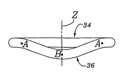
[0026] Figs. 7A-7C are plan, front, and side views of an exemplary
annuloplasty ring of the present invention having a posterior bow;
[0027] Figs. 8A-8C are plan, front, and side views of an alternative
annuloplasty ring of the present invention having a posterior bow between two
raised portions;
[0028] Figs 9A and 9B are front and side elevational views,
respectively, of an inner ring body of a further annuloplasty ring of the
present
invention having an off-center posterior bow and an anterior bow;
[0029] Fig. 10 is a top plan view of an inner ring body of an
annuloplasty ring of the present invention showing details of a composite band
construction; and
[0030] Figs. 11A-11B are plan and front views of an asymmetric
annuloplasty ring of the present invention having a posterior P 1 bow.
DESCRIPTION OF THE PREFERRED EMBODIMENTS.
[0031] The attached figures illustrate several exemplary
embodiments of the annuloplasty ring of the present invention, which can be
described as being continuous and having an anterior side, a posterior side
and
right and left sides. All of the sides are generally curvilinear with no
specific
demarcations to indicate abrupt transitions therebetween. Rather, smooth
transitional sections between the adjacent sides provide curvilinear
connections that give the ring a generally rounded (e.g., oval) configuration.
[0032] An exemplary annuloplasty ring 30 of the present invention
is shown in Fig. 4 implanted around a mitral annulus MA. As described
above, the mitral annulus has an anterior leaflet AL and a posterior leaflet
PL.
When the ring 30 is implanted, the leaflets are brought closer together and
supported so that they meet at a coaptation surface 32. The ring 30 thus
corrects the problem of functional mitral regurgitation.
59491 ECV-5661CIP PCT
SUBSTITUTE SHEET (RULE 26)
CA 02594886 2007-07-13
WO 2006/083529 PCT/US2006/001405
9
[0033] The ring 30 has an oval or somewhat D-shaped
configuration with a relatively straight anterior section 34 opposite a curved
posterior section 36. A pair of trigone or commissure markers 38a, 38b
generally delimit the anterior side 34, while a pair of opposed side sections
40a, 40b extend between each of these markers and the posterior section 36.
A plurality of knotted suture loops 42 are typically used to secure the ring
30
to the mitral annulus MA, although other fasteners such as staples, fibrin
glue,
or the like may be used.
[0034) In the pathological conditions for which the annuloplasty
ring 30 is best suited, the posterior aspect of the mitral annulus is
depressed
relative to the anterior aspect, as is illustrated in Fig. 2. In the view of
Fig. 4,
the posterior aspect will be depressed into the page relative to the anterior
aspect. The annuloplasty ring 30 of the present invention has a shaped
posterior section 36 that generally follows the modified shape of the mitral
annulus MA. In other words, the posterior section 36 is bowed into the page
relative to the anterior section 34. When secured in place with sutures 42,
for
example, the ring 30 supports the mitral annulus MA. in its modified shape,
rather than trying to revert the annulus back to the original substantially
planar
configuration. At the same time, the ring 30 desirably constricts the orifice
circumference defined by the annulus so as to bring the anterior leaflet AL
and
posterior leaflet PL closer together. Because the ring 30 does not pull the
posterior aspect of the mitral annulus MA upward from its modified position,
high stresses are not set up in the attachment sutures 42 and thus there is
less
potential for dehiscence.
[0035] Figs. 5 and 6 illustrate the exemplary annuloplasty ring 30
in perspective above a mitral annulus that is depressed on its posterior side.
The bow of the ring 30 in its posterior section 36 is seen best in Fig. 6
mimicking the depression of the posterior aspect of the mitral annulus MA in
the pathology encountered with functional mitral regurgitation.
59491 ECV-5661CIPPCT
SUBSTITUTE SHEET (RULE 26)
CA 02594886 2007-07-13
WO 2006/083529 PCT/US2006/001405
[0036] The exemplary annuloplasty ring 30 of Figs. 4-6 is shown
in more detail in Figs. 7A-7C. The ring 30 is shown complete with a fabric
covering. For purpose of orientation, Fig. 7A illustrates orthogonal axes
wherein the X- and Y-axes generally define the datum plane 20 as mentioned
5 above with respect to Figs. 1 and 2. The X-axis extends across the ring 30
from one side 40a to the opposite side 40b at the point of maximum
dimension. The X-axis thus defines a major axis of the ring 30. The Y-axis
defmes a plane of symmetry for the ring 30 extending between a midpoint of
the anterior side 34 to a midpoint of the posterior section 36. The Y-axis
also
10 defines a minor axis for the ring 30.
[0037] As with many conventional rings, the ratio of the minor
axis dimension to the major axis dimension is desirably about 3:4. This size
ratio is the "classic" shape of the mitral annulus, and may be the best
configuration of the annuloplasty ring 30. However, it is contemplated that
other shapes that have smaller minor axis-to-major axis ratios may actually
increase leaflet coaptation. Although not geometrically precise, the non-
circular ring configuration may be considered oval, elliptical or D-shaped. It
should be noted that the present invention could also take the form of a
discontinuous ring that has a C-shape, for example. The break in such a ring
may be in the anterior section, and the posterior section is continuous and
exhibits the downward bow as explained.
[0038] The Z-axis in Fig. 7B lies along of the axis of blood flow
through the ring 30 when implanted, and it will be understood that the
positive
Z direction is the "upward" direction, the negative Z direction is the
"downward" direction, and the ring 30 is designed to be implanted in a mitral
annulus such that blood will flow in the downward direction.
[0039] Several points are noted around the ring 30 to help describe
the posterior bow. These points, and the ones shown in Figs. 8A-8B, are
imaginary center points through the cross-section of the ring 30. Two points
59491 ECV-5661 CIP PCT
SUBSTITUTE SHEET (RULE 26)
CA 02594886 2007-07-13
WO 2006/083529 PCT/US2006/001405
11
A are symmetrically located on either side of the Y-axis at an angular
distance
0 from the X-axis. The midpoint of the posterior section 36 is denoted B. The
ring 30 has a posterior bow such that the point B is at the lowest elevation
along the Z-axis. The magnitude of this posterior bow is indicated by the
dimension Z1 in Fig. 7C. The points A on either side of the posterior section
36 represent the location where the posterior bow begins. That is, except for
the posterior section, the ring 30 is preferably substantially planar.
However,
the anterior section 34 can optionally be bowed upward by a distance of
between about 2-4 mm (0.08-0.16 inches), as in certain rings of the prior art.
In the latter example, the posterior section 36 bows downward in the Z-
direction relative to the elevation of the trigone markers 38a, 38b.
[0040] Various possible configurations for the ring 30 as seen in
Figs. 7A-7C are contemplated, with the dimension Z, and the angle 0 varying
between ranges determined by the overall size of the mitral annulus, the
extent of anatomical droop of the posterior aspect, and various other factors
including surgeon preference. Nevertheless, certain ranges are believed
suitable to support and correct a majority of the patients exhibiting the
particular anatomical irregularity as described herein. The downward bow or
posterior bow preferably extends along a majority of the posterior section 36
between the points A, which points are between 0 and 45 from the X-axis (0).
More preferably, the points A are between 20-40 , and more particularly about
from the X-axis. The magnitude of bow Z, may be between about 2-15
mm (0.08-0.59 inches), and more typically is between about 4-8 mm (0.16-
0.31 inches), depending on the size of the ring.
25 [0041] Although the ring 30 is shown in Figs. 7A-7C as symmetric
about the Y-axis, it does not necessarily have to be so. For example, the
point
B may be displaced from the Y-axis such that the downward bow is not
centered in the posterior section 36. An asymmetric ring is shown and
described below with reference to Figs. 9A and 9B.
59491 ECV-5661CIP PCT
SUBSTITUTE SHEET (RULE 26)
CA 02594886 2007-07-13
WO 2006/083529 PCT/US2006/001405
12
[0042] Figs. 9A-8C illustrate an alternative annuloplasty ring 50 of
the present invention that has both upward and downward bows. Again, the
ring 50 is shown complete with a fabric covering. The ring 50 includes an
anterior section 52, a posterior section 54, and a pair of side sections (not
numbered) therebetween. The ring 50 is generally planar on the anterior
section 52 and shaped on the posterior section 54. The points A symmetrically
disposed across the Y-axis again denote the locations on each side where the
ring 50 begins to curve out of a plane. In this embodiment, the ring curves
upward in the Z-direction from the points A, as best seen in Fig. 8B, to high
points C, and then dips downward to the midpoint B of the posterior section
54. The downward bow of the ring between points A and B is shown in Fig.
8C as the dimension Z2, which has a magnitude similar to that given for Z, in
Fig. 7C. The upward curve may be selected so as to better match the patient's
annulus shape. Furthermore, the anterior section 52 may be upwardly bowed
by a distance of between about 2-4 mm (0.08-0.16 inches).
[0043] Various permutations of the ring 50 shown in Figs. 8A-8C
are contemplated, with the dimensions being altered based on numerous
factors. In an exemplary embodiment, the points A are desirably disposed an
angular distance a from the X-axis of between about 0-15 , and more
desirably between about 5-10 . The points C of maximum height of the ring
50 are preferably spaced an angular distance (3 from the X-axis of between
about 15-45 , and more preferably between about 25-35 . The lowest point B
of the ring 50 may be bowed along the Z-axis as in the embodiment of Figs.
7A-7C, so that, as indicated Fig. 8C, Zz is desirably between about 2-15 mm
(0.08-0.59 inches), and more typically is between about 4-8 mm (0.16-0.31
inches), depending on the size of the ring. Therefore, the total height of the
ring 50 is at least 2 mm, and may be greater than 15 mm.
[0044] Figs. 9A and 9B show an inner ring body 60 for use in an
annuloplasty ring of the present invention. The ring body 60 has a posterior
59491 ECV-5661 CJP PCT
SUBSTITUTE SHEET (RULE 26)
CA 02594886 2007-07-13
WO 2006/083529 PCT/US2006/001405
13
bow 62 that is offset from the center of a posterior section 64. In the
illustrated embodiment, the bow 62 is offset toward the posterio-medial side
(to the right) by about 20% of the entire major axis width of the ring body
60.
Another way to state the offset is that, in plan view, the bow 62 is centered
at a
clock position, with 12:00 being centered in the anterior side. In that sense,
the bow 62 is centered between 3:00 and 6:00, and more preferably is centered
at about 5:00. The axial bow Z3 is shown and may vary from about 2.0 mm
(0.08 inches) to about 4.0 mm (0.16 inches), and more preferably from about
3.0 mm (0.12 inches) to about 3.8 mm (0.15 inches), depending on ring size.
In addition, the ring body 60 has an anterior section 66 that is upwardly
bowed
by a distance of between about 2-4 mm (0.08-0.16 inches).
[0045] The inner ring body 60 demonstrates an asymmetric ring
that conforms to patients that have a posterior annular bow that is displaced
from the midline. It is believed that most patients have such a malformed
anatomy resulting from the pathologic conditions described herein. However,
posterior bows that are centered or even offset to the left have been
observed.
Therefore, one configuration of ring that is embodied in the present invention
is one that is pre-shaped with a posterior bow in the middle or to the right,
and
that is malleable so that the bow can be exaggerated or diminished by the
surgeon after examination of the precise shape of the patient's annulus.
Further, in such a convertible ring the bow can even be displaced, from the
right to the left, for example. Although the material of the ring permits
manual deformation, it would be stiff enough to withstand further deformation
once implanted and subjected to normal physiologic stresses.
[0046] The ring preferably includes an inner ring body and an
outer sewing sheath that permits the ring body to be sutured into the mitral
annulus. The sewing sheath should be sufficiently porous and/or flexible to
permit sutures to be passed therethrough. One exemplary construction is to
enclose the inner ring body in a tubular sheath of suture-permeable material,
59491 ECV-5661CIP PCT
SUBSTITUTE SHEET (RULE 26)
CA 02594886 2007-07-13
WO 2006/083529 PCT/US2006/001405
14
such as silicone, which is then covered with a fabric tube, such as polyethyl
terapthalate.
[0047] As opposed to flexible annuloplasty rings that are designed
simply to reduce the circumference of the mitral annulus, the annuloplasty
ring
of the present invention must be semi-rigid. It must retain its posterior bow
in
opposition to the stresses that will be imparted by muscles of the heart
throughout each beating cycle. For example, the ring body may be made from
materials such as Elgiloy (a cobalt-nickel alloy), titanium, or Nitinol (a
nickel-
titanium alloy).
[0048] Fig. 10 illustrates one exemplary construction of the inner
body of the annuloplasty rings of the present invention that utilizes multiple
flat bands of Elgiloy in a composite structure. Specifically, there are four
bands 70a, 70b, 70c, and 70d from the outside to the inside. The four bands
are concentrically disposed in the shape of the ring. Each band is a flat
strip of
material having a width of between about 1.4-2.0 mm (0.056-0.078 inches).
In one embodiment, the bands 70 overlap in the anterior section 72 of the ring
body and are fastened together by, for example, spot welding at multiple
points. The width of each strip may also be greater in the anterior section 72
than in a posterior section 74, which means that the ring body is more
flexible
in the posterior section than in any other section. Although not shown, a
plurality of strips of protective film is used in between each band 70, and on
the outer face of the outer band 70a. The strips may be a polymer such as
Mylar. The strips help reduce rubbing between the bands 70 and also deflect
suture needles from the outer band 70a and thus prevent scratching thereto.
[0049] A still further alternative annuloplasty ring 80 is shown in
Figs. 11A-I1B with a fabric covering. As before, Fig. 11A illustrates
orthogonal axes wherein the X- and Y-axes generally define the datum plane
20 as mentioned above with respect to Figs. 1 and 2. The X-axis extends
across the ring 80 from one side 82a to the opposite side 82b at the point of
59491 ECV-5661CIP PCT
SUBSTITUTE SHEET (RULE 26)
CA 02594886 2007-07-13
WO 2006/083529 PCT/US2006/001405
maximum dimension. The X-axis thus defines a major axis of the ring 80.
The Y-axis extends between a midpoint of an anterior side 84 to a midpoint of
a posterior side 86 and defmes a minor axis for the ring 80. The posterior
side
86 extends around the lower portion as seen in Fig. 11A between a pair of
5 trigone markers 88a, 88b.
[0050] The Z-axis in Fig. 11B lies along of the axis of blood flow
through the ring 80 when implanted, and it will be understood that the
positive
Z direction is the "upward" direction, the negative Z direction is the
"downward" direction, and the ring 80 is designed to be implanted in a mitral
10 annulus such that blood will flow in the downward direction. An outline of
the annuloplasty ring 80 in plan view is superimposed on the elevational view
of Fig. 11B for a better understanding of the shape.
[0051] As seen in plan view of Fig. 11A, the ring 80 has an
asymmetric shape on its posterior side 86. For purposes of illustration, the
15 posterior side 86 may be divided into sections P1, P2, and P3 as shown. The
native posterior leaflet is divided into three scallops in sequence starting
from
the anterolateral trigone and continuing in a counterclockwise direction to
the
opposite trigone, and the ring sections P1, P2, P3, generally correspond to
these scallops. The asymmetry in the ring 80 is manifested by an extended Y-
axis dimension in the P2-P3 area, while the P 1 area is preferably more
conventionally shaped.
[0052] Points are noted around the ring 80 to help describe one
preferred embodiment of a posterior bow 90. As in the earlier illustrations,
these points are imaginary center points through the cross-section of the ring
80. Two points A represent the locations where the posterior bow 90 begins
and ends. In the illustrated embodiment, the ring 80 has a posterior bow 90
that is offset toward the P1 section, and bridges the P1-P2 sections.
Alternatively, the posterior bow 90 may be located entirely or substantially
within the P1 section. Except for the downward bow 90, the ring 80 is
59491 ECV-5661 CIP PCT
SUBSTITUTE SHEET (RULE 26)
CA 02594886 2007-07-13
WO 2006/083529 PCT/US2006/001405
16
preferably substantially planar, although the aforementioned upward curves as
in Figs. 8A-8C on either side of the posterior bow may be included. Also, the
anterior section 84 can optionally be bowed upward, such as, for example, by
a distance of between about 2-4 mm (0.08-0.16 inches), as in certain rings of
the prior art. In the latter example, the posterior section 86 bows downward
in
the Z-direction relative to the elevation of the trigone markers 88a, 88b. The
annuloplasty ring 80 is particularly useful for repairing pathologies
associated
with ischemic cardiomyopathy and anterior infarct.
[0053] It will also be readily apparent that supporting the mitral
valve annulus with the present annuloplasty ring will maintain the posterior
leaflet depressed below the anterior leaflet, and thus the area of coaptation
therebetween will be different than in a healthy valve. This is required by
the
pathology of the ventricle with displacement of the papillary muscles and
posterior leaflet. However, those of skill in the art will recognize that this
slight realigmnent of the leaflets is acceptable because of the surplus area
of
the leaflets available for coaptation, and because the realignment will be
offset
by other changes to the shape of the annulus that should over time improve
coaptation of the two leaflets and therefore decrease regurgitation.
[0054] It will also be appreciated by those of skill in the relevant
art that various modifications or changes may be made to the examples and
embodiments of the invention described in this provisional application,
without departing from the intended spirit and scope of the invention. In this
regard, the particular embodiments of the invention described herein are to be
understood as examples of the broader inventive concept disclosed in this
application.
59491 ECV-5661 CIP PCT
SUBSTITUTE SHEET (RULE 26)