Note: Descriptions are shown in the official language in which they were submitted.
CA 02595268 2007-07-18
WO 2006/081324 PCT/US2006/002708
DNA PURIFICATION AND ANALYSIS ON NANOENGINEERED SURFACES
GOVERNMENT INTERESTS
This invention was made with government support under SBIR Grant
No. GM072178-01. The government may have certain rights in the invention.
CROSS REFERENCE TO RELATED APPLICATIONS
This application claims benefit from U.S. Patent Application Serial No.
11/043,561, filed January 26, 2005, which application is incorporated herein
by reference.
BACKGROUND OF THE INVENTION
1. Field of the Invention
The present invention relates generally to the purification of DNA, and, in
particular, the use of nanoengineered surfaces in the purification and
analysis
of DNA.
2. Description of the Prior Art
Technology to simplify the isolation, handling and measurement of
genomic DNA is required for portable medical diagnostics or biodefense
applications. The quality of nucleic acid isolated from human tissue is
critical
for reproducible, accurate and informative genetic analysis data. As the
commercial importance of DNA isolation products has grown, there has been
much work done in this area.
One technique in which much progress has been made is the use of
microfluidic devices to perform genetic analysis. Many microarray assay
formats are available, and they share the ability to allow simultaneous
interrogation of a vast number of genetic targets from a single sample.
1
SUBSTITUTE SHEET (RULE 26)
CA 02595268 2007-07-18
WO 2006/081324 PCT/US2006/002708
Sensitive solution phase assays for measuring concentration of double
stranded DNA (dsDNA) have already been developed using fluorogenic minor,
groove binding molecules (MBs). When DNA is not present, the molecule can
rotate freely in solution, and this is critical for its low fluorescent
background.
Upon binding to dsDNA, the compound assumes a rigid planar conformation
in the hydrophobic minor groove that has increased fluorescence. By
carefully engineering the attachment of fluorogenic MBs to surfaces, the
quantity of DNA can be measured as it is transferred through a microfluidic
device.
SUMMARY OF THE INVENTION
It is therefore an object of the present invention to provide a
rnicrofluidic device that simultaneously isolates genomic DNA, measures the
amount of purified DNA, and distributes standardized DNA solutions of
specific concentration for downfield analysis.
It is a further object of the present invention to provide a microfluidic
device for DNA isolation and preparation in which the cost and complexity of
the device is minimized.
It is a further object of the present invention to provide a microfluidic
device that can simultaneously capture double stranded DNA (dsDNA) from
biological fluids and measure the amount of DNA immobilized.
It is a still further object of the present invention to synthesize
fluorogenic minor groove binder agents (MBs) with electrophilic or
nucleophilic
linker groups and demonstrate increased fluorescence in the presence of
dsDNA.
2
SUBSTITUTE SHEET (RULE 26)
CA 02595268 2007-07-18
WO 2006/081324 PCT/US2006/002708
These and other objects of the present invention will be more readily
apparent from the descriptions and drawings which follow.
BRIEF DESCRIPTION OF THE DRAWINGS
FIG. 1 is a depiction of the structure of Hoechst 33258 bound to a synthetic
12- mer DNA duplex;
FIG. 2 shows the structure of reactive analogs of H33258 developed for
attachment to linkers on DNA probes;
FIG. 3 is a depiction of the interaction of two fluids flowing within a
microfluidic
device;
FIG. 4 is a depiction of several amine-modified surfaces with attached
fluorogenic minor groove binders (MBs);
FIG. 5 shows the synthesis of fluorogenic MBs with reactive groups;
FIG. 6 is a depiction of the activation of amine modified glass surfaces of a
slide with cyanuric chloride in an organic solution;
FIG. 7 shows possible chamber designs for a microfluidic device suitable for
use in the present invention;
FIG. 8 is a front view of a microfluidic card suitable for use in the present
invention;
FIG. 9 is an exploded view of a microfluidic card with its layers separated;
FIGS. 10 A-E, taken together, depict a front view of the card of FIG. 9
showing the sequence of events of the card while in use;
FIG. 11 shows the synthesis of the fluorogenic Hoechst dye with an attached
hexylamine linker;
FIG. 12 is a chart showing the fluorescence of the BB-NH 2 molecule vs. BB-
3
SUBSTITUTE SHEET (RULE 26)
CA 02595268 2007-07-18
WO 2006/081324 PCT/US2006/002708
OH excited at 360 nm.
FIG. 13 is a chart showing the fluorescence at 460 nm for four different
bisbenzimide surfaces;
and
FIG. 14 is a chart showing the amount of DNA bound versus the amount of
DNA offered for glass and plastic cover slips.
DESCRIPTION OF THE PREFERRED EMBODIMENTS
Sensitive solution phase assays for measuring the concentration of ds
DNA have already been developed using fluorogenic minor groove binder
(MB) agents (Hoechst 33258). When DNA is not present, the molecule can
rotate freely in solution; this is critical for its low fluorescent
background.
Upon binding to dsDNA, the compound assumes a rigid planar conformation
in the hydrophobic minor groove that has increased fluorescence. By
carefully engineering the attachment of fluorogenic MBs to surfaces, the
quantity of DNA can be measured as it is transferred through a microfluidic
device. Conjugation chemistry is used to position the fluors on
macromolecular structures designed to maximize conformational flexibility and
access to target DNA.
The Hoechst 33258 molecule has a polycyclic structure that can form a
crescent shaped structure which is isohelical with the minor groove of B-form
DNA. FIG. I shows the solution NMR structure of Hoechst 33258 bound to a
synthetic 12 - mer DNA duplex (PDB structure ID number: I QSX). The
phenol terminus of the crescent shaped molecule is a convenient attachment
point for attachment in conjugate groups. The structure in aqueous solution is
4
SUBSTITUTE SHEET (RULE 26)
CA 02595268 2007-07-18
WO 2006/081324 PCT/US2006/002708
flexible, and relatively non-fluorescent. Excitation of the unbound molecule
with 356 nm light gives weak fluorescence at 492 nm. However, where bound
to dsDNA, the benzimidazole rings become fixed in a planar conformation,
and an extensive aromatic system develops that emits strong fluorescence at
458 nm. The molecule is shielded from water molecules in the minor groove,
and this further increases fluorescence. These fluorogenic properties of
H33258 have led to the development of sensitive assays for measuring
dsDNA concentrations and, as a simple assay, for cell counting.
There has been much interest in the structure of H33258 bound to
DNA. The MB is held in place by a combination of hydrophobic interactions,
van der Waals forces, and hydrogen binding by the imidazole residues. The
strong minor groove binding of H33258 may also provide a mechanism for
direct isolation of dsDNA. Minor groove binding of MBs to adenine/thymine
(A/T) rich sequences is preferred, and the DNA binding constants can be
orders of magnitude larger than other small MW agents such as intercalators.
Intercalating fluorgenic agents have also been extensively explored. An
example of this class of DNA binding agents is ethidium bromide, which is
commonly used in molecular biology to stain DNA in gels after
electrophoresis. Methidium dyes have been attached to sepharose particles
to provide a DNA isolation product. Intercalating dyes like methidium are not
as strong binding as the minor groove binder class of dyes. Methedium was
attached to sepharose particles using a cationic spermine linker, and this
undoubtedly improved DNA binding. In addition, ethidium only shows a small
increase in quantum yield upon binding to dsDNA. The fluorescent properties
5
SUBSTITUTE SHEET (RULE 26)
CA 02595268 2007-07-18
WO 2006/081324 PCT/US2006/002708
of the methidium-sepharose particles were not described.
The cyanine type intercalating dyes are much more fluorgenic than
ethidium intercalators. Thiazole orange (TO) is an asymmetric cyanine dye
composed of two bicyclic aromatic ring structures. When excited with 509 nm
light, these two structures can rotate freely with respect to each other, and
there is almost no fluorescence. However, in the presence of dsDNA, TO
intercalates between base pairs and forces the ring systems to become
planar. An extensive aromatic system forms that emits strong fluorescence at
533 nm. TO does not bind as tightly to dsDNA as the minor groove binding
agents, but two TO molecules can be linked together to form strong binding
bis-intercalators.
Sensitive DNA binding assays have been developed based on the fluorogenic
properties of the cyanine type dyes. TO also shows up to 3000 fold increase
in fluorescence in the presence of RNA. This property is interesting, and may
be applicable to an RNA isolation device.
These fluorogenic compounds have been tethered to oligonucleotides
to create DNA probes that can monitor the formation of dsDNA in
hybridization assays. Several reactive derivatives of H33258 were developed
for attachment to DNA probes. FIG. 2 shows several reactive analogs of
H33258 which were developed for attachment to linkers on DNA probes. For
example, bromoacetyl analogs of H33258 were conjugated to sulfhydryl
modified oligonucleotides. Sensitive DNA binding assays have been
developed based on flurogenic properties of the cyanine type dyes. TO also
shows up to 3000 fold increase in fluorescence in the presence of RNA. This
6
SUBSTITUTE SHEET (RULE 26)
CA 02595268 2007-07-18
WO 2006/081324 PCT/US2006/002708
property is interesting, and may be applicable to an RNA isolation device.
These fluorogenic compounds have been tethered to oligonucleotides
to create DNA probes that can monitor the formation of dsDNA in
hybridization assays. Several reactive derivatives of H33258 were developed
for attachment to DNA probes. FIG. 2 shows several reactive analogs of
H33258 which were developed for attachment to linkers on DNA probes.
Hybridization to complimentary single strand DNA targets gave increased
binding affinity and up to 23-fold increase in fluorescence upon binding. The
improved binding efficiency (DNA melting temperature) was sequence
specific. When A/T regions were located near the attachment point of ligand,
Tnn dramatically increased, indicating the strong DNA binding effects of
H33258. Similar synthetic chemistry may be used to prepare reactive analogs
of these fluorogenic dyes and conjugate them to amine modified glass or
plastic surfaces.
Cyanine dye conjugates of oligos also gave the expected hybridization
triggered fluorescence upon addition of complementary strands of DNA.
Fluorescence varied with the terminal sequence of the formed duplex.
Binding of TO to DNA duplexes is known to be sequence specific. NHS ester
analogs of cyanine dyes were reacted with amine modified oligonucleotides to
give DNA probes that were used in fluorogenic "real-time" PCR assays. Up to
20-fold increase in fluorescence was observed with the oligonucleotide
conjugates. DNA probe applications of both MGB and cyanine-type
fluorogenic oligonucleotide conjugates were complicated by sequence specific
7
SUBSTITUTE SHEET (RULE 26)
CA 02595268 2007-07-18
WO 2006/081324 PCT/US2006/002708
binding of the dyes. Measurement of dsDNA binding should not be affected
by sequence specificity of the binding sites, as the focus is heterogeneous
populations of genomic fragments of DNA.
Nucleic acids have a very high affinity for glass surfaces in the
presence of high concentrations of chaotropic salt solutions, and a number of
glass based DNA isolation products are commercially available. These
products are simple to use, but require multiple pipetting steps, Eppendorf
tubes or 96-well plates, and access to equipment such as vortexers, and
centrifuges. The operations take place in an open lab and require relatively
large sample volumes to avoid problems with evaporation of solutions. DNA
concentrations are usually measured by UV-vis spectrophotometers, and
good technique is required for reproducible results. Nonetheless, use of glass
surfaces for isolation of DNA from a variety of biological samples has been
demonstrated.
An early application of DNA binding to glass was for isolation of
electrophoretically purified DNA from agarose gels. The DNA binding
capacity of flat soda lime or borosilicate glass surfaces were comparable:
approximately 300 ng of DNA per 800 mm2. 800 mma is roughly the surface
area of a chamber formed between a glass microscope slide and a 20x20 mm
cover slip. Most macrofluidic DNA isolation applications use powdered glass
from tubes or fibers, silicon dioxide, or the silica skeletons of diatoms, as
these supports have a much higher surface area than flat surfaces. A
microfluidics device can use lower binding flat glass surfaces since the
volumes of solutions are smaller (no evaporation problems) and the amount of
8
SUBSTITUTE SHEET (RULE 26)
CA 02595268 2007-07-18
WO 2006/081324 PCT/US2006/002708
captured DNA required for PCR based genomic analysis is very small. Flat
glass is available in a variety of compositions, sizes, and roughness, and can
be further etched if more surface area is needed.
Solution phase analysis of mRNA or DNA can be accomplished during
PCR using DNA oligonucleotide probes bearing fluorescent reporter groups at
one terminus and a fluorescent quencher molecule at the other. In one format
(the TaqMan assay), the probes are short single stranded oligos that are
digested by the 3'-exonuclease activity of Taq polymerase. The other
fluorogenic format (Molecular Beacon assay) uses hairpin shaped oligos with
adjacent fluor and quencher molecules at the stem. The hairpins open in the
presence of complementary DNA strands to give a fluorescent signal. Each
of these fluorogenic formats can be used to monitor the progress of PCR, as
each successful amplification cycle gives an increase in fluorescent signal.
Both of these PCR based formats are ideal for microfluidics based platforms
since the lower limit of assay size is only dictated by handling problems
(drying of small volumes) and resolution of the available instruments. A
popular (high-throughput) thermal cycling fluorimeter is the ABI7900HT which
can analyze 384 well plates, with 10uL per well for each gene that is
measured during PCR. 10 pg - 10 ng of genomic DNA per well is
recommended, depending on efficiency of the PCR system, and the quality of
the DNA.
Glass microscope slides are a common substrate for DNA microarrays.
Many microarray assay formats are available, and share the ability to allow
simultaneous interrogation of vast number of genetic targets from a single
9
SUBSTITUTE SHEET (RULE 26)
CA 02595268 2007-07-18
WO 2006/081324 PCT/US2006/002708
sample. Arrays of synthetic oligonucleotides have been synthesized directly
from glass supports, or prepared by endpoint attachment of modified synthetic
probes. Amine modified glass is a popular substrate for endpoint attachment.
Gamma aminopropyl silane (GAPS) is a volatile reagent that can be used to
functionalize glass surfaces and there are several manufacturers of GAPS
slides. The amine coated surface of GAPS slides can be activated using
bifunctional linkers to create an electrophilic surface which react with
solutions
of amine modified oligos. Variations of this conjugation chemistry may be
used to attach fluorogenic DNA binding dyes.
The density of attachment of oligonucleotides is an important factor in
hybridization performance. High densities of oligos can be achieved by direct
synthesis from glass, but it has been shown that hybridization of labeled DNA
targets actually dropped when the capture probes were too tightly packed.
The hybridization performance also improved when the length of the linker
between the glass attachment point and the oligonucleotide sequence was
lengthened. Polyethylene glycol (PEG) linkers up to 80 atoms in length
continued to improve hybridization signal. This shows the importance of long
linkers between the solid support and the DNA probe in order to mimic the
solution phase performance.
Attachment of DNA probes to glass surfaces via long polyethylene
glycol linkers improved performance. Macromolecular PEG linkers with
various functional groups are commercially available (Quanta, Biodesign,
Powell, Ohio) and have been used primarily to attach to protein drugs to
improve pharmacokinetic properties. Other macromolecular structures have
r
10
SUBSTITUTE SHEET (RULE 26)
CA 02595268 2007-07-18
WO 2006/081324 PCT/US2006/002708
been used to improve performance of immobilized DNA probes. For
example, biotin modified molecular beacons have been attached to avidin
coated glass supports for use in sequence specific DNA analysis. The use of
avidin or streptavidin as spacers to position the DNA away from the glass
surface is likely important for the function of the probes. Another type of
macromolecular linkers to consider are dendrimeric polymers. These
commercially available (Aldrich Chemical Co., Milwaukee, WI) moiecules are
synthesized in alternating layers of amino groups and carboxylate groups
depending on the "generation" of synthesis. The 3-dimensional structures of
these molecules are well controlled, and the properties are well understood.
Amine rich dendrimeric structures are analogous to the histone proteins that
are critical for DNA packaging in cells. They have been used successfully for
transfection of DNA sequences into cells for potential gene therapy
applications. It is believed that dendrimeric linkers will provide excellent
scaffolds for presenting immobilized MB molecules into DNA rich solutions.
Biosensor films containing dendrimer linkers and fluorogenic DNA detecting
molecules (Sytox 13, Molecular Probes) were used to measure levels of live
bacteria on plastic surfaces. These materials did not contain a covalent link
between the linker, fluorogenic dye or plastic surface, but were held together
by hydrophobic forces.
DNA microarray technology provides miniaturized assays that benefit from
enclosed microfluidics processing. For example, a micromachined chemical
ampiifier operated with continuous flow demonstrated a 20-cycle polymerase
chain reaction (PCR) amplification of a 176-base pair fragment. PCR
11
SUBSTITUTE SHEET (RULE 26)
CA 02595268 2007-07-18
WO 2006/081324 PCT/US2006/002708
amplification of genomic DNA targets from white blood cells captured behind
weirs in the flow chamber has been demonstrated from whole blood in silicon-
glass microchips. A moderate density array method using DNA chips arrays
was developed for diagnostic sequencing of PCR products to reduce their
cost and complexity in use. Antibiotic resistant clinical isolates were
visually
detected within one hour after PCR amplification. Another application is the
DNA sequence-based identification of toxic medicinal plants used in
Traditional Chinese medicine. In 2001, Petrik reviewed the use of microarray
devices originally developed for genomic projects for mass screening of blood
donations for hepatitis C virus. An integrated system for PCR analysis was
attained by combining dual Peltier thermoelectric elements with
electrophoretic sizing and detection on a microchip. Using a DNA
concentration injection scheme enabled detection of PCR products within as
few as ten thermal cycles. A microfluidic cartridge was developed to prepare
spores for PCR analysis by sonication, addition of PCR reagent to the
disrupted spores, and insertion of the mixture in a PCR tube. The processing
and detection of the spore DNA was completed within 20 minutes. An
integrated microfluidic device was developed that combines stochastic PCR
amplification of a single DNA template molecule followed by capillary
electrophoretic analysis of the products. A histogram of the normalized peak
areas from repetitive PCR analyses revealed quantization due to single viable
template molecule copies in the reactor. A microfluidic chip for detecting RNA
amplified by nucleic-acid-sequence-based amplification (NASBA) was
developed. Samples of cryptosporidium parvum were detected and clearly
12
SUBSTITUTE SHEET (RULE 26)
CA 02595268 2007-07-18
WO 2006/081324 PCT/US2006/002708
distiguishable from controls without separating the amplified RNA from the
NASBA mixture. An automated rnicrof(uidic system for nanoliter DNA analysis
directly from cheek cells has been developed, including all the steps needed
for DNA analysis: injection, mixing, lysis, PCR, separation, sizing, and
detection. The possibility of further miniaturization of the system was
established. An integrated microfluidic chip-based system has been
developed for quality control testing of a recombinant, adenoviral, gene
therapy product. The viral identity test sizes and quantitates the DNA
fragments and requires 100-fold less sample than the agarose gel method.
It is desired to develop a DNA isolation device that will process a small
volume of human blood (<20 pL) and deliver standardized genomic DNA
samples for genetic analysis. A single use microfluidics card can be
developed which may be inserted into a micropump driven instrument that
controls fluid flow through various DNA processing chambers. The
immobilized and quantitated DNA is either released in solution for immediate
use, or stored for future analysis. The ability to measure the capture and
release of immobilized DNA in the device by fluorescence will allow in-
process control of this novel DNA processing system. The technology can be
developed into a stand-alone product for general use in molecular biology
research, or be incorporated into more sophisticated devices to simplify
genetic analysis for biomedical applications. The standardized solutions of
purified and denatured DNA that are isolated with the single well device
developed can be distributed via microfluidic channels to various microwell
plate formats for genetic analysis by real-time PCR or DNA microarrays.
13
SUBSTITUTE SHEET (RULE 26)
CA 02595268 2007-07-18
WO 2006/081324 PCT/US2006/002708
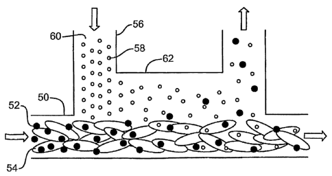
FIG. 3 is intended to display the micro-scale physics needed for development
of miniaturized biomedical devices that can be used for isolation of genomic
DNA from cells.
FIG. 3 shows the interaction between whole blood and another fluid
flowing within the channels of a microfluidic device where isolation of DNA
from whole blood is envisioned. It is envisioned that microscale physics used
in the development of miniaturized biomedical devices can be used for
isolation of genomic DNA from cells. Referring now to FIG. 3, a first stream
50 containing small particles 52 in whole blood 54, and a second stream 56
containing small particles 58 in a clear solution 60, are introduced into a
common channel 62, where they form a laminar fluid diffusion interface
(LFDI). Depending on their diffusion coefficient, particles 52, 58 start to
diffuse
across the LFDI, with the smaller particles 58 diffusing more quickly. The
device can be used to extract small particles from whole blood, or to
introduce
a reagent into whole blood in a predictable continuous way.
A preliminary observation from studies of Hoechst dyes is also
pertinent to this application. A droplet of Hoechst dye in water or buffer
solutions shows increased fluorescence as the spot dries. This relates to the
"hydration content" of the fluor environment and effects on fluorescent
background. The fluor must be well hydrated for low background, and
stresses the importance of hydrophilic surface coatings.
Initial focus will be synthesis and testing of fluorogenic solid supports
for binding and measurement of dsDNA. Fluorogenic MBs with amine
reactive functional groups will first be prepared using variations of known
14
SUBSTITUTE SHEET (RULE 26)
CA 02595268 2007-07-18
WO 2006/081324 PCT/US2006/002708
methods. These agents will be immobilized to amine modified glass or
plastic, and fluorescence will be measured before and after treatment with
DNA. Various linker structures will be evaluated with respect to their ability
to
measure fluorescence vs. DNA concentration. Fluorogenic surfaces with
attached MBs will be engineered to provide low fluorescent background and
good DNA binding. Illustrations of typical surfaces to be studied are shown in
FIG. 4. Variables such as solid support (glass or plastic), linker chemistry,
and capping groups will be explored. Stoichiometry (density of attachment) of
fluorogenic MBs will be examined to determine the surface with the best
dynamic range for measurement of DNA. DNA binding capacity will also be
examined.
Referring now to FIG. 4, device 100 has an amine-modified surface
102 with attached minor groove binders (MB) 104. Unreacted amines can be
left to provide a cationic surface. Device 110 shows that unreacted amines
can be capped by succinylation to provide an anionic surface 112. Device
120 shows that MBs 104 can be extended into solution by first attaching a
polymeric amine containing linker (starburst dedrimers, diamino-PEG, Poly-L-
lysine) to surface 122 and further reacting with MB derivatives.
Concurrent with the development of the required fluorogenic surfaces,
a microfluidics device for DNA isolation is envisioned. Various size DNA
samples will be obtained (Sigma), and efficiency of capture will be determined
by fluorescence measurement in both solid phase and solution phase using
the same fluorogenic compounds. Release of the DNA from the device will be
monitored to ensure that DNA purity and recovery are good. Conditions for
15
SUBSTITUTE SHEET (RULE 26)
CA 02595268 2007-07-18
WO 2006/081324 PCT/US2006/002708
the capture and release of DNA will be measured on a fluorescent plate
reader. After the DNA binding capacity and washing experiments are
executed, a microfluidic device can be constructed. After the fluorogenic
supports are developed, the DNA capture and measurement functions of the
device can be combined.
Since a variety of amine modified solid supports are available,
derivatives can be prepared with active electrophilic functional groups such
as
iodoacetate groups, cyanuric chloride groups, NHS ester groups, and other
amine reactive moieties. There are two synthetic approaches that have been
shown to give H33258 analogs with desired electrophilic or nucleophilic
"handles" that can react with complementary functional groups on the solid
support. The most direct route is from the fully assembled heterocyclic ring
system of H33258. Reaction of an bromoalkyl derivative of PEG has been
shown to react with H33258 under basic conditions to give the aryl ether in
high yield. H33258 is available from Aldrich Chemical Co. (500 mg =
$220.80). The t-butyloxycarbonyl protected hexylamine linker is easily
synthesized, and a tosylate derivative has been made. The bromo derivative
should be easily accessible and reacted with H33258, as is shown in FIG. 5.
Referring now to FIG. 5, Hoechst 33258 is treated with an alkylating Boc
derivative. The nucleophilic aminoalkyl group (R=H) can be immobilized
directly to the electrophilic surfaces. Alternatively, the amino group can be
converted to an electrophilic group such as the cyanuric chloride derivative,
as shown in FIG. 5, for immobilization to nucleophilic surfaces. The Boc is
removed by treatment with dilute acid, and then neutralized with base to give
16
SUBSTITUTE SHEET (RULE 26)
CA 02595268 2007-07-18
WO 2006/081324 PCT/US2006/002708
the primary hexylamine group (R = H). The nucleophilic primary amine can
react selectively with electrophiles in solution with little competing
alkylation
by the tertiary amine. This chemistry should be applicable to modification of
solid surfaces that are coated with electrophilic functional groups.
Alternatively, the H33258 hexylamine analog can be reacted with a
bifunctional linker that leaves an electrophilic residue. Cyanuric chloride
behaves like a bifunctional linker to join to amine containing functional
groups.
The remaining third chloro group is unreactive after the two amino groups are
linked. This conjugation chemistry can be utilized later for synthesis of
modified surfaces. Another alternative is to bind the cationic H33258 to
anionic surfaces, which as that formed by carboxylic acid groups. These
surfaces may be suitably stable and flexible to allow the desired fluorogenic
response with DNA.
Like most solid phase chemistry, large excess of reagent (with respect
to available surface groups) can be easily achieved and the surfaces are
easily washed to remove excess reagent. Therefore, coupling efficiencies
can be quite high (dense packing can be achieved). Surface area of glass
and plastic substrates, immobilization chemistry, linker type and density of
"linkable" fluorogenic compounds (fluors) will be optimized. Commercially
available dendrimeric linkers, poly-L-lysine, and polyethylene glycol diamines
should position the fluors away from the surface and toward dsDNA in
solution. The goal is to maximize conformational flexibility of the
immobilized
fluors (to minimize background) and maximize accessibility to dsDNA (to
maximize DNA binding and kinetics).
17
SUBSTITUTE SHEET (RULE 26)
CA 02595268 2007-07-18
WO 2006/081324 PCT/US2006/002708
Amine modified glass and plastic supports are available as 1x3 inch
microscope slides and are a common substrate for DNA microarrays. Most
glass slides are made from soda lime glass (Erie Scientific, Portsmouth, NH)
and modified with (gamma) aminopropylsilane groups to give a uniform
density of primary amines. GAPS slides are also available with other glass
types (Corning Glass), or with increased roughness (Erie). Aminopropyl
coated plastic slides are also available (Nunc, Denmark) for use. Polyester
films (Mylar) functionalized with amines are also available as sheets
(Diagnostic Laminations Engineering, Oceanside, CA or Adhesive Research,
Inc., Glen Rock, PA) and will also be examined. These substrates will either
be used directly for immobilization of electrophilic MB analogs, or they will
be
"activated" with bifunctional linkers to give an electrophilic surface. FIG. 6
shows one possible conjugation chemistry system. Cyanuric chloride
activation should be examined, as well as other aliphatic bifunctional linkers
(Pierce Chemical Co.). Glass slides can be activated in anhydrous organic
solvents with hydrophobic electrophiles such as cyanuric chloride to avoid
hydrolysis of reagents and achieve higher densities of electrophilic
modification. Thus, high loading of PEG amines (or other polyamines) may
be expected. Direct coupling of fluorogenic MB electrophiles (for example,
the cyanurate of FIG. 5) will be attempted first, and this (no linker)
fluorogenic
surface should be characterized and used as a control for fluorescence
studies of the various surfaces.
Plastic slides require milder aqueous activation conditions and
carbodiimide coupling or the use of activated ester linkers. PEG
18
SUBSTITUTE SHEET (RULE 26)
CA 02595268 2007-07-18
WO 2006/081324 PCT/US2006/002708
macromolecules containing NHS esters at each terminus are commercially
available. Commercially available polycarboxylate dendrimers can possibly
be coupled to amine containing supports using water soluble carbodiimide
(EDC) chemistry. The hydrophilic Generation 3.5 and 4.5 PAMAM
dendrimers are particularly attractive since these molecules have 64 and 128
surface carboxylates respectively. These molecules do not exhibit the dense
"starburst" crowding that affects larger dendrimers. After blocking residual
amines on the surface (with acetic anhydride) the carboxylates on the linker
can be activated again with EDC and coupled to the hexylamine containing
MB, as can be seen in FIG. 5. After thorough washing, the amount of MB
covalently bound to the slide is measured by absorbance on the plate reader.
The Hoechst 33258 chromophore absorbs at 356 nm, and the Bio-Tek plate
reader can be set up with appropriate filters to measure this wavelength
directly on microscope slides. The slides can be dried, and evaluated for
future use. Stability of monolayers of fluorescent dyes on the surfaces
(storage conditions) should be investigated.
The macromolecular linker structures will act as scaffolds to display the
fluorogenic MB sensors to dsDNA in solution. The fluorescent properties of
surface structures should be examined in the presence and absence of
dsDNA. It is important that the surfaces be fully hydrated when they are
evaluated for fluorogenic DNA binding properties, as H33258 derivatives are
known to fluoresce when dried down on glass. Equilibration times are more
rapid in a microfluidic environment, and this may be an advantage.
Fluorescent background of the various linker structures will first be measured
19
SUBSTITUTE SHEET (RULE 26)
CA 02595268 2007-07-18
WO 2006/081324 PCT/US2006/002708
by attaching a "perfusion chamber" to the surface of the slide of interest.
These are simple rubber gaskets that are adhered to the slides, and then
covered with a cover slip that has an inlet port and outlet port. After the
chamber is constructed, the analyte solution of interest is pipetted in and
the
ports can be sealed. A 200 uL chamber size will be used.
Various size DNA samples will be obtained from an outside source
(Sigma Chemical Co.). The genomic DNA will likely need to be sheared to
avoid tangling as it passes through the microchannels under the mild flow
conditions used in the microfluidics device. Shearing of genomic DNA is
easily accomplished by pulling the solution several times through an 18 gauge
needle. MW standards of various lengths (40 kb, 4000 bp, 400 bp, and 40 bp)
should also be considered. First, neutral buffer will be added over the
fluorogenic substrate of interest, and fluorescent background (at 458 nm and
492 nm) is measured over time on the plate reader (excitation = 356 nm).
Stable background measurements should be achieved when the MB coating
is fully hydrated. Then, various concentrations of dsDNA (measured by A260,
50ug/mL of dsDNA = 1 OD unit) wili be introduced into the perfusion chamber,
making sure to completely flush the chamber between readings. 458 nm
fluorescence (characteristic of dsDNA binding) should increase in a linear
fashion as DNA concentration increases. It is likely that density of MB is
related to the dynamic range of measurement, and this should be considered.
As described earlier, the density of MB packing is determined from the A356 of
the substrate. The microfluid'scs device will be handling highly concentrated
DNA solutions, and, thus, a DNA concentration "threshold" for fluorescence is
20
SUBSTITUTE SHEET (RULE 26)
CA 02595268 2007-07-18
WO 2006/081324 PCT/US2006/002708
the goal. If DNA concentration is below the fluorescent threshold, then the
isolation procedure is continued. Likewise, if fluorescent signal is above the
threshold, then the required minimum DNA concentration has been achieved.
Note that DNA detection using fluorogenic glass supports will be examined
under physiological salt concentrations, where DNA binding by the glass
should be minimal.
Concurrent with the development of fluorogenic surfaces, the
parameters required for efficient immobilization and release of dsDNA from
glass using small volumes of solutions is to be examined. Release of dsDNA
into small volumes of buffer will be optimized by mathematical modeling. The
processing steps in binding DNA to glass depend on the nature of the
biological sample and there are many variations in methods. The steps
described below were worked out for DNA rich sources and silica particles.
Cells are first lysed by adding a 2M solution of guanidinium thiocyanate
(GuSCN) buffer (pH 6.4). This chaotropic salt solution also removes histone
proteins from the genomic DNA, inactivates nucleases, and drives DNA-silica
complex formation. The immobilized DNA-silica is vortexed and centrifuged
(this likely shears the long DNA strands), and cellular debris is washed away
with more GuSCN buffer. Finally the DNA-silica is dried and the purified DNA
is eluted in a low salt buffer and measured by absorbance at 260 nm.
Although the process is simple, these steps require piecework and human
input to insure that the silica particles are homogeneous after vortexing. A
microfluidics format should streamline the process as described in Table A
below:
21
SUBSTITUTE SHEET (RULE 26)
CA 02595268 2007-07-18
WO 2006/081324 PCT/US2006/002708
TABLE A
1. lyse cells, remove histones and bind DNA to glass with high GuSCN
2. wash glass bound DNA with high GuSCN
3. release bound DNA from glass with physiological salt
4. read concentration of released DNA in chamber with fluorogenic
substrate
5. release the DNA from fluorogenic substrate and formulate for use in
PCR based assays
To evaluate the DNA binding capacity of glass substrates (microscope
slides), only steps 2, 3, 4 and 5 will be examined. The processing steps can
be executed in perfusion chambers with glass cover slips. Mixing various size
and concentrations of DNA with high GuSCN is critical for the DNA binding.
The goal is to approximate the published DNA binding capacity of 300 ng
between the glass surfaces of a slide and cover slip. After binding, the
chamber is drained of high GuSCN and filled with physiological salt. After
reaching equilibrium, the bound DNA should be free in solution and collected
from the chamber. An aliquot of the concentrated DNA is measured after
release, using the published fluorogenic method with H33258. Since the DNA
will ultimately be used for PCR based assays, elution buffers will be
consistent with this application.
The DNA binding capacity of fluorogenic MB substrates will also be
examined. The same perfusion chamber system will be used to examine
various DNA concentrations. The binding capacity will be examined using a
variety of buffers that would be encountered in the ultimate device. Of
22
SUBSTITUTE SHEET (RULE 26)
CA 02595268 2007-07-18
WO 2006/081324 PCT/US2006/002708
particular interest is the release condition of dsDNA from the MB coated
substrates. It is likely that bound DNA duplexes will need to be denatured to
be released from the substrates, as this is the case with the methidium DNA
capture beads. Dilute NaOH solutions were used in this case, and finally the
denatured DNA solutions were neutralized with ammonium acetate.
Alternatively, heat or very low salt can be used to denature the DNA.
The binding and release data produced in will be used as the basis for
the multiphysics model that will guide the design of the microfluidics device.
This model will calculate surface binding, diffusion, advection, and the
resulting local concentrations of salt and DNA based on prototype geometry,
solvent and solute properties, and the binding behavior characterized by the
generated data. The device design with best predicted performance will
become the desired prototype design. The overall performance will be
compared to the predictions of the model, which will provide knowledge of the
local physics in the interior of the device that cannot be experimentally
obtained.
Depending on the properties of the various surfaces, several designs
are feasible, as can be seen in FIG. 7. For example, it may be that a
fluorogenic MB support 150 can serve dual purpose for DNA binding and
measurement analogous to the use of methidium particles. Glass surfaces
may have greater binding capacity, and a single chamber design 152 could
combine a glass capture surface with a fluorogenic MB surface for DNA
measurement. It may be that different conditions are required for DNA
binding and measurement and a device 154 using two chambers would be the
23
SUBSTITUTE SHEET (RULE 26)
CA 02595268 2007-07-18
WO 2006/081324 PCT/US2006/002708
best design. Preliminary calculations suggest that a single chamber design
l
with DNA binding on a glass surface and MB measurement on an opposite
surface of plastic as illustrated in FIG. 8 would be able to capture and
measure 84 ng of DNA even with a smooth glass surface (a rough glass
surface could bind significantly more DNA).
FIG. 8 shows a 62 by 40 by 3.55 nm microfluidic device 160 that
contains a 5pf DNA binding chamber 162, a 750 ul waste channel 164 and a
25 pl channel 166 to hold the released DNA at the end of the process. Fluids
are inserted by pipette through an injection port 168. Fluid proceeds to waste
channel 164 or released DNA channel 166 depending on whether a waste
vent 168 or an outlet port 170 is open. The released DNA can be removed
from card 160 by pipette from either a product vent 172 or outlet port 170.
FIG. 9 shows the exploded view of all 5 laminate layers that constitute the
device. Layer 180 is composed of 0.125 mm vinyl; layer 182 is a 0.025 mm
adhesive layer; layer 184 is 3.175 nm PMMA; layer 186 is 0.100 mm ACA;
layer 188 is 0.125 mm vinyl and layer 190 consists of a Pyrex cover slip
insert
which fits into layer 180. Each layer contains 3 registration holes 192 for
alignment purposes. Layers 182 and 186 contain adhesive that hold the
device layers together in a leakproof manner.
The processing steps of Table A are illustrated sequentially in FIGS. 10
A-E. FIG. IOA shows microfluidic device 160 after the injection by pipette of
a
mixture 202 of DNA in high salt GuSCN containing lysed sample cells. After
washing with high salt GmSCN 203 flushed through binding channel 162 to
waste channel 164 (FIG. 10B), chamber 162 is flushed with physiological salt
24
SUBSTITUTE SHEET (RULE 26)
CA 02595268 2007-07-18
WO 2006/081324 PCT/US2006/002708
205 (t-16. 10G), uNA released from glass 190 by salt 205 attaches to MBs
which luminesce such that the dsDNA concentration 209 can be read (FIG.
10D). Finally, deionized water (DI) is pipetted through binding channel 162 to
densture the DNA and carry it into released DNA channel 166 for the final
distribution of purified from chamber 162, as shown in FIG. 10E. At this
point,
product vent 132 (see FIG. 8) could be opened to retrieve the DNA. The
device is small enough to easily fit on a 96-well plate reader and designed so
that 6 well-center locations are within the binding channel for accurate
multiple readings of the DNA concentration. At the end of the process, the
immobilized fluorogenic MB groups (presumably now non-fluorescent) will be
left on the solid support, and disposed of with the rest of the device in the
lab
waste.
EXAMPLES
Synthesis of the fluorogenic Hoechst dye with an attached hexylamine
linked was executed. Referring now to FIG. 11, the synthesis is shown.
Unless otherwise mentioned, reagents were obtained from Sigma-Adrich.
Anhydrous solvents were obtained in sure-seal bottles. 7 cm long TLC strips
were cut from 5x7 cm silica coated aluminum sheets (Merck), and were
generally visualized by fluorescence using a hand-held long wavelength UV
lamp to irradiate. Mobility of the fluorescent spot relative to the solvent
front
(Rf) was measured for the TLC assay. The Hoechst 33258 standard
(bisbenzimide, BB-OH) used for the fluorescent DNA binding assays was
obtained in a DNA Quantitation kit sold by Sigma. The kit contains
bisbenzimide (10 mg/mL in water), lOx fluorescent assay buffer (10x FAB =
25
SUBSTITUTE SHEET (RULE 26)
CA 02595268 2007-07-18
WO 2006/081324 PCT/US2006/002708
100mM 1-ris HCI, 10 mM EDTA, 2 M NaCi, pH 7.4), and a DNA standard
(sheared calf thymus DNA, 1 mg/mL in IxFAB). This kit was used as the
standard for measurement of the fluorogenic measurement of DNA shown in
FIG. 12.
The butyloxycarbonyl (Boc) protected aminohexanol starting material
was purchased from Sigma and 500 mg was converted to the desired alkyl
bromide according to a published procedure (Keller and Haner, Helv. Chim.
Acta, 76 (1993) 884-892). The product was isolated by column
chromatography over silica gel to yield 292 mg (45% yield) of the product as a
pale yellow liquid. The separation of the desired product from other
contaminants was made difficult by lack of a good TLC indicator for the
alkylbromide. Staining in an iodine chamber worked with poor sensitivity.
TLC (2:1 hexanes/ethylacetate): Rf of starting ROH = 0.17, Rf of RBr = 0.67.
The Hoechst 33258 dye (bisbenzimide, BB-OH) was purchased from
Aldrich as a trihydrochloride salt (pentahydrate). 9.5 mg of the dye (15.2
umoles) was dissolved in 5 mL of dry DMF in a dry 15 mL round bottom flask.
36.5 mg (265 umoles) of potassium carbonate and 7.1 uL (8.5 mg, 30.4
umoles) of the Boc protected bromohexylamine. The mixture was stirred
magnetically at 53 degrees for 6 hours and at room temperature for 2 days.
TLC (10% methanol, 5% ethyldiisopropylamine, 85% dichloromethane)
showed a mixture of blue fluorescent spots when irradiated with a long
wavelength UV lamp (365 nm). The starting BB-OH (Rf = 0) was converted to
the desired product (Rf = 0.3) and a higher mobility side product (Rf = 0.5).
The supernatant DMF solution was decanted and the residual potassium
26
SUBSTITUTE SHEET (RULE 26)
CA 02595268 2007-07-18
WO 2006/081324 PCT/US2006/002708
carbonate washed with an additional 2 mL of DMF. The combined mixture
was concentrated by rotary evaporation under vacuum. The residue was
dissolved in 2 mL of 5% methanol, 5% ethyidiisopropylamine, 90%
dichloromethane and applied to a 2x10 cm silica gel column (230-400 mesh)
packed with the same solvent. After a 50 mL forerun was discarded, the
higher mobility side product was collected in -75 mL. The progress of the
chromatography could be followed using a hand-held UV lamp. The %
methanol in the eluent was increased to 10%, and the desired product was
isolated in 90 mL of solvent. Removal of solvent on the rotary evaporator
gave 10.5 mg of yellow solid (theoretical yield = 9.5 mg). The product was
one major blue fluorescent band by TLC with only traces of other fluorescent
contaminants. The product was dissolved in 2 mL deuterochloroform for
future NMR. A 1 mL portion of the CDC13 solution was deprotected as
described below.
Trifluoroacetic acid (1 mL) from a sealed glass ampoule was added to
5 mg of BB-NHBoc in 1 mL of CDCI3. The homogeneous solution kept in a
sealed 1 dram glass vial. The deprotection was followed by TLC (35%
methanol, 5% ethyldiisopropylamine, 60% dichloromethane). The starting
BB-NHBoc (Rf = 0.7) was converted to a lower mobility salt (Rf = 0.5) that
was slowly deprotected to a lower mobility product (Rf = 0). After 24 hours a
100 uL aliquot of the reaction mixture was converted to the free base by
removing the excess TFA on the rotary evaporator, redissolving in methanol,
and adding a small amount of potassium carbonate. The free base form of
BB-NH2 obtained at room temperature showed a single major fluorescent
27
SUBSTITUTE SHEET (RULE 26)
CA 02595268 2007-07-18
WO 2006/081324 PCT/US2006/002708
band (Rf = 0.07) and a trace of fluorescent contaminants. The bulk reaction
mixture was converted to the free base as described above, and dissolved in
3 mL of 2:1/methanol:chloroform). The product was not weighed (- 1.7
mg/mL), but yield was calculated by comparison with the UV absorbance of
the starting bisbenzimide dye (BB-OH).
The UV-vis spectra of a 10 uM solution of BB-OH in 1 x FAB had a 340
nm absorbance maximum (0.18 units). A solution of BB-NH2 was prepared
by dissolving 4.3 uL of the -1.7 mg/mL solution in 995.7 uL of 1xFAB. The
absorbance maximum was at 342 nm (0.075 units). It was assumed that the
BB-OH and BB-NH2 have the same extinction coefficient and concentration of
UV-vis sample of BB-NH2 was calculated (10 uM x (0.075/0.18) = 4.4 uM).
This gives a measured concentration of the -1.7 mg/mL solution as 0.7
mg/mL (0.98 mM). The yield of BB-NH2 (2.1 mg, 2.9 mmoles) corresponds to
38% yield from the starting BB-OH. This assay is more accurate for
determination of yield than weighing small amounts - the presence of salts
and invisible (by NMR) trifluoroacetic acid could add error.
Fluorescence of the BB-OH molecule indicated at 220 in FIG. 12,
changes from a weak emission at 492 nm to a strong emission at 458 nm in
the presence of dsDNA. The DNA Quantitation kit from Sigma (BB-OH
based) was used to generate a standard curve on a Bio-Tek FL600
fluorescent plate reader. The assay was conducted in 96-well clear bottom
plates according to the supplied protocol. A BB-OH concentration of 0.1
ug/mL was used as the indicating buffer for each DNA standard (20-1000
ng/mL). Results are shown in FIG. 12.
28
SUBSTITUTE SHEET (RULE 26)
CA 02595268 2007-07-18
WO 2006/081324 PCT/US2006/002708
Ffc3. 12 shows that BB-NH2, indicated at 222, has retained the
fluorogenic DNA detection properties of the bisbenzimide dye. Modification of
the phenol on the Hoechst 33258 molecule has not altered the UV-vis
absorbance (Amax - 340 nm) and the hexylamine modified analog gives a
strong fluorescent signal (detection at 460 nm, 40 nm filter slit width) when
excited at 360 nm (40 nm slit width). The hexylamine linker improves the
DNA specific fluorescence, perhaps due to increased DNA affinity with the
cationic primary amine. The BB-NH2 molecule has cationic amino groups at
each end of the planar bisbenzimide structure, thus anchoring it in the minor
groove binder of DNA. Other DNA affinity agents (like oligonucleotides) have
been shown to function as fluorogenic probes in solution.
Various immobilization chemistries for preparation of fluorogenic MB
surfaces were considered. 3 major components in the design of the dsDNA
detecting surfaces are:
1. Indicator. fluorescent only in the presence of dsDNA;
linker attaches to spacer molecules with little fluorescent
background
2. Substrate. transparent in spectral regions of interest;
easily coated with spacer / indicator molecules
3. Spacer. easily conjugated to the fluorogenic indicator and the substrate;
allows access of the fluorogenic MB to dsDNA without steric
hindrance
The BB-OH and BB-NH2 DNA indicators described in FIG. 12 have the
desired DNA specific fluorescent properties and have unique functional
29
SUBSTITUTE SHEET (RULE 26)
CA 02595268 2007-07-18
WO 2006/081324 PCT/US2006/002708
groups that can serve as attachment points. Reaction with electrophiles such
as activated esters or cyanuric chloride can occur at multiple positions in
the
molecules and these side reactions can decrease the desired DNA specific
fluorescence. "Damaged" structures in the crescent shaped DNA binding
region can alter DNA specific fluorescence. Both molecules have a cyclic
tertiary alkyl amine group at one terminus that can be alkylated. The
imidazole groups also present a possible site for alkylation. Various surface
structures can have desirable DNA specific fluorescence, but it is
advantageous to have a homogeneous and reproducible surface structure to
give consistent DNA specific fluorescence. The BB-NH2 linker contains a
primary hexylamine group that can be selectively reacted with electrophiles.
If
needed, the linker arm can be extended with a polyethylene glycol linker.
Quantum Biodesign sells a Boc protected carboxylic acid that would provide a
more hydrophilic and conformationally flexible linker for attachment of the BB
fluor to the solid support (substrate).
Interest in plastic (mylar) substrates was dampened when it was
discovered that significant background fluorescence occurs at 460 nm when
irradiated with 360 nm light. ScotchT"' tape (used to seal ports on the
device)
was also found to have significant 460 nm fluorescence. Thus, the
experiment focused on glass substrates. There is a rich literature and several
chemistries that exist for immobilization of oligonucleotides to 1x3 cm slides
for DNA microarrays. Amine coated slides (aminopropylsilane) were obtained
from Sigma as a substrate for nanoengineering of the linker and indicator
molecules. Another commercially available slide chemistry (CodeLinkTM from
30
SUBSTITUTE SHEET (RULE 26)
CA 02595268 2007-07-18
WO 2006/081324 PCT/US2006/002708
Amersnam t3iosciences) was also examined. These slides have an extended
linker structure that terminate in activated ester (N-hydroxysuccinimide
ester)
groups. The CodeLink slides claim to "allow immobilization of amine
terminated oligonucleotides and give hybridization without need for long PEG
linkers". Since the BB-NH2 fluor could be reacted directly with this surface,
it
was examined first.
The immobilization of the BB-NH2 was attempted under a cover slip on
the 3D-Link slides at pH 8.5. A multiwell hybridization chamber was
purchased (Grace Bio-Labs) that allows up to 16 wells per slide to be
examined simultaneously. After reacting for various times, the slides were
rinsed free of excess BB-NH2 with buffer and capped using the same cover
slip method. Final rinsing with 1xFAB took place before evaluation of
fluorogenic DNA binding properties.
The properties of the BB-NH2 indicator can presumably be improved.
The fluorescent background at 460 nm is a key property that can be optimized
if high background or poor release of dsDNA is a problem, then other surfaces
will be explored using the amine coated slides. One key variable that will be
explored with the CodeLink slides is the effect of capping with various amines
after immobilization of the BB-NH2. This will alter the molecular properties
at
the interface of the BB fluorogenic molecule, the spacer and the dsDNA in
solution. The nature of this interface is likely to affect the binding of
fluor to
dsDNA and the binding kinetics of the DNA to the solid surface. CodeLink
recommends capping with 50 mM ethanolamine, and this should give a
neutral surface coating. Capping with diamines or other polyamines could
31
SUBSTITUTE SHEET (RULE 26)
CA 02595268 2007-07-18
WO 2006/081324 PCT/US2006/002708
give a net positive charge to the slide surface and bind DNA irreversibly (no
release). On the other hand, these positive charges could speed up the rate
of binding or increase the local concentration of dsDNA (better signal).
Capping the activated esters with lysine groups would give a zwitterionic
aminoacid surface that is non-attractive to DNA, thus "passivating" the
surface. Another method for introducing carboxylate groups onto the surface
is to simply hydrolyze the NHS esters after immobilization of the BB-NH2.
Since the immobilization will take place at high pH (8.5), keeping the slides
in
this buffer overnight will likely hydrolyze the available NHS esters on the
slide
surface. This would give a net negative charge at the interface of the slide
surface and the DNA, and may affect the fluorescent signal and background.
This can also be used as a method for immobilizing cationic fluorogenic
indicators.
CodeLink Slides were obtained from Amersham Biosciences and used
according to the following protocol. A single slide was used for examination
of
16 different immobilization reactions. A 16 well silicone rubber gasket was
adhered to the plate using a "96-well format" clamp system (ProPlateT"~ Grace
Bio-labs). The square wells that are created on the slide contain a volume of
up to 300 uL, and can be covered with a clear plastic adhesive sealing film
(provided). The activated ester slide surface faces up and the "CodeLink"
name on the slide is positioned at the top of wells 1 and 2 for orientation
purposes. The BB-OH (1 mg/mL in water) and BB-NH2 (0.7 mg/mL in
methanol) solutions were prepared as previously described. pH 8.5
bicarbonate (0.1 M) containing 3 mg/mL aminoethanol (50 mM) was used as
32
SUBSTITUTE SHEET (RULE 26)
CA 02595268 2007-07-18
WO 2006/081324 PCT/US2006/002708
the Cap Solution.
The BB-NH2 solution (100 uL) was mixed with 300 uL of methanol and
diluted with 600 mL of pH 8.5 sodium bicarbonate (0.1 M). This solution (70
ug/mL) was used to prepare 7 ug/mL BB-NH2 and 0.7 ug/mL solutions in pH
8.5 bicarbonate. The solutions had a pale green fluorescence under long
wavelength UV (356 nm) lamp. A 10 ug/mL BB-OH solution in pH 8.5
bicarbonate was prepared from the 1 mg/mL solution. The 16 wells on the
CodeLink slide surface were treated as follows: wells 1-4, 0.7 ug/mL BB-
NH2; wells 5-8, 7 ug/mL BB-NH2; wells 9-12, 70 ug/mL; wells 13-16, 10
ug/mL BB-OH. After filling the wells (0.2 mL per well) the slide module was
covered with adhesive film and kept at room temperature for 18 hours (in the
dark). The film was removed, the BB containing solutions were removed from
each well, 0.2 mL of Cap Solution was added and the wells were re-sealed.
After 7 hours, the Cap Solution was removed from each well and the gasket
was removed from the slide for washing. The slide was soaked in 40 mL of
40% methanol / 60% sodium bicarb (0.1 M) in a 50 mL centrifuge tube 20 min,
removed and soaked in lx FAB for 20 min. The slide was removed and
analyzed on the fluorescent plate reader.
Sensitivity and accuracy of the DNA indicating surfaces was
characterized. Evaluation of the BB-NH2 containing slide surfaces were
examined by using the dsDNA standards previously described and a similar
plate reader assay format.
The BB-NH2 treated wells and BB-OH treated wells previously
prepared were evaluated. The 16 well silicone gasket was reassembled on
33
SUBSTITUTE SHEET (RULE 26)
CA 02595268 2007-07-18
WO 2006/081324 PCT/US2006/002708
top of the treated and CodeLink slides. The residual 1 x FAB buffer was
removed from the slide surface by tapping on a Kimwipe (some droplets
remained). Each of the 4 wells for each immobilization mixture were
rehydrated with one of the following solutions: 1000 ng/mL DNA, 200 ng/mL
DNA, I xFAB, no liquid. The freshly prepared solutions were examined for
460 nm fluorescence as usual (bottom read, sensitivity = 115). After reading
the wells, the DNA was removed by washing with deionized water and re-
measured.
All of the BB treated slide surfaces showed increase in fluorescence at
460 nm in the presence of dsDNA as shown in FIG. 13. The immobilization
reactions with highest concentration of BB-NH2 (70 ug/mL), shown at 230,
showed the largest fluorescent signal. However, this high loading level also
gave high fluorescent background (>50,000 counts with no DNA). The 7
ug/mL immobilization reaction, shown at 232, gave the best performance. A
dose response was observed down to 200 ng/mL of DNA. The lowest level of
BB-NH2 in the immobilization reaction, shown at 234, also gave a dose
response down to 200 ng/mL of DNA. Although the signals were low, even
the BB-OH indicator, shown at 236, gave the desired increase in 460 nm
fluorescence with increased DNA concentration. After washing the DNA out
of the wells with water, the wells showed no evidence of DNA specific
fluorescence at 460 nm. All wells with the same level of immobilized fluor had
similar fluorescence.
There is no fluorescent dye in solution in the FIG. 13 data - the DNA
binding assay uses the immobilized BB-NH2 or BB-OH. All surfaces show
34
SUBSTITUTE SHEET (RULE 26)
CA 02595268 2007-07-18
WO 2006/081324 PCT/US2006/002708
some fluorescent signal at 460 nm in the BB-NH2 immobilization region, even
in the absence of DNA. When dsDNA is introduced to the indicating surface,
460 nm fluorescence increases. A control region of the slide surface (no
DNA) can be used to allow background subtraction.
The immobilized BB-NH2 fluor gives good dose response in the
presence of dsDNA. The fluorescent signal strength at 460 nm is about the
same as the BB-NH2 response in solution, but background is higher (see FIG.
13). The major advantage of the immobilized BB-NH2 is the small volume
requirement. Unlike the 200 uL well volume required for the solution phase
assay, only enough DNA solution is required to fully wet the immobilized BB
surface. This will allow the preparation of microfluidic devices for DNA
purification.
The use of glass surfaces to purify DNA from complex biological
mixtures has been shown by others, and these results have been verified
using the fluorogenic BB assay. The steps required for the microfluidic device
are shown below in Table B. The solution phase BB assay was used to
determine DNA binding and release levels of unmodified glass.
TABLE B
1. lyse cells, remove histones and bind DNA to glass with high GuSCN
2. wash glass bound DNA with high GuSCN
3. release bound DNA from glass with physiological salt
4. read concentration of released DNA in chamber with fluorogenic
substrate
35
SUBSTITUTE SHEET (RULE 26)
CA 02595268 2007-07-18
WO 2006/081324 PCT/US2006/002708
5. release the DNA from fluorogenic substrate and formulate for use in
PCR based assays
Guanidinium thiocynate (10M) was examined in the fluorogenic DNA
binding assay and found to give fluorescent background at 460 nm with high
concentrations. Another common chaotrope for DNA binding to glass is
sodium iodide (12M). It had low background, but is more expensive than
sodium perchlorate, and is more difficult to handle (solution discolor).
Therefore, sodium perchlorate (6M) was chosen for development. Solutions
of 50, 500 or 5000 ng of DNA in 0.1 mL were prepared in 6M sodium
perchlorate (pH 8 Tris). These 0.1 mL volumes were transferred to
polystyrene Petrie dishes and 22x22 mm cover slips (glass or plastic) were
placed over them. After 1.5 hours at room temperature, the slips were
carefully tilted up and removed from the residual liquid. The slips were
placed
on Kimwipes (DNA side down) to remove any residual droplets. Each slip
was transferred to a 0.1 mL volume of 1 x fluorescence assay buffer in a
Petrie
dish (1x FAB: 10 mM Tris HCI, 1 mM EDTA, 200 mM NaCI, pH 7.4). After 4
hours at room temperature, the slips were removed and the DNA side was
rinsed with 0.15 mL of 1 x FAB. The combined buffer from each slide was
brought to 0.27 mL with lx FAB and 30 uL of 1 ug/mL bisbenzimide (BB-OH)
solution (in lx FAB) was added. 200 microliter volumes from each slip were
transferred to a microwell (clear bottom) and the samples were read on a
Biotek FL-600 plate reader. The fluorescence was read at 460 nm and DNA
concentration was determined from a standard curve.
Referring now to FIG. 14, the efficiency of DNA capture and release
36
SUBSTITUTE SHEET (RULE 26)
CA 02595268 2007-07-18
WO 2006/081324 PCT/US2006/002708
from cover slips is shown. The plastic cover slips show no DNA binding at
250. The efficiency of DNA capture and release from glass cover slips,
shown at 252, is greatest for low amounts of DNA. At 50 ng of DNA per cover
slip, 7.2 ng was recovered (14% efficiency). At 500 ng of DNA per cover slip,
68 ng was recovered (14% efficiency). At 5000 ng of DNA per cover slip, 113
ng of DNA was recovered (2% efficiency). These results show that the glass
cover slips are suitable surfaces for microfluidic DNA purification, and at
least
68 ng of DNA can be efficiently recovered from a 500 ng sample.
Prototype cards have been optimized for hand-pipette addition of
solutions, and a flow rate of 100 uL per minute can be achieved with a trained
hand and Rainin 200 uL Pipettman. Slower flow rates (important to minimize
bubble formation) are more difficult to achieve, but a 200 uL pipettor was
successfully used to fill the DNA binding channel (20 uL) without introducing
bubbles.
The cards discussed previously were successful with some minor
variations. The glass lined channels wetted well with low flow rates, and
fluid
could be removed by flowing air through the channels. The DNA harvest well
was designed to collect all of the purified DNA for analysis using a
fluorescent
plate reader. With the immobilized BB surfaces, the DNA harvest well can be
designed to provide a low profile channel which can be easily configured to
distribute the DNA to other channels after fluorescent analysis. Since the
fluorogenic BB dye is immobilized, the purified DNA is not contaminated with
indicating fluor when transferred to downstream DNA analysis chambers.
While the present invention has been shown and described in terms of
37
SUBSTITUTE SHEET (RULE 26)
CA 02595268 2007-07-18
WO 2006/081324 PCT/US2006/002708
preferred embodiments thereof, it will be understood that this invention is
not
limited to any particular embodiment, and that changes may be made without
departing from the true spirit and scope of the invention as defined in the
appended claims.
10
20
38
SUBSTITUTE SHEET (RULE 26)