Note: Descriptions are shown in the official language in which they were submitted.
CA 02595657 2007-07-23
WO 2006/060373 PCT/US2005/043091
1
ULTRASONIC IMAGE AND VISUALIZATION AID
TECHNICAL FIELD
This invention relates to the control and positioning of ultrasound
technology and more particularly a three dimensional graphical image
display for use with three dimesnionaidimensional ultrasound systems. A
further purpose of the invention is to display an approximation of an organ
or tissue mass being scanned and the position of the scan plane of the
io ultrasound relative to the approximation of the tissue mass to allow an
ultrasound user to more quickly and accurately understand the location of
the ultrasound scan plane in relation to the tissue mass, improving the
users ability to image the organ or tissues and guide treatments or surgical
devices.
BACKGROUND ART
Ultrasound has become an important diagnostic tool for medical
professionals. Generally, ultrasound scanning means can be categorized
as either a"cavitaP' imaging device or a "body" imaging device. Cavital
imaging devices, often referred to as "probes", are often of a type that are
inserted into a cavity in the patient to image organs within the cavity or
juxtaposed to the cavity. Cavital probes are often specifically designed for
the cavity to be imaged. Cavital probe types include trans rectal imaging
probes, used for detection of prostate cancer and rectal cancer, and trans
vaginal probes. Further, ultrasound is used for a variety of non-medical
purposes as well, for example, checking mechanical parts for flaws or
damage.
CA 02595657 2007-07-23
WO 2006/060373 PCT/US2005/043091
2
Ultrasound is inherently a two dimensional imaging modality, in that
the image of an ultrasound system represents a very narrow slice of the
imaged system. For this reason, it is difficult for ultrasound system users to
initially interpret the position of the ultrasound scan plane in reference to
the
scanned tissue mass or organ. Consequently, users must spend time
during an initial ultrasound scan moving the scan plane to survey the tissue
mass or organ such that they understand the general location of the scan
plane, or image being displayed, in reference to the tissue mass or organ.
Three Dimensional ultrasound imaging is an increasingly important
io diagnostic tool for physicians. Ultrasound system scanners are moved to
capture multiple two dimensional scan planes in reference to a fixed point.
The location of the scanner and scan plane can be captured in a number of
ways. For a Cavital probe, this may include registers attached to a cradle in
which a probe is affixed, external electromagnetic or optical sensors, which
capture the specific location of the probe, or in the case of the
Envisioneering Scanning Probe (6,709,397), or a Solid State scanning
probe, through the probe's internal control of the scan plane position. For a
body imaging device, this may include registers attached to the device
which work with external electromagnetic or optical sensors to capture the
specific location of the body scanner. Three dimensional ultrasound is used
to estimate the volume of organs, plan treatments and procedures, to guide
less-invasive surgeries, and to guide targeted treatments. Three
dimensional ultrasound is similarly important for non-medical uses, for
example checking mechanical parts for flaws or damage.
A number of devices provide for the display of previously captured
real images as comparison points. Further, a number of systems combine
the images of different imaging modalities onto a single display.
CA 02595657 2007-07-23
WO 2006/060373 PCT/US2005/043091
3
Umemura (4,598,368) discloses a device which will display and
combine images from a plurality of imaging devices, such as X-Ray CT and
NMR CT apparatus. All of the images to be displayed are "real" - an image
of an actual tissue mass generated by an imaging means.
Pelizzari, et al (4,977,505) discloses a device which creates
composite images from disparate sets of tomographic images, specifically
for use with brain imaging. All of the images of the head and brain used in
the composite image are "real".
Hardy (5,099,846) discloses a device for presenting a plurality of
io scanning images in a video presentation. The device displays previously
captured images to allow their display side by side on a single video
monitor.
Kenet, et al (5,291,889) discloses a device for positioning a live
image in reference to a previously stored image to allow a composite image
to be displayed. Kenet utilizes a previously captured "real" image as its
comparison point.
Schneider (5,531,227) discloses a device for capturing in real time
an image from one device which can be corresponded to the image and
image point of view of a second device.
Gadonniex, et al (5,538,003) discloses a means of allowing a user to
superimpose a closed geographic figure over a previously scanned image,
and then adjust the boundaries of the boundaries of the figure to more
closely match an identified shape in the image.
Nafis, et al (5,740,802), discloses a computer graphic and live video
system which mixes images of the surface of a patient with computer
generated models of internal organ. The computer generated models are
derived from diagnostic images of the patient, i.e., previously captured
"real" images.
CA 02595657 2007-07-23
WO 2006/060373 PCT/US2005/043091
4
Holupka, et al (5,810,007) discloses a device for combining an
ultrasound image with a CT image. The device utilizes an internally
inserted ultrasound probe, which captures a close image of scanned tissue
or an organ. Concurrently, an external CT image is taken, which shows the
position of the probe in relation to the body. The device then inserts the
ultrasound image from the probe into the CT image, to provide the greater
level of detail.
Grimson, et al, (5,999,840) discloses a system for capturing and
displaying comparative three dimensional images. The images are
io captured by laser cameras, and then compared and combined on a video
monitor.
Rottem (6,032,678) discloses a device which is used as an adjunct
to diagnostic imaging systems. The system uses a real time image with
library stored images for assist doctors in making their diagnosis.
Hardy, et al (6,240,308) discloses a device for archiving and
simultaneously displaying brain scan images and maps.
Carol, et al (6,325,758) discloses a method and apparatus for target
position verification for radiation treatment. This method does include use
of ultrasound images, however again all of the images used are "real",
captured from the patient.
However, all of these inventions suffer from a number of
disadvantages. None allow the use of a non-real image, or approximation,
for display. Further, none specifically address the goal of allowing a user to
better understand the position of the image plane relative to a scanned
tissue mass during an exam. Therefore, users would benefit from a display
of a graphical image and current image plane.
CA 02595657 2007-07-23
WO 2006/060373 PCT/US2005/043091
It is the principal object of this invention to provide a graphical image
which, in conjunction with a projected image plane, allows the user of an
imaging means to more quickly and accurately understand the location and
position of the actual scan plane being generated by the imaging means,
5 whether the imaging means is being used for medical or non-medical
purposes.
Another object of the invention is to provide a graphical image which
approximates the typical shape of a specific organ or tissue mass,mass or
mechanical part.
Another object of the invention is to provide a graphical image which
may be increased or decreased in size.
Another object of the invention is to provide a graphical image which
may be increased or decreased in size in reference to data points selected
by a user to with reference to a tissue mass or organ or mechanical part
displayed on the imaging means display.
Another object of the invention is to provide a graphical image for
which can be positioned using a single data point selected by the user to
correlate to the boundary of an organ or mass being imaged by the imaging
means.
Another object of the invention is to,provide a graphical image for
which multiple data points may be used to position the graphical image to
correlate with the boundary of an organ or tissue mass or mechanical part
as displayed on the imaging means display.
Another object of the invention is to provide the user with the
approximate size and position of the organ or tissue mass or mechanical
part within the probe's imaging volume.
These and other objects, advantages and features are accomplished
according to the devices and methods of the following description of the
preferred embodiment of the invention.
CA 02595657 2007-07-23
WO 2006/060373 PCT/US2005/043091
6
SUMMARY OF THE INVENTION
This invention relates primarily to a three dimesnionaldimensional
display technology that is able to generate a rendered three dimensional
approximation of an organ or tissue mass or mechanical part being
scanned, and the position of the scan plane of the ultrasound scanner
relative to the approximation of the organ or tissue mass or mechanical
part.
In reference to medical, the device consists of a series of saved
graphical objects which are representative of tissue masses or organs of
lo the human body, for instance a prostate or rotator cuff of the shoulder, a
scanning means with some form of scan plane position register, a means of
noting the starting point and ending point of a scanned tissue mass or
organ, means of proportionally scaling the saved graphical objects to
correlate to the starting and ending points of the scanned tissue mass or
organ, means of monitoring the horizontal and longitudinal location of the
scan plane of an ultrasound system relative to a fixed point and means of
displaying the horizontal and longitudinal location of the scan plane relative
to the graphical object.
Referring to a scanning of the prostate, in use a trans-rectal
ultrasound probe is placed in the cradle of a stabilizer. The user then
advances and adjusts the cradle to allow the trans-rectal probe to be
inserted into the rectum of a patient. The user generates an ultrasound
image while positioning the probe to insure that the patient's prostate is
viewable within the viewing area of the probe. If the user is using a
scanning probe with the ability to move the probe plane without moving the
probe, as disclosed in Envisioneering's Scanning Probe patent No.
6,709,397, or a Solid State/phased array scanning probe, with the probe
imaging in Transverse mode the scanning probe is positioned such that the
scan plane intersects the apex of the prostate, or the portion of the prostate
CA 02595657 2007-07-23
WO 2006/060373 PCT/US2005/043091
7
most proximal to the use, and then locked into place. The user labels this
scan plane position by pressing the "Apex" button on the ultrasound
system. Next, with the probe still imaging in transverse mode, the user
moves the scan plane until it intersects the base of the prostate, or the
place most distal to the user. The user labels this scan plane position
by pressing the "Base" button the ultrasound system.
From the library of stored graphical object, the user selects that
object which equates with the shape of the prostate, or the user may select
a default geometric shape, such as an ellipse. Based upon the Apex and
io Base landmarks identified previously, the stored graphical object is
translated and scaled displayed on the monitor, appropriately placed within
a wire frame representing the possible imaging volume of the probe. A
semi-circular active imaging plane which correlates to the current position of
the scan plane of the ultrasound system is superimposed over the graphical
object, allowing the user to more easily identify the current position of the
scan plane within the possible imaging volume and in reference to the
organ or image mass being imaged. As the user changes the position of
the scan plane of the ultrasound system, the active imaging plane indicator
moves in reference to the stored graphical object, displaying the
2o approximate location of the scan plane in reference to the tissue mass or
organ being scanned.
BRIEF DESCRIPTION OF DRAWINGS
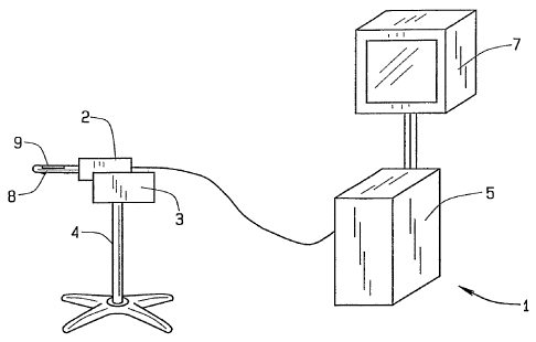
FIG. I discloses a perspective view of an ultrasound system utilizing
the image plane visualization aid;
FIG. 2 discloses a side view of an ultrasound probe and
stepper/stabilizer with external positioning registry;
FIG. 3 discloses a perspective view of an ultrasound body scanner
with external positioning registry;
CA 02595657 2007-07-23
WO 2006/060373 PCT/US2005/043091
8
FIG. 4 discloses the indicator showing a transverse imaging plane
approximately halfway between the base and apex planes in the center of
the imaging volume;
FIG. 5 discloses the image plane visualization aid showing a
transverse imaging plane that intersects the apex or most proximal point of
the organ;
FIG. 6 discloses the image plane visualization aid showing a
transverse imaging plane that intersects the base or most distal point of the
organ; and
FIG. 7 discloses the image plane visualization aid showing a sagittal
imaging plane approximately through the center of the organ.
DRAWING NUMBERS
ultrasound system 1
cavital probe 2
cradle 3
stabilizer 4
ultrasound system CPU 5
cavital probe position register 6
monitor 7
probe tip 8
probe imaging window 9
Body Scanner 15
External Position Registers 16a, b & c
Graphical Object 20
Transverse imaging plane 21
Sagital active imaging plane 22
Possible Imaging Volume 23
CA 02595657 2007-07-23
WO 2006/060373 PCT/US2005/043091
9
BEST MODE FOR CARRYING OUT THE INVENTION
The following detailed description illustrates the invention by way of
example and not by way of limitation. This description will clearly enable
one skilled in the art to make and use the invention, and describes several
embodiments, adaptations, variations, alternatives and uses of the
invention, including what is presently believed to be the best mode of
carrying out the invention. Additionally, it is to be understood that the
invention is not limited in its application to the details of construction and
the
arrangements of components set forth in the following description or
io illustrated in the drawings. The invention is capable of other embodiments
and of being practiced or being carried out in various ways. Also, it is to be
understood that the phraseology and terminology used herein is for the
purpose of description and should not be regarded as limiting.
As seen in Fig. 1, the device consists of an ultrasound system 1,
which in turn consists of a cavital probe, an ultrasound system CPU 5 and a
monitor 7, a stabilizer 4 and a cradle 3. It is understood that the ultrasound
system could be from a range of different manufacturers, for instance,
manufactured by Siemens Medical Solutions, located in Malvern,
Pennsylvania, or manufactured by Toshiba America Medical Systems, Inc.,
located in Tustin, California. As best seen in Fig. 4, the graphical
representation consists of a graphical object 20, a transverse active
imaging plane 21. Fig. 7 best displays the sagital image plane 22.
As seen in Fig. 2, a cavital probe 2 with cavital probe position
register 6 may be used.
As seen in Fig. 3, a body scanner 15 with external position registers
16a, 16b and 16c may be used.
CA 02595657 2007-07-23
WO 2006/060373 PCT/US2005/043091
In operation, a cavital probe 2 is placed in the cradle 3 of a stabilizer
4. The user then advances and adjusts the cradle 4 to allow the cavital
probe 2 to be inserted into the rectum of a patient. The user generates an
ultrasound image while positioning the probe to insure that the patient's
5 prostate is viewable within the probe imaging window 9 of the probe. If the
user is using a scanning probe with the ability to move the probe scan plane
without moving the probe, as disclosed in Envisioneering's Scanning Probe
(6,709,397), or a Solid State scanning probe with the probe imaging in
Transverse mode the scanning probe is positioned such that the scan plane
10 intersects the apex of the prostate, or the portion of the prostate most
proximal to the user, and then locked into place. The user labels this plane
by pressing the "Apex" button on the ultrasound system 1. Next, with the
probe still imaging in transverse mode, the user moves the transverse scan
plane until it intersects the base of the prostate, or the place most distal
to
the user. The user labels this plane position by pressing the "Base" button
the ultrasound system 1.
Monitor 7 displays Possible Imaging Volume 23 showing a frame,
such as a wire frame, representing the outer limits of the ultrasonic scan
representing the possible imaging area.
From the library of stored graphical object, the user selects that
graphical object 20 which equates with the shape of the prostate or the user
may select a default geometric shape, such as an ellipse. Based upon the
Apex and Base landmarks identified previously, the stored graphical object
20 is translated and scaled and displayed within the Possible Imaging
Volume 23 wire frame, on the monitor 7. A semi-circular transverse active
imaging plane 21 which correlates to the current position of the transverse
scan plane of the ultrasound system 1 is superimposed over the graphical
object 20, allowing the user to more easily identify the position of the scan
plane within the imaging volume. The transverse active imaging plane 21
CA 02595657 2007-07-23
WO 2006/060373 PCT/US2005/043091
11
may partially obscure graphical object 20, and further the intersection of the
transverse active imaging plane 21 and graphical object 20 may be
highlighted on monitor 7. As the user changes the scan plane of the
ultrasound system 1, the active imaging plane 21 moves in reference to the
stored graphical object 20, displaying the approximate location of the image
plane in reference to the scanned tissue mass or organ. The user may
change to sagital imaging mode, in which the scan plane parallels the axis
of the cavital probe. This causes monitor 7 to display sagital active imaging
plane 22.
In an alternative embodiment as disclosed in Fig. 2, the device may
be utilized with a traditional cavital probe 2 in conjunction with a cavital
probe register 6. In use the cavital probe 2 is placed in the cradle 3 of a
stabilizer 4. The user then advances and adjusts the cradle 4 to allow the
cavital probe 2 to be inserted into the rectum of a patient. The user
generates an ultrasound image while positioning the probe to insure that
the patient's prostate is viewable within the probe imaging window 9 of the
probe. With the probe imaging in transverse mode, the user positions the
cavital probe such that the scan plane intersects the apex of the prostate.
The user labels marks this cavital probe position in reference to the cavital
probe register, labeling this position "Apex" on the ultrasound system 1.
Next, with the probe still imaging in transverse mode, the user moves the
cavital probe 2 in the cradle 3 until the scan plane intersects the base of
the
prostate. The user labels this plane by pressing the "Base" button the
ultrasound system 1.
In an alternative embodiment as disclosed in Fig. 3, the device may
be utilized with a traditional body scanner 15 in conjunction with external
position registers 16a, 16b and 16c. The device may also be used with a
body scanner utilizing Envisioneering's scanning technology as disclosed in
Envisioneering's scanning probe patent (6,709,397).
CA 02595657 2007-07-23
WO 2006/060373 PCT/US2005/043091
12
In an alternative embodiment, a single data point can be used to
position the graphical object 20. The user sets one point as a reference,
and then the device displays a static graphical object representing a static
representation of an average organ. The organ size and position are
determined by the program designer and cannot be adjusted by the user.
In a further alternative embodiment, more than two data points can
be used. The device allows the user to label several planes or points on
several images as specific landmarks. These landmarks allow the three
dimensional display to adjust the size and placement of the representative
io organ within the displayed imaging volume. As the number of landmarks
increases, the accuracy of the reconstruction improves. An approximately
elliptical shaped organ like the prostate could be approximated with several
landmark choices (in increasing order of position and size accuracy).
Possible additional data points include two points indicating the widest
transverse extent of the organ, and two points indicating the tallest extent
of
the organ or scanned mass. The image of the organ is moved and scaled
so that its position and size approximate the organ position in the imaging
volume.
In a further alternative embodiment, the graphical object's size and
placement are determined by identification of the tissue boundaries of the
organ. These boundaries can either be drawn by the user or can be
determined automatically through a boundary recognition algorithm. The
boundaries are used to first create a skeleton of the organ, and finally a
surface rendering is made. The organ position and size are located within
the imaging volume based on the positions of the boundaries.
CA 02595657 2007-07-23
WO 2006/060373 PCT/US2005/043091
13
As various changes could be made in the above constructions
without departing from the scope of the invention, it is intended that all
matter contained in the above description or shown in the accompanying
drawings shall be interpreted as illustrative and not in a limiting sense.
Further, while the above description addresses an ultrasound system
utilized in medical imaging, it is understood that the device can be applied
to non-medical imaging uses as well, for instance imaging mechanical
parts.