Note: Descriptions are shown in the official language in which they were submitted.
DEMANDES OU BREVETS VOLUMINEUX
LA PRESENTE PARTIE DE CETTE DEMANDE OU CE BREVETS
COMPREND PLUS D'UN TOME.
CECI EST LE TOME 1 DE 2
NOTE: Pour les tomes additionels, veillez contacter le Bureau Canadien des
Brevets.
JUMBO APPLICATIONS / PATENTS
THIS SECTION OF THE APPLICATION / PATENT CONTAINS MORE
THAN ONE VOLUME.
THIS IS VOLUME 1 OF 2
NOTE: For additional volumes please contact the Canadian Patent Office.
CA 02595786 2010-02-26
72249-188
COMPOSITIONS AND METHODS FOR TREATING
FIBROTIC DISORDERS
FIELD OF THE INVENTION
This invention relates to compositions and methods for treating fibrotic
disorders.
BACKGROUND OF THE INVENTION
The process of tissue repair as a part of wound healing involves two phases.
The first
phase is the regenerative phase, in which injured cells are replaced by cells
of the same type. The
second phase is the formation of fibrous tissues, also called fibroplasia or
fibrosis, in which
connective tissue replaces normal parenchymal tissues. The tissue repair
process can become
pathogenic if the fibrosis phase continues unchecked, leading to extensive
tissue remodeling and
the formation of permanent scar tissue (Wynn, Nature Rev. Immuisol. 4, 583
(2004)).
It has been estimated that up to 45% of deaths in the United States can be
attributed to
fibroproliferative diseases, which can affect many tissues and organ systems.
(Wynn, supra, at
595 (2004)). Major organ fibrotic diseases include interstitial lung disease
(ILD), characterized
by pulmonary inflammation and fibrosis. ILD is known to have a number of
causes such as
sarcoidosis, silicosis, collagen vascular diseases, and systemic scleroderma.
However, idiopathic
pulmonary fibrosis, a common type of ILD, has no known cause. Other organ
fibrotic disorders
include liver cirrhosis, liver fibrosis resulting from chronic hepatitis B or
C infection, kidney
disease, heart disease, and eye diseases including raacular degeneration and
retinal and vitreal
retinopathy. Fibroproliferative disorders also include systemic and local
scleroderma, keloids and
hypertrophic scars, atherosclerosis, and restenosis. Additional
fibroprolifemtive diseases include
excessive scarring resulting from surgery, chemotherapeutic drag-induced
fibrosis, radiation-
induced fibrosis, and injuries and bums (Wynn, supra, page 585).
Currently, treatments are available for fibrotic disorders including general
immunosuppressive drugs such as corticosteroids, and other anti-inflammatory
treatments.
However, the mechanisms involved in regulation of fibrosis appear to be
distinctive from those of
inflammation, and anti-inflammatory therapies are not always effective in
reducing or preventing
1
CA 02595786 2007-07-24
WO 2006/083947
PCT/US2006/003519
fibrosis (Wynn, supa, page 591). Therefore, a need remains for developing
treatments to reduce
and prevent fibrosis and control fibrotic disorders.
The present invention addresses this need and provides methods and
compositions for
preventing or reducing fibrosis associated with fibrotic disorders.
SUMMARY OF THE INVENTION
The present invention provides methods for modulating fibroblast accumulation
and
collagen deposition in a tissue by modulating the amount or activity of the
cytokine thymic
stromal lymphopoietin (TSLP) in the tissue. In one aspect, the present
invention provides a
method of reducing or preventing fibrosis in a subject suffering from a
fibrotic disorder
comprising administering a therapeutically effective amount of at least one
TSLP antagonist. In
another aspect, the present invention provides for the use of at least one
TSLP antagonist in the
preparation of a medicament for the prevention or treatment of a fibrotic
disorder in a subject
suffering from such a disorder. The invention further provides a
pharmaceutical composition for
preventing or reducing fibrosis in a subject suffering from a fibrotic
disorder comprising a
therapeutically effective dosage of at least one antagonist to TSLP in
admixture with a
pharmaceutically acceptable carrier. The fibrotic disorders include, but are
not limited to,
scleroderma, interstitial lung disease (ILD), idiopathic pulmonary fibrosis
(IPF), liver fibrosis
resulting from chronic hepatitis B or C infection, radiation-induced fibrosis,
and fibrosis arising
from wound healing.
In one embodiment the TSLP antagonist is a TSLP ligand binding agent capable
of
binding to TSLP and reducing or blocking its activity. These antagonists
include, but are not
limited to, antagonistic antibodies, peptide or polypeptide binding agents,
soluble TSLP receptors
(TSLPR), soluble interleukin 7 receptor alpha (IL-7 R a)/TSLPR heterodimer
receptors
(heterodimer), and small molecule antagonists. The antagonistic antibodies
include, but are not
limited to, fully human, humanized, chimeric, single chain antibodies, and
antibody fragments.
The peptide or polypeptide binding agents, soluble receptor and soluble
heterodimer receptor
antagonists may further comprise Fc domains or other multimerizing components,
or carrier
molecules such as PEG.
In another embodiment, the TSLP antagonist is a TSLPR antagonist. TSLPR
antagonists
include antagonists which bind to the TSLP receptor, and antagonists which
bind to the IL-
7Ra/TSLPR heterodimer. These antagonists include, but are not limited to,
antagonistic
antibodies which bind to TSLPR; antagonistic antibodies which bind to the
heterodimer; soluble
2
CA 02595786 2016-08-16
=
54963-7
ligands which bind to the TSLPR; soluble ligands which bind to the
heterodimer; and small
molecules which bind to TSLPR and/or the IL-7Ra/TSLPR heterodimer. The
antagonistic
antibodies include, but are not limited to, human, humanized, chimeric, and
single-chain
antibodies, and antibody fragments. The soluble ligand may further comprise Fc
domains or other
multimerizing components, or carrier molecules such as PEG.
In another embodiment, the TSLP antagonist is a molecule which prevents
expression of the TSLP cytokine, TSLPR, or the heterodimer receptor. These
molecules include,
for example, antisense oligonucleotides which target mRNA, and interfering
messenger RNA.
In another embodiment, the methods and compositions of the present invention
further comprise at least one additional antagonist to one or more cytokine,
growth factor, or
chemokine which promotes fibrosis. These profibrotic factors include, but are
not limited to,
transforming growth factor13 (TGF-13), interleukin-4 (IL-4), interleukin-5 (IL-
5), interleukin-9
(IL-9), interleukin-13 (IL-13), granulocyte/macrophage-colony stimulating
factor (GM-CSF),
tumor necrosis factor alpha (TNF-a), interleukin-1 beta (IL-113), connective
tissue growth factor
(CTGF), interleukin-6 (IL-6), oncostatin M (OSM), platelet derived growth
factor (PDGF),
monocyte chemotactic protein 1 (CCL2/MCP-1), and pulmonary and activation-
regulated
chemokine (CCL18/PARC).
The present invention as claimed relates to:
- an antibody that specifically binds thymic stromal lymphopoietin (TSLP)
ligand
or to TSLP receptor and that blocks binding of TSLP ligand to the TSLP
receptor, for use in
reducing fibroblast proliferation;
- use of an antibody that specifically binds TSLP ligand or to TSLP receptor
and
that blocks binding of TSLP ligand to the TSLP receptor, for reducing
fibroblast proliferation;
- use of an antibody that specifically binds TSLP ligand or to TSLP receptor
and
that blocks binding of TSLP ligand to the TSLP receptor, in the manufacture of
a medicament for
reducing fibroblast proliferation;
3
CA 02595786 2016-08-16
54963-7
- a pharmaceutical composition for reducing or preventing fibrosis in a
subject
suffering from a fibrotic disorder comprising a therapeutically effective
amount of an antibody
that specifically binds TSLP ligand or to TSLP receptor and that blocks
binding of TSLP ligand to
the TSLP receptor, in admixture with a pharmaceutically acceptable carrier
thereof;
- an antibody that specifically binds TSLP ligand or to TSLP receptor and that
blocks binding of TSLP ligand to the TSLP receptor, for use in reducing or
preventing fibroblast
accumulation and collagen deposition in a tissue;
- use of an antibody that specifically binds TSLP ligand or to TSLP receptor
and
that blocks binding of TSLP ligand to the TSLP receptor, for reducing or
preventing fibroblast
accumulation and collagen deposition in a tissue; and
- use of an antibody that specifically binds TSLP ligand or to TSLP receptor
and
that blocks binding of TSLP ligand to the TSLP receptor, in the manufacture of
a medicament for
reducing or preventing fibroblast accumulation and collagen deposition in a
tissue.
BRIEF DESCRIPTION OF THE FIGURES
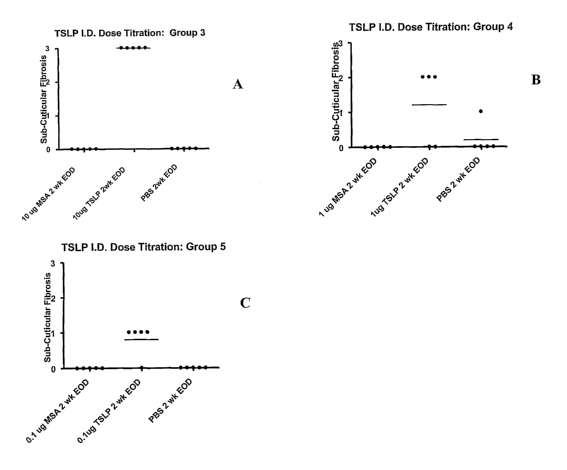
Figures 1A and 1B, and Figure 2A, 2B, and 2C show the results of injecting
five
groups of Balb/c mice intradermally with varying dosages of TSLP and a
negative control MSA
(mouse serum albumin) once a week for 1 week (Figure 1A, Group 1), once a week
for 2 weeks
(Figure 1B, Group 2); and three times a week for two weeks in Figure 2A (Group
3), 2B (Group 4)
and 2C (Group 5). Figure lA (Group 1) shows no subcuticular fibrosis induced
from a single
injection of 10 ug TSLP for one week; MSA alone; and PBS alone. Figure 1B
(Group 2) shows no
subcuticular fibrosis induced from a single injection on each of two weeks (2
total injections) of
10 ug TSLP; MSA alone, and PBS alone. Figure 2A (Group 3) shows subcuticular
fibrosis scored at
level 3 for 10 ug TSLP when injected three times a week for 2 weeks, but no
fibrosis for MSA alone,
and PBS alone. Figure 2B (Group 4) shows fibrosis scored at level 2 for 1 ug
TSLP when injected
three times a week for 2 weeks, but no fibrosis for MSA alone, and none for
PBS alone with the
exception of one animal showing fibrosis at level 1 for PBS alone. Figure 2C
(Group 5) shows
fibrosis scored at level 1 for .1 ug TSLP when injected three times a week for
2 weeks, but no
fibrosis for MSA alone or PBS alone.
3a
CA 02595786 2007-07-24
WO 2006/083947
PCT/US2006/003519
DETAILED DESCRIPTION OF THE INVENTION
The present invention provides methods of modulating fibroblast accumulation
and
collagen deposition in a tissue by modulating the amount or activity of the
cytokine thymic
stromal lymphopoietin (TSLP) in the tissue. TSLP has been found to induce
fibroblast
accumulation and collagen deposition characteristic of fibrotic disorders in
animals. In one
aspect, the invention provides a method of increasing fibrosis in situations
where this may be
advantageous, by administering TSLP or TSLP agonists. In another aspect, the
present invention
provides methods and compositions for reducing or preventing fibrosis in a
subject suffering from
a fibrotic disorder by treating the subject with a therapeutically effective
amount of at least one
antagonist to TSLP. In another aspect, the present invention provides for the
use of at least one
TSLP antagonist in the preparation of a medicament for the prevention or
treatment of a fibrotic
disorder in a subject suffering from such a disorder. In another aspect, the
present invention
provides a pharmaceutical composition for preventing or reducing fibrosis in a
subject comprising
a therapeutically effective dosage of at least one antagonist to TSLP in
admixture with a
pharmaceutically acceptable carrier.
As used herein the term "fibroproliferative disease" or "fibrotic disease or
disorder" refers
to conditions involving fibrosis in one or more tissues. As used herein the
term "fibrosis" refers
to the formation of fibrous tissue as a reparative or reactive process, rather
than as a normal
constituent of an organ or tissue. Fibrosis is characterized by fibroblast
accumulation and
collagen deposition in excess of normal deposition in any particular tissue.
As used herein the
term "fibrosis" is used synonymously with "fibroblast accumulation and
collagen deposition".
Fibroblasts are connective tissue cells, which are dispersed in connective
tissue throughout the
body. Fibroblasts secrete a nonrigid extracellular matrix containing type I
and/or type III
collagen. In response to an injury to a tissue, nearby fibroblasts migrate
into the wound,
proliferate, and produce large amounts of collagenous extracellular matrix.
Collagen is a fibrous
protein rich in glycine and proline that is a major component of the
extracellular matrix and
connective tissue, cartilage, and bone. Collagen molecules are triple-stranded
helical structures
called cc-chains, which are wound around each other in a ropelike helix.
Collagen exists in several
forms or types; of these, type I, the most common, is found in skin, tendon,
and bone; and type 111
is found in skin, blood vessels, and internal organs.
Fibrotic disorders include, but are not limited to, systemic and local
scleroderma, keloids
and hypertrophic scars, atherosclerosis, restinosis, pulmonary inflammation
and fibrosis,
idiopathic pulmonary fibrosis, liver cirrhosis, fibrosis as a result of
chronic hepatitis B or C
infection, kidney disease, heart disease resulting from scar tissue, and eye
diseases such as
4
CA 02595786 2007-07-24
WO 2006/083947
PCT/US2006/003519
macular degeneration, and retinal and vitreal retinopathy. Additional fibrotic
diseases include
fibrosis resulting from chemotherapeutic drugs, radiation-induced fibrosis,
and injuries and burns.
Scleroderma is a fibrotic disorder characterized by a thickening and
induration of the skin
caused by the o'verproduction of new collagen by fibroblasts in skin and other
organs.
Scleroderma may occur as a local or systemic disease. Systemic scleroderma may
affect a
number of organs. Systemic sclerosis is characterized by formation of
hyalinized and thickened
collagenous fibrous tissue, with thickening of the skin and adhesion to
underlying tissues,
especially of the hands and face. The disease may also be characterized by
dysphagia due to loss
of peristalsis and submucosal fibrosis of the esophagus, dyspnea due to
pulmonary fibrosis,
myocardial fibrosis, and renal vascular changes. (Stedman's Medical
Dictionary, 261 Edition,
Williams & Wilkins, 1995)). Pulmonary fibrosis affects 30 to 70% of
scleroderma patients, often
resulting in restrictive lung disease (Atamas et al. Cytokine and Growth
Factor Rev 14: 537-550
(2003)).
Idiopathic pulmonary fibrosis is a chronic, progressive and usually lethal
lung disorder,
thought to be a consequence of a chronic inflammatory process (Kelly et al.,
Cun- Pharma Design
9: 39-49 (2003)). The causes of this disease are not yet known.
As used herein the term "subject" refers to animals including mammals
including
humans. The term "mammal" includes primates, domesticated animals including
dogs, cats,
sheep, cattle, goats, pigs, mice, rats, rabbits, guinea pigs, captive animals
such as zoo animals, and
wild animals. As used herein the term "tissue" refers to an organ or set of
specialized cells such
as skin tissue, lung tissue, kidney tissue, and other types of cells.
TSLP
Thymic stromal lymphopoietin (TSLP) refers to a four a-helical bundle type I
cytokine
which is a member of the IL-2 family but most closely related to IL-7.
Cytokines are low
molecular weight regulatory proteins secreted in response to certain stimuli,
which act on
receptors on the membrane of target cells. Cytokines regulate a variety of
cellular responses.
Cytokines are generally described in references such as Cytokines, A. Mire-
Sluis and R. Thorne,
ed., Academic Press, New York, (1998).
TSLP was originally cloned from a murine thymic stromal cell line (Sims et al
J. Exp.
Med 192 (5), 671-680 (2000)), and found to support early B and T cell
development. Human
TSLP was later cloned and found to have a 43 percent identity in amino acid
sequence to the
5
CA 02595786 2010-02-26
72249-188
murine homolog (Quentmeier et al. Leukemia 15, 1286-1292(2001), and U.S.
Patent No:
6,555,520). The polynucleotide and amino acid sequence of human TSLP are
presented in SEQ ID
NO: 1 and 2 respectively. TSLP was found to bind with low affinity to a
receptor chain from the
hematopoietin receptor family called TSLP receptor (TSLPR), which is described
in U.S. publication
No: 2002/0068323 (SEQ ID NO: 4 and 5). The polynucleotide sequence encoding
human TSLPR is
presented as SEQ ID NO: 3 of the present application, and the amino acid
sequence is presented
as SEQ ID NO: 4 of the present application respectively. The soluble domain of
the TSLPR is
approximately amino acids 25 through 231 of SEQ ID NO: 4. TSLP binds with high
affinity to a
heterodimeric complex of TSLPR and the interleukin 7 receptor alpha IL-7Ra
(Park et al., J. Exp.
Med 192:5 (2000), U.S. publication number 2002/0068323). The sequence of IL-7
receptor a is shown
in Figure 2 of U.S. Patent No. 5,264,416. The sequence of the soluble domain
of the IL-7 receptor a is
amino acid 1 to 219 of Figure 2 in U.S. Patent No: 5,264,416.
Human TSLP can also be expressed in modified form, in which a furin cleavage
site has
been removed through modification of the amino acid sequence, as described in
PCT patent
application publication WO 03/032898. Modified TSLP retains activity but the
full length
sequence is more easily expressed in microbial or mammalian cells.
TSLP is produced in human epithelial cells including skin, bronchial,
tracheal, and airway
epithelial milk keratinocytes, stromal and mast cells, smooth muscle cells,
and lung and dermal
fibroblasts, as determined by quantitative mRNA analysis (Soumelis et al,
Nature ImmunoL 3(7)
673-680 (2002)). Both murine and human TSLP are involved in promoting allergic
inflammation. Soumelis et al, supra reported that the TSLP heterodimer
receptor complex is
expressed on human CD1 lc+ dendritic cells (DC cells). Dendritic cell culture
experiments have
shown that TSLP binding to DC cells induces the production of TH2 cell
attracting chemokines
TARC (thymus and activation-regulated chemokine; also known as CCL17) and MDC
(macrophage-derived chemokine, also known as CCL22), and upregulates
costimulatory
molecules HLA-DR, CD40, CD80, CD86, and CD83 on the surface of cells. TSLP-
activated
DCs in cell culture induced naive CD4+ (Soumelis, supra) and CD8+.T cell
differentiation into
pro-allergic effector cells (Gilliet et al, J. Exp. Med. 197(8), 1059-1063
(2003)) which produce
pro-allergic cytolcines IL-4, M-5, M-13 and TNF-a while down-regulating IL-10
and interferon.')'
(Soumelis et at., supra, Gilliet et al, supra). TSLP has been reported to be
expressed in tissue
samples of inflamed tonsilar epithelial cells, and keratinocytes within the
lesions of atopic
dermatis patients. (Soumelis et at., supra).
6
CA 02595786 2010-02-26
72249-188
TSLP Assays
TSLP activities can be measured in an assay using BAF cells expressing human
TSLPR
(BAF/HTR), which require active TSLP for proliferation as described in PCT
patent application
publication WO 03/032898. The BAF/HTR bioassay utilizes a murine pro B
lymphocyte cell
line, which has been transfected with the human TSLP receptor (cell line
obtained from Steven F.
Ziegler, Virginia Mason Research Center, Seattle, WA.). The BAF/HTR cells are
dependent
upon huTSLP for growth, and proliferate in response to active huTSLP added in
test samples.
Following an incubation period, cell proliferation is measured by the addition
of Alamar Blue dye
I (Biosource International Catalog # DAL1100, 10 uLtwell). Metabolically
active BAF/HRT
cells take up and reduce Alamar Blue*, which leads to change in the
fluorescent properties of the
dye. Additional assays for huTSLP activity include, for example, an assay
measuring induction -
of T cell growth from human bone marrow by TSLP as described in U.S. Patent
6,555,520.
Another TSLP activity is the ability to activate STAT5 as described in the
reference to Levin et
aL, J: Immunol. 162:677-683 (1999) and PCT patent application WO 03/032898.
Additional
assays include TSLP induced CCL17/TARC production from primary human monocytes
and
dendritic cells as described in the reference to Soumelis et al. supra.
TSLP has been found to induce fibroblast accumulation and collagen deposition
in
animals, as described in the Example below. Injection of murine TSLP
intradennally into mice
resulted in fibrosis within the subcutis of the mice, characterized by
fibroblast proliferation and
collagen deposition. Antagonizing TSLP activity would result in preventing or
decreasing
fibroblast proliferation and collagen deposition in a tissue. The present
invention provides
methods and compositions for reducing or preventing fibrosis in a subject
afflicted with a fibrotic
disorder by administering one or more TSLP antagonist to the subject.
As used herein the term "profibrotic factors" refers to cytokines, growth
factors or
chemokines in addition to TSLP which have been observed to promote the
accumulation of
fibroblasts and deposition of collagen in various tissues. A number of
cytokines and growth
factors have been reported to be involved in regulating tissue remodeling and
fibrosis. These
include the "profibrotic cytokines" such as transforming growth factor beta
(TGF-13), interleukin-4
(I1-4), interleukin-5 (IL-5), and interleukin-13 (I1-13), which have been
shown to stimulate
collagen synthesis and fibrosis in fibrotic tissues (Letterio et al. Ann Rev.
Immunol. 16, 137-161
(1998), Fertin et al., Cell Mol. Biol. 37, 823-829 (1991), Doucet et aL, J.
Clin. Invest. 101,2129-
2139 (1998). Interlenkin-9 (I1-9) has been shown to induce airway fibrosis in
the lungs of mice
*Trade -mark
7
CA 02595786 2007-07-24
WO 2006/083947
PCT/US2006/003519
( Zhu et al., J. Clin. Invest. 103, 779-788(1999)). In addition to TGF-P,
other cytokines or growth
factors which have been reported to increase fibrosis in the fibrotic disorder
idiopathic pulmonary
fibrosis (IPF) include granulocyte/macrophage-colony stimulating factor (GM-
CSF), tumor
necrosis factor alpha (TNF-a), interleukin-1 beta (IL-1 p), and connective
tissue growth factor
(CTGF) (Kelly et al. Curr Pharmaceutical Des 9: 39-49 (2003)). Cytokines and
growth factors
reported to be involved in promoting pulmonary fibrosis occurring in
scleroderma include TGF-P,
interleukin-1 beta (IL-113), interleukin-6 (IL-6), oncostatin M (OSM),
platelet derived growth
factor (PDGF), the type 2 cytokines IL-4 and IL-13, IL-9, monocyte chemotactic
protein 1
(CCL2/MCP-1), and pulmonary and activation- regulated chemokine (CCL18/PARC)
(Atamas et
al., Cyto Growth Fact Rev 14: 537-550 (2003)). Therefore, in one embodiment,
the methods and
compositions of the present invention further comprise administering at least
one additional
antagonist to one or more profibrotic factor in addition to at least one TSLP
antagonist to reduce
or prevent fibrosis in a subject suffering from a fibrotic disorder. In
another aspect, the present
invention provides for the use of at least one profibrotic antagonist in
addition to at least one
TSLP antagonist in the preparation of a medicament for the treatment or
prevention of a fibrotic
disorder in a subject. In another aspect, the present invention provides a
pharmaceutical
composition comprising, in addition to at least one TSLP antagonist, one or
more antagonists to
profibrotic factors, that is cytokines, growth factors or chemokines, in
admixture with a
pharmaceutically acceptable carrier. These profibrotic factors include, but
are not limited to, the
following cytokines, growth factors or chemokines: interleukin-4 (IL-4),
interleukin-5 (IL-5),
interleukin-9 (IL-9), interleukin-13 (IL-13), transforming growth factor beta
(TGF-I3),
granulocyte/macrophage-colony stimulating factor (GM-CSF), tumor necrosis
factor alpha (TNF-
a), interleukin-1 beta (IL-1 (3), connective tissue growth factor (CTGF),
interleukin-6 (IL-6),
oncostatin M (OSM), platelet derived growth factor (PDGF), monocyte
chemotactic protein 1
(CCL2/MCP-1), and pulmonary and activation- regulated chemokine (CCL18/PARC).
The
Accession numbers for these cytokines and their specific receptors (if
available) are found in
Table I below.
TABLE I
Protein Species Synonyms Database(s) Accession
Name (or Patent No.
Application) (or SEQ ID
No: )
TSLP Homo Thymic stromal lymphopoietin GenBank/ AAK67940/
sapiens protein US Patent SEQ ID
No.6555520 NO: 2
TSLP Mus Thymic stoma derived GenBank AAF81677
musculus lymphopoietin; Thymic stromal
8
CA 02595786 2007-07-24
WO 2006/083947
PCT/US2006/003519
derived lymphopoietin
TSLPR Homo Cytokine receptor-like 2 (CRL2); US SEQ ID
sapiens IL-XR; Thymic stromal 2002/0068323 NO: 5
lymphopoietin protein receptor
TSLPR Mus Cytokine receptor-like factor 2; Type GenBank, Q8CII9
I cytokine receptor delta 1; Cytokine SWISSPROT
receptor-like molecule 2 (CRLM-2);
Thymic stromal lymphopoietin
protein receptor
TNF- Homo Tumor necrosis factor; Tumor GenBank, P01375
alpha sapiens necrosis factor ligand superfamily SWISSPROT
member 2; TNF-a; Cachectin
TNF- Mus Tumor necrosis factor; Tumor GenBank, P06804
alpha necrosis factor ligand superfamily SWISSPROT
member 2; TNF-a; Cachectin
TNF-RI Homo Tumor necrosis factor receptor GenBank, P19438
sapiens superfamily member 1A; p60; SWISSPROT
TNF-R1; p55; CD120a
[contains: Tumor necrosis factor
binding protein 1 (TBPI)]
TNF-RI Mus GenBank, P25118
Tumor necrosis factor receptor
SWISSPROT
superfamily member 1A; p60;
TNF-R1; p55
TNF-RII Homo Tumor necrosis factor receptor GenBank, P20333
sapiens superfamily member 1B; Tumor necrosis SWISSPROT
factor receptor 2; p80; TNF-R2; p75;
CD120b; Etanercept
[contains: Tumor necrosis factor binding
protein 2 (TBPII)]
TNF-RII Mus Tumor necrosis factor receptor GenBank, P25119
superfamily member 1B; Tumor SWISSPROT
necrosis factor receptor 2; TNF-R2;
p75
IL-1 Homo Interleukin-1 alpha; Hematopoietin-1 GenBank, P01583
alpha sapiens SWISSPROT
IL-1 Mus Interleukin-1 alpha GenBank, P01582
alpha SWISSPROT
IL-1 R-1 Homo Interleukin-1 receptor, type I; IL-1R- GenBank, P14778
sapiens alpha; P80; Antigen CD121a SWISSPROT
IL-1 R-1 Mus Interleukin-1 receptor, type I; P80 GenBank, P13504
SWISSPROT
IL-1 R-2 Homo Interleukin-1 receptor, type II; IL- GenBank, P27930
sapiens 1R-beta; Antigen CDw121b SWISSPROT
IL-1 R-2 Mus Interleukin-1 receptor, type II GenBank, P27931
SWISSPROT
IL-4 Homo Interleukin-4; B-cell stimulatory GenBank, P05112
sapiens factor 1 (BSF-1); Lymphocyte SWISSPROT
stimulatory factor 1
IL-4 Mus Interleukin-4; B-cell stimulatory GenBank, P07750
factor 1 (BSF-1); Lymphocyte SWISSPROT
stimulatory factor 1; IGG1 induction
factor; B-cell IGG differentiation
factor; B-cell growth factor 1
9
CA 02595786 2007-07-24
WO 2006/083947
PCT/US2006/003519
IL-4R Homo Interleukin-4 receptor alpha chain GenBank,
P24394
sapiens (IL-4R-alpha; CD124 antigen) SWISSPROT
[contains: Soluble interleukin-4
receptor alpha chain
(sIL4Ralpha/prot); IL-4-binding
protein (IL4-BP)]
IL-4R Mus Interleukin-4 receptor alpha chain GenBank,
P16382
(IL-4R-alpha)
SWISSPROT
[contains: Soluble interleukin-4
receptor alpha chain; IL-4-binding
protein (1L4-BP)]
IL-5 Homo Interleukin-5; T-cell replacing factor GenBank, P05113
sapiens (TRF); Eosinophil differentiation SWISSPROT
factor; B cell differentiation factor I
IL-5 Mus Interleukin-5; T-cell replacing factor GenBank, P04401
(TRF); B-cell growth factor II SWISSPROT
(BCGF-II); Eosinophil differentiation
factor; Cytotoxic T lymphocyte
inducer
IL-5R Homo Interleukin-5 receptor alpha chain GenBank,
Q01344
sapiens (IL-5R-alpha); CD125 antigen SWISSPROT
IL-5R Mus Interleukin-5 receptor alpha chain GenBank,
P21183
(IL-5R-alpha) SWISSPROT
IL-9 Homo Interleukin-9; T-cell growth factor GenBank,
P15248
sapiens P40; P40 cytokine SWISSPROT
IL-9 Mus Interleukin-9; T-cell growth factor GenBank,
P15247
P40; P40 cytokine SWISSPROT
IL-9R Homo Interleukin-9 receptor GenBank, Q01113
sapiens SWISSPROT
IL-9R Mus Interleukin-9 receptor GenBank, Q01114
SWISSPROT
IL-13 Homo Interleukin-13 GenBank, P35225
sapiens SWISSPROT
IL-13 Mus Interleukin-13; T-cell activation GenBank,
P20109
protein P600 SWISSPROT
IL-13RA- Homo Interleukin-13 receptor alpha-1 chain GenBank, P78552
1 sapiens (IL-13R-alpha-1); CD213a1 antigen SWISSPROT _
IL-13RA- Mus Interleukin-13 receptor alpha-1 chain GenBank, 009030
1 (IL-13R-alpha-1); Interleukin-13 SWISSPROT
binding protein; NR4
IL-13RA- Homo Interleukin-13 receptor alpha-2 GenBank,
Q14627
2 sapiens chain; Interleukin-13 binding protein SWISSPROT
IL-13RA- Mus IL-13 receptor alpha 2 GenBank AAC33240
2 musculus
TGF-f3 1 Homo Transforming growth factor beta 1 SWISSPROT P01137
sapiens
TGF-13 1 Mus Transforming growth factor beta 1 SWISSPROT P04202
, musculus
TGF-I3 Homo Transforming growth factor beta SWISSPROT P36897
R1 sapiens receptor type I; serine/threonine-
protein kinase receptor R4 (SKR4);
activin receptor-like kinase 5 (ALK-
CA 02595786 2007-07-24
WO 2006/083947
PCT/US2006/003519
5)
TGF-13 Homo Transforming growth factor beta SWISSPROT P37173
R2 sapiens receptor type II
GM-CSF Homo granulocyte-macrophage colony- SWISSPROT P04141
sapiens stimulating factor; colony-
stimulating factor; sargramostim;
molgramostin
IL-6 Homo Interleukin 6; interferon, beta 2 Genbank AAH15511
sapiens
IL-6 Homo Interleukin-6 precursor; B-cell SWISSPROT P05231
sapiens stimulatory factor 2; interferon beta-
2; hybridoma growth factor; CTL
differentiation factor
IL-6 Mus Interleukin-6 precursor; interleukin SWISSPROT P08505
musculus HP-1; B-cell hybridoma growth
factor
IL-6 R 13 Homo interleukin-6 receptor beta chain; SWISSPROT P40189
sapiens membrane glycoprotein 130; gp130;
oncostatin M receptor; CDw130;
CD130 antigen
IL-6R- Homo Interleukin-6 receptor alpha chain SWISSPROT P08887
alpha sapiens precursor; CD126 antigen
IL-6R- Homo Interleukin-6 receptor beta chain Genbank; AAB63010;
beta sapiens SWISSPROT
IL-6 R- Mus Interleukin-6 receptor alpha chain SWISSPROT P22272
alpha musculus
IL-6R- Mus Intereleukin-6 receptor beta chain SWISSPROT Q00560
beta musculus
OSM Homo Oncostatin M Genbank AAA36388
sapiens
OSMR- Homo Oncostatin-M specific receptor beta Genbank; AAC50946;
beta sapiens subunit US Patent No. SEQ ID
subunit 5891997 NO: 2
OSM Mus Oncostatin M precursor SWISSPROT P53347
musculus
OSMR Mus Oncostatin M specific receptor Genbank AAC40122
musculus
CTGR Homo Connective tissue growth factor SWISSPROT P29279
sapiens precursor; hypertrophic chondrocyte-
specific protein 24
CTGR Mus Connective tissue growth factor SWISSPROT P29268
musculus precursor; FISP-12 protein;
hypertrophic chondrocyte-specific
protein 24
PDGF-1 Homo Platelet-derived growth factor, A SWISSPROT P04085
sapiens chain; platelet-derived growth factor
alpha polypeptide
PDGF-2 Homo Platelet-derived growth factor, B SWISSPROT P01127
sapiens chain; platelet-derived growth factor
beta polypeptide
PDGF-1 Mus Platelet-derived growth factor, A SWISSPROT P20033
musculus chain precursor
11
CA 02595786 2007-07-24
WO 2006/083947
PCT/US2006/003519
PDGF-2 Mus Platelet-derived growth factor, B SWISSPROT P31240
musculus chain precursor
PDGR-R- Homo Alpha platelet-derived growth factor SWISSPROT P16234
a sapiens receptor; CD140a antigen
PDGR-R- Homo Beta platelet-derived growth factor SWISSPROT P09619
sapiens receptor; CD140B antigen
CCL2 Homo Small inducible cytokine A2 SWISSPROT P13500
sapiens precursor; monocyte chemotactic
protein 1 (MCP-1); monocyte
chemotactic and activating factor
(MCAF); monocyte secretory protein
JE (HC11)
MCP-1-R Homo C-C chemokine receptor type 2 SWISSPROT P41597
sapiens (CCR2); Monocyte chemoattractant
protein 1 receptor
CCL18 Homo Small inducible cytokine Al 8 SWISSPROT P55774
sapiens precursor (CCL18); Macrophage
inflammatory protein 4 (MIP-4);
Pulmonary and activation-regulated
chemokine (CC chemokine PARC);
Alternative macrophage activation-
associated CC chemokine 1 (AMAC-
1); Dendritic cell chemokine 1 (DC-
CK1)
TSLP antagonists
A TSLP antagonist according to the present invention inhibits or blocks at
least one
activity of TSLP, or alternatively, blocks expression of the cytokine or its
receptor. Inhibiting or
blocking cytokine activity can be achieved, for example, by employing one or
more inhibitory
agents which interfere with the binding of the cytokine to its receptor,
and/or blocks signal
transduction resulting from the binding of the cytokine to its receptor.
In one embodiment, the TSLP antagonist comprises a TSLP binding agent, which
binds
to TSLP and prevents binding of the cytokine to its receptor, and/or blocks
signal transduction
resulting from the binding of the cytokine to its receptor. These antagonists
include, but are not
limited to, antagonistic antibodies, peptide or polypeptide binding agents,
soluble TSLPR, soluble
IL-7Ra/TSLPR heterodimers, and small molecule antagonists.
In another embodiment, the antagonist is a TSLPR antagonist, which binds to
this
receptor and blocks ligand binding and/or signal transduction. These
antagonists include, but are
not limited to, antagonistic antibodies, soluble ligands, and small molecules
which bind to TSLPR
and interfere with TSLP signal transduction and activity.
In another embodiment, the antagonist is an antagonist to the IL-7Ra/TSLPR
heterodimer, which binds to the heterodimer, and blocks ligand binding and/or
signal
12
CA 02595786 2007-07-24
WO 2006/083947
PCT/US2006/003519
transduction. These antagonists include, but are not limited to, antagonistic
antibodies, soluble
ligands, and small molecules which bind to the heterodimer and interfere with
TSLP signal
transduction and activity.
In another embodiment, the TSLP antagonist is a molecule which prevents
expression of
the TSLP cytokine, TSLPR, or heterodimer receptor. These molecules include,
for example,
antisense oligonucleotides which target mRNA, and interfering messenger RNA.
In another embodiment, the methods and compositions of the present invention
provide
an additional antagonist to one or more "profibrotic factors", including but
not limited to IL-4, IL-
5, IL-9, IL-13, TGF-B, GM-CSF, TNF-a, IL-1f3, CTGF, IL-6, OSM, PDGF, CCL2/MCP-
1, and
CCL18/PARC to prevent or reduce fibrosis in a subject suffering from a
fibrotic disorder.
Antagonists to these profibrotic factors can be selected from agents which
bind to the factor itself,
the receptor, or a heterodimeric receptor to which the factor may bind and
signal, wherein the
antagonist interferes with ligand/receptor binding and/or at least one
activity. In one embodiment,
the factor antagonist blocks expression of the factor or its receptor.
In one embodiment, the TSLP antagonists specifically bind to the TSLP ligand,
receptor
or heterodimer receptor. As used herein the tem). "specifically binds" refers
to antagonists such as
antibodies have a binding affinity (Ka) for TSLP, TSLPR, or the heterodimer,
(or corresponding
to a profibrotic cytokine, cytokine receptor, or cytokine heterodimer
receptor) of greater than or
equal to 106 M-1, in one embodiment 107M-1, in anther embodiment, 108 M-1, in
another
embodiment 109M-1, as determined by techniques well known in the art (such as,
for example
Scatchard, Ann NY Acad Sci 51:660-672 (1949), and as described below).
The antagonists for TSLP and profibrotic factors generally are described in
greater detail
below.
Particular Antagonists
Antibodies
Antagonists include antibodies that bind to either a cytokine or its receptor
and reduce or
block at least one activity of the cytokine. As used herein, the term
"antibody" refers to refers to
intact antibodies including polyclonal antibodies (see, for example
Antibodies: A Laboratory
Manual, Harlow and Lane (eds), Cold Spring Harbor Press, (1988)), and
monoclonal antibodies
(see, for example, U.S. Patent Nos. RE 32,011, 4,902,614, 4,543,439, and
4,411,993, and
Monoclonal Antibodies: A New Dimension in Biological Analysis, Plenum Press,
Kennett,
13
CA 02595786 2007-07-24
WO 2006/083947
PCT/US2006/003519
McKearn and Bechtol (eds.) (1980)). As used herein, the term "antibody" also
refers to a
fragment of an antibody such as F(ab), F(ab'), F(ab')2, Fv, complementarity
determining regions
(CDR) fragments, single chain antibodies (scFv), or combinations of these,
which can be
produced by DNA recombinant techniques or by enzymatic or chemical cleavage of
intact
antibodies. Antibodies also include polypeptides such as fusion proteins that
contain at least a
portion of an immunogloblin that is sufficient to confer specific antigen
binding to the
polypeptide. Antibodies also include dAb (VH domain), diabodies (bivalent
antibodies
comprising two polypeptide chains, each having VH and VL chains), and
triabodies and
tetrabodies (antibodies with three and four polypeptide chains respectively,
each having VH and
VL chains). Antibodies also include minibodies (as described in WO 94/09817),
and maxibodies
or scFv-Fc fusions (Powers et al, J. Immunol Meth 251, 123-135 (2001)),
produced by
recombinant DNA techniques or by enzymatic or chemical cleavage of intact
antibodies.
The term "antibody" also refers to bispecific or bifunctional antibodies which
are an
artificial hybrid antibody having two different heavy/light chain pairs and
two different binding
sites. Bispecific antibodies can be produced by a variety of methods including
fusion of
hybridomas or linking of Fab' fragments. (See Songsivilai et al, Clin. Exp.
Immunol. 79:315-321
(1990), Kostelny et al., J. Immuno1.148:1547-1553 (1992)). As used herein the
term "antibody"
also refers to chimeric antibodies, that is, antibodies having a human
constant antibody
immunoglobin domain is coupled to one or more non-human variable antibody
imanunoglobin
domain, or fragments thereof (see, for example, U.S. Patent No. 5,595,898 and
U.S. Patent No.
5,693,493). Antibodies also refer to "humanized" antibodies, and human
antibodies produced by
transgenic animals, both of which are described more fully below. The term
"antibodies" also
includes multimeric antibodies, or a higher order complex of proteins such as
heterdimeric
antibodies. "Antibodies" also includes anti-idiotypic antibodies. The
production of antibodies is
described in more detail below.
Polyclonal antibodies directed toward a cytokine or its receptor polypeptide
may be
produced in animals (e.g., rabbits or mice) by means of multiple subcutaneous
or intraperitoneal
injections of the polypeptide and an adjuvant It may be useful to conjugate
the antigen
polypeptide to a carrier protein that is immunogenic in the species to be
immunized, such as
keyhole limpet hemocyanin, serum, albumin, bovine thyroglobulin, or soybean
trypsin inhibitor.
Also, aggregating agents such as alum may be used to enhance the immune
response. After
immunization, the animals are bled and the serum is assayed for antibody
titer.
14
CA 02595786 2010-02-26
72249-188
Monoclonal antibodies that are immunoreactive with a cytokine or its receptor
are
produced using any method that provides for the production of antibody
molecules by continuous
cell lines in culture. Examples of suitable methods for preparing monoclonal
antibodies include
the hybridoma methods of Kohler et al. Nature 256:495-97 (1975) and the human
B-cell
hybridoma method (Kozbor, J. lininunol. 133:3001 (1984); Brodeur et aL,
Monoclonal Antibody
Production Techniques and Applications 51-63 (Marcel Dekker, Inc., 1987). Also
provided by
the invention are hybridoma cell lines that produce monoclonal antibodies
reactive with cytokines
or their receptors.
Monoclonal antibodies of the invention may be modified for use as
therapeutics. One
embodiment is a "chimeric" antibody in which a portion of the heavy (H) and/or
light (L) chain is
identical with or homologous to a corresponding sequence in antibodies derived
from a particular
species or belonging to a particular antibody class or subclass, while the
remainder of the chain(s)
is/are identical with or homologous to a corresponding sequence in antibodies
derived from
another species or belonging to another antibody class or subclass. Also
included are fragments
of such antibodies, so long as they exhibit the desired biological activity.
See U.S. Patent No.
4,816,567; Morrison et al., Proc. Natl. Acad. Sci. 81:6851-55 (1985).
A monoclonal antibody may also be a "humanimd" antibody. Methods for
humanizing
non-human antibodies are well known in the art. See U.S. Patent Nos. 5,585,089
and 5,693,762.
Generally, a humanized antibody has one or more amino acid residues introduced
into it from a
source that is non-human. Humanization can be performed, for example, using
methods
described in the art (see, for example, U.S. Pat. No. 4,816,567 and WO
94/10332, Jones et al.,
Nature 321:522-25 (1986); Riechmann et aL, Nature 332:323-27(1998); Verhoeyen
et al.,
Science 239:1534-36(1988)), by substituting at least a portion of a rodent
complementarity-
determining region for the corresponding regions of a human antibody.
Antibodies may be human antibodies. Using transgenic animals (e.g., mice) that
are
capable of producing a repertoire of human antibodies in the absence of
endogenous
irmnunoglobulin production such antibodies are produced by immunization with
the appropriate
antigen (i.e., having at least 6 contiguous amino acids), optionally
conjugated to a carrier. See,
e.g., Jakobovits et aL, Proc. NatlAcad.Sci. 90:2551-55 (1993); Jakobovits et
al., Nature 362:255-
58 (1993) Bruggermann et al.Year in Immune). 7:33 (1993), Mendez et al.,
Nature Genetics
15:146-156 (1997), and U.S. Patent No. 6,300,129. In one method, such
transgenic animals
are produced by incapacitating the endogenous loci encoding the heavy and
light
immunog1obulin chains therein, and inserting loci encoding human
=
CA 02595786 2007-07-24
WO 2006/083947
PCT/US2006/003519
heavy and light chain proteins into the genome thereof. Partially modified
animals, that is those
having less than the full complement of modifications, are then cross-bred to
obtain an animal
having all of the desired immune system modifications. When administered an
immunogen, these
transgenic animals produce antibodies with human (rather than, e.g., murine)
amino acid
sequences, including variable regions which are immunospecific for these
antigens. See PCT
App. Nos. PCT/US96/05928 and PCT/US93/06926. Additional methods are described
in U.S.
Patent No. 5,545,807, PCT App. Nos. PCT/U591/245 and PCT/GB89/01207, and in
European
Patent Nos. 546073B1 and 546073A1. Human antibodies can also be produced by
the expression
of recombinant DNA in host cells or by expression in hybridoma cells as
described herein.
Antibodies including human antibodies can also be produced from phage-display
libraries
(Hoogenboom et al., J. Mol. Biol. 227:381 (1991); Marks et al., J. MoL Biol.
222:581(1991)).
These processes mimic immune selection through the display of antibody
repertoires on the
surface of filamentous bacteriophage, and subsequent selection of phage by
their binding to an
antigen of choice. One such technique is described in PCT App. No.
PCT/US98/17364, which
describes the isolation of high affinity and functional agonistic antibodies
for MPL- and msk-
receptors using such an approach. Antibody phage display libraries are
available in which Fab
antibody fragments, for example, are displayed on phage and phagemid
libraries, which allow for
the selection and purification of soluble Fabs and IgGs, and which permit
affinity purification
(Dyax Corp).
Chimeric, CDR grafted, and humanized antibodies are typically produced by
recombinant
methods. Nucleic acids encoding the antibodies are introduced into host cells
and expressed .
using materials and procedures described herein. In one embodiment, the
antibodies are produced
in mammalian host cells, such as CHO cells. Monoclonal (e.g., human)
antibodies may be
produced by the expression of recombinant DNA in host cells or by expression
in hybridoma cells
as described herein.
Pentide/Polypeptide Antagonists
Antagonists to TSLP include peptides and polypeptides which are capable of
binding to
TSLP, TSLPR, or the IL-7Ra/TSLPR heterodimeric receptor, inhibiting or
blocking ligand-
receptor binding, and/or reducing or blocking cytoldne activity. Peptide and
polypeptide
antagonists to other profibrotic factors include peptides or polypeptides
capable of binding to the
ligand, the ligand receptor, or a heterodimer receptor, where applicable. As
used herein the term
"polypeptide" refers to any chain of amino acids linked by peptide bonds,
regardless of length or
post-translational modification. "Peptide"generally refers to a shorter chain
of amino acids,
16
CA 02595786 2010-02-26
. =
72249-188
between approximately two amino acids to approximately fifty amino acids.
Polypeptides and
peptides include natural proteins, synthetic or recombinant polypeptides and
peptides. As used
herein, the term "amino acid" refers to the 20 standard a-amino acids as well
as naturally
occurring and synthetic derivatives. A polypeptide may contain L or D amino
acids or a
combination thereof. As used herein the term "peplidornimetic" refers to
peptide-like structures
which have non-amino acid structures substituted for one or more amino acids.
The binding polypeptides and peptides of the present invention can include a
sequence or
partial sequence of naturally occurring proteins, randomized sequences derived
from naturally
occurring proteins, or entirely randomized sequences.
Peptide and polypeptide antagonists include fusion proteins wherein the amino
and/or
carboxy termini of the peptide or polypeptide is fused to another polypeptide,
a fragment thereof;
or to amino acids which are not generally recognized to be part of any
specific protein sequence.
Examples of such fusion proteins are immunogenic polypeptides such as
immunoglobulin
constant regions (Fc), marker proteins, proteins or polypeptides that
facilitate purification of the
desired peptide or polypeptide, sequences that promote formation of multimeric
proteins such as
leucine zipper motifs that are useful in dimer or trimer formation and to
promote stability and
longer circulating half-lives. Other useful fusion proteins include the
linking of functional
domains, such as active sites from enzymes, glycosylation domains, cellular
targeting signals or
transmembrane regions. These peptides or polypeptides can be further attached
to peptide linkers
in addition to multimerizing agents such as an Fc region in order to
mUltimerize the molecule and
thereby enhance binding affinity. Fusions of antibody fragments such as the Fc
domain of IgF,
IgM, or IgE with a polypeptide such as a soluble domain of a cytokine receptor
are well
known. The binding peptides or polypeptides may also be attached to carrier
molecules such as
polyethylene glycol (PEG).
Binding polypeptides and peptides further include peptibodies, which are
described in
U.S. Patent No. 6,660,841
Soluble ligands
Peptide and polypeptide antagonists include soluble ligand antagonists. As
used herein
the term "soluble ligand antagonist" refers to soluble peptides, polypeptides
or peptidomimetics
capable of binding the TSLP receptor or other profibrotic factor receptor, or
heterodimerie
receptor and blocking cytokine-receptor binding and/or signal transduction and
activity. Soluble
ligand antagonists include variants of the cytokine which maintain substantial
homology to, but
17
=
CA 02595786 2007-07-24
WO 2006/083947
PCT/US2006/003519
not the activity of the ligand, including truncations such an N- or C-terminal
truncations,
substitutions, deletions, and other alterations in the amino acid sequence,
such as substituting a
non-amino acid peptidonnmetic for an amino acid residue. Soluble ligand
antagonists, for
example, may be capable of binding the cytokine receptor, but not allowing
signal transduction.
For the purposes of the present invention a protein is "substantially similar"
to another protein if
they are at least 80%, preferably at least about 90%, more preferably at least
about 95% identical
to each other in amino acid sequence.
Soluble receptors
Peptide and polypeptide antagonists further include truncated versions or
fragments of the
cytokine receptor, modified or otherwise, capable of binding to TSLP, (or
other profibrotic
factors) and/or blocking or inhibiting TSLP receptor binding, and thereby
reducing or blocking
cytokine activity. These truncated versions of the cytokine receptor, for
example, includes
naturally occurring soluble domains, as well as variations due to proteolysis
of the N- or C-
termini. The soluble domain includes all or part of the extracellular domain
of the receptor, alone
or attached to additional peptides or modifications. The soluble domain of
human TSLPR is
approximately amino acids 25 to 231 of SEQ ID NO: 4. The soluble domain of IL-
7Ra is
approximately amino acids 1 to 219 of Figure 2 of U.S. Patent No. 5,264,416.
Soluble domains
of receptors can also be provided as fusion proteins, such as Pc fusions.
Cytokine antagonists also include cross-linked homo or heterodimeric receptors
or
fragments of receptors designed to bind cytokines, also known as "cytokine
traps". Cytokine
traps are fusion polypeptides capable of binding a cytokine to form a non-
functional complex. A
cytokine trap includes at least a cytokine binding portion of an extracellular
domain of the
specificity determining region of a cytokine's receptor together with a
cytokine binding portion of
the extracellular domain of the signal transducing component of the cytokine's
receptor and a
component such as an Fc which multimerizes the cytokine receptor fragments.
Cytokine traps are
described, for example, in U.S. Patent No. 6,472,179.
Peptides and polypeptides
The peptides and polypeptide antagonists of the present invention may be
generated by
any methods known in the art including chemical synthesis, digestion of
proteins, or recombinant
technology, phage display, RNA-peptide screening, and other affmity screening
techniques.
For example, polypeptides and peptides can be synthesized in solution or on a
solid support in
accordance with conventional techniques. Various automatic synthesizers are
commercially
available and can be used in accordance with known protocols. See, for
example, Stewart and
18
CA 02595786 2010-02-26
72249-188
Young (supra); Tarn et al., J Am Chem Soc, 105:6442, (1983); Merrifield,
Science 232:341-347
(1986); Barany and Merrifield, The Peptides, Gross and Meienhofer, eds,
Academic Press, New
York, 1-284; Barany et al., Int J Pep Protein Res, 30:705-739 (1987); and U.S.
Patent No.
5,424,398. Methods for solid phase peptide synthesis are described in Coligan
et al., Curr Prot Immunol,
Wiley Interscience, 1991, Unit 9, for example.
Solid phase peptide synthesis methods use a copoly(styrene-divinylbenzene)
containing
0.1-1.0 mM amines/g polymer. These methods for peptide synthesis use
butyloxycarbonyl (t-
BOC) or 9-fluorenylmethyloxy-carbonyl(FMOC) protection of alpha-amino groups.
Both
methods involve stepwise syntheses whereby a single amino acid is added at
each step starling
from the C-terminus of the peptide (See, Coligan et al., Curr Prot Irrununol,
Wiley Interscience,
1991, Unit 9). On completion of chemical synthesis, the synthetic peptide can
be deprotected to
remove the t-BOC or FMOC amino acid blocking groups and cleaved from the
polymer by
treatment with acid at reduced temperature (e.g., liquid HF-10% anisole for
about 0.25 to about 1
hours at 0 C). After evaporation of the reagents, the peptides are extracted
from the polymer with
1% acetic acid solution that is then lyophilized to yield the crude material.
This can normally be
purified by such techniques as gel filtration on Sephadex 0-15 using 5% acetic
acid as a solvent.
Lyophilization of appropriate fractions of the column will yield the
homogeneous peptides or
peptide derivatives, which can then be characterized by such standard
techniques as amino acid
analysis, thin layer chromatography, high performance liquid chromatography,
ultraviolet
absorption spectroscopy, molar rotation, solubility, and quantitated by the
solid phase Edman
degradation.
Phage display techniques represent another method for identifying peptides
capable of
binding the cytokines or their receptors. Briefly, a phage library is prepared
(using e.g m113, fd,
or lambda phage), displaying inserts of amino acid residues. The inserts may
represent, for
example, a completely degenerate or biased array. Phage-bearing inserts that
bind to the desired
antigen are selected and this process repeated through several cycles of
reselection of phage that
bind to the desired antigen. DNA sequencing is conducted to identify the
sequences of the
expressed peptides. The minimal linear portion of the sequence that binds to
the desired antigen
can be determined in this way. The procedure can be repeated using a biased
library containing
inserts containing part or all of the minimal linear portion plus one or more
additional degenerate
residues upstream or downstream thereof These techniques may identify peptides
with still
greater binding affinity for the cytokines or their receptors. Phage display
technology is
described, for example, in Scott et al. Science 249: 386 (1990); Devlin et
al., Science 249:404
(1990); U.S. Patent No. 5,223,409; U.S. Patent No. 5,733,731; U.S. Patent No.
5,498,530; U.S.
*Trade -mark
19
CA 02595786 2010-02-26
72249-188
Patent No. 5,432,018; U.S. Patent No. 5,338,665; U.S. Patent No. 5,922,545; WO
96/40987, and
WO 98/15833. Optionally, mutagenesis libraries are created and screened as
described above to
further optimize the sequence of the best binders. (Lowman, Ann Rev Biophys
Biomol Struct
26:401-24 (1997)).
Other methods of generating binding peptides include additional affinity
selection
techniques known in the art, including "E. colt display", "ribosome display"
methods employing
chemical linkage of peptides to RNA known collectively as "RNA-peptide
screening." Yeast two-
hybrid screening methods also may be used to identify peptides of the
invention that bind to
cytolcines or their receptors. In addition, chemically derived peptide
libraries have been
developed in which peptides are immobilized on stable, non-biological
materials, such as
olyethylene rods or solvent-permeable resins. Another chemically derived
peptide library uses
photolithography to scan peptides immobilized on glass slides. Hereinafter,
these and related
methods are collectively referred to as "chemical-peptide screening." Chemical-
peptide screening
may be advantageous in that it allows use of D-amino acids and other
analogues, as well as non-
peptide elements. Both biological and chemical methods are reviewed in Wells
and Lowman,
Curr Opin Biotechnol 3: 355-62 (1992).
Additionally, selected peptides, peptidomimetics, and small molecules capable
of binding
cytokines and cytokine receptors can be further improved through the use of
"rational drug
design". In one approach, the three-dimensional structure of a polypeptide of
the invention, a
ligand or binding partner, or of a polypeptide-binding partner complex, is
determined by x-ray
crystallography, by nuclear magnetic resonance, or by computer homology
modeling or, most
typically, by a combination of these approaches. Relevant structural
information is used to design
analogous molecules, to identify efficient inhibitors, such as small molecules
that may bind to a
polypeptide of the invention. Examples of algorithms, software, and methods
for modeling
substrates or binding agents based upon the three-dimensional structure of a
protein are described
in PCT publication W0107579.
= 30 Antagonists such as peptides, polypeptides, peptidometics,
antibodies, soluble domains,
and small molecules are selected by screening for binding to the target
cytokine or cytokine
receptor targets, followed by non-specific and specific elution. A number of
binding assays are
known in the art and include non-competitive and competitive binding assays.
Subsequently
inhibitory parameters such as IC50(concentration at which 50% of a designated
activity is
inhibited) and the binding affinity as measured by KD (dissociation constant)
or Ka (association
constant) can be determined using cell-based or other assays. IC can be
determined used cell
CA 02595786 2010-02-26
72249-188
based assays, for example, employing cell cultures expressing cytokine
receptors on the cell
surface, as well as a cytokine-responsive signaling reporter such as a pLuc-
MCS reporter vector
(Stratagene cat # 219087). The inhibition of signaling when increasing
quantities of inhibitor is
present in the cell culture along with the cytolcine can be used to determine
IC. As used herein,
the term "specifically binds" refers to a binding affinity of at least 106 M4,
in one embodiment,
107 M-1 or greater. Equilibrium constant KD or Ka can be determined by using
BlAeore assay
systems such as BIAcore03000 (Biacore, Inc., Piscataway, NJ) using various
concentrations of
candidate inhibitors according to the manufacturer's suggested-protocol. The
therapeutic value of
the antagonists can then be tested on various animal models such as the mnrine
models described
below in the Example.
Specific antagonists to profibrotic factors are known. In addition to
inhibitors to TSLP
activity, the methods and compositions of the present invention can employ
specific antagonists
including the TNF-a receptor Fc fusion protein known as etanercept (ENBRBUO),
sTNE-RI,
onercept, D2E7, and Remicademl, and antibodies specifically reactive with TNF-
a. and NF-a
receptor. Antagonists further include IL-lm antagonist molecules such as
anakimu, Kineret ,
IL-lra-like molecules such as 1L-1Hyl and 11-1Hy2; polypeptide inhibitors to
IL-la and IL-la
receptor, IL-1 soluble receptor antagonist IL-1 polypeptide inhibitors are
described in U.S.
Patent 6,599,873, describing glycosylated and nonglycosylated polypeptide
sequence. Kineret
differs from native human IL-1ra in that it has the addition of a single
methionine residue at its
amino terminus. Kineret blocks the biologic activity of IL-1 by competitively
inhibiting IL-1
binding to the interleukin-1 type I receptor (IL-1r1). Additional known
inhibitors include
antibodies which bind to IL-4 and IL-4 receptor, antibodies which bind to IL-5
and IL-5
receptors, and antibodies which bind to 1L-13 and IL-13 receptors.
Regardless of the manner in which the peptides or polypeptides are prepared, a
nucleic
acid molecule encoding each peptide or polypeptide can be generated using
standard recombinant
DNA procedures. The nucleotide sequence of such molecules can be manipulated
as appropriate
without changing the amino acid sequence they encode to account for the
degeneracy of the
nucleic acid code as well as to amount for codon preference in particular host
cells. Recombinant
DNA techniques also provide a convenient method for preparing polypeptide
agents of the
present invention, or fragments thereof including soluble receptor domains,
for example. A
polynucleotide encoding the polypeptide or fragment may be inserted into an
expression vector,
which can in turn be inserted into a host cell for production of the
polypepfides of the present
invention.
21
CA 02595786 2010-02-26
72249-188
A variety of expression vector/host systems may be utilized to express the
peptides and
polypeptide agents. These systems include but are not limited to
microorganisms such as bacteria
transformed with recombinant bacteriophage, plasmid or cosmid DNA expression
vectors; yeast
transformed with yeast expression vectors; insect cell systems infected with
virus expression
vectors (e.g., baculovirus); plant cell systems transfected with virus
expression vectors (e.g.,
cauliflower mosaic virus, CaMV; tobacco mosaic virus, TMV) or transformed with
bacterial
expression vectors (e.g., Ti or pBR322 plasmid); or animal cell systems.
Mammalian cells that
are useful in recombinant protein productions include but are not limited to
VERO cells, HeLa
cells, Chinese hamster ovary (CHO) cell lines, COS cells (such as COS-7),
WI38, BHK, HepG2,
3T3, RIN, MDCIC, A549, PC12, K562 and 293 cells.
The term "expression vector" refers to a plasmid, phage, virus or vector, for
expressing a
polypeptide from a polynucleotide sequence. An expression vector can comprise
a transcriptional
unit comprising an assembly of (1) a genetic element or elements having a
regulatory role in gene
expression, for example, promoters or enhancers, (2) a structural or sequence
that encodes the
polypeptide agent which is transcribed into mRNA and translated into protein,
and (3) appropriate
transcription initiation and termination sequences. Structural units intended
for use in yeast or
eukaryotic expression systems preferably include a leader sequence enabling
extracellular
secretion of translated protein by a host cell. Alternatively, where
recombinant protein is
expressed without a leader or transport sequence, it may include an amino
terminal methionyl
residue. This residue may or may not be subsequently cleaved from the
expressed recombinant
protein to provide a final polypeptide product. For example, the peptides and
peptibodies may be
recombinantly expressed in yeast using a commercially available expression
system, e.g., the
Pichia Expression System (Invitrogen, San Diego, CA), following the
manufacturer's instructions.
This system also relies on the pre-pro-alpha sequence to direct secretion, but
transcription of the
insert is driven by the alcohol oxidase (A0X1) promoter upon induction by
methanol. The
secreted polypeptide is purified from the yeast growth medium using the
methods used to purify
the polypeptide from bacterial and rnarnmplian cell supernatants.
Alternatively, the cDNA encoding the peptide and polypeptides can be cloned
into the
baculovirus expression vector pVL1393 (PharMingen, San Diego, CA). This vector
can be used
according to the manufacturer's directions (PharMingen) to infect Spodoptera
frugiperda cells in
sF9 protein-free media and to produce recombinant protein. The recombinant
protein can be
purified and concentrated from the media using a heparin-Sepharosec column
(Phannacia).
*Trade-mark
22
CA 02595786 2007-07-24
WO 2006/083947
PCT/US2006/003519
Alternatively, the peptide or polypeptide may be expressed in an insect
system. Insect
systems for protein expression are well known to those of skill in the art. In
one such system,
Autographa californica nuclear polyhedrosis virus (AcNPV) can be used as a
vector to express
foreign genes in Spodoptera frugiperda cells or in Trichoplusia larvae. The
peptide coding
sequence can be cloned into a nonessential region of the virus, such as the
polyhedrin gene, and
placed under control of the polyhedrin promoter. Successful insertion of the
peptide will render
the polyhedrin gene inactive and produce recombinant virus lacking coat
protein. The
recombinant viruses can be used to infect S. fiugiperda cells or Trichoplusia
larvae in which the
peptide is expressed (Smith et al., J Virol 46: 584 (1983); Engelhard et al.,
Proc Nat Acad Sci
(USA) 91: 3224-7 (1994)).
In another example, the DNA sequence encoding the peptide can be amplified by
PCR
and cloned into an appropriate vector for example, pGEX-3X (Pharmacia). The
pGEX vector is
designed to produce a fusion protein comprising glutathione-S-transferase
(GST), encoded by the
vector, and a protein encoded by a DNA fragment inserted into the vector's
cloning site. The
primers for PCR can be generated to include for example, an appropriate
cleavage site.
Alternatively, a DNA sequence encoding the peptide can be cloned into a
plasmid
containing a desired promoter and, optionally, a leader sequence (Better et
al., Science 240:1041-
43 (1988)). The sequence of this construct can be confirmed by automated
sequencing. The
plasmid can then be transformed into E. coli strain MC1061 using standard
procedures employing
CaC12 incubation and heat shock treatment of the bacteria (Sambrook et al.,
supra). The
transformed bacteria can be grown in LB medium supplemented with
carbenicillin, and
production of the expressed protein can be induced by growth in a suitable
medium. If present,
the leader sequence can effect secretion of the peptide and be cleaved during
secretion.
Mammalian host systems for the expression of recombinant peptides and
polypeptides
are well known to those of skill in the art. Host cell strains can be chosen
for a particular ability
to process the expressed protein or produce certain post-translation
modifications that will be
useful in providing protein activity. Such modifications of the protein
include, but are not limited
to, acetylation, carboxylation, glycosylation, phosphorylation, lipidation and
acylation. Different
host cells such as CHO, HeLa, MDCK, 293, WI38, and the like have specific
cellular machinery
and characteristic mechanisms for such post-translational activities and can
be chosen to ensure
the correct modification and processing of the introduced, foreign protein.
23
CA 02595786 2007-07-24
WO 2006/083947
PCT/US2006/003519
It is preferable that transformed cells be used for long-term, high-yield
protein
production. Once such cells are transformed with vectors that contain
selectable markers as well
as the desired expression cassette, the cells can be allowed to grow for 1-2
days in an enriched
media before they are switched to selective media. The selectable marker is
designed to allow
growth and recovery of cells that successfully express the introduced
sequences. Resistant
clumps of stably transformed cells can be proliferated using tissue culture
techniques appropriate
to the cell line employed.
A number of selection systems can be used to recover the cells that have been
transformed for recombinant protein production. Such selection systems
include, but are not
limited to, HSV thymidine kinase, hypoxanthine-guanine
phosphoribosyltransferase and adenine
phosphoribosyltransferase genes, in tk-, hgprt- or aprt- cells, respectively.
Also, anti-metabolite
resistance can be used as the basis of selection for dhfr which confers
resistance to methotrexate;
gpt which confers resistance to mycophenolic acid; neo which confers
resistance to the
amino glycoside G418 and confers resistance to chlorsulfuron; and hygro which
confers resistance
to hygromycin. Additional selectable genes that may be useful include trpB,
which allows cells to
utilize indole in place of tryptophan, or hisD, which allows cells to utilize
histinol in place of
histidine. Markers that give a visual indication for identification of
transformants include
anthocyanins, B-glucuronidase and its substrate, GUS, and luciferase and its
substrate, luciferin.
In some cases, the expressed polypeptides of this invention may need to be
"refolded" and
oxidized into a proper tertiary structure and disulfide linkages generated in
order to be
biologically active. Refolding can be accomplished using a number of
procedures well known in
the art. Such methods include, for example, exposing the solubilized
polypeptide agent to a pH
usually above 7 in the presence of a chaotropic agent. The selection of
chaotrope is similar to the
choices used for inclusion body solubilization, however a chaotrope is
typically used at a lower
concentration. Exemplary chaotropic agents are guanidine and urea. In most
cases, the
refolding/oxidation solution will also contain a reducing agent plus its
oxidized form in a specific
ratio to generate a particular redox potential which allows for disulfide
shuffling to occur for the
formation of cysteine bridges. Some commonly used redox couples include
cysteine/cystamine,
glutathione/dithiobisGSH, cupric chloride, dithiothreitol DTT/dithiane DTT,
and 2-
mercaptoethanol (bME)/dithio-bME. In many instances, a co-solvent may be used
to increase the
efficiency of the refolding. Commonly used cosolvents include glycerol,
polyethylene glycol of
various molecular weights, and arginine.
24
CA 02595786 2007-07-24
WO 2006/083947
PCT/US2006/003519
It is necessary to purify the peptides and polypeptides of the present
invention. Protein
purification techniques are well known to those of skill in the art. These
techniques involve, at
one level, the crude fractionation of the proteinaceous and non-proteinaceous
fractions. Having
separated the peptides or polypeptides from other proteins, the peptide or
polypeptide of interest
can be further purified using chromatographic and electrophoretic techniques
to achieve partial or
complete purification (or purification to homogeneity). Analytical methods
particularly suited to
the preparation of polypeptides and peptides are ion-exchange chromatography,
exclusion
chromatography; polyacrylamide gel electrophoresis; isoelectric focusing. A
particularly efficient
method of purifying peptides is fast protein liquid chromatography or even
HPLC. The term
"purified polypeptide or peptide" as used herein, is intended to refer to a
composition, isolatable
from other components, wherein the polypeptide or peptide is purified to any
degree relative to its
naturally-obtainable state. A purified peptide or polypeptide therefore also
refers to a polypeptide
or peptide that is free from the environment in which it may naturally occur.
Generally,
"purified" will refer to a peptide or polypeptide composition that has been
subjected to
fractionation to remove various other components, and which composition
substantially retains its
expressed biological activity. Where the term "substantially purified" is
used, this designation
will refer to a peptide or polypeptide composition in which the polypeptide or
peptide forms the
major component of the composition, such as constituting about 50%, about 60%,
about 70%,
about 80%, about 90%, about 95% or more of the proteins in the composition.
Various methods for quantifying the degree of purification of the peptide or
polypeptide
will be known to those of skill in the art in light of the present disclosure.
These include, for
example, determining the specific binding activity of an active fraction, or
assessing the amount
of peptide or polypeptide within a fraction by SDS/PAGE analysis. A preferred
method for
assessing the purity of a peptide or polypeptide fraction is to calculate the
binding activity of the
fraction, to compare it to the binding activity of the initial extract, and to
thus calculate the degree
of purification, herein assessed by a "-fold purification number." The actual
units used to
represent the amount of binding activity will, of course, be dependent upon
the particular assay
technique chosen to follow the purification and whether or not the polypeptide
or peptide exhibits
a detectable binding activity.
Various techniques suitable for use in purification will be well known to
those of skill in
the art. These include, for example, precipitation with ammonium sulphate,
PEG, antibodies
(immunoprecipitation) and the like or by heat denaturation, followed by
centrifugation;
CA 02595786 2007-07-24
WO 2006/083947
PCT/US2006/003519
chromatography steps such as affmity chromatography (e.g., Protein-A-
Sepharose), ion exchange,
gel filtration, reverse phase, hydroxylapatite and affinity chromatography;
isoelectric focusing;
gel electrophoresis; and combinations of such and other techniques. As is
generally known in the
art, it is believed that the order of conducting the various purification
steps may be changed, or
that certain steps may be omitted, and still result in a suitable method for
the preparation of a
substantially purified polypeptide.
Antagonists to polynucleotides
Antagonists to TSLP and other profibrotic cytokines according to the present
invention
include antagonists which prevent or reduce expression of the cytokine or its
receptor. These
include antisense or sense oligonucleotides comprising a single-stranded
polynucleotide sequence
(either RNA or DNA) capable of binding to target mRNA (sense) or DNA
(antisense) sequences.
Antisense or sense oligonucleotides, according to the invention, comprise
fragments of the
targeted polynucleotide sequence encoding either the cytokine or its receptor.
Such a fragment
generally comprises at least about 14 nucleotides, typically from about 14 to
about 30 nucleotides.
The ability to derive an antisense or a sense oligonucleotide, based upon a
nucleic acid sequence
encoding a given protein is described in, for example, Stein and Cohen (Cancer
Res. 48:2659,
1988), and van der Krol et al. (BioTechniques 6:958, 1988). Binding of
antisense or sense
oligonucleotides to target nucleic acid sequences results in the formation of
duplexes that block or
inhibit protein expression by one of several means, including enhanced
degradation of the mRNA
by RNAse H, inhibition of splicing, premature termination of transcription or
translation, or by
other means. The antisense oligonucleotides thus may be used to block
expression of proteins.
Antisense or sense oligonucleotides further comprise oligonucleotides having
modified sugar-
phosphodiester backbones (or other sugar linkages, such as those described in
W091/06629) and
wherein such sugar linkages are resistant to endogenous nucleases. Such
oligonucleotides with
resistant sugar linkages are stable in vivo (i.e., capable of resisting
enzymatic degradation) but
retain sequence specificity to be able to bind to target nucleotide sequences.
Other examples of sense or antisense oligonucleotides include those
oligonucleotides
which are covalently linked to organic moieties, such as those described in WO
90/10448, and
other moieties that increases affinity of the oligonucleotide for a target
nucleic acid sequence,
such as poly- (L)-lysine. Further still, intercalating agents, such as
ellipticine, and alkylating
agents or metal complexes may be attached to sense or antisense
oligonucleotides to modify
binding specificities of the antisense or sense oligonucleotide for the target
nucleotide sequence.
26
CA 02595786 2007-07-24
WO 2006/083947
PCT/US2006/003519
Antisense or sense oligonucleotides may be introduced into a cell containing
the target
nucleic acid by any gene transfer method, including, for example, lipofection,
CaPO4-mediated
DNA transfection, electroporation, or by using gene transfer vectors such as
Epstein-Barr virus or
adenovirus.
Sense or antisense oligonucleotides also may be introduced into a cell
containing the
target nucleic acid by formation of a conjugate with a ligand-binding
molecule, as described in
WO 91/04753. Suitable ligand binding molecules include, but are not limited
to, cell surface
receptors, growth factors, other cytokines, or other ligands that bind to cell
surface receptors.
Preferably, conjugation of the ligand-binding molecule does not substantially
interfere with the
ability of the ligand-binding molecule to bind to its corresponding molecule
or receptor, or block
entry of the sense or antisense oligonucleotide or its conjugated version into
the cell.
Alternatively, a sense or an antisense oligonucleotide may be introduced into
a cell
containing the target nucleic acid by formation of an oligonucleotide-lipid
complex, as described
in WO 90/10448. The sense or antisense oligonucleotide-lipid complex is
preferably dissociated
within the cell by an endogenous lipase.
Additional methods for preventing expression of targeted cytokines or cytokine
receptors
is RNA interference or RNAi produced by the introduction of specific double-
stranded RNA
(dsRNA), as described, for example in Bosher et al., Nature Cell Biol 2, E31-
E36 (2000).
Pharmaceutical Compositions
Pharmaceutical compositions containing one or more TSLP antagonists according
to the
present invention are within the scope of the present invention. In addition
pharmaceutical
compositions containing one or more TSLP antagonists in addition to
antagonists to profibrotic
factors are provided. Such compositions comprise a therapeutically or
prophylactically effective
amount of each antagonist in admixture with pharmaceutically acceptable
materials. An effective
amount, as used herein, is an amount sufficient to treat a subject for a
fibrotic disorder. Typically,
the antagonists will be sufficiently purified for administration to an animal.
The pharmaceutical composition may contain formulation materials for
modifying,
maintaining or preserving, for example, the pH, osmolarity, viscosity,
clarity, color, isotonicity,
odor, sterility, stability, rate of dissolution or release, adsorption or
penetration of the
composition. Suitable formulation materials include, but are not limited to,
amino acids (such as
27
CA 02595786 2010-02-26
72249-188
glycine, glutamine, asparagine, arginine or lysine); antimicrobials;
antioxidants (such as ascorbic -
acid, sodium sulfite or sodium hydrogen-sulfite); buffers (such as borate,
bicarbonate, Tris-HC1,
citrates, phosphates, other organic acids); bulking agents (such as mannitol
or glycine), chelating
agents (such as ethylenediamine tetraacetic acid (EDTA)); complexing agents
(such as caffeine,
polyvinylpyrrolidone, beta-cyclodextrin or hydroxypropyl-heta-cyclodextrin);
fillers;
monosaecharides; disurcharides and other carbohydrates (such as glucose,
mannose, or dextrins);
proteins (such as serum albumin, gelatin or immunoglobidins); coloring;
flavoring and diluting
agents; emulsifying agents; hydrophilic polymers (such as
polyvinylpyrrolidone); low molecular
weight polypeptides; salt-forming counterions (such as sodium); preservatives
(such as
benzalkonium chloride, benzoic acid, salicylic acid, thimerosal, phenethyl
alcohol,
methylparaben, propylparaben, chlorhexidine, sorbic acid or hydrogen
peroxide); solvents (such
as glycerin, propylene glycol or polyethylene glycol); sugar alcohols (such as
mannitol or
sorbitol); suspending agents; surfactants or wetting agents (such as
pluronics, PEG, sorbitan
esters, polysorbates such as polysorbate 20, polysoftte 80, triton,
tromethamine, lecithin,
cholesterol, tyloxapal); stability enhancing agents (sucrose or sorbitol);
tonicity enhancing agents
(such as alkali metal halides (preferably sodium or potassium chloride,
mannitol sorbitol);
delivery vehicles; diluents; excipients and/or pharmaceutical adjuvants.
(Remington's
Pharmaceutical Sciences, 18* Edition, A.R. Gennam, ed., Mack Publishing
Company, 1990).
The optimal pharmaceutical composition will be determined by one skilled in
the art
depending upon, for example, the intended route of administration, delivery
format, and desired
dosage. See for example, Remington's Pharmaceutical Sciences, supra. Such
compositions may
influence the physical state, stability, rate of in vivo release, and rate of
in vivo clearance of the
therapeutic molecule.
The primary vehicle or carrier in a pharmaceutical composition may be either
aqueous or
non-aqueous in nature. For example, a suitable vehicle or carrier may be water
for injection,
physiological saline solution or artificial cerebrospinal fluid, possibly
supplemented with other
materials common in compositions for parenteral administration. Neutral
buffered saline or saline
mixed with serum albumin are further exemplary vehicles. Other exemplary
pharmaceutical
compositions comprise Tris buffer of about pH 7.0-8.5, or acetate buffer of
about pH 4.0-5.5,
which may further include sorbitol or a suitable substitute therefore. In one
embodiment of the
present invention, antagonist compositions may be prepared for storage by
mixing the selected
composition having the desired degree of purity with optional formulation
agents (Remington's
Pharmaceutical Sciences, supra) in the form of a lyophilized cake or an
aqueous solution.
*Trade -mark
28
CA 02595786 2007-07-24
WO 2006/083947
PCT/US2006/003519
Further, the therapeutic antagonist may be formulated as a lyophilizate using
appropriate
excipients such as sucrose.
The pharmaceutical compositions can be selected for the condition to be
treated.
Treatment of fibrotic disorders may be delivered topically, orally or
delivered by injection, for
example. Alternatively, the compositions may be delivered, for example, by
inhalation therapy,
orally, or by injection. The preparation of such pharmaceutically acceptable
compositions is
within the skill of the art.
The formulation components are present in concentrations that are acceptable
to the site
of administration. For example, buffers are used to maintain the composition
at physiological pH
or at slightly lower pH, typically within a pH range of from about 5 to about
8.
When parenteral administration is contemplated, the therapeutic compositions
for use in
this invention may be in the form of a pyrogen-free, parenterally acceptable
aqueous solution
comprising the desired antagonist in a pharmaceutically acceptable vehicle. A
particularly
suitable vehicle for parenteral injection is sterile distilled water in which
an antagonist is
formulated as a sterile, isotonic solution, properly preserved. Yet another
preparation can involve
the formulation of the desired molecule with an agent, such as injectable
microspheres, bio-
erodible particles, polymeric compounds (polylactic acid, polyglycolic acid),
beads, or liposomes,
that provides for the controlled or sustained release of the product which may
then be delivered
via a depot injection. Hyaluronic acid may also be used, and this may have the
effect of
promoting sustained duration in the circulation. Other suitable means for the
introduction of the
desired molecule include implantable drug delivery devices.
In another aspect, pharmaceutical formulations suitable for parenteral
administration may
be formulated in aqueous solutions, preferably in physiologically compatible
buffers such as
Hanks' solution, ringer's solution, or physiologically buffered saline.
Aqueous injection
suspensions may contain substances that increase the viscosity of the
suspension, such as sodium
carboxymethyl cellulose, sorbitol, or dextran. Additionally, suspensions of
the active compounds
may be prepared as appropriate oily injection suspensions. Suitable lipophilic
solvents or vehicles
include fatty oils, such as sesame oil, or synthetic fatty acid esters, such
as ethyl oleate,
triglycerides, or liposomes. Non-lipid polycationic amino polymers may also be
used for
delivery. Optionally, the suspension may also contain suitable stabilizers or
agents to increase the
solubility of the compounds and allow for the preparation of highly
concentrated solutions. In
29
CA 02595786 2007-07-24
WO 2006/083947
PCT/US2006/003519
another embodiment, a pharmaceutical composition may be formulated for
inhalation. For
example, an antagonist may be formulated as a dry powder for inhalation.
Antagonists including
polypeptide or nucleic acid molecule inhalation solutions may also be
formulated with a
propellant for aerosol delivery. In yet another embodiment, solutions may be
nebulized.
Pulmonary administration is further described in PCT Application No.
PCT/US94/001875, which
describes pulmonary delivery of chemically modified proteins, and which is
herein incorporated
by reference.
It is also contemplated that certain formulations may be administered orally.
In one
embodiment of the present invention, molecules that are administered in this
fashion can be
formulated with or without those carriers customarily used in the compounding
of solid dosage
forms such as tablets and capsules. For example, a capsule may be designed to
release the active
portion of the formulation at the point in the gastrointestinal tract when
bioavailability is
maximized and pre-systemic degradation is minimized. Additional agents can be
included to
facilitate absorption of the antagonist molecule. Diluents, flavorings, low
melting point waxes,
vegetable oils, lubricants, suspending agents, tablet disintegrating agents,
and binders may also be
employed.
Pharmaceutical compositions for oral administration can also be formulated
using
pharmaceutically acceptable carriers well known in the art in dosages suitable
for oral
administration. Such carriers enable the pharmaceutical compositions to be
formulated as tablets,
pills, dragees, capsules, liquids, gels, syrups, slurries, suspensions, and
the like, for ingestion by
the patient.
Pharmaceutical preparations for oral use can be obtained through combining
active
compounds with solid excipient and processing the resultant mixture of
granules (optionally, after
grinding) to obtain tablets or dragee cores. Suitable auxiliaries can be
added, if desired. Suitable
excipients include carbohydrate or protein fillers, such as sugars, including
lactose, sucrose,
marmitol, and sorbitol; starch from corn, wheat, rice, potato, or other
plants; cellulose, such as
methyl cellulose, hydroxypropylinethyl-cellulose, or sodium
carboxymethylcellulose; gums,
including arabic and tragacanth; and proteins, such as gelatin and collagen.
If desired,
disintegrating or solubilizing agents may be added, such as the cross-linked
polyvinyl
pyrrolidone, agar, and alginic acid or a salt thereof, such as sodium
alginate.
CA 02595786 2007-07-24
WO 2006/083947
PCT/US2006/003519
Dragee cores may be used in conjunction with suitable coatings, such as
concentrated
sugar solutions, which may also contain gum arabic, talc,
polyvinylpyrrolidone, carbopol gel,
polyethylene glycol, and/or titanium dioxide, lacquer solutions, and suitable
organic solvents or
solvent mixtures. Dyestuffs or pigments may be added to the tablets or thagee
coatings for
product identification or to characterize the quantity of active compound,
i.e., dosage.
Pharmaceutical preparations that can be used orally also include push-fit
capsules made
of gelatin, as well as soft, sealed capsules made of gelatin and a coating,
such as glycerol or
sorbitol. Push-fit capsules can contain active ingredients mixed with fillers
or binders, such as
lactose or starches, lubricants, such as talc or magnesium stearate, and,
optionally, stabilizers. In
soft capsules, the active compounds may be dissolved or suspended in suitable
liquids, such as
fatty oils, liquid, or liquid polyethylene glycol with or without stabilizers.
Another pharmaceutical composition may involve an effective quantity of
antagonist in a
mixture with non-toxic excipients that are suitable for the manufacture of
tablets. By dissolving
the tablets in sterile water, or other appropriate vehicle, solutions can be
prepared in unit dose
form. Suitable excipients include, but are not limited to, inert diluents,
such as calcium carbonate,
sodium carbonate or bicarbonate, lactose, or calcium phosphate; or binders,
such as starch,
gelatin, or acacia; or lubricating agents such as magnesium stearate, stearic
acid, or talc.
Additional pharmaceutical compositions will be evident to those skilled in the
art,
including formulations involving molecules in sustained- or controlled-
delivery formulations.
Techniques for formulating a variety of other sustained- or controlled-
delivery means, such as
liposome carriers, bio-erodible microparticles or porous beads and depot
injections, are also
known to those skilled in the art. See for example, PCT/US93/00829 that
describes controlled
release of porous polymeric microparticles for the delivery of pharmaceutical
compositions.
Additional examples of sustained-release preparations include semipermeable
polymer matrices in
the form of shaped articles, e.g. films, or microcapsules. Sustained release
matrices may include
polyesters, hydrogels, polylactides (U.S. 3,773,919, EP 58,481), copolymers of
L-glutamic acid
and gamma ethyl-L-glutamate (Sidman et al., Biopolymers, 22:547-556 (1983),
poly (2-
hydroxyethyl-methacrylate) (Langer et al., J. Biomed. Mater. Res., 15:167-277,
(1981); Langer et
al., Chem. Tech.,12:98-105(1982)), ethylene vinyl acetate (Langer et al.,
supra) or poly-D(-)-3-
hydroxybutyric acid (EP 133,988). Sustained-release compositions also include
liposomes, which
can be prepared by any of several methods known in the art. See e.g., Eppstein
et al.,PNAS
(USA), 82:3688 (1985); EP 36,676; EP 88,046; EP 143,949.
31
CA 02595786 2007-07-24
WO 2006/083947
PCT/US2006/003519
The pharmaceutical composition to be used for in vivo administration typically
must be
sterile. This may be accomplished by filtration through sterile filtration
membranes. Where the
composition is lyophilized, sterilization using this method may be conducted
either prior to or
following lyophilization and reconstitution. The composition for parenteral
administration may
be stored in lyophilized form or in solution. In addition, parenteral
compositions generally are
placed into a container having a sterile access port, for example, an
intravenous solution bag or
vial having a stopper pierceable by a hypodermic injection needle.
Once the pharmaceutical composition has been formulated, it may be stored in
sterile
vials as a solution, suspension, gel, emulsion, solid, or a dehydrated or
lyophilized powder. Such
formulations may be stored either in a ready-to-use form or in a form (e.g.,
lyophilized) requiring
reconstitution prior to administration.
In a specific embodiment, the present invention is directed to kits for
producing a single-
dose administration unit. The kits may each contain both a first container
having a dried protein
and a second container having an aqueous formulation. Also included within the
scope of this
invention are kits containing single and multi-chambered pre-filled syringes
(e.g., liquid syringes
and lyosyringes).
An effective amount of a pharmaceutical composition to be employed
therapeutically will
depend, for example, upon the therapeutic context and objectives. One skilled
in the art will
appreciate that the appropriate dosage levels for treatment will thus vary
depending, in part, upon
the molecule delivered, the indication for which the molecule is being used,
the route of
administration, and the size (body weight, body surface or organ size) and
condition (the age and
general health) of the patient. Accordingly, the clinician may titer the
dosage and modify the
route of administration to obtain the optimal therapeutic effect. A typical
dosage may range from
about 0.1mg/kg to up to about 100 mg/kg or more, depending on the factors
mentioned above.
Antibodies may be preferably injected or administered intravenously.
For any compound, the therapeutically effective dose can be estimated
initially either in
cell culture assays or in animal models such as mice, rats, rabbits, dogs,
pigs, or monkeys. An
animal model may also be used to determine the appropriate concentration range
and route of
administration. Such information can then be used to determine useful doses
and routes for
administration in humans.
32
CA 02595786 2007-07-24
WO 2006/083947
PCT/US2006/003519
The exact dosage will be determined in light of factors related to the subject
requiring
treatment. Dosage and administration are adjusted to provide sufficient levels
of the active
compound or to maintain the desired effect. Factors that may be taken into
account include the
severity of the inflammatory condition, whether the condition is acute or
chronic, the general
health of the subject, the age, weight, and gender of the subject, time and
frequency of
administration, drug combination(s), reaction sensitivities, and response to
therapy. Long-acting
pharmaceutical compositions may be administered every 3 to 4 days, every week,
or biweekly
depending on the half-life and clearance rate of the particular formulation.
The frequency of dosing will depend upon the pharmacokinetic parameters of the
therapeutic antagonist molecule in the formulation used. Typically, a
composition is administered
until a dosage is reached that achieves the desired effect. The composition
may therefore be
administered as a single dose, or as multiple doses (at the same or different
concentrations/dosages) over time, or as a continuous infusion. Further
refinement of the
appropriate dosage is routinely made. Appropriate dosages may be ascertained
through use of
appropriate dose-response data. In addition, the composition may be
administered
prophylactically.
The route of administration of the pharmaceutical composition is in accord
with known
methods, e.g. orally, through injection by intravenous, intraperitoneal,
intracerebral (intra-
parenchymal), intracerebroventricular, intramuscular, intra-ocular,
intraarterial, intraportal,
intralesional routes, intramedullary, intrathecal, intraventricular,
transdermal, subcutaneous,
intraperitoneal, intranasal, enteral, topical, sublingual, urethral, vaginal,
or rectal means, by
sustained release systems or by implantation devices. Where desired, the
compositions may be
administered by bolus injection or continuously by infusion, or by
implantation device.
Alternatively or additionally, the composition may be administered locally via
implantation of a membrane, sponge, or another appropriate material on to
which the desired
molecule has been absorbed or encapsulated. Where an implantation device is
used, the device
may be implanted into any suitable tissue or organ, and delivery of the
desired molecule may be
via diffusion, timed-release bolus, or continuous administration.
In some cases, an antagonist of the present invention can be delivered by
implanting
certain cells that have been genetically engineered, using methods such as
those described herein,
to express and secrete the polypeptide. Such cells may be animal or human
cells, and may be
33
CA 02595786 2007-07-24
WO 2006/083947
PCT/US2006/003519
autologous, heterologous, or xenogeneic. Optionally, the cells may be
immortalized. In order to
decrease the chance of an immunological response, the cells may be
encapsulated to avoid
infiltration of surrounding tissues. The encapsulation materials are typically
biocompatible, semi-
permeable polymeric enclosures or membranes that allow the release of the
protein product(s) but
prevent the destruction of the cells by the patient's immune system or by
other detrimental factors
from the surrounding tissues.
Pharmaceutical compositions containing the therapeutic antagonists of the
present
invention are administered to a subject suffering from a fibrotic disorder to
prevent or reduce
fibrosis in the subject. Fibrotic disorders include local and systemic
scleroderma, interstitial lung
disease, idiopathic pulmonary fiborisis, fibrosis arising from chronic
hepatitis B or C, radiation-
induced fibrosis, and fibrosis arising from wound healing.
The invention having been described, the following examples are offered by way
of
illustration, and not limitation.
EXAMPLE
Murine TSLP (R&D Systems) was administered to 15 8 week old Balb/c female mice
(Charles River) according to the following protocol. The mice were divided
into three groups of
5 mice each. Group 1 was injected three times a week for one week (three
injections total); Group
2 was injected three times a week for two weeks (six injections total), and
Group 3 was injected
three times a week for six weeks (18 injections total). The mice were injected
intradermally with
10 ug of TSLP in 100 ul of PBS on the left flank, and 100 ul of PBS on the
right flank as a
control. 72 hours after the final injection, the animals were anesthetized,
terminal bleeds
performed, and the serum isolated for future analysis. The skin was harvested,
fixed in formalin,
and made into slides for H&E (Hematoxylin and Eosin) staining for pathological
evaluation.
Histopathological examination determined that after one or two weeks of
intradermal
muTSLP injections, the skin of mice contained infiltrates of mononuclear cells
and eosinophils
within the sub cutis, with extension into the cutaneous trunci muscle and
overlying adipose. The
sites treated with TSLP also showed mild to moderate edema and minimal to
moderate epithelial
hyperplasia in the skin. In contrast, sections of skin injected with PBS
showed only minimal
mononuclear cell and eosinophil infiltrates along the injection sites. The
skin lesions tended to be
multifocal to locally extensive. Lesion severity increased with increasing
duration of treatment.
34
=
CA 02595786 2007-07-24
WO 2006/083947
PCT/US2006/003519
However, after 6 weeks of injection, the TSLP injected skin showed no signs of
developing
flakiness or lesions.
The skin sections were stained with Masson's trichrome, which stains the
collagen green.
Staining showed that at the two week time point collagen was starting to be
deposited in TSLP
treated vs. PBS treated skin. This collagen deposit was not seen at the one-
week time point, but
showed up at the two week and six week time points.
Histopathology showed that after six weeks of intrademial administration of
muTSLP, the
subcutis contained diffuse moderate to severe infiltrates of mononuclear cells
and eosinophils;
the dermis contained mast cells, eosinophils and mononuclear cells, and the
epithelium was
mildly hyperplastic. Skin injected with PBS showed only diffuse infiltrates of
mononuclear cells
and eosinophils within the dermis, possibly caused by systemic muTSLP or a non-
specific
reaction to repeated injections.
Six weeks of TSLP treatment resulted in moderate fibrosis within the subcutis,
characterized by fibroblast proliferation and collagen deposition. This
observation was confirmed
with Trichrome staining. Neither fibroblast proliferation or collagen
deposition was seen in the
PBS treated subcutis. Staining of samples taken at six weeks showed an
increased number of
mast cells with in the dermis of inflamed muTSLP injected skin relative to a
sparse population of
mast cells in the dermis of PBS-tested skin.
The pathology scores for TSLP treated mouse skin at 1 week, 2 weeks, and six
weeks
after 3 injections per week compared with the PBS treated mouse skin at six
weeks are
summarized in Tables 2 to 5 shown below.
CA 02595786 2007-07-24
WO 2006/083947
PCT/US2006/003519
Group 1
Treatment TSLP wkl
Animal No. 1-1 1-2t 1-3t 1-4t 1-5 Mean SE
Inflammation 0 3 1 2 0 1.2 0.6
Neutrophils 0 1 1 1 0 0.6 0.2
Mononuclear cells 0 3 1 2 0 1.2 0.6
Eosinophils 0 3 1 2 0 1.2 0.6
Edema 0 2 2 2 0 1.2 0.5
Epithelial hyperplasia 0 2 1 1 0 0.8 0.4
*Total 0 7 4 5 0
*Mean Group Score 3.2 1.4
Table 2: 1 week TSLP treatment
Group 2
Treatment TSLP wk2
Animal No. 2-1 2-2t 2-3t 2-4t 2-5 Mean SE
Inflammation 3 2 2 3 4 2.8 0.4
Neutrophils 1 1 1 1 1 1.0 0.0
Mononuclear cells 3 2 2 3 4 2.8 0.4
Eosinophils 3 2 2 3 4 2.8 0.4
Edema 2 2 2 2 3 2.2 0.2
Epithelial hyperplasia 1 1 2 2 2 1.6 0.2
*Total 6 5 6 7 9
*Mean Group Score 6.6 0.7
Table 3: 2 week TSLP treatment
36
CA 02595786 2007-07-24
WO 2006/083947
PCT/US2006/003519
Group 3A
Treatment TSLP wk6
Animal No. 3-1 3-2f 3-3f 3-4f 3-5 Mean SE
Inflammation 3 3 4 4 3 3.4 0.2
Neutrophils 1 1 2 1 1 1.2 0.2
Mononuclear cells 3 3 4 4 3 3.4 0.2
Eosinophils 2 2 3 3 2 2.4 0.2
Edema 2 2 3 3 2 2.4 0.2
Epithelial hyperplasia 2 2 3 3 2 2.4 0.2
Fibrosis, subcuticular 3 3 3 3 3 3.0 0.0
*Total 10 10 13 13 10
*Mean Group Score 11.2 0.7
Table 4: 6 week TSLP treatment
Group 3B
Treatment PBS wk6
Animal No. 3-1 3-2f 3-3f 3-4f 3-5 Mean SE
Inflammation 2 1 1 1 2 1.4 0.2
Neutrophils 0 0 0 0 0 0.0 0.0
Mononuclear cells 2 1 1 1 2 1.4 0.2
Eosinophils 2 1 1 1 2 1.4 0.2
Edema 0 0 0 0 0 0.0 0.0
Epithelial hyperplasia 2 1 0 0 2 1.0 0.4
Fibrosis, subcuticular 0 1 0 0 0 0.2 0.2
*Total 4 3 1 1 4
Table 5: 6 week PBS control
Codes and Symbols
0= No Findings
1 = Minimal
2 = Mild
3 = Moderate
4 = Marked
* = Excludes cellular inflammation component scores (neutrophils, mononuclear
cells, eosinophils).
t = Mixed cellular infiltrate and edema in subcutis
These results demonstrate that injection of purified TSLP into the skin of
mice leads to
sub-epithelial fibroblast accumulation and collagen deposition as early as two
weeks post-
injection. This response is increased over the six-week time course and was
accompanied by
observed skin thickening, edema, and significant cellular accumulation in the
epidermis, dermis
and subcutin. This response demonstrates the involvement of TSLP in the
promotion of fibrotic
disease.
37
CA 02595786 2012-10-10
54963-7
In a follow-up experiment, five groups of 8-week-old Balb/c female mice
(Charles River)
were treated according to the following protocol. Each group contained 5 mice.
The groups were
injected intradermally on distinct parts of the back with 100 ul total
volumens described above.
Group 1 received one injection for one week (one injection total) of 10 ug MSA
(mouse serum
albumin, a negative control), 10 ug TSLP, and PBS on distinct parts of the
back. Group 2 was
injected once a week for two weeks (two injections total) with 10 ug MSA, 10
ug TSLP, and PBS
on distinct parts of the back. Group 3 was injected three times a week for two
weeks (6 injections
total) with 10 ug MSA, 10 ug TSLP, and PBS on distinct parts of the back.
Group 4 was injected
three times a week for two weeks (six injections total) with 1 ug MSA, 1 ug
TSLP, and PBS on
distinct parts of the back. Group 5 was injected 3 times a week for two weeks
(six injections
total) with Ølug MSA, 0.1ug TSLP, and PBS on distinct parts of the back. 72
hours after the
final injection, the animals in each group were sacrificed, the skin was
harvested, fixed in
formalin, and made into slides for H&E staining for pathological evaluation.
Evidence of
subcuticular fibrosis from the skin samples was scored on a scale from 1 to 4.
Fibrosis was
visually scored based on fibroblast accumulation in the subcuticular region of
the skin sections.
The results are given in Figures land 2. As seen in Figure 1, the single
weekly dosage of TSLP
for one week (Figure 1A, Group 1), or for two weeks (Figure 1B, Group 2) did
not result in
evidence of fibrosis. As seen in Figure 2A, the 10 ug dosage of TSLP
administered three times a
week for two weeks (Group 3) induced the greatest degree of fibrosis found in
the skin, resulting
a score of 3. As seen in Figure 2B, the 1 ug dosage of TSLP administered three
times a week for
two weeks (Group 4) resulted in a score of 2, and, as shown in Figure 2C, the
0.1 ug dosage of
TSLP administered three times a week for two weeks (Group 5) resulted in a
score of 1. The
control MSA, and PBS alone did not induce any sign of fibrosis in the skin of
the mice in any of
the groups, with the exception of a single animal scoring a 1 for PBS in Group
4 (Figure 2C).
This second experiment demonstrates that TSLP induces fibrosis in animals in a
dose dependent
manner.
38
DEMANDES OU BREVETS VOLUMINEUX
LA PRESENTE PARTIE DE CETTE DEMANDE OU CE BREVETS
COMPREND PLUS D'UN TOME.
CECI EST LE TOME 1 DE 2
NOTE: Pour les tomes additionels, veillez contacter le Bureau Canadien des
Brevets.
JUMBO APPLICATIONS / PATENTS
THIS SECTION OF THE APPLICATION / PATENT CONTAINS MORE
THAN ONE VOLUME.
THIS IS VOLUME 1 OF 2
NOTE: For additional volumes please contact the Canadian Patent Office.