Note: Descriptions are shown in the official language in which they were submitted.
CA 02595924 2007-07-25
WO 2006/079062 PCT/US2006/002422
ANALYSIS OF AUSCULTATORY SOUNDS USING VOICE RECOGNITION
TECHNICAL FIELD
[0001] The invention relates generally to medical devices and, in particular,
electronic
devices for analysis of auscultatory sounds.
BACKGROUND
[0002] Clinicians and other medical professionals have long relied on
auscultatory sounds
to aid in the detection and diagnosis of physiological conditions. For
example, a clinician
may utilize a stethoscope to monitor heart sounds to detect cardiac diseases.
As other
exainples, a clinician may monitor sounds associated with the lungs or abdomen
of a
patient to detect respiratory or gastrointestinal conditions.
[0003] Automated devices have been developed that apply algorithms to
electronically
recorded auscultatory sounds. One example is an automated blood-pressure
monitoring
device. Other examples include analysis systems that attempt to automatically
detect
physiological conditions based on the analysis of auscultatory sounds.. For
example,
artificial neural networks have been discussed as one possible mechanism for
analyzing
auscultatory sounds and providing an automated diagnosis or'suggested
diagnosis.
[0004] Using these conventional techniques, it is often difficult to provide
an automated
diagnosis of a specific physiological condition based on auscultatory sounds
with any
degree of accuracy. Moreover, it is often difficult to implement the
conventional
techniques in a manner that may be applied in real-time or pseudo real-time to
aid the
clinician.
SUMMARY
[0005] In general, the invention relates to techniques for analyzing
auscultatory sounds to
aid a medical professional in diagnosing physiological conditions of a
patient. The
techniques may be applied, for example, to aid a medical profession in
diagnosing a
variety of cardiac conditions. Example cardiac conditions that may be
automatically
detected using the techniques described herein include aortic regurgitation
and stenosis,
tricuspid regurgitation and stenosis, pulmonary stenosis and regurgitation,
mitrial
regurgitation and stenosis, aortic aneurisms, carotid artery stenosis, and
other cardiac
CA 02595924 2007-07-25
WO 2006/079062 PCT/US2006/002422
pathologies. The techniques may be applied to auscultatory sounds to detect
issues with
artificial heart valves as well as physiological conditions unrelated to the
heart. For
example the techniques may be applied to detect sounds recorded from a
patient's lungs,
abdomen or other areas to detect respiratory or gastrointestinal conditions.
[0006] In accordance with the techriiques described herein, singular value
decomposition
("SVD") is applied to clinical data that includes digitized representations of
auscultatory
sounds associated with known physiological conditions. The clinical data may
be
formulated as a set of matrices, where each matrix stores the digital
representations of
auscultatory sounds associated with a different one of the physiological
conditions.
Application of SVD to the clinical data decomposes the matrices into a set of
sub-
matrices that define a set of "disease regions" within a multidimensional
space.
[0007] One or more of the sub-matrices for each of the physiological
conditions may then
be used as configuration data within a diagnostic device. More specifically,
the
diagnostic device applies the configuration data to a digitized representation
of
auscultatory sounds associated with a patient to generate a set of one or more
vectors
within the multidimensional space. The diagnostic device deterinines whether
the patient
is experiencing a physiological condition, e.g., a cardiac pathology, based on
the
orientation of the vectors relative to the defined disease regions.
In one embodiment, a method comprises applying voice recognition to
auscultatory
sounds associated with known physiological conditions to generate voice
recognition
coefficients; and mapping the coefficients to a set of one or more disease
regions defined
witliin a multidimensional space.
[0008] In another embodiment, a method comprises applying singular value
decoinposition ("SVD") to digitized representations of auscultatory sounds
associated
with physiological conditions to map the auscultatory sounds to a set of one
or more
disease regions within a multidimensional space, and outputting configuration
data for
application by a diagnostic device based on the multidimensional mapping.
[0009] In another embodiment, a method comprises storing within a diagnostic
device
configuration data generated by the application of of voice recognition
techniques and
principle component analysis (PCA) to digitized representations of
auscultatory sounds
associated with known physiological conditions, wherein the configuration data
maps the
auscultatory sounds to a set of one or more disease regions within a
multidimensional
space. The method further comprises applying the configuration data to a
digitized
representation representative of auscultatory sounds associated with a patient
to select one
2
CA 02595924 2007-07-25
WO 2006/079062 PCT/US2006/002422
or more of the physiological conditions; and outputting a diagnostic message
indicating
the selected physiological conditions.
[0010] In another embodiment, a diagnostic device comprises a medium and a
control
unit. The medium stores data generated by the application of voice recognition
to
digitized representations of auscultatory sounds associated with known
physiological
conditions. The control unit applies the configuration data to a digitized
representation
representative of auscultatory sounds associated with a patient to select one
of the
physiological conditions. The control unit outputs a diagnostic message
indicating the
selected one of the physiological conditions.
[0011] In another embodiment, a data analysis system comprises an analysis
module and
a database. The analysis module applies voice recognition and principle
component
analysis (PCA) to digitized representations of auscultatoiy sounds associated
with known
physiological conditions to map the auscultatory sounds to a set of one or
more disease
regions within a multidimensional space. The database stores data generated by
the
analysis module.
[0012] In another embodiment, the invention is directed to a computer-readable
medium
containing instructions. The instructions cause a programmable processor to
apply
configuration data to a digitized representation representative of
auscultatory sounds
associated with a patient to select one of a set of physiological conditions,
wherein the
configuration maps the auscultatory sounds to a set of one or more disease
regions within
a multidimensional space using voice recognition and principle component
analysis
(PCA). The instructions further cause the programmable processor to output a
diagnostic
message indicating the selected one of the physiological conditions.
[0013] The techniques may offer one or more advantages. For example, the
application
of SVD may achieve more accurate automated diagnosis of the patient relative
to
conventional approaches. In addition, techniques allow configuration data to
be pre-
computed using the SVD, and then applied by a diagnostic device in real-time
or pseudo
real-time, i.e., by a clinician, to aid the clinician in rendering a diagnosis
for the patient.
[0014] The details of one or more embodiments of the invention are set forth
in the
accompanying drawings and the description below. Other features, objects, and
advantages of the invention will be apparent from the description and
drawings, and from
the claims.
3
CA 02595924 2007-07-25
WO 2006/079062 PCT/US2006/002422
BRIEF DESCRIPTION OF DRAWINGS
[0015] FIG. 1 is a block diagram illustrating an example system in wllich a
diagnostic
device analyzes auscultatory sounds in accordance with the techniques
described herein
to aid a clinician in rendering a diagnosis for a patient.
[0016] FIG. 2 is a block diagram of an exemplary embodiment of a portable
digital
assistant (PDA) operating as a diagnostic device in accordance with the
techniques
described herein.
[0017] FIG. 3 is a perspective diagram of an exemplary embodiment of an
electronic
stethoscope operating as a diagnostic device.
[0018] FIG. 4 is a flowchart that provides an overview of the techniques
described herein.
[0019] FIG. 5 is a flowchart illustrating a parametric analysis stage in which
singular
value decomposition is applied to clinical data.
[0020] FIG. 6 is a flowchart that illustrates exemplary pre-processing of an
auscultatory
sound recording.
[0021] FIG. 7 is a graph that illustrates an example result of wavelet
analysis and energy
thresholding while pre-processing the auscultatory sound recording.
[0022] FIG. 8 illustrates an example data structure of an auscultatory sound
recording.
[0023] FIG. 9 is a flowchart illustrating a real-time diagnostic stage in
which a diagnostic
device applies configuration data from the parametric analysis stage to
provide a
recommended diagnosis for a digitized representation of auscultatory sounds of
a patient.
[0024] FIGS. 1 OA and 1 OB are graphs that illustrate exemplary results of the
techniques
by comparing aortic stenosis data to norinal data.
[0025] FIGS. 11 A and 11 B are graphs that illustrate exemplary results of the
techniques
by comparing tricuspid regurgitation data to normal data.
[0026] FIGS. 12A and 12B are graphs that illustrate exemplary results of the
techniques
by comparing aortic stenosis data to tricuspid regurgitation data.
[0027] FIG. 13 is a flowchart that illustrates another exemplary technique in
which voice
recognition techniques are used to pre-process the auscultatory sound
recording prior to
application of SVD.
[0028] FIGS. 14-17 are exemplary graphs that illustrate the use of voice
recognition
techniques and, in particular, mel-cepstrum coefficients for computing a
disease within
multi-dimensional space.
4
CA 02595924 2007-07-25
WO 2006/079062 PCT/US2006/002422
DETAILED DESCRIPTION
[0029] FIG. 1 is a block diagram illustrating an example system 2 in which a
diagnostic
device 6 analyzes auscultatory sounds from patient 8 to aid clinician 10 in
rendering a
diagnosis. In general, diagnostic device 6 is programined in accordance with
configuration data 13 generated by data analysis system 4. Diagnostic device 6
utilizes
the configuration data to analyze auscultatory sounds from patient 8, and
outputs a
diagnostic message based on the analysis to aid clinician 10 in diagnosing a
physiological
condition of the patient. Although described for exemplary purposes in
reference to
cardiac conditions, the techniques may be applied to auscultatory sounds
recorded from
other areas of the body of patient 8. For example, the teclmiques may be
applied to
auscultatory sounds recorded from the lungs or abdomen of patient 8 to detect
respiratory
or gastrointestinal conditions.
[0030] In generating configuration data 13 for application by diagnostic
device 6, data
analysis system 4 receives and processes clinical data 12 that comprises
digitized
representations of auscultatory sounds recorded from a set of patients having
known
physiological conditions. For example, the auscultatory sounds may be recorded
from
patients having one or more known cardiac pathologies. Exaniple cardiac
pathologies
include aortic regurgitation and stenosis, tricuspid regurgitation and
stenosis, pulmonary
stenosis and regurgitation, mitrial regurgitation and stenosis, aortic
aneurisms, carotid
artery stenosis and other pathologies. In addition, clinical data 12 includes
auscultatory
sounds recorded from "normal" patients, i.e., patients having no cardiac
pathologies. In
one embodiment, clinical data 12 comprises recordings of heart sounds in raw,
unfiltered
format.
[0031] Analysis module 14 of data analysis system 4 analyzes the recorded
auscultatory
sounds of clinical data 12 in accordance with the techniques described herein
to define a
set of "disease regions" within a multi-dimensional energy space
representative of the
electronically recorded auscultatory sounds. Each disease region within the
multidimensional space corresponds to characteristics of the sounds within a
heart cycle
that have been mathematically identified as indicative of the respective
disease.
[0032] As described in further detail below, in one embodiment analysis module
14
applies singular value decomposition ("SVD") to define the disease regions and
their
boundaries within the multidimensional space. Moreover, analysis module 14
applies
SVD to maximize energy differences between the disease regions within the
CA 02595924 2007-07-25
WO 2006/079062 PCT/US2006/002422
multidimensional space, and to define respective energy angles for each
disease region
that maximizes a normal distance between each of the disease regions. Data
analysis
system 4 may include one or more computers that provide an operating
environment for
execution of analysis module 14 and the application of SVD, which may be a
computationally-intensive task. For example, data analysis system 4 may
include one or
more workstations or a mainframe computer that provide a mathematical modeling
and
numerical analysis environment.
[0033] Analysis module 14 stores the results of the analysis within parametric
database
16 for application by diagnostic device 6. For example, parametric database 16
may
include data for diagnostic device 6 that defines the multi-dimensional energy
space and
the energy regions for the disease regions witll the space. In other words,
the data may be
used to identify the characteristics of the auscultatory sounds for a heart
cycle that are
indicative of normal cardiac activity and the defined cardiac pathologies. As
described in
further detail below, the data may comprise one or more sub-matrices generated
during
that application of the SVD to clinical data 12.
[0034] Once analysis module 14 has processed clinical data 12 and generated
parametric
database 16, diagnostic device 6 receives or is otherwise programmed to apply
configuration data 13 to assist the diagnosis of patient 8. In the illustrated
embodiment,
auscultatory sound recording device 18 monitors auscultatory sounds from
patient 8, and
communicates a digitized representation of the sounds to diagnostic device 6
via
communication link 19. Diagnostic device 6 applies configuration data 13 to
analyze the
auscultatory. sounds recorded from patient 8.
[0035] In general, diagnostic device 6 applies the configuration data 13 to
map the
digitized representation received from auscultatory sound recording device 18
to the
multi-dimensional energy space computed by data analysis system 4 from
clinical data
12. As illustrated in further detail below, diagnostic device 6 applies
configuration data
13 to produce a set of vectors within the multidimensional space
representative of the
captured sounds. Diagnostic device 6 then selects one of the disease regions
based on the
orientation of the vectors within the multidimensional space relative to the
disease
regions. In one embodiment, diagnostic device 6 determines which of the
disease regions
defined within the multidimensional space has a minimum distance from its
representative vectors. Based on this determination, diagnostic device
presents a
suggested diagnosis to clinician 10. Diagnostic device 6 may repeat the
analysis for one
6
CA 02595924 2007-07-25
WO 2006/079062 PCT/US2006/002422
or more heart cycles identified with the recorded heart sounds of patient 8 to
help ensure
that an accurate diagnosis is reported to clinician 10.
[0036] In various embodiments, diagnostic device 6 may output a variety of
message
types. For example, diagnostic device 6 may output a "pass/fail" type of
message
indicating whether the physiological condition of patient 8 is normal or
abnormal, e.g.,
whether or not the patient is experiencing a cardiac pathology. In this
embodiment, data
analysis system 4 may define the multidimensional space to include two disease
regions:
(1) normal, and (2) diseased. In other words, data analysis system 4 need not
define
respective disease regions with the multidimensional space for each cardiac
disease.
During analysis, diagnostic device 6 need only determine whether the
auscultatory sounds
of patient 8 more closely maps to the "normal" region or the "diseased"
region, and
output the pass/fail message based on the determination. Diagnostic device 6
may display
a severity indicator based on a calculated distance from which the mapped
auscultatory
sounds of patient 8 is from the norinal region.
[0037] As another example, diagnostic device 6 may output diagnostic message
to
suggest one or more specific pathologies cutTently being experienced by
patient 8.
Alternatively, or in addition, diagnostic device 6 may output a diagnostic
message as a
predictive assessment of a pathology to which patient 8 may be tending. In
other words,
the predictive assessment indicates whether the patient may be susceptible to
a particular
cardiac condition. This may allow clinician 8 to proactively prescribe
therapies to reduce
the potential for the predicted pathology from occurring or worsening.
[0038] Diagnostic device 6 may support a user-configurable mode setting by
which
clinician 10 may select the type of message displayed. For example, diagnostic
device 6
may support a first mode in which only a pass/fail type message is displayed,
a second
mode in which one or more suggested diagnoses is displayed, and a third mode
in which
one or more predicted diagnoses is suggested.
[0039] Diagnostic device 6 may be a laptop computer, a handheld computing
device, a
personal digital assistant (PDA), an echocardiogram analyzer, or other device.
Diagnostic
device 6 may include an embedded microprocessor, digital signal processor
(DSP), field
programmable gate array (FPGA), application specific integrated circuit (ASIC)
or other
hardware, firmware and/or software for implementing the techniques. In other
words, the
analysis of auscultatory sounds from patient 8, as described herein, may be
implemented
in hardware, software, firmware, combinations thereof, or the like. If
implemented in
software, a computer-readable medium may store instructions, i.e., program
code, that
7
CA 02595924 2007-07-25
WO 2006/079062 PCT/US2006/002422
can be executed by a processor or DSP to carry out one of more of the
techniques
described above. For example, the computer-readable medium may comprise
magnetic
media, optical media, random access memory (RAM), read-only memory (ROM), non-
volatile random access memory (NVRAM), electrically erasable programmable read-
only
memory (EEPROM), flash memory, or other media suitable for storing program
code.
[0040] Auscultatory sound recording device 18 may be any device capable of
generating
an electronic signal representative of the auscultatory sounds of patient 8.
As one
example, auscultatory sound recording device 18 may be an electronic
stethoscope having
a digital signal processor (DSP) or other internal controller for generating
and capturing
the electronic recording of the auscultatory sounds. Alternatively, non-
stethoscope
products may be used, such as disposable / reusable sensors, microphones and
other
devices for capturing auscultatory sounds.
[0041] Application of the techniques described herein allow for the
utilization of raw data
in unfiltered form. Moreover, the techniques may utilize auscultatory sounds
captured by
auscultatory sound recording device 18 that is not in the audible range. For
example, an
electronic stethoscope may capture sounds ranging from 0 - 2000 Hz.
[0042] Altllough illustrated as separate devices, diagnostic device 6 and
auscultatoiy
sound recording device 18 may be integrated witllin a single device, e.g.,
within an
electronic stethoscope having sufficient coinputing resources to record and
analyze heart
sounds from patient 8 in accordance with the techniques described herein.
Communication link 19 may be a wired link, e.g., a serial or parallel
communication link,
a wireless infrared communication link, or a wireless communication link in
accordance
with a proprietary protocol or any of a variety of wireless standards, such as
802.11(a/b/g), Bluetooth, and the like.
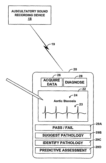
[0043] FIG. 2 is a block diagram of an exemplary embodiment of a portable
digital
assistant (PDA) 20 operating as a diagnostic device to assist diagnosis of
patient 8 (FIG.
1). In the illustrated embodiment, PDA 20 includes a touch-sensitive screen
22, input
keys 26, 28 and 29A-29D.
[0044] Upon selection of acquisition key 26 by clinician 10, diagnostic device
20 enters
an acquisition mode to receive via communication link 19 a digitized
representation of
auscultatory sounds recorded from patient 8. Once the digitized representation
is
received, clinician 10 actuates diagnose key 28 to direct diagnostic device 20
to apply
configuration data 13 and render a suggested diagnosis based on the received
auscultatory
8
CA 02595924 2007-07-25
WO 2006/079062 PCT/US2006/002422
sounds. Alternatively, diagnostic device 20 may automatically begin processing
the
sounds without requiring activation of diagnose key 28.
[0045] As described in further detail below, diagnostic device 20 applies
configuration
data 13 to map the digitized representation received from auscultatory sound
recording
device 18 to the multi-dimensional energy space computed by data analysis
system 4. In
general, diagnostic device 20 determines to which of the disease regions
defined within
the multi-dimensional space the auscultatory sounds of patient 8 most closely
maps.
Based on this determination, diagnostic device 20 updates touch-sensitive
screen 22 to
output one or more suggested diagnoses to clinician 10. In this example,
diagnostic
device 20 outputs a diagnostic message 24 indicating that the auscultatory
sounds indicate
that patient 8 may be experiencing aortic stenosis. In addition, diagnostic
device may
output a graphical representation 23 of the auscultatory sounds recorded from
patient 8.
[0046] Diagnostic device 20 may include a number of input lceys 29A-29D that
control
the type of analysis performed via the device. For example, based on which of
inputs
keys 29A-29D has been selected by clinician 10, diagnostic device 20 provides
a pass/fail
type of diagnostic message, one or more suggested pathologies that patient 8
may
currently be experiencing, one or more pathologies that patient 8 has been
identified as
experiencing, and/or a predictive assessment of one or more pathologies to
which patient
8 may be tending.
[0047] Screen 22 or an input key could also allow input of specific patient
information
such as gender, age and BMI (body mass index = weight (kilograms)/height
(meters)
squared. This information could be used in the analysis set forth here within.
[0048] In the embodiment illustrated by FIG. 2, diagnostic device 20 may be
any PDA,
such as a PalmPilot manufactured by Palm, Inc. of Milpitas, California or a
PocketPC
executing the Windows CE operating system from Microsoft Corporation of
Redmond,
Washington.
[0049] FIG. 3 is a perspective diagram of an exemplary embodiment of an
electronic
stethoscope 30 operating as a diagnostic device in accordance with the
techniques
described herein. In the illustrated embodiment, electronic stethoscope 30
comprises a
chestpiece 32, a sound transmission mechanism 34 and an earpiece assembly 36.
Chestpiece 32 is adapted to be placed near or against the body of patient 8
for gathering
the auscultatory sounds. Sound transmission mechanism 34 transmits the
gathered sound
to earpiece assembly 36. Earpiece assembly 36 includes a pair of earpieces
37A, 37B,
where clinician 10 may monitor the auscultatory sounds.
9
CA 02595924 2007-07-25
WO 2006/079062 PCT/US2006/002422
[0050] In the illustrated embodiment, chestpiece 32 includes display 40 for
output of a
diagnostic message 42. More specifically, electronic stethoscope 30 includes
an internal
controller 44 that applies configuration data 13 to map the auscultatory
sounds captured
by chestpiece 32 to the multidimensional energy space computed by data
analysis system
4. Controller 44 determines to which of the disease regions defined within the
energy
space the auscultatory sounds of patient 8 most closely maps. Based on this
determination, controller 44 updates display 40 to output diagnostic message
42.
[0051] Controller 44 is illustrated for exemplary purposes as located within
chestpiece
32, and may be located within other areas of electronic stethoscope 30.
Controller 44
may comprise an embedded microprocessor, DSP, FPGA, ASIC, or similar hardware,
firinware and/or software for implementing the techniques. Controller 44 may
include a
computer-readable medium to store computer readable instructions, i.e.,
program code,
that can be executed to carry out one of more of the techniques described
herein.
[0052] FIG. 4 is a flowchart that provides an overview of the techniques
described herein.
As illustrated in FIG. 4, the process may generally be divided into two
stages. The first
stage is referred to as the parametric analysis stage in which clinical data
12 (FIG. 1) is
analyzed using SVD to produce configuration data 13 for diagnostic device 6.
This
process may be computationally intensive. The second stage is referred to as
the
diagnosis stage in which diagnostic device 6 applies the results of the
analysis stage to aid
the diagnosis of a patient. For purposes of illustration, the flowchart of
FIG. 4 is
described in reference to FIG. 1.
[0053] Initially, clinical data 12 is collected (50) and provided to data
analysis system 4
for singular value decomposition (52). As described above, clinical data 12
comprises
electronic recordings of auscultatory sounds from a set of patients having
known cardiac
conditions.
[0054] Analysis module 14 of data analysis system 4 analyzes the recorded
heart sounds
of clinical data 12 in accordance with the techniques described herein to
define a set of
disease regions within a multi-dimensional space representative of the
electronically
recorded heart sounds (52). Each disease region within the multi-dimensional
space
corresponds to sounds within a heart cycle that have been mathematically
identified as
indicative of the respective disease. Analysis module 14 stores the results of
the analysis
within parametric database 16 (54). In particular, the results include
configuration data
13 for use by diagnostic device 6 to map patient auscultatory sounds to the
generated
multidimensional space. Once analysis module 14 has processed clinical data
12,
CA 02595924 2007-07-25
WO 2006/079062 PCT/US2006/002422
diagnostic device 6 receives or is otherwise programmed to apply configuration
data 13 to
assist the diagnosis of patient 18 (56). In this manner, data analysis system
can be viewed
as applying the techniques described herein, including SVD, to analyze a
representative
sample set of auscultatory sounds recorded from patients having known
physiological
conditions to generate parametric data that may be applied in real-time or
pseudo real-
time.
[0055] The diagnosis stage commences when auscultatory sound recording device
18
captures auscultatory sounds from patient 8. Diagnosis device 6 applies
configuration
data 13 to map the heart sounds received from auscultatory sound recording
device 18 to
the multi-dimensional energy space computed by data analysis system 4 from
clinical
data 12 (58). For cardiac auscultatory sounds, diagnostic device 6 may repeat
the real-
time diagnosis for one or more heart cycles identified with the recorded heart
sounds of
patient 8 to help ensure that an accurate diagnosis is reported to clinician
10. Diagnostic
device 6 outputs a diagnostic message based on the application of the
configuration and
the mapping of the patient auscultatory sounds to the multidimensional space
(59).
[0056] FIG. 5 is a flowchart illustrating the parametric analysis stage (FIG.
4) in further
detail. Initially, clinical data 12 is collected from a set of patients having
known cardiac
conditions (60). In one embodiment, each recording captures approximately
eight
seconds of auscultatory heart sounds, which represents approximately 9.33
heart cycles
for a seventy beat per minute heart rate. Each recording is stored in digital
form as a
vector R having 32,000 discrete values, which represents a sampling rate of
approximately 4000 Hz.
[0057] Each heart sound recording R is pre-processed (62), as described in
detail below
with reference to FIG. 6. During this pre-processing, analysis module 12
processes the
vector R to identify a starting time and ending time for each heart cycle. In
addition,
analysis module 14 identifies starting and ending times for the systole and
diastole
periods as well as the SI and S2 periods within each of the heart cycles.
Based on these
identifications, analysis module 14 normalizes each heart cycle to a common
heart rate,
e.g., 70 beats per minute. In other words, analysis module 14 may resample the
digitized
data corresponding to each heart cycle as necessary in order to stretch or
compress the
data associated with the heart cycle to a defined time period, such as
approximately 857
ms, which corresponds to a heart rate of 70 beats per minute.
[0058] After pre-processing each individual heart recording, analysis module
14 applies
singular value decomposition (SVD) to clinical data 12 to generate a
multidimensional
11
CA 02595924 2007-07-25
WO 2006/079062 PCT/US2006/002422
energy space and define disease regions within the multi-dimensional energy
space that
correlate to characteristics of the auscultatory sound (64).
[0059] More specifically, analysis module 14 combines N pre-processed sound
recordings R for patients having the same known cardiac condition to form an
MxN
matrix A as follows:
A=1... M
N... M '
where each row represents a different sound recording R having M digitized
values, e.g.,
3400 values.
[0060] Next, analysis module 14 applies SVD to decompose A into the product of
three
sub-matrices:
A=UDVT,
where U is an NxM matrix with orthogonal columns, D is an MxM non-negative
diagonal matrix and V is an MxM orthogonal matrix. This relationship may also
be
expressed as:
UTAV=diag(S) = diag(61,..., 6p),
where the elements of matrix S(61, ..., ap) are the singular values of A. In
this SVD
representation, U is the left singular matrix and V is the right singular
matrix. Moreover,
U can be viewed as an MxM weighting matrix that defines characteristics with
each R
that best define the matrix A. More specifically, according to SVD principles,
the U
matrix provides a weighting matrix that maps the matrix A to a defined region
within an
M dimensional space.
[0061] Analysis module 14 repeats this process for each cardiac condition. In
other
words, analysis module 14 utilizes sound recordings R for "normal" patients to
coinpute a
corresponding matrix ANORMAL and applies SVD to generate a UNORMAL matrix.
Similarly, analysis module computes an A matrix and a corresponding U matrix
for each
pathology. For example, analysis module 14 may generate a UAs, UAR, a UTR,
and/or a
UDISEASED=, where the subscript "AS" designates a U matrix generated from
patient or
population of patients known by other diagnostic tools to display aortic
stenosis. The
subscript "AR" designates aortic regurgitation and the subscript "TR"
designated
tricuspid regurgitation in analogous manner.
[0062] Next, analysis module 14 pair-wise multiplies each of the computed U
matrices
with the other U matrices, and performs SVD on the resultant matrices in order
to identify
12
CA 02595924 2007-07-25
WO 2006/079062 PCT/US2006/002422
which portions of the U matrices best characterize the characteristics that
distinguish
between the cardiac conditions. For example, assuming matrices of UNORn1AL,
UAS, and
UAR, analysis module computes the following matrices:
T1= UNORMAL * UAS,
T2= UNORMAL * UAR, and
T3= UAS * UAR=
[0063] Analysis module 14 next applies SVD on each of the resultant matrices
Tl, T2
and T3, wliich again returns a set of sub-matrices that can be used to identif-
y the portions
of each original U matrix that maximizes an energy differences within the
multidimensional space between the respective cardiac conditions. For example,
the
matrices coinputed via applying SVD to TI can be used to identify those
portions of
UN~RAI'AL and UAs that maximize the orthogonality of the respective disease
regions within
the multidimensional space.
[0064] Consequently, TI may be used to trim or otherwise reduce UNORnrAL and
UAsto
sub-matrices that may be more efficiently applied during the diagnosis (64).
For
example, S matrices computed by application of SVD to each of TI, T2 and T3
may be
used. An inverse cosine may be applied to each S matrix to compute an energy
angle
between the respective two cardiac conditions within the multidimensional
space. This
energy angle may then be used to identify which portions of each of the U
matrices best
account for the energy differences between the diseases reasons within the
multidimensional space.
[0065] Next, analysis module computes an average vector A V for each of the
cardiac
conditions (66). In particular, for each MxN A matrix formulated from cardiac
data 12,
analysis module 14 computes a 1 xN average vectorAVthat stores the average
digitized
values computed from the N sound recordings R within the matrix A. For
example,
analysis module 14 may compute AVAs, A VAR, A VTR, and/or A VDISEASED vectors.
[0066] Analysis module 14 stores the computed AV average vectors and the U
matrices,
or the reduced U matrices, in parametric database 16 for use as configuration
data 13. For
example, analysis module 14 may store AVAs , A VAR, A VTR, UNORMAL, UASa and
UAR, for
use as configuration data 13 by diagnostic device 6 (68).
[0067] FIG. 6 is a flowchart that illustrates in further detail one technique
for pre-
processing of an auscultatory sound recording R. In general, the pre-
processing
techniques separate the auscultatory sound recording R into heart cycles, and
further
separate each heart cycle into four parts: a first heart sound, a systole
portion, a second
13
CA 02595924 2007-07-25
WO 2006/079062 PCT/US2006/002422
heart sound, and a diastole portion. The pre-processing techniques apply
Shannon Energy
Envelogram (SEE) for noise suppression. The SEE is then thresholded making use
of the
relative consistency of the heart sound peaks. The threshold used can be
adaptively
generated based upon the specific auscultatory sound recording R.
[0068] Initially, analysis module 14 performs wavelet analysis on the
auscultatory sound
recording R to identify energy tllresholds within the recording (70). For
example,
wavelet analysis may reveal energy thresholds between certain frequency
ranges. In
other words, certain frequency ranges may be identified that contain
substantial portions
of the energy of the digitized recording.
[0069] Based on the identified energy thresholds, analysis module 14
decomposes the
auscultatory sound recording R into one or more frequency bands (72). Analysis
module
14 analyzes the characteristics of the signal within each frequency band to
identify each
heart cycle. In particular, analysis module 14 examines the frequency bands to
identify
the systole and diastole stages of the heart cycle, and the S 1 and S2 periods
during with
certain valvular activity occurs (74). To segment each heart cycle, analysis
module 14
may first apply a low-pass filter, e.g., an eiglit order Chebyshev-type low-
pass filter with
a cutoff frequency of 1kHz. The average SEE may then be calculated for every
.02
second segment throughout the auscultatory sound recording R with 0.01 second
segment
overlap as follows:
Es = - 1 jX n ,=õt (t ) log Xo,=õr (i)
N
where Xõo. is the low-pass filtered and normalized sample of the sound
recording and N
is the number of signal samples in the 0.02 second segment, e.g., N equals
200. The
normalized average Shannon Energy versus the time axis may then be computed
as:
__ Es (t) - M(Es (t))
PS (t) S(Es (t))
wliere M(Es(t)) is the mean of Es(t) and S(Es(t)) is the standard deviation of
Es(t). The
mean and standard deviation are then used as a basis for identifying the
pealcs with each
heart cycle and the starting and times for each segment with each heart cycle.
[0070] Once the starting and ending times for each heart cycle and each S 1
and S2
periods is determined within the auscultatory sound recording R, analysis
module 14 re-
samples the auscultatory sound recording R as necessary to stretch or compress
so that
each heart cycle and each S1 and S2 period occur over a time period (76). For
example,
analysis module 14 may normalize each heart cycle to a common heart rate,
e.g., 70 beats
14
CA 02595924 2007-07-25
WO 2006/079062 PCT/US2006/002422
per minute and may ensure that each S 1 and S2 periods within the cycle
correspond to an
equal length in time. This may advantageously allow the portions of the
auscultatory
sound recording R for the various phases of the cardiac activity to be more
easily and
accurately analyzed and compared with similar portions of the other
auscultatory sound
recordings.
[0071] Upon normalizing the heart cycles within the digitized sound recording
R,
analysis module 14 selects one or more of the heart cycles for analysis (78).
For example,
analysis module 14 may identify a "cleanest" one of the heart cycles based on
the amount
of noise present within the heart cycles. As other examples, analysis module
14 may
compute an average of all of the heart cycles or an average to two or more
randomly
selected heart cycles for analysis.
[0072] FIG. 7 is a graph that illustrates an example result of the wavelet
analysis and
energy thresholding described above in reference to FIG. 6. In particular,
FIG. 7
illustrates a portion of a sound recording R. In this example, analysis module
14 has
decomposes an exemplary auscultatory sound recording R into four frequency
bands
80A-80D, and each frequency band includes a respective frequency component 82A-
82D.
[0073] Based on the decoinposition, analysis module 14 detects changes to the
auscultatory sounds indicative of the stages of the heart cycle. By analyzing
the
decomposed frequencies and identifying the relevant characteristics, e.g.,
changes of
slope within one or more of the frequency bands 80, analysis module 14 is able
to reliably
detect the systole and diastole periods and, in particular, the start and end
to the S 1 and S2
periods.
[0074] FIG. 8 illustrates an example data structure 84 of an auscultatory
sound recording
R. As illustrated, data structure 84 may comprise a 1xN vector storing
digitized data
representative of the auscultatory sound recording R. Moreover, based on the
pre-
processing and re-sampling, data structure 84 stores data over a fixed number
of heart
cycles, and each S 1 and S2 regions occupy a pre-defined portion of the data
structure.
For example, S1 region 86 for the first heart cycle may comprise elements 0-
399 of data
structure 84, and systole region 87 of the first heart cycle may coinprises
elements 400-
1299. This allows multiple auscultatory sound recordings R to be readily
combined to
form an MxN matrix A, as described above, in which the S 1 and S2 regions for
a given
heart cycle are column-aligned.
[0075] FIG. 9 is a flowchart illustrating the diagnostic stage (FIG. 4) in
further detail.
Initially, auscultatory data is collected from patient 8 (90). As described
above, the
CA 02595924 2007-07-25
WO 2006/079062 PCT/US2006/002422
auscultatory data may be collected by a separate auscultatory sound recording
device 18,
e.g., an electronic stethoscope, and communicated to diagnostic device 6 via
link
communication 19. In another embodiment, the functionality of diagnostic
device 6 may
be integrated within auscultatory sound recording device 18. Similar to the
parametric
analysis stage, the collected auscultatory recording captures approximately
eight seconds
of auscultatory sounds from patient 8, and may be stored in digital form as a
vector RPAT
having 3400 discrete values.
[0076] Upon capturing the auscultatory data RPAT, diagnostic device 6 pre-
processes the
heart sound recording RPAT (92), as described in detail above with reference
to FIG. 6.
During this pre-processing, diagnostic device 6 processes the vector RPAT to
identify a
starting time and an ending time for each heart cycle, and starting and ending
times for
the systole and diastole periods as well as the S 1 and S2 periods of each of
the heart
cycles. Based on these identifications, diagnostic device 6 normalizes each
heart cycle to
a cominon heart rate, e.g., 70 beats per minute.
[0077] Next, diagnostic device 6 initializes a loop that applies configuration
data 13 for
each physiological condition examined during the analysis stage. For example,
diagnostic device may utilize configuration data of A VAS, A VAR, A VTR,
UNORMAL, UAS,
and UAR, to assist diagnosis of patient 8.
[0078] Initially, diagnostic device 6 selects a first physiological condition,
e.g., normal
(93). Diagnostic device 6 then subtracts the corresponding average vector A V
from the
captured auscultatory sound vector RPAT to generate a difference vector D
(94). D is
referred to generally as a difference vector as the resulting digitized data
of D represents
differences between the captured heart sound vector RPAT and the currently
selected
physiological condition. For example, diagnostic device 6 may calculate
DNoRMAL as
follows:
DNORMAL - RPAT - A VNORMAL =
[0079] Diagnostic device 6 then multiples the resulting difference vector D by
the
corresponding U matrix for the currently selected physiological condition to
produce a
vector P representative of patient 8 with respect to the currently selected
cardiac
condition (96). For example, diagnostic device 6 may calculate PNORMAL vector
as
follows:
PNORMAL = DNORMAL " UNORMAL.
Multiplying the difference vector D via the corresponding U matrix effectively
applies a
weighting matrix associated with the corresponding disease region within the
inulti-
16
CA 02595924 2007-07-25
WO 2006/079062 PCT/US2006/002422
dimensional space, and produces a vector P within the multidimensional space.
The
alignment of the vector P relative to the disease region of the current
cardiac condition
depends on the normality of the resulting difference vector D and the U matrix
determined during the analysis stage.
[0080] Diagnostic device 6 repeats this process for each cardiac condition
defined within
the multidimensional space to produce a set of vectors representative of the
auscultatory
sound recorded from patient 8 (98, 106). For example, assuming configuration
data 13
comprises AVAs , A VAR, A VTR, UNORMAL, UAS, and UAR, diagnostic device 6
calculates
four patient vectors as follows:
PNORMAL - DNORMAL - UNORMAL,
PAS = DAS UAS,
PAR = DAR x UAR, and
PTR = DTR UTR=
[0081] This set of vectors represents the auscultatory sounds recorded from
patient 8
within the multidimensional space generated during the analysis stage.
Consequently, the
distance between each vector and the corresponding disease region represents a
measure
of similarity between the characteristics of the auscultatory sounds from
patient 8 and the
characteristics of auscultatory sounds of patients known to have the
respective cardiac
conditions.
[0082] Diagnostic device 6 then selects one of the disease regions as a
fiuzction of the
orientation of the vectors and the disease regions within the multidimensional
space. In
one embodiment, diagnostic device determines which of the disease regions
defined
within the energy space has a minimum distance from the representative
vectors. For
example, diagnostic device 6 first calculates energy angles representative of
the minimum
angular distances between each of the vectors P and the defined disease
regions (100).
Continuing with the above example, diagnostic device 6 may compute the
following four
distance measurements:
DISTNORMAL = PNORMAL -MIN [PAS, PAR, PTRI,
DISTAS = PAS -MIN [PNORDIAL, PAR, PTRI,
DISTAR = PAR -MIN [PAS, PNORMAL, PTRI, and
DISTTR = PTR -MIN[PAS, PAR, PNORMAL]=
[0083] In particular, each distance measurement DIST is a two-dimensional
distance
between the respective patient vector P and the mean of each of the defined
disease
regions within the multidimensional space.
17
CA 02595924 2007-07-25
WO 2006/079062 PCT/US2006/002422
[0084] Based on the computed distances, diagnostic device 6 identifies the
smallest
distance measurement (102) and determines a suggested diagnosis for patient 8
to assist
clinician 10. For example, if of the set of patient vectors PAs is the minimum
distance
away from its respective disease space, i.e., the AS disease space, diagnostic
device 6
determines that patient 8 may likely be experiencing aortic stenosis.
Diagnostic device 6
outputs a representative diagnostic message to clinician 10 based on the
identification
(104). Prior to outputting the message, diagnostic device 6 may repeat the
analysis for
one or more heart cycles identified with the recorded heart sounds of patient
8 to lzelp
ensure that an accurate diagnosis is reported to clinician 10.
[0085] Examples
[0086] The techniques described herein were applied to clinical data for a set
of patients
known to have either "normal" cardiac activity or aortic stenosis. In
particular, a
multidimensional space was generated based on the exainple clinical data, and
then the
patients were assessed in real-time according to the techniques described
herein.
[0087] The following table shows distance calculations for the auscultatory
sounds for
the patients known to have normal cardiac conditions. In particular, vectors
were
computed for each of the measured heart cycles for each patient. Table 1 shows
distances
for the vectors, measured in volts, with respect to a disease region within
the
multidimensional space associated with the normal cardiac condition.
HEART PATIENT PATIENT PATIENT
CYCLE 1 2 3
1 0.45 0.25 0.20
2 0.64 0.14 0.18
3 0.38 0.21 0.32
4 0.36
0.20
6 0.33
Table 1
18
CA 02595924 2007-07-25
WO 2006/079062 PCT/US2006/002422
[0088] Table 2 shows distance calculations, measured in volts, for the
auscultatory
sounds for the patients known to have aortic stenosis. In particular, Table 2
shows energy
distances for the vectors wit11 respect to a region within the
multidimensional space
associated with the aortic stenosis cardiac condition.
HEART CYCLE PATIENT 4 PATIENT. 5
1 -0.43 -0.49
2 -0.67 -0.43
3 -0.55 -0.37
4 -0.43 -0.64
-0.34 -0.17
6 -0.44 -0.14
-0.60
Table 2
[0089] As illustrated by Table 1 and Table 2, the vectors are clearly separate
within the
multidimensional space, an indication that diagnosis can readily be made. All
five
patients followed a similar pattern.
[0090] FIGS. 10A and l OB are graphs that generally illustrate the exemplary
results. In
particular, FIGS. 10A and lOB illustrate aortic stenosis data compared to
normal data.
Similarly, FIGS. 1 lA and 11B are graphs that illustrate tricuspid
regurgitation data
compared to normal data. FIGS. 12A and 12B are graphs that illustrate aortic
stenosis
data compared to tricuspid regurgitation data. In general, the graphs of FIGS.
10A, l OB,
1 1A, and 11B illustrate that the techniques result in substantially non-
overlapping data for
the normal data and disease-related data.
[0091] FIG. 13 is a flowchart that illustrates another technique for pre-
processing of an
auscultatory sound recording R. In particular, FIG. 14 describes application
of voice
recognition techniques to generate inel-cepstrum coefficients for use by the
SVD process
described herein or other principle component analysis technique. Unlike the
pre-
processing technique described with respect to FIG. 6, application of voice
recognition
technology to the auscultatory sound recording R may eliminate the need to
separate the
auscultatory sound recording R into heart cycles, and further separate each
heart cycle
into four parts: a first heart sound, a systole portion, a second heart sound,
and a diastole
portion. Segmentation may be computationally intensive and time-consuming.
19
CA 02595924 2007-07-25
WO 2006/079062 PCT/US2006/002422
[0092] In general, a cepstrum is a discrete cosine transform of a log-spectrum
of a signal
and is coinmonly used in speech recognition systems. A mel-cepstrum is a
modified
version of the cepstrum and was designed to exploit the human auditory system
by
dividing the frequency domain in a non-uniform manner during cepstrum
computation.
[0093] First, analysis module 14 coinputes a Discrete Fourier transform (DFT)
of
auscultatory sound recording R using an FFT algorithm and a hanning window
(200).
Next, analysis module 14 divides the DFT(R) into M non-uniform sub-bands
throughout
the audible range (202). In particular, analysis module 14 may split lower
frequency
portion of the audible range into N equal sub-bands. For example, may split
the
frequency range of 20-500 Hz linearly into 12 sub-bands. Next, split the upper
frequency
band logarithmically into N sub-bands. For example, may split 500 to 200 Hz
logarithmically into 12 sub-bands. One reason for such a split is because
audible
components within the higher frequency band may be noise.
[0094] Analysis module 14 then formulates the resultant signal as a magnitude-
frequency
representation and determines mel-cepstrum coefficients for each of the
defined sub-
bands (204). A mel-cepstrum vector (c=[cl, c2, ..., cK] can be computed from
the
discrete cosine transform (DCT) of the auscultatory sound vecrtor R as
follows:
Ck =1~~ log(e~) cosLk(i - 0.5) /M1 , k=1,2,..., K
where M represents the number of sub-bands.
[0095] In particular, analysis module 14 selects the components of the mel-
cepstrum
coefficients that are most representative of variability of between the
disease states and
uses those coefficients as inputs to the SVD process described herein to
define the disease
regions and their boundaries within the multidimensional space (206). In this
case, the
SVD analysis utilizes a vector of the deterinined mel-cepstrum coefficients
instead of
using an auscultatory sound vector. One example of 1VIel-cepstrum-based
Principle
Component Analysis is described in "Classification of Closed- and Open-Shell
Pistachio
Nuts Using Voice-Recognition Technology," A. E. Cetin et al., Transactions of
ASAE,
Vol. 47(2): 659-664, 2004, hereby incorporated by reference. In other
embodiments, all
parametric and non-parametric techniques, such as the use of regressive
modeling, neural
networks or expert systems for feature extraction.
[0096] FIGS. 14-17 are graphs that illustrate exemplary mel-cepstrum
coefficients for a
single disease state, aortic regurgitation in this example. In particular,
FIG. 14 is a graph
that plots magnitudes of the mel-cepstrum coefficients determined over a
frequency range
CA 02595924 2007-07-25
WO 2006/079062 PCT/US2006/002422
of zero to 500 Hz. As illustrated, the techniques utilize a linear scale for
the sub-bands
for lower frequencies (e.g., 0 to 140 Hz) and a log scale for higher
frequencies (e.g., 140-
500 Hz).
[0097] FIG. 15 is a graph that plots magnitudes of the mel-cepstrum
coefficients for
aortic regurgitation versus FFT values for each frequency band.
[0098] FIG. 16 is a graph that plots perceived pitch for the mel-cepstrum
representation
over a frequency range of zero to 500 Hz.
[0099] FIG. 17 is a graph that plots magnitudes of the mel-cepstruin
coefficients
determined for an exemplary disease region over a frequency range of zero to
500 Hz.
[0100] Various embodiments of the invention have been described. For example,
although described in reference to sound recordings, the techniques may be
applicable to
other electrical recordings from a patient. The techniques may be applied, for
example, to
electrocardiogram recordings electrically sensed from a patient. These and
other
embodiments are within the scope of the following claims.
21