Note: Descriptions are shown in the official language in which they were submitted.
CA 02596957 2010-08-12
SUBSTRATE RECOGNITIONSY DIFFERENTIABLE HUMAN
= MESENCHYMAL STEM CELLS
FIELD OF THE INVENTION
= 100021 The present invention relates to compositions and
methods comprising a' three,
= dimensional nanofiber matrix of synthetic polymers. The synthetic
nanonber matrix can
=yeas ascaffOldisubstrate for delivery of differentiable human mesenchymal
cells for tissue
engineering applications.
GOVERNMENT SUPPORT =
10003.1 This work is supported at least in part by grams. from the
National Science
= Foundation to Dr. Atinzeh. The government May have certain righttin this
invention.
BACKGROUND OF THE INVENTION
100041 Orthopedic management of lesions to articular cartilage
remains a persistent
problem for the orthopedist and petienthecause articelar cartilage has a
limited intrinsic
ability to heal.. This has prompted the develeptnent of numerous procedures to
beat these
lesions and to halt or slOw the progression to diffuse arthritic thanges,
MOOS] Tissue engineering is the application of prineiPles.and methods of
engineering and
life kit:bees toward a: fundamental understanding and development of
biological substitutes
to-restore, maintain and improve human tissue functions. It may eliminate many
of the
problems associated with current surgical 'options. Current tissue engineering
methods are
aimed at filling the -cartilage 'defects with cells with or without scaffolds
to promote cartilage
regeneration. Implantation of scaffolds alone leads to a poor quality
reparative fissile.
= Chondroeytes implantixl either. alone or in combination with a scaffold
have failed 16 restore
a normal articular surface, and the hyaline cartilage formed early on in
response to
1
CA 02596957 2007-05-29
WO 2006/068809
PCT/US2005/043876
chondrocyte-containing scaffolds seems to deteriorate with time. Therefore,
improved tissue
engineering methods still are needed.
[0006] The use of stem cells for tissue engineering therapies is at the
forefront of
scientific investigation. In the body, adult stem cells often are localized to
specific
chemically and topologically complex microenvironments, or so-called "niches".
Increasing
experimental evidence supports the notion that stem cells can adjust their
properties
according to their surroundings and select specific lineages according to cues
they receive
from their niche (Xie L, Spradling, AC, "A Niche Maintaining Germ Line Stem
Cells In
Dropsophila Ovary," Science 290:328 (2000); Fuchs E, Segre J, "Stem Cells: A
New Lease
On Life," Cell 100: 143-155 (2000), Watt FM, Hogan BLM, "Out Of Eden: Stem
Cells And
Their Niches," Science 287:1427 (2000)). It follows that in order for an MSC
therapy to be
successful in the repair of a specific tissue type, the microenvironment of
the cells should be
designed to relay the appropriate chemical and physical signals to them.
Mimicking
characteristics of the microenvironment during cartilage development may be a
viable
approach. During cartilage development, one of the earliest events is pre-
cartilage
= mesenchymal cell aggregation and condensation resulting from cell-cell
interaction, which is
mediated by cell-cell and cell-matrix adhesion (fibronectin, proteoglycans,
and
collagens).(DeLise A.M., Fischer L, Tuan RS, "Cellular Interactions And
Signaling In
Cartilage Development. Osteoarthritis and Cartilage 8: 309-334 (2000)). Type I
collagen,
the predominant matrix protein present in the early stages of development, is
later
transformed to Type II collagen by increased cell synthesis during
differentiation. (Safronova
EE, Borisova NV, Mezentseva SV, Krasnopol'skaya KD, "Characteristics Of The
Macromolecular Components Of The Extracellular Matrix In Human Hyaline
Cartilage At
Different Stages Of Ontogenesis." Biomedical Science 2: 162-168 (1991)).
Multiple growth
factors and morphogens are also present contributing to the regulation of the
differentiation
process.
[0007] A few studies have demonstrated the use of MSCs for cartilage
repair through
intra-articular injection and have shown promise.(Murphy M, Fink DJ, Hunziker
EB, Barry
FP, "Stem Cell Therapy In A Caprine Model Of Osteoarthritis," Arthritis
Rheumatism 48:
3464-3474 (2003); Ponticello MS, Schinagel RM, Kadiyala, S, Barry FP, "Gelatin-
Based
Resorbable Spone As A Carrier Matrix For Human Mesenchymal Stem Cells In
Cartilage
Regeneration Therapy," J Biomed Materials Res 52: 246-255 (2000)). The MSCs
are
2
CA 02596957 2007-05-29
WO 2006/068809
PCT/US2005/043876
injected at a high cell density either alone (in saline) or in combination
with a
gelatinous/hydrogel matrix in order to promote cell-cell aggregation. However,
the use of
MSCs in combination with biomaterials of varying architectures that may
closely mimic the
physical architecture of the native extracellular matrix during development to
direct
chondrogenic differentiation has yet to be investigated.
100081 From a biological viewpoint, almost all human tissues and organs are
characterized by well-organized hierarchical fibrous structures through the
assembly of
nanoscale elements. It is believed that converting biopolymers into fibers and
networks that
mimic native structures will ultimately enhance the utility of these materials
as scaffolds.
Nanoscale fibrous scaffolds may provide an optimal template for stem cell
growth,
differentiation, and host integration.
[0009] It is known that cells will attach to synthetic polymer scaffolds
leading to the
formation of tissue. (Sachlos, E. and Czernuszka, Eur. Cells & Materials 5: 29-
40 (2003)).
Using fetal bovine chondrocytes maintained in vitro, Li et al. have shown that
scaffolds
constructed from electrospun three-dimensional nanofibrous poly(e-capro-
lactone) act as a
biologically preferred scaffold/substrate for proliferation and maintenance of
the chondrocyte
phenotype. (Wan-Ju Li, et al., J. Biomed. Mater. Res. 67A: 1105-1114 (2003)).
[0010] Most work to develop scaffold materials for tissue engineering,
however, has
relied on large diameter fibers, which do not mimic the morphological
characteristics of the
native fibrils of the extracellular matrix.
[0011] For example, U.S. Pat. No. 6,472,210, issued to Holy, describes a
macroporous
polymer scaffold for regenerating bone comprising macropores at least 50% of
which have a
diameter in the range of 0.5-3.5 mm, a range representative of that found in
human trabecular
bone, and interconnections as seen in trabecular bone and a process for making
the scaffold.
The scaffold comprises porous walls consisting of microporous polymer polymer
struts
defining macropores which are interconnected by macroporous passageways. The
microporous struts contain microporous passageways extending through the
microporous
polymer struts so that macropores on either side of a given strut are in
communication
through the strut. Cell colonization into the scaffolds required a minimum
interconnection
size of 0.35 mm and macropore size of 0.7 mm.
[0012] U.S. Pat. No. 6,214,369 and U.S. Pat. No. 5,906,934, issued to
Grande et al.,
describe a composition and method for growing new cartilage and or bone in
rabbits whereby
3
CA 02596957 2007-05-29
WO 2006/068809 PCT/US2005/043876
cells described as mesenchymal stem cells isolated from adult rabbit leg
skeletal muscle were
cultured on scaffolds of 12-14 tim diameter polyglycolic acid fibers in the
form of an
intertwined, woven or meshed matrix or a sponge matrix, and the MSC-seeded
matrix then
implanted into full thickness articular cartilage defects from syngeneic
rabbits. Because the
cells in this study are not characterized by their cell markers, it is
uncertain whether they
were stem cells, or at least multipotent cells.
[0013] U.S. Pat. No. 6,511,511, and U.S. Pat. No. 6,783,712, issued to
Slivka, describe a
fiber-reinforced, polymeric implant material comprising a polymeric matrix and
a method of
making this material for use as a scaffold for tissue implantation. The fibers
of the matrix,
which are preferably oriented predominantly parallel to each other but may
also be aligned in
a single plane, are described as preferably about 5 tim to about 50 Am in
diameter.
[0014] U.S. Pat. No. 6,790,455, issued to Chu describes a cell delivery
system
comprising viable cells and a fibrous matrix as a carrier physically
associated with the cells to
contain and release the cells at a controlled rate. In a preferred embodiment,
a layered cell
storage and delivery system comprises bone cells embedded between two layers
of a
poly(D,L-lactide (PLLA) or PLGA/ lactide monomer. The first or base layer
membrane
contains biodegradable nanofibers made from a 40% PLLA or PLGA/lactide monomer
by
electrospining. Bone cells are deposited on the surface of the membrane after
the membrane
has been prewet by dipping in a minimum essential medium solution containing
10% fetal
bovine serum to increase adhesion between the membrane surface and live bone
cells. The
cell containing membrane then is covered by a thin top layer of membrane to
encapsulate the
bone cells near the surface of membrane and to facilitate quick release of the
cells from the
membrane. The final sandwiched cell/membrane structure described thus consists
of three
parts: the bottom layer (120-200 itm thick), live cells (15-20 jim thick), and
the top layer
(several microns thick). While the patent teaches that isolated cells must be
undifferentiated
and suggests that the cell delivery system when positioned at a desired
location for cell
delivery to a mammal can guide the development and shape of new tissue, it
provides no
guidance on how to do so.
[0015] Human MSCs used in combination with nanoscale fibrous scaffolds may
be an
effective potential tissue engineering therapy. Since large diameter fibers do
not mimic the
morphological characteristics of the native fibrils of the extracellular
matrix, biopolymers
4
CA 02596957 2007-05-29
WO 2006/068809 PCT/US2005/043876
consisting of fibers and networks that mimic native structures may enhance the
utility of
these materials. The present invention addresses this problem.
SUMMARY OF THE INVENTION
[00161 The present invention provides compositions comprising a three-
dimensional
matrix of fibers used as an implantable scaffolding for delivery of
differentiable human
mesenchymal cells in tissue engineering applications and methods of preparing
and using
them. According to one embodiment of the invention, a structure for growing
isolated
=
differentiable human mesenchymal cells comprises a three-dimensional matrix of
fibers,
wherein the matrix is seeded with the isolated differentiable human
mesenchymal cells and
forms a supporting scaffold for growing the differentiable human mesenchymal
cells, and
wherein the isolated differentiable human mesenchymal cells differentiate into
a mature cell
phenotype on the scaffold. According to another embodiment of the present
invention, the
three dimensional matrix of fibers is formed of a polymeric material.
According to another
embodiment, the polymeric material of the structure of the present invention
is a
biocompatible polymer. According to another embodiment, the biocompatible
polymer of
the structure is poly D,L lactide glycolide. According to another embodiment,
the
biocompatible polymer of the structure is poly L-lactic acid. According to
another
embodiment, the structure's matrix of fibers is a non-woven mesh of
nanofibers. According
to another embodiment, the matrix of nanofibers is prepared by
electrospinning. According
to another embodiment, the non-woven mesh of nanofibers comprises poly D,L
lactide
glycolide. According to another embodiment, the non-woven mesh of nanofibers
comprises
poly L-lactic acid. According to another embodiment, the structure's matrix of
fibers
comprises a non-woven mesh of microfibers. According to another embodiment,
the matrix
of microfibers is prepared by electrospinning. According to another
embodiment, the non-
woven mesh of microfibers comprises poly D,L lactide glycolide. According to
another
embodiment, the non-woven mesh of microfibers comprises poly L-lactic acid.
According to
another embodiment, the scaffold of the structure is seeded with the isolated
differentiable
human mesenchymal cells. According to another embodiment, the isolated
differentiable
human mesenchymal cells are isolated from human bone marrow. According to
another
embodiment, the isolated differentiable human mesenchymal cells have a CD44+,
CD34"
,CD45- phenotype. According to another embodiment, the seeded isolated
differentiable
CA 02596957 2007-05-29
WO 2006/068809
PCT/US2005/043876
human mesenchymal cells are capable of growth throughout the scaffold.
According to
another embodiment, the mature cell phenotype comprises an osteogenic cell
phenotype.
According to another embodiment, the mature cell phenotype comprises a
chondrogenic cell
phenotype. According to another embodiment, the mature cell phenotype
mineralizes an
extracellular matrix throughout the scaffold. According to another embodiment,
the
extracellular matrix comprises calcium.
[00171 The
present invention also provides a composition for use in tissue engineering
comprising: isolated differentiable human mesenchymal cells; and a supporting
scaffold for
growing the isolated differentiable human mesenchymal cells, the supporting
scaffold
comprising a three-dimensional matrix of fibers, wherein the matrix is seeded
with the
isolated differentiable human mesenchymal cells, and wherein the
differentiable human
mesenchymal cells differentiate into a mature cell phenotype on the scaffold.
In one
embodiment, the three-dimensional matrix of fibers is formed from a polymeric
material.
According to another embodiment, the polymeric material of the composition is
a
biocompatible polymer. According to another embodiment, the biocompatible
polymer is
poly D,L lactide glycolide. According to another embodiment, the biocompatible
polymer is
poly L-lactic acid. According to another embodiment, the matrix of fibers of
the composition
is a non-woven mesh of nanofibers. According to another embodiment, the
composition's
matrix of nanofibers is prepared by electrospinning. According to another
embodiment, the
non-woven mesh of nanofibers comprises poly D,L lactide glycolide. According
to another
embodiment, the non-woven mesh of nanofibers comprises poly L-lactic acid.
According to
another embodiment, the matrix of fibers comprises a non-woven mesh of
microfibers.
According to another embodiment, the matrix of microfibers is prepared by
electrospinning.
According to another embodiment, the non-woven mesh of microfibers comprises
poly D,L
lactide glycolide. According to another embodiment, the non-woven mesh of
microfibers
comprises poly L-lactic acid. According to another embodiment, the
differentiable human
mesenchymal cells of the composition are isolated from human bone marrow.
According to
another embodiment, the isolated differentiable human mesenchymal cells of the
composition
have a CD44+, CD34-,CD45- phenotype. According to another embodiment, the
seeded
differentiable human mesenchymal cells are capable of growth throughout the
scaffold.
According to another embodiment, the mature cell phenotype comprises an
osteogenic cell
phenotype. According to another embodiment, the mature cell phenotype
comprises a
6
CA 02596957 2007-05-29
WO 2006/068809 PCT/US2005/043876
chondrogenic cell phenotype. According to another embodiment, the mature cell
phenotype
mineralizes an extracellular matrix throughout the scaffold. According to
another
embodiment, the extracellular matrix comprises calcium.
[0018] The present invention further provides a method of making an
implantable
scaffold, the method comprising the steps: (a) isolating differentiable human
mesenchymal
cells from a human donor; (b) preparing a three-dimensional matrix of fibers
to form a cell
scaffold; (c) seeding the cell scaffold with the isolated differentiable human
mesenchymal
cells; and (d) growing the differentiable human mesenchymal cells on the
scaffold so that the
differentiable human mesenchymal cells differentiate into a mature cell
phenotype on the
scaffold. According to another embodiment, step (a) of the method further
comprises the step
of obtaining the differentiable human mesenchymal cells from bone marrow.
According to
another embodiment, the differentiable human mesenchymal cells in step (a) of
the method
have a CD44+, CD34-,CD45- phenotype. According to another embodiment, the
three
dimensional matrix of nanofibers in step (b) of the method is formed from a
polymeric
material. According to another embodiment, the polymeric material is a
biocompatible
polymer. According to another embodiment, the biocompatible polymer is poly
D,L lactide
glycolide. According to another embodiment, the biocompatible polymer is poly
L-lactic
acid. According to another embodiment, the matrix of fibers is a non-woven
mesh of
nanofibers. According to another embodiment, the non-woven mesh of nanofibers
comprises
poly D,L lactide glycolide. According to another embodiment, the non-woven
mesh of .
nanofibers comprises poly L-lactic acid. According to another embodiment, the
matrix of
fibers comprises a non-woven mesh of microfibers. According to another
embodiment, the
non-woven mesh of microfibers comprises poly D,L lactide glycolide. According
to another
embodiment, the non-woven mesh of microfibers comprises poly L-lactic acid.
According to
another embodiment, in step (d) of the method, the mature cell phenotype
comprises an
osteogenic cell phenotype. According to another embodiment, in step (d) of the
method the
mature cell phenotype comprises a chrondrogenic cell phenotype. According to
another
embodiment, in step (d) of the method, the mature cell phenotype mineralizes
an extracellular
matrix throughout the three dimensional fiber matrix. According to another
embodiment, the
extracellular matrix comprises calcium.
[0019] The present invention further provides a method of repairing a
cartilaginous tissue
in a mammalian subject, including humans, in need thereof, the method
comprising the steps:
7
CA 02596957 2007-05-29
WO 2006/068809
PCT/US2005/043876
(a) isolating viable differentiable mammalian mesenchymal cells from a
mammalian donor;
(b) preparing a three-dimensional matrix of nanofibers to form a cell
scaffold; (c) seeding the
cell scaffold in vitro with the isolated viable differentiable mammalian
mesenchymal cells;
(d) growing the differentiable mammalian mesenchymal cells on the cell
scaffold in vitro so
that the differentiable mammalian mesenchymal cells differentiate into a
viable mature
mammalian cell phenotype on the scaffold; and (e) implanting the cell scaffold
comprising
the viable mature mammalian cell phenotype at a site where the cartilaginous
tissue of the
subject is in need of repair. According to another embodiment, step (a) of the
method further
comprises the step of obtaining the differentiable mammalian mesenchymal cells
from
mammalian bone marrow. According to another embodiment, the differentiable
mammalian
mesenchymal cells are obtained from autologous mammalian bone marrow.
According to
another embodiment, the differentiable mesenchymal cells in step (a) obtained
from a human
subject have the phenotype CD44+, CD34-, CD45-. According to another
embodiment, the
three dimensional matrix of fibers in step (b) of the method is formed from a
polymeric
material. According to another embodiment, the polymeric material is a
biocompatible
polymer. According to another embodiment, the biocompatible polymer comprises
poly D,L
lactide glycolide. According to another embodiment, the biocompatible polymer
comprises
poly L-lactic acid. According to another embodiment, the matrix of nanofibers
in step (b) of
the method is prepared by electrospinning. According to another embodiment,
the mature
cell phenotype in step (d) of the method comprises a chondrogenic cell
phenotype.
According to another embodiment, in step (d) of the method the mature cell
phenotype
mineralizes an extracellular matrix throughout the three dimensional nanofiber
matrix.
[0020] The composition and methods of the invention provide microscale and
nanoscale
fibrous scaffolds as a substrate for human mesenchymal cells that have the
potential to
differentiate in situ into mature cell phenotypes. This combination may
provide an effective
tissue engineering therapy.
BRIEF DESCRIPTION OF THE FIGURES
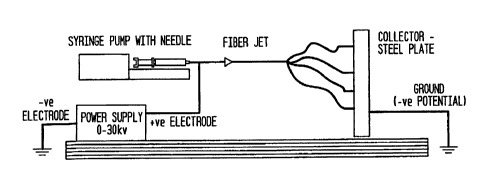
[0021] Fig. 1 is a diagrammatic representation of the electrospinning
equipment used
herein.
[0022] Fig. 2 shows scanning electron microscope (SEM) images of human MSC
loaded
PLLA at 14 days in culture containing normal growth media (a) nanofibers; (b)
microfibers.
8
CA 02596957 2007-05-29
WO 2006/068809
PCT/US2005/043876
[0023] Fig. 3 shows SEM images of human MSC loaded PLGA at 14 days in
culture
containing normal growth media (a) nanofihers; (b) microfibers.
[0024] Fig. 4 shows the growth kinetics of human MSCs grown in standard
growth media
on polymeric scaffolds.
[0025] Fig. 5 is a bar graph showing mineralization of the extracellular
matrix as
measured by calcium content on days 7 and 11 for cells grown on scaffolds in
OS media;
*p<0.05.
[0026] Fig. 6 is a linear plot showing mineralization of the extracellular
matrix as
measured by calcium content on days 7 and 11 for cells grown on scaffolds in
OS media.
*p<0.05.
[0027] Fig. 7 shows SEM images of human MSCs cultured in OS medium for 14
days on
(a) GSF and (b) PSF scaffolds.
[0028] Fig. 8 shows Type II collagen synthesis of cells grown on LF and SF
scaffolds in
inductive medium TGF-03.
DETAILED DESCRIPTION OF THE INVENTION
[0029] As used herein, the term "stem cells" refers to undifferentiated
cells having high
proliferative potential with the ability to self-renew that can migrate to
areas of injury and can
generate daughter cells that can undergo terminal differentiation into more
than one distinct
cell phenotype. These cells have the ability to differentiate into various
cells types and thus
promote the regeneration or repair of a diseased or damaged tissue of
interest. The term
"cellular differentiation" refers to the process by which cells acquire a cell
type. The term
"progenitor cell" as used herein refers to an immature cell in the bone marrow
that can be
isolated by growing suspensions of marrow cells in culture dishes with added
growth factors.
Progenitor cells are referred to as colony-forming units (CFU) or colony-
forming cells
(CFC). The specific lineage of a progenitor cell is indicated by a suffix,
such as, but not
limited to, CPU-F (fibroblastic).
[0030] As used herein, the terms "osteoprogenitor cells," "mesenchymal
cells,"
"mesenchymal stem cells (MSC)," or "marrow stromal cells" are used
interchangeably to
refer to multipotent stem cells that differentiate from CFU-F cells capable of
differentiating
along several lineage pathways into osteoblasts, chondrocytes, myocytes and
adipocytes.
When referring to bone or cartilage, MSCs commonly are known as
osteochondrogenic,
9
CA 02596957 2007-05-29
WO 2006/068809
PCT/US2005/043876
osteogenic, chondrogenic, or osteoprogenitor cells, since a single MSC has
shown the ability
to differentiate into chondrocytes or osteoblasts, depending on the medium.
[0031] The term "chondrocytes" as used herein refers to cells found in
cartilage that
produce and maintain the cartilaginous matrix. From least to terminally
differentiated, the
chondrocytic lineage is (i) Colony-forming unit-fibroblast (CFU-F); (ii)
mesenchymal stern
cell / marrow stromal cell (MSC); (3) chondrocyte . The term "chondrogenesis"
refers to the
formation of new cartilage from cartilage forming or chondrocompetent cells.
[0032] The term "osteoblasts" as used herein refers to cells that arise
when
osteoprogenitor cells or mesenchymal cells, which are located near all bony
surfaces and
within the bone marrow, differentiate under the influence of growth factors.
Osteoblasts,
which are responsible for bone matrix synthesis, secrete a collagen rich
ground substance
essential for later mineralization of hydroxyapatite and other crystals. The
collagen strands to
form osteoids: spiral fibers of bone matrix. Osteoblasts cause calcium salts
and phosphorus to
precipitate from the blood, which bond with the newly formed osteoid to
mineralize the bone
tissue. Once osteoblasts become trapped in the matrix they secrete, they
become osteocytes.
From least to terminally differentiated, the osteocyte lineage is (i) Colony-
forming unit-
fibroblast (CFU-F); (ii) mesenchymal stem cell / marrow stromal cell (MSC);
(3) osteoblast;
(4) osteocyte. The term "osteogenesis" refers to the formation of new bone
from bone
forming or osteocompetent cells.
[0033] Although the lineage of adipocytes is still unclear, it appears that
MSCs can
differentiate into two types of lipoblasts, one that give rise to white
adipocytes and the other
to brown adipocytes. Both types of adipocytes store fat.
[0034] The following examples are put forth so as to provide those of
ordinary skill in the
art with a complete disclosure and description of how to make and use the
present invention,
and are not intended to limit the scope of what the inventors regard as their
invention nor are
they intended to represent that the experiments below are all or the only
experiments
performed.
[0035] Example 1. Substrate Recognition by Differentiable Human MSC cells
[0036] We have evaluated two commonly used polymeric compositions in the
field of
tissue engineering, namely poly-L-lactic acid (PLLA) and poly-D,L-lactide
glycolide
(PLGA) at the nano- and microscale fiber diameter range for their ability to
support
CA 02596957 2007-05-29
WO 2006/068809
PCT/US2005/043876
mesenchymal stem cell attachment. We then compared the morphology and growth
characteristics of the attached cells on these substrates.
[0037] The term "nanoscale fiber" generally refers to fibers whose diameter
ranges from
about 1 to about 1000 nanometers. Nanoscale fibers whose average diameter
ranges from
about 400 to about 500 nanometers are most preferred. The term "microscale
fiber"
generally refers to fibers whose diameter ranges from about 1 to about 1000
micrometers.
Microscale fibers whose diameter ranges from about I to about 100 micrometers
are
preferred, and microscale fibers whose diameter averages from about 10
micrometers to
about 20 micrometers are most preferred.
[0038] Polymeric Substrate Materials
[0039] The present invention makes use of fibers formed from the
biodegradable
aliphatic polyester homopolymer poly L-lactic acid (PLLA) and from a copolymer
of poly L-
lactic acid and glycolic acid, 75/25 D,L High IV lactide-co-glycolide (PLGA).
PLLA and
PLGA were obtained from Alkermes, Inc. As used herein, the term
"biodegradable" refers to
the ability of a substance or material to break down into harmless substances
by the action of
living organisms. Other biodegradable and biocompatible polymers can be used
for the
described purpose. As used herein, the term "biocompatible material" refers to
a material
that the body generally accepts without a major immune response, which is
capable of
implantation in biological systems, for example, tissue implantation, without
causing
excessive fibrosis or rejection reactions.
[0040] The Electrospinning Process
[0041] Electrospinning is a fiber forming technique that relies on charge
separation to
produce nano- to microscale fibers. A nonwoven matrix of nanofibers was
created using the
electrospinning technique so that porosity, surface area, fineness and
uniformity, diameter of
fibers, and the pattern thickness of the matrix could be manipulated. The
terms "nonwoven
matrix", "nonwoven mesh" or "nonwoven scaffold" are used interchangeably
herein to refer
to a material comprising a randomly interlaced fibrous web of fibers.
[0042] The electrospinning process is affected by varying the electric
potential, flow rate,
solution concentration, capillary-collector distance, diameter of the needle,
and ambient
parameters like temperature.
[0043] Figure 1 is a diagrammatic representation of the electrospinning
setup used
herein, which is comprised of a syringe pump containing a 13-20 gauge needle.
The syringe
11
CA 02596957 2010-08-12
pump was mounted on a robotic ann in order to control the splaying of fibers
on the collector. .
An electrically grounded stainless steel plate of dimensions 15 x 30 cm was
used as the
collector. The syringe pump was filled with the polymer solution, and a
constant flow rate of
0.103 ml/min was maintained using the syringe pump. The positive Output lead
of a high
voltage power supply. (Gamma High Voltage, Inc.) was attached to the needle,
and a 25 bolt
voltage was applied to the solution. The collector-to-needle distance was 20
cm. When the
charge of the polymer at increasing voltage exceeded the surface tension at
the tip of the
needle, the polymer splayed randomly as fibers. These were collated as
nOnwriVen matt on
the grounded plate.
100441 Fabrication Of Tissue Engineering. Seaffelds
(0045) =In order to make fibers of two different sizeranges, scaffolds were
fabricated by
varying the solution concentration and diameter of the needle. Microfiber
scaffolds ofPLLA
and PLGA were madehy electinspinning using a 1 0% w/w solution concentration
ofthc =
polymer in methylene chloride using a 13-gauge needle. Nanofiber scaffolds
were made by
olectrospinning using a 5% w/w of polymersolution and a 20-gauge needle.
100461 The fiber diameter of eleetrospun MLA and PLGA fibers was
characterized using
Scanning Electron Microscopy (SEM) according to established methods. Porosity
and pore
size distribution of thc fibers Was analyzed by mercury intrusion porosimetry
(1i4P).
Thermal analysis was performed with a TA Model Q100 Differential Seaming
Calorimeter
(DSC).
100471 By varying the polymer solution concentration and the needle
diameter,. the
electrospirming process yielded very fine fibers with diameters in the
nanometer range. MIP
results showed that the microfiber and nanofiber scaffolds of PLLA had a
porosity of 39%
and 47%, respectively. The thermal analysis results show that the
electrospituring process as
performed herein does not alter the bulk characteristics of glass transition
and melting
temperatures of these polymers even when the polymer is processed at a high
voltage.
[00481 Themierofiber and nariofiber scaffolds of PLLA and PLGA were made
into 3-
dimensional 1 mm thick nonwoven mats and sterilized prior to cell seeding.
100491 Human MSCs
100501 Human MSCs (hMSCs) were prepared as described in Livingston, et al.,
J.
. Materidt &fence: Materials in Mai 14: 211-218 (2003) and in U.S.. Patent No.
5,484,359.
Bone.
12
CA 02596957 2010-08-12
Marrow aspirates of 30-50 ml were obtained from healthy human donors as
described.
Livingston, et al., I Materials Science: Materials in Med. 14: 211-21g (2003).
Marrow.
samples were washed with saline and centrifuged over a density cushion of
ficollTm. The
interface layer was. removed, washed, and the cells counted. Nucleated cells
recovered from
the density separation were washed and plated in tissue culture flasks
inpulbecco's Modified
Eagle's Medium (DMEM) ccmtaining10% fetal bovine serum ("FES', HyClone
Laboratories, Inc.). Non-adherent cells were washed from the culture during
biweekly
feedings. COlonyforrnation Was monitored for a 14-17 day period. MSC's
Werepassaged
when the tissue culture flasks were near confluent. At the end of the fait
passage, MSCs
were .eniyinatitally removed from the culture flask using trypsin-EDTA and
replatedat a
lower density for further expansion. At the end of the second passage, MSC's
were either
seeded onto scaffolds or cryopreserved until future use.
100511. Marker Analysis
1002] Human MSC cells were identified as multipotent stem cells based on
surface
marker characterization, which distinguishes the stem cells from other cell
types in the bone
marrow, for example white blood cells. Cells expreSsing CD44 surface antigen
and cells
froin Which CD45 and CD34 surface antigenswere absent were verified by
fluorescence-
actiYated-eell-sOrter.
[0053) As used hereiiiõ "CD44" refers 'to a common cell surface
glycoprotein antigen.
CD44 proteins have been implicated in several cellular functions including
cell-cell and cell,
matrix adhesion, migration, and tumor metastasis. (Naor D. et al., Adv. Cancer
Rat 71, 241,
319 (1997)).
100541 As used herein, "CD34÷ refers to a novel hernatopoietie stem cell
antigen
selectively expressed on hernatopoietie stem and progenitor cells derived from
human bone
marrow, blood.and fetal liVer: Yin et alõ Blood 90: 5002-5012 (1997);
.Miaglia, S. et al.,
Blood 90: 5013-21 (1997). Stomal cells do not express CD34. CD344- eels
derived from
adult bone /13.31TOW give rise in -vitro to t Majority ofthe
granulocyte/maCrophage progenitor
cells (CFU-GM),.some colony-forming units-mixed (C7U-Mix) anda minor
population Of
primitive erythroid progenitor Cells (burst Terming Units, erythrocytes or BFU-
E). Yeli, et al,
Circulation Id& 2070-73 (2003).
[0055) As used herein "CD45 ' refers to a protein tyrosine phospliatase
(FTP) located in
hematopoietic cells except .erythrocytesand platelets. It has several
isoforms. The specified
13
CA 02596957 2007-05-29
WO 2006/068809
PCT/US2005/043876
expression of the CD45 isoforms can be seen in the various stages of
differentiation of
normal hematopoietic cells ( Virts et al., Immunology 34(16-17) 1119-1197
(1997)).
[0056] Our results showed that cells were able to differentiate into three
cell types.
Osteogenic differentiation was characterized by the expression of alkaline
phosphatase
activity (as detected by hydrolysis of p-nitrophenylphosphate to p-
nitrophenol), by
osteocalcin protein expression (quantitated by a competitive immunoassay
(Metra
Biosystems, Inc), and by mineralization of the extracellular matrix (the
amount of calcium
present was determined by colorimetric assay). Chondrogenic differentiation
was determined
by safranin-O staining for glycosaminoglycan and by immunostaining for type II
collagen.
Adipogenic differentiation was characterized by Oil Red 0 stain for lipids.
[0057] Isolated and subcultured human MSCs were seeded at 1 x 10⁴ cells/cm2
onto the
microfiber and nanofiber scaffolds in 100 ill in serum-containing medium and
maintained at
37 C for 14 days. The term "seeded" refers to the process whereby MSC cells
are plated or
inoculated onto the scaffolds. Tissue culture plastic was used as a substrate
control. Cell
proliferation on the scaffolds was assessed using Vybrant MTT Cell
Proliferation Assay Kit
(Moledular Probes). The MTT assay involves the conversion of the water soluble
MTT (3-
(4,5-dimethylthiazol-2-y1)-2,5-diphenyltetrazolium bromide) to an insoluble
formazan. The
formazan then is solubilized and its concentration measured by colorimetric
techniques. Cell
morphology was examined by SEM.
[0058] Results
[0059] The results of the image analysis of the scaffolds are summarized in
table I.
Table I. Diameter of Nanofiber and Microfiber Scaffolds of PLLA and PLGA
PLLA PLGA
Microfiber Mean=17 7.6m (n= 4) Mean=16 7.6 m (n= 4)
Nanofiber Mean=400 920 nm (n= 4) Mean=500 880 nm 4)
[0060] The morphology of the cells on PLLA scaffolds is shown in Fig. 2.
Cells were
flat and spread out on the microfiber scaffolds, but appeared to be rounded on
the nanofiber
scaffolds. The hMSCs exhibited a similar morphology on the PLGA scaffolds
(Fig. 3). No
significant differences in hMSC proliferation were detected between the nano-
and micro-
14
CA 02596957 2007-05-29
WO 2006/068809 PCT/US2005/043876
fiber meshes for both PLLA and PLGA. Therefore, the materials used did not
alter the
growth characteristics of the h_MSC cells.
[0061] However, striking differences were detected in cell morphology
depending on the
size of the scaffold fibers. Cells adhered with rounded morphology on the
nanofiber
scaffolds whereas they appeared flat on the microfiber scaffolds of either
material. It is well
known that a rounded morphology in vitro is necessary both for chondrogenic
differentiation
and for maintenance of the chondrocyte phenotype of mature chondrocytes. (Li,
et al.,
Biomed. Mater. Res. 67A at 1110). Therefore, the rounded morphology of hMSC
cells on
nanofiber scaffolds might prove beneficial for MSC chondrogenic
differentiation leading,
ultimately, when implanted in vivo to treat patients suffering from connective
tissue damage,
to cartilage formation.
[0062] Example 2. Cell Proliferation
Human MSCs were isolated from adult, human whole bone marrow according to
standard
techniques and were seeded onto polymer scaffolds having the composition of
PLLA or
PLGA, each having fiber diameters on the micron scale (LF) or nano scale (SF)
and grown in
standard growth medium (DMEM, 10% fetal bovine serum, 1%
antibiotic/antimycotic) for 14
days. Cell proliferation was assessed using Vybrant's MTT Cell Proliferation
Assay Kit
(Molecular Probes, Inc.).
The growth curves for cells seeded onto PLLA micron scale fibers (PLLA-
LF),PLLA nano-
scale fibers (PLLA-SF), PLGA micron scale fibers (PLGA-LF) and PLGA nano-scale
fibers
(PLGASF) are shown in Fig. 4. Cells grown on PLLA and PLGA micron scale and
nanoscale fibers showed good comparable growth characteristics as measured by
the MTT
assay. No significant differences in human MSC proliferation were detected
between PLLA
and PLGA micron and nanoscale fibers.
[0063] Example 3. PLLA/PLGA micron and nano fiber diameter scaffolds
support
osteogenic differentiation.
[0064] Scaffolds were created by the process of electrospinning, and human
mesenchymal stem cells were grown on the scaffolds to determine whether
PLLA/PLGA
micron and nano-sized scaffolds support osteogenic differentiation.
[0065] Materials and Methods
[0066] hMSCs were grown in control medium (DMEM, 10% FBS, 1% antibiotic) or
osteogenic inducing medium (OS) (Control medium with 100nM dexamethasone, 10mM
b-
CA 02596957 2007-05-29
WO 2006/068809
PCT/US2005/043876
glycerophosphate,0.05mM L-ascorbic acid-2-phosphate) on PLLA or PLGA scaffolds
having
micron or nano sized fiber diameters.
[0067] The four scaffolds, PLLA microfiber ("PLF") scaffolds, PLLA
nanofiber
scaffolds ("PSF"), PLGA microfiber scaffolds ("GLF"), and PLGA nanofiber
scaffolds
("GSF"), were created by electrospinning.
[0068] On the day of cell seeding, scaffolds were soaked first in 100%
ethanol for 20
minutes, then three times in PBS, 20 minutes each, for sterilization.
Scaffolds then were
placed into assigned wells of a 96-well microtiter plate (B-D Falcon, Becton-
Dickinson, Inc.)
for each time point using forceps, and 150 AL of medium containing 10,000
cells were added
to each well. The cells were left in the incubator overnight at 37 degrees C
to allow cell
attachment to the scaffolds. Media were changed the next day so that half of
the wells
received control medium and the other half received osteogenic induction
medium. The
media were changed twice a week thereafter.
[0069] Calcium Assay
[0070] Mineralization of the extracellular matrix, as an indicator of
osteogenic
differentiation, was determined by measuring calcium content using a
colorimetric assay. 50
AL of 0.5 N HC1 were added to 4 different wells. Plates then were incubated at
room
temperature while the standards (Calcium/Phosphorus Combined Standard, Sigma,
Inc.) were
prepared for the assays. For the assay, 50 AL of sample was transferred into
microcentrifuge
tubes. The wells were rinsed with an additional 50 AL of 0.5 N HC1, and this
was added to
the tube. Tubes were vortexed overnight and then centrifuged for 2 minutes at
3,000 rpm at
room temperature. 20 AL of sample was pipetted into a new 96 well plate, and
190 AL of the
Working Solution (Cresolphtalin complexone, 0.10 mmoUL, 8-Hydroxyquinoline,
5.2
mmol/L, Polyvinylpyrrolidine, 0.07 mmol/L, 2-Amino-l-methyl proponal, 260
mmol/L,
Thermo Electron Calcium Kit) were added to each well. The standards were
pipetted in
duplicate and consisted of 0, 0.05, 0.1, 0.2, 0.4, 0.6, 0.8, 1.0, 1.5, and 2.0
jig of calcium. The
volume of the standard wells was brought up to 190 AL using the Working
Solution, and 20
AL of 0.5 N HC1 were added to the standard wells. The plate was incubated for
5 minutes at
room temperature before being read at 570 nm.
[0071] SEM
[0072] Scanning electron micrographs (SEMs) were taken to observe cells
growing on
the scaffolds and mineralization of the extracellular matrix.
16
CA 02596957 2007-05-29
WO 2006/068809
PCT/US2005/043876
[0073] Results
[0074] Fig. 5 shows mineralization of the extracellular matrix as measured
by calcium
content on days 7 and 11 for cells grown on scaffolds in control (C) or OS
media. Cells were
grown in control medium on PLLA large fiber scaffolds (PLF-C), in control
medium on
PLLA small fiber scaffolds (PSF-C), in control medium on PLGA large fiber
scaffolds (GLF-
C), in control medium on PLGA small fiber scaffolds (GSF-C); in osteogenic
inducing
medium on PLLA large fiber scaffolds (PLF-OS), in osteogenic inducing medium
on PLLA
small fiber scaffolds (PSF-OS), in osteogenic inducing medium on PLGA large
fiber
scaffolds (GLF-OS), and in osteogenic inducing medium on PLGA small fiber
scaffolds
(GSF-OS).
[0075] Fig. 5 and 6 show that calcium levels for cells grown on PLLA and
PLGA large
and small fiber scaffolds in OS media are significantly higher on day 11 than
on day 7. This
shows positive differentiation in OS medium and that all of the scaffolds can
support
differentiation successfully. No differences attributable to either fiber size
or polymer
composition were observed.
[0076] As shown in Fig. 7, SEMs of cells growing on (a) GSF and (b) PSF
scaffolds in
OS medium for 14 days show a uniform distribution of cells throughout all the
scaffolds and
an abundant mineralization of the extracellular matrix. Cells form a uniform
cell layer,
embedded in extracellular matrix, across the surface and interior of the
scaffolds.
[0077] Example 4. Chondrogenic differentiation
[0078] PLLA and PLGA were made into 1 mm thick non-woven mats of two
distinctly
different fiber diameters, nanometer (SF) and micrometer (LF), by
electrospinning as
described in Example 1 and were sterilized prior to cell seeding. SEM, mercury
intrusion
porosimetry (MlP), and differential scanning calorimetry (DSC) were used to
determine fiber
diameter and cell morphology; porosity and pore size distribution; and thermal
profile,
respectively.
[0079] To determine the chondrogenic potential of MSCs on LF and SF, MSCs
isolated
from whole bone marrow and subcultured as described in Example 1 were seeded
onto LF
and SF scaffolds at a density of 1 x 10⁵ cells/cm2. Cells were maintained in
chondrogenic
induction medium supplemented with TGF-03 (Cambrex BioScience, Inc.). Type II
collagen
content on LF and SF scaffolds was assessed with Arthrogen-CIA Capture ELISA
Kit
(Chondrex, Inc.). A tissue culture polystyrene plate (TCP) was used as
control.
17
CA 02596957 2007-05-29
WO 2006/068809
PCT/US2005/043876
[0080] The MT results showed that the PLLA microfiber (LF) and nanofiber
(SF)
scaffolds had a porosity of 39% and 47% respectively. The DSC results showed
that the
electrospinning process does not alter the characteristic thermal profile of
each polymer even
when processed at a high voltage.
[0081] Chondrogenic differentiation occurred on SF fibrous scaffolds at 3
weeks of
culture, but was absent on LF fibers, as demonstrated by Type II collagen
synthesis. Fig. 8
shows that type II collagen synthesis by cells grown on PLLA-SF and PLGA-SF
scaffolds
was significantly greater than synthesis by cells grown on PLLA-LF and PLGA-F
scaffolds.
[0082] Example 5. Cartilaginous tissue repair in a mammalian subject.
[0083] Unlike bone, liver, skin and other tissues with high cell turnover
rates, cartilage
generally is considered to have a limited capacity for self-repair. See, e.g.,
Laurencin, et al.
Ann. Rev. Biomed. Eng'g 1: 19-46, 35 (1999). Cartilage tissue is composed of
chondrocytes
and an extracellular matrix consisting of proteoglycans, collagen, and water.
Chondrocytes
are responsible for synthesis and breakdown of collagen and proteoglycans. The
collagen
fibers provide tear and shear resistance whereas the proteoglycans impart
elasticity to
cartilage. Because cartilaginous tissue is avascular, has a low oxygen
requirement, and has no
nerve structures, it may be most amenable to tissue engineering efforts.
[0084] According to another embodiment, the present invention will be used
in a partial
weight-bearing articular cartilage repair model (Aroen A, et al., "Articular
cartilage defects in
a rabbit model, retention rate of periosteal flap cover," Acta Orthop.
Apr;76(2):220-4 (2005)),
to repair a cartilaginous tissue in a mammalian subject. The method comprises
the steps of
(a) isolating viable differentiable mammalian mesenchymal cells from an
autologous
mammalian donor; (b) preparing a three-dimensional matrix comprising a
nonwoven mesh of
fibers to form a cell scaffold; (c) seeding the cell scaffold with the
isolated viable
differentiable mammalian mesenchymal cells in vitro; (d) growing the
differentiable
mammalian mesenchymal cells on the cell scaffold in vitro so that the
differentiable
mammalian mesenchymal cells differentiate into a viable mammalian chondrogenic
cell
phenotype on the scaffold; and (e) implanting the cell scaffold comprising the
viable
mammalian chondrogenic cell phenotype.at a site where the cartilaginous tissue
of the subject
is in need of repair. The differentiable mammalian mesenchymal cells are
obtained from
mammalian bone marrow. In another embodiment, the three-dimensional matrix of
fibers in
step (b) of the method is formed from a polymeric material. The polymeric
material is a
18
CA 02596957 2010-08-12
biocompitible polymer, preferably poly D,L 'betide glycolide or poly L-lactic
acid or a
mixture thereof.
[0085] Unless defined otherwise, all technical and scientific terms iised
herein have the
samemeaning as commonly understood by One of ordinary sIdll in the art to
which this
invention belongs. Although any methods and Materials similar or equivalent to
those
described herein can also be used in the practice or testing of the present
invention, the
preferred Methods and materials are now described.
(00861 it must be noted that as used herein and in the appended claims, the
singular forms
"a", "and", and "the" include plural references unless the commit clearly
dictates Otherwise.
All technical and stientific terms used herein have the same meaning. Efforts
have been
made to ensure accuracy With,respeet to nurithers.used (e.g. amounts,
temperature, etc.) but
some experimental errors and. deviations should be accounted for. Unless
indicated
otherwise, parts are parts by weight, molecular weight is weight average
molecular weight,
temperature is in degrees Centigrade, and plebbule is at or near atmospheric.
100/311 Where a range ofvalues is provided, it is understood that each
intervening value,
to the tenth of the unit of the lower limit unless the context clearly
dictates otherwise,
between the upper and lower limit of that range and any other stated or
intervening.value in
that stated range is encompassed within the invention. The upper and lower
limits of these
smaller ranges which may independently be included in the smaller ranges is
also
encompassed within the invention, subject to any specifically excluded limit
in the stated
range. Where. the stated range includes one or both of the limits, ranges
excluding either both
of those included limits are also included in the invention.
[00811] The publications discussed herein are provided solely for their
disclosure prior to
the filing date of the present application. Nothing herein is to be construed
is an admission
that the present invention is not entitled to antedate suth publication by
virtue of prior
invention. Further, the dates of publication provided may be different from
the actual
publication dates which may need to be independently confirmed.
100891 The invention has been described with reference to the preferred
embOdiinerit to
illustrate the principle; of the invention and not to limit the invention to
the particular
embodiment illushated. Modifications and alterations may occur to others upon
reading and
19
CA 02596957 2007-05-29
WO 2006/068809
PCT/US2005/043876
understanding the preceding detailed description. It is intended that the
scope of the
invention be construed as including all modifications and alterations that may
occur to others
upon reading and understanding the preceding detailed description insofar as
they come
within the scope of the following claims or equivalents thereof.