Note: Descriptions are shown in the official language in which they were submitted.
CA 02597325 2007-08-08
WO 2006/098998 PCT/US2006/008316
METHODS AND COMPOSITIONS FOR MODULATING VASCULAR INTEGRITY
RELATED APPLICATIONS
This application is a non-provisional application filed under 37 CFR
1.53(b)(1),
claiming priority under 35 USC 119(e) to provisional application number
60/660,172 filed
March 10, 2005, the contents of which are incorporated herein by reference.
TECHNICAL FIELD
The present invention relates generally to the fields of treatment of diseases
and other
pathological conditions. More specifically, the invention concerns methods and
compositions for
modulating molecular events and outcomes associated with angiogenesis.
BACKGROUND
The vascular system plays key roles in the maintenance of normal physiological
functions. For instance, endothelial cells of the vasculature normally form a
cellular barrier
that serves to regulate movement of fluid, electrolytes, and proteins into
tissues and organs.
Although the events that lead to loss of endotlielial barrier function is
rather complex and not
completely understood, the development and maintenance of integrity of the
vasculature has
been studied extensively. See, e.g., Kagsbrun & Moses, Chemistry & Biology
(1999),
6:R217-R224; Ferrara, Endocrine Reviews (2004), 25(4):581-611.
Nonetheless, what is clear is that perturbations of the integrity of the
vascular system
are implicated in the etiology of multiple diseases and pathological
conditions, ranging from
cancer to inflammatory diseases such as autoimmune disorders. See, e.g., US
Patent Appl.
Pub. No. 2004/0167198; Cines et al., Blood (1998), 91(10):3527-3561; Kumar et
al., Curr.
Opin. In Nephrology and Hypertension (2005), 14:33-37; Carmeliet & Collen Am.
J. Physiol.
(1997), 273:H2091-H2104. On the other hand, it should be noted that, under
certain
physiological settings, ability to develop and/or maintain a compromised state
of vasculature
is desirable, and could, if deficient, itself lead to pathological problems or
sub-optimal
therapeutic outcomes.
Inflammation associated with the vasculature has been identified as a
downstream
event of extracellular angiogenic stimuli that is often associated with
perturbation of vascular
integrity. Kirkpatrick et al., Int. J. Microcirc. (1997), 17:231-240. For
example, there is
increasing evidence that the inflammatory process may play a significant role
in the
CA 02597325 2007-08-08
WO 2006/098998 PCT/US2006/008316
pathophysiology of various vascular-related disorders, such as hypertension.
See, e.g., Touyz,
Cur. Opin. Nephrology and Hypertension (2005), 14:125-131; Kobayashi & Lin,
Frontiers in
Bioscience (2005), 10:666-674; Bacon, Curr. Opin. Rheumatol. (2004), 17:49-55;
Paleolog, J.
Clin. Pathol.: mol. Pathol. (1997), 50-225-233; Mullenix et al., Ann. Vasc.
Surg. (2005),
19:130-138. As noted in Touyz, events indicative of inflammation are observed
in the
vasculature, for example increased expression of surface adhesion molecues and
their ligands,
inflammatory cell infiltration, cytokine production, chemokine release,
oxidative stree and
activation of innate immunity. Endothelial cell perturbation may also be
reflected by the
release of von Willebrand factor and E-selectin, which is expressed
exclusively by endothelial
cells. Kuenen et al., Arterioscler. Thromb. Vasc. Biol. (2002), 22:1500-1505.
Persistent
and/or unregulated inflammation can result in tissue/vascular damage. The
inflammatory
response typically involves alterations in vascular permeability, leukocyte
extravasation
(adhesion, transmigration and chemotaxis) and tissue repair. Touyz, supra.
Unfortunately, to date, information regarding intracellular molecules that
mediate
signaling between extracellular angiogenic stimuli and related downstream
events (such as
endothelial cell-associated inflammation) has been lacking. This has hampered
efforts to
develop therapeutic strategies that would permit fine-tuning of the angiogenic
process to
minimize perturbations of vascular integrity resulting from undesired
inflammation, or to
induce perturbation of vascular integrity where desired. Therefore, there is a
real need to
identify "intermediate" molecular targets and pathways that could be the focus
of novel
therapeutic intervention. The invention described herein meets this need and
provides other
benefits.
All references cited herein, including patent applications and publications,
are incorporated
by reference in their entirety.
DISCLOSURE OF THE INVENTION
The invention is based at least in part on the discovery that a member of the
Down
syndrome critical region protein family is a critical intermediate molecular
link between
extracellular signaling by angiogenic factors and intracellular regulation of
downstream
events that modulate vascular integrity. Specifically, Down syndrome critical
region 1
(hereinafter "DSCR1") is shown herein to be an intracellular molecule that is
induced in
activated endothelial cells following stimulation by angiogenic regulators
such as vascular
endothelial growth factor (VEGF), whereby induction of DSCR1 in turn modulates
expression of genes associated with vascular/endothelial cell inflammation. As
described
2
CA 02597325 2007-08-08
WO 2006/098998 PCT/US2006/008316
above, inflammation associated with the vasculature has been implicated in a
multitude of
disorders. Notably, DSCR1 is a particularly advantageous target for
therapeutic modulation,
since it acts via a negative regulatory feedback loop in mediating between
extracellular
angiogenic signals and intracellular regulation of vascular inflammation.
Without being
bound by theory, depending on the cellular/physiological context, DSCR1, in
its capacity as a
component of a negative regulatory feedback loop, is expected to be sensitive
to both positive
or negative modulation to alter the molecular and signaling balance normally
associated with
a regulatory feedback loop. Consistent with this, as shown herein, both
increased and
decreased DSCRl activity in endothelial cells are empirically demonstrated to
result in
similar modulation of cell survival outcomes. Thus, the data disclosed herein
reveal an
important molecular target/pathway which, when interfered with, results in
inodulation of one
of the key downstream events associated with angiogenic signaling, namely
inflammation
associated with the vasculature. Under certain circumstances, such
inflammation, if left
unchecked, can lead to undesirable pathological effects. Yet in other
instances, enhancement
of such inflammation may be desirable. The invention provides inethods,
compositions, kits
and articles of manufacture useful for regulating such inflammation, by
interfering with
DSCR1 function/activity so as to modulate vascular inflammation and,
therefore, vascular
integrity.
In one aspect, the invention provides a DSCR1 modulator that modulates DSCR1
activity in endothelial cells. In one embodiment, the DSCR1 modulator
modulates DSCR1
activity induced by signaling through VEGF receptor 2 (VEGFR-2). A DSCR1
modulator of
the invention may modulate one or more aspects of the DSCR1-calcineurin A
(CnA) pathway.
For example, in one embodiment, a DSCR1 modulator of the invention modulates
one or
more cellular events associated with NFAT activity, including, for example,
NFAT
phosphorylation, NFAT nuclear translocation or NFAT transcriptional activity
(e.g., NFAT's
ability to mediate regulation of gene expression by CnA).
A DSCRl modulator of the invention can be provided in any form suitable for
clinical use. For example, a modulator of the invention can be provided and/or
administered
in the form of a nucleic acid or polypeptide. Accordingly, in one embodiment,
a DSCR1
modulator of the invention comprises a nucleic acid that encodes a polypeptide
capable of
antagonizing gene expression induced by CnA. For example, the polypeptide may
comprise a
peptide comprising at least a portion of DSCR1, wherein the peptide is capable
of interacting
with CnA. In one embodiment, the polypeptide comprises a portion, but not the
complete
mature DSCR1 protein. In one embodiment, the polypeptide comprises full length
mature
DSCR1 protein. In one embodiment, the polypeptide comprises amino acid from
about
3
CA 02597325 2007-08-08
WO 2006/098998 PCT/US2006/008316
position 96 to about 125, or about position 108 to about 122 of liuman DSCR1,
wherein
amino acid positions refer to the sequence depicted in SEQ ID NO:1 (Figure 9).
In one
embodiment, the polypeptide comprises a sequence having at least 50%, 60%,
70%, 80%,
90%, 95%, 98% sequence identity or similarity with the sequence of human DSCR1
from
about position 96 to about 125, or about 108 to about 122 of human DSCRl,
wherein amino
acid positions refer to the sequence depicted in SEQ ID NO: 1 (Figure 9),
provided the
polypeptide retains an ability to antagonize gene expression induced by CnA.
In one
embodiment, the polypeptide comprises at least a portion of the serine-proline
(SP) sequence
and at least a portion of the N and/or C-terminal calcineurin binding domain
of human
DSCR1, wherein the polypeptide is capable of binding to CnA to inhibit CnA-
regulated gene
expression. In one embodiment, the polypeptide comprises (i) the sequence of
human
DSCRl from about position 96 to about 125, or about 108 to about 122 of human
DSCR1,
wherein amino acid positions refer to the sequence depicted in SEQ ID NO:1
(Figure 9); (ii)
at least a portion of the serine-proline (SP) sequence; and (iii) at least a
portion of the N
and/or C-terminal calcineurin binding domain of human DSCR1, wherein the
polypeptide is
capable of binding to CnA to inhibit CnA-regulated gene expression.
In one aspect, the invention provides a DSCRl modulator capable of
antagonizing
DSCR1 activity. Accordingly, in one embodiment, a DSCR1 modulator of the
invention
comprises a nucleic acid that when present in an endothelial cell (e.g., an
activated endothelial
cell) inhibits activity (including but not limited to gene expression) of
DSCRl in the cell. In
one embodiment, the nucleic acid comprises an antisense oligonucleotide. In
another
embodiment, the nucleic acid comprises an inhibitory/interfering RNA. In one
embodiment,
the inhibitory/interfering RNA is a small inhibitory/interfering RNA (siRNA).
For example,
a DSCR1 modulator comprising siRNA may comprise one or more sequence selected
from
the group consisting of GCUCAGACCUUACACAUAG, GGACAUCACCUUUCAGUAU,
GAAAGAAUGAGGAGACCUA and GACAUCACCUUUCAGUAUU. In another example,
a DSCR1 modulator comprising siRNA may comprise one or more sequence selected
from
the group consisting of AACGUAUGACAAGGACAUCAC,
AAGGACAUCACCUUUCAGUAU, AAGUUAUAUUUUGCUCAGACC and
AAGAUGCGACCCCAGUCAUAA. In one embodiment, a DSCRl modulator comprising
siRNA comprises one or more sequence selected from the group consisting of
GCUCAGACCUUACACAUAG,GGACAUCACCUUUCAGUAU,
GAAAGAAUGAGGAGACCUA,GACAUCACCUUUCAGUAUU,
AACGUAUGACAAGGACAUCAC,AAGGACAUCACCUUUCAGUAU;
AAGUUAUAUUUUGCUCAGACCandAAGAUGCGACCCCAGUCAUAA.
4
CA 02597325 2007-08-08
WO 2006/098998 PCT/US2006/008316
In one embodiment, the nucleic acid comprises an aptamer.
In one embodiment, a DSCRl modulator of the invention comprises an antibody.
In
one embodiment, the antibody is administered such that it enters a target
endothelial cell to
inhibit DSCR1 in the cell, i.e., the antibody is internalized. In another
embodiment, the
antibody is encoded by a nucleic acid that is introduced (e.g., transfected)
into a target
endothelial cell.
In one aspect, the invention provides methods for treating a variety of
disorders
associated with inflammation associated with endothelial cells. For example,
in one aspect,
the invention provides a method of treating a disorder associated with
abnormal vascular
inflammation, said method comprising administering to a subject an effective
amount of a
DSCR1 modulator of the invention, thereby treating the disorder. In one
embodiment, the
modulator increases inflammation associated with the vasculature. In one
embodiment, the
modulator inhibits inflammation associated with the vasculature.
In one aspect, the invention provides a method of treating an immunological
disorder
associated with abnormal vascular inflammation, said method comprising
administering to a
subject an effective amount of a DSCRl modulator of the invention, thereby
treating the
disorder. In one embodiment, the modulator increases inflammation associated
with the
vasculature. In one embodiment, the modulator inhibits inflammation associated
with the
vasculature.
In yet another aspect, the invention provides a method of treating a disorder
associated with perturbation of vascular integrity, said method comprising
administering to a
subject an effective amount of a DSCR1 modulator of the invention, thereby
treating the
disorder. In one embodiment, the perturbation of vascular integrity is
associated with
vascular/endothelial cell-related inflammation, for example where there is an
undesirably high
or low amount of vascular/endothelial cell-related inflammation. In one
embodiment, the
modulator increases inflammation associated with the vasculature. In one
embodiment, the
modulator inhibits inflammation associated with the vasculature.
In one aspect, the invention provides a method of ameliorating side-effects
associated
with anti-inflainmatory therapy comprising administering to a subject a DSCR1
modulator of
the invention, in conjunction with the anti-inflammatory therapy, thereby
ameliorating said
side-effects. In one embodiment, the DSCR1 modulator inhibits DSCR1 activity
in an
endothelial cell. In one embodiment, the anti-inflammatory therapy comprises
an agent that
suppresses inflammation, for example the agent could inhibit immune cell
activity. In one
embodiment, the agent is of the cyclosporine class of inflammation
suppressors, such as
cyclosporine A (CsA). In one embodiment, the modulator increases inflammation
associated
CA 02597325 2007-08-08
WO 2006/098998 PCT/US2006/008316
with the vasculature. In one embodiment, the modulator inhibits inflammation
associated
with the vasculature.
In yet another aspect, the invention provides a method of treating a disorder
associated with undesirable angiogenesis (such as cancer), said method
comprising
administering to a subject with the disorder an anti-angiogenic agent and a
DSCR1 modulator
of the invention.
In another aspect, the invention provides a method of inhibiting tumor growth
or
growth of a cancer cell, said method comprising administering to the tumor or
the cancer cell
an effective amount of an anti-cancer agent and an effective amount of a DSCRl
modulator
of the invention, wherein the combined effective amounts inhibit tumor or
growth of the
cancer cell.
In one embodiment, an anti-cancer agent comprises an anti-angiogenic agent,
for
example a VEGF antagonist. In one embodiment, a VEGF antagonist is an anti-
VEGF
antibody. In one embodiment, an anti-VEGF antibody comprises an antigen
binding
sequence of antibody A4.6.1 or affinity matured variants thereof. In one
embodiment, the
antigen binding sequence comprises at least one, two or three of the
hypervariable regions of
the heavy chain of antibody A4.6.1 or affinity matured variants thereof. In
one embodiment,
the antigen binding sequence comprises at least one, two or three of the
hypervariable regions
of the light chain of antibody A4.6.1 or affinity matured variants thereof. In
one embodiment,
the antigen binding sequence comprises a humanized form of the heavy chain
variable
domain of antibody A4.6.1 or affinity matured variants thereof. In one
embodiment, the
antigen binding sequence comprises a humanized form of the light chain
variable domain of
antibody A4.6.1 or affinity matured variants thereof. Antibody A4.6.1 is an
anti-VEGF
antibody produced by the hybridoma cell line deposited under ATCC deposit no.
HB 10709
(American Type Culture Collection, Manassas, VA). In one embodiment, the anti-
VEGF
antibody is bevacizumab. In one embodiment, the anti-VEGF antibody is an
antibody that
binds to the same epitope of human VEGF as bevacizumab. In one embodiment, the
anti-
VEGF antibody is an antibody that competes with bevacizumab for binding to
human VEGF.
In one embodiment, the anti-VEGF antibody binds to a receptor of VEGF, for
example
VEGFR2. In one embodiment of these methods, a DSCR1 modulator of the invention
is
administered to a subject in conjunction with an anti-cancer agent such as a
chemotherapeutic
agent. In one embodiment, the DSCRl modulator inhibits side effects associated
with
perturbation of vascular integrity. For example, the perturbation of vascular
integrity may be
associated with thrombolysis.
6
CA 02597325 2007-08-08
WO 2006/098998 PCT/US2006/008316
In one aspect, the invention provides a method of treating a disorder by
enhancing
angiogenesis, said method comprising administering to a subject with the
disorder an
effective amount of an agent that induces angiogenesis and an effective amount
of a DSCR1
modulator, whereby the disorder is treated. In one embodiment, the agent that
induces
angiogenesis comprises a pro-angiogenic substance, for example one or more of
the
angiogenic factors disclosed herein. In one embodiment, the angiogenic factor
is VEGF. In
one embodiment, the DSCR1 modulator inhibits perturbation of vascular
integrity. A
disorder that can be treated by this method include any of those described
herein, including
for example a wound, ischemia, a cardiovascular disorder, atherosclerosis,
vasculitis, etc.
In one aspect, the invention provides a method of modulating survival of an
endothelial cell comprising contacting the cell with a DSCR1 modulator. In one
embodiment,
the endothelial cell is activated. In one embodiment, the endothelial cell is
transfected with
an expression vector encoding a DSCR1 antisense sequence. In one embodiment,
the
endothelial cell is transfected with an expression vector encoding a DSCR1
sense sequence.
In one embodiment, the DSCR1 modulator comprises an inhibitory/interfering
RNA, which in
one embodiment is a small interfering RNA (siRNA).
In methods of the invention wherein a DSCR1 modulator of the invention is
administered in conjunction with another therapeutic agent, the timing of
administration of
DSCR1 modulator relative to the timing of administration of said another
therapeutic agent
would be any that is deemed empirically and/or clinically to be
therapeutically beneficial. For
example, in one embodiment, the DSCR1 modulator is administered prior to
administration of
said another therapeutic agent. In one einbodiment, the DSCR1 modulator is
administered
subsequent to administration of said another therapeutic agent. In another
embodiment, the
DSCRl modulator is administered concurrently witli administration of said
another
therapeutic agent. In one embodiment, said another therapeutic agent has anti-
inflammatory
activity. In one embodiment, the two agents are administered as separate
compositions. In
one embodiment, the two agents are adniinistered as a single composition.
In one aspect, the invention provides a method of identifying a subject at
risk of
developing side effects associated with treatment of a disorder comprising
modulation of
angiogenesis, said method comprising measuring amount of DSCRl expression or
activity in
a tissue or cell and comparing the amount to that of a normal counterpart
tissue or cell,
wherein a difference in amount indicates a risk of developing said side
effects. Generally, the
side effects are due to perturbation of vascular integrity associated with
inflammation. The
method of identifying can be practiced at any convenient and/or beneficial
timepoint during
7
CA 02597325 2007-08-08
WO 2006/098998 PCT/US2006/008316
the clinical intervention process. For example, it can occur before, during or
after
administration of a therapeutic agent to treat the disorder in question.
In one aspect, the invention provides a method of identifying a DSCRl
modulator
(such as a molecule useful for modulating perturbations of vascular
integrity/vascular
inflammation), the method comprising contacting a candidate substance with
DSCR1 (or
functional variant thereof as defined hereinbelow), and determining amount of
the DSCR1 (or
functional variant thereof) in the presence and absence of the candidate
substance, wherein
differential amounts of the DSCR1 (or functional variant thereof) in the
presence and absence
of the candidate substance indicates that the candidate substance is a DSCRl
modulator.
DSCRl amount can be determined by any suitable qualitative or quantitative
measurement
known in the art. For example, DSCR1 amount may be indicated by level of RNA
or protein.
In another example, DSCRl amount is indicated by DSCR1 activity (e.g.,
inhibition of CnA).
Methods of the invention can be used either to identify DSCR1 antagonists or
agonists. For
exainple, in one embodiment, DSCR1 amount is lower in the presence of the
candidate
substance. In another embodiment, DSCRl amount is higher in the presence of
the candidate
substance. A modulator identified by a method of the invention can have any of
the
characteristics required for use in inethods of the invention. For example, a
candidate can be
selected that is capable of modulating vascular integrity and/or inflammation
associated with
endothelial cells. Alternatively, a candidate substance can be additionally
evaluated to select
a substance that lacks certain undesirable characteristics. For example, a
candidate substance
can be selected that does not substantially modulate endothelial cell
proliferation.
As noted above, DSCRl modulators and methods of the invention are useful in
treating a variety of disorders suspected or known to be associated with
improper regulation
of vascular integrity. For example, these disorders include, but are not
limited to, those that
are in the vascular (such as cardiovascular), endothelial, angiogenic (such as
tumor/cancer) or
immunological (such as autoimmune) categories of disorders. Nonetheless, in
some
embodiments, these disorders are not cardiovascular or immunological. In some
embodiments, these disorders are not endotlielial or angiogenic. Yet in other
embodiments,
the disorders are associated with tissue inflammation (acute or chronic), for
example
autoimmune disorders. It should also be noted that some diseases may have
characteristics
that overlap between two or more categories of disorders.
In general, the invention provides modulator molecules and methods that are
useful
for treatment of immune-mediated disorders. In one embodiment, the invention
relates to
treatment of autoimmune disorders, such as those indicated herein. In one
embodiment, a
disorder relates to graft rejection or graft versus host disease.
8
CA 02597325 2007-08-08
WO 2006/098998 PCT/US2006/008316
In some embodiments of methods of the invention wherein a treatment method
comprises promoting angiogenesis /neovascularization in order to treat a
disorder, the
disorder may be, but is not limited to, e.g., vascular trauma, wounds,
lacerations, incisions,
burns, ulcers (e.g., diabetic ulcers, pressure ulcers, haemophiliac ulcers,
varicose ulcers),
tissue growth, weight gain, peripheral arterial disease, induction of labor,
hair growth,
epidermolysis bullosa, retinal atrophy, bone fractures, bone spinal fusions,
meniscal tears, etc.
Where clinically appropriate or desired, methods of the invention can further
comprise additional treatment steps. For example, in one embodiment, a method
further
comprises a step wherein a targeted cell and/or tissue (e.g., a cancer cell)
is exposed to
radiation treatment or a chemotherapeutic agent.
In one aspect, the invention provides compositions comprising one or more
DSCR1
modulators of the invention and a carrier. In one embodiment, the carrier is
pharmaceutically
acceptable.
In one aspect, the invention provides nucleic acids encoding a DSCR1 modulator
of
the invention. In one embodiment, a nucleic acid of the invention encodes a
DSCR1
modulator which is or comprises a polypeptide (e.g., an oligopeptide). In one
embodiment, a
nucleic acid of the invention encodes a DSCR1 modulator which is or comprises
an antibody
or fragment thereof. In one embodiment, a nucleic acid of the invention
encodes a nucleic
acid sequence that inhibits DSCR1 expression/activity (e.g., DSCRl gene
transcription or
translation of DSCR1 protein).
In one aspect, the invention provides vectors comprising a nucleic acid of the
invention.
In one aspect, the invention provides host cells comprising a nucleic acid or
a vector
of the invention. A vector can be of any type, for example a recombinant
vector such as an
expression vector. Any of a variety of host cells can be used. In one
embodiment, a host cell
is a prokaryotic cell, for example, E. coli. In one embodiment, a host cell is
a eukaryotic cell,
for example a mammalian cell such as Chinese Hamster Ovary (CHO) cell. In one
embodiment, a nucleic acid or vector of the invention is expressed in an
endothelial cell.
In one aspect, the invention provides methods for making a DSCR1 modulator of
the
invention. For example, the invention provides a method of making a DSCR1
modulator
which is or comprises an antibody (or fragment thereof), said method
comprising expressing
in a suitable host cell a recombinant vector of the invention encoding said
antibody (or
fragment thereof), and recovering said antibody (or fragment thereof). In
another example,
the invention provides a method of making a DSCR1 modulator which is or
comprises a
polypeptide (such as an oligopeptide), said method comprising expressing in a
suitable host
9
CA 02597325 2007-08-08
WO 2006/098998 PCT/US2006/008316
cell a recombinant vector of the invention encoding said polypeptide (such as
an
oligopeptide), and recovering said polypeptide (such as an oligopeptide).
In one aspect, the invention provides an article of manufacture comprising a
container; and a composition contained within the container, wherein the
composition
comprises one or more DSCRl modulators of the invention. In one embodiment,
the
composition comprises a nucleic acid of the invention. In one embodiment, a
composition
comprising a DSCR1 modulator further comprises a carrier, which in some
embodiments is
pharmaceutically acceptable. In one embodiment, an article of manufacture of
the invention
further comprises instructions for administering the composition (e.g., the
DSCR1 modulator)
to treat a disorder indicated herein.
In one aspect, the invention provides a kit comprising a first container
comprising a
composition comprising one or more DSCR1 modulators of the invention; and a
second
container comprising a buffer. In one embodiment, the buffer is
pharmaceutically acceptable.
In one embodiment, a composition comprising a DSCR1 modulator further
comprises a
carrier, which in some embodiments is pharmaceutically acceptable. In one
embodiment, a
kit further comprises instructions for administering the composition (e.g.,
the DSCR1
modulator) to treat a disorder indicated herein.
BRIEF DESCRIPTION OF THE DRAWINGS
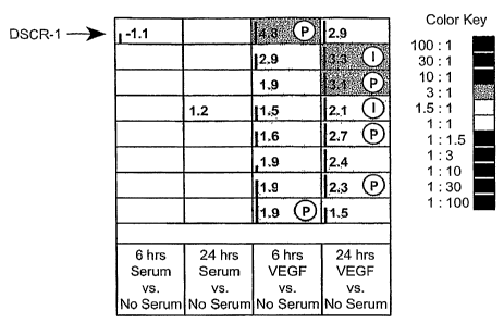
Figure 1. Gene expression analysis using GeneCall technology and RT-PCR. (A)
Primary HUVECs were exposed for 0, 6, or 24 hours to either serum-free media
or serum-free
media complemented with VEGF or 5% FCS, respectively. Endothelial cells were
lysed and
total RNA was isolated and analyzed for gene expression by using GeneCalling .
Each
column represents the output of a series of binary comparisons that have been
compared to
each other. However, genes marked with "P" (P = poisoning) or "I" (I =
Isolation, cloning,
sequencing, and poisoning) were confirmed. Selected genes shown were found
induced by
VEGF at 6 and 24 hours, but not by stimulation by 5% FCS; the fold stimulation
by VEGF is
indicated by a number in each box. Gray boxes indicate that no change in
expression was
observed for the genes in the binary comparisons for serum at 6 and 24 hours
versus no
serum. Vertical black bars in the enlarged thumbnail indicate the confidence
level of the gene
call. (B) DSCRl expression analysis by real-time PCR.21 HUVECs were kept in
serum-free
conditions and stimulated with either 5% FCS, b-FGF (10 ng/mL), VEGF (30
ng/mL), PIGF
(100 ng/mL), VEGFR1-sel (100 ng/mL), or VEGFR2-sel (30 ng/mL) during the
indicated
amount of time. The mutant proteins used were VEGFR2-sel (KDR-selective): VEGF
D63S/G65M/L66R and Flt-selective VEGF I43A/I46A/Q79A/I83.25 (C) DSCR1
expression
CA 02597325 2007-08-08
WO 2006/098998 PCT/US2006/008316
analysis by real-time PCR. HPAECs, HDMECs, and HUAECs were incubated in
presence of
0.5% FCS (NA), or in presence of 0.5% FCS and VEGF (30 ng/mL) or VEGF mutant
forms,
binding to either VEGFRI (VEGFR1-sel, 100 ng/mL) or VEGFR2 (VEGFR2-sel, 30
ng/mL)26 for 4( ~,) 8(0), and 24 (EI) hours. As control cells, HUPASMCs were
used, which
do not express significant levels of VEGF receptors (data not shown). Data
shown represent
the average SD of triplicate samples of one representative of total 3
independent
experiments. Relative RNA units (RRUs) were calculated as described by Gerber
et al3 by
using cyclophylin as reference gene. (D) DSCR1 expression analysis by real-
time PCR.
HUVECs were incubated for 2 hours in presence of CsA (1 M) prior stimulation
with VEGF
(30 ng/mL), VEGFR2-sel (30 ng/mL), VEGFRl-sel (100 ng/mL), PMA (200 ng/mL),
and IO
(5 M), thapsigargin (50 nM) or TNF-a (10 ng/mL) for 5.5 hours, as indicated.
Data shown
represent the average SD of duplicate samples run in parallel of one
representative of total 2
independent experiments. (E) Western blot analysis of whole cell extracts of
HUVECs grown
in 0% serum, 0.5% BSA and 5% FCS, hVEGF (30 ng/mL), b-FGF (10 ng/mL), TNF-a
(10
ng/mL) and PMA or ionomycine (IO) for 5 hours as indicated. As control, 100 ng
recombinant DSCRl protein was included.
Figure 2. Cellular localization of DSCR1 and NFATc1 phosphorylation. (A) IHC
analysis of HUVECs transduced with Ad-DSCRl-FLAG or control Ad-LacZ (MOI =
100)
and exposed to VEGF (30 ng/mL). After 20 minutes of stimulation by VEGF, cells
were fixed
and cellular localization of NFATc1 was analyzed. (B) Western blot analysis of
NFATc1
phosphorylation. HUVECs were transduced with the indicated adenoviral vectors
(MOI =
100) or exposed to transduction media only (NA). Two days after transduction,
CsA (1 [LM)
was added to cells and 2 hours later, cells were stimulated with PMA (200
ng/mL)/IO (5 M)
for 4 hours. Total cell extracts were subjected to Western blotting analysis
using an anti-
NFATc1 antibody, which recognizes NFATc 1 independently of the phosphorylation
status.
Data shown are from 1 representative of total 3 experiments.
Figure 3. DSCR1 interferes with transcriptional activity of NFAT in activated
endothelial cells. (A) Luciferase reporter gene analysis of HWECs transfected
with a
mixture including CMV-driven expression vectors encoding a C-terminally
epitope tagged
version of DSCR1 (DSCR1-FLAG) or equal amounts of an empty vector. Luciferase
reporter
constructs contained either 3 NFAT-binding sites (NFAT-Luc) or 3 copies of an
AP1 (B)
binding sites, respectively. Forty-eight hours after transfection, cells were
stimulated with
PMA (200 ng/mL), thapsigargin (50 nM), or 10 (5 RM) for 6 hours. Cells were
lysed and
analyzed for reporter gene expression. Data shown represent luciferase
activity relative to
11
CA 02597325 2007-08-08
WO 2006/098998 PCT/US2006/008316
SV40-RL activity. Data represent the means of triplicate samples SD of one
representative
of 4 independent experiments.
Figure 4. Repression of inflammatory marker gene expression by DSCR1 on
activated endothelial cells. (A) Real-time RT-PCR analysis of HUVECs
transduced with
adenoviral vectors (MOI = 100) encoding for DSCRl (Ad-DSCR1-FLAG) or control
LacZ
(Ad-LacZ). Two days after transduction, cells were incubated for 5 to 6 hours
with growth
medium alone or with growth medium complemented with PMA (200 ng/mL) and IO (5
M),
VEGF (30 ng/mL), thapsigargin (50 nM), or a combination thereof as indicated.
Total RNA
was isolated and levels for Cox-2 (i), E-selectin (ii), and TF (iii) were
analyzed by real-time
RT-PCR analysis. Data shown represent the average SD of triplicate samples
of one
representative of total 3 independent experiments. RRUs were calculated as
described by
Gerber et al3 by using GAPDH as reference gene. (B) FACS analysis of
transiently
transfected endothelial cells stimulated by various compounds. CsA (1 M) was
added to
transduced cells 2 hours prior to stimulating cells with either PBS (NA), PMA
(200 ng/mL),
(5 M), TNF-a (10 ng/mL), or a combination thereof. At 24 hours after
stimulation, cells
were removed from the culture dish by incubation in 25 mM EDTA
(ethylenediaminetetraacetic acid)/PBS for 10 minutes. Cells were analyzed for
expression of
E-selectin, VCAM-1, TF, and ICAM-l. White areas under the chromatograms
represent
expression levels on cells transduced with Ad-DSCRl; gray areas represent
expression levels
on LacZ-transduced cells. Data shown are from one representative of total 3
independent
experiments. (C) Western blotting analysis of total cell extracts of HUVECs
for Cox-2 and
TF, respectively. To control for equal loading, an antibody recognizing 2y-
adaptin was used.
Data shown are from one representative experiment of 3 independent
experiments.
Figure 5. Transient regulation of inflammatory marker genes by VEGF on
endotlielial cells. (A) Real-time RT-PCR analysis of HUVECs kept in serum-free
conditions
and stimulated with VEGF (30 ng/mL), or serum-free medium only, during the
indicated
amount of time. TaqMan analysis for expression levels was conducted. The
relative
expression levels shown were generated by dividing the RRUs of gene expression
in presence
of VEGF with the non-stimulated levels. (B) Schematic representation of the
negative
feedback regulatory loop by DSCR1 in activated endothelial cells leading to
interference with
calcineurin signaling after stimulation by VEGF or compounds activating CnA
signaling.
Figure 6. Knock down of endogenous DSCR1 by siRNA enhances NFAT activity in
activated endothelial cells. (A) Western blot analysis of HUVECs cotransfected
with siRNA
and PRKN-DSCR1-Flag expression vector. Whole cell extracts were analyzed using
an anti-
12
CA 02597325 2007-08-08
WO 2006/098998 PCT/US2006/008316
FLAG antibody. (B) HLTVECs transfected with siDSCRI were analyzed for
endogenous
levels of DSCRl by using a polyclonal antiserum detecting human DSCRl. (C)
Luciferase
reporter gene assay for NFAT activity in cell extracts from HUVECs after
transient
transfection with NFAT luciferase reporter constructs and the siRNA targeting
DSCR1,
NFATc 1, or control and stimulated with IO/PMA (6 hours) or VEGF (D; 12
hours),
respectively. Data shown are from one representative of a total of 3
independent experiments
run in triplicate. Error bars = SD. *P <.01, **P < .00 1, using analysis of
variance (ANOVA)
by comparing siControl and siDSCRl groups.
Figure 7. Knock down of endogenous DSCRl induces expression of
inflammatory markers on activated endothelial cells. (A) FACS analysis of
HUVECs
transiently cotransfected with an expression vector encoding for GFP and
siDSCRl,
siNFATcl, or siControl siRNA. Cells were analyzed for cell surface expression
of TF,
VCAM-l, and E-selectin on GFP+ cells 4 hours after stimulation with PMA/IO or
control
treatment (NA). (B) FACS analysis of HUVECs stimulated with VEGF and analyzed
for
expression of TF (4 hours), VCAM-1 (20 hours), aiid E-selectin (20 hours).
Data shown
correspond to one representative set of 3 independent experiments.
Figure 8 Modulation of DSCRl affects survival of endothelial cells.
Overexpression of DSCR1 by Ad-DSCR1 in endothelial cells exposed to stress
conditions,
including seram starvation, H202 exposure, and PMA/IO treatment induces
endothelial cell
death as did reduction of endogenous DSCR1 by expression of the DSCRl anti-
sense
construct. .(A) DSCRl modulation comprising adenoviral constructs encoding for
either
sense or anti-sense DSCR1 decreased survival of primary human endothelial
cells (HUVEC)
24 hours after serum starvation. (B) DSCR1 modulation comprising adenoviral
constructs
encoding either sense or anti-sense DSCR1 decreased survival of primary human
endothelial
cells (HUVEC) in the presence of ZVAD, a caspase inhibitor, at 24 hours after
serum
starvation.
Figure 9. An illustrative embodiment of an amino acid sequence of human DSCR1
(SEQ
ID NO:1).
13
CA 02597325 2007-08-08
WO 2006/098998 PCT/US2006/008316
MODES FOR CARRYING OUT THE INVENTION
The invention provides methods, compositions, kits and articles of manufacture
for
treating a variety of disorders by modulating DSCR1 in endothelial cells.
Details of these
methods, compositions, kits and articles of manufacture are provided herein.
General Techriiques
The practice of the present invention will employ, unless otherwise indicated,
conventional
techniques of molecular biology (including recombinant techniques),
microbiology, cell biology,
biochemistry, and immunology, which are within the skill of the art. Such
techniques are
explained fully in the literature, such as, "Molecular Cloning: A Laboratory
Manual", second
edition (Sambrook et al., 1989); "Oligonucleotide Synthesis" (M. J. Gait, ed.,
1984); "Aniinal Cell
Culture" (R. I. Freshney, ed., 1987); "Methods in Enzymology" (Academic Press,
Inc.); "Current
Protocols in Molecular Biology" (F. M. Ausubel et al., eds., 1987, and
periodic updates); "PCR:
The Polymerase Chain Reaction", (Mullis et al., ed., 1994); "A Practical Guide
to Molecular
Cloning" (Perbal Bernard V., 1988).
Defanitiotas
As used herein, the term "vascular" or "vasculature", and variations thereof,
refers to
the system of vessels carrying blood (including lymph fluids) throughout the
mammalian
body.
"Blood vessel" refers to any of the vessels of the mammaliaii vascular system,
including arteries, arterioles, capillaries, venules, veins, sinuses and vasa
vasorum. In one
aspect of the invention, a DSCR1 modulator is introduced directly into
vascular conduits
supplying blood to the myocardium. Such vascular conduits include the coronary
arteries as
well as vessels such as saphenous veins or internal mammary artery grafts. In
one
embodiment, a blood vessel comprises activated endothelial cells.
"Artery" refers to a blood vessel through which blood passes away from the
heart.
Coronary arteries supply the tissues of the heart itself (particularly the
myocardium), while
other arteries supply the remaining organs of the body. The general structure
of an artery
consists of a lumen surrounded by a multi-layered arterial wall.
The term "angiogenesis", as used herein, refers to a cellular event resulting
in
neovascularization, in which vascular endothelial cells proliferate, prune and
reorganize to
form new vessels from preexisting vascular networks. Angiogenesis is a cascade
of processes
that include (1) degradation of the extracellular matrix of a local venue
after the release of a
protease; (2) proliferation of capillary endothelial cells, and (3) migration
of capillary tubules
14
CA 02597325 2007-08-08
WO 2006/098998 PCT/US2006/008316
toward the angiogenic stimulus. See, e.g., Ferrara et al., Endocrine Rev.
(1992), 13:18-32;
Ferrara, Nature Reviews (2002), 2:795-803.
As used herein, the phrase "vascular integrity", and variations thereof,
refers to an
unimpaired state of the vasculature, wherein an unimpaired state includes
having a normal
physiological ability of the vasculature to act as a barrier for free movement
of molecules
between the two compartments/sides of the barrier. More specifically, the two
compartments/sides generally relate to the lumen of the vasculature and the
extralumenal
space (which generally comprises tissues and/or cells adjacent the the
vasculature).
As used herein, the phrase "perturbations of vascular integrity", and
variations
thereof, refers to an abnormal and/or undesirable change to vascular integrity
that results in
inability of the vasculature to act as a barrier for movement between the two
compartments of
the barrier. For example, inflammation of the vasculature is a common cause
associated with
perturbations of vascular integrity. Perturbations of vascular integrity can
be manifested in
the form of one or more tissue and cellular conditions, including but not
limited to those
associated with endothelial cell necrosis, endothelial cell apoptosis, trauina
to the
endothelium, injury, vascular leakage, hypertension, and vascular dainage. A
further example
include trauma affecting the vascular endothelium, e.g. trauma (such as
injuries) to the blood
vessels, including the vascular network of organs, to which an animal
(generally a inanunal)
or human is subjected; such trauma would include wounds, incisions, and ulcers
(e.g. diabetic
ulcers) and wounds or lacerations of the blood vessels/endothelial cells.
Trauma includes
conditions caused by internal events as well as those that are imposed by an
extrinsic agent
such as a pathogen.
As used herein, "disorder associated with perturbation of vascular integrity",
"disorder associated with abnormal vascular inflammation", and variations
thereof, generally
refer to pathological conditions thought or known to be associated with
perturbation of
vascular integrity and/or abnormal vascular inflammation. These disorders
include, but are
not limited to, vascular (such as cardiovascular) disorder, endothelial cell
disorder, angiogenic
disorder (e.g., cancer), and immunological disorder (e.g., inflammatory
disorder, including
automimmune disease). For example, these disorders include pathological
conditions
associated with dysregulation of angiogenesis. Disorders that can be treated
by modulator
molecules and methods of the invention include, but are not limited to,
undesired or aberrant
hypertrophy, arthritis, rheumatoid arthritis (RA), psoriasis, psoriatic
plaques, sarcoidosis,
atherosclerosis, atlierosclerotic plaques, edema from myocardial infarction,
diabetic and other
proliferative retinopathies including retinopathy of prematurity, retrolental
fibroplasia,
CA 02597325 2007-08-08
WO 2006/098998 PCT/US2006/008316
neovascular glaucoma, age-related macular degeneration, diabetic macular
edema, corneal
neovascularization, corneal graft neovascularization, corneal graft rejection,
retinal/choroidal
neovascularization, neovascularization of the angle (rubeosis), ocular
neovascular disease,
vascular restenosis, arteriovenous malformations (AVM), meningioma,
hemangioma,
angiofibroma, thyroid hyperplasias (including Grave's disease), corneal and
other tissue
transplantation, chronic inflammation, lung inflammation, acute lung
injury/ARDS, sepsis,
hypertension (e.g., primary pulmonary hypertension), malignant pulmonary
effusions,
cerebral edema (e.g., associated with acute stroke/ closed head injury/
trauma), synovial
inflammation, pannus formation in RA, myositis ossificans, hypertropic bone
formation,
osteoarthritis (OA), refractory ascites, polycystic ovarian disease,
endometriosis, 3rd spacing
of fluid diseases (pancreatitis, compartment syndrome, bums, bowel disease),
uterine fibroids,
premature labor, chronic inflammation such as IBD (Crohn's disease and
ulcerative colitis),
renal allograft rejection, inflammatory bowel disease, nephrotic syndrome,
undesired or
aberrant tissue mass growth (non-cancer), obesity, adipose tissue mass growth,
hemophilic
joints, hypertrophic scars, inhibition of hair growth, Osler-Weber syndrome,
pyogenic
granuloma retrolental fibroplasias, scleroderma, trachoma, vascular adhesions,
synovitis,
dermatitis, preeclampsia, ascites, pericardial effusion (such as that
associated with
pericarditis), pleural effusion, gastrointestinal ulceration, chronic airway
inflammation and
vasculitis.
Dysregulation of angiogenesis can lead to many disorders that can be treated
by
compositions and methods of the invention. These disorders include both non-
neoplastic and
neoplastic conditions. Neoplastic disorders include but are not limited to
those described
above. Non-neoplastic disorders include but are not limited to undesired or
aberrant
hypertrophy, arthritis, rheumatoid arthritis (RA), psoriasis, psoriatic
plaques, sarcoidosis,
atherosclerosis, atherosclerotic plaques, diabetic and other proliferative
retinopathies
including retinopathy of prematurity, retrolental fibroplasia, neovascular
glaucoma, age-
related macular degeneration, diabetic macular edema, corneal
neovascularization, corneal
graft neovascularization, corneal graft rejection, retinal/choroidal
neovascularization,
neovascularization of the angle (rubeosis), ocular neovascular disease,
vascular restenosis,
arteriovenous malformations (AVM), meningioma, hemangioma, angiofibroma,
thyroid
hyperplasias (including Grave's disease), comeal and other tissue
transplantation, chronic
inflammation, lung inflammation, acute lung injury/ARDS, sepsis, primary
pulmonary
hypertension, malignant pulmonary effusions, cerebral edema (e.g., associated
with acute
stroke/ closed head injury/ trauma), synovial inflammation, pannus formation
in RA, myositis
ossificans, hypertropic bone formation, osteoarthritis (OA), refractory
ascites, polycystic
16
CA 02597325 2007-08-08
WO 2006/098998 PCT/US2006/008316
ovarian disease, endometriosis, 3rd spacing of fluid diseases (pancreatitis,
compartment
syndrome, burns, bowel disease), uterine fibroids, premature labor, chronic
inflammation
such as IBD (Crohn's disease and ulcerative colitis), renal allograft
rejection, inflammatory
bowel disease, nephrotic syndrome, undesired or aberrant tissue mass growth
(non-cancer),
hemophilic joints, hypertrophic scars, inhibition of hair growth, Osler-Weber
syndrome,
pyogenic granuloma retrolental fibroplasias, scleroderma, trachoma, vascular
adhesions,
synovitis, dermatitis, preeclampsia, ascites, pericardial effusion (such as
that associated with
pericarditis), and pleural effusion.
A "disorder" is any condition that would benefit from treatment with a DSCR1
modulator and/or method of the invention. This includes chronic and acute
disorders or
diseases including those pathological conditions which predispose the mammal
to the disorder
in question. Non-limiting examples of disorders to be treated herein include
malignant and
benign tumors; non-leukemias and lymphoid malignancies; neuronal, glial,
astrocytal,
hypothalamic and other glandular, macrophagal, epithelial, stromal and
blastocoelic disorders;
and inflammatory (e.g., autoimmune), angiogenic and immunologic disorders.
An "autoiminune disease" herein is a non-malignant disease or disorder arising
from
and directed against an individual's own tissues. The autoimmune diseases
herein
specifically exclude malignant or cancerous diseases or conditions, especially
excluding B
cell lymphoma, acute lymphoblastic leukemia (ALL), chronic lymphocytic
leukemia (CLL),
Hairy cell leukemia and chronic myeloblastic leukemia. Examples of autoimmune
diseases or
disorders include, but are not limited to, inflanunatory responses such as
inflammatory skin
diseases including psoriasis and dermatitis (e.g. atopic dermatitis); systemic
scleroderma and
sclerosis; responses associated with inflammatory bowel disease (such as
Crohn's disease and
ulcerative colitis); respiratory distress syndrome (including adult
respiratory distress
syndrome; ARDS); dermatitis; meningitis; encephalitis; uveitis; colitis;
glomerulonephritis;
allergic conditions such as eczema and asthma and other conditions involving
infiltration of T
cells and chronic inflammatory responses; atherosclerosis; leukocyte adhesion
deficiency;
rheumatoid arthritis; systemic lupus erythematosus (SLE); diabetes mellitus
(e.g. Type I
diabetes mellitus or insulin dependent diabetes mellitis); multiple sclerosis;
Reynaud's
syndrome; autoimmune thyroiditis; allergic encephalomyelitis; Sjorgen's
syndrome; juvenile
onset diabetes; and immune responses associated with acute and delayed
hypersensitivity
mediated by cytokines and T-lymphocytes typically found in tuberculosis,
sarcoidosis,
polymyositis, granulomatosis and vasculitis; pernicious anemia (Addison's
disease); diseases
involving leukocyte diapedesis; central nervous system (CNS) inflammatory
disorder;
multiple organ injury syndrome; hemolytic anemia (including, but not limited
to
17
CA 02597325 2007-08-08
WO 2006/098998 PCT/US2006/008316
cryoglobinemia or Coombs positive anemia) ; myasthenia gravis; antigen-
antibody complex
mediated diseases; anti-glomerular basement membrane disease; antiphospholipid
syndrome;
allergic neuritis; Graves' disease; Lambert-Eaton myasthenic syndrome;
pemphigoid bullous;
pemphigus; autoimmune polyendocrinopathies; Reiter's disease; stiff-man
syndrome; Behcet
disease; giant cell arteritis; immune complex nephritis; IgA nephropathy; IgM
polyneuropathies; immune thrombocytopenic purpura (ITP) or autoimmune
thrombocytopenia etc.
An "angiogenic factor" or "angiogenic agent", as used herein, is a molecule
which
stimulates the development of blood vessels, e.g., promotes angiogenesis,
endothelial cell
growth, stability of blood vessels, and/or vasculogenesis, etc. In one
embodiment, an
angiogenic factor referred to in a claimed invention is VEGF and members of
the VEGF
family (A, B, C, D, and E). In one embodiment, an angiogenic factor referred
to in a claimed
invention is a meinber of the P1GF and/or PDGF faniily. In one embodiment, an
angiogenic
factor referred to in a claimed invention is a TIE ligand (Angiopoietins). In
one embodiment,
an angiogenic factor referred to in a claimed invention is ephrin. In one
embodiment, an
angiogenic factor referred to in a claimed invention is a factor that
accelerates wound healing,
including but not limited to one or more of growth hormone, insulin-like
growth factor-I
(IGF-1), VIGF, epidermal growth factor (EGF), CTGF and members of its family,
and TGF-a
and TGF-(3. See, e.g., Klagsbrun and D'Amore, Anfzu. Rev. Plzysiol., 53:217-39
(1991); Streit
and Detmar, Oncogene, 22:3172-3179 (2003); Ferrara & Alitalo, Nature Medicine
5(12):1359-1364 (1999); Tonini et al., Oncogene, 22:6549-6556 (2003) (e.g.,
Table 1 listing
known angiogenic factors); and Sato Int. J. Clin.. Oncol., 8:200-206 (2003).
The phrase "side effects", or variations tliereof, as used herein refers to
clinical,
medical, physical, physiological and/or biochemical effects that are
measurable and/or
observable in a subject undergoing treatment of a disorder, wherein the
effects are not part of
the intended treatment outcome. Generally, the effects are undesirable with
respect to the
state of health and/or comfort of the treated subject, health risks for the
treated subject, and/or
tolerability of the treatment for the treated subject.
As described above, in treating inflammatory diseases (e.g. autoimmune
diseases or
autoimmune related conditions) described herein, a subject can be treated with
a DSCR1
modulator of the invention, in conjunction with a second therapeutic agent,
such as an
immunosuppressive agent (i.e., an anti-inflammatory agent), such as in a multi
drug regimen.
The DSCR1 modulator can be administered concurrently, sequentially or
alternating with the
immunosuppressive agent. The immunosuppressive agent can be administered at
the same or
lesser dosages than as set forth in the art. The preferred adjunct
immunosuppressive agent
18
CA 02597325 2007-08-08
WO 2006/098998 PCT/US2006/008316
will depend on many factors, including the type of disorder being treated as
well as the
patient's history.
"Immunosuppressive agent", as used herein, refers to substances that act to
suppress
or mask the immune system and/or inflammatory response of a patient. Such
agents would
include substances that suppress cytokine production, down regulate or
suppress self-antigen
expression, or mask the MHC antigens. Examples of such agents include steroids
such as
glucocorticosteroids, e.g., prednisone, methylprednisolone, and dexamethasone;
2-amino-6-
aryl-5-substituted pyrimidines (see U.S. Pat. No. 4,665,077), azathioprine (or
cyclophosphamide, if there is an adverse reaction to azathioprine);
bromocryptine;
glutaraldehyde (which masks the MHC antigens, as described in U.S. Pat. No.
4,120,649);
anti-idiotypic antibodies for MHC antigens and MHC fragments; cyclosporin A;
cytokine or
cytokine receptor antagonists including anti-interferon-y, -(3, or -a
antibodies; anti-tumor
necrosis factor-a antibodies; anti-tumor necrosis factor-(3 antibodies; anti-
interleukin-2
antibodies and anti-IL-2 receptor antibodies; anti-L3T4 antibodies;
heterologous anti-
lymphocyte globulin; pan-T antibodies, preferably anti-CD3 or anti-CD4/CD4a
antibodies;
soluble peptide containing a LFA-3 binding domain (WO 90/08187 published
7/26/90);
streptokinase; TGF-(3; streptodornase; RNA or DNA from the host; FK506; RS-
61443;
deoxyspergualin; rapamycin; T-cell receptor (U.S. Pat. No. 5,114,721); T-cell
receptor
fragments (Offner et al., Science 251:430-432 (1991); WO 90/11294; and WO
91/01133); and
T cell receptor antibodies (EP 340,109) such as T10B9.
The term "DSCR1" as used herein encompasses native sequence polypeptides,
polypeptide variants and fragments of a native sequence polypeptide and
polypeptide variants
(which are further defined herein) that is capable of modulating calcineurin
(and, for example,
expression of inflammatory marker genes such as tissue factor, E-selectin, and
Cox-2) in a
manner similar to wild type DSCR1. The DSCR1 polypeptide described herein may
be that
which is isolated from a variety of sources, such as from human tissue types
or from another
source, or prepared by recombinant or synthetic methods. The terms "DSCRl",
"DSCR1
polypeptide", "DSCR1 protein", and "DSCR1 molecule" also include variants of a
DSCR1
polypeptide as disclosed herein. A "DSCR1 modulator" of the invention is a
molecule that
modulates the nonnal biological function/activity of a "DSCR1 polypeptide" or
"DSCR1
protein."
A "native sequence DSCR1 polypeptide" comprises a polypeptide having the same
amino acid sequence as the corresponding DSCR1 polypeptide derived from
nature. In one
embodiment, a native sequence DSCR1 polypeptide comprises the protein coding
amino acid
sequence of SEQ ID NO:1 (see Figure 9), wherein the first methionine is amino
acid position
19
CA 02597325 2007-08-08
WO 2006/098998 PCT/US2006/008316
1. Such native sequence DSCR1 polypeptide can be isolated from nature or can
be produced
by recombinant or synthetic means. The terms "DSCR1 polypeptide" and "DSCR1
protein",
as used herein, specifically encompass naturally-occurring truncated or
otherwise post-
translationally modified forms of the specific DSCR1 polypeptide, naturally-
occurring variant
forms (e.g., alternatively spliced forms) and naturally-occurring allelic
variants of the
polypeptide.
"DSCRl polypeptide variant", or variations thereof, means a DSCR1 polypeptide,
generally an active DSCR1 polypeptide, as defined herein having at least about
80% amino
acid sequence identity with a native sequence DSCR1 polypeptide sequence as
disclosed
herein. Such DSCR1 polypeptide variants include, for instance, DSCR1
polypeptides
wherein one or more amino acid residues are added, or deleted, at the N- or C-
terminus of a
native amino acid sequence. Ordinarily, a DSCR1 polypeptide variant will have
at least about
80% amino acid sequence identity, alternatively at least about 81%, 82%, 83%,
84%, 85%,
86%, 87%, 88%, 89%, 90%, 91%, 92%, 93%, 94%, 95%, 96%, 97%, 98%, or 99% amino
acid sequence identity, to a native sequence DSCRl polypeptide sequence as
disclosed herein.
Ordinarily, DSCRl variant polypeptides are at least about 10 amino acids in
length,
alternatively at least about 20, 30, 40, 50, 60, 70, 80, 90, 100, 110, 120,
130, 140, 150, 160,
170, 180, 190, 200, 210, 220, 230, 240, 250, 260, 270, 280, 290, 300, 310,
320, 330, 340, 350,
360, 370, 380, 390, 400, 410, 420, 430, 440, 450, 460, 470, 480, 490, 500,
510, 520, 530, 540,
550, 560, 570, 580, 590, 600 amino acids in length, or more. Optionally, DSCR1
variant
polypeptides will have no more than one conservative amino acid substitution
as compared to
a native DSCR1 polypeptide sequence, alternatively no more than 2, 3, 4, 5, 6,
7, 8, 9, or 10
conservative amino acid substitution as compared to the native DSCR1
polypeptide sequence.
"Percent (%) amino acid sequence identity" with respect to a peptide or
polypeptide
sequence is defined as the percentage of amino acid residues in a candidate
sequence that are
identical with the amino acid residues in the specific peptide or polypeptide
sequence, after
aligning the sequences and introducing gaps, if necessary, to achieve the
maximum percent
sequence identity, and not considering any conservative substitutions as part
of the sequence
identity. Alignment for purposes of determining percent amino acid sequence
identity can be
achieved in various ways that are within the skill in the art, for instance,
using publicly
available computer software such as BLAST, BLAST-2, ALIGN or Megalign
(DNASTAR)
software. Those skilled in the art can determine appropriate parameters for
measuring
alignment, including any algorithms needed to achieve maximal alignment over
the full
length of the sequences being compared. For purposes herein, however, % amino
acid
CA 02597325 2007-08-08
WO 2006/098998 PCT/US2006/008316
sequence identity values are generated using the sequence comparison computer
program
ALIGN-2, as described in US Pat. No. 6,828,146.
As used herein, the terms "peptide" and "polypeptide" are used
interchangeably,
except that the term "peptide" generally refers to polypeptide comprising
fewer than 200
contiguous amino acids.
The term "vector," as used herein, is intended to refer to a nucleic acid
molecule capable of
transporting another nucleic acid to which it has been linked. One type of
vector is a "plasmid",
which refers to a circular double stranded DNA loop into which additional DNA
segments may be
ligated. Another type of vector is a phage vector. Another type of vector is a
viral vector, wherein
additional DNA segments inay be ligated into the viral genome. Certain vectors
are capable of
autonomous replication in a host cell into which they are introduced (e.g.,
bacterial vectors having
a bacterial origin of replication and episomal mammalian vectors). Other
vectors (e.g., non-
episomal mammalian vectors) can be integrated into the genome of a host cell
upon introduction
into the host cell, and thereby are replicated along with the host genome.
Moreover, certain
vectors are capable of directing the expression of genes to which they are
operatively linked. Such
vectors are referred to herein as "recombinant expression vectors" (or simply,
"recombinant
vectors"). In general, expression vectors of utility in recombinant DNA
techniques are often in the
form of plasmids. In the present specification, "plasmid" and "vector" may be
used
interchangeably as the plasmid is the most commonly used form of vector.
"Polynucleotide," or "nucleic acid," as used interchangeably herein, refer to
polymers of
nucleotides of any length, and include DNA and RNA. The nucleotides can be
deoxyribonucleotides, ribonucleotides, modified nucleotides or bases, and/or
their analogs, or any
substrate that can be incorporated into a polymer by DNA or RNA polymerase, or
by a synthetic
reaction. A polynucleotide may comprise modified nucleotides, such as
methylated nucleotides
and their analogs. If present, modification to the nucleotide structure may be
imparted before or
after assembly of the polymer. The sequence of nucleotides may be interrupted
by non-nucleotide
components. A polynucleotide may be further modified after synthesis, such as
by conjugation
with a label. Other types of modifications include, for example, "caps",
substitution of one or more
of the naturally occurring nucleotides with an analog, internucleotide
modifications such as, for
example, those with uncharged linkages (e.g., methyl phosphonates,
phosphotriesters,
phosphoamidates, carbamates, etc.) and with charged linkages (e.g.,
phosphorothioates,
phosphorodithioates, etc.), those containing pendant moieties, such as, for
example, proteins (e.g.,
nucleases, toxins, antibodies, signal peptides, ply-L-lysine, etc.), those
with intercalators (e.g.,
acridine, psoralen, etc.), those containing chelators (e.g., metals,
radioactive metals, boron,
21
CA 02597325 2007-08-08
WO 2006/098998 PCT/US2006/008316
oxidative metals, etc.), those containing alkylators, those with modified
linkages (e.g., alpha
anomeric nucleic acids, etc.), as well as unmodified forms of the
polynucleotide(s). Further, any of
the hydroxyl groups ordinarily present in the sugars may be replaced, for
example, by phosphonate
groups, phosphate groups, protected by standard protecting groups, or
activated to prepare
additional linkages to additional nucleotides, or may be conjugated to solid
or semi-solid supports.
The 5' and 3' terminal OH can be phosphorylated or substituted with amines or
organic capping
group moieties of from 1 to 20 carbon atoms. Other hydroxyls may also be
derivatized to standard
protecting groups. Polynucleotides can also contain analogous forms of ribose
or deoxyribose
sugars that are generally known in the art, including, for example, 2'-O-
methyl-, 2'-O-allyl, 2'-
fluoro- or 2'-azido-ribose, carbocyclic sugar analogs, .alpha.-anomeric
sugars, epimeric sugars such
as arabinose, xyloses or lyxoses, pyranose sugars, furanose sugars,
sedoheptuloses, acyclic analogs
and abasic nucleoside analogs such as methyl riboside. One or more
phosphodiester linkages may
be replaced by alternative linking groups. These alternative linking groups
include, but are not
limited to, embodiments wherein phosphate is replaced by P(O)S("thioate"),
P(S)S ("dithioate"),
"(O)NR2 ("amidate"), P(O)R, P(O)OR', CO or CH2 ("formacetal"), in
which each R or R'
is independently H or substituted or unsubstituted alkyl (1-20 C.) optionally
containing an ether (-
0-) linkage, aryl, alkenyl, cycloalkyl, cycloalkenyl or araldyl. Not all
linkages in a polynucleotide
need be identical. The preceding description applies to all polynucleotides
referred to herein,
including RNA and DNA.
"Oligonucleotide," as used herein, generally refers to short, generally single
stranded,
generally synthetic polynucleotides that are generally, but not necessarily,
less than about 200
nucleotides in length. The terms "oligonucleotide" and "polynucleotide" are
not mutually
exclusive. The description above for polynucleotides is equally and fully
applicable to
oligonucleotides.
The term "host cell" (or "recombinant host cell"), as used herein, is intended
to refer
to a cell that has been genetically altered, or is capable of being
genetically altered by
introduction of an exogenous polynucleotide, such as a recombinant plasmid or
vector. It
should be understood that such terms are intended to refer not only to the
particular subject
cell but to the progeny of such a cell. Because certain modifications may
occur in succeeding
generations due to either mutation or environmental influences, such progeny
may not, in fact,
be identical to the parent cell, but are still included within the scope of
the term "host cell" as
used herein.
"Antibodies" (Abs) and "immunoglobulins" (Igs) are glycoproteins having the
same
structural characteristics. While antibodies exhibit binding specificity to a
specific antigen,
immunoglobulins include both antibodies and other antibody-like molecules
which generally
22
CA 02597325 2007-08-08
WO 2006/098998 PCT/US2006/008316
lack antigen specificity. Polypeptides of the latter kind are, for example,
produced at low
levels by the lymph system and at increased levels by myelomas.
The terms "antibody" and "immunoglobulin" are used interchangeably in the
broadest
sense and include monoclonal antibodies (e.g., full length or intact
monoclonal antibodies),
polyclonal antibodies, monovalent, multivalent antibodies, multispecific
antibodies (e.g.,
bispecific antibodies so long as they exhibit the desired biological activity)
and may also
include certain antibody fragments (as described in greater detail herein). An
antibody can be
chimeric, human, humanized and/or affinity matured.
"Antibody fragments" comprise only a portion of an intact antibody, wherein
the
portion preferably retains at least one, preferably most or all, of the
functions nonnally
associated with that portion when present in an intact antibody. In one
embodiment, an
antibody fragment comprises an antigen binding site of the intact antibody and
thus retains
the ability to bind antigen. In another embodiment, an antibody fragment, for
example one
that comprises the Fc region, retains at least one of the biological functions
normally
associated with the Fc region when present in an intact antibody, such as FcRn
binding,
antibody half life modulation, ADCC function and complement binding. In one
embodiment,
an antibody fragment is a monovalent antibody that has an in vivo half life
substantially
similar to an intact antibody. For example, such an antibody fragment may
comprise on
antigen binding arm linked to an Fc sequence capable of conferring in vivo
stability to the
fragment.
The "variable region" or "variable domain" of an antibody refers to the amino-
terminal domains of heavy or light chain of the antibody. These domains are
generally the
most variable parts of an antibody and contain the antigen-binding sites.
The term "hypervariable region", "HVR", or "HV", when used herein refers to
the
regions of an antibody variable domain which are hypervariable in sequence
and/or form
structurally defined loops. Generally, antibodies comprise six hypervariable
regions; three in
the VH (Hl, H2, H3), and three in the VL (L1, L2, L3). A number of
hypervariable region
delineations are in use and are encompassed herein. The Kabat Complementarity
Determining Regions (CDRs) are based on sequence variability and are the most
commonly
used (Kabat et al., Sequences of Proteii2s of Iminunological Interest, 5th Ed.
Public Health
Service, National Institutes of Health, Bethesda, MD. (1991)). Chothia refers
instead to the
location of the structural loops (Chothia and Lesk J. Mol. Biol. 196:901-917
(1987)). The
AbM hypervariable regions represent a compromise between the Kabat CDRs and
Chothia
structural loops, and are used by Oxford Molecular's AbM antibody modeling
software. The
23
CA 02597325 2007-08-08
WO 2006/098998 PCT/US2006/008316
"contact" hypervariable regions are based on an analysis of the available
complex crystal
structures. The residues from each of these hypervariable regions are noted
below.
Loop Kabat AbM Chothia Contact
---- ----- --- ------- -------
L1 L24-L34 L24-L34 L26-L32 L30-L36
L2 L50-L56 L50-L56 L50-L52 L46-L55
L3 L89-L97 L89-L97 L91-L96 L89-L96
H1 H31-H35B H26-H35B H26-H32 H30-H35B
(Kabat Numbering)
Hl H31-H35 H26-H35 H26-H32 H30-H35
(Chothia Numbering)
H2 H50-H65 H50-H58 H53-H55 H47-H58
H3 H95-H102 H95-H102 H96-H101 H93-H101
"Framework" or "FR" residues are those variable domain residues other than the
hypervariable region residues as herein defined.
The term "monoclonal antibody" as used herein refers to an antibody obtained
from a
population of substantially homogeneous antibodies, i.e., the individual
antibodies comprising
the population are identical except for possible naturally occurring mutations
that may be
present in minor amounts. Monoclonal antibodies are highly specific, being
directed against a
single antigen. Furthermore, in contrast to polyclonal antibody preparations
that typically
include different antibodies directed against different determinants
(epitopes), each
monoclonal antibody is directed against a single determinant on the antigen.
The monoclonal antibodies herein specifically include "chimeric" antibodies in
which
a portion of the heavy and/or light chain is identical with or homologous to
corresponding
sequences in antibodies derived from a particular species or belonging to a
particular antibody
class or subclass, while the remainder of the chain(s) is identical with or
homologous to
corresponding sequences in antibodies derived from another species or
belonging to another
antibody class or subclass, as well as fragments of such antibodies, so long
as they exhibit the
desired biological activity (U.S. Patent No. 4,816,567; and Morrison et al.,
Proc. Natl. Acad.
Sci. USA 81:6851-6855 (1984)).
"Humanized" forms of non-human (e.g., murine) antibodies are chimeric
antibodies
that contain minimal sequence derived from non-human immunoglobulin. For the
most part,
humanized antibodies are human immunoglobulins (recipient antibody) in which
residues
from a hypervariable region of the recipient are replaced by residues from a
hypervariable
24
CA 02597325 2007-08-08
WO 2006/098998 PCT/US2006/008316
region of a non-human species (donor antibody) such as mouse, rat, rabbit or
nonhuman
primate having the desired specificity, affinity, and capacity. In some
instances, framework
region (FR) residues of the human immunoglobulin are replaced by corresponding
non-
human residues. Furthermore, humanized antibodies may comprise residues that
are not
found in the recipient antibody or in the donor antibody. These modifications
are made to
further refine antibody performance. In general, the humanized antibody will
comprise
substantially all of at least one, and typically two, variable domains, in
which all or
substantially all of the hypervariable loops correspond to those of a non-
human
immunoglobulin and all or substantially all of the FRs are those of a human
immunoglobulin
sequence. The humanized antibody optionally will also comprise at least a
portion of an
immunoglobulin constant region (Fc), typically that of a human immunoglobulin.
For further
details, see Jones et al., Nature 321:522-525 (1986); Riechmann et al., Nature
332:323-329
(1988); and Presta, Curr-. Op. Struct. Biol. 2:593-596 (1992). See also the
following review
articles and references cited therein: Vaswani and Hamilton, Ann. Allergy,
Astlurza &
Inamunol. 1:105-115 (1998); Harris, Biochem. Soc. Transactions 23:1035-1038
(1995); Hurle
and Gross, Curr. Op. Biotech. 5:428-433 (1994).
A "human antibody" is one which possesses an amino acid sequence which
corresponds to that of an antibody produced by a human and/or has been made
using any of
the techniques for making human antibodies as disclosed herein. This
definition of a human
antibody specifically excludes a humanized antibody comprising non-human
antigen-binding
residues.
An "affinity matured" antibody is one with one or more alterations in one or
more
CDRs/HVRs thereof which result in an improvement in the affinity of the
antibody for
antigen, compared to a parent antibody which does not possess those
alteration(s). Preferred
affinity matured antibodies will have nanomolar or even picomolar affinities
for the target
antigen. Affinity matured antibodies are produced by procedures known in the
art. Marks et
al. Bio/Technology 10:779-783 (1992) describes affinity maturation by VH and
VL domain
shuffling. Random mutagenesis of CDR/HVR and/or framework residues is
described by:
Barbas et al. Proc Nat. Acad. Sci, USA 91:3809-3813 (1994); Schier et al. Gene
169:147-155
(1995); Yelton et al. J. linnaunol. 155:1994-2004 (1995); Jackson et al., J.
brzmuraol.
154(7):3310-9 (1995); and Hawkins et al, J. Mol. Biol. 226:889-896 (1992).
The term "Fc region" is used to define the C-terminal region of an
immunoglobulin
heavy chain which may be generated by papain digestion of an intact antibody.
The Fc region
may be a native sequence Fc region or a variant Fc region. Although the
boundaries of the Fc
region of an immunoglobulin heavy chain might vary, the human IgG heavy chain
Fc region
CA 02597325 2007-08-08
WO 2006/098998 PCT/US2006/008316
is usually defined to stretch from an amino acid residue at about position
Cys226, or from
about position Pro230, to the carboxyl-terminus of the Fc region. The Fc
region of an
immunoglobulin generally comprises two constant domains, a CH2 domain and a
CH3
domain, and optionally comprises a CH4 domain. By "Fc region chain" herein is
meant one
of the two polypeptide chains of an Fc region.
The phrase "substantially similar" or "substantially equivalent", as used
herein,
denotes a sufficiently higli degree of similarity between two numeric values
such that one of
skill in the art would consider the difference between the two values to be of
little or no
biological significance within the context of the biological characteristic
measured by said
values. The difference between said two values is preferably less than about
50%, preferably
less than about 40%, preferably less than about 30%, preferably less than
about 20%,
preferably less than about 10%.
The term "cytotoxic agent" as used herein refers to a substance that inhibits
or
prevents the function of cells and/or causes destruction of cells. The term is
intended to
include radioactive isotopes (e.g. At211, 1131, 1125, Y90, Re186, Rel$$,
Sm153' Bi212, p32 and
radioactive isotopes of Lu), chemotherapeutic agents, and toxins such as small
molecule
toxins or enzymatically active toxins of bacterial, fungal, plant or animal
origin, including
fragments and/or variants thereof.
A "chemotherapeutic agent" is a chemical coinpound useful in the treatment of
cancer. Examples of chemotherapeutic agents include alkylating agents such as
thiotepa and
CYTOXAN cyclosphosphamide; alkyl sulfonates such as busulfan, improsulfan and
piposulfan; aziridines such as benzodopa, carboquone, meturedopa, and uredopa;
ethylenimines and methylamelamines including altretamine, triethylenemelamine,
trietylenephosphoramide, triethiylenethiophosphoramide and
trimethylolomelamine;
acetogenins (especially bullatacin and bullatacinone); delta-9-
tetrahydrocannabinol
(dronabinol, MARINOL ); beta-lapachone; lapachol; colchicines; betulinic acid;
a
camptothecin (including the synthetic analogue topotecan (HYCAMTIN ), CPT-11
(irinotecan, CAMPTOSAR ), acetylcamptothecin, scopolectin, and 9-
aminocamptothecin);
bryostatin; callystatin; CC-1065 (including its adozelesin, carzelesin and
bizelesin synthetic
analogues); podophyllotoxin; podophyllinic acid; teniposide; cryptophycins
(particularly
cryptophycin 1 and cryptophycin 8); dolastatin; duocarmycin (including the
synthetic
analogues, KW-2189 and CB1-TM1); eleutherobin; pancratistatin; a sarcodictyin;
spongistatin; nitrogen mustards such as chlorambucil, chlornaphazine,
cholophosphamide,
estramustine, ifosfamide, mechlorethamine, mechlorethamine oxide
hydrochloride,
melphalan, novembichin, phenesterine, prednimustine, trofosfamide, uracil
mustard;
26
CA 02597325 2007-08-08
WO 2006/098998 PCT/US2006/008316
nitrosureas such as carmustine, chlorozotocin, fotemustine, lomustine,
nimustine, and
ranimnustine; antibiotics such as the enediyne antibiotics (e. g.,
calicheamicin, especially
calicheamicin gammalI and calicheamicin omegall (see, e.g., Agnew, Chem Intl.
Ed. Engl.,
33: 183-186 (1994)); dynemicin, including dyneniicin A; an esperamicin; as
well as
neocarzinostatin chromophore aud related chromoprotein enediyne antiobiotic
chromophores), aclacinomysins, actinomycin, authramycin, azaserine,
bleomycins,
cactinomycin, carabicin, carminomycin, carzinophilin, chromomycinis,
dactinomycin,
daunorubicin, detorubicin, 6-diazo-5-oxo-L-norleucine, doxorubicin (including
ADRIAMYCIN , morpholino-doxorubicin, cyanomorpholino-doxorubicin, 2-pyrrolino-
doxorubicin, doxorubicin HCl liposome injection (DOXII.. ) and
deoxydoxorubicin),
epirubicin, esorubicin, idarubicin, marcellomycin, mitomycins such as
mitomycin C,
mycophenolic acid, nogalamycin, olivomycins, peplomycin, potfiromycin,
puromycin,
quelamycin, rodorubicin, streptonigrin, streptozocin, tubercidin, ubenimex,
zinostatin,
zorubicin; anti-metabolites such as methotrexate, gemcitabine (GEMZAR ),
tegafur
(UFTORAL ), capecitabine (XELODA ), an epothilone, and 5-fluorouracil (5-FU);
folic
acid analogues such as denopterin, methotrexate, pteropterin, trimetrexate;
purine analogs
such as fludarabine, 6-mercaptopurine, thiamiprine, thioguanine; pyrimidine
analogs such as
ancitabine, azacitidine, 6-azauridine, cannofur, cytarabine, dideoxyuridine,
doxifluridine,
enocitabine, floxuridine; androgens such as calusterone, dromostanolone
propionate,
epitiostanol, mepitiostane, testolactone; anti- adrenals such as
aminoglutethimide, mitotane,
trilostane; folic acid replenisher such as frolinic acid; aceglatone;
aldophosphamide glycoside;
aminolevulinic acid; eniluracil; amsacrine; bestrabucil; bisantrene;
edatraxate; defofamine;
demecolcine; diaziquone; elfornithine; elliptinium acetate; etoglucid; gallium
nitrate;
hydroxyurea; lentinan; lonidainine; maytansinoids such as maytansine and
ansamitocins;
mitoguazone; mitoxantrone; mopidanmol; nitraerine; pentostatin; phenamet;
pirarubicin;
losoxantrone; 2-ethylhydrazide; procarbazine; PSK polysaccharide complex (JHS
Natural
Products, Eugene, OR); razoxane; rhizoxin; sizofiran; spirogermanium;
tenuazonic acid;
triaziquone; 2,2',2"-trichlorotriethylamine; trichothecenes (especially T-2
toxin, verracurin A,
roridin A and anguidine); urethan; vindesine (ELDISINE , FILDESINO);
dacarbazine;
mannomustine; mitobronitol; mitolactol; pipobroman; gacytosine; arabinoside
("Ara-C");
thiotepa; taxoids, e.g., paclitaxel (TAXOL ), albumin-engineered nanoparticle
formulation
of paclitaxel (ABRAXANETM), and doxetaxel (TAXOTERE ); chloranbucil; 6-
thioguanine;
mercaptopurine; methotrexate; platinum analogs such as cisplatin and
carboplatin; vinblastine
(VELBAN ); platinum; etoposide (VP-16); ifosfamide; mitoxantrone; vincristine
(ONCOVIN ); oxaliplatin; leucovovin; vinorelbine (NAVELBINEO); novantrone;
27
CA 02597325 2007-08-08
WO 2006/098998 PCT/US2006/008316
edatrexate; daunomycin; aminopterin; ibandronate; topoisomerase inhibitor RFS
2000;
difluorometlhylornithine (DMFO); retinoids such as retinoic acid;
pharmaceutically
acceptable salts, acids or derivatives of any of the above; as well as
combinations of two or
more of the above such as CHOP, an abbreviation for a combined therapy of
cyclophosphamide, doxorubicin, vincristine, and prednisolone, and FOLFOX, an
abbreviation
for a treatment regimen witli oxaliplatin (ELOXATINTM) combined with 5-FU and
leucovovin.
Also included in this definition are anti-hormonal agents that act to
regulate, reduce,
block, or inhibit the effects of hormones that can promote the growth of
cancer, and are often
in the form of systemic, or whole-body treatment. They may be hormones
themselves.
Examples include anti-estrogens and selective estrogen receptor modulators
(SERMs),
including, for example, tamoxifen (including NOLVADEXO tamoxifen), raloxifene
(EVISTAO), droloxifene, 4-hydroxytamoxifen, trioxifene, keoxifene, LY117018,
onapristone, and toremifene (FARESTONO); anti-progesterones; estrogen receptor
down-
regulators (ERDs); estrogen receptor antagonists such as fulvestrant
(FASLODEXO); agents
that function to suppress or shut down the ovaries, for example, leutinizing
hormone-releasing
hormone (LHRH) agonists such as leuprolide acetate (LUPRONO and ELIGARDO),
goserelin acetate, buserelin acetate and tripterelin; other anti-androgens
such as flutamide,
nilutamide and bicalutamide; and aromatase inhibitors that inhibit the enzyme
aromatase,
which regulates estrogen production in the adrenal glands, such as, for
example, 4(5)-
imidazoles, aminoglutethimide, megestrol acetate (MEGASEO), exemestane
(AROMASINO), formestanie, fadrozole, vorozole (RIVISORO), letrozole (FEMARAO),
and
anastrozole (ARIMIDEXO). In addition, such definition of chemotherapeutic
agents includes
bisphosphonates such as clodronate (for example, BONEFOSO or OSTACO),
etidronate
(DIDROCALO), NE-58095, zoledronic acid/zoledronate (ZOMETAO), alendronate
(FOSAMAXO), pamidronate (AREDIAO), tiludronate (SKELIDO), or risedronate
(ACTONELO); as well as troxacitabine (a 1,3-dioxolane nucleoside cytosine
analog);
antisense oligonucleotides, particularly those that inhibit expression of
genes in signaling
pathways implicated in abherant cell proliferation, such as, for example, PKC-
alpha, Raf, H-
Ras, and epidermal growth factor receptor (EGF-R); vaccines such as THERATOPEO
vaccine and gene therapy vaccines, for example, ALLOVECTINO vaccine,
LEUVECTINO
vaccine, and VAXIDO vaccine; topoisomerase 1 inhibitor (e.g., LURTOTECANO);
rmRH
(e.g., ABARELIXO); lapatinib ditosylate (an ErbB-2 and EGFR dual tyrosine
kinase small-
molecule inhibitor also known as GW572016); COX-2 inhibitors such as celecoxib
(CELEBREXO; 4-(5-(4-methylphenyl)-3-(trifluoromethyl)-1H-pyrazol-1-yl)
28
CA 02597325 2007-08-08
WO 2006/098998 PCT/US2006/008316
benzenesulfonamide; and pharmaceutically acceptable salts, acids or
derivatives of any of the
above.
A "blocking" antibody or an "antagonist" antibody is one which inhibits or
reduces
biological activity of the antigen it binds. Such blocking can occur by any
means, e.g. by
interfering with protein-protein interaction such as ligand binding to a
receptor. In on
embodiment, blocking antibodies or antagonist antibodies substantially or
completely inhibit
the biological activity of the antigen.
The terms "cancer" and "cancerous" refer to or describe the physiological
condition in
mammals that is typically characterized by unregulated cell
growth/proliferation. Examples
of cancer include but are not limited to, carcinoma, lymphoma (e.g., Hodgkin's
and non-
Hodgkin's lymphoma), blastoma, sarcoma, and leukemia. More particular examples
of such
cancers include squamous cell cancer, small-cell lung cancer, non-small cell
lung cancer,
adenocarcinoma of the lung, squamous carcinoma of the lung, cancer of the
peritoneum,
hepatocellular cancer, gastrointestinal cancer, pancreatic cancer,
glioblastoma, cervical
cancer, ovarian cancer, liver cancer, bladder cancer, hepatoma, breast cancer,
colon cancer,
colorectal cancer, endometrial or uterine carcinoma, salivary gland carcinoma,
kidney cancer,
liver cancer, prostate cancer, vulval cancer, thyroid cancer, hepatic
carcinoma and various
types of head and neck cancer.
As used herein, "treatment" refers to clinical intervention in an attempt to
alter the
natural course of the individual or cell being treated, and can be performed
either for
prophylaxis or during the course of clinical pathology. Desirable effects of
treatment include
preventing occurrence or recurrence of disease, alleviation of symptoms,
diminishment of any
direct or indirect pathological consequences of the disease, preventing
metastasis, decreasing
the rate of disease progression, amelioration or palliation of the disease
state, and remission or
improved prognosis. In some embodiments, modulator molecules and methods of
the
invention are used to delay development of a disease or disorder.
An "effective amount" refers to an amount effective, at dosages and for
periods of time
necessary, to achieve the desired therapeutic or prophylactic result. A
"therapeutically effective
amount" of a therapeutic agent may vary according to factors such as the
disease state, age, sex,
and weight of the individual, and the ability of the antibody to elicit a
desired response in the
individual. A therapeutically effective amount is also one in which any toxic
or detrimental effects
of the therapeutic agent are outweighed by the therapeutically beneficial
effects. A
"prophylactically effective amount" refers to an amount effective, at dosages
and for periods of
time necessary, to achieve the desired prophylactic result. Typically but not
necessarily, since a
29
CA 02597325 2007-08-08
WO 2006/098998 PCT/US2006/008316
prophylactic dose is used in subjects prior to or at an earlier stage of
disease, the prophylactically
effective amount will be less thaii the therapeutically effective amount.
U. Compositions and Methods of the Invention
A. DSCR1 Modulator Antibodies
In one embodiment, the present invention provides DSCR1 modulator antibodies
which may find use herein as therapeutic and/or diagnostic agents. Exemplary
antibodies
include polyclonal, monoclonal, humanized, bispecific, and heteroconjugate
antibodies.
Aspects of generating, identifying, characterizing, modifying and producing
antibodies are
well established in the art, e.g., as described in US Pat. Appl. Pub. No.
2005/0042216 from
paragraphs 522 through 563, 604 through 608, and 617 through 688.
The DSCRl modulator antibodies disclosed herein can be formulated in any
suitable
form for delivery to a target celUtissue. For example, the antibodies may be
formulated as
immunoliposomes. A "liposome" is a small vesicle composed of various types of
lipids,
phospholipids and/or surfactant which is useful for delivery of a drug to a
mammal. The
coinponents of the liposome are commonly arranged in a bilayer formation,
similar to the
lipid arrangement of biological membranes. Liposomes containing the antibody
are prepared
by methods known in the art, such as described in Epstein et al., Proc. Natl.
Acad. Sci. USA
82:3688 (1985); Hwang et al., Proc. Natl Acad. Sci. USA 77:4030 (1980); U.S.
Pat. Nos.
4,485,045 and 4,544,545; and W097/38731 published October 23, 1997. Liposomes
with
enhanced circulation time are disclosed in U.S. Patent No. 5,013,556.
Particularly useful liposomes can be generated by the reverse phase
evaporation
method witli a lipid composition comprising phosphatidylcholine, cholesterol
and PEG-
derivatized phosphatidylethanolamine (PEG-PE). Liposomes are extruded through
filters of
defined pore size to yield liposoines with the desired diameter. Fab'
fragments of the
antibody of the present invention can be conjugated to the liposomes as
described in Martin et
al., J. Biol. Chem. 257:286-288 (1982) via a disulfide interchange reaction. A
chemotherapeutic agent is optionally contained within the liposome. See
Gabizon et al., J.
National Cancer Inst. 81(19):1484 (1989).
B. DSCR1 Modulator Polypeptides
In one aspect, a DSCR1 modulator of the invention comprises a polypeptide. In
one
embodiment, the modulator polypeptide antagonizes NFAT activity in an
endothelial cell. In
one embodiment, the modulator polypeptide antagonizes CnA activity in an
endothelial cell.
In one embodiment, the polypeptides bind, preferably specifically, to DSCRl or
an
intracellular molecule that interacts with DSCR1 or CnA. The polypeptides may
be
chemically synthesized using known peptide synthesis methodology or may be
prepared and
CA 02597325 2007-08-08
WO 2006/098998 PCT/US2006/008316
purified using recombinant technology. In one embodiment, a DSCR1 modulator
polypeptide
is at least about 5 amino acids in length, alternatively at least about 6, 7,
8, 9, 10, 11, 12, 13,
14, 15, 16, 17, 18, 19, 20, 21, 22, 23, 24, 25, 26, 27, 28, 29, 30, 31, 32,
33, 34, 35, 36, 37, 38,
39, 40, 41, 42, 43, 44, 45, 46, 47, 48, 49, 50, 51, 52, 53, 54, 55, 56, 57,
58, 59, 60, 61, 62, 63,
64, 65, 66, 67, 68, 69, 70, 71, 72, 73, 74, 75, 76, 77, 78, 79, 80, 81, 82,
83, 84, 85, 86, 87, 88,
89, 90, 91, 92, 93, 94, 95, 96, 97, 98, 99, or 100 amino acids in length or
more, wherein such
polypeptides are capable of modulating DSCR1 activity. These polypeptides may
be
identified without undue experimentation using well known techniques. In this
regard, it is
noted that techniques for screening oligopeptide libraries for oligopeptides
that are capable of
specifically binding to a polypeptide target are well known in the art (see,
e.g., U.S. Patent
Nos. 5,556,762, 5,750,373, 4,708,871, 4,833,092, 5,223,409, 5,403,484,
5,571,689,
5,663,143; PCT Publication Nos. WO 84/03506 and W084/03564; Geysen et al.,
Proc. Natl.
Acad. Sci. U.S.A., 81:3998-4002 (1984); Geysen et al., Proc. Natl. Acad. Sci.
U.S.A., 82:178-
182 (1985); Geysen et al., in Synthetic Peptides as Antigens, 130-149 (1986);
Geysen et al., J.
Immunol. Meth., 102:259-274 (1987); Schoofs et al., J. Immunol., 140:611-616
(1988),
Cwirla, S. E. et al. (1990) Proc. Natl. Acad. Sci. USA, 87:6378; Lowman, H.B.
et al. (1991)
Biochemistry, 30:10832; Clackson, T. et al. (1991) Nature, 352: 624; Marks, J.
D. et al.
(1991), J. Mol. Biol., 222:581; Kang, A.S. et al. (1991) Proc. Natl. Acad.
Sci. USA, 88:8363,
and Smith, G. P. (1991) Current Opin. Biotechnol., 2:668).
In this regard, bacteriophage (phage) display is one well known technique
which
allows one to screen large oligopeptide libraries to identify member(s) of
those libraries
which are capable of specifically binding to a polypeptide target. Phage
display is a technique
by which variant polypeptides are displayed as fusion proteins to the coat
protein on the
surface of bacteriophage particles (Scott, J.K. and Smith, G. P. (1990)
Science, 249: 386).
The utility of phage display lies in the fact that large libraries of
selectively randomized
protein variants (or randon-fly cloned cDNAs) can be rapidly and efficiently
sorted for those
sequences that bind to a target molecule with high affinity. Display of
peptide (Cwirla, S. E.
et al. (1990) Proc. Natl. Acad. Sci. USA, 87:6378) or protein (Lowman, H.B. et
al. (1991)
Biochemistry, 30:10832; Clackson, T. et al. (1991) Nature, 352: 624; Marks, J.
D. et al.
(1991), J. Mol. Biol., 222:581; Kang, A.S. et al. (1991) Proc. Natl. Acad.
Sci. USA, 88:8363)
libraries on phage have been used for screening millions of polypeptides or
oligopeptides for
ones with specific binding properties (Smith, G. P. (1991) Current Opin.
Biotechnol., 2:668).
Sorting phage libraries of random mutants requires a strategy for constructing
and
propagating a large number of variants, a procedure for affinity purification
using the target
31
CA 02597325 2007-08-08
WO 2006/098998 PCT/US2006/008316
receptor, and a means of evaluating the results of binding enrichments. U.S.
Patent Nos.
5,223,409, 5,403,484, 5,571,689, and 5,663,143.
Although most phage display methods have used filamentous phage, lambdoid
phage
display systems (WO 95/34683; U.S. 5,627,024), T4 phage display systems (Ren
et al., Gene,
215: 439 (1998); Zhu et al., Cancer Research, 58(15): 3209-3214 (1998); Jiang
et al.,
Infection & Immunity, 65(11): 4770-4777 (1997); Ren et al., Gene, 195(2):303-
311 (1997);
Ren, Protein Sci., 5: 1833 (1996); Efimov et al., Virus Genes, 10: 173 (1995))
and T7 phage
display systems (Smith and Scott, Methods in Enzymology, 217: 228-257 (1993);
U.S.
5,766,905) are also known.
Many other improvements and variations of the basic phage display concept have
now been developed. These improvements enhance the ability of display systems
to screen
peptide libraries for binding to selected target molecules and to display
functional proteins
with the potential of screening these proteins for desired properties.
Combinatorial reaction devices for phage display reactions have been developed
(WO 98/14277) and phage display libraries have been used to analyze and
control
bimolecular interactions (WO 98/20169; WO 98/20159) and properties of
constrained helical
peptides (WO 98/20036). WO 97/35196 describes a method of isolating an
affinity ligand in
which a phage display library is contacted with one solution in which the
ligand will bind to a
target molecule and a second solution in which the affinity ligand will not
bind to the target
molecule, to selectively isolate binding ligands. WO 97/46251 describes a
method of
biopanning a random phage display library with an affinity purified antibody
and then
isolating binding phage, followed by a micropanning process using microplate
wells to isolate
high affinity binding phage. The use of Staphlylococcus aureus protein A as an
affinity tag
has also been reported (Li et al. (1998) Mol Biotech., 9:187). WO 97/47314
describes the use
of substrate subtraction libraries to distinguish enzyme specificities using a
combinatorial
library which may be a phage display library. A method for selecting enzymes
suitable for
use in detergents using phage display is described in WO 97/09446. Additional
methods of
selecting specific binding proteins are described in U.S. Patent Nos.
5,498,538, 5,432,018,
and WO 98/15833.
Methods of generating peptide libraries and screening these libraries are also
disclosed in U.S. Patent Nos. 5,723,286, 5,432,018, 5,580,717, 5,427,908,
5,498,530,
5,770,434, 5,734,018, 5,698,426, 5,763,192, and 5,723,323.
Methods of generating, identifying, characterizing, modifying and producing
modulator polypeptides are well established in the art, e.g., as described in
US Pat. Appl. Pub.
No. 2005/0042216 from paragraphs 606 through 608, 614 through 688.
32
CA 02597325 2007-08-08
WO 2006/098998 PCT/US2006/008316
In one embodiment, polypeptides for antagonizing DSCR1-dependent CnA Activity
can be designed based on DSCR1 domain structure, e.g. as described in
Rothermel et al.,
Trends Cardiovascular Med. (2003), 13(1):15-21; Vega et al., J. Biol. Chem.
(2002),
277(33):30401-30407. For example, the polypeptide may comprise amino acid from
about
position 96 to about 125, or about 108 to about 122 of human DSCR1. In another
embodiinent, the polypeptide comprises at least a portion of the serine-
proline (SP) sequence
and at least a portion of the N and/or C-terminal calcineurin binding domain
of human
DSCR1, wherein the polypeptide is capable of binding to CnA to inhibit CnA-
regulated gene
expression.
C. DSCR1 Modulator Small Molecules
In one embodiment, DSCR1 modulator small molecules are organic molecules other
than oligopeptides or antibodies as defined herein that modulate DSCR1
activity. DSCR1
modulator small molecules may be identified and chemically synthesized using
known
methodology (see, e.g., PCT Publication Nos. W000/00823 and W000/39585). DSCRl
modulator organic small molecules are usually less than about 2000 daltons in
size,
alternatively less than about 1500, 750, 500, 250 or 200 daltons in size,
wherein such organic
small molecules may be identified without undue experimentation using well
known
techniques. In this regard, it is noted that techniques for screening organic
small molecule
libraries for molecules that are capable of binding to a polypeptide target
are well known in
the art (see, e.g., PCT Publication Nos. W000/00823 and W000/39585). DSCRl
modulator
organic small molecules may be, for example, aldehydes, ketones, oximes,
hydrazones,
semicarbazones, carbazides, primary amines, secondary amines, tertiary amines,
N-
substituted hydrazines, hydrazides, alcohols, ethers, thiols, thioethers,
disulfides, carboxylic
acids, esters, amides, ureas, carbamates, carbonates, ketals, thioketals,
acetals, thioacetals,
aryl halides, aryl sulfonates, alkyl halides, alkyl sulfonates, aromatic
compounds, heterocyclic
compounds, anilines, alkenes, alkynes, diols, amino alcohols, oxazolidines,
oxazolines,
thiazolidines, thiazolines, enamines, sulfonamides, epoxides, aziridines,
isocyanates, sulfonyl
chlorides, diazo compounds, acid chlorides, or the like.
D. DSCR1 Modulators Comprising Nucleic Acids
In one aspect, a DSCR1 modulator of the invention comprises a nucleic acid
molecule. For example, the nucleic acid molecule may comprise a
sense/antisense
oligonucleotide, an inhibitory/interfering RNA (e.g., a small
inhibitory/interfering RNA
(siRNA)), or an aptamer. Methods for screening for, identifying and making
these nucleic
acid modulators are known in the art.
33
CA 02597325 2007-08-08
WO 2006/098998 PCT/US2006/008316
For example, siRNAs have proven capable of modulating gene expression where
traditional antagonists such as small molecules or antibodies have failed.
(Shi Y., Trends in
Gei2etics 19(1):9-12 (2003)). In vitro synthesized, double stranded RNAs that
are fewer than 30
nucleotides in length (e.g., about 15 to 25, 17 to 20, 18 to 20, 19 to 20, or
21 to 23 nucleotides) can
act as interfering RNAs (iRNAs) and can specifically inhibit gene expression
(see, e.g., Fire A.,
Trends in Genetics (1999), 391; 806-810; US Pat. Applns. 09/821832, 09/215257;
US Pat. No.
6,506,559; PCT/US01/10188; European Appln. Ser. No. 00126325). These iRNAs are
believed to
act at least in part by mediating degradation of their target RNAs. However,
since they are under
30 nuclotides in length, they do not trigger a cell antiviral defense
mechanism. Such mechanisms
include interferon production, and a general shutdown of host cell protein
synthesis. Practically,
siRNAs can be synthesized and then cloned into DNA vectors. Such vectors can
be transfected
and made to express the siRNA at high levels. The high level of siRNA
expression is used to
"knockdown" or significantly reduce the amount of protein produced in a cell,
and thus it is useful
in cellular settings where overexpression of a protein is believed to be
linked to a pathological
disorder.
Aptamers are nucleic acid molecules that are capable of binding to a target
molecule, such
as a DSCR1 protein. The generation and therapeutic use of aptamers are well
established in the
art. See, e.g., US Pat. No. 5,475,096, and the therapeutic efficacy of Macugen
(Eyetech, New
York) for treating age-related macular degeneration.
Anti-sense technology is well established in the art. Further details
regarding this
technology are provided hereinbelow.
E. Pharmaceutical Formulations
Therapeutic formulations of the DSCR1 modulators used in accordance with the
invention are prepared for storage by mixing the DSCR1 modulator having the
desired degree
of purity with optional pharmaceutically acceptable carriers, excipients or
stabilizers
(Remington's Pharmaceutical Sciences 16th edition, Osol, A. Ed. (1980)), in
the form of
lyophilized formulations or aqueous solutions. Acceptable carriers,
excipients, or stabilizers
are nontoxic to recipients at the dosages and concentrations employed, and
include buffers
such as acetate, Tris, phosphate, citrate, and other organic acids;
antioxidants including
ascorbic acid and methionine; preservatives (such as octadecyldimethylbenzyl
ainmonium
chloride; hexamethonium chloride; benzalkonium chloride, benzethonium
chloride; phenol,
butyl or benzyl alcohol; alkyl parabens such as methyl or propyl paraben;
catechol;
resorcinol; cyclohexanol; 3-pentanol; and m-cresol); low molecular weight
(less than about 10
residues) polypeptides; proteins, such as serum albumin, gelatin, or
immunoglobulins;
hydrophilic polymers such as polyvinylpyrrolidone; amino acids such as
glycine, glutamine,
34
CA 02597325 2007-08-08
WO 2006/098998 PCT/US2006/008316
asparagine, histidine, arginine, or lysine; monosaccharides, disaccharides,
and other
carbohydrates including glucose, mannose, or dextrins; chelating agents such
as EDTA;
tonicifiers such as trehalose and sodium chloride; sugars such as sucrose,
mannitol, trehalose
or sorbitol; surfactant such as polysorbate; salt-forming counter-ions such as
sodium; metal
complexes (e.g., Zn-protein complexes); and/or non-ionic surfactants such as
TWEEN ,
PLURONICS or polyethylene glycol (PEG).
The formulations herein may also contain more than one active compound as
necessary for the particular indication being treated, preferably those with
complementary
activities that do not adversely affect each other. For example, in addition
to a DSCR1
modulator, it may be desirable to include in the one formulation, an
additional modulator,
e.g., a second antibody which binds a different epitope on the DSCR1 protein,
or an antibody
to some other target. Alternatively, or additionally, the composition may
further comprise a
chemotherapeutic agent, cytotoxic agent, cytokine, growth inhibitory agent,
anti-hormonal
agent, and/or cardioprotectant. Such molecules are suitably present in
combination in
amounts that are effective for the purpose intended.
The active ingredients may also be entrapped in microcapsules prepared, for
example,
by coacervation techniques or by interfacial polymerization, for example,
hydroxymethylcellulose or gelatin-microcapsules and poly-(methylmethacylate)
microcapsules, respectively, in colloidal drug delivery systems (for example,
liposomes,
albumin microspheres, microemulsions, nano-particles and nanocapsules) or in
macroemulsions. Such techniques are disclosed in Remington's Pharmaceutical
Sciences,
16th edition, Osol, A. Ed. (1980).
Sustained-release preparations may be prepared. Suitable examples of sustained-
release preparations include semi-permeable matrices of solid hydrophobic
polymers
containing the antibody, which matrices are in the form of shaped articles,
e.g., films, or
microcapsules. Examples of sustained-release matrices include polyesters,
hydrogels (for
example, poly(2-hydroxyethyl-inethacrylate), or poly(vinylalcohol)),
polylactides (U.S. Pat.
No. 3,773,919), copolymers of L-glutaniic acid and y ethyl-L-glutamate, non-
degradable
ethylene-vinyl acetate, degradable lactic acid-glycolic acid copolymers such
as the LUPRON
DEPOT (injectable microspheres composed of lactic acid-glycolic acid
copolymer and
leuprolide acetate), and poly-D-(-)-3-hydroxybutyric acid.
The formulations to be used for in vivo administration must be sterile. This
is readily
accomplished by filtration through sterile filtration membranes.
CA 02597325 2007-08-08
WO 2006/098998 PCT/US2006/008316
F. Treatment with a DSCR1 modulator of the invention
DSCR1 modulators of the invention have various non-therapeutic applications.
The
modulators can be useful for staging or detecting DSCR1-expressing diseases
(e.g., in
radioinlaging). The antibodies, oligopeptides and organic small molecules are
also useful for
purification or immunoprecipitation of DSCR1 from cells, for detection and
quantitation of
DSCR1 in vitro, e.g., in an ELISA or a Western blot, to modulate cellular
events in a
population of cells.
Currently, depending on the stage of the cancer, cancer treatment involves one
or a
combination of the following therapies: surgery to remove the cancerous
tissue, radiation
therapy, and chemotherapy. Therapy comprising DSCR1 modulators may be
especially
desirable in elderly patients who do not tolerate the toxicity and side
effects of chemotherapy
well and in metastatic disease where radiation therapy has limited usefulness.
For therapeutic
applications, the DSCR1 modulators can be used alone, or in combination
therapy with, e.g.,
hormones, antiangiogens, or radiolabelled compounds, or with surgery,
cryotherapy, and/or
radiotherapy. The DSCR1 modulators can be administered in conjunction with
other forms of
conventional therapy, either consecutively with, pre- or post-conventional
therapy.
Chemotherapeutic drugs such as TAXOTERE (docetaxel), TAXOLO (palictaxel),
estramustine and mitoxantrone are used in treating cancer, in particular, in
good risk patients.
The DSCR1 modulators would generally be administered with a therapeutically
effective dose
of the cheinotherapeutic agent. In another embodiment, a DSCRl modulator is
administered
in conjunction with chemotherapy to reduce side-effects reslting from the
chemotherapeutic
agent, e.g., paclitaxel. The Physicians' Desk Reference (PDR) discloses
dosages of these
agents that have been used in treatment of various cancers. The dosing regimen
and dosages
of these aforementioned chemotherapeutic drugs that are therapeutically
effective will depend
on the particular cancer being treated, the extent of the disease and other
factors familiar to
the physician of skill in the art and can be determined by the physician.
The DSCR1 modulators are administered to a human patient, in accordance with
known methods, such as intravenous administration, e.g.,, as a bolus or by
continuous
infusion over a period of time, by intramuscular, intraperitoneal,
intracerobrospinal,
subcutaneous, intra-articular, intrasynovial, intrathecal, oral, topical, or
inhalation routes.
Intravenous or subcutaneous administration of the antibody, oligopeptide or
organic small
molecule is preferred in one embodiment of the invention.
Other therapeutic regimens may be combined with the administration of the
DSCRl
modulator. The combined administration includes co-administration, using
separate
formulations or a single pharmaceutical formulation, and consecutive
administration in either
36
CA 02597325 2007-08-08
WO 2006/098998 PCT/US2006/008316
order, wherein preferably there is a time period while both (or all) active
agents
simultaneously exert their biological activities. Preferably such combined
therapy results in a
synergistic therapeutic effect and/or reduction of unwanted side effects.
It may also be desirable to combine administration of the DSCR1 modulator,
with
administration of a therapeutic agent directed against another antigen
associated with the
particular pathological condition.
In another embodiment, the therapeutic treatment methods of the present
invention
involves the combined administration of a DSCRl modulator molecule and one or
more
chemotherapeutic agents or growth inhibitory agents, including co-
administration of cocktails
of different chemotherapeutic agents. Chemotherapeutic agents include
estramustine
phosphate, prednimustine, cisplatin, 5-fluorouracil, melphalan,
cyclophosphamide,
hydroxyurea and hydroxyureataxanes (such as paclitaxel and doxetaxel) and/or
anthracycline
antibiotics. Preparation and dosing schedules for such chemotherapeutic agents
may be used
according to manufacturers' instructions or as determined empirically by the
skilled
practitioner. Preparation and dosing schedules for such chemotherapy are also
described in
Chemotherapy Service Ed., M.C. Perry, Williams & Wilkins, Baltimore, MD
(1992).
The DSCR1 modulator may be combined with an anti-hormonal compound; e.g., an
anti-estrogen compound such as tamoxifen; an anti-progesterone such as
onapristone (see, EP
616 812); or an anti-androgen such as flutamide, in dosages known for such
molecules.
Where the cancer to be treated is androgen independent cancer, the patient may
previously
have been subjected to anti-androgen therapy and, after the cancer becomes
androgen
independent, the DSCRl modulator may be administered to the patient.
Sometimes, it may be beneficial to also co-administer a cardioprotectant (to
prevent
or reduce myocardial dysfunction associated with the therapy) or one or more
cytokines to the
patient. In addition to the above therapeutic regimes, the patient may be
subjected to surgical
removal of cancer cells and/or radiation therapy, before, simultaneously with,
or post DSCR1
modulator therapy. Suitable dosages for any of the above co-administered
agents are those
presently used and may be lowered due to the combined action (synergy) of the
agent and
DSCR1 modulator.
For the prevention or treatment of disease, the dosage and mode of
adniinistration
will be chosen by the physician according to known criteria. The appropriate
dosage of
DSCR1 modulator will depend on the type of disease to be treated, as defined
above, the
severity and course of the disease, whether the DSCR1 modulator is
administered for
preventive or therapeutic purposes, previous therapy, the patient's clinical
history and
response to the DSCR1 modulator, and the discretion of the attending
physician. The DSCR1
37
CA 02597325 2007-08-08
WO 2006/098998 PCT/US2006/008316
modulator is suitably administered to the patient at one time or over a series
of treatments. In
one embodiment, the DSCR1 modulator is administered by intravenous infusion or
by
subcutaneous injections. Depending on the type and severity of the disease,
about 1 g/kg to
about 50 mg/kg body weight (e.g., about 0.1-15mg/kg/dose) of antibody can be
an initial
candidate dosage for administration to the patient, whether, for example, by
one or more
separate administrations, or by continuous infusion. A dosing regimen can
comprise
administering an initial loading dose of about 4 mg/kg, followed by a weekly
maintenance
dose of about 2 mg/kg of the DSCR1 modulator antibody. However, other dosage
regimens
may be useful. A typical daily dosage might range from about 1 g/kg to 100
mg/kg or more,
depending on the factors mentioned above. For repeated administrations over
several days or
longer, depending on the condition, the treatment is sustained until a desired
suppression of
disease symptoms occurs. The progress of this therapy can be readily monitored
by
conventional methods and assays and based on criteria known to the physician
or other
persons of skill in the art.
Aside from adininistration of a polypeptide modulator (e.g., polypeptide,
antibody,
etc.) to the patient, the invention contemplates administration of a modulator
by gene therapy.
Such adniinistration of nucleic acid comprising/encoding the DSCR1 modulator
is
encompassed by the expression "administering a therapeutically effective
amount of a
DSCR1 modulator". See, for example, W096/07321 published March 14, 1996
concerning
the use of gene therapy to generate intracellular antibodies.
There are two major approaches to getting the nucleic acid (optionally
contained in a
vector) into the patient's cells; in vivo and ex vivo. For in vivo delivery
the nucleic acid is
injected directly into the patient, usually at the site where the DSCR1
modulator is required.
For ex vivo treatment, the patient's cells are removed, the nucleic acid is
introduced into these
isolated cells and the modified cells are administered to the patient either
directly or, for
example, encapsulated within porous membranes which are implanted into the
patient (see,
e.g., U.S. Patent Nos. 4,892,538 and 5,283,187). There are a variety of
techniques available
for introducing nucleic acids into viable cells. The techniques vary depending
upon whether
the nucleic acid is transferred into cultured cells in vitro, or in vivo in
the cells of the intended
host. Techniques suitable for the transfer of nucleic acid into mammalian
cells in vitro
include the use of liposomes, electroporation, niicroinjection, cell fusion,
DEAE-dextran, the
calcium phosphate precipitation method, etc. A commonly used vector for ex
vivo delivery of
the gene is a retroviral vector.
In one embodiment, in vivo nucleic acid transfer techniques include
transfection with
viral vectors (such as adenovirus, Herpes simplex I virus, or adeno-associated
virus) and
38
CA 02597325 2007-08-08
WO 2006/098998 PCT/US2006/008316
lipid-based systems (useful lipids for lipid-mediated transfer of the gene are
DOTMA, DOPE
and DC-Chol, for example). For review of the currently known gene marking and
gene
therapy protocols see Anderson et al., Science 256:808-813 (1992). See also WO
93/25673
and the references cited therein.
DSCR1 modulator antibodies of the invention can be in the different forms
encompassed by the definition of "antibody" herein. Thus, the antibodies
include full length
or intact antibody, antibody fragments, native sequence antibody or amino acid
variants,
humanized, chimeric or fusion antibodies, and functional fragments thereof.
The invention provides a composition comprising a DSCRl modulator, and a
carrier.
In a further embodiment, a composition can comprise a DSCR1 modulator in
combination
with other therapeutic agents such as cytotoxic or growth inhibitory agents,
including
chemotherapeutic agents. The invention also provides formulations coinprising
a DSCR1
modulator, and a carrier. In one embodiment, the formulation is a therapeutic
formulation
comprising a pharmaceutically acceptable carrier.
G. Articles of Manufacture and Kits
Another embodiment of the invention is an article of manufacture containing
materials useful for the treatment of a disorder using a DSCR1 modulator. The
article of
manufacture comprises a container and a label or package insert on or
associated with the
container. Suitable containers include, for example, bottles, vials, syringes,
etc. The
containers may be formed from a variety of materials such as glass or plastic.
The container
holds a composition which is effective for treating the condition and may have
a sterile access
port (for example the container may be an intravenous solution bag or a vial
having a stopper
pierceable by a hypodermic injection needle). At least one active agent in the
coinposition is
a DSCRl modulator of the invention. The label or package insert indicates that
the
composition is used for treating a particular disorder. The label or package
insert will further
comprise instructions for adininistering the composition to the patient.
Additionally, the
article of manufacture may further comprise a second container comprising a
pharmaceutically-acceptable buffer, such as bacteriostatic water for injection
(BWFI),
phosphate-buffered saline, Ringer's solution and dextrose solution. It may
further include
other materials desirable from a commercial and user standpoint, including
other buffers,
diluents, filters, needles, and syringes.
Kits are also provided that are useful for various purposes. Kits can be
provided
which contain DSCR1 modulators of the invention for detection and quantitation
of DSCR1
in vitro, e.g., in an ELISA or a Western blot. As with the article of
manufacture, the kit
comprises a container and a label or package insert on or associated with the
container. The
39
CA 02597325 2007-08-08
WO 2006/098998 PCT/US2006/008316
container holds a composition comprising at least one DSCR1 modulator of the
invention.
Additional containers may be included that contain, e.g., diluents and
buffers, control
antibodies. The label or package insert may provide a description of the
composition as well
as instructions for the intended in vitro or detection use.
H. DSCR1 modulators comprising polypeptides or nucleic acids - Specific forms
azzd applications
In one embodiment, nucleic acids of the invention include antisense or sense
oligonucleotides/polynucleotides comprising a singe-stranded nucleic acid
sequence (either
RNA or DNA) capable of binding to endogenous DSCR1 nucleic acids or that
encode
DSCR1 polypeptide. Antisense or sense oligonucleotides, according to the
present invention,
comprise at least a fragment of the coding region of DSCR1 DNA. Such a
fragment generally
comprises at least about 14 nucleotides, preferably from about 14 to 30
nucleotides. The
ability to derive an antisense or a sense oligonucleotide, based upon a cDNA
sequence
encoding a given protein is described in, for example, Stein and Cohen (Cancer
Res. 48:2659,
1988) and van der Krol et al. (BioTechni ues 6:958, 1988).
Binding of antisense or sense oligonucleotides to target nucleic acid
sequences results
in the formation of duplexes that block transcription or translation of the
target sequence by
one of several means, including enhanced degradation of the duplexes,
premature termination
of transcription or translation, or by other means. Such methods are
encompassed by the
present invention. The antisense oligonucleotides thus may be used to block
expression of a
DSCR1 protein in endothelial cells. Antisense or sense oligonucleotides
further comprise
oligonucleotides having modified sugar-phosphodiester backbones (or other
sugar linkages,
such as those described in WO 91/06629) and wherein such sugar linkages are
resistant to
endogenous nucleases. Such oligonucleotides with resistant sugar linkages are
stable in vivo
(i.e., capable of resisting enzymatic degradation) but retain sequence
specificity to be able to
bind to target nucleotide sequences.
Preferred intragenic sites for antisense binding include the region
incorporating the
translation initiation/start codon (5'-AUG / 5'-ATG) or termination/stop codon
(5'-UAA, 5'-
UAG and 5-UGA / 5'-TAA, 5'-TAG and 5'-TGA) of the open reading frame (ORF) of
the
gene. These regions refer to a portion of the mRNA or gene that encompasses
from about 25
to about 50 contiguous nucleotides in either direction (i.e., 5' or 3') from a
translation
initiation or termination codon. Other exemplary regions for antisense binding
include:
introns; exons; intron-exon junctions; the open reading frame (ORF) or "coding
region,"
which is the region between the translation initiation codon and the
translation termination
codon; the 5' cap of an mRNA which comprises an N7-methylated guanosine
residue joined
CA 02597325 2007-08-08
WO 2006/098998 PCT/US2006/008316
to the 5'-most residue of the mRNA via a 5'-5' triphosphate linkage and
includes 5' cap
structure itself as well as the first 50 nucleotides adjacent to the cap; the
5' untranslated region
(5'UTR), the portion of an mRNA in the 5' direction from the translation
initiation codon, and
thus including nucleotides between the 5' cap site and the translation
initiation codon of an
mRNA or coiresponding nucleotides on the gene; and the 3' untranslated region
(3'UTR), the
portion of an mRNA in the 3' direction from the translation termination codon,
and thus
including nucleotides between the translation termination codon and 3' end of
an niRNA or
corresponding nucleotides on the gene.
Specific examples of antisense compounds useful for inhibiting expression of
DSCRl
polypeptide include oligonucleotides containing modified backbones or non-
natural
internucleoside linkages. Oligonucleotides having modified backbones include
those that
retain a phosphorus atom in the backbone and those that do not have a
phosphorus atom in the
backbone. For the purposes of this specification, and as sometimes referenced
in the art,
modified oligonucleotides that do not have a phosphorus atom in their
internucleoside
backbone can also be considered to be oligonucleosides. Exemplary modified
oligonucleotide backbones include, for example, phosphorothioates, chiral
phosphorothioates,
phosphorodithioates, phosphotriesters, aminoalkylphosphotri-esters, methyl and
other alkyl
phosphonates including 3'-alkylene phosphonates, 5'-alkylene phosphonates and
chiral
phosphonates, phosphinates, phosphoramidates including 3'-amino
phosphoramidate and
aminoalkylphosphoramidates, thionophosphoramidates, thionoalkylphosphonates,
thionoalkylphosphotriesters, selenophosphates and borano-phosphates having
norinal 3'-5'
linkages, 2'-5' linked analogs of these, and those having inverted polarity
wherein one or more
internucleotide linkages is a 3' to 3', 5' to 5' or 2' to 2' linkage.
Exemplary oligonucleotides
having inverted polarity comprise a single 3' to 3' linkage at the 3'-most
internucleotide
linkage i.e. a single inverted nucleoside residue which may be abasic (the
nucleobase is
missing or has a hydroxyl group in place thereof). Various salts, mixed salts
and free acid
forms are also included. Representative United States patents that teach the
preparation of
phosphorus-containing linkages include, but are not limited to, U.S. Pat.
Nos.: 3,687,808;
4,469,863; 4,476,301; 5,023,243; 5,177,196; 5,188,897; 5,264,423; 5,276,019;
5,278,302;
5,286,717; 5,321,131; 5,399,676; 5,405,939; 5,453,496; 5,455,233; 5,466,677;
5,476,925;
5,519,126; 5,536,821; 5,541,306; 5,550,111; 5,563,253; 5,571,799; 5,587,361;
5,194,599;
5,565,555; 5,527,899; 5,721,218; 5,672,697 and 5,625,050, each of which is
herein
incorporated by reference.
Examples of modified oligonucleotide backbones that do not include a
phosphorus
atom therein have backbones that are formed by short chain alkyl or cycloalkyl
41
CA 02597325 2007-08-08
WO 2006/098998 PCT/US2006/008316
intemucleoside linkages, mixed heteroatom and alkyl or cycloalkyl
internucleoside linkages,
or one or more short chain heteroatomic or heterocyclic internucleoside
linkages. These
include those having morpholino linkages (formed in part from the sugar
portion of a
nucleoside); siloxane backbones; sulfide, sulfoxide and sulfone backbones;
formacetyl and
thioformacetyl backbones; methylene formacetyl and thioformacetyl backbones;
riboacetyl
backbones; alkene containing backbones; sulfamate backbones; methyleneimino
and
methylenehydrazino backbones; sulfonate and sulfonamide backbones; amide
backbones; and
others having mixed N, 0, S and CH2 component parts. Representative
United States
patents that teach the preparation of such oligonucleosides include, but are
not limited to,.
U.S. Pat. Nos.: 5,034,506; 5,166,315; 5,185,444; 5,214,134; 5,216,141;
5,235,033; 5,264,562;
5,264,564; 5,405,938; 5,434,257; 5,466,677; 5,470,967; 5,489,677; 5,541,307;
5,561,225;
5,596,086; 5,602,240; 5,610,289; 5,602,240; 5,608,046; 5,610,289; 5,618,704;
5,623,070;
5663,312; 5,633,360; 5,677,437; 5,792,608; 5,646,269 and 5,677,439, each of
which is
herein incorporated by reference.
In other examples of antisense oligonucleotides, both the sugar and the
internucleoside linkage, i.e., the backbone, of the nucleotide units are
replaced with novel
groups. The base units are maintained for hybridization with an appropriate
nucleic acid target
compound. One such oligomeric compound, an oligonucleotide mimetic that has
been shown
to have excellent hybridization properties, is referred to as a peptide
nucleic acid (PNA). In
PNA compounds, the sugar-backbone of an oligonucleotide is replaced with an
amide
containing backbone, in particular an aminoethylglycine backbone. The
nucleobases are
retained and are bound directly or indirectly to aza nitrogen atoms of the
amide portion of the
backbone. Representative United States patents that teach the preparation of
PNA compounds
include, but are not limited to, U.S. Pat. Nos.: 5,539,082; 5,714,331; and
5,719,262, each of
which is herein incorporated by reference. Further teaching of PNA compounds
can be found
in Nielsen et al., Science, 1991, 254, 1497-1500.
Examples of antisense oligonucleotides incorporate phosphorothioate backbones
and/or heteroatom backbones, and in particular -CH2-NH-O-CH2-, -CH2-N(CH3)-O-
CH2-
[known as a methylene (methylimino) or MMI backbone], -CH2-O-N(CH3)-CH2-, -CH2-
N(CH3)-N(CH3)-CH2- and -O-N(CH3)-CH2-CH2- [wherein the native phosphodiester
backbone is represented as -O-P-O-CHz-] described in the above referenced U.S.
Pat. No.
5,489,677, and the amide backbones of the above referenced U.S. Pat. No.
5,602,240.
Additional examples are antisense oligonucleotides having morpholino backbone
structures of
the above-referenced U.S. Pat. No. 5,034,506.
42
CA 02597325 2007-08-08
WO 2006/098998 PCT/US2006/008316
Modified oligonucleotides may also contain one or more substituted sugar
moieties.
Exemplary oligonucleotides comprise one of the following at the 2' position:
OH; F; 0-alkyl,
S-alkyl, or N-alkyl; 0-alkenyl, S-alkeynyl, or N-alkenyl; 0-alkynyl, S-alkynyl
or N-alkynyl;
or O-alkyl-O-alkyl, wherein the alkyl, alkenyl and alkynyl may be substituted
or
unsubstituted Cl to CIO alkyl or C2 to Clo alkenyl and alkynyl. Particularly
preferred are
O[(CH2)nO]mCH3, O(CH2).OCH3, O(CHANH2, O(CH2)nL'H3, O(~-'H2)nONH2, and
O(CH2)õON[(CH2)õCH3)]Z, where n and m are from 1 to about 10. Other exemplary
antisense
oligonucleotides comprise one of the following at the 2' position: Ci to Clo
lower alkyl,
substituted lower alkyl, alkenyl, alkynyl, alkaryl, aralkyl, 0-alkaryl or 0-
aralkyl, SH, SCH3,
OCN, Cl, Br, CN, CF3, OCF3, SOCH3, SO2 CH3, ONOz, NO2, N3, NHZ,
heterocycloalkyl,
heterocycloalkaryl, aminoalkylamino, polyalkylamino, substituted silyl, an RNA
cleaving
group, a reporter group, an intercalator, a group for improving the
pharmacokinetic properties
of an oligonucleotide, or a group for improving the pharmacodynamic properties
of an
oligonucleotide, and other substituents having similar properties. One
possible modification
includes 2'-methoxyethoxy (2'-O-CH2CH2OCH3, also known as 2'-O-(2-
methoxyethyl) or 2'-
MOE) (Martin et al., Helv. Chim. Acta, 1995, 78, 486-504) i.e., an
alkoxyalkoxy group. A
further preferred modification includes 2'-dimethylaminooxyethoxy, i.e., a
O(CHZ)2ON(CH3)2
group, also known as 2'-DMAOE, and 2'-dimethylaminoethoxyethoxy (also known in
the art
as 2'-O-dimethylaminoethoxyethyl or 2'-DMAEOE), i.e., 2'-O-CH2-O-CH2-N(CH2).
A further modification includes Locked Nucleic Acids (LNAs) in which the 2'-
hydroxyl group is linked to the 3' or 4' carbon atom of the sugar ring thereby
forming a
bicyclic sugar moiety. The linkage can be a methelyne (-CH2-),, group bridging
the 2' oxygen
atom and the 4' carbon atom wherein n is 1 or 2. LNAs and preparation thereof
are described
in WO 98/39352 and WO 99/14226.
Other modifications include 2'-methoxy (2'-O-CH3), 2'-aminopropoxy (2'-
OCH2CH2CH2 NH2), 2'-allyl (2'-CH2-CH=CH2), 2'-O-allyl (2'-O-CH2-CH=CH2) and 2'-
fluoro
(2'-F). The 2'-modification may be in the arabino (up) position or ribo (down)
position. One
2'-arabino modification is 2'-F. Siniilar modifications may also be made at
other positions on
the oligonucleotide, particularly the 3' position of the sugar on the 3'
terminal nucleotide or in
2'-5' linked oligonucleotides and the 5' position of 5' terminal nucleotide.
Oligonucleotides
may also have sugar mimetics such as cyclobutyl moieties in place of the
pentofuranosyl
sugar. Representative United States patents that teach the preparation of such
modified sugar
structures include, but are not limited to, U.S. Pat. Nos.: 4,981,957;
5,118,800; 5,319,080;
5,359,044; 5,393,878; 5,446,137; 5,466,786; 5,514,785; 5,519,134; 5,567,811;
5,576,427;
43
CA 02597325 2007-08-08
WO 2006/098998 PCT/US2006/008316
5,591,722; 5,597,909; 5,610,300; 5,627,053; 5,639,873; 5,646,265; 5,658,873;
5,670,633;
5,792,747; and 5,700,920, each of which is herein incorporated by reference in
its entirety.
Oligonucleotides may also include nucleobase (often referred to in the art
simply as
"base") modifications or substitutions. As used herein, "unmodified" or
"natural" nucleobases
include the purine bases adenine (A) and guanine (G), and the pyrimidine bases
thymine (T),
cytosine (C) and uracil (U). Modified nucleobases include other synthetic and
natural
nucleobases such as 5-methylcytosine (5-me-C), 5-hydroxymethyl cytosine,
xanthine,
hypoxanthine, 2-aminoadenine, 6-methyl and other alkyl derivatives of adenine
and guanine,
2-propyl and other alkyl derivatives of adenine and guanine, 2-thiouracil, 2-
thiothymine and
2-thiocytosine, 5-halouracil and cytosine, 5-propynyl (-C=C-CH3 or -CH2-C=CH)
uracil and
cytosine and other alkynyl derivatives of pyrimidine bases, 6-azo uracil,
cytosine and
thymine, 5-uracil (pseudouracil), 4-thiouracil, 8-halo, 8-amino, 8-thiol, 8-
thioalkyl, 8-
hydroxyl and other 8-substituted adenines and guanines, 5-halo particularly 5-
bromo, 5-
trifluoromethyl and other 5-substituted uracils and cytosines, 7-methylguanine
and 7-
methyladenine, 2-F-adenine, 2-amino-adenine, 8-azaguanine and 8-azaadenine, 7-
deazaguanine and 7-deazaadenine and 3-deazaguanine and 3-deazaadenine. Further
modified
nucleobases include tricyclic pyrimidines such as phenoxazine cytidine(1H-
pyrimido[5,4-
b][1,4]benzoxazin-2(3H)-one), phenothiazine cytidine (1H-pyrimido[5,4-
b][1,4]benzothiazin-
2(3H)-one), G-clamps such as a substituted phenoxazine cytidine (e.g. 9-(2-
aminoethoxy)-H-
pyrimido[5,4-b][1,4]benzoxazin-2(3H)-one), carbazole cytidine (2H-pyrimido[4,5-
b]indol-2-
one), pyridoindole cytidine (H-pyrido[3',2':4,5]pyrrolo[2,3-d]pyrimidin-2-
one). Modified
nucleobases may also include those in which the purine or pyrimidine base is
replaced with
other heterocycles, for example 7-deaza-adenine, 7-deazaguanosine, 2-
aminopyridine and 2-
pyridone. Further nucleobases include those disclosed in U.S. Pat. No.
3,687,808, those
disclosed in The Concise Encyclopedia Of Polymer Science And Engineering,
pages 858-859,
Kroschwitz, J. I., ed. John Wiley & Sons, 1990, and those disclosed by
Englisch et al.,
Angewandte Chemie, International Edition, 1991, 30, 613. Certain of these
nucleobases are
particularly useful for increasing the binding affinity of the oligomeric
compounds of the
invention. These include 5-substituted pyrimidines, 6-azapyrimidines and N-2,
N-6 and 0-6
substituted purines, including 2-aminopropyladenine, 5-propynyluracil and 5-
propynylcytosine. 5-methylcytosine substitutions have been shown to increase
nucleic acid
duplex stability by 0.6-1.2° C. (Sanghvi et al, Antisense Research and
Applications,
CRC Press, Boca Raton, 1993, pp. 276-278) and are exemplary base
substitutions, e.g., when
combined with 2'-O-methoxyethyl sugar modifications. Representative United
States patents
that teach the preparation of modified nucleobases include, but are not
liniited to: U.S. Pat.
44
CA 02597325 2007-08-08
WO 2006/098998 PCT/US2006/008316
No. 3,687,808, as well as U.S. Pat. Nos.: 4,845,205; 5,130,302; 5,134,066;
5,175,273;
5,367,066; 5,432,272; 5,457,187; 5,459,255; 5,484,908; 5,502,177; 5,525,711;
5,552,540;
5,587,469; 5,594,121, 5,596,091; 5,614,617; 5,645,985; 5,830,653; 5,763,588;
6,005,096;
5,681,941 and 5,750,692, each of which is herein incorporated by reference.
Another modification of antisense oligonucleotides comprises chemically
linking to
the oligonucleotide one or more moieties or conjugates which enhance the
activity, cellular
distribution or cellular uptake of the oligonucleotide. The compounds of the
invention can
include conjugate groups covalently bound to functional groups such as primary
or secondary
hydroxyl groups. Conjugate groups of the invention include intercalators,
reporter molecules,
polyamines, polyamides, polyethylene glycols, polyethers, groups that enhance
the
pharmacodynamic properties of oligomers, and groups that enhance the
pharmacokinetic
properties of oligomers. Typical conjugates groups include cholesterols,
lipids, cation lipids,
phospholipids, cationic phospholipids, biotin, phenazine, folate,
phenanthridine,
anthraquinone, acridine, fluoresceins, rhodamines, coumarins, and dyes. Groups
that enhance
the pharmacodynamic properties, in the context of this invention, include
groups that improve
oligomer uptake, enhance oligomer resistance to degradation, and/or strengthen
sequence-
specific hybridization with RNA. Groups that enhance the pharmacokinetic
properties, in the
context of this invention, include groups that improve oligomer uptake,
distribution,
metabolism or excretion. Conjugate moieties include but are not limited to
lipid moieties such
as a cholesterol moiety (Letsinger et al., Proc. Natl. Acad. Sci. USA, 1989,
86, 6553-6556),
cholic acid (Manoharan et al., Bioorg. Med. Chem. Let., 1994, 4, 1053-1060), a
thioether,
e.g., hexyl-S-tritylthiol (Manoharan et al., Ann. N.Y. Acad. Sci., 1992, 660,
306-309;
Manoharan et al., Bioorg. Med. Chem. Let., 1993, 3, 2765-2770), a
thiocholesterol
(Oberhauser et al., Nucl. Acids Res., 1992, 20, 533-538), an aliphatic chain,
e.g., dodecandiol
or undecyl residues (Saison-Behmoaras et al., EMBO J., 1991, 10, 1111-1118;
Kabanov et al.,
FEBS Lett., 1990, 259, 327-330; Svinarchuk et al., Biochimie, 1993, 75, 49-
54), a
phospholipid, e.g., di-hexadecyl-rac-glycerol or triethyl-ammonium 1,2-di-O-
hexadecyl-rac-
glycero-3-H-phosphonate (Manoharan et al., Tetrahedron Lett., 1995, 36, 3651-
3654; Shea et
al., Nucl. Acids Res., 1990, 18, 3777-3783), a polyamine or a polyethylene
glycol chain
(Manoharan et al., Nucleosides & Nucleotides, 1995, 14, 969-973), or
adamantane acetic acid
(Manoharan et al., Tetrahedron Lett., 1995, 36, 3651-3654), a palmityl moiety
(Mishra et al.,
Biochim. Biophys. Acta, 1995, 1264, 229-237), or an octadecylamine or
hexylamino-
carbonyl-oxycholesterol moiety. Oligonucleotides of the invention may also be
conjugated to
active drug substances, for example, aspirin, warfarin, phenylbutazone,
ibuprofen, suprofen,
fenbufen, ketoprofen, (S)-(+)-pranoprofen, carprofen, dansylsarcosine, 2,3,5-
triiodobenzoic
CA 02597325 2007-08-08
WO 2006/098998 PCT/US2006/008316
acid, flufenamic acid, folinic acid, a benzothiadiazide, chlorothiazide, a
diazepine,
indomethicin, a barbiturate, a cephalosporin, a sulfa drug, an antidiabetic,
an antibacterial or
an antibiotic. Oligonucleotide-drug conjugates and their preparation are
described in U.S.
patent application Ser. No. 09/334,130 (filed Jun. 15, 1999) and United States
patents Nos.:
4,828,979; 4,948,882; 5,218,105; 5,525,465; 5,541,313; 5,545,730; 5,552,538;
5,578,717,
5,580,731; 5,580,731; 5,591,584; 5,109,124; 5,118,802; 5,138,045; 5,414,077;
5,486,603;
5,512,439; 5,578,718; 5,608,046; 4,587,044; 4,605,735; 4,667,025; 4,762,779;
4,789,737;
4,824,941; 4,835,263; 4,876,335; 4,904,582; 4,958,013; 5,082,830; 5,112,963;
5,214,136;
5,082,830; 5,112,963; 5,214,136; 5,245,022; 5,254,469; 5,258,506; 5,262,536;
5,272,250;
5,292,873; 5,317,098; 5,371,241, 5,391,723; 5,416,203, 5,451,463; 5,510,475;
5,512,667;
5,514,785; 5,565,552; 5,567,810; 5,574,142; 5,585,481; 5,587,371; 5,595,726;
5,597,696;
5,599,923; 5,599,928 and 5,688,941, each of which is herein incorporated by
reference.
It is not necessary for all positions in a given compound to be uniformly
modified,
and in fact more than one of the aforementioned modifications may be
incorporated in a
single compound or even at a single nucleoside within an oligonucleotide. The
present
invention also includes antisense compounds which are chimeric compounds.
"Chimeric"
antisense compounds or "chimeras," in the context of this invention, are
antisense
compounds, particularly oligonucleotides, which contain two or more chemically
distinct
regions, each made up of at least one monomer unit, i.e., a nucleotide in the
case of an
oligonucleotide compound. These oligonucleotides typically contain at least
one region
wherein the oligonucleotide is modified so as to confer upon the
oligonucleotide increased
resistance to nuclease degradation, increased cellular uptake, and/or
increased binding affinity
for the target nucleic acid. An additional region of the oligonucleotide may
serve as a
substrate for enzymes capable of cleaving RNA:DNA or RNA:RNA hybrids. By way
of
example, RNase H is a cellular endonuclease which cleaves the RNA strand of an
RNA:DNA
duplex. Activation of RNase H, therefore, results in cleavage of the RNA
target, thereby
greatly enhancing the efficiency of oligonucleotide inhibition of gene
expression.
Consequently, comparable results can often be obtained with shorter
oligonucleotides when
chimeric oligonucleotides are used, compared to phosphorothioate
deoxyoligonucleotides
hybridizing to the same target region. Chimeric antisense compounds of the
invention may be
formed as composite structures of two or more oligonucleotides, modified
oligonucleotides,
oligonucleosides and/or oligonucleotide mimetics as described above. Exemplary
chimeric
antisense oligonucleotides incorporate at least one 2' modified sugar
(preferably 2'-O-(CH2)2-
O-CH3) at the 3' terminal to confer nuclease resistance and a region with at
least 4 contiguous
2'-H sugars to confer RNase H activity. Such compounds have also been referred
to in the art
46
CA 02597325 2007-08-08
WO 2006/098998 PCT/US2006/008316
as hybrids or gapmers. Exemplary gapmers have a region of 2' modified sugars
(preferably
2'-O-(CH2)2-O-CH3) at the 3'-terminal and at the 5' terminal separated by at
least one region
having at least 4 contiguous 2'-H sugars and may incorporate phosphorothioate
backbone
linkages. Representative United States patents that teach the preparation of
such hybrid
structures include, but are not limited to, U.S. Pat. Nos. 5,013,830;
5,149,797; 5,220,007;
5,256,775; 5,366,878; 5,403,711; 5,491,133; 5,565,350; 5,623,065; 5,652,355;
5,652,356; and
5,700,922, each of which is herein incorporated by reference in its entirety.
The antisense compounds used in accordance with this invention may be
conveniently and routinely made through the well-known technique of solid
phase synthesis.
Equipment for such synthesis is sold by several vendors including, for
example, Applied
Biosystems (Foster City, Calif.). Any other means for such synthesis known in
the art may
additionally or alternatively be employed. It is well known to use similar
techniques to
prepare oligonucleotides such as the phosphorothioates and alkylated
derivatives. The
compounds of the invention may also be admixed, encapsulated, conjugated or
otherwise
associated with other molecules, molecule structures or mixtures of compounds,
as for
example, liposomes, receptor targeted molecules, oral, rectal, topical or
other formulations,
for assisting in uptake, distribution and/or absorption. Representative United
States patents
that teach the preparation of such uptake, distribution and/or absorption
assisting formulations
include, but are not limited to, U.S. Pat. Nos. 5,108,921; 5,354,844;
5,416,016; 5,459,127;
5,521,291; 5,543,158; 5,547,932; 5,583,020; 5,591,721; 4,426,330; 4,534,899;
5,013,556;
5,108,921; 5,213,804; 5,227,170; 5,264,221; 5,356,633; 5,395,619; 5,416,016;
5,417,978;
5,462,854; 5,469,854; 5,512,295; 5,527,528; 5,534,259; 5,543,152; 5,556,948;
5,580,575; and
5,595,756, each of which is herein incorporated by reference.
Other examples of sense or antisense oligonucleotides include those
oligonucleotides
which are covalently linked to organic moieties, such as those described in WO
90/10048, and
other moieties that increases affinity of the oligonucleotide for a target
nucleic acid sequence,
such as poly-(L-lysine). Further still, intercalating agents, such as
ellipticine, and alkylating
agents or metal complexes may be attached to sense or antisense
oligonucleotides to modify
binding specificities of the antisense or sense oligonucleotide for the target
nucleotide
sequence.
Antisense or sense oligonucleotides may be introduced into a cell containing
the
target nucleic acid sequence by any gene transfer method, including, for
example, CaPO4-
mediated DNA transfection, electroporation, or by using gene transfer vectors
such as
Epstein-Barr virus. In an exemplary procedure, an antisense or sense
oligonucleotide is
inserted into a suitable retroviral vector. A cell containing the target
nucleic acid sequence is
47
CA 02597325 2007-08-08
WO 2006/098998 PCT/US2006/008316
contacted with the recombinant retroviral vector, either in vivo or ex vivo.
Suitable retroviral
vectors include, but are not limited to, those derived from the murine
retrovirus M-MuLV, N2
(a retrovirus derived from M-MuLV), or the double copy vectors designated
DCTSA, DCT5B
and DCT5C (see WO 90/13641).
Sense or antisense oligonucleotides also may be introduced into a cell
containing the
target nucleotide sequence by formation of a conjugate with a ligand binding
molecule, as
described in WO 91/04753. Suitable ligand binding molecules include, but are
not limited to,
cell surface receptors, growth factors, other cytokines, or other ligands that
bind to cell
surface receptors. In general, conjugation of the ligand binding molecule
preferably does not
substantially interfere with the ability of the ligand binding molecule to
bind to its
corresponding molecule or receptor, or block entry of the sense or antisense
oligonucleotide
or its conjugated version into the cell.
Alternatively, a sense or an antisense oligonucleotide may be introduced into
a cell
containing the target nucleic acid sequence by formation of an oligonucleotide-
lipid complex,
as described in WO 90/10448. The sense or antisense oligonucleotide-lipid
complex is
preferably dissociated within the cell by an endogenous lipase.
Antisense or sense RNA or DNA molecules are generally at least about 5
nucleotides
in length, alternatively at least about 6, 7, 8, 9, 10, 11, 12, 13, 14, 15,
16, 17, 18, 19, 20, 21,
22, 23, 24, 25, 26, 27, 28, 29, 30, 35, 40, 45, 50, 55, 60, 65, 70, 75, 80,
85, 90, 95, 100, 105,
110, 115, 120, 125, 130, 135, 140, 145, 150, 155, 160, 165, 170, 175, 180,
185, 190, 195, 200,
210, 220, 230, 240, 250, 260, 270, 280, 290, 300, 310, 320, 330, 340, 350,
360, 370, 380, 390,
400, 410, 420, 430, 440, 450, 460, 470, 480, 490, 500, 510, 520, 530, 540,
550, 560, 570, 580,
590, 600, 610, 620, 630, 640, 650, 660, 670, 680, 690, 700, 710, 720, 730,
740, 750, 760, 770,
780, 790, 800, 810, 820, 830, 840, 850, 860, 870, 880, 890, 900, 910, 920,
930, 940, 950, 960,
970, 980, 990, or 1000 nucleotides in length, wherein in this context the term
"about" means
the referenced nucleotide sequence length plus or minus 10% of that referenced
length.
Nucleic acid encoding a DSCR1 modulator polypeptide may also be used in gene
therapy. In gene therapy applications, genes are introduced into cells in
order to achieve ifa
vivo synthesis of a therapeutically effective genetic product, for example for
replacement of a
defective gene. "Gene tlierapy" includes both conventional gene therapy where
a lasting
effect is achieved by a single treatment, and the administration of gene
therapeutic agents,
which involves the one time or repeated administration of a therapeutically
effective DNA or
mRNA. Antisense RNAs and DNAs can be used as therapeutic agents for blocking
the
expression of certain genes in vivo. It has already been shown that short
antisense
oligonucleotides can be imported into cells where they act as inhibitors,
despite their low
48
CA 02597325 2007-08-08
WO 2006/098998 PCT/US2006/008316
intracellular concentrations caused by their restricted uptake by the cell
membrane.
(Zamecnik et al., Proc. Natl. Acad. Sci. USA 83:4143-4146 [1986]). The
oligonucleotides
can be modified to enhance their uptake, e.g. by substituting their negatively
charged
phosphodiester groups by uncharged groups.
There are a variety of techniques available for introducing nucleic acids into
viable
cells. The techniques vary depending upon whether the nucleic acid is
transferred into
cultured cells in vitro, or in vivo in the cells of the intended host.
Techniques suitable for the
transfer of nucleic acid into mammalian cells in vitro include the use of
liposomes,
electroporation, microinjection, cell fusion, DEAE-dextran, the calcium
phosphate
precipitation method, etc. The currently preferred in vivo gene transfer
techniques include
transfection with viral (typically retroviral) vectors and viral coat protein-
liposome mediated
transfection (Dzau et al., Trends in Biotechnology 11, 205-210 [1993]). In
some situations it
is desirable to provide the nucleic acid source with an agent that targets the
target cells, such
as an antibody specific for a cell surface membrane protein or the target
cell, a ligand for a
receptor on the target cell, etc. Where liposomes are employed, proteins which
bind to a cell
surface membrane protein associated with endocytosis may be used for targeting
and/or to
facilitate uptake, e.g. capsid proteins or fragments thereof tropic for a
particular cell type,
antibodies for proteins which undergo internalization in cycling, proteins
that target
intracellular localization and enhance intracellular half-life. The technique
of receptor-
mediated endocytosis is described, for example, by Wu et al., J. Biol. Chem.
262, 4429-4432
(1987); and Wagner et al., Proc. Natl. Acad. Sci. USA 87, 3410-3414 (1990).
For review of
gene marking and gene therapy protocols see Anderson et al., Science 256, 808-
813 (1992).
DSCR1 modulator polypeptides and nucleic acid molecules of the invention may
be
used diagnostically for tissue typing, wherein DSCR1 polypeptides may be
differentially
expressed in one tissue as compared to another, preferably in a diseased
tissue as compared to
a normal tissue of the same tissue type.
This invention encompasses methods of screening compounds to identify those
that
modulate DSCR1. Screening assays for antagonist drug candidates are designed
to identify
compounds that bind or complex with the DSCR1 polypeptide, or otherwise
interfere with the
interaction of the DSCR1 polypeptides with other cellular proteins, including
e.g., inhibiting
the expression of DSCR1 polypeptide from cells. Such screening assays will
include assays
amenable to high-throughput screening of chemical libraries, making them
particularly
suitable for identifying small molecule drug candidates.
49
CA 02597325 2007-08-08
WO 2006/098998 PCT/US2006/008316
The assays can be performed in a variety of formats, including protein-protein
binding assays, biochemical screening assays, immunoassays, and cell-based
assays, which
are well characterized in the art.
All assays for antagonists are common in that they call for contacting the
drug
candidate with a DSCR1 polypeptide under conditions and for a time sufficient
to allow these
two components to interact.
In binding assays, the interaction is binding and the complex formed can be
isolated
or detected in the reaction mixture. In a particular embodiment, the DSCR1
polypeptide or
the drug candidate is immobilized on a solid phase, e.g., on a microtiter
plate, by covalent or
non-covalent attachments. Non-covalent attachment generally is accomplished by
coating the
solid surface with a solution of the DSCRI polypeptide and drying.
Alternatively, an
immobilized antibody, e.g., a monoclonal antibody, specific for the DSCR1
polypeptide to be
immobilized can be used to anchor it to a solid surface. The assay is
performed by adding the
non-immobilized component, which may be labeled by a detectable label, to the
immobilized
coinponent, e.g., the coated surface containing the ancliored component. When
the reaction is
complete, the non-reacted components are removed, e.g., by washing, and
complexes
anchored on the solid surface are detected. When the originally non-
immobilized component
carries a detectable label, the detection of label immobilized on the surface
indicates that
complexing occurred. Where the originally non-immobilized component does not
carry a
label, complexing can be detected, for example, by using a labeled antibody
specifically
binding the immobilized complex.
If the candidate compound interacts with but does not bind to a DSCR1
polypeptide,
its interaction with DSCR1 can be assayed by methods well known for detecting
protein-
protein interactions. Such assays include traditional approaches, such as,
e.g., cross-linking,
co-immunoprecipitation, and co-purification through gradients or
chromatographic columns.
In addition, protein-protein interactions can be monitored by using a yeast-
based genetic
system described by Fields and co-workers (Fields and Song, Nature (London),
340:245-246
(1989); Chien et al., Proc. Natl. Acad. Sci. USA, 88:9578-9582 (1991)) as
disclosed by
Chevray and Nathans, Proc. Natl. Acad. Sci. USA, 89: 5789-5793 (1991). Many
transcriptional activators, such as yeast GAL4, consist of two physically
discrete modular
domains, one acting as the DNA-binding domain, the other one functioning as
the
transcription-activation domain. The yeast expression system described in the
foregoing
publications (generally referred to as the "two-hybrid system") takes
advantage of this
property, and employs two hybrid proteins, one in which the target protein is
fused to the
DNA-binding domain of GAL4, and another, in which candidate activating
proteins are fused
CA 02597325 2007-08-08
WO 2006/098998 PCT/US2006/008316
to the activation domain. The expression of a GALl-lacZ reporter gene under
control of a
GAL4-activated promoter depends on reconstitution of GAL4 activity via protein-
protein
interaction. Colonies containing interacting polypeptides are detected with a
chromogenic
substrate for 0-galactosidase. A complete kit (MATCHMAY-ERTM) for identifying
protein-
protein interactions between two specific proteins using the two-hybrid
technique is
conunercially available from Clontech. This system can also be extended to map
protein
domains involved in specific protein interactions as well as to pinpoint amino
acid residues
that are crucial for these interactions.
Compounds that interfere with the interaction of DSCR1 and other intra- or
extracellular components can be tested as follows: usually a reaction mixture
is prepared
containing DSCRl and the intra- or extracellular component under conditions
and for a time
allowing for the interaction and binding of the two products. To test the
ability of a candidate
compound to inhibit binding, the reaction is run in the absence and in the
presence of the test
compound. In addition, a placebo may be added to a third reaction mixture, to
serve as
positive control. The binding (complex formation) between the test compound
and the intra-
or extracellular component present in the mixture is monitored as described
hereinabove. The
formation of a complex in the control reaction(s) but not in the reaction
mixture containing
the test compound indicates that the test compound interferes with the
interaction of the test
compound and its reaction partner.
To assay for antagonists, the DSCRl polypeptide may be added to a cell along
with
the compound to be screened for a particular activity and the ability of the
compound to
inhibit the activity of interest in the presence of the DSCR1 polypeptide
indicates that the
compound is an antagonist to the DSCRl polypeptide. The DSCR1 polypeptide can
be
labeled, such as by radioactivity, such that the number of DSCRl polypeptide
molecules
bound to a receptor molecule can be used to determine the effectiveness of the
potential
antagonist.
A potential DSCR1 antagonist is an antisense RNA or DNA construct prepared
using
antisense technology, where, e.g., an antisense RNA or DNA molecule acts to
block directly
the translation of mRNA by hybridizing to targeted mRNA and preventing protein
translation.
Antisense technology can be used to control gene expression through triple-
helix formation or
antisense DNA or RNA, both of which methods are based on binding of a
polynucleotide to
DNA or RNA. For example, the 5' coding portion of the polynucleotide sequence
which
encodes the mature DSCR1 protein can be used to design an antisense RNA
oligonucleotide
of from about 10 to 40 base pairs in length. A DNA oligonucleotide is designed
to be
complementary to a region of the gene involved in transcription (triple helix -
see Lee et al.,
51
CA 02597325 2007-08-08
WO 2006/098998 PCT/US2006/008316
Nucl. Acids Res., 6:3073 (1979); Cooney et al., Science, 241: 456 (1988);
Dervan et al.,
Science, 251:1360 (1991)), thereby preventing transcription and the production
of the DSCRl
polypeptide. The antisense RNA oligonucleotide hybridizes to the mRNA in vivo
and blocks
translation of the mRNA molecule into the DSCRl polypeptide (antisense -
Okano,
Neurochem., 56:560 (1991); Oli og deoxynucleotides as Antisense Inhibitors of
Gene
Expression (CRC Press: Boca Raton, FL, 1988). The oligonucleotides described
above can
also be delivered to cells such that the antisense RNA or DNA may be expressed
in vivo to
inhibit production of the DSCRl polypeptide. When antisense DNA is used,
oligodeoxyribonucleotides derived from the translation-initiation site, e.g.,
between about -10
and +10 positions of the target gene nucleotide sequence, are preferred.
Potential antagonists include sinall molecules that bind to the active site,
the protein
interaction site, or other relevant binding site of the DSCR1 polypeptide,
thereby blocking the
normal biological activity of the DSCR1 polypeptide. Examples of small
molecules include,
but are not limited to, small peptides or peptide-like molecules, preferably
soluble peptides,
and synthetic non-peptidyl organic or inorganic compounds.
Ribozymes are enzymatic RNA molecules capable of catalyzing the specific
cleavage
of RNA. Ribozymes act by sequence-specific hybridization to the complementary
target
RNA, followed by endonucleolytic cleavage. Specific ribozyme cleavage sites
within a
potential RNA target can be identified by known techniques. For further
details see, e.g.,
Rossi, Current Biology, 4:469-471 (1994), and PCT publication No. WO 97/33551
(published
September 18, 1997).
Nucleic acid molecules in triple-helix formation used to inhibit transcription
should
be single-stranded and composed of deoxynucleotides. The base composition of
these
oligonucleotides is designed such that it promotes triple-helix formation via
Hoogsteen base-
pairing rules, which generally require sizeable stretches of purines or
pyrimidines on one
strand of a duplex. For further details see, e.g., PCT publication No. WO
97/33551, supra.
These small molecules can be identified by any one or more of the screening
assays
discussed hereinabove and/or by any other screening techniques well known for
those skilled
in the art.
In one embodiment, internalizing antibodies are preferred. Antibodies can
possess
certain characteristics, or modified to possess such characteristics, that
enhance delivery of
antibodies into cells. Techniques for achieving this are known in the art. In
yet another
embodiment, an antibody can be expressed in a target cell by introducing a
nucleic acid
capable of expressing the antibody into a targeted cell. See, e.g., US Pat.
Nos. 6,703,019;
6,329,173; and PCT Pub. No. 2003/077945. Lipofections or liposomes can also be
used to
52
CA 02597325 2007-08-08
WO 2006/098998 PCT/US2006/008316
deliver the antibody into cells. Where antibody fragments are used, the
smallest inhibitory
fragment that specifically binds to the binding domain of the target protein
is generally
advantageous. For example, based upon the variable-region sequences of an
antibody,
peptide molecules can be designed that retain the ability to bind the target
protein sequence.
Such peptides can be synthesized chemically and/or produced by recombinant DNA
technology. See, e.g., Marasco et al., Proc. Natl. Acad. Sci. USA, 90: 7889-
7893 (1993).
Entry of modulator polypeptides into target cells can be enhanced by methods
known
in the art. For example, certain sequences, such as those derived from HIV Tat
or the
Antennapedia homeodoinain protein are able to direct efficieiit uptake of
heterologous
proteins across cell inembranes. See, e.g., Chen et al., Proc. Natl. Acad.
Sci. USA (1999),
96:4325-4329.
The formulation herein may also contain more than one active compound as
necessary for the particular indication being treated, preferably those with
complementary
activities that do not adversely affect each other. Alternatively, or in
addition, the
composition may comprise an agent that enhances its function, such as, for
example, a
cytotoxic agent, cytokine, chemotherapeutic agent, or growth-inhibitory agent.
Such
molecules are suitably present in combination in amounts that are effective
for the purpose
intended.
The following are examples of the methods and compositions of the invention.
It is
understood that various other embodiments may be practiced, given the general
description
provided above. The examples are offered for illustrative purposes only, and
are not intended to
limit the scope of the present invention in any way.
EXAMPLES
We conducted a genome-wide analysis of genes that are regulated by an
angiogenic
factor in endothelial cells and identified DSCR1 to be one of the most
significantly induced
genes. Consistent with an antagonistic function on calcineurin (CnA)
signaling, expression of
DSCR1 in endothelial cells blocked dephosphorylation, nuclear translocation,
and activity of
nuclear factor of activated T cell (NFAT), a transcription factor involved in
mediating CnA
signaling. DSCR1 was not only induced by VEGF, but also by other compounds
activating
CnA signaling, suggesting a more general role for DSCR1 in activated
endothelial cells. As
shown herein, transient expression of DSCR1 attenuated a variety of
inflammatory marker
genes, identifying a previously unknown regulatory role for DSCR1 in activated
endothelial
cells. Knock-down of endogenous DSCR1 increased NFAT activity and stimulated
expression of inflammatory genes on activated endothelial cells. Thus, the
negative regulatory
53
CA 02597325 2007-08-08
WO 2006/098998 PCTIUS2006/008316
feedback loop between DSCR1 and CnA signaling in endothelial cells identified
represents a
potential molecular mechanism underlying the expression of inflammatory genes
following
activation of endothelial cells, for example activation by VEGF through
VEGFR2.
Vascular endothelial growth factor (VEGF) binds to 2 transmembrane tyrosine
kinase
receptors, termed VEGFR-1 and -2, which regulate diverse signal transduction
pathways
including phospholipase C-7, phosphatidylinositol 3(PI-3) kinase/AKT, Ras
guanosine
triphosphatase-activating protein, and the Src family, thereby controlling a
variety of vascular
functions (for a review, seeFerrara and Gerbez'-). VEGF represents the key
regulator of
physiologic and pathologic angiogenesis associated with a variety of diseases
including tumor
growth, chronic inflammation, and diabetic retinopathy. In general,
interference with VEGF
activity in malignancies associated with pathologic angiogenesis frequently
slowed down
disease progression, was well tolerated, and did not significantly affect
healthy, mature
vasculature (for a review, see Ferrara et a12). VEGF is also known as VPF-
vascular
permeability factor - based on its ability to perturb vascular integrity and
induce vascular
leakage36' 37 VEGF mutants that bind selectively to VEGFR-2 act as endothelial
cell
mitogens and permeability-enhancing agents whereas VEGFR-1 selective VEGF
mutants do
not appear to liave these activities. VEGF signaling through VEGFR-2 and
vascular
perturbation appear to be a major pathway for increased vascular leakage
thought to be
associated with some uses of VEGF in therapeutic angiogenic settings.
Identification of
novel genes specifically regulated by VEGF in endothelial cells has lead to
identification of
genes differentially expressed in pathologic versus normal vasculature.
We used a well-established in vitro endothelial cell survival assay, in which
addition
of VEGF potently suppressed endothelial cell apoptosis induced by serum
starvation. This
assay has been particularly useful in identifying molecular components of
signaling pathways
in endothelial cells. For example, this assay system was instrumental in the
identification of
the crucial role of the PI-3 kinase/Akt signal transduction pathway and the
importance of Bc12
for endothelial cell survival.-4 Similar conditions were selected for the
current study because
they mimic the remarkable dependency of endothelial cells for VEGF during
neoangiogenesis, as observed in preclinical studies in vivo. Using the
GeneCalling technology
(CuraGen, Branford, CT), we sought to detect VEGF target genes in the entire
human
endothelial transcriptome.5
Here we describe the identification of DSCRl as a novel VEGF target gene, and
the
functional characterization of DSCR1 as an anti-inflammatory signaling
molecule in
endothelial cells. The human gene (DSCR1) is one of 50 to 100 genes that
reside within the
54
CA 02597325 2007-08-08
WO 2006/098998 PCT/US2006/008316
minimal region on liuman chromosome 21 (HC21), which, when present in more
than 2
copies, causes mental retardation, collectively known as Down syndrome (DS).6
DSCR1
belongs to a family of conserved proteins, also termed calcipressins3a'39' 40
or modulatory
calcineurin-interacting proteins (MCIPs), comprising 3 members in humans,
DSCR1/MCIP1,
ZAKI-4/DSCRIL1/MCIP2, and DSCRIL2/MCIP3, that share in common the capacity to
bind
to calcineurin. DSCR1 functions as a small cytoplasmic signaling molecule
regulated by the
Caz+/calmodulin-dependent serine/threonine protein phosphatase 2B (PP2B) or
calcineurin A
(CnA). Consistent with a negative regulatory feedback loop, CnA activity is
suppressed by
association with DSCRl. DSCR1 is highly expressed in the developing central
nervous
system and heart, whereas in adults, moderate expression has been detected in
numerous
tissues and cell types, including striated muscle cells and cardiomyocytes 6
Among these,
DSCR1 was shown to interfere with CnA signaling in striated muscle cells2 and
to inhibit
cardiac hypertrophy in animal models in vivo.g Minami et al (J. Biol. Chem.
(2004),
279(48):50537-50554) has also reported that DSCR1 can be induced by thrombin.
Chronic
overexpression of DSCR1 has also been associated in disease, such as
Alzheimer's disease,
and vascular inflanunation and vascular perturbation have been implicated in
Alzheimer's
disease, vascular Dementia, mixed dementia, etc. Ermak et al., J. Biol. Chem.
(2001),
276(42):38787-3879441,43. Townsend et al.,Ann. N.Y. Acad. Sci. (2002)977:65-
76; and
Iadecola et al., Stroke, (2003)34;335-337.
CnA plays a crucial role during cellular responses to various extracellular
signals and
environmental stresses and is important in the regulation of apoptosis, memory
processes, and
skeletal and cardiac muscle growth and differentiation (for reviews, see
Rothermel et a.19 and
Crabtree and Olson10). Dephosphorylation of the nuclear factor of activated T-
cell
transcription factor (NFATcl) by CnA promotes its translocation to the
nucleus, where it acts
cooperatively with other transcription factors, including members of the AP1,
cMAF, GATA,
or MEF2 families.ll The 4 members of this gene family (NFATcl-c4) are
expressed in many
tissues and display crucial roles during neuronal guidance, skeletal and
cardiac muscle
hypertrophy, and cardiac valve development (for a review, see Hill-Eubanks et
a11). In
addition to their regulatory roles in inflammatory cells, recent gene ablation
experiments in
mice suggested independent vascular functions for some members of the NFAT
transcription
factor family. These findings suggested that NFAT transcription factors may
not be required
for endothelial cell survival, but play important roles for correct vessel
assembly during
vessel maturation.13"Is
CA 02597325 2007-08-08
WO 2006/098998 PCT/US2006/008316
Previous studies demonstrated phosphorylation and nuclear translocation of
NFAT in
endothelial cells exposed to agents stimulating CnA signaling such as
phorbol12-myristate13-
acetate (PMA) and ionomycine (IO).16'17 Cyclosporin A (CsA) is a potent
inhibitor of CnA
and based on the inhibitory effects of CsA on gene expression, several
potential CnA target
genes were described in activated endothelial cells, including granulocyte-
macrophage
colony-stimulating factor (GM-CSF),16 Cox-2,I8 interleukin 8 (IL-8),19 and
tissue factor
(TF).20 Thus, although several potential CnA target genes were identified in
endotlielial cells
and antagonistic activities of DSCR1 on CnA signaling were described in
lymphocytes and
cardiomyocytes, the role of DSCR1 in CnA signaling in endothelial cells was
not known.
The present study identifies DSCRl as a novel endogenous inhibitor of CnA in
activated endothelial cells and describes the biologic consequences of
modulation of DSCRl
levels on inflammatory marker gene expression in endothelial cells. For
exainple, as shown
herein, interference with endogenous DSCR1 increased NFAT activity and
stimulated
expression of several markers of inflanunation, demonstrating that modulation
of endogenous
levels of DSCR1 are sufficient to affect vascular inflammation.
Materials and methods
Clonifag of human DSCR1
DSCRl was cloned by polymerase chain reaction (PCR) using a commercially
available expression sequence tagged (EST) clone (Incyte, Palo Alto, CA) using
primers
designed to introduce a Clal site at the 5' end (DSCR1-5'Clal) and an EcoRl
site at the 3'
terminus (DSCR1-3'EcoRl). The PCR fragment was cloned into a cytomegalovirus
(CMV)-
driven expression vector PRKN (Genentech, South San Francisco, CA) cut with
Clal and
EeoRI and the inserts were verified by sequencing before subjected to further
experiments. At
the C-terminus, a FLAG epitope was introduced additionally (DSCR1-Flag).
Rea.geiits. All primary antibodies, unless indicated otherwise, were obtained
from BD
Pharmingen (San Diego, CA), recombinant human VEGF (rhVEGF) was obtained from
Genentech, and basic fibroblast growth factor (b-FGF) and tumor necrosis
factor a (TNF-a )
were purchased from R&D Systems (Minneapolis, MN).
Cells afad cell assays. Primary human endothelial cells were purchased from
Clonetics (Santa Rosa, CA) and cultured according to the manufacturer's
instructions. For
gene calling experiments, human umbilical vein endothelial cells (HUVECs) were
purchased
from Clonetics (BioWhittaker, Walkersville, MD) and maintained in endothelial
growth
medium (EBM-2) complemented with 5% fetal calf serum (FCS) following the
56
CA 02597325 2007-08-08
WO 2006/098998 PCT/US2006/008316
manufacturer's instructions. Cells (1 x 10⁷) were split at a cell density of
19 000 cell/cm2 and
triplicate experiments for each condition were run in parallel. Twenty-four
hours after plating
the cells, cell were washed 3 times with phosphate-buffered saline (PBS) and
media
(Clonetics) containing either 0.1% bovine serum albumin (BSA) or 0.1% BSA plus
VEGF
(30 ng/mL) or 5% FCS. RNA isolation and reverse transcription-PCR (RT-PCR)
analysis
was performed as described.21 For cell culture experiments, the following
conditions were
used: human (h) VEGF, 30 ng/mL; hTNF-a, 10 ng/mL; PMA, 200 ng/mL (Calbiochem,
LaJolla, CA); CsA, 1 M (Calbiochem); IO, 5 M (Sigma, St Louis, MO);
thapsigargin, 50
nM (Sigma). In one instance inhibiting (si) RNAs were generated by Dharinacon
(Lafayette,
CO) based on the full-length cDNAs of the respective gene and using the "Smart-
pool"
service (Dharmacon, Lafayette, CO) . For adenoviral transduction survival
experiments,
human umbilical vein endothelial cells (HUVECs) were maintained in endothelial
growth
medium (EBM-2) complemented with 5% FCS following the manufacturer's
instructions. 3 x
105 cells were split per well of a 6 well plate (Primaria, BD Biosciences,
Bedford, MA) and
duplicate experiments for each condition were run in parallel. Cells were
infected with
adenovirus (MOI=100) encoding for a gD peptide (Ad-Control), DSCRl (Ad-DSCR1)
or an
antisense construct witli the coding sequence inverted (Ad-DSCR1-AS) for 2 h
in serum free
media containing 10 mM CaC12 and lOnM MgC12 on a vertical shaker at room
temperature.
Thereafter, 5 times the volume of regular growth medium containing 5% FCS was
added and
cells were kept for 1 day in a C02 incubator. Thereafter, cells were washed
three times with
PBS and basal media (Clonetics, Santa Rosa, CA) containing 0.1 % BSA was added
and
PMA/IO, H202, and ZVAD as indicated. 24 hours after serum starvation and
dosing, cell
were counted on a Coulter brand style cell counter. For cell culture
experiments, the following
conditions were used: PMA: 200ng/ml (Calbiochem, LaJolla, CA), lonomycine: 5
M
(Sigma, St. Louis, MO), a 3% H202 solution was diluted 1:300 in basal media,
0.1% BSA to
result in a 0.01% H202 solution, ZVAD 5 M.
Generation of adenoviral vectors and transduction of endothelial cells
Ad-DSCRl-Flag and Ad-LacZ were constructed by cloning the Notl-Notl cDNA
insert into the polylinker site of the Ad-easy vector construction kit from
Stratagene (La Jolla,
CA), essentially as described by the manufacturer.
57
CA 02597325 2007-08-08
WO 2006/098998 PCT/US2006/008316
GefzeCalling
To identify novel genes that are differentially expressed, "open" technologies
such as
serial analysis of gene expression (SAGE) and GeneCalling were used.
GeneCalling has been
described elsewhere.5'22
Samples from each treatment group were reverse-transcribed, and subjected to
quantitative expression analysis (QEA), . Analyses focused on identification
of genes
regulated at least 2-fold. Initial analyses exploring condition-dependent gene
expression
profiles were carried out by QEA or GeneCalling, as described previously. 5
Western blotting and iinnaunoprecipitation
Cells cultured in 10-cm dishes were washed twice in Tris
(tris(hydroxymethyl)aminomethane)-buffered saline, and 0.6 mL RIPA buffer (10
mM PBS,
1% Nonidet P-40, 0.5% sodium deoxycholate, 0.1% sodium dodecyl sulfate [SDS],
100
mg/niI., phenylmethylsulfonyl fluoride, 30 [Ag/mL aprotinin, 1 mM sodium
orthovanadate),
and protease inhibitors (Roche Pharmaceuticals, Gaithersburg, MD) were added.
Mouse
monoclonal antibody (mAb) directed against human NFATcl, c2, and c3 was
purchased from
Santa Cruz Biotechnology (Santa Cruz, CA). Affinity-purified goat polyclonal
antibody
against human E-selectin, vascular cell adhesion molecule-1 (VCAM-1),
intercellular
adhesion molecule-1 (ICAM-1), and Cox-2 was from BD Pharmingen. Total
endothelial cell
extract, polyacrylamide gel electrophoresis (PAGE), blotting, and
immunodetection were
performed according to the Santa Cruz Biotechnology protocol. A total of 60 mg
whole-cell
extract was loaded on each lane of a 4% to 16% polyacrylamide gel. The
enhanced
chemiluminescence (ECL) reaction kit was purchased from Amersham Pharmacia
Biotech
(Piscataway, NJ), and horseradish peroxidase streptavidin from Vector
Laboratories
(Burlingame, CA) was used. For NFATc 1 experiments, equal amounts of cell
extract were
loaded to visualize NFATc1 (mAb 7A6; 1:500; Santa Cruz Biotechnology) and the
loading
control a -tubulin (mAb from ICN, Santa Ana, CA; 1:5000). For Cox-2 and TF
immunoblot
analysis equal amounts of cell lysate were loaded on a 6% to 12% gradient SDS-
PAGE. Cox-
2 was analyzed with an anti-Cox2 mAb (RDI, 1:500) and TF with an anti-hTF mAb
(clone
7G1 1) was generated at Genentech, and used at a 1:500 dilution. Before
loading the samples
on the gel, the protein amounts were quantified and adjusted accordingly. As
loading control,
an anti-y-adaptin mAb was used (1:1000) on a 4% to 20% polyacrylamide gel.
58
CA 02597325 2007-08-08
WO 2006/098998 PCT/US2006/008316
Analysis of ezzdothelial cell apoptosis
For fluorescence-activated cell sorting (FACS) analysis, cells were stained by
fluorescein isothiocyanate-conjugated annexin V and by the fluorescent dye
propidium iodide
(PI) essential as described in Gerber et al. J Biol Chem. 1998;273: 30336-
30343.
Transient transfectiozz of priinary hunzan endothelial cells azzd luciferase
assay
For HUVEC and human microvascular endothelial cell (HMVECs) transient
transfections, cells (7.5 x 10⁴) were plated per well of a 6-well dish in 3 mL
growth medium
(Primaria; BD Biosciences, Bedford, MA). The amounts of plasmid DNA used in
the
lipofection mixture were calculated for transfection of the total of 3 wells.
Then, 1.25 g
expression vector, 3.75 g luciferase reporter, and 2.0 g SV-Renilla
reference reporters were
mixed and 14 L Fl lipofectin reagent (Targeting Systems, Santee, CA) was
added. Then, 4.5
mL high-glucose Dulbecco modified Eagle medium (DMEM) was added to the
mixture.
Transient transfection was conducted as recommended by the manufacturer. For
reporter gene
expression experiments, cells were lysed by using the Dual Luciferase Reporter
Kit
(Promega, San Luis Obispo, CA) in 300 L of 1 x passive lysis buffer following
the
recommendations of the manufacturer.
For transient transfection experiments including siRNA, HUVECs were plated in
6-
well plates as described for transient transfection experiments. For 6
replicas, 600 pmol
siRNA (Dharmacon, Lafayette, CO), 7.5 g luciferase, and 4 g Renilla reporter
construct
were mixed in 6 mL high-glucose DMEM. For FACS and immunohistochemical (IHC)
experiments, 11.5 g enhanced green fluorescent protein (EGFP) plasmid was
added instead
of the luciferase plasmid. Prior to incubation at 37 C for 25 to 30 minutes,
30 L Targefect
solution A and 60 L Targefect solution B (Targeting Systems) were added to
the mixture; 1
mL of this solution was used for lipofection as described. Cells were washed
twice with PBS
prior to addition of serum-free medium and dosing for the indicated amount of
time.
siRNA Aiztagotzists of DSCR1 atad NFATCI
Gene Name Accession Number Based On Sequence of Senses Strand 5'-> 3'
Used as pool in angiogenic factor (e.g., VEGF) activation studies:
DSCR1 NM_004414 GCUCAGACCUACACAUAG
DSCR1 NM_004414 GGACAUCACCUUUCAGUAU
DSCR1 NM_004414 GAAAGAAUGAGGAGACCUA
DSCRl NM_004414 GACAUCACCUUUCAGUAUU
Used in PMA/IO activation studies:
DSCRl NM_004414 AACGUAUGACAAGGACAUCAC
59
CA 02597325 2007-08-08
WO 2006/098998 PCT/US2006/008316
Gene Name Accession Number Based On Sequence of Senses Strand 5'-> 3'
NFATC1 NM_172390, CGUAUGAGCUUCGGAUUGA
NFATC1 NM_172390, GCAGAGCACGGACAGCUA
NFATCI NM_172390, UCAGAAACUCCGACAUUGA
NFATCI NM_172390, GCCGGAAUCCUGAAACUCA
Note: All the siRNAs listed have UU as 3'-overhangs on each strand and 5'-
phosphate on the antisense strand only.
ImnaurzofZuorescence microscopy
Primary human endothelial cells (7 x 10³ cells/cm2) were seeded into each well
of an
8-chamber microscope slide (Nalgene Nunc, Naperville, IL) 24 hours prior to
staining. Cells
were infected with adenovirus (multiplicity of infection [MOI] = 100) encoding
for LacZ or
DSCR1 for 2 hours in serum-free media containing 10 mM CaC12 and 10 nM MgC12
on a
vertical shaker at room temperature. Thereafter, 5 times the volume of regular
growth
medium was added and cells were kept for 2 days in a C02 incubator. After
replacing the
media, cells were treated with hVEGF at a concentration of 30 ng/mL for 20
minutes. Prior to
incubation with various antibodies, cells were fixed with methanol for 4
minutes at -20 C
and washed with PBS at room temperature. Cells were incubated in PBS
containing the
primary mouse antihuman NFATcl antibody (sc7294, Santa Cruz Biotechnology) for
1 hour
at room temperature. Cells were then washed 3 times with PBS and incubated in
PBS
containing Alexa Fluor 488 antimouse IgG (Molecular Probes, Eugene, OR). Cells
were
finally washed 3 times with PBS and mounted beneath coverslips in Vectashield
medium
(Vector Laboratories). Microscopy and photography were conducted using a Zeiss
Axiovert
200 fluorescence microscope fitted with an AxioCam MR monochrome camera
(Zeiss,
Thornwood, NY) using a x 63 oil objective.
Generation of polyclonal antibodies for hu.inan DSCR1 in rabbits and Western
blot
analysis
DSCR1 was PCR amplified with primers DSCR1-EcoRI-Pro 5', adding an extra
proline and DSCRl-Stop Xhol 3', by using DSCR1 cDNA as a template and cloned
into
pGEX-KG. GST-DSCRl was expressed and purified essentially as described
previously.23
Polyclonal rabbit anti-GST-DSCR1 was prepared by immunization with recombinant
GST-
DSCR1. Filtered crude serum was used for Western blots (1:400). Oligo and
TaqMan probe
primer sets are shown in Table 1.
CA 02597325 2007-08-08
WO 2006/098998 PCT/US2006/008316
Table 1. Oligo and TaqMan probe primer sets
Primer set
HSDSCR1.1113FP 5' (FAM) -AGG TTG TGA AAA CAG CAG CAA TGC AAT GT- (TAMRA) P3'
HSDSCR1.1088.F CCA CAG GAA GCC GCC TAG T
HSDSCR1.1171.R TGA GGG AAG AAA GGA AAC GCT
HSGMCSF-571T 5' (FAM) -AGG TCC AGG CCA CTC CCA CC GT- (TAMRA) P3'
HSGMCSF-515F GTC ATC TTG GAG GGA CCA A
HSGMCSF-610R TFC TGC CAT GCC TGT ATC A
HSTF-1911T 5' (FAM) -TTA ACT GAC TTA AGT GGC ATT AAA CAT TTG AGA GCT AA
GT- (TAMRA) P3'
HSTF-1880F CAG CTT CCT AAT ATG CTT TAC AAT CT
HSTF-2025R CAA TGT CAA CCA TAG AAG CTT TAA GTA
HSCOX-2-3152T 5' (FAM) -CCT GGC TAC CTG CAT GCT GTT CC GT- (TAMRA) P3'
HSCOX-2-3130F CAG TAG GTG CAT TGG AAT CAA
HSCOX-2-3207R TCT TAG CAA AAT GGC TAA AAG AAG
HSCD34-2497T 5' (FAM) -ATC CCT GCT CAA CCC CTC TGG A GT- (TAMRA) P3'
HSCD34-2447F CCC AGG TCC TTG TTT GCT
HSCD34-2519R TCC AAC CGT CAT TGA AAC C
HSVCAM-1-2686T 5' (FAM) -CGA AGT TTG TTC ATC AGA CTC CTG TGC GT- (TAMRA) P3'
HSVCAM-1-2665F GCA TGG TAC GGA GAT GTT TC
HSVCAM-1-2730R GGC CAC ATT GGG AAA GTT
DSCR1-EcoRl-Pro 5' CCGAATTCCCATGCATI'ITAGAAACTTTAACTACAG
DSCR1-Stop-Xhol3' GGGCTCGAGCTAGCTGAGGTGGATCGGCGTGTACTCC
Results
Identification of novel VEGF target genes
Genes specifically regulated by VEGF in the absence of serum were identified
by
differential electronic subtraction of gene expression profiles derived from
HUVECs
stimulated with VEGF (30 ng/mL) or 5% FCS relative to basal conditions (no
addition [NA]),
61
CA 02597325 2007-08-08
WO 2006/098998 PCT/US2006/008316
respectively. A visual display of such analysis is shown in Figure lA,
demonstrating that
VEGF induced DSCR1 mRNA expression in HUVECs 4.8-fold after 6 hours and 2.9-
fold
after 24 hours of stimulation, respectively. The presence of 5% FCS did not
significantly alter
expression levels of DSCR1. Overall, we detected changes in 1.8% and 4.4% of
traces
representing alterations in expression of genes in FCS-stimulated cells after
6 and 24 hours,
respectively. For VEGF-stimulated cells, we observed changes in 1.7% and 3.4%
of genes
after 6 and 24 hours, respectively. The identities of 46 cDNA fragments
specifically induced
by VEGF but not FCS were determined as described previously. 5 Twenty-eight
cDNAs
corresponded to known genes or ESTs and among these, DSCR1 was most strongly
induced.
The remaining 18 cDNA fragments did not match any known sequences within the
public
database. Thirteen (46%) of the 28 known genes belong to the class of secreted
factors, which
further emphasizes the crucial role of the endothelium as secretory tissue.24
A total of 6 genes
(21%) belong to the class of cytoplasmic proteins, potentially involved in
signal transduction,
including DSCR1. The remaining 9 genes (37%), comprising 3 ribosomal, 2
mitochondrial, 2
transcription factors, 1 cell cycle regulatory gene, and one gene involved in
regulation of
translation, represent the class of genes involved in metabolic and cell cycle
control.
Conditions leading to activated endothelial cells induce DSCR1 expression
To verify the results obtained from GeneCalling technology, we analyzed RNA
levels
in various human endothelial cells stimulated with VEGF or other endothelial
cell mitogens,
by real-time RT-PCR analysis (TaqMan). Confirming our previous results (Figure
lA),
addition of FCS or b-FGF to serum-starved HUVECs did not alter DSCR1 levels
significantly, whereas addition of VEGF induced prolonged expression of DSCR1
in
HUVECs over 36 hours (Figure 1B). These fmdings confirmed the data from
GeneCalling
experiments and suggested that DSCRl is not a general marker expressed in
mitotic
endothelial cells.
To evaluate the extent to which each of the 2 VEGFRs is mediating the
induction of
DSCRl, we tested mutant VEGF forms, which activate each VEGFR in an exclusive
manner,
as described previously.25'26 The VEGFR-2 selective mutant form (VEGFR2-sel)
was equally
potent to VEGF in inducing DSCR1 expression. In contrast, VEGFR-1 selective
ligands
(VEGFRI-sel and PIGF) failed to induce alterations in DSCRl expression,
indicating
VEGFR-2 to be sufficient to induce DSCR1 expression (Figure 1B-C).
Endothelial cells isolated from different tissues may vary in their levels of
VEGFR
expression or in their signal transduction pathways, which may affect their
VEGF target gene
62
CA 02597325 2007-08-08
WO 2006/098998 PCT/US2006/008316
expression profiles. To test for such potential cell type-specific
differences, we stimulated
human pulmonary aortic endothelial cells (HPAECs), human dermal microvascular
endothelial cells (HMVECs), and human aortic endothelial cells (HUAECs) with
VEGF or
receptor selective mutant forms and analyzed DSCR1 expression by realtime RT-
PCR. As a
negative control, we tested human pulmonary aortic smooth muscle cells
(HPASMCs), which
do not express significant levels of VEGF receptors (data not shown). All
endothelial cells
tested displayed increased levels of DSCR1 expression in response to
stimulation by VEGF
and VEGFR2-sel but not VEGFR1-sel, indicating the VEGFR-2 was necessary and
sufficient
for induction of DSCR1 in a variety of endothelial cells (Figure 1C and data
not shown). In
general, inost cell types displayed a less prominent increase in DSCR1
expression after
prolonged stimulation by VEGF when compared to HUVECs (Figure 1B).
To assess whether conditions generally leading to activated endothelial cells
induce
DSCRl expression and whether CnA is involved in the regulation of DSCR1, we
stimulated
HUVECs with TNF-a or compounds activating CnA signaling such as the calcium
ionophore
A23187 [GenBank] (IO), alone or in combination with PMA and thapsigargin,
another
activator of CnA signaling. All compounds significantly increased DSCR1 mRNA
levels after
5.5 hours (Figure 1D) and 18 hours (data not shown) and such induction was
potently
repressed in presence of CsA, an inhibitor of CnA signaling. Interestingly,
TNF-a induced
DSCR1 to similar levels as observed for VEGF. Western blot analysis of whole
cell extract
prepared from HUVECs confirmed induction of DSCRl following 6 hours of
stimulation by
VEGF, PMA/IO, and thapsigargin (Figure lE). Similar to the findings for mRNA
levels, no
significant increase in DSCR1 protein levels were found following stimulation
with b-FGF or
5% FCS. In contrast to the findings for mRNA levels, we were unable to detect
increased
DSCR1 protein levels in cells stimulated with TNF-a, suggesting a different
mechanism of
regulation of DSCR1 expression by VEGF and TNF-a. Based on the inhibitory
effects of
CsA on DSCR1 levels on activated endothelial cells, we conclude that this
induction was
mostly mediated by CnA. However, the incomplete repression of mRNA expression
by CsA
following stimulation with TNF-a suggests the presence of additional, unknown
regulatory
mechanism in endothelial cells.
DSCR1 prevents nuclear translocation of NFAT
VEGF, when added to human pulmonary vein endothelial cells (HPVECs), induced
nuclear translocation of NFATcl as early as 20 minutes after stimulation.27
Thus, we wanted
to analyze the kinetics of the nuclear translocation of NFATc 1 in HUVECs and
to what
extent DSCRl may interfere with this process. IHC analysis of endothelial
cells revealed
63
CA 02597325 2007-08-08
WO 2006/098998 PCT/US2006/008316
prominent nuclear localization of NFATcl in cells stimulated by VEGF, which
was absent in
cells expressing DSCR1 20 minutes after stimulation (Figure 2A) and also after
prolonged
incubation for 60 and 120 minutes (data not shown). This, to our knowledge, is
the first
demonstration of a functional interference between DSCR1 and NFATc1 in
activated primary
human endothelial cells, preventing nuclear translocation of NFATc 1.
Additionally, we
studied subcellular localization of DSCRl in endothelial cells after
adenoviral transduction
with vectors encoding epitope-tagged DSCR1 (Ad-DSCR1-FLAG) or control vector
(Ad-
LacZ) by IHC methods. In agreement with previous reports,7'28'41 we detected
cytoplasmic
and nuclear localization of DSCR1 after transduction with Ad-DSCRl-FLAG, which
was
unaltered in the presence of VEGF or IO/PMA (data not shown).
Dephosphorylation of NFATcl by the serine threonine phosphatase CnA generates
the activated form of this transcription factor, capable of entering the
nucleus and stimulating
transcription28 Thus, we wanted to compare the effects of DSCR1 on the
phosphorylation
status of NFATcl in endothelial cells with the effects induced by CsA. Similar
to the findings
by Johnson et al,27 analysis of cell extracts of activated endothelial cells
demonstrated a shift
from the dephosphorylated form of NFATc 1 to the phosphorylated form in
response to
DSCR1 (labeled with "P" in Figure 2B). These changes were comparable to the
effects
induced by CsA and suggest blocking of the phosphatase activity of CnA. In
addition, overall
NFATcl protein levels were increased by DSCRl, indicating that DSCR1
expression may
affect NFATc1 protein stability. In contrast, CsA did not significantly alter
NFATc1 protein
levels, suggesting potentially different mechanisms of action between CsA and
DSCR1
regulating CnA activity and NFATc 1 phosphorylation.
DSCR1 attenuates transcriptional activity of NFAT
Experiments with transformed tumor cells demonstrated functional interference
between DSCRl and NFAT transcriptional activity in cells exposed to compounds
stimulating CnA such as IO and PMA.28 To test whether such a mechanism was
present in
endothelial cells, we transiently cotransfected expression vectors for DSCR1
and quantified
the levels of NFAT activity by analysis of luciferase reporter gene expression
in endothelial
cells. Siniilar to the effects induced by CsA, expression of DSCR1 strongly
repressed NFAT-
driven transcription in endothelial cells stimulated with PMA, thapsigargin,
TNF-a , or the
calcium ionophore A23187 [GenBank] (Figure 3A). DSCRl did not interfere
significantly
with APl activity (Figure 3B), demonstrating that the effects were specific
for NFAT.
64
CA 02597325 2007-08-08
WO 2006/098998 PCT/US2006/008316
DSCR1 suppresses expression of inflammatory maker genes on activated
endothelial
cells
To study the effects of increased expression of DSCR1 on CnA target gene
expression, we transduced endothelial cells with adenoviral vectors encoding
for DSCR1 and
analyzed CnA target gene expression levels following activation. The
conditions tested
included 4 hours of incubation with VEGF, PMA/IO or thapsigargin and
combinations
thereof. Analysis of mRNA levels by real-time RT-PCR revealed significant
induction of
COX2, E-selectin, and TF in activated endothelial cells (Figure 4A). In
agreement with
previous reports, the 2- to 3-fold increase in response to stimulation by VEGF
was markedly
below the 10- to 20-fold increase in gene expression induced by IO/PMA.20
These differences
probably reflect that PMA, acting intracellularly, is a more potent inducer of
CnA signaling
relative to VEGF, which binds to VEGFR2 expressed extracellularly. As
deinonstrated here
for the first tiine, expression of DSCR1 repressed inflammatory marker gene
expression in
endothelial cells stimulated by VEGF or IO/PMA.
To verify whether these changes in RNA levels correlate with alterations in
protein
levels, we conducted a series of FACS and Western blotting experiments. The
data shown in
Figure 4B-C demonstrate down-regulation of E-selectin, VCAM-1, TF, and Cox-2
by
DSCR1. The reductions in TF and Cox-2 protein levels were more pronounced in
response to
DSCR1 expression when compared to the effects induced by CsA alone (Figure 4C,
compare
lanes 4 and 7 with 4 and 5, and 6 and 9 with 6 and 5, respectively). These
findings
demonstrate that DSCRl reduces expression of several inflammatory maker genes
more
potently than CsA in activated human endothelial cells.
Next, we monitored the expression kinetics of VEGF target genes, including
DSCR1,
in response to VEGF stimulation. As expected, GM-CSF, TF, COX-2, VCAM-1, and E-
selectin RNA levels were increased between 0 and 2 hours after VEGF
administration (Figure
5A). However, such induction was transient and reversed into repression
between 6 and 36
hours after stimulation, when compared to control, untreated cells. As a
control, we analyzed
expression of Bcl-2 in response to VEGF stimulation, which was consistently
expressed over
the entire experiment (Figure 5A and Gerber et al3). In conclusion, decreased
expression of
VEGF target genes coincided with increased DSCR1 expression levels in response
to VEGF
stimulation. The combined data support our model, wherein DSCR1 may act in a
negative
feedback regulatory loop interfering with CnA signaling in activated
endothelial cells (Figure
5B).
CA 02597325 2007-08-08
WO 2006/098998 PCT/US2006/008316
Perturbation using adenovirus DSCRl modulates survival of endothelial cells
Overexpression of DSCR1 by Ad-DSCR1 in endothelial cells exposed to stress
conditions, induced endothelial cell death as did reduction of endogenous
DSCR1 by
expression of the DSCR1 anti-sense construct. This was seen under serum
starvation, H202
exposure and PMA/IO treatment and in the presence of Caspase inhibitors such
as ZVAD
specifically.
Knock down of DSCR1 induced NFAT activity and expression of inflammatory
marker genes on activated endothelial cells
To test the effects of transfection of endothelial cells with siRNA, we
conducted
Western blot analysis of cells cotransfected with an expression vector
encoding for an
epitope-tagged form of DSCR1 (DSCR1-FLAG) using an anti-Flag antibody.
Alternatively,
we assessed the endogenous levels of DSCR1 by using rabbit polyclonal
antiserum. We found
a complete absence of DSCR1 protein expression when an expression vector
encoding for a
FLAG-tagged version (PRKN-DSCRl-FLAG) was cotransfected (Figure 6A),
indicating that
siRNA targeting DSCR1 potently reduced DSCRl expression. As expected, we found
a 50%
to 70% reduction in endogenous DSCR1 levels, which is consistent with a
similar transfection
efficiency as assessed by fluorescence nucroscopy of cells cotransfected a GFP
expression
vector (data not shown). These data suggested the transient transfection of
endothelial cells
with siDSCRl efficiently reduced protein levels in endothelial cells.
Next we applied these conditions to investigate the effects of decreased
endogenous
DSCR1 levels on CnA downstream signaling events. We transiently cotransfected
siRNA
with luciferase reporter constructs containing NFAT-binding sites and
activated transfected
endothelial cells with VEGF or PMA/IO. There was a 2- and 3-fold increase in
NFAT activity
when cells were transfected with siDSCRl relative to control transfected cells
(siControl) in
presence of both, IO/PMA (Figure 6C) or VEGF (Figure 6D). In contrast to
siDSCRl-
transfected cells, there was a decrease in NFAT activity in siNFATcl-
transfected cells,
confirming the validity the siRNA cotransfection method for primary human
endothelial cells
(Figure 6C-D).
Finally, we wished to analyze the effects of knocking down endogenous DSCR1
expression on CnA target genes expression in activated endothelial cells. To
select for cells
transfected with siRNA, we added a GFP expression vector to the transfection
mixture. FACS
analysis of GFP+ cells revealed an increase in the expression levels of
several inflammatory
marker genes, including TF, E-selectin, and VCAM-1 in presence of siDSCRl
(Figure 7A),
66
CA 02597325 2007-08-08
WO 2006/098998 PCT/US2006/008316
whereas ICAM-1 expression was not significantly affected (data not shown). In
contrast,
cotransfection of cells with siNFATc 1 blocked gene expression, suggesting a
key role of this
transcription factor in E-selectin, VCAM-1, and TF expression in activated
endothelial cells.
Interestingly, basal and activated levels of VCAM-1 were both repressed by
siNFATcl,
demonstrating a role for NFATcl in basal expression of VCAM-1. The magnitude
of the
induction of inflammatory gene expression by siDSCR1 in cells stimulated by
VEGF was
clearly below the levels observed for cells stimulated with PMA/IO (Figure 7A-
B). Similar to
other reports,20 these findings further demonstrate a reduced potential of
VEGF to induce
calcineurin signaling relative to the pharmacologic compounds such as PMA/IO.
In summary,
our findings deinonstrate that endogenous levels of DSCR1 are sufficient to
repress
expression of inflammatory genes on endothelial cells stimulated by VEGF or
other activators
of CnA signaling.
Discussion
Differences in the inechanism of action between CsA and DSCRl
Although both DSCR1 and CsA interfere with CnA activity, they differ in their
molecular targets and their mechanism of interference. CsA and other
immunosuppressive
drugs like FK506 inhibit calcineurin by forming complexes with specific
cellular
immunophilins (FKB 12 and cyclophilin A, respectively) and this complex binds
to multiple
sites on calcineurin. In contrast, DSCRl binds CnA directly via structural
motifs that
resemble calcineurin interacting domains first identified in NFAT proteins.
These conserved
motifs within DSCR1 appear to have functional importance because truncated
forms of
DSCRl, lacking these regions, are defective for binding to CnA7 (for a review,
see Rothermel
et a19). Therefore, DSCR1 is believed to directly contact the active site of
calcineurin, perliaps
by inhibiting access of NFAT and other phosphoprotein substrates.
We analyzed whether the differences in their mechanism of action would
translate
into different pharmacologic effects. As shown in Figure 4, DSCRl and PMA/IO
significantly reduced expression of several NFAT target genes, including E-
selectin, TF, and
Cox-2 in endothelial cells stimulated with VEGF, thapsigargin, or PMA/IO.
However, the
reduction in gene expression and the hyperphosphorylation of NFATcl induced by
DSCR1
was generally more pronounced when compared to the effects induced by CsA
(Figure 4C).
In conclusion, the increased inhibitory effects of DSCR1 on expression of
inflammatory
genes relative to CsA appear to be caused by interference of DSCR1 with other
signal
transduction pathways regulating inflammatory events in endothelial cells.
Alternatively, the
67
CA 02597325 2007-08-08
WO 2006/098998 PCT/US2006/008316
differential response may be caused by the differences in the pharmacologic
properties of
CsA, which is added to the culture media as opposed to adenoviral vectors
expressing
DSCR1 in the cells.
DSCR1 and the regulation of inflanunatory genes on activated endothelial cells
CnA and the downstream transcription factor NFAT likely play a role during
developmental angiogenesis. Mice defective in both NFATcl alleles display
defective aortic
and pulmonary valve development with subsequent death around embryonic day
14.5, as a
consequence of congestive heart failure.13,15 Double knock-out experiments in
mice lacking
NFATc3 and NFATc4 did not impair valve development, but resulted in embryonic
lethality
around embryonic day 11 due to generalized defects in blood vessel assembly.14
A similar
vascular phenotype was observed in embryos deficient in both CnA alleles.
These findings
suggested that CnA signal transduction pathways do not directly control
endothelial cell
survival but are important for vessel maturation 29 In support of this notion,
we have not
detected any significant changes in endothelial cell survival in response to
alterations in
DSCR1 levels (data not shown).
A negative feedback loop by DSCR1 in endothelial cells
In endothelial cells, many of the genes repressed by CsA16,18-20 were
independently
described as being regulated by VEGF.16,18-20,30-33 These observations implied
that VEGF
may regulate this class of genes via CnA. In support of this theory, addition
of CsA to
endothelial cells stimulated with VEGF attenuated expression levels of IL-8,
GM-CSF, and
Cox-2.18,32-34 All of these genes are up-regulated by VEGF in a transient
manner and
prolonged incubation with VEGF results in their down-regulation. Our work in
endothelial
cells points to the concomitant up-regulation of NFATcl, a transcription
factor mediating
CnA signaling, and the inhibitory protein DSCR1, where DSCR1 generates a
negative
feedback loop (Figure 5B) in endothelial cells. In support of this signaling
pathway, we found
highest levels of E-selectin, TF, COX-2, and VCAM-1 expression 2 hours after
VEGF
stimulation. In contrast, 6 hours after treatment, expression levels of these
genes were equal
to or below the levels of untreated control cells. During the time course of
the experiment,
DSCRl levels increased for the first 6 hours after stimulation and reached a
plateau afterward
(Figure 5A). Consistent with negative feedback, expression of VEGF target
genes inversely
correlated with DSCRl expression.
68
CA 02597325 2007-08-08
WO 2006/098998 PCT/US2006/008316
The model of a negative feedback loop further predicts that knock down of the
inhibitory component would result in increased levels of inflammatory marker
genes on
endothelial cells (Figure 5B). Consistent with this expectation, siRNA
experiments targeting
endogenous DSCR1 revealed increased NFAT transcriptional activity (Figure 6C)
and
expression of CnA target genes, including TF, VCAM-1, and E-selectin on
activated
endothelial cells (Figure 7A). When analyzing endothelial cells treated with
VEGF, knock
down of endogenous DSCR1 by siRNA resulted in increased activity of NFAT
(Figure 6D)
and expression levels of TF, E-selectin, and VE-cadherin (Figure 7B). Overall,
the magnitude
of induction in VEGF-stimulated cells was less pronounced when compared to
P1VIA/1O
treatment, which may be explained by the superior potency of the later
compounds to
stimulate CnA activity.
Concomitant induction of stimulatory and inhibitory signaling events in
response to
cytokine stimulation has been described for other biologic systems. A negative
feedback loop
was described in lymphocytes to limit the effects of prolonged stimulation by
TNF-a or IL-1.
Both cytokines induce A20, an intracellular adaptor molecule that interferes
negatively with
TNF-a signaling in various cell types.35 Thus, similar to the protective role
of A20 in TNF-a
signaling, we believe DSCR1 could prevent vascular damage during periods of
prolonged
endothelial cell activation by angiogenic factors (e.g., VEGF) or otlier
stimuli.
In conclusion, we provide herein the first direct evidence for a role of DSCR1
in the
repression of inflammatory genes on endothelial cells stimulated with
proinflammatory
compounds. Given the prominent role of some of these genes as therapeutic
targets, including
Cox-2 and TF, our findings point to DSCR1 as a novel therapeutic target to
control
expression of inflammatory events on the vasculature, and affect the
perturbation of vascular
integrity
69
CA 02597325 2007-08-08
WO 2006/098998 PCT/US2006/008316
PARTIAL LIST OF REFERENCES
1. Ferrara N, Gerber HP. The role of vascular endothelial growth factor in
angiogenesis.
Acta Haematol. 2001;106: 148-156.
2. Ferrara N, Gerber H, LeCouter J. The biology of VEGF and its receptors. Nat
Med.
2003;9: 669-676.
3. Gerber HP, Dixit V, Ferrara N. Vascular endothelial growth factor induces
expression of the antiapoptotic proteins Bcl-2 and Al in vascular endothelial
cells. J
Biol Chem. 1998;273: 13313-13316.
4. Gerber HP, McMurtrey A, Kowalski J, et al. Vascular endothelial growth
factor
regulates endothelial cell survival through the phosphatidylinositol 3'-
kinase/Akt
signal transduction pathway. Requirement for Flk-l/KDR activation. J Biol
Chem.
1998;273: 30336-30343.
5. Shimkets RA, Lowe DG, Tai JT, et al. Gene expression analysis by transcript
profiling coupled to a gene database query. Nat Biotechnol. 1999; 17: 798-803.
6. Fuentes JJ, Pritchard MA, Planas AM, Bosch A, Ferrer I, Estivill X. A new
human
gene from the Down syndrome critical region encodes a prolinerich protein
highly
expressed in fetal brain and heart. Hum Mol Genet. 1995;4: 1935-1944.
7. Rothermel B, Vega RB, Yang J, Wu H, Bassel-Duby R, Williams RS. A protein
encoded within the Down syndrome critical region is enriched in striated
muscles and
inhibits calcineuriri signaling. J Biol Chem. 2000;275: 8719-8725.
8. Rothermel BA, McKinsey TA, Vega RB, et al. Myocyte-enriched calcineurin-
interacting protein, MCIP1, inhibits cardiac hypertrophy in vivo. Proc Natl
Acad Sci
U S A. 2001;98: 3328-3333.
9. Rothermel BA, Vega RB, Williams RS. The role of modulatory calcineurin-
interacting proteins in calcineurin signaling. Trends Cardiovasc Med. 2003;
13: 15-
21.
10. Crabtree GR, Olson EN. NFAT signaling: choreographing the social lives of
cells.
Cell. 2002; 109(Suppl): S67-79.
11. Rao A, Luo C, Hogan PG. Transcription factors of the NFAT family:
regulation and
function. Annu Rev Immunol. 1997; 15: 707-747.
CA 02597325 2007-08-08
WO 2006/098998 PCT/US2006/008316
12. Hill-Eubanks DC, Gomez MF, Stevenson AS, Nelson MT. NFAT regulation in
smooth muscle. Trends Cardiovasc Med. 2003;13: 56-62.
13. de la Pompa JL, Timmerman LA, Takimoto H, et al. Role of the NF-ATc
transcription factor in morphogenesis of cardiac valves and septum. Nature.
1998;392: 182-186.
14. Graef IA, Gastier JM, Francke U, Crabtree GR. Evolutionary relationships
among
Rel domains indicate functional diversification by recombination. Proc Natl
Acad Sci
U S A. 2001;98: 5740-5745.
15. Ranger AM, Grusby MJ, Hodge MR, et al.. The transcription factor NF-ATc is
essential for cardiac valve formation. Nature. 1998;392: 186-190.
16. Cockerill GW, Bert AG, Ryan GR, Gamble JR, Vadas MA, Cockerill PN.
Regulation of granulocyte-macrophage colony-stimulating factor and E-selectin
expression in endothelial cells by cyclosporin A and the T-cell transcription
factor
NFAT. Blood. 1995;86: 2689-2698.
17. Alvarez-Arroyo MV, Yague S, Wenger RM, et al. Cyclophilin-mediated
pathways in
the effect of cyclosporin A on endothelial cells: role of vascular endothelial
growth
factor. Circ Res. 2002;91: 202-209.
18. Hernandez GL, Volpert OV, Iniguez MA, et al.. Selective inhibition of
vascular
endothelial growth factor-mediated angiogenesis by cyclosporin A: roles of the
nuclear factor of activated T cells and cyclooxygenase 2. J Exp Med. 2001;193:
607-
620.
19. Boss V, Wang X, Koppelman LF, Xu K, Murphy TJ. Histamine induces nuclear
factor of activated T cell-mediated transcription and cyclosporin A-sensitive
interleukin-8 mRNA expression in human umbilical vein endothelial cells. Mol
Pharmacol. 1998;54: 264-272.
20. Armesilla AL, Lorenzo E, Gomez del Arco P, Martinez-Martinez S, Alfranca
A,
Redondo JM. Vascular endothelial growth factor activates nuclear factor of
activated
T cells in human endothelial cells: a role for tissue factor gene expression.
Mol Cell
Biol. 1999;19: 2032-2043. ~
21. Gerber HP, Kowalski J, Sherman D, Eberhard DA, Ferrara N. Complete
inhibition of
rhabdomyosarcoma xenograft growth and neovascularization requires blockade of
71
CA 02597325 2007-08-08
WO 2006/098998 PCT/US2006/008316
both tumor and host vascular endothelial growth factor. Cancer Res. 2000;60:
6253-
6258.
22. Quinn-Senger KE, Ramachandran R, Rininger JA, Kelly KM, Lewin DA. Staking
out novelty on the genomic frontier. Curr Opin Chem Biol. 2002; 6: 418-426.
23. Smith DB, Johnson KS. Single-step purification of polypeptides expressed
in
Escherichia coli as fusions with glutathione S-transferase. Gene. 1988;67: 31-
40.
24. LeCouter J, Moritz DR, Li B, et al. Angiogenesis-independent endothelial
protection
of liver: role of VEGFR-1. Science. 2003;299: 890-893.
25. Li B, Fuh G, Meng G, et al. Receptor-selective variants of human vascular
endothelial growth factor. Generation and characterization. J Biol Chem.
2000;275:
29823-29828.
26. Gille H, Kowalski J, Li B, et al. Analysis of biological effects and
signaling
properties of Flt-1 (VEGFR-1) and KDR (VEGFR-2). A reassessment using novel
receptor-specific vascular endothelial growth factor mutants. J Biol Chem.
2001;276:
3222-3230.
27. Johnson EN, Lee YM, Sander TL, et al. NFATcl mediates vascular endothelial
growth factor-induced proliferation of human pulmonary valve endothelial
cells. J
Biol Chem. 2003;278: 1686-1692.
28. Fuentes JJ, Genesca L, Kingsbury TJ, et al. DSCR1, overexpressed in Down
syndrome, is an inhibitor of calcineurin-inediated signaling pathways. Hum Mol
Genet. 2000;9: 1681-1690.
29. Graef IA, Chen F, Crabtree GR. NFAT signaling in vertebrate development.
Curr
Opin Genet Dev. 2001;11: 505-512.
30. Clauss M, Gerlach M, Gerlach H, et al. Vascular permeability factor: a
tumor-
derived polypeptide that induces endothelial cell and monocyte procoagulant
activity,
and promotes monocyte migration. J Exp Med. 1990;172: 1535-1545.
31. Clauss M, Weich H, Breier G, et al. The vascular endothelial growth factor
receptor
Flt-1 mediates biological activities. Implications for a functional role of
placenta
growth factor in monocyte activation and chemotaxis. J Biol Chem. 1996;271:
17629-
17634.
72
CA 02597325 2007-08-08
WO 2006/098998 PCT/US2006/008316
32. Kim I, Moon SO, Kim SH, Kim HJ, Koh YS, Koh GY. Vascular endothelial
growth
factor expression of intercellular adhesion molecule 1 (ICAM-1), vascular cell
adhesion molecule 1(VCAM-1), and E-selectin through nuclear factor-kappa B
activation in endothelial cells. J Biol Chem. 2001;276: 7614-7620.
33. Kim I, Moon SO, Park SK, Chae SW, Koh GY. Angiopoietin-1 reduces VEGF-
stimulated leukocyte adhesion to endothelial cells by reducing ICAM-1, VCAM-1
and E-selectin expression. Circ Res. 2001;89: 477-479.
34. Kluger MS, Johnson DR, Pober JS. Mechanism of sustained E-selectin
expression in
cultured human dermal microvascular endothelial cells. J Immunol. 1997;158:
887-
896.
35. Jaattela M, Mouritzen H, Elling F, Bastholm L. A20 zinc finger protein
inhibits TNF
and IL-1 signaling. J Immunol. 1996;156: 1166-1173.
36. Sanger DR, et al. Tumor cells secrete a vascular permeability factor that
promotes
accumulation of the ascites fluid. Science 1983;219: 983-985.
37. Dvorak, HF, et al., Vascular permeability factor/ vascular endothelial
growth factor,
microvascular hyperpermeability, and angiogenesis. Am. J. Pathol. 1995;146:
1026-
1039.
38. Ermak G. Harris CD. Davies KJ. The DSCRJ (Adapt78) isoform 1 protein
calcipressin 1 inhibits calcineurin and protects against acute calcium-
mediated stress
damage, including transient oxidative stress. FASEB Journal. 16(8):814-24,
2002
Jun.
39. Parry RV. June CH. Calcium-independent calcineurin regulation. Nature
Immunology. 4(9):821-3, 2003 Sep.
40. Ryeom S. Greenwald RJ. Sharpe AH. McKeon F. The threshold pattern of
calcineurin-dependent gene expression is altered by loss of the endogenous
inhibitor
calcipressin. Nature Immunology. 4(9):874-81, 2003 Sep.
41. Pfister SC. Machado-Santelli GM. Han SW. Henrique-Silva F. Mutational
analyses of
the signals involved in the subcellular location of DSCRI. BMC Cell Biology.
3(1):24, 2002 Sep 11.
42. Leahy KP. Crawford DR. adapt78 protects cells against stress damage and
suppresses
cell growth. Archives of Biochemistry & Biophysics. 379(2):221-8, 2000 Jul 15.
73
CA 02597325 2007-08-08
WO 2006/098998 PCT/US2006/008316
43. Ermak G. Davies KJ. DSCRl(Adapt78)--a Janus gene providing stress
protection but
causing Alzheimer's disease?. IUBMB Life. 55(1):29-31, 2003 Jan.
44. Vega RB. Yang J. Rothermel BA. Bassel-Duby R. Williams RS. Multiple
domains of
MCIP1 contribute to inhibition of calcineurin activity. Journal of Biological
Chemistry. 277(33):30401-7, 2002 Aug 16.
45. Detmar, VEGF up-regulates DSCR1: more surprises. Blood (2004), 104: 6-7.
74