Note: Descriptions are shown in the official language in which they were submitted.
CA 02597687 2007-08-13
WO 2006/087374 PCT/EP2006/060067
1
DIAGNOSIS OF PROSTATE CANCER
Field of the Invention
This invention pertains in general to the field of
antibodies, antibodies for use in a diagnostic method, and
a diagnostic method, which diagnoses and distinguishes
prostate cancer from other prostate complications, such as
benign prostatic hyperplasia, and prostatitis. More
particularly the invention relates to the use of said
method.
Background of the Invention
Prostate cancer is at the present time the most
common form of cancer among men. The prostate is a walnut
sized gland in men that produces fluid that is a component
in semen. The prostate has two, or more, lobes, or
sections, enclosed by an outer layer of tissue. The
prostate is located in front of rectum and just below the
urine bladder, and surrounds the urethra.
The occurrence of prostate cancer is highest in the
northwestern part of Europe and in the United States. The
growth of the tumour is usually a procedure that takes
place during a long period of time. Prostate cancer is
normally a mild form of cancer. In fact, the majority of
men diagnosed with prostate cancer do survive, and only a
minority of the men encounter a more aggressive form of
prostate cancer, which metastasizes in an early stage. This
form of prostate cancer may only be curable if it is
diagnosed in an early stage, before the cancer has spread
to extracapsular tissue.
The most common prostate problem is not prostate
cancer, but prostate inflammation or infection, called
prostatitis, and prostate enlargement (benign prostatic
hyperplasia or BPH).
It is very common that different prostate problems
have similar symptoms, such as frequent and urgent need to
CA 02597687 2007-08-13
WO 2006/087374 PCT/EP2006/060067
2
urinate, beginning a stream of urine. It is also a fact
that a man in the early stages of prostate cancer may have
no symptoms at all. This confusing array of symptoms makes
a thorough medical examination and testing very important.
Today diagnosis and monitoring of prostate cancer may
be performed by measuring the concentration of a prostate
specific antigen (PSA) in the blood of the patient. If the
concentration of PSA is markedly high in several
consecutive measurements, performed at different points of
time, the assessment is that there is a probability of
prostate cancer. At this point of time a biopsy may be
performed to verify prostate cancer.
PSA is a protein, constituted of a single chain of
237 amino acids, that is produced in the secretory cells of
the prostate. These secretory cells may be found in the
whole prostate gland. PSA is well established and
thoroughly researched marker in respect of prostate cancer.
By comparison with healthy cells the production of PSA is
lower in malign cells and higher in hyperplastic cells. It
is therefore contradicting that in fact the concentration
of PSA is higher in blood from men suffering from prostate
cancer. However, one explanation may be that the malign
cells have a deteriorated cell structure, and are therefore
more permeable to PSA.
Men suffering from benign prostatic hyperplasia (BPH)
do also have an increased concentration of PSA in the
blood. The increased concentration of PSA, in the blood of
men with BPH, is direcly proportional to the volume
increase of the prostate gland. Also men suffering from
prostatitis and gland trauma have an increased
concentration of PSA in the blood.
This presents a problem in the diagnosis and
monitoring of the different prostate complications. It may
be impossible to distinguish between the different
complications without performing biopsies of the prostate
CA 02597687 2007-08-13
WO 2006/087374 PCT/EP2006/060067
3
gland. A biopsy is a surgical procedure, which cause pain
and discomfort. Patients awaiting a biopsy may suffer from
anxiety prior to the surgical procedure, and it is common
that the patient therefore has to take some kind of
anxiolytic before the surgical procedure. Other problems
with biopsy are that the tumour is missed, which may result
in an erroneous diagnosis; risk of infection; the
concentration of PSA increases after biopsy, since the cell
structure is damaged and the permeation of PSA therefore
increases; and formation of scars, which results in an
altered structure of the prostate tissue that render future
biopsy procedures difficult. Further problems with biopsy
are transient haematuria (blood in the urine) and the use
of blood-thinning agents.
Another important serine protease, which may be
suitable for future diagnosis of prostate malfunction, is
human glandular kallikrein 2 (hK2). The gene coding hK2 is
located on chromosome 19, together with the gene coding for
PSA. hK2 is expressed mainly in the prostate tissue, just
as PSA. Immunohistochemical research in respect of hK2 has
shown that hK2 is expressed in relation to the level of
differentiation. This means that hK2 is expressed in a
higher yield in tissue of low differentiation, such as
tissue subjected to prostate cancer, and in a lower yield
in tissue of high differentiation, such as tissue subjected
to BPH.
Positron Emission Tomography (PET) is today used as a
radio diagnostic method to detect and evaluate neoplasia.
PET utilises the increased level of glycosylation in malign
tissue. Radiolabelled glucose analogues are injected
intravenously. Thereafter, the gamma radiation is detected
to determine the consumption of glucose. Areas comprising
cells with a high consumption of glucose are visualised as
areas of high attenuation. A three dimensional picture may
be created by adding picture screens, which have been
CA 02597687 2007-08-13
WO 2006/087374 PCT/EP2006/060067
4
produced by the tomography. This technique may be combined
with computer tomography (CT) or magnetic resonance
tomography (MRT), to obtain the exact anatomic location of
the attenuated structure.
Thus, there is a need for a new diagnostic method for
establishing and distinguishing prostate cancer from other
prostate complications, such as prostatitis, and benign
prostatic hyperplasia.
Hence, an improved diagnostic method for establishing
and distinguishing prostate cancer would be advantageous
and in particular a diagnostic method allowing for
differentiation between prostate cancer and other prostate
complications, such as benign prostatic hyperplasia, and
prostatitis, which diagostic method also may be used to
investigate metastasis, such as lymph gland metastasis,
post operative examinations, and examinations during or
after radiation, cytostatic, and androgen treatments, would
be advantageous, said method also avoiding the above
deficiencies in respect of biopsy.
Summary of the Invention
Accordingly, the present invention preferably seeks
to mitigate, alleviate or eliminate one or more of the
above-identified deficiencies in the art and disadvantages
singly or in any combination and solves at least the above
mentioned problems by providing a diagnostic method
according to the appended patent claims.
According to one aspect of the invention, antibodies,
and antibodies for use in a visualising method and
diagnostic method, are provided, which antibodies
visualize, diagnose, and distinguish prostate cancer from
other prostate complications, such as benign prostatic
hyperplasia, and prostatitis.
According to one aspect of the invention, a
diagnostic method is provided, which method diagnoses and
CA 02597687 2007-08-13
WO 2006/087374 PCT/EP2006/060067
distinguish prostate cancer from other prostate
complications, such as benign prostatic hyperplasia, and
prostatitis, which method includes visualisation of PSA
and/or hK2 producing tissue with tracer-labelled PSA and
5 hK2 specific antibodies.
According to another aspect of the invention, a
diagnostic method is provided, which method may be used to
investigate metastasis, such as lymph gland metastasis.
According to yet another aspect of the invention, a
diagnostic method is provided, which method may be used to
perform examinations during or after radiation, cytostatic,
and androgen treatments.
According to another aspect of the invention, tracer-
labelled antibodies, that are specific for PSA and/or hK2,
are provided, which labelled antibodies are used to
visualize PSA and/or hK2 producing tissue.
According to another aspect of the invention, use of
said methods are provided.
The diagnostic method according to the present
invention has the advantage over the prior art that it
allows for diagnosis of prostate cancer, and distinction
between prostate cancer and other prostate complications,
such as benign prostatic hyperplasia, and prostatitis,
while simultaneously erasing the deficiencies mentioned
above in respect of biopsy, and said diagostic method may
also be used to investigate metastasis, such as lymph gland
metastasis, post operative examinations, and examinations
during or after radiation, cytostatic, and androgen
treatments.
Brief Description of the Drawings
These and other aspects, features and advantages of
which the invention is capable of will be apparent and
elucidated from the following description of embodiments of
CA 02597687 2007-08-13
WO 2006/087374 PCT/EP2006/060067
6
the present invention, reference being made to the
accompanying drawings, in which
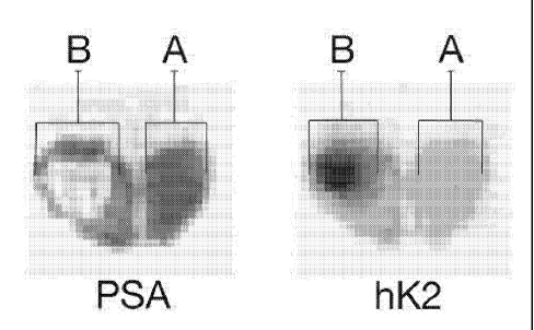
Fig. 1 is a schematic illustration of the combination
of visualisation with both PSA producing tissue and hK2
producing tissue in a cancerous prostate,
Fig. 2 is a schematic illustration of the combination
of visualisation with both PSA producing tissue and hK2
producing tissue in a non-cancerous prostate,
Fig. 3 is a schematic illustration of only
visualisation with PSA producing tissue in a cancerous
prostate, and
Fig. 4 is a schematic illustration of only
visualisation with PSA producing tissue in a cancerous
prostate.
Description of embodiments
The following description focuses on embodiments of
the present invention applicable to a diagnostic method of
prostatic cancer. However, it will be appreciated that the
invention is not limited to this application but may be
applied to many other medical examinations, and diagnostic
investigations, including for example lymph gland
metastasis, post operative examinations, and examinations
during or after radiation, cytostatic, and androgen
treatments. In respect of diagnostic investigation of
metastasis the metastases will be visible in lymph glands
and lymph vessels, since PSA and hK2 pass these regions.
In an embodiment of the invention, antibodies that
are specific for PSA and are labelled with a tracer are
then injected into the body, such as intravenously. Then
the tracer labelled antibodies, that are specific for PSA,
CA 02597687 2007-08-13
WO 2006/087374 PCT/EP2006/060067
7
bind to tissues that produce corresponding antigens, in
this case PSA. The biologic structures, to which the tracer
labelled PSA specific antibodies are bound, are
subsequently visualised with a suitable radiologic
visualisation method, such as PET-scan or other
scintigraphic methods by means of the tracer.
Thereafter, antibodies, that are specific for hK2,
are labelled with a tracer. These antibodies are then
injected intravenously. The tracer labelled antibodies,
that are specific for hK2, bind to tissues that produce
corresponding antigens. The biologic structures, to which
the tracer-labelled hK2 specific antibodies are bound, are
subsequently visualised with a suitable radiologic
visualisation method, such as PET-scan or other
scintigraphic methods.
In yet another embodiment the order may be reversed,
i.e. the visualisation hK2 producing tissue is performed
before the visualisation of PSA producing tissue.
In another embodiment of the present invention the
tracer-labelled antibodies are injected in any other way
into the bloodstream, or the lymphatic system, such as
intra-arterial infusion etc.
Variations in respect of attenuation are directly
corresponding to production and concentration relations of
PSA and hK2. These variations are then used to obtain
diagnostic information.
The visualisations of PSA and hK2 antibody bindings,
obtained from the radiologic visualisation methods
mentioned above, are then combined. From the attenuations
it is possible to directly determine whether the
investigated tissue is PSA producing, hK2 producing, or
CA 02597687 2007-08-13
WO 2006/087374 PCT/EP2006/060067
8
both. In respect of this determination it will be possible
to distinguish prostate cancer from other prostate
disorders, such as benign prostatic hyperplasia, and
prostatitis.
In one example the visualisation of PSA producing
tissue reveals that a part A of a prostate, according to
Fig. 1, has a higher PSA production than a part B. When
this visualisation is combined with the visualisation of
hK2 producing tissue, where the part A has a lower hK2
production than the part B, the physician will be able to
establish prostate cancer in the part B.
In another example, according to Fig. 2, the
visualisation of PSA producing tissue reveals that a
prostate has an even, and relatively high, production of
PSA. When this visualisation is combined with the
visualisation of hK2 producing tissue, where the production
of hK2 is evenly low in the prostate, the physician will be
able to establish that there is no prostate cancer.
In another embodiment of the invention the
visualisation of PSA producing tissue or the visualisation
of hK2 producing tissue may be used separately to visualise
the difference in PSA and hK2 production, respectively, in
the prostate. This embodiment presents the advantage of
being time saving in respect of performing two intravenous
injections of antibodies, and subsequently two
visualisations of the prostate. Nevertheless, the
combination of the visualisation of PSA producing tissue
and the visualisation of hK2 producing tissue presents a
more reliable diagnose and distinction in respect of
prostate cancer, and other prostate disorders, such as
benign prostatic hyperplasia, and prostatitis, since two
CA 02597687 2007-08-13
WO 2006/087374 PCT/EP2006/060067
9
indications of possible disorders in respect of antigen
production are obtained.
In an example of visualisation with only the aid of
the tracer labelled antibodies, that are specific for PSA,
according to Fig. 3, a part C of a prostate has a higher
PSA production than a part D. The physician will be able to
establish prostate cancer in the part D, since part D
differentiate in respect of PSA production from part C.
In yet an example of visualisation with only the aid
of the tracer labelled antibodies, that are specific for
hK2, according to Fig. 4, a part E of a prostate has a
lower hK2 production than a part F. The physician will be
able to establish prostate cancer in the part F, since part
F differentiate in respect of PSA production from part E.
The visualisation methods in the embodiments
according to the present invention reflect the production
of PSA and hK2. These methods aim at visualise malign and
non-malign patho-biological conditions, anatomic
characteristics, size of tumour, and degree of malignancy.
According to the above it will be possible to perform
examinations in respect of metastasis, and lymph glands.
In another embodiment RadioGuided Surgery (RGS) may
be used to identify tracer labeled PSA and/or hK2-
antibodies during and/or before surgery. In this embodiment
tracer labeled antibodies, such as labeled PSA and/or hK2-
antibodies, are first infused. Thereafter, RGS is used to
identify PSA/hK2 producing tissue with gamma detection
instrument, during or before surgery. RGS is well known to
the person skilled in the art as a surgical technique that
enables the surgeon to identify tissue "marked" by a
radionuclide.
CA 02597687 2007-08-13
WO 2006/087374 PCT/EP2006/060067
In still another embodiment of the present invention
the visualisations obtained according to above may be
combined with other radiological visualisation methods,
such as computed tomography (CT), computerized axial
5 tomography (CAT), and magnetic resonance tomography (MRT).
The term PSA is intended to include every known form
of PSA, such as free PSA, precursor forms of PSA,
internally nicked forms of PSA, low molecular weight free
PSA, standard weight free PSA, inactive mature PSA,
10 truncated forms of PSA, glycosylation variants of PSA,
BPSA, inactive pro-PSA, and every complex of PSA, such as
PSA bound to oc,1-antichymotrypsin (ACT), oc,1-protease
inhibitor (API), and oc,2-macroglobulin (AMG).
PSA, secreted from cancer cells, is in a more active
state in comparison with PSA, secreted from BPH tissue. In
the extracellular fluid PSA may be subjected to proteolytic
degradation, thus leading to loss of activity and formation
of complexes.
Thus, it is also within the scope of the present
invention to label compounds or entities, such as ACT, API,
and AMG, bound or complexed to/with PSA.
The term hK2 is intended to include all isomeric
forms of hK2, and any molecule or protein in complex with
hK2.
Most of the hK2 found in seminal plasma is inactive
and complexed with protein C inhibitor (PCI). It is also
possible that hK2 forms complexes with other extracellular
protease inhibitors. In vitro studies show that hK2 may
bind to (G2-antiplasmin (OG2-AP) , ACT, AMG, anti-thrombin III
(ATIII), C1-inactivator and plasminogen activator
inhibitor-1 (PAI-1).
CA 02597687 2007-08-13
WO 2006/087374 PCT/EP2006/060067
11
Thus, it is also within the scope of the present
invention to label compounds, molecules, proteins or any
other entity, such as PCI, oc2-antiplasmin ((c,2-AP) , ACT,
AMG, anti-thrombin III (ATIII), C1-inactivator and
plasminogen activator inhibitor-1 (PAI-1), bound or
complexed to/with hK2.
The term "tracer label" is intended to include all
possible radio-isotopes or the like, which may bind to PSA
or hK2 antibodies, and which may be used for detection with
a positron camera, such as gamma positron camera, or other
radiological visualisation technique. An example of a
tracer label is technetium-99m, but it is of course within
the scope of the present invention to use other suitable
tracer labels, which tracer labels fulfil the requirements
for labelling PSA and hK2 specific antibodies.
The term "antibody" is intended to include both human
and non-human antibodies, such as 4D4, 5C3, 241, 2E9, H117,
and 5A10, or fragment thereof, in respect of PSA, and 11B6,
and 7G1, or fragment thereof, in respect of hK2. It is of
course within the scope of the present invention to use
other suitable antibodies, which antibodies fulfil the
requirements of PSA and hK2 specific antibodies.
In yet another embodiment of the invention the
injection of tracer-labelled antibodies is performed in the
vicinity of the tissue or organ to be visualised. This
embodiment has the advantage of somewhat concentrating the
tracer-labelled antibodies on the area to be visualised.
More tracer-labelled antibodies may reach the area of
interest.
The invention can be implemented in any suitable
form. However, preferably, the invention is implemented as
CA 02597687 2007-08-13
WO 2006/087374 PCT/EP2006/060067
12
diagnostic method in respect of prostate cancer, and
distinction of prostate cancer from other prostate
complications, such as benign prostatic hyperplasia, and
prostatitis. The clinical field of use in respect of the
produced visualisations are for example detection and
monitoring of prostate cancer and other prostate
complications, such as benign prostatic hyperplasia, and
prostatitis, and examinations in respect of metastasis and
treatments. The present invention is also intended for
other urological clinic application, such as post operative
evaluation of radical treatment and treatment examinations
during or after lymph gland metastasis, radiation,
cytostatic, and androgen treatments. The elements and
components of an embodiment of the invention may be
physically, functionally and logically implemented in any
suitable way. Indeed, the functionality may be implemented
in a single unit, in a plurality of units or as part of
other functional units. As such, the invention may be
implemented in a single unit, or may be physically and
functionally distributed between different units.
Although the present invention has been described
above with reference to specific embodiments, it is not
intended to be limited to the specific form set forth
herein. Rather, the invention is limited only by the
accompanying claims and, other embodiments than the
specific above are equally possible within the scope of
these appended claims.
In the claims, the term "comprises/comprising" does
not exclude the presence of other elements or steps.
Furthermore, although individually listed, a plurality of
means, elements or method steps may be implemented by e.g.
CA 02597687 2007-08-13
WO 2006/087374 PCT/EP2006/060067
13
a single unit or processor. Additionally, although
individual features may be included in different claims,
these may possibly advantageously be combined, and the
inclusion in different claims does not imply that a
combination of features is not feasible and/or
advantageous. In addition, singular references do not
exclude a plurality. The terms "a", "an", "first", "second"
etc do not preclude a plurality. Reference signs in the
claims are provided merely as a clarifying example and
shall not be construed as limiting the scope of the claims
in any way.