Note: Descriptions are shown in the official language in which they were submitted.
CA 02597719 2007-08-13
WO 2006/089227 PCT/US2006/005848
-1-
DERMATOLOGICAL TREATMENT DEVICE
RELATED APPLICATIONS
This application claims priority to U.S. Provisional Application No.
60/654,130,
filed February 18, 2005 entitled Derinatological Treatfnent Device, the
contents of
which is hereby incorporated by reference in its entirety.
TECHNICAL FIELD
This invention relates generally to methods and apparatus for utilizing
energy,
e.g., optical radiation, to treat various dermatological and cosmetic
conditions.
BACKGROUND OF THE INVENTION
Fractional treatments generally have been directed to treating the epidermis,
which is at the surface of skin tissue. However, for certain applications
there is a need to
provide treatments that extend further into the tissue.
Heating tissue at depth can be done with various wavelengths of EMR, both
visible and non-visible. Infrared, also known as radiant heat, is a form of
energy that
heats objects directly through a process called conversion. Infrared radiation
is emitted
by any object that has a temperature (i.e. radiates heat). Infrared is not
visible, but can
be felt in the form of heat. The infrared segment of the electromagnetic
spectrum occurs
just below or "infra" to red light as the next lowest energy band of light.
SUMMARY OF TIiE INVENTION
One aspect of the invention is a handheld dermatological device that includes
a
light source assembly that has a source for generating EMR and a cooling
surface that
defines a target treatment area on the tissue when located in proximity to the
tissue. The
light source assembly is configured to transmit EMR from the source, and
through the
cooling surface during operation. The devices also has first cooling mechanism
for
cooling the radiation source, and a second cooling mechanism for cooling the
cooling
surface.
Preferred embodiments of this aspect of the invention may include some of the
following additional features. The dermatological treatment device can include
a fan
configured to pump air to cool the source, and a heatsink in thermal
communication with
CA 02597719 2007-08-13
WO 2006/089227 PCT/US2006/005848
-2-
the source. The fan pumps air over the heatsink to remove heat from the
heatsink device
during operation. The heatsink includes a plurality of cooling fms. The
heatsink is
thermally coupled to the source via a reflector, and the fan is configured to
cool the
source, the reflector, and the heatsink. The handheld dermatological device
also has a
control unit for controlling the first cooling mechanism. The control unit
fa.rther
includes a controller in electrical communication with a temperature sensor
and in
electrical communication with the fan, such that the controller can
automatically control
the first cooling mechanism based on information received from the temperature
sensor.
The second cooling mechanism is a circulatory system for circulating a coolant
that includes a chiller for cooling the tissue being treated to approximately
at least 5 C.
The second cooling mechanism also includes a pump, a cooling input, and a
cooling
output. The cooling input is connected to a cooling window at an input
connection and
the cooling output is connected to the cooling window at an output connection.
The
second cooling mechanism is configured to supply cooling fluid to the cooling
window
during operation via the cooling input and to extract heated coolant from the
cooling
window via the cooling output to cool the cooling window. The cooling
mechanism
further includes a chiller.
The second cooling mechanism also includes a temperature sensor for
monitoring the temperature of the tissue and a control unit for controlling
the second
cooling mechanism. The control unit further comprises a controller in
electrical
communication with a temperature sensor and in electrical communication with
the
pump. The controller is configured to automatically control the pump based on
information received from the temperature sensor.
Another aspect of the invention is a window of a dermatological treatment
device
that is configured to transmit EMR from a source of the device to tissue being
treated.
The window has a pane configured to allow EMR to pass from the dermatological
treatment device to the tissue being treated. The window also has a first
channel
extending across substantially across a length of the pane and a frame
extending about
the pane to secure the pane in the dermatological treatment device. The window
includes a first cooling input in fluid communication with a first end of the
first channel
and a first cooling output in fluid communication with a second end of the
first channel.
The window is configured to be cooled during operation by fluid traveling
through the
cooling input, through the first channel and out the second end of the fixst
channel.
CA 02597719 2007-08-13
WO 2006/089227 PCT/US2006/005848
-3-
Preferred embodiments of this aspect of the invention may include some of the
following additional features. The channel of the window is a groove having an
open
portion extending along a surface of the pane. The window also has an optical
surface
abutting the surface of the pane such that the groove is enclosed during
operation to
allow fluid to flow through the channel and to prevent the fluid from flowing
out of the
open portion. The window also has an optical material between the pane and the
optical
surface. The material allows some EMR to pass from the dermatological
treatment
device to the tissue being treated, and can be a dielectric coating.
Another aspect of the invention is a dermatological treatment device for
treating
tissue located at a depth of at least approximately 0.5 mm. The device
includes a
housing containing an EMR source and a window. The window is configured to
transmit EMR from the source to the tissue being treated. The source is
configured to
produce at least 500 W of EMR and the window has an area sufficiently large to
produce
a power density of less than 5 W/cma.
Preferred embodiments of this aspect of the invention may include some of the
following additional features. The pulse width of the power source is greater
than or
equal to 0.5 seconds and less than or equal to 600 seconds. The EMR source is
configured to produce at least 1000W.
Another aspect of the invention is an apparatus for performing a treatment on
tissue, that includes a housing having a cooling surface that defines a target
treatment
area on the tissue when located in proximity to the tissue, a radiation source
for
generating EMR that passes through the cooling surface, and a sensor to
indicate when
the cooling surface is in proximity to the tissue.
Preferred embodiments of this aspect of the invention may include some of the
following additional features. Activation of the sensor indicates that the
cooling surface
contacts the tissue. The sensor can be an e-field sensor, a capacitive sensor,
a resistive
sensor, a pressure sensor, or an H-field sensor. The sensor can be configured
to detect
changes in an electrical field.
The sensor is in electrical communication with a controller that is configured
to
provide signals in response to information obtained from the sensor. The
controller
issues a first signal corresponding to the detection by the sensor that no
tissue is in close
proximity and a second signal corresponding to the detection by the sensor
that a first
tissue is in close proximity. The controller issues a third signal
corresponding to the
CA 02597719 2007-08-13
WO 2006/089227 PCT/US2006/005848
-4-
detection by the sensor that a second tissue is in close proximity to the
sensor. The
controller distinguishes between tissue types based on the input from the
sensor. The
controller commands a first action in response to the detection of the first
tissue type and
a second action in response to the detection of the second tissue type. The
first action is
to treat the tissue. The second action is to not treat the tissue.
The sensor can include a first node and a second node disposed about the
cooling
surface. The nodes are in contact with the tissue when the cooling surface is
in contact
with the tissue and are not in contact with the tissue when the cooling
surface is not
completely in contact with the tissue. The sensor measures the current between
the
nodes when in contact with the skin. The sensor indicates that the skin is in
contact with
the sensor when a current is detected between the nodes.
The sensor can be mounted on the housing, and can be a microswitch. The
device also may have an output device operably connected to the sensor. The
output
device is one of a visual device, an audio device, or a vibrating device. A
feedback
mechanism may also be connected to the sensor. The feedback mechanism
indicates to
an operator of the apparatus the amount of time the cooling surface is
required to stay in
contact with the tissue for safe operation. The feedback mechanism prevents
firing of
the radiation source if contact of the cooling surface with the tissue is
broken. The
feedback mechanism prevents firing of the radiation source until after a
predetermined
cooling time has elapsed.
The device also has a control unit to implement a preset cooling time before
allowing firing of the radiation source. The control unit implements a preset
firing time
for the radiation source. The device can also be a handheld device, and the
control unit
can be operably coupled to the handheld device.
The radiation source can be a monochromatic source such as a laser.
Alternatively, the radiation source can be a halogen lamp, a radiant lamp, an
incandescent lamp, an arc lamp, and a fluorescent lamp.
The cooling surface can be made of a deformable or viscoelastic material, like
a
gel. The cooling surface can also be made of a solid material, such as glass,
sapphire or
plastic.
The device may have a contact frame that is operably coupled to the housing.
The contact frame is movable from an extended position to a retracted position
in which
it is in proximity to the cooling surface. The sensor activates when the frame
is in the
CA 02597719 2007-08-13
WO 2006/089227 PCT/US2006/005848
-5-
retracted position. The sensor activates when the cooling surface is in
proximity to the
contact frame. The contact frame has an interior portion that is open to allow
passage of
EMR. A push rod is connected to the contact frame and is operably coupled to
the
sensor, such that the push rod activates the sensor when the cooling surface
contacts the
contact frame. The sensor is mounted on one of the cooling surface and the
contact
frame.
Another aspect of the invention is an apparatus for performing a treatment on
tissue that includes a housing having a means for cooling the tissue. The
nieans for
cooling the tissue includes a surface that defines a target treatment area on
the tissue
when located in proximity to the tissue. The housing also includes a means for
generating EMR. The EMR passes through the surface during irradiation. The
housing
also includes a means for sensing contact of the means for cooling with the
tissue.
Preferred embodiments of this aspect of the invention may include some of the
following additional features. The means for sensing activates when the means
for
cooling contacts the contact frame. Activation of the means for sensing
indicates that
the means for cooling contacts the tissue. A contact frame is operably coupled
to the
housing. The contact frame is movable from an extended position to a position
in which
it is in contact with the means for cooling.
Another aspect of the invention is a method of operating a handheld
dermatological device, which includes sensing contact of a cooling surface of
the
handheld device with tissue, indicating to a user of the handheld device when
the
cooling surface contacts the tissue, and automatically interrupting firing of
a radiation
source of the handheld device if the cooling surface loses contact with the
tissue.
Preferred embodiments of this aspect of the invention may include some of the
following additional features. The method can include sensing contact of the
cooling
surface with tissue, indicating to the user if the cooling surface loses
contact with the
tissue. The act of sensing contact comprises determining when a contact frame
of the
handheld device contacts the cooling surface. The contact of the contact frame
with the
cooling surface indicates contact of the cooling surface with the tissue.
The method may further include distinguishing a first tissue type in contact
with
the sensor from a second tissue type, and taking an action based on the tissue
type. The
act of taking an action includes not irradiating the tissue if the tissue
corresponds to an
untreatable tissue type and irradiating the tissue if the tissue corresponds
to a treatable
CA 02597719 2007-08-13
WO 2006/089227 PCT/US2006/005848
-6-
tissue type. The act of indicating to the user includes activating one of a
visual indicator
and an audio indicator.
Another aspect of the invention is a method of automatically operating a
handheld dermatological device, which includes sensing contact of a cooling
surface of
the handheld device with tissue, instituting a preset cooling time for cooling
of the tissue
prior to irradiating the tissue with a radiation source of the handheld
device, instituting a
preset firing time of the radiation source after the preset cooling time, and
interrupting
firing of the radiation source if the cooling surface loses contact with the
tissue.
Preferred embodiments of this aspect of the invention may include some of the
following additional features. The method may further include indicating to
the user if
the cooling surface loses contact with the tissue, after sensing contact of
the cooling
surface with tissue. The act of indicating to the user includes activating one
of a visual
indicator and an audio indicator. The act of sensing contact comprises
determining
when a contact frame of the handheld device contacts the cooling surface,
wherein
contact of the contact frame with the cooling surface indicates contact of the
cooling
surface with the tissue.
BRIEF DESCRIPTION OF THE DRAWINGS
Non-limiting embodiments of the present invention will be described by way of
example with reference to the accompanying drawings in which:
FIG. 1 is a schematic diagram of one embodiment of the invention, shown in
proximity to a tissue sample;
FIG. 2 is a side view of a schematic diagram of part of a handheld
dermatological device according to one embodiment of the invention;
FIG. 3 is a second side view of the handheld dermatological device of FIG. 2;
FIG. 4 is a third side view of the handheld dermatological device of FIG. 2;
FIG. 5 is a fourth side view of the handheld dermatological device of FIG. 2;
FIG. 6 is a fifth side view of the handheld dermatological device of FIG. 2;
FIG. 7 is a sixth side view of the handheld dermatological device of FIG. 2;
FIG. 8 is a front view of the handheld dermatological device of FIG. 2;
CA 02597719 2007-08-13
WO 2006/089227 PCT/US2006/005848
-7-
FIG. 9 is a partial view from the front of a lamp, reflector, and optics of
the
handheld dermatological device of FIG. 2;
FIG. 10 is a perspective view of the handheld dermatological device of FIG. 2;
FIG. 11 is a second perspective view of the handheld dermatological device of
FIG.2;
FIG. 12 is a back view of the handheld dermatological device of FIG. 2;
FIG. 13 is a second back view of the handheld dermatological device of FIG. 2;
FIG. 14 is a bottom view of the handheld dermatological device of FIG. 2;
FIG. 15 is a side view of the housing structure and complete unit of the
handheld
dermatological device of FIG. 2;
FIG. 16 is a flow chart that illustrates the operation of one embodiment of
the
invention.
FIG. 17 is a graph showing the relationship between treatment time and the
depth
of heating for infrared radiation without pre-cooling the treated tissue; and
FIG. 18 is a graph showing the relationship between treatment time and surface
skin temperature;
FIG. 19 is a side view of an alternative embodiment of a handheld
dermatological device;
FIG. 20 is a cross-sectional side view of the handheld dermatological device
of
FIG.19;
FIG. 21 is a schematic top view of a window for use in the handheld
dermatological device of FIG. 19;
FIG. 22 is a schematic side view of the window of FIG. 21;
FIG. 23 is a schematic bottom view of an embodiment of a portion of the
handheld dermatological device of FIG. 19;
FIGS. 24A and 24B are schematic side views of the portion of the handheld
dermatological device shown in FIG. 23 during operation;
CA 02597719 2007-08-13
WO 2006/089227 PCT/US2006/005848
-8-
FIG. 25 is a schematic side view of an alternate embodiment for a window of a
dermatological device;
FIG. 26 is a schematic side view of an alternate embodiment of a waveguide;
FIG. 27 is a bottom view of the waveguide of FIG. 26; and
FIG. 28 is a bottom view of an alternate embodiment of a face of a
dermatological device.
DETAILED DESCRIPTION
The benefits of being able to raise and/or lower the temperature in a selected
region of tissue for various therapeutic and cosmetic purposes have been known
for
some time. For instance, heated pads or plates or various forms of
electromagnetic
radiation (EMR), including microwave radiation, electricity, infrared
radiation, and
ultrasound have previously been used for heating subdermal muscles, ligaments,
bones
and the like to, for example, increase blood flow, to otherwise promote the
healing of
various injuries and other damage, and for various therapeutic purposes, such
as frostbite
or hyperthermia treatment, treatment of poor blood circulation, physical
therapy,
stimulation of collagen, cellulite treatment, adrenergic stimulation, wound
healing,
psoriasis treatment, body reshaping, non-invasive wrinkle removal, etc. The
heating of
tissues has also been utilized as a potential treatment for removing cancers
or other
undesired growths, infections and the like. Heating may be applied over a
small,
localized area, over a larger area, for example to the hands or feet, or over
larger regions
of tissue, including the entire body.
Because most of the techniques described above involve applying energy to
tissue at depth through the subject's skin surface, peak temperature generally
occurs at
or near the subject's skin surface and decreases, sometimes significantly,
with depth.
The radiation is both highly scattered and highly absorbed in surface layers
of tissue,
precluding significant portions of such radiation from reaching the tissue
regions at
depth to cause heating tliereof. In view of the energy losses due to
scattering and
absorption, a substantial amount of optical (including near infrared) energy
must be
applied in order for enough energy to reach a region of tissues at depth to
have a desired
effect. However, such a high amount of optical energy can cause damage to the
surface
layers of tissue, making it difficult to achieve desired photothermal
treatments in tissue
CA 02597719 2007-08-13
WO 2006/089227 PCT/US2006/005848
-9-
regions at depth. For these reasons, optical radiation has heretofore had at
most limited
value for therapeutic and cosmetic treatments on tissue at depth.
Methods of deep heating are also desirable for fractional treatments, which
depend, in part, upon the discovery that, when using EMR to treat tissues,
there are
substantial advantages to producing lattices of EMR-treated islets in the
tissue rather
than large, continuous regions of EMR-treated tissue. The lattices are
periodic patterns
of islets in one, two or three dimensions in which the islets correspond to
local maxima
of EMR-treatment of tissue. The islets are separated from each other by non-
treated
tissue. The EMR-treatment results in a lattice of EMR-treated islets which
have been
exposed to a particular wavelength or spectrum of EMR, and which is referred
to herein
as a lattice of "optical islets." When the absorption of EMR energy results in
significant
temperature elevation in the EMR-treated islets, the lattice is referred to
herein as a
lattice of "thermal islets." When an amount of energy is absorbed that is
sufficient to
significantly disrupt cellular or intercellular structures, the lattice is
referred to herein as
a lattice of "damage islets." By producing EMR-treated islets rather than
continuous
regions of EMR-treatment, more EMR energy can be delivered while lowering the
risk
of bulk tissue damage
To more effectively treat tissue with near infrared radiation, the skin at the
surface of the tissue is typically cooled to a temperature of approximately 5
C, although
other temperatures are used. Thus, the technique of the present invention
combines
advantageous features of non-ablative and fractional techniques.
Applications in which the invention may be useful include the treatment of
various diseases and cosmetic enhancements, particularly, cellulite and
subcutaneous fat
treatment, physical therapy, muscle and skeletal treatments, including relief
of pain and
stiffness for muscles and joints, and treatment of spinal cord problems, and
treatment of
cumulative trauma disorders (CTD's) such as carpel tunnel syndrome (CTS),
tendonitis
and bursitis, fibromyalgia, lymphedema and cancer therapy and skin
rejuvenation
treatments, including, for example, skin smoothing, wrinkle and rhytide
reduction, pore
size reduction, skin lifting, improved tone and texture, stimulation of
collagen
production, shrinkage of collagen, reduction of skin dyschromia (i.e. pigment
non-
uniformities), reduction telangiectasia (i.e. vascular malformations),
improvement in
skin tensile properties (e.g. increase in elasticity, lifting, tightening),
treatment of acne,
CA 02597719 2007-08-13
WO 2006/089227 PCT/US2006/005848
-10-
hypertrophic scars, reducing body odor, removing warts and calluses, treating
psoriasis,
and decreasing body hair.
The present invention provides means for effective deep heating of tissue
using
both fractional and non-fractional procedures. For fractional procedures, the
embodiments described below may create non-uniform (modulated) temperature
profiles
(MTP), including deep in the dermis and in hypodermis (typically, at depths
exceeding
500 m) or superficially in the epiderinis and/or dermis. In some embodiments,
such
profiles result in formation of a pattern (lattice) of islets of damage (LID).
Active or
passive cooling can be applied to epidermal surface in order to prevent
epidermal
damage.
Creation of MTPs leads to improvements in skin structure and texture via the
following mechanisms (the list is not exclusive):
1. Lifting and tightening of skin as a result of shrinkage of collagen fibrils
subjected to elevated temperature.
2. Lifting and tightening of skin as a result of coagulation of localized
areas in
the dermis and hypodermis.
3. Improvement in skin texture as a result of coagulation of localized areas
in the
dermis and hypodermis.
4. Promotion of collagen production due to healing response to thermal stress
and/or thermal shock.
A number of other local and systemic pathologies can be treated with the
technique:
1. Cellulite: By changing mechanical stress distribution at the
dermis/hypodermis
border, the appearance of cellulite can be improved.
2. Acne: By selecting the wavelength of the optical radiation to promote
preferential absorption of the optical energy by sebum and/or organizing the
pattern to
target preferentially sebaceous glands, selective destTaction of the glands
can be
achieved.
3. Hypertrophic scars: By inducing tightening and shrinkage in the scar
tissue,
transformation of the abnormal connective tissue to normal one can be
initiated.
4. Odor reduction: By selectively targeting eccrine glands, production of
eccrine
sweat can be reduced, and its composition can be changed.
CA 02597719 2007-08-13
WO 2006/089227 PCT/US2006/005848
-11-
5. Non-skin-surface texturiniz: The technique can be used for organ
augmentation
(e.g., lips).
One embodiment of the invention is a handheld derrnatological device that
incorporates a mechanism for cooling a subject's skin surface concurrently
with the
application of optical radiation thereto. While the radiation reaches the
tissue at depth to
be treated quickly to begin the heating thereof, cooling propagates as a cold
wave,
protecting tissue above the treatment region and moving the depth of maximum
heating
further into the skin. In one embodiment, the cooling wave can propagate to a
depth just
above the treatment region, but does not extend to the treatment region at
least until
sufficient energy has been delivered to the treatment region to effect the
desired
treatment. The cooling mechanism of the device can cool the subject's skin
prior to,
during, and/or after the application of radiation thereto to more effectively
protect tissue
above the treatment region and to insure that the maximum temperature rise in
the
irradiated tissue occurs at or near a desired depth. This may also permit
higher energy
and shorter duration of radiation pulses to be applied to the skin without any
damage or
minimal damage to tissue above the desired depth. The head used to apply the
radiation
may also be used to apply cooling. The handheld dermatological device can
include a
sensor mounted adjacent the cooling mechanism near the subject's skin. Such a
sensor
can indicate when the cooling mechanism contacts the subject's skin (or looses
contact
with the subject's skin), thus indicating to the user when it is safe to begin
application of
radiation.
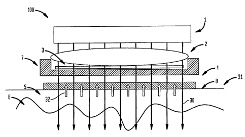
Figure 1 shows an apparatus 100 according to one embodiment of the invention.
For this apparatus, optical energy 30 from a suitable energy source 1 passes
through
optical (for example, focusing) device 2, filter 3, cooling mechanism 4 and
contact plate
8, before reaching tissue 31 (i.e., the subject's skin). In some embodiments
of the
invention, certain of these components, such as, for example, filter 3 where a
monochromatic energy source is utilized or optical device 2, may not
necessarily be
present. In other embodiments, the apparatus may not contact the skin. In yet
another
embodiment, there is no cooling mechanism 4 such that there is only passive
cooling
between the contact plate and the skin.
A suitable optical impedance matching lotion or other suitable substance would
typically be applied between plate 8 and tissue 31 to provide enhanced optical
and
CA 02597719 2007-08-13
WO 2006/089227 PCT/US2006/005848
-12-
thermal contact. Tissue 31, as shown in FIG. 1, is divided into an upper
region 5, which,
for applications where radiation is applied to the skin surface, would be the
epidermis
and dermis, and a lower region 6, which would be a subdermal region in the
previous
example. Region 6, for instance, can be the hypodermis.
Energy 30, possibly in conjunction with one or a combination of focusing from
optical device 2, and wavelength selection from filter 3, and with cooling
from cooling
mechanism 4, results in maximum heating occurring at a selected depth in
tissue 31.
The selected depth can be, as previously indicated, at or near the junction of
regions 5
and 6 or in lower region 6, and it can also be in region 5 or in the
hypodermis.
The energy source 1 may be any suitable electromagnetic radiation (EMR)
source, but will preferably be a source emitting visible light, or energy in
the near
infrared and infrared ranges. The light sources used in conjunction with the
invention
may be coherent and non-coherent sources, able to produce optical energy at a
desired
wavelength or a desired wavelength band or in multiple wavelength bands. The
exact
energy source 1, and the exact energy chosen, may be a function of the type of
treatment
to be performed, the tissue to be heated, the depth within the tissue at which
treatment is
desired, and of the absorption of that energy in the desired area to be
treated. Energy
source 1 may produce EMR, such as near infrared or visible light radiation
over a broad
spectrum, over a limited spectrum, or at a single wavelength, such as would be
produced
by a light emitting diode or a laser. In certain cases, a narrow spectral
source may be
preferable, as the wavelength(s) produced by the energy source may be targeted
towards
a specific tissue type or may be adapted for reaching a selected depth. In
other
embodiments, a wide spectral source may be preferable, for example, in systems
where
the wavelength(s) to be applied to the tissue may change, for example, by
applying
different filters, depending on the application. Acoustic, RF or other EMF
sources may
also be employed in suitable applications.
For example, UV, violet, blue, green, yellow light or infrared radiation
(e.g.,
about 290-600 nm, 1400 - 3000 nni) can be used for treatment of superficial
targets,
such as vascular and pigment lesions, fine wrinldes, skin texture and pores.
Blue, green,
yellow, red and near IR light in a range of about 450 to about 1300 nm can be
used for
treatment of a target at depths up to about 1 millimeter below the skin. Near
infrared
light in a range of about 800 to about 1400 nm, about 1500 to about 1800 mn or
in a
CA 02597719 2007-08-13
WO 2006/089227 PCT/US2006/005848
-13-
range of about 2050 nm to about 2350 nm can be used for treatment of deeper
targets
(e.g., up to about 3 millimeters beneath the skin surface). The following
table shows
examples of the wavelengths of electromagnetic energy that are thought to be
suitable
for treating various cosmetic and medical conditions.
TABLE 1: Uses of Light of Various Wavelengths In Photocosmetic Procedures
Treatment condition or application Wavelength of Light, nna
Anti-a 'n 400 -2700
Superficial vascular 290-600
1300-2700
Deep vascular 500-1300
Pigmented lesion, de pi entation 290-1300
Skin texture, stretch mark, scar, porous 290-2700
Deep wrinkle, elasticity 500-1350
Skin lifting 600-1350
Acne 290-700, 900-1850
Psoriasis 290-600
Hair owth control 400-1350
PFB 300-400, 450-1200
Cellulite 600-1350
Skin cleaning 290-700
Odor 290-1350
Oiliness 290-700, 900-1850
Lotion delivery into the skin 1200-20000
Color lotion delivery into the skin Spectrum of absorption of color center and
1200-20000
Lotion with PDT effect on skin Spectrum of absorption of photo sensitizer
condition including anti cancer effect
ALA lotion with PDT effect on skin 290-700
condition including anti cancer effect
Pain relief 500-1350
Muscular, joint treatment 600-1350
Blood, lymph, immune system 290 -1350
Direct singlet oxygen generation 1260-1280
The energy source 1 can be any variety of a coherent light source, such as a
solid-state laser, dye laser, diode laser, fiber laser, or other coherent
light source. For
example, energy source 1 may be a radiant lamp, a halogen lamp, an
incandescent lamp,
an arc lamp, a fluorescent lamp, a light emitting diode, a laser (including
diode and fiber
lasers), the sun, or other suitable optical energy source. As another example,
the energy
CA 02597719 2007-08-13
WO 2006/089227 PCT/US2006/005848
-14-
source 1 can be a neodymium (Nd) laser, such as a Nd:YAG laser. In addition,
multiple
energy sources may be used which are identical or different. For example,
multiple laser
sources may be used and they may generate optical energy having the same
wavelength
or different wavelengths. As another example, multiple lamp sources may be
used and
they may be filtered to provide the same or different wavelength band or
bands. In
addition, different types of sources may be included in the same device, for
example,
mixing both lasers and lamps.
In this exemplary embodiment, the energy source 1 includes a neodymium (Nd)
laser generating radiation having a wavelength around 1064 nm. Such a laser
includes a
lasing medium, e.g., in this embodiment a neodymium YAG laser rod (a YAG host
crystal doped with Nd}3 ions), and associated optics (e.g., milTors) that are
coupled to
the laser rod to form an optical cavity for generating lasing radiation. In
other
embodiments, other laser sources, such as chromium (Cr), Ytterbium (Yt) or
diode
lasers, or broadband sources, e.g., lamps, can be employed for generating the
treatment
radiation.
Lasers and other coherent light sources can be used to cover wavelengths
within
the 100 to 100,000 nm range. Examples of coherent energy sources are solid
state, dye,
fiber, and other types of lasers. For example, a solid state laser with lamp
or diode
pumping can be used. The wavelength generated by such a laser can be in the
range of
400 - 3,500 mn. This range can be extended to 100 - 20,000 nm by using non-
linear
frequency converting. Solid state lasers can provide maximum flexibility with
pulse
width range from femtoseconds to a continuous wave.
Another example of a coherent source is a dye laser with non-coherent or
coherent pumping, which provide wavelength-tunable light emission. Dye lasers
can
utilize a dye dissolved either in liquid or solid matrices. Typical tunable
wavelength
bands cover 400 - 1,200 nm and a laser bandwidth of about 0.1 - 10 nm.
Mixtures of
different dyes can provide multi wavelength emission. Dye laser conversion
efficiency is
about 0.1-1 % for non-coherent pumping and up to about 80 % with coherent
pumping.
Another example of a coherent source is a fiber laser. Fiber lasers are active
waveguides with a doped core or undoped core (Raman laser), with coherent or
non-
coherent pumping. Rare earth metal ions can be used as the doping material.
The core
and cladding materials can be quartz, glass or ceramic. The core diameter
could be from
CA 02597719 2007-08-13
WO 2006/089227 PCT/US2006/005848
-15-
microns to hundreds of microns. Pumping light could be launched into the core
through
the core facet or through cladding. The light conversion efficiency of such a
fiber laser
could be up to about 80% and the wavelength range can be from about 1,100 to
3,000
nrn. A combination of different rare-earth ions, with or without additional
Raman
conversion, can provide simultaneous generation of different wavelengths,
which could
benefit treatment results. The range can be extended with the help of second
harmonic
generation (SHG) or optical parametric oscillator (OPO) optically connected to
the fiber
laser output. Fiber lasers can be combined into the bundle or can be used as a
single
fiber laser.
Diode lasers can be used for the 400 -100,000 nm range. Since many
photodermatology applications require a high-power light source, the
configurations
described below using diode laser bars can be based upon about 10 -100 W, 1-cm-
long,
cw diode laser bar. Note that other sources (e.g., LEDs and microlasers) can
be
substituted in the configurations described for use with diode laser bars with
suitable
modifications to the optical and mechanical sub-systems.
Other types of lasers (e.g., gas, excimer, etc.) can also be used.
A variety of non-coherent sources of EMR (e.g., arc lamps, incandescence
lamps, halogen lamps, light bulbs) can be used in the invention for the energy
source 1.
There are several monochromatic lamps available such as, for example, hollow
cathode
lamps (HCL) and electrodeless discharge lamps (EDL). HCL and EDL could
generate
emission lines from chemical elements. For example, sodium emits bright yellow
light
at550nm.
Linear arc lamps use a plasma of noble gases overheated by pulsed electrical
discharge as a light source. Commonly used gases are xenon, krypton and their
mixtures,
in different proportions. The filling pressure can be from about several torr
to thousands
of torr. The lamp envelope for the linear flash lamp can be made from fused
silica,
doped silica or glass, or sapphire. The emission bandwidth is about 180-2,500
nm for
clear envelope, and could be modified with a proper choice of dopant ions
inside the
lamp envelope, dielectric coatings on the lamp envelope, absorptive filters,
fluorescent
converters, or a suitable combination of these approaches.
CA 02597719 2007-08-13
WO 2006/089227 PCT/US2006/005848
-16-
In some embodiments, a Xenon-filled linear flash lamp with a trapezoidal
concentrator made from BK7 glass can be used. As set forth in some embodiments
below, the distal end of the optical train can, for example, be an array of
microprisms
attached to the output face of the concentrator. The spectral range of EMR
generated by
such a lamp can be about 300 - 2000 nm.
Incandescent lamps are one of the most common light sources and have an
emission band from 300 to 4,000 nm at a filament temperature of about 2,500 C.
The
output emission can be concentrated on the target with reflectors and/or
concentrators.
Halogen tungsten lamps utilize the halogen cycle to extend the lifetime of the
lamp and permit it to operate at an elevated filament temperature (up to about
3,500 C),
which greatly improves optical output. The emission band of such a lamp is in
the range
of about 300 to 3,000 nm.
Light-emitting diodes (LEDs) that emit light in the 290-2,000 nm range can be
used to cover wavelengths not directly accessible by diode lasers.
Where optical device 2 is a focusing device, it may be any suitable device
able to
focus at least a portion of energy 30 arriving from energy source 1 at tissue
31, and in
particular at a selected depth in tissue 31. For example, device 2 may include
mirrors,
prisms, reflectors, lenses such as Fresnel lenses, collimating lenses or
focusing lenses,
diffraction gratings, or other optical devices. Device 2 may also include a
plurality or an
array of devices listed above.
Filter 3 may be any suitable filter able to select, or at least partially
select, certain
wavelengths or wavelength bands from energy source 1. In certain embodiments,
a
specific set of wavelengths may be blocked by filter 3. It is also possible
that undesired
wavelengths in the energy from source 1 may be wavelength shifted in ways
known in
the art so as to enhance the energy available in the desired wavelength bands.
Thus,
filter 3 may include elements designed to absorb, reflect or alter certain
wavelengths of
electromagnetic radiation. For example, filter 3 may be used to remove certain
types of
wavelengths that are absorbed by surrounding tissues. For instance, dermis,
hypodermis
and epidermis tissues are primarily composed of water, as is much of the rest
of the
human body. By using a filter that selectively removes wavelengths that excite
water
molecules, the absorption of these wavelengths by the body may be greatly
reduced,
CA 02597719 2007-08-13
WO 2006/089227 PCT/US2006/005848
-17-
which may contribute to a reduction in the amount of heat generated by light
absorption
in these molecules. Thus, by passing radiation through a water-based filter,
those
frequencies of radiation that may excite water molecules will be absorbed in
the water
filter, and will not be transmitted into tissue 31. Thus, a water-based filter
may be used
to decrease the amount of radiation absorbed in tissue above the treatment
region and
converted into heat. For other treatments, absorption of the radiation by
water may be
desired or required for treatment.
Figure 1 shows a cooling mechanism 4 adjacent to the surface of tissue 31.
Cooling mechanism 4 may be any suitable cooling mechanism able to reduce the
temperature of tissue 31. Heat energy 32 may be drawn from tissue 31 across
contact
plate 8 into coolirig mechanism 4. For example, cooling mechanism 4 may be air
or
other suitable gas that is blown over contact plate 8, cooling water, or a
cooling oil or
other fluid. Mixtures of these substances, such as an oil and water mixture,
may also be
envisioned. Cooling mechanism 4 may have any suitable configuration, for
example, a
flat plate, a series of conducting pipes, a sheathing blanket, or a series of
channels for the
passage of air, or other gases, or liquid across plate 8. For example, in one
embodiment,
cooling mechanism 4 may be a water-cooled contact plate. In another
embodiment,
cooling mechanism 4 may be a series of channels carrying a coolant fluid or a
refrigerant
fluid (for example, a cryogen), which channels are in contact with tissue 31
or with plate
8. In yet another embodiment, cooling mechanism 4 may comprise a water or
refrigerant fluid (for example R134A) spray, a cool air spray or air flow
across the
surface of tissue 31. In other embodiments, cooling may be accomplished
through
chemical reactions (for example, endothermic reactions), or through electronic
cooling,
such as Peltier cooling. In yet other embodiments, cooling mechanism 4 may
have more
than one type of coolant, or cooling mechanism 4 and/or contact plate 8 may be
absent,
for example, in embodiments where the tissue is cooled passively or directly,
for
example, through a cryogenic or other suitable spray. Sensors or other
monitoring
devices may also be embedded in cooling mechanism 4, for example, to monitor
the
temperature, or determine the degree of cooling required by tissue 31, and be
manually
or electronically controlled.
In certain cases, cooling mechanism 4 may be used to maintain the surface
temperature of tissue 31 at its normal temperature, which may be, for example,
37 or 32
CA 02597719 2007-08-13
WO 2006/089227 PCT/US2006/005848
-18-
C, depending on the type of tissue being heated. In other embodiments, cooling
mechanism 4 may be used to decrease the temperature of the surface of tissue
31 to a
temperature below the normal temperature of that type of tissue. For example,
cooling
mechanism 4 may be able to decrease the surface temperature of tissue 31 to,
for
example, a range between 25 C and -5 C.
In some embodiments of the invention, such as shown in FIG. 1, energy 30 from
energy source 1 may pass through cooling mechanism 4. In these types of
configurations, cooling mechanism 4 may be made from materials able to
transmit at
least a portion of energy 30, for example, air, water or other gases or
fluids, glass, or a
clear plastic. In other embodiments, cooling mechanism 4 may be formed out of
a series
of discrete channels, and energy 30 may pass between these channels. In other
embodiments of the invention, energy 30 may not be directed through cooling
mechanism 4.
Contact plate 8 may be made out of a suitable heat transfer material, and
also,
where the plate contacts tissue 31, of a material having a good optical match
with the
tissue. Sapphire is an example of a suitable material for plate 8. In some
embodiments,
contact plate 8 may have a high degree of thermal conductivity, for example,
to allow
cooling of the surface of the tissue by cooling mechanism 4. In other
embodiments,
contact plate 8 may be an integral part of cooling mechanism 4, or be absent.
Contact
plate 8 may be made out of a deformable or viscoelastic material in some
embodiments
of the invention, for example, a gel such as a hydrogel. In other embodiments,
contact
plate 8 may be made of a solid material, such as a glass, a crystal such as
sapphire, or a
plastic. In some embodiments of the invention, such as shown in FIG. 1, energy
30 from
energy source 1, or a fraction thereof, may pass through contact plate 8. In
these
configurations, contact plate 8 may be made out of materials able to transmit
at least a
portion of energy 30, for example glass, sapphire, or a clear plastic, or
contact plate 8
may be made in such a way as to allow at least a portion of energy 30 to pass
through
contact plate 8, for example, via a series of holes, passages, lenses, etc.
within contact
plate 8.
In some embodiments of the invention, energy source 1, optical device 2 and/or
filter 3 may also require a cooling mechanism. This cooling mechanism may or
may not
be the same as the cooling mechanism 4 that cools tissue 31 through contact
plate 8, as
CA 02597719 2007-08-13
WO 2006/089227 PCT/US2006/005848
-19-
indicated by arrows 32 in FIG. 1. For example, in the embodiment shown in FIG.
1,
cooling mechanism 7, shown separately from cooling mechanism 4, is used to
cool filter
3 and/or optical device 2. The design of cooling mechanism 7 may be a function
of the
components used in the construction of the apparatus. In FIG. 1, cooling
mechanism 7
and cooling mechanism 4 are illustrated as separate systems. However, in other
embodiments, cooling mechanism 7 and cooling mechanism 4 may be part of the
same
system, or one or both may be absent. Cooling mechanism 7 may be any suitable
cooling mechanism known in the art, such as air, water, or oil. Mixtures of
these
substances, such as an oil and water mixture, may also be envisioned. Cooling
of the
components may be accomplished through convective or conductive cooling.
One or more of energy source 1, optical device 2, filter 3, cooling mechanism
4,
or cooling mechanism 7 may be electronically controlled. For example, sensors
embedded in cooling mechanism 4 or contact plate 8 may determine the amount of
energy reaching tissue 31, and may direct energy source 1 to produce' more or
less
energy or may direct cooling mechanism 4 to increase or decrease cooling,
depending on
the application. Other sensors and the like may be embedded in any of the
components
illustrated herein. The controls may be, for example, electronically
preprogrammed, or
manually operable.
Figure 2 is a side cross-sectional view of the handheld dermatological device
200
according to this embodiment of the invention. Figure 2 illustrates most of
the
components of one embodiment of the handheld dermatological device 200. Figure
15,
on the other hand, is a side view of the complete handheld dermatological
device 200, in
a housing 300, according to one embodiment of the invention. Figures 3-14 are
views of
the handheld dermatological device 200 of FIG. 2 from varying angles, and
these figures
illustrate embodiments of the handheld dermatological device 200 in different
states of
construction. That is, FIGS. 3-14 do not depict the entire handheld
dermatological
device 200, including all of its components, in its housing 300. .
In the embodiment of FIGS. 2-15, a handheld dermatological device 200
includes many of the features discussed above in connection with FIG. 1.
Referring to
FIG. 2, the device 200 includes an energy source 202, which may be any
suitable optical
energy source able to produce optical energy at a wavelength that produces
heating
within tissue at the depth of a dosirod tr atment region. In the embodirnent
of FIG. 2,
CA 02597719 2007-08-13
WO 2006/089227 PCT/US2006/005848
-20-
the energy source 202 is, for example, a tungsten halogen lamp. Disposed above
and in
surrounding relation=to the energy source 202 is a reflector 206. The
reflector 206
serves to reflect energy from the energy source 202 (e.g. downward) toward
skin contact
plate 210. In other embodiments of the invention, such a reflector 206 is not
used. In
the embodiment of FIGS. 2, 8, and 9, the reflector 206 approximately semi-
circular in
cross-section (FIGS. 8, 9) and has a tubular length (FIG. 2). The reflector
206 can be
made from any material known to reflect radiation, such as, for example, a
metal.
Preferably, the surface of reflector 206 is gold, although any highly
reflective metal can
be used, including silver or copper.
Disposed between the energy source 202 and the skin contact plate 210 in the
embodiment of FIG. 2 is an optical device 204 and/or a filter (not shown). The
optical
device 204 can be a focusing device to focus at least a portion of energy from
energy
source 202 at tissue disposed below the device 200, and in particular at a
selected depth
in tissue. Optical device 204 may also be a waveguide, preferably made of
quartz. The
filter, if used, can be any suitable filter able to select, or at least
partially select, certain
wavelengths or wavelength bands from energy source 202. The optical device 204
and
the filter, if used, can be the same as those discussed above in connection
with the
embodiment of FIG. 1.
In the embodiment of FIGS. 2-15, the handheld device 200 includes a cooling
mechanism 208 disposed at a distal tip for application to the subject's skin
or tissue.
Such a cooling mechanism 208 can include a contact plate 210 to contact the
subject's
skin and a jacket 212 to hold the contact plate 210. The contact plate 210 can
be made
out of a suitable heat transfer material, such as those set forth above. The
contact plate
210 can allow the radiation from the energy source 202 to pass through it in
order to
irradiate the subject's skin. In other embodiments, a mask, screen or shield
(not shown),
incorporated within or disposed above or below the contact plate 210 within
the device
200, can block some of the radiation from reaching the subject's skin, thus
creating
selected areas of treatment on the subject's skin. In still other embodiments,
an array of
focusing elements (e.g., lenses, prisms) can be incorporated within or
disposed above or
below the contact plate 210 within the device 200 to focus or disperse the
radiation to
certain locations in the sldn, thus creating selected areas of treatment on
the subject's
skin. (A further description of such methods and apparatus are disclosed in
U.S. Patent
CA 02597719 2007-08-13
WO 2006/089227 PCT/US2006/005848
-21-
No. 6,997,923, issued February 14, 2006 and assigned to Palomar Medical
Technologies, Inc. US Patent No. 6,997,923 is incorporated herein by
reference.)
In one embodiment, the contact plate 210 is made from sapphire. The cooling
mechanism 208 can also include a jacket 212 disposed at the tip of the device
200 to
hold the contact plate 210. In one embodiment, the jacket 212 can be a metal
structure
disposed around the contact plate 210. The jacket 212 can have an opening
through its
middle to allow for passage of radiation through the jacket 212. In the
embodiment of
FIGS. 2-15, the jacket 212 is configured to receive a coolant, such as water,
air, or oil,
which can circulate within the jacket 212 to remove heat from the jacket 212
and contact
plate 210. The device 200 of FIGS. 2-15 also includes a cooling manifold 214
to supply
coolant to the jacket 212. Alternatively, optical device 204 can be a
waveguide which
passes through jacket 212 such that one end of the waveguide provides contact
surface
210. In use, the contact plate 210 defines the target treatment area on the
subject's
tissue.
The handheld device 200 can include a sensing mechanism 220 to indicate when
the contact plate 210 contacts the subject's skin. The sensing mechanism 220
includes a
contact frame 222, push rods 224, and a sensor 226. Sensor 226 can, for
example, be a
micro-switch. FIGS. 2-15 illustrate an embodiment of the invention, which
incorporates
a sensing mechanism 220 to sense contact of the cooling mechanism to the
subject's
skin. Sensing mechanism 220 is mounted adjacent the cooling mechanism and near
the
subject's skin. Such a sensing mechanism 220 can indicate when the cooling
mechanism contacts the subject's skin and/or when the cooling mechanism looses
contact with the subject's skin. Such a sensing mechanism 220 can also, in one
embodiment, be incorporated within the apparatus 100 of FIG. 1.
The contact frame 222 can have a rectangular cross-section, as shown in the
embodiment of FIGS. 10-11. In other embodiments, the contact frame 222 can
have a
square or circular cross-section, or any other desired shape. As shown in
FIGS. 10-11,
the contact frame 222 can be shaped as a frame so that an interior portion of
the frame
222 is open. Thus, radiation from the energy source 202 can be applied to the
subject's
skin through the interior portion of the contact frame 222. The contact frame
222 can be
made from metal, plastic, or any other suitable materials.
CA 02597719 2007-08-13
WO 2006/089227 PCT/US2006/005848
-22-
The sensor 226 is a device that senses when the contact surface 210 touches
the
subject's skin. More particularly, the sensor 226 senses when the contact
frame 222
touches the contact surface 210 of the cooliiig mechanism 208, which indicates
that the
contact surface 210 is in contact with the subject's skin. The sensor 226 can
be any
mechanical, optical, electro-optical, or other sensor that indicates contact
of the contact
surface 210 to the subject's skin. In one embodiment, the sensor 226 can be a
micro-
switch. The sensor 226 can be calibrated so that it is activated when the
contact surface
210 touches the contact frame 222.
In the embodiment of FIGS. 2-15, the push rods 224 operably connect the
contact frame 222 to the sensor 226. In the illustrative embodiment, two push
rods 224
are connected to the contact frame 222. In this embodiment, both push rods 224
connect
to one side of the contact frame 222. In other embodiments, the push rods 224
can be
disposed on different sides of the contact frame 222. In other embodiments,
only a
single push rod 224 can be used. In still other embodiments, more than two
push rods
224 can be used. In the embodiment of FIGS. 2-15, the push rods 224 contact
the sensor
226, activating it, when the contact frame 222 contacts the contact surface
210 of the
cooling mechanism 208.
The contact frame 222, push rods 224, and sensor 226 of the contact mechanism
220 can be operably connected to the device 200. In the illustrative
embodiment of
FIGS. 2-15, for example, the contact frame 222 is connected to the push rods
224, which
in turn are connected through housing 300 and links (not shown) to the lower
portion of
the device 200. Such a link or links secures the push rods 224, and therefore
also the
contact frame 222, to the device 200, while allowing the push rods 224 and
contact
frame 222 to move up and down with respect to the device 200. As shown in
FIGS. 3, 4,
and 15 by a double-headed arrow, the contact frame 222 can move up and down
with
respect to the contact plate 210. The sensor 226 can, in one embodiment, be
securely
mounted to a housing 300 of the device 200. In another embodiment, sensor 226
can be
located between the contact frame 222 and the contact plate 210, by being
securely
mounted to the contact frame 222 or the contact plate 210. In this embodiment,
the
sensor 226 is activated upon contact of the contact plate 210 with the contact
frame 222.
The contact mechanism 220 can also include, in some embodiments, a spring or
other
CA 02597719 2007-08-13
WO 2006/089227 PCT/US2006/005848
-23-
device to bias the contact frame 222 away from the contact plate 210 of the
cooling
mechanism 208.
In another embodiment of the invention, the sensor 226 can provide feedback to
the user to indicate contact of the cooling plate 210, or the lack of such
contact, with the
subject's skin. In one embodiment, the sensor 226 can have an output on the
handheld
device 200. For example, the handheld device 200 can include a visual
indicator, such
as a light, that indicates when the contact plate 210 is in contact with the
subject's skin.
For instance, if the light is on, that can indicate that the contact plate 210
is in contact
with the subject's skin, and if the light is off, that can indicate that the
contact plate 210
is not in contact with the subject's skin. The handheld device 200 can, in
other
embodiments, include a speaker or other audio device to communicate to the
user that
the contact plate 210 is in contact with the subject's skin. The audio device
can, in one
embodiment, beep to indicate contact with the skin. In addition, the audio
device can
beep to indicate that contact of the cooling plate 210 with the skin has
ended. In another
embodiment, the audio device can produce a continuous tone during the entire
period in
which the contact plate 210 is in contact with the subject's skin. When the
contact with
the skin is broken, for instance, the sound can end. In another embodiment,
tactile
feedback can be provided to the user, for example, the handheld device 200 may
vibrate
when the contact plate 210 is in contact with the subject's skin.
In another embodiment, the sensor 226 of the sensing mechanism 220 can be
electrically or optically connected through the cable (of connector 216) to
the control
unit (not shown). FIG. 2, for instance, depicts a wire 230 or cord that is
connected at
one end to the sensor 226. The other end of this wire 230 can be connected to
the
control unit through the connector 216. Thus, a visual and/or audio and/or
tactile
indicator, similar to those described above, can be produced at the control
unit to
indicate contact (or the lack thereof) of the cooling mechanism 208 with the
subject's
skin.
In one embodiment, the handheld device 200 of FIGS. 2-15 includes a
connection 216 (FIGS. 3, 4) for an umbilical cord or cable connection to a
control or
base unit (not shown) that can communicate through control signals with the
handheld
device 200. The control unit can include, for example, a supply of coolant for
the
cooling mechanism 208. FIG. 2, for instance, depicts the cooling manifold 214
CA 02597719 2007-08-13
WO 2006/089227 PCT/US2006/005848
-24-
connecting the jacket 212 to the connection 216 for the umbilical cord. In
another
embodiment, the control unit can include power settings and the like for the
energy
source 202 within the handheld device 200. In addition, the control unit can
include a
microcomputer and/or a controller to control certain features of the
invention, as will be
described below in greater detail. The cable connecting the control unit to
the
connection 216 of the handheld device 200 can include supply lines for coolant
and
wires for control and power of the handheld device 200. In other embodiments,
such a
connection 216 might not be used.
Another embodiment of the invention is an air cooling mechanism and process
for the handheld device 200. Referring to FIGS. 2-15, and more particularly to
FIGS.
10-11, one example of an air cooling mechanism includes a fan 240 and a
manifold 242.
In one embodiment, the fan 240 can be an electrical fan supplied with power
through the
cable from the control unit. In addition, in some embodiments, the power
(i.e., speed) of
the fan can be controlled through the control unit. Any type of fan 240 can be
used
within the scope of the invention. In the embodiment of FIGS. 2-15, the fan
240 is
compact enough to fit within the housing 300 of the handheld device 200.
In the embodiment of FIGS. 2-15, a manifold 242 surrounds the items within the
handlield device 200 that require cooling. For instance, the energy source 202
and the
reflector 206 may require cooling. In addition, nunierous other parts within
the device
200 might require cooling, such as the optical device 204, electrodes, and/or
other
reflecting surfaces within the device 200. The manifold 242 can be configured
to supply
cooling to such areas.
In the embodiment of FIGS. 2-15, the manifold 242 includes a plurality of fins
244. These fins 244 increase the cooling surface area of the manifold 242,
which
increases the cooling capacity of the device 200. The manifold 242 can be made
from
metal or any other suitable material. In addition to or in place of the fins
244, the
manifold 242 can include one or more radiators of different types that aid in
removing
heat from the device 200. The manifold 242 can also include fins 244 or
radiators that
extend near any of the structures that require cooling. The fins 244 can
extend in any
direction, including upward as shown in FIGS. 10-11.
The fan 240 blows air through the manifold 242, removing heat from the
manifold 242 and causing the device 200 to stay cool. With the incorporation
of a fan
CA 02597719 2007-08-13
WO 2006/089227 PCT/US2006/005848
-25-
240 of sufficiently small size and sufficiently high power, such a cooling
mechanism can
efficiently remove heat from the handheld device 200 in a cost effective
manner, without
sacrificing size.
The embodiment of the invention depicted in FIGS. 2-15 uses air cooling for
the
energy source 202 and reflector 206, and it uses water cooling for the cooling
mechanism 208 for contact with the subject's skin. In other embodiments, air
cooling
can also be used for the cooling mechanism 208. In addition, in such an
embodiment,
the cooling mechanism 208 can be part of the manifold 242.
When a halogen lamp is used as the energy source 202, the change in
temperature is so great that air cooling through one or more small,
inexpensive fans can
be sufficient for the halogen lamp and reflectors of the device. Because
generally the
surface of the skin is required to be cooled to a much lower temperature, it
is still
preferable to cool the contact plate 210 (or cooling mechanism 208) with a
coolant, such
as a chilled fluid or gas. Use of a small fan to cool the lamp reduces the
amount of
coolant coming into the handheld device 200 from the control unit. This
reduces the
size of the umbilical cord required to carry coolant and the size and cost of
the chiller
required to cool the coolant.
During operation, a user applies the device 200 to a subject's skin. The user
aligns the contact frame 222 around the precise area of the subject's skin
that the user
wants to treat. The operator then pushes down (or towards the skin surface) on
the
handheld device 200, causing the push rods 224 to extend upward within
handheld
device 200, to bring skin contact plate 210 into contact with the skin
surface. When the
user presses down or toward the skin on the handheld device 200, the contact
plate 210
of the device 200 approaches the contact frame 222 and skin. In other words,
as the user
presses down on the handheld device 200, the contact frame 222 is pressed
against the
subject's skin and the push rods 224 move into the housing 300 as the contact
plate 210
is forced toward contact frame 222 and skin. When skin contact plate 210 is in
contact
with the skin surface, push rods 224 activate sensor 226, which indicates such
contact to
the control unit and/or to the user of the handheld device.
Eventually, when the contact frame 222 comes into contact with the contact
plate
210, the push rods 224 contact and activate the sensor 226, indicating that
the contact
plate 210 is in contact with the subject's skin. Because contact plate 210 is
cooled,
CA 02597719 2007-08-13
WO 2006/089227 PCT/US2006/005848
-26-
activation of the sensor 226 indicates that cooling of the skin has begun. The
description
above describes, and FIGS. 2-15 depict, one embodiment of a sensing mechanism
220.
Other sensing mechanisms can also be used within the scope of the invention.
The use of a sensing mechanism 220 aids the user of the handheld device 200.
For instance, if the user desires to cool the subject's skin prior to
application of
radiation, the sensing mechanism 220 aids the user of the handheld device in
determining when the cooling mechanism 208 of the device 200 is in contact
with the
subject's skin. This prevents the user from accidentally believing that the
cooling plate
210 is in contact with the subject's skin when it is, in fact, not in contact.
Thus, in this
embodiment, the sensing mechanism 220 can provide a safety feature for the
device 200.
Once the user receives feedback indicating that the contact plate 210 is in
contact
with the skin, the user may fire the device 200 to irradiate the skin. Where
pre-cooling
is desired, the feedback from the sensor 226 indicating contact with the skin
may be
different for a pre-cooling time and may change to indicate to the operator
that
application of radiation can begin. For example, the feedback may provide a
beeping
sound while the device 200 is pre-cooling the skin and a continuous tone when
it is safe
for the user to fire the device 200 to irradiate the skin. In one embodiment,
the device
200 may prevent firing by the user until the pre-cooling time is met, and if
contact with
the skin is broken, the device 200 may start the cycle over. In another
embodiment, the
firing time of the device 200 is preset such that once the user initiates
firing, the device
200 will irradiate the skin for that preset time. In another embodiment, the
device 200
will stop the radiation if contact with the skin surface is broken. In another
embodiment,
the device will provide feedback to the user after irradiation to indicate a
post irradiation
cooling time.
Figure 16 is a flow chart, according to one embodiment of the invention, that
illustrates how the device 200 and a control unit can work during operation to
aid the
user in radiating the subject's skin. The first three steps shown in FIG. 16
can be steps
performed by the user. The remainder of the steps, in the embodiment of FIG.
16, can
be automatically performed by the device 200 and control unit. In other
embodiments,
some of the steps can be automated and others can be performed by the user.
First, at
blocks 1601 and 1602, as set forth above, the user begins the procedure and
aligns the
contact frame 222 around the target area of the subject's skin. The user next
depresses
CA 02597719 2007-08-13
WO 2006/089227 PCT/US2006/005848
-27-
the device 200 against the subject's skin (at block 1603) until the sensor 226
indicates
that the contact plate 210 contacts the skin in order to cool the skin. At
block 1604, the
device determines whether the contact plate 210 contacts the skin. When the
sensor 226
indicates that the contact plate 210 touches the subject's skin, an indication
is sent to the
user indicating that such contact exists (at block 1605). If the device 200 or
control unit
do not provide such an indication, in one embodiment, the user should begin
the process
again.
In one embodiment, as illustrated in FIG. 16, at block 1606, the control unit
and/or handhold device 200 can be configured with a preset cooling time. Such
a preset
cooling time is an amount of time that the device 200 will wait (or must
wait), while the
cooling mechanism contacts the subject's skin, before firing of the radiation.
Such a
preset cooling time can be used as a safety mechanism and/or as a method of
automating
treatment.
In some embodiments, as illustrated in FIG. 16, at block 1607, the control
unit
and/or handheld device 200 can be configured with a preset firing time of the
energy
source 202. Such a preset firing time is an amount of time that the energy
source 202
will fire in order to radiate the subject's skin. Alternatively, such a preset
firing time can
be the number of firing cycles or pulses for the energy source 202 or some
combination
of the number of firing cycles and length of pulses of the radiation. Such a
preset firing
time can be used as a safety mechanism and/or as a method of automating
treatment.
Further, the combination of the use of a preset cooling time and preset firing
time can be
used to create an automated process. Different preset cooling times and preset
firing
times can be used for different treatments.
In another embodiment of the invention, as illustrated in FIG. 16, at block
1608,
the sensor 226 can determine when contact of the cooling plate 222 with the
skin is lost
during treatment. As shown at block 1609 in FIG. 16, the control unit and/or
handheld
device 200 can be provided with an automatic interrupt if the sensor 226
indicates that
contact of the contact plate 222 of the cooling mechanism 208 with the
subject's skin
has been lost. Such an automated interrupt provides a safety mechanism so that
the
subject's skin is not damaged, for example, by excess heat and/or irradiation.
In such an
embodiment, if the sensor 226 indicates that contact has been lost, an
interrupt signal
can shut off the energy source 202. Such an interrupt signal can be generated
by the
CA 02597719 2007-08-13
WO 2006/089227 PCT/US2006/005848
-28-
control unit. In another embodiment, the interrupt signal can be generated on
the
handheld device 200 so that firing of the energy source 202 is automatically
interrupted
if contact of the cooling mechanism 208 with the subject's skin is lost. In
addition, as
shown at block 1610 of FIG. 16, the control unit and/or handheld device 200
can provide
an indication to the user that contact has been lost and firing has been
interrupted. The
user can then restart (block 1611) or abandon the process. In an alternative
embodiment,
such an automatic interrupt is not used. Instead, in such an embodiment, the
control unit
or handheld device 200 can indicate to the user that contact of the contact
plate 222 with
the subject's skin has been lost. In such an embodiment, the use of the device
200 can
continue to fire the energy source 202, if desired, after contact of the
cooling mechanism
208 with the subject's skin ends.
When a cycle of cooling and firing of radiation has been completed,
irradiation
of the tissues can end (block 1612) and the cycle can end (block 1613). The
control unit
and/or handheld device 200 can indicate to the user (through either a visual,
audio or
tactile signal) that it is safe to reposition the device 200 in order to begin
another cycle
on a different target area on the subject's skin.
As set forth above, many uses require cooling of the target area of the
subject's
skin prior to application of radiation. This can effectively protect tissue
above the
treatment region, can allow for higher fluences and shorter pulse durations,
and can
insure that the maximum temperature rise in the tissue occurs at or near a
desired depth.
Pre-cooling is preferable for certain applications, such as the treatment of
cellulite,
where light or other EMR is applied for a longer period to achieve heating at
greater
depths. In addition, application of cooling while the radiation is being
applied to the
subject's skin is necessary or desired for certain applications. Further, post
cooling may
be preferable in certain applications, for example, to dissipate following
applications of
light during vein treatments.
The time of radiation application may vary from approximately 2 seconds to
approximately 2 hours for depths of approximately 1 mm to 50 mm, respectively.
Depending on depth, the treatment being performed, and other factors, the
power density
may vary from approximately 0.2 to 50 W/cm2, more preferably from
approximately 0.5
to 20 W/cm2, and most preferably from 0.5 to 10 W/cm2 or 0.5 to 5 W/cm2. The
handheld device 200 and/or control unit can have such radiation application
times and
CA 02597719 2007-08-13
WO 2006/089227 PCT/US2006/005848
-29-
power densities preset for different applications, as described above in
connection with
FIG. 16. In addition, different preset cooling times can be used in connection
with
different radiation application times and/or power densities.
The graph in FIG. 17 illustrates the relationship between treatment time and
depth of heating for light sources operating in the infrared wavelength.
Although the
depth of heating will be dependent on various factors, including the
electromagnetic
wavelength used, the type of tissue treated and the power density of the
electromagnetic
wavelength, FIG. 17 provides a general guideline of the parameters for heating
tissue at
depth using infrared wavelengths and power densities generally in the range of
0.5-5.0
W/cm2. For comparison, the relationship between surface skin temperature
(median and
standard deviation) and treatment time when pre-cooling is used and the skin
is
continually cooled during treatment is shown in FIG. 18.
Referring to FIGS. 19 through 23, a handpiece 400 is capable of treating both
the
dermis and the fat or other tissue beneath the dermis. Alternatively,
embodiments of the
handpiece could be designed to heat tissue at relatively greater or shallower
depths.
To heat tissue more deeply, whether using fractional or conventional methods,
the handpiece 400 transmits light to the tissue at a relatively lower level of
power for a
longer period of time than prior art devices. In other words, the level of
irradiance of the
tissue is lower, but the power is delivered for a longer pulse width. For
example, for
some applications, such as collagen stimulation and certain types of pain
relief,
handpieces and other embodiments can be designed to deliver 10 W/cm2 for a
period of
1 to 10 seconds. To treat cellulite, however, lower power densities are
preferred over a
longer pulse width. Therefore, one embodiment of the handpiece 400 is designed
to
deliver 1-2 W/cm2 over the same period of time or longer, that is preferably
0.5s - 600s,
although longer periods are possible, depending on depth and extent of
treatment.
The following table provides preferable specifications for embodiments
designed
for several applications, although many other applications are possible.
CA 02597719 2007-08-13
WO 2006/089227 PCT/US2006/005848
-30-
TABLE 2: Specifications For Various Applications
Application Skin Remodeling Acne Cellulite
Spectrum of 900-1350 nm 900-1850 nm 900-1350 nm
Wavelengths
Window Size 12 cm x 28 cm 10 cm x 15 cm 40 cm x 40 cm
Power Density 50 W/cm2 85 W/crn 1-4 W/cm
Fluence 5-240 J/cm 5-400 J/cm Up to 2500 J/cm
Pulse Width 0.25 10 sec 0.25 - 5 sec 0.5 - 600 sec
Skin Cooling 50 C 5 C 5 C
Temperature
In an alternate embodiment of the invention, devices such as devices 100 and
200 described above can be used to provide a lower power density by increasing
the size
of the window through which EMR is transmitted. In other words, rather than
decreasing the power density by decreasing the relative amount of power that
is
produced by the device, the power density can be lowered by. enlarging the
area of the
window that transmits energy to the tissue being treated. In addition to
producing a
desirable power density, increasing the area has the additional advantage of
allowing the
handpiece 400 to be used with the same base unit as other handpieces, such as
the
embodiments described in conjunction with FIGS. 1-15.
Furthermore, handpiece 400 also has the advantage of increasing the area of
tissue that is treated at any one time, thereby making treatments faster and
more
efficient. Thus, the patient is required to spend relatively less time per
visit and the
person administering the treatment can perform relatively more treatments in
the same
amount of time.)
With the exception of the alternate window configuration and the inclusion of
certain other additional features that are described below, handpiece 400 is
essentially
the same in function, structure and operation as devices 100 and 200 described
above in
association with FIGS. 1-16. By way of comparison, however, the devices
described
above include relatively smaller windows through which EMR passes. For
example,
referring to FIG. 14, device 200 includes a window 223 that is 12 mm by 28 mm
and
CA 02597719 2007-08-13
WO 2006/089227 PCT/US2006/005848
-31-
that allows light to be transmitted from lamp 202 (shown in FIGS. 8 and 9) to
the tissue
being treated. Such a rectangular shaped window can be cooled evenly and
thoroughly,
e.g., by flushing chilled coolant (generally water) along one or both of the
longer 28 mm
edges of the window, using the circulatory system discussed above. Such
application of
chilled coolant causes the heat to be evenly dissipated across the narrow span
of the
rectangular window.
On the other hand, referring also to FIGS. 19 to 23, the handpiece 400 has a
relatively larger window 402 that, in this particular embodiment, is 40mm by
40 mm.
The larger window serves to reduce the power density to a level that is
particularly
suited to treat cellulite by increasing the area of the window relative to
smaller windows
while still using the same power supply and producing approximately the same
amount
of irradiance from the light source.
However, due to the large size of the window 402 in handpiece 400, passing
fluid along one or more sides of the window is insufficient to dissipate heat
from the
center of the window, and a relatively hotter area will be created during
operation of the
handpiece 402, due to the buildup of heat in the center of the window.
Therefore,
additional features are provided to adequately cool the window, and eliminate
any hot
spot on the window during operation. In addition to providing cooling along
the edges
of the window, as in the device 200 and window 223, the window 402 includes
two
intersecting grooves 404 and 406 that are etched into the upper surface of the
window
402. Additionally, window 402 is cooled on all four sides, while the window
223 is
cooled only along the two longer sides.
The grooves 404 and 406 extend downward into the window 402 for a distance
that is approximately two-thirds of the total thiclrness of the window. In
this
embodiment of the window, the grooves 404 and 406 are approximately 4 mm deep
while the total thickness of the window 402 is approximately 6 mm, and the
grooves are
approximately 0.5 mm wide.
The configuration of the grooves 404 and 406 of the window provide sufficient
cooling of the central portion of window 402 while obstructing only a minimal
amount
of light passing through the window during operation of the handpiece 400.
First, due to
the thin width of the grooves 404 and 406, the grooves 404 and 406 obstruct
only a
small portion of the window in the direction through which EMR passes. Second,
due to
I
CA 02597719 2007-08-13
WO 2006/089227 PCT/US2006/005848
-32-
the Total Internal Reflection (TIR) of light within the window against the
walls of the
grooves 404 and 406 as shown in FIG. 22, almost none of the light 408 or 409
that is
incident upon the walls of the grooves 404 and 406 will pass into the grooves,
whether
the light is traveling from the handpiece or has been reflected back by the
tissue. The
same is true for light that is reflected or scattered back from the skin
during use. The
advantageous optical characteristics of the grooves 404 and 406 are due, in
part to the
relative disparity in the indexes of refraction of the material that forms the
window 402
and the index of refraction of water.
Preferably, the grooves 404 and 406 are filled with water during operation.
The
index of refraction of water (which is approximately 1.33) is lower than the
index of
refraction of the sapphire window 402 (which is approximately 1.74).
Therefore, as will
be appreciated by one skilled in the art, light will have a tendency to be
reflected by the
boundary between the window 402 and the water due to the TIR. Only light that
is
incident against the boundary at very steep angles will pass through to the
water.
However, given the orientation of the light source to the window 402, almost
all of the
light will strike the boundary at an angle that will cause the light to be
reflected off the
boundary and to continue to pass through the window 402 to the tissue. Thus,
only a
small fraction of the light will pass into the grooves 404 and 406.
When the handpiece 400 is fully assembled, the upper surface of window 402
abuts a lower surface of a waveguide 403, essentially transforming grooves 406
and 408
into tunnels or capillaries through which cooling fluid can pass. The juncture
between
the waveguide and the sapphire window 402 preferably includes a dielectric
coating that
enhances the transmission of light from the waveguide 403 to the window 402
and also
serves to seal the junction.
During operation, coolant, preferably chilled water flows from the circulatory
system input tube 410 and into the groove circulatory inputs 414 and 416. The
water,
which has been chilled, preferably to approximately 5 C, flows through the
grooves 404
and 406 and along all four sides of the window 402 to cool the window 402. The
water
passes through an intersection of the grooves 404 and 406 and continues to
flow out of
the groove circulatory outputs 418 and 420. At that point, the water, which is
now
relatively hotter due to the transfer of heat from the window 402 to the
water, travels
CA 02597719 2007-08-13
WO 2006/089227 PCT/US2006/005848
-33-
through the output tube and back to the chiller located in the base unit (not
shown),
where the water is cooled again and pumped back through the circulatory
system.
It will be clear to one skilled in the art, however, that the parameters of
the
handpiece 400 can be altered to optimize the handpiece 400 for other
applications. For
example, many dimensions and shapes are possible in order to aid in the
treatment of the
tissue, cooling of the window, and/or for other reasons. Furthermore, a 40mm
by 40 mm
window or other large size window could be used in a handpiece that produced
light at
relatively higher power levels to allow the handpiece to be used for
treatments that
require relatively higher power densities. Treatments such as hair removal
that do not
require heating tissue as deeply as cellulite and benefit from higher power
densities
could be performed using a relatively larger window similar to the window 402
of the
handpiece 400. Use of such a handpiece would allow for hair removal treatments
to be
performed more quickly over larger areas of tissue, such as the back or legs.
Additionally, the configuration of the grooves could be altered or additional
grooves
could be added to facilitate cooling of the window or to acconunodate an even
larger
window. Also, hollow cuts, tunnels or capillaries could be created through a
window to
allow water to flow through the capillaries without having to abut the window
against
another object, such as a bottom surface of a waveguide, to provide a boundary
across
the top of a groove to contain the coolant. Additionally, the shape of the
groove, cuts,
tunnels or capillaries could be cut in various shapes, for example, with a "V"
shape, in
which the bottom of the "V" extends upwards in order to reduce or eliminate
the passage
of light through the flat portion of the grooves 404 and 406 that are largely
perpendicular
to the general direction of the EMR being irradiated. Again, the difference in
the
indexes of refraction of such a design would allow most of the light incident
on the walls
of the "V" portion to be reflected. The cuts may have circular, rectangular,
triangular or
other cross-section. The cuts may be distributed uniformly over the waveguide,
thereby
eliminating temperature gradients or at least decreasing the gradients from
what they
would be if only the sides are cooled. The cuts can be parallel or can
intersect. The
cooling may also be accomplished through evaporation of a liquid like Freon
from the
cut surfaces.
Similarly, as disclosed, window 402 is a monolithic plate, but it could also
be
composed of multiple pieces that are affixed together, e.g., glued together.
However, in
CA 02597719 2007-08-13
WO 2006/089227 PCT/US2006/005848
-34-
such an embodiment, the glue or binding material likely would absorb heat and,
thus,
decrease the thermal performance of the window. For comparison, referring to
FIG. 25,
an alternative method of cooling a window from the prior art is shown. A
window 502
is cooled by providing a horizontal space 504 between two plates, e.g.,
sapphire window
502 and a quartz waveguide 506, thereby forming a continuous optical structure
to
transmit light or other EMR when water is passed through the space 504 to cool
the
window. However, in such an embodiment, some of the light would be reflected
back
toward the light source at the interfaces between the water channel and the
waveguide
and the water channel and the window and the water would absorb some of the
energy
passing through the window.
Referring to FIGS. 19 and'20, handpiece 400 includes two cooling circuits,
each
particularly adapted to its purpose. The first cooling circuit cools a contact
surface of
the handpiece in order to cool the tissue being treated and the second cooling
circuit
cools the light source. The handpiece 400 is configured to irradiate tissue
using near
infrared EMR, and it includes a circulatory system to remove heat from the
surface of
the tissue to be treated and thereby cool the skin and a fan system to cool
the infrared
lamp. The circulatory system allows chilled fluid, typically water that is
chilled to
approximately 51 C, to flow from a base unit (not shown), into the handpiece
400
through input tubing 410, around the cooling window 402, and out of the
handpiece 400
through output tubing 412. The cooling window 402 can be made of various
suitable
materials, but is preferably sapphire in the present embodiment.
In the apparatus proposed, skin cooling is implemented through contact with
the
cooled tip of the sapphire window 402. Several mechanisms for cooling the
window 402
are possible. For example, the window should be of a material having good
thermal
conduction properties, such as sapphire, and cooling fluid can run along one
or more of
the edges of the window and/or the window can have a plurality of hollow cuts
or
capillaries extending through the window, with cooling liquid, preferably
chilled water,
or gas circulating through the cuts, as described above.
The handpiece 400 also includes a second cooling circuit to remove heat
generated by light source 422. Light source 422 is a halogen lamp that is
designed to
operate at a high temperature. The bulb of halogen lamp will be approximately
500 C
during operation, and relatively little heat energy must be removed to keep
the light
CA 02597719 2007-08-13
WO 2006/089227 PCT/US2006/005848
-35-
source 422 within operating limits and prevent overheating. Further, because
halogen
lamps work more efficiently as the temperature increases, removing too much
heat from
around halogen lamp 400 may reduce the efficiency of the lamp and the
performance of
handpiece 400. Thus, light source 422 can be cooled with a second circulatory
system
that does not require an additional cooling mechanism, such as a chiller.
Instead, a
simpler and less expensive air cooling system can be used.
In similar prior art handpieces, a single cooling circuit is used to cool both
the
tissue contacting surface and the light source. Using a single cooling circuit
means that
a compromise must be made between cooling the light source which, as indicated
above,
runs at a very high temperature, and cooling the skin which is maintained at a
much
lower temperature to prevent injury. For example, one prior art device
compromises by
using a single cooling circuit to cool both the light source and skin contact
surface to
C. Cooling the lamp to 20 C puts a very large burden on the chiller and also
does not
allow the lamp to run at the more efficient higher temperature. Cooling the
skin contact
15 surface and, thus, the skin, to only 20 C limits the amount of light that
can be applied to
the skin without injury.
Using the first and second cooling circuits as described above eliminates the
need for this compromise. The lamp can run at the much higher and more
efficient
temperature of, for example, 500 C, and be cooled with only a simple, small,
20 inexpensive cooling circuit, such as one or more fans, while the skin
contacting surface
can be cooled to much lower temperatures, for example, 5 C or lower, allowing
more
light to be applied to the skin without injury. As a result, the cooling
capacity of the
water from the chiller located in the base unit is not unnecessarily utilized
to cool the
lamp. This reduces the burden on the chiller and has the additional advantage
of
allowing the chiller to be smaller and less expensive or allowing the same
size cooler to
cool the skin contacting surface to a lower temperature.
Preferably, for devices utilizing halogen lamps, the lamp is coated or
otherwise
surrounded with a highly reflective material, which increases the efficiency
of the lamp.
Such an arrangement is disclosed in a U.S. Patent Application entitled "LAMP
FOR
USE IN A TISSUE TREATMENT DEVICE" filed February 17,2006 and assigned to
Palomar Medical Technologies, Inc.)
CA 02597719 2007-08-13
WO 2006/089227 PCT/US2006/005848
-36-
In the present embodiment, a fan unit 424 cools the light source, which
includes
a lamp 422, a reflector 423 and a heatsink 426. Fan unit 424 pumps air into
the
handpiece 400 and across heatsink 426, which is attached to the top of lamp
reflector
428 to allow heat to be transmitted from the reflector to the heatsink.
Reflector 428 is
preferably coated with gold or other highly reflective metal, such as silver
or copper.
The heatsink 426 includes fins 430 that dissipate heat to the air, as the air
flows around
the fins 430 and, subsequently, exits the handpiece 400. The air enters and
exits the
handpiece 400 via vents 432 and 434 respectively, which are located on
opposite ends of
the handpiece and are formed as an integrated part of a housing 436 of
handpiece 400.
In some embodiments, a mask can be used to block portions of the EMR
generated by the EMR source from reaching the tissue. The mask can contain a
number
of holes, lines, or slits, which function to spatially modulate the EMR to
create islets of
treatment. FIG. 23 illustrates an embodiment in which the islets of treatment
are formed
generally through the use of a mirror containing openings 452 that are small
holes.
Referring to FIGS. 20 and 23, the handpiece 400 transfers light to the tissue
being treated through the sapphire window 402 located in the face 440 of the
handpiece
400. The window 402 is adapted for fractional treatments and, therefore,
includes a
mask 450 having an array of relatively small circular openings 452, while the
remainder
of the mask covering the window 402 is opaque and does not pass EMR of other
wavelengths during operation. Although the mask may pass some EMR,
substantially
more will pass through the openings 452. (As discussed below, other
embodiments
could be adapted for non-fractional applications.) In one embodiment, the mask
450
consists of carbon particles in a film, which is placed in contact with the
surface of the
skin. The mask 450 is attached to the sapphire window 402, and the mask 450 is
positioned between the optical energy source, here lamp 422, and the target
tissue when
the apparatus is in use. The mask 450 may instead include one or more
dielectric layers
with a plurality of openings 452 for passage of EMR from the lamp 422 to the
target '
area. Handpiece 400 can, therefore, create treatment islets in the patient's
skin. Other
embodiments of dermatological devices having similar masks are disclosed in
U.S.
Patent Application No. 60/561,052, entitled Methods and Products for Producing
Lattices of EMR-Treated Islets in Tissues, and Uses Therefore and filed April
1, 2005,
which is incorporated herein by reference.
CA 02597719 2007-08-13
WO 2006/089227 PCT/US2006/005848
-37-
Light passes through the openings 452 in the mirror and strikes the patient's
skin,
creating islets of treatment. Light reflected by the mirror stays in the
optical system
through a system of reflectors and may be redirected through the holes to
improve
efficiency. One effective mask is a contact cooling mask (i.e., it contacts
the skin during
treatment) with a high reflection and minimum absorption for masking light.
In this aspect, the dielectric layers can have a high reflectance over a
spectral
band emitted by the lamp 422. The openings in the mask 450 can have various
shapes
or identical shapes. For instance, the openings can be lines, circles, slits,
rectangles,
ovals, or irregular shapes. In some aspects, the apparatus can include a
cooling or a
heating element for cooling or heating the mask during use. The optical energy
can be
over a wide wavelength band, and, in this case, infrared light is used. The
optical energy
can be applied with various pulse widths, preferably 100 msec to 1 sec.
Similarly, referring to FIGS. 26, other configurations of the face of the
handpiece
are possible. For example, the window 470 attached to a waveguide 472 may have
spatial non-uniformities. In this case, damage of the skin will be non-
uniform. The size
of the non-uniform fields may be less than 50 m. The non-uniform damage may be
useful for skin rejuvenation, or for vascular or pigmented lesions, tattoos,
etc., because it
decreases the peak of extremely strong damage of the skin: blistering, purpura
etc. At
the same time, the damaged islands heal quickly because tissue between the
damaged
islands is not damaged and can therefore provide cell proliferation.
In order to provide non-uniform damage of the skin surface, the window 470 of
the waveguide may have a modulated profile 474 as is shown in FIG. 26. A
spatial
mask 476 may also be coated (reflected mask) on the front surface of the
window 470,
for example a flat mask having square openings 478 as shown in FIG. 27.
Patterned
index variations (phase mask) in the waveguide may also be employed. Other
optical
techniques may also be utilized to accomplish this objective. At least some of
the
techniques indicated redistribute light to provide selected treatment spots.
Referring again to FIGS 20 and 23, a face 440 of handpiece 400 further
includes
proximity sensors 442 that are located about the perimeter of the window 402.
The
sensors can be aligned as shown in FIG. 23, or alternatively, many other
embodiments
are possible, including placing sensors on each side of a window, on adjacent
sides of a
CA 02597719 2007-08-13
WO 2006/089227 PCT/US2006/005848
-38-
window, at the corners of the window, or in various combinations of these or
other
configurations. During operation, the sensors 442 ensure that the face of the
handpiece
400 is in close proximity to or in contact with the skin or other tissue
before the
handpiece 400 can be "fired," i.e., engaged to cause light to be emitted by
the lamp 422
and from the handpiece 400. The proximity sensors 442 can be any of a number
of
appropriate sensors, including pressure sensors, similar in function to the
sensor
described in conjunction with device 200 that ensure that the handpiece 400 is
actually
in contact with and pressed against the tissue before the handpiece 400 can be
fired.
In the present embodiment, however, electrical field sensors (also know as e-
field sensors) are preferred. The e-field sensors 442 detect changes in a low-
level
electrical field when, e.g., a portion of tissue enters the field. Therefore,
the sensor can
be used to detect when the tissue is in close proximity to the sensors.
Because the
sensors are located on the face of the handpiece 400, and about the sapphire
window
402, the sensors are able to detect when the tissue is in close proximity to
or in contact
with the sapphire window 402, and are used to determine when the tissue is in
a suitable
position for firing the handpiece 400.
Referring to FIGS. 24A and 24B, the e-field sensors can also be used as
sensors
to determine the type of tissue that is in close proximity to the window 402.
The
underlying composition of tissue varies based on its location on the body. For
example,
normal skin tissue 480 has a relatively thicker dermal layer 482 than tissue
484 near the
eye, which has a relatively thinner dermal layer 486. Similarly, normal skin
tissue 480
has a relatively thicker layer of fat 488 underneath the dermis 482, while the
tissue 490
around the eye at similar depths is mostly water. The different compositions
of the
tissue will affect an electrical field 492 of an e-field sensor differently.
The e-field
sensors 442 can detect these different effects to differentiate between, e.g.,
normal skin
tissue and tissue located over or near the eye, or to differentiate other
types of tissue.
The proximity sensors 442, therefore, can be used to provide additional
features, such as
safety features. For example, if the proximity sensors 442 detect that the
face of the
handpiece 400 is in close proximity to skin over or near the eye, the
controller can cause
the handpiece 400 to stop operation or operate with a lower level of
irradiance to protect
the eye. Similarly, the controller can cause the handpiece 400 to provide
various
CA 02597719 2007-08-13
WO 2006/089227 PCT/US2006/005848
-39-
intensities or wavelengths of light for various tissue types to optimize the
treatment
being provided.
Alternatively, other sensors could be used to provide contact sensing as well
as
other features. For example, two electrical contacts could be located in the
portion of
the handpiece 400 in contact with the skin. When the resistance (or
capacitance)
measured between the two contact elements was within a range typical for skin,
the laser
would be enabled to fire. It may also be possible to use a magnetic sensor to
detect
skin/sapphire contact. Similarly, a capacitive sensor could be used in
conjunction with
image processing to allow for determination of whether the device is operating
on
biological skin or some form of other surface. It is possible under proper
sampling
conditions to extract the type of skin the device is located above. This is
accomplished
by comparing real time processed images to a stored pattern or calculated set
of
parameters. In addition, the combination of the capacitive sensor and image
pattern
recognition, navigation algorithm, and special lotion, can be used to
determine the
presence of skin hair and provide statistical information about the density
and size of the
hair.
Handpieces preferably include sensors to make them both eye and skin safe.
Many of the applications discussed above require high optical power (-80-500
W), and a
reliable contact sensor is typically used to enable the laser to fire only
when the optical
system (e.g., a sapphire element) is in good contact with the skin. For
example, an
embodiment of an apparatus to determine contact would include a small
illumination
source (e.g., diode laser or LED) mounted a few mm away from the window
through
which EMR passes (e.g., a sapphire element). The laser or diode is preferably
located
inside the device near the window 402. An illumination source is aimed at the
skin
surface and may emit at a different wavelength than the high-power light
source. A
detector having a filter to eliminate light at the treatment wavelength would
be located in
the handpiece to detect light from the illumination source that has been
reflected or
scattered from the skin. Thus, when the sapphire is in good contact with the
skin surface,
scattering and absorption in the skin would attenuate light from the
illumination laser.
In the case of poor or no skin contact, light from the illumination laser
would propagate
through the optical system to the detector. Thus, by setting an appropriate
threshold, the
laser could be configured to fire only when the detector is below a preset
level. Note
CA 02597719 2007-08-13
WO 2006/089227
PCT/US2006/005848
-40-
that such a detector could also be located in the base unit and an optical
fiber used to
couple light from the handpiece to the detector.
A second exemplary embodiment of an apparatus for determining optical contact
eliminates the use of an illumination source. In this case the detector is
configured to
detect only light from the treatment source by placing a bandpass filter in
front of the
detector. This method preferably activates the treatment source in a low-power
eye-safe
mode until fir.m contact with the skin is made. Thus, when there is no or poor
contact
between skin and handpiece, the detector output is relatively low. However,
when the
optical system (e.g., a sapphire element) is in good contact with the skin,
the detector
output will be relatively high. Thus, the treatment source would only fire
when the
detector output was above a preset threshold level.
A simple mechanical sensor could also be used to detect skin/sapphire contact.
A
spring-loaded pin that was depressed upon contact could be used to enable the
laser.
Multiple pins located around the perimeter of the sapphire could be used to
ensure that
the entire sapphire face was in good contact with skin. A commercially
available load
cell could also be used as a contact sensor.
Typical skin surface temperature is in the 30-32 C, and a temperature sensor
could be used to detect skin contact. If the location in which the device was
used was
with the standard 23-27 C range, the light source could be enabled when the
temperature
measured by the sensor was within the appropriate range. Alternatively, the
laser could
be enabled only when the proper temperature versus time slope was measured and
disabled when the measured temperature was outside the desired range.
Contact sensor design is described in greater detail in U.S. Application
09/847,043, by Henry Zenzie, filed April 30, 2001, entitled "Contact Detecting
Method
and Apparatus for an Optical Radiation Handpiece," the substance of which is
hereby
incorporated by reference.
Referring to FIGS. 19 to 23, handpiece 400 has additional features to assist
in
the treatment of tissue. For example, the handpiece 400 includes a frame 438
about the
window 402. The frame is 50 mm by 50 mm on the outer edge, and has a width of
5
mm and a thickness of 9 mm, The frame is mad of plastic. The junction between
the
frame and the face of the haudpiece 400 is airti.ght. In the present
embodiment, the
CA 02597719 2007-08-13
WO 2006/089227 PCT/US2006/005848
-41-
frame 438 is a separate piece that is attached to the face using screws and a
sealant. In
other embodiments, the frame could be, e.g., formed as an integral part of a
handpiece as
an injection molded plastic or other material.
The handpiece 400 further includes a pump 444, a connection tube 446 and a
pressure switch 448.
During the operation of the handpiece 400, the frame 438 is placed against the
tissue such that an area of tissue to be treated lies within an area bounded
by the frame
438. The pump 444 evacuates air from the volume of space 460 bounded by the
window
402, the frame and the tissue through the connection tube. Thus, the pump 444
creates a
vacuum, which, in turn, causes the tissue to be pulled into the evacuated
space.
Preferably the tissue is pulled against the window 402 of the handpiece 400.
During
operation, the pressure in the space 460 bounded by the tissue, the frame 438
and the
window 402 is 15 in Hg and forms a vacuum.
The pressure switch 448 is connected to the pump 444 via a wire. Both are
connected to a controller (not shown) in the base unit that receives inputs
from pressure
switch 448 and controls pump 444 via an umbilical chord that attaches to
handpiece 400
at connector 437. During operation, the pressure switch 448 ensures that the
skin
remains in contact with the handpiece 400 during treatment. Preferably, the
area of
tissue being treated will remain in contact with the window 402, but may be
treated even
when not in direct contact with the window 402. If the contact between the
tissue and
the frame 438 is broken or compromised, air will enter the previously-
evacuated space
and cause a change in pressure. The pressure switch 448 will sense the change
in
pressure and send a signal to the controller in the base unit that causes the
controller to
stop the operation of handpiece 400. When that happens, the handpiece 400 can
also
provide an alarm to the operator to notify the operator that the contact
between the skin
and the handpiece 400 has been compromised and/or is not complete. The
pressure
switch 448 is configured to send a signal indicating that the contact is
incomplete. The
alarm can be communicated to the operator by one or more of a number of
notifications,
including without limitation, a flashing light, a sound, or the display of an
error code or
other information.
The use of suction to pull the area of tissue being treated against (or in
close
proximity to) the window 402 of the handpiece 400, is thought to have several
CA 02597719 2007-08-13
WO 2006/089227 PCT/US2006/005848
-42-
advantages, such as the maintenance of good contact between the tissue and the
handpiece 400 during treatment. For example, if a handpiece relies on the
operator to
apply pressure to make contact between the tissue and the handpiece during
treatment,
the system may allow the operator to treat tissue even when the contact is not
optimal,
such as when pressure is applied unevenly and/or the entire window 402 of the
handpiece 400 is not in complete contact with the tissue during treatment.
The use of suction to provide contact also may have the benefit of increasing
blood flow to the skin by distending the tissue, and the blood vessels within
the tissue.
An increase in blood flow within the tissue being treated will assist in the
cooling of the
skin at the surface, as the additional blood flowing through the tissue during
treatment
will provide additional heat capacity, and the blood will carry heat from the
tissue as it
circulates through the circulatory system of the person being treated.
The handpiece can be further combined to provide for additional types of
stimulation intended to enhance the treatment of the tissue. For example, the
muscles in
the tissue, such as facial muscles, can be stimulated to induce muscle
contraction during
the treatment. Referring to FIG. 28, in an alternate embodiment of a window
assembly
500 that is suitable for use with the handpiece 400. Window assembly 500
includes a
frame 502 about a window 504. Window 504 is similar in structure to window
402,
having intersecting channels 506 and 508. In this embodiment, window 504 does
not
have a mask attached or applied, although such a mask could be included in
other
embodiments. A set of contact sensors 510 are disposed about two opposing
sides of the
frame 502, while a set of electrical pins 512 are provided along the other two
sides of the
frame 502. The electrical pins 512 allow for electrical stimulation of the
muscle tissue.
An electrical current is applied to the tissue via the electrical pins 512,
which causes a
contraction of the underlying muscles.
Similarly, a piezoelectric motor or a DC motor could be included to provide
for
vibration of the tissue during treatment. Such additional features are thought
to enhance
the treatment of the tissue.
While several embodiments of the invention have been described and illustrated
herein, those of ordinary skill in the art will readily envision a variety of
other means
and structures for performing the functions and/or obtaining the results
and/or
CA 02597719 2007-08-13
WO 2006/089227 PCT/US2006/005848
-43-
advantages described herein, and each of such variations or modifications is
deemed to
be within the scope of the present invention.
For example, those skilled in the art will appreciate that while embodiments
have
been described in the context of handpieces that can be used interchangeably
with a base
unit, many other embodiments are possible. For example, a single device could
incorporate the base unit and one or more handpieces as a solitary system.
Additionally,
devices other than handpieces are possible. For example, where applications
require
longer treatment pulses or longer treatment tinies to achieve deep heating of
tissue,
devices that are not required to be held during operation would be
advantageous. Thus,
a device intended to treat one area of tissue for an extended period could be
configured
in the form of a pressure cuff or a stationary heating pad that could be laid,
taped,
clipped, strapped, etc. to the person being treated.
More generally, those skilled in the art would readily appreciate that all
parameters, dimensions, materials, and configurations described herein are
meant to be
exemplary and that actual parameters, dimensions, materials, and
configurations will
depend upon specific applications for which the teachings of the present
invention are
used. Those skilled in the art will recognize, or be able to ascertain using
no more than
routine experimentation, many equivalents to the specific embodiments of the
invention
described herein. The present invention is directed to each individual
feature, system,
material and/or method described herein. In addition, any combination of two
or more
such features, systems, materials and/or methods, if such features, systems,
materials
and/or methods are not mutually inconsistent, is included within the scope of
the present
invention.