Note: Descriptions are shown in the official language in which they were submitted.
CA 02597924 2012-11-16
ANTI-C 19 ANTIBODIES AND USES IN
ONCOLOGY
This invention was made in part with government support under grant numbers
CA81776, CA105001, and CA96547 awarded by the National Cancer Institute of the
National Institutes of Health and under grant number A1563 63 awarded by the
National
Institute of Allergy and Infectious Disease of the National Institutes of
Health. The United
States Government has certain rights in the invention.
1. INTRODUCTION
The present invention is directed to methods for the treatment of B cell
disorders or
diseases in human subjects, including B cell malignancies, using therapeutic
antibodies that
bind to the human CD19 antigen. In a preferred embodiment, the therapeutic
anti-CD19
antibodies of the compositions and methods of the invention preferably mediate
human
antigen-dependent-cell-mediated-cytotoxicity (ADCC). The present invention is
further
directed to compositions comprising human, humanized, or chimeric anti-CD19
antibodies
of the IgG1 and/or IgG3 human isotype. The present invention is further
directed to
compositions comprising human, humanized, or chimeric anti-CD19 antibodies of
the IgG2
and/or IgG4 human isotype that preferably mediate human ADCC. The present
invention
also encompasses monoclonal human, humanized, or chimeric anti-CD19
antibodies.
2. BACKGROUND OF THE INVENTION
B cell surface markers have been generally suggested as targets for the
treatment of
B cell disorders or diseases, autoimmune disease, and transplantation
rejection. Examples
of B cell surface markers include CD10, CD19, CD20, CD21, CD22, CD23, CD24,
CD37,
CD53, CD72, CD74, CD75, CD77, CD79a, CD79b, CD80, CD81, CD82, CD83, CD84,
CD85, and CD86 leukocyte surface markers. Antibodies that specifically bind
certain of
1
CA 02597924 2007-08-14
WO 2006/089133 PCT/US2006/005676
these markers have been developed, and some have been tested for the treatment
of diseases
and disorders.
For example, chimeric or radiolabeled monoclonal antibody (mAb)-based
therapies
directed against the CD20 cell surface molecule specific for mature B cells
and their
malignant counterparts have been shown to be an effective in vivo treatment
for non-
Hodgkin's lymphoma (Tedder et al., Immunol. Today 15:450-454 (1994); Press et
al.,
Hematology, 221-240 (2001); Kaminski et al., N. Engl. J. Med., 329:459-465
(1993);
Weiner, Semin. Oncol., 26:43-51 (1999); Onrust et al., Drugs, 58:79-88 (1999);
McLaughlin et al., Oncology, 12:1763-1769 (1998); Reff et al., Blood, 83:435-
445 (1994);
Maloney et aL, Blood, 90:2188-2195 (1997); Maloney et al., J. Clin. Oncol.,
15:3266-3274
(1997); Anderson et al., Biochem. Soc. Transac., 25:705-708 (1997)). Anti-CD20
monoclonal antibody therapy has also been found to ameliorate the
manifestations of
rheumatoid arthritis, systemic lupus erythematosus, idiopathic
thrombocytopenic purpura
and hemolytic anemia, as well as other immune-mediated diseases (Silverman et
al.,
Arthritis Rheum., 48:1484-1492 (2002); Edwards et al., Rheumatology, 40:1-7
(2001); De
Vita et al., Arthritis Rheumatism, 46:2029-2033 (2002); Leandro et aL, Ann.
Rheum. Dis.,
61:883-888 (2002); Leandro et al., Arthritis Rheum., 46:2673-2677 (2001)). The
anti-CD22
monoclonal antibody LL-2 was shown to be effective in treating aggressive and
relapsed
lymphoma patients undergoing chemotherapeutic treatment (Goldenberg U.S.
Patent Nos:
6,134,982 and 6,306,393). The anti-CD20 (IgG1) antibody, RITUXANTm, has
successfully
been used in the treatment of certain diseases such as adult immune
thrombocytopenic
purpura, rheumatoid arthritis, and autoimmune hemolytic anemia (Cured et al.,
WO
00/67796). Despite the effectiveness of this therapy, most acute lymphoblastic
leukemias
(ALL) and many other B cell malignancies either do not express CD20, express
CD20 at
low levels, or have lost CD20 expression following CD20 irnmunotherapy (Smith
et al.,
Oncogene, 22:7359-7368 (2003)). Moreover, the expression of CD20 is not
predictive of
response to anti-CD20 therapy as only half of non-Hodgkin's lymphoma patients
respond to
CD20-directed immunotherapy.
The human CD19 molecule is a structurally distinct cell surface receptor
expressed
on the surface of human B cells, including, but not limited to, pre-B cells, B
cells in early
development (i.e., immature B cells), mature B cells through terminal
differentiation into
plasma cells, and malignant B cells. CD19 is expressed by most pre-B acute
lymphoblastic
leukemias (ALL), non-Hodgkin's lymphomas, B cell chronic lymphocytic leukemias
2
CA 02597924 2007-08-14
WO 2006/089133 PCT/US2006/005676
(CLL), pro-lymphocytic leukemias, hairy cell leukemias, common acute
lymphocytic
leukemias, and some Null-acute lymphoblastic leukemias (Nadler et aL,J
Immunol.,
131:244-250 (1983), Loken et al., Blood, 70:1316-1324 (1987), Uckun et al.,
Blood, 71:13-
29 (1988), Anderson et al., 1984. Blood, 63:1424-1433 (1984), Scheuermann,
Leuk.
Lymphoma, 18:385-397(1995)). The expression of CD on plasma cells further
suggests it
may be expressed on differentiated B cell tumors such as multiple myeloma,
plasmacytomas, Waldenstrom's tumors (Grossbard et al., Br. J. HaematoL,
102:509-
15(1998); Treon et al., Semin. Oncol., 30:248-52(2003)). Unlike CD20, the CD19
antigen
was thought to be expressed at higher levels and internalized by cells when
bound by an
anti-CD19 antibody.
The CD19 antigen has also been one of the many proposed targets for
immunotherapy. However, the perceived unavailability as a target due to
cellular
internalization, was thought to have presented obstacles to the development of
therapeutic
protocols that could be successfully used in human subjects. The CLB-CD19
antibody
(anti-CD19 murine IgG2a mAb) was shown to inhibit growth of human tumors
implanted in
athymic mice (Hooijberg et aL, Cancer Research, 55:840-846 (1995)). In another
study,
the monoclonal murine antibody FMC63 (IgG2a) was chimerized using a human IgG1
Fc
region. Administration of this chimeric antibodies to SCID mice bearing a
human B cell
lymphoma (xenotransplantation model) did not induce complement-mediated
cytotoxicity
or ADCC, but resulted in significant killing of the transplanted tumor cells
(Geoffrey et al.,
Cancer Immunol. Immunother., 41:53-60 (1995)).
The results obtained using xenotransplantation mouse models of tumor
implantation
led to studies using murine anti-CD19 antibodies in human patients. The murine
CLB-CD19 antibody was administered to six patients diagnosed with a
progressive non-
Hodgkin's lymphoma who had failed previous conventional therapy (chemotherapy
or
radiotherapy). These patients were given total antibody doses ranging from 225
to 1,000
mg (Heiman et al., Cancer Immunol. Immunotherapy, 32:364-372 (1991)). Although
circulating tumor cells were temporarily reduced in two patients after
antibody infusion,
only one patient achieved partial remission after two periods of antibody
treatment. No
conclusions regarding therapeutic efficacy could be drawn from this small
group of
refractory patients.
Subsequently, these investigators showed that the anti-tumor effects of
unconjugated
CD20 mAbs are far superior to those of CD19 mAbs in transplantation models
(Hooijberg
3
CA 02597924 2007-08-14
WO 2006/089133 PCT/US2006/005676
et al., Cancer Res., 55:840-846 (1995); and Hooijberg et al., Cancer Res.,
55:2627-2634
(1995)). Moreover, they did not observe additive or synergistic effects on
tumor incidence
when using CD19 and CD20 mAbs in combination (Hooijberg et aL, Cancer Res.,
55:840-
846 (1995)). Although the xenotransplantation animal models were recognized to
be poor
prognostic indicators for efficacy in human subjects, the negative results
achieved in these
animal studies discouraged interest in therapy with naked anti-CD19
antibodies.
The use of anti-CD19 antibody-based immunotoxins produced equally discouraging
results. In early clinical trials, the B4 anti-CD19 antibody (murine IgG1 mAb)
was
conjugated to the plant toxin ricin and administered to human patients having
multiple
myeloma who had failed previous conventional therapy (Grossbard et al.,
British Journal of
Haematology, 102:509-515(1998)), advanced non-Hodgkin's lymphoma (Grossbard et
aL,
Clinical Cancer Research, 5:2392-2398 (1999)), and refractory B cell
malignancies
(Grossbard et al., Blood, 79:576-585 (1992)). These trials generally
demonstrated the
safety of administering the B4-ricin conjugate to humans; however, results
were mixed and
response rates were discouraging in comparison to clinical trials with
RITUXANTm
(Grossbard et al., Clinical Cancer Research, 5:2392-2398 (1999)). In addition,
a significant
portion of the patients developed a human anti-mouse antibody (HAMA) response
or a
human anti-ricin antibody (HARA) response.
In another trial, seven low-grade non-Hodgkin's lymphoma patients previously
treated with conventional therapy were treated with the murine CLB-CD19
antibody in
combination with continuous infusion of low-dose interleukin-2 (Vlasveld et
al., Cancer
ImmunoL Immunotherapy, 40:37-47 (1995)). A partial remission occurred in one
leukemic
patient, and a greater than 50% reduction of circulating B cells was observed.
Circulating B
cell numbers were not changed in 4 of 5 remaining patients assessed. Thus, the
therapeutic
evaluation of murine anti-CD19 antibodies and anti-CD19 antibody-based
immunotoxins in
, humans, generated anecdotal data that could not be evaluated for
efficacy.
3. SUMMARY OF THE INVENTION
The invention relates to immunotherapeutic compositions and methods for the
treatment of B cell diseases and disorders in human subjects, such as, but not
limited to, B
cell malignancies, using therapeutic antibodies that bind to the human CD19
antigen and
that preferably mediate human ADCC. The present invention relates to
pharmaceutical
compositions comprising human or humanized anti-CD19 antibodies of the IgG1 or
IgG3
4
CA 02597924 2007-08-14
WO 2006/089133 PCT/US2006/005676
human isotype. The present invention relates to pharmaceutical compositions
comprising
human or humanized anti-CD19 antibodies of the IgG2 or IgG4 human isotype that
preferably mediate human ADCC. The present invention relates to pharmaceutical
compositions comprising chimerized anti-CD19 antibodies of the IgGl, IgG2,
IgG3, or
IgG4 isotype that mediate human ADCC. In preferred embodiments, the present
invention
relates to pharmaceutical compositions comprising monoclonal human, humanized,
or
chimeric anti-CD19 antibodies.
Therapeutic formulations and regimens are described for treating human
subjects
diagnosed with B cell malignancies that derive from B cells and their
precursors, including
but not limited to, acute lymphoblastic leukemias (ALL), Hodgkin's lymphomas,
non-
Hodgkin's lymphomas, B cell chronic lymphocytic leukemias (CLL), multiple
myeloma,
follicular lymphoma, mantle cell lymphoma, pro-lymphocytic leukemias, hairy
cell
leukemias, common acute lymphocytic leukemias and some Null-acute
lymphoblastic
leukemias.
The methods of the invention are demonstrated by way of example, using a
transgenic mouse model for evaluating CD19-directed immunotherapies in human
subjects.
In one embodiment, the invention provides for a pharmaceutical composition
comprising a monoclonal human or humanized anti-CD19 antibody of the IgG1 or
IgG3
human isotype in a pharmaceutically acceptable carrier. In another embodiment,
the
invention provides for a pharmaceutical composition comprising a
therapeutically effective
amount of a monoclonal chimerized anti-CD19 antibody of the IgG1 or IgG3 human
isotype
in a pharmaceutically acceptable carrier. In related embodiments, a
therapeutically effective
amount of a monoclonal chimerized anti-CD19 antibody of the IgG1 or IgG3 human
isotype
is less than 1 mg/kg of patient body weight. In other related embodiments, a
therapeutically
effective amount of a monoclonal chimerized anti-CD19 antibody of the IgG1 or
IgG3
human isotype is greater than 2 mg/kg of patient body weight.
According to one aspect, the invention provides for a pharmaceutical
composition
comprising a therapeutically effective amount of monoclonal human or humanized
anti-
CD19 antibody that mediates human antibody-dependent cellular cytotoxicity
(ADCC), in a
pharmaceutically acceptable carrier. According to another aspect, the
invention provides
for a pharmaceutical composition comprising a monoclonal chimerized anti-CD19
antibody
that mediates human antibody-dependent cellular cytotoxicity (ADCC), and/or
complement
5
CA 02597924 2007-08-14
WO 2006/089133 PCT/US2006/005676
dependent cytotoxicty (CDC) and/or apoptotic activity in a pharmaceutically
acceptable
carrier.
The present invention concerns a method of treating a B cell malignancy in a
human
comprising administering to a human in need thereof a monoclonal human or
humanized
anti-CD19 antibody of the IgG1 or IgG3 human isotype in an amount sufficient
to deplete
circulating B cells. The present invention also concerns a method of treating
a B cell
malignancy in a human comprising administering to a human in need thereof a
monoclonal
human or humanized anti-CD19 antibody that mediates human antibody-dependent
cellular
cytotoxicity (ADCC) in an amount sufficient to deplete circulating B cells.
The present
invention concerns a method of treating a B cell malignancy in a human patient
comprising
the administration of a therapeutically effective regimen of a monoclonal
human or
humanized anti-CD19 antibody of the IgG1 or IgG3 human isotype to a human
patient in
need of such treatment.
In one embodiment, the present invention provides a method of treating a B
cell
malignancy in a human patient comprising the administration of a
therapeutically effective
regimen of a monoclonal human or humanized anti-CD19 antibody that mediates
human
antibody-dependent cellular cytotoxicity (ADCC), to a human patient in need of
such
treatment. In another embodiment, the present invention provides a method of
treating an
early stage disease resulting from a B cell malignancy in a human patient
comprising
administration of a therapeutically effective regimen of a monoclonal anti-
CD19 antibody
that mediates human antibody-dependent cellular cytotoxicity (ADCC), to a
human in need
of such treatment. In a further embodiment, the present invention provides a
method of
treating a B cell malignancy in a human patient comprising administration of a
therapeutically effective regimen of a monoclonal anti-CD19 antibody that
mediates human
antibody-dependent cellular cytotoxicity (ADCC), to a human subject in need
thereof,
wherein the human subject has not previously received treatment for the
malignancy. Yet
another embodiment of the present invention provides a method of treating a B
cell
malignancy in a human patient comprising administration of a therapeutically
effective
regimen of a monoclonal anti-CD19 antibody that mediates human antibody-
dependent
cellular cytotoxicity (ADCC), to a human patient in need of such treatment,
wherein the B
cell malignancy is CD19 positive. In a further embodiment, the present
invention provides
a method of treating a B cell malignancy in a human patient comprising
administration of a
therapeutically effective regimen of a monoclonal anti-CD19 antibody that
mediates human
6
CA 02597924 2007-08-14
WO 2006/089133 PCT/US2006/005676
antibody-dependent cellular cytotoxicity (ADCC), to a human patient in need of
such
treatment, wherein the human patient has a monocyte count of at least 1 per dL
of
circulating blood.
3.1. DEFINITIONS
As used herein, the terms "antibody" and "antibodies" (immunoglobulins) refer
to
monoclonal antibodies (including full-length monoclonal antibodies),
polyclonal antibodies,
multispecific antibodies (e.g., bispecific antibodies) formed from at least
two intact
antibodies, human antibodies, humanized antibodies, camelised antibodies,
chimeric
antibodies, single-chain Fvs (scFv), single-chain antibodies, single domain
antibodies,
domain antibodies, Fab fragments, F(ab ' )2 fragments, antibody fragments that
exhibit the
desired biological activity, disulfide-linked Fvs (sdFv), and anti-idiotypic
(anti-Id)
antibodies (including, e.g., anti-Id antibodies to antibodies of the
invention), intrabodies,
and epitope-binding fragments of any of the above. In particular, antibodies
include
immunoglobulin molecules and immunologically active fragments of
immunoglobulin
molecules, i.e., molecules that contain an antigen-binding site.
Immunoglobulin molecules
can be of any type (e.g., IgG, IgE, IgM, IgD, IgA and IgY), class (e.g., IgGl,
IgG2, IgG3,
IgG4, IgAl and IgA2) or subclass.
Native antibodies are usually heterotetrameric glycoproteins of about 150,000
daltons, composed of two identical light (L) chains and two identical heavy
(H) chains.
Each light chain is linked to a heavy chain by one covalent disulfide bond,
while the number
of disulfide linkages varies between the heavy chains of different
immunoglobulin isotypes.
Each heavy and light chain also has regularly spaced intrachain disulfide
bridges. Each
heavy chain has at one end a variable domain (VH) followed by a number of
constant
domains. Each light chain has a variable domain at one end (VI) and a constant
domain at
its other end; the constant domain of the light chain is aligned with the
first constant domain
of the heavy chain, and the light chain variable domain is aligned with the
variable domain
of the heavy chain. Particular amino acid residues are believed to form an
interface
between the light and heavy chain variable domains. Such antibodies may be
derived from
any mammal, including, but not limited to, humans, monkeys, pigs, horses,
rabbits, dogs,
cats, mice, etc.
The term "variable" refers to the fact that certain portions of the variable
domains
differ extensively in sequence among antibodies and are responsible for the
binding
7
CA 02597924 2007-08-14
WO 2006/089133 PCT/US2006/005676
specificity of each particular antibody for its particular antigen. However,
the variability is
not evenly distributed through the variable domains of antibodies. It is
concentrated in
segments called Complementarity Determining Regions (CDRs) both in the light
chain and
the heavy chain variable domains. The more highly conserved portions of the
variable
domains are called the framework regions (FR). The variable domains of native
heavy and
light chains each comprise four FR regions, largely adopting a13-sheet
configuration,
connected by three CDRs, which form loops connecting, and in some cases
forming part of,
the 13-sheet structure. The CDRs in each chain are held together in close
proximity by the
FR regions and, with the CDRs from the other chain, contribute to the
formation of the
antigen-binding site of antibodies (see, Kabat et al., Sequences of Proteins
of
Immunological Interest, 5th Ed. Public Health Service, National Institutes of
Health,
Bethesda, MD (1991)). The constant domains are generally not involved directly
in antigen
binding, but may influence antigen binding affinity and may exhibit various
effector
functions, such as participation of the antibody in ADCC.
The term "hypervariable region" when used herein refers to the amino acid
residues
of an antibody which are responsible for binding to its antigen. The
hypervariable region
comprises amino acid residues from a "complementarity determining region" or
"CDR"
(e.g., residues 24-34 (L1), 50-56 (L2) and 89-97 (L3) in the light chain
variable domain and
31-35 (H1), 50-65 (H2) and 95-102 (H3) in the heavy chain variable domain;
Kabat et aL,
Sequences of Proteins of Immunological Interest, 5th Ed. Public Health
Service, National
Institutes of Health, Bethesda, MD (1991)) and/or those residues from a
"hypervariable
loop" (e.g., residues 26-32 (L1), 50-52 (L2) and 91-96 (L3) in the light chain
variable
domain and 26-32 (H1), 53-55 (H2) and 96-101 (H3) in the heavy chain variable
domain;
Chothia and Lesk, J. MoL BioL, 196:901-917 (1987)). "Framework" or "FR"
residues are
those variable domain residues other than the hypervariable region residues as
herein
defined, and include chimeric, humanized, human, domain antibodies, diabodies,
vaccibodies, linear antibodies, and bispecific antibodies.
The term "monoclonal antibody" as used herein refers to an antibody obtained
from
a population of substantially homogeneous antibodies, Le., the individual
antibodies
comprising the population are identical except for possible naturally
occurring mutations
that may be present in minor amounts. Monoclonal antibodies are highly
specific, being
directed against a single antigenic site. Furthermore, in contrast to
conventional
(polyclonal) antibody preparations which typically include different
antibodies directed
8
CA 02597924 2007-08-14
WO 2006/089133 PCT/US2006/005676
against different determinants (epitopes), each monoclonal antibody is
directed against a
single determinant on the antigen. In addition to their specificity, the
monoclonal antibodies
are advantageous in that they are synthesized by the hybridoma cells,
uncontaminated by
other immunoglobulin producing cells. Alternatively, the monoclonal antibody
may be
produced by cells stably or transiently transfected with the heavy and light
chain genes
encoding the monoclonal antibody.
The modifier "monoclonal" indicates the character of the antibody as being
obtained
from a substantially homogeneous population of antibodies, and is not to be
construed as
requiring engineering of the antibody by any particular method. The term
"monoclonal" is
used herein to refer to an antibody that is derived from a clonal population
of cells,
including any eukaryotic, prokaryotic, or phage clone, and not the method by
which the
antibody was engineered. For example, the monoclonal antibodies to be used in
accordance
with the present invention may be made by the hybridoma method first described
by Kohler
et al., Nature, 256:495 (1975), or may be made by any recombinant DNA method
(see, e.g.,
U.S. Patent No. 4,816,567), including isolation from phage antibody libraries
using the
techniques described in Clackson et al., Nature, 352:624-628 (1991) and Marks
et al.,
J. MoL BioL, 222:581-597 (1991), for example. These methods can be used to
produce
monoclonal mammalian, chimeric, humanized, human, domain antibodies,
diabodies,
vaccibodies, linear antibodies, and bispecific antibodies.
The term "chimeric" antibodies includes antibodies in which at least one
portion of
the heavy and/or light chain is identical with or homologous to corresponding
sequences in
antibodies derived from a particular species or belonging to a particular
antibody class or
subclass, and at least one other portion of the chain(s) is identical with or
homologous to
corresponding sequences in antibodies derived from another species or
belonging to another
antibody class or subclass, as well as fragments of such antibodies, so long
as they exhibit
the desired biological activity (U.S. Patent No. 4,816,567; Morrison et al.,
Proc. Natl. Acad.
ScL USA, 81:6851-6855 (1984)). Chimeric antibodies of interest herein include
"primatized" antibodies comprising variable domain antigen-binding sequences
derived
from a nonhuman primate (e.g., Old World Monkey, such as baboon, rhesus or
cynomolgus
monkey) and human constant region sequences (U.S. Patent No. 5,693,780).
"Humanized" forms of nonhuman (e.g., murine) antibodies are chimeric
antibodies
that contain minimal sequence derived from nonhuman immunoglobulin. For the
most part,
humanized antibodies are human immunoglobulins (recipient antibody) in which
residues
9
CA 02597924 2007-08-14
WO 2006/089133 PCT/US2006/005676
from a hypervariable region of the recipient are replaced by residues from a
hypervariable
region of a nonhuman species (donor antibody) such as mouse, rat, rabbit or
nonhuman
primate having the desired specificity, affinity, and capacity. In some
instances, framework
region (FR) residues of the human immunoglobulin are replaced by corresponding
nonhuman residues. Furthermore, humanized antibodies may comprise residues
that are not
found in the recipient antibody or in the donor antibody. These modifications
are made to
further refine antibody performance. In general, the humanized antibody will
comprise
substantially all of at least one, and typically two, variable domains, in
which all or
substantially all of the hypervariable loops correspond to those of a nonhuman
immunoglobulin and all or substantially all of the FRs are those of a human
immunoglobulin sequence. In certain embodiments, the humanized antibody will
comprise
at least a portion of an immunoglobulin constant region (Fc), typically that
of a human
immunoglobulin. For further details, see, Jones et al., Nature, 321:522-525
(1986);
Riechmann et aL,Nature, 332:323-329 (1988); and Presta, Curr. Op. Struct.
Biol., 2:593-
596 (1992).
A "human antibody" can be an antibody derived from a human or an antibody
obtained from a transgenic organism that has been "engineered" to produce
specific human
antibodies in response to antigenic challenge and can be produced by any
method known in
the art. According to preferred techniques, elements of the human heavy and
light chain
loci are introduced into strains of the organism derived from embryonic stem
cell lines that
contain targeted disruptions of the endogenous heavy chain and light chain
loci. The
transgenic organism can synthesize human antibodies specific for human
antigens, and the
organism can be used to produce human antibody-secreting hybridomas. A human
antibody
can also be an antibody wherein the heavy and light chains are encoded by a
nucleotide
sequence derived from one or more sources of human DNA. A fully human antibody
also
can be constructed by genetic or chromosomal transfection methods, as well as
phage
display technology, or in vitro activated B cells, all of which are known in
the art.
The "CD19" antigen refers to an antigen of about 90 kDa identified, for
example, by
the HD237 or B4 antibody (Kiesel et al., Leukemia Research II, 12:1119
(1987)). CD is
found on cells throughout differentiation of B-lineage cells from the stem
cell stage through
terminal differentiation into plasma cells, including but not limited to, pre-
B cells, B cells
(including naïve B cells, antigen-stimulated B cells, memory B cells, plasma
cells, and B
lymphocytes) and follicular dendritic cells. CD19 is also found on B cells in
human fetal
CA 02597924 2007-08-14
WO 2006/089133 PCT/US2006/005676
tissue. In preferred embodiments, the CD19 antigen targeted by the antibodies
of the
invention is the human CD19 antigen.
"Antibody-dependent cell-mediated cytotoxicity" and "ADCC" refer to a cell-
mediated reaction in which non-specific cytotoxic cells (e.g., Natural Killer
(NK) cells,
neutrophils, and macrophages) recognize bound antibody on a target cell and
subsequently
cause lysis of the target cell. In preferred embodiments, such cells are human
cells. While
not wishing to be limited to any particular mechanism of action, these
cytotoxic cells that
mediate ADCC generally express Fc receptors (FcRs). The primary cells for
mediating
ADCC, NK cells, express FcyRIII, whereas monocytes express FcyRI, FcyRII,
Fc7RIII
and/or FcyRIV. FcR expression on hematopoietic cells is summarized in Ravetch
and
Kinet, Annu. Rev. Immunol., 9:457-92 (1991). To assess ADCC activity of a
molecule, an
in vitro ADCC assay, such as that described in U.S. Patent No. 5,500,362 or
5,821,337 may
be performed. Useful effector cells for such assays include peripheral blood
mononuclear
cells (PBMC) and Natural Killer (NK) cells. Alternatively, or additionally,
ADCC activity
of the molecules of interest may be assessed in vivo, e.g., in an animal model
such as that
disclosed in Clynes et al., PNAS (USA), 95:652-656 (1998).
"Complement dependent cytotoxicity" or "CDC" refers to the ability of a
molecule
to initiate complement activation and lyse a target in the presence of
complement. The
complement activation pathway is initiated by the binding of the first
component of the
complement system (Clq) to a molecule (e.g., an antibody) complexed with a
cognate
antigen. To assess complement activation, a CDC assay, e.g., as described in
Gazzano-
Santaro et al., J. Immunol. Methods, 202:163 (1996), may be performed.
"Effector cells" are leukocytes which express one or more FcRs and perform
effector functions. Preferably, the cells express at least FcyRI, FCyRII,
FcyRIII and/or
FcyRIV and carry out ADCC effector function. Examples of human leukocytes
which
mediate ADCC include peripheral blood mononuclear cells (PBMC), natural killer
(NK)
cells, monocytes, cytotoxic T cells and neutrophils; with PBMCs and NK cells
being
preferred. In preferred embodiments the effector cells are human cells.
The terms "Fc receptor" or "FcR" are used to describe a receptor that binds to
the Fc
region of an antibody. The preferred FcR is a native sequence human FcR.
Moreover, a
preferred FcR is one which binds an IgG antibody (a gamma receptor) and
includes
receptors of the FcyRI, FcyRII, FcyRIII, and FcyRIV subclasses, including
allelic variants
and alternatively spliced forms of these receptors. FcyRII receptors include
FcyRIIA (an
11
CA 02597924 2007-08-14
WO 2006/089133
PCT/US2006/005676
"activating receptor") and FcylIIIB (an "inhibiting receptor"), which have
similar amino
acid sequences that differ primarily in the cytoplasmic domains thereof.
Activating receptor
FcyRIIA contains an immunoreceptor tyrosine-based activation motif (ITAM) in
its
cytoplasmic domain. Inhibiting receptor FcyRIIB contains an immunoreceptor
tyrosine-
based inhibition motif (ITIM) in its cytoplasmic domain. (See, Daeron, Annu.
Rev.
Immunol., 15:203-234 (1997)). FcRs are reviewed in Ravetech and Kinet, Annu.
Rev.
Immunol., 9:457-92 (1991); Capel et al., Immunomethods, 4:25-34 (1994); and de
Haas et
al., J. Lab. Clin. Med., 126:330-41 (1995). Other FcRs, including those to be
identified in
the future, are encompassed by the term "FcR" herein. The term also includes
the neonatal
receptor, FcRn, which is responsible for the transfer of maternal IgGs to the
fetus (Guyer et
al., Immunol., 117:587 (1976) and Kim et al., J. Immunol., 24:249 (1994)).
"Fv" is the minimum antibody fragment which contains a complete antigen-
recognition and binding site. This region consists of a dimer of one heavy and
one light
chain variable domain in tight, non-covalent or covalent association. It is in
this
configuration that the three CDRs of each variable domain interact to define
an antigen-
binding site on the surface of the VH-VL dimer. Collectively, the six CDRs
confer antigen-
binding specificity to the antibody. However, even a single variable domain
(or half of an
Fv comprising only three CDRs specific for an antigen) has the ability to
recognize and bind
antigen, although at a lower affinity than the entire binding site.
"Affinity" of an antibody for an epitope to be used in the treatment(s)
described
herein is a term well understood in the art and means the extent, or strength,
of binding of
antibody to epitope. Affinity may be measured and/or expressed in a number of
ways
known in the art, including, but not limited to, equilibrium dissociation
constant (KD or
Kd), apparent equilibrium dissociation constant (KD ' or Kd ' ), and IC50
(amount needed
to effect 50% inhibition in a competition assay). It is understood that, for
purposes of this
invention, an affinity is an average affinity for a given population of
antibodies which bind
to an epitope. Values of KD ' reported herein in terms of mg IgG per mL or
mg/mL
indicate mg Ig per mL of serum, although plasma can be used. When antibody
affinity is
used as a basis for administration of the treatment methods described herein,
or selection for
the treatment methods described herein, antibody affinity can be measured
before and/or
during treatment, and the values obtained can be used by a clinician in
assessing whether a
human patient is an appropriate candidate for treatment.
12
CA 02597924 2007-08-14
WO 2006/089133 PCT/US2006/005676
An "epitope" is a term well understood in the art and means any chemical
moiety
that exhibits specific binding to an antibody. An "epitope" can also comprise
an antigen,
which is a moiety or molecule that contains an epitope, and, as such, also
specifically binds
to antibody.
A "B cell surface marker" as used herein is an antigen expressed on the
surface of a
B cell which can be targeted with an agent which binds thereto. Exemplary B
cell surface
markers include the CD10, CD19, CD20, CD21, CD22, CD23, CD24, CD25, CD37,
CD53,
CD72, CD73, CD74, CD75, CD77, CD79a, CD79b, CD80, CD81, CD82, CD83, CD84,
CD85, and CD86 leukocyte surface markers. The B cell surface marker of
particular
interest is preferentially expressed on B cells compared to other non-B cell
tissues of a
mammal and may be expressed on both precursor B cells and mature B cells. In
one
embodiment, the preferred marker is CD19, which is found on B cells throughout
differentiation of the lineage from the pro/pre-B cell stage through the
terminally
differentiated plasma cell stage.
The term "antibody half-life" as used herein means a pharmacokinetic property
of an
antibody that is a measure of the mean survival time of antibody molecules
following their
administration. Antibody half-life can be expressed as the time required to
eliminate 50
percent of a known quantity of immunoglobulin from the patient's body or a
specific
compartment thereof, for example, as measured in serum, i.e., circulating half-
life, or in
other tissues. Half-life may vary from one immunoglobulin or class of
immunoglobulin to
another. In general, an increase in antibody half-life results in an increase
in mean
residence time (MRT) in circulation for the antibody administered.
The term "isotype" refers to the classification of an antibody. The constant
domains
of antibodies are not involved in binding to antigen, but exhibit various
effector functions.
Depending on the amino acid sequence of the heavy chain constant region, a
given antibody
or immunoglobulin can be assigned to one of five major classes of
immunoglobulins: IgA,
IgD, IgE, IgG, and IgM. Several of these classes may be further divided into
subclasses
(isotypes), e.g., IgG1 (gamma 1), IgG2 (gamma 2), IgG3 (gamma 3), and IgG4
(gamma 4),
and IgAl and IgA2. The heavy chain constant regions that correspond to the
different
classes of immunoglobulins are called a, 6, s, 7, and p, respectively. The
structures and
three-dimensional configurations of different classes of immunoglobulins are
well-known.
Of the various human immunoglobulin classes, only human IgGl, IgG2, IgG3,
IgG4, and
13
CA 02597924 2007-08-14
WO 2006/089133 PCT/US2006/005676
IgM are known to activate complement. Human IgG1 and IgG3 are known to mediate
ADCC in humans.
As used herein, the term "immunogenicity" means that a compound is capable of
provoking an immune response (stimulating production of specific antibodies
and/or
proliferation of specific T cells).
As used herein, the term "antigenicity" means that a compound is recognized by
an
antibody or may bind to an antibody and induce an immune response.
As used herein, the term "avidity" is a measure of the overall binding
strength (i.e.,
both antibody arms) with which an antibody binds an antigen. Antibody avidity
can be
determined by measuring the dissociation of the antigen-antibody bond in
antigen excess
using any means known in the art, such as, but not limited to, by the
modification of indirect
fluorescent antibody as described by Gray et al., J. Viral. Meth., 44:11-24.
(1993).
By the terms "treat," "treating" or "treatment of" (or grammatically
equivalent
terms) it is meant that the severity of the subject's condition is reduced or
at least partially
improved or ameliorated and/or that some alleviation, mitigation or decrease
in at least one
clinical symptom is achieved and/or there is an inhibition or delay in the
progression of the
condition and/or prevention or delay of the onset of a disease or illness.
Thus, the terms
"treat," "treating" or "treatment of' (or grammatically equivalent terms)
refer to both
prophylactic and therapeutic treatment regimes.
As used herein, a "sufficient amount" or "an amount sufficient to" achieve a
particular result refers to an amount of an antibody or composition of the
invention that is
effective to produce a desired effect, which is optionally a therapeutic
effect (i.e., by
administration of a therapeutically effective amount). For example, a
"sufficient amount"
or "an amount sufficient to" can be an amount that is effective to deplete B
cells.
A "therapeutically effective" amount as used herein is an amount that provides
some
improvement or benefit to the subject. Alternatively stated, a
"therapeutically effective"
amount is an amount that provides some alleviation, mitigation, and/or
decrease in at least
one clinical symptom. Clinical symptoms associated with the disorders that can
be treated
by the methods of the invention are well-known to those skilled in the art.
Further, those
skilled in the art will appreciate that the therapeutic effects need not be
complete or
curative, as long as some benefit is provided to the subject.
14
CA 02597924 2007-08-14
WO 2006/089133 PCT/US2006/005676
4. BRIEF DESCRIPTION OF THE DRAWINGS
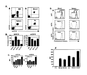
Figs. 1A-1E illustrate CD19 expression by hCD19TG mouse lines. Fig. 1A shows
human and mouse CD19 expression by B cells from hCD19TG (TG-1+/-) mice. Fig.
1B
shows the relative mean densities of human and mouse CD19 expression by CD19 +
blood B
cells from hCD19TG mice. Fig. 1C shows the relative densities of hCD19 and
mCD19
expression by CD19+ B cells from TG-1+/- mouse tissues. Fig. 1D shows CD19
antibody
binding density on mouse blood and spleen B220+ B cells from TG-1' - mice.
Fig. 1E
shows anti-CD19 antibody binding to hCD19 cDNA-transfected 300.19 cells.
Figs. 2A-2D show blood, spleen, and lymph node B cell depletion in hCD19TG
mice. Fig. 2A demonstrates representative B cell depletion from blood, spleen,
and lymph
node 7 days following CD19 or isotype-matched control (CTL) antibody treatment
of
TG-1+/- mice. Fig. 2B shows a time course of circulating B cell depletion by
anti-CD19
antibodies. Fig. 2C and Fig. 2D show spleen and lymph node B cell numbers
(SEM),
respectively, after treatment of TG-1+/- mice with CD (filled bars) or control
(open bars)
antibody at the indicated doses.
Figs. 3A-3F depict bone marrow B cell depletion following anti-CD19 antibody
treatment. Fig. 3A shows representative hCD19 and mCD19 expression by TG-1+1"
bone
marrow B cell subpopulations assessed by four-color immunofluorescence
staining with
flow cytometry analysis. Fig. 3B shows depletion of hCD19 cells in the bone
marrow of
hCD19TG mice seven days following FMC63 or isotype-matched control antibody
(250
1,1g) treatment assessed by two-color immunofluorescence staining with flow
cytometry
analysis. Fig. 3C shows representative B220+ B cell depletion in the bone
marrow seven
days following CD19 or isotype-matched control antibody (250 us) treatment of
TG-1+/-
mice. Fig. 3D shows representative B cell subset depletion seven days
following FMC63 or
isotype-matched control antibody (250 jig) treatment of TG-1+/- mice as
assessed by three-
color immunofluorescence staining. IgM-B2201 pro-/pre-B cells were further
subdivided
based on CD43 expression (lower panels). Fig. 3E shows representative
depletion of
CD25+B2201 pre-B cells seven days following FMC63 or isotype-matched control
antibody
(250 [tg) treatment of hCD19TG mouse lines as assessed by two-color
immunofluorescence
staining. Fig. 3F shows bar graphs indicating numbers ( SEM) of pro-B, pre-B,
immature,
and mature B cells within bilateral femurs seven days following FMC63 (closed
bars) or
control (open bars) antibody treatment of ?_-3 littermate pairs.
CA 02597924 2007-08-14
WO 2006/089133 PCT/US2006/005676
Figs. 4A-4C demonstrate that peritoneal cavity B cells are sensitive to anti-
CD19
antibody treatment. Fig. 4A shows human and mouse CD19 expression by
peritoneal cavity
= CD5+B220+ B la and CD513220hi B2 (conventional) B cells. Fig. 4B shows
depletion of
peritoneal cavity B220+ cells from TG-1+/- mice treated with CD19 (HB12a,
HB12b, and
FMC63 at 250 pz; B4 and HD237 at 50 Rg) antibodies or control antibody (250
ig). Fig.
4C shows representative depletion of CD5+B220+ Bla and CD5-13220hi B2 B cells
seven
days following anti-CD19 or control antibody treatment of hCD19TG mice.
Fig. 5A depicts the nucleotide (SEQ ID NO:1) and predicted amino acid (SEQ ID
NO:2) sequences for heavy chain VH-D-JH junctional sequences of the HB12a anti-
CD19
antibody. Fig. 5B depicts the nucleotide (SEQ ID NO:3) and predicted amino
acid (SEQ ID
NO:4) sequences for heavy chain VH-D-JH junctional sequences of the HB12b anti-
CD19
antibody.
Fig. 6A depicts the nucleotide (SEQ ID NO:15) and predicted amino acid (SEQ ID
NO:16) sequences for light chain sequences of the HB12a anti-CD19 antibody.
Fig. 6B
depicts the nucleotide (SEQ ID NO:17) and predicted amino acid (SEQ ID NO:18)
sequences for light chain sequences of the HB12b anti-CD19 antibody.
Figs. 7A-7B depict the amino acid sequence alignment of published mouse anti-
(human) CD19 antibodies. Fig. 7A shows a sequence alignment for heavy chain VH-
D-JH
junctional sequences including a consensus sequence (SEQ ID NO:5), HB12a (SEQ
ID
NO:2), 4G7 (SEQ ID NO:6), HB12b (SEQ ID NO:4), HD37 (SEQ ID NO:7), B43 (SEQ ID
NO:8), and FMC63 (SEQ ID NO:9). Fig. 7B shows light chain Vi amino acid
sequence
analysis of anti-CD19 antibodies. Consensus sequence (SEQ ID NO:10), HB12a
(SEQ ID
NO:16), HB12b (SEQ ID NO:18), HD37 (SEQ ID NO:11), B43 (SEQ ID NO:12), FMC63
(SEQ ID NO:13), and 4G7 (SEQ ID NO:14) are aligned.
Figs. 8A-8C demonstrate that CD19 density influences the efficiency of B cell
depletion by anti-CD19 antibodies in vivo. Representative blood and spleen B
cell
depletion in hCD19TG mice are shown following HB12b (Fig. 8A) or FMC63 (Fig.
8B)
antibody treatment (seven days, 250 tug/mouse). Fig. 8C shows the relative
anti-CD19 and
anti-CD20 antibody-binding densities on blood B220+ B cells from TG-1+/- mice.
Fig. 8D
shows the relative anti-CD19 and anti-CD20 antibody-binding densities on
spleen B220+ B
cells from TG-144- mice.
Figs. 9A-9D demonstrate B cell depletion following anti-CD19 antibody
treatment
is FcRy- and monocyte-dependent. Fig. 9A Representative blood and spleen B
cell
16
CA 02597924 2012-11-16
depletion 7 days after CD19 or isotype-control antibody treatment of hCD19 TG-
1' - FcRy+/-
+/-
or TG-1 FeRy littermates. Fig. 9B Blood and tissue B cell depletion seven days
after
antibody treatment of FcR74- littennates on day zero. Fig. 9C Representative B
cell
numbers in monocyte-depleted hCD19TG-1+/- mice. Fig. 9D Blood and tissue B
cell
depletion seven days after antibody treatment.
Figs. 10A-10D demonstrate duration and dose response of B cell depletion
following anti-CD19 antibody treatment. Fig. 10A shows numbers of blood B220+
B cells
and Thy-1+ T cells following FMC63 or isotype-control antibody treatment of TG-
1' - mice
on day zero. Figs. 1013-C show representative tissue B cell depletion in mice
shown in Fig.
10A at 11, 16, and 30 weeks following antibody treatment. Fig. 10D shows anti-
CD19
antibody dose responses for blood, bone marrow, and spleen B cell depletion.
Figs. 11A-11C demonstrate that CD19 is not internalized following antibody
binding in vivo. Cell surface CD19 expression and B cell clearance in TG-1+/-
mice treated
with HB12a (Fig. 11A), HB12b (Fig. 11B), FMC63 (Fig. 11C) or isotype-matched
control
antibody (250 ,g) in vivo.
Figs. 12A-12C demonstrate CD19 saturation following anti-CD19 antibody binding
in vivo. Fig. 12A shows B cell clearance in TG-1 +/- mice treated with FMC63
or isotype-
matched control antibody (250 fig) in vivo. Fig. 12B shows FMC63 antibody
treatment
(250 ug) saturates antibody-binding sites on hCD19 within 1 hour of
administration. Fig.
12C shows HB12b anti-CD19 antibody treatment (250 ug) saturates antibody-
binding sites
on hCD19 within 1 hour of administration as assessed in Fig. 12B.
Figs. 13A-13B demonstrate anti-CD19 antibody treatment reduces serum
immunoglobulin and autoantibody levels in TG-141- mice. Fig. 13A depicts serum
immunoglobulin levels and Fig. 13B anti-dsDNA, anti-ssDNA and anti-histone
autoantibody levels after anti-CD19 antibody treatment.
Figs. 14A-14B demonstrate anti-CD19 antibody treatment blocks humoral immune
responses in TG-1+1- mice. Antibody-treated mice were immunized with Fig. 14A
TNP-
LPS, Fig. 14B DNP-Ficoll and Figs. 14C-14D DNP-KLH. Littermates were treated
with
FMC63 (closed circles) or control (open circles) antibody (250 lig) either (A-
C) 7 days
before or (D) 14 days after priniary immunizations on day O.
Fig. 15 demonstrates that simultaneous anti-CD19 and anti-CD20 antibody
treatments are additive.
*Trademark
17
CA 02597924 2007-08-14
WO 2006/089133 PCT/US2006/005676
Fig. 16 demonstrates that subcutaneous (s.c.), intraperitoneal (i.p.) and i.v.
administration of anti-CD19 antibody effectively depletes circulating and
tissue B cells in
vivo.
Fig. 17A-17B. Anti-CD19 antibody treatment prevents hCD19+ lymphoma growth
in vivo (Fig. 17A) and increases survival rate (Fig. 17B).
5. DETAILED DESCRIPTION OF THE INVENTION
The invention relates to imrnunotherapeutic compositions and methods for the
treatment of B cell diseases and disorders in human subjects, such as, but not
limited to, B
cell malignancies, using therapeutic antibodies that bind to the CD19 antigen
and preferably
mediate human ADCC. The present invention relates to pharmaceutical
compositions
comprising human, humanized, or chimeric anti-CD19 antibodies of the IgG1 or
IgG3
human isotype. The present invention also relates to pharmaceutical
compositions
comprising human or humanized anti-CD19 antibodies of the IgG2 or IgG4 human
isotype
that preferably mediate human ADCC. In certain embodiments, the present
invention also
relates to pharmaceutical compositions comprising monoclonal human, humanized,
or
chimerized anti-CD19 antibodies that can be produced by means known in the
art.
Therapeutic formulations and regimens are described for treating human
subjects
diagnosed with B cell malignancies that derive from B cells and their
precursors, including
but not limited to, acute lymphoblastic leukemias (ALL), Hodgkin's lymphomas,
non-
Hodgkin's lymphomas, B cell chronic lymphocytic leukemias (CLL), multiple
myeloma,
follicular lymphoma, mantle cell lymphoma, pro-lymphocytic leukemias, hairy
cell
leukemias, common acute lymphocytic leukemias and some Null-acute
lymphoblastic
leukemias.
5.1. GENERATION OF ANTI-CD19 ANTIBODIES
5.1.1. POLYCLONAL ANTI-CD19 ANTIBODIES
Polyclonal antibodies are preferably raised in animals by multiple
subcutaneous (sc)
or intraperitoneal (i.p.) injections of the relevant antigen and an adjuvant.
It may be useful
to conjugate the relevant antigen to a protein that is immunogenic in the
species to be
immunized, e.g., keyhole limpet hemocyanin, serum albumin, bovine
thyroglobulin, or
18
CA 02597924 2007-08-14
WO 2006/089133 PCT/US2006/005676
soybean trypsin inhibitor using a bifunctional or derivatizing agent, for
example,
maleimidobertzoyl sulfosuccinimide ester (conjugation through cysteine
residues), N-
hydroxysuccinimide (through lysine residues), glutaraldehyde, succunic
anhydride, S0C12.
Animals are immunized against the antigen, immunogenic conjugates, or
derivatives
by combining, e.g., 100 iug or 5 fig of the protein or conjugate (for rabbits
or mice,
respectively) with 3 volumes of Freund's complete adjuvant and injecting the
solution
intradermally at multiple sites. One month later the animals are boosted with
1/5 to 1/10 the
original amount of peptide or conjugate in Freund's incomplete adjuvant by
subcutaneous
injection at multiple sites. Seven to 14 days later the animals are bled and
the serum is
assayed for antibody titer. Animals are boosted until the titer plateaus.
Preferably, the
animal is boosted with the conjugate of the same antigen, but conjugated to a
different
protein and/or through a different cross-linking reagent. Conjugates also can
be made in
recombinant cell culture as protein fusions. Also, aggregating agents such as
alum are
suitably used to enhance the immune response.
5.1.2. MONOCLONAL ANTI-CD19 ANTIBODIES
The monoclonal anti-CD19 antibodies of the invention exhibit binding
specificity to
human CD19 antigen and can preferably mediate human ADCC. These antibodies can
be
generated using a wide variety of techniques known in the art including the
use of
hybridoma, recombinant, and phage display technologies, or a combination
thereof.
Antibodies are highly specific, being directed against a single antigenic
site. Furthermore,
in contrast to conventional (polyclonal) antibody preparations which typically
include
different antibodies directed against different determinants (epitopes), each
monoclonal
antibody is directed against a single determinant on the human CD19 antigen.
For example,
the monoclonal antibodies to be used in accordance with the present invention
may be made
by the hybridoma method first described by Kohler et aL, Nature, 256:495
(1975), which
can be used to generate murine antibodies (or antibodies derived from other
nonhuman
mammals, e.g., rat, goat, sheep, cows, camels, etc.), or human antibodies
derived from
transgenic animals (see, U.S. Patent Nos. 6,075,181, 6,114,598, 6,150,584, and
6,657,103).
Alternatively, the monoclonal antibodies can be made by recombinant DNA
methods (see,
e.g., U.S. Patent No. 4,816,567) and include chimeric and humanized
antibodies. The
"monoclonal antibodies" may also be isolated from phage antibody libraries
using the
19
CA 02597924 2012-11-16
techniques described in Clackson et al., Nature, 352:624-628 (1991) and Marks
et al., J.
MoL Biol., 222:581-597 (1991), for example.
An engineered anti-CD19 antibody can be produced by any means known in the
art,
including, but not limited to, those techniques described below and
improvements to those
techniques. Large-scale high-yield production typically involves culturing a
host cell that
produces the engineered anti-CD19 antibody and recovering the anti-CD19
antibody from
the host cell culture.
5.1.3. HYBRIDOMA TECHNIQUE
Monoclonal antibodies can be produced using hybridoma techniques including
those
known in the art and taught, for example, in Harlow et al., Antibodies: A
Laboratory
Manual, (Cold Spring Harbor Laboratory Press, 2nd ed. 1988); Ham_merling et
al., in
Monoclonal Antibodies and T Cell Hybridomas, 563-68,1 (Elsevier, N.Y., 1981),
For example, in the hybridoma method, a mouse or other appropriate
host animal, such as a hamster or macaque monkey, is immunized to elicit
lymphocytes that produce or are capable of producing antibodies that
will specifically bind to the protein used for immunization. Alternatively,
lymphocytes may
be immunized ill vitro. Lymphocytes then are fused with myeloma cells using a
suitable
fusing agent, such as polyethylene glycol, to form a hybridoma cell (Goding,
Monoclonal
Antibodies: Principles and Practice, pp. 59-103 (Academic Press, 1986)).
The hybridoma cells thus prepared are seeded and grown in a suitable culture
medium that preferably contains one or more substances that inhibit the growth
or survival
of the unfused, parental myeloma cells. For example, if the parental myeloma
cells lack the
enzyme hypoxanthine guanine phosphoribosyl transferase (HGPRT or HPRT), the
culture
medium for the hybridomas typically will include hypoxanthine, aminopterin,
and
thymidine (HAT medium), which substances prevent the growth of HGPRT-deficient
cells.
Preferred myeloma cells are those that fuse efficiently, support stable high-
level
production of antibody by the selected antibody-producing cells, and are
sensitive to a
medium such as HAT medium. Among these, preferred myeloma cell lines are
murine
myeloma lines, such as those derived from MOPC-21 and MPC-11 mouse tumors
available
from the Salk Institute Cell Distribution Center, San Diego, CA, USA, and SP-2
or X63-
Ag8.653 cells available from the American Type Culture Collection, Rockville,
MD, USA.
Human myeloma and mouse-human heteromyeloma cell lines also have been
described for
CA 02597924 2007-08-14
WO 2006/089133 PCT/US2006/005676
the production of human monoclonal antibodies (Kozbor, J ImmunoL, 133:3001
(1984);
Brodeur et aL, Monoclonal Antibody Production Techniques and Applications, pp.
51-63
(Marcel Dekker, Inc., New York, 1987)).
Culture medium in which hybridoma cells are growing is assayed for production
of
monoclonal antibodies directed against the human CD19 antigen. Preferably, the
binding
specificity of monoclonal antibodies produced by hybridoma cells is determined
by
irnmunoprecipitation or by an in vitro binding assay, such as radioimmunoassay
(RIA) or
enzyme-linked immunoabsorbent assay (ELISA).
After hybridoma cells are identified that produce antibodies of the desired
specificity, affinity, and/or activity, the clones may be subcloned by
limiting dilution
procedures and grown by standard methods (Goding, Monoclonal Antibodies:
Principles
and Practice, pp. 59-103 (Academic Press, 1986)). Suitable culture media for
this purpose
include, for example, D-MEM or RPMI 1640 medium. In addition, the hybridoma
cells
may be grown in vivo as ascites tumors in an animal.
The monoclonal antibodies secreted by the subclones are suitably separated
from the
culture medium, ascites fluid, or serum by conventional immunoglobulin
purification
procedures such as, for example, protein A-Sepharose, hydroxylapatite
chromatography, gel
electrophoresis, dialysis, or affinity chromatography.
5.1.4. RECOMBINANT DNA TECHNIQUES
DNA encoding the anti-CD19 antibodies of the invention is readily isolated and
sequenced using conventional procedures (e.g., by using oligonucleotide probes
that are
capable of binding specifically to genes encoding the heavy and light chains
of the anti-
CD19 antibodies). The hybridoma cells serve as a preferred source of such DNA.
Once
isolated, the DNA may be placed into expression vectors, which are then
transfected into
host cells such as E. coli cells, simian COS cells, Chinese hamster ovary
(CHO) cells, or
myeloma cells that do not otherwise produce inununoglobulin protein, to obtain
the
synthesis of anti-CD19 antibodies in the recombinant host cells.
In phage display methods, functional antibody domains are displayed on the
surface
of phage particles which carry the polynucleotide sequences encoding them. In
particular,
DNA sequences encoding VH and VL domains are amplified from animal cDNA
libraries
(e.g., human or murine cDNA libraries of affected tissues). The DNA encoding
the VH and
VL domains are recombined together with an scFv linker by PCR and cloned into
a
21
CA 02597924 2012-11-16
phagemid vector. The vector is electroporated in E. coli and the E. coli is
infected with
helper phage. Phage used in these methods are typically filamentous phage
including fd and
M13 and the VH and VL domains are usually recombinantly fused to either the
phage gene
III or gene VIII. Phage expressing an antigen-binding domain that binds to a
particular
antigen can be selected or identified with antigen, e.g., using labeled
antigen or antigen
bound or captured to a solid surface or bead. Examples of phage display
methods that can
be used to make the antibodies of the present invention include those
disclosed in Brinkman
et al., 1995, J. Immunol. Methods, 182:41-50; Arnes et al., 1995, J Immunol.
Methods,
184:177-186; Kettleborough et al., 1994, Eur. J. Invnunol., 24:952-958; Persic
et al., 1997,
Gene, 187:9-18; Burton et al., 1994, Advances in Immunology, 57:191-280;
International
Application No. PCT/GB91/01 134; International Publication Nos. WO 90/02809,
WO
91/10737, WO 92/01047, WO 92/18619, WO 93/11236, WO 95/15982, WO 95/20401, and
W097/13844; and U.S. Patent Nos. 5,698,426, 5,223,409, 5,403,484, 5,580,717,
5,427,908,
5,750,753, 5,821,047, 5,571,698, 5,427,908, 5,516,637, 5,780,225, 5,658,727,
5,733,743,
and 5,969,108.
As described in the above references, after phage selection, the antibody
coding
regions from the phage can be isolated and used to generate whole antibodies,
including
hurnan antibodies, or any other desired antigen-binding fragment, and
expressed in any
desired host, including mammalian cells, insect cells, plant cells, yeast, and
bacteria, e.g., as
described below. Techniques to recombinantly produce Fab, Fab' and F(ab')2
fragments can
also be employed using methods known in the art such as those disclosed in PCT
Publication No. WO 92/22324; Mullinax et al., 1992, BioTechniques, 12(6):864-
869; Sawai
et al., 1995, A./RJ, 34:26-34; and Better et al., 1988, Science, 240:1041-
1043.
In a further embodiment, antibodies may be isolated from antibody phage
libraries
generated using the techniques described in McCafferty et al., Nature, 348:552-
554 (1990).
Clackson et al., Nature, 352:624-628 (1991). Marks et al., J. Mol. Biol.,
222:581-597
(1991) describe the isolation of murine and human antibodies, respectively,
using phage
libraries. Chain shuffling can be used in the production of high affinity (nM
range) human
antibodies (Marks et al., Bio/Technology, 10:779-783 (1992)), as well as
combinatorial
infection and iiz vivo recombination as a strategy for constructing very large
phage libraries
(Waterhouse et al.,Nuc. Acids. Res., 21:2265-2266 (1993)). Thus, these
techniques are
22
CA 02597924 2007-08-14
WO 2006/089133 PCT/US2006/005676
viable alternatives to traditional monoclonal antibody hybridoma techniques
for isolation of
anti-CD19 antibodies.
To generate whole antibodies, PCR primers including VH or VL nucleotide
sequences, a restriction site, and a flanking sequence to protect the
restriction site can be
used to amplify the VH or VL sequences in scFv clones. Utilizing cloning
techniques known
to those of skill in the art, the PCR amplified VH domains can be cloned into
vectors
expressing a VH constant region, e.g., the human gamma 4 constant region, and
the PCR
amplified VL domains can be cloned into vectors expressing a VI constant
region, e.g.,
human kappa or lamba constant regions. Preferably, the vectors for expressing
the VH or VL
domains comprise an EF-la promoter, a secretion signal, a cloning site for the
variable
domain, constant domains, and a selection marker such as neomycin. The VH and
VL
domains may also be cloned into one vector expressing the necessary constant
regions. The
heavy chain conversion vectors and light chain conversion vectors are then co-
transfected
into cell lines to generate stable or transient cell lines that express full-
length antibodies,
e.g., IgG, using techniques known to those of skill in the art.
The DNA also may be modified, for example, by substituting the coding sequence
for human heavy and light chain constant domains in place of the homologous
murine
sequences (U.S. Patent No. 4,816,567; Morrison et al., Proc. Natl. Acad. Sci.
USA, 81:6851
(1984)), or by covalently joining to the immunoglobulin coding sequence all or
part of the
coding sequence for a non-immunoglobulin polypeptide.
5.1.5. CHIMERIC ANTIBODIES
The anti-CD19 antibodies herein specifically include chimeric antibodies
(immunoglobulins) in which a portion of the heavy and/or light chain is
identical with or
homologous to corresponding sequences in antibodies derived from a particular
species or
belonging to a particular antibody class or subclass, while another portion of
the chain(s) is
identical with or homologous to corresponding sequences in antibodies derived
from
another species or belonging to another antibody class or subclass, as well as
fragments of
such antibodies, so long as they exhibit the desired biological activity
((J.S. Patent No.
4,816,567; Morrison et al., Proc. Natl. Acad. Sci. USA, 81:6851-6855 (1984)).
Chimeric
antibodies of interest herein include "primatized" antibodies comprising
variable domain
antigen-binding sequences derived from a nonhuman primate (e.g., Old World
Monkey,
23
CA 02597924 2012-11-16
such as baboon, rhesus or cynomolgus monkey) and human constant region
sequences (U.S.
Patent No. 5,693,780).
5.1.6. HUMANIZED ANTIBODIES
A humanized antibody can be produced using a variety of techniques known in
the
art, including but not limited to, CDR-grafting (see, e.g., European Patent
No. EP 239,400;
International Publication No. WO 91/09967; and U.S. Patent Nos. 5,225,539,
5,530,101,
and 5,585,089), veneering or resurfacing (see, e.g., European Patent Nos. EP
592,106 and
EP 519,596; Padlan, 1991, Molecular Immunology 28(4/5):489-498; Studnicka et
al.,
1994, Protein Engineering, 7(6):805-814; and Roguska et al., 1994, PNAS,
91:969-973),
chain shuffling (see, e.g., U.S. Patent No. 5,565,332), and techniques
disclosed in, e.g.,
U.S. Patent No. 6,407,213, U.S. Patent No. 5,766,886, International
Publication No. WO
9317105, Tan et al., J. ImmunoL, 169:1119-25 (2002), Caldas et al., Protein
Eng.,
13(5):353-60 (2000), Morea et al., Methods, 20(3):267-79 (2000), Baca et al.,
J. Biol.
Chem., 272(16):10678-84 (1997), Roguska et aL, Protein Eng., 9(10):895-904
(1996),
Couto et al., Cancer Res., 55 (23 Supp):5973s-5977s (1995), Couto et al.,
Cancer Res.,
55(8):1717-22 (1995), Sandhu JS, Gene, 150(2):409-10 (1994), and Pedersen et
al., J.
MoL Biol., 235(3):959-73 (1994). Often, framework residues in the framework
regions
will be substituted with the corresponding residue from the CDR donor antibody
to alter,
preferably improve, antigen binding. These framework substitutions are
identified by
methods well-known in the art, e.g., by modeling of the interactions of the
CDR and
framework residues to identify framework residues important for antigen
binding and
sequence comparison to identify unusual framework residues at particular
positions.
(See, e.g., Queen et al., U.S. Patent No. 5,585,089; and Riechmann et al.,
1988, Nature,
332:323).
A humanized anti-CD19 antibody has one or more amino acid residues introduced
into it from a source which is nonhuman. These nonhtunan amino acid residues
are often
referred to as "import" residues, which are typically taken from an "import"
variable
domain. Thus, humanized antibodies comprise one or more CDRs from nonhuman
immunoglobulin molecules and framework regions from human. Humanization of
24
CA 02597924 2012-11-16
antibodies is well-known in the art and can essentially be performed following
the method
of Winter and co-workers (Jones et al., Nature, 321:522-525 (1986); Riechmann
et al.,
Nature, 332:323-327 (1988); Verhoeyen et al., Science, 239:1534-1536 (1988)),
by
substituting rodent CDRs or CDR sequences for the corresponding sequences of a
human
antibody, i.e., CDR-grafting (EP 239,400; PCT Publication No. WO 91/09967; and
U.S.
Patent Nos. 4,816,567; 6,331,415; 5,225,539; 5,530,101; 5,585,089; 6,548,640).
In such humanized chimeric antibodies, substantially less than an intact human
variable
domain has been substituted by the corresponding sequence from a nonhuman
species.
In practice, humanized antibodies are typically human antibodies in which some
CDR
residues and possibly some FR residues are substituted by residues from
analogous sites
in rodent antibodies. Humanization of anti-CD19 antibodies can also be
achieved by
veneering or resurfacing (EP 592,106; EP 519,596; Padlan, 1991, Molecular
Immunology
28(4/5):489-498; Studnicka et al., Protein Engineering, 7(6):805-814 (1994);
and
Roguska et al., PNAS, 91:969-973 (1994)) or chain shuffling (U.S. Patent No.
5,565,332).
The choice of human variable domains, both light and heavy, to be used in
making
the humanized antibodies is to reduce antigenicity. According to the so-called
"best-fa"
method, the sequence of the variable domain of a rodent antibody is screened
against the
entire library of known human variable-domain sequences. The human sequence
which is
closest to that of the rodent is then accepted as the human framework (FR) for
the
humanized antibody (Sims et al., J. IrninunoL, 151:2296 (1993); Chothia et
al., J. MoL
Biol., 196:901 (1987), the contents of which are incorporated herein by
reference herein in
their entirety). Another method uses a particular framework derived from the
consensus
sequence of all human antibodies of a particular subgroup of light or heavy
chains. The
same framework may be used for several different humanized anti-CD19
antibodies (Carter
et aL, PMC. Natl. Acad. Sci. USA, 89:4285 (1992); Presta et al., J. Immunol.,
151:2623
(1993).
Anti-CD19 antibodies can be humanized with retention of high affinity for CD19
and other favorable biological properties. According to one aspect of the
invention,
humanized antibodies are prepared by a process of analysis of the parental
sequences and
various conceptual humanized products using three-dimensional models of the
parental and
humanized sequences. Three-dimensional immunoglobulin models are commonly
available
CA 02597924 2012-11-16
and are familiar to those skilled in the art. Computer programs are available
which illustrate
and display probable three-dimensional conformational structures of selected
candidate
immunoglobulin sequences. Inspection of these displays permits analysis of the
likely role
of the residues in the functioning of the candidate immunoglobulin sequence,
i.e., the
analysis of residues that influence the ability of the candidate
immunoglobulin to bind
CD19. In this way, FR residues can be selected and combined from the recipient
and
import sequences so that the desired antibody characteristic, such as
increased affinity for
CD19, is achieved. In general, the CDR residues are directly and most
substantially
involved in influencing antigen binding.
A "humanized" antibody retains a similar antigenic specificity as the original
antibody, i.e., in the present invention, the ability to bind human CD19
antigen. However,
using certain methods of humanization, the affinity and/or specificity of
binding of the
antibody for human CD19 antigen may be increased using methods of "directed
evolution,"
as described by Wu et al., J. MoL Biol., 294:151 (1999).
5.1.7. HUMAN ANTIBODIES
For in vivo use of antibodies in humans, it may be preferable to use human
antibodies. Completely human antibodies are particularly desirable for
therapeutic
treatment of human subjects. Human antibodies can be made by a variety of
methods
known in the art including phage display methods described above using
antibody libraries
derived from human immunoglobulin sequences, including improvements to these
techniques. See, also, U.S. Patent Nos. 4,444,887 and 4,716,111; and PCT
publications
WO 98/46645, WO 98/50433, WO 98/24893, W098/16654, WO 96/34096, WO 96/33735,
and WO 91/10741. A human antibody can also be an antibody wherein the heavy
and
light chains are encoded by a nucleotide sequence derived from one or more
sources of
human DNA.
Human anti-CD19 antibodies can also be produced using transgenic mice which
are
incapable of expressing functional endogenous immunoglobulins, but which can
express
human immunoglobulin genes. For example, the human heavy and light chain
immunoglobulin gene complexes may be introduced randomly or by homologous
recombination into mouse embryonic stein cells. Altematively, the human
variable region,
constant region, and diversity region may be introduced into mouse embryonic
stem cells in
26
CA 02597924 2012-11-16
addition to the human heavy and light chain genes. The mouse heavy and light
chain
immunoglobulin genes may be rendered non-functional separately or
simultaneously with
the introduction of human immunoglobulin loci by homologous recombination. For
example, it has been described that the homozygous deletion of the antibody
heavy chain
joining region (JH) gene in chimeric and germ-line mutant mice results in
complete
inhibition of endogenous antibody production. The modified embryonic stem
cells are
expanded and microinjected into blastocysts to produce chimeric mice. The
chimeric mice
are then bred to produce homozygous offspring which express human antibodies.
The
transgenic mice are immunized in the normal fashion with a selected antigen,
e.g., all or a
portion of a polypeptide of the invention. Anti-CD19 antibodies directed
against the human
CD19 antigen can be obtained from the immunized, transgenic mice using
conventional
hybridoma technology. The human immunoglobulin transgenes harbored by the
transgenic
mice rearrange during B cell differentiation, and subsequently undergo class
switching and
somatic mutation. Thus, using such a technique, it is possible to produce
therapeutically
useful IgG, IgA, IgM and IgE antibodies, including, but not limited to, IgG1
(gamma 1) and
IgG3. For an overview of this technology for producing human antibodies, see,
Lonberg
and Huszar (Int. Rev. Innnunol., 13:65-93 (1995)). For a detailed discussion
of this
technology for producing human antibodies and human monoclonal antibodies and
protocols for producing such antibodies, see, e.g., PCT Publication Nos. WO
98/24893, WO
96/34096, and WO 96/33735; and U.S. Patent Nos. 5,413,923; 5,625,126;
5,633,425;
5,569,825; 5,661,016; 5,545,806; 5,814,318; and 5,939,598.
In addition, companies such as Abgenix, Inc.
(Freemont, CA) and Genpharm (San Jose, CA) can be engaged to provide human
antibodies
directed against a selected antigen using technology similar to that described
above. For a
specific discussion of transfer of a human genn-line immunoglobulin gene array
in germ-
line mutant mice that will result in the production of human antibodies upon
antigen
challenge see, e.g.. Jakobovits et al., P7'OC. Natl. Acad. Sci. USA. 90:2551
(1993);
Jakobovits et al., Nature, 362:255-258 (1993); Bruggermann et al., Year in
Immunol., 7:33
(1993); and Duchosal et al., Nature, 355:258 (1992).
Human antibodies can also be derived from phage-display libraries (Hoogenboom
et
J Mol. Biol., 227:381 (1991); Marks et al., J AloL Biol., 222:581-597 (1991);
Vaughan
et al., Nature Biotech., 14:309 (1996)). nage display technology (McCafferty
et al.,
Nature, 348:552-553 (1990)) can be used to produce human antibodies and
antibody
27
CA 02597924 2012-11-16
fragments in vitro, from immunoglobulin variable (V) domain gene repertoires
from
unimrnunized donors. According to this technique, antibody V domain genes are
cloned in-
frame into either a major or minor coat protein gene of a filamentous
bacteriophage, such as
M13 or fd, and displayed as functional antibody fragments on the surface of
the phage
particle. Because the filamentous particle contains a single-stranded DNA copy
of the
phage genome, selections based on the functional properties of the antibody
also result in
selection of the gene encoding the antibody exhibiting those properties. Thus,
the phage
mimics some of the properties of the B cell. Phage display can be performed in
a variety of
formats; for their review see, e.g., Johnson, Kevin S. and Chiswell, David J.,
Current
Opinion in Structural Biology 3:564-571 (1993). Several sources of V-gene
segments can
be used for phage display. Clackson et al., Nature, 352:624-628 (1991)
isolated a diverse
array of anti-oxazolone antibodies from a small random combinatorial library
of V genes
derived from the spleens of unimmimized mice. A repertoire of V genes from
unimmunized
human donors can be constructed and antibodies to a diverse array of antigens
(including
self-antigens) can be isolated essentially following the techniques described
by Marks et al.,
Mol. Biol., 222:581-597 (1991), or Griffith et al., EMBO J., 12:725-734
(1993). See,
also, U.S. Patent Nos. 5,565,332 and 5,573,905.
Human antibodies may also be generated by in vitro activated B cells (see,
U.S.
Patents 5,567,610 and 5,229,275). Human antibodies may also be generated in
vitro
using hybridoma techniques such as, but not limited to, that described by
Roder et al.
(Methods En.zymol., 121:140-167 (1986)).
5.1.8. ALTERED/MUTANT ANTIBODIES
The anti-CD19 antibodies of the compositions and methods of the invention can
be
mutant antibodies. As used herein, "antibody mutant" or "altered antibody"
refers to an
amino acid sequence variant of an anti-CD19 antibody wherein one or more of
the amino
acid residues of an anti-CD19 antibody have been modified. The modifications
to the
amino acid sequence of the anti-CD19 antibody, include modifications to the
sequence to
improve affinity or avidity of the antibody for its antigen, and/or
modifications to the Fc
portion of the antibody to improve effector function. The modifications may be
made to
any known anti-CD19 antibodies or anti-CD19 antibodies identified as described
herein.
28
CA 02597924 2007-08-14
WO 2006/089133 PCT/US2006/005676
Such altered antibodies necessarily have less than 100% sequence identity or
similarity with
a known anti-CD19 antibody. In a preferred embodiment, the altered antibody
will have an
amino acid sequence having at least 25%, 35%, 45%, 55%, 65%, or 75% amino acid
sequence identity or similarity with the amino acid sequence of either the
heavy or light
chain variable domain of an anti-CD19 antibody, more preferably at least 80%,
more
preferably at least 85%, more preferably at least 90%, and most preferably at
least 95%. In
a preferred embodiment, the altered antibody will have an amino acid sequence
having at
least 25%, 35%, 45%, 55%, 65%, or 75% amino acid sequence identity or
similarity with
the amino acid sequence of the heavy chain CDR1, CDR2, or CDR3 of an anti-CD19
antibody, more preferably at least 80%, more preferably at least 85%, more
preferably at
least 90%, and most preferably at least 95%. In a preferred embodiment, the
altered
antibody will maintain human CD19 binding capability. In certain embodiments,
the anti-
CD19 antibody of the invention comprises a heavy chain that is about 10%, 15%,
20%,
25%, 30%, 35%, 40%, 45%, 50%, 55%, 60%, 65%, 70%, 75%, 80%, 85%, 90%, 95% or
more identical to an amino acid sequence of SEQ ID NO:2 (Fig. 5A)
corresponding to the
heavy chain of HB12a. In certain embodiments, the anti-CD19 antibody of the
invention
comprises a heavy chain that is about 10%, 15%, 20%, 25%, 30%, 35%, 40%, 45%,
50%,
55%, 60%, 65%, 70%, 75%, 80%, 85%, 90%, 95% or more identical to an amino acid
sequence of SEQ ID NO:4 (Fig. 5B) corresponding to the heavy chain of HB12b.
In certain
embodiments, the anti-CD19 antibody of the invention comprises a heavy chain
that is
about 10%, 15%, 20%, 25%, 30%, 35%, 40%, 45%, 50%, 55%, 60%, 65%, 70%, 75%,
80%, 85%, 90%, 95% or more identical to an amino acid sequence of SEQ ID NO:16
(Fig.
6A) corresponding to the light chain of HB12a. In certain embodiments, the
anti-CD19
antibody of the invention comprises a light chain that is about 10%, 15%, 20%,
25%, 30%,
35%, 40%, 45%, 50%, 55%, 60%, 65%, 70%, 75%, 80%, 85%, 90%, 95% or more
identical
to an amino acid sequence of SEQ ID NO:18 (Fig. 6B) corresponding to the light
chain of
HB12b. Hybridomas producing HB12a and HB12b anti-CD19 antibodies have been
deposited under ATCC deposit nos. PTA-6580 and PTA-6581.
Identity or similarity with respect to this sequence is defined herein as the
percentage of amino acid residues in the candidate sequence that are identical
(i.e., same
residue) or similar (i.e., amino acid residue from the same group based on
common side-
chain properties, see below) with anti-CD19 antibody residues, after aligning
the sequences
and introducing gaps, if necessary, to achieve the maximum percent sequence
identity.
29
CA 02597924 2012-11-16
None of N-terminal, C-teintinal, or internal extensions, deletions, or
insertions into the
antibody sequence outside of the variable domain shall be construed as
affecting sequence
identity or similarity.
"% identity," as known in the art, is a measure of the relationship between
two
polynucleotides or two polypeptides, as determined by comparing their
sequences. In
general, the two sequences to be compared are aligned to give a maximum
correlation
between the sequences. The alignment of the two sequences is examined and the
number of
positions giving an exact amino acid or nucleotide correspondence between the
two
sequences determined, divided by the total length of the alignment and
multiplied by 100 to
give a % identity figure. This % identity figure may be determined over the
whole length of
the sequences to be compared, which is particularly suitable for sequences of
the same or
very similar length and which are highly homologous, or over shorter defined
lengths,
which is more suitable for sequences of unequal length or which have a lower
level of
homology.
For example, sequences can be aligned with the software clustalw under Unix
which
generates a file with an ".aln" extension, this file can then be imported into
the Bioedit
program (Hall, T.A. 1999, BioEdit: a user-friendly biological sequence
alignment editor
and analysis program for Windows 95/98/NT. NucL Acids. Symp. Ser. 41:95-98)
which
opens the .aln file. In the Bioedit window, one can choose individual
sequences (two at a
time) and alignment them. This method allows for comparison of the entire
sequence.
Methods for comparing the identity of two or more sequences are well-known in
the
art. Thus for instance, programs are available in the Wisconsin Sequence
Analysis Package,
version 9.1 (Devereux J. et al., Nucleic Acids Res., 12:387-395, 1984,
available from
Genetics Computer Group, Madison, WI, USA). The determination of percent
identity
between two sequences can be accomplished using a mathematical algorithm. For
example,
the programs BESTFIT and GAP, may be used to determine the % identity between
two
polynucleotides and the % identity between two polypeptide sequences. BESTFIT
uses the
"local homology" algorithm of Smith and Waterman (Advances in Applied
Mathematics,
2:482-489, 1981) and finds the best single region of similarity between two
sequences.
BESTFIT is more suited to comparing two polynucleotide or two polypeptide
sequences
which are dissimilar in length, the program assuming that the shorter sequence
represents a
portion of the longer. In comparison, GAP aligns two sequences finding a
"maximum
similarity" according to the algorithm of Neddleman and Wunsch (J. Alol.
Biol.,
*Trademark
CA 02597924 2012-11-16
48:443-354, 1970). GAP is more suited to comparing sequences which are
approximately
the same length and an alignment is expected over the entire length.
Preferably the
parameters "Gap Weight " and "Length Weight" used in each program are 50 and 3
for
polynucleotides and 12 and 4 for polypeptides, respectively. Preferably %
identities and
similarities are determined when the two sequences being compared are
optimally aligned.
Other programs for determining identity and/or similarity between sequences
are
also known in the art, for instance the BLAST family of programs (Karlin &
Altschul, 1990,
Proc. Natl. Acad. Sci. USA, 87:2264-2268, modified as in Karlin & Altschul,
1993, Proc.
Natl. Acad. Sci. USA, 90:5873-5877, available from the National Center for
Biotechnology
Information (NCB), Bethesda, MD, USA and accessible through the home page of
the
NCBI). These programs exemplify a preferred, non-limiting
example of a mathematical algorithm utilized for the comparison of two
sequences. Such
an algorithm is incorporated into the NBLAST and XBLAST programs of Altschul
et al.,
1990, J. MoL Biol., 215:403-410. BLAST nucleotide searches can be performed
with the
NBLAST program, score = 100, wordlength = 12 to obtain nucleotide sequences
homologous to a nucleic acid molecule encoding all or a portion if an anti-
CD19 antibody
of the invention. BLAST protein searches can be performed with the XBLAST
program,
score = 50, wordlength = 3 to obtain amino acid sequences homologous to a
protein
molecule of the invention. To obtain gapped alignments for comparison
purposes, Gapped
BLAST can be utilized as described in Altschul et al., 1997, Nucleic Acids
Res., 25:3389-
3402. Alternatively, PSI-Blast can be used to perform an iterated search which
detects
distant relationships between molecules (Id.). When utilizing BLAST, Gapped
BLAST, and
PSI-Blast programs, the default parameters of the respective programs (e.g.,
)(BLAST and
NBLAST) can be used. Another preferred, non-limiting
example of a mathematical algorithm utilized for the comparison of sequences
is the
algorithm of Myers and Miller, 1988, CABIOS 4:11-17. Such an algorithm is
incorporated
into the ALIGN program (version 2.0) which is part of the GCG sequence
alignment
software package. When utilizing the ALIGN program for comparing amino acid
sequences, a PAM120 weight residue table, a gap length penalty of 12, and a
gap penalty of
4 can be used.
Another non-limiting example of a program for determining identity and/or
similarity between sequences known in the art is FASTA (Pearson W.R. and
Lipman D.J.,
Proc. Nat. Acad. Sci. USA, 85:2444-2448, 1988, available as part of the
Wisconsin
31
CA 02597924 2007-08-14
WO 2006/089133
PCT/US2006/005676
Sequence Analysis Package). Preferably the BLOSUM62 amino acid substitution
matrix
(Henikoff S. and Henikoff J.G., Proc. Nat. Acad. Sci. USA, 89:10915-10919,
1992) is used
in polypeptide sequence comparisons including where nucleotide sequences are
first
translated into amino acid sequences before comparison.
Yet another non-limiting example of a program known in the art for determining
identity and/or similarity between amino acid sequences is SeqWeb Software (a
web-based
interface to the GCG Wisconsin Package: Gap program) which is utilized with
the default
algorithm and parameter settings of the program: blosum62, gap weight 8,
length weight 2.
The percent identity between two sequences can be determined using techniques
similar to those described above, with or without allowing gaps. In
calculating percent
identity, typically exact matches are counted.
Preferably the program BESTFIT is used to determine the % identity of a query
polynucleotide or a polypeptide sequence with respect to a polynucleotide or a
polypeptide
sequence of the present invention, the query and the reference sequence being
optimally
aligned and the parameters of the program set at the default value.
To generate an altered antibody, one or more amino acid alterations (e.g.,
substitutions) are introduced in one or more of the hypervariable regions of
the species-
dependent antibody. Alternatively, or in addition, one or more alterations
(e.g.,
substitutions) of framework region residues may be introduced in an anti-CD19
antibody
where these result in an improvement in the binding affinity of the antibody
mutant for the
antigen from the second mammalian species. Examples of framework region
residues to
modify include those which non-covalently bind antigen directly (Amit et al.,
Science,
233:747-753 (1986)); interact with/effect the conformation of a CDR (Chothia
et al., J Mol.
Biol., 196:901-917 (1987)); and/or participate in the VL-VH interface (EP 239
400B1). In
certain embodiments, modification of one or more of such framework region
residues
results in an enhancement of the binding affinity of the antibody for the
antigen from the
second mammalian species. For example, from about one to about five framework
residues
may be altered in this embodiment of the invention. Sometimes, this may be
sufficient to
yield an antibody mutant suitable for use in preclinical trials, even where
none of the
hypervariable region residues have been altered. Normally, however, an altered
antibody
will comprise additional hypervariable region alteration(s).
The hypervariable region residues which are altered may be changed randomly,
especially where the starting binding affinity of an anti-CD19 antibody for
the antigen from
32
CA 02597924 2007-08-14
WO 2006/089133 PCT/US2006/005676
the second mammalian species is such that such randomly produced altered
antibody can be
readily screened.
One useful procedure for generating such an altered antibody is called
"alanine
scanning mutagenesis" (Cunningham and Wells, Science, 244:1081-1085 (1989)).
Here,
one or more of the hypervariable region residue(s) are replaced by alanine or
polyalanine
residue(s) to affect the interaction of the amino acids with the antigen from
the second
mammalian species. Those hypervariable region residue(s) demonstrating
functional
sensitivity to the substitutions then are refined by introducing additional or
other mutations
at or for the sites of substitution. Thus, while the site for introducing an
amino acid
sequence variation is predetermined, the nature of the mutation per se need
not be
predetermined. The Ala-mutants produced this way are screened for their
biological
activity as described herein.
Another procedure for generating such an altered antibody involves affinity
maturation using phage display (Hawkins et al., J. MoL Biol., 254:889-896
(1992) and
Lowman et al., Biochemistry, 30(45):10832-10837 (1991)). Briefly, several
hypervariable
region sites (e.g., 6-7 sites) are mutated to generate all possible amino acid
substitutions at
each site. The antibody mutants thus generated are displayed in a monovalent
fashion from
filamentous phage particles as fusions to the gene III product of M13 packaged
within each
particle. The phage-displayed mutants are then screened for their biological
activity (e.g.,
binding affinity) as herein disclosed.
Mutations in antibody sequences may include substitutions, deletions,
including
internal deletions, additions, including additions yielding fusion proteins,
or conservative
substitutions of amino acid residues within and/or adjacent to the amino acid
sequence, but
that result in a "silent" change, in that the change produces a functionally
equivalent anti-
CD19 antibody. Conservative amino acid substitutions may be made on the basis
of
similarity in polarity, charge, solubility, hydrophobicity, hydrophilicity,
and/or the
amphipathic nature of the residues involved. For example, non-polar
(hydrophobic) amino
acids include alanine, leucine, isoleueine, valine, proline, phenylalanine,
tryptophan, and
methionine; polar neutral amino acids include glycine, serine, threonine,
cysteine, tyrosine,
asparagine, and glutamine; positively charged (basic) amino acids include
arginine, lysine,
and histidine; and negatively charged (acidic) amino acids include aspartic
acid and
glutamic acid. In addition, glycine and proline are residues that can
influence chain
orientation. Non-conservative substitutions will entail exchanging a member of
one of these
33
CA 02597924 2012-11-16
classes for another class. Furthermore, if desired, non-classical amino acids
or chemical
amino acid analogs can be introduced as a substitution or addition into the
antibody
sequence. Non-classical amino acids include, but are not limited to, the D-
isomers of the
common amino acids, a -amino isobutyric acid, 4-aminobutyric acid, Abu, 2-
amino butyric
acid, 7-Abu, s-Ahx, 6-amino hexanoic acid, Aib, 2-amino isobutyric acid, 3-
amino
propionic acid, omithine, norleucine, norvaline, hydroxyproline, sarcosine,
citrulline,
cysteic acid, t-butylglycine, t-butylalanine, phenylglycine,
cyclohexylalanine,13-alanine,
fluoro-amino acids, designer amino acids such as f3-methyl amino acids, Ca-
methyl amino
acids, Na-methyl amino acids, and amino acid analogs in general.
In another embodiment, the sites selected for modification are affinity
matured using
phage display (see above).
Any technique for mutagenesis known in the art can be used to modify
individual
nucleotides in a DNA sequence, for purposes of making amino acid
substitution(s) in the
antibody sequence, or for creating/deleting restriction sites to facilitate
further
manipulations. Such techniques include, but are not limited to, chemical
mutagenesis, in
vitro site-directed mutagenesis (Kunkel, Proc. Natl. Acad. Sci. USA, 82:488
(1985);
Hutchinson, C. et al., J. Biol. Chem., 253:6551 (1978)), oligonucleotide-
directed
mutagenesis (Smith, Ann. Rev. Genet., 19:423-463 (1985); Hill et al., Methods
Enzynzol.,
155:558-568 (1987)), PCR-based overlap extension (Ho et al., Gene, 77:51-59
(1989)),
PCR-based megaprimer mutagenesis (Sarkar et al., Biotechniques, 8:404-407
(1990)), etc.
Modifications can be confirmed by double-stranded dideoxy DNA sequencing.
In certain embodiments of the invention the anti-CD19 antibodies can be
modified
to produce fusion proteins; i.e., the antibody, or a fragment fused to a
heterologous protein,
polypeptide or peptide. In certain embodiments, the protein fused to the
portion of an anti-
CD19 antibody is an enzyme component of ADEPT. Examples of other proteins or
polypeptides that can be engineered as a fusion protein with an anti-CD19
antibody include,
but are not limited to toxins such as ricin, abrin, ribonuclease, DNase I,
Staphylococcal
enterotoxin-A, pokeweed anti-viral protein, gelonin, diphtherin toxin,
Pseudomonas
exotoxin, and Pseudomonas endotoxin. See, for example, Pastan et al., Cell,
47:641 (1986),
and Goldenberg et al., Cancer Journal for Clinicians, 44:43 (1994).
Enzymatically active
toxins and fragments thereof which can be used include diphtheria A chain, non-
binding
active fragments of diphtheria toxin, exotoxin A chain (from Pseudomonas
aeruginosa),
ricin A chain, abrin A chain, modeccin A chain, alpha-sarcin, Aleurites fordii
proteins,
*Trademark
34
CA 02597924 2012-11-16
dianthin proteins, Phytolaca americana proteins (PAPI, PAPH, and PAP-S),
momordica
charantia inhibitor, curcin, crotin, sapaonaria officinalis inhibitor,
gelonin, mitogellin,
restrictocin, phenomycin, enomycin and the tricothecenes. See, for example, WO
93/21232
published October 28, 1993.
Additional fusion proteins may be generated through the techniques of gene-
shuffling, motif-shuffling, exon-shuffling, and/or codon-shuffling
(collectively referred to
as "DNA shuffling"). DNA shuffling may be employed to alter the activities of
SYNAGIS or fragments thereof (e.g., an antibody or a fragment thereof with
higher
affinities and lower dissociation rates). See, generally, U.S. Patent Nos.
5,605,793;
5,811,238; 5,830,721; 5,834,252; and 5,837,458, and Patten et al., 1997, Curr.
Opinion
Biotechnol., 8:724-33 ; Harayama, 1998, Trends Biotechnol. 16(2):76-82;
Hansson et al.,
1999, J. Mol. Biol., 287:265-76; and Lorenzo and Blasco, 1998, Biotechniques
24(2):308-
313. The antibody can further be a binding-domain immunoglobulin fusion
protein as
described in U.S. Publication 20030118592, U.S. Publication 200330133939, and
PCT Publication WO 02/056910, all to Ledbetter et al.
5.1.9. DOMAIN ANTIBODIES
The anti-CD19 antibodies of the compositions and methods of the invention can
be
domain antibodies, e.g., antibodies containing the small functional binding
units of
antibodies, corresponding to the variable regions of the heavy (VH) or light
(VI) chains of
human antibodies. Examples of domain antibodies include, but are not limited
to, those
available from Domantis Limited (Cambridge, UK) and Domantis Inc. (Cambridge,
MA,
USA) that are specific to therapeutic targets (see, for example, W004/058821;
W004/003019; U.S. Patent Nos. 6,291,158; 6,582,915; 6,696,245; and 6,593,081).
Commercially available libraries of domain antibodies can be used to identify
anti-CD19
domain antibodies. In certain embodiments, the anti-CD19 antibodies of the
invention
comprise a CD19 functional binding unit and a Fc gamma receptor functional
binding unit.
5.1.10. DIABODIES
The term "diabodies" refers to small antibody fragments with two antigen-
binding
sites, which fragments comprise a heavy chain variable domain (VH) connected
to a light
CA 02597924 2007-08-14
WO 2006/089133 PCT/US2006/005676
chain variable domain (VI) in the same polypeptide chain (VH-VL). By using a
linker that is
too short to allow pairing between the two domains on the same chain, the
domains are
forced to pair with the complementary domains of another chain and create two
antigen-
binding sites. Diabodies are described more fully in, for example, EP 404,097;
WO
93/11161; and Hollinger et aL, Proc. Natl. Acad. Sci. USA, 90:6444-6448
(1993).
5.1.11. VACCIBODIES
In certain embodiments of the invention, the anti-CD19 antibodies are
Vaccibodies.
Vaccibodies are dimeric polypeptides. Each monomer of a vaccibody consists of
a scFv
with specificity for a surface molecule on APC connected through a hinge
region and a Cy3
domain to a second scFv. In other embodiments of the invention, vaccibodies
containing as
one of the scFv's an anti-CD19 antibody fragment may be used to juxtapose
those B cells to
be destroyed and an effector cell that mediates ADCC. For example, see, Bogen
et al., U.S.
Patent Application Publication No. 20040253238.
5.1.12. LINEAR ANTIBODIES
In certain embodiments of the invention, the anti-CD19 antibodies are linear
antibodies. Linear antibodies comprise a pair of tandem Fd segments (VH-CHI-VH-
CH1)
which form a pair of antigen-binding regions. Linear antibodies can be
bispecific or
monospecific. See, Zapata et al., Protein Eng., 8(10):1057-1062 (1995).
5.1.13. PARENT ANTIBODY
In certain embodiments of the invention, the anti-CD19 antibody is a parent
antibody. A "parent antibody" is an antibody comprising an amino acid sequence
which
lacks, or is deficient in, one or more amino acid residues in or adjacent to
one or more
hypervariable regions thereof compared to an altered/mutant antibody as herein
disclosed.
Thus, the parent antibody has a shorter hypervariable region than the
corresponding
hypervariable region of an antibody mutant as herein disclosed. The parent
polypeptide
may comprise a native sequence (Le., a naturally occurring) antibody
(including a naturally
occurring allelic variant) or an antibody with pre-existing amino acid
sequence
modifications (such as other insertions, deletions and/or substitutions) of a
naturally
36
CA 02597924 2007-08-14
WO 2006/089133 PCT/US2006/005676
occurring sequence. Preferably the parent antibody is a humanized antibody or
a human
antibody.
5.1.14. ANTIBODY FRAGMENTS
"Antibody fragments" comprise a portion of a full-length antibody, generally
the
antigen binding or variable region thereof. Examples of antibody fragments
include Fab,
Fab' , F(ab )2, and Fv fragments; diabodies; linear antibodies; single-chain
antibody
molecules; and multispecific antibodies formed from antibody fragments.
Traditionally, these fragments were derived via proteolytic digestion of
intact
antibodies (see, e.g., Morimoto et al., Journal of Biochemical and Biophysical
Methods,
24:107-117 (1992) and Brennan et al., Science, 229:81 (1985)). However, these
fragments
can now be produced directly by recombinant host cells. For example, the
antibody
fragments can be isolated from the antibody phage libraries discussed above.
Alternatively,
Fab' -SH fragments can be directly recovered from E. coli and chemically
coupled to form
F(ab ' )2 fragments (Carter et aL,Bio/Technology,10:163-167 (1992)). According
to
another approach, F(ab ' )2 fragments can be isolated directly from
recombinant host cell
culture. Other techniques for the production of antibody fragments will be
apparent to the
skilled practitioner. In other embodiments, the antibody of choice is a single-
chain Fv
fragment (scFv). See, for example, WO 93/16185. In certain embodiments, the
antibody is
not a Fab fragment.
5.1.15. BISPECIFIC ANTIBODIES
Bispecific antibodies are antibodies that have binding specificities for at
least two
different epitopes. Exemplary bispecific antibodies may bind to two different
epitopes of
the B cell surface marker. Other such antibodies may bind a first B cell
marker and further
bind a second B cell surface marker. Alternatively, an anti-B cell marker
binding arm may
be combined with an arm which binds to a triggering molecule on a leukocyte
such as a T
cell receptor molecule (e.g., CD2 or CD3 ), or Fc receptors for IgG (FcyR), so
as to focus
cellular defense mechanisms to the B cell. Bispecific antibodies may also be
used to
localize cytotoxic agents to the B cell. These antibodies possess a B cell
marker-binding
arm and an arm which binds the cytotoxic agent (e.g., saporin, anti-interferon-
6, vinca
alkaloid, ricin A chain, methola-exate or radioactive isotope hapten).
Bispecific antibodies
37
CA 02597924 2007-08-14
WO 2006/089133 PCT/US2006/005676
can be prepared as full-length antibodies or antibody fragments (e.g., F(ab '
): bispecific
antibodies).
Methods for making bispecific antibodies are known in the art. (See, for
example,
Millstein et al., Nature, 305:537-539 (1983); Traunecker et al., EMBO j.,
10:3655-3659
(1991); Suresh et al., Methods in Enzymology, 121:210 (1986); Kostelny et aL,
J. IminunoL,
148(5):1547-1553 (1992); Hollinger et al., Proc. Natl Acad. ScL USA, 90:6444-
6448
(1993); Gruber et al., J. ImmunoL, 152:5368 (1994); U.S. Patent Nos.
4,474,893; 4,714,681;
4,925,648; 5,573,920; 5,601,81; 95,731,168; 4,676,980; and 4,676,980, WO
94/04690; WO
91/00360; WO 92/200373; WO 93/17715; WO 92/08802; and EP 03089.)
In certain embodiments of the invention, the compositions and methods do not
comprise a bispecific murine antibody with specificity for human CD19 and the
CD3
epsilon chain of the T cell receptor such as the bispecific antibody described
by Daniel et
al., Blood, 92:4750-4757 (1998). In preferred embodiments, where the anti-CD19
antibody
of the compositions and methods of the invention is bispecific, the anti-CD19
antibody is
human or humanized and has specificity for human CD19 and an epitope on a T
cell or is
capable of binding to a human effector cell such as, for example, a
monocyte/macrophage
and/or a natural killer cell to effect cell death.
5.1.16. ENGINEERING EFFECTOR FUNCTION
It may be desirable to modify the anti-CD19 antibody of the invention with
respect
to effector function, so as to enhance the effectiveness of the antibody in
treating B cell
malignancies, for example. For example, cysteine residue(s) may be introduced
in the Fc
region, thereby allowing interchain disulfide bond formation in this region.
The
homodimeric antibody thus generated may have improved internalization
capability and/or
increased complement-mediated cell killing and/or antibody-dependent cellular
cytotoxicity
(ADCC). See, Caron et al., J. Exp Med, 176:1191-1195 (1992) and Shopes, B., J.
Immunol., 148:2918-2922 (1992). Homodimeric antibodies with enhanced anti-
tumor
activity may also be prepared using heterobifunctional cross-linkers as
described in Wolff et
al., Cancer Research, 53:2560-2565 (1993). Alternatively, an antibody can be
engineered
which has dual Fc regions and may thereby have enhanced complement lysis and
ADCC
capabilities. See, Stevenson et al., Anti-Cancer Drug Design, 3:219-230
(1989).
Other methods of engineering Fc regions of antibodies so as to alter effector
functions are known in the art (e.g., U.S. Patent Publication No. 20040185045
and PCT
38
CA 02597924 2012-11-16
Publication No. WO 2004/016750, both to Koenig et al., which describe altering
the Fc
region to enhance the binding affinity for FcTRIIB as compared with the
binding affinity for
FC-yRIIA; see, also, PCT Publication Nos. WO 99/58572 to Armour et al., WO
99/51642 to
Idusogie et al., and U.S. 6,395,272 to Deo et al.). Methods of modifying the
Fc region to
decrease binding affinity to FcyRIIB are also known in the art (e.g., U.S.
Patent
Publication No. 20010036459 and PCT Publication No. WO 01/79299, both to
Ravetch
et al.). Modified antibodies having variant Fc regions with enhanced binding
affinity for
FcyRIIIA and/or FeyIIA as compared with a wildtype Fc region have also been
described
(e.g., PCT Publication No. WO 2004/063351, to Stavenhagen et al.).
In vitro assays known in the art can be used to determine whether the anti-
CD19
antibodies used in the compositions and methods of the invention are capable
of mediating
=
ADCC, such as those described in Section 5.3.2.
5.1.17. VARIANT Fc REGIONS
The present invention provides formulation of proteins comprising a variant Fc
region. That is, a non naturally occurring Fc region, for example an Fc region
comprising
one or more non naturally occurring amino acid residues. Also encompassed by
the variant
Fc regions of present invention are Fc regions which comprise amino acid
deletions,
additions and/or modifications.
It will be understood that Fc region as used herein includes the polypeptides
comprising the constant region of an antibody excluding the first constant
region
immunoglobulin domain. Thus Fc refers to the last two constant region
immunoglobulin
domains of IgA, IgD, and IgG, and the last three constant region
immunoglobulin domaMs
of IgE and IgM, and the flexible hinge N-terminal to these domains. For IgA
and IgM Fc
may include the J chain. For IgG, Fc comprises immunoglobulin domains Cgamma2
and
Cgamma3 (Cy2 and Cy3) and the hinge between Cgarnmal (Cyl) and Cganuna2 (Cy2).
Although the boundaries of the Fc region may vary, the human IgG heavy chain
Fc region is
usually defined to comprise residues C226 or P230 to its carboxyl-terminus,
wherein the
numbering is according to the EU index as in Kabat et al. (1991, NIH
Publication 91-3242,
National Technical Information Service, Springfield, VA). The "EU index as set
forth in
39
CA 02597924 2007-08-14
WO 2006/089133 PCT/US2006/005676
Kabat" refers to the residue numbering of the human IgG1 EU antibody as
described in
Kabat et al. supra. Fe may refer to this region in isolation, or this region
in the context of an
antibody, antibody fragment, or Fc fusion protein. An Fc variant protein may
be an
antibody, Fc fusion, or any protein or protein domain that comprises an Fc
region.
Particularly preferred are proteins comprising variant Fc regions, which are
non naturally
occurring variants of an Fc. Note: Polymorphisms have been observed at a
number of Fc
positions, including but not limited to Kabat 270, 272, 312, 315, 356, and
358, and thus
slight differences between the presented sequence and sequences in the prior
art may exist.
The present invention encompasses Fc variant proteins which have altered
binding
properties for an Fc ligand (e.g., an Fc receptor, Clq) relative to a
comparable molecule
(e.g., a protein having the same amino acid sequence except having a wild type
Fc region).
Examples of binding properties include but are not limited to, binding
specificity,
equilibrium dissociation constant (KD), dissociation and association rates
(Koff and Kon
respectively), binding affinity and/or avidity. It is generally understood
that a binding
molecule (e.g., a Fc variant protein such as an antibody) with a low KD is
preferable to a
binding molecule with a high KD. However, in some instances the value of the
kon or koff
may be more relevant than the value of the KD. One skilled in the art can
determine which
kinetic parameter is most important for a given antibody application.
The affinities and binding properties of an Fc domain for its ligand, may be
determined by a variety of in vitro assay methods (biochemical or
immunological based
assays) known in the art for determining Fe-Fcylt interactions, i.e., specific
binding of an Fc
region to an Fc7R including but not limited to, equilibrium methods (e.g.,
enzyme-linked
ithmunoabsorbent assay (ELISA), or radioimmunoassay (RIA)), or kinetics (e.g.,
BIACORE analysis), and other methods such as indirect binding assays,
competitive
inhibition assays, fluorescence resonance energy transfer (FRET), gel
electrophoresis and
chromatography (e.g., gel filtration). These and other methods may utilize a
label on one or
more of the components being examined and/or employ a variety of detection
methods
including but not limited to chromogenic, fluorescent, luminescent, or
isotopic labels. A
detailed description of binding affinities and kinetics can be found in Paul,
W.E., ed.,
Fundamental Inununology, 4th Ed., Lippincott-Raven, Philadelphia (1999), which
focuses
on antibody-immunogen interactions.
In one embodiment, the Fe variant protein has enhanced binding to one or more
Fc
ligand relative to a comparable molecule. In another embodiment, the Fc
variant protein
CA 02597924 2007-08-14
WO 2006/089133 PCT/US2006/005676
has an affinity for an Fc ligand that is at least 2 fold, or at least 3 fold,
or at least 5 fold, or at
least 7 fold, or a least 10 fold, or at least 20 fold, or at least 30 fold, or
at least 40 fold, or at
least 50 fold, or at least 60 fold, or at least 70 fold, or at least 80 fold,
or at least 90 fold, or
at least 100 fold, or at least 200 fold greater than that of a comparable
molecule. In a
specific embodiment, the Fc variant protein has enhanced binding to an Fc
receptor. In
another specific embodiment, the Fc variant protein has enhanced binding to
the Fc receptor
FcyRIIIA. In still another specific embodiment, the Fc variant protein has
enhanced binding
to the Fe receptor FcRn. In yet another specific embodiment, the Fc variant
protein has
enhanced binding to Clq relative to a comparable molecule.
The serum half-life of proteins comprising Fc regions may be increased by
increasing the binding affinity of the Fc region for FcRn. In one embodiment,
the Fc
variant protein has enhanced serum half life relative to comparable molecule.
"Antibody-dependent cell-mediated cytotoxicity" or "ADCC" refers to a form of
cytotoxicity in which secreted Ig bound onto Fc receptors (FcRs) present on
certain
cytotoxic cells (e.g., Natural Killer (NK) cells, neutrophils, and
macrophages) enables these
cytotoxic effector cells to bind specifically to an antigen-bearing target
cell and
subsequently kill the target cell with cytotoxins. Specific high-affinity IgG
antibodies
directed to the surface of target cells "arm" the cytotoxic cells and are
absolutely required
for such killing. Lysis of the target cell is extracellular, requires direct
cell-to-cell contact,
and does not involve complement. It is contemplated that, in addition to
antibodies, other
proteins comprising Fc regions, specifically Fc fusion proteins, having the
capacity to bind
specifically to an antigen-bearing target cell will be able to effect cell-
mediated cytotoxicity.
For simplicity, the cell-mediated cytotoxicity resulting from the activity of
an Fc fusion
protein is also referred to herein as ADCC activity.
The ability of any particular Fc variant protein to mediate lysis of the
target cell by
ADCC can be assayed. To assess ADCC activity an Fc variant protein of interest
is added
to target cells in combination with immune effector cells, which may be
activated by the
antigen antibody complexes resulting in cytolysis of the target cell.
Cytolysis is generally
detected by the release of label (e.g. radioactive substrates, fluorescent
dyes or natural
intracellular proteins) from the lysed cells. Useful effector cells for such
assays include
peripheral blood mononuclear cells (PBMC) and Natural Killer (NK) cells.
Specific
examples of in vitro ADCC assays are described in Wisecarver et al., 1985
79:277-282;
Bruggemann et al., 1987, J Exp Med 166:1351-1361; Wilkinson et al., 2001, J
hnmunol
41
CA 02597924 2007-08-14
WO 2006/089133 PCT/US2006/005676
Methods 258:183-191; Patel et al., 1995 J Immunol Methods 184:29-38.
Alternatively, or
additionally, ADCC activity of the Fc variant protein of interest may be
assessed in vivo,
e.g., in a animal model such as that disclosed in Clynes et al., 1998, PNAS
USA 95:652-
656.
In one embodiment, an Fc variant protein has enhanced ADCC activity relative
to a
comparable molecule. In a specific embodiment, an Fc variant protein has ADCC
activity
that is at least 2 fold, or at least 3 fold, or at least 5 fold or at least 10
fold or at least 50 fold
or at least 100 fold greater than that of a comparable molecule. In another
specific
embodiment, an Fc variant protein has enhanced binding to the Fc receptor
FcyRIIIA and
has enhanced ADCC activity relative to a comparable molecule. In other
embodiments, the
Fc variant protein has both enhanced ADCC activity and enhanced serum half
life relative
to a comparable molecule.
"Complement dependent cytotoxicity" and "CDC" refer to the lysing of a target
cell
in the presence of complement. The complement activation pathway is initiated
by the
binding of the first component of the complement system (Clq) to a molecule,
an antibody
for example, complexed with a cognate antigen. To assess complement
activation, a CDC
assay, e.g. as described in Gazzano-Santoro et al., 1996, J. Immunol. Methods,
202:163,
may be performed. In one embodiment, an Fc variant protein has enhanced CDC
activity
relative to a comparable molecule. In a specific embodiment, an Fc variant
protein has
CDC activity that is at least 2 fold, or at least 3 fold, or at least 5 fold
or at least 10 fold or at
least 50 fold or at least 100 fold greater than that of a comparable molecule.
In other
embodiments, the Fc variant protein has both enhanced CDC activity and
enhanced serum
half life relative to a comparable molecule.
In one embodiment, the present invention provides formulations, wherein the Fc
region comprises a non naturally occurring amino acid residue at one or more
positions
selected from the group consisting of 234, 235, 236, 239, 240, 241, 243, 244,
245, 247, 252,
254, 256, 262, 263, 264, 265, 266, 267, 269, 296, 297, 298, 299, 313, 325,
326, 327, 328,
329, 330, 332, 333, and 334 as numbered by the EU index as set forth in Kabat.
Optionally,
the Fc region may comprise a non naturally occurring amino acid residue at
additional
and/or alternative positions known to one skilled in the art (see, e.g., U.S.
Patents
5,624,821; 6,277,375; 6,737,056; PCT Patent Publications WO 01/58957; WO
02/06919;
WO 04/016750; WO 04/029207; WO 04/035752 and WO 05/040217).
42
CA 02597924 2007-08-14
WO 2006/089133 PCT/US2006/005676
In a specific embodiment, the present invention provides an Fc variant protein
formulation, wherein the Fc region comprises at least one non naturally
occurring amino
acid residue selected from the group consisting of 234D, 234E, 234N, 234Q,
234T, 234H,
234Y, 2341, 234V, 234F, 235A, 235D, 235R, 235W, 235P, 235S, 235N, 235Q, 235T,
235H, 235Y, 2351, 235V, 235F, 236E, 239D, 239E, 239N, 239Q, 239F, 239T, 239H,
239Y,
2401, 240A, 240T, 240M, 241W, 241 L, 241Y, 241E, 241 R. 243W, 243L 243Y, 243R,
243Q, 244H, 245A, 247V, 247G, 252Y, 254T, 256E, 2621, 262A, 262T, 262E, 2631,
263A,
263T, 263M, 264L, 2641, 264W, 264T, 264R, 264F, 264M, 264Y, 264E, 265G, 265N,
265Q, 265Y, 265F, 265V, 2651, 265L, 265H, 265T, 2661, 266A, 266T, 266M, 267Q,
267L,
269H, 269Y, 269F, 269R, 296E, 296Q, 296D, 296N, 296S, 296T, 296L, 2961, 296H,
269G,
297S, 297D, 297E, 298H, 2981, 298T, 298F, 2991, 299L, 299A, 299S, 299V, 299H,
299F,
299E, 313F, 325Q, 325L, 3251, 325D, 325E, 325A, 325T, 325V, 325H, 327G, 327W,
327N, 327L, 328S, 328M, 328D, 328E, 328N, 328Q, 328F, 3281, 328V, 328T, 328H,
328A,
329F, 329H, 329Q, 330K, 330G, 330T, 330C, 330L, 330Y, 330V, 3301, 330F, 330R,
330H,
332D, 332S, 332W, 332F, 332E, 332N, 332Q, 332T, 332H, 332Y, and 332A as
numbered
by the EU index as set forth in Kabat. Optionally, the Fc region may comprise
additional
and/or alternative non naturally occurring amino acid residues known to one
skilled in the
art (see, e.g., U.S. Patents 5,624,821; 6,277,375; 6,737,056; PCT Patent
Publications WO
01/58957; WO 02/06919; WO 04/016750; WO 04/029207; WO 04/035752 and WO
05/040217).
In another embodiment, the present invention provides an Fc variant protein
formulation, wherein the Fc region comprises at least a non naturally
occurring amino acid
at one or more positions selected from the group consisting of 239, 330 and
332, as
numbered by the EU index as set forth in Kabat. In a specific embodiment, the
present
invention provides an Fc variant protein formulation, wherein the Fc region
comprises at
least one non naturally occurring amino acid selected from the group
consisting of 239D,
330L and 332E, as numbered by the EU index as set forth in Kabat. Optionally,
the Fc
region may further comprise addtional non naturally occurring amino acid at
one or more
positions selected from the group consisting of 252, 254, and 256, as numbered
by the EU
index as set forth in Kabat. In a specific embodiment, the present invention
provides an Fc
variant protein formulation, wherein the Fc region comprises at least one non
naturally
occurring amino acid selected from the group consisting of 239D, 330L and
332E, as
numbered by the EU index as set forth in Kabat and at least one non naturally
occurring
43
CA 02597924 2007-08-14
WO 2006/089133 PCT/US2006/005676
amino acid at one or more positions are selected from the group consisting of
252Y, 254T
and 256E, as numbered by the EU index as set forth in Kabat.
In one embodiment, the Fc variants of the present invention may be combined
with
other known Fc variants such as those disclosed in Ghetie et al., 1997, Nat
Biotech. 15:637-
40; Duncan et al, 1988, Nature 332:563-564; Lund et al., 1991, J. Immunol
147:2657-2662;
Lund et al, 1992, Mol Immunol 29:53-59; Alegre et al, 1994, Transplantation
57:1537-
1543; Hutchins et al., 1995, Proc Natl. Acad Sci U S A 92:11980-11984;
Jefferis et al,
1995, Irrn-nunol Lett. 44:111-117; Lund et al., 1995, Faseb J 9:115-119;
Jefferis et al, 1996,
Immunol Lett 54:101-104; Lund et al, 1996, J Immunol 157:4963-4969; Armour et
al.,
1999, Eur J Immunol 29:2613-2624; Idusogie et al, 2000, J Immunol 164:4178-
4184;
Reddy et al, 2000, J Irnmunol 164:1925-1933; Xu et al., 2000, Cell Immunol
200:16-26;
Idusogie et al, 2001, J Immunol 166:2571-2575; Shields et al., 2001, J Biol
Chem
276:6591-6604; Jefferis et al, 2002, Immunol Lett 82:57-65; Presta et al.,
2002, Biochem
Soc Trans 30:487-490); U.S. Patent Nos. 5,624,821; 5,885,573; 5,677,425;
6,165,745;
6,277,375; 5,869,046; 6,121,022; 5,624,821; 5,648,260; 6,528,624; 6,194,551;
6,737,056;
6,821,505; 6,277,375; U.S. Patent Publication Nos. 2004/0002587 and PCT
Publications
WO 94/29351; WO 99/58572; WO 00/42072; WO 02/060919; WO 04/029207; WO
04/099249; WO 04/063351. Also encompassed by the present invention are Fc
regions
which comprise deletions, additions and/or modifications. Still other
modifications/substitutions/additions/deletions of the Fc domain will be
readily apparent to
one skilled in the art.
Methods for generating non naturally occurring Fc regions are known in the
art. For
example, amino acid substitutions and/or deletions can be generated by
mutagenesis
methods, including, but not limited to, site- directed mutagenesis (Kunkel,
Proc. Natl. Acad.
Sci. USA 82:488-492 (1985) ), PCR mutagenesis (Higuchi, in "PCR Protocols: A
Guide to
Methods and Applications", Academic Press, San Diego, pp. 177-183 (1990)), and
cassette
mutagenesis (Wells et al., Gene 34:315-323 (1985)). Preferably, site-directed
mutagenesis is
performed by the overlap-extension PCR method (Higuchi, in "PCR Technology:
Principles
and Applications for DNA Amplification", Stockton Press, New York, pp. 61-70
(1989)).
Alternatively, the technique of overlap-extension PCR (Higuchi, ibid.) can be
used to
introduce any desired mutation(s) into a target sequence (the starting DNA).
For example,
the first round of PCR in the overlap- extension method involves amplifying
the target
sequence with an outside primer (primer 1) and an internal mutagenesis primer
(primer 3),
44
CA 02597924 2007-08-14
WO 2006/089133 PCT/US2006/005676
and separately with a second outside primer (primer 4) and an internal primer
(primer 2),
yielding two PCR segments (segments A and 13). The internal mutagenesis primer
(primer
3) is designed to contain mismatches to the target sequence specifying the
desired
mutation(s). In the second round of PCR, the products of the first round of
PCR (segments
A and B) are amplified by PCR using the two outside primers (primers 1 and 4).
The
resulting full-length PCR segment (segment C) is digested with restriction
enzymes and the
resulting restriction fragment is cloned into an appropriate vector. As the
first step of
mutagenesis, the starting DNA (e.g., encoding an Fc fusion protein, an
antibody or simply
an Fc region), is operably cloned into a mutagenesis vector. The primers are
designed to
reflect the desired amino acid substitution. Other methods useful for the
generation of
variant Fc regions are known in the art (see, e.g., U.S. Patent Nos.
5,624,821; 5,885,573;
5,677,425; 6,165,745; 6,277,375; 5,869,046; 6,121,022; 5,624,821; 5,648,260;
6,528,624;
6,194,551; 6,737,056; 6,821,505; 6,277,375; U.S. Patent Publication Nos.
2004/0002587
and PCT Publications WO 94/29351; WO 99/58572; WO 00/42072; WO 02/060919; WO
04/029207; WO 04/099249; WO 04/063351).
In some embodiments, an Fc variant protein comprises one or more engineered
glycoforms, i.e., a carbohydrate composition that is covalently attached to
the molecule
comprising an Fc region. Engineered glycoforms may be useful for a variety of
purposes,
including but not limited to enhancing or reducing effector function.
Engineered glycoforms
may be generated by any method known to one skilled in the art, for example by
using
engineered or variant expression strains, by co-expression with one or more
enzymes, for
example DI N-acetylglucosaminyltransferase III (GnTI11), by expressing a
molecule
comprising an Fc region in various organisms or cell lines from various
organisms, or by
modifying carbohydrate(s) after the molecule comprising Fc region has been
expressed.
Methods for generating engineered glycoforms are known in the art, and include
but are not
limited to those described in Umana et al, 1999, Nat. Biotechnol 17:176-180;
Davies et al.,
20017 Biotechnol Bioeng 74:288-294; Shields et al, 2002, J Biol Chem 277:26733-
26740;
Shinkawa et al., 2003, J Biol Chem 278:3466-3473) U.S. Pat. No. 6,602,684;
U.S. Ser. No.
10/277,370; U.S. Ser. No. 10/113,929; PCT WO 00/61739A1; PCT WO 01/292246A1;
PCT WO 02/311140A1; PCT WO 02/30954A1; PotillegentTM technology (Biowa, Inc.
Princeton, N.J.); GlycoMAbTm glycosylation engineering technology (GLYCART
biotechnology AG, Zurich, Switzerland). See, e.g., WO 00061739; EA01229125; US
20030115614; Okazald et al., 2004, JMB, 336: 1239-49.
CA 02597924 2007-08-14
WO 2006/089133 PCT/US2006/005676
5.1.18. GLYCOSYLATION OF ANTIBODIES
In still another embodiment, the glycosylation of antibodies utilized in
accordance
with the invention is modified. For example, an a glycoslated antibody can be
made (i.e.,
the antibody lacks glycosylation). Glycosylation can be altered to, for
example, increase the
affinity of the antibody for a target antigen. Such carbohydrate modifications
can be
accomplished by, for example, altering one or more sites of glycosylation
within the
antibody sequence. For example, one or more amino acid substitutions can be
made that
result in elimination of one or more variable region framework glycosylation
sites to
thereby eliminate glycosylation at that site. Such aglycosylation may increase
the affinity
of the antibody for antigen. Such an approach is described in further detail
in U.S. Patent
Nos. 5,714,350 and 6,350,861. Alternatively, one or more amino acid
substitutions can be
made that result in elimination of a glycosylation site present in the Fc
region (e.g.,
Asparagine 297 of IgG). Furthermore, a glycosylated antibodies may be produced
in
bacterial cells which lack the necessary glycosylation machinery.
Additionally or alternatively, an antibody can be made that has an altered
type of
glycosylation, such as a hypofucosylated antibody having reduced amounts of
fucosyl
residues or an antibody having increased bisecting GleNAc structures. Such
altered
glycosylation patterns have been demonstrated to increase the ADCC ability of
antibodies.
Such carbohydrate modifications can be accomplished by, for example,
expressing the
antibody in a host cell with altered glycosylation machinery. Cells with
altered
glycosylation machinery have been described in the art and can be used as host
cells in
which to express recombinant antibodies of the invention to thereby produce an
antibody
with altered glycosylation. See, for example, Shields, R.L. et al. (2002) J
Biol. Chem.
277:26733-26740; Umana et al. (1999) Nat. Biotech. 17:176-1, as well as,
European Patent
No: EP 1,176,195; PCT Publications WO 03/035835; WO 99/54342.
5.2. MANUFACTURE/PRODUCTION OF ANTI-CD19 ANTIBODIES
Once a desired anti-CD19 antibody is engineered, the anti-CD19 antibody can be
produced on a commercial scale using methods that are well-known in the art
for large scale
46
CA 02597924 2012-11-16
manufacturing of antibodies. For example, this can be accomplished using
recombinant
expressing systems such as, but not limited to, those described below.
5.2.1. RECOMBINANT EXPRESSION SYSTEMS
=
Recombinant expression of an antibody of the invention or variant thereof,
generally
requires construction of an expression vector containing a polynucleotide that
encodes the
antibody. Once a polynucleotide encoding an antibody molecule or a heavy or
light chain
of an antibody, or portion thereof (preferably, but not necessarily,
containing the heavy or
light chain variable domain), of the invention has been obtained, the vector
for the
production of the antibody molecule may be produced by recombinant DNA
technology
using techniques well-known in the art. See, e.g., U.S. Patent No. 6,331,415.
Thus, methods for preparing a protein by
expressing a polynucleotide containing an antibody encoding nucleotide
sequence are
described herein. Methods which are well-known to those skilled in the art can
be used to
construct expression vectors containing antibody coding sequences and
appropriate
transcriptional and translational control signals. These methods include, for
example, in
vitro recombinant DNA techniques, synthetic techniques, and in vivo genetic
recombination.
The invention, thus, provides replicable vectors comprising a nucleotide
sequence encoding
an antibody molecule of the invention, a heavy or light chain of an antibody,
a heavy or
light chain variable domain of an antibody or a portion thereof, or a heavy or
light chain
CDR, operably linked to a promoter. Such vectors may include the nucleotide
sequence
encoding the constant region of the antibody molecule (see, e.g.,
International Publication
Nos. WO 86/05807 and WO 89/01036; and U.S. Patent No. 5,122,464) and the
variable
domain of the antibody may be cloned into such a vector for expression of the
entire heavy,
the entire light chain, or both the entire heavy and light chains.
In an alternate embodiment, the anti-CD19 antibodies of the compositions and
methods of the invention can be made using targeted homologous recombination
to produce
all or portions of the anti-CD19 antibodies (see, U.S. Patent Nos. 6,063,630,
6,187,305, and
6,692,737). In certain embodiments, the anti-CD19 antibodies of the
compositions and
methods of the invention can be made using random recombination techniques to
produce
all or portions of the anti-CD19 antibodies (see, U.S. Patent Nos. 6,361,972,
6,524,818,
6,541,221, and 6,623,958). Anti-CD19 antibodies can also be produced in cells
expressing
an antibody from a genomic sequence of the cell comprising a modified
immunoglobulin
47
CA 02597924 2007-08-14
WO 2006/089133 PCT/US2006/005676
locus using Cre-mediated site-specific homologous recombination (see, U.S.
Patent No.
6,091,001). Where human antibody production is desired, the host cell should
be a human
cell line. These methods may advantageously be used to engineer stable cell
lines which
permanently express the antibody molecule.
Once the expression vector is transferred to a host cell by conventional
techniques,
the tran.sfected cells are then cultured by conventional techniques to produce
an antibody of
the invention. Thus, the invention includes host cells containing a
polynucleotide encoding
an antibody of the invention or fragments thereof, or a heavy or light chain
thereof, or
portion thereof, or a single-chain antibody of the invention, operably linked
to a
heterologous promoter. In preferred embodiments for the expression of double-
chained
antibodies, vectors encoding both the heavy and light chains may be co-
expressed in the
host cell for expression of the entire immunoglobulin molecule, as detailed
below.
A variety of host-expression vector systems may be utilized to express the
anti-
CD19 antibodies of the invention or portions thereof that can be used in the
engineering and
generation of anti-CD19 antibodies (see, e.g., U.S. Patent No. 5,807,715). For
example,
mammalian cells such as Chinese hamster ovary cells (CHO), in conjunction with
a vector
such as the major intermediate early gene promoter element from human
cytomegalovirus is
an effective expression system for antibodies (Foecking et aL, Gene, 45:101
(1986); and
Cockett et aL, Bio/Technology, 8:2 (1990)). In addition, a host cell strain
may be chosen
which modulates the expression of inserted antibody sequences, or modifies and
processes
the antibody gene product in the specific fashion desired. Such modifications
(e.g.,
glycosylation) and processing (e.g., cleavage) of protein products may be
important for the
function of the protein. Different host cells have characteristic and specific
mechanisms for
the post-translational processing and modification of proteins and gene
products.
Appropriate cell lines or host systems can be chosen to ensure the correct
modification and
processing of the antibody or portion thereof expressed. To this end,
eukaryotic host cells
which possess the cellular machinery for proper processing of the primary
transcript,
glycosylation, and phosphorylation of the gene product may be used. Such
mammalian host
cells include but are not limited to CHO, VERY, BHK, Hela, COS, MDCK, 293,
3T3,
W138, BT483, Hs578T, HTB2, BT20 and T47D, NSO (a murine myeloma cell line that
does not endogenously produce any immunoglobulin chains), CRL7030 and HsS78Bst
cells.
48
CA 02597924 2007-08-14
WO 2006/089133 PCT/US2006/005676
In preferred embodiments, human cell lines developed by immortalizing human
lymphocytes can be used to recombinantly produce monoclonal human anti-CD19
antibodies. In preferred embodiments, the human cell line PER.C6. (Crucell,
Netherlands)
can be used to recombinantly produce monoclonal human anti-CD19 antibodies.
In bacterial systems, a number of expression vectors may be advantageously
selected depending upon the use intended for the antibody molecule being
expressed. For
example, when a large quantity of such an antibody is to be produced, for the
generation of
pharmaceutical compositions comprising an anti-CD19 antibody, vectors which
direct the
expression of high levels of fusion protein products that are readily purified
may be
desirable. Such vectors include, but are not limited to, the E. coli
expression vector
pUR278 (Ruther et al., EMBO, 12:1791 (1983)), in which the antibody coding
sequence
may be ligated individually into the vector in frame with the lac Z coding
region so that a
fusion protein is produced; pIN vectors (Inouye & Inouye, 1985, Nucleic Acids
Res.
13:3101-3109 (1985); Van Heeke & Schuster, 1989, J. Biol. Chem., 24:5503-5509
(1989));
and the like. pGEX vectors may also be used to express foreign polypeptides as
fusion
proteins with glutathione 5-transferase (GST). In general, such fusion
proteins are soluble
and can easily be purified from lysed cells by adsorption and binding to
matrix glutathione
agarose beads followed by elution in the presence of free glutathione. The
pGEX vectors
are designed to include thrombin or factor Xa protease cleavage sites so that
the cloned
target gene product can be released from the GST moiety.
In an insect system, Autographa californica nuclear polyhedrosis virus (AcNPV)
is
used as a vector to express foreign genes. The virus grows in Spodoptera
frugiperda cells.
The antibody coding sequence may be cloned individually into non-essential
regions (for
example, the polyhedrin gene) of the virus and placed under control of an
AcNPV promoter
(for example, the polyhedrin promoter).
In mammalian host cells, a number of viral-based expression systems may be
utilized. In cases where an adenovirus is used as an expression vector, the
antibody coding
sequence of interest may be ligated to an adenovirus transcription/translation
control
complex, e.g., the late promoter and tripartite leader sequence. This chimeric
gene may
then be inserted in the adenovirus genome by in vitro or in vivo
recombination. Insertion in
a non-essential region of the viral genome (e.g., region El or E3) will result
in a
recombinant virus that is viable and capable of expressing the antibody
molecule in infected
hosts (e.g., see, Logan & Shenk, Proc. Natl. Acad. Sci. USA, 81:355-359
(1984)). Specific
49
CA 02597924 2007-08-14
WO 2006/089133 PCT/US2006/005676
initiation signals may also be required for efficient translation of inserted
antibody coding
sequences. These signals include the ATG initiation codon and adjacent
sequences.
Furthermore, the initiation codon should generally be in phase with the
reading frame of the
desired coding sequence to ensure translation of the entire insert. These
exogenous
translational control signals and initiation codons can be of a variety of
origins, both natural
and synthetic. The efficiency of expression may be enhanced by the inclusion
of
appropriate transcription enhancer elements, transcription terminators, etc.
(see, e.g., Bittner
et aL, Methods in EnzyrnoL, 153:51-544(1987)).
For long-term, high-yield production of recombinant proteins, stable
expression is
preferred. For example, cell lines which stably express the antibody molecule
may be
engineered. Rather than transient expression systems that use replicating
expression vectors
which contain viral origins of replication, host cells can be transformed with
DNA
controlled by appropriate expression control elements (e.g., promoter,
enhancer, sequences,
transcription terminators, polyadenylation sites, etc.), and a selectable
marker. Following
the introduction of the foreign DNA, engineered cells may be allowed to grow
for 1-2 days
in an enriched media, and then are switched to a selective media. The
selectable marker in
the recombinant plasmid confers resistance to the selection and allows cells
to stably
integrate the plasmid into their chromosomes and grow to form foci which in
turn can be
cloned and expanded into cell lines. Plasmids that encode the anti-CD19
antibody can be
used to introduce the gene/cDNA into any cell line suitable for production in
culture.
Alternatively, plasmids called "targeting vectors" can be used to introduce
expression
control elements (e.g., promoters, enhancers, etc.) into appropriate
chromosomal locations
in the host cell to "activate" the endogenous gene for anti-CD19 antibodies.
A number of selection systems may be used, including, but not limited to, the
herpes
simplex virus thymidine kinase (Wigler et aL,Ce11,11:223 (1977)),
hypoxanthineguanine
phosphoribosyltransferase (Szybalska & Szybalski, Proc. Natl. Acad Sci. USA,
48:202
(1992)), and adenine phosphoribosyltransferase (Lowy et al., Cell, 22:8-17
(1980)) genes
can be employed in tk-, hgprf or aprrcells, respectively. Also, antimetabolite
resistance
can be used as the basis of selection for the following genes: dhfr, which
confers resistance
to methotrexate (Wigler et al.,Natl. Acad. Sci. USA, 77:357 (1980); O'Hare et
al., Proc.
NatL Acad. Sci. USA, 78:1527 (1981)); gpt, which confers resistance to
mycophenolic acid
(Mulligan & Berg, Proc. Natl. Acad Sci. USA, 78:2072 (1981)); neo, which
confers
resistance to the aminoglycoside G-418 (Wu and Wu, Biotherapy 3:87-95 (1991);
CA 02597924 2012-11-16
Tolstoshev, Ann. Rev. PharmacoL Toxicol 32:573-596 (1993); Mulligan, Science
260:926-
932 (1993); and Morgan and Anderson, Ann. Rev. Biochem. 62:191-217 (1993);
May, TIB
TECH 11(5):155-2 15 (1993)); and hygro, which confers resistance to hygromycin
(Santerre
et al., Gene, 30:147 (1984)). Methods commonly known in the art of recombinant
DNA
technology may be routinely applied to select the desired recombinant clone,
and such
methods are described, for example, in Ausubel et al. (eds.), Current
Protocols in
Molecular Biology, John Wiley & Sons, NY (1993); ICriegler, Gene Transfer and
Expression, A Laboratory Manual, Stockton Press, NY (1990); and in Chapters 12
and 13,
Dracopoli et al. (eds.), Current Protocols in Hunian Genetics, John Wiley &
Sons, NY
(1994); Colberre-Garapin et al., 1981,1 Mol. Biol., 150:1.
The expression levels of an antibody molecule can be increased by vector
amplification (for a review, see, Bebbington and Hentschel, The use of vectors
based on
gene amplification for the expression of cloned genes in mammalian cells in
DNA cloning,
Vol. 3. Academic Press, New York (1987)). When a marker in the vector system
expressing antibody is amplifiable, increase in the level of inhibitor present
in culture of
host cell will increase the number of copies of the marker gene. Since the
amplified region
is associated with the antibody gene, production of the antibody will also
increase (Crouse
et al., MoL Cell. Biol., 3:257 (1983)). Antibody expression levels may be
amplified through
the use recombinant methods and tools known to those skilled in the art of
recombinant
protein production, including technologies that remodel surrounding chromatin
and enhance
transgene expression in the form of an active artificial transcriptional
domain.
The host cell may be co-transfected with two expression vectors of the
invention, the
first vector encoding a heavy chain derived polypeptide and the second vector
encoding a
light chain derived polypeptide. The two vectors may contain identical
selectable markers
which enable equal expression of heavy and light chain polypeptides.
Alternatively, a
single vector may be used which encodes, and is capable of expressing, both
heavy and light
chain polypeptides. In such situations, the light chain should be placed
before the heavy
chain to avoid an excess of toxic free heavy chain (Proudfoot, Nature 322:562-
65 (1986);
and Kohler, 1980, Proc. Natl. Acad. Sci. USA, 77:2197 (1980)). The coding
sequences for
the heavy and light chains may comprise cDNA or genomic DNA.
Once an antibody molecule of the invention has been produced by recombinant
expression, it may be purified by any method known in the art for purification
of an
51
CA 02597924 2007-08-14
WO 2006/089133 PCT/US2006/005676
immunoglobulin molecule, for example, by chromatography (e.g., ion exchange,
affinity,
particularly by affinity for the specific antigen after Protein A, and sizing
column
chromatography), centrifugation, differential solubility, or by any other
standard technique
for the purification of proteins. Further, the antibodies of the present
invention or fragments
thereof may be fused to heterologous polypeptide sequences described herein or
otherwise
known in the art to facilitate purification.
5.2.2. ANTIBODY PURIFICATION AND ISOLATION
When using recombinant techniques, the antibody can be produced
intracellularly, in
the periplasmic space, or directly secreted into the medium. If the antibody
is produced
intracellularly, as a first step, the particulate debris, either host cells or
lysed fragments, is
removed, for example, by centrifugation or ultrafiltration. Carter et al.,
Bio/Technology,
10:163-167 (1992) describe a procedure for isolating antibodies which are
secreted into the
periplasmic space of E. coll. Briefly, cell paste is thawed in the presence of
sodium acetate
(pH 3.5), EDTA, and phenylmethylsulfonylfluoride (PMSF) over about 30 min.
Cell debris
can be removed by centrifugation. Where the antibody mutant is secreted into
the medium,
supernatants from such expression systems are generally first concentrated
using a
commercially available protein concentration filter, for example, an Amicon or
Millipore
Pellicon ultrafiltration unit. A protease inhibitor such as PMSF may be
included in any of
the foregoing steps to inhibit proteolysis and antibiotics may be included to
prevent the
growth of adventitious contaminants.
The antibody composition prepared from the cells can be purified using, for
example, hydroxylapatite chromatography, hydrophobic interaction
chromatography, ion
exchange chromatography, gel electrophoresis, dialysis, and/or affinity
chromatography
either alone or in combination with other purification steps. The suitability
of protein A as
an affinity ligand depends on the species and isotype of any immunoglobulin Fc
domain
that is present in the antibody mutant. Protein A can be used to purify
antibodies that are
based on human 71, y 2, or 7 4 heavy chains (Lindmark et al., J. Immunol.
Methods, 62:1-13
(1983)). Protein G is recommended for all mouse isotypes and for human 73
(Guss et aL,
EMBO J, 5:15671575 (1986)). The matrix to which the affinity ligand is
attached is most
often agarose, but other matrices are available. Mechanically stable matrices
such as
controlled pore glass or poly(styrenedivinyl)benzene allow for faster flow
rates and shorter
processing times than can be achieved with agarose. Where the antibody
comprises a CH3
52
CA 02597924 2012-11-16
domain, the Bakerbond ABX resin (J.T. Baker, Phillipsburg, NJ) is useful for
purification.
Other techniques for protein purification such as fractionation on an ion-
exchange column,
ethanol precipitation, Reverse Phase HPLC, chromatography on silica,
chromatography on
heparin, SEPHAROSE chromatography on an anion or cation exchange resin (such
as a
polyaspartic acid column), chromatofocusing, SDS-PAGE, and ammonium sulfate
precipitation are also available depending on the antibody to be recovered.
Following any preliminary purification step(s), the mixture comprising the
antibody
of interest and contaminants may be subjected to low pH hydrophobic
interaction
chromatography using an elution buffer at a pH between about 2.5-4.5,
preferably
performed at low salt concentrations (e.g., from about 0-0.25 M salt).
5.3. THERAPEUTIC ANTI-CD19 ANTIBODIES
The anti-CD19 antibody used in the compositions and methods of the invention
is
preferably a human antibody or a humanized antibody that preferably mediates
human
ADCC, or is selected from known anti-CD19 antibodies that preferably mediate
human
ADCC. In certain embodiments, the anti-CD19 antibodies can be chimeric
antibodies. In
preferred embodiments, anti-CD19 antibody is a monoclonal human, humanized, or
chimeric anti-CD19 antibody. The anti-CD19 antibody used in the compositions
and
methods of the invention is preferably a human antibody or a humanized
antibody of the
IgG1 or IgG3 human isotype. In other embodiments, the anti-CD19 antibody used
in the
compositions and methods of the invention is preferably a human antibody or a
humanized
antibody of the IgG2 or IgG4 human isotype that preferably mediates ADCC.
While such antibodies can be generated using the techniques described above,
in
other embodiments of the invention, the murine antibodies HB12a and HB12b as
described
herein or other commercially available anti-CD19 antibodies can be chimerized,
humanized,
or made into human antibodies.
For example, known anti-CD19 antibodies that can be used include, but are not
limited to, HD37 (IgG1) (DAKO, Carpinteria, CA), BU12 (G.D. Johnson,
University of
Birmingham, Birmingham, United Kingdom), 4G7 (IgG1) (Becton-Dickinson,
Heidelberg,
Germany), J4.119 (Beckman Coulter, Krefeld, Germany), B43 (PharMingen, San
Diego,
CA), SJ25C1 (BD PharMingen, San Diego, CA), FMC63 (IgG2a) (Chemicon
Temecula, CA) (Nicholson et al., MoL 1117117211701., 34:1157-1165 (1997);
Pietersz et al.,
Cancer ItninunoL Innnzinotherapy, 41:53-60 (1995); and Zola et al., InnininoL
Cell Biol.,
*Trademark
53
CA 02597924 2007-08-14
WO 2006/089133 PCT/US2006/005676
69:411-422 (1991)), B4 (IgG1) (Beckman Coulter, Miami, FL) Nadler et al., J.
Immunol.,
131:244-250 (1983), and/or HD237 (IgG2b) (Fourth International Workshop on
Human
Leukocyte Differentiation Antigens, Vienna, Austria, 1989; and Pezzutto et
al., J. Immunol.,
138:2793-2799 (1987)).
In certain embodiments, the anti-CD19 antibody of the invention comprises the
heavy chain of HB12a comprising an amino acid sequence of SEQ ID NO:2 (Fig.
5A). In
other embodiments, the anti-CD19 antibody of the invention comprises the heavy
chain of
HB12b comprising an amino acid sequence of SEQ ID NO:4 (Fig. 5B).
In certain embodiments, the anti-CD19 antibody of the invention comprises the
light
chain of HB12a comprising an amino acid sequence of SEQ ID NO:16 (Fig. 6A). In
other
embodiments, the anti-CD19 antibody of the invention comprises the light chain
of HB12b
comprising an amino acid sequence of SEQ ID NO:18 (Fig. 6B).
In certain embodiments, the antibody is an isotype switched variant of a known
antibody (e.g., to an IgG1 or IgG3 human isotype) such as those described
above (e.g.,
HB12a or HB12b).
The anti-CD19 antibodies used in the compositions and methods of the invention
can be naked antibodies, imrnunoconjugates or fusion proteins. Preferably the
anti-CD19
antibodies described above for use in the compositions and methods of the
invention are
able to reduce or deplete B cells and circulating immunoglobulin in a human
treated
therewith. Depletion of B cells can be in circulating B cells, or in
particular tissues such as,
but not limited to, bone marrow, spleen, gut-associated lymphoid tissues,
and/or lymph
nodes. Such depletion may be achieved via various mechanisms such as antibody-
dependent cell-mediated cytotoxicity (ADCC) and/or complement dependent
cytotoxicity
(CDC), inhibition of B cell proliferation and/or induction of B cell death
(e.g., via
apoptosis). By "depletion" of B cells it is meant a reduction in circulating B
cells and/or B
cells in particular tissue(s) by at least about 25%, 40%, 50%, 65%, 75%, 80%,
85%, 90%,
95% or more as described in Section 5.4.3. In particular embodiments,
virtually all
detectable B cells are depleted from the circulation and/or particular
tissue(s). By
"depletion" of circulating immunoglobulin (Ig) it is meant a reduction by at
least about
25%, 40%, 50%, 65%, 75%, 80%, 85%, 90%, 95% or more as described in Section
5.4.3.
In particular embodiments, virtually all detectable Ig is depleted from the
circulation.
54
CA 02597924 2007-08-14
WO 2006/089133 PCT/US2006/005676
5.3.1. SCREENING OF ANTIBODIES FOR HUMAN CD19 BINDING
Binding assays can be used to identify antibodies that bind the human CD19
antigen.
Binding assays may be performed either as direct binding assays or as
competition-binding
assays. Binding can be detected using standard ELISA or standard Flow
Cytometry assays.
In a direct binding assay, a candidate antibody is tested for binding to human
CD antigen.
In certain embodiments, the screening assays comprise, in a second step,
determining the
ability to cause cell death or apoptosis of B cells expressing human CD19.
Competition-
binding assays, on the other hand, assess the ability of a candidate antibody
to compete with
a known anti-CD19 antibody or other compound that binds human CD19.
In a direct binding assay, the human CD19 antigen is contacted with a
candidate
antibody under conditions that allow binding of the candidate antibody to the
human CD
antigen. The binding may take place in solution or on a solid surface.
Preferably, the
candidate antibody is previously labeled for detection. Any detectable
compound may be
used for labeling, such as but not limited to, a luminescent, fluorescent, or
radioactive
isotope or group containing same, or a nonisotopic label, such as an enzyme or
dye. After a
period of incubation sufficient for binding to take place, the reaction is
exposed to
conditions and manipulations that remove excess or non-specifically bound
antibody.
Typically, it involves washing with an appropriate buffer. Finally, the
presence of a CD19-
antibody complex is detected.
In a competition-binding assay, a candidate antibody is evaluated for its
ability to
inhibit or displace the binding of a known anti-CD19 antibody (or other
compound) to the
human CD antigen. A labeled known binder of CD may be mixed with the candidate
antibody, and placed under conditions in which the interaction between them
would
normally occur, with and without the addition of the candidate antibody. The
amount of
labeled known binder of CD that binds the human CD may be compared to the
amount
bound in the presence or absence of the candidate antibody.
In a preferred embodiment, to facilitate antibody antigen complex formation
and
detection, the binding assay is carried out with one or more components
immobilized on a
solid surface. In various embodiments, the solid support could be, but is not
restricted to,
polycarbonate, polystyrene, polypropylene, polyethylene, glass,
nitrocellulose, dextran,
nylon, polyacrylamide and agarose. The support configuration can include
beads,
membranes, microparticles, the interior surface of a reaction vessel such as a
microtiter
CA 02597924 2007-08-14
WO 2006/089133 PCT/US2006/005676
plate, test tube or other reaction vessel. The immobilization of human CD19,
or other
component, can be achieved through covalent or non-covalent attachments. In
one
embodiment, the attachment may be indirect, i.e., through an attached
antibody. In another
embodiment, the human CD19 antigen and negative controls are tagged with an
epitope,
such as glutathione S-transferase (GST) so that the attachment to the solid
surface can be
mediated by a commercially available antibody such as anti-GST (Santa Cruz
Biotechnology).
For example, such an affinity binding assay may be performed using the human
CD19 antigen which is immobilized to a solid support. Typically, the non-
mobilized
component of the binding reaction, in this case the candidate anti-CD19
antibody, is labeled
to enable detection. A variety of labeling methods are available and may be
used, such as
luminescent, chromophore, fluorescent, or radioactive isotope or group
containing same,
and nonisotopic labels, such as enzymes or dyes. In a preferred embodiment,
the candidate
anti-CD19 antibody is labeled with a fluorophore such as fluorescein
isothiocyanate (FITC,
available from Sigma Chemicals, St. Louis).
Finally, the label remaining on the solid surface may be detected by any
detection
method known in the art. For example, if the candidate anti-CD19 antibody is
labeled with
a fluorophore, a fluorimeter may be used to detect complexes.
Preferably, the human CD antigen is added to binding assays in the form of
intact
cells that express human CD antigen, or isolated membranes containing human CD
antigen. Thus, direct binding to human CD19 antigen may be assayed in intact
cells in
culture or in animal models in the presence and absence of the candidate anti-
CD19
antibody. A labeled candidate anti-CD19 antibody may be mixed with cells that
express
human CD19 antigen, or with crude extracts obtained from such cells, and the
candidate
anti-CD19 antibody may be added. Isolated membranes may be used to identify
candidate
anti-CD19 antibodies that interact with human CD19. For example, in a typical
experiment
using isolated membranes, cells may be genetically engineered to express human
CD19
antigen. Membranes can be harvested by standard techniques and used in an in
vitro
binding assay. Labeled candidate anti-CD19 antibody (e.g., fluorescent labeled
antibody) is
bound to the membranes and assayed for specific activity; specific binding is
determined by
comparison with binding assays performed in the presence of excess unlabeled
(cold)
candidate anti-CD19 antibody. Alternatively, soluble human CD19 antigen may be
recombinantly expressed and utilized in non-cell based assays to identify
antibodies that
56
CA 02597924 2007-08-14
WO 2006/089133 PCT/US2006/005676
bind to human CD19 antigen. The recombinantly expressed human CD19
polypeptides can
be used in the non-cell based screening assays. Alternatively, peptides
corresponding to one
or more of the binding portions of human CD19 antigen, or fusion proteins
containing one
or more of the binding portions of human CD19 antigen can be used in non-cell
based assay
systems to identify antibodies that bind to portions of human CD19 antigen. In
non-cell
based assays the recombinantly expressed human CD19 is attached to a solid
substrate such
as a test tube, microtiter well or a column, by means well-known to those in
the art (see,
Ausubel et aL, supra). The test antibodies are then assayed for their ability
to bind to
human CD19 antigen.
Alternatively, the binding reaction may be carried out in solution. In this
assay, the
labeled component is allowed to interact with its binding partner(s) in
solution. If the size
differences between the labeled component and its binding partner(s) permit
such a
separation, the separation can be achieved by passing the products of the
binding reaction
through an ultrafilter whose pores allow passage of unbound labeled component
but not of
its binding partner(s) or of labeled component bound to its partner(s).
Separation can also
be achieved using any reagent capable of capturing a binding partner of the
labeled
component from solution, such as an antibody against the binding partner and
so on.
In one embodiment, for example, a phage library can be screened by passing
phage
from a continuous phage display library through a column containing purified
human CD19
antigen, or derivative, analog, fragment, or domain, thereof, linked to a
solid phase, such as
plastic beads. By altering the stringency of the washing buffer, it is
possible to enrich for
phage that express peptides with high affinity for human CD19 antigen. Phage
isolated
from the coltunn can be cloned and affinities can be measured directly.
Knowing which
antibodies and their amino acid sequences confer the strongest binding to
human CD19
antigen, computer models can be used to identify the molecular contacts
between CD19
antigen and the candidate antibody.
In another specific embodiment of this aspect of the invention, the solid
support is
membrane containing human CD antigen attached to a microtiter dish. Candidate
antibodies, for example, can bind cells that express library antibodies
cultivated under
conditions that allow expression of the library members in the microtiter
dish. Library
members that bind to the human CD19 are harvested. Such methods, are generally
described by way of example in Parmley and Smith, 1988, Gene, 73:305-318;
Fowlkes et
al., 1992, BioTechniques, 13:422-427; PCT Publication No. W094/18318; and in
57
CA 02597924 2007-08-14
WO 2006/089133 PCT/US2006/005676
references cited hereinabove. Antibodies identified as binding to human CD19
antigen can
be of any of the types or modifications of antibodies described above.
5.3.2. SCREENING OF ANTIBODIES FOR HUMAN ADCC EFFECTOR FUNCTION
Antibodies of the human IgG class are preferred for use in the invention
because
they have functional characteristics such a long half-life in serum and can
mediate various
effector functions (Monoclonal Antibodies: Principles and Applications, Wiley-
Liss, Inc.,
Chapter 1 (1995)). The human IgG class antibody is further classified into the
following 4
subclasses: IgGl, IgG2, IgG3 and IgG4. A large number of studies have so far
been
conducted for ADCC and CDC and apoptotic activity as effector functions of the
IgG class
antibody, and it has been reported that among antibodies of the human IgG
class, the IgG1
subclass has the highest ADCC activity and CDC activity in humans (Chemical
Immunology, 65, 88 (1997)).
Expression of ADCC activity and CDC activity and apoptotic activity of the
human
IgG1 subclass antibodies generally involves binding of the Fc region of the
antibody to a
receptor for an antibody (hereinafter referred to as "FcyR") existing on the
surface of
effector cells such as killer cells, natural killer cells or activated
macrophages. Various
complement components can be bound. Regarding the binding, it has been
suggested that
several amino acid residues in the hinge region and the second domain of C
region
(hereinafter referred to as "Cy2 domain") of the antibody are important (Eur.
J. Immunol.,
23, 1098 (1993), Immunology, 86, 319 (1995), Chemical Immunology, 65, 88
(1997)) and
that a sugar chain in the Cy2 domain (Chemical Immunology, 65, 88 (1997)) is
also
important.
The anti-CD19 antibodies of the invention can be modified with respect to
effector
function, e.g., so as to enhance ADCC and/or complement dependent cytotoxicity
(CDC)
and/or apoptotic activity of the antibody. This may be achieved by introducing
one or more
amino acid substitutions in the Fc region of an antibody. Alternatively or
additionally,
cysteine residue(s) may be introduced in the Fc region, allowing for
interchain disulfide
bond formation in this region. In this way a homodimeric antibody can be
generated that
may have improved internalization capability and or increased complement-
mediated cell
killing and ADCC (Caron et al., J. Exp. Med., 176:1191-1195 (1992) and Shopes,
Immunol., 148:2918-2922 (1992)). Heterobifunctional cross-linkers can also be
used to
generate homodimeric antibodies with enhanced anti-tumor activity (Wolff et
al., Cancer
58
CA 02597924 2012-11-16
Research, 53:2560-2565 (1993)). Antibodies can also be engineered to have two
or more
Fc regions resulting in enhanced complement lysis and ADCC capabilities
(Stevenson et al.,
Anti-Cancer Drug Design, (3)219-230 (1989)).
Other methods of engineering Fc regions of antibodies so as to alter effector
functions are known in the art (e.g., U.S. Patent Publication No. 20040185045
and PCT
Publication No. WO 2004/016750, both to Koenig et al., which describe altering
the Fc
region to enhance the binding affinity for FcyRIEB as compared with the
binding affinity for
FCTRIIA; see also PCT Publication Nos. WO 99/58572 to Armour et al., WO
99/51642 to
Idusogie et al., and U.S. 6,395,272 to Deo et al.). Methods of modifying the
Fc region to
decrease binding affinity to FcyRIIB are also known in the art (e.g., U.S.
Patent
Publication No. 20010036459 and PCT Publication No. WO 01/79299, both to
Ravetch
et al.). Modified antibodies having variant Fc regions with enhanced binding
affinity for
FcyRIIIA and/or FeyIIA as compared with a wildtype Fc region have also been
described
(e.g., PCT Publication No. WO 2004/063351, to Stavenhagen et al.).
At least four different types of FcyR have been found, which are respectively
called
FcyRI (CD64), FcyRII (CD32), FcyRIII (CD16), and FcyRIV. In human, FcyRII and
FcyRIII are further classified into FcyRIIa and FcyRlIb, and FcyRIIIa and
FcyRIIIb,
respectively. FcyR is a membrane protein belonging to the immunoglobulin
superfamily,
FcyRII, FcyRIII, and FcyRIV have an a chain having an extracellular region
containing two
immunoglobulin-like domains, FcyRI has an a chain having an extracellular
region
containing three immunoglobulin-like domains, as a constituting component, and
the a
chain is involved in the IgG binding activity. In addition, FcyRI and FcyRIII
have a y chain
or chain as a constituting component which has a signal transduction function
in
association with- the a chain (Annu. Rev. Immunol.,18, 709 (2000), Annu. Rev.
Immunol.,
19, 275 (2001)). FcyRIV has been described by Bruhns et al., Clin. Invest.
Med., (Canada)
27:3D (2004).
To assess ADCC activity of an anti-CD19 antibody of interest, an in vitro ADCC
assay can be used, such as that described in U.S. Patent No. 5,500,362 or
5,821,337. Useful
effector cells for such assays include peripheral blood mononuclear cells
(PBMC) and
Natural Killer (NK) cells. For example, the ability of any particular antibody
to mediate
lysis of the target cell by complement activation and/or ADCC can be assayed.
The cells of
59
CA 02597924 2007-08-14
WO 2006/089133 PCT/US2006/005676
interest are grown and labeled in vitro; the antibody is added to the cell
culture in
combination with immune cells which may be activated by the antigen antibody
complexes;
i.e., effector cells involved in the ADCC response. The antibody can also be
tested for
complement activation. In either case, cytolysis of the target cells is
detected by the release
of label from the lysed cells. In fact, antibodies can be screened using the
patient's own
serum as a source of complement and/or immune cells. The antibodies that are
capable of
mediating human ADCC in the in vitro test can then be used therapeutically in
that
particular patient. Alternatively, or additionally, ADCC activity of the
molecule of interest
may be assessed in vivo, e.g., in an animal model such as that disclosed in
Clynes et al.,
PNAS (USA) 95:652-656 (1998). Moreover, techniques for modulating (L e.,
increasing or
decreasing) the level of ADCC, and optionally CDC activity, and optionally
apoptotic
activity of an antibody are well-known in the art. See, e.g., U.S. Patent No.
6,194,551. (see,
e.g., Chaouchi et aL, J. Immunol., 154(7): 3096-104 (1995); Pedersen et al.,
Blood, 99(4):
1314-1318 (2002); Alberts et aL, Molecular Biology of the Cell; Steensma et
al., Methods
Mol Med., 85: 323-32, (2003)). Antibodies of the present invention preferably
are capable
or have been modified to have the ability of inducing ADCC and/or CDC and/or
an
apoptotic response. Preferably, such assays to determined ADCC function are
practiced
using humans effector cells to assess human ADCC function.
5.3.3. IMMUNOCONJUGATES AND FUSION PROTEINS
According to certain aspects of the invention, therapeutic agents or toxins
can be
conjugated to chimerized, human, or humanized anti-CD19 antibodies for use in
the
compositions and methods of the invention. In certain embodiments, these
conjugates can
be generated as fusion proteins (see, Section 5.1.8). Examples of therapeutic
agents and
toxins include, but are not limited to, members of the enediyne family of
molecules, such as
calicheamicin and esperamicin. Chemical toxins can also be taken from the
group
consisting of duocarmycin (see, e.g., U.S. Patent No. 5,703,080 and U.S.
Patent No.
4,923,990), methotrexate, doxorubicin, melphalan, chlorambucil, ARA-C,
vindesine,
mitomycin C, cis-platinum, etoposide, bleomycin and 5-fluorouracil. Examples
of
chemotherapeutic agents also include Adriamycin, Doxorubicin, 5-Fluorouracil,
Cytosine
arabinoside (Ara-C), Cyclophosphamide, Thiotepa, Taxotere (docetaxel),
Busulfan,
Cytoxin, Taxol, Methotrexate, Cisplatin, Melphalan, Vinblastine, Bleomycin,
Etoposide,
Ifosfamide, Mitomycin C, Mitoxantrone, Vincreistine, Vinorelbine, Carboplatin,
CA 02597924 2007-08-14
WO 2006/089133 PCT/US2006/005676
Teniposide, Daunomycin, Carminomycin, Aminopterin, Dactinomycin, Mitomycins,
Esperamicins (see, U.S. Patent No. 4,675,187), Melphalan, and other related
nitrogen
mustards.
In other embodiments, for example, "CVB" (1.5 g/m2 cyclophosphamide, 200-400
mg/m2 etoposide, and 150-200 mg/m2 carmustine) can be used in the combination
therapies
of the invention. CVB is a regimen used to treat non-Hodgkin's lymphoma (Patti
et aL,
Eur. J. HaematoL, 51:18 (1993)). Other suitable combination chemotherapeutic
regimens
are well-known to those of skill in the art. See, for example, Freedman et
aL,"Non-
Hodgkin's Lymphomas," in Cancer Medicine, Volume 2, 3rd Edition, Holland et
al. (eds.),
pp. 2028-2068 (Lea & Febiger 1993). As an illustration, first generation
chemotherapeutic
regimens for treatment of intermediate-grade non-Hodgkin's lymphoma include C-
MOPP
(cyclophosphamide, vincristine, procarbazine and prednisone) and CHOP
(cyclophosphamide, doxorubicin, vincristine, and prednisone). A useful second
generation
chemotherapeutic regimen is m-BACOD (methotrexate, bleomycin, doxorubicin,
cyclophosphamide, vincristine, dexamethasone, and leucovorin), while a
suitable third
generation regimen is MACOP-B (methotrexate, doxorubicin, cyclophosphamide,
vincristine, prednisone, bleomycin, and leucovorin). Additional useful drugs
include phenyl
butyrate and brostatin-1.
Other toxins that can be used in the immunoconjugates of the invention include
poisonous lectins, plant toxins such as ricin, abrin, modeccin, botulina, and
diphtheria
toxins. Of course, combinations of the various toxins could also be coupled to
one antibody
molecule thereby accommodating variable cytotoxicity. Illustrative of toxins
which are
suitably employed in the combination therapies of the invention are ricin,
abrin,
ribonuclease, DNase I, Staphylococcal enterotoxin-A, pokeweed anti-viral
protein, gelonin,
diphtherin toxin, Pseudomonas exotoxin, and Pseudomonas endotoxin. See, for
example,
Pastan et al., Cell, 47:641 (1986), and Goldenberg et al., Cancer Journal for
Clinicians,
44:43 (1994). Enzymatically active toxins and fragments thereof which can be
used include
diphtheria A chain, non-binding active fragments of diphtheria toxin, exotoxin
A chain
(from Pseudomonas aeruginosa), ricin A chain, abrin A chain, modeccin A chain,
alpha-
sarcin, Aleurites fordii proteins, dianthin proteins, Phytolaca atnericana
proteins (PAPI,
PAPII, and PAP-S), Momordica charantia inhibitor, curcin, crotin, Sapaonaria
officinalis
inhibitor, gelonin, mitogellin, restrictocin, phenomycin, enomycin and the
tricothecenes.
See, for example, WO 93/21232 published October 28, 1993.
61
CA 02597924 2007-08-14
WO 2006/089133 PCT/US2006/005676
Suitable toxins and chemotherapeutic agents are described in Remington's
Pharmaceutical Sciences, 19th Ed. (Mack Publishing Co. 1995), and in Goodman
And
Gilman's The Pharmacological Basis of Therapeutics, 7th Ed. (MacMillan
Publishing Co.
1985). Other suitable toxins and/or chemotherapeutic agents are known to those
of skill in
the art.
The anti-CD19 antibody of the present invention may also be used in ADEPT by
conjugating the antibody to a prodrug-activating enzyme which converts a
prodrug (e.g., a
peptidyl chemotherapeutic agent, see, W081/01145) to an active anti-cancer
drug. See, for
example, WO 88/07378 and U.S. Patent No. 4,975,278. The enzyme component of
the
immunoconjugate useful for ADEPT includes any enzyme capable of acting on a
proclrug in
such a way so as to covert it into its more active, cytotoxic form.
Enzymes that are useful in the method of this invention include, but are not
limited
to, alkaline phosphatase useful for converting phosphate-containing prodrugs
into free
drugs; arylsulfatase useful for converting sulfate-containing prodrugs into
free drugs;
cytosine deaminase useful for converting non-toxic 5-fluorocytosine into the
anti-cancer
drug, 5-fluorouracil; proteases, such as serratia protease, thermolysin,
subtilisin,
carboxypeptidases and cathepsins (such as cathepsins B and L), that are useful
for
converting peptide-containing prodrugs into free drugs; D-
alanylcarboxypeptidases, useful
for converting prodrugs that contain D-amino acid substituents; carbohydrate-
cleaving
enzymes such as P-galactosidase and neuraminidase useful for converting
glycosylated
prodrugs into free drugs; P-lactamase useful for converting drugs derivatized
with a-lactams
into free drugs; and penicillin amidases, such as penicillin V amidase or
penicillin G
amidase, useful for converting drugs derivatized at their amine nitrogens with
phenoxyacetyl or phenylacetyl groups, respectively, into free drugs.
Alternatively,
antibodies with enzymatic activity, also known in the art as "abzymes," can be
used to
convert the prodrugs of the invention into free active drugs (see, e.g.,
Massey, Nature
328:457-458 (1987)). Antibody-abzyme conjugates can be prepared as described
herein for
delivery of the abzyme as desired to portions of a human affected by a B cell
malignancy.
The enzymes of this invention can be covalently bound to the antibody by
techniques well-known in the art such as the use of the heterobifunctional
crosslinking
reagents discussed above. Alternatively, fusion proteins comprising at least
the antigen-
binding region of an antibody of the invention linked to at least a
functionally active portion
62
CA 02597924 2007-08-14
WO 2006/089133
PCT/US2006/005676
of an enzyme of the invention can be constructed using recombinant DNA
techniques well-
known in the art (see, e.g., Neuberger et al., Nature, 312:604-608 (1984)).
Covalent modifications of the anti-CD19 antibody of the invention are included
within the scope of this invention. They may be made by chemical synthesis or
by
enzymatic or chemical cleavage of the antibody, if applicable. Other types of
covalent
modifications of the anti-CD19 antibody are introduced into the molecule by
reacting
targeted amino acid residues of the antibody with an organic derivatizing
agent that is
capable of reacting with selected side chains or the N- or C-terminal
residues.
Cysteinyl residues most commonly are reacted with a-haloacetates (and
corresponding amines), such as chloroacetic acid or chloroacetamide, to give
carboxymethyl or carboxyamidomethyl derivatives. Similarly, iodo-reagents may
also be
used. Cysteinyl residues also are derivatized by reaction with
bromotrifluoroacetone, a-
bromo-3-(5-imidozoy1)propionic acid, chloroacetyl phosphate, N-
alkylmaleimides, 3-nitro-
2-pyridyl disulfide, methyl 2-pyridyl disulfide, p-chloromercuribenzoate, 2-
chloromercuri-
4-nitrophenol, or chloro-7-nitrobenzo-2-oxa-1,3-diazole.
Histidyl residues are derivatized by reaction with diethylpyrocarbonate at pH
5.5-7.0
because this agent is relatively specific for the histidyl side chain. Para-
bromophenacyl
bromide also is useful; the reaction is preferably performed in 0.1 M sodium
cacodylate at
pH 6Ø
Lysyl and amino-terminal residues are reacted with succinic or other
carboxylic acid
anhydrides. Derivatization with these agents has the effect of reversing the
charge of the
lysinyl residues. Other suitable reagents for derivatizing a-amino-containing
residues
and/or s-amino-containing residues include imidoesters such as methyl
picolinimidate,
pyridoxal phosphate, pyridoxal, chloroborohydride, trinitrobenzenesulfonic
acid, 0-
methylisourea, 2,4-pentanedione, and transaminase-catalyzed reaction with
glyoxylate.
Arginyl residues are modified by reaction with one or several conventional
reagents,
among them phenylglyoxal, 2,3-butanedione, 1,2-cyclohexanedione, and
ninhydrin.
Derivatization of arginyl residues generally requires that the reaction be
performed in
alkaline conditions because of the high pKa of the guanidine functional group.
Furthermore, these reagents may react with the s-amino groups of lysine as
well as the
arginine epsilon-amino group.
The specific modification of tyrosyl residues may be made, with particular
interest
in introducing spectral labels into tyrosyl residues by reaction with aromatic
diazonium
63
CA 02597924 2007-08-14
WO 2006/089133 PCT/US2006/005676
compounds or tetranitromethane. Most commonly, N-acetylimidizole and
tetranitromethane are used to form 0-acetyl tyrosyl species and 3-nitro
derivatives,
respectively. Tyrosyl residues are iodinated using 1251 or 1311 to prepare
labeled proteins for
use in radioirnmunoassay.
Carboxyl side groups (aspartyl or glutamyl) are selectively modified by
reaction
with carbodiimides (R--N¨C=N--R'), where R and R' are different alkyl groups,
such as 1-
cyclohexy1-3-(2-morpholinyl-- 4-ethyl) carbodiimide or 1-ethy1-3-(4-azonia-4,4-
dimethylpentyl) carbodiimide. Furthermore, aspartyl and glutamyl residues are
converted
to asparaginyl and glutaminyl residues by reaction with ammonium ions.
Glutaminyl and asparaginyl residues are frequently deamidated to the
corresponding
glutamyl and aspartyl residues, respectively. These residues are deamidated
under neutral
or basic conditions. The deamidated form of these residues falls within the
scope of this
invention.
Other modifications include hydroxylation of proline and lysine,
phosphorylation of
hydroxyl groups of seryl or threonyl residues, methylation of the a-amino
groups of lysine,
arginine, and histidine side chains (T.E. Creighton, Proteins: Structure and
Molecular
Properties, W.H. Freeman & Co., San Francisco, pp. 79-86 (1983)), acetylation
of the N-
terminal amine, and amidation of any C-terminal carboxyl group.
Another type of covalent modification involves chemically or enzymatically
coupling glycosides to the antibody. These procedures are advantageous in that
they do not
require production of the antibody in a host cell that has glycosylation
capabilities for N- or
0-linked glycosylation. Depending on the coupling mode used, the sugar(s) may
be
attached to (a) arginine and histidine, (b) free carboxyl groups, (c) free
sulfhydryl groups
such as those of cysteine, (d) free hydroxyl groups such as those of serine,
threonine, or
hydroxyproline, (e) aromatic residues such as those of phenylalanine,
tyrosine, or
tryptophan, or (f) the amide group of glutamine. These methods are described
in WO
87/05330 published 11 Sep. 1987, and in Aplin and Wriston, CRC Grit Rev.
Biochem., pp.
259-306 (1981).
5.4. PHARMACEUTICAL FORMULATIONS, ADMINISTRATION AND DOSING
The pharmaceutical formulations of the invention contain as the active
ingredient
human, humanized, or chimeric anti-CD19 antibodies. The formulations contain
naked
antibody, immunoconjugate, or fusion protein in an amount effective for
producing the
64
CA 02597924 2007-08-14
WO 2006/089133 PCT/US2006/005676
desired response in a unit of weight or volume suitable for administration to
a human
patient, and are preferably sterile. The response can, for example, be
measured by
determining the physiological effects of the anti-CD19 antibody composition,
such as, but
not limited to, circulating B cell depletion, tissue B cell depletion,
regression of a B cell
malignancy, or decrease of disease symptoms. Other assays will be known to one
of
ordinary skill in the art and can be employed for measuring the level of the
response.
5.4.1. PHARMACEUTICAL FORMULATIONS
An anti-CD19 antibody composition may be formulated with a pharmaceutically
acceptable carrier. The term "pharmaceutically acceptable" means one or more
non-toxic
materials that do not interfere with the effectiveness of the biological
activity of the active
ingredients. Such preparations may routinely contain salts, buffering agents,
preservatives,
compatible carriers, and optionally other therapeutic agents. Such
pharmaceutically
acceptable preparations may also routinely contain compatible solid or liquid
fillers,
diluents or encapsulating substances which are suitable for administration
into a human.
When used in medicine, the salts should be pharmaceutically acceptable, but
non-
pharmaceutically acceptable salts may conveniently be used to prepare
pharmaceutically
acceptable salts thereof and are not excluded from the scope of the invention.
Such
pharmacologically and pharmaceutically acceptable salts include, but are not
limited to,
those prepared from the following acids: hydrochloric, hydrobromic, sulfuric,
nitric,
phosphoric, maleic, acetic, salicylic, citric, boric, formic, malonic,
succinic, and the like.
Also, pharmaceutically acceptable salts can be prepared as alkaline metal or
alkaline earth
salts, such as sodium, potassium or calcium salts. The term "carrier" denotes
an organic or
inorganic ingredient, natural or synthetic, with which the active ingredient
is combined to
facilitate the application. The components of the pharmaceutical compositions
also are
capable of being co-mingled with the antibodies of the present invention, and
with each
other, in a manner such that there is no interaction which would substantially
impair the
desired pharmaceutical efficacy.
According to certain aspects of the invention, the anti-CD19 antibody
compositions
can be prepared for storage by mixing the antibody or immunoconjugate having
the desired
degree of purity with optional physiologically acceptable carriers, excipients
or stabilizers
(Remington 's Pharmaceutical Sciences, 16th edition, Osol, A. Ed. (1999)), in
the form of
lyophilized formulations or aqueous solutions. Acceptable carriers,
excipients, or stabilizers
CA 02597924 2012-11-16
are nontoxic to recipients at the dosages and concentrations employed, and
include buffers
such as phosphate, citrate, and other organic acids; antioxidants including
ascorbic acid and
methionine; preservatives (such as octadecyldimethylbenzyl ammonium chloride;
hexamethonium chloride; benzalkonium chloride, ben.zethonium chloride; phenol,
butyl or
benzyl alcohol; alkyl parabens such as methyl or propyl paraben; catechol;
resorcinol;
cyclohexanol; 3-pentanol; and m-cresol); low molecular weight (less than about
10
residues) polypeptide; proteins, such as serum albumin, gelatin, or
immunoglobulins;
hydrophilic polymers such as polyvinylpyrolidone; amino acids such as glycine,
glutamine,
asparagine, histidine, arginine, or lysine; monosaccharides, disaccharides,
and other
carbohydrates including glucose, mannose, or dextrins; chelating agents such
as EDTA;
sugars such as sucrose, mannitol, trehalose or sorbitol; salt-forming counter-
ions such as
sodium; metal complexes (e.g., Zn-protein complexes); and/or non-ionic
surfactants such as
TWEEN, PLURONICSTM or polyethylene glycol (PEG).
The anti-CD19 antibody compositions also may contain, optionally, suitable
preservatives, such as: benzalkonium chloride; chlorobutanol; parabens and
thimerosal.
The anti-CD19 antibody compositions may conveniently be presented in unit
dosage
form and may be prepared by any of the methods well-known in the art of
pharmacy. All
methods include the step of bringing the active agent into association with a
carrier which
constitutes one or more accessory ingredients. In general, the compositions
are prepared by
uniformly and intimately bringing the active compound into association with a
liquid
carrier, a finely divided solid carrier, or both, and then, if necessary,
shaping the product.
Compositions suitable for parenteral administration conveniently comprise a
sterile
aqueous or non-aqueous preparation of anti-CD19 antibody, which is preferably
isotonic
with the blood of the recipient. This preparation may be formulated according
to known
methods using suitable dispersing or wetting agents and suspending agents. The
sterile
injectable preparation also may be a sterile injectable solution or suspension
in a non-toxic
parenterally acceptable diluent or solvent, for example, as a solution in 1,3-
butane diol.
Among the acceptable vehicles and solvents that may be employed are water,
Ringer's
solution, and isotonic sodium chloride solution. In addition, sterile, fixed
oils are
conventionally employed as a solvent or suspending medium. For this purpose
any bland
fixed oil may be employed including synthetic mono-or di-glycerides. In
addition, fatty
acids such as oleic acid may be used in the preparation of injectables.
Carrier formulation
suitable for oral, subcutaneous, intravenous, intramuscular, etc.
administration can be found
*Trade-mark 66
CA 02597924 2007-08-14
WO 2006/089133 PCT/US2006/005676
in Remington 's Pharmaceutical Sciences, Mack Publishing Co., Easton, PA. In
certain
embodiments, carrier formulation suitable for various routes of administration
can be the
same or similar to that described for RITUXANTm. See, Physicians' Desk
Reference
(Medical Economics Company, Inc., Montvale, NJ, 2005), pp. 958-960 and 13'54-
1357,
which is incorporated herein by reference in its entirety. In certain
embodiments of the
invention, the anti-CD19 antibody compositions are formulated for intravenous
administration with sodium chloride, sodium citrate dihydrate, polysorbate 80,
and sterile
water where the pH of the composition is adjusted to approximately 6.5. Those
of skill in
the art are aware that intravenous injection provides a useful mode of
administration due to
the thoroughness of the circulation in rapidly distributing antibodies.
Intravenous
administration, however, is subject to limitation by a vascular barrier
comprising endothelial
cells of the vasculature and the subendothelial matrix. Still, the vascular
barrier is a more
notable problem for the uptake of therapeutic antibodies by solid tumors.
Lymphomas have
relatively high blood flow rates, contributing to effective antibody delivery.
Intralymphatic
routes of administration, such as subcutaneous or intramuscular injection, or
by
catheterization of lymphatic vessels, also provide a useful means of treating
B cell
lymphomas. In preferred embodiments, anti-CD19 antibodies of the compositions
and
methods of the invention are self-administered subcutaneously. In such
preferred
embodiments, the composition is formulated as a lyophilized drug or in a
liquid buffer (e.g.,
PBS and/or citrate) at about 50 mg/mL.
The formulation herein may also contain more than one active compound as
necessary for the particular indication being treated, preferably those with
complementary
activities that do not adversely affect each other. For example, it may be
desirable to further
provide an immunosuppressive agent. Such molecules are suitably present in
combination
in amounts that are effective for the purpose intended.
The active ingredients may also be entrapped in microcapsule prepared, for
example,
by coacervation techniques or by interfacial polymerization, for example,
hydroxymethylcellulose or gelatin-microcapsule and poly-(methylmethacylate)
microcapsule, respectively, in colloidal drug delivery systems (for example,
liposomes,
albumin microspheres, microemulsions, nano-particles and nanocapsules) or in
macroemulsions. Such techniques are disclosed in Remington 's Pharmaceutical
Sciences
16th edition, Osol, A. Ed. (1980).
67
CA 02597924 2007-08-14
WO 2006/089133 PCT/US2006/005676
The formulations to be used for in vivo administration are typically sterile.
This is
readily accomplished by filtration through sterile filtration membranes.
Sustained-release preparations may be prepared. Suitable examples of sustained-
release preparations include semipermeable matrices of solid hydrophobic
polymers
containing the anti-CD19 antibody, which matrices are in the form of shaped
articles, e.g.,
films, or microcapsule. Examples of sustained-release matrices include
polyesters,
hydrogels (for example, poly(2-hydroxyethyl-methacrylate), or
poly(vinylalcohol)),
polylactides (U.S. Patent No. 3,773,919), copolymers of L-glutamic acid and y
ethyl-L-
glutamate, non-degradable ethylene-vinyl acetate, degradable lactic acid-
glycolic acid
copolymers such as the LUPRON DEPOTTm (injectable microspheres composed of
lactic
acid-glycolic acid copolymer and leuprolide acetate), and poly-D-(-)-3-
hydroxybutyric acid.
While polymers such as ethylene-vinyl acetate and lactic acid-glycolic acid
enable release
of molecules for over 100 days, certain hydrogels release proteins for shorter
time periods.
When encapsulated antibodies remain in the body for a long time, they may
denature or
aggregate as a result of exposure to moisture at 37 C, resulting in a loss of
biological
activity and possible changes in immunogenicity. Rational strategies can be
devised for
stabilization depending on the mechanism involved. For example, if the
aggregation
mechanism is discovered to be intermolecular S--S bond formation through thio-
disulfide
interchange, stabilization may be achieved by modifying sulfhydryl residues,
lyophilizing
from acidic solutions, controlling moisture content, using appropriate
additives, and
developing specific polymer matrix compositions. In certain embodiments, the
pharmaceutically acceptable carriers used in the compositions of the invention
do not affect
human ADCC or CDC.
The anti-CD19 antibody compositions disclosed herein may also be formulated as
immunoliposomes. A "liposome" is a small vesicle composed of various types of
lipids,
phospholipids and/or surfactant which is useful for delivery of a drug (such
as the anti-
CD19 antibodies disclosed herein) to a human. The components of the liposome
are
commonly arranged in a bilayer formation, similar to the lipid arrangement of
biological
membranes. Liposomes containing the antibodies of the invention are prepared
by methods
known in the art, such as described in Epstein et al., Proc. Natl. Acad. Sci.
USA, 82:3688
(1985); Hwang et aL, Proc. Natl. Acad. Sci. USA, 77:4030 (1980); and U.S.
Patent Nos.
4,485,045 and 4,544,545. Liposomes with enhanced circulation time are
disclosed in U.S.
Patent No. 5,013,556. Particularly useful liposomes can be generated by the
reverse phase
68
CA 02597924 2007-08-14
WO 2006/089133
PCT/US2006/005676
evaporation method with a lipid composition comprising phosphatidylcholine,
cholesterol
and PEG-derivatized phosphatidylethanolamine (PEG-PE). Liposomes are extruded
through filters of defined pore size to yield liposomes with the desired
diameter. The
antibody of the present invention can be conjugated to the liposomes as
described in Martin
et al., J. Biol. Chem., 257:286-288 (1982) via a disulfide interchange
reaction. A
therapeutic agent can also be contained within the liposome. See, Gabizon et
al., J.
National Cancer Inst., (19)1484 (1989).
Some of the preferred pharmaceutical formulations include, but are not limited
to:
(a) a sterile, preservative-free liquid concentrate for intravenous (i.v.)
administration of anti-CD19 antibody, supplied at a concentration of 10 mg/ml
in either 100
mg (10 mL) or 500 mg (50 mL) single-use vials. The product can be formulated
for i.v.
administration using sodium chloride, sodium citrate dihydrate, polysorbate
and sterile
water for injection. For example, the product can be formulated in 9.0 mg/mL
sodium
chloride, 7.35 mg/mL sodium citrate dihydrate, 0.7 mg/mL polysorbate 80, and
sterile water
for injection. The pH is adjusted to 6.5.
(b) A sterile, lyophilized powder in single-use glass vials for
subcutaneous (s.c.)
injection. The product can be formulated with sucrose, L-histidine
hydrochloride
monohydrate, L-histidine and polysorbate 20. For example, each single-use vial
can contain
150 mg anti-CD19 antibody, 123.2 mg sucrose, 6.8 mg L-histidine hydrochloride
monohydrate, 4.3 mg L-histidine, and 3 mg polysorbate 20. Reconstitution of
the single-
use vial with 1.3 ml sterile water for injection yields approximately 1.5 ml
solution to
deliver 125 mg per 1.25 ml (100 mg/ml) of antibody.
(c) A sterile, preservative-free lyophilized powder for intravenous (i.v.)
administration. The product can be formulated with a-trehalose dihydrate, L-
histidine HC1,
histidine and polysorbate 20 USP. For example, each vial can contain 440 mg
anti-CD19
antibody, 400 mg a,a-trehalose dihydrate, 9.9 mg L-histidine HC1, 6.4 mg L-
histidine, and
1.8 mg polysorbate 20, USP. Reconstitution with 20 ml of bacteriostatic water
for injection
(BWFI), USP, containing 1.1% benzyl alcohol as a preservative, yields a multi-
dose
solution containing 21 mg/ml antibody at a pH of approximately 6.
(d) A sterile,
lyophilized powder for intravenous infusion in which the anti-
CD19 antibody is formulated with sucrose, polysorbate, monobasic sodium
phosphate
monohydrate, and dibasic sodium phosphate dihydrate. For example, each single-
use vial
can contain 100 mg antibody, 500 mg sucrose, 0.5 mg polysorbate 80, 2.2 mg
monobasic
69
CA 02597924 2007-08-14
WO 2006/089133 PCT/US2006/005676
sodium phosphate monohydrate, and 6.1 mg dibasic sodium phosphate dihydrate.
No
preservatives are present. Following reconstitution with 10 ml sterile water
for injection,
USP, the resulting pH is approximately 7.2.
(e) A sterile, preservative-free solution for subcutaneous administration
supplied
in a single-use, 1 ml pre-filled syringe. The product can be formulated with
sodium
chloride, monobasic sodium phosphate dihydrate, dibasic sodium phosphate
dihydrate,
sodium citrate, citric acid monohydrate, mannitol, polysorbate 80 and water
for injection,
USP. Sodium hydroxide may be added to adjust pH to about 5.2.
For example, each syringe can be formulated to deliver 0.8 ml (40 mg) of drug
product. Each 0.8 ml contains 40 mg anti-CD19 antibody, 4.93 mg sodium
chloride, 0.69
mg monobasic sodium phosphate dihydrate, 1.22 mg dibasic sodium phosphate
dihydrate,
0.24 mg sodium citrate, 1.04 citric acid monohydrate, 9.6 mg mannitol, 0.8 mg
polysorbate
80 and water for injection, USP.
(f) A sterile, preservative-free, lyophilized powder contained in a single-
use vial
that is reconstituted with sterile water for injection (SWFI), USP, and
administered as a
subcutaneous (s.c.) injection. The product can be formulated with sucrose,
histidine
hydrochloride monohydrate, L-histidine, and polysorbate. For example, a 75 mg
vial can
contain 129.6 mg or 112.5 mg of the anti-CD19 antibody, 93.1 mg sucrose, 1.8
mg L-
histidine hydrochloride monohydrate, 1.2 mg L-histidine, and 0.3 mg
polysorbate 20, and is
designed to deliver 75 mg of the antibody in 0.6 ml after reconstitution with
0.9 ml SWFI,
USP. A 150 mg vial can contain 202.5 mg or 175 mg anti-CD19 antibody, 145.5 mg
sucrose, 2.8 mg L-histidine hydrochloride monohydrate, 1.8 mg L-histidine, and
0.5 mg
polysorbate 20, and is designed to deliver 150 mg of the antibody in 1.2 ml
after
reconstitution with 1.4 ml SWFI, USP.
(g) A sterile, hyophilized product for reconstitution with sterile water
for
injection. The product can be formulated as single-use vials for intramuscular
(IM)
injection using mannitol, histidine and glycine. For example, each single-use
vial can
contain 100 mg antibody, 67.5 mg of mannitol, 8.7 mg histidine and 0.3 mg
glycine, and is
designed to deliver 100 mg antibody in 1.0 ml when reconstituted with 1.0 ml
sterile water
for injection. Alternatively, each single-use vial can contain 50 mg antibody,
40.5 mg
mannitol, 5.2 mg histidine and 0.2 mg glycine, and is designed to deliver 50
mg of antibody
when reconstituted with 0.6 ml sterile water for injection.
CA 02597924 2007-08-14
WO 2006/089133 PCT/US2006/005676
(h) A sterile, preservative-free solution for intramuscular (IM)
injection,
supplied at a concentration of 100 mg/ml. The product can be formulated in
single-use vials
with histidine, glycine, and sterile water for injection. For example, each
single-use vial can
be formulated with 100 mg antibody, 4.7 mg histidine, and 0.1 mg glycine in a
volume of
1.2 ml designed to deliver 100 mg of antibody in 1 ml. Alternatively, each
single-use vial
can be formulated with 50 mg antibody, 2.7 mg histidine and 0.08 mg glycine in
a volume
of 0.7 ml or 0.5 ml designed to deliver 50 mg of antibody in 0.5 ml.
In certain embodiments, the pharmaceutical composition of the invention is
stable at
4 C. In certain embodiments, the pharmaceutical composition of the invention
is stable at
room temperature.
5.4.2. ANTIBODY HALF-LIFE
In certain embodiments, the half-life of an anti-CD19 antibody of the
compositions
and methods of the invention is at least about 4 to 7 days. In certain
embodiments, the
mean half-life of the anti-CD19 antibody of the compositions and methods of
the invention
is at least about 2 to 5 days, 3 to 6 days, 4 to 7 days, 5 to 8 days, 6 to 9
days, 7 to 10 days, 8
to 11 days, 8 to 12, 9 to 13, 10 to 14, 11 to 15, 12 to 16, 13 to 17, 14 to
18, 15 to 19, or 16 to
days. In other embodiments the half-life of an anti-CD19 antibody of the
compositions
and methods of the invention can be up to about 50 days. In certain
embodiments, the half-
lives of the antibodies of the compositions and methods of the invention can
be prolonged
20 by methods known in the art. Such prolongation can in turn reduce the
amount and/or
frequency of dosing of the antibody compositions of the invention. Antibodies
with
improved in vivo half-lives and methods for preparing them are disclosed in
U.S. Patent No.
6,277,375; and International Publication Nos. WO 98/23289 and WO 97/3461.
The serum circulation of the anti-CD19 antibodies of the invention in vivo may
also
be prolonged by attaching inert polymer molecules such as high molecular
weight
polyethyleneglycol (PEG) to the antibodies with or without a multifunctional
linker either
through site-specific conjugation of the PEG to the N¨ or C-terminus of the
antibodies or
via epsilon-amino groups present on lysyl residues. Linear or branched polymer
derivatization that results in minimal loss of biological activity will be
used. The degree of
conjugation can be closely monitored by SDS-PAGE and mass spectrometry to
ensure
proper conjugation of PEG molecules to the antibodies. Unreacted PEG can be
separated
from antibody-PEG conjugates by size-exclusion or by ion-exchange
chromatography.
71
CA 02597924 2012-11-16
PEG-derivatized antibodies can be tested for binding activity as well as for
in vivo efficacy
using methods known to those of skill in the art, for example, by immunoassays
described
herein.
Further, the antibodies of the compositions and methods of the invention can
be
conjugated to albumin in order to make the antibody more stable in vivo or
have a longer
half-life in vivo. The techniques are well known in the art, see, e.g.,
International
Publication Nos. WO 93/15199, WO 93/15200, and WO 01/77137; and European
Patent
No. EP 413, 622.
5.4.3. ADMINISTRATION AND DOSING
Administration of the compositions of the invention to a human patient can be
by
any route, including but not limited to intravenous, intradermal, transdermal,
subcutaneous,
intramuscular, inhalation (e.g., via an aerosol), buccal (e.g., sub-lingual),
topical (i.e., both
skin and mucosal surfaces, including airway surfaces), intrathecal,
intraarticular, intraplural,
intracerebral, intra-arterial, intraperitoneal, oral, intralymphatic,
intranasal, rectal or vaginal
administration, by perfusion through a regional catheter, or by direct
intralesional injection.
In a preferred embodiment, the compositions of the invention are administered
by
intravenous push or intravenous infusion given over defined period (e.g., 0.5
to 2 hours).
The compositions of the invention can be delivered by peristaltic means or in
the form of a
depot, although the most suitable route in any given case will depend, as is
well known in
the art, on such factors as the species, age, gender and overall condition of
the subject, the
nature and severity of the condition being treated and/or on the nature of the
particular
composition (i.e., dosage, formulation) that is being administered. In
particular
embodiments, the route of administration is via bolus or continuous infusion
over a period
of time, once or twice a week. In other particular embodiments, the route of
administration
is by subcutaneous injection, optionally once or twice weekly. In one
embodiment, the
compositions, and/or methods of the invention are administered on an
outpatient basis.
In certain embodiments, the dose of a composition comprising anti-CD19
antibody
is measured in units of mg/kg of patient body weight. In other embodiments,
the dose of a
composition comprising anti-CD19 antibody is measured in units of mg/kg of
patient lean
body weight (i.e., body weight minus body fat content). In yet other
embodiments, the dose
of a composition comprising anti-CD19 antibody is measured in units of mg/m2
of patient
body surface area. In yet other embodiments, the dose of a composition
comprising anti-
72
CA 02597924 2012-11-16
CD19 antibody is measured in units of mg per dose administered to a patient.
Any
measurement of dose can be used in conjunction with the compositions and
methods of the
invention and dosage units can be converted by means standard in the art.
Those skilled in the art will appreciate that dosages can be selected based on
a
number of factors including the age, sex, species and condition of the subject
(e.g., stage of
B cell malignancy), the desired degree of cellular depletion, the disease to
be treated and/or
the particular antibody or antigen-binding fragment being used and can be
determined by
one of skill in the art. For example, effective amounts of the compositions of
the invention
may be extrapolated from dose-response curves derived in vitro test systems or
from animal
model (e.g., the cotton rat or monkey) test systems. Models and methods for
evaluation of
the effects of antibodies are known in the art (Wooldridge et al., Blood,
89(8): 2994-2998
(1997). In certain embodiments, for particular B cell malignancies,
therapeutic regimens
standard in the art for antibody therapy can be used with the compositions and
methods
of the invention.
Examples of dosing regimens that can be used in the methods of the invention
include, but are not limited to, daily, three times weekly (intermittent),
weekly, or every 14
days. In certain embodiments, dosing regimens include, but are not limited to,
monthly
dosing or dosing every 6-8 weeks.
Those skilled in the art will appreciate that dosages are generally higher
and/or
frequency of administration greater for initial treatment as compared with
maintenance
regimens.
In embodiments of the invention, the anti-CD19 antibodies bind to B cells and,
thus,
can result in more efficient (i.e., at lower dosage) depletion of B cells (as
described herein).
Higher degrees of binding may be achieved where the density of human CD19 on
the
surface of a patient's B cells is high. In exemplary embodiments, dosages of
the antibody
(optionally in a pharmaceutically acceptable carrier as part of a
pharmaceutical
composition) are at least about 0.0005, 0.001, 0.05, 0.075, 0.1, 0.25, 0.375,
0.5, 1, 2.5, 5,
10, 20, 37.5, or 50 mg/m2 and/or less than about 500, 475, 450, 425, 400, 375,
350, 325,
300, 275, 250, 225, 200, 175, 150, 125, 100, 75, 60, 50, 37.5, 20, 15, 10, 5,
2.5, 1, 0.5,
0.375, 0.1, 0.075 or 0.01 mg/m2. In certain embodiments, the dosage is between
about
0.0005 to about 200 mg/m2, between about 0.001 and 150 mg/m2, between about
0.075 and
125 mg/m2, between about 0.375 and 100 mg/m2, between about 2.5 and 75 mg/m2,
between about 10 and 75 mg/m2, and between about 20 and 50 mg/m2. In related
73
CA 02597924 2007-08-14
WO 2006/089133 PCT/US2006/005676
embodiments, the dosage of anti-CD19 antibody used is at least about 0.1, 0.2,
0.3, 0.4, 0.5,
0.6, 0.7, 0.8, 0.9, 1, 1.5, 2, 2.5, 3, 3.5, 4, 4.5, 5, 5.5, 6, 6.5, 7, 7.5, 8,
8.5, 9, 9.5, 10, 10.5, 11,
11.5, 12, 12.5, 13, 13.5, 14, 14.5, 15, 15.5, 16, 16.5, 17, 17.5, 18, 18.5,
19, 19.5, 20, 20.5
mg/kg of body weight of a patient. In certain embodiments, the dose of naked
anti-CD19
antibody used is at least about 1 to 10, 5 to 15, 10 to 20, or 15 to 25 mg/kg
of body weight
of a patient. In certain embodiments, the dose of anti-CD19 antibody used is
at least about
1 to 20, 3 to 15, or 5 to 10 mg/kg of body weight of a patient. In preferred
embodiments,
the dose of anti-CD19 antibody used is at least about 5, 6, 7, 8, 9, or 10
mg/kg of body
weight of a patient. In certain embodiments, a single dosage unit of the
antibody (optionally
in a pharmaceutically acceptable carrier as part of a pharmaceutical
composition) can be at
least about 0.5, 1, 2, 4, 6, 8, 10, 12, 14, 16, 18, 20, 22, 24, 26, 28, 30,
32, 34, 36, 38, 40, 42,
44, 46, 48, 50, 52, 54, 56, 58, 60, 62, 64, 66, 68, 70, 72, 74, 76, 78, 80,
82, 84, 86, 88, 90,
92, 94, 96, 98, 100, 102, 104, 106, 108, 110, 112, 114, 116, 118, 120, 122,
124, 126, 128,
130, 132, 134, 136, 138, 140, 142, 144, 146, 148, 150, 152, 154, 156, 158,
160, 162, 164,
166, 168, 170, 172, 174, 176, 178, 180, 182, 184, 186, 188, 190, 192, 194,
196, 198, 200,
204, 206, 208, 210, 212, 214, 216, 218, 220, 222, 224, 226, 228, 230, 232,
234, 236, 238,
240, 242, 244, 246, 248, or 250 micrograms/m2. In other embodiments, dose is
up to 1 g
per single dosage unit.
All of the above doses are exemplary and can be used in conjunction with the
compositions and methods of the invention, however where an anti-CD19 antibody
is used
in conjunction with a toxin or radiotherapeutic agent the lower doses
described above are
preferred. In certain embodiments, where the patient has low levels of CD19
density, the
lower doses described above are preferred.
In certain embodiments of the invention where chimeric anti-CD19 antibodies
are
used, the dose or amount of the chimeric antibody is greater than about 2, 3,
4, 5, 6, 7, 8, 9,
10, 11, 12, 13, 14, 15, or 16 mg/kg of patient body weight. In other
embodiments of the
invention where chimeric anti-CD19 antibodies are used, the dose or amount of
the
chimeric antibody is less than about 1, 0.9, 0.8, 0.7, 0.6, 0.5, 0.4, 0.3,
0.2, or 0.1 mg/kg of
patient body weight.
In some embodiments of the methods of this invention, antibodies and/or
compositions of this invention can be administered at a dose lower than about
375 mg/m2; at
a dose lower than about 37.5 mg/m2; at a dose lower than about 0.375 mg/m2;
and/or at a
dose between about 0.075 mg/m2 and about 125 mg/m2. In preferred embodiments
of the
74
CA 02597924 2007-08-14
WO 2006/089133 PCT/US2006/005676
methods of the invention, dosage regimens comprise low doses, administered at
repeated
intervals. For example, in one embodiment, the compositions of the invention
can be
administered at a dose lower than about 375 mg/m2 at intervals of
approximately every 1, 2,
3, 4, 5, 6, 7, 8, 9, 10, 15, 20, 25, 30, 35, 40, 45, 50, 60, 70, 80, 90, 100,
125, 150, 175, or
200 days.
The specified dosage can result in B cell depletion in the human treated using
the
compositions and methods of the invention for a period of at least about 1, 2,
3, 5, 7, 10, 14,
20, 30, 45, 60, 75, 90, 120, 150 or 180 days or longer. In certain
embodiments, pre-B cells
(not expressing surface immunoglobulin) are depleted. In certain embodiments,
mature B
cells (expressing surface immunoglobluin) are depleted. In other embodiments,
all non-
malignant types of B cells can exhibit depletion. Any of these types of B
cells can be used
to measure B cell depletion. B cell depletion can be measured in bodily fluids
such as blood
serum, or in tissues such as bone marrow. In preferred embodiments of the
methods of the
invention, B cells are depleted by at least 30%, 40%, 50%, 60%, 70%, 80%, 90%,
or 100%
in comparison to B cell levels in the patient being treated before use of the
compositions
and methods of the invention. In preferred embodiments of the methods of the
invention, B
cells are depleted by at least 30%, 40%, 50%, 60%, 70%, 80%, 90%, or 100% in
comparison to typical standard B cell levels for humans. In related
embodiments, the
typical standard B cell levels for humans are determined using patients
comparable to the
patient being treated with respect to age, sex, weight, and other factors.
In certain embodiments of the invention, a dosage of about 125 mg/m2 or less
of an
antibody or antigen-binding fragment results in B cell depletion for a period
of at least about
7, 14, 21, 30, 45, 60, 90, 120, 150, or 200 days. In another representative
embodiment, a
dosage of about 37.5 mg/m2 or less depletes B cells for a period of at least
about 7, 14, 21,
30, 45, 60, 90, 120, 150, or 200 days. In still other embodiments, a dosage of
about 0.375
mg/m2 or less results in depletion of B cells for at least about 7, 14, 21,
30, 45 or 60 days.
In another embodiment, a dosage of about 0.075 mg/m2 or less results in
depletion of B
cells for a period of at least about 7, 14, 21, 30, 45, 60, 90, 120, 150, or
200 days. In yet
other embodiments, a dosage of about 0.01 mg/m2, 0.005 mg/m2 or even 0.001
mg/m2 or
less results in depletion of B cells for at least about 3, 5, 7, 10, 14, 21,
30, 45, 60, 90, 120,
150, or 200 days. According to these embodiments, the dosage can be
administered by any
suitable route, but is optionally administered by a subcutaneous route.
CA 02597924 2007-08-14
WO 2006/089133
PCT/US2006/005676
As another aspect, the invention provides the discovery that B cell depletion
and/or
treatment of B cell disorders can be achieved at lower dosages of antibody or
antibody
fragments than employed in currently available methods. Thus, in another
embodiment, the
invention provides a method of depleting B cells and/or treating a B cell
disorder,
comprising administering to a human, an effective amount of an antibody that
specifically
binds to CD19, wherein a dosage of about 500, 475, 450, 425, 400, 375, 350,
325, 300, 275,
250, 225, 200, 175, 150, 125, 100, 75, 60, 50, 37.5, 20, 10, 5, 2.5, 1, 0.5,
0.375, 0.25, 0.1,
0.075, 0.05, 0.001, 0.0005 mg/m2 or less results in a depletion of B cells
(circulating and/or
tissue B cells) of 25%, 35%, 50%, 60%, 75%, 80%, 85%, 90%, 95%, 98% or more
for a
period at least about 3, 5, 7, 10, 14, 21, 30, 45, 60, 75, 90, 120, 150, 180,
or 200 days or
longer. In representative embodiments, a dosage of about 125 mg/m2 or 75 mg/m2
or less
results in at least about 50%, 75%, 85% or 90% depletion of B cells for at
least about 7, 14,
21, 30, 60, 75, 90, 120, 150 or 180 days. In other embodiments, a dosage of
about 50, 37.5
or 10 mg/m2 results in at least about a 50%, 75%, 85% or 90% depletion of B
cells for at
least about 7, 14, 21, 30, 60, 75, 90, 120 or 180 days. In still other
embodiments, a dosage
of about 0.375 or 0.1 mg/m2 results in at least about a 50%, 75%, 85% or 90%
depletion of
B cells for at least about 7, 14, 21, 30, 60, 75 or 90 days. In further
embodiments, a dosage
of about 0.075, 0.01, 0.001, or 0.0005 mg/m2 results in at least about a 50%,
75%, 85% or
90% depletion of B cells for at least about 7, 14, 21, 30 or 60 days.
In certain embodiments of the invention, the dose can be escalated or reduced
to
maintain a constant dose in the blood or in a tissue, such as, but not limited
to, bone
marrow. In related embodiments, the dose is escalated or reduced by about 2%,
5%, 8%,
10%, 15%, 20%, 30%, 40%, 50%, 60%, 70%, 80%, 90%, and 95% in order to maintain
a
desired level of the antibody of the compositions and methods of the
invention.
In certain embodiments, the dosage can be adjusted and/or the infusion rate
can be
reduced based on patient's immunogenic response to the compositions and
methods of the
invention.
According to one aspect of the methods of the invention, a loading dose of the
anti-
CD19 antibody and/or composition of the invention can be administered first
followed by a
maintenance dose until the B cell malignancy being treated progresses or
followed by a
defined treatment course (e.g., CAMPATHTm, MYLOTARGTm, or RITUXANTm, the
latter
of which allow patients to be treated for a defined number of doses that has
increased as
additional data have been generated).
76
CA 02597924 2007-08-14
WO 2006/089133 PCT/US2006/005676
According to another aspect of the methods of the invention, a patient may be
pretreated with the compositions and methods of the invention to detect,
minimize
immunogenic response, or minimize adverse effects of the compositions and
methods of the
invention.
5.4.4. TOXICITY TESTING
The tolerance, toxicity and/or efficacy of the compositions and/or treatment
regimens of the present invention can be determined by standard pharmaceutical
procedures
in cell cultures or experimental animals, e.g., for determining the LD50 (the
dose lethal to
50% of the population), the ED50 (the dose therapeutically effective in 50% of
the
population), and IC50 (the dose effective to achieve a 50% inhibition). In a
preferred
embodiment, the dose is a dose effective to achieve at least a 60%, 70%, 80%,
90%, 95%,
or 99% depletion of circulating B cells or circulating immunogloblulin, or
both. The dose
ratio between toxic and therapeutic effects is the therapeutic index and it
can be expressed
as the ratio LD50/ED50. Therapies that exhibit large therapeutic indices are
preferred.
While therapies that exhibit toxic side effects may be used, care should be
taken to design a
delivery system that targets such agents to CD19-expressing cells in order to
minimize
potential damage to CD19 negative cells and, thereby, reduce side effects.
Data obtained from the cell culture assays and animal studies can be used in
formulating a range of dosages of the compositions and/or treatment regimens
for use in
humans. The dosage of such agents lies preferably within a range of
circulating
concentrations that include the ED50 with little or no toxicity. The dosage
may vary within
this range depending upon the dosage form employed and the route of
administration
utilized. For any therapy used in the methods of the invention, the
therapeutically effective
dose can be estimated by appropriate animal models. Depending on the species
of the
animal model, the dose is scaled for human use according to art-accepted
formulas, for
example, as provided by Freireich et al., Quantitative comparison of toxicity
of anticancer
agents in mouse, rat, monkey, dog, and human, Cancer Chemotherapy Reports, NCI
1966
40:219-244. Data obtained from cell culture assays can be useful for
predicting potential
toxicity. Animal studies can be used to formulate a specific dose to achieve a
circulating
plasma concentration range that includes the IC50 (i.e., the concentration of
the test
compound that achieves a half-maximal inhibition of symptoms) as determined in
cell
culture. Such information can be used to more accurately determine useful
doses in
77
CA 02597924 2007-08-14
WO 2006/089133 PCT/US2006/005676
humans. Plasma drug levels may be measured, for example, by high performance
liquid
chromatography, ELISA, or by cell based assays.
5.5. PATIENT DIAGNOSIS, STAGING AND THERAPEUTIC REGIMENS
According to certain aspects of the invention, the treatment regimen and dose
used
with the compositions and methods of the invention is chosen based on a number
of factors
including, but not limited to, the stage of the B cell disease or disorder
being treated.
Appropriate treatment regimens can be determined by one of skill in the art
for particular
stages of a B cell disease or disorder in a patient or patient population.
Dose response
curves can be generated using standard protocols in the art in order to
determine the
effective amount of the compositions of the invention for treating patients
having different
stages of a B cell disease or disorder. In general, patients having more
advanced stages of a
B cell disease or disorder will require higher doses and/or more frequent
doses which may
be administered over longer periods of time in comparison to patients having
an early stage
B cell disease or disorder.
The anti-CD19 antibodies, compositions and methods of the invention can be
practiced to treat B cell diseases, including B cell malignancies. The term "B
cell
malignancy" includes any malignancy that is derived from a cell of the B cell
lineage.
Exemplary B cell malignancies include, but are not limited to: B cell subtype
non-Hodgkin's
lymphoma (NHL) including low grade/follicular NHL, small lymphocytic (SL) NHL,
intermediate grade/follicular NHL, intermediate grade diffuse NHL, high grade
immunoblastic NHL, high grade lymphoblastic NHL, high grade small non-cleaved
cell
NHL; mantle-cell lymphoma, and bulky disease NHL; Burkitt's lymphoma; multiple
myeloma; pre-B acute lymphoblastic leukemia and other malignancies that derive
from
early B cell precursors; common acute lymphocytic leukemia (ALL); chronic
lymphocytic
leukemia (CLL) including including immunoglobulin-mutated CLL and
immunoglobulin-
unmutated CLL; hairy cell leukemia; Null-acute lymphoblastic leukemia;
Waldenstrom's
Macroglobulinemia; diffuse large B cell lymphoma (DLBCL) including germinal
center B
cell-like (GCB) DLBCL, activated B cell-like (ABC) DLBCL, and type 3 DLBCL;
pro-
lymphocytic leukemia; light chain disease; plasmacytoma; osteosclerotic
myeloma; plasma
cell leukemia; monoclonal garnmopathy of undetermined significance (MGUS);
smoldering
multiple myeloma (SMM); indolent multiple myeloma (IMM); Hodgkin's lymphoma
including classical and nodular lymphocyte pre-dominant type;
lymphoplasmacytic
78
CA 02597924 2007-08-14
WO 2006/089133 PCT/US2006/005676
lymphoma (LPL); and marginal-zone lymphoma including gastric mucosal-
associated
lymphoid tissue (MALT) lymphoma.
The inventors have shown that the inventive antibodies and compositions can
deplete mature B cells. Thus, as another aspect, the invention can be employed
to treat
mature B cell malignancies (i.e., express Ig on the cell surface) including
but not limited to
follicular lymphoma, mantle-cell lymphoma, Burkitt's lymphoma, multiple
myeloma,
diffuse large B¨cell lymphoma (DLBCL) including germinal center B cell-like
(GCB)
DLBCL, activated B cell-like (ABC) DLBCL, and type 3 DLBCL, Hodgkin's lymphoma
including classical and nodular lymphocyte pre-dominant type,
lymphoplasmacytic
lymphoma (LPL), marginal-zone lymphoma including gastric mucosal-associated
lymphoid
tissue (MALT) lymphoma, and chronic lymphocytic leukemia (CLL) including
immunoglobulin-mutated CLL and immunoglobulin-unmutated CLL.
Further, CD19 is expressed earlier in B cell development than, for example,
CD20,
and is therefore particularly suited for treating pre-B cell and immature B
cell malignancies
(i.e., do not express Ig on the cell surface), for example, in the bone
marrow. Illustrative
pre-B cell and immature B cell malignancies include, but are not limited to,
acute
lymphoblastic leukemia.
In other particular embodiments, the invention can be practiced to treat
extranodal
tumors.
5.5.1. DIAGNOSIS AND STAGING OF B CELL MALIGNANCIES
The progression of cancer, such as a B cell disease or disorder capable of
tumor
formation (e.g., non-Hodgkin lymphoma, diffuse large B cell lymphoma,
follicular
lymphoma, and Burldtt lymphoma) is typically characterized by the degree to
which the
cancer has spread through the body and is often broken into the following four
stages which
are prognostic of outcome. Stage I: The cancer is localized to a particular
tissue and has not
spread to the lymph nodes. Stage II: The cancer has spread to the nearby lymph
nodes, L e.,
metastasis. Stage III: The cancer is found in the lymph nodes in regions of
the body away
from the tissue of origin and may comprise a mass or multiple tumors as
opposed to one.
Stage IV: The cancer has spread to a distant part of the body. The stage of a
cancer can be
determined by clinical observations and testing methods that are well known to
those of
skill in the art. The stages of cancer described above are traditionally used
in conjunction
with clinical diagnosis of cancers characterized by tumor formation, and can
be used in
79
CA 02597924 2007-08-14
WO 2006/089133 PCT/US2006/005676
conjunction with the compositions and methods of the present invention to
treat B cell
diseases and disorders. Typically early stage disease means that the disease
remains
localized to a portion of a patient's body or has not metastasized.
With respect to non-tumor forming B cell diseases and disorders such as, but
not
limited to, multiple myeloma, the criteria for determining the stage of
disease differs. The
Durie-Salmon Staging System has been widely used. In this staging system,
clinical stage
of disease (stage I, II, or III) is based on several measurements, including
levels of M
protein, the number of lytic bone lesions, hemoglobin values, and serum
calcium levels.
Stages are further divided according to renal (kidney) function (classified as
A or B).
According to the Durie-Salmon Staging System Stage I (low cell mass) is
characterized by
all of the following: Hemoglobin value >10 g/dL; Serum calcium value normal or
< 12
mg/dL; Bone x-ray, normal bone structure (scale 0) or solitary bone
plasmacytoma only;
and Low M-component production rate: IgG value <5 g/dL, IgA value <3 g/d,
Bence Jones
protein <4 g/24 h. Stage I patients typically have no related organ or tissue
impairment or
1 5 symptoms. Stage II (intermediate cell mass) is characterized by fitting
neither stage I nor
stage III. Stage III (high cell mass) is characterized by one or more of the
following:
Hemoglobin value <8.5 g/dL; Serum calcium value >12 mg/dL; Advanced lytic bone
lesions (scale 3); High M-component production rate: IgG value >7 g/dL, IgA
value >5
g/dL, Bence Jones protein >12 g/24 h Subclassification (either A or B), where
A is
Relatively normal renal function (serum creatinine value <2.0 mg/dL) and B is
Abnormal
renal function (serum creatinine value > 2.0 mg/dL).
Another staging system for myeloma is the International Staging System (ISS)
for
myeloma. This system can more effectively discriminate between staging groups
and is
based on easily measured serum levels of beta 2-microglobulin (132-M) and
albumin.
According to the ISS for myeloma, Stage I is characterized by 132-M <3.5 and
Albumin >
3.5, Stage II is characterized by 132-M <3.5 and albumin <3.5 or 132-M 3.5 ¨
5.5, and Stage
III is characterized by 132-M >5.5 (Multiple Myeloma Research Foundation, New
Canaan,
CT).
The stage of a B cell malignancy in a patient is a clinical determination. As
indicated above, with respect to solid tumors, the spread, location, and
number of tumors
are the primary factors in the clinical determination of stage. Determination
of stage in
patients with non-tumor forming B cell malignancies can be more complex
requiring serum
level measurements as described above.
CA 02597924 2007-08-14
WO 2006/089133 PCT/US2006/005676
The descriptions of stages of B cell diseases and disorders above are not
limiting.
Other characteristics known in the art for the diagnosis of B cell diseases
and disorders can
be used as criteria for patients to determine stages of B cell diseases or
disorders.
5.5.2. CLINICAL CRITERIA FOR DIAGNOSING B CELL MALIGNANCIES
Diagnostic criteria for different B cell malignancies are known in the art.
Historically, diagnosis is typically based on a combination of microscopic
appearance and
immunophenotype. More recently, molecular techniques such as gene-expression
profiling
have been applied to develop molecular definitions of B cell malignancies
(see, e.g., Shaffer
et al., Nature 2:920-932(2002)). Exemplary methods for clinical diagnosis of
particular B
cell malignancies are provided below. Other suitable methods will be apparent
to those
skilled in the art.
5.5.2.1. FOLLICULAR NHL
In general, most NHL (with the exception of mantle-cell lymphoma) have highly
mutated immunoglobulin genes that appear to be the result of somatic
hypermutation
(SHM). The most common genetic abnormalities in NHL are translocations and
mutations
of the BCL6 gene.
Follicular NHL is often an indolent B cell lymphoma with a follicular growth
pattern. It is the second most common lymphoma in the United States and
Western Europe.
The median age at which this disease presents is 60 years and there is a
slight female
predominance. Painless lymphadenopathy is the most common symptom. Tests often
indicate involvement of the blood marrow and sometimes the peripheral blood.
Follicular
NHL is divided into cytologic grades based on the proportion of large cells in
the follicle
with the grades forming a continuum from follicular small cleaved-cell to
large-cell
predominance. (See, S. Freedman, et al., Follicular Lymphoma, pp. 367-388, In
Non--
Hodgkin's Lymphomas, P. Mauch et al., eds., Lippincott Williams & Wilkins,
Philadelphia,
PA (2004); T. Lister et at, Follicular Lymphoma, pp. 309-324, In Malignant
Lymphoma, B.
Hancock et al., eds., Oxford University Press, New York, N.Y. (2000)).
Most follicular NHL is characterized by a translocation between chromosomes 14
and 18 resulting in overexpression of BCL2. Follicular NHL is also
characterized by both
SHM and ongoing SHM and a gene expression profile similar to germinal center
(GC) B
cells (see, e.g., Shaffer et al., Nature 2:920-932 (2002)), which are the
putative cells of
81
CA 02597924 2007-08-14
WO 2006/089133 PCT/US2006/005676
origin for this malignancy. Heavy- and light chain rearrangements are typical.
The tumor
cells of this disease express monoclonal surface irnmunoglobulin with most
expressing IgM.
Nearly all follicular NHL tumor cells express the antigens CD19, CD20, CD79a,
CD21,
CD35 and CD10 but lack expression of CD5 and CD43. Paratrabecular infiltration
with
small cleaved cells is observed in the bone marrow. (See, S. Freedman et al.,
Follicular
Lymphoma, pp. 367-388, In Non-Hodgkin's Lymphon2as, P. Mauch et al., eds.,
Lippincott
Williams & Wilkins, Philadelphia, PA (2004); T. Lister et aL, Follicular
Lymphoma, pp.
309-324, In Malignant Lymphoma, B. Hancock et al., eds., Oxford University
Press, New
York, N.Y. (2000)).
Diagnosis of follicular NHL generally relies on biopsy of an excised node in
order to
evaluate tissue architecture and cytological features. Fine-needle aspirations
are usually not
adequate since this procedure is less likely to provide tissue that can be
evaluated and it fails
to provide enough tissue for additional tests. Bilateral bone marrow biopsies
are also
indicated since involvement can be patchy. Additional diagnostic procedures
include chest
x-rays, chest, abdomen, neck and pelvis computed tomography (CT) scans,
complete blood
count, and chemistry profile. Flow cytometry and immunohistochemistry can be
used to
distinguish between follicular NHL and other mature B cell lymphomas. (See, S.
Freedman
et al., Follicular Lymphoma, pp. 367-388, In Non-Hodgkin's Lymphomas, P. Mauch
et al.,
eds., Lippincott Williams & Wilkins, Philadelphia, PA (2004); T. Lister et
al., Follicular
Lymphoma, pp. 309-324, In Malignant Lymphoma, B. Hancock et al., eds., Oxford
University Press, New York, N.Y. (2000)).
5.5.2.2. MANTLE-CELL LYMPHOMA
Mantle-cell lymphoma localizes to the mantle region of secondary follicles and
is
characterized by a nodular and/or diffuse growth pattern. Mantle-cell lymphoma
patients
have median age of 60-65 years with the disease affecting predominantly males.
For
diagnostic purposes, the usual presenting feature is a generalized
lymphadenopathy.
Additionally, the spleen is often enlarged. This B cell lymphoma is associated
with a
t(11;14) between the IgH locus and cyclin D1 gene, which results in
overexpression of
cyclin D1. More than 50% of cases show additional chromosomal abnormalities.
Mantle-
cell lymphoma is typically not characterized by SHM. (See,W W. Hiddemaim et
al., Mantle
Cell Lynzphoma, pp. 461-476, In Non-Hodgkin's Lymphomas, P. Mauch et al.,
eds.,
Lippincott Williams & Wilkins, Philadelphia, PA (2004); D. Weisenburger et
al., Mantle
82
CA 02597924 2007-08-14
WO 2006/089133 PCT/US2006/005676
Cell Lymphoma, pp. 28-41, In Malignant Lymphoma, B. Hancock et al., eds.,
Oxford
University Press, New York, N.Y. (2000)).
Immunophenotyping (flow cytometry or frozen section) immunohistochemistry of
mantle cell lymphoma cells shows them to nearly always be monoclonal, bearing
surface
IgM. Mantle cell lymphoma cells have also been noted to bear surface IgD. The
cells
express the antigens CD19, CD20, CD22 and CD24, but not CD23. They also
express
surface antigens CD5 but not for CD10, distinguishing them from true follicle
center-cell
lymphomas which are almost always CD5 negative. Frequently, extranodal
involvement is
found including bone marrow infiltration and tumors of the liver and
gastrointestinal tract.
Mild anemia and leukemic expression is not uncommon with mantle-cell lymphoma.
(See,
A. Lal et al., Role of Fine Needle Aspiration in Lymphoma, pp. 181-220, In W.
Finn et aL,
eds., Hematopathology in Oncology, Kluwer Academic Publishers, Norwell, MA
(2004);
W. Hiddemann et al., Mantle Cell Lymphoma, pp. 461-476, In Non-Hodgkin's
Lymphomas,
P. Mauch et al., eds., Lippincott Williams & Wilkins, Philadelphia, PA
(2004)).
Diagnosis of mantle-cell lymphoma involves examination of the peripheral blood
as
well as bone marrow and lymph node biopsies. In addition, cytogenetic studies
and
immunophenotyping are useful in differential diagnosis. (See, W. Hiddemann, et
al.,
Mantle Cell Lymphoma pp. 461-476, In Non-Hodgkin's Lymphomas, P. Mauch, et
al., eds.,
Lippincott Williams & Wilkins, Philadelphia, PA (2004); D. Weisenburger, et
al., Mantle
Cell Lymphoma, pp. 28-41, In Malignant Lymphoma, B. Hancock, et al., eds.,
Oxford
University Press, New York, N.Y. (2000)).
5.5.2.3. BURKITT'S LYMPHOMA
Burkitt's lymphoma is an aggressive B cell lymphoma typically observed in
children
and young adults and is 'usually associated with bulky disease of the jaw
and/or abdomen.
Approximately 20% of patients have bone marrow involvement. An endemic form of
Burkitt's lymphoma involves Epstein-Barr virus (EBV) infection of malignant
cells; the
sporadic form is independent of EBV infection. A translocation of c-myc to
immunoglobulin loci, which results in deregulation of the c-myc gene, is
characteristic of
this disease (t(8;14)(q24;q32)). Interestingly, deletions of the c-myc
sequences appear to be
involved in the sporadic form of the disease, while the endemic form usually
involves point
mutations or insertions. (See, V. Pappa, et al., Molecular Biology, pp. 133-
157, In
Malignant Lymphoma, B. Hancock, et al., eds., Oxford University Press, New
York, N.Y.
83
CA 02597924 2007-08-14
WO 2006/089133 PCT/US2006/005676
(2000)). Burkitt's lymphoma is also characterized by SHM, and the malignant
cells have a
gene expression profile similar to GC B cells, suggesting that this malignancy
is derived
from GC B cells.
Immunophenotype of Burkett's lymphoma shows the cells of this disease express
CD19, CD20, CD22, and CD79a, but not CD5, CD23, cyclin D or terminal
deoxynucleotidyl transferase. Frequently, these cells are positive for CD10
and BCL6 and
usually negative for BCL2. (See, I. Magrath, et al., Burkitt's Lymphoma, pp.
477-501, In
Non-Hodgkin's Lymphomas, P. Mauch, et al., eds., Lippincott Williams &
Wilkins,
Philadelphia, PA (2004)).
High grade B cell Burkitt's-like lymphoma is a lymphoma borderline between
Burkitt's lymphoma and large B cell lymphoma. The cells of this lymphoma
express CD19
and CD20 but expression of CD10, which is nearly always present in true
Burkitt's
lymphoma, is frequently absent. Because of this and other characteristics,
some believe this
lymphoma should be classified as a diffuse large B cell lymphoma. (See, K.
Maclennan,
Diffuse Aggressive B cell Lymphoma, pp. 49-54, In Malignant Lymphoma, B.
Hancock, et
al., eds., Oxford University Press, New York, N.Y. (2000)).
Diagnosis of Burkitt's lymphoma generally relies on detection of the
translocation
associated with this lymphoma; thus, conventional cytogenetic analysis is
usually
performed. Long distance polymerase chain reaction techniques and fluorescent
in situ
hybridization (FISH) have been used to detect Ig-myc junctions in the
translocations and
other genetic alterations associated with this disease. (See, R. Siebert, et
al., Blood 91:984-
990 (1998); T. Denyssevych, et al., Leukemia, 16:276-283 (2002)).
5.5.2.4. DIFFUSE LARGE B CELL LYMPHOMA (DLBCL)
DLBCL is the most common non-Hodgkin's lymphoma and can arise from small B
cell lymphoma, follicular lymphoma or marginal zone lymphoma. Typically,
patients
present with lymphadenopathy; however, a large percent of patients present in
extranodal
sites as well, with gastrointestinal involvement being the most common. Bone
marrow
involvement is observed in about 15% of patients. (See, Armitage, et al.,
Diffuse Large B
cell Lymphoma, pp. 427-453, In Non-Hodgkin's Lymphomas, P. Mauch, et al.,
eds.,
Lippincott Williams & Wilkins, Philadelphia, PA (2004)). Heterogeneity in
clinical,
biological and morphological characteristics makes this group of lymphomas
difficult to
subclassify. However, two distinct subgroups have been identified with one
expressing
84
CA 02597924 2007-08-14
WO 2006/089133 PCT/US2006/005676
genes characteristic of germinal center B cells (GC-DLBCL) and the other
overexpressing
genes in peripheral blood B cells. Survival rates are significantly better for
patients with
GC-DLBCL than those with activated B cell type (ABC)-DLBCL. (See, W. Chan,
Archives
of Pathology and Laboratory Medicine 128(12):, 1379-1384 (2004)).
DLBCLs express the cell surface antigens CD19, CD20, CD22, and CD79a. CD10
is expressed in the large majority of cases and CD5 expression is observed in
about 10% of
cases. (See, K. Maclennan, Diffuse Aggressive B cell Lymphoma, pp. 49-54, In
Malignant
Lymphoma, B. Hancock, et al., eds., Oxford University Press, New York, N.Y.
(2000)).
DLBCL is often marked by abnormalities of BCL6 and/or translocations of BCL. 2
.to the IgH
locus. GC B cell like (GC) DLBCL is characterized by SHM with highly mutated
immunoglobulin genes and ongoing SHM in malignant clones with a GC B cell-like
gene
expression profile. Most GC DLBCL have undergone immunoglobulin class
switching.
ABC-DLBCL is characterized by high level expression of NF-KB target genes
including
BCL2, interferon regulatory factor 4, CD44, FLIP and cyclin D. SHM, but not
ongoing
SHM, is present, and ABC-DLBCL does not have a GC B cell gene expression
profile.
Almost all ABC-DLBCL express a high level of IgM.
5.5.2.5. EXTRANODAL MARGINAL ZONE LYMPHOMA
Extranodal marginal-zone lymphoma is an extranodal lymphoma that occurs in
organs normally lacking organized lymphoid tissue (e.g., stomach, salivary
glands, lungs
and thyroid glands). It is largely a disease that affects older adults with a
median age of
over 60 years. Often, chronic inflammation or autoimmune processes precede
development
of the lymphoma. Gastric mucosal-associated lymphoid tissue (MALT) lymphoma,
the
most common type of marginal-zone lymphoma, is associated with Helicobacter
pylori
infection. Studies have shown a resolution of symptoms with eradication of the
H pylori
infection following an antibiotic regimen. The presenting symptoms for gastric
MALT
lymphoma include nonspecific dyspepsia, epigastric pain, nausea,
gastrointestinal bleeding
and anemia. Systemic symptoms are uncommon, as are elevated levels of lactate
acid
dehydrogenase. (See, J. Yahalom, et al., Extranodal Marginal Zone B cell
Lymphoma of
Mucosa- Associated Lymphoid Tissue, pp. 345-360, In Non-Hodgkin's Lymphomas,
P.
Mauch, et al., eds., Lippincott Williams & Wilkins, Philadelphia, PA (2004);
J. Radford,
Other Low-Grade Non-Hodgkin's Lymphomas, pp. 325-330, In Malignant Lymphoma,
B.
Hancock, et al., eds., Oxford University Press, New York, N.Y. (2000).
Systemic B
CA 02597924 2007-08-14
WO 2006/089133 PCT/US2006/005676
_
symptoms include fevers greater than 38 C for longer than 2 weeks without sign
of
infection, night sweats, extreme fatigue or unintentional weight loss of
greater than or equal
to 10% of body weight over the previous 6 months).
The immunophenotye of MALT lymphoma is characterized by expression of CD20,
CD79a, CD21 and CD35 and lack of expression of CD5, CD23, and CD10. About half
of
MALT lymphomas express CD43. The immunoglobulin typically expressed in the
tumor
cells of this disease is IgM while IgD is not expressed. These features are
critical in
distinguishing this lymphoma from other small B cell lymphomas such as mantle
cell
lymphoma, lymphocytic lymphoma and follicular lymphoma. Trisomy 3 has been
reported
in 60% of MALT lymphoma cases. In 25-40% of gastric and pulmonary MALT
lymphomas a t(11;18) is observed. This translocation is observed much less
frequently in
other MALT lymphomas. T(11;18) is associated with nuclear expression of BCLIO.
(See,
J. Yahalom, et al., Extranodal Marginal Zone B cell Lymphoma of Mucosa-
Associated
Lymphoid Tissue, pp. 345-360, In Non-Hodgkin's Lymphomas, P. Mauch, et al.,
eds.,
Lippincott Williams & Wilkins, Philadelphia, PA (2004)). Marginal-zone
lymphomas are
generally characterized by SHM and ongoing SHM.
Diagnostic procedures include immunophenotyping or flow cytometry to determine
the identity of the cell surface markers. In addition, molecular genetic
analysis should be
done to determine the presence of t(11;18) as this is an indicator that the
disease will not
respond to antibiotics. Histology can be used to determine the presence of H.
pylori.
Additional tests should include a complete blood count, basic biochemical
tests including
that for lactate acid dehydrogenase; CT scans of the abdomen, chest and pelvis
and a bone
marrow biopsy. (See, J. Yahalom, et al., Extranodal Marginal Zone B cell
Lymphoma of
Mucosa- Associated Lymphoid Tissue, pp. 345-360, In Non-Hodgkin's Lymphomas,
P.
Mauch, et al., eds., Lippincott Williams & Wilkins, Philadelphia, PA (2004)).
5.5.2.6. NODAL MARGINAL ZONE B CELL LYMPHOMA
Nodal Marginal Zone B cell Lymphoma is a relatively newly classified lymphoma
thus little has been published on it. It is a primary nodal B cell lymphoma
sharing genetic
and morphological characteristics with extranodal and splenic marginal zone
lymphomas,
but does not localize to the spleen or extranodally. Hepatitis C virus has
been reported to be
associated with this lymphoma as has SjOgren's syndrome. (See, F. Berger, et
al., Nodal
86
CA 02597924 2007-08-14
WO 2006/089133 PCT/US2006/005676
Marginal Zone B cell Lymphoma, pp. 361-365, M Non-Hodgkin's Lymphomas, P.
Mauch, et
al., eds., Lippincott Williams & Wilkins, Philadelphia, PA (2004)).
Nodal marginal zone lymphoma has a heterogeneous cytology and morphology.
Due to its relatively high proportion of large cells this lymphoma, unlike the
other marginal
lymphomas (splenic and extranodal), cannot be classified as true low grade B
cell
lymphoma. The genetic and immunological phenotype of nodal marginal zone
lymphoma
includes expression of CD19, CD20, BCL2, sIgM and cytoplasmic IgG (cIg). These
cells
do not express CD5, CD10, CD23, CD43 or cyclin D1. The translocation
characteristic of
MALT lymphoma, t(11;18), is not observed for nodal marginal zone lymphoma.
These
characteristics aid in the differential diagnosis of this lymphoma from other
small B cell
lymphomas. (See, F. Berger, et al., Nodal Marginal Zone B cell Lymphoma, pp.
361-365,
In Non-Hodgkin's Lymphomas, P. Mauch, et al., eds., Lippincott Williams &
Wilkins,
Philadelphia, PA (2004)).
5.5.2.7. SPLENIC MARGINAL ZONE LYMPHOMA
Splenic Marginal Zone Lymphoma is an indolent micro-nodular B cell lymphoma
with a characteristic clinical presentation of prominent splenomegaly and
infiltration of the
peripheral blood and the bone marrow. In addition, a relatively high level of
liver
involvement has been reported. A role for hepatitis C virus has been
postulated for this
lymphoma. The immunophenotype of splenic marginal zone lymphoma is typically
CD20+,
IgD+, BCL2+, p27+, CD3-, CD5-,CD10-, CD23-, CD38-, CD43-, BCL-6", and cyclin D
r.
Genetic characteristics include a 7q deletion, p53 alterations and SHM. (See,
M. Piris, et
al., Splenic Marginal Zone Lymphoma, pp. 275-282, In Non-Hodgkin's Lymphomas,
P.
Mauch, et al., eds., Lippincott Williams & Wilkins, Philadelphia, PA (2004)).
Diagnosis generally relies on immunophenotyping to determine the identity of
the
cell surface markers. Genetic and biochemical analysis, in combination with
data on cell
surface markers, help to differentiate this lymphoma from other small B cell
lymphomas.
(See, M. Piris, et al., Splenic Marginal Zone Lymphoma, pp. 275-282, In Non-
Hodgkin's
Lymphomas, P. Mauch, et al., eds., Lippincott Williams & Wilkins,
Philadelphia, PA
(2004)).
87
CA 02597924 2007-08-14
WO 2006/089133 PCT/US2006/005676
5.5.2.8. ACUTE (B CELL) LYMPHOCYTIC LEUKEMIA (ALL)
ALL is a marrow-based neoplasm largely affecting children with the highest
incidence between 1-5 years. Most common symptoms at presentation include
fatigue,
lethargy, fever and bone and joint pain. Fatigue and lethargy correlates with
the degree of
anemia present. An elevated white blood cell count is common at presentment.
Radiographs of the chest often show skeletal lesions. Extramedullary spread is
common
and involves the central nervous system, testes, lymph nodes, liver, spleen
and kidney.
Anterior mediastinal masses are observed in only about 5-10% of newly
diagnosed cases.
(See, J. Whitlock, et al., Acute Lymphocytic Leukemia, pp. 2241-2271, In
Wintrobe's
Clinical Hematology, Tenth Edition, G. Lee, et al., eds. Williams & Wilkins,
Baltimore,
MD (1999)).
The immunophenotype of ALL is CD10+, CD19+, CD20+, and CD24+. Pre-B cell
ALL cells express cytoplasmic but not surface inununoglobulin, while mature B
cell ALL
(which accounts for only 1-2% of ALL cases) is distinguished from other
leukemias of B
cell lineage by the expression of surface immunoglobulin. Cytogenetic
characteristics of
ALL includes t(8;14), t(2;8) and t(8;22). Although rarely detected at the
cytogenetic level
t(12;21) may be the most common cytogenetic abnormality associated with
childhood ALL
(observed in about 25% of cases). (See, M. Kinney, et al., Classification and
Differentiation of the Acute Leukemias, pp. 2209-2240, In Wintrobe's Clinical
Hematology,
Tenth Edition, G. Lee, et al., eds. Williams & Wilkins, Baltimore, MD (1999);
J Whitlock,
et al., Acute Lymphocytic Leukemia, pp. 2241-2271; In Wintrobe's Clinical
Hematology,
Tenth Edition, G. Lee, et al., eds. Williams & Wilkins, Baltimore, MD,
(1999)).
Precise diagnosis of acute leukemia usually relies on a bone aspirate and
biopsy.
Aspirate smears are used for morphological, immunological and cytological
assessments.
The demonstration of lymphoblasts in the bone marrow is diagnostic of ALL. The
presence
of greater than 5% leukemic lymphoblast cells in the bone marrow confirms ALL
diagnosis
but most require greater than 25% for a definitive diagnosis. Lumbar punctures
are used to
diagnose central nervous system involvement. Serum uric acids levels and serum
lactate
dehydrogenase levels have been found to be elevated in ALL. (See, M. Kinney,
et al.,
Classification and Differentiation of the Acute Leukemias, pp. 2209-2240, In
Wintrobe's
Clinical Hematology, Tenth Edition, G. Lee, et al., eds. Williams & Wilkins,
Baltimore,
MD (1999); J. Whitlock, et al., Acute Lymphocytic Leukemia, pp. 2241-2271; In
Wintrobe's
88
CA 02597924 2007-08-14
WO 2006/089133 PCT/US2006/005676
Clinical Hematology, Tenth Edition, G. Lee, et al., eds. Williams & Wilkins,
Baltimore,
MD, (1999)).
5.5.2.9. CHRONIC LYMPHOCYTIC LEUKEMIA (CLL)/SMALL B CELL
LYMPHOCYTIC LYMPHOMA (SLL)
CLL/SLL is the most common type of leukemia. When the disease involves the
peripheral blood and bone marrow it is referred to as CLL. However, when the
lymph
nodes and other tissues are infiltrated by cells that are immunologically and
morphologically identical to those in CLL, but where leukemic characteristics
of the disease
are absent, then the disease is referred to as SLL. This disease largely
afflicts the elderly
with a greater incidence of the disease occurring in men than women. Painless
lymphadenopathy is the most common finding at presentation.
Hypogammaglobulinemia is
common with most cases of CLL/SLL exhibiting reduced levels of all
immunoglobulins
rather than any particular subclass of immunoglobulins. Asymptomatic patients
are
frequently diagnosed during routine blood counts (lymphocyte count of over
5000x109/L).
As many as 20% of CLL/SLL cases report B symptoms. An additional diagnostic
feature is
infiltration of the bone marrow by more than 30% by immature lymphocytes.
Lymph node
biopsies generally show infiltration of involved nodes with well-
differentiated lymphocytes.
Autoimmune phenomena are often associated with CLL/SLL including autoimmune
hemolytic anemia and immune thrombocytopenia. (See, J. Gribben, et al., Small
B cell
Lymphocytic Lymphoma/Chronic Lymphocytic Leukemia and Prolymphocytic Leukemia,
pp. 243-261, In Non-Hodgkin's Lymphomas, P. Mauch, et al., eds., Lippincott
Williams &
Wilkins, Philadelphia, PA (2004); K. Maclennan, Diffuse Indolent B cell
Neoplasms, pp.
43-47, In Malignant L)'mphoma, B. Hancock, et al., eds., Oxford University
Press, New
York, N.Y. (2000); Clinical Oncology, A. Neal, et aL, Neal, Hoskin and Oxford
University
Press, co-publ., New York, NY (2003)).
In contrast with many of the low-grade B cell malignancies, nonrandom
reciprocal
translocations are rarely found in CLL/SLL. However, other cytogenetic
abnormalities
have been reported including deletions at 13q14, 11q22-23 and 17q13, with the
latter two
involving the p53 locus. Approximately 20% of cases exhibit trisomy 12. An
elevated
level of B-2 microglobulin, higher levels of CD38 expression and the
production of tumor
necrosis factor-alpha are all characteristic of CLL/SLL. The immunophenotype
of
CLL/SLL is very diagnostic and includes weak expression of surface
imrnunoglobulin
89
CA 02597924 2007-08-14
WO 2006/089133 PCT/US2006/005676
usually IgM, or IgM and IgG, as well as expression of the cell antigens CD19,
CD20 and
usually CD5 and CD23. (See, J. Gribben, et al., Small B cell Lymphocytic
Lymphoma/Chronic Lymphocytic Leukemia and Prolymphocytic Leukemia, pp. 243-
261, In
Non-Hodgkin's Lymphomas, P. Mauch, et al., eds., Lippincott Williams &
Wilkins,
Philadelphia, PA (2004); K. Maclennan, Diffuse Indolent B cell Neoplasms, pp.
43-47, In
Malignant Lymphoma, B. Hancock, et al., eds., Oxford University Press, New
York, N.Y.
(2000)).
5.5.2.10. B CELL PROLYMPHOCYTIC LEUKEMIA (PLL)
PLL, once considered a variant of CLL, is now understood to be a distinct
disease.
PLL is generally a disease of elderly men and is characterized by a very high
white blood
cell count (greater than 200x109/L) and splenomegaly. Additional symptoms
include
anemia and thrombocytopenia. Prolymphocytes in PLL comprise more than 55% of
the
cells in the blood and bone marrow. In contrast with CLL, autoimmune phenomena
are
rarely observed in PLL. (See, J. Gribben, et al., Small B cell Lymphocytic
Lymphoma/Chronic Lymphocytic Leukemia and Prolymphocytic Leukemia, pp. 243-
261, In
Non-Hodgkin's Lymphomas, P. Mauch, et al., eds., Lippincott Williams &
Wilkins,
Philadelphia, PA (2004)).
The immunophenotype of PLL is characterized by expression of CD19, CD21,
CD22, CD24 and FMC7. The cells of PLL do not express CD23 and most do not
express
CD5. PLL cells exhibit complex chromosomal abnormalities, with deletions at
13q14 and
11q23 being some of the most frequent. The pattern of p53 mutation in PLL
cells is
different from that observed for CLL. Differential diagnosis usually relies on
complete
blood count, histological, immunophenotypic, and genetic analyses. (See, J.
Gribben, et al.,
Small B cell Lymphocytic Lymphoma/Chronic Lymphocytic Leukemia and
Prolymphocytic
Leukemia, pp. 243-261, In Non-Hodgkin's Lymphomas, P. Mauch, et al., eds.,
Lippincott
Williams & Wilkins, Philadelphia, PA (2004)).
5.5.2.11. HAIRY CELL LEUKEMIA (HCL)
HCL is a rare, indolent chronic leukemia affecting more men than women and
largely those of middle age. The typical symptoms include massive splenomegaly
and
pancytopenia. The peripheral blood and bone marrow contain the typical "hairy
cells,"
which are B lymphocytes with cytoplasmic projections. Over 90% of HCL patients
have
CA 02597924 2007-08-14
WO 2006/089133
PCT/US2006/005676
bone marrow infiltration. (See, Clinical Oncology, A. Neal, et al., Neal,
Hoskin and Oxford
University Press, co-publ., New York, NY (2003); J. Johnston, Hairy Cell
Leukemia, pp.
2428-2446, In Wintrobe's Clinical Hematology, Tenth Edition, G. Lee et al.,
eds. Williams
& Wilkins, Baltimore, MD (1999)).
Cytogenetic analysis has shown that clonal abnormalities are present in 19% of
cases and involve numerical and structural abnormalities of chromosomes 5, 7
and 14. The
serum level of TNF-ot is elevated in hairy cell leukemia and correlates with
tumor burden.
Hairy cell leukemia cells express surface imrnunoglobulins (IgG and IgM) and
CD1 1 c,
CD19, CD20, CD22 and typically CD25. In addition, FMC7, HC-2 and CD103 are
expressed. HCL cells do not express CD5 or CD10. Diagnosis generally involves
the use
of bone marrow aspirates, cytogenetics, blood smears and immunophenotyping.
(See,
Clinical Oncology, A. Neal, et al., Neal, Hoskin and Oxford University Press,
co-publ.,
New York, NY (2003); J. Johnston, Hairy Cell Leukemia, pp. 2428-2446, In
Wintrobe's
Clinical Hematology, Tenth Edition, G. Lee et al., eds. Williams & Wilkins,
Baltimore, MD
(1999)).
5.5.2.12. PRECURSOR B CELL LYMPHOBLASTIC LYMPHOMA/PRE-B CELL
ACUTE LYMPHOBLASTIC LEUKEMIA/LYMPHOBLASTIC LYMPHOMA
Precursor B cell lymphoblastic lymphoma/pre-B cell acute lymphoblastic
leukemia/Lymphoblastic lymphoma is a disease of precursor T or B cells. The T
and B cell
lymphoblastic lymphomas are morphologically identical, but clinical
distinctions may be
made based on degree of bone marrow infiltration or bone marrow involvement.
85-90% of
lymphoblastic lymphomas are T-cell derived with the remainder being B cell
derived.
Lymphoblastic lymphoma has a median age of 20 years with a male predominance.
Peripheral lymph node involvement is a common feature at presentation,
occurring
especially in the cervical, supraclavicular and axillary regions. This disease
frequently
presents with bone marrow involvement. Central nervous system is less common
at
presentment but often appears in cases of relapse. Other sites of involvement
can include
liver, spleen, bone, skin, pharynx and testes (See, J. Sweetenham, et al.,
Precursor B- and
T-Cell Lymphoblastic Lymphoma, pp. 503-513, In Non-Hodgkin's Lymphomas, P.
Mauch,
et al., eds., Lippincott Williams & Wilkins, Philadelphia, PA (2004)).
Precursor B cell lymphoblastic lymphomas express immature markers B cell
markers such as CD99, CD34 and terminal deoxynucleotidyl transferase. These
cells also
91
CA 02597924 2007-08-14
WO 2006/089133 PCT/US2006/005676
express CD79a, CD19, and sometimes CD20 and typically lack expression of CD45
and
surface immunoglobulin. Translocations at 11q23, as well as t(9;22)(q34;q11.2)
and
t(12;21)(p13;q22), have been associated with poor prognosis. Good prognosis is
associated
with hyperdiploid karyotype, especially that associated with trisomy 4, 10,
and 17 and
t(12;21)(p13;q22). (See, J. Sweetenham, et al., Precursor B- and T-Cell
Lymphoblastic
Lymphoma, pp. 503-513, In Non-Hodgkin's Lymphomas, P. Mauch, et al., eds.,
Lippincott
Williams & Wilkins, Philadelphia, PA (2004)).
Diagnostic tests include lymph node biopsies, blood tests, x-rays, CT scans,
and
lumbar punctures to examine the cerebralspinal fluid for malignant cells.
5.5.2.13. PRIMARY MEDIASTINAL LARGE B CELL LYMPHOMA
Primary mediastinal large B cell lymphoma is a diffuse large B cell lymphoma
occurring predominantly in young women and characterized by a locally invasive
anterior
mediastinal mass originating in the thymus. Distant spread to peripheral nodes
and bone
marrow involvement is unusual. Systemic symptoms are common. While this
disease
resembles nodal large cell lymphomas, it has distinct genetic, immunological,
and
morphological characteristics.
The immunophenotype of tumor cells of primary mediastinal large B cell
lymphoma
are often surface immunoglobulin negative but do express such B cell
associated antigens as
CD19, CD20, CD22, and CD79a. CD10 and BCL6 are also commonly expressed.
Expression of plasma cell associated markers CD15, CD30, epithelial membrane
antigen
(EMA) is rare. BCL6 and c-myc gene arrangements are also uncommon. The
presence of
clonal immunoglobulin rearrangements, immunoglobulin variable region and gene
hypermutation along with BCL6 hyperrnutation suggest that this lymphoma
derives from a
mature germinal center or post-germinal center B cell. The chromosomal
translocations that
seem to be associated with tumors of this disease are similar to those
observed in other
forms of diffuse large cell lymphoma. (See, P. Zinzani, et al., Primary
Mediastinal Large
B cell Lymphoma, pp. 455-460, In Non-Hodgkin's Lymphomas, P. Mauch, et al.,
eds.,
Lippincott Williams & Wilkins, Philadelphia, PA (2004)).
The diagnostic evaluation for primary mediastinal large B cell lymphoma
generally
includes a complete physical examination, complete hematological and
biochemical
analysis, total-body computerized tomography and bone marrow biopsy. Gallium-
67
scanning is a useful test for staging, response to treatment and for
assessment of relapse.
92
CA 02597924 2007-08-14
WO 2006/089133 PCT/US2006/005676
(See, P. Zinzani et al., Primary Mediastinal Large B cell Lymphoma, pp. 455-
460, In Non-
Hodgkin's Lymphomas, P. Mauch, et al., eds., Lippincott Williams & Wilkins,
Philadelphia,
PA (2004)).
5.5.2.14. LYMPHOPLASMACYTIC LYMPHOMA (LPL)/LYMPHOPLASMACYTIC
IMMUNOCYTOMA/WALDSTROM'S MACROGLOBULINEMIA
LPL/Lymphoplasmacytic immunocytoma/Waldstrom's Macroglobulinemia is a
nodal lymphoma that is usually indolent, and often involves bone marrow, lymph
nodes and
spleen. This is generally a disease of older adults with males slightly
predominating. Most
patients have monoclonal IgM paraprotein in their serum (>3g/dL) resulting in
hyperviscosity of the serum. Tumor cells have a plasmacytic morphology. A
subset of LPL
is characterized by recurrent translocations between chromosomes 9 and 14,
which involves
the PAX5 and immunoglobulin heavy-chain loci. LPL is characterized by SHM as
well as
ongoing SHM, and is believed to be derived from post-GC B cells. (See, A.
Rohatiner, et
al., Lymphoplasmacytic Lymphoma and Waldstrom's Macroglobulinemia, pp. 263-
273, In
Non-Hodgkin's Lymphomas, P. Mauch, et al., eds., Lippincott Williams &
Wilkins,
Philadelphia, PA (2004); K. Maclennan, Diffuse Indolent B cell Neoplasms, pp.
43-47, In
Malignant Lymphoma, B. Hancock, et al., eds., Oxford University Press, New
York, N.Y.
(2000); A. Lal, et al., Role of Fine Needle Aspiration in Lymphoma, pp. 181-
220, In W.
Finn, et al., eds., Hematopathology in Oncology, Kluwer Academic Publishers,
Norwell,
MA (2004)).
The immunophenotype of this disease shows expression of the B cell associated
antigens CD19, CD20, CD22, and CD79a and a lack of expression of CD5, CD10,
and
CD23. Presence of strong surface immunoglobulin and CD20, the lack of
expression of
CD5, and CD23 and the presence of cytoplasmic immunoglobulin are
characteristics that
aid in distinguishing this disease from chronic lymphocytic leukemia. Also
diagnostic of
this disease is t(9;14)(p13;q32). (See, A. Rohatiner, et al.,
Lymphoplasmacytic Lymphoma
and Waldstrom's Macroglobulinemia, pp. 263-273, In Non-Hodgkin's Lymphomas, P.
Mauch, et al., eds., Lippincott Williams & Wilkins, Philadelphia, PA (2004);
K.
Maclennan, Diffuse Indolent B cell Neoplasms, pp. 43-47, In Malignant
Lymphoma, B.
Hancock, et al., eds., Oxford University Press, New York, N.Y. (2000); R.
Chaganti, et al.,
Cytogenetics of Lymphoma, pp. 809-824, In Non-Hodgkin's Lymphomas, P. Mauch,
et al.,
eds., Lippincott Williams & Wilkins, Philadelphia, PA (2004)).
93
CA 02597924 2007-08-14
WO 2006/089133 PCT/US2006/005676
Diagnostic tests typically include a complete blood count, renal and liver
function
tests, CT scans, biopsy and aspiration of the bone marrow, protein
electrophoresis to
quantify and characterize the paraprotein and serum viscosity. Measurement of
B2-
microglobulin is used as a prognostic test. (See, A. Rohatiner, et al.,
Lymphoplasmacytic
Lymphoma and Waldstrom's Macroglobulinemia, pp. 263-273, In Non-Hodgkin's
Lymphomas, P. Mauch, et al., eds., Lippincott Williams & Wilkins,
Philadelphia, PA
(2004)).
5.5.2.15. NULL-ACUTE LYMPHOBLASTIC LEUKEMIA
Null-acute lymphoblastic leukemia is a subset of ALL which lacks B- or T-cell
characteristics. Phenotypic analysis of leukemic blasts shows a typical null
ALL pattern,
i.e., CD10 (common ALL antigen)-negative, strongly HLA-DR-positive, and CD19
(B4)-
positive (see Katz et al. (1988) Blood 71(5):1438-47).
5.5.2.16. HODGKIN'S LYMPHOMA
Hodgkin's lymphoma usually arises in the lymph nodes of young adults. It can
be
divided into classical subtype and a less common nodular lymphocytic
predominant
subtype. The classical type exhibits SHM, but not ongoing SHM, and does not
have a GC
B cell gene expression profile. The nodular lymphocyte predominant type, in
contrast, is
characterized by SHM and ongoing SHM and a GC B cell gene expression profile.
While
the two types differ clinically and biologically, they do share certain
features such as a lack
of neoplastic cells within a background of benign inflammatory cells. B.
Schnitzer et al.,
Hodgkin Lymphoma, pp. 259-290, In W. Finn and L. Peterson, eds.,
Hematopathology in
Oncology, Kluwer Academic Publishers, Norwell, MA (2004)).
The most common features at presentation are painless enlargement of lymph
nodes,
usually in the neck, but occasionally in the inguinal region. Waxing and
waning of nodes is
also characteristic of this disease. B symptoms are observed in about one-
third of patients.
Isolated extranodal involvement is rare and in cases where dissemination has
occurred
extranodal involvement is observed about 10-20% of the time. (See, P. Johnson
et al.,
Hodgkin's Disease: Clinical Features, pp. 181-204, In Malignant Lymphoma, B.
Hancock,
et al., eds., Oxford University Press, New York, N.Y. (2000)).
Reed-Sternberg (RS) cells are the malignant cells of Hodgkin's lymphoma. RS
cells
and their variants express CD15, CD25, CD30 and transferrin receptor. In
addition these
94
CA 02597924 2007-08-14
WO 2006/089133 PCT/US2006/005676
cells express polyclonal cytoplasmic immtmoglobulin. In most cases of
Hodgkin's
lymphoma the RS cells do not express CD45, a feature that aids in
distinguishing this
disease from non-Hodgkin's Lymphomas. Epstein Barr virus has been demonstrated
to be
present in Reed-Sternberg cells in about one-half of Hodgkin's lymphoma cases
but its role
is unclear.
Diagnosis is most frequently made by lymph node biopsy. Additional diagnostic
tests include a full blood count (often hematological tests are normal; white
blood cell
counts of less than 1.0 x 10⁹/L are seen in about 20% of cases), erythrocyte
sedimentation
rate (often elevated in advanced stages of the disease), biochemical tests
including
electrolytes, urea, creatinine, urate, calcium (hypercalcemia is rare but when
present is
associated with extensive bone involvement), liver blood tests, lactate
dehydrogenase
(elevated levels often associated with advanced disease), albumin and beta2-
microglobulin
(02-m). Lymphanigiograms and chest x-rays and CT scans of the chest, abdomen
and
pelvis are important in identifying abnormal lymph nodes and the extent of
extranodal
involvement. Bone marrow biopsies are typically considered optional as bone
marrow
involvement is unusual and the results of such biopsies appear not to affect
clinical
management or prognosis. Splenechtomies are not usually performed today as it
rarely
influences management and CT or MRI imaging provides information on splenic
status.
Significantly elevated levels of p55, TNF and sICAM-1 are correlated to the
stage of the
disease, presence of symptoms and complete response rate. (See, P. Johnson, et
al.,
Hodgkin's Disease: Clinical Features, pp. 181-204, In Malignant Lymphoma, B.
Hancock,
et al., eds., Oxford University Press, New York, N.Y. (2000); Clinical
Oncology, A. Neal,
et al., Neal, Hoskin and Oxford University Press, co-publ., New York, NY
(2003); R. Stein,
Hodgkin's Disease, pp. 2538-2571, In Wintrobe's Clinical Hematology, Tenth
Edition, G.
Lee et al., eds. Williams & Wilkins, Baltimore, MD (1999)).
5.5.2.17. MULTIPLE MYELOMA
Multiple myeloma is a malignancy of plasma cells. Neoplastic cells are located
in
the bone marrow, and osteolytic bone lesions are characteristic. Reciprocal
chromosomal
translocations between one of the immunoglobulin loci and a variety of other
genes, e.g.,
cyclin D1, cyclin D3, c-MAF, MMSET (multiple myeloma SET-domain protein) or
fibroblast growth factor receptor 3 are believed to be the primary oncogenic
events.
Multiple myeloma is characterized by SHM, and the putative cell of origin is a
post-GC B
CA 02597924 2007-08-14
WO 2006/089133 PCT/US2006/005676
cell. Multiple myeloma is typically first identified by symptoms such as
recurrent infection,
fatigue, pain, and kidney problems and is confirmed with clinical testing
(see, for example,
Cancer: Principles and Practice of Oncology. 6th edition. DeVita, V.T.,
Hellman, S. and
Rosenberg, S. A. editors. 2001 Lippincott Williams and Wilkins Philadelphia,
PA 19106
pp. 2465-2499).
In certain embodiments, patients who are candidates for treatment by the
compositions and methods of the invention can undergo further diagnostic tests
on blood
and/or urine to confirm the diagnosis or suspicion of multiple myeloma
including, but not
limited to, complete blood count (CBC) tests to determine if the types of
cells reported in a
CBC are within their normal ranges which are well known in the art, blood
chemistry
profile to determine whether levels of various blood components, such as
albumin, blood
urea nitrogen (BUN), calcium, creatinine, and lactate dehydrogenase (LDH),
deviate from
standard values. Serum levels of beta2-microglobulin (132-M) can also be
examined and
surrogate markers for IL-6, a growth factor for myeloma cells. Urinalysis can
be used to
measure the levels of protein in the urine. Electrophoresis can be used to
measure the levels
of various proteins, including M protein in the blood (called serum protein
electrophoresis,
or SPEP) or urine (called urine electrophoresis, or UEP). An additional test,
called
immunofixation electrophoresis (IFE) or immunoelectrophoresis, may also be
performed to
provide more specific information about the type of abnormal antibody proteins
present.
Assessing changes and proportions of various proteins, particularly M protein,
can be used
to track the progression of myeloma disease and response to treatment
regimens. Multiple
myeloma is characterized by a large increase in M protein which is secreted by
the myeloma
tumor cells.
Diagnostic tests on bone can also be conducted to confirm the diagnosis or
suspicion
of multiple myeloma including, but not limited to, X-rays and other imaging
tests¨
including a bone (skeletal) survey, magnetic resonance imaging (MRI), and
computerized
axial tomography (CAT), also known as computed tomography (CT)¨can assess
changes
in the bone structure and determine the number and size of tumors in the bone.
Bone
marrow aspiration or bone marrow biopsy can be used to detect an increase in
the number
of plasma cells in the bone marrow. Aspiration requires a sample of liquid
bone marrow,
and biopsy requires a sample of solid bone tissue. In both tests, samples are
preferably
taken from the pelvis (hip bone). The sturnum (breast bone) can also be used
for aspiration
of bone marrow.
96
CA 02597924 2007-08-14
WO 2006/089133 PCT/US2006/005676
Patients with multiple myeloma are typically categorized into the following
three
groups that help define effective treatment regimens. Monoclonal garnmopathy
of
undetermined significance (MGUS) is typically characterized by a serum M
protein level of
less than 3 g/dL, bone marrow clonal plasma cells of less than 10%, no
evidence of other B
cell disorders, and no related organ or tissue impairment, such as
hypercalcemia (increased
serum calcium levels), impaired kidney function noted by increased serum
creatinine,
anemia, or bone lesions. Asymptomatic myelomas are typically stage I and
includes
smoldering multiple myeloma (SMM) and indolent multiple myeloma (IMM). SMM is
characterized by serum M protein greater than or equal to 3 g/dL and IMM is
characterized
by bone marrow clonal plasma cells greater than or equal to 10% of the bone
marrow cells.
Symptomatic myeloma is characterized by M protein in serum and/or urine and
includes
Stage II multiple myeloma characterized by the presence of bone marrow clonal
plasma
cells or plasmacytoma and Stage III multiple myeloma characterized by related
organ or
tissue impairment.
Osteosclerotic myeloma is a component of the rare POEMS syndrome
(polyneuropathy, organomegaly, endocrinopathy, monoclonal gammopathy and skin
lesions). Peak incidence is at 40 to 50 years of age. Systemic features
include skeletal
lesions, marrow-plasma cells < 5%, a normal CBC, increased platelets, and
organomegaly.
The CSF has a high protein with no cells present. The M-protein levels are low
(< 3g/d1,
median = 1.1 g/dl); heavy chain class - usually a or 7; light chain class -
usually X; rare urine
monoclonal and occasional cryoglobulinemia. Neuropathy occurs in 50% of the
patients
with weakness both proximal and distal, sensory loss is greater in larger than
small fibers;
and demyelination and long distal latency.
Smoldering multiple myeloma patients generally present with stable disease for
months/years; no anemia, bone lesions, renal insufficiency or hypercalcemia;
have >10%
plasma cells in bone marrow and monoclonal serum protein. The criteria for
smoldering
multiple myeloma is compatible with the diagnosis of multiple myeloma;
however, there is
no evidence of progressive course. These are cases with a slow progression,
the tumor cell
mass is low at diagnosis and the percentage of bone marrow plasma cells in S
phase is low
(<0.5%). Characteristic clinical features include: serum M protein levels >3
g/dL and/or
bone marrow plasma cells >10%; absence of anemia, renal failure,
hypercalcemia, lytic
bone lesions.
97
CA 02597924 2007-08-14
WO 2006/089133 PCT/US2006/005676
Indolent (or asymptomatic) multiple myeloma is a multiple myeloma diagnosed by
chance in the absence of symptoms, usually after screening laboratory studies.
Indolent
multiple myeloma is similar to smoldering myeloma but with few bone lesions
and mild
anemia. Most cases of indolent multiple myeloma develop overt multiple myeloma
within
3 years. Diagnostic criteria are the same as for multiple myeloma except: no
bone lesions or
one asymptomatic lytic lesion (X-ray survey); M component level <3 g/dL for
IgG, 2 g/dL
for IgA urine light chain < 4 g/24h; hemoglobin > 10 g/dl, serum calcium
normal, serum
creatinine <2 mg/dL, and no infections.
5.5.2.18. SOLITARY PLASMACYTOMA
Solitary plasmacytoma is one of a spectrum of plasma cell neoplasms which
range
from benign monoclonal gatnmopathy to solitary plasmacytoma to multiple
myeloma.
Approximately seventy per cent of all solitary plasmacytoma cases eventually
result in
multiple myeloma. These diseases are characterized by a proliferation of B
cells which
produce the characteristic paraprotein. Solitary plasmacytoma results in a
proliferation of
clonal plasma cells in a solitary site, usually a single bone or
extramedullary tissue site.
Diagnostic criteria of solitary plasmacytoma include a histologically
confirmed single
lesion, normal bone biopsy, negative skeletal survey, no anemia, normal
calcium and renal
function. Most cases exhibit minimally elevated serum M-protein (paraprotein).
The
median age at diagnosis is 50-55, about 5-10 years younger than the median age
for
multiple myeloma. (See, C. Wilson, The Plasma Cell Dycrasias, pp. 113-144, In
W. Finn
and L. Peterson, eds., Hematopathology in Oncology, Kluwer Academic
Publishers,
Norwell, MA (2004), S. Chaganti, et al., Cytogenetics of Lymphoma, pp. 809-
824, In Non-
Hodgkin's Lymphomas, P. Mauch, et al., eds., Lippincott Williams & Wilkins,
Philadelphia,
PA, (2004)).
The immunophenotypic and genetic features of plasmacytoma appear to be similar
to multiple myeloma.
5.5.2.19. LIGHT CHAIN DISEASE/LIGHT CHAIN DEPOSITION DISEASE (LCDD)
LCDD is a plasma cell dycrasias disorder caused by the over-synthesis of
immunoglobulin light chains (usually kappa light chains) that are deposited in
tissues.
Patients commonly present with organ dysfunction, weakness, fatigue and weight
loss. In
approximately 80% of cases of LCDD a monoclonal immunoglobulin is detected.
98
CA 02597924 2007-08-14
WO 2006/089133 PCT/US2006/005676
Detection of monoclonal kappa light chains using irnmunofluorescent techniques
is limited
by the tendency of light chains to give excess background staining, therefore,
ultrastructural
immunogold labeling may be necessary. (See, C. Wilson, The Plasma Cell
Dycrasias, pp.
113-144, In W. Finn and L. Peterson, eds., Hematopathology in Oncology, Kluwer
Academic Publishers, Norwell, MA (2004)).
5.5.2.20. PLASMA CELL LEUKEMIA (PCL),
PCL, a plasma cell dycrasias, is a rare aggressive variant of multiple
myeloma. The
criteria for plasma cell leukemia is a peripheral blood absolute plasma cell
count of greater
than 2x10⁹/L or plasma cells greater than 20% of white blood cells.
Determination of the
presence of a CD138 population with cytoplasmic light chain restriction by
flow cytometry
will distinguish PCL from lymphoid neoplasm with plasmacytic features. PCL
cells are
also characterized by the lack of surface light chain and CD19 expression, and
either no or
weak expression of CD45. About 50 % of cases of PCL express CD20 and about 50%
lack
expression of CD56. The genetic abnormalities observed in PCL patients are the
same as
those observed for multiple myeloma patients but they are found at higher
frequency in
PCL. (See, C. Wilson, The Plasma Cell Dycrasias, pp. 113-144, In W. Finn and
L.
Peterson, eds., Hematopathology in Oncology, Kluwer Academic Publishers,
Norwell, MA,
(2004)).
Plasma cell leukemia has two forms: if initial diagnosis is based on leukemic
phase
of myeloma then the primary form is present, otherwise it is secondary.
Primary plasma
cell leukemia is associated with a younger age, hepatosplenomegaly,
lymphadenopathy, and
fewer lytic bone lesions but poorer prognosis than the secondary form. The
peripheral
blood of plasma cell leukemic patients has greater than 20% plasma cells with
absolute
count of 2000/m1 or more.
5.5.2.21. MONOCLONAL GAMMOPATHY OF UNKNOWN SIGNIFICANCE (MGUS)
MGUS is a relatively common condition characterized by the presence of
electrophoretically homogeneous immunoglobulins or benign M-components. The
occurrence of this condition appears to increase with age. Most individuals
carrying the M-
components never develop malignant plasma cell dycrasias, such as multiple
myeloma.
However, some individuals with this condition have associated malignant
conditions. When
symptomatic, patients can have enlarged liver or spleen and pleuroneuropathy.
(See, J.
99
CA 02597924 2007-08-14
WO 2006/089133 PCT/US2006/005676
Foerster, Plasma Cell Dycrasias: General Considerations, pp. 2612-2630, In
Wintrobe's
Clinical Hematology, Tenth Edition, G. Lee et al., eds. Williams & Wilkins,
Baltimore, MD
(1999)).
MGUS can be differentiated from multiple myeloma by the presence of increased
number of monoclonal plasma cells circulating in the peripheral blood. The
serological
characteristics of M-components are identical to other plasma cell dycrasias
conditions,
however, the total concentration of M-component is usually less than 30 g/L.
The
paraprotein is usually IgG; however multiple paraproteins may be present
including IgG,
IgA, IgM. The relative amount of each of the individual immunoglobulin classes
is
typically proportional to that found in normal serum. Proteinemia or
proteinuria is rare.
Serial measurements of M-protein levels in the blood and urine, and continued
monitoring
of the clinical and laboratory features (including protein electrophoresis) is
the most reliable
method of differentiating MGUS from early stage plasma cell dycrasias. In
Wintrobe's
Clinical Hematology, Tenth Edition, G. Lee et al., eds. Williams & Wilkins,
Baltimore, MD
(1999)).
5.5.2.22. MATURE B CELL MALIGNANCIES:
The inventors have shown that the inventive anti-CD19 compositions can deplete
mature B cells. Thus, as another aspect, the invention can be practiced to
treat mature B
cell malignancies including but not limited to follicular lymphoma, mantle-
cell lymphoma,
Burkitt's lymphoma, multiple myeloma, diffuse large B¨cell lymphoma (DLBCL)
including germinal center B cell-like (GCB) DLBCL, activated B cell-like (ABC)
DLBCL,
and type 3 DLBCL, Hodgkin's lymphoma including classical and nodular
lymphocyte pre-
dominant type, lymphoplasmacytic lymphoma (LPL), marginal-zone lymphoma
including
gastric rnucosal-associated lymphoid tissue (MALT) lymphoma, and chronic
lymphocytic
leukemia (CLL) including immunoglobulin-mutated CLL and immunoglobulin-
unmutated
CLL.
5.5.2.23. PRE-B CELL MALIGNANCIES:
Further, CD19 is expressed earlier in B cell development than, for example,
CD20,
and is therefore particularly suited for treating pre-B cell and immature B
cell malignancies,
e g., in. the bone marrow. Representative pre-B cell and immature B cell
malignancies
include but are not limited to mantle cell lymphoma, pre-B cell acute
lymphoblastic
100
CA 02597924 2007-08-14
WO 2006/089133 PCT/US2006/005676
leukemia, precursor B cell lymphoblastic lymphoma, and other malignancies
characterized
by CD19 expression.
5.5.3. DETERMINING CD19 DENSITY IN A SAMPLE OR SUBJECT
While not required, assays for CD19 density can be employed to further
characterize
the patient's diagnosis. Methods of determining the density of antibody
binding to cells are
known to those skilled in the art (See, e.g., Sato et al., J. Immunology
165:6635-6643
(2000); which discloses a method of assessing cell surface density of specific
CD antigens).
Other standard methods include Scatchard analysis. For example, the antibody
or fragment
can be isolated, radiolabeled, and the specific activity of the radiolabeled
antibody
determined. The antibody is then contacted with a target cell expressing CD19.
The
radioactivity associated with the cell can be measured and, based on the
specific activity,
the amount of antibody or antibody fragment bound to the cell determined.
Alternatively, fluorescence activated cell sorting (FACS) analysis can be
employed.
Generally, the antibody or antibody fragment is bound to a target cell
expressing CD19. A
second reagent that binds to the antibody is then added, for example, a
flourochrome labeled
anti-immunoglobulin antibody. Flourochrome staining can then be measured and
used to
determine the density of antibody or antibody fragment binding to the cell.
As another suitable method, the antibody or antibody fragment can be directly
labeled with a detectable label, such as a fluorophore, and bound to a target
cell. The ratio
of label to protein is determined and compared with standard beads with known
amounts of
label bound thereto. Comparison of the amount of label bound to the cell with
the known
standards can be used to calculate the amount of antibody bound to the cell.
In yet another aspect, the present invention provides a method for detecting
in vitro
or in vivo the presence and/or density of CD in a sample or individual. This
can also be
useful for monitoring disease and effect of treatment and for determining and
adjusting the
dose of the antibody to be administered. The in vivo method can be performed
using
imaging techniques such as PET (positron emission tomography) or SPECT (single
photon
emission computed tomography). Alternatively, one could label the anti-CD19
antibody
with Indium using a covalently attached chelator. The resulting antibody can
be imaged
using standard gamma cameras the same way as ZEVALINTM (Indium labeled anti-
CD20
mAb) (Biogen Idec) is used to image CD20 antigen.
101
CA 02597924 2007-08-14
WO 2006/089133 PCT/US2006/005676
In one embodiment, the in vivo method can be performed by contacting a sample
to
be tested, optionally along with a control sample, with a human anti-CD19
antibody of the
invention under conditions that allow for formation of a complex between an
antibody of
the invention and the human CD19 antigen. Complex formation is then detected
(e.g., using
an FACS analysis or Western blotting). When using a control sample along with
the test
sample, a complex is detected in both samples and any statistically
significant difference in
the formation of complexes between the samples is indicative of the presence
of human
CD19 in the test sample.
In other embodiments, mean florescence intensity can be used as a measure of
CD19
density. In such embodiments, B cells are removed from a patient and stained
with CD19
antibodies that have been labeled with a florescent label and the fluorescence
intensity is
measured using flow cytometry. Fluorescence intensities can be measured
and.expressed as
an average of intensity per B cell. Using such methods, mean florescence
intensities that
are representative of CD19 density can be compared for a patient before and
after treatment
using the methods and compositions of the invention, or between patients and
normal levels
of hCD19 on B cells.
In patients where the density of CD19 expression on B cells has been
determined,
the density of CD19 may influence the determination and/or adjustment of the
dosage
and/or treatment regimen used with the anti-CD19 antibody of the compositions
and
methods of the invention. For example, where density of CD is high, it may be
possible
to use anti-CD19 antibodies that less efficiently mediate ADCC in humans. In
certain
embodiments, where the patient treated using the compositions and methods of
the
invention has a low CD19 density, a higher dosage of the anti-CD19 antibody of
the
compositions and methods of the invention may be used. In other embodiments,
where the
patient treated using the compositions and methods of the invention has a low
CD19
density, a low dosage of the anti-CD19 antibody of the compositions and
methods of the
invention may be used. In certain embodiments, where the patient treated using
the
compositions and methods of the invention has a high CD19 density, a lower
dosage of the
anti-CD19 antibody of the compositions and methods of the invention may be
used. In
certain embodiments, CD19 density can be compared to CD20 density in a
patient, CD19
density can be compared to an average CD19 density for humans or for a
particular patient
population, or CD19 density can be compared to CD19 levels in the patietn
prior to therapy
or prior to onset of a B cell disease or disorder. In certain embodiments, the
patient treated
102
CA 02597924 2007-08-14
WO 2006/089133 PCT/US2006/005676
using the compositions and methods of the invention has a B cell malignancy
where CD19
is present on the surface of B cells.
5.6. IMMUNOTHERAPEUTIC PROTOCOLS
The anti-CD19 antibody compositions used in the therapeutic regimen/protocols,
referred to herein as "anti-CD19 immunotherapy" can be naked antibodies,
immunoconjugates and/or fusion proteins. The compositions of the invention can
be used
as a single agent therapy or in combination with other therapeutic agents or
regimens. The
anti-CD19 antibodies or immunoconjugates can be administered prior to,
concurrently with,
or following the administration of one or more therapeutic agents. Therapeutic
agents that
can be used in combination therapeutic regimens with the compositions of the
invention
include any substance that inhibits or prevents the function of cells and/or
causes
destruction of cells. Examples, include, but are not limited to, radioactive
isotopes,
chemotherapeutic agents, and toxins such as enzymatically active toxins of
bacterial, fungal,
plant or animal origin, or fragments thereof.
The therapeutic regimens described herein, or any desired treatment regimen
can be
tested for efficacy using a transgenic animal model such as the mouse model
described
below in Section 6.2, which expresses human CD19 antigen addition to or in
place of native
CD19 antigen. Thus, an anti-CD19 antibody treatment regimen can be tested in
an animal
model to determine efficacy before administration to a human.
The anti-CD19 antibodies, compositions and methods of the invention can be
practiced to treat B cell diseases, including B cell malignancies. The term "B
cell
malignancy" includes any malignancy that is derived from a cell of the B cell
lineage.
Exemplary B cell malignancies include, but are not limited to: B cell subtype
non-Hodgkin's
lymphoma (NHL) including low grade/follicular, NHL, small lymphocytic (SL)
NHL,
intermediate grade/follicular NHL, intermediate grade diffuse NHL, high grade
immunoblastic NHL, high grade lymphoblastic NHL, high grade small non-cleaved
cell
NHL; mantle-cell lymphoma, and bulky disease NHL; Burkitt's lymphoma; multiple
myeloma; pre-B acute lymphoblastic leukemia and other malignancies that derive
from
early B cell precursors; common acute lymphocytic leukemia (ALL); chronic
lymphocytic
leukemia (CLL) including including immunoglobulin-mutated CLL and
immunoglobulin-
unmutated CLL; hairy cell leukemia; Null-acute lymphoblastic leukemia;
Waldenstrom's
Macroglobulinemia; diffuse large B cell lymphoma (DLBCL) including germinal
center B
=
103
CA 02597924 2007-08-14
WO 2006/089133 PCT/US2006/005676
cell-like (GCB) DLBCL, activated B cell-like (ABC) DLBCL, and type 3 DLBCL;
pro-
lymphocytic leukemia; light chain disease; plasmacytoma; osteosclerotic
myeloma; plasma
cell leukemia; monoclonal gammopathy of undetermined significance (MGUS);
smoldering
multiple myeloma (SMM); indolent multiple myeloma (IMM); Hodgkin's lymphoma
including classical and nodular lymphocyte pre-dominant type;
lymphoplasmacytic
lymphoma (LPL); and marginal-zone lymphoma including gastric mucosal-
associated
lymphoid tissue (MALT) lymphoma.
The inventors have shown that the inventive antibodies and compositions can
deplete mature B cells. Thus, as another aspect, the invention can be employed
to treat
mature B cell malignancies (i.e., express Ig on the cell surface) including
but not limited to
follicular lymphoma, mantle-cell lymphoma, Burkitt's lymphoma, multiple
myeloma,
diffuse large B¨cell lymphoma (DLBCL) including germinal center B cell-like
(GCB)
DLBCL, activated B cell-like (ABC) DLBCL, and type 3 DLBCL, Hodgkin's lymphoma
including classical and nodular lymphocyte pre-dominant type,
lymphoplasmacytic
lymphoma (LPL), marginal-zone lymphoma including gastric mucosal-associated
lymphoid
tissue (MALT) lymphoma, and chronic lymphocytic leukemia (CLL) including
immunoglobulin-mutated CLL and immunoglobulin-unmutated CLL.
Further, CD19 is expressed earlier in B cell development than, for example,
CD2O,
and is therefore particularly suited for treating pre-B cell and immature B
cell malignancies
(i.e., do not express Ig on the cell surface), for example, in the bone
marrow. Illustrative
pre-B cell and immature B cell malignancies include but are not limited to
acute
lymphoblastic leukemia
In other particular embodiments, the invention can be practiced to treat
extranodal
tumors.
5.6.1. ANTI-CD19 IMMUNOTHERAPY
In accordance with the present invention "anti-CD19 immunotherapy" encompasses
the administration of any of the anti-CD19 antibodies of the invention in
accordance with
any of the therapeutic regimens described herein. The anti-CD19 antibodies can
be
administered as naked antibodies, or immunoconjugates or fusion proteins.
Anti-CD19 immunotherapy encompasses the administration of the anti-CD19
antibody as a single agent therapeutic for the treatment of a B cell
malignancy. Anti-CD19
immunotherapy encompasses methods of treating an early stage disease resulting
from a B
104
CA 02597924 2007-08-14
WO 2006/089133 PCT/US2006/005676
cell malignancy. Anti-CD19 immunotherapy encompasses methods of treating a B
cell
malignancy wherein the anti-CD19 antibody mediates ADCC. Anti-CD19
immunotherapy
encompasses methods of treating a B cell malignancy wherein the anti-CD19
antibody is
administered before the patient has received any treatment for the malignancy,
whether that
therapy is chemotherapy, radio chemical based therapy or surgical therapy.
In a preferred embodiment, a human subject having a B cell malignancy can be
treated by administering a human or humanized antibody that preferably
mediates human
ADCC. In cases of early stage disease, or single agent therapies, any anti-
CD19 antibody
that preferably mediates ADCC can be used in the human subjects (including
murine and
chimeric antibodies); however, human and humanized antibodies are preferred.
Antibodies of the IgG1 or IgG3 human isotypes are preferred for therapy.
However,
the IgG2 or IgG4 human isotypes can be used, provided they mediate human ADCC.
Such
effector function can be assessed by measuring the ability of the antibody in
question to
mediate target cell lysis by effector cells in vitro or in vivo.
The dose of antibody used should be sufficient to deplete circulating B cells.
Progress of the therapy can be monitored in the patient by analyzing blood
samples. Other
signs of clinical improvement can be used to monitor therapy.
Methods for measuring depletion of B cell that can be used in connection with
the
compositions and methods of the invention are well known in the art and
include, but are
not limited to the following embodiments. In one embodiment, circulating B
cells depletion
can be measured with flow cytometry using a reagent other than an anti-CD19
antibody that
binds to B cells to define the amount of B cells. In other embodiments,
antibody levels in
the blood can be monitored using standard serum analysis. In such embodiments,
B cell
depletion is indirectly measured by defining the amount to an antibody known
to be
produced by B cells. The level of that antibody is then monitored to determine
the depletion
and/or functional depletion of B cells. In another embodiment, B cell
depletion can be
measured by immunochemical staining to identify B cells. In such embodiments,
B cells
extracted from patient tissues can be placed on microscope slides, labeled and
examined for
presence or absence. In related embodiments, a comparison is made between B
cells
extracted prior to therapy and after to determine differences in the presence
of B cells.
Tumor burden can be measured and used in connection with the compositions and
methods of the invention. Methods for measuring tumor burden are well known in
the art
and include, but are not limited to the following embodiments. In certain
embodiments,
105
CA 02597924 2007-08-14
WO 2006/089133 PCT/US2006/005676
PET scans can be used to measure metabolic activity and identify areas of
higher activity
which are indicative of tumors. CT scans and MRI can also be used to examine
soft tissue
for the presence and size of tumors. In other embodiments, bone scans can be
used to
measure tumor volume and location. In yet other embodiments, tumor burden can
be
measured by examining the blood flow into and out of a tumor using doppler
technology
(e.g., ultrasound). In such embodiments, changes in blood flow over time or
deviations
from normal blood flow in the appropriate tissue of a patient can be used to
calculate an
estimate to tumor burden. Such methods for measuring tumor burden can be used
prior to
and following the methods of -treatment of the invention.
In preferred embodiments of the methods of the invention B cells are depleted
and/or tumor burden is decreased while ADCC function is maintained.
In embodiments of the invention where the anti-CD19 antibody is administered
as a
single agent therapy, the invention contemplates use of different treatment
regimens.
According to certain aspects of the invention, the anti-CD19 antibody used in
the
compositions and methods of the invention, is a naked antibody. In related
embodiments,
the dose of naked anti-CD19 antibody used is at least about 0.1, 0.2, 0.3,
0.4, 0.5, 0.6, 0.7,
0.8, 0.9, 1, 1.5, 2, 2.5, 3, 3.5, 4, 4.5, 5, 5.5, 6, 6.5, 7, 7.5, 8, 8.5, 9,
9.5, 10, 10.5, 11, 11.5, 12,
12.5, 13, 13.5, 14, 14.5, 15, 15.5, 16, 16.5, 17, 17.5, 18, 18.5, 19, 19.5,
20, 20.5 mg/kg of
body weight of a patient. In certain embodiments, the dose of naked anti-CD19
antibody
used is at least about 1 to 10, 5 to 15, 10 to 20, or 15 to 25 mg/kg of body
weight of a
patient. In certain embodiments, the dose of naked anti-CD19 antibody used is
at least
about 1 to 20, 3 to 15, or 5 to 10 mg/kg of body weight of a patient. In
preferred
embodiments, the dose of naked anti-CD19 antibody used is at least about 5, 6,
7, 8, 9, or 10
mg/kg of body weight of a patient.
In certain embodiments, the dose comprises about 375 mg/m2 of anti-CD19
antibody
administered weekly for 4 to 8 consecutive weeks. In certain embodiments, the
dose is at
least about 1, 2, 3, 4, 5, 6, 7, 8, 9, 10, 11, 12, 13, 14, or 15 mg/kg of body
weight of the
patient administered weekly for 4 to 8 consecutive weeks.
The exemplary doses of anti-CD19 antibody described above can be administered
as
described in Section 5.4.3. In one embodiment, the above doses are single dose
injections.
In other embodiments, the doses are administered over a period of time. In
other
embodiments, the doses are administered multiple times over a period of time.
The period
of time may be measured in days, months or weeks. Multiple doses of the anti-
CD19
106
CA 02597924 2007-08-14
WO 2006/089133 PCT/US2006/005676
antibody can be administered at intervals suitable to achieve a therapeutic
benefit while
balancing toxic side effects. For example, where multiple doses are used, it
is preferred to
time the intervals to allow for recovery of the patient's monocyte count prior
to the repeat
treatment with antibody. This dosing regimen will optimize the efficiency of
treatment,
since the monocyte population reflects ADCC function in the patient.
In certain embodiments, the compositions of the invention are administered to
a
human patient as long as the patient is responsive to therapy. In other
embodiments, the
compositions of the invention are administered to a human patient as long as
the patient's
disease does not progress. In related embodiments, the compositions of the
invention are
administered to a human patient until a patient's disease does not progress or
has not
progressed for a period of time, then the patient is not administered the
compositions of the
invention unless the disease reoccurs or begins to progress again. For
example, a patient
can be treated with any of the above doses for about 4 to 8 weeks, during
which time the
patient is monitored for disease progression. If disease progression stops or
reverses, then
the patient will not be administered the compositions of the invention until
that patient
relapses, i.e., the disease being treated reoccurs or progresses. Upon this
reoccurrence or
progression, the patient can be treated again with the same dosing regimen
initially used or
using other doses described above.
In certain embodiments, the compositions of the invention can be administered
as a
loading dose followed by multiple lower doses (maintenance doses) over a
period of time.
In such embodiments, the doses may be timed and the amount adjusted to
maintain effective
B cell depletion. In preferred embodiments, the loading dose is about 10, 11,
12, 13, 14, 15,
16, 17, or 18 mg/kg of patient body weight and the maintenance dose is at
least about 5 to
10 mg/kg of patient body weight. In preferred embodiments, the maintenance
dose is
administered at intervals of every 7, 10, 14 or 21 days. The maintenance doses
can be
continued indefinitely, until toxicity is present, until platelet count
decreases, until there is
no disease progression, until the patient generates an immune response to the
drug, or until
disease progresses to a terminal state. In yet other embodiments, the
compositions of the
invention are administered to a human patient until the disease progresses to
a terminal
stage.
In embodiments of the invention where circulating monocyte levels of a patient
are
monitored as part of a treatment regimen, doses of anti-CD19 antibody
administered may be
spaced to allow for recovery of monocyte count. For example, a composition of
the
107
CA 02597924 2007-08-14
WO 2006/089133 PCT/US2006/005676
invention may be administered at intervals of every 8, 9, 10, 11, 12, 13, 14,
15, 16, 17, 18,
19, 20, 21, 22, 23, 24, 25, 26, 27, 28, 29, or 30 days.
In embodiments of the invention where an anti-CD19 antibody is conjugated to
or
administered in conjunction with a toxin, one skilled in the art will
appreciate that the dose
of anti-CD19 antibody can be adjusted based on the toxin dose and that the
toxin dose will
depend on the specific type of toxin being used. Typically, where a toxin is
used, the dose
of anti-CD19 antibody will be less than the dose used with a naked anti-CD19
antibody.
The appropriate dose can be determined for a particular toxin using techniques
well known
in the art. For example, a dose ranging study can be conducted to determine
the maximum
tolerated dose of anti-CD19 antibody when administered with or conjugated to a
toxin.
In embodiments of the invention where an anti-CD19 antibody is conjugated to
or
administered in conjunction with a radiotherapeutic agent, the dose of the
anti-CD19
antibody will vary depending on the radiotherapeutic used. In certain
preferred
embodiments, a two step process is used. First, the human patient is
administered a
composition comprising a naked anti-CD19 antibody and about 6, 7, 8, 9, or 10
days later a
small amount of the radiotherapeutic is administered. Second, once the
tolerance,
distribution, and clearance of the low dose therapy has been determined, the
patient is
administered a dose of the naked anti-CD19 antibody followed by a therapeutic
amount of
the radiotherapeutic is administered. Such treatment regimens are similar to
those approved
for treatment of Non-Hodgkin's lymphoma using ZEVALINTM (Indium labeled anti-
CD20
mAb) (Biogen Idec) or BEXXARTM (GSK, Coulter Pharmaceutical).
5.6.2. COMBINATION WITH CHEMOTHERAPEUTIC AGENTS
Anti-CD19 immunotherapy (using naked antibody, immunoconjugates, or fusion
proteins) can be used in conjunction with other therapies including but not
limited to,
chemotherapy, radioimmunotherapy (RIT), chemotherapy and external beam
radiation
(combined modality therapy, CMT), or combined modality radioirnmunotherapy
(CMRIT)
alone or in combination, etc. In certain preferred embodiments, the anti-CD19
antibody
therapy of the present invention can be administered in conjunction with CHOP
(Cyclophosphamide-Hydroxydoxorubicin-Oncovin (vincristine)-Prednisolone), the
most
common chemotherapy regimen for treating non-Hodgkin's lymphoma. As used
herein, the
term "administered in conjunction with" means that the anti-CD19 immunotherapy
can be
administered before, during, or subsequent to the other therapy employed.
108
CA 02597924 2007-08-14
WO 2006/089133 PCT/US2006/005676
In certain embodiments, the anti-CD19 irnmunotherapy is in conjunction with a
cytotoxic radionuclide or radiotherapeutic isotope. For example, an alpha-
emitting isotope
224Ac, K 211m, 212Bi, 213Bi, 212pb,
2241,a, 221
such as 225AC, or ---Ra. Alternatively, the
cytotoxic
radionuclide may a beta-emitting isotope such as 186Re, 188Re, 90y, 1311,
67cu, 177Lu, 153sm,
166Ho, or 64Cu. Further, the cytotoxic radionuclide may emit Auger and low
energy
electrons and include the isotopes 1251, 1231 or 77Br. In other embodiments
the isotope may
be 1.98Au,
r and the like. In certain embodiments, the amount of the radionuclide
administered to the subject is between about 0.001 mCi/kg and about 10 mCi/kg.
In some preferred embodiments, the amount of the radionuclide administered to
the
subject is between about 0.1 mCi/kg and about 1.0 mCi/kg. In other preferred
embodiments, the amount of the radionuclide administered to the subject is
between about
0.005 mCi/kg and 0.1 mCi/kg.
In certain embodiments, the anti-CD19 imrnunotherapy is in conjunction with a
chemical toxin or chemotherapeutic agent. Preferably the chemical toxin or
chemotherapeutic agent is selected from the group consisting of an enediyne
such as
calicheamicin and esperamicin; duocarmycin, methotrexate, doxorubicin,
melphalan,
chlorambucil, ARA-C, vindesine, mitomycin C, cis-platinum, etoposide,
bleomycin and 5-
fluorouracil.
Suitable chemical toxins or chemotherapeutic agents that can be used in
combination
therapies with the anti-CD19 immunotherapy include members of the enediyne
family of
molecules, such as calicheamicin and esperamicin. Chemical toxins can also be
taken from
the group consisting of duocarmycin (see, e.g., U.S. Pat. No. 5,703,080 and
U.S. Pat. No.
4,923,990), methotrexate, doxorubicin, melphalan, chlorambucil, ARA-C,
vindesine,
mitomycin C, cis-platinum, etoposide, bleomycin and 5-fluorouracil. Examples
of
chemotherapeutic agents also include Adriamycin, Doxorubicin, 5-Fluorouracil,
Cytosine
arabinoside ("Ara-C"), Cyclophosphamide, Thiotepa, Taxotere (docetaxel),
Busulfan,
Cytoxin, Taxol, Methotrexate, Cisplatin, Melphalan, Vinblastine, Bleomycin,
Etoposide,
Ifosfamide, Mitomycin C, Mitoxantrone, Vincreistine, Vinorelbine, Carboplatin,
Teniposide, Daunomycin, Carminomycin, Aminopterin, Dactinomycin, Mitomycins,
Esperamicins (see, U.S. Pat. No. 4,675,187), Melphalan and other related
nitrogen
mustards.
In other embodiments, for example, "CVB" (1.5 g/m2 cyclophosphamide, 200-400
mg/m2 etoposide, and 150-200 mg/m2 carmustine) can be used in the combination
therapies
109
CA 02597924 2007-08-14
WO 2006/089133 PCT/US2006/005676
of the invention. CVB is a regimen used to treat non-Hodgkin's lymphoma. Patti
et aL,
Eur. J. HaematoL 51:18 (1993). Other suitable combination chemotherapeutic
regimens are
well-known to those of skill in the art. See, for example, Freedman et
al.,"Non-Hodgkin's
Lymphomas," in CANCER MEDICINE, VOLUME 2, 3rd Edition, Holland et al. (eds.),
pp.
2028-2068 (Lea & Febiger 1993). As an illustration, first generation
chemotherapeutic
regimens for treatment of intermediate-grade non-Hodgkin's lymphoma include C-
MOPP
(cyclophosphamide, vincristine, procarbazine and prednisone) and CHOP
(cyclophosphamide, doxorubicin, vincristine, and prednisone). A useful second
generation
chemotherapeutic regimen is m-BACOD (methotrexate, bleomycin, doxorubicin,
cyclophosphamide, vincristine, dexamethasone and leucovorin), while a suitable
third
generation regimen is MACOP-B (methotrexate, doxorubicin, cyclophosphamide,
vincristine, prednisone, bleomycin and leucovorin). Additional useful drugs
include phenyl
butyrate and brostatin-1. In a preferred multimodal therapy, both
chemotherapeutic drugs
and cytokines are co-administered with an antibody, immunoconjugate or fusion
protein
according to the present invention. The cytokines, chemotherapeutic drugs and
antibody,
immunoconjugate or fusion protein can be administered in any order, or
together.
Other toxins that are preferred for use in the compositions and methods of the
invention include poisonous lectins, plant toxins such as ricin, abrin,
modeccin, botulina and
diphtheria toxins. Of course, combinations of the various toxins could also be
coupled to
one antibody molecule thereby accommodating variable cytotoxicity.
Illustrative of toxins
which are suitably employed in the combination therapies of the invention are
ricin, abrin,
ribonuclease, DNase I, Staphylococcal enterotoxin-A, pokeweed antiviral
protein, gelonin,
diphtherin toxin, Pseudomonas exotoxin, and Pseudomonas endotoxin. See, for
example,
Pastan et aL, Cell 47:641 (1986), and Goldenberg et aL, Cancer Journal for
Clinicians
44:43 (1994). Enzymatically active toxins and fragments thereof which can be
used include
diphtheria A chain, nonbinding active fragments of diphtheria toxin, exotoxin
A chain (from
Pseudomonas aeruginosa), ricin A chain, abrin A chain, modeccin A chain, alpha-
sarcin,
Aleurites fordii proteins, dianthin proteins, Phytolaca americana proteins
(PAPI, PAPII, and
PAP-S), momordica charantia inhibitor, curcin, crotin, sapaonaria officinalis
inhibitor,
gelonin, mitogellin, restrictocin, phenomycin, enomycin and the tricothecenes.
See, for
example, WO 93/21232 published October 28, 1993.
Suitable toxins and chemotherapeutic agents are described in REMINGTON'S
PHARMACEUTICAL SCIENCES, 19th Ed. (Mack Publishing Co. 1995), and in
110
CA 02597924 2007-08-14
WO 2006/089133
PCT/US2006/005676
GOODMAN AND GILMAN'S THE PHARMACOLOGICAL BASIS OF
THERAPEUTICS, 7th Ed. (MacMillan Publishing Co. 1985). Other suitable toxins
and/or
chemotherapeutic agents are known to those of skill in the art.
The anti-CD19 immunotherapy of the present invention may also be in
conjunction
with a prodrug-activating enzyme which converts a prodrug (e.g., a peptidyl
chemotherapeutic agent, see, W081/01145) to an active anti-cancer drug. See,
for example,
WO 88/07378 and U.S. Patent No. 4,975,278. The enzyme component of such
combinations includes any enzyme capable of acting on a prodrug in such a way
so as to
covert it into its more active, cytotoxic form. The terrn "prodrug" as used in
this application
refers to a precursor or derivative form of a pharmaceutically active
substance that is less
cytotoxic to tumor cells compared to the parent drug and is capable of being
enzymatically
activated or converted into the more active parent form. See, e.g., Wilman,
"Prodrugs in
Cancer Chemotherapy" Biochemical Society Transactions, 14, pp. 375-382, 615th
Meeting
Belfast (1986) and Stella et al., "Prodrugs: A Chemical Approach to Targeted
Drug
Delivery," Directed Drug Delivery, Borchardt et aL (ed.), pp. 247-267, Humana
Press
(1985). Prodrugs that can be used in combination with the anti-CD19 antibodies
of the
invention include, but are not limited to, phosphate-containing prodrugs,
thiophosphate-
containing prodrugs, sulfate-containing prodrugs, peptide-containing prodrugs,
D-amino
acid-modified prodrugs, glycosylated prodrugs, a-lactam-containing prodrugs,
optionally
substituted phenoxyacetamide-containing prodrugs or optionally substituted
phenylacetamide-containing prodrugs, 5-fluorocytosine and other 5-
fluorouridine prodrugs
which can be converted into the more active cytotoxic free drug. Examples of
cytotoxic
drugs that can be derivatized into a prodrug form for use in this invention
include, but are
not limited to, those chemotherapeutic agents described above.
In certain embodiments, administration of the compositions and methods of the
invention may enable the postponement of toxic therapy and may help avoid
unnecessary
side effects and the risks of complications associated with chemotherapy and
delay
development of resistance to chemotherapy. In certain embodiments, toxic
therapies ancVor
resistance to toxic therapies is delayed in patients administered the
compositions and
methods of the invention delay for up to about 6 months, 1, 2, 3, 4, 5, 6, 7,
8, 9, or 10 years.
111
CA 02597924 2012-11-16
5.6.3. COMBINATION WITH THERAPEUTIC ANTIBODIES
The anti-CD19 immunotherapy described herein may be administered in
combination with other antibodies, including, but not limited to, anti-CD20
mAb, anti-
CD52 mAb, anti-CD22 antibody (as described, for example, in U.S. Patent No.
5,484,892,
U.S. patent publication number 2004/0001828, U.S. patent publication number
2003/0202975, for their teachings of CD22 antigens and anti-CD22 antibodies),
and anti-
CD20 antibodies, such as RITUXANTm (C2B8; RITUXIMABTm; Biogen Idec).
Other examples of therapeutic antibodies that can be used in combination with
the
antibodies of the invention or used in the compositions of the invention
include, but are
not limited to, HERCEPTINTm (Trastuzumab; Genentech), MYLOTARGTm
(Gemtuzumab ozogamicin; Wyeth Pharmaceuticals), CAMPATHTm (Alemtuzumab;
Berlex), ZEVALINTM (Ipritumomab tiuxetan; Biogen Idec), BEXXARTM (Tositumomab;
GlaxoSmithKline Corixa), ERBITUXTm (Cetuximab; Imclone), and AVASTINTm
(Bevacizumab; Genentech)._
In certain embodiments, the anti-CD19 and anti-CD20 and/or anti-CD22 mAb can
be administered, optionally in the same pharmaceutical composition, in any
suitable ratio.
To illustrate, the ratio of the anti-CD19 and anti-CD20 antibody can be a
ratio of about
1000:1, 500:1, 250:1, 100:1, 90:1, 80:1, 70:1, 60;1, 50:1, 40:1, 30:1. 20:1,
19:1, 18:1, 17:1,
16:1, 15:1, 14:1, 13:1, 12:1, 11:1, 10:1, 9:1, 8:1, 7:1, 6:1, 5:1, 4:1, 3:1,
2:1, 1:1, 1:2, 1:3,1:4,
1:5, 1:6, 1:7, 1:8, 1:9, 1:10, 1:11, 1:12, 1:13, 1:14, 1:15, 1:16, 1:17, 1:18,
1:19, 1:20, 130,
1:40, 1:50, 1:60, 1:70, 1:80, 1:90. 1:100, 1:250, 1:500 or 1:1000 or more.
Likewise, the
ratio of the anti-CD19 and anti-CD22 antibody can be a ratio of about 1000:1,
500:1, 250:1,
100:1, 90:1, 80:1, 70:1, 60;1, 50:1, 40:1, 30:1. 20:1, 19:1, 18:1, 17:1, 16:1,
15:1, 14:1, 13:1,
12:1, 11:1, 10:1, 9:1, 8:1, 7:1, 6:1, 5:1, 4:1, 3:1, 2:1, 1:1, 1:2, 1:3,1:4,
1:5, 1:6, 1:7, 1:8, 1:9,
1:10, 1:11, 1:12, 1:13, 1:14, 1:15, 1:16, 1:17, 1:18, 1:19, 1:20, 1:30, 1:40,
1:50, 1:60, 1:70,
1:80, 1:90. 1:100, 1:250, 1:500 or 1:1000 or more.
5.6.4. COMBINATION COMPOUNDS THAT ENHANCE MONOCYTE
OR MACROPHAGE FUNCTION
In certain embodiments of the methods of the invention, a compound that
enhances
monocyte or macrophage number or function (e.g., at least about 25%, 50%, 75%,
85%,
112
CA 02597924 2007-08-14
WO 2006/089133 PCT/US2006/005676
90%, 95% or more) can be used in conjunction with the anti-CD19 immunotherapy.
Such
compounds are known in the art and include, without limitation, cytokines such
as
interleukins (e.g., IL-12), and interferons (e.g., alpha or gamma interferon).
The compound that enhances monocyte or macrophage function or enhancement can
be formulated in the same pharmaceutical composition as the antibody,
immunoconjugate
or antigen-binding fragment. When administered separately, the
antibody/fragment and the
compound can be administered concurrently (within a period of hours of each
other), can be
administered during the same course of therapy, or can be administered
sequentially (i.e.,
the patient first receives a course of the antibody/fragment treatment and
then a course of
the compound that enhances macrophage/monocyte function or vice versa). In
such
embodiments, the compound that enhances monocyte or macrophage function is
administered to the human subject prior to, concurrently with, or following
treatment with
other therapeutic regimens and/or the compositions of the invention. In one
embodiment,
the human subject has a blood leukocyte, monocyte, neutrophil, lymphocyte,
and/or
basophil count that is within the normal range for humans. Normal range for
human blood
leukocytes (total) is about 3.5- about 10.5 (109/4 Normal range for human
blood
neutrophils is about 1.7- about 7.0 (109/L), monocytes is about 0.3- about 0.9
(109/L),
lymphocytes is about 0.9- about 2.9 (109/L), basophils is about 0- about 0.3
(109/L), and
eosinophils is about 0.05- about 0.5 (109/L). In other embodiments, the human
subject has a
blood leukocyte count that is less than the normal range for humans, for
example at least
about 0.01, 0.05, 0.1, 0.2, 0.3, 0.4, 0.5, 0.6, 0.7, or 0.8 (109/L)
leukocytes.
This embodiment of the invention can be practiced with the antibodies,
immunoconjugates or antibody fragments of the invention or with other
antibodies known
in the art and is particularly suitable for subjects that are resistant to
anti-CD19, anti-CD20
and/or anti-CD22 antibody therapy (for example, therapy with existing
antibodies such as
C2B8), subjects that are currently being or have previously been treated with
chemotherapy,
subjects that have had a relapse in a B cell disorder, subjects that are
immunocompromised,
or subjects that otherwise have an impairment in macrophage or monocyte
function. The
prevalence of patients that are resistant to therapy or have a relapse in a B
cell disorder may
be attributable, at least in part, to an impairment in macrophage or monocyte
function.
Thus, the invention provides methods of enhancing ADCC and/or macrophage
and/or
monocyte function to be used in conjunction with the methods of administering
anti-CD19
antibodies and antigen-binding fragments.
113
CA 02597924 2007-08-14
WO 2006/089133 PCT/US2006/005676
5.6.5. COMBINATION WITH IMMUNOREGULATORY AGENTS
The anti-CD19 immunotherapy of the present invention may also be used in
conjunction with an immunoregulatory agent. In this approach, the use of
chimerized
antibodies is preferred; the use of human or humanized anti-CD19 antibody is
most
preferred. The term "immunoregulatory agent" as used herein for combination
therapy
refers to substances that act to suppress, mask, or enhance the immune system
of the host.
This would include substances that suppress cytokine production, downregulate
or suppress
self-antigen expression, or mask the MHC antigens. Examples of such agents
include 2-
amino-6-aryl-5-substituted pyrimidines (see, U.S. Pat. No. 4,665,077),
azathioprine (or
cyclophosphamide, if there is an adverse reaction to azathioprine);
bromocryptine;
glutaraldehyde (which masks the MHC antigens, as described in U.S. Pat. No.
4,120,649);
anti-idiotypic antibodies for MHC antigens and MHC fragments; cyclosporin A;
steroids
such as glucocorticosteroids, e.g., prednisone, methylprednisolone, and
dexamethasone;
cytokine or cytokine receptor antagonists including anti-interferon-y, -13, or
-a antibodies;
anti-tumor necrosis factor-a antibodies; anti-tumor necrosis factor-13
antibodies; anti-
interleukin-2 antibodies and anti-IL-2 receptor antibodies; anti-L3T4
antibodies;
heterologous anti-lymphocyte globulin; pan-T antibodies, preferably anti-CD3
or anti-
CD4/CD4a antibodies; soluble peptide containing a LFA-3 binding domain (WO
90/08187
published Jul. 26, 1990); streptokinase; TGF-13; streptodornase; RNA or DNA
from the
host; FK506; RS-61443; deoxyspergualin; rapamycin; T-cell receptor (U.S. Pat.
No.
5,114,721); T-cell receptor fragments (Offner et al., Science 251:430-432
(1991); WO
90/11294; and WO 91/01133); and T-cell receptor antibodies (EP 340,109) such
as T10B9.
Examples of cytokines include, but are not limited to lymphokines, monokines,
and
traditional polypeptide hormones. Included among the cytokines are growth
hormone such
as human growth hormone, N-methionyl human growth hormone, and bovine growth
hormone; parathyroid hormone; thyroxine; insulin; proinsulin; relaxin;
prorelaxin;
glycoprotein hormones such as follicle stimulating hormone (FSH), thyroid
stimulating
hormone (TSH), and luteinizing hormone (LH); hepatic growth factor; fibroblast
growth
factor; prolactin; placental lactogen; tumor necrosis factor -a; mullerian-
inhibiting
substance; mouse gonadotropin-associated peptide; inhibin; activin; vascular
endothelial
growth factor; integrin; thrombopoiotin (TP0); nerve growth factors such as
NGF-a;
platelet-growth factor; transforming growth factors (TGFs) such as TGF-a and
TGF- a;
114
CA 02597924 2012-11-16
insulin-like growth factor-I and -II; erythropoietin (EPO); osteoinductive
factors;
interferons; colony stimulating factors (CSFs) such as macrophage-CSF (M-CSF);
granulocyte-macrophage-CgP (GM-CSP); and granulocyte-CSF (G-CSF); interleukins
(ILs)
such as IL-1, IL-la, IL-2, IL-3, IL-4, IL-5, IL-6, IL-7, IL-8, IL-9, IL-11, IL-
12, IL-15; a
tumor necrosis factor such as TNF-a or TNF-I3; and other polypeptide factors
including LIF
and kit ligand (KL). As used herein, the term cytokine includes proteins from
natural
sources or from recombinant cell culture and biologically active equivalents
of the native
sequence cytokines. In certain embodiments, the methods further include
administering to
the subject one or more immunomodulatory agents, preferably a cytokine.
Preferred
cytokines are selected from the group consisting of interleukin-1 (IL-1), IL-
2, IL-3, IL-12,
IL-15, IL-18, G-CSF, GM-CSF, thrombopoietin, and 7 interferon.
These immunoregulatory agents are administered at the same time or at separate
times from the anti-CD19 antibodies of the invention, and are used at the same
or lesser
dosages than as set forth in the art. The preferred immunoregulatory agent
will depend on
many factors, including the type of disorder being treated, as well as the
patient's history,
but a general overall preference is that the agent be selected from
cyclosporin A, a
glucocorticosteroid (most preferably prednisone or methylprednisolone), OKT-3
monoclonal antibody, azathioprine, bromocryptine, heterologous anti-lymphocyte
globulin,
or a mixture thereof.
5.6.6. COMBINATION WITH OTHER THERAPEUTIC AGENTS
Agents that act on the tumor neovasculature can also be used in conjunction
with
anti-CD19 immunotherapy and include tubulin-binding agents such as
combrestatin A4
(Griggs et al., Lancet Oncol. 2:82, (2001)) and angiostatin and endostatin
(reviewed in
Rosen, Oncologist 5:20, 2000). Immunomodulators suitable for use in
combination
with anti-CD19 antibodies include, but are not limited to, a-interferon, y-
interferon,
and tumor necrosis factor alpha (TNFa). In certain embodiments, the
therapeutic agents
used in combination therapies using the compositions and methods of the
invention are
peptides.
In certain embodiments, the anti-CD19 immunotherapy is in conjunction with one
or
more calicheamicin molecules. The calicheamicin family of antibiotics are
capable of
producing double-stranded DNA breaks at sub-picomolar concentrations.
Structural
analogues of calicheamicin which may be used include, but are not limited to,
711, y21, y31,
115
CA 02597924 2007-08-14
WO 2006/089133 PCT/US2006/005676
N-acetyl- 711, PSAG and 011 (Hinman et al., Cancer Research 53:3336-3342
(1993) and
Lode et aL, Cancer Research 58: 2925-2928 (1998)).
Alternatively, a fusion protein comprising an anti-CD19 antibody of the
invention
and a cytotoxic agent may be made, e.g., by recombinant techniques or peptide
synthesis.
In yet another embodiment, an anti-CD19 antibody of the invention may be
conjugated to a "receptor" (such as streptavidin) for utilization in tumor
pretargeting
wherein the antagonist-receptor conjugate is administered to the patient,
followed by
removal of unbound conjugate from the circulation using a clearing agent and
then
administration of a "ligand" (e.g., biotin) which is conjugated to a
therapeutic agent (e.g., a
radionucleotide).
In certain embodiments, a treatment regimen includes compounds that mitigate
the
cytotoxic effects of the anti-CD19 antibody compositions of the invention.
Such
compounds include analgesics (e.g., acetaminophen), bisphosphonates,
antihistamines (e.g.,
chlorpheniramine maleate), and steroids (e.g., dexamethasone, retinoids,
deltoids,
betamethasone, cortisol, cortisone, prednisone, dehydrotestosterone,
glucocorticoids,
mineralocorticoids, estrogen, testosterone, progestins).
In certain embodiments, the therapeutic agent used in combination with the
anti-
CD19 immunotherapy of the invention is a small molecule (i.e., inorganic or
organic
compounds having a molecular weight of less than about 2500 daltons). For
example,
libraries of small molecules may be commercially obtained from Specs and
BioSpecs B.V.
(Rijswijk, The Netherlands), Chembridge Corporation (San Diego, CA), Comgenex
USA
Inc. (Princeton, NJ), and Maybridge Chemicals Ltd. (Cornwall PL34 OHW, United
Kingdom).
In certain embodiments the anti-CD19 immunotherapy can be administered in
combination with an anti-bacterial agent. Non-limiting examples of anti-
bacterial agents
include proteins, polypeptides, peptides, fusion proteins, antibodies, nucleic
acid molecules,
organic molecules, inorganic molecules, and small molecules that inhibit
and/or reduce a
bacterial infection, inhibit and/or reduce the replication of bacteria, or
inhibit and/or reduce
the spread of bacteria to other cells or subjects. Specific examples of anti-
bacterial agents
include, but are not limited to, antibiotics such as penicillin,
cephalosporin, imipenem,
axtreonam, vancomycin, cycloserine, bacitracin, chloramphenicol, erythromycin,
clindamycin, tetracycline, streptomycin, tobramycin, gentamicin, amikacin,
kanamycin,
116
CA 02597924 2007-08-14
WO 2006/089133 PCT/US2006/005676
neomycin, spectinomycin, trimethoprim, norfloxacin, rifampin, polymyxin,
amphotericin B,
nystatin, ketocanazole, isoniazid, metronidazole, and pentamidine.
In certain embodiments the anti-CD19 immunotherapy of the invention can be
administered in combination with an anti-fungal agent. Specific examples of
anti-fungal
agents include, but are not limited to, azole drugs (e.g., miconazole,
ketoconazole
(NIZORAL6), caspofungin acetate (CANCIDAS ), imidazole, triazoles (e.g.,
fluconazole
(DIFLUCAN )), and itraconazole (SPORANOX8)), polyene (e.g., nystatin,
amphotericin B
(FUNGIZONE ), amphotericin B lipid complex ("ABLC")(ABELCET ), amphotericin B
colloidal dispersion ("ABCD")(AMPHOTEC8), liposomal amphotericin B
(AMBISONE )), potassium iodide (KI), pyrimidine (e.g., flucytosine (ANCOBON
)), and
voriconazole (VFEND ). Administration of anti-bacterial and anti-fungal agents
can
mitigate the effects or escalation of infectious disease that may occur in the
methods of the
invention where a patient's B cells are significantly depleted.
In certain embodiments of the invention, the anti-CD19 immunotherapy of the
invention can be administered in combination with one or more of the agents
described
above to mitigate the toxic side effects that may accompany administration of
the
compositions of the invention. In other embodiments, the anti-CD19
immunotherapy of the
invention can be administered in combination with one or more agents that are
well known
in the art for use in mitigating the side effects of antibody administration,
chemotherapy,
toxins, or drugs.
In certain embodiments of the invention where the anti-CD19 immunotherapy of
the
invention is administered to treat multiple myeloma, the compositions of the
invention may
be administered in combination with or in treatment regimens with high-dose
chemotherapy
(melphalan, melphalan/prednisone (MP), vincristine/doxorubicin/dexamethasone
(VAD),
liposomal doxorubicinkincristine, dexamethasone (DVd), cyclophosphamide,
etoposide/dexamethasone/cytarabine, cisplatin (EDAP)), stem cell transplants
(e.g.,
autologous stem cell transplantation or allogeneic stem cell transplantation,
and/or mini-
allogeneic (non-myeloablative) stem cell transplantation), radiation therapy,
steroids (e.g.,
corticosteroids, dexamethasone, thalidomide/dexamethasone, prednisone,
melphalan/prednisone), supportive therapy (e.g., bisphosphonates, growth
factors,
antibiotics, intravenous immunoglobulin, low-dose radiotherapy, and/or
orthopedic
interventions), THALOMIDTm (thalidomide, Celgene), and/or VELCADETM
(bortezomib,
Millennium).
117
CA 0 2 5 97 92 4 2 0 0 7-0 8-1 4
WO 2006/089133 PCT/US2006/005676
In embodiments of the invention where the anti-CD1 9 immunotherapy of the
invention are administered in combination with another antibody or antibodies
and/or agent,
the additional antibody or antibodies and/or agents can be administered in any
sequence
relative to the administration of the antibody of this invention. For example,
the additional
antibody or antibodies can be administered before, concurrently with, and/or
subsequent to
administration of the anti-CD1 9 antibody or immunoconjugate of the invention
to the
human subject. The additional antibody or antibodies can be present in the
same
pharmaceutical composition as the antibody of the invention, and/or present in
a different
pharmaceutical composition. The dose and mode of administration of the
antibody of this
1 0 invention and the dose of the additional antibody or antibodies can be
the same or different,
in accordance with any of the teachings of dosage amounts and modes of
administration as
provided in this application and as are well known in the art.
5.7. USE OF ANTI-CD1 9 ANTIBODIES IN DIAGNOSING B CELL MALIGNANCIES
The present invention also encompasses anti-CD1 9 antibodies, and compositions
thereof, that immunospecifically bind to the human CD1 9 antigen, which anti-
CD1 9
antibodies are conjugated to a diagnostic or detectable agent. In preferred
embodiments, the
antibodies are human or humanized anti-CD1 9 antibodies. Such anti-CD1 9
antibodies can
be useful for monitoring or prognosing the development or progression of a B
cell
malignancy as part of a clinical testing procedure, such as determining the
efficacy of a
particular therapy. Such diagnosis and detection can be accomplished by
coupling an anti-
CD1 9 antibody that immunospecifically binds to the human CD1 9 antigen to a
detectable
substance including, but not limited to, various enzymes, such as but not
limited to,
horseradish peroxidase, alkaline phosphatase, beta-galactosidase, or
acetylcholinesterase;
prosthetic groups, such as but not limited to, streptavidin/biotin and
avidin/biotin;
fluorescent materials, such as but not limited to, umbelliferone, fluorescein,
fluorescein
isothiocynate, rhodamine, dichlorotriazinylamine fluorescein, dansyl chloride
or
phycoerythrin; luminescent materials, such as but not limited to, luminol;
bioluminescent
materials, such as but not limited to, luciferase, luciferin, and aequorin;
radioactive
materials, such as but not limited to iodine (1311, 1251, 1231, 1217,
) carbon (14C), sulfur (35S),
tritium em, indium (115m, 1131n,
) and technetium (99Tc), thallium (201Ti),
gallium (68Ga, 67Ga), palladium (1 3Pd), molybdenum (99Mo), xenon (133Xe),
fluorine (18F),
"3Sm, 177Lu, 159Gd, lopm, 140La, 17sy1, 166/10, 90y, 475e, 186Re, 188Re,
142pr, 105¨ ,
Rh 97Ru,
118
CA 02597924 2007-08-14
WO 2006/089133 PCT/US2006/005676
68-e,
57Co, 65Zn, 85sr, 32p, 153Gd, 169M, 51cr,
ivin 75Se, 113Sn, and 117Tin; positron
emitting metals using various positron emission tomographies, noradioactive
paramagnetic
metal ions, and molecules that are radiolabelled or conjugated to specific
radioisotopes.
Any detectable label that can be readily measured can be conjugated to an anti-
CD19
antibody and used in diagnosing B cell malignancies. The detectable substance
may be
coupled or conjugated either directly to an antibody or indirectly, through an
intermediate
(such as, for example, a linker known in the art) using techniques known in
the art. See,
e.g., U.S. Patent No. 4,741,900 for metal ions which can be conjugated to
antibodies for use
as a diagnostics according to the present invention. In certain embodiments,
the invention
provides for diagnostic kits comprising an anti-CD19 antibody conjugated to a
diagnostic or
detectable agent.
5.8. KITS
The invention provides a pharmaceutical pack or kit comprising one or more
containers filled with a composition of the invention for the prevention,
treatment,
management or amelioration of a B cell malignancy, or one or more symptoms
thereof,
potentiated by or potentiating a B cell malignancy.
The present invention provides kits that can be used in the above-described
methods.
In one embodiment, a kit comprises a composition of the invention, in one or
more
containers. In another embodiment, a kit comprises a composition of the
invention, in one
or more containers, and one or more other prophylactic or therapeutic agents
useful for the
prevention, management or treatment of a B cell malignancy, or one or more
symptoms
thereof, potentiated by or potentiating a B cell malignancy in one or more
other containers.
Preferably, the kit further comprises instructions for preventing, treating,
managing or
ameliorating a B cell malignancy, as well as side effects and dosage
information for method
of administration. Optionally associated with such container(s) can be a
notice in the form
prescribed by a governmental agency regulating the manufacture, use or sale of
pharmaceuticals or biological products, which notice reflects approval by the
agency of
manufacture, use or sale for human administration.
6. EXAMPLES
=
In the examples below, a transgenic mouse model was used for evaluating human
CD19 directed immunotherapies. These data show that antibodies that both bind
the CD19
119
CA 02597924 2007-08-14
WO 2006/089133 PCT/US2006/005676
antigen and mediate ADCC are effective at inducing B cell depletion in vivo,
in subjects
having effector cells that express FcyR, (preferably, FcyRIII or FcyRIV) and
carry out
ADCC. Such antibodies can be used to induce a durable depletion of B cells in
vivo, and in
certain embodiments can eliminate virtually all B cells from the circulation,
spleen and
lymph nodes. Surprisingly, bone marrow B cells and their precursors that
express relatively
low densities of the CD antigen are depleted as well. The effectiveness of B
cell
depletion is not dependent on which region of human CD19 an anti-CD19 antibody
binds,
but is influenced by CD density (in the patient sample). The efficiency of B
cell
clearance may correlate with the anti-CD19 antibody's ability to mediate ADCC.
The
efficiency of B cell clearance using anti-CD19 antibodies may also correlate
with host
effector FcyR expression/function.
6.1. MATERIALS AND METHODS
The murine HB12a and HB12b anti-CD19 antibodies described herein are
exemplary of antibodies that bind to human CD19. Such antibodies can be used
to engineer
human, humanized, or chimeric anti-CD19 antibodies using the techniques
described above
in Section 5.1. Human, humanized, or chimeric anti-CD19 antibodies having the
same
specificity for human CD or portions thereof as the HB12a and HB12b antibodies
are
contemplated for use in the compositions and methods of the invention. In
particular,
human, humanized, or chimeric anti-CD19 antibodies having the same or similar
heavy
chain CDR1, CDR2, and/or CDR3 regions as the HB12a or HB12b are contemplated
for
use in the compositions and methods of the invention.
6.1.1. Materials and Methods
Antibody Generation and Sequence Analysis. The HB12a and HB12b antibodies
were generated in Balb/c mice immunized with a mouse pre-B cell line that was
transfected
with cDNAs encoding human CD19 (Zhou et al., Mol. Cell Biol., 14:3884-94
(1994)). Both
antibodies were submitted to the Fifth International Workshop and Conference
on Human
Leukocyte Differentiation Antigens that was held in Boston on November 3-7,
1993.
Heavy chain gene utilization was determined using RNA extracted from 1-5 x 10⁶
hybridoma cells using the RNEASY Mini Kit (QIAGEN , Valencia, CA). First
strand
cDNA was synthesized in a volume of 20 pL from 2 lig of total RNA using 200
units of
SUPERSCRIPT III reverse transcriptase and first strand cDNA synthesis buffer
from
120
CA 02597924 2007-08-14
WO 2006/089133 PCT/US2006/005676
INVITROGEN (Carlsbad, CA), 20 ng random hexamer primers and 20 units of RNAse
inhibitor from PROMEGA (Madison, WI), and 80 nmoles of dNTP from Denville
(Metuchen, NJ). One 1.t1 of cDNA solution was used as template for PCR
amplification of
heavy chain (VH) genes. PCR reactions were carried out in a 50-0 volume of a
reaction
mixture composed of 10 mM Tris-HC1 (pH 8.3), 5 mM NH4C1, 50 mM KC1, 1.5 mM
MgC12, 800 IAM dNTP (Denville), 400 pmol of each primer, and 2.5 U of Taq DNA
polymerase (Invitrogen) with 10% pfu proofreading polymerase (Stratagene,
LaJolla, CA).
For VL, PCR reactions were carried out in a 50-R1 volume of a reaction mixture
composed
of 20 mM Tris-HC1 (pH 8.4), 50 mM KC1, 1.5 mM MgC12, 800 [tY1 dNTP (Denville),
400
pmol of each primer, and 2.5 U of Taq DNA polymerase (Invitrogen) spiked with
10% pfu
proofreading polymerase (Stratagene). After a 3 min denaturation step,
amplification was
for 32 cycles (94 C for 1 min, 58 C for 1 min, 72 C for 1 min) followed by a
10 minute
extension at 72 C (Thermocycler, Perkin Elmer). Heavy chain cDNA was amplified
using
a promiscuous sense 5' VH primer (MsVHE; 5' GGG AAT TCG AGG TGC AGC TGC
AGG AGT CTG G 3 ' ) (SEQ ID NO:19) as previously described (Kantor et al., J.
Immunol., 158:1175-1186 (1997)) and an antisense primer complementary to the
Cy coding
region (primer Cyl; 5 ' GAG TTC CAG GTC ACT GTC ACT GGC TCA GGG A 3 ' )
(SEQ ID NO:20).
Light chain gene utilization was determined using cytoplasmic RNA extracted as
described for heavy chain. The 5' variable region nucleotide sequence was
obtained from
cDNA that was generated using the GeneRacerTM kit (Invitrogen). Total RNA was
dephosphorylated with calf intestinal phosphatase. The 5' cap structure was
removed from
intact, full-length mRNA with tobacco acid pyrophosphatase. A GeneRacer RNA
oligo was
ligated to the 5' end of the mRNA using T4 RNA ligase providing a known 5'
priming site
for GeneRacer PCR primers after the mRNA was transcribed into cDNA. The
ligated
mRNA was reverse transcribed with SuperscriptTM III RT and the GeneRacer
random
primer. The first strand cDNA was amplified using the GeneRacer 5 ' primer
(homologous
to the GeneRacer RNA oligo) and a constant region specific antisense 3' primer
(GAC
TGA GGC ACC TCC AGA TGT TAA CTG) (SEQ ID NO:21). Touchdown PCR
amplifications were carried out in a 50-4 volume with buffers as recommended
by
Invitrogen, using 2.5 U of Taq DNA polymerase (Invitrogen) with 10% pfu
proofreading
polymerase (Stratagene) added. After a 2 min denaturation step, Taq and pfu
was added
121
CA 02597924 2007-08-14
WO 2006/089133 PCT/US2006/005676
and amplification was carried out in 3 steps: five cycles of 94 C for 30 s, 72
C for 60 s; 5
cycles of 94 C for 30 s, 72 C for 60 s; 20 cycles of 94 C for 30 s, 65 C for
30 s, 72 C for
60 s, followed by 10 min extension at 72 C. 2.5 U of Taq was added and the
extension
allowed to proceed for another 10 min to ensure intact 3 'A-overhangs.
Amplified PCR
products were cloned into the pCR4-TOPO vector for sequencing and transformed
into
OneShot TOP10 competent cells. DNA inserts from 8 clones was sequenced for
each
mAb light chain using the pCR4-TOPO vector specific "M13 Forward" and "M13
Reverse"
primers, as described for heavy chain.
The purified heavy and light chain PCR products were sequenced directly in
both
directions using an ABI 377 PRISM DNA sequencer after amplification using the
Perkin
Elmer Dye Terminator Sequencing system with AmpliTaq DNA polymerase and the
same primers used for initial PCR amplification or pCR4-TOPO vector specific
primers, as
described for light chain. The HB12a and HB12b heavy and light chain regions
were
sequenced completely on both the sense and anti-sense DNA strands.
Antibodies and Immunofluorescence Analysis. Monoclonal mouse anti-CD19
antibodies that bind to the human CD19 antigen used herein included HB12a
(IgG1) and
HB12b (IgG1), FMC63 (IgG2a, Chemicon International, Temecula, CA), B4 (IgGl,
Beckman Coulter, Miami, FL) (Nadler et al., J. Immunol., 131:244-250 (1983)),
and HD237
(IgG2b, Fourth International Workshop on Human Leukocyte Differentiation
Antigens,
Vienna, Austria, 1989), an isotype switch variant of the HD37 antibody
(Pezzutto et al.,'
Immunol., 138:2793-2799 (1987)). Other antibodies included: monoclonal mouse
anti-
CD19 antibody which binds to mouse CD19, MB19-1 (IgA) (Sato et al., J.
Immunol.,
157:4371-4378 (1996)); monoclonal mouse CD20-specific antibodies (Uchida et
al., IntL
Immunol., 16:119-129 (2004)); B220 antibody RA3-6B2 (DNAX Corp., Palo Alto,
CA);
Thy1.2 antibody (CALTAGTm Laboratories, Burlingame, CA); and CD5, CD43 and
CD25
antibodies (BD PHARMINGENTm, Franklin Lakes, NJ). Isotype-specific and anti-
mouse
Ig or IgM antibodies were from Southern Biotechnology Associates, Inc.
(Birmingham,
AL).
The mouse pre-B cell line, 300.19 (Alt et al., Cell, 27:381-388 (1981)),
transfected
with hCD19 cDNA (Tedder and Isaacs, J. Immunol., 143:712-717 (1989)), or
single-cell
leukocyte suspensions were stained on ice using predetermined optimal
concentrations of
each antibody for 20-30 minutes according to established methods (Zhou et al.,
MoL CelL
Biol., 14:3884-3894 (1994)). Cells with the forward and side light scatter
properties of
122
CA 02597924 2007-08-14
WO 2006/089133 PCT/US2006/005676
lymphocytes were analyzed on FACSCAN or FACSCALIBUR flow cytometers (Becton
Dickinson, San Jose, CA). Background staining was determined using =reactive
control
antibodies (CALTAGTm Laboratories, Burlingame, CA) with gates positioned to
exclude
> 98% of the cells. For each sample examined, ten-thousand cells with the
forward and side
light scatter properties of mononuclear cells were analyzed for each sample
whenever
, possible, with fluorescence intensities shown on a four-decade log scale.
Mice. Transgenic mice expressing human CD19 (h19-1) and their wild-type (WT)
littermates were produced as previously described (Zhou et al., Mol. Cell.
Biol., 14:3884-
3894 (1994)). TG-1 mice were generated from the original h19-1 founders
(C57BL/6 x
B6/SJL), and were crossed onto a C57BL/6 background for at least 7
generations. TG-2
mice were generated from the original h19-4 founders (C57BL/6 x B6/SJL). After
multiple
generations of backcrossing, TG-1+/+ mice were obtained the B cells of which
expressed cell
surface density of human CD19 at about the same density found on human B
cells. Human
CD19 expressing mice have been further described and used as a model in
several studies
(Engel et al., Immunity, 3:39-50 (1995); Sato et al., Proc. Natl. Acad. Sci.
USA, 92:11558-
11562 (1995); Sato et al., J. Immunol., 157:4371-4378 (1996); Tedder et al.,
Immunity,
6:107-118 (1997); Sato et al., J. ImmunoL, 158:4662-4669 (1997); Sato et al.,
J. Immunol.,
159:3278-3287 (1997); Sato et al., Proc. Natl. Acad Sci. USA, 94:13158-13162
(1997);
Inaoki et aL, J. Exp. Med., 186:1923-1931 (1997); Fujimoto et al., J. ImmunoL,
162:7088-
7094 (1999); Fujimoto et al., Immunity, 11:191-200 (1999); Satterthwaite et
al., Proc. Natl.
Acad. Sci. USA, 97:6687-6692 (2000); Fujimoto et al., Immunity, 13:47-57
(2000); Sato et
aL, J. Immunol., 165:6635-6643 (2000); Zipfel et al., J. Immunol., 165:6872-
6879 (2000);
Qian et al., J. ImmunoL, 166:2412-2419 (2001); Hasegawa et al., J. Immunol.,
167:2469-
2478 (2001); Hasegawa et al., J. ImmunoL, 167:3190-3200 (2001); Fujimoto et
al., J. Biol.
Chem., 276:44820-44827 (2001); Fujimoto et al., J. ImmunoL, 168:5465-5476
(2002); Saito
et al., J. Clin. Invest., 109:1453-1462 (2002); Yazawa et al., Blood, 102:1374-
80 (2003);
Shoham et al., J: Immunol., 171:4062-4072 (2003)). CD19-deficient (CD19') mice
and
their WT littermates are also as previously described (Engel et al., Immunity,
3:39-50
(1995)). Expression of human CD19 in transgenic mice has been shown to lower
endogenous mouse CD19 expression (Sato et al., J. Immunol., 157:4371-4378
(1996); and
Sato et al., J. Immunol., 158:4662-4669 (1997)) and hypotheses regarding this
lowering of
endogenous mouse CD19 expression have also been assessed (Shoham et al., J.
Immunol.,
171:4062-4072 (2003)). Densities of CD19 expression in transgenic mice
expressing
human CD19 have also been assessed (Sato et al., J. IinmunoL, 165:6635-6643
(2000)).
123
CA 02597924 2012-11-16
TG-14/+ mice were bred with FcR (Fc receptor) common y chain (FcR7)-deficient
mice (FcR7-1-, B6.129P2-Fcerg11m1) from Taconic Farms (Germantown, NY) to
generate
hCD19+/- FcIty-/- and WT littermates. Mice hemizygous for a c-Myc transgene
cMycTG, C57B1/6J-TgN(IghMyc); The Jackson Laboratory, Bar Harbor, ME) were as
described (Harris et al., J. Exp. Med., 167:353 (1988) and Adams et al.,
Nature, 318:533
(1985)). c-MycTG mice (B6/129 background) were crossed with hCD19TG-1+/+ mice
to
generate hemizygous hCD19TG-1+/- cMycTG+/- offspring as determined by PCR
screening.
Rag14- (B6.129S7-Rag/tmlMomiµJ)'s mice were from The Jackson Laboratory.
Macrophage-
deficient mice were generated by tail vein injections of clodronate-
encapsulated liposomes
(0.1 mL/10 gram body weight; Sigma Chemical Co., St. Louis, MO) into C57BL/6
mice on
day -2, 1 and 4 in accordance with standard methods (Van Rooijen and Sanders,
J
Immunol. Methods, 174:83-93 (1994)). All mice were housed in a specific
pathogen-free
barrier facility and first used at 6-9 weeks of age.
ELISAs. Serum Ig concentrations were determined by ELISA using affinity-
purified
mouse IgM, IgGl, IgG2a, IgG2b, IgG3, and IgA (Southern Biotechnology
Associates, Inc.)
to generate standard curves as described (Engel et al., Immunity, 3:39
(1995)). Serum IgM
and IgG autoantibody levels against dsDNA, ssDNA and histone were determined
by
ELISA using calf thymus double-stranded (ds) DNA (Sigma-Aldrich), boiled calf
thymus
DNA (which contains single-stranded (ss) DNA) or histone (Sigma-Aldrich)
coated
microtiter plates as described (Sato et al., J. Minima, 157:4371 (1996)).
Immunotherapy. Sterile anti-CD19 and unreactive, isotype control antibodies
(0.5-
250 lig) in 200 uL, phosphate-buffered saline (PBS) were injected through
lateral tail veins.
All experiments used 250 ug of antibody unless indicated otherwise. Blood
leukocyte
numbers were quantified by hemocytometer following red cell lysis, B220+ B
cell
frequencies were determined by immunofluorescence staining with flow cytometry
analysis.
Antibody doses in humans and mice were compared using the Oncology Tool Dose
Calculator,
Immunizations. Two-month old WT mice were immunized i.p. with 50 ug of 2,4,6-
trinitrophenyl (TNP)-conjugated lipopolysaccharide (LPS) (Sigma, St. Louis,
MO) or 25 jig
2,4-dinitrophenol-conjugated (DNP)-FICOLL (Biosearch Technologies, San
Rafael, CA)
in saline. Mice were also immunized i.p. with 100 jig of DNP-conjugated
keyhole limpet
hemocyanin (DNP-KLH, CALBIOCHEM -NOVABIOCHEM Corp., La Jolla, CA) in
complete Freund's adjuvant and were boosted 21 days later with DNP-KLH in
incomplete
Freund's adjuvant. Mice were bled before and after immunizations as indicated.
DNP- or
124
CA 02597924 2007-08-14
WO 2006/089133 PCT/US2006/005676
TNP-specific antibody titers in individual serum samples were measured in
duplicate using
ELISA plates coated with DNP-BSA (CALBIOCHEM6-NOVABIOCHEM Corp., La
Jolla, CA) or TNP-BSA (Biosearch Technologies, San Rafael, CA) according to
standard
methods (Engel et al., Immunity, 3:39-50 (1995)). Sera from TNP-LPS immunized
mice
were diluted 1:400, with sera from DNP-FICOLL and DNP-BSA immunized mice
diluted
1:1000 for ELISA analysis.
Tumor Studies. Spontaneous lymph node tumor from a hCD19TG-1+/- c-mycTG+/-
mouse was isolated and expanded in vivo. Tumor cells (10-5/mouse) were
administered i.v.
to Rag-/- recipient mice on day 0, with FMC63 and isotype-matched control mAbs
(250
g/ml) given i.v. on days 1 and 7. Blood leukocytes from recipient mice were
isolated
weekly with the number of circulating mouse CD19+ B220+ cells quantified by
immunofluorescent staining with flow cytometry analysis.
Statistical Analysis. All data are shown as means SEM. The Student's t-test
was
used to determine the significance of differences between sample means.
6.2. EXAMPLE 1: HUMAN CD19 EXPRESSION IN TRANSGENIC MICE
The transgenic hCD19TG mice described herein or other transgenic animals
expressing human CD can be used to assess different therapeutic regimens
comprising
human, humanized, or chimeric anti-CD19 antibodies, such as variations in
dosing
concentration, amount, or timing. The efficacy in human patients of different
therapeutic
regimens can be predicted using the two indicators described below, i.e., B
cell depletion in
certain bodily fluids and/or tissues and the ability of a monoclonal human or
humanized
anti-CD19 antibody to bind B cells. In particular embodiments, treatment
regimens that are
effective in human CD19 transgenic mice can be used with the compositions and
methods
of the invention to treat B cell malignancies in humans.
In order to determine whether human CD19 was expressed on B cells from
transgenic mice (hemizygous TG-1+/-) expressing the human CD transgene, B
cells were
extracted from the bone marrow, blood, spleen and peritoneal lavage of these
mice. Human
CD19 and mouse CD19 expression were assessed in these cells by contacting the
cells with
mouse monoclonal anti-CD19 antibodies that bind CD19. Binding of the antibody
to the B
lineage cells was detected using two-color immunofluorescence staining with
flow
cytometry analysis.
125
CA 02597924 2007-08-14
WO 2006/089133 PCT/US2006/005676
The results are shown in Fig. 1A in graphs of the detected expression of
murine
CD19 (mCD19) (x-axis) plotted against the detected expression of human CD19
(hCD19)
(y-axis) for bone marrow (BM), blood, spleen and peritoneal lavage (PL). The
units of the
axis represent a four decade log scale beginning with 1 on the lower left. The
B4 anti-CD19
antibody that binds to human CD19 (Beckman/Coulter) was used to visualize
human CD19
expression and the 1D3 CD19 antibody that binds to mouse CD19 (PharMingen) was
used
to visualize mouse CD19 expression (also used for Figs. 1B and 1C). While
human CD19
expression increases incrementally during human B cell development, murine
CD19 is
expressed at high levels during mouse bone marrow B cell development. Fig. 1A
shows
that human CD19 expression parallels mouse CD19 expression on peripheral B
cells found
in blood, spleen and peritoneal lavage (PL) demonstrating that the mouse anti-
hCD19
antibody (that binds human CD19) binds the peripheral B cell populations. In
addition, a
small population of bone marrow (BM) derived B cells express endogenous mouse
CD19
but not human CD19 (monoclonal mouse anti-CD19 antibody that binds to human
CD19).
Thus, bone marrow B cells fall into two categories in hemizygous TG-1+/- mice,
mature B
lineage cells that are hCD19+mCD19+ and less mature B lineage cells that are
only mCD19+
(Fig. 1A). These results are consistent with the findings of Zhou et aL (Ma
Cell. Biol.,
14:3884-3894 (1994)) which indicated that human CD19 expression in these
transgenic
mice correlates with B cell maturation. All mature B cells within the blood,
spleen, and
peritoneal cavity were both hCD19 + and mCD19.
The relative expression levels of mCD19 and hCD19, as assessed by measuring
mean fluorescence intensity (mouse anti-CD19 for hCD19 and mouse anti-CD19 for
mCD19) respectively, are shown in Fig. 1B. Among TG-1 mice homozygous for the
hCD19 transgene hCD19 expression on blood borne B cells was
comparable to
hCD19 expression on human B cells. To compare the relative densities of hCD19
and
mCD19 expression in TG-144+, TG-1+/-, and TG-241+ transgenic mouse lines,
blood derived
B cells were extracted and assayed for CD expression as described above. The
results are
shown in Fig. 1B in histograms showing the percent human CD19 expression for
human
blood B cells, TG-1+/+, TG-1+/-, and TG-2 +/+ blood B cells from hCD19TG mice
(left) and
the percent mouse CD19 expression for wild type (WT) mouse blood B cells, TG-
1+/+, TG-
14', and TG-2+/+ CD19 + blood B cells from hCD19TG mice (right). The values
(linear
values of mean fluorescent intensity) represent the mean relative densities of
CD
expression ( SEM) compared to blood B cells from humans or wild-type (WT) mice
126
CA 02597924 2007-08-14
WO 2006/089133 PCT/US2006/005676
(shown as 100%). The results show that in homozygous TG-1'+ mice, blood B
cells
expressed hCD19 at densities as measured by mean fluorescence intensities
about 72%
higher than human blood B cells. Blood B cells in TG-141- mice expressed hCD19
at
densities similar to human blood B cells, while blood B cells in TG-2'+ mice
expressed
hCD19 at densities 65% lower than human blood B cells.
Further comparisons of the relative densities of hCD19 and mCD19 expression in
B
cells from TG-1 '4- mouse tissues are shown in Fig. 1C in histograms showing
the mean
fluorescence intensities (MFISEM) of anti-CD19 antibody staining for B cells
from bone
marrow, blood, spleen, lymph node, and PL for hCD19 (left) and mCD19 (right).
The
results demonstrate that in TG- mice, mice, hCD19 was expressed at increasing
levels by
B220+ cells in the bone marrow (63% of human blood levels) < blood (100%) <
spleen
(121%) = lymph node (120%) and < peritoneal cavity (177%). Human CD19
expression
had a small influence on mCD19 expression. Levels of mRNA for hCD19 and mCD19
did
not change.
To determine whether mouse anti-hCD19 antibodies (that bind to human CD19) of
the IgG1 (HB12a, HB12b, B4), IgG2a (FMC63) and IgG2b (HD237) isotypes react
differently, blood and spleen B220+ B cells were isolated from TG-1 mice.
mice. The isolated
cells were contacted in vitro with the above-mentioned anti-CD19 antibodies
and assessed
for their ability to bind human CD19 expressing transgenic mouse (hCD19TG) B
cells
using monoclonal antibody staining which was visualized using isotype-specific
PE-
conjugated secondary antibodies with flow cytometry analysis.
The results are shown in Fig. 1D in graphs of the fluorescence intensity (x-
axis)
versus the relative B cell number (y-axis) for IgG2b (murine isotype), IgG2a
(murine
isotype), and IgG1 (murine isotype) anti-CD19 antibodies at 5 lig/mL. The
fluorescence
intensity of B220+ cells stained with anti-CD19 antibody are shown as solid
lines and the
fluorescence intensity of the isotype-matched control (CTL) is shown as a
dashed line.
Each antibody reached saturating levels of reactivity with spleen B cells at a
concentration
of 5 [tg/mL. The results demonstrate that anti-CD19 antibody binding density
on mouse
blood and spleen B220+ B cells from TG-1' - mice is uniform for the antibody
isotypes
tested and for both blood and spleen B cells.
To determine whether mean fluorescence intensities were independent of anti-
CD19
antibody isotype, the binding activity of individual anti-CD19 antibodies (at
5 lig/mL) was
assessed by staining a mouse pre-B cell line, 300.19, transfected with a hCD19
cDNA using
127
CA 02597924 2007-08-14
WO 2006/089133 PCT/US2006/005676
the same anti-mouse Ig secondary antibody. Antibody staining (MH SEM) was
visualized
using mouse Ig-specific PE-conjugated secondary antibody with flow cytometry
analysis.
The results are shown in Fig. 1E in a histogram of anti-CD19 antibody binding
(as shown
by staining intensity, y-axis) to hCD19 cDNA-transfected 300.19 cells, for
HB12a, HB12b,
B4, FMC63, HD237 anti-CD19 antibodies and a control antibody (CTL). Each
antibody
stained cells with characteristic mean fluorescence intensities that were
independent of anti-
CD19 antibody isotype, with HB12b showing the lowest levels of staining and
HD237
demonstrating the highest. Thus, the results shown demonstrate that 300.19
cells are a
model in vitro system for the comparison of the ability of anti-CD19
antibodies to bind
CD19 in vitro.
Thus, taken together, the results shown in Fig. 1 demonstrate that hCD19TG
mice
and the 300.19 cells represent appropriate in vitro and in vivo model systems
for assessing
the ability of anti-hCD19 antibodies to bind B cells when hCD19 is expressed
over a range
of densities.
Figs. 1A-D represent results obtained with > 3 mice of each genotype.
6.3. EXAMPLE 2: ANTI-CD19 ANTIBODY DEPLETION OF B CELLS IN VIVO
Mouse anti-CD19 antibodies (that bind to human CD19) were assessed for their
ability to deplete hCD19TG (TG-141") blood, spleen, and lymph node B cells in
vivo. Each
antibody was given to mice at either 250 or 50 jag/mouse, a single dose about
10 to 50-fold
lower than the 375 mg/m2 dose primarily given four times for anti-CD20 therapy
in humans
(Maloney et al., J Clin. Oncol., 15:3266-74(1997) and McLaughlin et al.,
12:1763-9
(1998)).
The results are shown in Fig. 2A in a plot of B cell amount 7 days following
CD
or isotype-matched control (CTL) treatment with HB12a, HB12b, or FMC63 anti-
CD19
antibodies or a control. Separate plots are provided for lymph nodes, spleen
and blood
tissues for each anti-CD19 antibody. The percentage of gated lymphocytes
depleted at 7
days shown on each plot demonstrates representative B cell depletion from
blood, spleen
and lymph nodes of TG-1 mice as determined by immunofluorescence staining with
flow
cytometry analysis. Fig. 2B shows mean numbers ( SEM per ml) of B220+ blood B
cells
following treatment with anti-CD19 (closed circles) or isotype-control (open
circles)
antibodies. The value shown after time 0 represents data obtained at 1 hour.
Fig. 2C and
Fig. 2D show spleen and lymph node B cell numbers ( SEM), respectively, after
treatment
128
CA 02597924 2007-08-14
WO 2006/089133
PCT/US2006/005676
of TG-1+/- mice with anti-CD19 (filled bars) or control (open bars) antibody
at the indicated
doses. In Figs. 2B-D, significant differences between mean results for anti-
CD19 or
isotype-control antibody treated mice (> 3 mice per data point) are indicated;
*p<0.05,
**p<0.01, in comparison to controls.
Each antibody depleted the majority of circulating B cells within one hour of
treatment (Fig. 2B), with potent depleting effects on spleen and lymph node B
cell
frequencies (Fig. 2A) and numbers (Figs. 2C-D) by day seven. The HB12a
antibody
depleted 98% of blood B cells and 90-95% of splenic and lymph node B cells by
day seven.
Similarly, the HB12b, B4, FMC63, and HD237 antibodies depleted 99%, 96%, 99%,
and
97% of blood B cells, respectively. The HB12b, B4, FMC63, and HD237 antibodies
depleted 88-93%, 64-85%, 72-95%, and 88-90% of splenic and lymph node B cells,
respectively. The few remaining peripheral B cells primarily represented
phenotypically
immature cells that were potential emigrants from the bone marrow. None of the
CD19
antibodies had significant effects when given to WT mice, and isotype-matched
control
antibodies given under identical conditions did not affect B cell numbers
(Figs. 2A-D).
Thus, anti-hCD19 antibodies effectively depleted B cells from the circulation,
spleen and
lymph nodes of hCD19TG mice by day seven. A summary of B cell depletion in TG-
1+/-
mice is provided in Table 1.
TABLE 1
Tissue B subset' Control mAbb CD19 mAb %
Depletion
BM: B220+ 3.41 0.57 (11) 0.82 0.13 (11) 76**
Pro-B 0.75 0.1 (5) 0.97 0.22 (5) 0
Pre-B 1.74 0.58 (5) 0.10 0.01 (5) 94**
immature 0.70 0.16 (5) 0.04 0.01 (5) 93**
mature 0.86 0.14 (5) 0.004 0.0004 (5) 99**
Blood: B220+ 0.82 0.14 (11) 0.004 0.0006
99**
Spleen: B220+ 25.2 2.2 (11) 1.7 0.2 (11) 93**
LN: B220+ 0.89 0.11 (11) 0.06 0.01 (11) 93**
Peritoneum: B220+ 1.16 0.11 (11) 0.37 0.03 (11) 68**
Bla 0.86 0.12 (5) 0.31 0.06 (5) 61**
129
CA 02597924 2007-08-14
WO 2006/089133 PCT/US2006/005676
B2 0.34 0.06 (5) 0.08 0.02 (5) 73**
aB cell subsets were: bone marrow (BM) pro-B (CD43+IgM-132201 ), pre-B (CD43"
IgM1322010), immature B (IgM+B2201 ), mature B (IgM+B220bi); peritoneal B la
(CD5+B2201 ), B2 (CD513220bi).
bValues ( SEM) indicate cell numbers (x 10-6) present in mice seven days after
antibody treatment (250 [tg). BM values are for bilateral femurs. Blood
numbers are
per/ml. LN numbers are for bilateral inguinal and axillary nodes. Mouse
numbers are
indicated in parentheses. Significant differences between means are indicated;
*p<0.05,
**p<0.01.
6.3.1. DEPLETION OF BONE MARROW B CELLS
Known anti-CD19 antibodies were tested in hCD19TG mice to determine whether
such antibodies were effective in depleting B cells from various bodily fluids
and tissues.
The assays described herein can be used to determine whether other anti-CD19
antibodies,
for example, anti-CD19 antibodies that bind to specific portions of the human
CD19
antigen, will effectively deplete B cells. The results using anti-CD19
antibodies identified
as capable of depleting B cells can be correlated to use in humans. Antibodies
with
properties of the identified antibodies can be used in the compositions and
methods of the
invention for the treatment of B cell malignancies in humans. Figs. 3A-3F
depict bone
marrow B cell depletion following CD antibody treatment.
Fig. 3A shows graphs of the fluorescence intensity (x-axis) versus the
relative B cell
number (y-axis) for hCD19 and mCD19 expression by TG-1+/- bone marrow B cell
subpopulations assessed by four-color immunofluorescence staining with flow
cytometry
analysis of cells with the forward- and side-scatter properties of
lymphocytes. Pro-B cells
were defined as CD43+IgM-B22010, pre-B cells were CD431gM-1322010, immature B
cells
were IgM+B220I and mature B cells were IgM+B220hi. Bar graphs (right) show
relative
mean MFI ( SEM) values for CD expression by each B cell subset (> 3 mice/data
point).
As in hCD19TG mice (Fig. 1A), CD19 expression is heterogeneous in humans as B
cells
mature and exit the bone marrow. Only a small fraction of pro-B cells (20%,
CD43biIgM-
B22010) expressed hCD19 in TG-1+/- mice, while most pre-B cells were hCD19+
and the
majority of mature B cells in the bone marrow expressed hCD19 at relatively
high levels.
Half of pro-B cells (55%, IgM13220+) expressed mCD19 in TG-1 +/- mice, while
mCD19
130
CA 02597924 2007-08-14
WO 2006/089133
PCT/US2006/005676
was expression by the majority of pre-B cells and mature B cells in the bone
marrow at
relatively high levels.
Fig. 3B shows depletion of hCD19+ cells in hCD19TG mice seven days following
FMC63 or isotype-matched control antibody (250 lag) -treatment assessed by two-
color
immunofluorescence staining with flow cytometry analysis. Numbers represent
the relative
frequency of cells within the indicated gates. Results represent those
obtained with three
littermate pairs of each mouse genotype. Following CD19 antibody treatment,
the vast
majority of hCD19+ cells in the bone marrow of TG-1+/+, TG-1+/- and TG-2'+
mice were
depleted by the FMC63 antibody given at 250 ug/mouse.
Fig. 3C shows representative B220+ B cell depletion seven days following anti-
CD19 or isotype-matched control antibody (250 ug) treatment of TG-1+/- mice.
Bar graph
values represent the total number ( SEM) of B220+ cells within the bilateral
femurs of
antibody treated mice. Significant differences between sample means (> 3 mice
per group)
are indicated; *p<0.05, **p<0.01. Unexpectedly, a large fraction of mCD19+ pre-
B cells
that expressed hCD19 at low to undetectable levels were also depleted from the
bone
marrow. Consistent with this, the FMC63, HB12a, HB12b, B4 and HD237 antibodies
depleted the majority of bone marrow B220+ cells.
Fig. 3D shows representative bone marrow B cell subset depletion seven days
following FMC63 or isotype-matched control antibody (250 lig) treatment of TG-
1 +/- mice
as assessed by three-color immunofluorescence staining. IgM-B22010 pro-/pre-B
cells were
further subdivided based on CD43 expression (lower panels). Fig. 3E shows
representative
depletion or CD25+B22010 pre-B cells of bone marrow seven days following FMC63
or
isotype-matched control antibody (250 ug) treatment of hCD19TG mouse lines as
assessed
by two-color immunofluorescence staining. Results are from experiments carried
out on
different days so the gates were not identical. When the individual bone
marrow
subpopulations were analyzed, the majority of CD43hiIgM-B22010 pro-B cells
(Fig. 3D)
were not affected by FMC63 antibody treatment in TG-1+/+, TG-1+/- or TG-244+
mice, while
the majority of CD25+CD431 IgM132201 pre-B cells (Fig. 3E) were depleted.
Fig. 3F
shows bar graphs indicating numbers ( SEM) of pro-B, pre-B, immature, and
mature B
cells within bilateral femurs seven days following FMC63 (closed bars) or
control (open
bars) antibody treatment of > 3 littermate pairs. The results demonstrate that
the majority of
immature and mature B cells were also depleted from the bone marrow of TG-
1+/+, TG-144"
131
CA 02597924 2007-08-14
WO 2006/089133 PCT/US2006/005676
and TG-244+ mice. Thus, most hCD19 + cells were depleted from the bone marrow
by CD19
antibody treatment, including pre-B cells that expressed hCD19 at low levels.
6.3.2. DEPLETION OF PERITONEAL B CELLS
Peritoneal cavity B cells in TG-1' - mice express hCD19 at higher levels than
other
tissue B cells (Fig. 1A and Fig. 1C), primarily due to the presence of
CD5+IgMhiB22010 B1
cells that expressed hCD19 at approximately 25% higher densities than the CDS'
IgMI B220hi subset of conventional (B2) B cells (Fig. 4A). Figs. 4B-4C
demonstrate that
peritoneal cavity B cells are sensitive to anti-CD19 antibody treatment.
Fig. 4A shows plots of human and mouse CD19 expression (x-axis) versus the
relative number of peritoneal cavity CD5+B220+ B la and CD513220hi B2
(conventional) B
cells (y-axis). Single-cell suspensions of peritoneal cavity lymphocytes were
examined by
three-color immunofluorescence staining with flow cytometry analysis. Bar
graphs
represent mean MFI (SEM) values for CD19 expression by 3 littermate pairs of
TG-1+/-
mice.
Fig. 4B shows depletion of peritoneal cavity B220+ cells from TG-1 mice mice
treated
with CD19 (HB12a, HB12b, and FMC63 at 250 g; B4 and HD237 at 50 g)
antibodies or
control antibody (250 g). Numbers represent the relative frequencies of B220+
cells within
the indicated gates on day seven. Bar graph values represent the total number
(SEM) of
B220+ cells within the peritoneum of antibody treated mice (> 3 mice per
group).
Significant differences between sample means are indicated; *p<0.05, "p<0.01.
The
results demonstrate that anti-CD19 antibody treatment at 2504mouse depleted a
significant portion of peritoneal B220+ B cells by day seven. The results
shown in Fig. 4B
are in part explained by the depletion of both B1 and conventional B2 cells.
When hCD19
was expressed at the highest densities in TG-1+/+ mice, the majority of B1 and
B2 cells were
depleted. However, CD19-mediated depletion of B1 and B2 cells was less
efficient in TG-
1'/- and TG-211+ mice where hCD19 levels were lower. Thus, CD19 antibody
treatment
depleted peritoneal B1 and B2 cells depending on their density of CD19
expression as
assessed using mean fluorescence intensity, although peritoneal B cells were
more resistant
to anti-CD19 antibody-mediated depletion than spleen and lymph node B cells.
Fig. 4C shows representative depletion of CD5+B220+ Bla and CD5-132201'i B2 B
cells seven days following anti-CD19 antibody or control antibody treatment of
hCD19TG
mice. Numbers represent the relative frequencies of each B cell subset within
the indicated
132
CA 02597924 2007-08-14
WO 2006/089133 PCT/US2006/005676
gates. Bar graph values represent the total number ( SEM) of each cell subset
within the
peritoneum of antibody treated mice (> 3 mice per group). Significant
differences between
sample means are indicated; *p<0.05, **p<0.01.
6.3.3. DISTINCT ANTI-CD19 ANTIBODIES MEDIATE B CELL CLEARANCE
In order to determine whether HB12a and HB12b anti-CD19 antibodies are
distinct
from known anti-CD19 antibodies, the amino acid sequence of each anti-CD19
antibody
variable region used herein was analyzed (Figs. 5A and 5B, 6A and 6B, 7A and
7B).
Fig. 5A depicts the nucleotide (SEQ ID NO:1) and predicted amino acid (SEQ ID
NO:2) sequences for heavy chain VH-D-JH junctional sequences of the HB12a anti-
CD19
antibody. Sequences that overlap with the 5' PCR primer are indicated by
double
underlining and may vary from the actual DNA sequence since redundant primers
were
used. Approximate junctional borders between V, D, and J sequences are
designated in the
sequences by vertical bars (1). Nucleotides in lower case letters indicate
either nucleotide
additions at junctional borders or potential sites for somatic hypermutation.
The amino-
terminal residue of the antibody (E) is marked as residue 1.
Fig. 5B depicts the nucleotide (SEQ ID NO:3) and predicted amino acid (SEQ ID
NO:4) sequences for heavy chain VH-D-JH junctional sequences of the HB12b anti-
CD19
antibody. Sequences that overlap with the 5' PCR primer are indicated by
double
underlining and may vary from the actual DNA sequence since redundant primers
were
used. Approximate junctional borders between V, D, and J sequences are
designated in the
sequences by vertical bars (1). Nucleotides in lower case letters indicate
either nucleotide
additions at junctional borders or potential sites for somatic hypermutation.
The amino-
terminal residue of the antibody (E) is marked as residue 1.
Fig. 6A depicts the nucleotide (SEQ ID NO:15) and predicted amino acid
sequence
(SEQ ID NO:16) sequences for light chain Vic-Jic junctional sequences of the
HB12a anti-
CD19 antibody. Fig. 6B depicts the nucleotide (SEQ ID NO:17) and predicted
amino acid
(SEQ ID NO:18) sequences for the light chain V-J junctional sequences of the
HB12b anti-
CD19 antibody. The amino-terminal amino acid of the mature secreted protein
deduced by
amino acid sequence analysis is numbered as number 1. Sequences that overlap
with the 3'
PCR primer are indicated by double underlining. Predicted junctional borders
for the V-J-C
regions are indicated (/) with J region nucleotides representing potential
sites for somatic
hypermutation in bold.
133
CA 02597924 2007-08-14
WO 2006/089133 PCT/US2006/005676
Fig. 7A and 7B depict the amino acid sequence alignment of published mouse
anti-
CD19 antibodies. Fig. 7A shows a sequence alignment for heavy chain VH-D-JH
junctional
sequences including a consensus sequence (SEQ ID NO:5), HB12a (SEQ ID NO:2),
4G7
(SEQ ID NO:6), HB12b (SEQ ID NO:4), HD37 (SEQ ID NO:7), B43 (SEQ ID NO:8), and
FMC63 (SEQ ID NO:9). Amino acid numbering and designation of the origins of
the
coding sequences for each antibody V, D and J region are according to
conventional
methods (Kabat et al., Sequences of Proteins ofimmunological Interest., U.S.
Government
Printing Office, Bethesda, MD (1991)) where amino acid positions 1-94 and
complementarity-determining regions CDR1 and 2 are encoded by a VH gene. A
dash
indicates a gap inserted in the sequence to maximize alignment of similar
amino acid
sequences. A dot indicates identity between each anti-CD19 antibody and the
consensus
amino acid sequence for all antibodies. CDR regions are highlighted for
clarity. Fig. 7B
shows light chain Vic amino acid sequence analysis of anti-CD19 antibodies.
Consensus
sequence (SEQ ID NO:10), HB12a (SEQ ID NO:16); HB12b (SEQ ID NO:18); HD37
(SEQ ID NO:11), B43 (SEQ ID NO:12), FMC63 (SEQ ID NO:13), and 4G7 (SEQ ID
NO:14) are aligned. Amino acid numbering and designation of the origins of the
coding
sequence for each anti-CD19 antibody is according to conventional methods
(Kabat et al.,
(1991) Sequences of Proteins of Immunological Interest., U.S. Government
Printing Office,
Bethesda, MD). The amino acid following the predicted signal sequence cleavage
site is
numbered 1. A dash indicates a gap inserted in the sequence to maximize
alignment of
similar amino acid sequences. CDR regions are highlighted (boxed) for clarity.
Since each anti-CD19 antibody examined in this study depleted significant
numbers
of B cells in vivo, the amino acid sequence of each anti-CD19 antibody
variable region was
assessed to determine whether these antibodies differ in sequence and
potentially bind to
different CD19 epitopes. Antibodies bind target antigens through molecular
interactions
that are mediated by specific amino acids within the variable regions of each
antibody
molecule. Thus, complex interactions between protein antigens and the
antibodies that bind
to specific epitopes on these antigens are almost unique to each antibody and
its specific
amino acid sequence. This level of complexity in antigen and antibody
interactions is a
reflection of a diverse antibody repertoire to most protein antigens. While
antibody
interactions with target antigens are primarily mediated by amino acids within
complementarity-determining regions (CDR) of antibody molecules, framework
amino
acids are also critical to antigen-binding activity. Thus, structurally
similar antibodies are
134
CA 02597924 2007-08-14
WO 2006/089133 PCT/US2006/005676
likely to bind to the same antigens or region of a target molecule, while
structurally
dissimilar antibodies with different V and CDR regions are likely to interact
with different
regions of antigens through different molecular interactions.
Since antibodies that interact with and bind to the same molecular region (or
epitope) of a target antigen are structurally similar by definition, the amino
acid sequences
of HB12a, HB12b, FMC63 and other published anti-CD19 antibodies were compared
including the HD37 (Kipriyanov et al., J. Immunol. Methods, 196:51-62(1996);
Le Gall et
al., FEBS Letters, 453:164-168 (1999)), 2G7 (Meeker et al., Hybridoma, 3:305-
320 (1984);
Brandl et al., Exp. Hematol., 27:1264-1270 (1999)), and B43 (Bejcek et al.,
Cancer Res.,
55:2346-2351 (1995)) antibodies. The heavy chains of the anti-CD19 antibodies
were
generated through different combinations of V(D)J gene segments with the V
regions
derived from the V1S39, V1S56, V1S136, or V251 gene segments, D regions
derived from
FL16.1 gene segments, and J regions derived from either J2 or J4 gene segments
(Table 2).
The published heavy and light chain variable regions of the B43 and HD37
antibodies were
virtually identical in amino acid sequence (Figs. 7A-B). This level of
conservation reflects
the fact that each of these antibodies is also remarkably similar at the
nucleotide level,
having identical VH(D)JH and VA junctions, with most differences accounted for
by the
use of redundant primers to PCR amplify each cDNA sequence. This indicates
that the
HD37 and B43 and antibodies share a common, if not identical, origin and
therefore bind to
identical epitopes on the CD19 protein. The HB12a and 4G7 antibodies were also
distinct
from other anti-CD19 antibodies. Although the heavy chain regions of the HB12a
and 4G7
antibodies were similar and are likely to have derived from the same germline
VH(D)JH
gene segments, different junctional borders were used for D-JH assembly (Fig.
7A). The
HB12b antibody utilized a distinct VH gene segment (Table 2) and had
distinctly different
CDR3 sequences (Fig. 7A) from the other anti-CD19 antibodies. The FMC63
antibody also
had a very distinct amino acid sequence from the other anti-CD19 antibodies.
TABLE 2
Heavy Chain Light Chain
J Accession #b V J Accession #
V1-133* J2*
HB12a V1S136 (12,8) FL16.1 J2 01 01
J4*
HB12b V1556(27,14) FL16.1 J2 V3-2*01 01
4G7 V1S136 (10,8) FL16.1 J2 AJ555622 V2-137 J5 AJ555479
135
CA 02597924 2007-08-14
WO 2006/089133 PCT/US2006/005676
B43 V1S39 (37,17) FL16.1 J4 S78322 V3-4 J1 S78338
HD37 V1S39 (34,16) FL16.1 J4 X99230 V3-4 J1 X99232
FMC63 V2S1 (20,16) FL16.1 J4 Y14283 V10-96 J2 Y14284
N.D., not determined.
'Numbers in parenthesis indicate the number of nucleotide differences between
the CD
antibody encoding gene and the most homologous gerrnline sequence identified
in current
databases, excluding regions overlapping with PCR primers.
bGENBANK accession numbers for gene sequences.
As shown in Fig. 7B, the HB12a, HB12b, FMC63, 4G7, and HD37/B43 antibodies
each utilize distinct light chain genes (Fig. 7B). Light chains were generated
from multiple
V and J gene segments. The lack of homogeneity among these six anti-CD19
antibodies H
and L chain sequences suggests that these antibodies bind to several distinct
sites on human
CD19. A comparison of amino acid sequences of paired heavy and light chains
further
indicates that most of these anti-CD19 antibodies are structurally distinct
and will therefore
bind human CD19 through different molecular interactions. Thus, the ability of
anti-CD19
antibodies to deplete B cells in vivo is not restricted to a limited number of
antibodies that
bind CD19 at identical sites, but is a general property of anti-CD19
antibodies as a class.
6.3.4. CD19 DENSITY INFLUENCES THE EFFECTIVENESS OF CD19 ANTIBODY-
INDUCED B CELL DEPLETION
To determine whether an anti-CD19 antibody's ability to deplete B cells is
dependent on CD19 density, the HB12b and FMC63 anti-CD19 antibodies were
administered to mice having varying levels of CD expression. The results
demonstrate
that human CD19 density on B cells and antibody isotype can influence the
depletion of B
cells in the presence of an anti-CD19 antibody. The same assay can be used to
determine
whether other anti-CD19 antibodies can effectively deplete B cells and the
results can be
correlated to treatment of human patients with varying levels of CD
expression. Thus,
the methods for examining CD presence and density in human subjects described
in
Section 5.5.3 can be used to identify patients or patient populations for
which certain anti-
CD19 antibodies can deplete B cells and/or to determine suitable dosages.
The results presented above indicate that although all five anti-CD19
antibodies
tested were similarly effective in TG-1+/- mice when used at 250 or 50 g, the
extent of B
cell depletion for B cells from blood bone marrow and spleen appeared to
correlate with
antibody isotype, IgG2a>IgG1>IgG2b (Figs. 2A-2D). Therefore, the effectiveness
of the
HB12b (IgG1) and FMC63 (IgG2a) antibodies was compared in homozygous TG-144+,
136
CA 02597924 2007-08-14
WO 2006/089133 PCT/US2006/005676
heterozygous TG-1' - and homozygous TG-24/4 mice that express CD19 at
different
densities (Figs. 1A-E).
To determine whether CD19 density influences the effectiveness of anti-CD19
antibody-induced B cell depletion representative blood and spleen B cell
depletion was
examined in hCD19TG mice after HB12b (Fig. 8A) or FMC63 (Fig. 8B) antibody
treatment
(7 days, 250 tg/mouse). Numbers indicate the percentage of gated B2204
lymphocytes.
Bar graphs indicate numbers ( SEM) of blood (per mL) or spleen (total number)
B cells
following treatment with anti-CD19 antibodies (closed bars) or isotype-control
(open bars)
antibodies. Significant differences between mean results for anti-CD19
antibody or isotype-
control antibody treated mice (> 3 mice per data point) are indicated;
*p<0.05, **p<0.01.
The results presented in Figs. 8A-8D demonstrate that CD19 density influences
the
efficiency of B cell depletion by anti-CD19 antibodies in vivo. Low-level CD19
expression
in TG-24/4 mice had a marked influence on circulating or tissue B cell
depletion by the
HB12b antibody on day seven (Fig. 8A). Differences in CD19 expression by TG-
14/4, TG-
VI- and TG-24/4 mice also influenced circulating and tissue B cell depletion
by the FMC63
antibody but did not significantly alter circulating B cell depletion (Fig.
8B).
To further verify that CD19 density is an important factor in CD19 mAb-
mediated B
cell depletion, the relative depletion rates of CD19TG-14/4 and CD19TG-24/4 B
cells were
compared directly. Splenocytes from CD19TG-1414 and CD19TG-24/4 mice were
differentially labeled with CFSE by labeling tmfractionated splenocytes from
hCD19TG-
14/4 and hCD19TG-24/4 mice were labeled with 0.1 and 0.01 !LIM VybrantTM CFDA
SE
(CFSE; Molecular Probes), respectively, according to the manufacture's
instructions. The
relative frequency of B2204 cells among CFSE-labeled splenocytes was
determined by
immunofluorescence staining with flow cytometry analysis. Subsequently, equal
numbers
of CFSE-labeled B2204 hCD19TG-14/4 and hCD19TG-24/4 splenocytes (2.5x10⁵) were
injected into the peritoneal cavity of three wild type B6 mice. After 1 hour,
the mice were
given either FMC63 or control mAb (250 lig, i.p.). After 24 hours, the labeled
lymphocytes
were recovered with the relative frequencies of CFSE-labeled B2204 and B220-
cells
assessed by flow cytometry. The gates in each histogram in Fig. 8C indicate
the
frequencies of B2204 cells within the CD19TG-14/4 (CFSE) and CD19TG-
2414(CFSEI0w)
splenocyte populations. The bar graph indicates the number of CFSE labeled
cell
population present in anti-CD19 mAb treated mice relative to control tnAb-
treated mice.
Results represent hCD19TG-14/4 splenocytes (filled bars) and hCD19TG-24/4
splenocytes
137
CA 02597924 2007-08-14
WO 2006/089133 PCT/US2006/005676
(open bars) transferred into _?_3 wild type recipient mice, with significant
differences
between sample means ( SEM) indicated; **p<0.01.
B cell clearance was assessed 24 hours after anti-CD19 or control mAb
treatment of
individual mice. CD19TG-1.11+ B220+ B cells were depleted at significantly
faster rates
(p<0.01) than CD19TG-2+/+ B cells in anti-CD19 mAb-treated mice compared with
control
mAb-treated mice (Fig. 8C). Furthermore, the relative frequency of CD19TG-1+/+
B220+ B
cells to CD19TG-2+/+ B220+ B cells in anti-CD19 mAb treated mice was
significantly lower
(p<0.01) than the ratio of CD19TG-1+/+ B220+ B cells to CD19TG-241+ B220+ B
cells in
control mAb treated mice. Likewise, the numbers of CD19TG-1.41+ and CD19TG-
2+/+
CFSE-labeled B220- cells in anti-CD19 or control mAb mice were also
comparable. Thus,
CD19TG-1+/+ B cells that express high density CD19 were depleted at a faster
rate than
CD19TG-2+/+ B cells that express CD19 at a low density.
Fig. 8D shows fluorescence intensity of B220+ cells stained with CD19 (thick
lines),
CD20 (thin lines) or isotype-matched control (CTL, dashed lines) antibodies (5
psimL),
with antibody staining visualized using isotype-specific, PE-conjugated
secondary antibody
with flow cytometry analysis. Results represent those obtained in 4
experiments. The
results show the relative anti-hCD19 and anti-mCD20 antibody binding densities
on spleen
B220+ B cells from TG-144" mice. The density of anti-mCD20 antibody binding
was 10-
64% as high as anti-CD19 antibody binding irrespective of which antibody
isotype was
used for each antibody (Fig. 8D). Although mCD20 expression was generally
lower than
hCD19 expression, the levels of hCD19 expression in TG-1+/- mice are still
comparable to
levels of hCD19 expression found on human B cells (Fig. 111). Thus, anti-CD19
antibodies
effectively depleted TG-2"+ B cells that expressed hCD19 at relatively low
densities (Fig.
1B), although high level CD19 expression by TG-1 +/+ and TG-1+/- B cells
obfuscated the
relative differences in effectiveness of IgG2a and IgG1 antibodies. Although
there is a
direct inverse correlation between numbers of B cells and density of hCD19
expression in
TG-1 and TG-2 transgenic mice, density of hCD19 is an important factor
contributing to the
depletion of B cells. Anti-CD19 antibody levels were saturated when
administered at 250
ilg/mouse (see, also, saturating levels in Fig. 12). Thus, free anti-CD19
antibody levels
were in excess regardless of B cell number.
138
CA 02597924 2007-08-14
WO 2006/089133 PCT/US2006/005676
6.4. EXAMPLE 3: TISSUE B CELL DEPLETION IS FCyR-DEPENDENT
The following assays were used to determine whether B cell depletion by an
anti-
CD19 antibody was dependent on FcyR expression. Through a process of
interbreeding
hCD19tg with mice lacking expression of certain FcyR, mice were generated that
expressed
hCD19 and lacked expression of certain FcyR. Such mice were used in assays to
assess the
ability of anti-CD19 antibodies to deplete B cells through pathways that
involve FcyR
expression, e.g., ADCC. Thus, anti-CD19 antibodies identified in these assays
can be used
to engineer chimeric, human or humanized anti-CD19 antibodies using the
techniques
described above in Section 5.1. Such antibodies can in turn be used in the
compositions and
methods of the invention for the treatment of B cell malignanices in humans.
The innate immune system mediates B cell depletion following anti-CD20
antibody
treatment through FcyR-dependent processes. Mouse effector cells express four
different
FcyR classes for IgG, the high-affinity FcyRI (CD64), and the low-affinity
FcyRII (CD32),
FcyRIII (CD16), and FcyRIV molecules. FeyRI, FcyRIII and FcyRIV are hetero-
oligomeric
complexes in which the respective ligand-binding a chains associate with a
common y chain
(FcRy). FcRy chain expression is required for FcyR assembly and for FcyR
triggering of
effector functions, including phagocytosis by macrophages. Since FcRy" mice
lack high-
affinity FcyRI (CD64) and low-affinity FcyRIII (CD16) and FcyRIV molecules,
FcRy
miceexpressing hCD19 were used to assess the role of FcyR in tissue B cell
depletion
following anti-CD19 antibody treatment. Fig. 9A shows representative blood and
spleen B
cell depletion seven days after anti-CD19 or isotype-control antibody
treatment of FcRy+/-
or FcRy-/- littermates. Numbers indicate the percentage of B220+ lymphocytes
within the
indicated gates. Fig. 9B shows blood and tissue B cell depletion seven days
after antibody
treatment of FcRy 4- littermates on day zero. For blood, the value shown after
time zero
represents data obtained at 1 hour. Bar graphs represent mean B220+ B cell
numbers
( SEM) after anti-CD19 (filled bars) or isotype-control (open bars) antibody
treatment of
mice (> 3 mice per group). Significant differences between mean results for
anti-CD19 or
isotype-control antibody treated mice are indicated; *p<0.05, **p<0.01. The
results
presented in Figs. 9A and 9B demonstrate that B cell depletion following anti-
CD19
antibody treatment is FcRy-dependent. There were no significant changes in
numbers of
bone marrow, blood, spleen, lymph node and peritoneal cavity B cells in FcRy 4-
mice
following FMC63 antibody treatment when compared with FcRy4- littermates
treated with a
139
CA 02597924 2007-08-14
WO 2006/089133 PCT/US2006/005676
control IgG2a antibody. By contrast, anti-CD19 antibody treatment depleted
most B cells in
FcRy+/- littermates. Thus, anti-CD19 antibody treatment primarily depletes
blood and tissue
B cells through pathways that require FcyRI and FcyRIII expression.
Fig. 9C shows representative B cell numbers in monocyte-depleted hCD19TG-14.
mice. Mice were treated with clodronate-liposomes on day -2, 1 and 4, and
given FMC63
(n=9), isotype control (n=6), or CD20 (n=3) mAb (250 lig) on day O. Mice
treated with
PBS-liposomes and FMC63 anti-CD19 antibody (n=3) served as controls.
Representative
blood and spleen B cell depletion is shown 7 days after antibody treatment
with the
percentage of lymphocytes within the indicated gates indicated.
Fig. 9D shows blood and tissue B cell depletion 7 days after antibody
treatment as in
(C). Bar graphs represent mean B220+ B cell numbers ( SEM) after antibody
treatment of
mice (> 3 mice per group). For blood, values indicate numbers of circulating B
cells in
PBS-treated mice with FMC63 anti-CD19 antibody (closed triangles), or monocyte-
depleted mice treated with control antibody (open circles), CD20 antibody
(closed squares),
or FMC63 anti-CD19 antibody (closed circles). Significant differences between
mean
results for isotype-control mAb-treated mice and other groups are indicated;
*p<0.05,
**p<0.01.
The results presented in Fig. 9 show B cell depletion following anti-CD19
antibody
treatment is FcRy and monocyte-dependent. Mice rendered macrophage-deficient
by
treatment with liposome-encapsulated clodronate did not significantly deplete
circulating B
cells 1 day after FMC63, anti-CD20 (MB20-11) or control anti-CD19 antibody
treatment,
while FMC63 antibody treatment eliminated circulating B cells in mice treated
with PBS-
loaded liposomes (Figs. 9C-D). After 4-7 days, circulating B cell numbers were
significantly depleted by both FMC63 and anti-CD20 antibody treatment, with
anti-CD19
antibody treatment having more dramatic effects on B cell numbers in
clodronate-treated
mice. Similarly, anti-CD19 and anti-CD20 antibody treatment decreased bone
marrow
B220+ cell numbers by 55% in clodronate-treated mice on day 7 relative to
control antibody
treated littermates, while anti-CD19 antibody treatment decreased bone marrow
B220+ cell
numbers by 88% in PBS-treated mice. Anti-CD19 antibody treatment decreased
spleen B
cell nurnbers by 52% in clodronate-treated mice on day 7 relative to control
antibody treated
littermates, while anti-CD20 antibody depleted B cells minimally, and anti-
CD19 antibody
treatment decreased spleen B cell numbers by 89% in PBS-treated mice. Both
anti-CD19
and anti-CD20 antibody treatment decreased lymph node B cell numbers by 48-53%
in
140
CA 02597924 2007-08-14
WO 2006/089133 PCT/US2006/005676
clodronate-treated mice on day seven relative to control antibody treated
littermates, while
anti-CD19 antibody treatment decreased lymph node B cell numbers by 93% in PBS-
treated
mice. In blood, spleen and lymph nodes, anti-CD19 antibody treatment was
significantly
less effective in clodronate-treated mice than in PBS-treated littermates
(p<0.01). These
findings implicate macrophages as major effector cells for depletion of CD19+
and CD20+ B
cells in vivo, and indicate that anti-CD19 antibody therapy may be more
effective than anti-
CD20 antibody therapy when monocyte numbers or function are reduced.
6.5. EXAMPLE 4: ANTI-CD19 ANTIBODY-INDUCED B CELL DEPLETION IS
DURABLE
In order to assess the efficacy and duration of B cell depletion, the hCD19TG
mice
were administered a single low dose 250 p.g injection of anti-CD19 antibody.
Figs. 10A-
10C demonstrate duration and dose response of B cell depletion following anti-
CD19
antibody treatment. Fig. 10A shows numbers of blood B220+ B cells and Thy-1+ T
cells
following FMC63 or isotype-control antibody treatment of TG-1' - mice on day
zero.
Values represent mean ( SEM) results from six mice in each group. The results
demonstrate that circulating B cells were depleted for 13 weeks with a gradual
recovery of
blood-borne B cells over the ensuing 13 weeks. Thy-1+ T cell representation
was not
altered as a result of anti-CD19 treatment.
Figs. 10B-10C show representative tissue B cell depletion in the mice shown in
Fig.
10A at 11, 16, and 30 weeks following antibody treatment. Numbers indicate the
percentage of B220+ lymphocytes within the indicated gates. The results in
Fig. 10B show
that the bone marrow, blood, spleen, lymph node, and peritoneal cavity were
essentially
devoid of B cells 11 weeks after antibody treatment (significant differences
between sample
means are indicated; *p<0.05, **p<0.01). After the first appearance of
circulating B cells,
it took >10 additional weeks for circulating B cell numbers to reach the
normal range. By
week 16 post-antibody treatment, blood, spleen, LN and PL B cell numbers had
begun to
recover while the BM B cell compartment was not significantly different from
untreated
controls. as shown in Fig. 10C. By week 30, all tissues were repopulated with
B cells at
levels comparable to those in normal controls.
Fig. 10D shows anti-CD19 antibody dose responses for blood, bone marrow and
spleen B cell depletion. Mice were treated with anti-CD19 antibodies on day
zero with
tissue B cells representation assessed on day seven. Results represent those
obtained with
141
CA 02597924 2007-08-14
WO 2006/089133 PCT/US2006/005676
three mice in each group for each antibody dose. Control antibody doses were
250 jig.
Significant differences between sample means are indicated; *p<0.05, "p<0.01.
A single
FMC63 antibody dose as low as 2 pg/mouse depleted significant numbers of
circulating B
cells, while 10 lug the HB12b antibody was required to significantly reduce
circulating B
cell numbers (Fig. 10D). Significant depletion of bone marrow and spleen B
cells by day
seven required 5-fold higher antibody doses of 10-50 pzimouse. Thus, CD19
antibody
treatment at relatively low doses can deplete the majority of circulating and
tissue B cells
for significant periods of time.
6.5.1. CD19 PERSISTS ON THE B CELL SURFACE AFTER ADMINISTRATION
OF ANTI-CD19 ANTIBODY
Whether CD internalization influenced B cell depletion in vivo was assessed by
comparing cell-surface CD19 expression following HB12a, HB12b and FMC63
antibody
treatment (250 lig).
Figs. 11A-11C show cell surface CD expression and B cell clearance in TG-
mice treated with HB12a (Fig. 11A), HB12b (Fig. 11B), FMC63 (Fig. 11C) or
isotype-
matched control antibody (250 lig) in vivo. At time zero (prior to anti-CD19
administration), and at 1, 4, and 24 hours post-antibody administration,
spleen B cells were
harvested and assessed for CD19 (thick line) and control (thin line) antibody
binding by
treating cells with isotype-specific secondary antibody in vitro with flow
cytometry
analysis. Isolated B cells were also treated in vitro with saturating
concentrations of each
CD19 antibody plus isotype-specific secondary antibody in vitro with flow
cytometry
analysis to visualize total cell surface CD expression. Each time point
represents results
with one mouse. The results presented in Figs. 11A-11C demonstrate that cell
surface
CD is not eliminated from the cell surface following antibody binding
in vivo and show
that the majority of spleen B cells expressed uniform high levels of cell
surface hCD19 for
up to 24 hours after antibody treatment although a subset of B cells expressed
reduced
levels of hCD19 at 1 hour following FMC63 antibody treatment (Fig. 11C). The
results
shown in Figs. 11A-11C also demonstrate that the amount of CD19 on the surface
of B
cells is constant, indicating that the capability of the B cells to mediate
ADCC is
maintained.
The results demonstrate that CD surprisingly exhibited lower levels of
internalization than expected following administration of anti-CD19
antibodies. In
142
CA 02597924 2007-08-14
WO 2006/089133 PCT/US2006/005676
particular, the results demonstrate that CD19 unexpectedly persists on the
cell surface
following binding of an anti-CD19 antibody, thus, the B cell remains
accessible to the
ADCC activity. These results demonstrate, in part, why the anti-CD19
antibodies and
treatment regimens of the invention are efficacious in treating B cell
malignancies.
Figs. 12A-12C document the extent of B cell depletion and the ability of anti-
hCD19 antibodies to bind hCD19 and thus inhibit the binding of other anti-
hCD19
antibodies. The results in Fig. 12A demonstrate that a single administration
of FMC63 (250
[tg) to TG-1+/- mice results in significant depletion of both blood and spleen
B cells within 1
hour of antibody administration. In this experiment, blood and spleen cells
were harvested
and assessed for B cell frequencies prior to anti-CD19 antibody administration
or at various
times thereafter (1, 4, or 24 hours). Blood samples were stained with anti-
Thy1.2 and anti-
B220 to identify B cells in the lower right quadrant. Spleen cells were
stained with anti-
IgM and anti-B220 antibodies to identify B cells within the indicated gate.
Each time point
represents results with one mouse. Unexpectedly, blood B cells were cleared
more rapidly
than splenic B cells.
The B cell depletion described in Fig. 12A suggested that the administered
antibody
rapidly saturated available antibody-binding sites on hCD19 within 1 hour of
administration. To confirm this observation, mice were treated with either
FMC63 (hCD19
binding antibody) or isotype-control antibody. At various time thereafter
blood and spleen
B cells were stained with the fluorochrome-conjugated B4 antibody to identify
unoccupied
antibody binding sites on the surface of mCD19+ or mCD20+ B cells. The
frequencies of
cells within the upper and lower-right quadrants are indicated. Each time
point represents
results obtained from one mouse. The results indicate FMC63 treatment resulted
in a
progressive depletion of hCD19 bearing cells over the course of the experiment
with blood
B cells being depleted more rapidly than spleen. Those B cells remaining at
each time point
could be identified by their expression of mCD19 or mCD20, but were not
stained by B4
suggesting that the administered FMC63 was bound to the remaining B cells.
These finding
confirm the ability of FMC63 to bind and deplete B cells in vivo. Moreover,
FMC63
prevents B4 binding suggesting that these antibodies recognize overlapping
epitopes on
hCD19. The results in Fig. 12C confirm that HB12b antibody treatment (250
1.1g) also
saturates antibody-binding sites on hCD19 within 1 hour of administration and
results in the
depleting of hCD19 positive B cells. Unexpectedly, the HB12b antibody did not
completely inhibit binding of the B4 antibody suggesting that unlike FMC63,
HB12b
143
CA 02597924 2007-08-14
WO 2006/089133 PCT/US2006/005676
recognizes an epitope on hCD19 that is distinct from that recognized by B4.
The results
shown in Figs. 12B-12C demonstrate that most anti-CD19 antibodies inhibit the
binding of
most other anti-CD19 antibodies, indicating that most anti-CD19 antibodies
bind to similar,
the same, or overlapping regions or epitopes on the CD19 protein.
Alternatively, these
observations may also result from the relatively small size of the CD19
extracellular domain
compared with the size of antibody molecules.
6.6. EXAMPLE 5: ANTI-CD19 ANTIBODY TREATMENT ABROGATES HUMORAL
IMMUNITY AND AUTOIMMUNITY
The assays described in this example can be used to determine whether an anti-
CD19 antibody is capable of eliminating or attenuating immune responses. Anti-
CD19
antibodies identified in these assays can be used to engineer chimeric, human
or humanized
anti-CD19 antibodies using the techniques described above in Section 5.1. Such
antibodies
can in turn be used in the compositions and methods of the invention for the
treatment of B
cell malignancies in humans.
The effect of anti-CD19 antibody-induced B cell depletion on serum antibody
levels
was assessed by giving hCD19TG+/- mice a single injection of anti-CD19
antibody. Fig.
13A shows CD19 antibody treatment reduces serum immunoglobulin levels in TG-
1+/-
mice. Two-month-old littermates were treated with a single injection of FMC63
(closed
circles) or control (open circles) antibody (250 g) on day 0. Antibody levels
were
determined by ELISA, with mean values ( SEM) shown for each group of? 5 mice.
Differences between CD19 or control mAb-treated mice were significant;
*p<0.05,
**p<0.01. The results show that after 1 to 2 weeks, serum IgM, IgG2b, IgG3,
and IgA
antibody levels were significantly reduced, and remained reduced for at least
10 weeks (Fig.
13A). IgG1 and IgG2a serum levels were significantly below normal at 6 and 4
weeks post-
treatment.
Since hCD19TG+/- mice produce detectable autoantibodies after 2 mos of age
(Sato
et al., J. Immunol., 157:4371(1996)), serum autoantibody binding to ssDNA,
dsDNA and
histones was assessed. Fig. 13B shows anti-CD19 antibody treatment reduces
autoantibody
anti-dsDNA, anti-ssDNA and anti-histone autoantibody levels after anti-CD19
antibody
treatment. The results show that anti-CD19 antibody treatment significantly
reduced serum
IgM autoantibody levels after 2 weeks and prevented the generation of isotype-
switched
IgG autoantibodies for up to 10 weeks (Fig. 13B). Thus, B cell depletion
substantially
144
CA 02597924 2007-08-14
WO 2006/089133 PCT/US2006/005676
reduced acute and long-term antibody responses and attenuated class-switching
of normal
and pathogenic immune responses.
The influence of B cell depletion on T cell-independent type 1 (T1-1) and type
2 (TI-
2) antibody responses was assessed by immunizing hCD19TG+/- mice with TNP-LPS
or
DNP-Ficoll (at day zero), 7 days after anti-CD19 antibody (FMC63) or control
antibody
treatment. Significant hapten-specific IgM, IgG, and IgA antibody responses
were not
observed in anti-CD19 antibody-treated mice immunized with either antigen
(Figs. 14A and
14B). Antibody responses to the T cell-dependent (TD) Ag, DNP-KLH, were also
assessed
using mice treated with anti-CD19 antibody 7 days before immunization (Fig.
14B). Fig.
14C shows that DNP-KLH immunized mice treated with anti-CD19 antibody showed
reduced humoral immunity. Littermates were treated with FMC63 (closed circles)
or
control (open circles) antibody (250 lig) seven days before primary
immunizations on day
zero, with serum obtained on the indicated day. For DNP-KLH immunizations, all
mice
were challenged with 100 lig of DNP-KLH on day 21. All values are mean (1SEM)
ELISA
OD units obtained using sera from five mice of each group. Differences between
anti-CD19
or control antibody-treated mice were significant; *p<0.05, ** p<0.01. The
results show
that control antibody-treated littermates generated primary IgM antibody
responses 7 days
after DNP-KLH immunization and secondary responses after antigen challenge on
day 21
(Fig. 14C). However, significant hapten-specific IgM, IgG or IgA antibody
responses were
not detected in CD19 mAb-treated mice immunized or re-challenged with antigen.
To
assess the effect of B cell depletion on secondary antibody responses, mice
were also
immunized with DNP-KLH and treated with anti-CD19 antibody 14 days later
(arrows)
(Fig. 14D). By day 21, serum IgM, IgG, and IgA anti-DNP antibody responses had
decreased in CD19 mAb-treated mice to levels below those of immunized mice
treated with
control mAb. However, re-challenge of control mAb-treated mice with DNP-KLH on
day
21 induced significant secondary antibody responses, while CD19 mAb-treated
mice did not
produce anti-DNP antibodies after DNP-KLH rechallenge. Thus, CD19 mAb-induced
B
cell depletion substantially reduced both primary and secondary antibody
responses and
prevented class-switching during humoral immune responses.
145
CA 02597924 2007-08-14
WO 2006/089133 PCT/US2006/005676
6.7. EXAMPLE 6: ANTI-CD19 ANTIBODY TREATMENT IN CONJUNCTION WITH
ANTI-CD20 ANTIBODY TREATMENT
The assay described herein can be used to determine whether other combination
or
conjugate therapies, e.g., anti-CD19 antibodies in combination with
chemotherapy, toxin
therapy or radiotherapy, have beneficial effects, such as an additive or more
that additive
depletion of B cells. The results of combination therapies tested in animal
models can be
correlated to humans by means well-known in the art.
* Anti-CD20 antibodies are effective in depleting human and mouse B
cells in vivo.
Therefore, the benefit of simultaneous treatment with anti-CD19 (FMC63) and
anti-CD20
(MB20-11) antibodies was assessed to determine whether this enhanced B cell
depletion.
Mice were treated with suboptimal 2 ug doses of each antibody individually, or
a
combination of both antibodies at 1 p,g, or with combined 2 [tg doses. Fig. 15
shows the
results of TG-1+1- mice treated with control (250 fig), FMC63 (CD19, 2 g),
MB20-11
(CD20, 2 g), FMC63+MB20-11 (1 ug each), or FMC63+MB20-11 (2 pg each)
antibodies
on day zero. Blood B cell numbers were measured at time zero, one hour, and on
days one,
four and seven. Tissue B cell numbers were determined on day seven. Values
represent
means ( SEM) from groups of three mice. The results shown in Fig. 15
demonstrate that
simultaneous anti-CD19 and anti-CD20 antibody treatments are beneficial. B
cell depletion
in mice treated with a combination of both antibodies at 1 ug was intermediate
or similar to
depletion observed following treatment of mice with 2 I..tg of each individual
antibody (Fig.
15). However, the simultaneous treatment of mice with both antibodies at 2 pg
lead to
significantly more B cell depletion than was observed with either antibody
alone. Thus,
combined anti-CD19 and anti-CD20 antibody therapies had beneficial effects
that enhanced
B cell depletion. This likely results from the accumulation of more
therapeutically effective
antibody molecules on the surface of individual B cells.
6.8. EXAMPLE 7: SUBCUTANEOUS (S.C.) ANTI-CD19 ANTIBODY
ADMINISTRATION IS THERAPEUTICALLY EFFECTIVE
The assay described herein can be used to determine whether a subcutaneous
route
of administration of an anti-CD19 antibody can effectively deplete B cells.
The results of
the efficacy of different delivery routes tested in animal models can be
correlated to humans
by means well-known in the art.
146
CA 02597924 2007-08-14
WO 2006/089133 PCT/US2006/005676
Since anti-CD19 antibody given i.v. effectively depletes circulating and
tissue B
cells, it was assessed whether anti-CD19 antibody given s.c. or i.p. depleted
B cells to an
equivalent extent. Wild-type mice were treated with the FMC63 antibody at 250
p.g either
subcutaneous (s.c.), intraperitoneal (i.p.) or i.v. Values represent mean (
SEM) blood (per
mL), bone marrow, spleen, lymph node, and peritoneal cavity B220+ B cell
numbers on day
seven (n23) .as assessed by flow cytometry. Significant differences between
mean results
for each group of mice are indicated; *p<0.05, **p<0.01 in comparison to the
control. The
results in Fig. 16 demonstrate that subcutaneous (s.c.), intraperitoneal
(i.p.) and i.v.
administration of CD19 antibody effectively depletes circulating and tissue B
cells in vivo.
The vast majority of circulating and tissue B cells were depleted in mice
given anti-CD19
antibodies as 250 .g doses either i.v., i.p., or s.c. (Fig. 16). Unexpectedly,
giving anti-
CD19 antibody i.p. did not deplete peritoneal B cells significantly better
than i.v. treatment.
Accordingly, an anti-CD19 antibody can be used to effectively deplete both
circulating and
tissue B cells when given as < 64 mg s.c. injections. Since anti-CD19
antibodies are
effective down to 10 [tg doses i.v. (Fig. 10D) even lower s.c. antibody doses
are likely to be
effective.
6.9. EXAMPLE 8: ANTI-CD19 ANTIBODY TREATMENT ABROGATES TUMOR
GROWTH IN VIVO
Burkitt's lymphoma, a B cell malignancy in humans, is characterized by
translocations of the c-myc proto-oncogene to Ig gene promoter regions,
leading to aberrant
c-Myc over-expression. Similarly, Eg-cMyc transgenic (cMycTG) mice, in which
the c-
myc proto-oncogene is under the control of the Ig heavy chain enhancer,
develop aggressive
B cell-derived lymphomas at an early age, have about 90% mortality rate by 20
weeks of
age, and have a median age of survival at about 12 weeks (Harris et al., J.
Exp. Med.
167:353 (1988) and Adams et al., Nature 318:533(1985)). Tumors from c-MycTG
mice are
not restricted to a specific B cell developmental stage, but predominantly
present with Ig
gene rearrangements and phenotypes characteristic of pre-B or immature B cells
(Adams et
al., Nature 318:533(1985)). To assess the efficacy of CD19-directed
immunotherapy in
vivo, hCD19TG-1+/+ and cMycTG mice were crossed to generate hCD19TG-1+/-
cMycTG
mice that developed aggressive B cell-derived lymphomas at an early age. Tumor
cells
derived from one mouse were isolated, expanded in vitro, and characterized
phenotypically
to be hCD19+ and mouse CD19+ CD20" CD43- IgM+ IgD" B220+ lymphoblasts, which
are
147
CA 02597924 2012-11-16
typical of the pre-B/immature B cell tumors that develop in c-mycTG+I- mice
(Harris et al.,
J. Exp. Med. 167:353 (1988) and Adams et al., Nature 318:533 (1985)). Tumor
cells (105)
from hCD19TG-1+/- c-mycTG+ mice were transplanted i.v. into 20 Rae- mice on
day 0.
Equal numbers of randomly selected mice were treated with FMC63 (filled
circles) or
control (open circles) antibody (250 lig) on days 1 and 7. Fig. 17A shows the
numbers of
circulating tumor cells ( SEM) quantified by flow cytometry over a 6 week
period and Fig.
17B shows mouse percent survival over a 7 week period. Each value indicates
the
percentage of viable mice on each day they were examined. The results in Fig.
17
demonstrate that anti-CD19 antibody treatment prevents hCD19+ lymphoma growth
in vivo.
Transplantation of these tumor cells into twenty Rae- mice resulted in the
appearance of
circulating mouse CD19+ and B2201 lymphoblasts by 2 weeks in ten randomly
selected
recipients that were treated with a control mAb, with death by 3.5 weeks. By
contrast,
treating ten mice with anti-CD19 antibody (day 1 and 7) following tumor
transplantation
prevented the appearance of circulating tumor cells in all 10 recipients for
up to 7 weeks.
One anti-CD19 antibody-treated mouse died during blood harvest, but never
displayed
circulating tumor cells. Thus, anti-CD19 antibody treatment may offer an
effective therapy
for treating patients with B cell lineage malignancies, especially those with
tumors that do
not express CD20 or express CD20 at low levels.
148
CA 02597924 2007-08-14
WO 2006/089133
PCT/US2006/005676
International Application No: PCT/Lisofi O 5(07,
MICROORGANISMS
Optional Sheet in connection with the microorganism referred to on page 2,
lines 28 of the
description'
A. IDENTIFICATION OF DEPOSIT
Further deposits are identified on an additional sheet 3
Name of depositary institution 4
American Type Culture Collection
Address of depositary institution (including postal code and country) 4
10801 University Blvd.
Manassas, VA 20110-2209
US
Date of deposit 5 February 11, 2005 Accession Number PTA-6580
11.11110MONAI INDICATIONS 7 ((eave blank If not applicable). This information
is continued on a separate attached sheet
C. DESIGNATED STATES FOR WHICH INDICATIONS AR MADE 8 (if the indications are
not all designated States)
D. SEPIMATE FURNISHING OF INDICATIONS' (leave blank if not applicable)
The indications listed below will be submitted to the international Bureau
later 9 (Specify the general nature of the indications e g ,
"Accession Number of Deposit")
E. VI-his sheet was received with the International application when filed (to
be checked by the receiving
Office)
(Authorized Officer) juillteL.A)---1
0 The date of receipt (from the applicant) by the International Bureau 10
was
(Authorized Officer)
Form PCT/RO/134 (January 1981)
149
CA 02597924 2007-08-14
WO 2006/089133
PCT/US2006/005676
International Application No: PCT/ /
Form PCT/RO/134 (cont.)
American Type Culture Collection
10801 University Blvd.,
Manassas, VA 20110-2209
US
Accession No. Date of Deposit
PTA-6581 February 11, 2005
150