Note: Descriptions are shown in the official language in which they were submitted.
CA 02598029 2007-08-14
WO 2006/104609 PCT/US2006/006411
ISOLATED MYELOID-LIKE BONE MARROW CELL POPULATIONS
AND METHODS OF TREATMENT THEREWITH
CROSS-REFERENCE TO RELATED APPLICATIONS
This application claims the benefit of U.S. Provisional Patent
Application No. 60/656,037, filed on February 24, 2005, which is incorporated
herein by reference.
STATEMENT OF GOVERNMENT INTEREST
A portion of the work described herein was supported by grants
number EY1 1254 and EY12598 from the National Eye Institute of the National
Institutes of Health. The United States Government has certain rights in this
invention.
FIELD OF THE INVENTION
This invention relates to isolated, mammalian, bone marrow cells.
More particularly the invention is related to isolated bone marrow cell
populations
that have myeloid cell characteristics and are capable of being incorporated
into
retinal vasculature when intravitreally injected into the eye. The invention
also
relates to methods of treating ocular degenerative diseases by administering
isolated bone marrow cells to the eye of a mammal.
BACKGROUND OF THE INVENTION
Age related macular degeneration (ARNID) and diabetic
retinopathy (DR) are the leading causes of visual loss in industrialized
nations and
do so as a result of abnormal retinal neovascularization. Since the retina
consists
of well-defined layers of neuronal, glial, and vascular elements, relatively
small
disturbances such as those seen in vascular proliferation or edema can lead to
significant loss of visual function. Inherited retinal degenerations, such as
retinitis
pigmentosa (RP), are also associated with vascular abnormalities, such as
arteriolar narrowing and vascular atrophy. Most inherited human retinal
CA 02598029 2007-08-14
WO 2006/104609 PCT/US2006/006411
-2-
degenerations specifically affect rod photoreceptors, but there is also a
concomitant
loss of cones, the principal cellular component of the macula, the region of
the
retina in humans that is responsible for central, fine visual acuity. Cone-
specific
survival factors have been described recently (Mohand-Said et al. 1998, Proc.
Natl.
Acad. Sci. USA, 95: 8357-8362) and may facilitate cone survival in mouse
models
of retinal degeneration.
Inherited degenerations of the retina affect as many as 1 in 3500
individuals and are characterized by progressive night blindness, visual field
loss,
optic nerve atrophy, arteriolar attenuation, altered vascular permeability and
central loss of vision often progressing to complete blindness (Heckenlively,
J. R.,
editor, 1988; Retinitis Pigmentosa, Philadelphia: JB Lippincott Co.).
Molecular
genetic analysis of these diseases has identified mutations in over 110
different
genes accounting for only a relatively small percentage of the known affected
individuals (Humphries et al., 1992, Science 256:804-808; Farrar et al. 2002,
EMBO J. 21:857-864.). Many of these mutations are associated with enzymatic
and structural components of the phototransduction machinery including
rhodopsin, cGMP phosphodiesterase, rds peripherin, and RPE65. Despite these
observations, there are still no effective treatments to slow or reverse the
progression of these retinal degenerative diseases. Recent advances in gene
therapy have led to successful reversal of the rds (Ali et al. 2000, Nat.
Genet.
25:306-3 10) and rd (Talcahashi et al. 1999, J. Virol. 73:7812-7816)
phenotypes in
mice and the RPE65 phenotype in dogs (Acland et al. 2001, Nat. Genet. 28:92-
95)
when the wild type transgene is delivered to photoreceptors or the retinal
pigmented epithelium (RPE) in animals with a specific mutation.
For many years it has been known that a population of stem cells
exists in the normal adult circulation and bone marrow. Different sub-
populations
of these cells can differentiate along hematopoietic lineage positive (Lin')
or
lineage negative (Liri ) lineages. Furthermore, the lineage negative
hematopoietic
CA 02598029 2007-08-14
WO 2006/104609 PCT/US2006/006411
-3-
stem cell (HSC) population has recently been shown to contain endothelial
progenitor cells (EPC) capable of forming blood vessels in vitro and in vivo
(See
Asahara et al. 1997, Science 275: 964-7). These cells can participate in
normal
and pathological postnatal angiogenesis (See Lyden et al. 2001 Nat. Med. 7,
1194-
201; Kalka et al. 2000, Proc. Natl. Acad. Sci. U. S. A. 97:3422-7; and Kocher
et al.
2001, Nat. Med. 7: 430-6) as well as differentiate into a variety of non-
endothelial
cell types including hepatocytes (See Lagasse et al. 2000, Nat. Med. 6:1229-
34),
microglia (See Priller et al. 2002 Nat. Med. 7:1356-61), cardiomyocytes (See
Orlic
et al. 2001, Proc. Natl. Acad. Sci. U. S. A. 98:10344-9) and epithelium (See
Lyden
et al. 2001, Nat. Med. 7:1194-1201). Although these cells have been used in
several experimental models of angiogenesis, the mechanism of EPC targeting to
neovasculature is not known, and no strategy has been identified that will
effectively increase the number of cells that contribute to a particular
vasculature.
Hematopoietic stem cells from bone marrow are currently the only
type of stem cell commonly used for therapeutic applications. Bone marrow
HSC's have been used in transplants for over 40 years. Currently, advanced
methods of harvesting purified stem cells are being investigated to develop
therapies for treatment of leukemia, lymphoma, and inherited blood disorders.
Clinical applications of stem cells in humans have been investigated for the
treatment of diabetes and advanced kidney cancer in limited numbers of human
patients.
SUMMARY OF THE INVENTION
The present invention provides an isolated myeloid-like bone
marrow (MLBM) cell population produced by positively selecting cells that
express CD44, CDl lb, or both antigens, from bone marrow of a mammal. These
cells exhibit beneficial vasculotrophic and neurotrophic activity when
intraocularly administered to the eye of a mammal, particularly a mammal
suffering from an ocular degenerative disease. The MLBM cell population of the
CA 02598029 2007-08-14
WO 2006/104609 PCT/US2006/006411
-4-
invention can be isolated by treating bone marrow cells with an antibody
against
CD44 (hyaluronic acid receptor), an antibody against CD 11 b, or antibodies
against both antigens, and positively selecting cells that immunoreact with
the
antibody or antibodies, as the case may be (e.g., using flow cytometry or
antibody-coated or bound beads to separate the cells). A majority of the cells
of
the MLBM cell population of the invention are lineage negative and express
both
the CD44 antigen and the CD 11 b antigen.
The present invention also provides a method of treating
vasculotrophic and neurotrophic retinal diseases in a mammal. The method
comprises administering isolated cells from the MLBM cell population to the
diseased eye of a mammal, preferably by intraocular injection. Preferably, the
MLBM cell population is autologous to the mammal being treated (i.e., the
MLBM cell population was isolated from the bone marrow of the individual
mammal to be treated). The present treatment method ameliorates vascular
degeneration and degeneration of photoreceptor neurons in the retina of a
mammal that suffers from an ocular disease. The cells are administered in an
amount sufficient to retard vascular and neural degeneration in the retina.
Beneficially, the cells from the MLBM cell population incorporate into the
vasculature of the retina and differentiate into endothelial cells, while at
the same
time incorporating into the neuronal network and ameliorating the degeneration
of
cone cells in the retina. The isolated, mammalian, MLBM cell population,
includes cells that selectively target activated retinal astrocytes when
intravitreally
injected into the eye, and remain stably incorporated into neovasculature and
neuronal networlc of the eye. Preferably the mammal is a human.
In a preferred embodiment, at least about 75 percent of the cells in
the isolated myeloid-like bone marrow cell population express CD44, more
preferably at least about 90 percent.
CA 02598029 2007-08-14
WO 2006/104609 PCT/US2006/006411
-5-
In one preferred embodiment, cells from the MLBM cell
population are transfected with a therapeutically useful gene. For example,
the
cells can be transfected with polynucleotides that operably encode for
neurotrophic agents or anti-angiogenic agents that selectively target
neovasculature and inhibit new vessel formation without affecting already
established vessels through a form of cell-based gene therapy. In one
embodiment, isolated, MLBM cell population of the invention include a gene
encoding an angiogenesis inhibiting peptide. The angiogenesis inhibiting cells
from the MLBM cell population are useful for modulating abnormal blood vessel
growth in diseases such as ARMD, DR and certain retinal degenerations
associated with abnormal vasculature. In another preferred embodiment, the
isolated, cells from the MLBM cell population of the present invention are
transfected to include a gene encoding a neurotrophic peptide. The
neurotrophic
transfected MLBM cells are useful for promoting neuronal rescue in ocular
diseases involving retinal neural degeneration, such as glaucoma, retinitis
pigmentosa, and the like.
A particular advantage of ocular treatments with the isolated
MLBM cell population of the present invention is a vasculotrophic and
neurotrophic rescue effect observed in eyes intravitreally treated with cells
from
the MLBM cell population. Retinal neurons and photoreceptors, particularly
cones, are preserved and some measure of visual function can be maintained in
eyes treated with cells from the MLBM cell population of the invention.
The present invention also provides a method for isolating a
myeloid-like bone marrow cell population from bone marrow by negative cell
marker selection. The method comprises contacting a plurality of bone marrow
cells with antibodies specific for Ter119, CD45RB220, and CD3e, removing cells
from the plurality of bone marrow cells that immunoreact with Ter119,
CD45RB220, and CD3e antibodies, and recovering myeloid-like bone marrow
CA 02598029 2007-08-14
WO 2006/104609 PCT/US2006/006411
-6-
cells that are deleted in Ter119, CD45RB220, and CD3e-expressing cells. Using
this method, a cell population can be recovered in which greater than 90
percent
of the cells express CD44.
Preferably, the diseased retina to be treated by the MLBM cell
population and methods of the invention includes activated astrocytes. This
can
be accomplished by early treatment of the eye when there is an associated
gliosis,
or by using a laser to stimulate local proliferation of activated astrocytes.
In addition to therapeutic uses, the isolated myeloid-like bone
marrow cell populations of the invention are useful as research tools to
investigate
the physiology of vascular development in the eye, and to deliver specific
genes
to specific locations (e.g., astrocytes) within the eye. Such uses provide a
valuable tool for investigation of gene function and potential therapeutic
mechanisms.
BRIEF DESCRIPTION OF THE DRAWINGS
In the DRAWINGS:
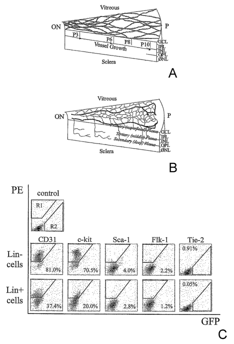
FIG. 1 depicts schematic diagrams of developing mouse retina. (a)
Development of primary plexus. (b) The second phase of retinal vessel
formation. GCL, ganglion cell layer; IPL, inner plexus layer; INL, inner
nuclear
layer; OPL, outer plexus layer; ONL, outer nuclear layer; RPE, retinal pigment
epithelium; ON, optic nerve; P, periphery. Panel (c) depicts flow cytometric
characterization of bone marrow-derived Lin' HSC and Liri HSC separated cells.
Top row: Dot plot distribution of non-antibody labeled cells, in which R1
defines
the quantifiable-gated area of positive PE-staining; R2 indicates GFP-
positive;
Middle row: Liri HSC (C57B/6) and Bottom row: Lin' HSC (C57B/6) cells, each
cell line labeled with the PE-conjugated antibodies for Sca-1, c-kit, Flk-
1/KDR,
CD3 1. Tie-2 data was obtained from Tie-2-GFP mice. Percentages indicate
percent of positive-labeled cells out of total Liri HSC or Lin' HSC
population.
CA 02598029 2007-08-14
WO 2006/104609 PCT/US2006/006411
-7-
FIG. 2 depicts engraftment of Liri HSCs into developing mouse
retina. (a) At four days post-injection (P6) intravitreally injected eGFP+
Liri HSC
cells attach and differentiate on the retina. (b) Liri HSC (B6.129S7-Gtrosa26
mice, stained with (3-gal antibody) establish themselves ahead of the
vasculature
stained with collagen IV antibody (asterisk indicates tip of vasculature). (c)
Most
of Lin+HSC cells (eGFP}) at four days post-injection (P6) were unable to
differentiate. (d) Mesenteric eGFP' murine EC four days post-injection (P6).
(e)
Liri HSCs (eGFP+) injected into adult mouse eyes. (f) Low magnification of
eGFP+ Lin HSCs (arrows) homing to and differentiating along the pre-existing
astrocytic template in the GFAP-GFP transgenic mouse. (g) Higher magnification
of association between Lin cells (eGFP) and underlying astrocyte (arrows). (h)
Non-injected GFAP-GFP transgenic control. (i) Four days post-injection (P6),
eGFP+ Liri HSCs migrate to and undergo differentiation in the area of the
future
deep plexus. Left figure captures Liri HSC activity in a whole mounted retina;
right figure indicates location of the Lin cells (arrows) in the retina (top
is vitreal
side, bottom is scleral side). (j) Double labeling with a-CD31-PE and
a-GFP-alexa 488 antibodies. Seven days after injection, the injected Liri HSCs
(eGFP, red) were incorporated into the vasculature (CD3 1). Arrowheads
indicate
the incorporated areas. (k) eGFP+ Liri HSC cells form vessels fourteen days
post-injection (P17). (1 and m) Intra-cardiac injection of rhodamine-dextran
indicates that the vessels are intact and functional in both the primary (1)
and deep
plexus (m).
FIG. 3 shows that eGFP+ Liri HSC cells home to the gliosis
(indicated by GFAP expressing-astrocytes, far left image) induced by both
laser
(a) and mechanical (b) induced injury in the adult retina (asterisk indicates
injured
site). Far right images are a higher magnification, demonstrating the close
association of the Liri HSCs and astrocytes. Calibration bar= 20 M.
CA 02598029 2007-08-14
WO 2006/104609 PCT/US2006/006411
-~-
FIG. 4 shows that Liri HSC cells rescue the vasculature of the
retinal degeneration mouse. (a-d) Retinas at 27 days post-injection (P33) with
collagen IV staining; (a) and (b), retinas injected with Lin+ HSC cells
(Balb/c)
showed no difference in vasculature from normal FVB mice; (c) and (d) retinas
injected with Lin HSCs (Balb/c) exhibited a rich vascular network analogous to
a
wild-type mouse; (a) and (c), frozen sections of whole retina (top is vitreal
side,
bottom is scleral side) with DAPI staining; (b) and (d), deep plexus of
retinal
whole amount; (e) bar graph illustrating the increase in vascularity of the
deep
vascular plexus formed in the Liri HSC cell-injected retinas (n=6). The extent
of
deep retinal vascularization was quantified by calculating the total length of
vessels within each image. Average total length of vessels/high power field
(in
microns) for Liri HSC, Lin+HSC or control retinas were compared. (f)
Comparison of the length of deep vascular plexus after injection with Liri HSC
(R, right eye) or Lin+HSC (L, left eye) cells from rd1Nd mouse. The results of
six
independent mice are shown (each color represents a separate mouse). (g) and
(h)
Liri HSC cells also (Balb/c) rescued the f d/rd vasculature when injected into
P15
eyes. The intermediate and deep vascular plexus of Lin HSC (G) or Lin+HSC
(H) cell injected retinas (one month after injection) are shown.
FIG. 5 depicts photomicrographs of mouse retinal tissue: (a) deep
layer of retinal whole mount (rd/rd mouse), five days post-injection (P11)
with
eGFP+ Liri HSCs visible (gray). (b) and (c) P60 retinal vasculature of Tie-2-
GFP
(rd/rd) mice that received Balb/c Liri cells (b) or LinkHSC cell (c) injection
at P6.
Only endogenous endothelial cells (GFP-stained) are visible in the left panels
of
(b) and (c). The middle panels of (b) and (c) are stained with CD31 antibody;
arrows indicate the vessels stained with CD31 but not with GFP, the right
panels
of (b) and (c) show staining with both GFP and CD3 1. (d) a-SMA staining of
Lin HSC injected (left panel) and control retina (right panel).
CA 02598029 2007-08-14
WO 2006/104609 PCT/US2006/006411
-9-
FIG. 6 shows that T2-TrpRS-transfected Lin HSCs inhibit the
development of mouse retinal vasculature. (a) Schematic representation of
human
TrpRS, T2-TrpRS and T2-TrpRS with an Igk signal sequence at the amino
terminus. (b) T2-TrpRS transfected Lin HSC-injected retinas express T2-TrpRS
protein in vivo. (1) Recombinant T2-TrpRS produced in E. coli; (2) Recombinant
T2-TrpRS produced in E. coli; (3) Recombinant T2-TrpRS produced in E. coli;
(4) control retina; (5) Liri HSC + pSecTag2A (vector only) injected retina;
(6)
Liri HSC + pKLe135 (Igk-T2-TrpRS in pSecTag) injected retina. (a)
Endogenous TrpRS. (b) Recombinant T2-TrpRS. (c) T2-TrpRS of Liri HSC
injected retina. (c-f) Representative primary (superficial) and secondary
(deep)
plexuses of injected retinas, seven days post-injection; (c) and (d) Eyes
injected
with empty plasmid-transfected Liri HSC developed normally; (e) and (f) the
majority of T2-TrpRS-transfected Lin HSC injected eyes exhibited inhibition of
deep plexus; (c) and (e) primary (superficial) plexus; (d) and (f) secondary
(deep)
plexus). Faint outline of vessels observed in (f) are "bleed-through" images
of
primary network vessels shown in (e).
FIG. 7 shows the DNA sequence encoding His6-tagged T2-TrpRS,
SEQ ID NO: 1.
FIG. 8 shows the amino acid sequence of His6-tagged T2-TrpRS,
SEQ ID NO: 2.
FIG. 9 illustrates photomicrographs and electroretinograms (ERG)
of retinas from mice whose eyes were injected with the Liri HSC and with
Lin+ HSC (controls).
FIG. 10 depicts statistical plots showing a correlation between
neuronal rescue (y-axis) and vascular rescue (x-axis) for both the
intermediate
(Int.) and deep vascular layers of rd/rd mouse eyes treated with Lin HSC.
CA 02598029 2007-08-14
WO 2006/104609 PCT/US2006/006411
-10-
FIG. 11 depicts statistical plots showing no correlation between
neuronal rescue (y-axis) and vascular rescue (x-axis) for rd/rd mouse eyes
that
were treated with LinHSC.
FIG. 12 is a bar graph of vascular length (y-axis) in arbitrary
relative units for rd/rd mouse eyes treated with the Lin HSC (darlc bars) and
untreated (light bars) rd/rd mouse eyes at time points of 1 month (1M), 2
months
(2M), and 6 months (6M) post-injection.
FIG. 13 includes three bar graphs of the number of nuclei in the
outer neural layer (ONR) of Nd/r=d mice at 1 month (1M), 2 months (2M) and 6
months (6M), post-injection, and demonstrates a significant increase in the
number of nuclei for eyes treated with Lin HSC (dark bars) relative to control
eyes treated with Lin+ HSC (light bars).
FIG. 14 depicts plots of the number of nuclei in the outer neural
layer for individual yd/rd mice, comparing the right eye (R, treated with
Lin HSC) relative to the left eye (L, control eye treated with Lin+ HSC) at
time
points (post injection) of 1 month (1M), 2 months (2M), and 6 months (6M);
each
line in a given plot compares the eyes of an individual mouse.
FIG. 15 depicts retinal vasculature and neural cell changes in
rdl/rdl (C3H/HeJ, left panels) or wild type mice (C57BL/6, right panels).
Retinal vasculature of intermediate (upper panels) or deep (middle panels)
vascular plexuses in whole-mounted retinas (red: collagen IV, green: CD3 1)
and
sections (red: DAPI, green: CD3 1, lower panels) of the same retinas are shown
(P: postnatal day). (GCL: ganglion cell layer, INL: inter nuclear layer, ONL:
outer
nuclear layer).
FIG. 16 shows that Liri HSC injection rescues the degeneration of
neural cells in rdl/rdl mice. (A, B and C), retinal vasculature of
intermediate
(Int.) or deep plexus and sections of Lin HSC injected eye (right panels) and
contralateral control cell (CD31-) injected eye (left panels) at P30 (A), P60
(B),
CA 02598029 2007-08-14
WO 2006/104609 PCT/US2006/006411
-11-
and P 180 (C). (D), the average total length of vasculature (+ or - standard
error
of the mean) in Liri HSC injected or control cell (CD31-) injected retinas at
P30
(left, n=10), P60 (middle, n=10), and P180 (right, n=6). Data of intermediate
(Int.) and deep vascular plexus are shown separately (Y axis: relative length
of
vasculature). (E), the average numbers of cell nuclei in the ONL at P30 (left,
n=10), P60 (middle, n=10), or P180 (right, n=6) of control cell (CD31-) or
Lin HSC injected retinas (Y axis: relative number of cell nuclei in the ONL).
(F),
Linear correlations between the length of vasculature (X axis) and the number
of
cell nuclei in the ONL (Y axis) at P30 (left), P60 (middle), and P180 (right)
of
Liri HSC or control cell injected retinas.
FIG. 17 demonstrates that retinal function is rescued by Liri HSC
injection. Electroretinographic (ERG) recordings were used to measure the
function of Liri HSC or control cell (CD31- ) injected retinas. (A and B),
Representative cases of rescued and non-rescued retinas 2 months after
injection.
Retinal section of Liri HSC injected right eye (A) and CD31- control cell
injected
left eye (B) of the same animal are shown (green: CD31 stained vasculature,
red:
DAPI stained nuclei). (C), ERG results from the same animal shown in (A) and
(B).
FIG. 18 shows that a population of human bone marrow cells can
rescue degenerating retinas in the rdl mouse (A-C). The rescue is also
observed
in another model of retinal degeneration, rd10 (D-K). A, human Liri HSCs
(hLiri HSCs) labeled with green dye can differentiate into retinal vascular
cells
after intravitreal injection into C3SnSmn.CB17-Prkdc SCID mice. (B and C),
Retinal vasculature (left panels; upper: intermediate plexus, lower: deep
plexus)
and neural cells (right panel) in hLin HSC injected eye (B) or contralateral
control eye (C) 1.5 months after injection. (D-K), Rescue of rd10 mice by
Liri HSCs (injected at P6). Representative retinas at P21 (D: Liri HSCs, H:
control cells), P30 (E: Liri HSCs, I: control cells), P60 (F: Liri HSCs, J:
control
CA 02598029 2007-08-14
WO 2006/104609 PCT/US2006/006411
-12-
cells), and P105 (G: Liri HSCs, K: control cells) are shown (treated and
control
eyes are from the same animal at each time point). Retinal vasculature (upper
image in each panel is the intermediate plexus; the middle image in each panel
is
the deep plexus) was stained with CD31 (green) and Collagen IV (red). The
lower image in each panel shows a cross section made from the same retina
(red:
DAPI, green: CD31).
FIG. 19 demonstrates that crystallin aA is up regulated in rescued
outer nuclear layer cells after treatment with Lin HSCs but not in
contralateral
eyes treated with control cells. Left panel; IgG control in rescued retina,
Middle
panel; crystallin ocA in rescued retina, Right panel; crystallin aA in non-
rescued
retina.
FIG. 20 includes tables of genes that are upregulated in murine
retinas that have been treated with the Liri HSCs of the present invention.
(A)
Genes whose expression is increased 3-fold in mouse retinas treated with
murine
Lin HSCs. (B) Crystallin genes that are upregulated in mouse retinas treated
with murine Liri HSC. (C) Genes whose expression is increased 2-fold in mouse
retinas treated with human Liri HSCs. (D) Genes for neurotrophic factors or
growth factors whose expression is upregulated in mouse retinas treated with
human Lin HSCs.
FIG. 21 illustrates the distribution of CD31 and integrin a6 surface
antigens on CD133 positive (DC133+) and CD133 negative (CD133-) human
Liri HSC populations. The left panels show flow cytometry scatter plots. The
center and right panels are histograms showing the level of specific antibody
expression on the cell population. The Y axis represents the number of events
and the X axis shows the intensity of the signal. A filled histogram shifted
to the
right of the outlined (control) histogram represents an increased fluorescent
signal
and expression of the antibody above background level.
CA 02598029 2007-08-14
WO 2006/104609 PCT/US2006/006411
- 13-
FIG. 22 illustrates postnatal retinal development in wild-type
C57/B 16 mice raised in normal oxygen levels (normoxia), at post natal days P0
through P30.
FIG. 23 illustrates oxygen-induced retinopathy model in C57B 16
mice raised in high oxygen levels (hyperoxia; 75% oxygen) between P7 and P 12,
followed by normoxia from P 12-P 17.
FIG. 24 demonstrates vascular rescue by treatment with the
Liri HSC populations in the oxygen-induced retinopathy (OIR) model.
FIG. 25 shows rescued photoreceptors in rdl mouse outer nuclear
layer (ONL) following intravitreal injection of Lin-HSC are predominantly
cones.
A small percentage of photoreceptors in the wild type mouse retina (upper
panel)
were cones as evidenced by expression of red/green cone opsin (A) while most
cells of the ONL were positive for rod specific rhodopsin (B). Retinal
vasculature
autofluoresces with pre-immune serum (C) but nuclear layers were completely
negative for staining with rod or cone-specific opsins. Rd1rd mouse retinas
(lower
panels) had a diminished inner nuclear layer and a nearly completely atrophic
ONL, both of which were negative for cone (D) or rod (Panel G) opsin. Control,
CD31- HSC treated eyes are identical to non-injected rd/rd retinas, without
any
staining for cone (E) or rod (H) opsin. Lin-HSC treated contralateral eyes
exhibited a markedly reduced, but clearly present ONL that is predominantly
comprised of cones, as evidenced by positive immunoreactivity for cone
red/green opsin (F). A small number of rods were also observed (I).
FIG. 26 shows scatter plots from flow cytometry characterization
of lineage negative and lineage positive stem cell populations (upper left and
lower left plots, respectively) showing percentages of cells that express the
CD44
antigen (data points in red); as well as plots of CD31 negative and CD31
positive
cell populations (upper right and lower right plots, respectively), showing
percentages of cells that express the CD44 antigen (data points in red).
CA 02598029 2007-08-14
WO 2006/104609 PCT/US2006/006411
-14-
FIG. 27 shows scatter plots from flow cytometry characterization
of a lineage negative cell population that expresses a significant level of
CD44
antigen (left set of plots) and a sub-population of bone marrow cells that do
not
express a significant level of CD44 antigen (right set of plots) illustrating
the
relative percentages of cells expressing various other cell surface antigens.
FIG. 28 shows photomicrographic images of a retina from a mouse
intravitreally injected with cells from the MLBM cell population of the
invention
(left panel) compared to a retina from a mouse intravitreally injected with
CD44"
cells.
FIG. 29 shows photomicrographic images of retinas from eyes
injected with cells from the MLBM cell population (CD44") and with CD44'
cells.
FIG. 30 shows bar graphs demonstrating the beneficial effects of
the MLBM cell population for ameliorating pathogenic angiogensis and
promoting beneficial physiological revascularization of mouse retinas in the
oxygen induced retinopathy model of retinopathy of prematurity. The upper
graph compares pre-retinal neovascular tuft area for control retina (first
bar),
retina treated with CD44" cells (middle bar) and retinas treated with cells
from
the MLBM cell population (right bar). The lower graph compares vascular
obliteration area for control retina (first bar), retina treated with CD44'
cells
(middle bar) and retinas treated witli cells from the MLBM cell population
(right
bar).
FIG. 31 is a photomicrographic image demonstrating that once
cells from the MLBM cell population have incorporated into the vasculature of
the retina, the cells express vascular endothelial growth factor (VEGF), as
indicated by the green staining of the cells in the lower portion of the
image.
CA 02598029 2007-08-14
WO 2006/104609 PCT/US2006/006411
- 15-
FIG. 32 depicts photomicrographic images demonstrating that cells
from the CD11b+ MLBM cell population of the invention selectively target the
vasculature of the retina.
FIG. 33 depicts photomicrographic images demonstrating that
CD44- CD 11 b- bone marrow cells do not selectively target the vasculature of
the
retina.
FIG. 34 shows the amino acid residue sequence of the T2 fragment
of TrpRS (SEQ ID NO: 3) and of the T2-TrpRS-GD variation thereof (SEQ ID
NO: 4).
FIG. 35 shows the amino acid residue sequence of mini-TrpRS
(SEQ ID NO: 5).
FIG. 36 shows the amino acid residue sequence of T1-TrpRS (SEQ
ID NO: 6).
FIG. 37 shows normal retinal vascular development in the mouse,
the oxygen-induced retinopathy (OIR) model, and the rescue effect following
intra-
vitreal transplantation of Lin- bone-marrow derived-cells. The mouse is born
with a
largely avascular retina. as shown at postnatal day 2 (P2) (Panel a, retinal
whole-
mount) where the vessels are found in the superficial retina occupying a
single
plane as shown in b. Panels b,d and f are images taken from 3D renderings of
en
face confocal z-series data sets rotated 90 degrees. During the first week
after birth,
the superficial retinal vasculature grows in a radial fashion from the optic
nerve
head nearly reaching the periphery by P 10 (c). The deep retinal vasculature
is then
established from branching of the superficial layer during the second week
(d).
Finally, a third plexus of vessels forms between the first two, and
establishes the
mature retinal vasculature at around P30 (e,f). Panel g shows that ewxposure
to
hyperoxia in the OIR model causes central vaso-obliteration as shown here at P
10.
Panel h shows that after removal to normoxia at P 12, the central retina
starts to
revascularize and characteristic pre-retinal neovascular tufts are formed at
the
CA 02598029 2007-08-14
WO 2006/104609 PCT/US2006/006411
-16-
interface between the vascularized (peripheral) and avascular (central)
retina. These
tufts stain strongly with isolectin. Panels i-n show that Lin hematopoietic
progenitor cells promote vascular repair in the OIR model. Liri cells injected
intravitreally prior to high oxygen exposure dramatically accelerate
revascularization of the central retina when compared to the vehicle-treated
fellow
eye at P 17. While retinas treated with vehicle show partial absence of the
superficial vasculature (i) and complete absence of the deep retinal
vasculature
(k,m), the Lin cell-treated fellow eye shows relatively normal retinal
vasculature
(j) with all three plexuses present (k,m). Panel o shows that at P 17, OIR
eyes
treated with Liri cells are fully revascularized significantly more often than
uninjected eyes or those injected with vehicle. Vessels were visualized by
cardiac
perfusion of fluorescein-dextran, as shown in Panels a-f,i,j and by GS lectin
in
Panels g,h,k-n. Nuclei in Panels k-n were labeled with DAPI.
FIG. 38 shows Liri cells accelerate retinal revascularization and
reduce pre-retinal neovascular tuft formation in OIR. Panels a-d show a
computer
image analysis method was used to calculate the area of retinal vessel
obliteration,
as well as pre-retinal neovascular tuft formation (red) in retinal whole-
mounts from
OIR eyes at postnatal day 17. Panel e shows retinas treated with Lin- cells
prior to
hyperoxia showed an almost 6-fold reduction in obliterated area versus
uninjected
controls and an approximately 5-fold reduction compared to eyes treated with
vehicle alone. Panel f shows Liri cell treatment significantly reduced two-
dimensional area of neovascular tufts compared to uninjected eyes and vehicle-
treated eyes. Panel g shows Liri cell-transplantation is effective at reducing
the
area of obliteration not only when administered prior to hyperoxia, but also
at P9-
P12 during hyperoxia and just after return to normoxia. (graphs represent Mean
~
SEM; * p < 0.001).
FIG. 39 shows bone marrow cell treatment has little or no long term
toxic effects. Retinas evaluated at 5 or 6 months after receiving Liri cell
treatment
CA 02598029 2007-08-14
WO 2006/104609 PCT/US2006/006411
-17-
have normal-appearing retinal vasculature and the neural retina appears
histologically preserved on cross sections (a-f, non-injected versus Liri cell-
injected retina 6 months post-transplant). No tumors were observed, and the
only
abnormality was an occasional "rosette" in the neural retina which could also
be
seen in control non-injected eyes (g,h).
FIG. 40 shows CD44H' cells are prevalent in the Liri population and
effectively promote vascular repair in the OIR model. Panel a shows bone
marrow
contains CD44HI and CD44L fractions and the Liri population is enriched for
CD44' cells compared to control CD cells. Insets show light scattering
properties
of the CD44' cells which are typical of monocytes and granulocytes, while
light-
scattering properties of CD44LO cells are typical of lymphocytes. Panel b
shows
representative P 17 retinas from eyes treated with CD44LO and CD44HI bone
marrow
cells prior to oxygen exposure. The lower panels exemplify the quantified
areas of
obliteration and neovascularization at P 17 used to create the data shown in
panel c.
Panel c shows vascular obliteration and pre-retinal neovascularization are
reduced
in eyes treated with CD44H' cells with efficacy similar to eyes treated with
Liri
cells. Areas of vascular obliteration (*) and pre-retinal neovascularization
(**)
were significantly lower in CD44H' and Liri eyes compared with vehicle
injection
or no injection (p<10"5 in all cases). Area of obliteration in Liri cell-
treated eyes
was also reduced compared to CD44H' (p = 0.03), but to a much lesser degree.
Areas of pre-retinal neovascularization did not significantly differ between
Lin
and CD44HI-treated eyes (p = 0.25).
FIG. 41 shows the CD44' subpopulation expresses myeloid
markers. In Panel a, two-color flow cytometry was used to further characterize
CD44 populations. All cells were labled with an antibody against CD44 and co-
labeled with the various antibodies shown. The CD44H' population showed strong
labeling for CD 11 a, CD 11 b and Ly6GC. Fractions of CD44hi cells were
positive
for CD14, F4/80, cKit, and CD115. Most of these antigens are present on
myeloid
CA 02598029 2007-08-14
WO 2006/104609 PCT/US2006/006411
- 18-
lineage cells. CD441o cells labeled strongly with Terl 19 and CD45R B220,
which
are markers for erythroblasts and B cells, respectively.
FIG. 42 shows that CD44' cells take on a perivascular localization
in the retina. Confocal imaging was used to create a series of images in the z
dimension which were then rendered into 3D. In Panel a, a projection of this
is
shown the CD31-labeled vascular endothelium and GFP expression from the
introduced bone marrow cells are shown. The bone marrow cell appears to have
assumed a perivascular position. 3D data show that the lumen of the vessel and
the
relative position of the GFP+ bone marrow cell are visualized. The numbers
listed
in (b) correspond to cross-sectional positions indicated in (a). The GFP
signal was
detected outside of the lumen in all cases, except Panel b, No. 3, which was a
section through the cell body with intense fluorescence where bleed-through of
the
signal was evident.
FIG. 43 shows an in situ analysis of injected CD44HI bone marrow
cells in the OIR model. Labeling of a control retina that received no cell
treatment
shows the presence of endogenous F4/80+ perivascular cells (a-c). Injected
CD44HI
cells target the retinal vasculature and have a localization, morphology and
F4/80
expression pattern similar to endogenous cells (d-i). Transplanted
perivascular
bone marrow cells lose CD44 expression, while cells not associated with the
retinal
vasculature retain CD44 expression (j-o).
FIG. 44 shows an expression array analysis, which revealed a high
expression of myeloid-associated genes in the CD44HI population while the
CD44LO
cells expressed genes associated with lymphoid cells. AFFYMETRIX arrays
were used to compare gene expression profiles between these two bone marrow
cell
populations. Genes shown had a minimum 5-fold difference in expression. A
significantly higher level of CD44 expression in the CD44HI population was
observed versus CD44L0 cells.
CA 02598029 2007-08-14
WO 2006/104609 PCT/US2006/006411
-19-
FIG. 45 demonstrates that CD44HI cells can differentiate into cells
with microglial characteristics. Panels A and B show that injected CD44H'
cells
express CD11b and F4/80 and have morphology and perivascular localization
similar to endogenous microglia. Panel C provides 3d imaging of the
perivascular localization of an injected CD44HI cell. Panel D shows a high
magnification view of the morphology of injected CD44HI cells.
FIG. 46 demonstrates that CD44 HI cells can be isolated by negative
selection. Panel A shows that depletion of mouse bone marrow by MACS using
antibodies selective for CD45R/B220, TER1 19, and CD3e yields a population of
cells that are greater than 90 percent CD44H' cells. Panel B shows the
negative
fraction (CD44HI population) is essentially free from CD45R/B220, TER1 19, and
CD3e cells. Panel C shows negatively selected CD44H' cells retain retinal
targeting and differentiation capabilities.
DETAILED DESCRIPTION OF PREFERRED EMBODIMENTS
Bone marrow cells include a sub-population of cells that express
the CD44 antigen (i.e., the hyaluronic acid receptor) and CD1 lb (integrin
aM). A
myeloid-like population of bone marrow cells enriched in CD44 and CD 11 b
expressing cells can be isolated from bone marrow by treating bone marrow
cells
with an antibody to CD44 antigen (anti-CD44) and/or an antibody to CD11b
antigen (anti-CD 11 b), and then selecting cells that immunoreact with the
antibody. The antibody then can be removed from the cells by methods that are
well known in the art. The cells can be selected, for example, using by flow
cytometry, using antibodies bound to or coated on beads followed by
filtration, or
other separation methods that are well known in the art. A majority of the
selected cells are lineage negative and express both the CD44 antigen and the
CDl lb antigen, regardless of which antibody is utilized in the isolation.
Bone marrow includes stem cells. Stem cells are typically
identified by the distribution of antigens on the surface of the cells (for a
detailed
CA 02598029 2007-08-14
WO 2006/104609 PCT/US2006/006411
-20-
discussion see Stein Cells: Scientific Progress and Future Directions, a
report
prepared by the National Institutes of Health, Office of Science Policy, June
2001,
Appendix E: Stem Cell Markers, which is incorporated herein by reference to
the
extent pertinent). Approximately 75% of lineage negative hematopoietic stems
cells isolated from bone marrow are also CD44 positive. In a preferred
embodiment, a majority of the cells from the MLBM cell population are lineage
negative hematopoietic stem cells (i.e., CD44+Liri HSC).
The present invention provides a method of ameliorating vascular
and neuronal degeneration in the retina of a mammal that suffers from an
ocular
disease. Isolated MLBM cell population of the invention is administered to the
retina of the mammal, preferably by intravitreal injection. The cells are
administered in an amount sufficient to ameliorate vascular and/or neuronal
degeneration in the retina. Preferably, the isolated MLBM cell population is
autologous to the mammal to be treated. Preferably, the cells from the MLBM
cell population are administered in a physiologically tolerable medium, such
as
phosphate buffered saline (PBS).
A preferred method comprises isolating the MLBM cell population
from the bone maiTow of the mammal to be treated and then administering the
cells to the mammal in a number sufficient to ameliorate the vascular and/or
neuronal degeneration of the retina. The cells can be isolated from a mammal
suffering from an ocular degenerative disease, preferably at an early stage of
the
ocular disease or from a healthy mammal known to be predisposed to an ocular
degenerative disease (i.e., through genetic predisposition). In the latter
case, the
isolated MLBM cell population can be stored after isolation, and can then be
injected prophylactically during early stages of a later developed ocular
disease.
Preferably the diseased retina includes activated astrocytes, to which the
cells
from the MLBM cell population are targeted. Accordingly, early treatment of
the
eye when there is an associated gliosis is beneficial. Alternatively, the
retina can
CA 02598029 2007-08-14
WO 2006/104609 PCT/US2006/006411
-21 -
be treated with a laser to stimulate local proliferation of activated
astrocytes in the
retina prior to administering the autologous MLBM cell population.
Hematopoietic stem cells are stem cells that are capable of
developing into various blood cell types e.g., B cells, T cells, granulocytes,
platelets, and erythrocytes. The lineage surface antigens are a group of
cell-surface proteins that are markers of mature blood cell lineages,
including
CD2, CD3, CD 11, CD 11 a, Mac-1 (CD 11 b: CD 18), CD 14, CD 16, CD 19, CD24,
CD33, CD36, CD38, CD45, CD45RA, murine Ly-6G, murine TER-119, CD56,
CD64, CD68, CD86 (B7.2), CD66b, human leucocyte antigen DR (HLA-DR),
and CD235a (Glycophorin A). Hematopoietic stem cells that do not express
significant levels of these antigens are commonly referred to a lineage
negative
(Liri ). Human hematopoietic stem cells commonly express other surface
antigens
such as CD31, CD34, CD117 (c-kit) and/or CD133. Murine hematopoietic stem
cells commonly express other surface antigens such as CD34, CD 117 (c-kit),
Thy-1, and/or Sca-1.
Isolated hematopoietic stem cells that do not express significant
levels of a "lineage surface antigen" (Lin) on their cell surfaces are
referred to
herein as "lineage negative" or "Lin " hematopoietic stem cells i.e., Liri
HSC. A
majority of the cells of the MLBM cell populations of the present invention
are
Liri and express both a relatively high amount of the CD44 antigen (CD44") as
well as the CD 11 b antigen. These CD44+CD 11 b'Liri HSC are capable of
incorporating into developing vasculature and then differentiating to become
vascular endothelial cells.
As used herein and in the appended claims, the phrase "adult" in
reference to bone marrow and bone marrow cells, includes bone marrow isolated
postnatally, i.e., from juvenile and adult individuals, as opposed to embryos.
Accordingly, the term "adult mammal" refers to both juvenile (postnatal) and
fully mature mammals, as opposed to an embryo or prenatal individual.
CA 02598029 2007-08-14
WO 2006/104609 PCT/US2006/006411
-22-
The isolated MLBM cell populations of the present invention
selectively target astrocytes and incorporate into the retinal neovasculature
when
intravitreally injected into the eye of the mammalian species, such as a mouse
or a
human, from which the cells were isolated.
The isolated MLBM cell populations of the present invention
include cells that differentiate to endothelial cells and generate vascular
structures
within the retina. In particular, the MLBM cell population of the present
invention is useful for the treatment of retinal neovascular and retinal
vascular
degenerative diseases, and for repair of retinal vascular injury. The MLBM
cell
population of the present invention also promotes neuronal rescue in the
retina
and promote upregulation of anti-apoptotic genes. Additionally, the MLBM cell
population of the invention can be utilized to treat retinal defects in the
eyes of
neonatal mammals, such as mammals suffering from oxygen induced retinopathy
or retinopathy of prematurity.
It has been found that bone marrow cells that do not express CD44
(CD44LO cells) generally express one or more of the following cell markers:
Terl 19, CD45RB220, and CD3e. Utilizing this fact, CD44HI MLBM cells of the
present invention can be isolated by a method involving negative cell-marker
selection. The method comprises contacting a plurality of bone marrow cells
with
antibodies specific for Ter119, CD45RB220, and CD3e, removing cells from the
plurality of bone marrow cells that immunoreact with Ter 119, CD45RB220, and
CD3e antibodies, and recovering myeloid-like bone marrow cells that are
deleted
in Ter119, CD45RB220, and CD3e-expressing cells. Using this method, a cell
population can be recovered in which greater than 90 percent of the cells
express
CD44.
The present invention also provides a method of treating ocular
diseases in a mammal comprising isolating from the bone maiTow of the mammal
a MLBM cell population, and intravitreally injecting cells from the MLBM cell
CA 02598029 2007-08-14
WO 2006/104609 PCT/US2006/006411
- 23 -
population into an eye of the mammal in a number sufficient to arrest the
disease.
The present method can be utilized to treat ocular diseases such as retinal
degenerative diseases, retinal vascular degenerative diseases, ischemic
retinopathies, vascular hemorrhages, vascular leakage, and choroidopathies in
neonatal, juvenile or fully mature mammals. Examples of such diseases include
age related macular degeneration (ARMD), diabetic retinopathy (DR), presumed
ocular histoplasmosis (POHS), retinopathy of prematurity (ROP), sickle cell
anemia, and retinitis pigmentosa, as well as retinal injuries.
The number of cells from the MLBM cell population injected into
the eye is sufficient for arresting the disease state of the eye. For example,
the
amount of injected cells can be effective for repairing retinal damage of the
eye,
stabilizing retinal neovasculature, maturing retinal neovasculature, and
preventing
or repairing vascular leakage and vascular hemorrhage.
Cells from the MLBM cell population of the present invention can
be transfected with therapeutically useful genes, such as genes encoding
antiangiogenic proteins for use in ocular, cell-based gene therapy and genes
encoding neurotrophic agents to enhance neuronal rescue effects.
The transfected cells can include any gene which is therapeutically
useful for treatment of retinal disorders. In one preferred embodiment, the
transfected cells from the MLBM cell population of the present invention
include
a gene operably encoding an antiangiogenic peptide, including proteins, or
protein
fragments such as TrpRS or antiangiogenic (i.e., angiostatic) fragments
thereof,
e.g., the fragments of TrpRS designated T2-TrpRS (SEQ ID NO: 3 in FIG. 34),
T2-TrpRS-GD (SEQ ID NO: 4 in FIG. 34), both of which are preferred
angiostatic peptides, as well as mini-TrpRS (SEQ ID NO: 5 in FIG. 35), and T1-
TrpRS(SEQ ID NO: 6 in FIG. 36). The transfected cells from the MLBM cell
population encoding an antiangiogenic peptide of the present invention are
useful
for treatment of retinal diseases involving abnormal vascular development,
such
CA 02598029 2007-08-14
WO 2006/104609 PCT/US2006/006411
-24-
as diabetic retinopathy, and like diseases. Preferably, the cells from the
MLBM
cell population are human cells.
In another preferred embodiment, the transfected cells from the
MLBM cell population of the present invention include a gene operably encoding
a neurotrophic agent such as nerve growth factor, neurotrophin-3, neurotrophin-
4,
neurotrophin-5, ciliary neurotrophic factor, retinal pigmented epithelium-
derived
neurotrophic factor, insulin-like growth factor, glial cell line-derived
neurotrophic
factor, brain-derived neurotrophic factor, and the like. Such neurotrophic
cells
from the MLBM cell population are useful for promoting neuronal rescue in
retinal neuronal degenerative diseases such as glaucoma and retinitis
pigmentosa,
in treatment of injuries to the retinal nerves, and the like. Implants of
ciliary
neurotrophic factor have been reported as useful for the treatment of
retinitis
pigmentosa (see Kirby et al. 2001, Mol Ther. 3(2):241-8; Farrar et al. 2002,
EMBO Journal 21:857-864). Brain-derived neurotrophic factor reportedly
modulates growth associated genes in injured retinal ganglia (see Fournier, et
al.,
1997, J. Neurosci. Res. 47:561-572). Glial cell line derived neurotrophic
factor
reportedly delays photoreceptor degeneration in retinitis pigmentosa (see
McGee
et al. 2001, Mol Ther. 4(6):622-9).
The present invention also provides methods for treating ocular
angiogenic diseases by administering transfected cells from the MLBM cell
population of the present invention by intravitreal injection of the cells
into the
eye. Such transfected cells from the MLBM cell population comprise cells from
the MLBM cell population transfected with a therapeutically useful gene, such
as
a gene encoding antiangiogenic or neurotrophic gene product. Preferably the
transfected cells from the MLBM cell population are human cells.
Preferably, at least about 1 x 10⁵ cells from the MLBM cell
population or transfected cells from the MLBM cell population are administered
by intravitreal injection to a mammalian eye suffering from a retinal
degenerative
CA 02598029 2007-08-14
WO 2006/104609 PCT/US2006/006411
- 25 -
disease. The number of cells to be injected may depend upon the severity of
the
retinal degeneration, the age of the mammal and other factors that will be
readily
apparent to one of ordinary skill in the art of treating retinal diseases. The
cells
from the MLBM cell population may be administered in a single dose or by
multiple dose administration over a period of time, as determined by the
clinician
in charge of the treatment.
The MLBM cell populations of the present invention is useful for
the treatment of retinal injuries and retinal defects involving an
inteiTuption in or
degradation of the retinal vasculature or retinal neuronal degeneration. Human
MLBM cell populations also can be used to generate a line of genetically
identical
cells, i.e., clones, for use in regenerative or reparative treatment of
retinal
vasculature, as well as for treatment or amelioration of retinal neuronal
degeneration. Further more, the MLBM cell populations of the present invention
are useful as research tools to study retinal vascular development and to
deliver
genes to selected cell targets, such as astrocytes.
Murine Retinal Vascular Development.
A Modelfor Ocular Angiogenesis. The mouse eye provides a
recognized model for the study of mammalian retinal vascular development, such
as human retinal vascular development. During development of the murine
retinal vasculature, ischemia-driven retinal blood vessels develop in close
association with astrocytes. These glial elements migrate onto the third
trimester
human fetus, or the neonatal rodent, retina from the optic disc along the
ganglion
cell layer and spread radially. As the murine retinal vasculature develops,
endothelial cells utilize this already established astrocytic template to
determine
the retinal vascular pattern (See FIG. 1 (a and b)). FIG. 1 (a and b) depicts
schematic diagrams of developing mouse retina. Panel (a) depicts development
of
the primary plexus (darlc lines at upper left of the diagram) superimposed
over the
astrocyte template (light lines) whereas, (b) depicts the second phase of
retinal
CA 02598029 2007-08-14
WO 2006/104609 PCT/US2006/006411
-26-
vessel formation. In FIG. 1, GCL stands for ganglion cell layer; IPL stands
for
inner plexus layer; INL stands for inner nuclear layer; OPL stands for outer
plexus layer; ONL stands for outer nuclear layer; RPE stands for retinal
pigment
epithelium; ON stands for optic nerve; and P stands for periphery.
At birth, retinal vasculature is virtually absent. By postnatal day 14
(P 14) the retina has developed complex primary (superficial) and secondary
(deep) layers of retinal vessels coincident with the onset of vision.
Initially,
spoke-like peripapillary vessels grow radially over the pre-existing
astrocytic
network towards the periphery, becoming progressively interconnected by
capillary plexus formation. These vessels grow as a monolayer within the nerve
fiber through P10 (FIG. 1 (a)). Between P7-P8 collateral branches begin to
sprout
from this primary plexus and penetrate into the retina to the outer plexiform
layer
where they form the secondary, or deep, retinal plexus. By P21, the entire
network undergoes extensive remodeling and a tertiary, or intermediate, plexus
forms at the inner surface of inner nuclear layer (FIG. 1 (b)).
The neonatal mouse retinal angiogenesis model is useful for
studying the role of HSC during ocular angiogenesis for several reasons. In
this
physiologically relevant model, a large astrocytic template exists prior to
the
appearance of endogenous blood vessels, permitting an evaluation of the role
for
cell-cell targeting during a neovascular process. In addition, this consistent
and
reproducible neonatal retinal vascular process is known to be hypoxia-driven,
in
this respect having similarities to many retinal diseases in which ischemia is
lcnown to play a role.
Enrichment of Endothelial Progenitor Cells (EPC) From Bone Marrow.
Although cell surface marker expression has been extensively
evaluated on the EPC population found in preparations of HSC, markers that
uniquely identify EPC are still poorly defined. To enrich for EPC,
hematopoietic
lineage marker positive cells (Lin+), i.e., B lymphocytes (CD45), T
lymphocytes
CA 02598029 2007-08-14
WO 2006/104609 PCT/US2006/006411
-27-
(CD3), granulocytes (Ly-6G), monocytes (CD11), and erythrocytes (TER-119),
were depleted from bone marrow mononuclear cells of mice. Sca-1 antigen was
used to further enrich for EPC. A comparison of results obtained after
intravitreal
injection of identical numbers of either Lin7 Sca-1} cells or Lin-cells, no
difference was detected between the two groups. In fact, when only Lin Sca-1-
cells were injected, far greater incorporation into developing blood vessels
was
observed.
Liri HSC populations are enriched with EPCs, based on functional
assays. Furthermore, LinHSC populations functionally behave quite differently
from the Lin HSC populations. Epitopes commonly used to identify EPC for
each fraction (based on previously reported in vitro characterization studies)
were
also evaluated. While none of these markers were exclusively associated with
the
Lin7 fraction, all were increased about 70 to about 1800% in the Lin7 HSC,
compared to the LinHSC fraction (FIG. 1 (c)). FIG. 1, Panel (c) illustrates
flow
cytometric characterization of bone marrow-derived Lin+ HSC and Lin7 HSC
separated cells. The top row of Panel (c) shows a hematopoietic stem cell dot
plot distribution of non-antibody labeled cells. Rl defines the quantifiable-
gated
area of positive PE-staining; R2 indicates GFP-positive. Dot plots of Lin7 HSC
are shown in the middle row and dot plots of Lin~ HSC are shown in the bottom
row. The C57B/6 cells were labeled with the PE-conjugated antibodies for Sca-
1,
c-kit, Flk-1/KDR, CD3 1. Tie-2 data was obtained from Tie-2-GFP mice. The
percentages in the corners of the dot plots indicate the percent of positive-
labeled
cells out of total Liri or Lin' HSC population. Interestingly, accepted EPC
markers like Flk-1/KDR, Tie-2, and Sca-1 were poorly expressed and, thus, not
used for further fractionation.
Lin HSC can be isolated by (a) extracting bone marrow from an
adult mammal; (b) separating a plurality of monocytes from the bone marrow;
(c)
labeling the monocytes with biotin-conjugated lineage panel antibodies to one
or
CA 02598029 2007-08-14
WO 2006/104609 PCT/US2006/006411
- 28 -
more lineage surface antigens, preferably lineage surface antigens selected
from
the group consisting of CD2, CD3, CD4, CD 11, CD 11 a, Mac-1, CD 14, CD 16,
CD 19, CD24, CD33, CD36, CD38, CD45, Ly-6G (murine), TER-119 (murine),
CD45RA, CD56, CD64, CD68, CD86 (B7.2), CD66b, human leucocyte antigen
DR (HLA-DR), and CD235a (Glycophorin A); (d) removing monocytes that are
positive for said one or more lineage surface antigens from the plurality of
monocytes; and (e) recovering a population of lineage negative hematopoietic
stem cells therefrom.
When the Lin HSC are isolated from adult human bone marrow,
preferably the monocytes are labeled with biotin-conjugated lineage panel
antibodies to lineage surface antigens CD2, CD3, CD4, CD 11 a, Mac-1, CD 14,
CD16, CD19, CD33, CD38, CD45RA, CD64, CD68, CD86 (B7.2), and CD235a.
When the Lin HSC are isolated from adult murine bone marrow, preferably the
monocytes are labeled with biotin-conjugated lineage panel antibodies to
lineage
surface antigens CD3, CD11, CD45, Ly-6G, and TER-119.
Intravitreally Injected HSC Liri Cells Contain EPC That Target Astrocytes
and Incorporate into Developing Retinal Vasculature.
To determine whether intravitreally injected Liri HSC can target
specific cell types of the retina, utilize the astrocytic template and
participate in
retinal angiogenesis, approximately 105 cells from a Liri HSC composition of
the
present invention or Lin' HSC cells (control, about 105 cells) isolated from
the
bone marrow of adult (GFP or LacZ transgenic) mice were injected into
postnatal
day 2 (P2) mouse eyes. Four days after injection (P6), many cells from the
Lin HSC composition of the present invention, derived from GFP or LacZ
transgenic mice were adherent to the retina and had the characteristic
elongated
appearance of endothelial cells (FIG. 2 (a)). FIG. 2 illustrates engraftment
of Liri
cells into developing mouse retina. As shown in FIG. 2, Panel (a), the four
days
CA 02598029 2007-08-14
WO 2006/104609 PCT/US2006/006411
-29-
post-injection (P6) intravitreally injected eGFP+ Lin HSC attach and
differentiate
on the retina.
In many areas of the retinas, the GFP-expressing cells were
arranged in a pattern conforming to underlying astrocytes and resembled blood
vessels. These fluorescent cells were observed ahead of the endogenous,
developing vascular network (FIG. 2 (b)). Conversely, only a small number of
Lin+HSC (FIG. 2 (c)), or adult mouse mesenteric endothelial cells (FIG. 2 (d))
attached to the retinal surface. In order to determine whether cells from an
injected Lin HSC population could also attach to retinas with already
established
vessels, a Lin HSC composition was injected into adult eyes. Interestingly, no
cells were observed to attach to the retina or incorporate into established,
norinal
retinal blood vessels (FIG. 2 (e)). This indicates that the Liri HSC
compositions
of the present invention do not disrupt a normally developed vasculature and
will
not initiate abnormal vascularization in normally developed retinas.
In order to determine the relationship between an injected Liri HSC
compositions of the present invention and retinal astrocytes, a transgenic
mouse
was used, which expressed glial fibrillary acidic protein (GFAP, a marker of
astrocytes) and promoter-driven green fluorescent protein (GFP). Examination
of
retinas of these GFAP-GFP transgenic mice injected with Lin- HSC from eGFP
transgenic mice demonstrated co-localization of the injected eGFP EPC and
existing astrocytes (FIG. 2(f-h), arrows). Processes of eGFP+Liri HSC were
observed to conform to the underlying astrocytic network (arrows, FIG. 2 (g)).
Examination of these eyes demonstrated that the injected, labeled cells only
attached to astrocytes; in P6 mouse retinas, where the retinal peripheiy does
not yet
have endogenous vessels, injected cells were observed adherent to astrocytes
in
these not yet vascularized areas. Surprisingly, injected, labeled cells were
observed
in the deeper layers of the retina at the precise location where normal
retinal vessels
will subsequently develop (FIG. 2 (i), arrows).
CA 02598029 2007-08-14
WO 2006/104609 PCT/US2006/006411
-30-
To determine whether injected Liri HSC are stably incorporated into
the developing retinal vasculature, retinal vessels at several later time
points were
examined. As early as P9 (seven days after injection), Liri HSC incorporated
into
CD31+structures (FIG. 2(j)). By P16 (14 days after injection), the cells were
already extensively incorporated into retinal vascular-like structures (FIG. 2
(k)).
When rhodamine-dextran was injected intravascularly (to identify functional
retinal
blood vessels) prior to sacrificing the animals, the majority of Liri HSC were
aligned with patent vessels (FIG. 2 (1)). Two patterns of labeled cell
distribution
were observed: (1) in one pattern, cells were interspersed along vessels in
between
unlabeled endothelial cells; and (2) the other pattern showed that vessels
were
composed entirely of labeled cells. Injected cells were also incorporated into
vessels of the deep vascular plexus (FIG. 2 (m)). While sporadic incorporation
of
Lin HSC-derived EPC into neovasculature has been previously reported, this is
the
first report of vascular networks being entirely composed of these cells. This
demonstrates that cells from a population of bone marrow-derived Lin7 HSC,
injected intravitreally, can efficiently incorporate into any layer of the
forming
retinal vascular plexus.
Histological examination of non-retinal tissues (e.g., brain, liver,
heart, lung, bone marrow) did not demonstrate the presence of any GFP positive
cells when examined up to 5 or 10 days after intravitreal injection. This
indicates
that a sub-population of cells within the Liri HSC fraction selectively target
to
retinal astrocytes and stably incorporate into developing retinal vasculature.
Since
these cells have many characteristics of endothelial cells (association with
retinal
astrocytes, elongate morphology, stable incorporation into patent vessels and
not
present in extravascular locations), these cells represent EPC present in the
Liri HSC population. The targeted astrocytes are of the same type observed in
many of the hypoxic retinopathies. It is well known that glial cells are a
prominent
component of neovascular fronds of tufts observed in DR and other forms of
retinal
CA 02598029 2007-08-14
WO 2006/104609 PCT/US2006/006411
-31-
injury. Under conditions of reactive gliosis and ischemia-induced
neovascularization, activated astrocytes proliferate, produce cytokines, and
up-regulate GFAP, similar to that observed during neonatal retinal vascular
template formation in many mammalian species including humans.
Lin HSC populations will target activated astrocytes in adult mouse
eyes as they do in neonatal eyes, Liri HSC cells were injected into adult eyes
with
retinas injured by photo-coagulation (FIG. 3 (a)) or needle tip (FIG. 3 (b)).
In both
models, a population of cells with prominent GFAP staining was observed only
around the injury site (FIG. 3 (a and b)). Cells from injected Liri HSC
compositions localized to the injury site and remained specifically associated
with
GFAP-positive astrocytes (FIG. 3 (a and b)). At these sites, Liri HSC cells
were
also observed to migrate into the deeper layer of retina at a level similar to
that
observed during neonatal formation of the deep retinal vasculature. Uninjured
portions of retina contained no Liri HSC cells, identical to that observed
when
Liri HSC were injected into normal, uninjured adult retinas (FIG. 2 (e)).
These
data indicate that Liri HSC compositions can selectively target activated
glial cells
in injured adult retinas with gliosis as well as neonatal retinas undergoing
vascularization.
Intravitreally Injected Liri HSC Can Rescue and Stabilize Degenerating
Vasculature.
Since intravitreally injected Lin HSC compositions target astrocytes
and incorporate into the normal retinal vasculature, these cells also
stabilize
degenerating vasculature in ischemic or degenerative retinal diseases
associated
with gliosis and vascular degeneration. The rd/f d mouse is a model for
retinal
degeneration that exhibits profound degeneration of photoreceptor and retinal
vascular layers by one month after birth. The retinal vasculature in these
mice
develops normally until P 16 at which time the deeper vascular plexus
regresses; in
CA 02598029 2007-08-14
WO 2006/104609 PCT/US2006/006411
-32-
most mice the deep and intermediate plexuses have nearly completely
degenerated
by P30.
To determine whether HSC can rescue the regressing vessels, Lin+ or
Liri HSC (from Balb/c mice) were injected into rd1rd mice intravitreally at
P6. By
P33, after injection with Lin+ cells, vessels of the deepest retinal layer
were nearly
completely absent (FIG. 4 (a and b)). In contrast, most Lin HSC-injected
retinas
by P33 had a nearly normal retinal vasculature with three parallel, well-
formed
vascular layers (FIG. 4 (a and d)). Quantification of this effect demonstrated
that
the average length of vessels in the deep vascular plexus of Liri injected
rd/rd eyes
was nearly three times greater than untreated or Lin+ cell-treated eyes (FIG.
4 (e)).
Surprisingly, injection of a Liri HSC composition derived from t d/rd adult
mouse
(FVB/N) bone marrow also rescued degenerating rd/f d neonatal mouse retinal
vasculature (FIG. 4 (f)). Degeneration of the vasculature in rd/rd mouse eyes
in
observed as early as 2-3 weeks post-natally. Injection of Liri HSC as late as
P15
also resulted in partial stabilization of the degenerating vasculature in the
rd/rd mice
for at least one month (FIG. 4 (g and h)).
A Liri HSC composition injected into younger (e.g., P2) rd/rd mice
also incorporated into the developing superficial vasculature. By P 11, these
cells
were observed to migrate to the level of the deep vascular plexus and form a
pattern
identical to that observed in the wild type outer retinal vascular layer (FIG.
5 (a)).
In order to more clearly describe the manner in which cells from injected Liri
HSC
compositions incorporate into, and stabilize, degenerating retinal vasculature
in the
rd/rd mice, a Liri HSC composition derived from Balb/c mice was injected into
Tie-2-GFP FVB mouse eyes. The FVB mice have the rd/rd genotype and because
they express the fusion protein Tie-2-GFP, all endogenous blood vessels are
fluorescent.
When non-labeled cells from a Liri HSC composition are injected
into neonatal Tie-2-GFP FVB eyes and are subsequently incorporated into the
CA 02598029 2007-08-14
WO 2006/104609 PCT/US2006/006411
- 33 -
developing vasculature, there should be non-labeled gaps in the endogenous,
Tie-2-GFP labeled vessels that correspond to the incorporated, non-labeled
Lin HSC that was injected. Subsequent staining with another vascular marker
(e.g., CD-3 1) then delineates the entire vessel, perinitting determination as
to
whether non-endogenous endothelial cells are part of the vasculature. Two
months
after injection, CD3 1 -positive, Tie-2-GFP negative, vessels were observed in
the
retinas of eyes injected with the Lin HSC composition (FIG. 5 (b)).
Interestingly,
the majority of rescued vessels contained Tie-2-GFP positive cells (FIG. 5
(c)).
The distribution of pericytes, as determined by staining for smooth muscle
actin,
was not changed by Liri HSC injection, regardless of whether there was
vascular
rescue (FIG. 5 (d)). These data clearly demonstrate that intravitreally
injected
Liri HSC cells migrate into the retina, participate in the formation of normal
retinal
blood vessels, and stabilize endogenous degenerating vasculature in a
genetically
defective mouse.
Inhibition of Retinal Angiogenesis by Transfected Cells from Lin HSC.
The majority of retinal vascular diseases involve abnormal vascular
proliferation rather than degeneration. Transgenic cells targeted to
astrocytes can
be used to deliver an anti-angiogenic protein and inhibit angiogenesis. Cells
from
Liri HSC compositions were transfected with T2-tryptophanyl-tRNA synthetase
(T2-TrpRS). T2-TrpRS is a 43 kD fragment of TrpRS that potently inhibits
retinal
angiogenesis (FIG. 6 (a)). On P12, retinas of eyes injected with a control
plasmid-transfected Lin HSC composition (no T2-TrpRS gene) on P2 had normal
primary (FIG. 6 (c)) and secondary (FIG. 6 (d)) retinal vascular plexuses.
When the
T2-TrpRS transfected Liri HSC composition of the present invention was
injected
into P2 eyes and evaluated 10 days later, the primaiy network had significant
abnormalities (FIG. 6 (e)) and formation of the deep retinal vasculature was
nearly
completely inhibited (FIG. 6 (f)). The few vessels observed in these eyes were
CA 02598029 2007-08-14
WO 2006/104609 PCT/US2006/006411
-34-
markedly attenuated with large gaps between vessels. The extent of inhibition
by
T2-TrpRS-secreting Liri HSCs is detailed in Table 1.
T2-TrpRS is produced and secreted by cells in the Liri HSC
composition in vitro and after injection of these transfected cells into the
vitreous, a
30 kD fragment of T2-TrpRS in the retina (FIG. 6 (b)) was observed. This 30 kD
fragment was specifically observed only in retinas injected with transfected
Lin HSC and this decrease in apparent molecular weight compared to the
recombinant or in vitro-synthesized protein may be due to processing or
degradation
of the T2-TrpRS in vivo. These data indicate that Lin HSC compositions can be
used to deliver functionally active genes, such as genes expressing
angiostatic
molecules, to the retinal vasculature by targeting to activated astrocytes.
While it is
possible that the observed angiostatic effect is due to cell-mediated activity
this is
very unlikely since eyes treated with identical, but non-T2-transfected Lin
HSC
compositions had normal retinal vasculature.
Table 1. Vascular Inhibition by T2-TrpRS-secreting Liri HSCs
Primary Plexus Deep Plexus
Inhibited Normal Complete Partial Normal
T2-TrpRS 60% 40% 33.3% 60% 6.7%
(15 eyes) (9 eyes) (6 eyes) (5 eyes) (9 eyes) (1 eye)
Control 0% 100% 0% 38.5% 61.5%
(13 eyes) (0 eyes) (13 eyes) (0 eyes) (5 eyes) (8 eyes)
Intravitreally injected Liri HSC populations localize to retinal
astrocytes, incorporate into vessels, and can be useful in treating many
retinal
diseases. While most cells from injected HSC compositions adhere to the
astrocytic
template, small numbers migrate deep into the retina, homing to regions where
the
deep vascular network will subsequently develop. Even though no GFAP-positive
CA 02598029 2007-08-14
WO 2006/104609 PCT/US2006/006411
-35-
astrocytes were observed in this area prior to 42 days postnatally, this does
not rule
out the possibility that GFAP-negative glial cells are already present to
provide a
signal for Lin HSC localization. Previous studies have shown that many
diseases
are associated with reactive gliosis. In DR, in particular, glial cells and
their
extracellular matrix are associated with pathological angiogenesis.
Since cells from injected Lin HSC compositions specifically
attached to GFAP-expressing glial cells, regardless of the type of injury,
Liri HSC
compositions of the present invention can be used to target pre-angiogenic
lesions
in the retina. For example, in the ischemic retinopathies, such as diabetes,
neovascularization is a response to hypoxia. By targeting Lin- HSC
compositions
to sites of pathological neovascularization, developing neovasculature can be
stabilized preventing abnormalities of neovasculature such as hemorrhage or
edema
(the causes of vision loss associated with DR) and can potentially alleviate
the
hypoxia that originally stimulated the neovascularization. Abnormal blood
vessels
can be restored to normal condition. Furthermore, angiostatic proteins, such
as
T2-TrpRS can be delivered to sites of pathological angiogenesis by using
transfected Liri HSC compositions and laser-induced activation of astrocytes.
Since laser photocoagulation is commonly used in clinical ophthalmology, this
approach has application for many retinal diseases. While such cell-based
approaches have been explored in cancer therapy, their use for eye diseases is
more
advantageous since intraocular injection makes it possible to deliver large
numbers
of cells directly to the site of disease.
Neurotrophic and Vasculotrophic Rescue by Liri HSC.
MACS was used to separate Liri HSC from bone marrow of
enhanced green fluorescent protein (eGFP), C3H (rd/rd), FVB (rd/rc) mice as
described above. Liri HSC containing EPC from these mice were injected
intravitreally into P6 C3H or FVB mouse eyes. The retinas were collected at
various time points (1 month, 2 months, and 6 months) after injection. The
CA 02598029 2007-08-14
WO 2006/104609 PCT/US2006/006411
-36-
vasculature was analyzed by scanning laser confocal microscope after staining
with
antibodies to CD31 and retinal histology after nuclear staining with DAPI.
Microarray gene expression analysis of mRNA from retinas at varying time
points
was also used to identify genes potentially involved in the effect.
Eyes of r d/rd mice had profound degeneration of both neurosensory
retina and retinal vasculature by P21. Eyes of r d/r d mice treated with Liri
HSC on
P6 maintained a normal retinal vasculature for as long as 6 months; both deep
and
intermediate layers were significantly improved when compared to the controls
at
all time points (1M, 2M, and 6M) (see FIG. 12). In addition, we observed that
retinas treated with Lin'HSC were also thicker (1M; 1.2-fold, 2M; 1.3-fold,
6M;
1.4-fold) and had greater nuinbers of cells in the outer nuclear layer (1M;
2.2-fold,
2M; 3.7-fold, 6M; 5.7-fold) relative to eyes treated with Lin+ HSC as a
control.
Large scale genomic analysis of "rescued" (e.g., Liri HSC) compared to control
(untreated or non-Liri treated) rd/rd retinas demonstrated a significant
upregulation
of genes encoding sHSPs (small heat shock proteins) and specific growth
factors
that correlated with vascular and neural rescue, including genes encoding the
proteins listed in FIG. 20, panels A and B.
The bone marrow derived Lin HSC populations significantly and
reproducibly induced maintenance of a normal vasculature and dramatically
increased photoreceptor and other neuronal cell layers in the r d/rd mouse.
This
neurotrophic rescue effect cor-related with significant upregulation of small
heat
shock proteins and growth factors and provides insights into therapeutic
approaches
to currently untreatable retinal degenerative disorders.
Rdl/rdl Mouse Retinas Exhibit Profound Vascular and Neuronal Degeneration.
Normal postnatal retinal vascular and neuronal development in mice
has been well described and is analogous to changes observed in the third
trimester
human fetus (Dorrell et al., 2002, Invest. Ophthalrnol. Vis. Sci. 43:3500-
3510).
Mice homozygous for the rdl gene share many characteristics of human retinal
CA 02598029 2007-08-14
WO 2006/104609 PCT/US2006/006411
-37-
degeneration (Frasson et al., 1999, Nat. Med. 5:1183-1187) and exhibit rapid
photoreceptor (PR) loss accompanied by severe vascular atrophy as the result
of a
mutation in the gene encoding PR cGMP phosphodiesterase (Bowes et al. 1990,
Nature 347:677-680). To examine the vasculature during retinal development and
its subsequent degeneration, antibodies against collagen IV (CIV), an
extracellular
matrix (ECM) protein of mature vasculature, and CD31 (PECAM-1), a marlcer for
endothelial cells, were used (FIG. 15). Retinas of rdl/rdl (C3H/HeJ) developed
normally until approximately postnatal day (P) 8 when degeneration of the
photoreceptor-containing outer nuclear layer (ONL) began. The ONL rapidly
degenerated and cells died by apoptosis such that only a single layer of
nuclei
remained by P20. Double staining of the whole-mounted retinas with antibodies
to
both CIV and CD31 revealed details of the vascular degeneration in f dl/rdl
mice
similar to that described by others (Blanks et al., 1986, J. Comp. Neurol.
254:543-
553). The primary and deep retinal vascular layers appeared to develop
normally
though P12 after which there is a rapid loss of endothelial cells as evidenced
by the
absence of CD31 staining. CD31 positive endothelial cells were present in a
normal distribution through P 12 but rapidly disappeared after that.
Interestingly,
CIV positive staining remained present throughout the time points examined,
suggesting that the vessels and associated ECM formed normally, but only the
matrix remained after P13 by which time no CD31 positive cells were observed.
(FIG. 15, middle panels). The intermediate vascular plexus also degenerates
after
P21, but the progression is slower than that observed in the deep plexus (FIG.
15,
upper panel). Retinal vascular and neural cell layers of a normal mouse are
shown
for comparison to the rdl/ydl mouse (right panels, FIG. 15).
Neuroprotective Effect of Bone Marrow-Derived Liri HSCs in rdl/rdl Mice.
Intravitreally injected Liri HSCs incorporate into endogenous retinal
vasculature in all three vascular plexuses and prevent the vessels from
degenerating.
Interestingly, the injected cells are virtually never observed in the outer
nuclear
CA 02598029 2007-08-14
WO 2006/104609 PCT/US2006/006411
-38-
layer. These cells either incorporate into the forming retinal vessels or are
observed
in close proximity to these vessels. Murine Lin HSCs (from C3H/HeJ) were
intravitreally injected into C3H/HeJ (rdl/rdl) mouse eyes at P6, just prior to
the
onset of degeneration. By P30, control cell (CD31-)-injected eyes exhibited
the
typical ydl/rdl phenotype, i.e., nearly complete degeneration of the deep
vascular
plexus and ONL was observed in every retina examined. Eyes injected with
Lin HSCs maintained normal-appearing intermediate and deep vascular plexuses.
Surprisingly, significantly more cells were observed in the internuclear layer
(INL)
and ONL of Liri HSC-injected eyes than in control cell-injected eyes (FIG. 16
(A)).
This rescue effect of Lin HSCs could be observed at 2 months (FIG. 16 (B)) and
for as long as 6 months after injection (FIG. 16 (C)). Differences in the
vasculature
of the intermediate and deep plexuses of Liri HSC-injected eyes, as well as
the
neuronal cell-containing INL and ONL, were significant at all time points
measured
when rescued and non-rescued eyes were compared (FIG. 16 (B and Q. This
effect was quantified by measuring the total length of the vasculature (FIG.
16 (D))
and counting the number of DAPI-positive cell nuclei observed in the ONL (FIG.
16 (E)). Simple linear-regression analysis was applied to the data at all time
points.
A statistically significant correlation was observed between vascular
rescue and neuronal (e.g., ONL thiclcness) rescue at P30 (p < 0.024) and P60
(p <
0.034) in the Liri HSC-injected eyes (FIG. 16 (F)). The correlation remained
high,
although not statistically significant (p< 0.14) at P180 when comparing Liri
HSC-
injected retinas to control cell-injected retinas (FIG. 16 (F)). In contrast,
control
cell-injected retinas showed no significant correlation between the
preservation of
vasculature and ONL at any time point (FIG. 16 (F)). These data demonstrate
that
intravitreal injection of Liri HSCs results in concomitant retinal vascular
and
neuronal rescue in retinas of rdl/t-dl mice. Injected cells were not observed
in the
ONL or any place other than within, or in close proximity to, retinal blood
vessels.
CA 02598029 2007-08-14
WO 2006/104609 PCT/US2006/006411
-39-
Functional Rescue of Liri HSC-injected rdlyd Retinas.
Electroretinograms (ERGs) were performed on mice 2 months after
injection of control cells or murine Liri HSCs (FIG. 17). Immunohistochemical
and microscopic analysis was done with each eye following ERG recordings to
confirm that vascular and neuronal rescue had occurred. Representative ERG
recordings from treated, rescued and control, non-rescued eyes show that in
the
rescued eyes, the digitally subtracted signal (treated minus untreated eyes)
produced
a clearly detectable signal with an amplitude on the order of 8-10 microvolts
(FIG.
17). Clearly, the signals from both eyes are severely abnormal. However,
consistent and detectable ERGs were recordable from the Liri HSC-treated eyes.
In all cases the ERG from the control eye was non-detectable. While the
amplitudes of the signals in rescued eyes were considerably lower than normal,
the
signals were consistently observed whenever there was histological rescue and
were
on the order of magnitude of those reported by other, gene based, rescue
studies.
Overall these results are demonstrate of some degree of functional rescue in
the
eyes treated with the Lin- HSCs.
Rescued rd/rd retinal cell types are predominantly cones.
Rescued and non-rescued retinas were analyzed
immunohistochemically with antibodies specific for rod or cone opsin. The same
eyes used for the ERG recordings presented in FIG. 17 were analyzed for rod or
cone
opsin. In wild type mouse retinas, less than about 5% of photoreceptors
present are
cones (Soucy et al. 1998, Neuron 21: 481-493) and the immunohistochemical
staining patterns observed with red/green cone opsin as shown in FIG. 25 (A)
or rod
rhodopsin as shown in FIG. 25 (B), were consistent with this percentage of
cone cells.
When wild type retinas were stained with pre-immune IgG, no staining was
observed
anywhere in the neurosensory retinas other than autoflouresence of the blood
vessels
(FIG. 25 (C)). Two months after birth, retinas of non-injected rd/rd mice had
an
CA 02598029 2007-08-14
WO 2006/104609 PCT/US2006/006411
-40-
essentially atrophic outer nuclear layer that does not exhibit any staining
with
antibodies to red green cone opsin (FIG. 25 (D)) or rhodopsin (FIG. 25 (G)).
Eyes
injected with control, CD3 1- HSC also did not stain positively for the
presence of
either cone (FIG. 25 (E))) or rod (FIG. 25 (H)) opsin. In contrast,
contralateral eyes
injected with Lin-HSC had about 3 to about 8 rows of nuclei in a preserved
outer
nuclear layer; most of these cells were positive for cone opsin (FIG. 25 (F))
with
approximately 1-3% positive for rod opsin (FIG. 25 (1)). Remarkably, this is
nearly
the reverse of what is ordinarily observed in the normal mouse retina, which
is
rod-dominated. These data demonstrate that the injection of Lin-HSC preserves
cones for extended periods of time during which they would ordinarily
degenerate.
Human bone marrow (hBM)-derived Lin HSCs also Rescue Degenerating
Retinas.
Liri HSCs isolated from human bone marrow behave similarly to
murine Liri HSCs. Bone marrow was collected from human donors and the Lin+
HSCs were depleted, producing a population of human Liri HSCs (hLin HSCs).
These cells were labeled ex-vivo with fluorescent dye and injected into
C3SnSmn.CB17-Prkdc SCID mouse eyes. The injected hLin HSCs migrated to,
and targeted, sites of retinal angiogenesis in a fashion identical to that
observed
when murine Liri HSCs were injected (FIG. 18 (A)). In addition to the vascular
targeting, the human Lin HSCs also provided a robust rescue effect on both the
vascular and neuronal cell layers of the rdl/rdl mice (FIG. 18 (B and Q. This
observation confirms the presence of cells in human bone marrow that target
retinal
vasculature and can prevent retinal degeneration.
Lin' HSCs have Vasculo- and Neurotrophic Effects in the t=d10/rd10 Mouse.
While the rdl/rdl mouse is the most widely used and best
characterized model for retinal degeneration (Chang et al. 2002, Vision Res.
42:517-
525), the degeneration is veiy rapid and in this regard differs from the
usual, slower
time course observed in the human disease. In this strain, photoreceptor cell
CA 02598029 2007-08-14
WO 2006/104609 PCT/US2006/006411
-41-
degeneration begins around P8, a time when the retinal vasculature is still
rapidly
expanding (FIG. 15). Subsequent degeneration of the deep retinal vasculature
occurs even while the intermediate plexus is still forming and, thus, the
retinas of
rdl/rdl mice never completely develops, unlike that observed in most humans
with
this disease. An rd10 mouse model, which has a slower time course of
degeneration and more closely resembles the human retinal degenerative
condition,
was used to investigate Liri HSC-mediated vascular rescue. In the rdlO mouse,
photoreceptor cell degeneration begins around P21 and vascular degeneration
begins shortly thereafter.
Since normal neurosensory retinal development is largely complete
by P21, the degeneration is observed to start after the retina has completed
differentiation and in this way is more analogous to human retinal
degenerations
than the rdl/Ndl mouse model. Lin HSCs or control cells from rdlO mice were
injected into P6 eyes and the retinas were evaluated at varying time points.
At P21
the retinas from both Liri HSC and control cell-injected eyes appeared normal
with
complete development of all vascular layers and normal development of the INL
and ONL (FIG. 18 (D and H)). At approximately P21 the retinal degeneration
began and progressed with age. By P30, the control cell-injected retinas
exhibited
severe vascular and neuronal degeneration (FIG. 18 (I)), while the Liri HSC-
injected retinas maintained nearly normal vascular layers and photoreceptor
cells
(FIG. 18 (E)). The difference between the rescued and non-rescued eyes was
more
pronounced at later time points (compare FIG. 18 (F and G) to 18 (J and K)).
In the
control treated eyes, the progression of vascular degeneration was very
clearly
observed by immunohistochemical staining for CD31 and collagen IV (FIG. 18 (I-
K)). The control-treated eyes were nearly completely negative for CD3 1,
whereas
collagen IV-positive vascular "tracks" remained evident, indicating that
vascular
regression, rather than incomplete vascular formation, had occurred. In
contrast,
CA 02598029 2007-08-14
WO 2006/104609 PCT/US2006/006411
-42-
Liri HSC-treated eyes had both CD31 and collagen IV-positive vessels that
appeared very similar to normal, wild-type eyes (compare FIG. 18 (F and I)).
Gene Expression Analysis of rd/rd Mouse Retinas after Lin HSC Treatment.
Large scale genomics (microarray analysis) was used to analyze
rescued and non-rescued retinas to identify putative mediators of neurotrophic
rescue. Gene expression in r=dl/rdl mouse retinas treated with Liri HSCs was
compared to uninjected retinas as well as retinas injected with control cells
(CD31-). These comparisons each were performed in triplicate. To be considered
present, genes were required to have expression levels at least 2-fold higher
than
background levels in all three triplicates. Genes that were upregulated 3-fold
in
Liri HSC-protected retinas compared to control cell-injected and non-injected
rd/f d
mouse retinas are shown in FIG. 20, panels A and B. Coefficient of variance
(COV)
levels were calculated for the expressed genes by dividing the standard
deviation by
the mean expression level of each cRNA replicate. In addition, the correlation
between expression levels and noise variance was calculated by correlating the
mean
and standard deviation (SD). A correlation between gene expression level and
standard deviation for each gene was obtained, allowing background levels and
reliable expression level thresholds to be determined. As a whole, the data
fell well
within acceptable limits (Tu et al. 2002, Proc. Natl. Acad. Sci. U S A 99:
14031-14036). The genes that are discussed individually, below, exhibited
expression levels above these critical expression levels. Paired "t-test"
values for the
discussed genes were also determined. In each case, p-values are reasonable
(near or
below 0.05), which demonstrates that there are similarities between replicates
and
probable significant differences between the different test groups. Many of
the
significantly upregulated genes, including MAD and Ying Yang-1 (YY-1) (Austen
et
al. 1997, Curr. Top. Microbiol. Irnmunol. 224: 123-130.), encode proteins with
functions involving the protection of cells from apoptosis. A number of
crystallin
genes, which have sequence homology and similar functions to known heat-shock
CA 02598029 2007-08-14
WO 2006/104609 PCT/US2006/006411
- 43 -
proteins involving protection of cells from stress, were also upregulated by
Lin- HSC
treatment. Expression of a-crystallin was localized to the ONL by
immunohistochemical analysis (FIG. 19). FIG. 19 shows that crystallin aA is
upregulated in rescued outer nuclear layer cells after treatment with Lin HSCs
but
not in contralateral eyes treated with control cells. The left panel shows IgG
staining (control) in rescued retina. The middle panel shows crystallin aA in
a
rescued retina. The right panel shows crystallin aA in non-rescued retina.
Messenger RNA from rdl/rdl mouse retinas rescued with human
Lin HSCs were hybridized to human specific Affymetrix U133A microarray chips.
After stringent analysis, a number of genes were found wllose mRNA expression
was human specific, above background, and significantly higher in the human
Liri HSC rescued retinas compared to the murine Lin HSC rescued retinas and
the
human control cell-injected non-rescued retinas (FIG. 20, panel C). CD6, a
cell
adhesion molecule expressed at the surface of primitive and newly
differentiated
CD34+hematopoietic stem cells, and interferon alpha 13, another gene expressed
by hematopoietic stem cells, were both found by the microarray bioinformatics
technique, validating the evaluation protocol. In addition, several growth
factors
and neurotrophic factors were expressed above background by human Liri HSC
rescued mouse retina samples (FIG. 20, panel D).
Markers for lineage-committed hematopoietic cells were used to
negatively select a population of bone marrow-derived Liri HSC containing EPC.
While the sub-population of bone marrow-derived Liri HSC that can serve as EPC
is not characterized by commonly used cell surface marlcers, the behavior of
these
cells in developing or injured retinal vasculature is entirely different than
that
observed for Lin+ or adult endothelial cell populations. These cells
selectively
target to sites of retinal angiogenesis and participate in the formation of
patent
blood vessels.
CA 02598029 2007-08-14
WO 2006/104609 PCT/US2006/006411
-44-
Inherited retinal degenerative diseases are often accompanied by loss
of retinal vasculature. Effective treatment of such diseases requires
restoration of
function as well as maintenance of complex tissue architecture. While several
recent studies have explored the use of cell-based delivery of trophic factors
or stem
cells themselves, some combination of both may be necessary. For example, use
of
growth factor therapy to treat retinal degenerative disease resulted in
unregulated
overgrowth of blood vessels resulting in severe disruption of the normal
retinal
tissue architecture. The use of neural or retinal stem cells to treat retinal
degenerative disease may reconstitute neuronal function, but a functional
vasculature will also be necessary to maintain retinal functional integrity.
Incorporation of cells from a Liri HSC population into the retinal vessels of
rd/i d
mice stabilized the degenerative vasculature without disrupting retinal
structure.
This rescue effect was also observed when the cells were injected into P15
rd/rd
mice. Since vascular degeneration begins on P 16 in rd/rd mice, this
observation
expands the therapeutic window for effective Liri HSC treatment. Retinal
neurons
and photoreceptors are preserved and visual function is maintained in eyes
injected
with the Liri HSC cells.
Adult bone marrow-derived Lin-HSCs exert profound vasculo- and
neurotrophic effects when injected intravitreally into mice with retinal
degenerative
disease. This rescue effect persists for up to 6 months after treatment and is
most
efficacious when the Lin HSCs are injected prior to complete retinal
degeneration
(up to 16 days after birth in mice that ordinarily exhibit complete retinal
degeneration by 30 days postnatally). This rescue is observed in two mouse
models
of retinal degeneration and, remarkably, can be accomplished with adult human
bone marrow-derived HSCs when the recipient is an immunodeficient rodent with
retinal degeneration (e.g., the SCID mouse) or when the donor is a mouse with
retinal degeneration. While several recent reports have described a partial
phenotypic rescue in mice or dogs with retinal degeneration after viral based
gene
CA 02598029 2007-08-14
WO 2006/104609 PCT/US2006/006411
- 45 -
rescue with the wild type gene (Ali, et al. 2000, Nat Genet 25:306-310;
Takahashi et
al. 1999, J. Virol. 73:7812-7816; Acland et al. 2001, Nat. Genet. 28:92-95.),
the
present invention is the first generic cell-based therapeutic effect achieved
by
vascular rescue. Thus, the potential utility of such an approach in treating a
group
of diseases (e.g., retinitis pigmentosa) with over 100 known associated
mutations is
more practical than creating individual gene therapies to treat each known
mutation.
The precise molecular basis of the neurotrophic rescue effect remains
unknown, but is observed only when there is concomitant vascular
stabilization/rescue. The presence of injected stem cells, per se, is not
sufficient to
generate a neurotrophic rescue and the clear absence of stem cell-derived
neurons in
the outer nuclear layer rules out the possibility that the injected cells are
transforming into photoreceptors. Data obtained by microarray gene expression
analysis demonstrated a significant up-regulation of genes lcnown to have anti-
apoptotic effects. Since most neuronal death observed in retinal degenerations
is by
apoptosis, such protection may be of great therapeutic benefit in prolonging
the life
of photoreceptors and other neurons critical to visual function in these
diseases.
C-myc is a transcription factor that participates in apoptosis by upregulation
of
various downstream apoptosis-inducing factors. C-myc expression was increased
4.5 fold in rd/t d mice over wild-type indicating potential involvement in the
photoreceptor degeneration observed in the rdl/Ndl mouse. Madl and YY-1, two
genes dramatically upregulated in Liri HSC-protected retinas (FIG. 20, panel
A),
are known to suppress the activity of c-myc, thus inhibiting c-myc induced
apoptosis. Overexpression of Madl has also been shown to suppress Fas-induced
activation of caspase-8, another critical component of the apoptotic pathway.
Upregulation of these two molecules may play a role in protection of the
retina
from vascular and neural degeneration by preventing the initiation of
apoptosis that
normally leads to degeneration in t=dlrd mice.
CA 02598029 2007-08-14
WO 2006/104609 PCT/US2006/006411
- 46 -
Another set of genes that were greatly upregulated in Liri HSC
protected retinas includes members of the crystallin family (FIG. 20, panel
B).
Similar to heat-shock and other stress-induced proteins, crystallins may be
activated
by retinal stress and provide a protective effect against apoptosis.
Abnormally low
expression of crystallin aA is correlated with photoreceptor loss in a rat
model of
retinal dystrophy and a recent proteomic analysis of the retina in the rd/rd
mouse
demonstrated induction of crystallin upregulation in response to retinal
degeneration. Based on our microarray data of EPC-rescued rd/rd mouse retinas,
upregulation of crystallins appear to play a key role in EPC mediated retinal
neuroprotection.
Genes such as c-myc, Madt, Yx-1 and the crystallins are likely to be
downstream mediators of neuronal rescue. Neurotrophic agents can regulate anti-
apoptotic gene expression, although our microarray analysis of retinas rescued
with
mouse stem cells did not demonstrate induction of increased levels of known
neurotrophic factors. Analysis of human bone marrow-derived stem cell-mediated
rescue with human specific chips did, on the other hand, demonstrate low, but
significant increases in the expression of multiple growth factor genes.
The upregulated genes include several members of the fibroblast
growth factor family and otoferlin. Mutations in the otoferlin gene are
associated
with genetic disorders leading to deafness due to auditory neuropathy. It is
possible
that otoferlin production by injected Liri HSCs contributes to the prevention
of
retinal neuropathy as well. Historically, it has long been assumed that
vascular
changes observed in patients and animals with retinal degeneration were
secondary
to decreased metabolic demand as the photoreceptors die. The present data
indicate
that, at least for mice with inherited retinal degeneration, preserving normal
vasculature can help maintain components of the outer nuclear layer as well.
Recent reports in the literature would support the concept that tissue-
specific
vasculature has trophic effects that go beyond that expected from simply
providing
CA 02598029 2007-08-14
WO 2006/104609 PCT/US2006/006411
-47-
vascular "nourishment." For example, liver endothelial cells can be induced to
produce, after VEGFRI activation, growth factors critical to hepatocyte
regeneration and maintenance in the face of hepatic injury (LeCouter et al.
2003,
Science 299:890-893).
Similar indicative interactions between vascular endothelial cells and
adjacent hepatic parenchymal cells are reportedly involved in liver
organogenesis,
well before the formation of functional blood vessels. Endogenous retinal
vasculature in individuals with retinal degeneration may not facilitate so
dramatic a
rescue, but if this vasculature is buttressed with endothelial progenitors
derived
from bone marrow hematopoietic stem cell populations, they may make the
vasculature more resistant to degeneration and at the same time facilitate
retinal
neuronal, as well as vascular, survival. In humans with retinal degeneration,
delaying the onset of complete retinal degeneration may provide years of
additional
sight. The animals treated with Liri HSCs had significant preservation of an
ERG,
which may be sufficient to support vision.
Clinically, it is widely appreciated that there may be substantial loss of
photoreceptors and other neurons while still preserving functional vision. At
some
point, the critical threshold is crossed and vision is lost. Since nearly all
of the human
inherited retinal degenerations are of early, but slow, onset, an individual
with retinal
degeneration can be identified and treated intravitreally with a graft of
autologous
bone marrow stem cells of the invention to delay retinal degeneration and
concomitant loss of vision. To enhance targeting and incorporation of the stem
cells
of the invention, the presence of activated astrocytes is desirable (Otani et
al. 2002,
Nat. Med. 8: 1004-1010); this can be accomplished by early treatment when
there is
an associated gliosis, or by using a laser to stimulate local proliferation of
activated
astrocytes. Optionally, ex vivo transfection of the stem cells with one or
more
neurotrophic substances prior to intraocular injection can be used to enhance
the
rescue effect. This approach can be applied to the treatment of other visual
neuronal
CA 02598029 2007-08-14
WO 2006/104609 PCT/US2006/006411
-48-
degenerative disorders, such as glaucoma, in which there is retinal ganglion
cell
degeneration.
The Liri HSC populations from adult bone marrow contain a
population of EPC that can promote angiogenesis by targeting reactive
astrocytes
and incorporate into an established template without disrupting retinal
structure.
The Lin HSC also provide a long-term neurotrophic rescue effect in eyes
suffering
from retinal degeneration. In addition, genetically modified, autologous Liri
HSC
compositions containing EPC can be transplanted into ischemic or abnormally
vascularized eyes and can stably incorporate into new vessels and neuronal
layers
and continuously deliver therapeutic molecules locally for prolonged periods
of
time. Such local delivery of genes that express pharmacological agents in
physiologically meaningful doses represents a new paradigm for treating
currently
untreatable ocular diseases.
Photoreceptors in the norinal mouse retina, for example, are
predominantly rods, but the outer nuclear layer observed after rescue with
Lin-HSCs of the invention contained predominantly cones. Most inherited human
retinal degenerations occur as a result of primaiy rod-specific defects, and
loss of
the cones is believed to be secondary to rod dysfunction, which is likely
related to
the loss of some trophic factor expressed by rods.
EXAMPLES
Example 1. Cell Isolation and Enrichment; Preparation of Murine Lin HSC
Populations A and B.
General Procedure. All in vivo evaluations were performed in
accordance with the NIH Guide for the Care and Use of Laboratory Animals, and
all evaluation procedures were approved by The Scripps Research Institute
(TSRI,
La Jolla, CA) Animal Care and Use Committee. Bone marrow cells were extracted
from B6.129S7-Gtrosa26, Tie-2GFP, ACTbEGFP, FVB/NJ (rd/rd mice) or
Balb/cBYJ adult mice (The Jackson Laboratory, ME).
CA 02598029 2007-08-14
WO 2006/104609 PCT/US2006/006411
-49-
Monocytes were then separated by density gradient separation using
HISTOPAQUEO polysucrose gradient (Sigma, St. Louis, MO) and labeled with
biotin conjugated lineage panel antibodies (CD45, CD3, Ly-6G, CD11, TER-119,
Pharmingen, San Diego, CA) for Liri selection in mice. Lineage positive (Lin+)
cells were separated and removed from Liri HSC using a magnetic separation
device (AUTOMACSTM sorter, Miltenyi Biotech, Auburn, CA). The resulting
Liri HSC population, containing endothelial progenitor cells was further
characterized using a FACSTM Calibur flow cytometer (Becton Dickinson,
Franklin
Lakes, NJ) using the following antibodies: PE-conjugated-Sca-1, c-kit, KDR,
and
CD31 (Pharmingen, San Diego, CA). Tie-2-GFP bone marrow cells were used for
the characterization of Tie-2.
To harvest adult mouse endothelial cells, mesenteric tissue was
surgically removed from ACTbEGFP mouse and placed in collagenase
(Worthington, Lakewood, NJ) to digest the tissue, followed by filtration using
a
45 m filter. Flow-through was collected and incubated with Endothelial Growth
Media (Clonetics, San Diego, CA). Endothelial characteristics were confirmed
by
observing morphological cobblestone appearance, staining with CD31 mAb
(Pharmingen) and examining cultures for the formation of tube-like structures
in
MATRIGELTM matrix (Beckton Dickinson, Franklin Lakes, NJ).
Murine Lin HSC Population A. Bone marrow cells were extracted
from ACTbEGFP mice by the General Procedure described above. The Liri HSC
cells were characterized by FACS flow cytometry for CD3 1, c-lcit, Sca-1, Fllc-
1,
and Tie-2 cell surface antigen markers. The results are shown in FIG. 1 (c).
About
81% of the Liri HSC exhibited the CD31 marker, about 70.5% of the Lin HSC
exhibited the c-kit marlcer, about 4% of the Lin HSC exhibited the Sca-1
marker,
about 2.2% of the Liri HSC exhibited the Flk-1 marker and about 0.91% of the
Liri HSC cell exhibited the Tie-2 marker. In contrast, the Lin+ HSC that were
CA 02598029 2007-08-14
WO 2006/104609 PCT/US2006/006411
-50-
isolated from these bone marrow cells had a significantly different cell
marker
profile (i.e., CD31: 37.4%; c-kit: 20%; Sca-l: 2.8%; Flk-: 0.05%).
Murine Lin HSC Population B. Bone marrow cells were extracted
from Balb/C, ACTbEGFP, and C3H mice by the General Procedure described
above. The Liri HSC cells were analyzed for the presence of cell surface
markers
(Sca-1, Flk-1/KDR, c-kit (CD117), CD34, CD31 and various integrins: al, a2,
a3,
a4, a5, a6, aL, aM aV, axI allb NI, P21 N31 PO N5 and P,). The results are
shown in
Table 2.
CA 02598029 2007-08-14
WO 2006/104609 PCT/US2006/006411
-51-
Table 2. Characterization of Liri HSC Population B.
Cell Marker Lin HSC
a1 0.10
a2 17.57
0 0.22
a4 89.39
a5 82.47
a6 77.70
aL 62.69
aM 35.84
aX 3.98
aV 33.64
aIIb 0.25
p 1 86.26
(32 49.07
(33 45.70
P4 0.68
P5 9.44
P7 11.25
CD31 51.76
CD34 55.83
Flk-1/KDR 2.95
c-kit (CD 117) 74.42
Sca-1 7.54
CA 02598029 2007-08-14
WO 2006/104609 PCT/US2006/006411
-52-
Example 2. Intravitreal Administration of Cells in a Murine Model.
An eyelid fissure was created in a mouse eyelid with a fine blade to
expose the P2 to P6 eyeball. Lineage negative HSC Population A of the present
invention (approximately 105 cells in about 0.5 l to about 1 l of cell
culture
medium) was then injected intravitreally using a 33-gauge (Hamilton, Reno, NV)
needled-syringe.
Example 3. EPC Transfection.
Murine Lin HSC (Population A) were transfected with DNA
encoding the T2 fragment of TrpRS also enclosing a His6 tag (SEQ ID NO: 1,
FIG.
7) using FuGENETM6 Transfection Reagent (Roche, Indianapolis, IN) according to
manufacturer's protocol. Liri HSC cells (about 106 cell per ml) were suspended
in
OPTI-MEM medium (Invitrogen, Carlsbad, CA) containing stem cell factor
(PeproTech, Rocky Hill, NJ). DNA (about 1 g) and FuGENE reagent (about 3 l)
mixture was then added, and the mixtures were incubated at about 37 C for
about
18 hours. After incubation, cells were washed and collected. The transfection
rate
of this system was approximately 17% as confirmed by FACS analysis. T2-TrpRS
production was confirmed by western blotting. The amino acid sequence of His6-
tagged T2-TrpRS is shown as SEQ ID NO: 2, FIG. 8.
Example 4. Immunohistochemistry and Confocal Analysis.
Mouse retinas were harvested at various time points and were
prepared for either whole mounting or frozen sectioning. For whole mounts,
retinas
were fixed with 4% paraformaldehyde, and blocked in 50% fetal bovine serum
(FBS) and 20% normal goat serum for one hour at ambient room temperature.
Retinas were processed for primary antibodies and detected with secondary
antibodies. The primaries used were: anti-Collagen IV (Chemicon, Temecula, CA,
anti-(3-gal (Promega, Madison, WI), anti-GFAP (Dako Cytomation, Carpenteria,
CA), anti-a-smooth muscle actin ((x-SMA, Dako Cytomation). Secondary
antibodies used were conjugated either to Alexa 488 or 594 fluorescent markers
CA 02598029 2007-08-14
WO 2006/104609 PCT/US2006/006411
-53-
(Molecular Probes, Eugene, OR). Images were taken using an MRC 1024 Confocal
microscope (Bio-Rad, Hercules, CA). Three-dimensional images were created
using LASERSHARP software (Bio-Rad) to examine the three different layers of
vascular development in the whole mount retina. The difference in GFP pixel
intensity between enhanced GFP (eGFP) mice and GFAP/wtGFP mice,
distinguished by confocal microscopy, was utilized to create the 3 dimensional
images.
Example 5. In vivo Retinal Angiogenesis Quantification Assay in Mice.
For T2-TrpRS analysis, the primary and deep plexus were
reconstructed from the three dimensional images of mouse retinas. The primary
plexus was divided into two categories: normal development, or halted vascular
progression. The categories of inhibition of deep vascular development were
construed based upon the percentage of vascular inhibition including the
following
criteria: complete inhibition of deep plexus formation was labeled "Complete",
normal vascular development (including less than 25% inhibition) was labeled
"Normal" and the remainder labeled "Partial." For the i=d/rd mouse rescue
data,
four separate areas of the deeper plexus in each whole mounted retina was
captured
using a 10x lens. The total length of vasculature was calculated for each
image,
summarized and compared between the groups. To acquire accurate information,
Liri HSC were injected into one eye and Lin+ HSC into another eye of the same
mouse. Non-injected control retinas were taken from the same litter.
Example 6. Adult Retinal Injury Murine Models.
Laser and scar models were created using either a diode laser (150
mW, 1 second, 50 mm) or mechanically by puncturing the mouse retina with a 27
gauge needle. Five days after injury, cells were injected using the
intravitreal
method. Eyes were harvested from the mice five days later.
CA 02598029 2007-08-14
WO 2006/104609 PCT/US2006/006411
-54-
Example 7. Neurotrophic Rescue of Retinal Regeneration.
Adult murine bone marrow derived lineage negative hematopoietic
stem cells (Lin HSC) have a vasculotrophic and neurotrophic rescue effect in a
mouse model of retinal degeneration. Right eyes of 10-day old mice were
injected
intravitreally with about 0.5 microliters containing about 105 Liri HSC of the
present invention and evaluated 2 months later for the presence of retinal
vasculature and neuronal layer nuclear count. The left eyes of the same mice
were
injected with about the same number of Lin'HSC as a control, and were
similarly
evaluated. As shown in FIG. 9, in the Liri HSC treated eyes, the retinal
vasculature
appeared nearly normal, the inner nuclear layer was nearly normal and the
outer
nuclear layer (ONL) had about 3 to about 4 layers of nuclei. In contrast, the
contralateral LinHSC treated eye had a markedly atrophic middle retinal
vascular
layer, a completely atrophic outer retinal vascular layer; the inner nuclear
layer was
markedly atrophic and the outer nuclear layer was completely gone. This was
dramatically illustrated in Mouse 3 and Mouse 5. In Mouse 1, there was no
rescue
effect and this was true for approximately 15% of the injected mice.
When visual function was assessed with electroretinograms (ERG),
the restoration of a positive ERG was observed when both the vascular and
neuronal rescue was observed (Mice 3 and 5). Positive ERG was not observed
when there was no vascular or neuronal rescue (Mouse 1). This correlation
between vascular and neurotrophic rescue of the f d/rd mouse eyes by the Liri
HSC
of the present invention is illustrated by a regression analysis plot shown in
FIG. 10.
A correlation between neuronal (y-axis) and vascular (x-axis) recovery was
observed for the intermediate vasculature type (r=0.45) and for the deep
vasculature
(r=0.67).
FIG. 11 shows the absence of any statistically significant correlation
between vascular and neuronal rescue by Lin+ HSC. The vascular rescue was
quantified and the data are presented in FIG. 12. Data for mice at 1 month
(1M), 2
CA 02598029 2007-08-14
WO 2006/104609 PCT/US2006/006411
-55-
months (2M), and 6 months (6M), post-injection shown in FIG. 12, demonstrate
that vascular length was significantly increased in eyes treated with the Lin
HSC of
the present invention (dark bars) relative to the vascular length in untreated
eyes
from the same mouse (light bars), particularly at 1 month and 2 months, post-
injection. The neurotrophic rescue effect was quantified by counting nuclei in
the
inner and outer nuclear layers about two months after injection of Liri HSC or
Lin+HSC. The results are presented in FIG. 13 and 14.
Example 8. Human Liri HSC Population.
Bone marrow cells were extracted from healthy adult human
volunteers by the General Procedure described above. Monocytes were then
separated by density gradient separation using HISTOPAQUEO polysucrose
gradient (Sigma, St. Louis, MO). To isolate the Lin HSC population from human
bone marrow mononuclear cells the following biotin conjugated lineage panel
antibodies were used with the magnetic separation system (AUTOMACSTM sorter,
Miltenyi Biotech, Auburn, CA): CD2, CD3, CD4, CD 11 a, Mac-1, CD 14, CD 16,
CD19, CD33, CD38, CD45RA, CD64, CD68, CD86, CD235a (Pharmingen).
The human Liri HSC population was further separated into two sub-
populations based on CD133 expression. The cells were labeled with biotin-
conjugated CD133 antibodies ans separated into CD133 positive and CD133
negative sub-populations.
Example 9. Intravitreal Administration of Human and Murine Cells in Murine
Models for Retinal Degeneration.
C3H/HeJ, C3SnSmn.CB17-Prkdc SCID, and rdld mouse strains were
used as retinal degeneration models. C3H/HeJ and C3SnSmn.CB17-Pf kdc SCID
mice (The Jackson Laboratory, Maine) were homozygous for the retinal
degeneration 1(rdl) mutation, a mutation that causes early onset severe
retinal
degeneration. The mutation is located in exon 7 of the Pde6b gene encoding the
rod
photoreceptor cGMP phosphodiesterase (3 subunit. The mutation in this gene has
CA 02598029 2007-08-14
WO 2006/104609 PCT/US2006/006411
-56-
been found in human patients with autosomal recessive retinitis pigmentosa
(RP).
C3SnSmn.CBl7-Prkdc SCID mice are also homozygous for the severe combined
immune deficiency spontaneous mutation (Prkdc SCID) and were used for human
cell transfer experiments. Retinal degeneration in rdlO mice is caused by a
mutation in exon 13 of Pde6b gene. This is also a clinically relevant RP model
with
later onset and milder retinal degeneration than ydl/rdl). All evaluations
were
performed in accordance with the NIH Guide for the Care and Use of Laboratory
Animals, and all procedures were approved by The Scripps Research Institute
Animal Care and Use Committee.
An eyelid fissure was created in a mouse eyelid with a fine blade to
expose the P2 to P6 eyeball. Lineage negative HSC cells for murine population
A
or human population C (approximately 105 cells in about 0.5 l to about 1 l
of cell
culture mediuin) were then injected in the mouse eye intravitreally using a 33-
gauge
(Hamilton, Reno, NV) needled-syringe. To visualize the injected human cells,
cells
were labeled with dye (Cell tracker green CMFDA, Molecular Probes) before
injection.
Retinas were harvested at various time points and fixed with 4%
paraformaldehyde (PFA) and methanol followed by blocking in 50% FBS/20%
NGS for one hour at room temperature. To stain retinal vasculature, retinas
were
incubated with anti-CD31 (Pharmingen) and anti-collagen IV (Chemicon)
antibodies followed by Alexa 488 or 594 conjugated secondary antibodies
(Molecular Probes, Eugene, Oregon). The retinas were laid flat with four
radial
relaxing incisions to obtain a whole mount preparation. Images of vasculature
in
intermediate or deep retinal vascular plexuses (see Dorrell et al. 2002 Invest
Ophthaltnol. Vis. Sci. 43:3500-3510) were obtained using a Radiance MP2100
confocal microscope and LASERSHARP software (Biorad, Hercules, California).
For quantification of vasculature, four independent fields (900 m x 900 m)
were
chosen randomly from the mid portion of the intermediate or deep vascular
layer
CA 02598029 2007-08-14
WO 2006/104609 PCT/US2006/006411
-57-
and the total length of vasculature was measured using LASERPIX analyzing
software (Biorad). The total lengths of these four fields in the same plexus
were
used for further analysis.
The flat-mounted retinas were re-embedded for cryostat sections.
Retinas were placed in 4% PFA overnight followed by incubation with 20%
sucrose. The retinas were embedded in optimal cutting temperature compound
(OCT: Tissue-Tek; Sakura FineTech, Torrance, CA). Cryostat sections (10 m)
were re-hydrated in PBS containing the nuclear dye DAPI (Sigma-Aldrich, St.
Louis, Missouri). DAPI-labeled nuclear images of three different areas (280 m
width, unbiased sampling) in a single section that contained optic nerve head
and
the entire peripheral retina were taken by confocal microscope. The numbers of
the
nuclei located in ONL of the three independent fields in one section were
counted
and summed up for analysis. Simple linear-regression analysis was performed to
examine the relationship between the lengtll of vasculature in the deep plexus
and
the number of cell nuclei in the ONL.
Following overnight dark-adaptation, mice were anesthetized by
intraperitoneal injection of 15 g/gm ketamine and 7 g/gm xylazine.
Electroretinograms (ERGs) were recorded from the corneal surface of each eye
after pupil dilation (1% atropine sulfate) using a gold loop corneal electrode
together with a mouth reference and tail ground electrode. Stimuli were
produced
with a Grass Photic Stimulator (PS33 Plus, Grass Instruments, Quincy, MA)
affixed
to the outside of a highly reflective Ganzfeld dome. Rod responses were
recorded
to short-wavelength (Wratten 47A; ~,max = 470 nm) flashes of light over a
range of
intensities up to the maximum allowable by the photic stimulator (0.668 cd-s/m
Z).
Response signals were amplified (CP511 AC amplifier, Grass Instruments),
digitized (PCI-1200, National Instruments, Austin, TX) and computer-analyzed.
Each mouse served as its own internal control with ERGs recorded from both the
treated and untreated eyes. Up to 100 sweeps were averaged for the weakest
CA 02598029 2007-08-14
WO 2006/104609 PCT/US2006/006411
-58-
signals. The averaged responses from the untreated eye were digitally
subtracted
from the responses from the treated eye and this difference in signal was used
to
index functional rescue.
Microarray analysis was used for evaluation of Lin HSC-targeted
retinal gene expression. P6 rd/rd mice were injected with either Liri or CD31-
HSCs. The retinas of these mice were dissected 40 days post-injection in RNase
free medium (rescue of the retinal vasculature and the photoreceptor layer is
obvious at this time point after injection). One quadrant from each retina was
analyzed by whole mount to ensure that normal HSC targeting as well as
vasculature and neural protection had been achieved. RNA from retinas with
successful injections was purified using a TRIzol (Life Technologies,
Rockville,
MD), phenol/chloroform RNA isolation protocol. RNA was hybridized to
Affymetrix Mu74Av2 chips and gene expression was analyzed using
GENESPRING software (SiliconGenetics, Redwood City, CA). Purified human
or mouse HSCs were injected intravitreally into P6 mice. At P45 the retinas
were
dissected and pooled into fractions of 1) human HSC-injected, rescued mouse
retinas, 2) human HSC-injected, non-rescued mouse retinas, and 3) mouse HSC-
injected, rescued mouse retinas for purification of RNA and hybridization to
human-specific U133A Affymetrix chips. GENESPRING software was used to
identify genes that were expressed above background and with higher expression
in
the human HSC-rescued retinas. The probe-pair expression profiles for each of
these genes were then individually analyzed and compared to a model of normal
human U133A microarray experiments using dChip to determine human species
specific hybridization and to eliminate false positives due to cross-species
hybridization.
FIG. 21 illustrates flow cytometry data comparing the expression of
CD31 and integrin alpha 6 surface antigens on CD133 positive (CD133+) and
CD133 negative (CD133-) human Lin HSC populations of the present invention.
CA 02598029 2007-08-14
WO 2006/104609 PCT/US2006/006411
-59-
The left panels show flow cytometry scatter plots. The center and right panels
are
histograms showing the level of specific antibody expression on the cell
population.
The Y axis represents the number of events and the X axis shows the intensity
of
the signal. The outlined histograms are isotype IgG control antibodies showing
the
level of non-specific background staining. The filled histograms show the
level of
specific antibody expression on the cell population. A filled histogram
shifted to
the right of the outlined (control) histogram represents an increased
fluorescent
signal and expression of the antibody above background level. Comparing the
position of the peaks of the filled histograms between the two cell
populations
represents the difference in protein expression on the cells. For example,
CD31 is
expressed above background on both CD 133+ and CD 133- cells of the invention;
however, there are more cells expressing lower levels of CD31 in the CD133+
cell
population than there are in the CD133- population. From this data it is
evident that
CD31 expression varies between the two populations and that the alpha 6
integrin
expression is largely limited to cells in the Liri population, and thus may
serve as a
marker of cells with vasculo- and neurotrophic rescue function.
When the CD133 positive and CD133 negative Lin HSC sub-
population was intravitreally injected into the eyes of neonatal SCID mice,
the
greatest extent of incorporation into the developing vasculature was observed
for
the CD133 negative sub-population, which expresses both CD3 1 and integrin a6
surface antigens (see FIG. 21, bottom). The CD133 positive sub-population,
which
does not express CD3 1 or integrin a6 (FIG. 21, top) appears to target sites
of
peripheral ischemia-driven neovascularization, but not when injected into eyes
undergoing angiogenesis.
Rescued and non-rescued retinas were analyzed
immunohistochemically with antibodies specific for rod or cone opsin. The same
eyes used for the ERG recordings presented in FIG. 17 were analyzed for rod or
cone
opsin. In wild type mouse retinas, less than 5% of photoreceptors present are
cones
CA 02598029 2007-08-14
WO 2006/104609 PCT/US2006/006411
-60-
(Soucy et al. 1998, Neuron 21: 481-493) and the immunohistochemical staining
patterns observed with red/green cone opsin as shown in FIG. 25 (A) or rod
rhodopsin as shown in FIG. 25 (B), were consistent with this percentage of
cone cells.
Antibodies specific for rod rhodopsin (rho4D2) were provided by Dr. Robert
Molday
of the University of British Columbia and used as described previously (Hicks
et al.
1986, Exp. Eye Res. 42: 55-71). Rabbit antibodies specific for cone red/green
opsin
were purchased from Chemicon (AB5405) and used according to the manufacturer's
instructions.
Example 10. Intravitreal Administration of Murine Cells in Murine Models for
Oxygen Induced Retinal Degeneration.
New born wild-type C57B16 mice were exposed to hyperoxia (75%
oxygen) between postnatal days P7 to P12 in an oxygen-induced retinal
degeneration (OIR) model. FIG 22 illustrates normal postnatal vascular
development in C57B 16 mice from P0 to P30. At P0 only budding superficial
vessels can be observed around the optic disc. Over the next few days, the
primary
superficial network extends toward the periphery, reaching the far periphery
by day
P 10. Between P7 and P 12, the secondary (deep) plexus develops. By P 17, an
extensive superficial and deep network of vessels is present (FIG. 22,
insets). In the
ensuing days, remodeling occurs along with development of the tertiary
(intermediate) layer of vessels until the adult structure is reached
approximately at
P21.
In contrast, in the OIR model described herein, following exposure to
75% oxygen at P7-P12, the normal sequence of events is severely disrupted
(FIG.
23). Adult murine Lin HSC populations of the invention were intravitreally
injected at P3 in an eye of a mouse that was subsequently subjected to OIR,
the
other eye was injected with PBS or CD31 negative cells as a control. FIG. 24
illustrates that the Lin HSC populations can reverse the degenerative effects
of
high oxygen levels in the developing mouse retina. Fully developed superficial
and
CA 02598029 2007-08-14
WO 2006/104609 PCT/US2006/006411
-61-
deep retinal vasculature was observed at P17 in the treated eyes, whereas the
control eyes showed large avascular areas with virtually no deep vessels (FIG.
24).
Approximately 100 eyes of mice in the OIR model were observed. Normal
vascularization was observed in 58% of the eyes treated with the Lin HSC
populations, compared to 12% of the control eyes treated with CD31- cells and
3%
of the control eyes treated with PBS.
Example 11. Isolation of Myeloid-Like Bone Marrow Cells From Murine Bone
Marrow by CD44 Selection.
Bone marrow cells were extracted from adult mice (The Jackson
Laboratory, ME). The whole bone marrow was treated with a murine CD44
antibody and flow cytometry was used to isolate CD44 expressing cells from the
bone marrow. The cells were separated from the antibody and stored in a buffer
solution for future use. A population of cells that do not significantly
express CD44
was also isolated (CD44' BM).
Example 12. Isolation of Myeloid-Like Bone Marrow Cells From Murine Bone
Marrow by CD44 Selection.
Bone marrow cells were also positively selected using an antibody to
CD11b in place of CD44, as described in Example 11. A myeloid-like bone
marrow cell population that was CD44"' and CD1 lb+ was isolated, which had
similar activity characteristics to the CD44"' population isolated in Example
11
using CD44. A CD44' CD11b- population was also isolated, which was found to
be inactive.
Example 13. Characterization of the MLBM Cell Populations.
Although the role of CD44 in this context is not clear, it is possible
that this receptor mediates cell survival, cell migration and/or cell
differentiation in
the hyaluronic acid-rich vitreous following injection of cells into the eye.
Distinct
populations of CD44h' (i.e., MLBM) and CD44' cells were present in
unfractionated mouse bone marrow. The MLBM cell population represents 76% of
CA 02598029 2007-08-14
WO 2006/104609 PCT/US2006/006411
-62-
the Lin population used in previous examples, whereas only about 37% and 4%,
respectively, of Lin+ and CD31-/CD34-/CD1 lb- cell populations from bone
marrow
expressed CD44 (FIG. 26). Accordingly, there is an excellent correlation
between
CD44 expression and the vasculotrophic and neurotrophic activities observed in
these three populations, i.e. Liri cells were the most effective while
CD31-/CD34-/CD1 lb- cells were consistently the least effective. Using a panel
of
lineage-specific antibodies, the majority of CD44" cells were determined to
have
strongly myeloid characteristics (FIG. 27). Similarly, nearly all of the
CD44h' bone
marrow cells are also CD11b+ (FIG. 27).
MLBM positively selected using CD 11b antibody in Example 12
(CD44" CDl 1b*) gave activity results similar to those obtained with MLBM
isolated using CD44 antibody selection in the vascular targeting experiments.
The cell surface antigen characteristics of the MLBM cell population
of Example 12 and of the CD44' CDl lb+ cells isolated in Example 12 are shown
in Table 3, below. In Table 3, a greater number of plus signs (+) indicates
relatively higher expression of the antigen. A minus sign (-) indicates no
expression detected.
CA 02598029 2007-08-14
WO 2006/104609 PCT/US2006/006411
-63-
Table 3
Antigen CD44h'/CD1lb+ CD44'0/CDllb-
CD 11 a +++ +
CD31 + ++
CD34 + -
alpha 6 ++ -
KDR + -
Sca-1 + +
c-Kit + -
CD115 + -
CD45R/B220 + ++
TER 119 - +++
Ly6G&C (GR-1) +++ -
Ly6G +++ -
Example 14. Vasculotrophic and Neurotrophic Effects of The MLBM Cell
Population.
The MLBM cell population of Example 11 retained the properties of
Lin cells in terms of vascular targeting and vasculo- and neurotrophic
effects, while
CD44' BM cells showed little or no activity. Vascular targeting activity was
demonstrated by injecting cells from a GFP+ MLBM cell population
intravitreally
into postnatal day 7 (P7) mice and analyzing retinas at P14. After labeling
blood
vessels with GS isolectin, GFP+ cells were observed to target the retinal
vasculature
and assume a perivascular localization, without evidence of incorporation.
These
events were common when using MLBM, but infrequent or absent in eyes treated
with CD44' BM (FIG. 28).
Vasculo- and neurotrophic activity of the MLBM cell population of
Example 11 was evaluated using a mouse model of retinal degeneration as
described above for Lin HSC. The rdl/r=dl mouse shows characteristic features
of
retinal degenerative disease including photoreceptor death and atrophy of the
deep
retinal vasculature. As described above, Lin HSC bone marrow cells preserved
the
CA 02598029 2007-08-14
WO 2006/104609 PCT/US2006/006411
-64-
deep retinal vasculature and partially rescued photoreceptors. The MLBM cell
population of the present invention performs the same function (FIG. 29).
The oxygen-induced retinopathy model shares features with
retinopathy of prematurity. The pathology associated with this model is
significantly reduced when eyes are treated with cells from the MLBM cell
population. The effects of cells from the MLBM cell population in this model
were
similar to those observed using Liri HSCs described above. Eyes treated with
cells
from the MLBM cell population showed significant reduction in the two
parameters
used to quantify the degree of pathology in this model: vascular obliteration
area
and neovascular tuft area. In contrast, eyes treated with CD44' BM cells
showed no
improvement over eyes treated with vehicle controls (FIG. 30).
In addition to targeting retinal vasculature, cells from the MLBM cell
population differentiate into macrophage-like (F4/80+) cells, penetrate the
retina,
and take a position closely opposed to the retinal pigment epitllelium (RPE).
This
localization facilitates the observed vascular and photoreceptor rescue
effects of the
cells from the MLBM cell population. Furthermore, once in place near the RPE,
the cells from the MLBM cell population produce vascular endothelial growth
factor (VEGF), as demonstrated by injection of cells from a MLBM cell
population
derived from a VEGF-GFP mouse, in which green fluorescent protein (GFP) is
expressed upon VEGF gene activation (FIG. 31). Thus, the cells from the MLBM
cell population appear to be in a VEGF "activated" state. The introduced cells
from
the MLBM cell population appear to recruit endogenous cells of the same type,
since both GFP} (introduced) and GFP- (endogenous) cells were observed in the
RPE region. This localization has been observed in wild type mice during
normal
retinal vascular development, in rescued retinas in the ydl/rdl mouse and in
the
oxygen-induced retinopathy model.
Similar vascular targeting results were found for the MLBM cell
population of Example 12. FIG. 32 shows that by P20, CD44" CDl 1b' cells of
CA 02598029 2007-08-14
WO 2006/104609 PCT/US2006/006411
-65-
Example 12 (green) specifically targeted the vasculature (red) when injected
at P2,
in a manner similar to the CD44-high population of Example 11. FIG. 33 shows
that the CD44'0 CD11b- of Example 12 did not specifically target the
vasculature.
The MLBM cell population of the present invention provide an
effective and versatile treatment for ocular diseases. The cells are readily
isolated
from autologous bone marrow, thus minimizing potential immunogenicity often
observed in cell-based therapies. In addition, the MLBM cell population of the
invention can be transfected with useful genes for delivering functional genes
to the
retina.
Example 15. Further Characterization of Bone Marrow Cell Subpopulations.
As described in the previous examples, all experiments were
performed in accordance with the NIH Guide for the Care and Use of Laboratoiy
Animals, and all experimental procedures were approved by the TSRI Animal Care
and Use Committee. OIR was induced in C57B16 mice according to the protocol
described above. Post-natal day 7 pups and their mothers were transferred from
room
air to an environment of 75% oxygen for 5 days, and afterwards returned to
room air.
Oxygen levels were monitored using an FDA-approved oxygen analyzer (AX-300,
Teledyne Analytical Instruments, CA, USA). Under these conditions, large
hypovascular areas are formed in the central retina during hyperoxia and
abnormal
pre-retinal neovascularization occurs after return to normoxia, peaking at
around P 17
and ultimately resolving (Figure 37, Panels g-i; Figure 2, Panels a,c).
Cell Preparation: Mouse bone marrow cell extraction was performed
substantially as follows: Bone marrow cells were harvested from femurs and
tibia of
actGFP mice and were processed using two different methods. In the first
method,
mononuclear cells were separated by density gradient using FICO/LITE LM
(Atlanta Biologicals, Norcross, Georgia) and labeled with biotin-conjugated
lineage
antibodies (CD45R/B220, CD3e, Ly-6G/C, CD11b, TER1 19, Pharmingen, San
Diego, CA). This was followed by incubation with strepavidin or anti-biotin
CA 02598029 2007-08-14
WO 2006/104609 PCT/US2006/006411
-66-
magnetic beads and sorting using the MACS cell sorting system (Miltenyi
Biotech,
Auburn, CA) to obtain Liri HSC populations. In the second method, whole bone
marrow was incubated with an antibody directed against CD44, which was
conjugated to a fluorescent label. Fluorescence activated cell sorting (FACS)
was
then used to isolate CD44H' cells (i.e., an MLBM cell population cell
population in
which as majority of the cells express CD44) and CD44LO cells (i.e., a cell
population
in which as minority of the cells express CD44).
Bone Marrow Cell Characterization: Further analysis of the cell
subpopulations obtained by the above methods was performed using two
procedures:
(1) two-color flow cytometry in combination with antibodies against various
lineage
and progenitor cell surface markers, including CD11a, CD11b, Ly6G/C, CD43,
F4/80, CD14, cKit, CD34, a6 integrin, and CD 115 (all from Pharmingen, San
Diego,
CA); and (2) gene expression analysis using AFFYMETRIX Mu430 Chips
(Affymetrix, Santa Clara, CA) using standard methods known in the art. Gene
expression was analyzed using GENESPRING software (Agilent Technologies,
Palo Alto, CA).
Intravitreal injection: An eyelid fissure was created by gentle
dissection to expose the globe in P2-P7 (pre-hyperoxia) mice. In one eye of
each
animal, about 150,000 to 250,000 bone marrow-derived cells in 0.5 1 vehicle
(PBS
containing 0.5% BSA and 2mM EDTA) were injected into the vitreous using a
Hamilton syringe and a 33 gauge needle (Hamilton, Reno, Nevada). In the contra
lateral control eye, an approximately equal number of control cells or vehicle
alone
was injected, and in some cases no injection was performed at all to observe
the
natural course of disease. In subsets of experiments, cell transplantation was
performed at later ages, between P9 and P12.
Staining of retinal vasculature: Retinas were harvested at P 17 for
imaging of the vasculature and to localize and characterize the injected
cells. In some
cases, animals were anesthetized and intra-cardiac flourescein-labeled high
molecular
CA 02598029 2007-08-14
WO 2006/104609 PCT/US2006/006411
-67-
weight dextran (FITC Dextran, Sigma) was injected prior to dissection of the
retinas
to visualize patent vessels. In other cases, immunohistological techniques to
stain
blood vessels and GFP-expressing cells were used. The retinas were fixed in 4%
perfluoroacetic acid (PFA) and methanol, followed by blocking in 20% FBS/20%
NGS for one hour at room temperature. This was followed by overnight
incubation
with isolectin GS-IB4 conjugated to ALEXA 594 to identify vessels (Molecular
Probes, Eugene, Oregon). Retinas were laid flat with radial relaxing incisions
to
obtain whole-mount preparations, or embedded in OCT and cryo-sectioned to
obtain
cross sections of the retina which are counter-stained with DAPI prior to
mounting.
In order to characterize the transplanted cells, imrnunohistological
techniques were used to identify the following cellular markers in subsets of
eyes:
F4/80 (Caltag, Burlingame, CA), CD44, CD31 (Pharmingen, San Diego, CA), and
NG2 (Chemicon, Temecula, CA). All retinas were triple stained with lectin,
anti-GFP and one of the above described markers.
Imaging and Inaage Analysis: Images of the retinal vasculature were
obtained using a RADIANCE 2100MP laser scanning confocal microscope (Biorad,
Hercules, CA). Quantification of vaso-obliteration and neovascularization was
carried out as follows: The area of vascular obliteration was measured by
carefully
outlining the avascular zones in the central retina of GS lectin-stained
retinas and
calculating the total area using PHOTOSHOP (Adobe) or VOLOCITY software
(Improvision, Lexington MA). Similarly, the area of pre-retinal
neovascularization
("tufts") was calculated by using confocal images focused at the pre-retinal
plane and
selecting tufts based on pixel intensities (tufts label more brightly that
normal
vasculature). Selected regions were then summed to generate total area of
neovascularization. A T-test was used to statistically compare the different
experimental groups.
Three dimensional images of retinal vasculature and perivascular bone
marrow cells were generated by collecting a z-series of confocal images and
CA 02598029 2007-08-14
WO 2006/104609 PCT/US2006/006411
-68-
rendering them into volumes using VOLOCITY software. It was then possible to
view retinal vessels in cross section and determine the position of
transplanted bone
marrow cells relative to the vascular lumen
Retinal vascular development and the mouse model of oxygen-induced
retinopathy. Normal retinal vascular development in post-natal mice grown
under
normoxic conditions is shown in Fig. 37, Panels a-f. At post natal day 2 (P2)
only
budding superficial vessels are observed occupying a single plane around the
optic
disc (Fig. 37, Panels a,b). Over the course of the next week, the primary
superficial
network extends towards the periphery, reaching the far periphery at
approximately
P12 (Fig. 37, Panel c). Between P7-P12, the secondary (deep) plexus developes
(Fig.
37, Panel d). By the end of the first month, remodeling occurs in the fully
vascularized retina (Fig. 37, Panel e) along with development of the tertiary
(intermediate) layer of vessels, and the adult structure is reached (Fig. 37,
Panel f).
In contrast, in the OIR model, exposure to 75% oxygen from P7- P12
severely disrupts the normal sequence of events: marked regression of the
superficial
network of vessels that have already formed in the central retina occurs,
especially
along the arteries (Fig. 37, Panel g (P 10) and Panels h,i (P 17)), and
development of
the deep plexus is severely delayed (Fig. 37, Panels k,m, retinal cross
sections at
P17). Vascular growth, in an abnormal fashion, commences again only after
returning to normoxic conditions at P 12. In essence, these are now relatively
hypoxic
conditions for the severely hypovascular retina. At P17, some deep vessels can
be
identified in the periphery, but abnormal pre-retinal neovascular tufts,
associated with
leak of intravascular dye, can be seen in the mid periphery, at the border
between the
hypovascular central retina and the more vascularized periphery (Fig. 37,
Panel h).
Over the ensuing days, the superficial and deep vessels slowly develop in the
avascular areas, but neovascular tufts protruding above the inner limiting
membrane
(ILM) of the retina into the vitreous often persist until P21 or even later.
By P25-P30,
CA 02598029 2007-08-14
WO 2006/104609 PCT/US2006/006411
-69-
the retinal vasculature has remodeled and resembles the normal vasculature at
this
time.
Injection of hematopoietic progenitor cells prior to hyperoxia
promotes vascular repair in the retina following oxygen-induced vaso-
obliteration.
Injection of Lin7 HSCs of the invention at P2-P7 dramatically changed the
ability of
the retinal vasculature to recover following hyperoxic exposure (Fig. 37,
Panels
j,l,n,o, and Fig. 38, Panels b,d,e,f,g). Injection of vehicle alone did not
induce such
changes. In over 50% of cases, fully developed superficial and deep retinal
vasculature was seen in Lin7 HSC -injected eyes at P17 while contra lateral
vehicle-injected eyes show large avascular areas and practically no deep
vessels (Fig.
37, Panels l,n, compared to Panels h,i,k,m, and to Panel o). In some cases,
especially
when the injury in the contra lateral control eye was very severe, recovery
was not
complete by P17 in the Lin7 cell-injected eye, but was significantly better in
the large
majority of cases. This comparison between fellow eyes in the same animal
provides
further support for the efficacy of the Liri HSCs, effectively equalizing most
other
genetic and environmental factors.
Vascular obliteration has been an underappreciated feature in this
model, since most studies have only analyzed pre-retinal neovascular tuft
formation
in serial retinal sections. Vascular obliteration and tuft formation can be
evaluated in
the same retina using confocal microscopy and digital image analysis (see
e.g., Fig.
38, Panels a-d). P17 was selected as the main time point for analysis, because
tuft
formation is often maximal at this time, while significant vascular
obliteration is still
present in control eyes. Using the novel method of combined analysis,
substantial
differences between treated and control eyes were identified. Vascular
obliteration
measured at P17 was significantly reduced (over 75% reduction in obliterated
area) in
Liri treated retinas compared to eyes receiving vehicle alone, or no injection
(Fig. 38,
Panel e). No significant difference was observed between vehicle injection and
no
injection in this regard. Similarly, eyes treated with Lin7 cells had an
approximately
CA 02598029 2007-08-14
WO 2006/104609 PCT/US2006/006411
-70-
70% reduction in neovascular tuft area compared to vehicle-injected eyes and
greater
than 80% reduction versus non-injected controls (Fig. 38, Panel f). Thus,
treatment
of eyes with Liri HSCs had a dramatic effect on the two major vascular injury
and
repair parameters of the mouse OIR model, i.e., simultaneously reducing
formation of
neovascular tufts while accelerating "physiologic" inner-retina
revascularization.
Accelerated repair was also observed when treatment was perforined
during hyperoxia and upon return to normoxia, but the effect was reduced. The
experiments described thus far involved injections performed on days P2-P7,
prior to
exposure to hyperoxia. To determine whether Lin cells could also affect
vascular
repair if injected later, during the hyperoxia phase of the cycle and upon
return to
normoxia, injections were performed at P9, P 11 or P 12, and retinas were
evaluated at
various later time points. The results are shown in Fig. 38, Panel g and
demonstrate
that injection of Liri HSCs was effective at accelerating vascular repair and
reducing
the area of obliteration even when administered during hyperoxia and at P 12.
The
effect, however, appeared to be somewhat attenuated, indicating that maximal
efficacy is achieved when treatment is performed prior to high oxygen
exposure.
Following treatment with Lin hematopoietic progenitor cells, long
term retinal structure and function were well preserved. The long-term effects
and
possible side effects of treatment witli Liri HSCs were also studied. To this
end 12
retinas were taken from mice at 3-6 months of age that had undergone Liri cell
injection and exposure to hyperoxia according to the established model (Fig.
39). No
tumors were observed and the neural retina appeared to be preserved
histologically in
all cases. The only notable abnormality was an occasional "rosette" formation
within
the retina, a finding also present in control eyes (Fig. 39, Panels g,h). The
retinal
vasculature from Liri HSC-injected eyes had a normal appearance, and no
obvious
differences from non-injected control retinas were found (Fig. 39, Panels a-
f).
Long term persistence of the transplanted cells was also studied. GFP+
cells were observed in only a small percentage of eyes (10%) indicating that
the
CA 02598029 2007-08-14
WO 2006/104609 PCT/US2006/006411
-71-
majority of injected cells did not survive beyond several months. When
present,
surviving cells were often located in close proximity to the retinal
vasculature.
Retinal function, as measured by electroretinographic recordings performed at
17days
to 6 months post-transplantation, showed no difference between Lin HSC-
transplanted eyes and normal, non-OIR age-matched controls. To examine the
possibility that transplanted cells may exit the eye and disseminate
systemically,
spleens and/or livers from 15 mice were analyzed for the presence of GFP+
cells
about 7 to 10 days after injection. No extra-ocular cells were observed.
Verifying the active cell type: The Lin population is enrichedfor
CD44' cells. In an effort to better understand the mechanisms that may be
active
during these processes and to simplify the cell selection procedure, an
attempt was
made to identify a single marker that could be used to isolate active HSCs
from the
bone marrow. Based on characteristics such as involvement in cell migration
and
differentiation, a large panel of candidate bone marrow progenitor markers was
assembled. Using flow cytometry, these markers were screened, comparing their
expression in the active Liri cells versus that in control BM cells that were
previously
shown to be inactive in a number of experimental systems. CD44 proved to be
differentially expressed in these two populations: CD44HI cells were present
in a
significantly higher proportion of the Liri cells (76%) than in the control BM
cell
population (4%) (Fig. 40, Panel a). As noted above, CD44 is a cell surface
receptor
for hyaluronic acid, and has been shown to participate in the regulation of
several
cellular functions that are believed to be important in mediating the rescue
effect
including survival, migration and differentiation. The distribution of CD44H'
cells,
being highly prevalent in the active cell population and quite rare in the
control cells
with reduced activity, indicated that CD44 is, indeed, an effective indicator
of
activity.
For example, CD44"I cells promote vascular repair in the OIR model,
while CD44''0 cells do not. The efficacy of CD44" cells was verified in the
OIR
CA 02598029 2007-08-14
WO 2006/104609 PCT/US2006/006411
-72-
model for their ability to facilitate vascular repair. Using the same
experimental
design as that described for Liri cell injections, CD44' cells were
demonstrated to
promote retinal vascular repair in this model with efficacy similar to that
observed
with Lin cells (Fig. 40, Panels b,c). In contrast, CD441o cells had no
positive effect
on repair. It is of value to point out that often few or no injected cells
were observed
within the retinas of CD44LO -treated animals, suggesting that these cells
have
reduced ability to survive in the vitreous and/or migrate into the retina. It
is not
known whether the CD44' cells are the only active bone marrow sub-population
or
one of others that have this activity.
CD44' cells express genes and markers suggestive of myeloid origin.
Further characterization of the CD44HI population was performed by large-scale
expression analysis and by antibody labeling of Liri and progenitor-specific
markers
followed by flow cytometry (Fig. 41, and Fig. 44). Both methods revealed that
CD44H' cells have an expression profile suggestive of myeloid origin. Strong
expression of CD 11 a, CD 11 b, and Ly6G/C was observed on these cells at the
protein
level, while less intense positive labeling was detected for F4/80, CD 14,
cKit and
CD 115 by flow cytometry. Several myeloid-specific genes including CD204, CD
114,
CD3 3 and CD 115 were highly expressed on expression analysis as compared with
CD441o cells (Fig. 44). In contrast, at the protein level, the CD44''O
population had
significant expression of Ter119 and CD45R B220, which are marlcers of
erythroblasts/erythrocytes and B cells, respectively. On the expression array,
a
number of genes associated with lymphocytes were highly expressed in CD44LO as
compared with CD44" cells including CD 19, CD79a and CD22 (Fig. 44). Thus,
analysis at the transcriptional and protein levels identifies the active CD44"
population as primarily myeloid in origin while the inactive CD44L cells are
largely
lymphoid.
Analysis of transplanted cells in situ - evidence foi diffeyentiation:
Having more clearly defined the population of active cells from the bone
marrow, the
CA 02598029 2007-08-14
WO 2006/104609 PCT/US2006/006411
-73-
fate of these cells after introduction into the eye was investigated. To this
end,
CD44HI -injected retinas from the OIR model were analyzed by
immunohistochemistry with various markers. The vast majority of introduced
cells
selectively targeted the retinal vasculature and assumed a perivascular
localization,
often forming elongated structures tightly associated with host vessels (Fig.
42, Panel
a). Using antibodies against CD31 and NG2, these markers were not detected on
the
GFP-expressing perivascular bone marrow cells, suggesting that these cells are
not
differentiating into endothelial cells or pericytes, respectively. In
addition, the
transplanted cells did not appear to form any portion of the vessel lumen
(Fig. 42,
Panel b), thus demonstrating that these cells are unlikely to be
differentiating into
endothelial cells. In contrast, the macrophage/microglia marker F4/801abled
many,
but not all, perivascular GFP} cells in CD44HI -treated eyes (Fig. 43, Panels
d-i).
These introduced F4/80+ cells had an appearance very similar to endogenous
perivascular cells which also labled with F4/80 (Fig. 43, Panels a-c),
suggesting that
the transplanted cells were assuming an identity similar to native cells in
the OIR
model.
One of the possible advantages of cell therapy, particularly in
comparison to conventional pharmaceutical treatment, is the potential of the
cells to
respond to local cues and undergo modification in changing environments.
Transplanted cells at P17 (10 days after injection) that had targeted the
retinal
vasculature and assumed a perivascular location were observed to have
down-regulated CD44 to undetectable levels (Fig. 43, Panels j-o). Cells that
were not
associated with the vasculature retained expression of CD44, however. Thus,
sub-populations of implanted cells that were originally selected by FACS on
the basis
of high CD44 expression down-regulated this receptor in-vivo, which correlated
with
the location of the cells in the retina. This suggests that the introduced
cells do,
indeed, undergo selective changes (differentiation) within the environment of
the eye.
CA 02598029 2007-08-14
WO 2006/104609 PCT/US2006/006411
-74-
The results detailed above indicate that cell-based therapy can be used
to treat ROP, and other ischemic retinopathies. The results observed in the
mouse
model indicate that this approach is efficacious in reducing the vascular
pathology
associated with high oxygen exposure and shows little or no toxicity. The
advantage
of using cell therapy, as opposed to single factor therapy, may lie in the
ability of the
cell to adapt and respond to a changing environment. The evolution from single
factor therapeutics, to combinations of drugs and interventions, to the
selection and
delivery of sophisticated, adaptable cells that can orchestrate and conduct a
complicated sequence of responses while interacting with the host tissue is an
exciting new concept. In this respect, the present invention provides a
"paradigm
shift" in the approach to ischemic retinopathies/vasculopathies, i.e.,
emphasizing
healing and stabilization instead of inhibition and obliteration.
The isolated MLBM cell populations of the invention target the retinal
vasculature, can be used to deliver angiostatic agents, and have vasculo- and
neurotrophic effects in models of retinal degeneration. In the present study,
specific
subpopulations of Lin isolated MLBM cell populations are highly effective in
accelerating the repair of OIR. Interestingly, the active cells express
markers that
suggest that they are of myeloid origin, and perhaps undergo differentiation
and
modification following transplantation.
The use of cell therapy to promote vascularization has been
spearheaded by the field of cardiology with the goal of collateralizing
infarcted
arteries. A substantial amount of evidence indicates that certain bone marrow
cells
are effective at improving perfusion and cardiac function. It is not yet
clear, however,
which cell type(s) are responsible for the observed effects. Numerous studies
investigating the potential role of bone marrow-derived endothelial progenitor
cells
(EPCs) have concluded that these cells are present in new or collateral
vessels, but the
small number of incorporated cells reported in some of these studies raises
questions
regarding their importance. Additionally, heterogeneous bone marrow
populations,
CA 02598029 2007-08-14
WO 2006/104609 PCT/US2006/006411
-75-
such as mononuclear cells or unfractionated cells, which contain very small
numbers
of stem cells and/or EPCs, can also significantly enhance collateral
development,
suggesting other mechanisms beyond direct incorporation into vessels are at
work.
While not intending to be bound by theory, it is possible that these cells
play a
supportive, paracrine role, by which factors secreted from them act to
optimize the
conditions for the host vasculature. Many bone marrow subpopulations have been
shown to be a source of angiogenic factors, and monocytic cells are known to
secrete
a variety of such factors. Thus, the potential exists for bone marrow cells to
serve in
a paracrine fashion, complementing the role of EPCs in collateral vessel
formation
and interacting with the host immune system.
Although the precise mechanisms at work in this system are not yet
clear, significant progress has been made in terms of understanding the nature
of the
functional bone marrow cells. With the identification of an active myeloid
population
within bone marrow, as provided by the cells of the present invention, some
suggestions regarding mechanism can be made. Myeloid cells, notably monocytes
and macrophages, have established abilities to influence blood vessel growth
through
secretion of angiogenic growth factors. In addition, macrophages have been
shown to
be more tolerant of hypoxia than other cells types and respond to low oxygen
conditions by secretion of angiogenic factors. Thus, introducing myeloid
progenitors
into ischemic retinas could provide a cell that can withstand hypoxic
conditions and
can promote vascular repair in a paracrine manner. The presence of host-
derived
F4/80-' perivascular cells in the OIR retina suggests that this type of cell
has a role in
the process, and perhaps the delivery of a large pool of similar cells (or
their
progenitors) by direct transplantation into the eye augments this effect. This
scenario
highlights the paradoxical observation that, as observed in the present
studies,
injection of cell populations of the present invention promotes
revascularization of
the retina while suppressing pre-retinal neovascularization. Although the
basis for
this is not yet fully known, it is possible that accelerated "physiologic"
CA 02598029 2007-08-14
WO 2006/104609 PCT/US2006/006411
-76-
revascularization may reduce the hypoxia experienced by the retina such that
ischemia-stimulated neovascular tufts do not form to the same degree.
The idea of myeloid-like cell support of vessel growth may have
relevance to some earlier worlc relating to the rdl and rd10 mouse models of
retinal
degeneration. Injected myeloid progenitors could act to maintain the deep
retinal
vasculature through secreted factors and prevent the vessel degeneration that
is
observed in these models. Some macrophage-secreted angiogenic factors, such as
bFGF, have demonstrated neurotrophic activity as well. Thus, the observed
reduced
photoreceptor death upon injection of the cell populations of the present
invention in
rd mice could be mediated though a paracrine mechanism, in which neurotrophic
factors are produced by the transplanted bone marrow-derived myeloid cells. In
support of this mechanism, the present studies indicate that THE isolated MLBM
cell
populations of the invention are capable of vascular and neuronal rescue in
the rd
model with efficacy similar to that observed upon injection of isolated MLBM
cells.
In a clinical treatment for ROP, fetal cord blood cells are harvested
during the birth of a high risk premature infant, the cells are then sorted to
enrich
for the specific subpopulation which mediates the rescue effect, and these
autologous progenitor cells can then be injected into the eye of the infant.
One of the main current limitations for the use of cell therapy is the
fact that in many cases the exact molecular mechanisms of action are not yet
clear,
and in fact these mechanisms may differ between models. However, this may
actually be the greatest advantage of cell-based therapies, i.e., the ability
to respond
in a different way and with a wide repertoire to changing conditions and cues.
This
is true not only between different experimental systems and challenges, but
also
temporally within one system. In other words, such cells may be secreting
certain
factors at one time point and different factors at another and ultimately, if
the need
for them subsides, may cease acting altogether. This is something that current
chemical-based drug therapies cannot do, and is based on the fact that cells
CA 02598029 2007-08-14
WO 2006/104609 PCT/US2006/006411
-77-
fu.ndamentally use and respond to feedback. The modification of cellular
markers
in the transplanted cells in vivo observed in the present study supports this
concept.
Example 16. MLBM Cells Differentiate Into Cells With Microglial
Characteristics.
Analysis of retinas following injection of CD44" cells indicates that
the CD44Hpopulation of bone marrow cells are differentiating into microglia
after
injection into the eye. Microglia are the resident myeloid population in the
retina
and express characteristic markers including CDl lb and F4/80. These cells are
also
distinguished by their ramified (branched) morphology and assume perivascular
localization. The localization, morphology and surface marker expression of
CD44hi cells at various points after injection into eyes has been analyzed. It
is
obseived that injected CD44H' GFP+ cells display all of the described
characteristics
of endogenous retinal microglia (Fig. 45). Panels A and B in FIG. 45 show that
injected CD44HI cells express CD11b and F4/80 and have morphology and
perivascular localization similar to endogenous microglia. Panel C provides 3D
imaging analysis that demonstrates that injected CD44HI cells localize in the
perivascular region. Panel D shows a high magnification view of the morphology
of injected CD44HI cells.
Example 17. Isolation of MLBM Cells By Negative Selaction.
It is desirable for the purposes of experimentation and clinical
applications to inject cells that are free of surface-bound selection agents,
such as
antibodies and/or magnetic beads. One way of achieving this goal is to utilize
a
negative selection strategy to isolate CD44 HI cells. Through characterization
of the
surface marker expression profiles of the CD44H' and CD44LO cell populations
described herein, it has been discovered that CD44'0 cells displayed high
expression of Ter119 and CD45RB220, marlcers of erythroid cells and B cells,
respectively. Antibodies against these markers, with the addition of the T
cell
marker CD3e, efficiently labeled the CD44'0 population and allowed for their
CA 02598029 2007-08-14
WO 2006/104609 PCT/US2006/006411
_7$_
removal via magnetic or FACS separation, leaving "untouched" CD44HI cells as
the
product. Cells separated by FACS using this strategy show the typical
functional
characteristics of the MLBM cell populations of the present invention (Fig.
46).
FIG. 46, Panel A shows that depletion of mouse bone marrow by
MACS using antibodies selective for CD45R/B220, TER119, and CD3e yields a
population of cells that are greater than 90 percent CD44H' cells. Panel B
shows the
negative fraction (CD44' population) is essentially free from CD45R/B220,
TER1 19, and CD3e cells. Panel C shows negatively selected CD44' cells retain
retinal targeting and differentiation capabilities.
Numerous variations and modifications of the embodiments
described above may be effected without departing from the spirit and scope of
the
novel features of the invention. No limitations with respect to the specific
embodiments illustrated herein are intended or should be inferred.