Note: Descriptions are shown in the official language in which they were submitted.
DEMANDE OU BREVET VOLUMINEUX
LA PRESENTE PARTIE DE CETTE DEMANDE OU CE BREVET COMPREND
PLUS D'UN TOME.
CECI EST LE TOME 1 DE 2
CONTENANT LES PAGES 1 A 38
NOTE : Pour les tomes additionels, veuillez contacter le Bureau canadien des
brevets
JUMBO APPLICATIONS/PATENTS
THIS SECTION OF THE APPLICATION/PATENT CONTAINS MORE THAN ONE
VOLUME
THIS IS VOLUME 1 OF 2
CONTAINING PAGES 1 TO 38
NOTE: For additional volumes, please contact the Canadian Patent Office
NOM DU FICHIER / FILE NAME:
NOTE POUR LE TOME / VOLUME NOTE:
CA 02599080 2007-08-31
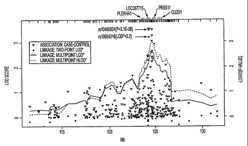
WO 2006/096561 PCT/US2006/007725
Genetic Variants
Increase the Risk of Age-Related
Macular Degeneration
[01] This invention was made using funds from U.S. government grant
no.Ul0EY012118.
and EY015216 from the National Institutes of Health (NIH)/National Eye
Institute and
by grant AG1 1268 from the NIH/National Institute on Aging and by RR 00095
from
the National Institutes of Health GCRC. Therefore the U.S. government retains
certain rights in the invention.
TECHNICAL FIELD OF THE INVENTION
[02] This invention is related to the area of genetic testing, drug discovery,
and Age-
Related Macular Degeneration. In particular, it relates to genetic variants
which
increase the risk of Age-Related Macular Degeneration, particularly in
combination
with certain behavior.
BACKGROUND OF THE INVENTION
[03] Age-related macular degeneration (AMD) causes progressive impairment of
central
vision and is the leading cause of irreversible vision loss in older Americans
(1). The
most severe form of AMD involves neovascular/exudative (wet) and/or atrophic
(dry)
changes to the macula. Although the etiology of AMD remains largely unknown,
implicated risk factors include age, ethnicity, smoking, hypertension, obesity
and diet
(2). Familial aggregation (3), twin studies (4), and segregation analysis (5)
suggest
that there is also a significant genetic contribution to the disease. The
candidate gene
approach, which focuses on testing biologically relevant candidates, has
implicated
variants in the ABCA4, FBLN6, and APOE genes as risk factors for AMD.
_sf
Replication of the ABCA4 and FBLN6 firrdings has been difficult, and in toto
these
variants explain only a small proportion of AMD (6-8). An alternative genomic
approach uses a combination of genetic linkage and association to identify
novel
genes iiivolved in AMD. We participated in a recent collaborative genome-wide
linkage screen (9) in which chromosome 1q32 was identified as a likely region
for an
1
CA 02599080 2007-08-31
WO 2006/096561 PCT/US2006/007725
AMD risk gene, a location also supported by other studies (10, 11). This
region
contains between over 100 genes, (see On-line Mendelian Inheritance in Man at
the
NCBI website) and no particular gene was identified by this work.
[04] Age-related macular degeneration (AMD) is a common complex disorder that
affects
the central region of the retina (macula) and is the leading cause of legal
blindness in
older American adults. The prevalence of AMD and its significant morbidity
will rise
sharply as the population ages. AMD is a clinically heterogeneous disorder
with a
poorly ixnderstood etiology. Population-based longitudinal studies (Klaver et
al. 2001;
van Leeuwen et al. 2003; Klein et al. 2003) have established that the presence
of
extracellular protein/lipid deposits (drusen) between the basal lamina of the
retinal
pigment epithelium (RPE) and the inner layer of Bruchs' membrane is associated
with
an increased risk of progressing to an advanced form of AMD, either geographic
atrophy or exudative disease. The presence of large and indistinct (soft)
drusen
coupled with RPE abnonnalities is considered an early form of the disorder and
is
often referred to as age-related maculopathy (ARM).
[05] Epidemiologically, AMD is a complex disorder with contributions of
environmental
factors as well as genetic susceptibility (Klein et al. 2004). Many
enviromnental and
lifestyle factors have been postulated, but by far the most consistently
implicated non-
genetic risk factor for AMD is cigarette smoking (Smith et al. 2001). Much
progress
has recently been made in identifying and characterizing the genetic basis of
AMD. In
a remarkable example of the convergence of methods for disease gene discovery,
multiple independent research efforts identified the Y402H variant in the
complement
factor H (CFH [(MIM 134370]) gene on chromosome 1q32 as the first major AMD
susceptibility allele (Haines et al. 2005; Hageman et al. 2005; Klein et al.
2005;
Edwards et al. 2005; Zareparsi et al. 2005; Conley et al. 2005). While one of
the
studies was able to pinpoint CFH on the basis of a whole-genome association
study
(Klein et al. 2005), most studies focused on the 1q32 region because it had
consistently been implicated by several whole-genome linkage scans. A second
genomic region with similarly consistent linkage evidence is chromosome 10q26,
which was identified as the single most promising region by a recent meta-
analysis of
published linkage screens (Fisher et al. 2005).
2
CA 02599080 2007-08-31
WO 2006/096561 PCT/US2006/007725
[06] Two recent studies have suggested specific AMD susceptibility genes
located on
chromosome 10q26. One used a combination of family-based and case-control
analyses to implicate the PLEKHAl gene (pleckstrin homology domain containing,
family A (phosphoinositide binding specific) member 1[MIIvl 607772]) and the
predicted LOC387715 gene (Jakobsdottir et al. 2005). However, the association
signals for single-nucleotide polymorphisms (SNPs) in these two genes were
statistically indistinguishable. A second study using two independent case-
control
datasets concluded that the T allele of SNP rs10490924 in LOC387715, a coding
change (Ala69Ser) in exon 1 of this poorly characterized gene, was the most
likely
AMD susceptibility allele (Rivera et al. 2005). Botli studies reported that
the
chromosome 10q26 variant confers an AMD risk similar in magnitude to that of
the
Y402H variant in CFH. Here, we describe highly significant association of SNPs
in
LOC387715 with AMD. In our data, only SNPs in this gene, including rs10490924,
explain the strong linkage and association signal in this region. Given a
previous
report of an effect of cigarette smoking on the linkage evidence in the 10q26
region
(Weeks et al. 2004; 9), we tested whether smoking modified this association.
[07] There is a continuing need in the art to identify individual genes that
are involved in
the pathogenesis of AMD and/or to identify particular alleles that are
involved in the
pathogenesis of AMD, as well as to identify the interaction of the genes with
modifiable behaviors.
SUMMARY OF THE INVENTION
[08] According to one embodiment of the invention a method is provided for
assessing
increased risk of Age Related Macular Degeneration. The identity is determined
of at
least one nucleotide residue of Complement Factor H coding sequence of
a.person.
The nucleotide residue is identified as normal or variant by comparing it to a
normal
sequence of Complement Factor H coding sequence as shown in SEQ ID NO: 1. A
person with a variant sequence has a higher risk of Age Related Macular
Degeneration
than a person with a normal sequence.
3
CA 02599080 2007-08-31
WO 2006/096561 PCT/US2006/007725
[09] According to another embodiment a method is provided for assessing
increased risk of
Age Related Macular Degeneration. The identity is determined of at least one
amino
acid residue of Complement Factor H protein of a person. The residue is
identified as
normal or variant by comparing it to a normal sequence of Complement Factor H
as
shown in SEQ ID NO: 2. A person with a variant sequence has a higher risk of
Age
Related Macular Degeneration than a person with a normal sequence.
[10] Another embodiment of the invention provides a method for screening for a
potential
drug for treating Age Related Macular Degeneration. A Complement Factor H
protein is contacted with a test agent in the presence of a polyanion. Binding
of the
polyanion to Complement Factor H is. measured. A test agent is identified as a
potential drug for treating Age Related Macular Degeneration if it increases
binding of
Complement Factor H to the polyanion.
[11] Another embodiment of the invention is a method for screening for a
potential drug
for treating Age Related Macular Degeneration. A Complement Factor H protein
is
contacted with a test agent in the presence of C-Reactive Protein. C-Reactive
Protein
binding to Complement Factor H is measured. A test agent is identified as a
potential
drug for treating Age Related Macular Degeneration if it increases binding of
Complement Factor H to C-Reactive Protein.
[12] A further embodiment of the invention is a method to assess risk of AMD
in a patient.
The presence of a T allele at rs 10490924 is determined in a patient. Whether
the
patient is a cigarette smoker is determined. The patient is identified as
being at high
risk of AMD if the patient has the T allele and is a cigarette smoker. The
patient is
identified as being at lower risk of AMD if the patient has the T allele but
is not a
cigarette smoker or is a cigarette smoker but does not have the T allele. The
patient is
identified as being at lowest risk if the patient does not have the T allele
and is not a
cigarette smoker.
[13] Yet another embodiment of the invention is a method to assess risk and
treat AMD in
a patient. The presence of a T allele at rs10490924 is determined in a
patient.
Whether the patient is a cigarette smoker is determined. If the patient has
the T allele
4
CA 02599080 2007-08-31
WO 2006/096561 PCT/US2006/007725
at rs10490924 and is a cigarette smoker, behavioral therapy is provided to the
patient
to encourage smoking cessation.
[14] Still another embodiment of the invention is a method to assess risk and
treat AMD in
a patient. The presence of a T allele at rs10490924 is determined in a
patient.
Whether the patient is a cigarette smoker is determined. If the patient has
the T allele
at rs10490924 and is a cigarette smoker, the patient is provided with
smokeless
nicotine to encourage smoking cessation.
BRIEF DESCRIPTION OF THE DRAWINGS
[15] Fig. 1. Haploview plot defining haplotype block structure of AMD
associated region.
The relative physical position of each SNP is given in the upper diagram, and
the
pairwise linlcage disequilibrium (D') between all SNPs is given below each SNP
combination. Dark red shaded squares indicated D' values >0.80. D'=1.0 when no
number is given.
[16] Fig. 2. Plot of family-based and case-control P values for all SNPs
within the AMD-
associated haplotype. The genomic region spanning each gene is indicated in
green. -
logio of the nominal P values are plotted for each SNP. Results for both the
family-
based and case-control data sets converge within the CFH gene.
[17] Fig. 3. Results of linkage (left axis: two-point and multipoint lod
scores) and
association analysis (right axis: loglo-transformed p-values from logistic
regression of
case-control dataset, using additive coding described in text and adjusted for
age and
sex). For exact p-values in 122-127 Mb region that are smaller than 10-3, see
Table 5.
[18] Fig. 4. LD pattern in region from PLEKHAI [MIM 607772] to CUZDl [HGNC
17937]. The relative physical position of each SNP is given in the upper
diagram, and
the pairwise D' between all SNPs is given below each SNP combination. Red-
shaded
squares indicate D' values >0.80. D'=1.0 when no number is given, which is
either
CA 02599080 2007-08-31
WO 2006/096561 PCT/US2006/007725
significant (dark-red shading) or non-significant (blue shading) based on the
Haploview default definition (Gabriel et al. 2002)
[19] Fig. 5A. genotype fiequencies at rs10490924 in unrelated AMD patients, by
pack-
years of cigarette smolcing. Fig. 5B, genotype frequencies at rs10490924 in
unrelated
controls without AMD, by pack-years of cigarette smoking
[20] Fig. 6. Ordered subset analysis of 90 multiplex AMD families with
information on
pack-years of cigarette smoking. Dashed line: Multipoint LOD* in 90 families.
Solid
line: Multipoint LOD* in 40 families with _44 pack-years, averaged across
family
members affected with AMD.
[21] Fig. 7: Table 4. Demographic and clinical characteristics of study
population
[22] Fig. 8: Table 5. SNPs in 122-127 Mb region with p_0.005 in case-control
association
analysis. MAF: minor allele frequency. Odds ratios (OR) adjusted for age and
sex,
estimated separately for heterozygous (het) and homozygous (het) carriers of
minor
allele. P-value from additive coding of SNP covariate described in text. GIST:
Genotype-IBD sharing test (Li et al. 2004).
[23] Fig. 9: Table 6. Two-locus genotype frequencies (%) and odds ratios for
rs10490924
in LOC3 87715 and Y402H in CFH. All odds ratios adjusted for age and sex.
[24] Fig. 10: Table 7. Results of fitting two-factor models by logistic
regression, adjusted
for age and sex. Factor 1 is rs10490924, model defmitions in text. Akaike's
information criterion (AIC) difference is difference of the AIC from the best-
fitting
model.
[25] Fig.11 Table 8. Joint frequencies (%) and odds ratios for rs10490924 in
LOC387715
and smoking history (ever vs. never). All odds ratios adjusted for age and
sex.
6
CA 02599080 2007-08-31
WO 2006/096561 PCT/US2006/007725
[26] Fig. 12: Table 9. Minor allele frequency (MAF) and genotype frequencies
(number of
individuals) at rs10490924 by AMD grade. Data for smokers and non-smokers
estimated from dataset used for logistic regression modeling (Table 8). Data
for all
genotyped individuals estimated by combining family-based and case-control
dataset,
including related individuals.
[27] Fig. 13: Supplemental Table 1. SNPs identified in LOC387715 sequencing of
individuals homozygous for rs10490924 variant
[28] Fig. 14: Supplemental Table 2. SNPs identified in CUZD1 sequencing of
individuals
homozygous for rs1891110 variant
[29] Fig. 15: Supplemental Table 3. Case-control association results for all
SNPs in 112-
132 Mb region.
DETAILED DESCRIPTION OF THE INVENTION
[30] The inventors have developed methods for assessing risk of developing Age-
Related
Macular Degeneration (AMD) in affected families and in individuals not known
to be
in affected families. Although developing the disease is a multi-factorial
process,
presence of a polymorphism in the CFH gene (or complement factor H protein)
indicates a greatly - increased risk (approximately double). Interestingly,
one
polymorphism is so prevalent in the Caucasian population that 1/3 of
individuals carry
at least one copy of that form. Moreover, identification of the CFH gene as
involved
in AMD pathogenesis permits the use of the CFH protein in drug screening
assays. In
addition, we have identified a coding change (Ala69Ser) in the LOC387715 gene
as a
second major susceptibility allele for AMD. The overall effect of the gene on
risk is
7
CA 02599080 2007-08-31
WO 2006/096561 PCT/US2006/007725
driven by a highly significant statistical interaction between the LOC387715
variant
and cigarette smoking.
[31] The Y402H polymoiphism (encoded by the T1277C polymorphism) is located in
the
domain known as SCR7. See Table 3. SCR7 is known to contain binding sites for
both C-Reactive Protein (CRP) and polyanions, such as heparin and sialic acid.
The
location of this highly informative polymorphism suggests that not only is the
CFH
protein involved in the pathogenesis of AMD, but that the ability to bind one
or both
of C-Reactive protein and polyanions is also involved. Variations in other
domains of
CFH may also relate to pathogenesis of AMD, including variations in domains
that
are involved in binding of complement factor C3b. Such variations may have an
effect alone or in conjunction with the Y402H variant.
[32] Any change in the CFH gene or encoded protein can be determined by
cornparing to
the sequences of the major allele in the Caucasian population as shown in SEQ
ID
NO: 1 and 3, for nucleotide and protein, respectively. Methods of detecting
sequence
differences between a test subject's CFH and the major allele or major protein
can be
any method known in the art. These include side-by-side comparisons of physico-
chemical properties of proteins, immunological assays, primer extension
methods,
hybridization methods, nucleotide sequencing, amino acid sequencing,
hybridization,
amplification, PCR, oligonucleotide mismatch ligation assays, primer extension
assays, heteroduplex analysis, allele-specific amplification, allele-specific
primer
extension, SCCP, DGGE, TGCE, mass spectroscopy, high pressure liquid
chromatography, and combinations of these techniques.
[33] Binding assays between Complement Factor H and either polyanions or C-
Reactive
Protein (CRP) can be performed using any format known in the art. Binding can
be
measured in solution or on a solid support. One of the partners may, for
example, be
labeled with a radiolabel or fluorescent label. Partners can be identified
using first
antibodies which are either themselves labeled or measured using second
antibodies
which are labeled and reactive with the first antibodies. Assay formats can be
competitive or non-competitive.
8
CA 02599080 2007-08-31
WO 2006/096561 PCT/US2006/007725
[34] Test agents can be natural products or synthetic, purified or mixtures.
They can be the
products of combinatorial chemistry or individual products or families of
products
which are selected on the basis of structural information. Test agents are
identified as
candidates for treating AMD if they increase the binding of complement factor
H to
any of its physiological binding partners, including but not limited to C3b,
sialic acid,
heparin, and CRP.
[35] The T allele is the variant of r00490924 that has a T at nucleotide 26 as
shown in
SEQ ID NO: 9. Other variant alleles as shown in SEQ ID NO: 7-56 can be
detected
and used to assess risk of AMD. The other variants may be used independently
or
may be used in conjunction with an assessment of smoker status. Current
smokers are
individuals who smoke at least once per week. However, historical smoking in
an
individual's past can also modify their risk of AMD.
[36] Behavioral therapies which can be recommended for smoking cessation
include but
are not limited to counseling, classes, printed information, electronic
information,
video or audio tapes. Providing a behavioral therapy may involve merely
recommending it to a patient, prescribing it, or actually delivering the
therapy.
Smokeless nicotine is also a possible means for weaning'persons from a smoking
habit. Smokeless nicotine, like behavioral therapies, may or may not require a
physician's prescription. Smokeless forms of nicotine that can be used for
smoking
cessation or abatement include but are not limited to nicotine gums,
transdermal
patches, nasal sprays, and inhalers.
[37] Because the data indicate that the variant of CFH and the variant of
LOC387715 are
independent predictive factors, they can both be assessed in the same person.
Together, these two types of variants are believed to account for the majority
of cases
of AMD. Additional factors as discovered can also be tested, as they become
available to the art.
[38] Using iterative high-density SNP association mapping, we have identified
a coding
change in the LOC387715 gene, at SNP rs10490924, as the most likely second
major
AMD susceptibility allele. We also generated statistical evidence of gene-
9
CA 02599080 2007-08-31
WO 2006/096561 PCT/US2006/007725
environment interaction for this variant, suggesting that a genetic
susceptibility
coupled with a modifiable lifestyle factor such as cigarette smoking confers a
significantly higher risk of AMD than either factor alone. Genotype
frequencies at
rs10490924 were strongly correlated with pack-years of smoking in AMD
patients,
consistent with heterogeneity analysis of the genetic linkage data. It is
striking that we
have observed evidence for gene-environment interaction in two different
datasets
using two statistically independent approaches . However, the presence of
statistical
interaction does not prove biological interaction, and much work remains to be
done
to identify the molecular mechanism underlying the increased AMD risk.
[391 Our data did not support the previously reported association of AMD with
the
GRK5/RGS10 region at -121 Mb (Jakobsdottir et al. 2005) since the four SNPs
(hcv1809962, rs871196, rs1537576, rs1467813) that we genotyped in this region
did
not demonstrate significant association (p>0.05). The GIST and conditional
haplotype
analyses suggested that only rs10490924, and surrounding SNPs in LOC387715 in
high LD with it, explained the linkage and association signals in this region.
See other
SNPs in LOC387715 at SEQ ID NO: 7-56. Neither analysis supported SNPs in the
nearby PLEKHA1 and PRSS 11 genes as being responsible for either the linkage
or
association evidence. Consistent with these results, the most significant
single-SNP
associations, the highest odds ratios, and the highest nonparametric two-point
lod
score of 3.2 were contributed by SNPs in the LOC387715 gene. While we did not
re-
sequence the nearby PLEKHAl and PRSS 11 genes, we genotyped the vast majority
of
SNPs examined by the earlier studies in our dataset. Several SNPs in the CUZD1
gene, which is not in LD with the PLEKHA1/LOC387715 LD block, gave substantial
association signals with logistic regression (smallest p-value: 0.0002), but
allele
frequency differences in cases and controls were much less pronounced for
these
SNPs (MAFcases -55%, 1VIAFcontro1s ~48%), compared to SNPs in LOC387715
(MAFcases-41%, MAFcontrols -26%). In addition, the GIST method and the
conditional
haplotype analysis suggested that these SNPs did not explain the linkage and
association signals in this region.
[40] The limitations of any retrospective epidemiologic study apply to our
findings,
including the potential for recall bias of past exposures. The validity of the
summary
CA 02599080 2007-08-31
WO 2006/096561 PCT/US2006/007725
PAR% estimates depends on the extent to which our case-control dataset is
representative of a population-based sample of AMD patients and controls.
Since our
dataset was used to identify the LOC387715 susceptibility variant, it is
possible that
its effect size, and hence its PAR%, was overestimated (Lohmueller et al.
2003;
loannidis et al. 2001). Independent population-based studies of large sample
size,
ideally collected in a prospective fashion, are needed to confirm the
statistical
interaction between smoking and rs10490924 in contributing to AMD and its
clinical
subtypes, and to refme estimates of their individual and joint PAR%.
[41] There is currently no biological explanation for the mechanism by which
LOC387715
may increase the risk of AMD. It is not clear whether this statistical
association
provides further support to the role of the innate immunity system that was
highlighted by the recent discovery of the CFH gene. LOC387715 is a two-exon
gene
that encodes a protein of 107 amino acids, whose only homologue is a
chimpanzee
gene of 97% protein identity. No significant matches were found with any known
protein motifs. ESTs have been recovered from the placenta and the testis, and
this
gene has recently been reported to be weakly expressed in the retina (Rivera
et al.
2005).
[42] In summary, we have replicated and refmed previous reports implicating a
coding
change in LOC387715 as the second major AMD susceptibility allele. The effect
of
rs10490924 appears to be completely independent of the Y402H variant in the
CFH
gene. The joint effect of these two susceptibility genes is consistent with a
multiplicative model, and together, they may explain as much as 65% of the PAR
of
AMD. Previous data by our group suggested that the joint effects of CFH and
smoking are also consistent with a multiplicative model (Scott et al. 2005).
In
contrast, the effect of rs10490924 appears to be strongly modified by
cigarette
smoking. Smoking and LOC387715 together may explain as much as 34% of AMD.
While the marginal effect of rs10490924 was strong enough to be detected
without
incorporating smolcing history information, an effect modification of a
genetic
susceptibility by a lifestyle factor like smoking has important implications
for the
clinical interpretation of this finding. Our data suggest that the T allele at
rs10490924
may only moderately increase the AMD risk in non-smokers and likely exerts its
11
CA 02599080 2007-08-31
WO 2006/096561 PCT/US2006/007725
strongest effect on heavy smokers. This has the potential to reduce the impact
of an
AMD susceptibility allele on the aging population by public health efforts,
such as
smoking prevention and smoking cessation programs. Our replication of the
10q26
linkage heterogeneity due to smoking, and the consistency of results from
multiple
statistically independent approaches for assessing gene-environment
interaction
reported here, are unusual in genetic studies of complex human diseases and
provide
substantial support to our fmdings.
[43] We used iterative association mapping to identify a susceptibility gene
for age-related
macular degeneration (AMD) on chromosome 10q26, which is one of the most
consistently implicated linkage regions for this disorder. We employed linkage
analysis methods, followed by family-based and case-control association
analysis
using two independent datasets. To identify statistically the most likely AMD
susceptibility allele, we used the Genotype-IBD Sharing Test (GIST) and
conditional
haplotype analysis. To incorporate the two most important known AMD risk
factors,
smoking and the Y402H variant of the complement factor H (CFH) gene, we used
logistic regression modeling to test for gene-gene and gene-environment
interaction in
the case-control dataset, and the ordered subset analysis (OSA) to account for
genetic
linkage heterogeneity in the family-based dataset. Our results strongly
implicate a
coding change (Ala69Ser) in the LOC387715 gene as the second major AMD
susceptibility allele, confirming earlier suggestions. Its effect on AMD is
statistically
independent of CFH and of similar magnitude to Y402H. The overall effect is
driven
primarily by a strong association in smokers, as we observed significant
evidence for a
statistical interaction of the LOC387715 variant with a history of cigarette
smoking.
This gene-environment interaction is supported by statistically independent
family-
based and case-control analysis methods. We estimate that LOC287715 and
smoking
together explain 34% of the population-attributable risk (PAR) of AMD.
Further, we
estimate that LOC387715 and CFH together account for 65% of the PAR of AMD.
For the first time, we demonstrate that a genetic susceptibility coupled with
a
modifiable lifestyle factor such as cigarette smoking confers a significantly
higher risk
of AMD than either factor alone.
12
CA 02599080 2007-08-31
WO 2006/096561 PCT/US2006/007725
[44] The above disclosure generally describes the present invention. All
references
disclosed herein are expressly incorporated by reference. A more complete
understanding can be obtained by reference to the following specific examples
which
are provided herein for purposes of illustration only, and are not intended to
limit the
scope of the invention.
EXAMPLE 1
[45] To identify the responsible gene on chromosome 1q32, we initially
genotyped 44
SNPs (12) across the 24 megabases (Mb) incorporating this linkage region. We
examined two independent data sets: the first contained 182 families (111
multiplex
and 71 discordant sibpairs) and the second contained 495 AMD cases and 185
controls. Each SNP was tested for association independently in both data sets.
Two
SNPs (rs2019724 and rs6428379) in moderate linkage disequilibrium with each
other
(r2=0.61) generated highly significant associations with AMD in both the
family-
based data set (rs2019724, P=0.0001; rs6428379, P=0.0007) and in the case-
control
data set (rs2019724, P<0.0001; rs6428379, P<0.0001). These SNPs lie
approximately
263 kilobases (Kb) apart.
EXAMPLE 2
[46] To defme the extent of linkage disequilibrium completely, an additional
17 SNPs
were genotyped across approximately 655 Kb flanked by rs1538687 and rs1537319
and encompassing the 263 Kb region. . Two linkage disequilibrium blocks of 11
Kb
and 74 Kb were identified and were separated by 176 Kb (Fig. 1). The 11 Kb
block
contained rs2019724 and the 74 Kb block contained rs6428379. Association
analysis
of the 17 SNPs identified multiple additional SNPs giving highly significant
associations in one or both of the family-based and case-control data sets
(Fig. 2). In
the case-control data set, a five SNP haplotype (GAGGT, defined by SNPs
rs1831281,
rs3753395, rs1853883, rs10494745, and rs6428279, respectively) comprised 46%
of
the case and 33% of the control chromosomes (P=0.0003). This same haplotype
was
also significantly over-transmitted to affected individuals in the family-
based data set
13
CA 02599080 2007-08-31
WO 2006/096561 PCT/US2006/007725
(P=0.00003). The convergence of the most significant associations to this same
haplotype in the two independent data sets strongly suggests that this region
contains a
commonly inherited variant in an AMD risk gene.
[47] The associated GAGGT haplotype spans approximately 261 Kb. It contains
the
Complement Factor H gene (CFH, OMIM #:134370, Accession #:NM_000186) and
the five Factor H-related genes CFHL1-5, and Iies within the Regulator of
Complement Activation (RCA) gene cluster. The most consistent association
results
(Fig. 2) from both the family-based and case-control data sets converge within
the
CFH gene implicating CFH as the AMD susceptibility gene. The biological role
of
Complement Factor H as a component of the innate immune system that modulates
inflammation through regulation of complement (reviewed in (13)) enhances its
attractiveness as a candidate AMD susceptibility gene. Inflammation has been
repeatedly implicated in AMD pathology. C-reactive protein levels are elevated
in
advanced disease (14), anti-retinal autoantibodies have been detected in AMD
patients
(15), macrophages are localized near neovascular lesions (16), and the
hallmark
drusen deposits contain many complement-related proteins (17).
EXAMPLE 3
[48] We screened for potential risk-associated sequence variants in the coding
region of
CFH by sequencing 24 cases with severe neovascular disease and 24 controls
with no
evidence of AMD. To maximize the likelihood of identifying the risk-associated
allele, all sequenced cases and controls were homozygous for the GAGGT
haplotype.
Five novel and six known sequence variants were detected (Table 1). Only one
variant (rs1061170, sequence: T1277C, protein: Y402H) was present
significantly
more often in cases than controls, occurring on 45/48 haplotypes in the cases
and on
22/48 haplotypes in the controls (P<0.0001). The frequency of sequence
variants
within the CFH coding region on the associated haplotype was significantly
reduced
in cases compared to controls (12% vs. 18%, P=0.002). When the over-
represented
T1277C variant was removed from the analysis, this difference became more
14
CA 02599080 2007-08-31
WO 2006/096561 PCT/US2006/007725
pronounced (3% vs. 16%, P<0.00001). Thus T1277C is the primary DNA sequence
variant differentiating between the case and control haplotypes.
CA 02599080 2007-08-31
WO 2006/096561 PCT/US2006/007725
Table 1. CFH sequence variants identified in neovascular AMD cases and normal
controls.
All individuals were homozygous for the AMD-associated GAGGT haplotype. The 24
affected individuals selected for sequencing had severe neovascular disease
(grade 5) (12)
with diagnosis before age 74 (mean age at diagnosis: 65.8 yrs). The 24 control
individuals
selected for sequencing had no evidence of AMD (grade 1) with age at exam
after age 64
(mean age at exam: 69.8 yrs). The six previously identified SNPs are labeled
using standard
nomenclature. The five novel variants are labeled given their base pair
location on
chromosome 1, Ensembl build 35. Five SNPs create non-synonymous amino acid
changes
within CFH and five SNPs create synonymous changes. Exon 1 is not translated.
Location SNP ID effect Minor Allele Frequency (%)
AMD Controls
exon 1 rs3753394 n/a 18 24
exon 2 rs800292 V621 0 6
exon 6 193,380,486 A/G R232R 0 2
exon 7 rs1061147 A307A 10 38
exon 8 193,390,164 C/T H332Y 0 5
exon 9 rs1061170 Y402H 94 46
exon 11 193,414,604 A/G A473A 0 31
exon 12 193,416,415 A/G T519A 0 2
exon 14 rs3753396 Q672Q 0 23
exon 18 193,438,299 C/T H878H 6 2
exon 19 HGVbase 000779895 E936D 0 23
16
CA 02599080 2007-08-31
WO 2006/096561 PCT/US2006/007725
EXAMPLE 4
[49] We screened for potential risk-associated sequence variants in the coding
region of
CFH by sequencing 24 cases with severe neovascular disease and 24 controls
with no
evidence of AMD. To maximize the likelihood of identifying the risk-associated
allele, all sequenced cases and controls were homozygous for the GAGGT
haplotype.
Five novel and six known sequence variants were detected (Table 1). Only one
variant (rs1061170, sequence: T1277C, protein: Y402H) was present
significantly
more often in cases than controls, occurring on 45/48 haplotypes in the cases
and on
22/48 haplotypes in the controls (P<0.0001). The frequency of sequence
variants
within the CFH coding region on the associated haplotype was significantly
reduced
in cases compared to controls (12% vs. 18%, P=0.002). When the over-
represented
T1277C variant was removed from the analysis, this difference became more
pronounced (3% vs. 16%, P<0.00001). Thus T1277C is the primary DNA sequence
variant differentiating between the case and control haplotypes.
EXAMPLE 5
[50] Complete genotyping of T1277C in the family-based and case-control data
sets
revealed a significant over-transmission in the families (P=0.019) (12) and a
highly
significant over-representation in the cases compared to controls (P=0.00006).
The
odds ratio for AMD was 2.45 (95% CI: 1.41-4.25) for carriers of one C allele
and 3.33
(95% CI: 1.79-6.20) for carriers of two C alleles. When the analysis was
restricted to
only neovascular AMD, these odds ratios increased to 3.45 (95% CI: 1.72-6.92)
and
5.57 (95% CI: 2.52-12.27), respectively. This apparent dose effect for risk
associated
with the C allele was highly significant (P<0.0001). There was no apparent
allelic or
genotypic effect of T1277C on age at AMD diagnosis (mean age at diagnosis: TT:
76.5yrs; TC 77.5yrs; CC 75.5 yrs). The population attributable risk percent
for
carrying at least one C allele was 43% (95% confidence interval 23-68%).
17
CA 02599080 2007-08-31
WO 2006/096561 PCT/US2006/007725
[51] The Y402H variant is predicted to have functional consequences consistent
with
AMD pathology. Residue 402 is located within binding sites for heparin (18)
and C-
reactive protein (CRP) (19). Binding to either of these partners increases the
affinity
of CFH for the complement protein C3b (20, 21), augmenting its ability to down-
regulate complement's effect. The observed co-localization of CFH, CRP, and
proteoglycans in the superficial layer of the arterial intima suggests that
CFH may
protect the host arterial wall from excess complement activation (22). We
hypothesize that allele-specific changes in the activities of the binding
sites for
heparin and CRP would alter CFH's ability to suppress complement-related
damage to
arterial walls, and might ultimately lead to vessel injury and subsequent
neovascular/exudative changes such as those seen in neovascular AMD. Our data
support this hypothesis since the risk associated with the C allele is more
pronounced
when the analyses are restricted to neovascular AMD. Given the known
functional
interactions of genes within the RCA gene cluster (13), variants within these
genes
could interact with or modify the effect of the T1277C variant.
[521 Interestingly, plasma levels of CFH are known to decrease both with age
and with
smoking (23), two known risk factors for AMD (2). This confluence of genetic
and
environmental risk factors suggests an integrated etiological model of AMD
involving
chronic inflammation. Identification of the increased risk of AMD associated
with the
T1277C variant should enhance our ability to develop presymptomatic tests for
AMD,
possibly allowing earlier detection and better treatment of this debilitating
disorder.
EXAMPLE 6 (relates to examples 1-5)
Participants
[53] We ascertained AMD patients and their affected and unaffected family
members
through two clinics in the Southeastern United States - Duke University
Medical
Center (DUMC) and Vanderbilt University Medical Center (VUMC). Unrelated
controls of similar age and ethnic background were enrolled via (i) study
advertisement in DUMC- and VUMC-affiliated newsletters; (ii) recruitment
presentations by study coordinators at local retirement communities, who were
likely
18
CA 02599080 2007-08-31
WO 2006/096561 PCT/US2006/007725
to obtain health care at DUMC or VUMC, respectively; (iii) AMD-related
seminars
for the general public sponsored by DUMC or VUMC ophthalmology clinics. (iv)
referrals from other clinics in the Duke and Vanderbilt Eye Centers of
individuals
without evidence of ocular disease. Spouses of AMD patients were also asked to
participate as potential controls. Controls eligible for enrollment were
offered a free
comprehensive eye exam including fundus photography to ensure that the same
methodology was used to assign AMD grades as for the AMD patients and their
relatives ascertained in clinic. All cases and controls included in this study
were
Caucasian and at least 55 years of age. The study protocol was approved by the
respective Institutional Review Boards (IRB) at DUMC and VUMC, and the
research
adhered to the tenets of the Declaration of Helsinki.
[54] The family-based data set consisted of 111 multiplex families with at
least two
individuals with grade 3 or higher AMD in at least one eye. Seventy-three
families
had two affected individuals, 29 families had three affected individuals, and
nine
families had four or more affected individuals. Unaffected spouses and
siblings were
collected whenever possible. 71 additional families consisted of one affected
individual and at least one unaffected sibling (discordant sibpairs).
Clinical Assessment
[55] The assignment of AMD affection status was based on the clinical
evaluation of
stereoscopic color fundus photographs of the macula (EAP, AA), according to a
5-
grade system described previously (SI). Grade 1 has no AMD features, grade 2
has
only small non-extensive drusen, grade 3 has extensive intermediate and/or
large
drusen, grade 4 is geographic atrophy, and grade 5 is neovascular AMD. This
system
is a slight modification of the Age-Related Eye Disease Study (AREDS) grading
system and uses example slides from the Wisconsin Grading System .(S2) and the
International Classification System (S3) as guides. Affection status was
defined by
the most severe grade in either eye. All questionnaire data and samples were
collected
after informed consent was obtained.
19
CA 02599080 2007-08-31
WO 2006/096561 PCT/US2006/007725
Molecular Analyses
[56] Genomic DNA was extracted from wliole blood by the Duke CHG or Vanderbilt
CHGR DNA banking cores using the PureGene system (Gentra Systems,
Minneapolis, MN) on an Autopure LS. Genotyping was performed using Taqman on
the ABI Prism 7900HT, and analyzed with the SDS software. SNP Assays-On-
Demand or Assays-By-Design were obtained from Applied Biosystems Incorporated
(Foster City, CA). The initial set of 44 SNPs was chosen to approximate a 500
Kb
spacing between markers.
[57] Exons of CFH were PCR amplified from genomic DNA, sequenced using Big Dye
v3.1 (ABI) on an ABI 3730 automated sequencer, and analyzed using Mutation
Surveyor software (Softgenetics, State College, PA). T1277C falls within a
genomic
duplication and could not be genotyped using TaqMan assays. All individuals
were
sequenced using primers GGTTTCTTCTTGAAAATCACAGG (SEQ ID NO: 5) and
CCATTGGTAAAACAAGGTGACA (SEQ ID NO: 6) to determine T1277C
genotypes.
Statistical Analyses
[58] Linkage disequilibrium and Hardy-Weinberg equilibrium calculations were
done
using Haploview version 3.0 using all case and control samples and one random
individual from each of the families (S4). Haplotype blocks were defmed using
the D'
parameter and the default definitions within Haploview. Allele frequency
differences
were tested using a x2 test.
[59] Single-locus and haplotype family-based association was tested using the
Association
in the Presence of Linkage (APL) method (S5) that performs a correct TDT-style
test
of association in the presence of linkage, using nuclear families with at
least one
affected individual and any number of unaffected siblings or parents. Odds
ratios
were calculated using standard logistic regression models (SAS version 9.1,
SAS
Institute, Cary, NC). The outcome variable was AMD affection status and
genotypes
were coded according to a log-additive model. Dose-response was tested using
the xZ
CA 02599080 2007-08-31
WO 2006/096561 PCT/US2006/007725
test for trend. Haplotype analysis in the case-control data set was tested
using the
"haplo.stats" program that uses a lilcelihood-based method to estimate
haplotype
frequencies (S6).
[60] The 95% confidence interval for the population attributable risk percent
(PAR%) for
T1277C was calculated on the point estimate of the PAR% (43%), which was
calculated from the combined frequency of genotypes CT and CC in controls and
the
unadjusted odds ratio (OR) of AMD for these genotypes relative to the TT
reference
group (S7). Calculation of the PAR% from case-control data assumes that the
controls
are representative of the general population and the disease is rare (< 5%
population
prevalence across all exposure levels). PAR% calculated from OR adjusted for
age
and sex was similar.
[61] We note that the P-value of the T1277C association in the family-based
data set is not
as significant as the P-value for the two original SNPs. This results from the
ascertainment bias toward severe disease in the family collection, which
results in an
oversampling of T1277C-CC homozygotes. Family-based tests of association
depend
on both transmission and association. Oversampling for homozygosity reduces
the
power of any family-based transmission disequilibrium test. Since the original
SNPs
have low linkage disequilibrium values with T1277C (rz=0.00 and 0.14 for
rs2019724
and rd6428379, respectively), they were not over-sampled for homozygosity to
the
extent of T1277C. In the case-control data set where the sampling bias is not
as
profound, the P-values for all three SNPs are similarly highly significant.
Haplotype Analysis
[62] The five SNP haplotype block, defined by SNPs rs1831281, rs3753395,
rs1853883,
rs10494745, and rs6428279, identified five common haplotypes that capture over
95%
of the haplotype variation (Table 2). The GAGGT haplotype is the most common
in
both the cases and controls, but is significantly more frequent in the cases.
Table 2. The haplotypes and their frequencies calculated from the case-control
data. The
haplotype consists of SNPs rs1831281, rs3753395, rs1853883, rs10494745, and
rs6428279,
respectively.
21
CA 02599080 2007-08-31
WO 2006/096561 PCT/US2006/007725
Haplotype Haplotype Frequency
Cases Controls
GAGGT 0.46 0.33
GAGAT 0.16 0.11
GACGC 0.15 0.15
ATCGC 0.13 0.22
GTCGC 0.08 0.16
Other 0.02 0.03
Table 3. Location of SCR domains in protein.
SCR start aa position in mature end aa position in mature length start in pre-
protein end in pre-protein
protein protein
1 1 62 62 19 80
2 63 123 61 81 141 .
3 124 188 65 142 206
4 189 245 57 207 163
246 302 57 164 320
6 303 367 65 321 385
7 368 425 58 386 443
8 426 488 63 444 506
9 489 547 59 507 565
548 606 59 566 624
11 607 668 62 625 686
12 669 729 61 687 747
13 730 787 58 748 805
14 788 847 60 806 865
848 908 61 866 926
16 909 967 59 927 985
17 968 1026 59 986 1044
18 1027 1085 59 1045 1103
19 1086 1146 61 1104 1164
1147 1213 67 1165 1231
EXAMPLE 7
Linkage and Association Analysis
[63] Resequencing of the LOC387715 and CUZD1 genes identified 21 known and 23
novel SNPs (Supplemental Tables 1 and 2). Sequencing primers and conditions
are
available from the authors (MAH) upon request. Of these 44 SNPs, 19 were
genotyped in our entire dataset. Genotypes for all SNPs analyzed here were in
Hardy-
Weinberg equilibrium in unrelated controls (p>0.01). We observed high LD
(D'>0.9)
22
CA 02599080 2007-08-31
WO 2006/096561 PCT/US2006/007725
across a 60 kb region including a frequent coding SNP in exon 12 of PLEKHAI
(rs1045216), three coding SNPs in LOC387715 (rs10490923, rs2736911,
rs10490924) and several additional non-coding PLEKHAl and LOC387715 SNPs,
replicating earlier observations (Rivera et al. 2005). Notably, the adjacent
downstream
gene PRSS 11 (HtrA serine peptidase 1(HTR.A1), [MIM 602194]) was not included
in
this 60 kb region (figure 2).
[64] In the family-based linkage analysis, a peak multipoint lod score was
obtained at
124.7 Mb (HLOD 3.0 under affecteds-only dominant model, nonparametric LOD*
2.6, figure 1). SNP rs10664316 in LOC387715 (124.2 Mb) gave a maximum
nonparametric two-point lod score of 3.2. In the case-control analysis, four
highly
correlated SNPs in the LOC387715 gene, including the frequent coding change
rs10490924 in exon 1 previously implicated (Rivera et al. 2005), were very
strongly
associated with AMD, with logistic regression p-values on the order of 10"g
(table 5).
The minor allele frequency (MA.F) of these highly correlated SNPs was -41.7%
in
cases, very similar to that reported by Rivera et al., and -25.8% in controls,
somewhat
higher than the 19.6% reported by Rivera et al. Within the 60 kb LD block, and
in the
entire 122-127 Mb region, association signals of this order of magnitude were
observed only for this set of highly correlated SNPs. In particular, the
coding SNP in
exon 12 of PLEKHAI (rs1045216) showed substantially weaker evidence for
association, both in terms of magnitude (odds ratio, OR) and statistical
significance
(MAFcases: 28=2%, MAFcontrols: 36.8%, OR=0.6, p=0.02). Unlike the previous
reports,
we detected a second region of association 400 kb distal to LOC387715 that
included
several SNPs in the CUZDl gene and an even more distal SNP in the FAM24A gene
(family with sequence similarity 24, member A [HGNC: 23470]). These SNPs,
which
were in LD with each other but not in LD with the associated SNPs in LOC387715
(figure 2), showed independent evidence for association with AMD risk,
although at
much lower statistical significance (MAFcases: ~'55%, IVIAFeoritrols: -48%,
p=0.0002-
0.005 8).
EXA.MPLE 8
GIST Analysis
23
CA 02599080 2007-08-31
WO 2006/096561 PCT/US2006/007725
[65] All SNPs with p-values _0.005 in the case-control analysis were analyzed
with GIST
to test if they explained the linkage signal in the region. Under the additive
weighting
scheme suggested by the case-control analysis (Li et al. 2004), only the four
SNPs in
the LOC387715 gene were significant in the GIST analysis (table 5). This
suggests
that the LOC387715 gene alone is responsible for the 10q261inkage evidence.
EXAMPLE 9
Conditional Haplotype Analysis
[66] With the combined case-control dataset, we used conditional haplotype
modeling to
identify the statistically most likely AMD susceptibility variant from among
all the
SNPs with strong evidence for association. We tested each SNP in table 5,
conditioning on the risk allele of the most strongly associated SNP in CUZD1,
FAM24A and LOC3 87715. Conditioning on the risk allele at rs 1891110 in CUZD
1,
rs10490924 was strongly associated (p=7.6E-05) while none of the other SNPs
were
significant (p>0.05). Conditioning on the risk allele at rs2293435 in FAM24A,
rs10490924 was strongly associated (p=7.1E-05) while none of the other SNPs
were
significant (p>0.05). Only conditioning on the risk allele at rs10490924 fully
explained the association signal in the region, such that none of the other
SNPs
showed any evidence for association (p>0.6). Thus, this analysis also strongly
implicates the LOC387715 gene alone in AMD, consistent with the Rivera et al.
study.
EXAMPLE 10
Gene-Gene Interaction analysis
[67] We estimated joint odds ratios for all genotype combinations of the Y402H
variant in
CFH and the rs10490924 variant in LOC387715 (table 6). The TT/GG combination
was used as the referent group. For individuals with the TT genotype at Y402H,
the
GT genotype at rs10490924 conferred a 2.7-fold increase in AMD risk (p=0.02)
and
the TT genotype conferred a 13.1-fold increase (p=0.003). For individuals with
the
24
CA 02599080 2007-08-31
WO 2006/096561 PCT/US2006/007725
CC genotype at Y402H, which conferred a 4=fold increase in AMD risk for TT
genotypes at rs10490924 (p=0.0007), the GT genotype conferred a 12.6-fold
increase
in AMD risk (p<0.0001) and the TT genotype conferred a 23.8-fold increase
(p<0.0001). Consistent with results of the AIC modeling strategy (table 7),
the joint
action of the Y402H and the rs10490924 variants was therefore best described
by
independent multiplicative effects, without statistically significant evidence
for
dominance effects or epistatic interaction. The joint effect of Y402H and rs
10490924
accounted for 65.1% of the population attributable risk (PAR) of AMD (Bruzzi
et al.
1985).
EXAMPLE 11
Case-Control Gene-Environment Interaction Analysis
[68] In contrast, we found strong evidence for statistical interaction of
smoking and
genotypes at rs10490924. The model with the ADD SMOKE INT term provided a
significantly better fit to the data by 5.2 AIC units, compared to the model
without this
term (table 7). A significant product term with positive regression
coefficient for
smoking and rs10490924 in the logistic regression model indicated more than
multiplicative joint effects (p=0.007). In our dataset, the presence of the
LOC387715
susceptibility allele did not confer a significantly increased risk of AMD to
non-
smokers (p=0.59 for GT genotype, p=0.12 for TT genotype, table 8), while the
GT
genotype in smokers increased the risk 2.7-fold (p=0.001) and the TT genotype
in
smokers increased the risk 8.2-fold (p<0.0001). A case-only analysis of
rs10490924
and pack-years of smoking (as a continuous variable) also supported the
presence of
gene-environment interaction (p=0.05 adjusted for age and sex). The relative
frequency of TT genotypes in affected individuals increased ahnost linearly
with
increasing pack-years of smoking, with a corresponding decrease of GG genotype
frequencies (figure 3, panel A). This pattern was strikingly similar to
results for
simulated data when the disease status was generated with a logistic
regression model
including a gene-environment interaction term (Schmidt et al. 2005). Genotype
frequencies at rs10490924 were not related to pack-years of smoking in our
control
sample (Fig. 5B), confirming that the result in cases was due to gene-
environment
CA 02599080 2007-08-31
WO 2006/096561 PCT/US2006/007725
interaction rather than population correlation of the two factors. The joint
effect of
rs10490924 and smolcing accounted for 34.3% of the PAR of AMD.
EXAMPLE 12
Family-Based Gene-Environment Interaction Analysis
[691 The highly significant association of AMD with rs10490924 that was
observed in the
initial case-control analysis was not replicated in the family-based analysis
with APL.
This could be due to the smaller size of our family-based dataset, or to
between-family
heterogeneity. To test the latter possibility, we applied OSA to our multiplex
family
dataset, using the average pack-years of smoking in affected individuals as
the OSA
covariate (ordered from high to low). OSA indicated that the majority of
linkage
evidence in the 10q26 region was contributed by only 40 families with an
average of >_
44 pack-years of smoking (figure 4). The difference in nonparametric lod
scores
between the 90 multiplex families with sufficient information to calculate
average
smoking pack-years and the 40 families with heavy smokers was significant
(p=0.048), based on 10,000 runs of the OSA permutation test (Hauser et al.
2004).
When the APL analysis was repeated using only multiplex and singleton families
which met the "heavy smoking" criterion in affected individuals (family-
average of >_
44 pack-years of smoking, 46 families total), the results confirmed the case-
control
association analysis: The APL p-value for rs10490924 and rs3750848 in
LOC387715
was 0.02. Three SNPs in other genes also had p-values of 0.02: rs760336 in
PRSS11
adjacent to LOC387715, rsl052715 in DMBT1 (deleted in malignant brain tu.mors
1
[MIM 601969]) and hcv2917031 in GPR26 (G protein-coupled receptor 26 [MIM
604847]). Neither SNP had a case-control association p-value<0.05 in the
overall
analysis.
EXAMPLE 13
Clinical Subgroup Analysis
[70] It is of great clinical interest to determine whether the modification of
the LOC387715
association by cigarette smoking is observed in both geographic atrophy (GA,
grade 4)
26
CA 02599080 2007-08-31
WO 2006/096561 PCT/US2006/007725
and neovascular AMD (CNV, grade 5). Table 9 shows that the strong association
with
LOC387715 in smokers was primarily due to genotype frequency differences
between
grade 1 controls (8.3% with genotype TT) and CNV patients (29.3% with genotype
TT). When all genotyped individuals regardless of smoking history information
were
evaluated, the frequency of the T allele was higher in patients with CNV
(47.6%)
compared to GA (39.0%). Our dataset had limited statistical power for the AMD
subtype comparison since it included a much smaller number of GA patients,
compared to CNV patients (table 4), and since smolcing history information was
not
available for all study participants.
EXAMPLE 14 (relates to examples 7-13)
Study population
(71] As part of an ongoing large-scale study of genetic and environmental risk
factors for
AMD, we have ascertained AMD patients, their affected and unaffected. family
members, and a group of unrelated controls of similar age and ethnic
background at
two sites in the Southeastern United States: Duke University Eye Center (DUEC)
and
Vanderbilt University Medical Center (VUMC). Using stereoscopic color fundus
photographs, all enrolled individuals were assigned (by EAP and AA) one of
five
different grades of macular findings, as described previously (Schmidt et al.
2000;
Seddon et al. 1997) and suxnmarized in Table 4. Our AMD classification is a
modification of the AREDS grading system, using Wisconsin grading system
example
slides (Klein et al. 1991) and the International Classification System (Bird
et al. 1995)
as guides. The more severely affected eye was used to classify individuals.
Unrelated
controls were enrolled via (i) study advertisement in DUEC- and VUMC-
affiliated
newsletters; (ii) recruitment presentations by study coordinators at local
retirement
communities, which were likely to obtain health care at DUEC or VUMC,
respectively; and (iii) AMD-related seminars for the general public sponsored
by
DUEC or VUMC ophthalmology clinics. Spouses of AMD patients were also asked to
participate as controls. All cases and controls included in this study were
white and at
least 55 years of age. The study protocol was approved by the Institutional
Review
Boards (IRB) of the Duke University Medical Center and VUMC, the research
27
CA 02599080 2007-08-31
WO 2006/096561 PCT/US2006/007725
adhered to the tenets of the Declaration of Helsinki, and informed consent was
obtained from all study participants. Blood samples were collected and genomic
DNA
was extracted from whole blood using the PureGene system (Gentra Systems,
Minneapolis, MN) on an Autopure LS.
[72] Information about the smoking history of study participants was obtained
from a self-
administered questionnaire that was formatted to maximize readability for
individuals
with low vision. However, if participants indicated that they could not
complete the
form, a project coordinator offered to assist the participants in filling out
the
questionnaire. Regular cigarette smoking was assessed by two questions: 1)
"Have
you smoked at least 100 cigarettes in your lifetime?" and 2) "Did you ever
smoke
cigarettes at least once per week?" Individuals answering "yes" to both
questions
were asked the average number of cigarettes they smoked per day, the year that
they
started smoking, whether they had quit smoking, and if so, what year. This.
information was used to calculate pack-years of smokiing as (cigarettes per
day * years
smoked) / 20 cigarettes per pack. The most general measurement of smoking
history
was constructed as an "ever/never" variable based on a participant's response
to
question 1) above.
[73] The study population for the analysis presented here included 810
unrelated AMD
patients with early (grade 3) or advanced (grades 4 and 5) AMD. Of these, 200
had at
least one sampled (affected or unaffected) relative and thus contributed to
the family-
based association analysis. The remaining 610 AMD patients without sampled
relatives, and 259 unrelated controls without AMD (grades 1 and 2), made up an
independent case-control dataset. Demographic and clinical information for
these
individuals is shown in table 4.
Genotyping, Linkage and Association Analysis
[74] Previous work by our group (Kenealy et al. 2004) and others (Weeks et al.
2004;
Majewski et al. 2003; Seddon et al. 2003; Iyengar et al. 2004) suggested the
presence
of an AMD susceptibility locus on chromosome 10q26, with the linkage pealc
centered
28
CA 02599080 2007-08-31
WO 2006/096561 PCT/US2006/007725
at approximately 122 Mb. To narrow down the region most likely to harbor an
AMD
susceptibility allele, we genotyped 103 SNPs in the 112 to 132 Mb interval,
extending
Mb to either side of the reported linkage peak. We started with a density of
approximately 1 SNP per 1 Mb and filled in the 117-127 Mb region immediately
surrotinding the 122 Mb peak with a higher density of one SNP per 140 lcb on
average. All SNPs were selected using SNPSelector software (Xtt et al. 2005)
to have
approximately equal spacing with minor allele frequency _ 5%. Genotyping was
performed with the TaqMan allelic discrimination assay, using either Assays-On-
Demand or Assays-By-Design products from Applied Biosystems. For quality
control
(QC) procedures, two CEPH standards were included on each 96-well plate, and
samples from six individuals were duplicated across all plates, with the
laboratory
technicians blinded to their identities. Analysis required matching QC
genotypes
within and across plates and at least 95% genotyping efficiency. The Y402H
variant
of the CFH gene was genotyped by sequencing, as previously described (Haines
et al.
2005).
[75] Following the first round of genotyping and statistical analysis, we
applied iterative
association mapping (Oliveira et al. 2005) to select another set of SNPs in
the peak
region, defined approximately as the 1-lod-score-unit support interval
surrounding the
peak multipoint lod score. In addition to using SNPSelector (Xu et al. 2005),
SNPs
were identified through resequencing of the LOC387715 gene and the CUZD1 gene
(CUB and zona pellucida-like domains 1[HGNC: 17937]) in 48-72 unrelated
affected
and unaffected individuals. Our final SNP density was an average of one SNP
per 43
kb, for a total of 117 SNPs in the 122-127 Mb region, and an average of one
SNP
every 220 kb outside of this interval, for a total of 185 SNPs in the 112-132
Mb
region.
[76] The genotype data were analyzed with MERLIN (Abecasis et al. 2002) to
calculate
nonparametric two-point and multipoint LOD* scores (Kong and Cox 1997), using
the exponential model. Allele frequencies were estimated from all genotyped
individuals. Parametric affecteds-only heterogeneity lod scores (HLODs)
assuming a
dominant (disease allele frequency 0.01) or recessive (disease allele
frequency 0.2)
model were also computed with MERLIN. To avoid an inflation of linkage
evidence
29
CA 02599080 2007-08-31
WO 2006/096561 PCT/US2006/007725
due to inter-marlcer linkage disequilibrium (LD) (Boyles et al. 2005), we used
recently
described methods based on estimated haplotype frequencies of SNP clusters in
high
pairwise LD, using a threshold of r2=0.16 to define these clusters (Abecasis
and
Wigginton 2005). The LD pattern in the region of interest was analyzed with
the
Haploview program (Barrett et al. 2005), using the generated genotypes from
unrelated AMD patients as the input. Association analysis was applied to all
SNPs in
the 122-127 Mb region, using the family-based Association in the Presence of
Linkage
(APL) test (Martin et al. 2003) and standard logistic regression analysis for
case-
control comparisons with adjustment for age and sex (SAS version 8.02, SAS
Institute
Inc., Cary, NC). An additive coding scheme was used, with the SNP model
covariate
taking on values -1, 0 and 1 for genotypes 1/1, 1/2, and 2/2, and 2 being the
minor
allele in controls. As described above, we divided our total sample into cases
contributing to the APL analysis (affected individuals with at least one
sampled
relative, n=200 families), and an independent sample of cases without sampled
relatives (n=610) who were compared to 259 unrelated controls. We used the
Genotype-IBD Sharing Test (GIST) method (Li et al. 2004) to examine which of
the
most strongly associated SNPs best explained the linkage evidence in the
region. We
also used the COCAPHASE module of the UNPHASED software package
(Dudbridge 2003) to perform conditional haplotype analysis. This analysis
tested
whether conditioning on the risk allele at a particular SNP accounted for the
association signal in the region. If the association signal in the region was
driven by a
single SNP, conditioning on its effect was expected to remove all evidence of
association for the remaining SNPs.
Interaction Analysis
[77] We conducted additional analyses to incorporate effects of the two most
important
known AMD risk factors, smoking and the CFH gene. First, we fit a series of
logistic
regression models to the combined case-control data set (including probands
from
family-based dataset) to identify the model that best described (1) the joint
effects of
CFH and LOC387715, and (2) the joint effects of smoking and LOC387715. We
followed a recently proposed modeling strategy (North et al. 2005) in which
the best-
CA 02599080 2007-08-31
WO 2006/096561 PCT/US2006/007725
fitting model was derived on the basis of Akaike's Information Criterion
(AIC). The
AIC compares different models with a log-likelihood ratio test that is
penalized for the
number of model parameters to identify the most parsimonious model that
adequately
fits the data. For each genotype, two model terms were tested: one coding for
additive
effects at the first, second, or both loci (ADD1, ADD2, ADDBOTH), using the
coding
described above, and the other one coding for dominance effects (DOM1, DOM2,
DOMBOTH), with a value of -0.5 for genotypes 1/1 and 2/2, and a vahie of 0.5
for
genotype 1/2. Three additional models (ADDINT, ADDDOM, DOMINT) were fit to
test for deviation from joint additive or joint dominance effects of CFH and
LOC387715, and two additional models (ADD_SMOKE INT, DOIVI SMOKE INT)
were fit for LOC387715 and smoking (comparing ever- vs. never-smokers). Models
for which the AIC differed by less than 2 units were considered statistically
indistinguishable (North et al. 2005), and the model with fewer parameters was
chosen as the best fitting one. For example, when the addition of the ADDINT
term
did not provide a substantially better model fit, this was interpreted as lack
of
evidence for statistical interaction between the two factors. Thus, they each
had
independent main effects that were multiplicative (additive on the logarithmic
scale)
such that the best estimate of the odds ratio for being exposed to both
factors was the
product of the two main effect odds ratios.
[78] Our second approach for incorporating AMD-associated covariates was
motivated by
earlier reports of the 10q26 linkage evidence being due primarily to families
with
heavy smokers (Weeks et al. 2004). Similar to the previous study, we used an
ordered
subset analysis (OSA) (Hauser et al. 2004) with the family-average of smoking
pack-
years as a covariate. To avoid an undue influence of zero pack-years values on
family
averages, pack-years were coded as missing for non-smokers. Using the high-to-
low
ordering of family-averaged pack-years, OSA tested whether a subset of
families with
heavy smokers provided significantly greater linkage evidence than the
reference
dataset, which in this case was restricted to families for whom non-missing
covariate
values could be computed. Thus, the baseline lod score was computed for
families in
which there was at least one affected smoker with pack-years information.
31
CA 02599080 2007-08-31
WO 2006/096561 PCT/US2006/007725
References
[79] The disclosure of each ref.erence cited is expressly incorporated herein
for the ptupose
to which is referenced in the text.
1. Centers for Disease Control and Prevention (CDC), MMWR Morb. Mof tal.
Wlcly. Rep.
53, 1069 (2004).
2. J. Ambati, B. K. Ambati, S. H. Yoo, S. Ianchulev, A. P. Adamis, Surv.
Ophtlialmol.
48, 257 (2003).
3. C. C. Klaver et al., Arch. Ophthalmol. 116, 1646 (1998).
4. C. J. Hammond et al., Ophthalnaology. 109, 730 (2002).
5. M. Heiba, R. C. Elston, B. E. Klein, R. Klein, Genet. Epidemiol. 11, 51
(1994).
6. E. M. Stone et al., Nat. Genet. 20, 328 (1998).
7. S. Schmidt et al., Ophtlaalmic Genet. 23, 209 (2002).
8. E. M. Stone et al., N. Engl. J. Med. 351, 346 (2004).
9. D. E. Weeks et al., Am. J. Hum. Genet. 75, 174 (2004).
10. G. R. Abecasis et al., Am. J. Hurn. Genet. 74, 482 (2004).
11. S. K. Iyengar et al., Am. J. Hunz. Genet. 74, 20 (2004).
12. Materials and methods are provided in Examples 6.
13. D. C. Rodriguez, J. Esparza-Gordillo, d. J. Goicoechea, M. Lopez-Trascasa,
P.
Sanchez-Corral, Mol. Irnrnunol. 41, 355 (2004).
14. J. M. Seddon, G. Gensler, R. C. Milton, M. L. Klein, N. Rifai, JAMA 291,
704 (2004).
15. D. H. Gume, M. O. Tso, D. P. Edward, H. Ripps, Ophthalmology. 98, 602
(1991).
16. M. C. Killingsworth, J. P. Sarks, S. H. Sarlcs, Eye 4 (Pt 4), 613 (1990).
17. R. F. Mullins, S. R. Russell, D. H. Anderson, G. S. Hageman, FASEB J. 14,
835
(2000).
32
CA 02599080 2007-08-31
WO 2006/096561 PCT/US2006/007725
18. T. K. Blackmore, V. A. Fischetti, T. A. Sadlon, H. M. Ward, D. L. Gordon,
Infect.
Immun. 66, 1427 (1998).
19. E. Giannakis et al., Eur. J. Immunol. 33, 962 (2003).
20. D. T. Fearon, Pr-oc. Natl. Acad. Sci. U. S. A 75, 1971 (1978).
21. C. Mold, M. Kingzette, H. Gewurz, J. Immunol. 133, 882 (1984).
22. R. Oksjoki et al., Arterioscler. Thromb. Tlasc. Biol. 23, 630 (2003).
23. J. Esparza-Gordillo et al., Irnmunogenetics 56, 77 (2004).
S1. J. M. Seddon, U. A. Ajani, B. D. Mitchell, Am. J Ophthalmol. 123, 199
(1997).
S2. The Age-Related Eye Disease Study Research Group, Control Clin. Trials 20,
573
(1999).
S3. C. Bird et al., Survey of Ophthalmology 39, 367 (1995).
S4. J. C. Barrett, B. Fry, J. Maller, M. J. Daly, BioinfoYrnatics. 21, 263
(2005).
S5. E. R. Martin, M. P. Bass, E. R. Hauser, N. L. Kaplan, Am. J. Hunz. Genet.
73, 1016
(2003).
S6. S. L. Lake et al., Hum. Hered. 55, 56 (2003).
S7. N. E. Breslow, N. E. Day, IARC Sci. Publ. 32, 5 (1980).
Additional References
Abecasis GR, Chemy SS, Cookson WO, and Cardon LR (2002) Merlin--rapid analysis
of
dense genetic maps using sparse gene flow trees. Nat Genet 30:97-101
Abecasis GR and Wigginton JE (2005) Handling marker-marker linkage
disequilibrium:
pedigree analysis with clustered markers. Am J Hum Genet 77:754-767
Boyles AL, Scott WK, Martin ER, Schmidt S, Li YJ, Ashley-Koch A, Bass MP,
Schmidt M,
Pericak-Vance MA, Speer MC, and Hauser ER (2005) Linkage disequilibrium
inflates
33
CA 02599080 2007-08-31
WO 2006/096561 PCT/US2006/007725
type I error rates in multipoint linlcage analysis when parental genotypes are
missing.
Hum Hered 59:220-227
Bruzzi P, Green SB, Byar DP, Brinton LA, and Schairer C (1985) Estimating the
population
attributable risk for multiple risk factors using case-control data. Am J
Epidemiol
122:904-914
Conley YP, Thalamuthu A, Jalcobsdottir J, Weeks DE, Mah T, Ferrell RE, and
Gorin MB
(2005) Candidate gene analysis suggests a role for fatty acid biosynthesis and
regulation
of the complement system in the etiology of age-related maculopathy. Hum Mol
Genet
14:1991-2002
Dudbridge F (2003) Pedigree disequilibrium tests for multilocus haplotypes.
Genet Epidemiol
25:115-121
Edwards AO, Ritter R, III, Abel KJ, Manning A, Panhuysen C, and Farrer LA
(2005)
Complement factor H polymorphism and age-related macular degeneration. Science
308:421-424
Fisher SA, Abecasis GR, Yashar BM, Zareparsi S, Swaroop A, Iyengar SK, Klein
BE, Klein
R, Lee KE, Majewski J, Schultz DW, Klein ML, Seddon JM, Santangelo SL, Weeks
DE, Conley YP, Mah TS, Schmidt S, Haines JL, Pericak-Vance MA, Gorin MB,
Schulz
HL, Pardi F, Lewis CM, and Weber BH (2005) Meta-analysis of genome scans of
age-
related macular degeneration. Hum Mol Genet 14:2257-2264
Gabriel SB, Schaffner SF, Nguyen H, Moore JM, Roy J, Blumenstiel B, Higgins J,
DeFelice
M, Lochner A, Faggart M, Liu-Cordero SN, Rotimi C, Adeyeino A, Cooper R, Ward
R,
Lander ES, Daly MJ, and Altshuler D (2002) The structure of haplotype blocks
in the
human genome. Science 296:2225-2229
Hageman GS, Anderson DH, Johnson LV, Hancox LS, Taiber AJ, Hardisty LI,
Hageman JL
et al (2005) A common haplotype in the complement regulatory gene factor H
34
CA 02599080 2007-08-31
WO 2006/096561 PCT/US2006/007725
(HF1/CFH) predisposes individuals to age-related macular degeneration: Proc
Natl
Acad Sci U S A
Haines JL, Hauser MA, Schmidt S,. Scott WK, Olson LM, Gallins P, Spencer KL,
Kwan SY,
Noureddine M, Gilbert JR, Schnetz-Boutaud N, Agarwal A, Postel EA, and Pericak-
Vance MA (2005) Complement factor H variant increases the risk of age-related
macular degeneration. Science 308:419-421
Hauser ER, Watanabe RM, Duren WL, Bass MP, Langefeld CD, and Boehnke M (2004)
Ordered subset analysis in genetic linkage mapping of complex traits. Genet
Epidemiol
27:53-63
Ioamiidis JP, Ntzani EE, Trikalinos TA, and Contopoulos-loannidis DG (2001)
Replication
validity of genetic association studies. Nat Genet 29:306-309
Jakobsdottir J, Conley YP, Weeks DE, Mah TS, Ferrell RE, and Gorin. MB (2005)
Susceptibility genes for age-related maculopathy on chromosome 1Oq26. Am J Hum
Genet 77:389-407
Kenealy SJ, Schmidt S, Agarwal A, Postel EA, De La Paz MA, Pericak-Vance MA,
and
Haines JL (2004) Linkage analysis for age-related macular degeneration
supports a gene
on chromosome 10q26. Mol Vis 10:57-61
Klaver CCW, Assink JJM, van Leeuwen R, Wolfs RCW, Vingerling JR, Stijnen T,
Hofman
A, and De Jong PTVM (2001) Incidence and progression rates of age-related
maculopathy: The Rotterdam study. Invest Ophthalmol Vis Sci 42:2237-2241
Klein R, Davis MD, Magli YL, Segal P, Klein BE, and Hubbard L(1991) The
Wisconsin
age-related maculopathy grading system. Ophthalmology 98:1128-1134
Klein R, Klein BE, Tomany SC, and Cruickshanks KJ (2003) The association of
cardiovascular disease with the long-term incidence of age-related
maculopathy: the
Beaver Dam Eye Study. Ophthalmology 110:1273-1280
CA 02599080 2007-08-31
WO 2006/096561 PCT/US2006/007725
Klein R, Peto T, Bird A, and Vannewkirk MR (2004) The epidemiology of age-
related
macular degeneration. Am J Ophthalmol 137:486-495
Klein RJ, Zeiss C, Chew EY, Tsai JY, Sackler RS, Haynes C, Henning AK,
Sangiovanni JP,
Mane SM, Mayne ST, Bracken MB, Ferris FL, Ott J, Bamstable C, and Hoh J (2005)
Complement factor H polymorphism in age-related macular degeneration. Science
308:385-389
Kong A and Cox NJ (1997) Allele-sharing models: LOD scores and accurate
linkage tests.
Am J Hum Genet 61:1179-1188
Li C, Scott LJ, and Boehnke M (2004) Assessing Whether an Allele Can Account
in Part for
a Linkage Signal: The Genotype-IBD Sharing Test (GIST). Am J Hum Genet 74:418-
431
Lohmueller KE, Pearce CL, Pike M, Lander ES, and Hirschhom JN (2003) Meta-
analysis of
genetic association studies supports a contribution of common variants to
susceptibility
to common disease. Nat Genet 33:177-182
Majewski J, Schultz DW, Weleber RG, Schain MB, Edwards AO, Matise TC, Acott
TS, Ott
J, and Klein ML (2003) Age-related macular degeneration--a genome scan in
extended
families. Am J Hum Genet 73:540-550
North BV, Curtis D, and Sham PC (2005) Application of logistic regression to
case-control
association studies involving two causative loci. Hum Hered 59:79-87
Oliveira SA, Li YJ, Noureddine MA, Zuchner S, Qin X, Pericak-Vance MA, and
Vance JM
(2005) Identification of Risk and Age-at-Onset Genes on Chromosome ip in
Parkinson
Disease. Am J Hum Genet 77:252-264
Rivera A, Fisher SA, Fritsche LG, Keilhauer CN, Lichtner P, Meitinger T, and
Weber BH
(2005) Hypothetical LOC387715 is a second major susceptibility gene for age-
related
36
CA 02599080 2007-08-31
WO 2006/096561 PCT/US2006/007725
macular degeneration, contributing independently of complement factor H to
disease
risk. Hum Mol Genet 14:3227-3236
Schmidt M, Hauser ER, Martin ER, and Schmidt S (2005) Extension of the SIMLA
package
for generating pedigrees with complex inheritance patterns: Environmental
covariates,
gene-gene and gene-environment interaction. Stat Appl Genet Mol Bio14:1
Schmidt S, Saunders AM, De La Paz MA, Postel EA, Heinis RM, Agarwal A, Scott
WK,
Gilbert JR, McDowell JG, Bazyk A, Gass JD, Haines JL, and Pericak-Vance MA
(2000) Association of the apolipoprotein E gene with age-related macular
degeneration:
possible effect modification by family history, age, and gender. Mol Vis 6:287-
293
Scott WK, Schmidt S, Hauser MA, Gallins P, Kwan S, Olson LM, Schnetz-Boutaud
N,
Spencer KL, Gilbert JR, Agarwal A, Postel EA, Haines JL, and Pericak-Vance MA
(2005) Interaction of CFH T1277C polymorphism and cigarette smoking in age-
related
macular degeneration. American Society of Human Genetics 55th Annual Meeting,
Salt
Lake City, UT
Seddon JM, Santangelo SL, Book K, Chong S, and Cote J (2003) A genomewide scan
for
age-related macular degeneration provides evidence for linkage to several
chromosomal
regions. Am J Hum Genet 73:780-790
Smith W, Assink J, Klein R, Mitchell P, Klaver CC, Klein BE, Hofnian A, Jensen
S, Wang
JJ, and de Jong PT (2001) Risk factors for age-related macular degeneration:
Pooled
findings from three continents. Ophthalmology 108:697-704
van Leeuwen R, Klaver CC, Vingerling JR, Hofman A, and de Jong PT (2003) The
risk and
natural course of age-related maculopathy: follow-up at 6 1/2 years in the
Rotterdam
study. Arch Ophthalmol 121:519-526
37
CA 02599080 2007-08-31
WO 2006/096561 PCT/US2006/007725
Xu H, Gregory SG, Hauser ER, Stenger JE, Pericak-Vance MA, Vance JM, Zuchner
S, and
Hauser MA (2005) SNPselector: a web tool for selecting SNPs for genetic
association
studies. Biolnformatics 21:4181-4186
Zareparsi S, Branham KE, Li M, Shah S, Klein RJ, Ott J, Hoh J, Abecasis GR,
and Swaroop
A (2005) Strong Association of the Y402H Variant in Complement Factor H at
1q32
with Susceptibility to Age-Related Macular Degeneration. Am J Hum Genet 77:149-
153
Web Resources
[80] Online Mendelian Inheritance in Man (OMIM),. HUGO Gene Nomenclature
Committee (HGNC) Software for Ordered Subset Analysis and Association in the
Presence of Linkage Test, Software for Genotype-IBD Sharing Test,.and
UNPHASED software.
38
DEMANDE OU BREVET VOLUMINEUX
LA PRESENTE PARTIE DE CETTE DEMANDE OU CE BREVET COMPREND
PLUS D'UN TOME.
CECI EST LE TOME 1 DE 2
CONTENANT LES PAGES 1 A 38
NOTE : Pour les tomes additionels, veuillez contacter le Bureau canadien des
brevets
JUMBO APPLICATIONS/PATENTS
THIS SECTION OF THE APPLICATION/PATENT CONTAINS MORE THAN ONE
VOLUME
THIS IS VOLUME 1 OF 2
CONTAINING PAGES 1 TO 38
NOTE: For additional volumes, please contact the Canadian Patent Office
NOM DU FICHIER / FILE NAME:
NOTE POUR LE TOME / VOLUME NOTE: