Note: Descriptions are shown in the official language in which they were submitted.
CA 02599287 2007-08-27
WO 2006/095193 PCT/GB2006/000873
1
Title: Improvements relating to skin dressings
Field of the, Invention
This invention relates to skin dressings for application to a part of a human
or animal body
for treatment of skin (for therapeutic or cosmetic purposes), and relates
particularly (but not
exclusively) to wound dressings for treatment of compromised skin,
particularly skin lesions,
i.e. any interruption in the surface of the skin, whether caused by injury or
disease, including
skin ulcers, burns, cuts, punctures, lacerations, blunt traumas, acne lesions,
boils etc. The
term "skin dressing" covers dressings such as patches, plasters, bandages and
gauze etc. for
use in connection with transdermal delivery of agents. The term also includes
material in
amorphous or liquid form. The term covers dressings for application to body
surfaces
generally, including internal and external tissues, particularly the skin
including the scalp.
The invention is based on the beneficial properties of nitric oxide (NO).
Background to the Invention
Physical and chemical properties of nitric oxide
Nitric oxide (NO) is a short-lived, unstable gaseous substance. Its
instability is due to the
unpaired electron of nitrogen:
=N=0
As an unstable substance with an unpaired electron, nitric oxide can be
described as a free
radical. However, compared with typical free radicals (e.g. hydroxyl radical
or superoxide),
whose life-time is in the order of milliseconds, nitric oxide is relatively
stable. Typically, it
is converted to a more stable chemical species within- seconds of its
production. Thus, for
example, if gaseous nitric oxide contacts air, it reacts rapidly with oxygen
to generate
nitrogen dioxide (which is a brown gas) as follows:
2 NO + 02 2 NO2 N2O 4 (1)
CA 02599287 2007-08-27
WO 2006/095193 PCT/GB2006/000873
2
Under some conditions, for instance in pure gaseous state, NO can be stored
without
significant losses for a very long time. It is also relatively stable in pure
deoxygenated
aqueous solutions.
NO is a very hydrophobic compound and its solubility in water is therefore
limited.
Maximum solubility in water achievable under normal conditions is
approximately 1.7 MM,
with the solubility being similar to that of oxygen.
In aqueous solutions, the oxidation of dissolved nitric oxide by dissolved
oxygen occurs, as
shown in the reaction scheme below. Nevertheless, given the rate constants and
low
concentrations of dissolved NO and 02 this reaction is not as rapid as in the
gaseous state,
where the concentration of oxygen is very high. However, the reaction is
accelerated in
heterogeneous environment containing water and lipids. In pure hydrophobic
environments
(e.g. lipid membranes) the reaction is accelerated as much as 300 fold. Once
produced,
nitrogen dioxide reacts rapidly with another molecule of nitric oxide giving
rise to dinitrogen
trioxide (N203). N203 is a potent nitrosating agent capable of converting
thiols into
nitrosothiols. On hydration, N203 produces nitrous acid which dissociates to
nitrite. Nitrite is
oxidised in the presence of oxygen to nitrate.
Rapidly
NO NO2 N2O3 ~ N02 + H+
02 NO H2O
02
H2O R-S H
N03 + NO3 + H+ R-SNO N03
CA 02599287 2007-08-27
WO 2006/095193 PCT/GB2006/000873
3
Nitric oxide and free radicals
Although nitric oxide can be described as a free radical (see above) it is
also an important
scavenger of another potent free-radical called superoxide (02-). Reaction of
nitric oxide
with superoxide results in generation of peroxynitrite:
NO -+ 02- 0=N-0-0 (2)
Peroxynitrite is a powerful oxidant and a nitrating agent. During its short
life it may function
as a toxic chemical by oxidising, for example, parts of cell membranes and
thus killing the
cell. Peroxynitrite is thus a useful tool in the fight against infectious
bacteria. Importantly, its
potency to harm the cells of the host is minimal due to the rapid
isomerisation reaction that
converts peroxynitrate to nitrate:
isomerisation
0=N-O-O O=t -O (3)
0
Any excess of peroxynitrate thus becomes a benign species (nitrate) that is
ideal for
excretion in urine. This prevents a build-up of peroxynitrate capable of
causing serious harm
to the cells of the host.
S-Nitrosothiols
S-Nitrosothiols (sometimes referred to simply as nitrosothiols) are compounds
capable of
releasing nitric oxide. S-nitrosothiols can be produced by nitrosating thiols
using either N203
(equation 4) or nitrosonium cation (equation 5) as the nitrosating agent:
R-SH + N203 0 R-SNO + N02 + H+ (4)
R-SH + NO+ R-SNO + H+ (5)
Whilst the process using N203 as the nitrosating species is very significant
in vivo the second
process is useful for production of nitrosothiols in vitro. Nitrosonium cation
can be
generated from nitrite at acidic pH:
CA 02599287 2007-08-27
WO 2006/095193 PCT/GB2006/000873
4
N02 + 2 H+ NO+ + H2O (6)
S-nitrosothiols can thus be easily produced in laboratory by mixing a thiol
(e.g. glutathione)
with a source of nitrite (e.g. potassium nitrite) in acidic solution. The
reaction proceeds at pH
<6, the rate of the reaction increasing with the acidity of the solution:
R-SH + N02 + H+ R-SNO + H2O (7)
Nitrosothiols can release free nitric oxide by spontaneous decomposition:
2 O=N-S-R 2 NO + R-S-S-R (8)
The rate of decomposition varies considerably depending on the side chain of
the thiol. For
example, whilst nitrosocysteine can be totally decomposed within minutes under
normal
conditions, it takes hours/days to achieve 100% decomposition of
nitrosoglutathione. The
decomposition is generally accelerated in the presence of Cu2+ and HgZ+.
WO 98/20015 discloses a compound comprising an S-nitrosothiol group linked via
an
intervening moiety to a mono-, di-, or trisaccharide moiety, the intervening
moiety
stabilising the S-nitrosothiol group, slowing down its degradation.
Transdermal patches are
disclosed, with the S-nitrosothiol being in active form, i.e. functioning to
generate nitric
oxide without requiring activation.
Nitrosothiols are also able to donate nitric oxide directly onto another thiol
group. This
process, which is called trans-nitrosation, is quite common in vivo:
R1-SNO + R2-SH RI-SH + R2-SNO
Nitric oxide in biological systems
Nitric oxide svnthase
CA 02599287 2007-08-27
WO 2006/095193 PCT/GB2006/000873
Nitric Oxide Synthase (NOS) is the enzyme that generates NO in vivo from L-
arginine:
NOS
L-Arginine + 02 L-Citrulline + NO
BH+
NADPH + H+ NADP+
Apart from the substrates (L-arginine and oxygen), the enzyme requires the
presence of co-
factors nicotinamide adenine dinucleotide phosphate (NADPH) and
tetrahydrobiopterine
(BH+)
The enzyme exists in three different isoforms. Each isoform synthesises NO but
does so
under different conditions.
NOS I (or nNOS) is the neural isoform which can be found in neurons. Nitric
oxide
generated by this isoform is involved in synaptic transmission, the processing
of nervous
information across gaps between neurons.
NOS2 (or iNOS) is an inducible form which is produced by macrophages. NOS2
takes
several hours to be mobilised and the response is due to an injury or
infectious process. This
enzyme generates extremely high concentrations of NO, in part to kill bacteria
and in part to
initiate tissue repair processes. In other words, when the body mounts an
inflammatory
response to injury, macrophages are attracted to the site of injury where they
locally produce
high concentrations of NO (100 to 1000 times normal). Unlike NOS I, which is
active at all
times as part of normal neurotransmission, there must be something abnormal (a
wound,
tissue damage, hypoxia, bacterial infection, etc.) to induce iNOS.
The third isoform is NOS3 (or eNOS). This isoform is active at all times and
is found in
endothelial cells (the cells that line the inner surface of blood vessels and
lymph ducts). NO
produced by eNOS maintains the diameter of blood vessel so that perfusion of
tissues (skin,
muscle, nerves, and bone) is maintained at optimal levels. In addition, eNOS
mediated NO
CA 02599287 2007-08-27
WO 2006/095193 PCT/GB2006/000873
6
causes neovascularisation, which is the growth of new blood vessels. This is
especially
important in healing an ulcer or wound on the skin.
Biological effects of nitric'oxide
Nitric oxide has a multitude of effects in living tissues. The mechanism of
these effects is
nearly always based on interaction of nitric oxide either with metal component
(typically
iron) or with thiol groups of key enzymes and other proteins. Depending on the
particular
enzyme, such interaction can. lead to either activation or inhibition of the
enzyme. An
example of an effect based on the activation of the enzyme is that of
vasodilatation: nitric
oxide binds to the haem iron of the enzyme guanylate cyclase, which results in
conformational change exposing the catalytic site of the enzyme. This leads to
catalytic
conversion of GTP to cGMP. This conversion initiates the whole cascade of
reactions
leading to protein phosphorylation and muscle relaxation (vasodilatation).
Other effects based on activation of enzymes or growth factors by nitric oxide
include
stimulation of cell division (proliferation) and cell maturation, stimulation
of cell
differentiation and formation of cell receptors, neovascularisation, formation
of fibroblasts in
the wound and thereby enhancement of collagen formation, etc. In short, nitric
oxide is the
pivotal point of regulating cellular growth and differentiation.
Nevertheless, nitric oxide is also capable of causing the opposite effect,
namely cellular
death. This can be typically achieved by NO binding to the iron of the iron-
sulphur clusters
of vital enzymes (e.g. enzymes involved in respiratory chain such as
cytochrome c) leading
to the enzyme inhibition and subsequent cellular death. There is also some
experimental
evidence suggesting that NO can stimulate the gene responsible for the process
called
apoptosis or programmed cell death. Apoptosis is the continuous process
involved in the
daily maintenance of mature organs by removing the ageing or faulty cells that
are being
replaced by fresh ones.
The involvement of nitric oxide in a great number of processes of fundamental
importance,
as outlined briefly above, makes this molecule the pivotal point in regulation
of the growth
CA 02599287 2007-08-27
WO 2006/095193 PCT/GB2006/000873
7
and maintenance of healthy living tissues. Its importance in repair of damaged
tissues is even
greater and is briefly outlined in the following section.
US 6,103,275 discloses a biocompatible system for generating nitric oxide by
bringing
together a nitrite, a reductant and a particular acid. The nitrite and acid
are typically kept
separate until the moment of use.
Nitric oxide in wound healing
Both iNOS and eNOS play an important role in repair mechanisms of wound
healing. In the
first stage of normal wound healing process, NO is generated from iNOS in
order to (i) fight
infection, (ii) remove irreversibly damaged necrotic tissue, and (iii)
initiate the
neovascularisation. This is often referred to as the inflammatory stage of
wound repair.
Typically, this phase lasts for approximately 10 days. By the end of this
phase the
granulation tissue is robust. The neovascularisation results in increase of
activity of eNOS
that gradually takes over from iNOS.
The increased activity of eNOS causes further neovascularisation and
vasodilation to
continue the healing process. Vasodilation increases blood supply both to the
repairing
tissues and away from the damaged tissue. The latter removes metabolic waste
products,
reduces oedema, and prevents swelling that would otherwise compress
capillaries. In the
absence of adequate blood supply, tissue will remain hypoxic and heal only
slowly, if at all.
Moreover, since iNOS is produced in large part by white blood cells,
vasodilation permits
delivery of additional blood cells to the area that needs to be protected from
infection.
In diabetic patients, however, eNOS activity is often well below normal so
these patients
cannot produce NO at normal levels and the wound healing is thus retarded. The
availability
of NO is further lowered in diabetic patients by increased production of
superoxide that
scavenges the NO, and by production of dimethylarginine (due to kidney
disfunction), which
is the competitive inhibitor of NOS.
CA 02599287 2012-09-20
8
Insufficient vascularisation and excessive oxidative stress (i.e. high
production of
superoxide) also limits the beneficial effects of NO in the healing of venous
ulcers.
Summary of the Invention
The present invention provides a skin dressing adapted, on activation, to
release one or
more S-nitrosothiols. The dressing is therefore inactive, in the sense that it
does not
release one or more S-nitrosothiols until it is activated. The invention thus
relates to an
inactive skin dressing, that is, a dressing in a form in which it does not
function to release
one or more S-nitrosothiols. However, the dressing can be activated, as
discussed below,
to a form in which it functions to release one or more S-nitrosothiols. Prior
to use, the
dressing is kept in inactive condition, being activated when required for use.
In one particular embodiment there is provided a skin dressing adapted, on
activation, to
release one or more S-nitrosothiols, wherein the one or more S-nitrosothiols
are
generated by reacting together reagents in the dressing and wherein the
reagents
comprise a nitrite, N02 and a thiol.
S-nitrosothiols (released on activation) decompose spontaneously to produce
nitric oxide
and the oxidised form of the thiol, as discussed above and as set out in
equation 8 above.
The dressing, on activation, typically in use on skin, thus functions as a
nitric oxide
donor, generating nitric oxide from the released S-nitrosothiol, typically on
or in the
vicinity of the skin being treated, e.g. with nitric oxide being released into
a wound.
Nitric oxide has beneficial effects on tissues, particularly in wound healing,
as discussed
above. Nitric oxide also functions as a vasodilator, causing blood capillaries
in the
vicinity to open up. This effect can enhance transdermal delivery of
materials, e.g.
hormones, analgesics etc., by accelerating delivery and uptake of the
materials. The
dressing can thus be used as an adjuvant for transdermal delivery, typically
by having a
composite dressing or patch, plaster, bandage, gauze etc. also including
material for
delivery.
Nitric oxide may be of particular use in alleviating a condition known as
Raynaud's
Syndrome.
CA 02599287 2012-09-20
8a
Also of interest to the inventors is a means of treating or preventing
restenosis
(narrowing) and/or thrombosis of blood vessels following surgical procedures:
physical
damage to, or removal of, the endothelium during percutaneous transluminal
angioplasty
(PCTA) is a major contributory factor in the high incidence of restenosis
following
PCTA (Langford et al, Lancet 344, 1458-1460).
CA 02599287 2007-08-27
WO 2006/095193 PCT/GB2006/000873
9
Suitable S-nitrosothiols include S-nitrosoglutathione (preferably S-nitroso-L-
glutathione, as
this is the physiologically important version), S-nitrosocysteine, S-nitroso-N-
acetylcysteine,
S-nitrosocaptopril, S-nitro somercaptoethylamine, S-nitroso-3-
mercaptopropanoic acid,
S-nitroso-D-thioglucose and S-nitroso-N-acetyl-D, L-penicillamine. S-
nitrosoglutathione is
currently preferred, because of its relatively slow rate of decomposition to
generate nitric
oxide, resulting in satisfactory stability of the S-nitrosothiol in the
dressing and
consequential slow release of nitric oxide at an appropriate rate for skin
benefits.
The dressing includes one or more dressing components typically including one
or more
reagents (either an S-nitrosothiol or precursors thereof) that function to
release the
S-nitrosothiol (possibly after generation in the dressing) from the dressing
on activation of
the dressing. Prior to use, the dressing is kept in inactive condition to
prevent premature
release of S-nitrosothiol.
The one or more S-nitrosothiols are preferably generated by reacting together
reagents in the
dressing.
The dressing may include reagents that react together in the dressing on
activation to
generate and release S-nitrosothiol. For example, the dressing may include a
nitrite, e.g.
potassium nitrite, and a thiol, e.g. L-glutathione. When brought together in
acidic solution,
the reagents react together to generate -S-nitrosothiol, as set out in,
equation 7 above. The
reagents are suitably provided in separate components of the dressing that are
kept apart (e.g.
in separate packages) until required for use. To activate the dressing in use,
the two dressing
components are brought into contact (in the presence of a source of water and
protons, if
required), resulting in production in the dressing of the S-nitrosothiol that
is then released
from the dressing.
Alternatively, reagents may react in the dressing to generate S-nitrosothiol
before activation
to release S-nitrosothiol as is discussed further below. In order to remain in
inactive
condition, the S-nitrosothiol should be in dry condition. On wetting of the
dressing, the
S-nitrosothiol is released. The dressing is thus readily activated by exposure
to water, e.g.
CA 02599287 2007-08-27
WO 2006/095193 PCT/GB2006/000873
on contact with a moist wound bed. S-nitrosothiol is typically generated in
the dressing by
reaction of nitrite and thiol, as discussed above.
Suitable amounts of the reagents can be readily determined to produce desired
amounts of
S-nitrosothiols. In general, amounts of each reagent in the range 1-50 mM are
likely to be
appropriate.
The or each dressing component may be in the form of a layer, e.g. in the form
of a sheet,
slab or film, that may produce from an amorphous material, not having any
fixed form or
shape, that can be deformed and shaped in three dimensions, including being
squeezed
through a nozzle.
The or each dressing component conveniently comprises a carrier or support,
typically in the
form of a polymeric matrix. The carrier may be solid or amorphous, as
discussed below.
The carrier or support conveniently comprises a hydrated hydrogel. A hydrated
hydrogel
means one or more water-based or aqueous gels, in hydrated form. A hydrated
hydrogel
thus includes a source of water, for activation of the dressing. A hydrated
hydrogel can also
act to absorb water and other materials exuded from a wound site, enabling the
dressing to
perform a valuable and useful function by removing such materials from a wound
site. The
hydrated hydrogel also provides a source of moisture, that can act in use to
maintain a
wound site moist, aiding healing.
Suitable hydrated hydrogels are disclosed in WO 03/090800. The hydrated
hydrogel
conveniently comprises hydrophilic polymer material. Suitable hydrophilic
polymer
materials include polyacrylates and methacrylates, e.g. as supplied by First
Water Ltd in the
form of proprietary hydrogels, including poly 2-acrylamido-2-methyl-propane
sulphonic acid
(poly-AMPS) and/or salts thereof (e.g. as described in WO 01/96422),
polysaccharides e.g.
polysaccharide gums particularly xanthan gum (e.g. available under the Trade
Mark Keltrol),
various sugars, polycarboxylic acids (e.g. available under the Trade Mark
Gantrez AN-169
BF from ISP Europe), poly(methyl vinyl ether co-maleic anhydride) (e.g.
available under the
Trade Mark Gantrez AN 139, having a molecular weight in the range 20,000 to
40,000),
CA 02599287 2007-08-27
WO 2006/095193 PCT/GB2006/000873
11
polyvinyl pyrrolidone (e.g. in the form of commercially available grades known
as PVP K-
30 and PVP K-90), polyethylene oxide (e.g. available under the Trade Mark
Polyox WSR-
301), polyvinyl alcohol (e.g. available under the Trade Mark Elvanol), cross-
linked
polyacrylic polymer (e.g. available under the Trade Mark Carbopol EZ-1),
celluloses and
modified celluloses including hydroxypropyl cellulose (e.g. available under
the Trade Mark
Klucel EEF), sodium carboxymethyl cellulose (e.g. available under the Trade
Mark
Cellulose Gum 7LF) and hydroxyethyl cellulose (e.g. available under the Trade
Mark
Natrosol 250 LR).
Mixtures of hydrophilic polymer materials may be used in a gel.
In a hydrated hydrogel of hydrophilic polymer material, the hydrophilic
polymer material is
desirably present at a concentration of at least 1%, preferably at least 2%,
more preferably at
least 5%, yet more preferably at least 10%, or at least 20%, desirably at
least 25% and even
more desirably at least 30% by weight based on the total weight of the gel.
Even higher
amounts, up to about 40% by weight based on the total weight of the gel, may
be used.
Good results have been obtained with use of a hydrated hydrogel of poly-AMPS
and/or salts
thereof in an amount of about 30% by weight of the total weight of the gel.
By using a gel comprising a relatively high concentration (at least 2% by
weight) of
hydrophilic polymer material, the gel can function particularly effectively to
take up water in
use of the dressing, e.g. from serum exudates while in contact with a wound.
Because the
gel is an aqueous system, use of the dressing does not have the effect of
inducing an overall
dryness of the wound which would be undesirable. This is because water vapour
pressure is
maintained in the enclosed environment surrounding the skin in use of the
dressing. The gel
thus functions as an absorbent entity for the removal of moisture, e.g. wound
exudate, that
also provides a helpful background level of excess moisture.
The water-uptake capacity of a hydrated hydrogel, including a high
concentration gel,
enables the dressing to aid wound healing by removing substantial amounts of
exudates,
swelling-up as it does so. By using a carefully formulated, ready-hydrated
gel, the wound is
CA 02599287 2007-08-27
WO 2006/095193 PCT/GB2006/000873
12
prevented from reaching a state of unhelpful dryness. Ready hydration also
ensures the
quick formation of an aqueous liquid interface between the dressing and the
wound, thus
preventing adhesion, which otherwise would interfere with easy lifting of the
dressing when
it has to be replaced. A good aqueous liquid interface between the wound and
the dressing is
also important in allowing any beneficial products carried in the gel to enter
the wound
through all of the available surface.
The hydrated hydrogel material is typically in the form of a solid layer,
sheet or film of
material that is typically cross-linked, and that may incorporate a mechanical
reinforcing
structure. The size and shape of the layer, sheet or film can be selected to
suit the intended
use of the dressing. Thicknesses in the range 0.05 to 5 mm, preferably 0.5 to
3 mm are
particularly suitable.
Alternatively, the hydrated hydrogel may be in the form of an amorphous gel
not having a
fixed form or shape, that can be deformed and shaped in three dimensions,
including being
squeezed through a nozzle. Amorphous gels are typically not cross-linked or
have low
levels of cross-linking. A shear-thinning amorphous gel may be used. Such a
gel is liquid
when subjected to shear stress (e.g. when being poured or squeezed through a
nozzle) but set
when static. Thus the gel may be in the form of a pourable or squeezable
component that
may be dispensed, e.g. from a compressible tube or a syringe-like dispenser,
comprising a
piston and cylinder, typically with a nozzle of about 3 mm diameter. Such a
gel may be
applied in the form of a surface layer, or into a wound cavity as a fully
conformable gel that
fills the available space and contacts the wound surface.
A typical example of an amorphous gel formulation is: 15% w/w AMPS (sodium
salt),
0.19% polyethylene glycol diacrylate and 0.01% hydroxycyclohexyl phenyl
ketone, with the
volume made up to 100% with analytical grade DI water. The reagents are
thoroughly
mixed and dissolved, then polymerised for between 30-60 seconds, using a W-A
lamp
delivering approximately 100 mW/cm2, to form the required hydrogel. This may
be
contained in plastic syringes from which the amorphous gel may then be
dispensed from a
syringe to a target site, as a surface layer or to fill a cavity.
CA 02599287 2007-08-27
WO 2006/095193 PCT/GB2006/000873
13
While it is generally preferred to use a hydrated hydrogel as the carrier or
support, the carrier
or support may instead comprise material in dry condition, with the reagent
typically present
in a dried polymeric matrix.
Dry condition means that there is no free water in the material, such that no
significant or
measurable water loss occurs through evaporation under normal ambient
conditions of
temperature, pressure and humidity. Dry condition includes desiccated
condition, which is
an extra thoroughly dried condition. Desiccated condition means a condition
maintained by
storage in an environment enclosed by a moisture impermeable barrier, wherein
the material
is kept scrupulously free of water by means of an added desiccant.
Because the material is in dry condition the reagent is in stable condition
and is retained in
the material. The material can be stored under suitable conditions for an
extended period of
time, with the reagent remaining stable therein.
When the material is wetted, e.g. by contact with a source of water, the
reagent is solubilised
and released. Sufficient water is required to form a contact liquid junction
between the
material and a water source.
The reagent is typically incorporated in the solid material, being dispersed
throughout the
material. The solid material typically comprises a matrix with the reagent
dispersed therein,
preferably in a reasonably homogeneous manner.
The solid material preferably comprises a polymer material.
One preferred polymer material comprises polyvinyl alcohol (PVA). PVA has
convenient
and acceptable properties for skin yea ent use, e.g. being non-toxic. PVA is
also easy to
handle and use, readily forming a film on drying =of a PVA solution in water,
with the
resulting film being easy to handle. PVA is also readily available and cheap.
Cross-linking
is not required to form a solid material, e.g. in the form of a film, although
cross-linking may
optionally be employed. PVA is available in a wide range of grades based on
molecular
weight and degree of hydrolysis, which affect the physical properties of the
material.
CA 02599287 2007-08-27
WO 2006/095193 PCT/GB2006/000873
14
Appropriate grades of PVA can be readily selected to produce a polymer product
having
desired properties for a particular intended use. For example, for use in skin
dressings, good
results have been obtained by use of PVA with a molecular weight in the range
100,000 to
200,000, substantially fully hydrolysed (98-99% hydrolysed), e.g. in the form
of code
36,316-2 from Aldrich, in non-cross-linked form.
Another suitable polymer material comprises polyvinylpyrrolidone (PVP). The
properties of
PVP are very similar to those of PVA, and PVP is also acceptable for skin
treatment use.
PVP is readily available in a range of different molecular weights.
Appropriate grades of
PVP can be readily selected. For example, good results have been obtained
using a PVP
having a molecular weight average of 360,000,-e.g. in the form of code PVP360
from Sigma,
in a non-crosslinked form.
Mixtures of polymer materials may be used.
The solid material is conveniently in the form of a sheet, layer or film,
typically having a
thickness in the range 0.01 to 1.0mm, preferably in the range 0.05 to 0.5mm.
The solid material may optionally include a support to provide rigidity when
wet.
The solid material of the invention is conveniently made by mixing a solution
of a polymer
(e.g. an aqueous solution of PVA and/or PVP) and reagent, and drying the
mixture to
produce a solid material, e.g. forming film by a casting procedure. Suitable
techniques are
well known to those skilled in the art.
The polymer material or materials are suitably used in appropriate amounts
that result in
on~~e~~.__
formation of a film, with, the upper limit of concentration typically being
dictated by the
limit of solubility (generally in water) and the lower limit of concentration
being the point at
which a film does not form. For PVA code 36,316-2 from Aldrich, the limit of
solubility in
water is about 6% w/w, resulting in a concentration of PVA in the film prior
to drying of
about 5%.
CA 02599287 2007-08-27
WO 2006/095193 PCT/GB2006/000873
While the use of a hydrated hydrogel, particularly poly-AMPS, as the carrier
or support is
generally preferred, practical difficulties arise in incorporating the thiol L-
glutathione in a
poly-AMPS hydrogel, so this reagent is instead generally provided in a carrier
comprising
dry material as discussed above, e.g. a dried PVA polymeric matrix.
Thus, in one preferred embodiment the invention comprises a first component
comprising a
layer of hydrated hydrogel, preferably poly-AMPS and/or salts thereof,
containing a source
of nitrite, e.g. potassium nitrite, and a second component comprising a dry
polymeric matrix,
preferably dried PVA, containing a thiol, e.g. L-glutathione. The first
component may be
used in contact with the skin, as the hydrated hydrogel has beneficial
properties for skin
contact, as discussed above, with the second component being placed on top of
the first
component. Provided the components are kept separate prior to use, the
dressing remains in
non-activated condition. However, when the two components are brought into
contact, this
has the effect of activating the dressing. The water in the hydrated hydrogel
of the first
component functions to provide a suitable aqueous environment, with L-
glutathione, which
is acidic, acting as a source of protons, providing the necessary acidic
environment for
reaction.
On activation of the dressing, nitrite starts diffusing from the first
component (or primary
layer) into the second component (or secondary layer), and the thiol diffuses
in the opposite
direction. Mixing of the nitrite with the thiol in acidic solution results in
slow generation of
S-nitrosothiol. If the thiol is L-glutathione, then the product of reaction is
S-nitroso-L-
glutathione. Once produced, the S-nitrosothiol is released from the dressing
into the
surrounding environment, e.g. into a wound bed, where it decomposes to produce
nitric
oxide, with consequential beneficial effects. These reactions are as
illustrated below.
CA 02599287 2007-08-27
WO 2006/095193 PCT/GB2006/000873
16
L
a
CU
L
CU
Diffusion RSH I
0
0
N
U)
N02 Diffusion
N02 + RSH + 2 H+ RSNO + H2O E
a
IF -0
2 NO + RSSR 4 2 RSNO
0
The preferred nitric oxide donor to be generated by the activated dressing is
S-
nitrosoglutathione (GSNO). The rate of GSNO production in aqueous environment
containing glutathione and nitrite is pH dependent. Whilst the rate is
negligible at pH 7 it can
be observed at acidic pH (<6). pH 5 (or lower) is sufficient to drive the
reaction forward at
satisfactory rate. The pH of poly-AMPS hydrogel (with sodium counterion), the
preferred
material of the first component or primary layer, is approximately 7 and a
source of protons
is therefore needed in order to achieve pH 5 or less to drive the GSNO
production forward.
Glutathione (incorporated in the second component or secondary layer) is
itself an acidic
compound capable of donating protons to allow generation of GSNO in the
activated
dressing, so an additional source of protons is not essential.
The production of GSNO in the activated dressing peaks approximately 2 h after
the
activation (see Example 2 below) after which point the total amount of GSNO
declines
gradually due to utilisation of reagents (nitrite and glutathione) and slow
decomposition of
the GSNO. The rate of release of GSNO from the activated dressing was modelled
using a
piece of blank poly-AMPS hydrogel as the substitute of the wound bed (see
Example 3
below).
CA 02599287 2007-08-27
WO 2006/095193 PCT/GB2006/000873
17
The dressing may optionally include and/or generate on activation a (possibly
additional)
source of protons.
For example, an acid, e.g. lactic acid, more preferably an acidic buffer, e.g.
citrate buffer,
citrate-phosphate buffer etc., may be included in one or both of the first and
second dressing
components.
Incorporation of the additional source of protons allows a degree of control
over the rate of
S-nitrosothiol production inside the activated dressing. The rate of the
production increases
with the acidity of the dressing regulated by the buffer incorporated. Thus,
for example, the
rate of the S-nitrosothiol production will be slower if phosphate buffer (pH
5.5) is
incorporated as the source of protons compared with incorporation of citrate
buffer (pH 3).
Optionally, the source of protons can be used to activate the dressing by
reducing the pH to a
point where reaction of e.g. nitrite and thiol, can occur. For example nitrite
and thiol could
be kept together at a pH above, say, 7Ø Upon activation, the pH drops to
initiate reaction to
generate one or more S-nitrosothiols.
As a further possibility, protons may be generated in the dressing on
activation, e.g. from an
oxidase enzyme/substrate system. An oxidase enzyme catalyses reation of an
appropriate
substrate with oxygen to produce hydrogen peroxide and an acid, which
dissociates to
produce protons. Various enzyme/substrate pairs are disclosed in WO 03/090800.
The
preferred oxidase/substrate system is glucose oxidase and glucose. Glucose
oxidase catalyses
oxidation of glucose by oxygen to produce hydrogen peroxide and gluconic acid.
Gluconic
acid dissociates to produce gluconate anion and a proton and can thus serve as
the source of
protons:
Glucose oxidase
Glucose + 02 Gluconic acid + H202
Gluconic acid Gluconate + H}
CA 02599287 2007-08-27
WO 2006/095193 PCT/GB2006/000873
18
The enzyme and corresponding substrate are conveniently incorporated in
separate dressing
components (which may correspond to or be different from the first and second
components
discussed above) so they are not in contact prior to activation of the
dressing. However, on
activation of the dressing, the enzyme and substrate are brought into
communication
permitting contact, resulting in generation of protons.
The substrate is conveniently incorporated in the first component or primary
layer, and the
enzyme is preferably incorporated in a component not in contact with the skin
in use, e.g. the
second component or secondary layer referred to above. Thus, in one preferred
arrangement,
the first dressing component or primary layer comprises a polymeric matrix
(preferably
poly-AMPS hydrated hydrogel) which contains a source of nitrite (preferably
potassium
nitrite) and the substrate for the oxidase enzyme (preferably glucose), and
second dressing
component or the secondary layer comprises a polymeric matrix (preferably
dried PVA)
which contains a thiol (preferably L-glutathione) and the oxidase enzyme
(preferably
glucose oxidase).
On activation, nitrite and glucose start diffusing from the primary layer into
the secondary
layer and the thiol diffuses in the opposite direction. The mobility of
glucose oxidase in the
polymeric matrix is very limited, so the enzyme will typicaly remain confined
in the
secondary layer. Whilst mixing of glucose with the glucose oxidase in the
secondary layer
results in generation of protons, mixing of the nitrite with the thiol results
in generation of S-
nitrosothiol. Protons generated in the secondary layer increase the rate of
the S-nitrosothiol
production inside of the dressing. Once produced, the S-nitrosothiol is
released from the
dressing into the surrounding environment where it decomposes to produce
nitric oxide.
As a further possibility, the dressing can comprise three components: a first
component or
primary layer comprising a polymeric matrix (preferably poly-AMPS hydrogel)
which
contains a source of nitrite (preferably potassium nitrite) and the substrate
for the oxidase
enzyme (preferably glucose); a second component or secondary layer comprising
a
polymeric matrix (preferably dried PVA) which contains a thiol (preferably L-
glutathione);
and a third component or a tertiary layer comprising a polymeric matrix
(preferably poly-
AMPS hydrogel) which contains the oxidase enzyme (preferably glucose oxidase).
The
CA 02599287 2007-08-27
WO 2006/095193 PCT/GB2006/000873
19
dressing can be activated by bringing all three layers together. The
particular order in which
the layers are assembled is not crucial. Nevertheless the preferred
arrangement is that in
which the secondary layer is sandwiched between the primary and the tertiary
layer.
On activation, nitrite and glucose start diffusing from the primary layer into
the secondary
and tertiary layer and the thiol diffuses away from the secondary layer. The
mobility of
glucose oxidase in the polymeric matrix is very limited, so the enzyme will
mostly remain
confined in the tertiary layer. Whilst mixing of glucose with the glucose
oxidase in the
tertiary layer results in generation of protons mixing of the nitrite with the
thiol results in
generation of S-nitrosothiol. Protons generated in the tertiary layer spread
across the entire
dressing and increase the rate of the S-nitrosothiol production inside of the
dressing. Once
produced, the S-nitrosothiol is released from the dressing into the
surrounding environment
where it decomposes to produce nitric oxide. The reactions are as represented
below.
Glucose oxidase
Glucose + 02 - Gluconate + H+ + H202 L
Co
----------------
CU
z3
RSH 0
----------------
N02 Glucose
Co
NOZ + RSH + 2 H+ RSNO + H E
20 n
2 NO + RSSR s 2 RSNO
0
Instead of the dressing including reagents that react together in the dressing
on activation to
generate and release S-nitrosothiol, the dressing may include S-nitrosothiol
(possibly pre-
CA 02599287 2007-08-27
WO 2006/095193 PCT/GB2006/000873
generated in situ) in inactive condition, with the S-nitrosothiol being
released on activation
of the dressing. The S-nitrosothiol (preferably S-nitroso-L-glutathione) is
conveniently
provided in a dressing component as discussed above, preferably in the form of
a layer, e.g.
in the form of a sheet, slab or film. The dressing component conveniently
comprises a
carrier or support, typically in the form of a polymeric matrix.
In order to remain in inactive condition, the S-nitrosothiol should be in dry
condition. The
carrier or support should also be in dry condition, with the S-nitrosothiol
conveniently being
present in a dried polymeric matrix. This is as described above, and
conveniently comprises
dried PVA. On wetting of the dressing, the S-nitrosothiol is released. The
dressing is thus
readily activated by exposure to water, e.g. on contact with a moist wound
bed.
S-nitrosothiol is conveniently pre-generated in the dressing component,
typically by reaction
of nitrite and thiol as discussed above.
In one preferred embodiment of this type, the dressing comprises a layer of
dried PVA
containing pre-generated S-nitrosothiol. The layer is formed from PVA, a
source of nitrite
(preferably potassium nitrite) and a thiol (preferably L-glutathione). In a
typical example of
this embodiment both of these additives are added to the PVA solution prior to
the drying. S-
nitrosothiol (GSNO in the preferred case) is generated within the layer during
the drying
step. Nitrosothiols are known to be rather unstable in aqueous solutions.
Nevertheless,
GSNO was found very stable in the dried layer of PVA, especially if stored in
a moisture-
free atmosphere.
GSNO can be released from the layer simply by applying the layer onto a moist
surface (e.g.
wound bed). Once released, GSNO undergoes a slow decomposition to generate
nitric oxide.
TL of GSNO :&orm th~.~~e layer is relatively rapid. This can be demonstrated
by
0~u
111e release o~ ..
applying the layer onto a moist skin, which results in rapid (within
approximately 1 min)
reddening of the skin due to dermal vasodilation. The reddening is totally
reversible and
disappears within several minutes after removal of the patch.
An additional layer consisting of a hydrated polymeric matrix (preferably poly-
AMPS
hydrogel) can be used in this embodiment (and generally in other embodiments)
as a
CA 02599287 2007-08-27
WO 2006/095193 PCT/GB2006/000873
21
transition layer between the skin and the dried PVA layer to slow down the
release of the
nitric oxide donor into the surrounding environment. The hydrated polymeric
matrix also
functions as a source of water to activate the dressing.
The dressing may optionally include a source of water. This may be similar to
the second
component of the dressing described in WO 2004/108176, and conveniently
comprises a
hydrated poly-AMPS hydrogel.
In a preferred embodiment, the dressing comprises two components which are
amorphous.
The components can be in the form of e.g. a gel, semi-solid, paste, cream,
lotion or liquid
e.g. an aqueous solution. Hydrated hydrogels may be conveniently employed, as
discussed
above.
In embodiments of this type, each component preferably contains a reagent
which, when
brought together, activate to release one or more S-nitrosothiols. Preferably
one component
contains a nitrite and the other contains a thiol. Alternatively the nitrite
and thiol could be
kept together at a high enough pH to prevent reaction thereof, e.g. at a pH
above 7, the
second component containing a source of acidity. Another possibility is that
one component
contains anhydrous S-nitrosothiol and the second component contains water.
The two amorphous components are kept separate until it is desired to apply
the dressing to a
body surface. Conveniently they are packaged in a container having a nozzle,
through which
the amorphous components can be delivered. Preferably, the two components are
packaged
in a two compartment dispenser, preferably being operable to deliver both
components
simultaneously.
Preferred embodiments comprise two dressing components, one containing nitrite
and the
other containing thiol, e.g. glutathione. The two components can take a wide
variety of
material forms, as discussed above. However, the following examples of
combinations are
currently preferred:
CA 02599287 2007-08-27
WO 2006/095193 PCT/GB2006/000873
22
Nitrite component Thiol component
Water Dry PVA film
Water PVA film with glycerol humectant
Viscous aqueous solution Dry PVA film
Viscous aqueous solution Suspension in glycerol
Water Suspension in propylene glycol
Amorphous gel Water
Water Water
Amorphous gel Amorphous gel
Water Sheet hydrogel
Sheet hydrogel Water
High water content sheet hydrogel High water content sheet hydrogel
The dressing optionally includes, or is used with, a skin contact layer,
preferably comprising
a hydrated hydrogel of poly-AMPS and/or salts thereof, as mentioned above.
The dressing optionally includes, or is used with, a covering or outer layer
for adhering the
dressing to the skin of a human or animal in known manner.
Dressings in accordance with the invention can be manufactured in a range of
different sizes
and shapes for treatment of areas of skin e.g. wounds of different sizes and
shapes.
Appropriate amounts of reagents for a particular dressing can be readily
determined by
experiment,
Dressing components are suitably stored prior to use in sterile, sealed, water-
impervious
packages, e.g. laminated aluminium foil packages. In the case of components
comprising
dry material, desiccant material is desirably included in the packages.
CA 02599287 2007-08-27
WO 2006/095193 PCT/GB2006/000873
23
In use, the dressing component or components are removed from their packaging
and located
in appropriate order on the skin of a human or animal, e.g. over a wound or
other region of
skin to be treated for cosmetic or therapeutic purposes. The dressing may also
be used as an
adjuvant for transdermal delivery, as noted above. The dressing is activated,
in the case of
multiple component dressings, by bringing the components into contact, and in
the case of
dry dressings by bringing into contact with a source of water (e.g. from a
wound), resulting
in release from the dressing of one or more S-nitrosothiols (possibly after
generation in the
dressing after activation). S-nitrosothiols decompose spontaneously to produce
nitric oxide,
which has beneficial effects on tissues and also causes vasodilation.
In another aspect, the present invention provides a method of generating
nitric oxide for
therapeutic and/or cosmetic purposes on or in the vicinity of a body surface,
the method
comprising reacting a nitrite and a thiol in a dressing to generate one or
more S-nitrosothiols
which decompose spontaneously to deliver the nitric oxide.
The invention will be farther described, by way of illustration, in the
following Examples,
and with reference to the accompanying drawings, in which:
Figure 1 is a graph of concentration of S-nitrosoglutathione (in mM) versus
time (in
minutes) showing the effect of pH on the rate of production and subsequent
decomposition
of S-nitrosoglutathione in solutions containing potassium nitrite (5mM) and L-
glutathione (5
MM);
Figure 2 is a graph of GSNO concentration (in mM) versus time (in hours)
showing the
concentration profile of S-nitrosoglutathione in a dressing following
activation of the
dressing at time 0;
Figure 3 is a graph of GSNO concentration (in mM) versus time (in hours)
showing the
concentration profile of S-nitrosoglutathione in a dressing and in a blank
hydrogel placed
beneath the dressing following activation of the dressing at time 0;
CA 02599287 2012-09-20
24
Figure 4 is a graph of GSNO concentration (in mM) versus time (in hours)
showing the
oncentration profile of S-nitrosoglutathione in various dressing layers
following activation of
the dressing at time 0;
Figure 5 is a graph of GSNO concentration (in mg) versus time (in hours)
showing the
release of S-nitrosoglutathione from a dry PVA layer into a transition
hydrogel layer and
subsequently into another layer of hydrogel following activation of the
dressing at time 0;
and
Figure 6 is a schematic illustration of an embodiment of wound dressing in
accordance with
the invention.
Examples
Materials and Methods
Chemicals & other materials
Water (conductivity < 10 S cnf1; either analytical reagent grade, Fisher or
Sanyo Fistreem
MultiPure)
AMPS (2-acrylamido-2-methylpropane sulphonic) sodium salt, 50% aqueous
solution,
hydrogel monomer - LubrizolTM, AMPS 2405
AMPS (2-acrylamido-2-methylpropane sulphonic) ammonium salt, 50% aqueous
solution,
hydrogel monomer - Lubrizol, AMPS 2411
1-hydroxy cyclo hexyl phenyl ketone (99%); photoinitiator - Aldrich: 40561-2
EbecrylTM 11 (PEG 400 diacrylate); crosslinker - UCB Chemicals
Potassium nitrite (>98%) - Fluka: 60417
L-Glutathione, reduced (>99%) - Sigma:G4251
Glucose -Fisher - analytical grade, code G050061
Glucose Oxidase - Biocatalysts - G63 8P (-70U /mg solid)
PVA (polyvinyl alcohol, Mr = 124,000 to 186,000, 98-99% hydrolysed) - Aldrich:
36316-2
CA 02599287 2007-08-27
WO 2006/095193 PCT/GB2006/000873
Poly-AMPS hydrogel preparation
The components were mixed in the combinations and quantities indicated in
Table 1,
following the basic procedure set out below:
Stock solutions (as supplied by the manufacturer) of ammonium AMPS and/or
sodium
AMPS were dispensed into a 250 ml polypropylene, screw-top reaction jar as the
basis of the
pre-gel fluid. Glucose oxidase and the additive(s) (if required) were added to
the mixture and
allowed to dissolve completely. In a separate vessel the photoinitiator powder
was dispersed
in the liquid cross-linker and the mixture was warmed gently to dissolve the
photoinitiator
into the cross-linker. This solution was then mixed into the pre-gel fluid. To
cast the gels, the
complete pre-gel fluid was poured into a flat bottomed tray, to a depth of 1-
2mm. The gels
were set by W irradiation from a 1 kW lamp, at a vertical distance of 15 cm,
for 25 seconds.
The gels were allowed to cool before use.
Table 1. Composition of hydrogels used in the study.
Concentration of the stock Concentration in the final gel
Component solution (w/w) (w/w)
Blank gel
Sodium AMPS 50% aq 30%
Cross-linker undiluted 0.20%
Photoinitiator undiluted 0.01%
Water to total weight
Enzyme (Glucose oxidase) gel
Sodium AMPS 50% aq 15%
Ammonium AMPS 50% aq 15%
Cross-linker undiluted 0.20%
Photoinitiator undiluted 0.01%
Glucose oxidase solid powder 350 uglg
Water to total weight
Nitrite gel
Sodium AMPS 50% aq 30%
Cross-linker undiluted 0.20%
Photoinitiator undiluted 0.01%
Potassium nitrite - 0.25%(-- approx. 30 mM)
Water to total weight
Nitrite/Glucose gel
CA 02599287 2007-08-27
WO 2006/095193 PCT/GB2006/000873
26
Sodium AMPS 50% aq 30%
Cross-linker undiluted 0.20%
Photoinitiator undiluted 0.01%
Glucose solid powder 5%
Potassium nitrite 0.25% (= approx. 30 mM)
Water to total weight
Preparation of the PVA layers
A stock solution of PVA (5% w/w) was prepared in water. Either potassium
nitrite and/or L-
glutathione were added to the PVA solution to achieve the required
concentration (see Table
2). The solution was then dispensed into a Petri-dish (12 g over 60 cm2) and
dried at 40 C
overnight.
Table 2. Composition of PVA layers used in the study. The composition is of
the PVA-based mixtures before
drying.
Concentration of the aqueous stock Concentration in the final
Component solution (w/w) mixture (w/w)
Glutathione layer
PVA 5% aq 4.5%
L-Glutathione 300 mm 30 mM
Water to total weight
Glutathione/Nitrite layer
PVA 5% aq 4%
L-Glutathione 300 mM 30 mM
Potassium nitrite 300 mM 30 mM
Water to total weight
Measurement of S-nitrosoglutathione concentration
Measurement of S-nitro soglutathione concentration in aqueous solutions
The following reagents were prepared:
Reagent 1: Na-phosphate buffer (pH 7.4, 0.1 M)
CA 02599287 2007-08-27
WO 2006/095193 PCT/GB2006/000873
27
Reagent 2: Griess reagent: 20 mg of N-(1-Naphthyl)ethylendiamine
dihydrochloride
(NADD) + 500 mg of sulphanilamide dissolved in 2 mL of DMSO. (N.B.
This solution is light sensitive and should be kept in the dark as much as
possible)
Reagent 3: Mercuric chloride (10 mM) in DMSO (13.58 mg of HgC12 in 5 mL of
DMSO)
The six-step procedure set out below was then followed:
1. Dispense 1.5 mL of Reagent 1 into a plastic cuvette
2. Add 200 L of the sample (i.e. sample in which GSNO concentration is to be
determined)
3. Add 1.17 mL of DI water
4. Add 100 L of Reagent 2
5. Add 30 .tL of Reagent 3 and give the solution a good mix
6. Read absorbance of the resulting mixture at 496 run in 10 min.
The concentration of GSNO can be calculated from the absorbance reading using
the molar
absorption coefficient for GSNO = 12,500 M"1 cm 1.
Measurement of S-nitrosoglutathione concentration in hydrogels
The following reagents were prepared:
Reagent 1: Na-phosphate buffer (pH 7.4, 0.1 M)
Reagent 2: Griess reagent: 20 mg of N-(1-Naphthyl)ethylendiamine
dihydrochloride
(NADD) + 500 mg of sulphanilamide dissolved in 2 mL of DMSO. (N.B.
This solution is light sensitive and should be kept in the dark as much as
possible)
Reagent 3: Mercuric chloride (10 mM) in DMSO (13.58 mg of HgC12 in 5 mL of
DMSO)
The five-step procedure set out below was then followed:
CA 02599287 2007-08-27
WO 2006/095193 PCT/GB2006/000873
28
1. Dispense 25 mL of Reagent 1 and 825 L of Reagent 2 into a 250 ml
polypropylene pot.
2. Weigh precisely approximately 300 mg of the gel and immerse it in the
reagent
mix. Incubate while shaking mildly for 30 min.
3. Transfer 2.6 mL of the reagent mix from the polypropylene pot into a
plastic
cuvette
4. Add 25 L of Reagent 3
5. Read absorbance of the resulting mixture at 496 nm in 10 min.
The concentration of GSNO in the reagent mix can be calculated from the
absorbance
reading using the molar absorption coefficient for GSNO = 12,500 M-1 cm 1.
This can then
be used to calculate the original concentration of GSNO in the gel.
Example 1: Effect of pH on the rate of production/degradation of GSNO in
aqueous
system containing L-glutathione and potassium nitrite
The rate of the GSNO production and subsequent decomposition in solutions
containing
potassium nitrite (5 mM) and L-glutathione (5 mM) was studied at pH 3 (citrate-
phosphate
buffer, 0.1 M), pH 5 (citrate-phosphate buffer, 0.1 M) and pH 7 (phosphate
buffer, 0.1 M).
The results are shown in Figure 1. No production of GSNO was observed at pH 7.
The initial
production of GSNO was slower at pH 5 compared with pH 3. The stability of
GSNO
produced appeared to be slightly higher at pH 5 compared with that at pH 3.
Example 2: Generation of S-nitrosoglutathione in (unbuffered) activated
hydrogel
dressin
The concentration profile of GSNO in the activated dressing was measured in
the absence of
additional source of protons. The primary layer of the dressing consisted of
polyAMPS
hydrogel containing potassium nitrite (30 mM). The secondary layer consisted
of dried PVA
(5%) containing L-glutathione (30 mM). There was no additional source of
protons
incorporated in the dressing. The layers were brought together to activate the
dressing, and
CA 02599287 2007-08-27
WO 2006/095193 PCT/GB2006/000873
29
the results are shown in Figure 2. The concentration of GSNO generated in the
dressing
peaked after approximately 2 hours. Then there was a slow steady decline in
the
concentration of GSNO due to its slow decomposition. GSNO was still measurable
in the
dressing 48 hours after the activation.
Example 3: Release of S-nitrosoglutathione from activated hydrogel dressing
(with no
additional source of protons incorporated) into a blank hydro gel
The primary layer of the dressing consisted of poly-AMPS hydrogel containing
potassium
nitrite (30 mM). The secondary layer consisted of dried PVA (5%) containing L-
glutathione
(30 mM). There was no additional source of protons incorporated in the
dressing. The
dressing was activated by bringing together the primary and the secondary
layer. The
activated dressing was placed onto a blank piece of hydrogel (30% poly-AMPS).
The
generation of GSNO inside of the dressing and its gradual release into the
blank hydrogel
was measured, and the results are shown in Figure 3.
The concentration of GSNO in the activated dressing peaked approximately 2
hours after the
dressing was activated. Then there was a slow steady decline in the
concentration of GSNO
due to its slow decomposition. A slow gradual release of GSNO from the
activated dressing
into the blank hydrogel was demonstrated. The concentration of GSNO in the
blank
hydrogel almost equilibrated with that in the dressing in about 25 hours.
Example 4: Release of S-nitrosoglutathione from activated hydrogel dressing
(with
glucose oxidase/glucose system as the additional source of protons
incorporated) into a
blank hydro gel
The dressing consisted of three layers: The primary layer consisted of a
polymeric matrix of
poly-AMPS hydrogel which contained potassium nitrite (30 mM) and glucose (5%
w/w).
The secondary layer consisted of dried PVA (5%) containing L-glutathione (30
mM). The
tertiary layer consisted of a polymeric matrix of poly-AMPS hydrogel which
contained
glucose oxidase (0.035% w/w). The dressing was activated by bringing all three
layers
CA 02599287 2007-08-27
WO 2006/095193 PCT/GB2006/000873
together in the arrangement where the secondary layer is sandwiched between
the primary
and the tertiary layer. The activated dressing was placed onto a blank piece
of hydro gel (30%
poly-AMPS). The generation of GSNO inside of the dressing and its gradual
release into the
blank hydrogel was measured, and the results are shown in Figure 4.
The concentration profile of GSNO in the dressing and in the blank hydrogel
was very
similar to that observed in the absence of additional source of protons (see
Example 3). The
concentration of GSNO in the activated dressing peaked approximately 2 hours
after the
dressing was activated. Then there was a slow steady decline in the
concentration of GSNO
due to its slow decomposition. A slow gradual release of GSNO from the
activated dressing
into the blank hydrogel was demonstrated.
Example 5: Release of S-nitrosoglutathione from dried PVA layer containing pre-
generated S-nitrosoglutathione
A dry PVA layer containing S-nitrosoglutathione was prepared by mixing 9 mL of
PVA
solution (5% w/w) with 1 mL of L-Glutathione (300 mM) and 1 mL of potassium
nitrite
(300 mM). The mixture was dried on a Petri dish (60 cm2) at 40 C for 5 h.
This resulted in a
dry film containing approximately 8 mg of S-nitrosoglutathione per cm2. The
release of S-
nitrosoglutahione from this film was demonstrated by placing 1 cm2 of the film
onto two
layers of blank poly-AMPS (30% w/w) hydrogel and measuring GSNO in all three
layers at
given time-points (2 h, 6 h and 24 h after applying the film). Whilst the
first hydrogel layer
(i.e. the one in contact with GSNO-containing film) served as the transition
layer the second
hydrogel (beneath the transition layer) was used to mimic the surrounding
environment such
as wound bed. The rate of release of GSNO is shown in Figure 5.
GS NO was reieasea rapidly from the v ._. GSNn-c~uaining film into the
transition layer where
ont~
the GSNO concentration peaked at approximately 2 h. GSNO was detectable in the
bottom
hydrogel several hours later and its concentration kept increasing gradually.
The total
amount of GSNO detectable in the entire system was gradually declining due to
slow
decomposition of GSNO.
CA 02599287 2012-09-20
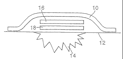
31
Figure 6 illustrates schematically a skin dressing in accordance with the
invention.
The illustrated dressing is of layered construction and comprises an optional
outer layer or
covering 10 in the form of a plaster suitable for adhering to the skin 12 of a
subject, so as to
cover a wound 14. Covering 10 encloses a first component or primary layer 18
and a second
component or secondary layer 16.
The first component 18 comprises a layer of poly-AMPS hydrogel incorporating
potassium
nitrite, in the form of a nitrite gel as specified in Table 1 above. The
second component 16
comprises a layer of dried PVA incorporating L-glutathione, in the form of a
glutathione
layer as specified in Table 2 above.
The dressing is initially supplied as a multi-part system, with the individual
components
separately packaged in respective sealed, sterile packages. When required for
use, the
dressing components are removed from the packages and applied to a wound in
appropriate
manner and order to produce the final dressing as shown. When the first and
second
components are brought together, this activates the gel.
On activation, nitrite starts diffusing from the primary layer into the
secondary layer and the
thiol diffuses in the opposite direction. Mixing of the nitrite with the thiol
results in
generation of S-nitrosoglutathione (GSNO) Once produced, the GSNO is released
from the
dressing into the surrounding environment where it decomposes to produce
nitric oxide.
Example 6: a delivM system for the generation of nitric oxide
GSNO is not suitable for long term storage because it decomposes readily to
release NO.
Using a dual dressing component storage and dispensing configuration allows
separate
storage of reactants which, when dispensed, are mixed to initiate the reaction
to generate S-
nitrosoglutathione.
The first component was prepared as follows: the base carrier used to add
viscosity and
spreadability was a hydrogel based material, commercially named PlexajelTM ASC
(United)
CA 02599287 2007-08-27
WO 2006/095193 PCT/GB2006/000873
32
Guardian Inc.). Into this, phosphate buffered saline was diluted 1/20 from a 1
Ox concentrated
stock (Sigma, D1408). Potassium nitrite (Fluka, 60417) was added to give a
final
concentration of 60mM and allowed to dissolve.
The second component was prepared as follows: L-glutathione (reduced form
Sigma,
G4251) was suspended in propylene glycol (Fluka 82281) to give a final
concentration
equivalent to 60mM.
Both components were stored separately. A dual dispenser was obtained from
Versdial Inc,
which allows variable control of the dispensing volumes from the two chambers.
Samples
came into contact with each other only when they were dispensed from their
respective
isolated chambers.
To demonstrate the effect of NO generated from the two separately stored
reagents, blood
flow to the surface of the forearm skin was investigated using laser Doppler.
A Moor
Instruments DRT4 tissue blood-flow and temperature monitor with associated
skin probes
was used to measure the skin laser Doppler flux. Two probes were attached to
the skin,
positioned to avoid major veins and arteries, at approximately 5-10cm apart.
The instrument
was run for lmin to ensure a flat response. One of the chambers was filled
with nitrite and
the other with L-glutathione. As a control, a second set of chambers were
filled with water.
The nitrite and L-glutathione were mixed just prior to use, in equal
quantities. The mix was
stirred to ensure the L-glutathione particles within the propylene glycol were
dissolved in the
aqueous nitrite hydrogel. The skin laser Doppler flux was then measured until
a plateau was
observed, indicating the maximum vaso-dilation effect had been reached.
Table 1 demonstrates the vaso-dilation effect on the skin; when using the 2
component
system. The LDF value for the mixture of L-glutathione and nitrite begins to
increase after 2
minutes (post sample application) indicating the speed of NO production and
dermal
transmission. The water control remains flat thus indicating the increase in
blood flow to be
due to the nitrite/GSH reaction. The maximal response of approximately 150 LDF
units is
regarded as being a strong response.
CA 02599287 2007-08-27
WO 2006/095193 PCT/GB2006/000873
03
Table 1
Time (rains) Laser Doppler Flux
Test Water control
0 29.3 45.6
1 13.7 24.3
2 30.3 31.2
3 34.6 35.0
4 52.0 33.3
85.2 42.7
6 108.3 34.5
7 161.4 35.0
8 151.6 30.3
9 165.3 33.7