Note: Descriptions are shown in the official language in which they were submitted.
CA 02599606 2007-08-22
WO 2006/091209 PCT/US2005/015638
BISPECIFIC BINDING AGENTS FOR MODULATING BIOLOGICAL
ACTIVITY
CROSS-REFERENCES TO RELATED APPLICATIONS
[0001] This application claims the benefit of U.S. Provisional Application No.
60/655,836,
filed February 23, 2005, the contents of wllich are hereby incorporated by
reference.
STATEMENT AS TO RIGHTS TO INVENTIONS MADE UNDER
FEDERALLY SPONSORED RESEARCH AND DEVELOPMENT
[0002] NOT APPLICABLE
REFERENCE TO A "SEQUENCE LISTING," A TABLE, OR A COMPUTER
PROGRAM LISTING APPENDIX SUBMITTED ON A COMPACT DISK.
[0003] NOT APPLICABLE
BACKGROUND OF THE INVENTION
[0004] Many diseases and disorders are caused by inappropriate or excessive
activation of
signal transduction pathways caused by activation of cell surface receptors,
e.g,. by the
binding of receptor-specific ligands. Receptors involved in the initiation or
progression of
diseases and disorders, such as cancer and autoimmune disorders, have emerged
as prime
targets for the development of therapeutics that reduce or prevent receptor
activation.
Examples of target receptors include, e.g., the epidermal growth factor
receptor ("EGFR"),
the insulin-like growth factor 1 receptor ("IGF 1 -R"), and the platelet-
derived growth factor
receptor ("PDGFR"), which tend to be overexpressed or aberrantly activated in
many disease
states, such as in the most common solid tumors, including non-small cell lung
cancer and
cancers of the breast, prostate, and colon, and in many autoimmune disease,
such as
myasthenia gravis, systemic lupus erythematosus, and rheumatoid arthritis.
Activation of the
receptor results in autophosphorylation, which drives signal transduction
pathways that lead
to disease progression.
CA 02599606 2007-08-22
WO 2006/091209 PCT/US2005/015638
[0005] Seininal studies with receptor inhibitors have clearly demonstrated
that by
preventing the activation of a receptor associated with a disease state, the
development of that
disease state can be altered. Generally, though, the receptor or receptors
responsible for the
disease state are expressed on many different cells and tissues in addition to
the diseased cells
or tissues. Although receptor inhibitors, e.g., Herceptin , wllich targets
ErbB2 ("HER2"),
are becoming available for clinical use, new challenges include identifying a
therapeutic
agent that will effectively target the diseased cells or tissue without
targeting non-affected
cells and tissues.
[0006] One approach to targeting agents specifically to diseased cells has
been the use of
bispecific binding agents, sometimes referred to herein as "bsBAs". Bispecific
binding
agents comprise two binding domains, each of which specifically recognizes and
binds to a
separate molecule (for convenience, the molecule specifically bound by each
respective
binding doinain may be referred to as the "ligand" for that binding domain).
Bispecific
binding agents have been attempted for some time, as exemplified by Schmidt M,
et al., "A
bivalent single-chain antibody-toxin specific for ErbB-2 and the EGF
receptor," Int J Cancer,
65(4):538-46 (1996), Lu D, et al., "Simultaneous blockade of both the
epidermal growth
factor receptor and the insulin-like growth factor receptor signaling pathways
in cancer cells
with a fully human recombinant bispecific antibody," J Biol Chem. 279(4):2856-
65 (2004),
and Francois C, et al., "Antibodies directed at mouse IL-2-R alpha and beta
chains act in
synergy to abolish T-cell proliferation in vitro and delayed type
hypersensitivity reaction in
vivo," Traiispl Int. 9(l):46-50 (1996). Because bsBAs often use antibodies as
one or both of
the binding domains, bsBAs are sometimes included in the class of agents
referred to as
immunotherapeutics.
[0007] Unfortunately, the universe of molecules that can be used as targets
for bsBAs is
limited. Only a relatively small nuinber of molecules are expressed on
diseased cells but not
on normal cells, and which therefore can be used to target agents exclusively
to diseased
cells. An additional number of molecules are expressed in greater numbers on
diseased cells
than on normal cells. These molecules can permit some preferential delivery of
agents to
diseased cells over normal cells, depending on the degree to which the
molecule is
overexpressed in diseased cells compared to normal cells.
[0008] Even with substantial overexpression of the target molecule on target
cells,
however, delivery of targeted therapeutic agents have often been accompanied
by adverse
2
CA 02599606 2007-08-22
WO 2006/091209 PCT/US2005/015638
side effects due to binding of the agent to normal cells expressing the target
molecule. For
example, the HER2 (erbB2) receptor that is the target for the FDA-approved
immunotherapeutic agent Herceptin , is overexpressed at levels some 10 to 100
times more
than the expression of the HER2 receptor in non-cancer cells. Nonetheless, a
percentage of
patients develop cardiac arrhythmia and other adverse side effects due to
binding of
Herceptin to normal cells.
[0009] Thus, it would be desirable to increase the therapeutic window of
immunotherapeutic agents by developing bsBAs with an improved ability to bind
to diseased
cells without binding to normal cells.
BRIEF SUMMARY OF THE INVENTION
[0010] The present invention provides methods for modulating biological
activity or
activities of target molecules on a target cell. The methods comprise
providing a bispecific
binding agent having a first binding domain having a Kd for a first target
molecule on the
surface of the cell of at least 10-7 M and a second binding domain having an
affinity for a
second target molecule on the surface of said cell that is at least 10 times
lower than the Kd
of the first binding domain; wherein the first and the second target molecules
each have a biological activity, which activity may be the same or different
and, contacting the bispecific
binding agent with the target cell under conditions that permit the first and
second binding
domains to bind to the first and second target molecules, respectively,
wherein the binding of
the first and second target molecules modulates the biological activity or
biological activities
of the target molecueles. Iti some embodiments, the bispecific binding agent
comprises two
antibodies. In some embodiments, the antibodies are diabodies, two single
chain Fvs
connected directly or by a linker, disulfide stabilized Fvs, or combinations
thereof. In some
embodiments, the target cell is a cancer cell. In some embodiments, the first
target molecule
is a tumor-associated antigen, cytokine receptor, or growth factor receptor.
In some
embodiments, the first target molecule is a tyrosine kinase receptor selected
from the group
consisting of EGFR and ErbB2. In some embodiments, the second target molecule
is ErbB3
(HER3), insulin-lilce growth factor-1 receptor (IGF1-R), any of FGF receptors
1-4, HGF
receptor, insulin receptor, either of PDGF receptors cr and beta, C-KIT, or
ErbB4. In some
embodiments, the Kd of the first binding domain to the first target molecule
is between 10-8
and 10"12 M. In some embodiments, the Kd of the second binding domain to the
second
3
CA 02599606 2007-08-22
WO 2006/091209 PCT/US2005/015638
target molecule is at least 20 times lower than the Kd of the first binding
domain to the first
target molecule.
[0011] In another group of embodiments, the invention provides methods for
modulating a
desired biological activity or activities of target molecules on target cells
in an organism
having target and non-target cells, wherein the target cells have a first
target molecule on
their exterior and a second target molecule on their exterior surface, and
wherein (i) the first
and second target molecules do not share a common ligand, (ii) the first
target molecule is at
least 10 times more abundant on the surface of the target cells than on non-
target cells that
also bear the second target molecule, and (iii) the first target molecule and
the second target
molecule each have a biological acitivity, which may be the same or different.
The method
coinprises providing a bispecific binding agent having a first binding domain
having a Kd for
the first target molecule of at least 10-7 M and a second binding domain
having a Kd for the
second target molecule that is at least 10 times lower than the Kd of the
first binding domain;
and contacting the bispecific binding agent with the target cells under
conditions that permit
the first and second binding domains to bind to the first and second target
molecules,
respectively, wherein said binding of the first and the second binding domains
modulates the
biological activity or activities of the first and the second target
molecules, respectively. In
some embodiments, the bispecific binding agent comprises two antibodies. In
some of these
embodiments, the antibodies are diabodies, two single chain Fvs comiected
directly or by a
linker, disulfide stabilized Fvs, or combinations thereof. In some
embodiments, the target
cell is a cancer cell. The first target molecule may be a tumor-associated
antigen, cytokine
receptor, or growtll factor receptor. The first target molecule can be a
tyrosine kinase
receptor selected from the group consisting of EGFR and ErbB2. In some
embodiments, the
second target molecule is ErbB3 (HER3), insulin-like growth factor-1 receptor
(IGF1-R), any
of FGF receptors 1-4, HGF receptor, insulin receptor, either of PDGF receptors
cx and beta,
C-KIT, or ErbB4. In some embodiments, the Kd of the first binding domain to
the first target
molecule is between 10-$ and 10-1' M. In some embodiments, the Kd of the
second binding
domain to the second target molecule is at least 20 times lower than the Kd of
the first
binding domain to the first target molecule, while in others it is at least 50
times lower than
the Kd of the first binding domain to the first target molecule. In some
embodiments, the
modulation of the biological activity means involves decreasing the activity
of a tyrosine
kinase receptor.
4
CA 02599606 2007-08-22
WO 2006/091209 PCT/US2005/015638
[0012] In another group of embodiments, the invention provides bispecific
binding agents
(bsBAs) comprising a first binding domain having a Kd of at least 10-7 M for a
first target
molecule on a target cell and a second binding domain having a Kd for a second
target
molecule on a target cell which Kd is at least 10 times lower than the Kd of
the first binding
domain for the first target molecule, wherein (i) the first and second target
molecules do not
have the same natural ligand, (ii) the first target molecule and the second
target molecule
each have a biological activity, which may be the same or different, and (iii)
the first and the
second binding domains, when bound to the first and the second target
molecules, modulate
the biological activity or activities of the first and second target
molecules, respectively. In
some embodiments, the Kd of the second binding domain is more than 50 times
lower t11an
the Kd of the first binding domain, while in others, the Kd of the second
binding domain is
100 or more times lower than the Kd of the first binding domain. In some
embodiments, the
bsBA comprises two antibodies. In some these einbodiments, the antibodies are
diabodies,
two single chain Fvs connected directly or by a linker, disulfide stabilized
Fvs, or
combinations thereof. In some embodiments, the first binding domain binds to a
tumor-
associated antigen, cytokine receptor, or growtli factor receptor. In some
embodiments, the
first binding domain binds to a tyrosine kinase receptor selected from the
group consisting of
EGFR and ErbB2. In some embodiments, the second binding domain binds ErbB3
(HER3),
insulin-like growth factor-1 receptor (IGF1-R), any of FGF receptors 1-4, HGF
receptor,
insulin receptor, either of PDGF receptors cx and beta, C-KIT, or ErbB4. In
some
embodiments, the first binding domain binds to EGFR and said second binding
domain binds
ErbB3 (HER3). In some embodiments, the Kd of the first binding domain is
between 10"$
and 10-12 M. In some einbodiments, the first target molecule is overexpressed
by at least 10
times on target cells as compared to its expression on normal cells.
[0013] In still another group of embodiments, the invention provides
compositions of (a) a
bispecific binding agent (bsBA) comprising a first binding domain having a Kd
for a first
target molecule on a target cell of at least 10-7 M, and a second binding
domain having a Kd
for a second target molecule on a target cell that is at least 10 times lower
than the Kd of the
first binding domain, wherein the first and the second target molecules do not
have the same
natural ligand, aiid further wherein (i) the first target molecule and the
second target molecule
each have a biological activity, which may be the saine or different, and (ii)
the first and the
second binding domains, when bound to the first and the second target
molecules, modulate
the biological activity or activities of the first and second target
molecules, respectively, and,
5
CA 02599606 2007-08-22
WO 2006/091209 PCT/US2005/015638
(b) a pharmaceutically acceptable carrier. In some embodiments, the Kd of the
second
binding domain is more than 50 times lower than the Kd of the first binding
domain, while in
some embodiments, the Kd of the second binding domain is 100 or more times
lower than the
Kd of the first binding domain. In some embodiments, the bsBA comprises two
antibodies.
In some of these embodiments, the antibodies are diabodies, two single chain
Fvs connected
directly or by a linlcer, disulfide stabilized Fvs, or combinations tliereof.
In some
embodiments, the first binding domain binds to a tumor-associated antigen,
cytokine
receptor, or growth factor receptor. In some embodiinents, the first target
molecule is
overexpressed by at least 10 times on target cells as compared to its
expression on normal
cells.
[0014] In still another group of embodiments, the invention provides for the
use of a
bispecific binding agent (bsBA) comprising a first binding domain having a Kd
for a first
target molecule on a target cell of at least 10"7 M and a second binding
domain having a Kd
for a second target molecule on a target cell that is at least 10 times lower
than the Kd of the
first binding domain, wherein said first and second target molecules do not
have the same
natural ligand, and further wherein said first target molecule and said second
target molecule
each have a biological activity, which may be the same or different, and said
first and said
second binding domains, when bound to said first and said second target
molecules, modulate
the biological activity or activities of the first and second target
molecules, respectively, for
the manufacture of a medicament. In some embodiments, the Kd of said second
binding
domain is more than 50 times lower than the Kd of the first binding domain,
while in others it
is 100 or more times lower than the Kd of the first binding domain. In some
embodiments,
the bsBA comprises two antibodies. In some of these embodiments, the
antibodies are
diabodies, two single chain Fvs connected directly or by a linker, disulfide
stabilized Fvs, or
combinations thereof. In some embodiments, the target molecules bound by the
first binding
domain and by the second binding domain are independently selected from the
group
consisting of a tumor-associated antigen, a cytokine receptor, and a growth
factor receptor,
provided that the first binding domain and the second binding domain do not
bind the same
tumor-associated antigen, cytokine receptor, or growth factor receptor. In
some
embodiments, the first target molecule is overexpressed by at least 10 times
on target cells as
compared to its expression on normal cells. In some embodiments, the
medicament is for
inhibiting the proliferation of cancer cells.
6
CA 02599606 2007-08-22
WO 2006/091209 PCT/US2005/015638
BRIEF DESCRIPTION OF THE DRAWINGS
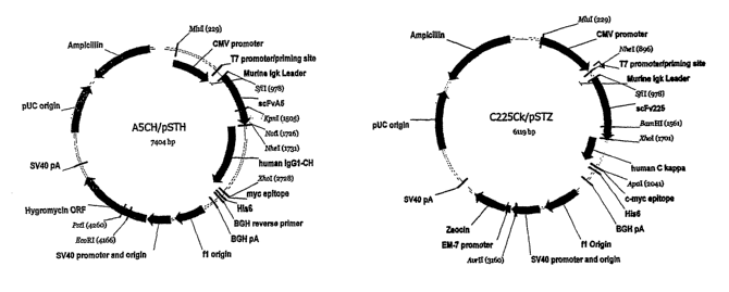
[0015] Figure 1 shows the two expression plamids A5CH/pSTH and C225Ck/pSTZ
used
for the expression of C225-A5 bi-specific antibody in 293T cells.
[0016] Figure 2 shows the binding of C225-A5 bi-specific antibody to A431
cancer cells
by flow cytometry. A431 cells were incubated with 1 ug/mL of C225-A5 or no
antibody for
30 minutes on ice and detected by anti-human IgG antibody labeled with Alexa
Fluor 488
and quantitated in a FACSCalibur instruinent.
[0017] Figure 3 shows the effect of the bispecific antibody concentration on
AKT
phosphorylation. A dose-response experiment was performed on the C225-A5
bispecific
antibody which binds to EGFR with high affinity aiid ErbB3 with low affinity.
A431 cells
were incubated witli increasing concentrations of the C225-A5 bispecific
antibody for 30
minutes before stimulating with heregulin for five minutes. The tumor cells
were then lysed
in detergent and analysed for AKT phosphorylation. Data from the experiment is
shown
plotted as AKT phosphorylation ratio in cell lysates versus antibody
concentration. The ratio
represents the ainount of phosphorylated AKT divided by the amount of total
AKT in the
sample as determined using antibody microarrays.
DETAILED DESCRIPTION OF THE INVENTION
Introduction
[0018] One problem with current immunotherapeutic agents is that their
tendency to bind
to normal cells as well as to diseased cells causes adverse side effects.
Thus, one goal of the
scientific coinmunity has been to develop immunotherapeutic agents with an
improved ability
to bind target cells (e.g., diseased cells) without also binding non-target
cells (that is, normal
cells).
[0019] The present invention provides compositions and methods for improving
the
specificity of one class of immunotherapeutic agents for binding target cells.
Surprisingly, it
has now been discovered that the specificity for targeting diseased cells by
the
immunotherapeutic agents Icnown as bispecific binding agents ("bsBAs") can be
increased by
controlling the differences in the binding affinities of the two binding
domains of the bsBAs.
The bsBAs of the invention can then be used for increasing or decreasing the
biological
activity of target molecules on the target cells, and thereby provide an
improved ability to
7
CA 02599606 2007-08-22
WO 2006/091209 PCT/US2005/015638
modulate biological activity of the target cells with reduced, if any, effect
on the
corresponding activity of non-target cells.
[0020] As the name implies, bsBAs have two binding domains, each specific for
a different
target molecule. The first binding domain is generally used to target the bsBA
to a cell of
choice, sometimes referred to as a "target cell." The second binding domain
binds to a
second target molecule on the target cell. The binding of the binding domains
to their target
molecules is typically intended to modulate a specific biological effect
(tllat is, to increase or
to inhibit that biological activity). Binding domains with capabilities to
modulate biological
activities in different ways are lcnown in the art.
[0021] Often, the biological activities are inhibited by the binding of the
binding domain to
its target molecule. For example, if the molecule bound by the binding domain
is part of
cytokine receptor, the binding of the binding domain to that receptor can
block access of the
cytokine to the receptor, thereby inhibiting the biological activity that
would otherwise be
induced by that binding. Similarly, the binding of the binding domain to the
receptor can
prevent the receptor from forming a heterodiiner, which is required for the
full activation of
some cytokine receptors, such as the interleukin (IL)-2 receptor. Or, the
binding of the
binding domain may change the confonnation of the receptor so that it cannot
bind its natural
ligand and thereby be activated. Conversely, the binding domain can be one
selected for its
ability to increase the biological activity by binding to the receptor. For
example, the binding
of the binding domain to the receptor can mimic the effect of the natural
ligand for the
receptor, so that the binding activates the receptor, or the binding of the
binding domain may
induce a conformational change which causes a low affinity receptor to become
a high
affinity receptor for its natural ligand.
[0022] Because the bsBAs of the invention have two binding domains, they can
be selected
to achieve the desired effect. For example, the domains can be selected so
that they both
inhibit the biological activities of tyrosine kinase receptors. Or, one can be
chosen that will
inhibit the biological activity of a kinase receptor while the other is
selected to enchance or
activate another receptor whose activity is desirable. The ability to select
binding domains
with desired effects on the activities of the target molecules on the target
cells increases the
flexibility of the methods of the invention.
[0023] While both binding domains in the bsBAs of the invention are intended
to modulate
biological activities of the target cells, the first binding domain of the
bsBAs of the invention
8
CA 02599606 2007-08-22
WO 2006/091209 PCT/US2005/015638
also serves to target the bsBAs to the target cell, while the second serves
primarily to induce
an effect on the target cell. For convenience in distinguishing the two
domains, therefore, the
first binding domain is sometimes referred to herein as the "targeting
domain," while the
second binding domain is sometimes referred to herein as the "effector
domain". Similarly,
for convenience in distinguishing the molecules bound by the two binding
domains, the target
molecule for the effector domain will sometimes be referred to as the
"effector target
molecule," while the term "target molecule" by itself will refer to the target
of the targeting
domain.
[0024] Previous bsBAs have typically been constructed using binding domains
with the
highest available affinity for each of the respective target molecules.
Persons of skill will
appreciate that it is unlikely that one domain will have exactly the same
affinity for its
respective target as does the other, and the two binding domains therefore
usually have a
difference in affinity. The difference, however, is typically not great and
may or may not be
significant in terms of actual effect on binding.
[0025] In the methods and compositions of the invention, however, the
targeting domain is
selected to have at least an order of magnitude higher binding affinity for
its ligand than the
affinity the effector domain has for its ligand. That is, the targeting domain
has at least 10
times or greater affinity for the molecule to which it recognizes and binds
than the effector
domain has for the molecule to which it recognizes and binds. In some
embodiments, the
affinity of the targeting domain for its ligand is at least 15 times higher
than that of the
effector domain, in others it is 20 times or more higher, in other
embodiments, it is 25 time or
more higher, and in some embodiments, it has an affinity 30, 40, 50 or even
100 times or
more higher than the affinity of the effector domain for its target, with each
respective higher
binding affinity being more preferred. Since there is at least an order of
magnitude difference
in binding affinity between the two binding domains of the bsBAs of the
invention, the
bsBAs are occasionally referred to herein as "hi-lo" bsBAs.
[0026] The intentional and substantial differential in binding affinity
between the target
binding domain and the effector binding domain provides surprising and
previously
unrecognized advantages over prior bispecific molecules. As noted above,
previously known
bispecific agents have had binding moieties with affinities as high as
possible for the target
ligands. But, bispecific molecules with binding moieties that have similar
affinities are
limited in the molecules to which they can be targeted and the situations in
which they can be
9
CA 02599606 2007-08-22
WO 2006/091209 PCT/US2005/015638
used, compared to the compositions and methods of the invention. Some of the
advantages
of the invention can be considered by referring to a hypothetical example.
[0027] Consider the case of a cancer cell which has two receptors, receptor A,
which is
overexpressed on the cancer cell compared to normal cells, and receptor B,
which is
expressed on norinal cells in about the same number of copies as are present
on the cancer
cell. A bispecific binding agent with binding domains with approximately equal
affinity for
both receptors will tend to have roughly equal effects on both cancer cells
and on normal,
non-cancer cells. This is particularly in the case where high concentrations
of bsBAs are
achieved, since the bsBAs will tend to saturate both receptors by monovalent
binding.
[0028] By contrast, a bsBA of the invention, having a higher affinity
targeting domain
targeted to receptor A and a lower affinity effector domain targeted to
receptor B, and having
10, 20, 30, or even more times more affinity for receptor A than for receptor
B, will
preferentially bind to the cancer cells, and by normal kinetic interactions,
will bind in larger
numbers to the cancer cells as compared to normal cells. Instead of
promiscuously binding to
cells bearing receptor B, including substantial numbers of normal cells,
therefore, the effector
domain will be selectively delivered to the cancer cells. Thus, the invention
permits more
selective targeting of effector domain to target cells.
[0029] Further, the binding of the higher affinity binding domain to receptor
A tethers the
lower affinity, effector domain in proxiinity to the cell surface, where it is
available to
interact with receptor B over time. This perinits the effector domain to bind
receptor B even
though its relatively low affinity for receptor B might not normally be
sufficient to hold it to
the receptor were the effector domain provided as a "free standing,"
monovalent (or
"univalent") entity.
[0030] Persons of skill will appreciate that the dissociation constant ("Kd")
of an antibody
or other ligand is determined botll by the koõ and by the koff of the ligand.
That is, the Kd
represents the balance between the time the antibody or other ligand is bound
to the target
molecule and the time that it is not. A low affinity binding domain therefore
often has a low
affinity precisely because it has a high tendency to dissociate from its
target molecule.
During this period, an untethered binding domain can be moved away from its
target
molecule by Brownian movement, fluid flow, or other lcinetic forces acting on
the binding
domain molecule. The tethering of the low affinity domain by the high affinity
domain of the
bsBA aids in maintaining the low affinity binding domain in proximity to the
receptor
CA 02599606 2007-08-22
WO 2006/091209 PCT/US2005/015638
targeted by the low affinity domain, and thus tends to increase the
probability that at any
point in time the low affinity domain will be able to bind its target
molecule. Since the target
molecule of the low affinity domain of the bsBAs of the invention are receptor
kinases, this
tends to increase the ability of the bsBA to bind the target receptor kinase
and therefore
increases their biological effect on target cells.
[0031] In preferred embodiments, the two binding domains of the bsBAs of the
invention
bind target molecules that are not normally bound by the same ligand. Persons
of skill are
aware that some ligands, such as the interleukin IL-2, for example, are bound
by two different
receptor chains, and that the two chains - with bound IL-2 - then interact to
form the fully
biologically active unit. While bsBAs directed to the two receptor chains can
therefore
prevent full activation of such a receptor, both of the binding domains of
such bsBAs are, of
course, directed to the same receptor.
[0032] Further, while bsBAs targeted to two chains of the same receptor
interfere with the
biological activity mediated by that receptor, bsBAs targeted to two different
receptors can
modulate the biological activity of both receptors. It is believed that
affecting the activity of
two receptors at once affords a more robust effect on the target cell than
that of interfering
with the activity of only one.
[0033] Finally, the formation of the triiner between the bsBA and the two
target molecules
bound by the binding domains has the additional advantage of binding the
target molecules in
close vicinity to one another and preventing their nonnal diffusion through
the lipid bilayer of
the cell membrane. The crosslinlcing of different receptors by bsBAs is itself
believed to
contribute to cytotoxic or cytostatic effects of the bsBAs on target cells.
[0034] In one group of embodiments, the targeting domain of the bsBA binds to
a cell
surface receptor that is preferentially expressed or overexpressed on a target
cell that is
associated with a disease or disorder (for example, a breast cancer cell) and
the effector
domain binds to a cell surface receptor that is promiscuously or ubiquitously
expressed on
target cells and on non-target cells. Exemplar cell surface receptors that can
be targeted by
the bsBA of the invention are described below. In preferred embodiments, the
molecule to be
bound by the targeting domain is expressed on target cells at levels that are
higher than the
levels of the molecule to be bound by the effector domain. Thus, the bsBAs and
methods of
the invention are particularly useful for improving the specific delivery of
effector molecules
to cells with target molecules that would be promiscuously bound by
conventional antibodies
11
CA 02599606 2007-08-22
WO 2006/091209 PCT/US2005/015638
or bispecific, agents or by both. Persons of skill are aware that cells of
different cancers may
overexpress different antigens or may overexpress the same antigen to
different degrees than
do cells of a different cancer type. Thus, in designing the bsBAs of the
invention, it is
contemplated that the practitioner will select a targeting domain that targets
a cell surface
receptor overexpressed on the particular cells to be targeted by the
particular bsBA.
[0035] In some embodiments, the targeting domain of the bsBA binds to a first
cell surface
receptor that is preferentially expressed or overexpressed on a target cell
that is associated
with a disease or disorder (for example, a cancer cell) and the effector
domain binds to a
second cell surface receptor that is overexpressed on disease cells (such as
cancer cells)
compared to normal cells, but is expressed at lower levels than is the first
cell surface
receptor. In these embodiments, the differential in expression level between
the first and the
second cell surface receptors again improves the specific delivery of effector
molecules to
cells with target molecules.
[0036] As noted in the Background, even though HER2 is overexpressed in breast
cancer
cells at levels some 10 to 100 times that of its expression on normal cells,
some adverse side
effects are seen in patients from binding of the immunotherapeutic agent,
HERCEPTIN , to
normal cells. Thus, even substantial overexpression of a target molecule is
not necessarily
sufficient to keep high affinity binding agents from binding to normal cells,
with adverse side
effects.
[0037] By contrast, the bsBAs of the invention have a targeting domain that is
chosen to
have an affinity for its target molecule that is at least 10 times higher, and
often much higlzer,
than that of the effector domain. Preferably, the dissociation constant of the
targeting domain
for its target molecule is in the range of 10-8 to 10-12 M. The target
molecule is selected either
because it is not present on normal cells, or because it is highly
overexpressed on cancer cells
than on normal cells, preferably at least 20 times and even more preferably
100 times more
than it is expressed on normal cells. As noted, due to the high affinity of
the targeting
domain for the target molecule, it will tend to bind the bsBA preferentially
to the target cell.
Thus, it is anticipated that the effector domain can target a target molecule
expressed on
normal cells and still achieve selective binding that provides a therapeutic
window larger than
that of conventional bsBAs.
[0038] Persons of skill will appreciate that cancer cells, in particular, tend
to upregulate the
expressioil of many normal proteins, including many with roles in maintaining
homeostasis in
12
CA 02599606 2007-08-22
WO 2006/091209 PCT/US2005/015638
normal cells. Thus, even proteins not normally considered to be cancer or
tumor antigens
tend to be upregulated on cancer cells. For instance, insulin receptor, which
is not considered
a tumor antigen, is often upregulated 3-5 fold on tumor cells as coinpared to
normal cells
(see, e.g., Milazzo et al., Cancer Res. 52(14):3924-30 (1992)).
[0039] As an example, the ErbB3 receptor is somewhat overexpressed on some
cancer cells
compared to its expression on normal cells. It can, however, be used as the
effector target
molecule of a bsBA when the targeting domain is directed to a target molecule
that is even
more highly overexpressed. The Examples present an exemplar bsBA of the
invention in
which the targeting domain is directed to EGFR and the effector domain is
targeted to ErbB3.
[0040] It is desirable that the targeting domain is directed to a target
molecule (such as a
tyrosine receptor) that is overexpressed on the target cells, while the
effector domain is
directed to a molecule (e.g., a second tyrosine receptor) that is expressed at
a lower level than
is the target molecule for the targeting domain. While it is only necessary
that the target
molecule is expressed at higller levels than the molecule targeted by the
effector domain, in
general, significant differences between the expression of the target molecule
and the
expression of the effector molecule are advantageous, since the effector
molecules can be
saturated at bsBA concentrations that are below the Kd of the targeting
domain.
[0041] In general, it is preferable that the target molecule for the targeting
domain is
overexpressed at levels 10, 20, 50, 100, or more times higher than expression
of that
molecule on non-target cells, with each successively higher level being more
preferred. In
general, it is further preferred that the effector molecule be expressed
either at the level it is
expressed on non-target cells or, if it is overexpressed, that it is
overexpressed at levels of 2
to 5 times that of non-target cells. In other words, it is preferable that the
targeting molecule
be expressed (or overexpressed) at high levels relative to the molecule bound
by the effector
domain.
[0042] Where the target cell is a disease cell, such as a cancer cell, the
expression level of
the target molecule is measured against the expression of the same molecule on
cells of the
same tissue type as that from which the cancer cell originates. That is, if
the disease cell is a
breast cancer cell, the expression level is measured against a breast cell,
while the expression
level of molecules of an ovarian cancer cell is measured against expression
levels on normal
ovary cells. Usually, a population of cells is used and an average value of
expression level
(e.g., number of molecules expressed per cell) is determined.
13
CA 02599606 2007-08-22
WO 2006/091209 PCT/US2005/015638
[0043] Further, in some preferred embodiments, the bsBAs of the invention, the
cell
surface antigens recognized and bound by the targeting domain and by the
effector domain
are chosen to themselves have a biological activity that can be modulated by
the binding of
the domains. For example, the cell surface antigen targeted by each domain can
be cytokine
or growtll factor receptors, the blockage of which by the domains will
contribute to
restoration of the target cell to a normal phenotype. In this way, the
therapeutic effect of the
baBA is increased over that which would be due to the action of a single
domain alone. In
the case of the exemplar bsBA discussed above, for exainple, the two domains
each bloclc a
different cytokine receptor. It is expected that the blocking of the two
receptors will result in
downregulating the pathways activated by those receptors, decreasing the rate
of proliferation
of the cell.
[0044] As noted above, antibodies are also known which can act as agonists of
cytokine
receptors and the like; that is, they act to enhance the activity of the
target molecule. Thus,
depending on the target molecules and binding agents selected by the
practitioner, one
binding domain of a bsBA of the invention may inhibit the activity of a target
molecule,
while the other binding domain enhances the activity of its target molecule.
In other
embodiments, both binding domains can be chosen that will inhibit the
activities of their
respective molecules, while in still other einbodiinent, binding domains can
be chosen that
will enhance the activities of their respective target molecules, that might
be the same or
different. The ability to independently select binding domains that increase
or decrease the
activity of the respective target molecules affords the practitioner
considerable flexibility in
designing bsBAs effective for a range of conditions. To indicate that the
activities of the
target molecules can be enhanced or decreased, at the practitioner's option,
by the judicious
selection of binding agents, the effect of the bsBAs on the target molecules
is sometimes
referred to herein as "modulating" the activity of the target molecule.
[0045] For example, some cancers result from the mutation of a gene encoding a
receptor
that acts as a tyrosine kinase, resulting in the receptor becoming either
constitutively active or
overexpressed, so that the cell proliferates more than it would with a normal
receptor or with
one expressed in normal amounts. To decrease the activity of the receptor, the
practitioner
may, in this example, select a binding agent whose binding is known to change
the
conformation of the constitutively active receptor to reduce its activity or,
in the case of an
overexpressed receptor, to simply bloclc it from being bound by its natural
ligand, thus
preventing the overexpression from resulting in an inappropriate increase in
signaling within
14
CA 02599606 2007-08-22
WO 2006/091209 PCT/US2005/015638
the target cell. Conversely, if the target molecule is one whose activity it
is desirable to
enhance, the practitioner may select a binding agent whose binding is known to
act as an
agonist of the activity.
[0046] The use of bsBAs with a single high affinity binding domain is
sufficient to provide
specific binding to cells of interest. Studies have shown that binding agents
with two high
affinity binding domains directed to a single target molecule have only about
three fold the
affinity for the target molecule compared to a univalent binding agent with
the same binding
domain. Nielsen, U. et al., Cancer Res. 60(22):6434-40 (2000). A univalent
binding agent
will therefore typically still have a Kd in the nanomolar range. Since
therapeutic agents are
typically administered in amounts to provide up to micromolar concentrations,
a thousand
times the Kd of the binding agent, the high concentration of the binding agent
relative to the
Kd of the high affinity targeting domain is expected to permit binding of the
agent to target
cells bearing the target molecule. Thus, the high affinity targeting domain of
the bsBAs of
the invention is expected to provide specific binding of the bsBAs under the
conditions in
which they will be administered.
[0047] It is expected that the practitioner can select appropriate
combinations of target
molecules for the targeting domain and for the effector domain. While a number
of preferred
target molecules and effector target molecules are described below, it may be
helpful to list
some preferred target molecules and effector target molecules. Some preferred
target
molecules are EGFR and ErbB2. Some preferred effector target molecules are:
ErbB3,
ErbB4, any of fibroblast growth factor (FGF) receptors 1-4, hepatocyte growth
factor
receptor, insulin-like growth factor 1 receptor (IGF1-R), insulin receptor,
Platelet Derived
Growth Factor (PDGF) receptors alpha and beta, and C-KIT. Each of these
molecules is
lcnown in the art and is identified by its reference number in the SWISS-PROT
database in a
later section.
Definitions
[0048] Units, prefixes, and symbols are denoted in their Systeme International
de Unites
(SI) accepted form. Numeric ranges are inclusive of the numbers defining the
range. Unless
otherwise indicated, nucleic acids are written left to right in 5' to 3'
orientation; amino acid
sequences are written left to right in amino to carboxy orientation. The
headings provided
herein are not limitations of the various aspects or embodiments of the
invention, which can
CA 02599606 2007-08-22
WO 2006/091209 PCT/US2005/015638
be had by reference to the specification as a wllole. Accordingly, the terms
defined
immediately below are more fully defined by reference to the specification in
its entirety.
Terms not defined herein have their ordinary meaning as understood by a person
of skill in
the art.
[0049] "Affinity" of binding agents is typically stated in terms of their
dissociation
constant, or "K d". Typically, useful binding agents have Kds stated in
nanomolar
concentrations. Persons of skill will recognize that an antibody with a Kd of
10-8 M has an
affinity 10 times as high as one with a Kd of 10"7, and 100 times the affinity
of an antibody
with a IQ of 10-6. Thus, a higher affinity agent has a Kd stated as a lower
number (that is, 10-8
is a smaller number than is 10-6.)
[0050] "Antibodies" exist as intact immunoglobulins or as a nuinber of well
characterized
fragments produced by digestion with various peptidases. Thus, for example,
pepsin digests
aii antibody below the disulfide linkages in the hinge region to produce
F(ab)'2, a dimer of
Fab which itself is a light chain joined to VH--CH by a disulfide bond. The
F(ab)'2 may be
reduced under mild conditions to break the disulfide linkage in the hinge
region thereby
converting the (Fab')2 dimer into a Fab' monoiner. The Fab' monomer is
essentially a Fab
with part of the hinge region (see, W. E. Paul, ed., Fundamental Immunology,
Raven Press,
N.Y. (1993), for a more detailed description of these and other antibody
fragments). While
various antibody fragments are defined in terms of the digestion of an intact
antibody, one of
skill will appreciate that such Fab' fragments may be synthesized de novo
either chemically
or by utilizing recombinant DNA methodology.
[0051] For convenience of reference, as used herein, the term "antibody"
includes whole
antibodies, antibody fragments that retain antigen recognition and binding
capability, wliether
produced by the modification of whole antibodies or synthesized de novo using
recombinant
DNA methodologies, monoclonal antibodies, polyclonal antibodies, and antibody
mimics,
unless otherwise required by context. The antibody may be an IgM, IgG (e.g.
IgG1, IgG2,
IgG3 or IgG4), IgD, IgA or IgE.
[0052] The term "antibody fragments" means molecules that comprise a portion
of an
intact antibody, generally the antigen binding or variable region of the
intact antibody.
Examples of antibody fragments include Fab, Fab', F(ab')2, domain antibody
(dAb), and Fv
fragments; helix-stabilized antibodies (see, e.g., Arndt et al., J Mol
Bio1312:221-228 (2001);
diabodies (see below); single-chain antibody molecules ("scFvs," see, e.g.,
U.S. Patent No.
16
CA 02599606 2007-08-22
WO 2006/091209 PCT/US2005/015638
5,888,773); disulfide stabilized antibodies ("dsFvs", see, e.g., U.S. Patent
No. 5,747,654), and
domain antibodies ("dAbs," see, e.g., Holt et al., Trends Biotech 21(11):484-
490 (2003),
Ghahroudi et al., FEBS Lett. 414:521-526 (1997), Lauwereys et al., EMBO J
17:3512-3520
(1998), Reiter et al., J. Mol. Biol. 290:685-698 (1999), Davies and Riechmann,
Biotechnology, 13:475-479 (2001)).
[0053] The term "diabodies" refers to small antibody fragments with two
antigen-binding
sites, which fraginents coinprise a variable heavy domain (VH) connected to a
variable ligllt
domain (VL) in the same polypeptide chain (VH-VL). By using a linker that is
too short to
allow pairing between the two domains on the same chain, the domains are
forced to pair
with the complementary domains of another chain and create two antigen-binding
sites.
Diabodies are described more fully in, for example, EP 404,097; WO 93/11161;
and
Hollinger et al., Proc. Natl. Acad. Sci. USA, 90: 6444-6448 (1993).
[0054] Typically, an immunoglobulin has a heavy and light chain. Each heavy
and ligllt
chain contains a constant region and a variable'region, (the regions are also
lu-iown as
"domains"). Light and heavy chain variable regions contain a "framework"
region
interrupted by three hypervariable regions, also called "complementarity-
detennining
regions" or "CDRs". The extent of the framework region and CDRs have been
defined. See,
Kabat and Wu, if~fi~a. The sequences of the framework regions of different
light or heavy
chains are relatively conserved within a species. The frameworlc region of an
antibody, that
is the combined framework regions of the constituent light and heavy chains,
serves to
position and align the CDRs in three dimensional space.
[0055] The CDRs are primarily responsible for binding to an epitope of an
antigen. The
CDRs of each chain are typically referred to as CDR1, CDR2, and CDR3, numbered
sequentially starting from the N-terminus, and are also typically identified
by the chain in
which the particular CDR is located. Thus, a VH CDR3 is located in the
variable domain of
the heavy chain of the antibody in which it is found, whereas a VL CDR1 is the
CDR1 from
the variable domain of the light chain of the antibody in which it is found.
[0056] References to "VH" or a"VL" refer to the variable region of an
immunoglobulin
heavy chain, including an Fv, scFv , dAb, dsFv or Fab. References to "VL" or a
"VL" refer to
the variable region of an irmnunoglobulin light chain, including of an Fv,
scFv , dsFv, dAb,
or Fab.
17
CA 02599606 2007-08-22
WO 2006/091209 PCT/US2005/015638
[0057] The phrase "single chain Fv" or "scFv" refers to an antibody in which
the variable
domains of the heavy chain and of the light chain of a traditional two chain
antibody have
been joined to form one chain. Optionally, a linker (usually a peptide) is
inserted between the
two chains to allow for proper folding and creation of an active binding site.
[0058] "Bispecific binding agents", or "bsBA," are binding molecules that are
capable of
specific binding to more than one target molecule simultaneously.
[0059] A "binding agent" is any molecule capable of specifically binding a
target molecule,
and include antibodies, antibody fragments, aptamers, peptides (e.g., Williams
et al., J Biol
Chem 266:5182-5190 (1991)), and antibody mimics, such as those that can be
created from
the tenth fibronectin type III domain (see, e.g., Xu, L., et al., Chem Biol.
9(8):933-42 (2002),
Koide et al., J Mol Biol 284:1141-1151, Skerra, J Mol Recognit 13:167-187
(2000), Main et
al., Cell, 71:671-678 (1992), and Dickinson et al., J Mol Biol, 236:1079-1092
(1994)) and
can comprise natural proteins and proteins modified or engineered to include
non-natural
residues. In one group of embodiments, which is somewhat less preferred, the
binding agent
for one or both binding domains of a bsBA can be the natural ligand for a
receptor or a
fragment of the natural ligand that retains the ability to specifically bind
the receptor (e.g., IL-
13 can be used as a binding agent to bind the IL-13 receptor).
[0060] "Aptainer" refers in general to eitlier an oligonucleotide of a single
defined
sequence or a mixture of said oligonucleotides, wherein the mixture retains
the properties of
binding specifically to the target molecule. Thus, as used herein "aptamer"
denotes both
singular and plural sequences of oligonucleotides. Structurally, the aptamers
of the invention
are specifically binding oligonucleotides. Oligonucleotides include not only
those with
conventional bases, sugar residues and internucleotide linkages, but also
those which contain
modifications of any or all of these three moieties. U.S. Pat. No. 5,756,291,
incorporated
herein by reference, provides a description of aptamers, methods of preparing
and testing
aptamers, and uses thereof.
[0061] "Target molecule" is used herein to refer to a molecule specifically
bound by a
binding domain of a bispecific binding agent of the invention. The terms
"first target
molecule" and "second target molecule" are used herein to refer to molecules
of two distinct
molecular species, rather than two molecules of the same molecular species.
Such molecular
species may be, for example, two different receptor tyrosine kinases (such as
the basic
fibroblast growth factor receptor 1 and the hepatocyte growth factor
receptor). Some
18
CA 02599606 2007-08-22
WO 2006/091209 PCT/US2005/015638
cytokine receptors and other receptors are composed of subunits known as
"chains", and the
receptor in some cases becomes fully activated by recruiting chains once the
ligand for the
receptor binds to one of the chains. As used herein, all the chains of a
particular receptor
(e.g., the IL-2 receptor) are considered to be of the same molecular species;
therefore, if a
chain of a given receptor is to be the "first target molecule" to be bound by
a first binding
domain of a bsBA of the invention, the "second target molecule" cannot be a
second chain of
the same receptor.
[0062] As used herein, "biological activity" refers to a defined, known
activity performed
by a target molecule. Most commonly, the biological activity of the molecules
targeted by
the bsBAs of the invention is signal transduction. For example, a later
section of this
specification lists a number of growth factor receptors as molecules that can
be target
molecules. These receptors typically have a ligand binding domain on the
extracellular
surface of the cell, a transmembrane domain, and a cytosolic domain which has
tyrosine
kinase enzyme activity. Typically, the tyrosine kinase activity is activated
by the binding of a
ligand to the ligand binding domain. The receptor kinase activity then
initiates a signal
cascade. Thus, the biological activity of these target molecules is signal
transduction.
Persons of skill will appreciate that the biological activity of a target
molecule ultimately has
an effect on the cell in which the target molecule is located. For example,
the signal
transduction cascade initiated by activating a growth factor receptor in a
cancer cell
overexpressing that receptor is likely to increase the growth and
proliferation of the cell,
while inhibiting the activity of the receptor is likely to inhibit or slow
that proliferation.
Thus, the term "biological activity" may also be used herein more broadly in
connection with
an activity of a cell in contrast to the activity of a target molecule. Which
meaning is
intended will be clear in context.
[0063] Detennining wliether any given molecule on a cell surface does or does
not have a
biological activity for purposes of the present invention can be performed by
the following
means. A culture of human cells bearing the cell surface molecule can be
divided to form
two separate cultures. The first culture is contacted with a binding domain
that specifically
binds to the cell surface molecule and that is expected to block binding of
any natural ligand
for the molecule. The other group is not. The two groups are then cultured
under otherwise
identical conditions. For purposes of the present invention, the target
molecule is considered
not to have a"biological activity" if the binding of the molecule by the
binding agent does not
evoke an observable difference in cell proliferation, viability, apoptosis,
activation of
19
CA 02599606 2007-08-22
WO 2006/091209 PCT/US2005/015638
downstreain kinases, transcriptional activation, adhesion to surfaces, or
ability to grow
colonies in soft agar. Whether or not there is a difference between the
cultures with respect
to these aspects can be measured by standard assays known in the art, some of
which are
discussed in more detail below.
[0064] As used herein, "modulation" of a biological activity refers to
increasing or
inhibiting the biological activity of a target molecule, as the practitioner
desires. For
example, if the target molecule is a receptor considered to increase the
proliferation of cancer
cells (e.g., an ErbB3 receptor), the practitioner may desire to inhibit the
receptor's activity by
using a binding domain to bind to the receptor, blocking binding of the
receptor by a natural
ligand of the receptor. Frequently, these target molecules are receptors that
act as tyrosine
kinases upon binding of a natural ligand. Conversely, if the biological
activity of the target
molecule is one that the practitioner wishes to increase, the practitioner
can, for example, use
as the binding domain an antibody known in the art to act as an agonist of the
target
molecule. The result is intended to be beneficial to a disease or disease
state being treated.;
e.g. for the treatment of malignancies and some autoimmune disorders, the
desired effect
would usually be inhibition of cell growth or the induction of apoptosis or it
could be the
induction of the proliferation of a certain cell type, e.g. T-regulatory T-
cells for the treatment
of autoiminune disease.
[0065] It is understood that cell surface receptors have ligands that
specifically bind to
those receptors. With respect to a given receptor, therefore, the term
"natural ligand" refers
to a molecule that binds to that receptor in the course of normal physiology.
For example,
interleukin ("IL")-13 is the natural ligand for the IL-13 receptor, IL-2 is
the natural ligand for
the IL-2 receptor, epidermal growth factor is a natural ligand for the EGF
receptor, and so on.
[0066] The terms "effective ainount" or "amount effective to" or
"therapeutically effective
amount" includes reference to a dosage of an agent sufficient to produce a
desired result, such
as inhibiting cell protein synthesis by at least 50%, or killing the cell.
[0067] "Effector molecules" are defined as cell surface receptors which may be
used to
mod"ulate the behavior of a cell, e.g. by signaling, phosphorylation, inducing
proliferation, or
inducing cell death, when contacted by a binding molecule, such as the
effector domain of a
bsBA of the invention.
[0068] "Kd" is the ratio of the reverse and forward rate constants for a
reaction of the type:
CA 02599606 2007-08-22
WO 2006/091209 PCT/US2005/015638
A+B=AB.
[0069] At equilibrium, the equilibrium constant (K) equals the product of the
concentrations of reactants divided by the concentration of product and has
dimensions of
concentration.
Kd =(concentration A x concentration B) / (concentration AB).
[0070] "Univalent binding agent" and "univalent binding composition" are
defined as a
binding molecule with a single domain for binding a cell surface marker, as
opposed, for
example, to an intact immunoglobulin G molecule, which has two binding
domains. A
univalent binding agent is typically an isolated fragment of one of the two
binding domains
that fonn a bi-specific antibody such as an scFv, Fab', single domain
antibody, etc.
[0071] A "target cell" is a cell to which a bispecific binding agent of the
invention is
intended to preferentially bind by virtue of its high affinity targeting
domain.
[0072] The term "contacting" includes reference to placement in direct
physical
association.
[0073] Cells are generally understood in the art to be bounded by a plasma
ineinbrane
(commonly referred to as the "cell membrane") comprising a lipid bilayer, in
which various
proteins, such as transporters, ion channels, and cytokine receptors, are
situated. See,
generally, Alberts et al., MOLECULAR BIOLOGY OF THE CELL, Garland Publishing,
Inc., New
Yorlc (3rd Ed., 1994), Chapter 10. The cell membrane may be considered to have
a surface
facing on the cytosol, or the interior of the cell, and a surface facing on
the exterior of the
cell, or the extracellular space. Transmembrane proteins are often
amphipathic, that is, they
have regions that are hydrophobic and regions that are hydrophilic. Regions
that pass
through the membrane are hydrophobic and interact with the hydrophobic tails
of the lipid
molecules comprising the bilayer. Regions that are hydrophilic are exposed to
water on
either the cytosolic or the extracellular side of the membrane. The
transmembrane domain of
transmembrane proteins are either in an alpha helix or multiple beta strands.
See, e.g., Lodish
et al., MOLECULAR CELL BIOLOGY, W.E. Freeman and Co., New Yorlc (4th Ed.,
2000), at
chapter 3.
[0074] By "cytokine" is meant a generic term for proteins released by one cell
population
which act on the same cell population (autocrine) or another cell population
(paracrine) as
intercellular mediators. Examples of such cytokines are lymphokines,
monokines, and
21
CA 02599606 2007-08-22
WO 2006/091209 PCT/US2005/015638
traditional polypeptide hormones. Included among the cytokines are growth
hormones, such
as human growth hormone, N-methionyl human growth hormone, and bovine growth
honnone; parathyroid hormone; thyroxine; insulin; proinsulin; relaxin;
prorelaxin;
glycoprotein hormones such as follicle stimulating hormone (FSH), thyroid
stimulating
hormone (TSH), luteinizing hormone (LH); hepatic growth factor; fibroblast
growth factor;
prolactin; placental lactogen; tumor necrosis factor-a and 0; mullerian-
inhibiting substance;
mouse gonadotropin-associated peptide; inhibin; activin; vascular endotllelial
growth factor
(VEGF); integrin; thrombopoietin (TPO); nerve growth factors such as NGF-0;
platelet-
derived growth factor (PDGF); transforming growth factors (TGFs) such as TGF-a
and TGF-
0; insulin-like growth factor (IGF), e.g., IGF-I and IGF-II; erythropoietin
(EPO);
osteoinductive factors; interferons such as interferon-ca, -0, and -y, colony
stimulating factors
(CSFs) such as macrophage-CSF (M-CSF); granulocyte-macrophage-CSF (GM-CSF);
and
granulocyte-CSF (G-CSF); interleukins (ILs) such as IL-1, IL-1a, IL-2, IL-3,
IL-4, IL-5, IL-
6, IL-7, IL-8, IL9, IL-11, IL-12; and other polypeptide factors including LIF
and kit ligand
(KL, also known as "steel factor").
[0075] Unless otherwise indicated, references herein to amino acid positions
of antibody
heavy or light chains refer to the numbering of the amino acids under the
"Kabat and Wu""
system. See, Kabat, E., et al., Sequences of Proteins of Iinmunological
Interest, U.S.
Government Printing Office, NIH Publication No. 91-3242 (1991), which is
hereby
incorporated by reference (the Kabat and Wu database and numbering system are
also
referred to herein as the "Kabat" system and numbering). The Kabat and Wu
database is the
most widely used system in the art for numbering amino acid residues of
antibodies and is
now too large to be conveniently printed. It is now maintained as a
subscription service
online, which can be found by entering "http://" followed by
"immuno.bme.nwu.edu/". The
number accorded to a residue under the Kabat and Wu system does not
necessarily
correspond to the nuinber that one might obtain for a residue in a given heavy
or light chain
by counting from the amino terminus of that chain.
[0076] The term "residue" or "amino acid residue" or "amino acid" includes
reference to an
amino acid that is incorporated into a protein, polypeptide, or peptide
(collectively "peptide").
The amino acid can be a naturally occurring amino acid and, unless otherwise
limited, can
encoinpass analogs of natural amino acids that can function in a similar
mamler as naturally
occurring amino acids.
22
CA 02599606 2007-08-22
WO 2006/091209 PCT/US2005/015638
[0077] A "conservative substitution", when describing a protein refers to a
change in the
amino acid composition of the protein that does not substantially alter the
protein's activity.
Thus, "conservatively modified variations" of a particular amino acid sequence
refers to
amino acid substitutions of those amino acids that are not critical for
protein activity or
substitution of ainino acids with other amino acids having similar properties
(e.g., acidic,
basic, positively or negatively charged, polar or non-polar, etc.) such that
the substitutions of
even critical amino acids do not substantially alter activity. Conservative
substitution tables
providing functionally similar amino acids are well known in the art. The
following six
groups in Table A each contain amino acids that are conservative substitutions
for one
another:
Table A
1) Alanine (A), Serine (S), Threonine (T);
2) Aspartic acid (D), Glutamic acid (E);
3) Asparagine (N), Glutamine (Q);
4) Arginine (R), Lysine (K);
5) Isoleucine (I), Leucine (L), Methionine (M), Valine (V); and
6) Phenylalanine (F), Tyrosine (Y), Tryptophan (W).
[0078] See also, Creighton, Proteins, W.H. Freeman and Company, New York
(1984).
[0079] The terms "selectively reactive" and "selectively binds" refer, with
respect to an
antigen, the preferential association of an antibody, in whole or part, with a
cell or tissue
bearing that antigen and not to cells or tissues lacking that antigen. It is,
of course,
recognized that a certain degree of non-specific interaction may occur between
a molecule
and a non-target cell or tissue. Nevertheless, selective reactivity, may be
distinguished as
mediated through specific recognition of the antigen. Although selectively
reactive
antibodies bind antigen, they may do so with low affinity. On the other hand,
specific
binding results in a much stronger association between the antibody and cells
bearing the
antigen than between the bound antibody and cells lacking the antigen.
Specific binding
typically results in greater than 2-fold, preferably greater than 5-fold, more
preferably greater
than 10-fold and most preferably greater than 100-fold increase in amount of
bound antibody
(per unit time) to a cell or tissue bearing the target antigen or marker as
compared to a cell or
tissue laclcing that antigen or marker. Specific binding to a protein under
such conditions
requires an antibody that is selected for its specificity for a particular
protein. A variety of
23
CA 02599606 2007-08-22
WO 2006/091209 PCT/US2005/015638
immunoassay formats are appropriate for selecting antibodies specifically
immunoreactive
with a particular protein. For example, solid-phase ELISA immunoassays are
routinely used
to select monoclonal antibodies specifically immunoreactive with a protein.
See Harlow &
Lane, Antibodies, A Laboratory Manual, Cold Spring Harbor Publications, New
York (1988),
for a description of immunoassay formats and conditions that can be used to
determine
specific immunoreactivity.
[0080] The term "immunologically reactive conditions" includes reference to
conditions
which allow an antibody generated to a particular epitope to bind to that
epitope to a
detectably greater degree than, and/or to the substantial exclusion of,
binding to substantially
all other epitopes. Immunologically reactive conditions are dependent upon the
format of the
antibody binding reaction and typically are those utilized in immunoassay
protocols or those
conditions encountered in vivo. See Harlow & Lane, supra, for a description of
immunoassay formats and conditions. Preferably, the immunologically reactive
conditions
employed in the methods of the present invention are "physiological
conditions" which
include reference to conditions (e.g., temperature, osmolarity, pH) that are
typical inside a
living mammal or a mammalian cell. While it is recognized that some organs are
subject to
extreme conditions, the intra-organismal and intracellular environment
normally lies around
pH 7 (i.e., from pH 6.0 to pH 8.0, more typically pH 6.5 to 7.5), contains
water as the
predominant solvent, and exists at a temperature above 0 C and below 50 C.
Osmolarity is
within the range that is supportive of cell viability and proliferation.
Coupling the bsBAs to therapeutic agents or labels
[0081] While the binding of the bsBAs to their ligands itself is intended to
modulate the
biological activity of the target cell by, for example, blocking access of
cytokines to their
receptors, the effect of the bsBA on biological activity can be increased by
coupling a
therapeutic agent to the bsBA. hi some embodiments, therefore, the bsBAs are
derivatized to
introduce functional groups permitting the attachment of a therapeutic agent
through a
biologically releasable bond. The bsBA can be derivatized to introduce, for
example, side
chains terminating in hydrazide, hydrazine, primary amine, or secondary amine
groups.
Therapeutic agents can be conjugated tlirough, for example, a Schiffs base
linlcage, a
hydrazone or acyl hydrazone bond or a hydrazide linker (see, e.g., U.S. Pat.
Nos. 5,474,765
and 5,762,918, each of which is specifically incorporated herein by
reference). A number of
24
CA 02599606 2007-08-22
WO 2006/091209 PCT/US2005/015638
other chemistries suitable for conjugating therapeutic agents to bsABs of the
invention are
well known in the art, as exemplified by Hermanson, G., Bioconjugate
Techniques,
Academic Press, San Diego, CA (1996).
[0082] Therapeutic agents can be selected from anti-neoplastic agents, anti-
metabolic
agents, radioactive agents, cytotoxic agents, and chemotherapeutic agents.
[0083] Cytotoxic agents include anti-cancer agents, such as the following:
gemcitabine;
methotrexate; 5-FU; FUDR; FdUMP; hydroxyurea; docetaxel; discodermolide;
epothilones;
vincristine; vinblastine; vinorelbine; meta-pac; irinotecan; SN-38; 10-OH
campto; topotecan;
etoposide; adriamycin; flavopiridol; cisplatin; carboplatin; bleomycin;
mitomycin C;
mithramycin; capecitabine; cytarabine; 2-C1-2'deoxyadenosine; mitoxantrone;
mitozolomide;
pentostatin; and raltitrexed.
[0084] The bsBAs of the invention can further be modified or labeled to
facilitate
diagnostic or therapeutic uses. For example, detectable labels such as a
radioactive,
fluorescent, heavy metal, or other label, may be conjugated to the bsBAs of
the invention.
Single, dual, or multiple labeling of the bsBAs may be advantageous. For
exainple, a bsBA
can be dual labeled, with both radioactive iodination of one or more residues
and the coupling
of, for example, 90Y via a chelating group to amine-containing side or
reactive groups. This
combination labeling can be useful for specialized diagnostic needs such as
identification of
widely dispersed small neoplastic cell masses.
[0085] Radioisotopes for radiolabeling the bsBAs of the invention include any
radioisotope
that can be conjugated or coupled to a residue of the bsBAs. The radioisotopes
can be
selected from radioisotopes that emit either beta or gamma radiation, or
alternatively, the
peptide agents can be modified to contain chelating groups that, for example,
can be
covalently bonded to lysine residue(s) of the analog. The chelating groups can
then be
modified to contain any of a variety of radioisotopes, such as gallium,
indium, technetiuin,
ytterbium, rhenium, or thallium (e.g., 125I, 67Ga, 111In, 99mTc, 169y-b,
186Re).
[0086] Chelating groups may be used to indirectly couple detectable labels or
other
molecules to the bsBAs of the invention. For example, a bifunctional stable
chelator may be
linked to one or more terminal or internal amino acid reactive groups via an
isothiocyanate
beta-Ala or an appropriate non alpha-amino acid linker which prevents Edman
degradation.
Examples of chelators known in the art include, for example, the
ininocarboxylic and
CA 02599606 2007-08-22
WO 2006/091209 PCT/US2005/015638
polyaminopolycarboxylic reactive groups, DTPA (N,N-Bis[2-[bis(carboxymethyl)
amino]etllyl]glycine), and DOTA (1,4,7,10-tetraazacyclododecane-1,4,7,10-
tetraacetic acid).
[0087] In terms of cancer diagnosis and treatment, the bsBAs of the invention
can be used
to prepare diagnostic and imaging compositions, and kits utilizing the bsBAs
in diagnostic
and imaging methods (e.g., in vivo and in vitro diagnostic methods). For
example, a
vascularized tumor may be imaged using a diagnostically effective amount of a
bsBA that
includes at least a first binding molecule that binds to an accessible
component of a tumor
cell, tumor vasculature, or tumor stroma, attached to an in vivo diagnostic
imaging agent.
[0088] In another preferred einbodiment in which the disease or disorder is
cancer, pre-
imaging before cancer treatment may be carried out by: (a) administering to
the animal or
patient a diagnostically effective amount of a pharmaceutical composition
comprising a
detectably-labeled bsBA of the invention that has a first binding molecule
that binds with
high affinity to a highly expressed receptor characteristic of a tuinor cell,
or to the tumor
vasculature or tumor stroma, and a second binding molecule that binds with at
least an order
of magnitude lower affinity to a second ubiquitously-expressed receptor (e.g.,
ErbB3 or
ErbB4); and (b) subsequently detecting the detectably-labeled bsBA bound to
the tumor cells,
tumor blood vessels, or tumor stroma; thereby obtaining an image of the tumor,
tumor
vasculature, and/or tumor stroma.
[0089] Without wishing to be bound by theory, the bsBA can reduce, prevent, or
inhibit
cell signaling by competing with a natural ligand for binding to a cell
surface receptor. In
this situation, the bsBA functions by blocking cell signaling induced upon
ligand binding.
The bsBA can also act by inducing internalization/downregulation of the cell
surface
receptors. The reduction in the number of receptors at the cell surface caused
by
internalizationldownregulation results in reduced receptor activation, which
reduces or
prevents cell signaling along the signal transduction pathway for those
receptors. Finally, in
cases where receptor diinerization is required for signal transduction, the
bsBA can act by
preventing dimerization of the two cell surface receptors.
Selecting cell markers for use as targets and for effectors
[0090] Cell markers used for targeting bsBAs typically are expressed at higher
levels on the
target cell than in non-target cells, or are not expressed on non-target
cells. For example, a
26
CA 02599606 2007-08-22
WO 2006/091209 PCT/US2005/015638
target marker may be highly overexpressed in particular cancers compared to
its expression
in non-target cells, or may not be expressed by cells that are not cancerous.
Preferably, the
target marker is involved in a biological function in the cell that is related
to the desired
biological effect.
[0091] The cell inarlcers used as effectors are typically involved in cellular
signaling, such
as growth factor, cytokine, or chemokine receptors, that are beneficial to
modulate in given
pathological conditions. Because of the selectivity provided by the hi-lo
bsBAs of the
invention, the expression of the marker bound by the bsBA effector domain need
not be
confined to the target cell and may be expressed on otller cells of the
organism.
Measuring Kcfl
[0092] The binding affinity of the binding molecules of the bsBA can be
determined using
methods known in the art, e.g., as described in U.S. Patent No. 6,703,020,
incorporated herein
by reference. The binding affinity of the first and second binding molecule of
the bsBA to its
target receptors and the off-rate of a bsBA-receptor complex can be determined
by
competitive binding assays. One example of a coinpetitive binding assay is a
radioiinmunoassay comprising incubating one or botll receptors, labeled with,
for example,
3H or 125I, with a bsBA of interest in the presence of increasing amounts of
unlabeled
receptor, and detecting bsBA bound to the labeled receptor. The affinity of
the bsBA of
interest for a particular receptor and the binding off-rates can be detennined
from the data by,
for example, Scatchard plot analysis. The Kd may also be determined by flow
cytometry as
described, for example, in Nielsen et al. (Cancer Res. 60(22):6434-40 (2000)).
An exemplary
assay for determining Kd is set forth in the Examples.
[0093] In preferred methods, the receptor-binding affinities of the bsAb is
determined from
association and dissociation rate constants measured using a BlAcore surface
plasmon
resonance system (BIAcore, Inc., Piscataway, NJ). Typically, the affinity of
the bsBA to the
ligand molecule is determined by immobilizing an appropriate amount (e.g., 500
resonance
units) on a biosensor chip (BlAcore). On- and off- rates of the bsBAs are
typically measured
in PBS by injecting 25 g/ml of the bsBA over the chip surface for 5 minutes
and then
allowing the bound material to dissociate for 5 minutes by flowing the buffer
solution over
the chip. Binding lcinetics can be analyzed using, for example, BIAevaluation
2.1 software
(BlAcore). For purposes of determining whether a particular bsBA falls within
the scope of
this invention, the affinities of the binding domains of the bsBA are
determined after the
27
CA 02599606 2007-08-22
WO 2006/091209 PCT/US2005/015638
bsBA is formed (as opposed to measuring the affinity of the domains
individually prior to
their placement in the bsBA) so that the relative affinity of the binding
domains can be
determined.
Measuring desired biological effects
[0094] The bsBAs of the invention are preferably first tested in vitro for the
desired
therapeutic or prophylactic activity. For example, in vitro assays that can be
used to
demonstrate the therapeutic or prophylactic utility of the bsBAs include the
effect of a bsBA
on a cell line or a patient tissue sample.
[0095] The effect of the bsBA on the cell line and/or tissue sample can be
determined
utilizing techniques known to those of skill in the art including, e.g., cell
proliferation assays,
cell viability assays, assays of protein pliosphorylation, protein kinase
activity, apoptosis
assays, and protein synthesis inhibition studies, among otllers. Antibody
microarrays can be
used to determine the effect on multiple protein in a protein pathway. Such
assays are
described in, for example, Nielsen et al. (Proc Natl Acad Sci U S A.,
100(16):9330-5. (2003))
("Nielsen 2003"). The bsBAs are then tested in vivo for efficacy in non-human
animals prior
to commencing human clinical trials.
Creating Univalent binding compositions
[0096] When the bsBA is created by chemical cross linking, the non-cross-
linlced
fragments are themselves univalent binding fragments. In the case of the
genetically linked
(that is, recombinantly expressed) bsBA's, the univalent binding compositions
can be created
by cloning the individual binding proteins into expression vectors that allow
the expression
and isolation of the monovalent binding protein. The affinity of the
individual, univalent
binding compositions can then be determined as set forth above.
Testing for equivalent Kd
[0097] Equivalent Kd's may be determined by the methods set forth above, using
the
univalent binding coinpositions.
28
CA 02599606 2007-08-22
WO 2006/091209 PCT/US2005/015638
Comparing the binding of bsBAs and univalent binding compositions
[0098] The univalent binding compositions and the bsBA may be subjected to
biological
activity assays, e.g., in order to evaluate its effectiveness as a therapeutic
and to compare the
effectiveness of the bsBA. Such assays are known in the art and depend on the
target
antigen. Examples include the measurement of AKT phosphorylation by ELISA or
immunoblotting following activation with heregulin or other growth factors,
growth
inhibition assays (as described for example, in WO 89/06692); or assays of
apoptosis.
[0099] For phosphorylation, standard immunoblotting may be used to determine
the effect
of the binding molecules on e.g. activation of the effector molecule itself or
downstreain
kinases or antibody arrays may be employed as is described in detail in
Nielsen 2003, supra.
[0100] For apoptosis, DNA fragmentation can be detected in vitro using
standard
electrophoresis or TUNEL (terminal deoxynucleotidyl transferase dUTP nick end
labeling)
assays that detect DNA nicks in apoptotic cells. Once the cells are fixed, DNA
strand breaks
can be detected in situ using mammalian terminal deoxSniucleotidyl transferase
(TdT), which
covalently adds labeled nucleotides to the 3'-hydroxyl ends of these DNA
fragments in a
template-independent fashion.
[0101] Cell viability and proliferation can be monitored with various chemical
and
biological reagents. For example, antibodies to cell-cycle-related markers or
fluorescence-
based cell viability and proliferation assays such as tritiated thymidine
assays, trypan blue
exclusion assays, the ATCC BioproductsTM MTT Cell Proliferation Assay
(American Type
Culture Collection, Manassas, VA), or the CellTiter-Blue Cell Viability Assay
(Promega,
Madison, WI).
Use of bsBAs to bind growth factor receptors
[0102] A number of molecules can be bound by the effector domain of a bsBA of
the
invention. In one important series of embodiments, the effector domain binds a
growth factor
receptor on a cell surface, such as a tyrosine kinase receptor. The
overexpression or
inappropriate activation of a number of important growth factor receptors and
their ligands,
such as those belonging to the epidermal growth factor (EGF), fibroblast
growth factor
29
CA 02599606 2007-08-22
WO 2006/091209 PCT/US2005/015638
(FGF), insulin-like growth factor (IGF-1), platelet-derived growth factor
(PDGF), and
vascular endothelial growth factor (VEGF) families are known to be associated
with or
responsible for the initiation and progression of diseases and disorders, such
as cancer and
autoimmune diseases. It is thought that these growth factors act in an
autocrine and/or
paracrine manner to stiinulate survival, proliferation, or migration of
diseased cells. Binding
of growth factors to their receptors results in activation of the receptor,
e.g., by receptor
dimerization, which results in receptor autophosphorylation and subsequent
signal
transduction via an array of different signaling molecules.
[0103] Interfering with the activity of the binding of the growth factor or
activation of the
tyrosine kinase receptors in such cells can restore a non-cancerous phenotype
or reduce the
rate of proliferation of affected cells. That is, contacting the receptor with
a bsBA of the
invention blocks the signals generated upon the binding of ligands, e.g.,
growth factors, to
their cell surface receptors. The bsBA prevents or reduces activation of the
signal
transduction pathway by preventing or reducing ligand binding to the receptor,
or by
preventing or reducing ligand-induced receptor dimerization.
[0104] Exemplar tyrosine kinase receptors (with alternative names shown in
parentlleses)
that can be bound by the effector domain of the bsBA to effect a useful result
in the methods
of the invention include:
ALK (anaplastic lymphoma kinase), a tyrosine kinase receptor expressed as part
of the
cllimeric NPM-ALK protein, in anaplastic large cell lyinphomas (ALCLs);
Discoidin domain receptor (DDR), a receptor tyrosine kinase that is
distinguished by a unique
extracellular domain homologous to the lectin Discoidin I (Discoidin receptor
tyrosine
kinase) (Tyrosine-protein kinase CAK) (Cell adhesion kinase) (TRK E) (Protein-
tyrosine
kinase RTK 6) (CD 1 67a antigen);
Discoidin domain receptor 2 precursor (Receptor protein-tyrosine kinase TKT)
(Tyrosine-
protein kinase TYRO 10) (Neurotrophic tyrosine kinase, receptor-related 3);
Epidennal growth factor receptor (Receptor protein-tyrosine kinase ErbB-1);
Receptor protein-tyrosine kinase erbB-2 (p185erbB2) (NEU proto-oncogene) (C-
erbB-2);
Receptor protein-tyrosine kinase erbB-3 precursor (c-erbB3) (Tyrosine kinase-
type cell
surface receptor);
CA 02599606 2007-08-22
WO 2006/091209 PCT/US2005/015638
Receptor protein-tyrosine kinase erbB-4 (p180erbB4) (Tyrosine kinase-type cell
surface
receptor);
Basic fibroblast growth factor receptor 1 (FGFR-1) (bFGF-R) (Fms-like tyrosine
kinase-2)
(c-fgr);
FL cytokine receptor (Tyrosine-protein kinase receptor FLT3) (Stem cell
tyrosine kinase 1)
(STK-1) (CD 13 5 antigen);
Mast/stem cell growth factor receptor (SCFR) (Proto-oncogene tyrosine-protein
kinase Kit)
(c-kit) (CD 117 antigen);
Leukocyte tyrosine kinase receptor (Protein tyrosine kinase-1);
Hepatocyte growth factor receptor (Met proto-oncogene tyrosine kinase) (c-met)
(HGF
receptor) (HGF-SF receptor);
Protein-tyrosine phosphatase eta (R-PTP-eta) (HPTP eta) (Protein-tyrosine
phosphatase
receptor type J) (Density enhanced phosphatase-1) (DEP-1) (CD148 antigen);
Proto-oncogene tyrosine-protein kinase receptor ret (C-ret);
Tyrosine-protein kinase transmeinbrane receptor ROR1 (Neurotrophic tyrosine
kinase,
receptor-related 1);
Tyrosine-protein kinase transmeinbrane receptor ROR2 (Neurotrophic tyrosine
kinase,
receptor-related 2);
Tyro sine-protein kinase receptor Tie-1;
Angiopoietin 1 receptor (Tyrosine-protein kinase receptor TIE-2) (Tyrosine-
protein kinase
receptor TEK) (P 140 TEK) (Tunica intema endotllelial cell kinase) (CD202b
antigen);
High affinity nerve growth factor receptor (TRK1 transforming tyrosine kinase
protein)
(p 140-Tr1cA) (Tr1c-A);
BDNF/NT-3 growth factors receptor (TrkB tyrosine kinase) (GP145-TrkB) (Trk-B);
NT-3 growth factor receptor (Tr1cC tyrosine kinase) (GP145-TrkC) (Trk-C);
Vascular endothelial growth factor receptor 1 (VEGFR-1) (Vascular permeability
factor
receptor) (Tyrosine-protein kinase receptor FLT) (Flt-1) (Tyrosine-protein
kinase FRT)
(Fms-like tyrosine kinase 1);
31
CA 02599606 2007-08-22
WO 2006/091209 PCT/US2005/015638
Vascular endothelial growth factor receptor 2 (VEGFR-2) (Kinase insert domain
receptor)
(Protein-tyrosine kinase receptor Flk-1); and
Vascular endothelial growth factor receptor 3 precursor (EC 2.7.1.112) (VEGFR-
3)
(Tyrosine-protein kinase receptor FLT4).
[0105] The discussion below describes more specific information about receptor
tyrosine
kinases that are particularly useful to bind with an effector domain, as well
as other receptor
families that can usefully be regulated by the methods of the invention.
Epidermal Growth Factor Receptor (EGFR)/ErbB Receptor
[0106] The EGFR/ErbB family of single-spanning, receptor tyrosine kinases
consists of
four members: epidermal growth factor receptor (EGFR), ErbB2 (HER2/neu), ErbB3
(HER3)
and ErbB4 (HER4). A number of ligands, all of wllich are different gene
products, have been
identified that bind and activate the ErbB receptors. These receptors and
ligands play key
roles in normal cell growth and differentiation.
[0107] Aberrant signaling and/or unregulated activation of ErbB receptor
proteins has been
linlced to the development and progression of many cancers. Uncontrolled
cellular
proliferation mediated via dysfunctional ErbB receptor pathways can be found
in a wide
variety of solid cancers of epithelial origin and data have linked tumor ErbB
receptor
expression, overexpression and/or dysregulation to advanced disease,
metastatic phenotype,
resistance to chemotherapy and an overall poorer prognosis. Furtllermore, data
has also
implicated ErbB receptors in increased tumor invasion, inhibition of cellular
apoptosis,
increased cellular adhesion and angiogenesis. In particular, increased
expression of the
EGFR has been observed in more aggressive carcinomas of the breast, bladder,
lung and
stomach (Modjtahedi and Dean, Int. J. Oncol. 4:277-296, (1994)).
Overexpression of human
ErbB2 has been associated with breast and ovarian cancers (Slamon et al.,
Science 235:177-
182 (1987) and Slamon et al., Science 244:707-712 (1989)), and carcinomas of
the stomach,
endometrium, salivary gland, lung, kidney, colon and bladder. Markedly
elevated levels of
ErbB3 have been associated with certain human mammary tumor cell lines
indicating that
ErbB3, like ErbB 1 and ErbB2, plays a role in human malignancies.
Specifically, ErbB3 has
been found to be overexpressed in breast (Lemoine et al., Br. J. Cancer
66:1116-1121, 1992),
gastrointestinal (Poller et al., J. Pathol. 168:275-280, 1992; Rajkumer et
al., J. Pathol.
32
CA 02599606 2007-08-22
WO 2006/091209 PCT/US2005/015638
170:271-278, 1993; and Sanidas et al., Int. J. Cancer 54:935-940, 1993), and
pancreatic
cancers (Lemoine et al., J. Pathol. 168:269-273, 1992, and Friess et al.,
Clinical Cancer
Research 1:1413-1420, 1995). Finally, increased ErbB4 expression is also
closely correlated
with human carcinomas, e.g., carcinomas of epithelial origin, including breast
adenocarcinomas.
[0108] Signal transduction mediated by the ErbB family of protein receptors
occurs, in
many instances, upon ligand-induced receptor heterodimerization. "Receptor
cross-talking"
following heterdimerization results in activation of the ErbB receptor kinase
domain and
cross-phosphorylation of the ErbB receptors, which is I'alown to occur
between, e.g., EGFR
and ErbB2. ErbB2 and ErbB3, and ErbB2 and ErbB4 (see, e.g., Wada et al., Cell
61:1339-
1347 (1990); Plowman et al., Nature 336:473-475 (1993); Carraway and Cantley,
Cell 78:5-8
(1994); Riese et al., Oncogene 12:345-353 (1996); Kokai et al., Ce1158:287-292
(1989);
Stern et al., EMBO J. 7:995-1001 (1988); and King et al., Oncogene 4:13-18
(1989)). BsBAs
in which one binding domain binds to ErbB2 and the other binds to ErbB3 are
somewhat less
preferred.
[0109] Preferred binding molecules for use in preparing the bsBAs of the
invention are
described in, e.g., U.S. Pat. Nos. 5,183,884, 5,480,968, 5,968,511, 5,977,322,
and 6,512,097;
Kraus et al., Proc. Natl. Acad. Sci. USA 86:9193-9197 (1989); European Pat.
Appln. No.
444,961A1; and Kraus et al., Proc. Natl. Acad. Sci. USA 90:2900-2904 (1993),
each of which
is incorporated herein by reference. Embodiments of the method of treatment
encompass a
disease state or states in addition to cancer, such as immunological
disorders, neurological
disorders, such as neurofibromatosis and peripheral neuropatliy, and cardiac
disorders, such
as cardiac hypertrophy.
Insulin Receptor (IR) and Insulin-like Growth Factor Receptor (IGF-R)
[0110] The insulin receptor and IGF-1 receptors are closely related proteins
that are
important targets for the development of new therapeutics for two major
diseases, diabetes
and cancer. Diabetes is a global health problem of increasing iinportance. It
is the only non-
infectious disease classified by the World Health Organization as an epidemic.
Worldwide
the incidence of diabetes is 2-3%, rising to 6% in the USA and other Western
countries.
[0111] There is growing evidence that the insulin-like growth factor receptors
(IGFRs) play
an important role in certain cancers and psoriasis. Deregulated signaling by
these receptors is
associated with the pathogenesis of, e.g., Wilm's tumorigenesis,
hepatoblastoma,
33
CA 02599606 2007-08-22
WO 2006/091209 PCT/US2005/015638
hepatocarcinoina, colorectal cancer, breast cancer, adenocortical carcinoma,
multiple
myeloma, lymphoma, leulcemia, prostate cancer, and lung cancer, with
resistance to
radiotherapy and chemotherapy. The insulin and IGF receptors are closely
related to the
ErbB receptor family.
[0112] The insulin-like growth factor receptors (IGFRs) are involved in the
maintenance of
normal function of many cells of the body. IGF-II receptor is commonly
expressed by tumor
cells and may act as an autocrine growth factor; occasionally even reaching
target tissues and
causing tumor-induced hypoglycemia. IGF-I receptor is commonly overexpressed
in many
cancers, and many recent studies have identified new signaling pathways
emanating from the
IGF-I receptor that affect cancer cell proliferation, adhesion, migration, and
cell death;
functions that are critical for cancer cell survival and metastases (see,
e.g., LeRoith et al.,
Cancer Lett. 195:127-137 (2003)). The IGF-I receptor has not been viewed as a
likely target
for cancer therapeutics because many normal cells also express this receptor.
Scientific
evidence suggests that IGF-I receptor inliibition impacts multiple
intracellular signals related
to cell proliferation or tumor development and provides possible mechanisms to
explain how
IGF-I receptor inhibition can make tumor cells more sensitive to conventional
chemotherapy
or other anticancer agents. Perhaps most significantly, inhibiting the
signaling of IGF-I
receptor suppresses tumor growth, prolongs patient survival, and enhances the
antitumor
effect of chemotherapy in clinically relevant mouse models of inultiple
myeloma and other
hematological malignancies. Therefore, it is envisioned that a bsBA in wllich
the low affinity
binding molecule of the bsBA binds to the IGF-I receptor will overcome the
deficiencies of
the prior art therapeutics that preferentially target the IGF-I receptor
(i.e., by avoiding the
inhibition of IGF-I receptor signaling in non-target, non-diseased cells). In
this instance, it is
preferable that the high affinity binding molecule of the bsBA binds to a cell
surface receptor
that specifically targets the bsBA to the diseased cells (e.g., cells in which
inhibition of
overstimulation of the IGF-I receptor is desired). Once preferentially
targeted to diseased
cells, the low affinity binding molecule of the bsBA can then bind to IGF-I
and inhibit the
inappropriate cell signaling associated with the disease state.
[0113) Therapeutic strategies for patients with advanced-stage adenocarcinoma
of the
breast frequently include the use of cytotoxic chemotherapy. IGF-I receptor, a
key factor in
cell-cycle regulation, is frequently overexpressed in high-grade breast
cancers and represents
a primary target in these cancers. It has also been noted that patients being
treated for breast
cancer using an anti-HER2/neu receptor monoclonal antibody (Trastuzumab, also
lalown as
34
CA 02599606 2007-08-22
WO 2006/091209 PCT/US2005/015638
Herceptin ), which inhibits growth of ErbB2-overexpressing breast cancer
cells, commonly
develop resistance to the antibody. It has been observed that insulin-like
growth factor-I
(IGF-I), which activates cell survival signals, interferes with the growth-
inhibitory action of
trastuzumab. By preventing, reducing, or inhibiting cell signaling through the
IGF-I receptor
using a bsBA of the invention, trastuzumab-induced growth inhibition can be
restored (see,
e.g., Lu et al., J. Natl. Cancer Inst. 93:1852-1857, 2001). Thus, one possible
use of the bsBA
of the invention is to target IGF-I receptor signaling to prevent or delay
development of
resistance to trastuzumab or other current or future anti-cancer therapeutics.
[0114] Central nervous system (CNS) atypical teratoid/rhabdoid tumors
(ATT/RhT) are
among the pediatric malignant tumors with the worst prognosis and fatal
outcome. To date
there are no explanations for their remarkable resistance to cytostatic drugs
and radiotherapy.
IGF-I receptor plays a critical role in cell survival, proliferation,
transformation, and
regulation of apoptosis. IGF-I receptor protects cancer cells from apoptosis
induced by a
variety of anticancer drugs and radiation, but when impaired by inhibitors
such as antisense
strategies, dominant negative mutants, or triple-helix formation, tumor cells
undergo massive
apoptosis, resulting in an inhibition of tumorigenesis and metastases in
experimental animal
models. A bsBA of the invention that targets the IGF-I receptor can be used to
prevent or
reduce the signal transduction via activation of the IGF-I receptor, and
thereby treat the
disease conditions, such as cancer.
[0115] Cross-talk between insulin-like growth factor (IGF)- and estrogen
receptor (ER)-
signaling pathways results in synergistic growth. Estrogen enhances IGF
signaling by
inducing expression of IGF-I receptor and its downstream signaling molecules,
and insulin
receptor substrate (IRS)-1 and IRS-2. Estrogen induction of IGF-I receptor and
IRS
expression results in enhanced tyrosine phosphorylation of IRS-1 after IGF-I
stimulation,
followed by enhanced mitogen-activated protein kinase activation. This
indicates that
activation of the IGF-I receptor is involved in estrogen-mediated growth and
breast cancer
pathogenesis (see, e.g., Lee et al., Mol. Endocrinol. 13:787-796, 1999).
Therefore, a bsBA of
the invention that targets the IGF-I receptor can be used to prevent or reduce
the signal
transduction via activation of the IGF-I receptor, and thereby treat estrogen-
induced breast
cancer disease conditions.
CA 02599606 2007-08-22
WO 2006/091209 PCT/US2005/015638
Vascular Endothelial Growth Factor Receptor (VEGFR)
[0116] Vascular endothelial growth factor (VEGF) is a multifunctional cytokine
that is
induced by hypoxia and oncogenic mutations. VEGF is a primary stimulant of the
development and maintenance of a vascular network in embryogenesis. It
functions as a
potent permeability-inducing agent, an endothelial cell chemotactic agent, an
endothelial
suivival factor, and an endothelial cell proliferation factor (Thomas, J.
Biol. Chem. 271:603-
606, 1996; and Neufeld et al., FASEB J. 13:9-22, 1999). VEGF is an important
factor
driving angiogenesis or vasculogenesis in numerous physiological and
pathological
processes, including wound healing (Frank et al., 1995; Burke et al., 1995),
diabetic
retinopathy (Alon et al., 1995; Malecaze et al., 1994), psoriasis (Detinar et
al., 1994),
atherosclerosis (Inoue et al., 1998), rheumatoid artliritis (Harada et al.,
1998; Nagashima et
al., 1999), and solid tumor growth (Plate et al., 1994; Claffey et al., 1996).
[0117] A wide variety of cells and tissues produce VEGF. VEGF dimers bind with
high
affinity to two well-characterized receptors, VEGFRI (FLT-1) and VEGFR2
(KDR/F1k-1),
which are selectively expressed on endotllelial cells (Flt-1 and Flk-1 are the
mouse
homologues). The Kd of VEGF binding to VEGFR1 and VEGFR2 is 15-100 pM and 400-
800 pM, respectively (Terrnan et al., 1994). A recently identified third cell
surface protein,
neuropilin-1, also binds VEGF with high affinity (e.g., Soker et al., Cell.
92(6):735-45
(1998)).
[0118] VEGFRl and VEGFR2 are members of the Type III receptor tyrosine kinase
(RTK
III) family that is characterized by seven extracellular IgG-like repeats, a
single spanning
transmembrane domain, and an intracellular split tyrosine kinase domain
(Mustonen and
Alitalo, 1995). Until very recently, VEGFR1 and VEGFR2 were thought to be
almost
exclusively expressed on endothelial cells (Mustonen and Alitalo, 1995).
Recent studies have
shown that each of VEGF, VEGFRI, and VEGFR2 are essential for vasculogenesis,
angiogenesis, and embryo development. VEGFR1 has a higher affinity for VEGF
than
VEGFR2, although it has a lower tyrosine kinase activity.
[0119] Binding of the VEGF dimer to the VEGF receptor is believed to induce
receptor
dimerization. Dimerization of the receptor then causes
autotransphosphorylation of specific
tyrosine residues, which leads to a signal transduction cascade. The
intracellular events
fiirther downstream in VEGF-induced signaling are less clear, although a
number of groups
have shown that nitric oxide (NO) is produced after VEGF activation of VEGFR2
(Kroll and
36
CA 02599606 2007-08-22
WO 2006/091209 PCT/US2005/015638
Waltenberger, Biochem Biophys Res Commun. 252(3):743-6 (1998)). Activation of
VEGFR2, but not VEGFRl, by VEGF has also been shown to activate Src and the
Ras-MAP
kinase cascade, including the MAP kinases, ERK 1 and 2 (Kroll and
Waltenberger, J Biol
Chem. 272(51):32521-7 (1997)).
[0120] Preferred binding molecules for use in preparing the bsBA of the
invention are
described in, e.g., U.S. Pat. Nos. 5,840,301, 5,874,542, 6,703,020, and in WO
99/40118, each
of wliich is incorporated herein by reference. A preferred binding molecule
for use in
preparing the bsBA is monoclonal antibody 2C3 (ATCC PTA 1595). RNA aptamers,
antisense molecules and ribozymes against the VEGF receptors can also be used
as a binding
molecule in the bsBA. Preferred RNA antisense molecules, aptamers and
ribozymes are
described in, e.g., Saleh et al., Cancer Res. 56(2):393-401 (1996); Cheng et
al., Proc Natl
Acad Sci U S A. 93(16):8502-7 (1996); Ke et al., Int J Oncol. 12(6):1391-6
(1998); and Parry
et al., Nucleic Acids Res. 27(13):2569-77. (1999); each of which is
incorporated herein by
reference.
[0121] The coinpositions and methods of use of the present invention are
particularly
intended for use in animals and patients (e.g., human patients) that have, or
are at risk for
developing, any form of vascularized tumor; macular degeneration, including
age-related
macular degeneration; arthritis, including rheumatoid arthritis;
atherosclerosis and
atherosclerotic plaques; diabetic retinopathy and other retinopathies; thyroid
hyperplasias,
including Grave's disease; hemangioma; neovascular glaucoma; and psoriasis,
which are
associated with inappropriate or excessive activation of a VEGF receptor.
[0122] The compositions and methods of use of the invention are further
intended for the
treatment of animals and patients that have, or are at risk for developing,
arteriovenous
inalformations (AVM), meningioma, and vascular restenosis, including
restenosis following
angioplasty, conditions that are also associated with inappropriate or
excessive activation of a
VEGF receptor. Other intended targets of the therapeutic methods and uses are
animals and
patients that have, or are at risk for developing, the following VEGF receptor-
related
conditions: angiofibroma, dermatitis, endometriosis, hemophilic joints,
hypertrophic scars,
inflammatory diseases and disorders, pyogenic granuloma, scleroderma,
synovitis, trachoma
and vascular adhesions.
[0123] The treatment groups listed above are not exhaustive of the conditions
that can be
treated by the bsBAs of the invention. U.S. Pat. No. 5,712,291, incorporated
herein by
37
CA 02599606 2007-08-22
WO 2006/091209 PCT/US2005/015638
reference, discloses a method of identifying a number of other conditions that
may be
effectively treated by using the bsBA of the invention when the effector
domain is directed to
one of the VEGF receptors. Furthermore, additional conditions that can be
treated using a
bsBA of the invention that has the effector domain binding a VEGFR can be
found in, e.g.,
U.S. Patent No. 6,703,020, incorporated herein by reference.
Tumor Necrosis Factor Receptor (TNFR)
[0124] Tumor necrosis factors (TNF) alpha and beta are cytokines that act
through TNF
receptors to regulate numerous biological processes, including protection
against infection
and induction of shock and inflammatory disease. The TNF molecules belong to
the "TNF-
ligand" superfamily, and act together with their receptors or counter-ligands,
the "TNF-
receptor" superfamily. So far, nine members of the TNF ligand superfainily
have been
identified and ten members of the TNF-receptor superfamily have been
characterized.
Among the ligands there are included TNF-, lymphotoxin- (LT-, also known as
TNF-(3), LT-
0 (found in complex heterotrimer LT-2- 0), FasL, CD40L, CD27L, CD30L, 4-1BBL,
OX40L
and nerve growth factor (NGF). The superfamily of TNF receptors includes the
p55TNF
receptor, p75TNF receptor, TNF receptor-related protein, FAS antigen or APO-1,
CD40,
CD27, CD30, 4-1BB, OX40, low affinity p75 and NGF-receptor (see, e.g., A.
Meager,
Biologicals 22:291-295 (1994)).
[0125] Many members of the TNF-ligand superfamily are expressed by activated T-
cells,
implying that they are necessary for T-cell interactions with other cell types
which underlie
cell ontogeny and functions. (Meager 1994, supra). Considerable insight into
the essential
functions of several members of the TNF receptor family has been gained from
the
identification and creation of mutants that abolish the expression of these
proteins. For
example, naturally occurring mutations in the FAS antigen and its ligand cause
lymphoproliferative disease (see, e.g., Watanabe-Fulcunaga et al., Nature
356:314 (1992)),
perhaps reflecting a failure of prograinmed cell death. Mutations of the CD40
ligand cause
an X-linked immunodeficiency state characterized by high levels of
immunoglobulin M and
low levels of immunoglobulin G in plasma, indicating faulty T-cell-dependent B-
cell
activation (see, e.g., Allen et al., Science 259:990 (1993)). Targeted
mutations of the low
affinity nerve growth factor receptor cause a disorder characterized by faulty
sensory
innovation of peripheral structures (see, e.g., Lee et al., Ce1169:737
(1992)).
38
CA 02599606 2007-08-22
WO 2006/091209 PCT/US2005/015638
[0126] The TNFR ligands, TNF and LT-, are capable of binding to two TNF
receptors (the
55- and 75-kd TNF receptors). A large nuinber of biological effects elicited
by TNF and LT-,
acting through their receptors, include hemorrhagic necrosis of transplanted
tumors,
cytotoxicity, a role in endotoxic shock, inflammation, immunoregulation,
proliferation and
anti-viral responses, as well as protection against the deleterious effects of
ionizing radiation.
TNF and LT- are involved in the pathogenesis of a wide range of diseases,
including
endotoxic shock, cerebral malaria, tumors, autoimmune disease, AIDS and graft-
host
rejection (Beutler and Von Huffel, Science 264:667-668 (1994)). Mutations in
the p55
receptor cause increased susceptibility to microbial infection.
[0127] Another TNFR, TNF-related apoptosis-inducing ligand or "TRAIL," is
expressed in
many human tissues (e.g., spleen, lung, prostate, thymus, ovary, small
intestine, colon,
peripheral blood lymphocytes, placenta, kidney). It has been shown that TRAIL
acts
independently from the FAS ligand and activates apoptosis rapidly, within a
time frame that
is similar to death signaling by Fas/Apo-1L, but much faster than TNF-induced
apoptosis.
[0128] Tumor Necrosis Factor (TNF) family ligands are known to be among the
most
pleiotropic cytokines, inducing a large number of cellular responses,
including cytotoxicity,
anti-viral activity, irmnunoregulatory activities, and the transcriptional
regulation of several
genes. Cellular response to TNF-family ligands include not only normal
physiological
responses, but also diseases associated with increased apoptosis or the
inhibition of apoptosis.
Apoptosis-programined cell death is a physiological mechanism involved in the
deletion of
peripheral T lyinphocytes of the iinmune systein, and its dysregulation can
lead to a number
of different pathogenic processes. Diseases associated with increased cell
survival, or the
inhibition of apoptosis, include cancers, autoimmune disorders, viral
infections,
inflamination, graft vs. host disease, acute graft rejection, and chronic
graft rejection.
Diseases associated with increased apoptosis include AIDS, neurodegenerative
disorders,
myelodysplastic syndromes, ischemic injury, toxin-induced liver disease,
septic shoclc,
cachexia, and anorexia.
[0129] The bsBAs of the invention can be prepared so that at least one of the
two binding
domains specifically binds to a TNFR. Such a bsBA can then be used in methods
for treating
cancers, autoimmune disorders, viral infections, inflammation, graft vs. host
disease, acute
graft rejection, and chronic graft rejection by activating a TNFR, e.g., by
binding the TNFR,
thereby promoting apoptosis and preventing or reducing inappropriate cell
growth (e.g., in
39
CA 02599606 2007-08-22
WO 2006/091209 PCT/US2005/015638
cases of cancer). In a preferred embodiment, one of the binding molecules of
the bsBA binds
a TNFR and the second binding molecule binds to a second receptor that is
known to be
expressed on the target cell (e.g., a second, different TNFR, or a disease-
specific receptor).
In the case of cancer, a preferable second receptor is a receptor of the ErbB
family, such as
ErbB2. Preferably, the binding molecule of the bsBA that binds the TNFR has a
lower
affinity than the affinity that the second binding molecule has for its
receptor.
[0130] Alternatively, the bsBA of the invention can be prepared so that at
least one of the
two binding molecules of the bsBA specifically binds to a TNFR and prevents or
reduces
activation of the TNFR, e.g., by bloclcing ligand binding to the TNFR. Such a
bsBA can then
be used in methods for treating AIDS, neurodegenerative disorders,
myelodysplastic
syndromes, ischemic injury, toxin-induced liver disease, septic shock,
cachexia, and anorexia
by preventing or reducing activation of a TNFR, e.g., by preventing or
reducing ligand
binding to the TNFR, thereby preventing or reducing apoptosis. Preferably, a
bsBA in which
at least one of the two binding molecules specifically binds to a TNFR is used
to treat a
disease wherein increased apoptosis is exhibited (e.g., ischemic injury). In a
preferred
embodiment, the targeting domain of the bsBA is directed to an antigen or a
second receptor
that is expressed on the target cell (i.e., a receptor other than the TNFR),
which is used to
target the bsBA to the target cell.
Fibroblast Growth Factor Receptor (FGFR)
[0131] The fibroblast growth factor (FGF) signaling pathway is an important
part of normal
development and wound healing. The FGFs produce their effects through cell
surface
receptors, which are meinbers of the tyrosine kinase family. In humans, 4
different fibroblast
growth factor receptors (FGFRs) have been identified (FGFR1-FGFR4).
[0132] The FGFRs are activated by autophosphorylation following dimerization.
The
dimerization occurs after ligand binding in the presence of heparan sulfate
and results in
phosphorylation of several tyrosine residues within the cytoplasmic domain of
the FGFRs.
Phosphorylation of the FGFRs activates the kinase activity and leads to
activation of MAPK,
P13 kinase, and Statl/3 pathways.
[0133] Mutations in FGFR genes typically result in gain-of-function mutations,
which
result in diseases or disorders due to inappropriate activation of the
receptors. FGFR
mutations have been linked to several developmental disorders, including,
e.g., Pfeiffer
Syndrome, Jackson-Weiss Syndrome, Crouzon syndrome, Apert Syndrome, Beare-
Stevenson
CA 02599606 2007-08-22
WO 2006/091209 PCT/US2005/015638
Cutis Gyrata Syndrome, Saethre-Chotzen Syndrome, Achondroplasia, Thanatophoric
Dysplasia, Hypochondroplasia, Muenlce Syndrome, and Severe Achondroplasia with
Developmeiital Delay and Acanthosis Nigricans (SADDAN) dysplasia. FGFRs are
also
found to be overexpressed in many tumor samples when compared to normal
tissues by
immunohistochemistry. For example, FGFR overexpression has been identified in
primary
colorectal cancer, pancreatic cancer, breast cancer, and colon cancer. FGF
molecules act as
mitogenic, angiogenic, and antiapoptotic factors and are likely involved in
carcinogenesis.
[0134] The bsBAs of the invention can be prepared so that at least one of the
two binding
molecules of the bsBA specifically binds to an FGFR and prevents or reduces
activation of
the FGFR, e.g., by blocking ligand binding to the FGFR or by preventing or
reducing FGFR
dimerization. Such a bsBA can then be used in methods for treating Pfeiffer
Syndrome,
Jackson-Weiss Syndrome, Crouzon syndrome, Apert Syndrome, Beare-Stevenson
Cutis
Gyrata Syndrome, Saethre-Chotzen Syndrome, Achondroplasia, Thanatophoric
Dysplasia,
Hypochondroplasia, Muenke Syndrome, Severe Achondroplasia with Developmental
Delay
and Acanthosis Nigricans (SADDAN) dysplasia, primary colorectal cancer,
pancreatic
cancer, breast cancer, and colon cancer by preventing or reducing activation
of an FGFR. In
a preferred embodiment, the targeting domain of the bsBA is directed to a
second receptor
that is expressed on the target cell (i.e., a receptor other than the FGFR),
which is used to
target the bsBA to the target cell.
Platelet-Derived Growth Factor Receptor (PDGFR)
[0135] The PDGFR family activates downstream signaling enzymes that stimulate
the
growth and motility of connective tissue cells, such as vascular smooth muscle
cells
(VSMCs), oligodendrocytes (cells of the tissue encasing nerve fibers), and
chondrocytes
(cartilage cells). The PDGF beta receptor is essential for directing the
differentiation of
VSMCs.
[0136] Overexpression of the PDGFR pathway has been linlced to a variety of
serious
diseases, including atherosclerosis and cancer, which are associated with
inappropriate or
increased activation of the PDGFR. Alternatively, it may be desirable to
promote activation
of the PDGFR to promote repair of bone, periodontium, ligament, and cartilage.
[0137] The bsBAs of the invention can be prepared so that at least one of the
two binding
molecules of the bsBA specifically binds to an PDGFR and prevents or reduces
activation of
the PDGFR, e.g., by blocking ligand binding to the PDGFR. Such a bsBA can then
be used
41
CA 02599606 2007-08-22
WO 2006/091209 PCT/US2005/015638
in methods for treating, e.g., antherosclerosis and cancer, by preventing or
reducing
activation of an PDGFR. In a preferred embodiment, the second binding molecule
of the
bsBA is directed to a second receptor that is expressed on the target cell
(i.e., a receptor other
than the PDGFR), which is used to target the bsBA to the target cell.
Preferably, the binding
molecule of the bsBA that binds the PDGFR has a lower affinity than the
affinity that the
second binding molecule has for its receptor.
[0138] Alternatively, the bsBAs of the invention can be prepared so that at
least one of the
two binding molecules of the bsBA specifically binds to and activates a PDGFR.
Such a
bsBA can then be used in methods for promoting repair of bone, periodontium,
ligament, and
cartilage by activating a PDGFR. In a preferred embodiment, the effector
domain of the
bsBA binds a PDGFR and the targeting domain binds to an antigen or to a second
receptor
that is known to be expressed on the target cell (e.g., a second, different
PDGFR, or a bone-,
periodontiuin-, ligainent-, or cartilage-specific receptor).
C-Kit Receptor (also known as the Steel Factor Receptor)
[0139] The c-Kit proto-oncogene is a transmembrane tyrosine kinase type
receptor that is
crucial for melanocyte development and proliferation. The proto-oncogene c-Kit
encodes a
transmembrane tyrosine kinase receptor related to the platelet-derived growth
factor
PDGF/CSF-1 (c-fins) receptor subfamily. C-Kit has been found to play a pivotal
role in the
normal growth and differentiation of embryonic melanoblasts. Malignant
transformation of
melanocytes and progression of human melanoma is associated with the loss of
expression of
the c-Kit proto-oncogene. The expression of the tyrosine kinase receptor
encoded by the c-
Kit proto-oncogene gradually declines during the tuinor growth and invasion of
human
melanoma.
[0140] The bsBA of the invention can be prepared so that at least one of the
two binding
molecules of the bsBA specifically binds to and activates the c-Kit receptor.
Such a bsBA
can then be used in methods for treating, e.g., melanomas. In a preferred
embodiment, the
targeting domain of the bsBA is directed to an antigen or to a second receptor
that is
expressed on the target cell (i.e., a receptor other than the c-Kit receptor),
which is used to
target the bsBA to the target cell.
42
CA 02599606 2007-08-22
WO 2006/091209 PCT/US2005/015638
Fc Receptors (FcR)
[0141] Fc receptors are specific cell-surface receptors for antigen-antibody
complexes or
aggregated immunoglobulins that bind a site in the Fc portion of an
immunoglobulin
molecule and may exhibit specificity for particular immunoglobulin classes.
FcRs are found
on B cells, K cells, macrophages, neutrophils, and eosinophils, and, during
some
developmental stages, on T cells; those on K cells, macrophages, and
neutrophils bind to
opsonizing antibodies bound to antigens and trigger phagocytosis of the
antigen.
[0142] Human FcRs are lclown to be associated with the development or
progression of
autoiminune diseases (e.g., systemic lupus erythematosus, autoiminune
thrombocytopenic
purpura, myasthenia gravis, multiple sclerosis, uveitis, and thyroid-
associated
ophthalmopathy) and allergic reaction to allergens. Natural inhibition of FcRs
is responsible
for maintaining peripheral tolerance, thereby preventing the development of
autoimmunity
and autoimmune disease. Conversely, deficiency of activation FcRs results in a
protective
phenotype, uncoupling autoimmunity from autoimmune disease.
[0143] The bsBAs of the invention can be prepared so that at least one of the
two binding
molecules of the bsBA specifically binds to an FcR and prevents or reduces
activation of the
FcR, e.g., by blocking its binding to Ig molecules on immune cells. Such a
bsBA can then be
used in methods for treating, e.g., autoirmnune disease, such as systemic
lupus
erythematosus, autoimmune throinbocytopenic purpura, myasthenia gravis,
multiple
sclerosis, uveitis, and thyroid-associated ophthalmopathy. Typically, the
targeting domain of
the bsBA is directed to an antigen or to a second receptor that is expressed
on the target cell
(i.e., a receptor other than the FcR), which is used to target the bsBA to the
target cell.
Use of bsBAs to bind cytokine receptors
[0144] In an important group of embodiments, the targeting domain or the
effector domain,
or both, can be used to bind a cytokine receptor on a cell surface.
[0145] Exeinplar cytokine receptors (with alternative naines shown in
parentheses) that can
be bound as desired by the targeting domain or the effector domain of the bsBA
to effect a
useful result in the methods of the invention include:
Cytokine receptor common gamma chain (Gamma-C) (Interleukin-2 receptor gamma
chain)
(IL-2R gamma chain) (P64) (CD132 antigen);
Interleukin-10 receptor alpha chain (IL-10R-A) (IL-lOR1);
43
CA 02599606 2007-08-22
WO 2006/091209 PCT/US2005/015638
Interleukin-10 receptor beta chain (IL-lOR-B) (IL-10R2) (Cytokine receptor
class-II CRF2-
4);
Interleukin-12 receptor beta-1 chain (IL- 1 2R-betal) (Interleukin- 12
receptor beta) (IL- 12
receptor beta component) (IL-12RB1);
Interleukin-12 receptor beta-2 chain (IL-12 receptor beta-2) (IL-12R-beta2);
Interleukin-13 receptor alpha-1 chain (IL-13R-alpha-1) (IL-13RA-1) (CD213al
antigen);
Interleulcin-13 receptor alpha-2 chain (Interleukin- 13 binding protein);
Interleukin-17 receptor (IL-17 receptor);
Interleukin-17B receptor (IL-17B receptor) (IL-17 receptor homolog 1) (IL-
17Rh1)
(IL17Rh1) (Cytokine receptor CRL4) (IJNQ2501/PRO19612);
Interleukin 21 receptor precursor (IL-21R);
Interleukin-1 receptor, type I(IL-1 R-1) (IL-1 R-alpha) (P80) (Antigen CD 121
a);
Interleulcin-1 receptor, type II (IL-1R-2) (IL-1R-beta) (Antigen CDw121b);
Interleukin-1 receptor antagonist protein (IL-lra) (IRAP) (ILl inhibitor) (IL-
1RN) (ICIL-
1RA);
Interleukin-2 receptor alpha chain (IL-2 receptor alpha subunit) (P55) (TAC
antigen) (CD25
antigen);
Interleukin-2 receptor beta chain (IL-2 receptor) (P70-75) (High affinity IL-2
receptor beta
subunit) (CD 122 antigen);
Interleukin-3 receptor alpha chain (IL-3R-alpha) (CD123 antigen);
Interleukin-4 receptor alpha chain (IL-4R-alpha) (CD 124 antigen);
Interleukin-5 receptor alpha chain (IL-5R-alpha) (CD125 antigen)
Interleukin-6 receptor alpha chain (IL-6R-alpha) (IL-6R 1) (CD126 antigen);
Interleulcin-6 receptor beta chain (IL-6R-beta) (Interleulcin 6 signal
transducer) (Meinbrane
glycoprotein 130) (gp130) (Oncostatin M receptor) (CDw130) (CD130 antigen);
Interleukin-7 receptor alpha chain (IL-7R-alpha) (CDwl27) (CD127 antigen);
High affinity interleukin-8 receptor A (IL-8R A) (IL-8 receptor type 1) (CXCR-
1)
(CDw128a);
High affinity interleukin-8 receptor B (IL-8R B) (CXCR-2) (GRO/MGSA receptor)
(IL-8
receptor type 2) (CDw128b);
Interleulcin-9 receptor (IL-9R);
Interleukin-18 receptor 1(IL1 receptor-related protein) (IL-1Rrp);
Interleukin-1 receptor-like 1 precursor (ST2 protein);
Interleukin-1 receptor-like 2(IL-1Rrp2) (Interleukin-1 receptor related
protein 2) (IL1R-
44
CA 02599606 2007-08-22
WO 2006/091209 PCT/US2005/015638
rp2);
Toll-like receptor 1 (Toll/interleukin-1 receptor-like) (TIL);
Toll-like receptor 2 (Toll/interleukin 1 receptor-like protein 4);
Toll-like receptor 5(Toll/interleukin-1 receptor-like protein 3);
CX3C chemokine receptor 1 (C-X3-C CKR-1) (CX3CRl) (Fractalkine receptor)
(GPR13)
(V28) (Beta chemokine receptor-like 1) (CMK-BRL-1) (CMKBLR1);
C-X-C chemokine receptor type 3 (CXC-R3) (CXCR-3) (CKR-L2) (CD183 antigen);
C-X-C chemokine receptor type 4 (CXC-R4) (CXCR-4) (Stromal cell-derived factor
1
receptor) (SDF-1 receptor) (Fusin) (Leulcocyte-derived seven transmembrane
domain
receptor) (LESTR) (LCRl) (FB22) (NPYRL) (HM89) (CD184 antigen);
C-X-C chemokine receptor type 5 (CXC-R5) (CXCR-5) (Burkitt'S lymphoma receptor
1)
(Monocyte-derived receptor 15) (MDR15);
C-X-C chemokine receptor type 6 (CXC-R6) (CXCR-6) (G protein-coupled receptor
bonzo)
(G protein-coupled receptor STRL33);
Cheinolcine binding protein 2 (Chemokine-binding protein D6) (C-C chemokine
receptor D6)
(Cheinokine receptor CCR-9) (CC-Chemokine receptor CCR10);
C-C chemokine receptor type 1 (C-C CKR-1) (CC-CKR-1) (CCR-1) (CCRl)
(Macrophage
inflainmatory protein-1 alpha receptor) (MIP-lalpha-R) (RANTES-R) (HM145)
(LD78
receptor);
C-C chemokine receptor type 2 (C-C CKR-2) (CC-CKR-2) (CCR-2) (CCR2) (Monocyte
chemoattractant protein 1 receptor) (MCP- 1 -R);
C-C chemokine receptor type 3 (C-C CKR-3) (CC-CKR-3) (CCR-3) (CCR3) (CKR3)
(Eosinophil eotaxin receptor);
C-C chemokine receptor type 4 (C-C CKR-4) (CC-CKR-4) (CCR-4) (CCR4) (K5-5);
C-C chemokine receptor type 5 (C-C CKR-5) (CC-CKR-5) (CCR-5) (CCR5) (HIV-1
fusion
coreceptor) (CHEMR13) (CD195 antigen);
C-C chemokine receptor type 6 (C-C CKR-6) (CC-CKR-6) (CCR-6) (LARC receptor)
(GPR-
CY4) (GPRCY4) (Chemokine receptor-like 3) (CKR-L3) (DRY6);
C-C chemokine receptor type 7 precursor (C-C CKR-7) (CC-CKR-7) (CCR-7) (MIP-3
beta
receptor) (EBV-induced G protein-coupled receptor 1) (EBI1) (BLR2);
C-C chemokine receptor type 8 (C-C CKR-8) (CC-CKR-8) (CCR-8) (GPR-CY6)
(GPRCY6)
(Chemokine receptor-like 1) (CKR-Ll) (TERl) (CMKBRL2) (CC-chemokine receptor
CHEMR1);
C-C chemokine receptor type 9 (C-C CKR-9) (CC-CKR-9) (CCR-9) (GPR-9-6);
CA 02599606 2007-08-22
WO 2006/091209 PCT/US2005/015638
C-C chemokine receptor type 10 (C-C CKR-10) (CC-CKR-10) (CCR-10) (G-protein
coupled
receptor 2);
C-C chemokine receptor type 11 (C-C CKR-1 1) (CC-CKR-1 1) (CCR-1 1) (Chemokine
receptor-like 1) (CCRL1) (CCX CKR);
Chemokine receptor-like 1 (G-protein coupled receptor DEZ) (G protein-coupled
receptor
ChemR23),
Chemokine receptor-like 2(IL8-related receptor DRY12) (Flow-induced
endothelial G
protein-coupled receptor) (FEG- 1) (G protein-coupled receptor GPR3 0) (GPCR-
BR);
Chemokine XC receptor 1 (XC chemokine receptor 1) (Lylnphotactin receptor) (G
protein-
coupled receptor 5).
Use of bsBAs to bind tumor-associated antigens
[0146] In a particularly iinportant group of embodiments, the targeting domain
of the
bsBAs bind to a tumor-associated antigen. As the name implies, tumor-
associated antigens
(TAA) are typically antigens that are expressed on cells of particular
tuinors, but that are
typically not expressed in normal cells. Often, TAA are antigens that are
normally expressed
in cells only at particular points in an organism's development (such as
during fetal
development) and that are being inappropriately expressed in the organism at
the present
point of development, or are antigens not expressed in normal tissues or cells
of an organ
now expressing the antigen. A nuinber of TAA are known in the art, including
MART-1,
carcinoembryonic antigen ("CEA"), gplOO, tyrosinase; MAGE-1, HER-2, trp-1, and
LewisY
antigens, and antigens identified in, e.g., U.S. Patent Nos. 5,922,566 and
6,020,478 and WO
2004/016643 A2.
[0147] Other tumor-associated antigens suitable for targeting with the bsBA of
the
invention include:
- hematopoietic differentiation antigens -- glycoproteins usually associated
with cluster
differentiation (CD) groupings, such as CD5, CD19, CD20, CD22, CD33, CD45,
CD52, and
CD147;
-cell surface differentiation antigens, including glycoproteins, such as
carcinoembryonic
antigen (CEA, Swiss-Prot ID No. P06731), sialyl Tn antigen (TAG-72),
polymorphic
epithelial mucin (PEM), epithelial cell adhesion molecule (Ep-CAM), MUC-1,
A33, G250,
46
CA 02599606 2007-08-22
WO 2006/091209 PCT/US2005/015638
E-cadherin, prostate-specific membrane antigen (PSMA, Swiss-Prot ID No.
Q04609) and
prostate-specific antigen (PSA), glycolipids, such as gangliosides, e.g., GD2,
GD3, GM2)
and carbohydrates, such as blood group-related antigens, including LEY and LEb
(LEY is
"LewisY", also known as "CD174"; it is a difucosylated tetrasaccharide found
on the type 2
blood group oligosaccharides of glycolipids and glycoproteins);
- growth factor receptors, including epidermal growth factor receptor (EGFR,
ErbB 1, Swiss-
Prot ID P00533) and its mutant form EGFRvI1I, ErbB2 (HER-2hieu, Swiss-Prot ID
No.
P04626), ErbB3 (HER-3, Swiss-Prot ID No. P21860) and IL-2 receptor.
- angiogenesis and stromal antigens, including fibroblast activation protein
(FAP), vascular
endothelial growth factor receptor (VEGFR), tenascin and integrin; and,
- the Frizzled receptor family (e.g. Fz-2)
[0148] In some embodiments, the targeting domain of the bsBA is targeted to
bind the
TAA, while the effector domain binds to a growth factor receptor.
[0149] In some preferred embodiments, the targeting domain is targeted to a
molecule
selected from CEA ( Swiss-Prot ID No. P06731), ErbB2 (Swiss-Prot ID No. P04626
), EGFR
(Swiss-Prot ID No. P00533 ), LewisY, MUC-1 (Swiss-Prot ID No. P15941 ), EpCAM
(the
target of mAb 17-1A (edrecolomab, Panorex , Glaxo Wellcome GmbH)), CA125
(Swiss-
Prot ID No. Q96RK2), PSMA (Swiss-Prot ID No. Q04609), the target of the TAG72
antibody, CD20 (Swiss-Prot ID No. P11836), CD19 (Swiss-Prot ID No. P15391),
CD22
(Swiss-Prot ID No. P20273), and CD36 (Swiss-Prot ID No. P16671).
[0150] In some preferred embodiments, the effector domain binds to a molecule
selected
from ErbB3 (Swiss-Prot ID No. P21860), ErbB4 (Swiss-Prot ID No. Q15303), FGF
recptors
1-4 (Swiss-Prot ID Nos. P22455, P11362, P21802, P22607), HGF receptor (Swiss-
Prot ID
No. P08581), IGF1-R (Swiss-Prot ID No. P08069), Insulin receptor (Swiss-Prot
ID No.
P06213), PDGF receptors alpha and beta (Swiss-Prot ID Nos. P16234, P09619,
respectively)
and C-KIT (Swiss-Prot ID No. P10721). In some particularly preferred
einbodiments, the
targeting domain binds to a molecule as described in the preceding paragraph
and the effector
domain binds to a molecule described in this paragraph.
47
CA 02599606 2007-08-22
WO 2006/091209 PCT/US2005/015638
Aptamers
[0151] BsBAs in which one or both of the binding molecules are aptamers can be
prepared
as described in U.S. Patent No. 5,756,291, incorporated herein by reference.
Aptainers are
usually prepared by the "SELEX" ( short for "systematic evolution of ligands
by exponential
enrichment") method. This is an iterative process used to identify an aptamer
to a chosen
molecular target. To begin, a large "library" of nucleic acid molecules is
generated. In a
selection step the molecules with the greatest affinity for the target of
interest are isolated.
The library of nucleotide sequences is exposed to the cell surface protein and
allowed to
incubate for a period of time. The molecules in the library with weak or no
affinity for the
target are washed away and the target-bound molecules, among which are the
highest affinity
aptamers, are purified away from the target and used for the subsequent steps
in the SELEX
process.
[0152] The captured, purified sequences are copied enzymatically, or
"amplified", to
generate a new library of molecules that is substantially enriched for those
that can bind to
the target. The enriched library is used to initiate a new cycle of selection,
partitioning and
amplification. After 5-15 cycles of the complete process, the library of
molecules is reduced
from 1015 of unique sequences to a small number that bind tightly to the cell
surface protein
of interest. Individual molecules in the mixture are then isolated, their
nucleotide sequences
are determined, and their properties wit11 respect to binding affinity is
measured essentially
as for antibodies. Often the aptamers are further refined to eliminate any
nucleotides that do
not contribute to target binding or aptamer structure. Aptamers truncated to
their core binding
domain typically range in length from 15 to 60 nucleotides. Two aptamers may
be linked by
a nucleotide linker or chemically cross linked to form bi-specific aptamers or
a single
aptamer may be similarly linked to an antibody or antibody fragment to forin a
chimeric
antibody-DNA molecule.
Creating bi-specific BAs by chemical cross linking
[0153] The two binding molecules of bispecific blocking agents, such as
bispecific
antibodies, can be joined using conventional conjugation methods lcnown to the
skilled
artisan, such as those described in Hermanson, supra. In some embodiments, the
two binding
molecules of the bsBA are joined using chemical linlcages. One example of a
prior bispecific
antibody prepared using a chemical linlcage is described by Brennan et al.,
(Science, 229: 81
48
CA 02599606 2007-08-22
WO 2006/091209 PCT/US2005/015638
(1985)), and can also be used to prepare bsBAs of the present invention.
Intact antibodies are
proteolytically cleaved to generate F(ab')2 fragments. These fragments are
reduced in the
presence of the dithiol complexing agent sodium arsenite to stabilize vicinal
dithiols and
prevent intermolecular disulfide formation. The Fab' fragments generated are
then converted
to thionitrobenzoate (TNB) derivatives. One of the Fab'-TNB derivatives is
then reconverted
to the Fab'-thiol by reduction with mercaptoethylamine and is mixed with an
equimolar
amount of the other Fab'-TNB derivative to form the bispecific antibody. The
bispecific
antibodies produced can be used as agents for the selective immobilization of
enzymes.
[0154] Another preferred chemical linkage employs bis-maleimidohexane or bi-
maleimidoethane for cross-linking. Antibody fragments containing -SH groups
for cross-
linking can also be prepared recombinantly (e.g. Shalaby et al., J. Exp. Med.,
175:217-225
(1992)) to avoid proteolytic cleavage of full length antibodies.
Creating bsBAs by recombinant or synthetic techniques
[0155] BsBAs can also be prepared by recombinant techniques. Nucleic acid
sequences
encoding the bsBAs can be prepared by any suitable method including, for
exainple, cloning
of appropriate sequences or by direct cheinical synthesis by methods such as
the
phosphotriester method of Narang, et al., Meth. Enzymol. 68:90-99 (1979); the
phosphodiester method of Brown, et al., Meth. Enzymol. 68:109-151 (1979); the
diethylphosphoramidite method of Beaucage, et al., Tetra. Lett. 22:1859-1862
(1981); the
solid phase phosphoramidite triester method described by Beaucage & Caruthers,
Tetra.
Letts. 22(20):1859-1862 (1981), e.g., using an automated synthesizer as
described in, for
example, Needham-VanDevanter, et al. Nucl. Acids Res. 12:6159-6168 (1984);
and, the solid
support method of U.S. Patent No. 4,458,066. Chemical synthesis produces a
single stranded
oligonucleotide. This may be converted into double stranded DNA by
hybridization with a
complementary sequence, or by polymerization with a DNA polymerase using the
single
strand as a template. One of skill would recognize that while chemical
synthesis of DNA is
limited to sequences of about 100 bases, longer sequences may be obtained by
the ligation of
shorter sequences.
[0156] In a preferred embodiment, nucleic acid sequences encoding bsBAs are
prepared by
cloning techniques. Exainples of appropriate cloning and sequencing
techniques, and
instructions sufficient to direct persons of skill through many cloning
exercises are found in
49
CA 02599606 2007-08-22
WO 2006/091209 PCT/US2005/015638
SaTnbrook, et al., MOLECULAR CLONING: A LABORATORY MANUAL (2ND ED.), Vols. 1-
3,
Cold Spring Harbor Laboratory (1989)), Berger and Kimmel (eds.), GUIDE TO
MOLECULAR
CLONING TECHNIQUES, Academic Press, Inc., San Diego CA (1987)), or Ausubel, et
al.
(eds.), CURRENT PROTOCOLS rN MOLECULAR BIOLOGY, Greene Publishing and John
Wiley &
Sons, Inc., (1987, 1995 Supplement) (Ausubel)). Product information from
manufacturers of
biological reagents and experimental equipment also provide useful
information. Such
manufacturers include the SIGMA chemical company (Saint Louis, MO), R&D
systems
(Minneapolis, MN), Pharmacia Amersham (Piscataway, NJ), CLONTECH Laboratories,
Inc.
(Palo Alto, CA), Chem Genes Corp., Aldrich Chemical Company (Milwaukee, WI),
Glen
Research, Inc., GIBCO BRL Life Technologies, Inc. (Gaithersburg, MD), Fluka
Chemica-
Biochemika Analytika (Fluka Chemie AG, Buchs, Switzerland), Invitrogen (San
Diego, CA),
and Applied Biosystems (Foster City, CA), as well as many otlier commercial
sources known
to one of skill.
[0157] Once nucleic acids encoding a bsBA are cloned, one may express the
desired
protein in a recoinbinantly engineered cell such as bacteria, plant, yeast,
insect and
mainmalian cells. It is expected that those of skill in the art are
knowledgeable in the
numerous expression systems available for expression of proteins including E.
coli, other
bacterial hosts, yeast, and various higher eukaryotic cells such as the COS,
CHO, HeLa and
myeloma cell lines. No attempt to describe in detail the various methods known
for the
expression of proteins in prokaryotes or eukaryotes will be made.
[0158] One of skill would recognize that modifications can be made to a
nucleic acid
encoding a bsBA without diminishing its biological activity. Some
modifications may be
made to facilitate the cloning, expression, or incorporation of the targeting
molecule into a
fusion protein. Such modifications are well known to those of skill in the art
and include, for
example, termination codons, a methionine added at the amino terminus to
provide an
initiation site, and additional amino acids placed on either terminus to
create conveniently
located restriction sites.
[0159] In addition to recombinant methods, the bsBAs can also be constructed
in whole or
in part using standard peptide synthesis. Solid phase synthesis of the
polypeptides of the
present invention of less than about 50 amino acids in length may be
accomplished by
attaching the C-terminal amino acid of the sequence to an insoluble support
followed by
sequential addition of the remaining amino acids in the sequence. Techniques
for solid phase
CA 02599606 2007-08-22
WO 2006/091209 PCT/US2005/015638
synthesis are described by Barany & Merrifield, The Peptides: Analysis,
Synthesis, Biology.
Vol. 2: Special Methods in Peptide Synthesis, Part A. pp. 3-284; Merrifield,
et al. J. Am.
Chem. Soc. 85:2149-2156 (1963), and Stewart, et al., Solid Phase Peptide
Synthesis, 2nd ed. ,
Pierce Chem. Co., Rockford, Ill. (1984). Proteins of greater length may be
synthesized by
condensation of the amino and carboxyl termini of shorter fragments. Metllods
of forming
peptide bonds by activation of a carboxyl terminal end (e.g., by the use of
the coupling
reagent N, N'-dicycylohexylcarbodiimide) are known to those of skill.
[0160] Once expressed, the recoinbinant bsBAs can be purified according to
standard
procedures of the art, including ammonium sulfate precipitation, affinity
coluinns, colunm
cliromatography, and the like (see, generally, R. Scopes, Protein
Purification, Springer-
Verlag, N.Y. (1982)). Substantially pure compositions of at least about 90 to
95%
homogeneity are preferred, and 98 to 99% or more homogeneity are most
preferred for
pharmaceutical uses. Once purified, partially or to homogeneity as desired, if
to be used
therapeutically, the polypeptides should be substantially free of endotoxin.
[0161] Methods for expression of single chain antibodies and/or refolding to
an appropriate
active fonn, including single chain antibodies, from bacteria such as E. coli
have been
described and are well-known and are applicable to the bsBAs of this
invention, particularly
those which employ antibodies. See, Buchner, et al., Anal. Biochem. 205:263-
270 (1992);
Pluckthun, Biotechnology 9:545 (1991); Huse, et al., Science 246:1275 (1989)
and Ward, et
al., Nature 341:544 (1989), all incorporated by reference herein.
[0162] Often, functional heterologous proteins from E. coli or other bacteria
are isolated
from inclusion bodies and require solubilization using strong denaturants, and
subsequent
refolding. During the solubilization step, as is well-known in the art, a
reducing agent must
be present to separate disulfide bonds. An exemplary buffer with a reducing
agent is: 0.1 M
Tris pH 8, 6 M guanidine, 2 mM EDTA, 0.3 M DTE (dithioerythritol). Reoxidation
of the
disulfide bonds can occur in the presence of low molecular weight thiol
reagents in reduced
and oxidized form, as described in Saxena, et al., Biochemistry 9: 5015-5021
(1970),
incorporated by reference herein, and especially as described by Buchner, et
al., supra.
[0163] Renaturation is typically accomplished by dilution (e.g., 100-fold) of
the denatured
and reduced protein into refolding buffer. An exemplary buffer is 0.1 M Tris,
pH 8.0, 0.5 M
1-arginine, 8 mM oxidized glutathione (GSSG), and 2 mM EDTA.
51
CA 02599606 2007-08-22
WO 2006/091209 PCT/US2005/015638
[0164] As a modification to the two chain antibody purification protocol, the
heavy and
light chain regions are separately solubilized and reduced and then combined
in the refolding
solution. A preferred yield is obtained when these two proteins are mixed in a
molar ratio
such that a 5 fold molar excess of one protein over the other is not exceeded.
It is desirable to
add excess oxidized glutathione or other oxidizing low molecular weigllt
compounds to the
refolding solution after the redox-shuffling is completed.
BsBA-Based Therapeutic Uses
[0165] The present invention is further directed to bsBA-based therapies which
involve
administering bsBAs of the invention to an animal, preferably a mammal, and
most
preferably a human patient, for treating one or more of the described diseases
or disorders.
Therapeutic compounds of the invention include, but are not limited to, bsBAs
of the
invention. The bsBAs of the invention can be used to treat, inhibit, or
prevent the diseases
and disorders disclosed herein that are associated with aberrant expression
and/or activity of a
cell surface receptor. The treatment and/or prevention of diseases and
disorders associated
wit11 aberrant expression and/or activity of a cell surface receptor includes,
but is not limited
to, alleviating symptoms associated with those diseases and disorders. BsBAs
of the
invention may be provided in pharmaceutically acceptable compositions as known
in the art
or as described herein. Armed with the teachings provided herein, one of
ordinary skill in the
art will know how to use the bsBAs of the present invention for diagnostic,
monitoring, or
therapeutic purposes without undue experimentation.
[0166] The bsBAs of the invention may be administered alone or in combination
with other
types of treatments (e.g., radiation therapy, chemotherapy, honnonal therapy,
immunotherapy
and anti-tumor agents).
BsBA-Based Therapeutic/Prophylactic Composition and Administration Thereof
[0167] The invention provides methods of treatment, inhibition, and
prophylaxis by
administration to a subject of an effective amount of a bsBA of the invention,
preferably a
bispecific antibody of the invention. In a preferred aspect, the bsBA is
substantially purified
(e.g., substantially free from substances that limit its effect or produce
undesired side effects).
52
CA 02599606 2007-08-22
WO 2006/091209 PCT/US2005/015638
The subject is preferably an animal, including but not limited to animals such
as cows, pigs,
horses, cliickens, cats, and dogs, and is preferably a mammal, and most
preferably a human.
[0168] Various delivery systems are known and can be used to administer a bsBA
of the
invention, e.g., encapsulation in liposomes, microparticles, microcapsules,
recombinant cells
capable of expressing the bsBA, receptor-mediated endocytosis (see, e.g., Wu
and Wu, J.
Biol. Chem. 262:4429-4432, 1987), construction of a nucleic acid as part of a
retroviral or
other vector, etc. Methods of introduction include but are not limited to
intradermal,
intrainuscular, intraperitoneal, intravenous, subcutaneous, intranasal,
epidural, and oral
routes. The bsBAs may be administered by any convenient route, for example by
infusion or
bolus injection, by absorption through epithelial or mucocutaneous linings
(e.g., oral mucosa,
rectal and intestinal mucosa, etc.), and may be administered together with
other biologically
active agents. Administration can be systemic or local. In addition, it may be
desirable to
introduce the bsBA of the invention into the central nervous system by any
suitable route,
including intraventricular and intrathecal injectioh; intraventricular
injection may be
facilitated by an intraventricular catlieter, for example, attached to a
reservoir, such as an
Oininaya reservoir. Pulmonary administration can also be employed, e.g., by
use of an
inhaler or nebulizer, and formulation with an aerosolizing agent.
[0169] In a specific embodiment, it may be desirable to administer the bsBAs
of the
invention locally to the area in need of treatment; this may be achieved by,
for example, and
not by way of limitation, local infusion during surgery, topical application,
e.g., in
conjunction with a wound dressing after surgery, by injection, by means of a
catheter, by
means of a suppository, or by means of an implant, said implant being of a
porous, non-
porous, or gelatinous material, including membranes, such as sialastic
membranes, or fibers.
Preferably, when administering a bsBA of the invention, e.g., an antibody,
care must be talcen
to use materials to which the bsBA does not absorb.
[0170] In another embodiment, the bsBA can be delivered in a vesicle, in
particular a
liposome (see Langer, Science 249:1527-1533 (1990); and Treat et al., in
Liposomes in the
Therapy of Infectious Disease and Cancer, Lopez-Berestein and Fidler (eds.),
Liss, New
York, pp. 353-365 (1989)).
[0171] In yet another embodiment, the bsBA can be delivered in a controlled
release
system. In one embodiment, a pump may be used (see Langer, supra; Sefton, CRC
Crit. Ref.
Biomed. Eng. 14:201 (1987); Buchwald et al., Surgery 88:507, 1980; Saudek et
al., N. Engl.
53
CA 02599606 2007-08-22
WO 2006/091209 PCT/US2005/015638
J. Med. 321:574, 1989). In another embodiment, polymeric materials can be used
(see
Medical Applications of Controlled Release, Langer and Wise (eds.), CRC Pres.,
Boca Raton,
Fla. (1974); Controlled Drug Bioavailability, Drug Product Design and
Performance, Smolen
and Ball (eds.), Wiley, N.Y. (1984); Ranger and Peppas, J., Macromol. Sci.
Rev. Macromol.
Chem. 23:61 (1983); see also Levy et al., Science 228:190 (1985); During et
al., Ann. Neurol.
25:351 (1989); Howard et al., J. Neurosurg. 71:105 (1989)). In yet another
embodiment, a
controlled release system can be placed in proximity of the therapeutic
target, e.g., an
affected organ of the body, such as the brain, lungs, kidney, liver, ovary,
testes, colon,
pancreas, breast, and skin, thus requiring only a fraction of the systemic
dose (see, e.g.,
Goodson, in Medical Applications of Controlled Release, supra, vol. 2, pp. 115-
138. Other
controlled release systems are discussed in the review by Langer (Science
249:1527-1533
(1990)).
[0172] The present invention also provides bsBAs provided in a pharmaceutical
composition. Such compositions comprise a therapeutically effective amount of
a bsBA and
a pharmaceutically acceptable carrier. In a specific embodiment, the tenn
"pharmaceutically
acceptable" means approved by a regulatory agency of the Federal or a state
government or
listed in the U.S. Pharmacopeia or other generally recognized pharmacopeia for
use in
aniinals, and more particularly in huinans. The term "carrier" refers to a
diluent, adjuvant,
excipient, or vehicle with which the therapeutic is administered. Such
pharmaceutical
carriers can be sterile liquids, such as water and oils, including those of
petroleum, animal,
vegetable or synthetic origin, such as peanut oil, soybean oil, mineral oil,
sesame oil and the
like. Water is a preferred carrier when the pharmaceutical composition is
administered
intravenously. Saline solutions and aqueous dextrose and glycerol solutions
can also be
employed as liquid carriers, particularly for injectable solutions. Suitable
pharmaceutical
excipients include starch, glucose, lactose, sucrose, gelatin, malt, rice,
flour, chalk, silica gel,
sodium stearate, glycerol monostearate, talc, sodimn chloride, dried skim
milk, glycerol,
propylene, glycol, water, ethanol and the like. The composition, if desired,
can also contain
minor amounts of wetting or emulsifying agents, or pH buffering agents. These
compositions
can take the form of solutions, suspensions, emulsion, tablets, pills,
capsules, powders,
sustained-release formulations and the like. The composition can be formulated
as a
suppository, with traditional binders and carriers such as triglycerides. Oral
fonnulation can
include standard carriers such as pharmaceutical grades of mannitol, lactose,
starch,
magnesium stearate, sodium saccharine, cellulose, magnesium carbonate, etc.
Examples of
54
CA 02599606 2007-08-22
WO 2006/091209 PCT/US2005/015638
suitable phannaceutical carriers are described in "Remington: The Science and
Practice of
Pharmacy," A.R. Gennaro, ed. Lippincott Williams & Wilkins, Philadelphia, PA
(20th Ed.,
2003). Such compositions will contain a therapeutically effective amount of
the compound,
preferably in purified form, together with a suitable amount of carrier so as
to provide the
form for proper administration to the patient. The formulation should suit the
mode of
administration.
[0173] In a preferred embodiment, the composition is fonnulated in accordance
with
routine procedures as a pharmaceutical composition adapted for intravenous
adininistration to
human beings. Typically, compositions for intravenous adininistration are
solutions in sterile
isotonic aqueous buffer. Where necessary, the composition may also include a
solubilizing
agent and a local anesthetic such as lignocaine to ease pain at the site of
the injection.
Generally, the ingredients are supplied either separately or mixed together in
unit dosage
form, for example, as a dry lyophilized powder or water free concentrate in a
hermetically
sealed container such as an ampoule or sachette indicating the quantity of
active agent.
Where the composition is to be administered by infusion, it can be dispensed
with an infusion
bottle containing sterile pharmaceutical grade water or saline. Where the
coinposition is
administered by injection, an ampoule of sterile water for injection or saline
can be provided
so that the ingredients may be mixed prior to administration.
[0174] The bsBAs, when formulated in pharmaceutical compositions, can be
formulated as
neutral or salt forms. Pharmaceutically acceptable salts include those forined
with anions
such as those derived from hydrochloric, phosphoric, acetic, oxalic, tartaric
acids, etc., and
those formed with cations such as those derived from sodium, potassium,
ainmonium,
calcium, ferric hydroxides, isopropylamine, triethylamine, 2-ethylamino
ethanol, histidine, or
procaine.
[0175] The amount of the bsBA of the invention that will be effective in the
treatment,
inhibition and prevention of a disease or disorder associated with aberrant
expression and/or
activity of a cell surface receptor can be determined by standard clinical
techniques. In
addition, in vitro assays may optionally be employed to help identify optimal
dosage ranges.
The precise dose to be employed in the formulation will also depend on the
route of
administration, and the seriousness of the disease or disorder, and should be
decided
according to the judgment of the practitioner and each patient's
circumstances. Effective
CA 02599606 2007-08-22
WO 2006/091209 PCT/US2005/015638
doses may be extrapolated from dose-response curves derived from in vitro or
animal model
test systems.
[0176] For bsBAs, the dosage adininistered to a patient is typically 0.1
mg/lcg to 100 mg/kg
of the patient's body weight. Preferably, the dosage administered to a patient
is between 0.1
mg/kg and 20 mg/kg of the patient's body weight, more preferably 1 mg/kg to 10
mg/kg of
the patient's body weight. Generally, human antibodies have a longer half-life
within the
human body than antibodies from other species due to the immune response to
the foreign
polypeptides. Thus, bsBAs derived from human antibodies can be administered in
smaller
dosages and with less frequent administration. Furtller, the dosage and
frequency of
administration of bsBAs of the invention may be reduced by enhancing uptake
and tissue
penetration of the antibodies by niodifications such as, for example,
lipidation.
Kits
[0177] The present invention further encompasses kits for use in detecting
cells expressing
or overexpressing target molecules in vivo, or in biological samples. In some
preferred
embodiments, the kits contain bsBAs targeted by bispecific scFv antibodies.
Depending on
use, the antibodies can be functionalized with linkers or chelators, or both,
for coupling to an
effector (e.g. a radioactive moiety, a liposome, a cytotoxin, another
antibody, etc.) as
described herein. The kits optionally further comprise buffers and
coinpositions to be used
for detection of the bsBAs.
[0178] The kits can also include instructional materials teaclling the use of
the antibodies
for detecting, e.g. cancer cells, and/or teaching the combination of the
antibodies with
functionalizing reagents or teaching the use of functionalized antibodies for
imaging and/or
therapeutic applications. In certain embodiments, the bsBA is provided
functionalized with a
linlcer and/or a chelator (in one container) along with one or more effectors,
e.g. cytotoxins,
radioactive labels (in a second container) such that the two components can be
separately
administered (e.g. in pre-targeting approaches) or such that the two
components can be
administered shortly before use.
[0179] Certain instructional materials will provide recommended dosage
regimen, counter
indications, and the like. While the instructional materials typically
comprise written or
printed materials, any medium capable of storing such instructions and
communicating them
to an end user is contemplated by this invention. Such media include, but are
not limited to
electronic storage media.(e.g., magnetic discs, tapes, cartridges, chips),
optical media (e.g.,
56
CA 02599606 2007-08-22
WO 2006/091209 PCT/US2005/015638
CD ROM), and the like, or internet locations that provide the instructions.
The invention also
provides a pharmaceutical pack or kit comprising one or more containers filled
with one or
more of the ingredients of the pharnzaceutical compositions of the invention.
Optionally
associated with such container(s) can be a notice in the form prescribed by a
governinental
agency regulating the manufacture, use or sale of pharmaceuticals or
biological products,
which notice reflects approval by the agency of manufacture, use or sale for
lluman
administration.
EXAMPLES
Example 1 Diabodies and (scFv)2
[0180] The production of diabodies is disclosed, for example, in EP 404,097;
WO
93/11161; and Hollinger et al. (Proc. Natl. Acad. Sci. USA, 90:6444-6448,
(1993)).
Diabodies are constructed from antibody fragments, usually from two scFv's, by
using a
linker that is too short to allow pairing between the two domains on the same
chain; the
domains are forced to pair with the complementary domains of another chain and
create two
antigen-binding sites. Alternatively, two scFv's may be linked by a
genetically encoded
linker that covalently links the two molecules thereby forming a(scFv)2 that
is a bivalent
antibody.
Example 2
[0181] Different types of "dimerization domains" maybe used to heterodimerize
two
antibody fragments. For instance, by genetically fusing a bispecific/divalent
diabody to, via
the hinge region, the N-terminus of the CH(3) domain of an IgG (Lu et al. J
Immunol
Methods. 2003 Aug; 279(1-2):219-32), creating a construct termed a "di-
diabody". The
result is a tetravalent diabody dimer resulting from dimerization between the
hinge region
and the CH(3) domains.
[0182] The natural CH1 domain of an antibody may also be used to
heterodimerize two
antibody fragments by genetically fusing a single-chain Fv (scFv) to the C-
terminus of either
the ligllt chain or the heavy chain of a Fab fragment of different antigen-
binding specificity
(Lu et al. Immunol Methods. 267(2):213-26 (2002)). The natural dimerization
mechanism
between IgG heavy and light chains may also be used. Two single-chain Fv
(scFv) of
different specificity can be fused to the constant domain of human kappa chain
(C(L)) and the
57
CA 02599606 2007-08-22
WO 2006/091209 PCT/US2005/015638
first constant domain of 1luman heavy chain (C(H1)), to form two polypeptides,
(scFv)(l)-
C(L) and (scFv)(2)-C(Hl)-C(H2)-C(H3), respectively. Co-expression of the two
polypeptides in mammalian cells results in the formation of a covalently
linked IgG-like
hetero-tetramer, Bs(scFv)(4)-IgG, with dual specificity (Zuo et al. Protein
Eng. 13(5):361-7
(2000), Lu et al. J. Biol Chem. 279(4):2856-65 (2004)).
[0183] Heterodimer formation of two antibody fragments may also be forced
through non-
covalent interaction in a dimerization domain, e.g. with heterodimer-forming
leucine zippers
Fos and Jun that can mediate the formation of bispecific F(ab')Z when they are
fused
separately to two different Fab' fragments (Tso et al J Hematother. 4(5):389-
94 (1995)).
Example 3 Determining suitable target and effector markers
[0184] Suitable target marlcers may be determined in a number of ways such as
by mRNA
profiling of target and non-target tissue to identify target molecules that
are over-expressed in
target tissue, or by proteomic methods such as 2D electrophoresis of target
and non-target
cells for comparison of protein expression levels and subsequent
identification by mass
spectroscopy.
[0185] For example, mRNA profiling typically employs Affyinetrix microarrays
and is
perfonned as described in Cao et al (BMC Genomics. 5(1):26 (2004)) by
coinparing cRNA
prepared from target and non-target tissue (e.g. tumor and adjacent nonnal
tissue)
[0186] In proteomic methods, target and non-target cells are typically lysed
or
homogenized and then subjected to electrophoresis in two dimensions. The
proteins are then
fixed in the gel and stained for visualization. Image analysis of the gels
from the target and
non-target cells can reveal proteins spots than are differentially expressed.
These spots can
then be identified by excision of the protein spot, in-gel trypsin digestion,
and analysis by
mass spectrophotometer. The process is described in, for example, Van
Greevenbroek et al.
(J Lipid Res. 45(6):1148-54 (2004)).
[0187] Suitable effector markers can be identified in a number of ways, such
as by
identifying receptors with putative phosphorylation sites. Protein or DNA
sequences can be
obtained from GenBank or other public databases and potential phosphorylation
sites can be
predicted by publicly available search engines, such as ScanSite (found on-
line by entering
"http://", followed by "scansite.mit.edu/") or NetPhos (found on the web by
entering "www."
58
CA 02599606 2007-08-22
WO 2006/091209 PCT/US2005/015638
followed by "cbs.dtu.dk/services/NetPhos/"). Receptors with phosphorylation
sites are more
likely to be good effector inarkers since these are often involved in
signaling. Alternatively,
suitable effector markers may be identified by contacting target cells with
antibodies to
markers on the cell and then assaying for the desired biological activity as
described above.
Example 4: Testing univalent and bivalent binding domains
[0188] To test the univalent affinity of the binding domains or of the
bispecific binding
molecule, bsBAs are produced as described above. To measure the binding
kinetics by
surface plasmon resonance, a biosensor chip can activated for covalent
coupling of the
receptor using N-ethyl-N'-(3-dimethylaminopropyl)-carbodiimide hydrochloride
(EDC) and
N-hydroxysuccinimide (NHS) according to the manufacturer's (BIAcore)
instructions. The
marker is then coupled e.g. by injection in 10 mM sodium acetate buffer (pH
4.5) to obtain a
signal of ideally less than 400 response units (RU) of immobilized material.
For kinetics
measurements, two-fold serial dilutions of the univalent or bispecific binding
domain is
injected over the antigen chip in PBS/Tween buffer (0.05% Tween-20 in
phosphate buffered
saline) at 25 C. using a flow rate of 20 l/min. Dissociation data is then
fit to a one-site
model to obtain koff and the pseudo-first order rate constant (ks) can be
calculated for each
association curve, and plotted as a function of protein concentration to
obtain k01 +/-s.e.
Equilibriuin dissociation constant, Kd can then be calculated from SPR
measurements as
k ff/k ,,. The absence of experimental artifacts, such as rebinding of
dissociated bsBA, must
be determined by performing above measurements on several surfaces of
different densities,
e.g., 100, 200, and 400 RU.
[0189] Alternatively, their affinities may also be determined by flow
cytometry as
described in, for example, Nielsen et al. (Cancer Res. 60(22):6434-40 (2000)).
Example 5: Measuring effector function in cells
[0190] Effector function of a binding domain can be determined by contacting
target cells
grown in culture with the effector binding domain at different concentrations
for e.g. 30
minutes. At this point the cells are in some cases stimulated with exogenous
growth factor to
promote the biological effect that the molecule seeks to alter. Extracts of
the treated cells,
grown in 6- or 12-well tissue culture plates, are prepared by passing cells 5
times through a
27G needle in lysis buffer (20 mM Tris (pH 7.5), 150 mM NaCI, 1 mM EDTA, 1 mM
EGTA,
59
CA 02599606 2007-08-22
WO 2006/091209 PCT/US2005/015638
1% Triton X-100, 0.5% NP40, 10 mM 0-glycerolphosphate, 10 mM NaF, 1 mM Na3VO4)
containing protease inhibitors (1 mM PMSF, 1 g/mL Leupeptin, 1 g/mL
Pepstatin) on ice.
Before lysis, cells are washed twice in cold PBS. The lysates are then
analyzed e.g. by
immunoblotting with phospho-specific antibodies: Total cell protein extracts
(50 g of total
proteins/lane) is resolved by electrophoresis using 7.5% SDS-PAGE precast gels
(Invitrogen,
Carlsbad, CA), transferred to nitrocellulose filters, and incubated with
antibodies that detect
activation of the marlcer or downstream associated proteins. Alternatively,
the lysates may be
analyzed by antibody microarrays as described in Nielsen et al. (Proc Natl
Acad Sci U S A.
100(16):9330-5 (2003)).
[0191] Effector function of a binding domain may also be determined by other
readouts of
the desired biological function e. g. cell proliferation assays. Target cells
can be seeded at 5 x
103 per well in a 96-well dish containing DMEM and 5% FCS and varying
concentrations of
binding domain. After 72 h, the cells are lysed and the amount of ATP can be
determined by
Ce1lTiter-Glo Luminescent Cell Viability Assay (Promega, Madison, WI)
according to the
manufacturer's protocol.
[0192] The results of the phosphorylation and proliferation assays can be
plotted with
degree of inhibition as a function of the log of the concentration and the
IC50 determined for
the effector and targeting domains by fitting to the equation:
(Tap - Bottom)
Y = Bottom +
1 + 10LogEC50-X
Example 6: Testing of BsBAs
[0193] To test the effect of a bsBA for its ability to prevent, reduce, or
inhibit cell signaling
mediated by ErbB receptors, cancer cells such as A431 (an estrogen dependent
breast cancer
cell line, National Center for Biotechnology Information Accession GDS121) are
incubated
with bsBAs at various concentrations for 30 minutes before challenging the
cells with growth
factors (e.g., heregulin or EGF) for up to two hours. Cells are then lysed in
Triton buffer
followed by sonication. The lysates are then analyzed for changes in
phosphorylation either
by iminunoblotting or using antibody microarrays that are sensitive to
phosphorylation of
proteins (see, e.g., Nielsen et al., 2003, PNAS 100:9330).
CA 02599606 2007-08-22
WO 2006/091209 PCT/US2005/015638
[0194] BsBAs can be used for inhibition of the growth of cancers that express
appropriate
antigens. The effect of the bsBAs can be auginented by conjugating small
molecule drugs to
the bsBA. The drugs can be, for example, a standard cytotoxic agents, such as
a
chemotherapeutic, or a tyrosine kinase inhibitors, such as Gleevec (iinatinib
mesylate).
Example 7: ErbB BsBAs
[0195] Computer simulation of heregulin (HRG)-induced ERK and AKT activation
in
A431 cells showed that ErbB bsBAs are most efficient if one of the ErbB
receptor binding
molecules of the bsBA has a lower affinity for its receptor than the other
ErbB binding
molecule has for its receptor.
[0196] If the low affinity binding molecule of the bsBA of the invention is
directed to
either ErbB3 or ErbB4 and the high affinity binding molecule of the bsBA is
directed to
another ErbB receptor (e.g., ErbBl or ErbB2), the bsBA reduces, prevents, or
inhibits cell
signaling mediated by the ErbB receptors by, it is believed, sequestering
ErbB3 or ErbB4 into
a trimeric complex consisting of the ErbB3 or ErbB4 receptor, the bsBA, and an
ErbBl or
ErbB2 receptor (i.e., ErbB3/4:bsBA:ErbBl/2).
[0197] The binding molecules of the bsBA can also be directed to ErbB3 and
ErbB4. Such
a bsBA is believed to inhibit the dimerization of these ErbB receptors with
ErbB 1 or ErbB2.
Because the dimerization of ErbB3 or ErbB4 with ErbBl or ErbB2 is necessary
for signal
transduction, the bsBA effectively prevents, reduces, or inhibits cell
signaling by blocking
formation of the dimer. Preferably, the low affinity binding molecule of this
bsBA binds to
ErbB3.
[0198] The binding molecules of the bsBA can also be prepared so that they
bind to ErbBl
and ErbB2, thereby crosslinking these two receptors. This bsBA functions by
reducing,
preventing, or inhibiting dimerization of ErbB 1 and ErbB2 with ErbB3 or
ErbB4, which, as is
discussed above, is necessary for signal transduction. Preferably, the low
affinity binding
molecule of this bsBA binds to ErbB 1. Exemplar bsBAs include a low affinity
binding
molecule that binds to ErbB3 with a high binding molecule for EGFR, ErbB2, or
ErbB4.
[0199] BsBAs can also be used to localize cytotoxic or chemotherapeutic agents
to cells
which express an ErbB receptor. These agents possess two binding molecules,
each of which
is specific for a different ErbB receptor, and a cytotoxic or chemotherapeutic
agent (e.g.
61
CA 02599606 2007-08-22
WO 2006/091209 PCT/US2005/015638
saporin, anti-interferon-ca, vinca alkaloid, ricin A chain, methotrexate or
radioactive isotope
hapten) conjugated to the bsBA. BsBAs can be prepared as full length
antibodies or antibody
fragments (e.g. F(ab')2 or (Fv)2 bispecific antibodies), diabodies, or as an
aptamer with two
different binding molecules.
[0200] The model has been tested using the ErbB family of receptors.
Stimulation of the
ErbB receptors with EGF or HRG leads to the simultaneous activation of the
pathways
leading to phosphorylation of ERK and AKT, which crosstalk on various levels
within the
signal cascade. The sensitivity of analysis enables us to apportion the
uncertainty of the
model output to different sources of uncertainty in the model input. As tumor
cells are
characterized by distinct receptor expression levels, we identified ErbB3 and
ErbB4 as very
sensitive targets when HRG was the ligand.
[0201] In general, when the binding molecules of the bsBA are antibodies, or
fragments
thereof, these binding molecules can be well characterized biochemically
because their
dissociation constants or even the association and dissociation rates can be
easily
detennined. Likewise, the mechanism of action of antibodies can be easily
described using a
mathematical model and the effect of using a lulown antibody as an inhibitor
can be tested in
silico. Using our computational model, we tested the idea of using bispecific
antibodies (i.e.,
an antibody that has two distinct binding molecules, in which each binding
region binds a
different ErbB receptor) to block the ErbB cell signaling pathway. The in
silico results
confirm that an antibody having binding specificity for two different ErbB
receptors, in
which the binding affinity for one ErbB receptor is greater than the binding
affinity for the
other ErbB receptor, is ideal for blocking or preventing cell signaling
through the ErbB
pathway.
[0202] Using an in silico approach, we compared the ability of bispecific
antibodies that
target two different ErbB receptors to block or prevent activation of the ErbB
signaling
pathway in three different cell lines that exhibit differential expression of
ErbB receptors
(Table 1) versus the ability of conventional iiihibitors that target only one
ErbB receptor.
Signal inhibition in each cell line was modeled in silico for bispecific
antibodies, as well as
for current therapeutic monospecific antibodies. Our model predicted that
inhibition of
cellular signaling by bispecific antibodies that have a differential affinity
for two different
ErbB receptor would be much higher than the inhibition of cellular signaling
mediated by
traditional single receptor inhibitors. The data generated by our computer
modeling is shown
62
CA 02599606 2007-08-22
WO 2006/091209 PCT/US2005/015638
in Table 2. Depending on the receptor ratios present in tuinors, distinct
bsAbs, having a high
affinity binding molecule and a low affinity binding molecule, are predicted
to be
substantially more potent inllibitors than monospecific antibodies or
bispecific antibodies in
which both binding molecules bind to their respective antigens with the same
binding
affinity.
Table 1: Receptor expression profiles of ErbB receptors on different cell
lines
Cell Type Cell Type Cell Type
A B C
ErbB1 xxx xx x
ErbB2 xx xxx xx
ErbB3 x xx x
ErbB4 -- xx x
Table 2: Inhibition by BsBA compared to traditional monospecific receptor
inhibitors
BsAb(1- BsAb(1- BsAb(2- BsAb(3- ErbBl- ErbB2- ErbB3/4-
2) 3/4) 3/4) 4) Inh Inh Inh
ERKI ERKI ERKI ERKI ERKI ERKI ERKI
AKT AKT AKT AKT AKT AKT AKT
Cell Type
A EGF +1+ ++1++ --1+ --~+ +1+ --~-- --~+
HRG +1+ ++1++ ++1++ --~-- --~-- +1+ +1+
Cell Type
B EGF +1+ ++1++ --1+ --~+ +1+ --~-- --~+
HRG +1+ +1+ ++1++ +1+ --~-- +1+ +1+
Cell Type
C EGF +1+ ++1++ --1++ --1+ +1+ +1+ --1+
HRG +1+ ++++ ++++ +1+ ---- +1+ ++
[0203] We identified a bispecific antibody against ErbB1 (high affinity) and
ErbB3 or
ErbB4 (low affinity) as the most effective blocking agent for preventing cell
signaling due to
activation of the ErbB pathway under all stimulation conditions. An ErbB 1-
ErbB2 bsAb was
also quite effective as a ErbB cell signaling blocking agent under all
stimulation conditions.
When HRG was used as the activating agent, an ErbB3-ErbB4 or ErbB2-ErbB3/4
bsAb was
the most effective blocking agent of the ErbB pathway. Therefore, a preferred
bsBA is one in
which at least one feature of the blocking agent is the ability to target an
ErbB3 or ErbB4
receptor (using a low affinity binding molecule). In fact, with the help of
the computational
model, we have identified the strong signaling properties of ErbB3/4. Cross-
linking ErbB3/4
to themselves, or to a more typical cancer antigen such as ErbB 1 or ErbB2,
serves as
63
CA 02599606 2007-08-22
WO 2006/091209 PCT/US2005/015638
mechanism to inhibit ErbB3/4 signaling by either the simultaneous inhibition
of two
receptors or the sequestration of the receptors.
[0204] One of the benefits associated with using a bsBA, rather than a
traditional
monospecific blocking agent, such as an monospecific antibody, is that the
bispecific
blocking agent forms stable trimers (i.e., ErbB receptor-bispecific blocking
agent-ErbB
receptor). Therefore, the efficiency of the bsBA is much higher than that of a
traditional
single receptor inhibitor, as shown in Table 2.
[0205] By binding to two different ErbB receptors, the bsBA of the invention
sequesters
the ErbB receptor from interacting with the same or a different ErbB receptor.
The bsBAs
form a very stable (irreversible) trimer complex that prevents, reduces, or
inhibits the cell
sigiialing activities of the bound ErbB receptors. The initial binding step of
the bsBA to
either ErbBl, ErbB2, ErbB3, or ErbB4 can be a reversible step and the second
binding step to
the remaining ErbB receptor leads to the formation of a very stable trimer.
Alternatively, the
first binding step of the bsBA to eitller ErbBl, ErbB2, ErbB3, or ErbB4 may be
irreversible
and the second binding step is reversible, thereby allowing the bsBA to form
multiple
different trimer complexes. The formation of an ErbB receptor:bsBA:ErbB
receptor trimer
results in a complex that cannot induce cell signaling. Furthermore, by
sequestering ErbB 1,
ErbB2, ErbB3, and ErbB4, the bsBA prevents, reduces, or inhibits dimerization
of these ErbB
receptors with the same or a different ErbB receptor. Preferably, the bsBA has
a higher
affinity for ErbBl or ErbB2 and a lower affinity for ErbB3 or ErbB4.
[0206] The formation of an incomplete bsBA-ErbB receptor dimer, in which only
one of
the two binding molecules of the bsBA is engaged, does not result in a complex
that impairs
cell signaling; only the formation of the trimeric complex (i.e., ErbB
receptor:bsBA: ErbB
receptor) prevents cell signaling. Furthermore, trimer formation is not
possible between two
ErbB receptors that are bound by a bsBA.
[0207] Other characteristics of a bsBA include the ability of one or both
binding domains
to reduce, prevent, or inhibit cell signaling by competing with the natural
ligand for the ErbB
receptor, such as HRG.
[0208] Our in silico data demonstrate that an ErbB3-ErbB4 bsBA is more
efficient at
blocking cell signaling than a monospecific ErbB3 or ErbB4 inhibitor. A
monospecific
iiillibitor directed solely to ErbB3 or ErbB4 does not inhibit AKT
phosphorylation as
effectively as the ErbB3-ErbB4 bsBA, primarily due to the high cell surface
expression level
64
CA 02599606 2007-08-22
WO 2006/091209 PCT/US2005/015638
of ErbB3 and ErbB4. AKT phosphorylation is only prevented when both an ErbB3
and an
ErbB4 monospecific inhibitor are used.
[0209] Our in silico analysis also confirrns the effectiveness of a bsBA that
binds ErbB2
and ErbB3 receptors. An ErbB2-ErbB3 bsBA is more effective for preventing
cellular
signaling than a monospecific ErbB2 or ErbB3 inhibitor when the cells are
stimulated with
HRG. The effectiveness of an ErbB2-ErbB3 bsBA trimer complex in the absence of
stabilization of trimer fonnation is reduced. An ErbB2-ErbB3 bsBA trimer is
less effective
in blocking AKT phosphorylation if both binding molecules of the bsBA have an
equivalent
binding affinity for their receptor.
[0210] Because the bsBA does not have any inhibitory effect in an unbound
state or as
dimeric complex with only one ErbB receptor, an increase in the inliibitor
concentration, such
that the ErbB receptors become saturated with bsBA, results in a decrease in
the inhibitory
effect of the bsBA. This effect can be reversed by providing a bsBA that has
an increased
affinity of for ErbB2 such that the binding affinity of the bsBA is greater
for ErbB2 than for
ErbB3 or ErbB4 (i.e., KdErbB2> KdErbB3 or ErbB4).
[0211] The bsBAs discussed above are particularly efficient in a HRG dominated
regime.
In general the bsBAs are efficient at much lower doses compared to ErbB
receptor inllibitors
that target only one receptor. For example, at a concentration of 0.1 nM, an
ErbB2/ErbB3
bsBA of the invention promotes inhibition of AKT phosphorylation, whereas an
ErbB2 or
ErbB3 monospecific inhibitor with the same Kd as the bsBA would not be as
effective at a
concentration of 0.1 nM.
[0212] Another preferred embodiment of the present invention is a bsBA in
which one
binding molecule of the bsBA has binding specificity for ErbB 1(high affinity
binding) and
the other binding molecule has binding specificity for ErbB3 or ErbB4 (low
affinity binding).
In general, tumor cells express high amounts of ErbBl (i.e., often greater
than 100,000
receptors/cell), whereas the receptor expression for ErbB3 and ErbB4 ranges
from between
5,000 to 20,000 receptors/cell. A bsBA that antagonizes ligand binding will
successfully
inhibit receptor signaling even though the receptor expression levels differ
more than 10 fold.
[0213] Our in silico analysis confirms the effectiveness of a bispecific
ErbBl/ErbB3 and
ErbBl/ErbB4 bsBA. In the case of ErbBl, a bsBA, such as a bispecific scFV
antibody, can
compete with the natural ErbB 1 ligand for the same binding domain, which
makes the bsBA
CA 02599606 2007-08-22
WO 2006/091209 PCT/US2005/015638
very effective in an EGF or TGF-a dominated regime. The inhibition of the
ErbB3 receptor
inhibits signaling in a HRG dominated regime.
[0214] Based on our in silico analysis of the ErbB cell signaling pathway, we
identified the
ErbBl binding molecule as the high affinity binding site with a low
dissociation constant KD
< 1nM, but which is not irreversible. ErbB3 and ErbB4 receptors are expressed
at much
lower levels than ErbBl receptors. Because the bsBA binds to both ErBl and
ErbB3/ErbB4
receptors, and because the bsBA has a higher affinity for the more abundant
ErbB 1 receptor,
the bsBA can effectively block cell signaling mediated by dimerization of ErbB
1 with either
ErbB3 or ErbB4 at a much lower concentration than conventional monospecific
ErbB
inhibitors. The high affinity of the bsBA for ErbBl leads to a high efficiency
at low bsBA
concentrations.
[0215] Our approach takes advantage of computational modeling to identify the
optimal
receptors for cross linking as well as the desired affinities of the two
binding molecules. The
differential affinity of the two binding molecules of the bsBA effectively
target the bsBA to
receptors, such as ErbB3, that are not thought to be specific to cancer cells.
Cross-linking
ErbB3 to a more typical cancer antigen such as ErbBl provides a means to
specifically target
cancer cells and to modulate ErbB3 receptor activity in these cells.
[0216] Using our computational model, we identified the necessity for the
differential
affinity of the two binding molecules of the bsBA. The differential affinity
promotes
stabilization of the bsBA trimer complex. Generally speaking, the active
binding molecule of
the bsBA should have the lower affinity compared to the inactive or less
active binding
molecule. If both binding molecules of the bsBA are inactive or less active,
the binding
molecule targeting the higher expressed or stronger signaling receptor should
have the higher
affinity. Having a differential affinity for one of the receptors targeted
results in the
following: if the bsBA is adininistered at a concentration above the Kd of
higher affinity
interaction (e.g. ErbBl), but below the Kd of lower affinity interaction (e.g.
ErbB3) the bsBA
should only accumulate onto cells expressing the antigen for the higher
affinity interaction.
The other end of the bsBA is available to interact witll the low affinity
antigen on these cells
to interfere with its biological function (e.g. ErbB3; to prevent downstream
signaling).
Which receptor will be the low or high affinity interaction depends on the
receptor's specific
signaling strength (importance as a target) and the mode of action of the
bsBA.
66
CA 02599606 2007-08-22
WO 2006/091209 PCT/US2005/015638
Example 8
[0217] This example describes the production of an exemplar anti-EGFR/ErbB3
bsBA
having the characteristics described above.
Materials and Metlzods
[0218] Cell Lines. Mouse hybridoma cell line 225, the 293T cell line, and
human breast
cancer cell line A431 were obtained from the American Type Culture Collection
(Manassas,
VA). All cell lines were cultured in DMEM medium supplemented with 10% fetal
bovine
serum, or with low IgG fetal bovine serum (FBS) (Invitrogen), or CD293 serum-
free mediuin
(Invitrogen) as indicated and supplemented with 2 mM glutamine, 100 U/mL
penicillin, and
100 g/mL streptomycin ("growth medium").
Cloning of nzouse anti-EGFR antibody
[0219] RNA was extracted from cell line ATCC No. HB-8508 using a RNAeasy kit
(QIAGEN) as described by the manufacturer. First-chain cDNA was synthesized
using a
cDNA Synthesis kit (Amersham Biosciences Corp., Piscataway, NJ) as described
by the
manufacturer with the RT-primer, pd(N)6, provided in the kit. The heavy chain
of the C225
antibody was amplified from the cDNA using a mixture of mouse primers:
VH1: CTA GCT AGC GGG GCC ATG GCC S AGG TYC AGC TBC AGC AGT C (SEQ
ID NO:1);
VH2: CTA GCT AGC GGG GCC ATG GCC C AGG TTC ACC TGC AGC ART C (SEQ
ID NO:2);
VH3: CTA GCT AGC GGG GCC ATG GCC C AGG TRC AGC TGA AGG AGT C (SEQ
ID NO:3);
VH4: CTA GCT AGC GGG GCC ATG GCC C AGG TCC AAC TVC AGC ARC C (SEQ
ID NO:4);
VH5: CTA GCT AGC GGG GCC ATG GCC C AGA TCC AGT TGG TVC AGT C (SEQ
ID NO:5);
VH6: CTA GCT AGC GGG GCC ATG GCC C AGG TGC AGC TGA AGS AST C
(SEQ ID NO:6);
VH7: CTA GCT AGC GGG GCC ATG GCC G AGG TGC AGS KGG TGG AGT C (SEQ
ID NO:7);
VH8: CTA GCT AGC GGG GCC ATG GCC G AAG TGA ARS TTG AGG AGT C (SEQ
67
CA 02599606 2007-08-22
WO 2006/091209 PCT/US2005/015638
ID NO:8);
VH9: CTA GCT AGC GGG GCC ATG GCC G AKG TSV AGC TTC AGG AGT C (SEQ
ID NO:9);
VH10: CTA GCT AGC GGG GCC ATG GCC G AGG TGA ASS TGG TGG AAT C (SEQ
ID NO:10);
VH11: CTA GCT AGC GGG GCC ATG GCC G AGG TGA AGC TGR TGG ART C (SEQ
ID NO:11);
VH12: CTA GCT AGC GGG GCC ATG GCC G ARG TGA AGC TGR TGG AGT C (SEQ
ID NO:12);
VH13: CTA GCT AGC GGG GCC ATG GCC G AAG TGC AGC TGT TGG AGA C (SEQ
ID NO:13);
VH14: CTA GCT AGC GGG GCC ATG GCC G ARG TGA AGC TTC TCS AGT C (SEQ
ID NO:14);
VH15: CTA GCT AGC GGG GCC ATG GCC C ARG TTA CTC TGA AAG AGT (SEQ ID
NO:15);
JHl-mGAP: cc tgg ttt ccc aga acc get ctg cgc gcc g CT CGA GAC GGT GAC CGT GGT
CCC (SEQ ID NO:16);
JH2-mGAP: cc tgg ttt ccc aga acc gct ctg cgc gcc g CT CGA GAC TGT GAG AGT GGT
GCC (SEQ ID NO:17);
JH3-mGAP: cc tgg ttt ccc aga acc gct ctg cgc gcc g CT CGA GAC AGT GAC CAG AGT
CCC (SEQ ID NO:18);
JH4-mGAP: cc tgg ttt ccc aga acc get ctg cgc gcc g CT CGA GAC GGT GAC TGA GGT
TCC (SEQ ID NO:19).
The amplified product was then subjected to a second PCR using primers Hind3-
C225-VH-5'
(5'- CTA GCT AGC GGG AAG CTT CAG GTA CAA CTG CAG GAG TCA-3' (SEQ ID
NO:20)) and C225-VH-3' (5'- AGA GGA AAC GGT GAC CGT GGT -3' (SEQ ID
NO:21)). The light chain of the of the C225 antibody was amplified from the
cDNA using a
mixture of primers
VK1: TAT TCG TCG ACG GAT ATT GTG ATG ACB CAG DC (SEQ ID NO:22);
VK2: TAT TCG TCG ACG GAT RTT KTG ATG ACC CAR AC (SEQ ID NO:23);
VK3: TAT TCG TCG ACG GAA AAT GTG CTC ACC CAG TC (SEQ ID NO:24);
VK4: TAT TCG TCG ACG GAY ATT GTG ATG ACA CAG TC (SEQ ID NO:25);
VK5: TAT TCG TCG ACG GAC ATC CAG ATG ACA CAG AC (SEQ ID NO:26);
VK6: TAT TCG TCG ACG GAY ATT GTG CTS ACY CAR TC (SEQ ID NO:27);
68
CA 02599606 2007-08-22
WO 2006/091209 PCT/US2005/015638
VK7: TAT TCG TCG ACG GAC ATC CAG ATG ACY CAR TC (SEQ ID NO:28);
VK8: TAT TCG TCG ACG CAA ATT GTT CTC ACC CAG TC (SEQ ID NO:29);
JK1/2: TTT CTC GTG CGG CCG CAC GTT TKA TTT CCA GCT TGG (SEQ ID NO:30);
JK4: TTT CTC GTG CGG CCG CAC GTT TTA TTT CCA ACT TTG (SEQ ID NO:31);
JK5: TTT CTC GTG CGG CCG CAC GTT TCA GCT CCA GCT TGG (SEQ ID NO:32)).
The amplified product was then subjected to a second PCR using primers C225-VH-
Link-
VL-UP (ACC ACG GTC ACC GTT TCC TCT GGT GGA GGC GGT TCA GGC GGA
GGT GGC TCT GGC GGT GGC GGA TCG GAC ATC CAG CTG ACC CAG TCT (SEQ
ID NO:33)) and C225-VL-Xho-His6-NotI (CCC GCC TGC GGC CGC TCA GTG GTG
GTG GTG GTG GTG CTC GAG TTT GAT CTC CAG CTT GGT CCC AGC ACC GAA
(SEQ ID NO:34)). All PCR was performed under standard PCR metliods and
insti-umentation. The PCR products were purified using QlAquick as described
by the
manufacturer and assembled into scFv C225 by mixing 100 ng of each purified
heavy and
light chains products and subjecting to seven cycles of PCR without primers
and then another
25 cycles of PCR with primers Nco1-C225 (CAG CCG GCC ATG GCC cag gta caa ctg
cag
gag tc (SEQ ID NO:35)) and C225-Xhol-3' (GAT CTC GAG CTT GGT CCC AGC (SEQ
ID NO:36)). The -750 bp band was purified from agarose gel using QlAquick as
described
by the manufacturer and then cloned using TOPO TA Cloning Kit (Invitrogen)
also as
described by the manufacturer. The cloned product was transformed into E. coli
strain XL1
using standard methods and sequenced using standard DNA sequencing technology.
The
Ckappa (kappa constant) was cloned from cDNA obtained from human leukocytes
(obtained
as described above) using primers 5'-Xho-Ck (GAG CTC GAG ATC AAA CGA ACT GTG
GCT GCA CCA (SEQ ID NO:37)) and Ck-Apa1-3' (CGC GGG CCC TCA ACA CTC TCC
CCT GTT GAA GC (SEQ ID NO:38)). The CH (heavy constant) was cloned using
primers
5'-CHl-gammal (CCAAGAGCACCTCTGGGG (SEQ ID NO:39)) and HuIgG-Xho-Xba-3
(GGCCCTCTAGACTCGAGTAATTCATTTACCCGGAGACAGGG (SEQ ID NO:40)).
The product of this was used as a template in a second PCR with primers (5'-
Notl Nhel CH1
gtg gcg gcc gct agc acc aag ggc cca tcg gtc ttc ccc ctg gca ccc tcc tcc aag
agc acc tct ggg g
(SEQ ID NO:41)) and HuIgG-Xho-Xba-3'
(GGCCCTCTAGACTCGAGTAATTCATTTACCCGGAGACAGGG (SEQ ID NO:42)).
The products were purified using QlAquick purification kit (Qiagen) as
recommended by the
manufacturer, and then cloned using the TOPO TA cloning kit and the final
constructs were
verified by DNA sequencing. The CH product was then sub-cloned into pSecTaq2A
Hygro
vector (Invitrogen) at Notl and Xhol site to make CH/pSecTaq2A Hygro (CH/pSTH)
and
69
CA 02599606 2007-08-22
WO 2006/091209 PCT/US2005/015638
the Ck into pSecTaq2A vector at Xhol and Apal site to make Ck/pSecTaq2A
(Ck/pSTZ).
Finally, the A5 anti-ErbB3 scFv (kindly provided by Dr. James Marlcs,
University of
California at San Francisco) was sub-cloned into the CH/pSTH construct using
Sfi1 and Notl
sites to form A5CH/pSTH and the C225 scFv was sub-cloned into CK/pSTZ at Sfil
and
Xhol sites to form C225Ck/pSTZ.
Expression of the recombinant bsAb
[0220] Transient transfection of 293T Cells. The 293T cell line was co-
transfected with
A5CH/pSTH and C225Ck/pSTZ using FuGeneTM transfection reagent (Roche Applied
Science, Indianapolis, IN) as described by the manufacturer (cells were
transfected in DMEM
with ultra low IgG FBS, then changed into CD293 serum-free medium 1 day after
transfection). Transfected cells were cultured in growth medium for 5 days
before
purification on Protein G (Pierce Biotechnology, Inc., Rockford, IL) as
recommended by the
manufacturer to form the C225-A5 Ig-scFv fusion.
Flow cytofnetry analysis of antibody binding
[0221] A431 cells were incubated with 1 ug/mL of C225-A5 Ig-scFv fusion for 30
mins on
ice, then washed twice in wash buffer (phosphate buffered saline, 2%FBS, 0.1%
Azide) and
binding detected using an anti-human IgG antibody labeled with Alexa Fluor
488
(Invitrogen) and quantitated in a FACSCalibur instrument (Becton-Dickinson,
Franklin
Lakes, NJ).
Testing of inhibition of growth factof induced AKT phosphorylation by bsAb
[0222] For the antibody dose-response experiments, A431 cells were incubated
with
varying amounts of C225-A5 Ig-scFv fusion for 30 minutes in DMEM (no seruin)
before
stimulating the cells with 50 ng/mL heregulin-beta (R&D Systems Inc.,
Minneapolis, MN) or
50 ng/mL EGF (PeproTech, Inc., Rocky Hill, NJ) for five minutes. The cells
were lysed in
lysis buffer (20 mM Tris, pH7.4; 150 mM NaCl; 2 mM EDTA; 1 mM EGTA; 1 mM NaF;
10
mM beta-glycerophosphate; 1% Triton X-100; Protease Inhibitor Coclctail (Sigma-
Aldrich
Corp., St. Louis, MO); 1 mM Sodium orthovanadate; 50 M Phenylarsine; 10 M
bpV and
100ug/ml DNase) and the reduction of growth factor stimulated phosphorylation
of AKT was
determined using antibody microarray assay as described in Nielsen et al. Proc
Natl Acad Sci
USA 100(16):9330-9335 (2003). AKT antibodies were obtained from Cell Signaling
Technology (Beverly, MA).
CA 02599606 2007-08-22
WO 2006/091209 PCT/US2005/015638
Example 9
[0223] This example describes the results of tests of an exemplar anti-
EGFR/ErbB3 bsBA
having the characteristics described above.
Results
[0224] The bi-specific antibody C225-A5 Ig-scFv fusion was expressed by co-
transfecting
the two expression plamids A5CH/pSTH and C225Ck/pSTZ (described in Fig. 1) in
293T
cells. The expressed C225-A5 Ig-scFv fusion was purified from the supematant
and the
product evaluated for binding to cancer cell line A431 by flow cytometry. A431
cells
overexpress EGF receptor, and as expected, the C225-A5 Ig-scFv fusion bound
well to these
cells, as shown in Fig. 2.
[0225] The expressed C225-A5-Ig-scFv was subsequently tested for inhibition of
growtli
factor induced AKT phosphorylation in the cancer cell line A43 1. Cells were
pre-incubated
with the C225-A5 Ig-scFv fusion for 30 minutes at varying concentrations
before stimulating
witli eitller heregulin (HRG) or EGF for five minutes. Cells were then lysed
in buffer
containing detergent and assayed for AKT phosphorylation. The results are
depicted in Fig.
3. The C225-A5 Ig-scFv fusion inhibited the phosphorylation of AKT in response
to EGF
with an approximate IC50 of 2 nM. Heregulin is a ligand of the ErbB3 receptor
that leads to
activation of several intracellular kinases, including AKT. Despite the
relatively low affinity
of the anti-ErbB3 antibody A5 (about 700 nM), the C225-A5 Ig-scFv fusion
inhibited
heregulin induced activation with an approximate IC50 of 0.2 nM. This
demonstrates that a
"hi-lo" bispecific agent (an agent with a high affinity for a first antigen
and a low affinity for
a second antigen) can very potently iiihibit the activity of the kinases
targeted by the
antibody.
[0226] It is understood that the examples and embodiments described herein are
for
illustrative purposes only and that various modifications or changes in light
thereof will be
suggested to persons skilled in the art and are to be included within the
spirit and purview of
this application and scope of the appended claims. All publications, patents,
and patent
applications cited herein are hereby incorporated by reference in their
entirety for all
purposes.
71