Note: Descriptions are shown in the official language in which they were submitted.
DEMANDES OU BREVETS VOLUMINEUX
LA PRESENTE PARTIE I)E CETTE DEMANDE OU CE BREVETS
COMPREND PLUS D'UN TOME.
CECI EST LE TOME DE _2
NOTE: Pour les tomes additionels, veillez contacter le Bureau Canadien des
Brevets.
JUMBO APPLICATIONS / PATENTS
THIS SECTION OF THE APPLICATION / PATENT CONTAINS MORE
THAN ONE VOLUME.
THIS IS VOLUME 1 OF 2
NOTE: For additional volumes please contact the Canadian Patent Office.
~
CA 02599875 2007-08-30
Description
PREVENTIVE/THERAPEUTIC AGENT FOR CANCER
Technical Field
The present invention relates to novel preventive/remedy
agents for cancer, agents for promoting the apoptosis in cancer
cells, and agents for inhibiting cancer cell growth.
Specifically, the present invention relates to
preventive/remedy agents for cancer comprising substances that
control the expression and/or activities of novel target
proteins, agents for promoting the apoptosis in cancer cells,
and agents for inhibiting cancer cell growth. The present
invention also relates to diagnostic agents for cancer
comprising substances that can detect the expression of the
target proteins or genes encoding the proteins. Further, the
present invention relates to preventive/remedy for cancer using
the target proteins or the nucleic acids encoding the proteins,
and to a method of screening for the substance that promotes
the apoptosis in cancer cells and/or inhibits cell growth.
Background Art
Rapid advance in molecular biology study has enabled the
elucidation of the molecular mechanism of cancer cell growth
or malignant alteration. By clarifying the molecular target
involved in the mechanism and the function thereof, there has
- 1 -
.
CA 02599875 2007-08-30
initiated an investigation with a new concept of molecular
target-based remedy which leads to a remedy by controlling the
function, and developed new molecular target-based medicines
such as Herceptin, Gleevec, Iressa, and the like, and thus
accomplished certain achievement. However, since the
medicines are consistently limited on the effectiveness, it is
still an important task to search a novel drug development
target molecule for treating cancer.
It is predicted that a cancer could be assessed for its
pathological conditions by microarray profiling data for the
gene. Actually in leukemia, it is reportedly possible to
classify leukemia by gene expression profiles. By clarifying
the gene expression profile of each cancerous tissue and
accumulating its classification, it is considered possible to
predict responsiveness to a particular cancer therapy or
discover a novel drug development target protein for a
particular cancer. Specifically, where enhanced expression of
a certain protein is observed in a certain cancer, it becomes
possible to induce an anti-tumor activity in patients newly
diagnosed to be antigen positive, by means of (i) reducing its
expression level, (ii) suppressing its function, (iii)
eliciting immune response of host to the protein, etc. At the
same time, patients diagnosed to be antigen negative can
immediately switch over to another cancer therapy, assuming to
eliminate any concern of imposing a superfluous burden on
- 2 -
CA 02599875 2007-08-30
patients. As such, it is expected that the expression profile
analysis would greatly contribute to molecular diagnosis of a
cancer and development of molecular target-based drugs.
Desmocollin-3 gene is a gene isolated from human bladder
cancer cell line HT-1376 cDNA library in 1994. The
Desmocollin-3 gene comprises a Desmocollin-3a gene (Refseq
Accession No. NM 001941) and a Desmocollin-3b gene (Refseq
Accession No. NM 024423) which is a splicing variant thereof,
and encodes proteins comprising 896 amino acids (Refseq
Accession No. NP 001932) and 839 amino acids (Refseq Accession
No. NP 077741), respectively (hereinafter, Desmocollin-3a
gene and Desmocollin-3b gene may be generally referred to as
Desmocollin-3). Further, a mouse gene (RefSeq Accession No.
NM 007882) which is homologous to the Desmocollin-3a gene has
been cloned, and encodes a protein comprising 895 amino acids
(RefSeq Accession No. NP031908) . The mouse gene has about 78%
homology in a base sequence and about 78% homology in an amino
acid sequence, to the Desmocollin-3a gene. The proteins
encoded by Desmocollin-3a gene and the Desmocollin-3b gene
belong to a Desmocollin family and amino acid sequences from
the first amino acid to the 831 amino acid are the same but amino
acid sequences from the 832 amino acid to C terminal which is
an intracellular region are different from each other. The
Desmocollin family is a molecular group belonging to a cadherin
superfamily, which is comprised of three subfamily of
- 3 -
CA 02599875 2007-08-30
Desmocollin-1, Desmocollin-2 and Desmocollin-3 (hereinafter,
they may be generally referred to as Desmocollin) . Desmocollin
is one of main constituent molecules of desmosome, and serves
as an intercellular adhesion molecule outside the cell.
Desmocollin is a single transmembrane molecule and has four
extracellular subdomains EC1-EC4 highly-conserved in its
extracellular region. Functional difference between
Desmocollin-3a and Desmocollin-3b is not apparent. However,
Desmocollin-3a binds with many proteins belonging to
Plakoglobin and Plakophilin families, but Desmocollin-3b binds
with only Plakophilin-3, as evidenced at present.
Desmocollin forms a trans-interaction outside the cell.
Counter molecules of the trans-interaction includes various
bonds such as heterophilic interaction, homophilic interaction,
etc., but generally it is considered that heterophilic
interaction between Desmoglein which is also a main constituent
molecule and Desmocollin is dominant. Furthermore,
Desmocollin molecular binds to molecules belonging to armadillo
family, Plakoglobin and Plakophilin, and then interacts with
intermediate filaments via the binding within the cell.
Recently, it is reported that Desmocollin has not only
a function as a molecule getting involved in an intercellular
mechanical bond, but also a function of molecules for the
morphogenesis of a tissue or determining positional
relationship between individual cells within the tissue. For
- 4 -
CA 02599875 2007-08-30
example, in mammary gland epithelial tissue comprising two
layers of gland ductal epithelial cells and myoepithelial cells,
Desmocollin-3 is expressed only in myoepithelial cells at a
basal side (Desmocollin-2 is expressed in both layers, and
Desmocollin-1 does not expressed in any layer), and it is
disclosed that the inhibition of its function breaks the
positional relationship of both cells (Nat. Cell Biol. (2001),
3: 823-830).
Regarding the relationship between Desmocollin-3 and a
cancer, it is reported that the expression level of
Desmocollin-3 in breast cancer cell line is decreased compared
with in normal cell (Int J Oncol. (2001), 19(1): 169-174).
Furthermore, Desmocollin-3 is reported as useful for the
diagnosis of lung cancer (Publication No. WO 2002/86443) and
useful for discovering breast cancer, ovary cancer or the like
(Publication No. WO 2003/000012). However, these usefulness
is only analogized from a gene expression analysis, and a
particular case proving the assumption has not yet been
clarified.
TM4SF13 gene (RefSeq Accession No. NM014399) is a gene
found from an EST library by applying a homology to sequence
of ten genes (CD9, CD37, CD53, CD63, CD81, CD82, CD151, Co-029,
NAG-2 and Talla-1) belonging to tetraspanin superfamily and
amino acid sequence of proteins encoded by these genes an
indicator, and encodes a protein comprising 204 amino acids
- 5 -
CA 02599875 2007-08-30
1 r
(RefSeq Accession No. NP055214). Furthermore, a mouse gene
(RefSeq Accession No. NM025359) which is homologous to TM4SF13
gene has been cloned from cDNA of mouse tissue origin, and
encodes a protein comprising 204 amino acids (RefSeq Accession
No. NP079635) . The mouse gene has about 88% homology in a base
sequence and about 96% homology in an amino acid sequence, to
the TM4SF13 gene. TM4SF13 also known as NET-6 belongs to a
tetraspanin superfamily which is tetra-transmembrane molecule
family and is expected to be localized on a cell membrane.
TM4SF13 is reported as one of proteins useful for the
detection of cancer (Publication No. WO 2000/53756), and one
of genes useful for the detection of breast cancer or the like
(Publication No. US 2002/081609) However, these usefulness
is only analogized from a gene expression analysis, and a
particular case proving the assumption has not yet been
clarified. Since TM4SF13 level is decreased in an estrogen
receptor-negative high grade breast cancer and the ectopic
expression thereof has an inhibition effect against growth and
infiltration of a cancer, TM4SF13 is also reported as a breast
cancer inhibition gene, thus the position of this gene in
cancers is still unknown.
TM4SF6 gene (RefSeq Accession No. NM003270) is a gene
isolated from human fetal lung cDNA library, and encodes a
protein comprising 245 amino acids (RefSeq Accession No.
NP003261). Furthermore, a mouse gene (RefSeq Accession No.
- 6 -
CA 02599875 2007-08-30
4 ~
NM 019656) which is homologous to TM4SF6 gene has been cloned
from cDNA of mouse tissue origin, and encodes a protein
comprising 245 amino acids (RefSeq Accession No. NP 062630).
The mouse gene has about 87% homology in a base sequence and
about 93% homology in an amino acid sequence, to the TM4SF6 gene.
TM4SF6 belongs to a tetraspanin superfamily which is
tetra-transmembrane molecule family and is expected to be
localized on a cell membrane.
The TM4SF6 gene is reported as one of the useful proteins
for the diagnosis of cancers (Publication No. WO 2003/057160),
and one of useful genes for the diagnosis of a colon cancer or
the like (Publication No. WO 2001/22920). However, the
usefulness thereof is only analogized from a gene expression
analysis, a particular case proving the assumption has not yet
been clarified.
LY-6K gene (RefSeq Accession No. NM 017527) is a gene
isolated from head and neck mucosal cancer cell line UM-SCC-22A
cDNA and encodes a protein comprising 223 amino acids (RefSeq
Accession No. NP 059997) . LY-6K is also known as HSJ001348 and
is a molecule belonging to LY-6 family.
LY-6K gene is reported as a useful gene for the diagnosis
of prostate cancer (Publication No. WO 2002/30268) and lung
cancer (Publication No. WO 2002/86443) However, the
usefulness thereof is only analogized from a gene expression
analysis, a particular case proving the assumption has not yet
- 7 -
CA 02599875 2007-08-30
been clarified.
Disclosure of the Invention
As described above, a safe novel medicine which leads to
a growth inhibition of cancer cells by targeting the molecule
specifically expressed in a cancer cell is desired. In a cancer
cell, many genes of which the expression significantly varies
is known, but there are still many to be elucidated to find among
them, which gene and expression product thereof can be the drug
development target for treating cancer. Accordingly, the
object of the invention is to identify a molecule which can be
the novel target of a cancer molecular target-based treatment,
to provide a preventive/remedy approach for cancer by
controlling the expression or function of the target molecule,
and to provide a method of screening for the substance having
cancer preventive/remedy activity with the use of the target
molecule.
In order to solve the foregoing problems, the present
inventors made extensive investigations and as a result,
discovered a gene showing a significantly increased expression
in cancer tissues. Further, they found that siRNA for the gene
inhibits a cancer cell growth, thus proved that the gene can
be the target for a cancer preventive/remedy. In addition, the
inventors have found the TM4SF13 influences on the relationship
with an extracellular matrix by interacting with integrin, and
accomplished the clarification of a part of working mechanism
- 8 -
CA 02599875 2007-08-30
ti
of the gene to cancers. Further investigation was carried out
based on these findings, thereby the inventors completed the
invention.
That is, the invention provides:
(1) An agent for promoting the apoptosis in cancer cells and/or
inhibiting the growth of cancer cells, which comprises an
antibody against a protein comprising the same or substantially
the same amino acid sequence as the amino acid sequence
represented by SEQ ID NO: 2 or SEQ ID NO: 4, or against partial
peptide thereof,
(2) The agent according to (1) above, which is for
preventive/remedy for cancer,
(3) An agent for promoting the apoptosis in cancer cells and/or
inhibiting the growth of cancer cells, which comprises an
antisense polynucleotide comprising a base sequence or a part
of the base sequence, wherein the base sequence is complimentary
or substantially complimentary to a base sequence of
polynucleotide encoding a protein which includes the same or
substantially the same amino acid sequence as the amino acid
sequence represented by SEQ ID NO: 2 or SEQ ID NO: 4,
(4) The agent according to (3) above, which is for
preventive/remedy for cancer,
(5) An agent for promoting the apoptosis in cancer cells and/or
inhibiting the growth of cancer cells, which comprises a
substance that inhibits the expression and/or activity of a
- 9 -
CA 02599875 2007-08-30
protein comprising the same or substantially the same amino acid
sequence as the amino acid sequence represented by SEQ ID NO:
2 or SEQ ID NO: 4,
(6) The agent according to (5) above, which is for
preventive/remedy for cancer,
(7) A method of promoting the apoptosis in cancer cells and/or
inhibiting the growth of cancer cells, the method comprises
inhibiting the expression and/or activity of a protein
comprising the same or substantially the same amino acid
sequence as the amino acid sequence represented by SEQ ID NO:
2 or SEQ ID NO: 4,
(8) The method according to (7) above, which is for
preventive/remedy for cancer,
(9) Use of a substance that inhibits the expression and/or
activity of a protein comprising the same or substantially the
same amino acid sequence as the amino acid sequence represented
by SEQ ID NO: 2 or SEQ ID NO: 4, to manufacture an agent for
promoting the apoptosis in cancer cells and/or inhibiting the
growth of cancer cells,
(10) The use according to (9) above, wherein the agent for
promoting the apoptosis in cancer cells and/or inhibiting the
growth of cancer cells is for preventive/remedy for cancer,
(11) A method of screening for a substance that promotes the
apoptosis in cancer cells and/or inhibits the growth of cancer
cells, the method includes using a protein comprising the same
- 10 -
CA 02599875 2007-08-30
or substantially the same amino acid sequence as the amino acid
sequence represented by SEQ ID NO: 2 or SEQ ID NO: 4, or partial
peptide thereof,
(12) The method according to (11) above, wherein the protein
comprising the same or substantially the same amino acid
sequence as the amino acid sequence represented by SEQ ID NO:
2 or SEQ ID NO: 4, or partial peptide thereof, is provided in
the form of a cell capable of producing the protein or the partial
peptide,
(13) The method according to (12) above, comprises further using
any one selected from a group consisting of antibodies against
a protein comprising the same or substantially the same amino
acid sequence as the amino acid sequence represented by SEQ ID
NO: 2 or SEQ ID NO: 4, or against partial peptide thereof, and
polynucleotide encoding the protein or polynucleotide
including a part of the base sequence,
(14) The method according to (11) above, which is for the
screening for a preventive/remedy substance for cancer,
(15) An agent for promoting the apoptosis in cancer cells and/or
inhibiting the growth of cancer cells, which comprises a
substance that controls the expression of a protein comprising
the same or substantially the same amino acid sequence as the
amino acid sequence represented by SEQ ID NO: 6 and/or the
interaction between the protein and integrin,
(16) The agent according to (15) above, wherein the substance
- 11 -
CA 02599875 2007-08-30
that controls the interaction between the protein comprising
the same or substantially the same amino acid sequence as the
amino acid sequence represented by SEQ ID NO: 6 and integrin
is a neutralizing antibody against the protein,
(17) The agent according to (15) above, wherein the substance
that controls the expression of the protein comprising the same
or substantially the same amino acid sequence as the amino acid
sequence represented by SEQ ID NO: 6 is an antisense
polynucleotide including a base sequence or a part of the base
sequence, the base sequence being complementary or
substantially complementary to a base sequence of
polynucleotide encoding the protein,
(18) The agent according to (15) above, wherein the substance
that controls the interaction between the protein comprising
the same or substantially the same amino acid sequence as the
amino acid sequence represented by SEQ ID NO: 6 and integrin
is a non-neutralizing antibody against the protein,
(19) The agent according to (18) above, wherein the antibody
is an agonist antibody,
(20) The agent according to (15) above, wherein the substance
that controls the expression of the protein comprising the same
or substantially the same amino acid sequence as the amino acid
sequence represented by SEQ ID NO: 6 is the protein or partial
peptide thereof, or a polynucleotide that encodes the protein
or the partial peptide,
- 12 -
CA 02599875 2007-08-30
(21) The agent according to (15) above, which is for
preventive/remedy for cancer,
(22) A method of promoting the apoptosis in cancer cells and/or
inhibiting the growth of cancer cells, the method comprises
controlling the expression of a protein comprising the same or
substantially the same amino acid sequence as the amino acid
sequence represented by SEQ ID NO: 6 and/or the interaction
between the protein and integrin,
(23) The method according to (22) above, which is for
preventive/remedy for cancer,
(24) Use of a substance that controls the expression of a protein
comprising the same or substantially the same amino acid
sequence as the amino acid sequence represented by SEQ ID NO:
6 and/or the interaction between the protein and integrin, to
manufacture an agent for promoting the apoptosis in cancer cells
and/or inhibiting the growth of cancer cells,
(25) The use according to (24) above, wherein the agent for
promoting the apoptosis in cancer cells and/or inhibiting the
growth of cancer cells is for preventive/remedy for cancer,
(26) A method of screening for a substance that promotes the
apoptosis in cancer cells and/or inhibits the growth of cancer
cells, the method comprises using a protein comprising the same
or substantially the same amino acid sequence as the amino acid
sequence represented by SEQ ID NO: 6, or partial peptide
thereof,
- 13 -
CA 02599875 2007-08-30
(27) A method according to (26) above, wherein the protein
comprising the same or substantially the same amino acid
sequence as the amino acid sequence represented by SEQ ID NO:
6, or partial peptide thereof, is provided in the form of a cell
capable of producing the protein or the partial peptide,
(28) The method according to (27) above, comprises further using
any one selected from a group consisting of antibodies against
a protein comprising the same or substantially the same amino
acid sequence as the amino acid sequence represented by SEQ ID
NO: 6, or against partial peptide thereof, and polynucleotide
encoding the protein or polynucleotide including a part of the
base sequence thereof,
(29) The method according to (26) above, which is for the
screening for a preventive/remedy substance for cancer,
(30) A method of screening for a substance that promotes the
apoptosis in cancer cells and/or inhibits the growth of cancer
cells, the method comprises using a protein comprising the same
or substantially the same amino acid sequence as the amino acid
sequence represented by SEQ ID NO: 6 or partial peptide thereof,
and integrin,
(31) The method according to (30) above, wherein the protein
comprising the same or substantially the same amino acid
sequence as the amino acid sequence represented by SEQ ID NO:
6 or partial peptide thereof, and/or integrin is provided in
the form of a cell capable of producing the protein or the partial
- 14 -
CA 02599875 2007-08-30
peptide and/or integrin,
(32) The method according to (30) above, which is for the
screening for a preventive/remedy substance for cancer,
(33) An agent for promoting the apoptosis in cancer cells and/or
inhibiting the growth of cancer cells, which comprises an
antibody against a protein comprising the same or substantially
the same amino acid sequence as the amino acid sequence
represented by SEQ ID NO: 8 or SEQ ID NO: 10, or against partial
peptide thereof,
(34) The agent according to (33) above, which is for
preventive/remedy for cancer,
(35) An agent for promoting the apoptosis in cancer cells and/or
inhibiting the growth of cancer cells, which comprises an
antisense polynucleotide comprising a base sequence or a part
of the base sequence, wherein the base sequence is complimentary
or substantially complimentary to a base sequence of
polynucleotide encoding a protein which includes the same or
substantially the same amino acid sequence as the amino acid
sequence represented by SEQ ID NO: 8 or SEQ ID NO: 10,
(36) The agent according to (35) above, which is for
preventive/remedy for cancer,
(37) An agent for promoting the apoptosis in cancer cells and/or
inhibiting the growth of cancer cells, which comprises a
substance that inhibits the expression and/or activity of a
protein comprising the same or substantially the same amino acid
- 15 -
CA 02599875 2007-08-30
sequence as the amino acid sequence represented by SEQ ID NO:
8 or SEQ ID NO: 10,
(38) The agent according to (37) above, which is for
preventive/remedy for cancer,
(39) A method of promoting the apoptosis in cancer cells and/or
inhibiting the growth of cancer cells, the method comprises
inhibiting the expression and/or activity of a protein
comprising the same or substantially the same amino acid
sequence as the amino acid sequence represented by SEQ ID NO:
8 or SEQ ID NO: 10,
(40) The method according to (39) above, which is for
preventive/remedy for cancer,
(41) Use of a substance that inhibits the expression and/or
activity of a protein comprising the same or substantially the
same amino acid sequence as the amino acid sequence represented
by SEQ ID NO: 8 or SEQ ID NO: 10, to manufacture an agent for
promoting the apoptosis in cancer cells and/or inhibiting the
growth of cancer cells,
(42) The use according to (41) above, wherein the agent for
promoting the apoptosis in cancer cells and/or inhibiting the
growth of cancer cells is for a preventive/remedy for cancer,
(43) A method of screening for a substance that promotes the
apoptosis in cancer cells and/or inhibits the growth of cancer
cells, the method comprises using a protein comprising the same
or substantially the same amino acid sequence as the amino acid
- 16 -
CA 02599875 2007-08-30
sequence represented by SEQ ID NO: 8 or SEQ ID NO: 10, or partial
peptide thereof,
(44) The method according to (43) above, wherein the protein
comprising the same or substantially the same amino acid
sequence as the amino acid sequence represented by SEQ ID NO:
8 or SEQ ID NO: 10, or partial peptide thereof, is provided in
the form of a cell capable of producing the protein or the partial
peptide,
(45) The method according to (44) above, comprises further using
any one selected from a group consisting of antibodies against
a protein comprising the same or substantially the same amino
acid sequence as the amino acid sequence represented by SEQ ID
NO: 8 or SEQ ID NO: 10, or against partial peptide thereof, and
polynucleotide encoding the protein or polynucleotide
including a part of the base sequence,
(46) The method according to (43) above, which is for the
screening for a preventive /remedy substance for cancer, and the
like.
The protein comprising the same or substantially the same
amino acid sequence as the amino acid sequence represented by
SEQ ID NO: 2, SEQ ID NO: 4, SEQ ID NO: 6, SEQ ID NO: 8 or SEQ
ID NO: 10, and the polynucleotide encoding the protein are
specifically expressed in cancer tissue, thus can be a diagnosis
marker for cancer. Further, an antibody against the protein,
an antisense polynucleotide to the polynucleotide, and a
- 17 -
CA 02599875 2007-08-30
substance that controls the expression and/or activity of the
protein, can be safely used, for example, as preventive/remedy
agents for cancer (e.g., colon cancer, breast cancer, lung
cancer, prostate cancer, esophageal cancer, gastric cancer,
liver cancer, biliary tract cancer, spleen cancer, renal cancer,
bladder cancer, uterine cancer, ovary cancer, testicular cancer,
thyroid cancer, pancreatic cancer, brain tumor, blood tumor,
etc.) (preferably as preventive /remedy agents for breast cancer,
lung cancer, colon cancer, prostate cancer, ovary cancer,
pancreatic cancer, etc.), agents for promoting apoptosis in
cancer cells, agents for inhibiting cancer cell growth, or the
like. Further, the above-mentioned protein, polynucleotide,
antibody and the like are useful for screening for a
preventive/remedy substance for cancer (e.g., colon cancer,
breast cancer, lung cancer, prostate cancer, esophageal cancer,
gastric cancer, liver cancer, biliary tract cancer, spleen
cancer, renal cancer, bladder cancer, uterine cancer, ovary
cancer, testicular cancer, thyroid cancer, pancreatic cancer,
brain tumor, blood tumor, etc.) (preferably for a
preventive/remedy substance for breast cancer, lung cancer,
colon cancer, prostate cancer, ovary cancer, pancreatic cancer,
etc.), a substance promoting the apoptosis in cancer cells, a
substance inhibiting the growth of cancer cells, or the like.
Brief Description of the Drawings
Fig. 1 shows a gene expression profile of Democollin-3
- 18 -
CA 02599875 2007-08-30
prepared by using various cancer tissue origin mRNAs and
neighboring normal tissue origin mRNAs.
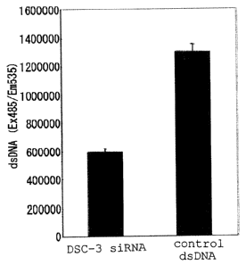
FIG. 2 shows a result of administering siRNA for a
Democollin-3 gene. Fig 2A shows that the cell growth of human
lung cancer cell line is inhibited by administering the siRNA;
Fig 2B shows that the level of mRNA expression of the
Democollin-3 gene is reduced by administering the siRNA; and
Fig. 2C shows that the expression level of a Democollin-3
protein is reduced by administering the siRNA.
Fig. 3 shows a gene expression profile of TM4SF13 prepared
by using various cancer tissue origin mRNAs and neighboring
normal tissue origin mRNAs.
Fig. 4 shows a result of administering siRNA for a TM4SF13
gene. Fig 4A shows that the cell growth of human breast cancer
cell line is inhibited by administering the siRNA; and Fig 4B
shows that the level of mRNA expression of the TM4SF13 gene is
reduced by administering the siRNA.
Fig. 5 shows that a TM4SF13 protein is localized on a
cytoplasmic membrane. From left, DAPI staining,
immunofluorescence staining and a chromatic figure combining
them are shown, respectively.
FIG. 6 shows that the TM4SF13 protein interacts with an
integrin-a3 (T: TM4SF13-3xFLAG, V: empty vector (control)).
FIG. 7 shows that the TM4SF13 protein interacts with an
integrin-a5 (T: TM4SF13-3xFLAG, V: empty vector (control)).
- 19 -
CA 02599875 2007-08-30
Fig. 8 shows the morphology change of a cellby the
expression of a TM4SF13 protein. The upper panels show a result
of culturing the cell on a plate coated with fibronectin, and
the lower panels show a result of culturing the cell on a plate
coated with laminin. From left, a TM4SF13 expressing cell, a
LacZ expressing cell and a nontransfectant (NT) are shown.
Best Mode for Carrying Out the Invention
A protein used in the present invention is a protein
comprising the same or substantially the same amino acid
sequence as the amino acid sequence represented by SEQ ID NO:
2, SEQ ID NO: 4, SEQ ID NO: 6, SEQ ID NO: 8 or SEQ ID NO: 10
(hereinafter these proteins are sometimes referred to as the
protein of the present invention). The protein of the present
invention may be any protein isolated and purified from cells
[for example, hepatocytes, splenocytes, nerve cells, glial
cells, (3 cells of pancreas, bone marrow cells, mesangial cells,
Langerhans' cells, epidermic cells, epithelial cells, goblet
cells, endothelial cells, smooth muscle cells, fibroblasts,
fibrocytes, myocytes, fat cells, immune cells (e.g.,
macrophages, T cells, B cells, natural killer cells, mast cells,
neutrophils, basophils, eosinophils, monocytes),
megakaryocytes, synovial cells, chondrocytes, bone cells,
osteoblasts, osteoclasts, mammary cells, or interstitial
cells; or the corresponding precursor cells, stem cells, cancer
cells, etc.] of human and other warm-blooded animals (e.g.,
- 20 -
CA 02599875 2007-08-30
guinea pig, rat, mouse, chicken, rabbit, swine, sheep, bovine,
simian, etc. ); or any tissue where such cells are present [for
example, brain or each part of brain (e.g., olfactory bulb,
amygdaloid nucleus, basal ganglia, hippocampus, thalamus,
hypothalamus, cerebral cortex, medulla oblongata, cerebellum),
spinal cord, hypophysis, stomach, pancreas, kidney, liver,
gonad, thyroid, gall-bladder, bone marrow, adrenal gland, skin,
muscle (e.g., smooth muscle, skeletal muscle), lung,
gastrointestinal tract (e.g., large intestine and small
intestine), blood vessel, heart, thymus, spleen, submandibular
gland, peripheral blood, prostate, testis, ovary, placenta,
uterus, bone, joint, adipose tissue (e.g., white adipose tissue,
brown adipose tissue) , etc. ]. The protein may also be a protein
biochemically synthesized by a chemical synthesis or cell-free
translation system, or a recombinant protein produced from a
transfectant in which the nucleic acid comprising a base
sequence encoding the above-mentioned amino acid sequence is
transfected.
The amino acid sequence whish is substantially the same
amino acid sequence as that represented by SEQ ID NO: 2, SEQ
ID NO: 4, SEQ ID NO: 6, SEQ ID NO: 8 or SEQ ID NO: 10, includes
amino acid sequences having about 50% or more homology,
preferably about 60% homology or more, more preferably about
70% or more homology, even more preferably about 80% or more
homology, particularly preferably about 90% or more homology
- 21 -
CA 02599875 2007-08-30
and most preferably about 95% or more homology, to the amino
acid sequence shown by SEQ ID NO: 2, SEQ ID NO: 4, SEQ ID NO:
6, SEQ ID NO: 8 or SEQ ID NO: 10, and the like. Herein, the
'Homology' means a ratio (%) of the same amino acid and similar
amino acid residue to the total overlapped amino acid residue,
in the best alignment when two amino acid sequences are aligned
with the use of a mathematical algorithm commonly known in the
technical field (preferably, the algorithm considers
introduction of gaps on one or both side of the sequence for
the best alignment). The term 'similar amino acid' refers to
an amino acid similar in its physiochemical properties, and the
examples include amino acids classified in a same group such
as aromatic amino acid (Phe, Trp, Tyr), aliphatic amino acid
(Ala, Leu, Ile, Val) , polar amino acid (Gln, Asn) , basic amino
acid (Lys, Arg, His) , acidic amino acid (Glu, Asp) , amino acid
including a hydroxyl group (Ser, Thr) , amino acid having a short
side chain(Gly, Ala, Ser, Thr, Met), and the like. A
substitution by such similar amino acid is expected to give no
change in the phenotype of protein (thus is a conservative amino
acid substitution). A specific example of the conservative
amino acid substitution is well-known in the technical field,
and is disclosed in various documents (for example, refer Bowie
et al, Science, 247: 1306-1310 (1990)).
Homology of the amino acid sequences in the present
specification can be calculated under the following conditions
- 22 -
CA 02599875 2007-08-30
(an expectation value = 10; gaps are allowed; matrix = BLOSUM62;
filtering = OFF) using a homology scoring algorithm NCBI BLAST
(National Center for Biotechnology Information Basic Local
Alignment Search Tool). Other algorithm for determining
homology of the amino acid sequence is exemplified by an
algorithm disclosed in Karlin et al, Proc. Natl. Acad. Sci. USA,
90: 5873-5877 (1993) [this algorithm is incorporated in NBLAST
and XBLAST program (version 2.0) (Altschul et al, Nucleic Acids
Res., 25:3389-3402(1997))]; an algorithm disclosed in
Needleman et al, J. Mol. Biol., 48: 444-453 (1970) [This
algorithm is incorporated in a GAP program in a GCG software
package]; an algorithm disclosed in Myers and Miller, CABIOS,
4: 11-17 (1988) [This algorithm is incorporated in ALIGN program
(version 2.0) which is a part of a CGC sequence alignment
software package]; an algorithm disclosed in Pearson et al. Proc.
Natl. Acad. Sci. USA, 85: 2444-2448 (1998) [This algorithm is
incorporated in an FASTA program in a GCG software package],
etc., and these may be also preferably used.
More preferably, the amino acid sequence which is
substantially the same amino acid sequence as that represented
by SEQ ID NO: 2, SEQ ID NO: 4, SEQ ID NO: 6, SEQ ID NO: 8 or
SEQ ID NO: 10, includes amino acid sequences having about 50%
or more identity, preferably about 60% or more identity, more
preferably about 70% or more identity, even more preferably
about 80% or more identity, particularly preferably about 90%
- 23 -
CA 02599875 2007-08-30
or more identity and most preferably about 95% or more identity,
to the amino acid sequence shown by SEQ ID NO: 2, SEQ ID NO:
4, SEQ ID NO: 6, SEQ ID NO: 8 or SEQ ID NO: 10.
The protein used in the present invention is a protein
comprising substantially the same amino acid sequence as the
amino acid sequence represented by SEQ ID NO: 2, SEQ ID NO: 4,
SEQ ID NO: 6, SEQ ID NO: 8 or SEQ ID NO: 10, and having an activity
substantially equivalent to the protein comprising the amino
acid sequence represented by SEQ ID NO: 2, SEQ ID NO: 4, SEQ
ID NO: 6, SEQ ID NO: 8 or SEQ ID NO: 10.
As the substantially equivalent activity described above,
there are, for example, the ligand binding activity, and signal
transduction, and the like. The substantially equivalent is
used to mean that the nature of the activities is equivalent
in terms of quality (e.g., physiologically or
pharmacologically) . Thus, the activities of the protein of the
present invention are preferably equivalent, but differences
in quantitative factors such as a level of these activities (e. g. ,
about 0.01 to 100 times, preferably about 0.1 to 10 times, more
preferably 0. 5 to 2 times) , a molecular weight of the protein,
and the like may be present and allowable.
The activity such as the ligand binding activity and
signal transduction can be assayed by publicly known methods,
for example, it can be assayed by the method to be described
later, comprises screening for a compound that inhibits the
- 24 -
CA 02599875 2007-08-30
activity of the protein used in the invention, or salts thereof.
Examples of the protein used in the present invention
include so-called muteins such as proteins having (i) the amino
acid sequence represented by SEQ ID NO: 2, SEQ ID NO: 4, SEQ
ID NO: 6, SEQ ID NO: 8 or SEQ ID NO: 10, of which 1 or 2 or more
(e. g. , about 1 to about 50, preferably about 1 to about 30, more
preferably about 1 to about 10 and much more preferably several
(1 to 5, 4, 3 or 2)) amino acids are deleted; (ii) the amino
acid sequence represented by SEQ ID NO: 2, SEQ ID NO: 4, SEQ
ID NO: 6, SEQ ID NO: 8 or SEQ ID NO: 10, to which 1 or 2 or more
(e. g. , about 1 to about 50, preferably about 1 to about 30, more
preferably about 1 to about 10 and much more preferably several
(1 to 5, 4, 3 or 2) ) amino acids are added; (iii) the amino acid
sequence represented by SEQ ID NO: 2, SEQ ID NO: 4, SEQ ID NO:
6, SEQ ID NO: 8 or SEQ ID NO: 10, in which 1 or 2 or more (e.g.,
about 1 to about 50, preferably about 1 to about 30, more
preferably about 1 to about 10 and much more preferably several
(1 to 5, 4, 3 or 2)) amino acids are inserted; (iv) the amino
acid sequence represented by SEQ ID NO: 2, SEQ ID NO: 4, SEQ
ID NO: 6, SEQ ID NO: 8 or SEQ ID NO: 10, in which 1 or 2 or more
(e.g., about 1 to about 50, preferably about 1 to about 30, more
preferably about 1 to about 10 and much more preferably several
(1 to 5, 4, 3 or 2) ) amino acids are substituted by other amino
acids; or (v) a combination of these amino acid sequences; and
the like.
- 25 -
CA 02599875 2007-08-30
Where the amino acid sequence is inserted, deleted or
substituted as described above, the position of its insertion,
deletion, or substitution is not particularly limited.
Preferable examples of the protein used in the present
invention include human Desmocollin-3a comprising the amino
acid sequence represented by SEQ ID NO: 2(Refseq Accession No.
NP 001932) or the homolog thereof (for example, mouse homolog
registered as RefSeq Accession No. NP031908 in GenBank) in
other mammals, human Desmocollin-3b comprising the amino acid
sequence represented by SEQ ID NO: 4 (Refseq Accession No.
NP 077741) or the homolog thereof in other mammals, human
TM4SF13 comprising the amino acid sequence represented by SEQ
ID NO: 6(RefSeq Accession No. NP055214) or the homolog thereof
(for example, mouse homolog registered as RefSeq Accession No.
NP 079635 in GenBank) in other mammals, human TM4SF6 comprising
the amino acid sequence represented by SEQ ID NO: 8 (RefSeq
Accession No. NP 003261) or the homolog thereof in other mammals
(for example, mouse homolog registered as RefSeq Accession No.
NP 062630 in GenBank) , and human LY-6K comprising the amino acid
sequence represented by SEQ ID NO: 10 (RefSeq Accession No.
NP 059997) or the homolog thereof in other mammals.
In the present specification, the proteins and peptides
are represented in accordance with the conventional way of
describing peptides, that is, the N-terminus (amino terminus)
at the left hand and the C-terminus (carboxyl terminus) at the
- 26 -
CA 02599875 2007-08-30
right hand. In the protein used in the present invention which
includes the protein comprising the amino acid sequence
represented by SEQ ID NO: 2, SEQ ID NO: 4, SEQ ID NO: 6, SEQ
ID NO: 8 or SEQ ID NO: 10, the C-terminus may be in any form
of a carboxyl group (-COOH), carboxylate (-COO-), an amide
(-CONH2) or an ester (-COOR).
Herein, examples of R in the ester include a C1-6 alkyl
group such as methyl, ethyl, n-propyl, isopropyl, n-butyl,
etc. ; a C3-8 cycloalkyl group such as cyclopentyl, cyclohexyl,
etc. ; a C6-12 aryl group such as phenyl, a-naphthyl, etc. ; a C7_14
aralkyl such as a phenyl-C1-2 alkyl group, e.g., benzyl,
phenethyl, etc. or a-naphthyl-C1-2 alkyl group such as
a-naphthylmethyl, etc.; pivaloyloxymethyl group and the like.
Where the protein used in the present invention contains
a carboxyl group (or a carboxylate) at a position other than
the C-terminus, the carboxyl group may be amidated or esterified
and such an amide or ester is also included within the protein
used in the present invention. Examples of the ester in this
case may be the C-terminal esters described above, etc.
Furthermore, examples of the protein used in the present
invention also include those wherein the amino group at the
N-terminal amino acid residues (e.g., methionine residue) is
protected with a protecting group ( e. g., a C1_6 acyl group such
as a C1-6 alkanoyl, e.g., formyl group, acetyl group, etc.);
those wherein the N-terminal region is cleaved in vivo and the
- 27 -
CA 02599875 2007-08-30
glutamine residue thus formed is pyroglutaminated; those
wherein a substituent (e.g., -OH, -SH, amino group, imidazole
group, indole group, guanidino group, etc.) on the side chain
of an amino acid in the molecule is protected with a suitable
protecting group (e. g. , a C1-6 acyl group such as a C1-6 alkanoyl
group, e.g., formyl group, acetyl group, etc.), or conjugated
proteins such as so-called glycoproteins having sugar chains;
etc.
The partial peptide of the protein used in the present
invention may be any peptide as long as it is a peptide having
a partial amino acid sequence of the protein used in the present
invention described above and has the activity substantially
equivalent to that of the protein used in the present invention
described above. Herein, the 'activity substantially
equivalent' means the same as mentioned above. The 'activity
substantially equivalent' can also be assayed in a same manner
as in the case of protein used in the present invention as above
mentioned.
For example, there are used peptides containing, e.g.,
at least 20 or more, preferably 50 or more, more preferably 70
or more, much more preferably 100 or more and most preferably
150 or more amino acids, in the constituent amino acid sequence
of the protein used in the present invention, etc.
The partial peptide used in the present invention may be
peptides containing the amino acid sequence, of which 1 or 2
- 28 -
CA 02599875 2007-08-30
or more (preferably about 1 to about 20, more preferably about
1 to about 10 and much more preferably several (1 to 5, 4, 3
or 2)) amino acids may be deleted; to which 1 or 2 or more
(preferably about 1 to about 20, more preferably about 1 to about
and much more preferably several (1 to 5, 4, 3 or 2) ) amino
acids may be added; in which 1 or 2 or more (preferably about
1 to about 20, more preferably about 1 to about 10 and much more
preferably several (1 to 5, 4, 3 or 2)) amino acids may be
inserted; or in which 1 or 2 or more (preferably about 1 to about
20, more preferably about 1 to about 10, and mush more preferably
several (1 to 5, 4, 3 or 2)) amino acids may be substituted by
other amino acids.
In the partial peptide used in the present invention, the
C-terminus may be in any form of a carboxyl group (-COOH), a
carboxylate (-COO-), an amide (-CONH2) or an ester (-COOR).
Herein, as the 'R' in ester, same ones as in the protein used
in the present invention can be exemplified. Where the partial
peptide of the present invention contains a carboxyl group (or
a carboxylate) at a position other than the C-terminus, the
carboxyl group may be amidated or esterified and such an amide
or ester is also included within the partial peptide of the
present invention. Examples of the ester in this case may be
the same as C-terminal esters described above, etc.
Furthermore, the partial peptide used in the present
invention also includes those wherein the amino group at the
- 29 -
CA 02599875 2007-08-30
N-terminal amino acid residues (e.g., methionine residue) is
protected with a protecting group; those wherein the N-terminal
region is cleaved in vivo and the glutamine residue thus formed
is pyroglutaminated; those wherein a substituent on the side
chain of an amino acid in the molecule is protected with a
suitable protecting group, or conjugated peptides such as
so-called glycopeptides having sugar chains; etc., as in the
protein used in the present invention described above.
The partial peptide used in the present invention may also
be used as an antigen for producing antibodies.
The protein, or its partial peptide used in the present
invention, maybe a free form, or salt form (it is true throughout
the present specification unless otherwise specified) As the
salts, salts with physiologically acceptable acids (e.g.,
inorganic acids, organic acids, etc.) or bases (e.g., alkali
metal salts, etc.), preferably physiologically acceptable acid
addition salts can be used. Examples of such salts include
salts with inorganic acids (e.g., hydrochloric acid, phosphoric
acid, hydrobromic acid, sulfuric acid), salts with organic
acids (e.g., acetic acid, formic acid, propionic acid, fumaric
acid, maleic acid, succinic acid, tartaric acid, citric acid,
malic acid, oxalic acid, benzoic acid, methanesulfonic acid,
benzenesulfonic acid) and the like.
The protein used in the present invention may be
manufactured by publicly known methods used to purify a protein
- 30 -
CA 02599875 2007-08-30
from human or warm-blooded animal cells or tissues described
above. Specifically, mammalian tissues or cells are
homogenized in the presence of surfactant, and then crude tissue
extract fraction obtained is subjected to chromatography
techniques such as reverse phase chromatography, ion exchange
chromatography, affinity chromatography and the like, thereby
the proteins used in the present invention can be prepared.
The protein used in the present invention or its partial
peptide can be manufactured by publicly known methods for
peptide synthesis.
For the methods for peptide synthesis, for example,
either solid phase synthesis or liquid phase synthesis may be
used. That is, the partial peptide or amino acids that can
constitute the protein or its partial peptide used in the
present invention are condensed with the remaining part. Where
the product contains protecting groups, these protecting groups
are removed to give the desired protein (peptide). Publicly
known methods for condensation and elimination of the
protecting groups are, for example, described in (i) to (v)
below.
(i) M. Bodanszky & M.A. Ondetti: Peptide Synthesis,
Interscience Publishers, New York (1966)
( ii ) Schroeder & Luebke: The Peptide, Academic Press, New York
(1965)
(iii) Nobuo Izumiya, et al.: Peptide Gosei-no-Kiso to Jikken
- 31 -
CA 02599875 2007-08-30
(Basics and experiments of peptide synthesis), published by
Maruzen Co. (1975)
(iv) Haruaki Yajima & Shunpei Sakakibara: Seikagaku Jikken Koza
(Biochemical Experiment) 1, Tanpakushitsu no Kagaku (Chemistry
of Proteins) IV, 205 (1977)
(v) Haruaki Yajima ed. : Zoku Iyakuhin no Kaihatsu (A sequel to
Development of Pharmaceuticals), Vol. 14, Peptide Synthesis,
published by Hirokawa Shoten
The protein (peptide) obtained in the above manner may
be purified and isolated by a known purification methods such
as solvent extraction, distillation, column chromatography,
liquid chromatography and recrystallization.
When the protein (peptide) obtained by the above methods
is in a free form, it can be converted into an appropriate salt
by a publicly known method or its modification; conversely when
the protein (peptide) is obtained in a salt form, it can be
converted into a free form or other different salt form by a
publicly known method or its modification.
To synthesize the protein or its partial peptide used in
the present invention, or amides thereof, commercially
available resins that are used for protein synthesis may be
usually used. Examples of such resins include chloromethyl
resin, hydroxymethyl resin, benzhydrylamine resin,
aminomethyl resin, 4-benzyloxybenzyl alcohol resin,
4-methylbenzhydrylamine resin, PAM resin,
- 32 -
CA 02599875 2007-08-30
4-hydroxymethylmethylphenyl acetamidomethyl resin,
polyacrylamide resin,
4-(2',4'-dimethoxyphenyl-hydroxymethyl)phenoxy resin,
4-(2',4'-dimethoxyphenyl-Fmoc-aminoethyl) phenoxy resin, etc.
Using these resins, amino acids, in which a-amino groups and
functional groups on the side chains are appropriately
protected, are condensed on the resin in accordance with the
sequence of the objective protein according to various
condensation methods publicly known in the art. At the end of
the reaction, the protein or partial peptide is excised from
the resin and at the same time, the protecting groups are removed.
Then, intramolecular disulfide bond-forming reaction is
performed in a highly diluted solution to obtain the objective
protein or partial peptide, or amides thereof.
For condensation of the protected amino acids described
above, a variety of activation reagents available for protein
synthesis may be used, and carbodiimides are particularly
preferable. Examples of such carbodiimides include DCC,
N,N'-diisopropylcarbodiimide,
N-ethyl-N'-(3-dimethylaminopropyl)carbodiimide, etc. For
activation by these reagents, the protected amino acids in
combination with a racemization inhibitor (e.g., HOBt, HOOBt)
may be added directly to the resin, or the protected amino acids
may be previously activated in the form of symmetric acid
anhydrides, HOBt esters or HOOBt esters, followed by adding the
- 33 -
CA 02599875 2007-08-30
thus activated protected amino acids to the resin.
Solvents for use to activate the protected amino acids
or condense them with the resin may be appropriately chosen from
solvents that are known to be usable for protein condensation
reactions. Examples of such solvents are acid amides such as
N,N-dimethylformamide, N,N-dimethylacetamide,
N-methylpyrrolidone, etc.; halogenated hydrocarbons such as
methylene chloride, chloroform, etc.; alcohols such as
trifluoroethanol, etc.; sulfoxides such as dimethylsulfoxide,
etc.; ethers such as pyridine, dioxane, tetrahydrofuran, etc.;
nitriles such as acetonitrile, propionitrile, etc.; esters such
as methyl acetate, ethyl acetate, etc.; or appropriate mixtures
of these solvents. The reaction temperature is appropriately
chosen from the range known to be applicable to protein binding
reactions and is usually selected in the range of approximately
-20 C to 50 C. The activated amino acid derivatives are used
generally in an excess of 1. 5 to 4 times. The condensation is
examined using the ninhydrin reaction; when the condensation
is insufficient, the condensation can be completed by repeating
the condensation reaction without removal of the protecting
groups. When the condensation is yet insufficient even after
repeating the reaction, unreacted amino acids are acetylated
with acetic anhydride or acetylimidazole to avoid any possible
effect on the subsequent reaction.
Protection of the functional groups that should not be
- 34 -
CA 02599875 2007-08-30
involved in the reaction of the starting materials, protecting
groups, elimination of the protecting groups, activation of the
functional groups involved in the reaction, and the like may
be appropriately chosen from publicly known groups and publicly
known means.
Examples of the protecting groups used to protect the
starting amino groups include Z, Boc, t-pentyloxycarbonyl,
isobornyloxycarbonyl, 4-methoxybenzyloxycarbonyl, Cl-Z, Br-Z,
adamantyloxycarbonyl, trifluoroacetyl, phthaloyl, formyl,
2-nitrophenylsulphenyl, diphenylphosphinothioyl, Fmoc, etc.
A carboxyl group can be protected by, e.g., alkyl
esterification (in the form of linear, branched or cyclic alkyl
esters of the alkyl, e.g., methyl, ethyl, propyl, butyl, t-butyl,
cyclopentyl, cyclohexyl, cycloheptyl, cyclooctyl, 2-adamantyl,
etc.), aralkylesterification (e.g., esterificationin theform
of benzyl ester, 4-nitrobenzyl ester, 4-methoxybenzyl ester,
4-chlorobenzyl ester, benzhydryl ester, etc.), phenacyl
esterification, benzyloxycarbonyl hydrazidation,
t-butoxycarbonyl hydrazidation, trityl hydrazidation, or the
like.
The hydroxyl group of serine can be protected through,
for example, its esterification or etherification. Examples
of groups appropriately used for the esterification include a
lower (C1-6) alkanoyl group, such as acetyl group, an aroyl group
such as benzoyl group, and a group derived from carbonic acid
- 35 -
CA 02599875 2007-08-30
such as benzyloxycarbonyl group, ethoxycarbonyl group, etc.
Examples of a group appropriately used for the etherification
include benzyl group, tetrahydropyranyl group, t-butyl group,
etc.
Examples of groups for protecting the phenolic hydroxyl
group of tyrosine include Bzl, C12-Bzl, 2-nitrobenzyl, Br-Z,
t-butyl, etc.
Examples of groups used to protect the imidazole moiety
of histidine include Tos, 4-methoxy-2,3,6-trimethyl-
benzenesulfonyl, DNP, benzyloxymethyl, Bum, Boc, Trt, Fmoc,
etc.
To eliminate (split off) the protecting groups, there are
used catalytic reduction under hydrogen gas flow in the presence
of a catalyst such as Pd-black or Pd-carbon; an acid treatment
with anhydrous hydrogen fluoride, methanesulfonic acid,
trifluoromethanesulfonic acid, trifluoroacetic acid, or a
mixture solution of these acids; a treatment with a base such
as diisopropylethylamine, triethylamine, piperidine or
piperazine; reduction with sodium in liquid ammonia, etc. The
elimination of the protecting group by the acid treatment
described above is carried out generally at a temperature of
approximately -20 C to 40 C. In the acid treatment, it is
efficient to add a cation scavenger such as anisole, phenol,
thioanisole, m-cresol, p-cresol, dimethylsulfide,
1,4-butanedithiol, 1,2-ethanedithiol, etc. Furthermore,
- 36 -
CA 02599875 2007-08-30
2,4-dinitrophenyl group known as the protecting group for the
imidazole of histidine is removed by a treatment with thiophenol.
Formyl group used as the protecting group of the indole of
tryptophan is eliminated by the aforesaid acid treatment in the
presence of 1,2-ethanedithiol, 1,4-butanedithiol, etc. as well
as by a treatment with an alkali such as a dilute sodium hydroxide
solution, dilute ammonia, etc.
Examples of the activated carboxyl groups in the starting
material include the corresponding acid anhydrides, azides,
activated esters [esters with alcohols (e.g.,
pentachlorophenol, 2,4,5-trichlorophenol, 2,4-dinitrophenol,
cyanomethyl alcohol, p-nitrophenol, HONB, N-hydroxysuccimide,
N-hydroxyphthalimide, HOBt)]. As example of the activated
amino groups in the starting material, the corresponding
phosphoric amides are employed.
In another method for obtaining the amides of the desired
protein or partial peptide, for example, the a-carboxyl group
of the carboxy terminal amino acid is first protected by
amidation; the peptide (protein) chain is then extended from
the amino group side to a desired length. Subsequently, a
protein or partial peptide, in which only the protecting group
of the N-terminal a-amino group of the peptide chain has been
eliminated, and a protein or partial peptide, in which only the
protecting group of the C-terminal carboxyl group has been
eliminated, are manufactured. The two proteins or peptides are
- 37 -
CA 02599875 2007-08-30
condensed in a mixture of the solvents described above. The
details of the condensation reaction are the same as described
above. After the protected protein or peptide obtained by the
condensation is purified, all the protecting groups are
eliminated by the method described above to give the desired
crude protein or peptide. This crude protein or peptide is
purified by various known purification means. Lyophilization
of the major fraction gives the amide of the desired protein
or peptide.
To prepare the esterified protein or peptide, for example,
the a-carboxyl group of the carboxy terminal amino acid is
condensed with a desired alcohol to prepare the amino acid ester,
which is followed by procedures similar to the preparation of
the amidated protein or peptide above to give the desired
esterified protein or peptide.
The partial peptide of the protein used in the present
invention can be manufactured by cleaving the protein used in
the present invention with an appropriate peptidase.
Further, the protein used in the present invention or
partial peptides thereof can also be manufactured by culturing
a transformant containing polynucleotide encoding the same and
then by separating and purifying the protein or partial peptide
from the obtained culture. The polynucleotide encoding the
protein used in the present invention or its partial peptide
may be DNA or RNA, or DNA/RNA chimera, and preferably is DNA.
- 38 -
CA 02599875 2007-08-30
In addition, the polynucleotide may be a double-strand, or
single-strand. The double-strand may include a
double-stranded DNA, a double-stranded RNA, and DNA:RNA hybrid.
The single-strand may include a sense strand (that is, coding
strand) and an antisense strand (that is, non-coding strand)
The polynucleotide encoding the protein used in the
present invention or its partial peptide can be exemplified by
genomic DNA, genomic DNA library, cDNA derived from any
mammalian (e.g., human, bovine, simian, horse, swine, sheep,
goat, canine, feline, guinea pig, rat, mouse, rabbit, hamster,
etc.) cells [for example, hepatocytes, splenocytes, nerve cells,
glial cells, (3 cells of pancreas, bone marrow cells, mesangial
cells, Langerhans' cells, epidermic cells, epithelial cells,
goblet cells, endothelial cells, smooth muscle cells,
fibroblasts, fibrocytes, myocytes, fat cells, immune cells
(e.g., macrophages, T cells, B cells, natural killer cells, mast
cells, neutrophils, basophils, eosinophils, monocytes),
megakaryocytes, synovial cells, chondrocytes, bone cells,
osteoblasts, osteoclasts, mammary cells, or interstitial
cells; or the corresponding precursor cells, stem cells, cancer
cells, etc. ] ; or any tissues where such cells are present [for
example, brain or each part of brain (e.g., olfactory bulb,
amygdaloid nucleus, basal ganglia, hippocampus, thalamus,
hypothalamus, cerebral cortex, medulla oblongata, cerebellum),
spinal cord, hypophysis, stomach, pancreas, kidney, liver,
- 39 -
CA 02599875 2007-08-30
gonad, thyroid, gall-bladder, bone marrow, adrenal gland, skin,
muscle, lung, gastrointestinal tract (e.g., large intestine and
small intestine), blood vessel, heart, thymus, spleen,
submandibular gland, peripheral blood, prostate, testis, ovary,
placenta, uterus, bone, joint, adipose tissue (e.g., white
adipose tissue, brown adipose tissue), skeletal muscle, etc.],
synthetic DNA, etc. The genomic DNA and cDNA encoding the
protein used in the present invention or its partial peptide
can be directly amplified by Polymerase Chain Reaction
(hereinafter, abbreviate to "PCR") and Reverse Transcriptase-
PCR (hereinafter, abbreviate to "RT-PCR") with the use of each
genomic DNA fraction, and total RNA or mRNA fraction prepared
from the above-described cells or tissues as a template.
Further, the genomic DNA and cDNA encoding the protein used in
the present invention or its partial peptide can be respectively
cloned from genomic DNA library and cDNA library which are
prepared by inserting the fragment of genomic DNA, and total
RNA or mRNA prepared from the above-described cells or tissues
into an appropriate vector, in accordance with a colony or
plaque hybridization assay or PCR method. The vector used for
the library may be any of bacteriophage, plasmid, cosmid,
phagemid and the like.
Examples of the DNA encoding the protein used in the
present invention may be any of DNA comprising a base sequence
hybridizable to DNA comprising the base sequence represented
- 40 -
CA 02599875 2007-08-30
by SEQ ID NO: 1, SEQ ID NO: 3, SEQ ID NO: 5, SEQ ID NO: 7 or
SEQ ID NO: 9 under high stringent conditions and encoding a
protein which has the activity substantially equivalent to the
protein comprising the amino acid sequence represented by SEQ
ID NO: 2, SEQ ID NO: 4, SEQ ID NO: 6, SEQ ID NO: 8 or SEQ ID
NO: 10.
As the DNA that is hybridizable to the base sequence
represented by SEQ ID NO: 1, SEQ ID NO: 3, SEQ ID NO: 5, SEQ
ID NO: 7 or SEQ ID NO: 9 under high stringent conditions, there
are employed, for example, DNAs comprising base sequences
having about 50% or more homology, preferably about 60% or more
homology, more preferably about 70 0 or more homology, even more
preferably about 80% or more homology, particularly preferably
about 90% or more homology, and most preferably about 95% or
more homology, to the base sequence represented by SEQ ID NO:
1, SEQ ID NO: 3, SEQ ID NO: 5, SEQ ID NO: 7 or SEQ ID NO: 9.
Homology of the base sequences in the present
specification, for example, can be calculated under the
following conditions (an expectation value = 10; gaps are
allowed; filtering = ON; match score = 1; mismatch score = -3)
using a homology scoring algorithm NCBI BLAST (National Center
for Biotechnology Information Basic Local Alignment Search
Tool) . As the other algorithm for determining homology of the
base sequence, same ones as the above-described homology
scoring algorithms for the amino acid sequence are preferably
- 41 -
CA 02599875 2007-08-30
exemplified.
The hybridization can be carried out by publicly known
methods or by modifications thereof, for example, by the method
described in Molecular Cloning, 2nd ed. (J. Sambrook et al.,
Cold Spring Harbor Lab. Press, 1989) . A commercially available
library can also be used according to the instructions of the
attached manufacturer's protocol. The hybridization can be
carried out more preferably under high stringent conditions.
The high stringent conditions used herein are, for
example, those in a sodium concentration at about 19 to 40 mM,
preferably about 19 to 20 mM at a temperature of about 50 to
70 C, preferably about 60 to 65 C. In particular,
hybridization conditions in a sodium salt concentration at
about 19 mM at a temperature of about 65 C are most preferred.
Those skilled in the art can simply regulate the condition to
a desired stringency by appropriately changing a concentration
of hybridization solution, temperature of hybridization
reaction, probe concentration, length of probe, number of
mismatch, time for hybridization reaction, salt concentration
of washing solution, temperature for washing, etc.
Preferable examples of the DNA encoding the protein used
in the present invention include DNA comprising the base
sequence represented by SEQ ID NO: 1 (Refseq Accession No.
NM001941) or the homolog thereof (for example, mouse homolog
registered as RefSeq Accession No. NM 007882 in GenBank) in
- 42 -
CA 02599875 2007-08-30
other mammals, DNA comprising the base sequence represented by
SEQ ID NO: 3 (Refseq Accession No. NM 024423) or the homolog
thereof in other mammals, DNA comprising the based sequence
represented by SEQ ID NO: 5 (RefSeq Accession No. NM 014399)
or the homolog thereof (for example, mouse homolog registered
as RefSeq Accession No. NM 025359 in GenBank) in other mammals,
DNA comprising the base sequence represented by SEQ ID NO: 7
(RefSeq Accession No. NM 003270) or the homolog thereof (for
example, mouse homolog registered as RefSeq Accession No.
NM 019656 in GenBank) in other mammals, DNA comprising the base
sequence represented by SEQ ID NO: 9 (RefSeq Accession No.
NM 017527) or the homolog thereof in other mammals.
The polynucleotide (e.g., DNA) encoding the partial
peptide of the protein used in the present invention may be any
polynucleotide so long as it contains the base sequence encoding
the partial peptide of the protein used in the present invention
described above. The polynucleotide may also be any of genomic
DNA, genomic DNA library, cDNA derived from the cells and
tissues described above, cDNA library derived from the cells
and tissues described above, and synthetic DNA.
As the DNA encoding the partial peptide of the protein
used in the present invention, there is employed, for example,
DNA containing a partial base sequence of the base sequence
represented by SEQ ID NO: 1, SEQ ID NO: 3, SEQ ID NO: 5, SEQ
ID NO: 7 or SEQ ID NO: 9; or DNA containing a base sequence
- 43 -
CA 02599875 2007-08-30
hybridizable to polynucleotide which comprises the base
sequence represented by SEQ ID NO: 1, SEQ ID NO: 3, SEQ ID NO:
5, SEQ ID NO: 7 or SEQ ID NO: 9 under high stringent conditions,
and also encoding a peptide which has the activity substantially
equivalent to the protein used in the present invention. The
DNA hybridizable to the base sequence represented by SEQ ID NO:
1, SEQ ID NO: 3, SEQ ID NO: 5, SEQ ID NO: 7 or SEQ ID NO: 9 represent
the same meaning as described above. Further, same
hybridization method and high stringent conditions as described
above can be used.
For cloning of DNAs that encode the protein used in the
present invention and its partial peptide (hereinafter
sometimes merely referred to as the protein of the present
invention in the description of cloning of DNAs encoding the
same and their expression), the DNA can be either amplified by
PCR using synthetic DNA primers containing a part of the base
sequence encoding the protein of the present invention, or the
DNA inserted into an appropriate vector can be selected by
hybridization with those labeled with DNA fragment or synthetic
DNA that encodes a part or entire region of the protein of the
present invention. The hybridization can be carried out, for
example, according to the method described in Molecular Cloning,
2nd ed. (J. Sambrook et al. , Cold Spring Harbor Lab. Press, 1989) .
Where the hybridization is carried out using commercially
available library, the procedures may be conducted in
- 44 -
CA 02599875 2007-08-30
accordance with the protocol described in the attached
instructions.
Conversion of the base sequence of DNA can be effected
by publicly known methods such as the ODA-LA PCR method, the
Gapped duplex method, the Kunkel method, etc., or modification
thereof, using PCR, a publicly known kit available as
MutanTM-super Express Km (Takara Bio) or MutanTM-K (Takara Bio) ,
etc.
The cloned DNA encoding the protein can be used as it is,
depending upon purpose or, if desired, after digestion with a
restriction enzyme or after addition of a linker thereto. The
DNA may contain ATG as a translation initiation codon at the
5' end thereof and TAA, TGA or TAG as a translation termination
codon at the 3' end thereof. These translation initiation and
termination codons may also be added by using an appropriate
synthetic DNA adapter.
The expression vector for the protein of the present
invention can be manufactured, for example, by excising the
desired DNA fragment from the DNA encoding the protein of the
present invention, and then ligating the DNA fragment with an
appropriate expression vector downstream a promoter in the
vector.
Examples of the vector include plasmids derived form E.
coli (e.g., pBR322, pBR325, pUC12, pUC13), plasmids derived
from Bacillus subtilis (e.g., pUB110, pTP5, pC194), plasmids
- 45 -
CA 02599875 2007-08-30
derived from yeast (e.g., pSH19, pSH15), bacteriophages such
as a.phage, etc., animal viruses such as retrovirus, vaccinia
virus, baculovirus, etc. as well as pAl-11, pXT1, pRc/CMV,
pRc/RSV, pcDNA I/Neo, etc.
The promoter used in the present invention may be any
promoter if it matches well with a host to be used for gene
expression. For example, when animal cells are used as the host,
examples of the promoter are SRa promoter, SV40 promoter, LTR
promoter, CMV promoter, HSV-TK promoter, etc..
Among them, it is preferable to use CMV (cytomegalovirus)
promoter, SRa promoter, etc. When bacteria of the genus
Escherichia is used as a host, preferred examples of the
promoter are trp promoter, lac promoter, recA promoter, XPL
promoter, lpp promoter, T7 promoter, etc. When bacteria of the
genus Bacillus is used as the host, preferred example of the
promoter are SPOl promoter, SP02 promoter, penP promoter, etc.
When yeast is used as the host, preferred examples of the
promoter are PH05 promoter, PGK promoter, GAP promoter, ADH
promoter, etc. When insect cells are used as the host,
preferred examples of the promoter are polyhedrin promoter, P10
promoter, etc.
In addition to the foregoing examples, the expression
vector may further optionally contain an enhancer, a splicing
signal, a poly A addition signal, a selection marker, SV40
replication origin (hereinafter sometimes abbreviated as
- 46 -
CA 02599875 2007-08-30
SV40ori), etc. Examples of the selection marker include
dihydrofolate reductase (hereinafter sometimes abbreviated as
dhfr) gene [methotrexate (MTX) resistance], ampicillin
resistant gene (hereinafter sometimes abbreviated as Ampr),
neomycin resistant gene (hereinafter sometimes abbreviated as
Neor, G418 resistance) , etc. In particular, when dhfr gene is
used as the selection marker using dhfr gene-deficient Chinese
hamster cells, the objective gene can also be selected on a
thymidine free medium.
If necessary, a signal sequence that matches with a host
is added to the N-terminus of the protein of the present
invention. The signal sequence that can be used are PhoA signal
sequence, OmpA signal sequence, etc. when bacteria of the genus
Escherichia is used as the host; a-amylase signal sequence,
subtilisin signal sequence, etc. when bacteria of the genus
Bacillus is used as the host; MFa signal sequence, SUC2 signal
sequence, etc. when yeast is used as the host; and insulin signal
sequence, a-interferon signal sequence, antibody molecule
signal sequence, etc. when animal cells are used as the host,
respectively.
Using the vector containing the DNA encoding the protein
of the present invention thus constructed, transformants can
be manufactured. Examples of the host, which may be employed,
are bacteria belonging to the genus Escherichia, bacteria
belonging to the genus Bacillus, yeast, insect cells, insects,
- 47 -
CA 02599875 2007-08-30
animal cells, etc.
Specific examples of the bacteria belonging to the genus
Escherichia include Escherichia coli K12 DH1 [Proc. Natl. Acad.
Sci. U.S.A., 60, 160 (1968)], JM103 [Nucleic Acids Research,
9, 309 (1981)], JA221 [Journal of Molecular Biology, 120, 517
(1978)], HB101 [Journal of Molecular Biology, 41, 459 (1969)],
C600 [Genetics, 39, 440 (1954)], etc.
Examples of the bacteria belonging to the genus Bacillus
include Bacillus subtilis MI114 [Gene, 24, 255 (1983)], 207-21
[Journal of Biochemistry, 95, 87 (1984)], etc.
Examples of yeast include Saccharomyces cerevisiae AH22,
AH22R , NA87-11A, DKD-5D, 20B-12, Schizosaccharomyces pombe
NCYC1913, NCYC2036, Pichia pastoris KM71, etc.
Examples of insect cells include, for the virus AcNPV,
Spodoptera frugiperda cell (Sf cell), MG1 cell derived from
mid-intestine of Trichoplusia ni, High FiveTM cell derived from
egg of Trichoplusia ni, cells derived from Mamestra brassicae,
cells derived from Estigmena acrea, etc.; and for the virus
BmNPV, Bombyx mori N cell (BmN cell) , etc. Examples of the Sf
cell which can be used are Sf9 cell (ATCC CRL1711), Sf21 cell
(both cells are described in Vaughn, J. L. et al., In Vivo, 13,
213-217 (1977)), etc.
As the insect, for example, a larva of Bombyx mori can
be used [Maeda et al., Nature, 315, 592 (1985)].
Examples of animal cells include simian cell COS-1, COS-3,
- 48 -
CA 02599875 2007-08-30
COS-7, Vero, Chinese hamster ovary cell (hereinafter simply
referred to as CHO cell), dhfr gene-deficient CHO cell
(hereinafter simply referred to as CHO (dhfr-) cell), mouse L
cell, mouse AtT-20, mouse myeloma cell, mouse ATDC5 cell, rat
GH3, human FL cell, human 293 cell, human HeLa cell, etc.
Bacteria belonging to the genus Escherichia can be
transformed, for example, by the method described in Proc. Natl.
Acad. Sci. U.S.A., 69, 2110 (1972), Gene, 17, 107 (1982), etc.
Bacteria belonging to the genus Bacillus can be
transformed, for example, by the method described in Molecular
& General Genetics, 168, 111 (1979), etc.
Yeast can be transformed, for example, by the method
described in Methods in Enzymology, 194, 182-187 (1991), Proc.
Natl. Acad. Sci. U.S.A., 75, 1929 (1978), etc.
Insect cells or insects can be transformed, for example,
according to the method described in Bio/Technology, 6,
47-55(1988), etc.
Animal cells can be transformed, for example, according
to the method described in Saibo Kogaku (Cell Engineering),
extra issue 8, Shin Saibo Kogaku Jikken Protocol (New Cell
Engineering Experimental Protocol), 263-267 (1995) (published
by Shujunsha), or Virology, 52, 456 (1973).
Thus, the transformants transformed with the expression
vectors bearing the DNAs encoding the protein can be obtained.
Where the host is bacteria belonging to the genus
- 49 -
CA 02599875 2007-08-30
Escherichia or the genus Bacillus, the transformant can be
appropriately cultured in a liquid medium which contains
materials required for growth of the transformant such as carbon
sources, nitrogen sources, inorganic materials, and the like.
Examples of the carbon sources include glucose, dextrin,
soluble starch, sucrose, etc.; examples of the nitrogen sources
include inorganic or organic materials such as ammonium salts,
nitrate salts, corn steep liquor, peptone, casein, meat extract,
soybean cake, potato extract, etc.; and, examples of the
inorganic materials are calcium chloride, sodium
dihydrogenphosphate, magnesium chloride, etc. In addition,
yeast extracts, vitamins, growth promoting factors etc. may
also be added to the medium. Preferably, pH of the medium is
adjusted to about 5 to about 8.
A preferred example of the medium for culturing the
bacteria belonging to the genus Escherichia is M9 medium
supplemented with glucose and Casamino acids [Miller, Journal
of Experiments in Molecular Genetics, 431-433, Cold Spring
Harbor Laboratory, New York, 1972] . If necessary, a chemical
such as 3(3-indolylacrylic acid can be added to the medium
thereby to activate the promoter efficiently.
Where the bacteria belonging to the genus Escherichia are
used as the host, the transformant is usually cultivated at
about 15 to 43 C for about 3 to 24 hours. If necessary, the
culture may be aerated or agitated.
- 50 -
CA 02599875 2007-08-30
Where the bacteria belonging to the genus Bacillus are
used as the host, the transformant is cultured generally at
about 30 to 40 C for about 6 to 24 hours. If necessary, the
culture can be aerated or agitated.
Where yeast is used as the host, the transformant is
cultivated, for example, in Burkholder's minimal medium [Proc.
Natl. Acad. Sci. U.S.A., 77, 4505 (1980)] or in SD medium
supplemented with 0. 5% Casamino acids [Proc. Natl. Acad. Sci.
U.S.A., 81, 5330 (1984)]. Preferably, pH of the medium is
adjusted to about 5 to 8. In general, the transformant is
cultivated at about 20 to 35 C for about 24 to 72 hours. If
necessary, the culture can be aerated or agitated.
Where insect cells or insects are used as the host, the
transformant is cultivated in, for example, Grace's Insect
Medium (Nature, 195, 788 (1962)) to which an appropriate
additive such as inactivated 10% bovine serum is added.
Preferably, pH of the medium is adjusted to about 6. 2 to about
6.4. Normally, the transformant is cultivated at about 27 C
for about 3 days to about 5 days and, if necessary, the culture
can be aerated or agitated.
Where animal cells are employed as the host, the
transformant is cultured in, for example, MEM medium containing
about 5 to 20% fetal bovine serum [Science, 122, 501 (1952)],
DMEM medium [Virology, 8, 396 (1959)], RPMI 1640 medium [The
Journal of the American Medical Association, 199, 519 (1967)],
- 51 -
CA 02599875 2007-08-30
199 medium [Proceeding of the Society for the Biological
Medicine, 73, 1(1950) ], etc. Preferably, pH of the medium is
adjusted to about 6 to about 8. The transformant is usually
cultivated at about 30 C to about 40 C for about 15 to 60 hours
and, if necessary, the culture can be aerated or agitated.
As described above, the protein of the present invention
can be produced in the transformant, on the cell membrane of
the transformant, or outside the transformant.
The protein of the present invention can be separated and
purified from the culture described above, for example, by the
following procedures.
When the protein of the present invention is extracted
from the bacteria or cells of the culture, the bacteria or cell
is collected after culturing by a publicly known method and
suspended in an appropriate buffer. The bacteria or cell is
then disrupted by methods such as ultrasonication, a treatment
with lysozyme and/or freeze-thaw cycling, followed by
centrifugation, filtration, etc. to produce crude extract of
the protein. The buffer used for the procedures may contain
a protein denaturant such as urea or guanidine hydrochloride,
or a surfactant such as Triton X-100TM, etc. When the protein
is secreted in the culture broth, the supernatant can be
separated, after completion of the cultivation, from the
bacteria or cell to collect the supernatant by a publicly known
method.
- 52 -
CA 02599875 2007-08-30
The protein of the present invention contained in the
supernatant or the extract thus obtained can be purified by
appropriately combining the publicly known methods for
separation and purification. Such publicly known methods for
separation and purification include a method utilizing
difference in solubility such as salting out, solvent
precipitation, etc.; a method mainly utilizing difference in
molecular weight such as dialysis, ultrafiltration, gel
filtration, SDS-polyacrylamide gel electrophoresis, etc.; a
method utilizing difference in electric charge such as ion
exchange chromatography, etc.; a method utilizing difference
in specific affinity such as affinity chromatography, etc.; a
method utilizing difference in hydrophobicity such as reverse
phase high performance liquid chromatography, etc.; a method
utilizing difference in isoelectric point such as
isoelectrofocusing electrophoresis; and the like.
When the protein of the present invention thus obtained
is in a free form, the protein can be converted into the salt
by publicly known methods or modifications thereof. On the
other hand, when the protein is obtained in the form of a salt,
it can be converted into the free form or in the form of a
different salt by publicly known methods or modifications
thereof.
The protein produced by the recombinant can be treated,
prior to or after the purification, with an appropriate
- 53 -
CA 02599875 2007-08-30
protein-modifying enzyme so that the protein can be subjected
to addition of an appropriate modification or removal of a
partial polypeptide. Examples of the protein-modifying enzyme
include trypsin, chymotrypsin, arginyl endopeptidase, protein
kinase, glycosidase and the like.
The presence of the thus produced protein of the present
invention can be determined by an enzyme immunoassay or western
blotting using a specific antibody.
The protein used in the present invention or its partial
peptide can be synthesized by the in vitro translation with the
use of a cell-free protein translation system comprising rabbit
reticulocyte lysate, wheat germ lysate, E. coli lysate, etc.,
and RNA which corresponds to DNA encoding the same as a template.
The protein or its partial peptide can also be synthesized by
a cell-free transcription/translation system further
comprising RNA polymerase with the use of DNA encoding
calmodulin or its partial peptide as a template. For the
cell-free protein (transcription /) translation system,
commercially available ones and a method known per se can be
employed. In particular, the Escherichia coli extract
solution can be prepared according to a method disclosed in
Pratt J.M. et al, "Transcription and Tranlation", Hames B.D.
and Higgins S.J. edition, IRL Press, Oxford 179-209 (1984) . As
the commercially available cell lysate, Escherichia coli
derived lysates such as E. coli S30 extract system (manufactured
- 54 -
= CA 02599875 2007-08-30
by Promega) and RTS 500 Rapid Tranlation System (manufactured
by Roche), etc.; rabbit reticulocyte derived lysates such as
Rabbit Reticulocyte Lysate System (manufactured by Promega),
etc.; and wheat germ derived lysates such as
PROTEIOSTM(manufactured by TOYOBO), etc., can be exemplified.
Among them, the wheat germ lysate is preferably used. As the
method of manufacturing wheat germ lysate, for example, methods
disclosed in Johnston F.B. et al, Nature, 179, 160-161 (1957),
Erickson A.H. et al, Meth. Enzymol., 96, 38-50 (1996), etc.,
can be employed.
As a system or device for the protein synthesis, a batch
method (Pratt, J.M. et al, (1984) mentions above); continuous
cell-free protein synthesis system (Spirin A.S. et al, Science,
242, 1162-1164 (1988)) in which an amino acid, energy source,
etc. is continuously supplied in a reaction system; dialysis
(Kikawa, et al, 21st Annual meeting of the Molecular Biology
Society of Japan, WID6)); a double layer method (PROTEIOSTM
Wheat germ cell-free protein synthesis core kit instruction
manual, manufactured by TOYOBO), and the like can be exemplified.
In addition, a method which comprises supplying RNA template,
amino acid, energy source, etc., into a synthesis reaction
system according to necessity, and eliminating a synthesized
or degraded material according to necessity (Japanese Patent
Laid-Open No. 2000-333673) can be used.
The antibody against the protein used in the present
- 55 -
CA 02599875 2007-08-30
invention or against its partial peptide (hereinafter,
sometimes abbreviated as "antibody of the present invention")
can be any of polyclonal or monoclonal antibody, as long as it
recognizes the protein of the present invention or its partial
peptide. The isotype of the antibody is not particularly
limited, but it is preferably IgG, IgM or IgA, particularly
preferably IgG.
The antibody of the present invention is not subject to
limitation, as long as it has at least a complementality
determining region (CDR) for specifically recognizing and
binding to the target antigen; in addition to the whole antibody
molecule, the antibody may, for example, be a fragment such as
Fab, Fab', or F(ab')2, a genetically engineered conjugate
molecule such as scFv, scFv-Fc, minibody, or diabody, or a
derivative thereof modified with a molecule having protein
stabilizing action, such as polyethylene glycol (PEG), or the
like, and the like.
The antibody against the protein used in the present
invention or against its partial peptide (hereinafter sometimes
merely referred to as the protein of the present invention in
the description of antibody) can be manufactured according to
a method of producing an antibody or antisera known per se.
Hereinafter, a preparation method of immunogen of an
antibody of the present invention and a production method of
the antibody will be described.
- 56 -
CA 02599875 2007-08-30
(1) Preparation of antigen
As an antigen used to prepare the antibody of the present
invention, any antigen such as the above-mentioned protein of
the present invention or its partial peptide, or a (synthetic)
peptide having 1 or 2 or more antigenic determinants, which are
the same as in the above-mentioned protein of the present
invention or its partial peptide, etc. may be used (hereinafter
these antigens are sometimes merely referred to as the antigen
of the present invention).
The aforesaid protein of the present invention or its
partial peptide can be, for example, (a) prepared from
warm-blooded animal tissues or cells of human, simian, rat,
mouse, chicken, etc., by publicly known methods or with
modifications, (b) chemically synthesized by publicly known
peptide synthesis methods using a peptide synthesizer, etc.,
(c) produced by culturing a transformant bearing DNA encoding
protein of the present invention or its partial peptide, or (d)
biochemically synthesized from nucleic acid encoding protein
of the present invention or its partial peptide as a template
by using a cell-free transcription/ translation system.
(a) Where the protein of the present invention is prepared
from the warm-blooded animal tissues or cells, the tissues or
cells are homogenized, and then the crude extract (e.g.,
membrane fraction, soluble fraction) can be also used per se
as an antigen. Alternatively, the tissues or cells are
- 57 -
CA 02599875 2007-08-30
extracted with an acid, a surfactant, an alcohol, etc. , and the
extract solution can be purified and isolated by a combination
of salting-out, dialysis, gel filtration, chromatography
techniques such as reverse phase chromatography, ion exchange
chromatography, affinity chromatography and the like. Thus
obtained protein of the present invention can be used as per
se as an immunogen, and can also be used as the immunogen in
the form of a partial peptide prepared by limited degradation
using a peptidase, etc.
(b) Where the antigen of the present invention is prepared
chemically, the synthetic peptides used are, for example, a
peptide having the same structure as the protein of the present
invention purified from natural material by the above method
(a), specifically, a peptide containing 1 or 2 or more amino
acid sequences, which are the same amino acid sequences
consisting of 3 or more, preferably 6 or more amino acids in
an optional region of the amino acid sequence of the protein.
(c) Where the antigen of the present invention is produced
using the DNA-bearing transformants, the DNA can be produced
in accordance with publicly known cloning techniques [e.g., the
method described in Molecular Cloning 2nd ed., (J. Sambrook et
al., Cold Spring Harbor Lab. Press, 1989), etc.]. The cloning
techniques include (1) a method in which DNAs encoding the
antigen are isolated from cDNA library by a hybridization method
using DNA probes designed on the basis of the gene sequence
- 58 -
CA 02599875 2007-08-30
encoding the protein of the present invention, and (2) a method
in which DNAs encoding the antigen are produced from cDNA as
a template by PCR using DNA primers designed based on the gene
sequence encoding the protein of the present invention, and the
DNA are inserted into an expression vector suitable for a host.
Incubating the transformants which is obtained from host by
being transformed with the expression vector, in appropriate
medium, the desired antigen can be obtained.
(d) When a cell-free transcription/translation system is
utilized, a method for synthesizing an mRNA by using an
expression vector incorporating DNA that encodes the antigen
(for example, an expression vector wherein the DNA is placed
under the control of the T7 or SP6 promoter and the like, and
the like) as the template, that was prepared by the same method
as (c) above, and a transcription reaction mixture comprising
an RNA polymerase matching the promoter and its substrates
(NTPs) ; and thereafter performing a translation reaction with
the mRNA as the template using a known cell-free translation
system (e.g., extractfrom E. co1i, rabbit reticulocytes, wheat
germ, etc.), and the like can be mentioned. By adjusting the
salt concentration and the like appropriately, the
transcription reaction and the translation reaction can also
be carried out in the same reaction mixture at one time.
As the immunogen, a whole protein molecule of the present
invention or a peptide having a partial amino acid sequence of
- 59 -
CA 02599875 2007-08-30
the protein molecule can be used. As examples of the partial
amino acid sequence, those comprising 3 or more continuous amino
acid residues, preferably those comprising 4 or more, more
preferably 5 or more, still more preferably 6 or more continuous
amino acid residues, can be mentioned. Alternatively, as
examples of the amino acid sequence, those comprising 20 or less
continuous amino acid residues, preferably those comprising 18
or less, more preferably 15 or less, still more preferably 12
or less continuous amino acid residues, can be mentioned. A
portion of these amino acid residues (e.g., 1 to several
residues) may be substituted with a substituent group (e.g.,
Cys, hydroxyl group, etc. ). The peptide used as the immunogen
has an amino acid sequence comprising one to several such
partial amino acid sequences.
Warm-blooded animal cells itself which express the
protein of the present invention can also be used directly as
the antigen of the present invention. As the Warm-blooded
animal cells, there can be used the naturally occurring cells
as described in (a) above, cells transformed by the methods as
described in (c) above, etc. Hosts used for the transformation
may be any cells as long as they are the cells collected from
human, simian, rat, mouse, hamster, chicken etc. and preferably
used are HEK293, COS7, CHO-Kl, NIH3T3, Balb3T3, FM3A, L929,
SP2/0, P3U1, B16, P388, or the like. Naturally occurring
warm-blooded animal cells or transformed warm-blooded animal
- 60 -
CA 02599875 2007-08-30
cells, which express the protein of the present invention, can
be injected to immune animal as a suspension of the cells in
a medium used for tissue culture (e.g., RPMI 1640) or buffer
(e.g., Hanks' Balanced Salt Solution). Immunization may be
done by any method, as long as it can stimulate antibody
production, and preferably used are intravenous injection,
intraperitoneal injection, intramuscular injection,
subcutaneous injection, etc.
The antigen of the present invention permit direct use
for immunization in an insolubilized form, as long as it has
immunogenicity; when an antigen of low molecular weight ( i. e.,
molecular weight about 3, 000 or less) having only one to several
antigenic determinants in the molecule thereof (for example,
a partial peptide of the protein of the present invention) is
used, it can be used for immunization in the form of a complex
bound or adsorbed to a suitable carrier because these antigens
are normally hapten molecules of low immunogenicity. As the
carrier, a naturally occurring or synthetic polymer can be used.
As examples of the naturally occurring polymer, serum albumin
of a mammal such as bovine, rabbit, or human, thyroglobulin of
a mammal such as bovine or rabbit, ovalbumin of chicken etc.,
hemoglobin of a mammal such as bovine, rabbit, human, or sheep,
keyhole limpet hemocyanin (KLH) and the like can be used. As
examples of the synthetic polymer, various latexes of polymers
or copolymers of polyamino acids, polystyrenes, polyacryls,
- 61 -
CA 02599875 2007-08-30
polyvinyls, polypropylenes and the like, and the like can be
mentioned.
A mixing ratio of the carrier to the hapten may be in any
ratio of any type, as long as the antibody can be efficiently
produced to the antigen bound or adsorbed to the carrier. The
above described naturally occurring or synthetic high molecular
carrier conventionally used to produce an antibody against a
hapten may be used in a weight ratio of 0.1 to 100 based on 1
of hapten.
For coupling of the hapten and the carrier, a variety of
condensing agents can be used. Examples of the condensing
agents, which are advantageously employed, are diazonium
compounds such as bis-diazotized benzidine capable of
crosslinking tyrosines, histidines or tryptophans; dialdehyde
compounds such as glutaraldehyde, etc. capable of crosslinking
amino groups with each other; diisocyanate compounds such as
toluene-2,4-diisocyanate, etc.; dimaleimide compounds such as
N,N'-o-phenylenedimaleimide, etc. capable of crosslinking
thiol groups with each other; maleimide activated ester
compounds capable of crosslinking an amino group with a thiol
group; carbodiimide compounds capable of crosslinking an amino
group with a carboxyl group; etc. In the crosslinking of amino
groups with each other, one amino group is reacted with an
activated ester reagent having dithiopyridyl group (e.g.,
N-succinimidyl 3-(2-pyridyldithio)propionate (SPDP), etc.)
- 62 -
CA 02599875 2007-08-30
and then reduced to introduce the thiol group, whereas another
amino group is introduced with a maleimide group using a
maleimide activated ester reagent, and the two groups may be
reacted with each other.
(2) Preparation of monoclonal antibody
(a) Preparation of monoclonal antibody producing-cell
An antigen of the present invention is administered as
is, or along with a carrier or a diluent, to a warm-blooded animal
at a site enabling antibody production by the methods such as
intraperitoneal injection, intravenous injection,
subcutaneous injection, intradermal injection and the like.
In order to increase antibody productivity upon the
administration, Freund's complete adjuvant or Freund's
incomplete adjuvant may be administered. Dosing is normally
performed about two to 10 times in total, with one time every
1 to 6 weeks. As examples of the warm-blooded animal used,
simians, rabbits, canine, guinea pigs, mice, rats, hamsters,
sheep, goats, donkeys and chickens can be mentioned. Although
it is preferable to use a mammal of the same species as the
recipient in order to avoid the problem of anti-Ig antibody
production, mice and rats are generally preferably used for
generating a monoclonal antibody.
Since artificial immunization to humans is ethically
difficult, it is preferable, when the antibody of the present
invention targets a human, (i) to obtain a human antibody by
- 63 -
CA 02599875 2007-08-30
immunizing a human antibody-producing animal (e.g., mouse)
produced according to a method described below, (ii) to produce
a chimeric antibody, humanized antibody or fully human antibody
according to a method described below, or (iii) to obtain a human
antibody using in combination the in vitro immunization method
and cell immortalization with virus, human-human (or -mouse)
hybridoma production technique, phage display method and the
like. Note that the in vitro immunization method can also be
used preferably as a method for obtaining an antigen against
an antigen that is unstable and difficult to prepare in large
amounts for the purpose of preparing a non-human animal-derived
antibody, because there is the possibility of obtaining an
antibody against an antigen for which antibody production is
suppressed by ordinary immunization, because it is possible to
obtain an antibody with an amount of antigen on the nanogram
to microgram order, because immunization completes in several
days, and for other reasons.
As the animal cells used in the in vitro immunization
method, lymphocytes, preferably B-lymphocytes and the like,
isolated from peripheral blood, spleen, lymph node and the like
of a human and the above-described warm-blooded animals
(preferably mouse or rat) can be mentioned. For example, in
the case of mouse or rat cells, the spleen is extirpated from
an about 4- to 12-week-old animal, and splenocytes are separated
and rinsed with a appropriate medium [e.g., Dulbecco's modified
- 64 -
CA 02599875 2007-08-30
Eagle medium (DMEM) , RPMI1640 medium, Ham's F12 medium and the
like], after which the splenocytes are suspended in an
antigen-containing medium supplemented with fetal calf serum
(FCS; about 5 to 20%) and cultured using a C02 incubator and
the like for about 4 to 10 days. Examples of the antigen
concentration include, but are not limited to, 0.05 to 5 g.
It is preferable to prepare a culture supernatant of thymocytes
of an animal of the same strain (preferably at about 1 to 2 weeks
of age) according to a conventional method, and to add the
supernatant to the medium.
Since it is difficult to obtain a thymocyte culture
supernatant in in vitro immunization of human cells, it is
preferable to perform immunization by adding, to the medium,
several kinds of cytokines such as IL-2, IL-4, IL-5, and IL-6
and the like, and if necessary, an adjuvant substance (e.g.,
muramyldipeptide and the like) along with the antigen.
In preparing a monoclonal antibody, it is possible to
establish an antibody-producing hybridoma by selecting an
individual or cell population showing an elevated antibody
titerfrom among antigen-immunized warm-blooded animals (e.g.,
mice, rats) or animal cells (e.g., human, mouse, rat),
respectively; collecting spleens or lymph nodes at 2 to 5 days
after the final immunization or collecting the cells after 4
to 10 days of cultivation after in vitro immunization to isolate
antibody-producing cells; and fusing the isolated cells with
- 65 -
CA 02599875 2007-08-30
myeloma cells. A measurement of serum antibody titer can be
performed by, for example, reacting a labeled antigen and an
antiserum, and thereafter determining the activity of the label
bound to the antibody.
Although the myeloma cells are not subject to limitation,
as long as they are capable of producing a hybridoma that
secretes a large amount of antibody, those that do not produce
or secrete the antibody per se are preferable, with greater
preference given to those of high cell fusion efficiency. To
facilitate hybridoma selection, it is preferable to use a cell
line that is sensitive to HAT (hypoxanthine, aminopterin,
thymidine) . As examples of the mouse myeloma cells, NS-1, P3U1,
SP2/0, AP-1 and the like can be mentioned; as examples of the
rat myeloma cells, R210.RCY3, Y3-Ag 1.2.3 and the like can be
mentioned; as examples of the human myeloma cells, SKO-007, GM
1500-6TG-2, LICR-LON-HMy2, UC729-6 and the like can be
mentioned.
Fusion operation can be performed according to a known
method, for example, the method of Koehler and Milstein [Nature,
256, 495 (1975)]. As a fusion promoter, polyethylene glycol
(PEG) , Sendai virus and the like can be mentioned, and PEG and
the like are preferably used. Although the molecular weight
of PEG is not subject to limitation, PEG1000 to PEG6000, which
are of low toxicity and relatively low viscosity, are preferable.
As examples of the PEG concentration, about 10 to 80%,
- 66 -
= CA 02599875 2007-08-30
preferably about 30 to 50%, can be mentioned. As the solution
for diluting PEG, various buffers such as serum-free medium
(e.g., RPMI1640), complete medium comprising about 5 to 20%
serum, phosphate buffered saline (PBS) , and Tris buffer can be
used. DMSO (e. g. , about 10 to 20%) can also be added as desired.
As examples of the pH of the fusion solution, about 4 to 10,
preferably about 6 to 8 can be mentioned.
The ratio by number of antibody-producing cells
(splenocytes) and myeloma cells is preferably normally about
1:1 to 20:1, and the cell fusion can be efficiently performed
by incubation normally at 20 to 40 C, preferably at 30 to 37 C,
normally for 1 to 10 minutes.
An antibody-producing cell line can also be obtained by
infecting antibody-producing cells with a virus capable of
transforming lymphocytes to immortalize the cells. As such
viruses, for example, Epstein-Barr (EB) virus and the like can
be mentioned. Although the majority of persons have immunity
because they have ever been infected with this virus in an
asymptomatic infection of infectious mononucleosis, virion is
also produced when the ordinary EB virus is used; therefore,
appropriate purification must be performed. As an EB system
free from the possibility of viral contamination, it is also
preferable to use a recombinant EB virus that retains the
capability of immortalizing B lymphocytes but lacks the
capability of replicating virion (for example, deficiency of
- 67 -
CA 02599875 2007-08-30
the switch gene for transition from latent infection state to
lytic infection state and the like).
Since marmoset-derived B95-8 cells secrete EB virus, B
lymphocytes can be easily transformed by using a culture
supernatant thereof. An antibody-producing B cell line can be
obtained by, for example, culturing these cells using a medium
supplemented with serum and penicillin/streptomycin (P/S)
(e. g., RPMI1640) or a serum-free medium supplemented with a cell
growth factor, thereafter separating the culture supernatant
by filtration or centrifugation and the like, suspending
therein antibody-producing B lymphocytes at a suitable
concentration (e.g., about 107 cells/mL), and incubating the
suspension normally at 20 to 40 C, preferably at 30 to 37 C,
normally for about 0.5 to 2 hours. When human
antibody-producing cells are provided as mixed lymphocytes, it
is preferable to previously remove T lymphocytes by allowing
them to form an E rosette with, for example, sheep erythrocytes
and the like, to increase transformation frequency of EB virus,
because the majority of persons have T lymphocytes which exhibit
cytotoxicity to cells infected with EB virus. It is also
possible to select lymphocytes specific for the target antigen
by mixing sheep erythrocytes, previously bound with a soluble
antigen, with antibody-producing B lymphocytes, and separating
the rosette using a density gradient of Percoll and the like.
Furthermore, because antigen-specific B lymphocytes are capped
- 68 -
CA 02599875 2007-08-30
by adding the antigen in large excess so that they no longer
present IgG on the surface, mixing with sheep erythrocytes bound
with anti-IgG antibody results in the formation of rosette only
by antigen-nonspecific B lymphocytes. Therefore, by
collecting a layer of cells that don't form rosette from this
mixture using a density gradient of Percoll and the like, it
is possible to select antigen-specific B lymphocytes.
Human antibody-secreting cells having acquired the
capability of proliferating infinitely by the transformation
can be back fused with mouse or human myeloma cells in order
to stably sustain the antibody-secreting ability. As the
myeloma cells, the same as those described above can be used.
Hybridoma screening and breeding are normally performed
using a medium for animal cells (e.g., RPMI1640) containing 5
to 20% FCS or a serum-free medium supplemented with cell growth
factors, with the addition of HAT (hypoxanthine, aminopterin,
thymidine) . As examples of the concentrations of hypoxanthine,
aminopterin and thymidine, about 0.1 mM, about 0.4 M and about
0.016 mM and the like, respectively, can be mentioned. For
selecting a human-mouse hybridoma, ouabain resistance can be
used. As human cell lines are more sensitive to ouabain than
mouse cell lines, it is possible to eliminate unfused human
cells by adding ouabain at about 10-7 to 10-3 M to the medium.
In selecting a hybridoma, it is preferable to use feeder
cells or culture supernatants of certain cells. As the feeder
- 69 -
= CA 02599875 2007-08-30
cells, an allogenic cell species having a lifetime limited so
that it dies after helping the emergence of hybridoma, cells
capable of producing large amounts of a growth factor useful
for the emergence of hybridoma with their proliferation potency
reduced by irradiation and the like, and the like are used. For
example, as the mouse feeder cells, splenocytes, macrophage,
blood, thymocytes and the like can be mentioned; as the human
feeder cells, peripheral blood mononuclear cells and the like
can be mentioned. As examples of the cell culture supernatant,
primary culture supernatants of the above-described various
cells and culture supernatants of various established cell
lines can be mentioned.
Moreover, a hybridoma can also be selected by reacting
a fluorescein-labeled antigen with fusion cells, and thereafter
separating the cells that bind to the antigen using a
fluorescence-activated cell sorter (FACS) . In this case,
efforts for cloning can be lessened significantly because a
hybridoma that produces an antibody against the target antigen
can be directly selected.
For cloning a hybridoma that produces a monoclonal
antibody against the target antigen, various methods can be
used.
It is preferable to remove aminopterin as soon as possible
because it inhibits many cell functions. In the case of mice
and rats, aminopterin can be removed 2 weeks after fusion and
- 70 -
CA 02599875 2007-08-30
beyond because most myeloma cells die within 10 to 14 days.
However, a human hybridoma is normally maintained in a medium
supplemented with aminopterin for about 4 to 6 weeks after
fusion. It is desirable that hypoxanthine and thymidine be
removed more than one week after the removal of aminopterin.
That is, in the case of mouse cells, for example, a complete
medium (e.g., RPMI1640 supplemented with 10% FCS) supplemented
with hypoxanthine and thymidine (HT) is added or exchanged 7
to 10 days after fusion. About 8 to 14 days after fusion,
visible clones emerge. Provided that the diameter of clone has
reached about 1 mm, the amount of antibody in the culture
supernatant can be measured.
A measurement of the amount of antibody can be performed
by, for example, a method comprising adding the hybridoma
culture supernatant to a solid phase (e.g., microplate) to which
the target antigen or a derivative thereof or its partial
peptide (including the partial amino acid sequence used as the
epitope) is adsorbed directly or with a carrier, subsequently
adding an anti-immunoglobulin (IgG) antibody (an antibody
against IgG derived from an animal of the same species as the
animal from which the original antibody-producing cells are
derived is used) or protein A, which had been labeled with a
radioactive substance (e. g. , 125I, 131I, 3H, 19C) , enzyme (e. g. ,
(3-galactosidase, (3-glucosidase, alkaline phosphatase,
peroxidase, malate dehydrogenase), fluorescent substance
- 71 -
= CA 02599875 2007-08-30
(e.g., fluorescamine, fluorescein isothiocyanate),
luminescent substance (e.g., luminol, luminol derivative,
luciferin, lucigenin) and the like, and detecting the antibody
against the target antigen (epitope) bound to the solid phase,
a method comprising adding the hybridoma culture supernatant
to a solid phase to which an anti-IgG antibody or protein A is
adsorbed, adding the target antigen, or a derivative thereof,
or its partial peptide labeled with the same labeling agent as
described above, and detecting the antibody against the target
antigen (epitope) bound to the solid phase, and the like.
Although limiting dilution is normally used as the
cloning method, cloning using soft agar and cloning using FACS
(described above) are also possible. Cloning by limiting
dilution can be performed by, for example, the following
procedures, which, however, are not to be construed as limiting.
The amount of antibody is measured as described above,
and positive wells are selected. Selected suitable feeder
cells are previously added to a 96-well plate. Cells are
collected from the antibody-positive wells and suspended in
complete medium (e.g., RMPI1640 supplemented with 10% FCS and
P/S) to obtain a density of 30 cells/mL; 0.1 mL (3 cells/well)
of this suspension is added to the well plate with feeder cells
added thereto; the remaining cell suspension is diluted to 10
cells/mL and sown to other wells (1 cell/well) in the same way;
the still remaining cell suspension is diluted to 3 cells/mL
- 72 -
CA 02599875 2007-08-30
and sown to other wells (0.3 cells/well). The cells are
cultured for about 2 to 3 weeks until a visible clone appears,
when the amount of antibody is measured to select positive wells,
and the selected cells are recloned in the same way. In the
case of human cells, cloning is relatively difficult, so that
a plate in which cells are seeded at 10 cells/well is also
prepared. Although a monoclonal antibody-producing hybridoma
can be obtained normally by two times of subcloning, it is
desirable to repeat recloning regularly for several more months
to confirm the stability thereof.
Hybridomas can be cultured in vitro or in vivo.
As a method of in vitro culture, a method comprising
gradually scaling up a monoclonal antibody-producing hybridoma
obtained as described above, from a well plate, while keeping
the cell density at, for example, about 105 to 106 cells/mL,
and gradually lowering the FCS concentration, can be mentioned.
As a method of in vivo culture, for example, a method
comprising intraperitoneally injecting a mineral oil to a mouse
(a mouse that is histocompatible with the parent strain of the
hybridoma) to induce plasmacytoma (MOPC), intraperitoneally
injecting about 106 to 107 cells of hybridoma to the mouse5 to
days later, and collecting ascites fluid under anesthesia
2 to 5 weeks later, can be mentioned.
(b)Purification of the monoclonal antibody
Separation and purification of the monoclonal antibody
- 73 -
CA 02599875 2007-08-30
can be performed according to a publicly known method, for
example, separation and purification method of immunoglobulin
[e.g., salting-out, alcohol precipitation, isoelectric point
precipitation, electrophoresis, adsorption-desorption with an
ion exchanger (e.g., DEAE, QEAE), ultracentrifugation, gel
filtration, specific purification comprising selectively
collecting the antibody by means of an antigen-coupled solid
phase or an active adsorbent such as protein A or protein G,
and dissociating the linkage to obtain the antibody, and the
like].
As described above, a monoclonal antibody can be produced
by culturing a hybridoma in or outside the living body of a
warm-blooded animal, and harvesting an antibody from the body
fluid or culture thereof.
When using the antibody of the present invention for
cancer preventive / remedy, since the antibody is required to
have antitumor activity, it is necessary to examine the level
of antitumor activity of provided monoclonal antibody. The
antitumor activity can be assayed by comparing the cancer cell
growth or induction of apoptosis, in the presence and absence
of antibody.
In a preferred mode of embodiment, since the antibody of
the present invention is used as a pharmaceutical product having
humans as the subject of administration thereof, the antibody
of the present invention (preferably a monoclonal antibody) is
- 74 -
CA 02599875 2007-08-30
an antibody whose risk of showing antigenicity when
administered to a human has been reduced; to be specific, the
antibody is a fully human antibody, a humanized antibody, a
mouse-human chimeric antibody and the like, particularly
preferably a fully human antibody. A humanized antibody and
a chimeric antibody can be prepared by genetic engineering
technology according to the method described below. Although
a fully human antibody can also be produced from the
above-described human-human (or -mouse) hybridoma, it is
desirable to produce it using a human antibody-producing animal
(e.g., mouse) or the phage display method described below in
order to stably supply the antibody in large amounts at low
costs.
(i) Preparation of chimeric antibody
As used herein, "a chimeric antibody" means an antibody
wherein the sequences of the variable regions of the H chain
and L chain (VH and VL) thereof are derived from a mammalian
species, and wherein the sequences of the constant regions (CH
and CL) are derived from another mammalian species. The
sequences of the variable regions are preferably derived from,
for example, an animal species permitting easy preparation of
a hybridoma, such as mouse, and the sequences of the constant
regions are preferably derived from the recipient mammalian
species.
As examples of the method of preparing a chimeric antibody,
- 75 -
= CA 02599875 2007-08-30
the method described in US Patent No. 6, 331, 415 or a partially
modified method thereof and the like can be mentioned. To be
specific, first, mRNA or total RNA is prepared from a monoclonal
antibody-producing hybridoma (for example, mouse-mouse
hybridoma) obtained as described above, according to a
conventional method, to synthesize cDNA. DNAs that encode VH
and VL are amplified and purified by PCR according to a
conventional method with the cDNA as the template, using
appropriate primers [for example, oligo DNAs comprising the
base sequences that encode the N-terminal sequences of VH and
VL, respectively, as the sense primers, and oligo DNAs that
hybridize to the base sequences that encode the N-terminal
sequences of CH and CL, respectively, as the antisense primer
(see, for example, Bio/Technology, 9: 88-89, 1991)]. In the
same manner, DNAs that encode CH and CL are amplified and purified
from an RNA prepared from lymphocytes and the like of other
mammal ( e. g., human) by RT-PCR. VH and CH, and VL and CL, are
ligated together, respectively, using a conventional method,
and the chimeric H chain DNA and chimeric L chain DNA obtained
are inserted into respective appropriate expression vectors
[for example, vectors comprising promoters that have
transcription activity in CHO cells, COS cells, mouse myeloma
cells and the like (e.g., CMV promoter, SV40 promoter and the
like)]. The DNAs that encode the two chains may be inserted
into separate vectors, and may be inserted into a single vector
- 76 -
CA 02599875 2007-08-30
in tandem. Host cells are transformed with the chimeric H chain
and chimeric L chain expression vector(s) obtained. As the host
cells, animal cells, for example, Chinese hamster ovary (CHO)
cells, simian-derived COS-7 cells, Vero cells, rat-derived GHS
cells and the like, in addition to the above-described mouse
myeloma cells, can be mentioned. For the transformation, any
method applicable to animal cells can be used, with preference
given to electroporation method and the like. It is possible
to isolate a chimeric monoclonal antibody by culturing the host
cells in a medium suitable thereto for a given period, and
thereafter collecting the culture supernatant and purifying it
in the same manner as described above. Alternatively, it is
also possible to obtain a chimeric monoclonal antibody easily
and in large amounts from milk or eggs of transgenic animals
which are produced by a conventional method using germ line
cells of an animal such as bovine, goat, or chicken as the host
cells, for which a transgenic technique has been established
and a know-how of mass propagation as a domestic animal
(domestic fowl) has been accumulated. Furthermore, it is also
possible to obtain a chimeric monoclonal antibody in large
amounts from the seeds, leaves and the like of a transgenic plant,
produced by using microinjection and electroporation into
protoplast, the particle gun method and Ti-vector method for
intact cells and the like, with cells of a plant such as corn,
rice, wheat, soybean, or tobacco as the host cells, for which
- 77 -
CA 02599875 2007-08-30
a transgenic technique has been established, and which is
cultured in large amounts as a major crop.
When the chimeric monoclonal antibody obtained is
digested with papain, Fab is obtained; when the same is digested
with pepsin, F(ab')2 is obtained.
It is also possible to make scFv by ligating DNAs that
encode mouse VH and VL via a suitable linker, for example, DNA
that encodes a peptide consisting of 1 to 40 amino acids,
preferably 3 to 30 amino acids, more preferably 5 to 20 amino
acids [e.g., [Ser-(Gly)m]n or [(Gly)m-Ser]n (m is an integer
from 0 to 10, n is an integer from 1 to 5) and the like].
Furthermore, it is possible to make a minibody monomer by
ligating DNA that encodes CH3 via a suitable linker thereto,
or make a scFv-Fc by ligating DNA that encodes CH full length
via a suitable linker thereto. The DNA encoding such an
antibody molecule modified (coupled) by genetic engineering can
be expressed in a microorganism such as E. coli or yeast under
the control of a suitable promoter, to produce the antibody
molecule in large amounts.
When DNAs encoding mouse VH and VL are inserted into the
downstream of one promoter in tandem and introduced into E. coli,
a dimer named as Fv is formed by monocistronic gene expression.
When an appropriate amino acid in the FRs of VH and VL is
substituted with Cys using molecule modeling, a dimer named as
dsFv is formed via the intermolecular disulfide bond between
- 78 -
CA 02599875 2007-08-30
the two chains.
(ii) Humanized antibody
As used herein, "a humanized antibody" means an antibody
wherein the sequences of all regions present in the variable
region, other than the complementality determining region (CDR),
[i.e., framework region (FR) in constant region and variable
region] are derived from a human, and wherein only the sequence
of CDR is derived from another mammalian species. The other
mammalian species is preferably an animal species, for example,
mouse and the like, with which production of hybridomas can be
easily performed.
As examples of the method of preparing a humanized
antibody, the methods described in US Patent Nos. 5,225,539,
5,585,089, 5,693,761 and 5,693,762 or partially modified
methods therefrom and the like can be mentioned. To be specific,
DNAs that encode VH and VL derived from a non-human mammalian
species (e.g., mouse) are isolated in the same manner as with
the above-described chimeric antibody, after which sequencing
is performed by a conventional method using an automated DNA
sequencer (e.g., manufactured by Applied Biosystems and the
like), and the base sequences obtained or deduced amino acid
sequences therefrom are analyzed using a known antibody
sequence database [for example, Kabat database (see Kabat et
al., "Sequences of Proteins of Immunological Interest", edited
by US Department of Health and Human Services, Public Health
- 79 -
CA 02599875 2007-08-30
Service, NIH, 5th edition, 1991) and the like] to determine the
CDR and FR of the two chains. A base sequence wherein the CDR
encoding region of a base sequence that encodes the L chain and
H chain of a human antibody having an FR sequence similar to
the determined FR sequence [e . g., human x type L chain subgroup
I and human H chain subgroup II or III (see Kabat et al., 1991
(supra))] is substituted with the determined base sequence that
encodes the CDR of another animal species, is designed, and the
base sequence is divided into fragments of about 20 to 40 bases,
and a sequence complementary to the base sequence is divided
into fragments of about 20 to 40 bases so that they alternately
overlap with the aforementioned fragments. It is possible to
construct DNAs that encode VH and VL having human-derived FR
and a CDR derived from another mammalian species by synthesizing
individual fragments using a DNA synthesizer, and hybridizing
and ligating them in accordance with conventional methods. In
order to transfer a CDR derived from another mammalian species
into human-derived VH and VL more quickly and more efficiently,
it is preferable to use PCR-based site directed mutagenesis.
As examples of such a method, the sequential CDR grafting method
described in Japanese Patent Unexamined Publication No.
HEI-5-227970 and the like can be mentioned. It is possible to
obtain cells or transgenic animal/plant that produces a
humanized antibody by ligating the thus-obtained DNAs that
encode VH and VL to DNAs that encode human-derived CH and CL,
- 80 -
CA 02599875 2007-08-30
respectively, in the same manner as with the above-described
chimeric antibody, and introducing the ligated product into
suitable host cells.
A humanized antibody, like a chimeric antibody, can be
modified to scFv, scFv-Fc, minibody, dsFv, Fv and the like by
using genetic engineering techniques; and they can be produced
in a microorganism such as E. coli or yeast by using a suitable
promoter.
The technology for preparing a humanized antibody can
also be applied to, for example, preparing a monoclonal antibody
that can be preferably administered to another animal species
for which no hybridoma production technology has been
established. For example, animals widely propagated as
domestic animals (domestic fowls) such as bovine, swine, sheep,
goat, and chicken, and pet animals such as canine and felines,
and the like can be mentioned as the subject animal species.
(iii) Preparation of fully human antibody using human
antibody-producing animal
Provided that a functional human Ig gene is introduced
into a non-human warm-blooded animal having the endogenous
immunoglobulin (Ig) gene knocked out (KO) therein, and that this
animal=is immunized with an a=ntigen, a human antibody is
produced in place of the antibody derived from the animal.
Therefore, provided that an animal such as mice, for which a
technique for producing a hybridoma has been established, is
- 81 -
CA 02599875 2007-08-30
used, it is possible to acquire a fully human monoclonal
antibody by the same method as the conventional method used to
prepare a mouse monoclonal antibody. First, some of the human
monoclonal antibodies, that were generated by using a human
antibody-producing mouse (see Immunol. Today, 17: 391-397,
1996) obtained by crossing a mouse transfected with minigenes
of the human Ig H chain and L chain using an ordinary transgenic
(Tg) technique with a mouse wherein the endogenous mouse Ig gene
has been inactivated using an ordinary KO technique, are already
in clinical stage, and to date production of anti-human Ig human
antibody (HAHA) has not been reported.
Later, Abgenix Inc. [trade name: XenoMouse (see Nat.
Genet., 15: 146-156, 1997; US Patent No. 5,939,598 and the
like)] and Medarex Inc. [trade name: Hu-Mab Mouse (see Nat.
Biotechnol. , 14: 845-851, 1996; US Patent No. 5, 545, 806 and the
like)] established Tg mice transfected with even a larger human
Ig gene using a yeast artificial chromosome (YAC) vector, thus
enabling the production of human antibodies of richer
repertoire. However, because the human Ig gene, for example,
in the case of the H chain, exhibits its diversity as the VDJ
exon, which is a variable combination of about 80 kinds of V
fragments, about 30 kinds of D fragments and 6 kinds of J
fragments, encodes the antigen binding site, the full length
thereof is as large as about 1.5 Mb (14th chromosome) for the
H chain, about 2 Mb (2nd chromosome) for the KL chain, and about
- 82 -
CA 02599875 2007-08-30
1 Mb (22nd chromosome) for the XL chain. To reproduce the
diverse antibody repertoire in another animal species as in
human, it is desirable to introduce the full length of each Ig
gene. However, DNA that is insertable into a conventional
transfection vector (plasmid, cosmid, BAC, YAC and the like)
is normally several kb to several hundred kb in length, and it
has been difficult to introduce the full length of Ig genes by
the conventional technique for establishing a transgenic animal,
which comprises inserting a cloned DNA into a fertilized egg.
Tomizuka et al. (Nat. Genet., 16: 133-143, 1997) prepared
a mouse having the full-length human Ig gene by introducing a
natural fragment of a human chromosome harboring the Ig gene
(hCF) into a mouse [transchromosomic (TC) mouse]. That is,
first, a human-mouse hybrid cell having human chromosomes in
which the 14th chromosome comprising the H chain gene and the
2nd chromosome comprising the xL chain gene, both labeled with,
for example, a drug-resistance marker and the like, is treated
with a spindle formation inhibitor (e.g., colcemid) for about
48 hours to prepare a microcell wherein one to several
chromosomes or fragments thereof are enveloped in nuclear
membrane, and the chromosomes are introduced into a mouse ES
cell by the. micronuclear fusion method. A hybrid ES cell
retaining the chromosomes having the human Ig gene or fragments
thereof is selected using a medium containing a drug, and the
cell is microinjected into a mouse embryo in the same manner
- 83 -
CA 02599875 2007-08-30
as with the preparation of an ordinary KO mouse. A germ line
chimera is selected among the chimeric mice obtained, with coat
color as the index, and the like, to establish a TC mouse strain
carrying the human 14th chromosome fragment (TC(hCF14)) and a
TC mouse strain carrying the human 2nd chromosome fragment
(TC(hCF2)). After establishing mouse strains wherein the
endogenous H chain gene and xL chain gene are knocked out,
respectively [KO (IgH) and KO (IgK) ] by a conventional method,
it is possible to establish a mouse strain having all the four
kinds of gene modifications (double TC/KO) by repeating the
crossing of these four strains.
Provided that the same method as that for producing an
ordinary mouse monoclonal antibody is applied to a double TC/KO
mouse established as described above, it is possible to obtain
an antigen-specific human monoclonal antibody-producing
hybridoma. However, there is the drawback of a lower efficiency
to obtain hybridomas than that with the ordinary mouse, because
hCF2 containing the KL chain gene is unstable in the mouse cells.
On the other hand, because the aforementioned Hu-Mab
mouse has a structure wherein the variable region cluster are
doubled although it has about 50% of the KL chain gene, it
exhibits a x chain diversity equivalent to that with full length
(on the other hand, HuMab mouse exhibits a low H chain diversity
and inadequate response to antigen because it carries only about
10% of the H chain gene). And the K chain is stably retained
- 84 -
CA 02599875 2007-08-30
in the mouse cells because it is inserted in mouse chromosome
via a YAC vector (Igx-YAC). Making use of this advantage, it
is possible to get the efficiency for obtaining hybridomas and
affinity of antibody to antigen that are equivalent to those
with the ordinary mouse, by crossing a TC(hCF14) mouse with a
Hu-Mab mouse to establish a mouse that stably retains both hCF14
and Igx-YAC (trade name: KM mouse).
Furthermore, it is also possible to establish a human
antibody-producing animal in which the XL chain gene is further
transfected to reconstruct the diverse human antibody
repertoire more completely. Such an animal can also be obtained
by producing a TC mouse in which the human 22nd chromosome or
a fragment thereof harboring the XL chain gene is introduced
in the same manner as described above[TC(hCF22)], and crossing
the mouse with the above-described double TC/KO mouse or KM
mouse, or can also be obtained by, for example, constructing
a human artificial chromosome (HAC) comprising both the H chain
locus and the XL chain locus, and introducing it into a mouse
cell (Nat. Biotechnol., 18: 1086-1090, 2000).
When an antibody of the present invention is used as a
medicine, an antibody of the present invention is desirably a
monoclonal antibody, but it may be a polyclonal antibody. When
the antibody of the present invention is a polyclonal antibody,
it is not necessary to use hybridoma; therefore, provided that
a human antibody-producing animal is produced in the same manner
- 85 -
CA 02599875 2007-08-30
as described above using an animal species for which no
technique for preparing a hybridoma has been established but
a transgenic technique has been established, preferably an
ungulate such as bovine, it is also possible to produce a human
antibody in larger amounts at low costs (see, for example, Nat.
Biotechnol., 20: 889-894, 2002). The human polyclonal
antibody thus obtained can be purified by collecting blood,
ascites fluid, milk, egg and the like, preferably milk or egg,
of the human antibody-producing animal, in combination with the
same purification techniques as described above.
(iv) Preparation of fully human antibody using phage
display human antibody library
Another approach to produce a fully human antibody is a
method using phage display. This method sometimes encounters
cases in which a mutation due to PCR is introduced into a site
other than CDRs; for this reason, a few reports of cases of HAHA
production in clinical stage are available. On the other hand,
however, the method has advantages such as no risk of
cross-species viral infection derived from the host animal and
the indefinite specificity of the antibody (antibodies against
forbidden clone, sugar chain and the like can also be easily
prepared).
The method of preparing a phage display human antibody
library include, but are not limited to, for example, the
methods described below.
- 86 -
= CA 02599875 2007-08-30
Although a phage used is not subject to limitation,
filamentous phage (Ff bacteriophage) is normally preferably
used. As the method of presenting a foreign protein on the phage
surface, a method comprising expressing and presenting the
foreign protein as a fusion protein with any of the coat proteins
g3p, and g6p to g9p on the coat protein can be mentioned; and
a method comprising fusing the foreign protein to the N-terminal
side of g3p or g8p is often used. As the phage display vector,
1) one in which the foreign gene is introduced in the form of
fusion gene with the coat protein gene of the phage genome, to
allow all the coat proteins presented on the phage surface to
be presented as a fusion protein with the foreign protein, 2)
one in which the gene encoding the fusion protein is inserted
separately from the wild-type coat protein gene to allow the
fusion protein and the wild-type coat protein to be expressed
simultaneously, and 3) one in which an E. coli having a phagemid
vector harboring the gene that encodes the fusion protein is
infected with a helper phage having the wild-type coat protein
gene to produce phage particles that express the fusion protein
and the wild-type coat protein simultaneously, and the like can
be mentioned. However, a phage display vector of the type 2)
or 3) is used for the preparation of an antibody library, because
in the case of 1), the capability of infection is lost when a
large foreign protein is fused.
As a specific vector, those described by Holt et al. (Curr.
- 87 -
CA 02599875 2007-08-30
Opin. Biotechnol., 11: 445-449, 2000) can be mentioned as
examples. For example, pCES1 (see J. Biol. Chem., 274:
18218-18230, 1999) is an Fab-expressing phagemid vector wherein
DNA encoding the KL chain constant region allocated to
downstream of the g3p signal peptide, and DNA encoding CH3,
His-tag, c-myc tag, and the amber stop codon (TAG) followed by
the g3p coding sequence, allocated to downstream of the g3p
signal peptide, are arranged under the control of one lactose
promoter. When this is introduced to an E. coli having an amber
mutation, Fab is presented onto the g3p coat protein, but when
it is expressed in the HB2151 strain and the like, which do not
have an amber mutation, a soluble Fab antibody is produced. And
as the scFv-expressing phagemid vector, for example, pHEN1 (J.
Mol. Biol., 222: 581-597, 1991) and the like are used.
Meanwhile as examples of the helper phage, M13-K07,
VCSM13 and the like can be mentioned.
And as another phage display vector, a vector that is
designed so that a sequence comprising the cysteine-encoding
codon is linked to each of the 3' end of the antibody gene and
the 5' end of the coat protein gene to express the two genes
simultaneously and separately (not in the form of a fusion
protein) , and to present the antibody onto the coat protein on
the phage surface via S-S bonds between the introduced cysteine
residues (CysDisplayTM technology of Morphosys AG) and the like,
can be mentioned.
- 88 -
CA 02599875 2007-08-30
As the kind of human antibody library, a
naive/non-immunized library, a synthetic library, an immunized
library and the like can be mentioned.
The naive/non-immunized library is a library obtained by
acquiring the VH and VL genes retained by a normal human by RT-PCR,
and randomly cloning them into the above-described phage
display vector. Normally, mRNA derived from lymphocytes of
peripheral blood, bone marrow, tonsil and the like of a normal
human, and the like are used as the template. A library prepared
by selectively amplifying IgM-derived mRNA in which a class
switch due to antigen sensitization is not undergoing, to avoid
V gene biases such as clinical history, is particularly called
a naive library. Representatively, the library of Cambridge
Antibody Technology (see J. Mol. Bio1.,222: 581-597, 1991; Nat.
Biotechnol., 14: 309-314, 1996), the library of Medical
Research Council (see Annu. Rev. Immunol., 12: 433-455, 1994),
the library of Dyax Corp. (see J. Biol. Chem., 1999 (supra);
Proc. Natl. Acad. Sci. USA, 14: 7969-7974, 2000) and the like
can be mentioned.
A synthetic library is obtained by selecting a functional
particular antibody gene in human B cells, and substituting a
portion of antigen-binding region in a V gene fragment, for
example, CDR3 and the like, with DNAs encoding a random amino
acid sequence of appropriate length, to construct a library.
It is recognized to be excellent in antibody expression
- 89 -
= CA 02599875 2007-08-30
efficiency and stability because the library can be constructed
with the combination of the VH and VL genes, which produce
functional scFv and Fab, since the beginning.
Representatively, the HuCAL library of Morphosys AG (see J. Mol.
Biol., 296: 57-86, 2000), the library of BioInvent (see Nat.
Biotechnol., 18: 852, 2000), the library of Crucell (see Proc.
Nat1. Acad. Sci. USA, 92: 3938, 1995; J. Immunol. Methods, 272:
219-233, 2003) and the like can be mentioned.
An immunized library is a library obtained by preparing
an mRNA from lymphocytes collected from a human such as a patient
with cancer, autoimmune disease, infectious disease and the
like or a recipient of vaccination, having an elevated blood
antibody titer against the target antigen, or from human
lymphocytes and the like which are artificially immunized with
the target antigen by the above-described in vitro immunization
method, in the same manner as with the above-described
naive/non-immunized library, and amplifying the VH and VL genes
by RT-PCR, to construct a library. It is possible to obtain
the desired antibody even from such libraries of relatively
small size because the desired antibody gene is contained in
the library already at the beginning.
The wider the diversity of the library is, the better;
actually, however, an appropriate library size is about 108 to
1011 clones, taking into consideration of the number of phages
handlable in the following panning operation (1011 to 1013
- 90 -
CA 02599875 2007-08-30
phages) and the number of phages necessary to isolate and
amplify clones in ordinary panning (100 to 1,000phages/clone);
it is possible to screen for an antibody normally having a Kd
value on the order of 10-9 with a library of about 108 clones.
The process for selecting an antibody against the target
antigen by the phage display method is referred to as panning.
To be specific, for example, a phage presenting an
antigen-specific antibody is concentrated by repeating a series
of operations of bringing an antigen-immobilized carrier and
a phage library into contact with each other, washing out the
unbound phage, thereafter eluting the bound phage from the
carrier, and infecting the phage to E. coli to propagate it,
about 3 to 5 times . As the carrier for immobilizing the antigen,
various carriers used in ordinary antigen-antibody reactions
or affinity chromatography, for example, insoluble
polysaccharides such as agarose, dextran, and cellulose,
synthetic resins such as polystyrene, polyacrylamide, and
silicon, or microplates, tubes, membranes, columns, beads and
the like comprising glass, metal and the like, and surface
plasmon resonance (SPR) sensor chips, and the like can be
mentioned. For the antigen immobilization, physical
adsorption may be used, and a method using a chemical bond used
to insolubilize and immobilize a protein or enzyme and the like
is also acceptable. For example, a biotin-(strept)avidin
system and the like are preferably used. When the endogenous
- 91 -
CA 02599875 2007-08-30
ligand, that is a target antigen, is a small molecule such as
a peptide, it is necessary to pay special attention to prevent
masking of the portion used as the epitope by conjugating with
the carrier. For washing the unbound phage, a blocking solution
such as BSA solution (1 to 2 times) , a PBS containing a surfactant
such as Tween (3 to 5 times) and the like can be used sequentially.
There is also a report mentioning that the use of citrate buffer
(pH 5) and the like is preferable for the washing. For elution
of the specific phage, an acid (e.g., 0.1 M hydrochloric acid
and the like) is normally used; cleavage with a specific
protease (for example, a gene sequence that encodes the trypsin
cleavage site can be introduced into the linkage site between
the antibody gene and the coat protein gene. In this case, E.
coli infection and propagation are possible even if all the coat
protein is expressed in the form of a fusion protein because
the wild-type coat protein is presented on the surface of the
eluted phage), competitive elution with a soluble antigen, or
elution by reduction of S-S bond (for example, in the
aforementioned CysDisplayTM, the antigen-specific phage can be
recovered by dissociating the antibody and the coat protein by
using a suitable reducing agent after performing panning.) is
also possible. When elution has been performed with an acid,
the eluate is neutralized with Tris and the like, and the eluted
phage is then infected to E. coli, which is cultured; after which
the phage is recovered by a conventional method.
- 92 -
CA 02599875 2007-08-30
After the phage presenting the antigen-specific antibody
is concentrated by panning, the phage is infected to E. coli
and the cells are sown onto a plate to perform cloning. The
phage is again collected, and the antigen binding activity is
confirmed by the above-described antibody titer assay (e.g.,
ELISA, RIA, FIA and the like) or a measurement utilizing FACS
or SPR.
Isolation and purification of the antibody from the
selected phage clone that presents the antigen-specific
antibody can be performed by, for example, when using a vector
incorporating an amber stop codon at the linker site of the
antibody gene and the coat protein gene as the phage display
vector, infecting the phage to an E. coli that does not have
amber mutation (e.g., HB2151 strain) to produce and secrete
soluble antibody molecules in periplasm or the medium, lysing
the cell wall with lysozyme and the like, collecting the
extracellular fraction, and purifying using the same
purification technique as described above. Provided that the
His-tag or c-myc tag has been introduced in advance, the
antibody can easily be purified by using IMAC, an anti-c-myc
antibody column and the like. When cleavage with a specific
protease is utilized in panning, the antibody molecule is
separated from the phage surface by an action of the protease,
so that the desired antibody can be purified by performing the
same purification operation as above mentioned.
- 93 -
CA 02599875 2007-08-30
The technology for producing a fully human antibody using
a human antibody-producing animal and a phage display human
antibody library can also be applied to the production of a
monoclonal antibody derived from another animal species. For
example, animals widely propagated as domestic animals
(domestic fowls) such as bovine, swine, sheep, goat, and chicken,
and pet animals such as canine and feline, and the like can be
mentioned as the subject animal species. In non-human animals,
the utilization of an immunized library is more effective
because there are fewer ethical problems concerning artificial
immunization with the target antigen.
(3) Preparation of polyclonal antibody
The polyclonal antibody of the present invention can be
manufactured by publicly known methodsor ormodifications thereo
For example, a warm-blooded animal is immunized with an
immunogen (protein or peptide antigen) per se, or with a complex
of immunogen and a carrier protein formed in a manner similar
to the method described above for the manufacture of monoclonal
antibodies. The product containing the antibody against the
protein of the present invention is collected from the immunized
animal followed by separation and purification of the antibody.
In the complex of immunogen and carrier protein used to
immunize a warm-blooded animal, the type of carrier protein and
the mixing ratio of carrier protein to hapten may be any type
and in any ratio, as long as the antibody is efficiently produced
- 94 -
CA 02599875 2007-08-30
to the hapten immunized by crosslinking to the carrier protein.
For example, bovine serum albumin, bovine thyroglobulin or
hemocyanin is coupled to hapten in a carrier-to-hapten weight
ratio of approximately 0.1 to 20, preferably about 1 to 5.
A variety of condensation agents can be used for the
coupling of carrier protein to hapten. Glutaraldehyde,
carbodiimide, maleimide activated ester and activated ester
reagents containing thiol group or dithiopyridyl group are used
for the coupling.
The condensation product is administered to warm-blooded
animals either solely or together with carriers or diluents to
the site that can produce the antibody by the administration.
In order to potentiate the antibody productivity upon the
administration, complete Freund's adjuvant or incomplete
Freund's adjuvant may be administered. The administration is
usually made once every about 1 to 6 weeks and about 2 to 10
times in total.
The polyclonal antibody can be collected from the blood,
ascites, etc., preferably from the blood of warm-blooded animal
immunized by the method described above.
The polyclonal antibody titer in antiserum can be assayed,
by.the same procedure as the assay of antibody titer of the
antiserum described above. The separation and purification of
the polyclonal antibody can be carried out, following the method
for the separation and purification of immunoglobulins
- 95 -
CA 02599875 2007-08-30
performed as in the separation and purification of monoclonal
antibodies described hereinabove.
The polynucleotide having the base sequence
complementary to the target region of target polynucleotides,
that is, the polynucleotides hybridizable to target
polynucleotides, can be denoted to be 'antisense' to the target
polynucleotides.
The antisense polynucleotide having a complementary or
substantially complementary base sequence or a part thereof to
a base sequence of the polynucleotide (e.g., DNA (hereinafter
these DNAs that encode the protein of the present invention are
sometimes collectively referred to as the DNA of the present
invention in the description of antisense polynucleotide)) that
encodes the protein of the present invention, can be any
antisense polynucleotide, as long as it has a complementary or
substantially complementary base sequence or a part thereof to
a base sequence of the polynucleotide (e.g., DNA) that encodes
the protein of the present invention, and is capable of
suppressing the expression of the polynucleotide.
The base sequence substantially complementary to the DNA
of the present invention may include, for example, a base
sequence having about v0% or more homology, preferably about
80% or more homology, more preferably about 90% or more homology
and most preferably about 95% or more homology, for the region
overlapping to the base sequence complementary to the DNA of
- 96 -
CA 02599875 2007-08-30
the present invention (i.e., complementary strand to the DNA
of the present invention), and the like. The term 'homology'
has the same meaning as the case of the DNA of the present
invention mentioned above. Especially in the entire base
sequence of the complementary strand to the DNA of the present
invention, preferred are (a) an antisense polynucleotide having
about 70% or more homology, preferably about 80% or more
homology, more preferably about 90% or more homology and most
preferably about 95% or more homology, to the complementary
strand of the base sequence which encodes the N-terminal region
of the protein of the present invention (e.g., the base sequence
around the initiation codon) in the case of antisense
polynucleotide directed to translation inhibition and (b) an
antisense polynucleotide having about 70% or more homology,
preferably about 80% or more homology, more preferably about
90% or more homology and most preferably about 95% or more
homology, to the complementary strand of the entire base
sequence of the DNA of the present invention having intron, in
the case of antisense polynucleotide directed to RNA
degradation by RNaseH, respectively.
Specific examples include an antisense polynucleotide
containing the entire or part of a base sequence complementary
or substantially complementary to the base sequence represented
by SEQ ID NO: 1, SEQ ID NO: 3, SEQ ID NO: 5, SEQ ID NO: 7 or
SEQ ID NO: 9, preferably an antisense polynucleotide containing
- 97 -
CA 02599875 2007-08-30
the entire or part of a base sequence complementary to the base
sequence represented by SEQ ID NO: 1, SEQ ID NO: 3, SEQ ID NO:
5, SEQ ID NO: 7 or SEQ ID NO: 9.
The antisense polynucleotide having a complementary or
substantially complementary base sequence or a part thereof to
a base sequence of the DNA of the present invention (hereinafter
it will also be called as the antisense polynucleotide of the
invention) can be designed and synthesized on the basis of the
base sequence information of cloned or identified
protein-encoding DNA of the present invention. Such an
antisense polynucleotide can inhibit replication or expression
of the genes encoding the protein of the invention (hereinafter
will also simply called as the 'gene of the present invention' ).
Accordingly, an antisense polynucleotide of the present
invention is hybridizable to RNA transcribed from a gene of the
present invention (mRNA or primary transcript) to inhibit the
synthesis (processing) or function (translation to a protein)
of said mRNA or is capable of modulating and/or controlling the
expression of a gene of the present invention via interaction
with RNA associated with the gene of the present invention.
Polynucleotides complementary to the selected sequences of RNA
associated with the gene of the present invention and
polynucleotides specifically hybridizable to RNA associated
with the gene of the present invention are useful in
modulating/controlling the in vivo and in vitro expression of
- 98 -
= CA 02599875 2007-08-30
the gene of the present invention, and are useful for the
treatment or diagnosis of diseases, etc.
Target region of an antisense polynucleotide of the
present invention is not particularly limited in its length as
long as the translation into a protein of the present invention
is inhibited as a result of hybridization of an antisense
polynucleotide, and it may be a whole sequence or a partial
sequence of mRNA encoding the protein which can be exemplified
by a short strand of about 10 bases and a long strand of mRNA
or whole sequence of early transcription product. In
consideration of a simple synthesis and an antigenic problem,
oligonucleotide comprising about 10 to 40 bases, particularly
about 15 to 30 bases is preferable, but it is not limited thereto.
In specific, in the genes of the present invention, the 5' end
hairpin loop, 5' end 6-base-pair repeats, 5' end untranslated
region, translation initiation codon, protein coding region,
ORF translation termination codon, 3' end untranslated region,
3' end palindrome region, 3' end hairpin loop, and the like,
may be selected as preferred target regions of an antisense
polynucleotide, though any other region may be selected as a
target in the genes of the present invention. For example, an
intron-part of the gene can be exemplified as the target region.
Further, the antisense polynucleotide of the present
invention may be a polynucleotide in which the translation into
a protein is inhibited by hybridizing with mRNA of a protein
- 99 -
CA 02599875 2007-08-30
of the present invention or with early transcription product,
and it may as well as be the polynucleotide capable of forming
a triplex by binding with the double-stranded DNA which is the
gene of the present invention and inhibiting the transcription
of RNA. Moreover, it may be a polynucleotide forming a DNA: RNA
hybrid and directing the degradation by RNaseH.
To prevent digestion with a hydrolase such as nuclease,
etc., the phosphoric acid residue (phosphate) of each
nucleotide that constitutes the antisense polynucleotide may
be substituted with chemically modified phosphoric acid
residues, e.g., phosphorothioate, methyl phosphonate,
phosphorodithionate, etc. Also, the sugar (deoxyribose) in
each nucleotide may be replaced by a chemically modified
structure such as 2'-O-methylation, etc. The base part
(pyrimidine, purine) may also be chemically modified and may
be any one which hybridizes to DNA containing the base sequence
represented by SEQ ID NO: 1, SEQ ID NO: 3, SEQ ID NO: 5, SEQ
ID NO: 7 or SEQ ID NO: 9.
Examples of the antisense polynucleotides include
polynucleotides containing 2-deoxy-D-ribose, polynucleotides
containing D-ribose, any other type of polynucleotides which
are N-glycosides of a purine or pyrimidine base, or other
polymers containing non-nucleotide backbones (e.g.,
commercially available protein nucleic acids and synthetic
sequence-specific nucleic acid polymers) or other polymers
- 100 -
CA 02599875 2007-08-30
containing nonstandard linkages (provided that the polymers
contain nucleotides having such a configuration that allows
base pairing or base stacking, as is found in DNA or RNA) , etc.
The antisense polynucleotides may be double-stranded DNA,
single-stranded DNA, double-stranded RNA, single-stranded RNA
or a DNA:RNA hybrid, and may further include unmodified
polynucleotides (or unmodified oligonucleotides), those with
publicly known types of modifications, for example, those with
labels known in the art, those with caps, methylated
polynucleotides, those with substitution of one or more
naturally occurring nucleotides by their analogue, those with
intramolecular modifications of nucleotides such as those with
uncharged linkages (e.g., methyl phosphonates,
phosphotriesters, phosphoramidates, carbamates, etc.) and
those with charged linkages or sulfur-containing linkages (e.g.,
phosphorothioates, phosphorodithioates, etc.), those having
side chain groups such as proteins (nucleases, nuclease
inhibitors, toxins, antibodies, signal peptides,
poly-L-lysine, etc.), saccharides (e.g., monosaccharides,
etc.), those with intercalators (e.g., acridine, psoralen,
etc.), those containing chelators (e.g., metals, radioactive
metals, boron, oxidative metals, etc.), those containing
alkylating agents, those with modified linkages (e.g., a
anomeric nucleic acids, etc.), and the like. Herein the terms
'nucleoside', 'nucleotide' and 'nucleic acid' are used to refer
- 101 -
CA 02599875 2007-08-30
to moieties that contain not only the purine and pyrimidine
bases, but also other heterocyclic bases, which have been
modified. Such modifications may include methylated purines
and pyrimidines, acylated purines and pyrimidines or other
heterocyclic rings. Modified nucleotides and modified
nucleotides also include modifications on its sugar moiety,
wherein, for example, one or more hydroxyl groups may optionally
be substituted with a halogen atom ( s), an aliphatic group ( s),
etc., or may be converted into the functional groups such as
ethers, amines, or the like.
The antisense polynucleotide of the present invention is
RNA, DNA or a modified nucleic acid (RNA, DNA). Specific
examples of the modified nucleic acid are, but not limited to,
sulfur and thiophosphate derivatives of nucleic acids and those
resistant to degradation such as polynucleoside amides or
oligonucleoside amides. The antisense polynucleotide of the
present invention can be modified, for example, based on the
following design, that is, by increasing the intracellular
stability of the antisense polynucleotide, enhancing the cell
permeability of the antisense polynucleotide, increasing the
affinity of the nucleic acid to the targeted sense strand to
a higher level, or minimizing the toxicity, if any, of the
antisense polynucleotide. Many of such modifications are
known in the art, as disclosed in J. Kawakami, et al., Pharm.
Tech. Japan, Vol. 8, pp. 247 or pp. 395, 1992; Antisense Research
- 102 -
CA 02599875 2007-08-30
and Applications, CRC Press, 1993; etc.
The antisense polynucleotide of the present invention may
contain altered or modified sugars, bases or linkages. The
antisense polynucleotide may also be provided in a specialized
form such as liposomes, microspheres, or may be applied to gene
therapy, or may be provided in combination with attached
moieties. Such attached moieties include polycations such as
polylysine that act as charge neutralizers of the phosphate
backbone, or hydrophobic moieties such as lipids (e.g.,
phospholipids, cholesterols, etc.) that enhance the
interaction with cell membranes or increase uptake of the
nucleic acid. Preferred examples of the lipids to be attached
are cholesterols or derivatives thereof (e.g., cholesteryl
chloroformate, cholic acid, etc.). These moieties may be
attached to the nucleic acid at the 3' or 5' ends thereof and
may also be attached thereto through a base, sugar, or
intramolecular nucleoside linkage. Other moieties may be
capping groups specifically placed at the 3' or 5' ends of the
nucleic acid to prevent degradation by nucleases such as
exonuclease, RNase, etc. Such capping groups include, but are
not limited to, hydroxyl protecting groups known in the art,
including glycols such as polyethylene glycol, tetraethylene
glycol and the like.
In addition, the antisense polynucleotide of the present
invention includes a ribozyme capable of specifically cleaving
- 103 -
= CA 02599875 2007-08-30
the internal (intron is included in early transcription
products) coding region of mRNA which encodes the protein of
the present invention or an early transcription product.
'ribozyme' is RNA having enzymatic activity for cleaving
nucleic acid, but since recently it is discovered that the
oligoDNA having a base sequence of the enzymatic activity site
also has the activity for cleaving nucleic acid, the present
specification also includes DNA as long as it has a sequence
specific enzymatic activity for cleaving nucleic acid.
Riobzyme with mostly high-generality includes self-splicing
RNA which can be found in infectious RNA such as viroid, a
virusoid, etc., and hammer-head type or hairpin type are known.
The hammer-head type exhibits enzymatic activity at about 40
bases, and it is possible to specifically cleave only the target
mRNA by complementary arranging several bases (about 10 bases
in total) of both ends adjacent to the part having a hammer-head
structure. This type of ribozyme has a further advantage that
genomic DNA is never targeted as its substrate is only RNA. When
mRNA of protein of the present invention forms itself a
double-stranded structure, the target sequence can be formed
into a single-strand by using hybrid ribozyme coupled with RNA
motif derived from viral nucleic acid which specifically binds
to RNA helicase [Proc. Natl. Acad. Sci. USA, 98 (10) : 5572-5577
(2001)]. Also, in a case where ribozyme is used in the form
of an expression vector having DNA which encodes the ribozyme,
- 104 -
CA 02599875 2007-08-30
the ribozyme can be hybrid ribozyme further coupled with the
sequence of modified tRNA so as to promote transport to
cytoplasm of a transcriptional product [Nucleic Acids Res., 29
(13): 2780-2788 (2001)].
In the present specification, the antisense
polynucleotide of the present invention is also defined to
include so-called siRNA which is double-stranded RNA comprising
oligoRNA complementary to a partial sequence of coding region
(intron is included in early transcription products) in mRNA
of the protein of the present invention or early transcription
products and a strand complementary to the oligoRNA. The
phenomenon of so-called RNA interference (RNAi) in which mRNA
complementary to the RNA introduced is degraded by introducing
short-stranded mRNA into a cell, is known to occur in nematode,
an insect, plant, etc., but after it is confirmed that the
phenomenon also occurs in animal cells [Nature, 411 (6836):
494-498 (2001) ], it is widely used as an alternative technology
of ribozyme. siRNA can be appropriately designed on the basis
of base sequence information of target mRNA by using a
commercially available software (e.g., RNAi Designer;
Invitrogen).
The antisense oligonucleotide of the present invention
and ribozyme can be prepared by determining a target sequence
of mRNA or early transcription product on the basis of a cDNA
sequence or genomic DNA sequence of the gene of the present
- 105 -
CA 02599875 2007-08-30
invention, and synthesizing its complementary sequence with the
use of a commercially available DNA/RNA automatic synthesizer
(Applied Biosystems, Beckman, etc.). siRNA can be prepared
according a process comprising synthesizing a sense strand and
an antisense strand respectively with the DNA/RNA automatic
synthesizer, denaturing in a suitable annealing-buffer
solution at about 90 to 95 C for about 1 minute, and annealing
at about 30 to 70 C for about 1 to 8 hours. In addition, longer
double-stranded polynucleotide can be prepared according to a
process comprising synthesizing complementary
oligonucleotides in an alternately overlapping manner,
annealing the oligonucleotides, and ligating with ligase.
Alternatively, siRNA can be designed such that it is synthesized
as RNA (shRNA) in which a sense strand and an antisense strand
are linked via a linker of suitable length (for example, about
3 to 10 bases) , and that it can be subjected for processing by
an enzyme dicer or the like in an animal cell to which the RNA
is introduced. Further, siRNA may be formed by preparing an
expression vector in which DNAs encoding a sense strand and an
antisense strand are under control of respective Pol III
promoter such as U6 or H1, or an expression vector in which DNA
encoding an RNA strand, which comprises the sense strand and
the antisense strand linked via a linker, is under control of
Pol III promoter, and expressing the vector in an animal cell.
Inhibitory activity of the antisense polynucleotide of
- 106 -
CA 02599875 2007-08-30
the present invention can be examined by transformants to which
gene of the present invention is introduced, in vivo or in vitro
gene expression system of the present invention, or in vivo or
in vitro protein translation system of the present invention.
Hereinafter, the use of protein of the present invention
or its partial peptide (hereinafter, sometimes merely
abbreviated as 'protein of the present invention'), DNA
encoding the protein of the present invention or its partial
peptide (hereinafter, sometimes merely abbreviated as 'DNA of
the present invention'), antibody of the present invention
described above, and antisense polynucleotide of the present
invention described above, will be explained.
The protein of the present invention can be used as a
disease marker as its expression is increased in cancer tissue.
In other words, the protein is useful as a marker for early
diagnosis in cancer tissue, judgment of symptomatic disease
severity, prediction of disease progression.
In specific, by the inhibition of expression and/or
activity of Desmocollin-3, TM4SF6, and LY-6K of the protein of
the present invention, an apoptosis in cancer cells is promoted,
growth of cancer cells is inhibited. Therefore, a medicine
comprising the antisense polynucleotide of the present
invention, antibody of the present invention, or substance
inhibiting the expression and/or activity of protein described
above (e.g., low molecular compound) can be used as an agent
- 107 -
CA 02599875 2007-08-30
for promoting the apoptosis in cancer cells and/or inhibiting
the growth of cancer cells, that is, it can be used as
preventive/remedy agents for cancer (e.g., colon cancer, breast
cancer, lung cancer, prostate cancer, esophageal cancer,
gastric cancer, liver cancer, biliary tract cancer, spleen
cancer, renal cancer, bladder cancer, uterine cancer, ovary
cancer, testicular cancer, thyroid cancer, pancreatic cancer,
brain tumor, blood tumor, etc.) (preferably, preventive/remedy
agents for breast cancer, lung cancer, colon cancer, prostate
cancer, ovary cancer, pancreatic cancer, etc.) . In the present
specification, term 'cancer cell' is used to include cells
directing to turn cancerous in the future.
TM4SF13 can interact with integrin and change relations
between the integrin and an extracellular matrix component (for
example, fibronectin, laminin, etc.) . There is reported that
expression of the integrin is closely related to a malignant
grade such as a growth rate and metastasis in in vivo of cancer
cells, and it has been known that the integrin binds with its
ligand such as f ibronectin, laminin, thereby plays an important
role in metastasis process of cancer cells such as basal
membrane invasion or the like. There is reported that HER-2
signal is suppressed with high expression of the integrin, and
thus growth, tumorigenicity of colon cancer are reduced (Mol
Biol Cell. 1995 ;6(6):725-740., J Biol Chem.
2005 ;280(19):19027-19035).
- 108 -
= CA 02599875 2007-08-30
By controlling (inhibition, promotion, or the like) the
expression of TM4SF13 and/or an interaction of the protein and
integrin, the apotosis in cancer cells is promoted and growth
of cancer cells is suppressed, therefore, a medicine comprising
the antisense polynucleotide of the present invention, antibody
of the present invention, or substance controlling the
expression of the above protein and/or interaction of the
protein and integrin (e.g., antibody, polynucleotide, etc.) can
be used as an agent for promoting the apoptosis in cancer cells
and/or inhibiting the growth of cancer cells, that is, it can
be used as preventive/remedy agents for cancer (e.g., colon
cancer, breast cancer, lung cancer, prostate cancer, esophageal
cancer, gastric cancer, liver cancer, biliary tract cancer,
spleen cancer, renal cancer, bladder cancer, uterine cancer,
ovary cancer, testicular cancer, thyroid cancer, pancreatic
cancer, brain tumor, blood tumor, etc.) (preferably,
preventive/remedy agents for breast cancer, lung cancer, colon
cancer, prostate cancer, ovary cancer, pancreatic cancer,
etc.).
(1)Pharmaceutical comprising antibody of the present
invention
The antibody (neutralizing antibody) of the present
invention is capable of neutralizing activity of the protein
of the present invention, therefore can be used as an agent for
promoting apoptosis in cancer cells, agent for inhibiting
- 109 -
CA 02599875 2007-08-30
cancer cell growth, and preventive/remedy agent for cancer
(e.g., colon cancer, breast cancer, lung cancer, prostate
cancer, esophageal cancer, gastric cancer, liver cancer,
biliary tract cancer, spleen cancer, renal cancer, bladder
cancer, uterine cancer, ovary cancer, testicular cancer,
thyroid cancer, pancreatic cancer, brain tumor, blood tumor,
etc.) (preferably, preventive/remedy agents for breast cancer,
lung cancer, colon cancer, prostate cancer, ovary cancer,
pancreatic cancer, etc.). The term 'neutralizing' in the
present invention is defined to cover antibody-dependent
cell-mediated cytotoxicity activity (ADCC activity),
complement-dependent cytotoxicity activity (CDC activity),
inhibition of growth signal in cancer cells (neutralization
activity in the narrow sense) , induction of an apoptosis, etc.
(not limited thereto).
Since there is a possibility that TM4SF13 act to promote
or inhibit the cell growth depending on the environment of cell
periphery (e.g., organs, cancer types, etc.) as described above,
an antibody for TM4SF13 can be any one of neutralizing antibody
or non-neutralizing antibody (preferably, agonist antibody)
depending on the environment of cell periphery. In the present
invention, the term 'non-neutralizing antibody' means that the
antibody does not inhibit the interaction of TM4SF13 and
integrin, or inhibit TM4SF13 to such an extent of maintaining
the interaction of TM4SF13 and integrin necessary for
- 110 -
CA 02599875 2007-08-30
suppressing cancers, as a whole. Namely, due to the
stabilization and increased level of TM4SF13 by the antibody,
TM4SF13 molecules may exert the desired activity as a whole,
even if the activity of each TM4SF13 molecule is partially
inhibited. As a result, the antibody does not appear to
neutralize TM4SF13. In the present invention, the agonist
antibody means an antibody increasing the interaction of
TM4SF13 and integrin.
The preventive/remedy agents for the above diseases and
the promoting agent comprising the antibody of the present
invention are low-toxic and can be administered as they are in
the form of liquid preparations, or as pharmaceutical
compositions of suitable preparations to human or mammals (e. g. ,
rats, rabbits, sheep, swine, bovine, feline, canine, simian,
etc.), orally or parenterally (e.g., intravascularly,
subcutaneously, etc.).
The antibody of the present invention may be administered
in itself, or may be administered as an appropriate
pharmaceutical composition. The pharmaceutical composition
used for the administration may contain a pharmacologically
acceptable carrier, diluent or excipient with the antibody of
the present invention and salt thereof. Such a pharmaceutical
composition is provided in the form of pharmaceutical
preparations suitable for oral or parenteral administration.
Examples of the composition for parenteral
- 111 -
CA 02599875 2007-08-30
administration are injectable preparations, suppositories,
etc. The injectable preparations may include dosage forms such
as intravenous, subcutaneous, intracutaneous and
intramuscular injections, drip infusions, etc. These
injectable preparations may be prepared by methods publicly
known. For example, the injectable preparations may be
prepared by dissolving, suspending or emulsifying the antibody
of the present invention described above or the salts thereof
in a sterile aqueous medium or oily medium conventionally used
for injections. As the aqueous medium for injections, there
are, for example, physiological saline, an isotonic solution
containing glucose and other auxiliary agents, etc., which may
be used in combination with an appropriate solubilizing agent
such as an alcohol (e.g., ethanol), a polyalcohol (e.g.,
propylene glycol, polyethylene glycol), a nonionic surfactant
[e.g., polysorbate 80, HCO-50 (polyoxyethylene (50 mol) adduct
of hydrogenated castor oil)], etc. As the oily medium, there
are employed, e.g., sesame oil, soybean oil, etc., which may
be used in combination with a solubilizing agent such as benzyl
benzoate, benzyl alcohol, etc. The injection thus prepared is
preferably filled in an appropriate ampoule. The suppository
used for rectal administration may be prepared by blending the
antibody described above or the salts thereof with conventional
bases for suppositories.
For example, the composition for oral administration
- 112 -
CA 02599875 2007-08-30
includes solid or liquid preparations, specifically, tablets
(including dragees and film-coated tablets), pills, granules,
powdery preparations, capsules (including soft capsules),
syrup, emulsions, suspensions, etc. Such a composition is
manufactured by publicly known methods and may contain a carrier,
a diluent or excipient conventionally used in the field of
pharmaceutical preparations. Examples of the carrier or
excipient for tablets are lactose, starch, sucrose, magnesium
stearate, etc.
Advantageously, the pharmaceutical compositions for
parenteral or oral use described above are prepared into
pharmaceutical preparations with a unit dose suited to fit a
dose of the active ingredients. Such unit dose preparations
include, for example, tablets, pills, capsules, injections
(ampoules), suppositories, etc. The amount of the antibody
contained is generally 5 to 500 mg per dosage unit form; it is
preferred that the antibody described above is contained in 5
to 100 mg especially in the form of injection, and in 10 to 250
mg for the other forms.
The dose of the agent described above containing the
antibody of the present invention may vary depending upon
subject to be administered, target disease, conditions, route
of administration, etc. For example, when used for the purpose
of treating/preventing breast cancer in adult, it is
advantageous to administer the antibody of the present
- 113 -
CA 02599875 2007-08-30
invention intravenously in a dose of about 0. 01 to about 20 mg/kg
body weight, preferably about 0. 1 to about 10 mg/kg body weight
and more preferably about 0. 1 to about 5 mg/kg body weight, about
1 to 5 times/day, preferably about 1 to 3 times/day. In other
parenteral and oral administration, the agent can be
administered in a dose corresponding to the dose given above.
When the condition is especially severe, the dose may be
increased according to the condition.
The antibody of the present invention may be administered
in itself, or may be administered as an appropriate
pharmaceutical composition. The pharmaceutical composition
used for the administration may contain a pharmacologically
acceptable carrier, diluent, or excipient with the antibody or
the salts thereof. Such a composition is provided in the form
of pharmaceutical preparations suitable for oral or parenteral
administration (e.g., intravascular injection, subcutaneous
injection, etc.).
Each composition described above may further contain
other active components unless they cause any adverse
interaction with the antibody described above due to blending.
Furthermore, the antibody of the present invention may
be used in combination with other drugs, for example, alkylating
agents (e.g., cyclophosphamide, ifosfamide, etc.), metabolic
antagonists (e.g., methotrexate, 5-fluorouracil, etc.),
antitumor antibiotics (e.g., mitomycin, adriamycin, etc.),
- 114 - -
CA 02599875 2007-08-30
plant-derived antitumor agents (e.g., vincristine, vindesine,
Taxol, etc.), cisplatin, carboplatin, etoposide, irinotecan,
etc. The antibody of the present invention and the substance
described above may be administered simultaneously or at
staggered times to the patient.
(2) Pharmaceutical comprising antisense polynucleotide
The antisense polynucleotide of the present invention
which is capable of binding complementarily to a transcription
product of the gene of the present invention and suppressing
the expression of the gene is low toxic and can suppress the
in vivo functions or actions of the protein or gene of the present
invention to induce apoptosis in cancer cells. Thus, the
antisense polynucleotide can also be used as an agent for
promoting apoptosis in cancer cells, agent for inhibiting
cancer cell growth, and preventive/remedy agent for cancer
(e.g., colon cancer, breast cancer, lung cancer, prostate
cancer, esophageal cancer, gastric cancer, liver cancer,
biliary tract cancer, spleen cancer, renal cancer, bladder
cancer, uterine cancer, ovary cancer, testicular cancer,
thyroid cancer, pancreatic cancer, brain tumor, blood tumor,
etc.) (preferably, preventive/remedy agents for e.g., breast
cancer, lung cancer, colon cancer, prostate cancer, ovary
cancer, pancreatic cancer, etc.).
For the use of the antisense polynucleotide of the present
invention as a preventive / remedy agent, an agent for promoting
- 115 -
CA 02599875 2007-08-30
apoptosis, an agent for inhibiting growth as above described
and the like, it can be prepared into pharmaceutical
preparations by publicly known methods, which are provided for
administration.
In addition, for example, the antisense polynucleotide
can be administered alone, or the antisense polynucleotide can
be inserted into an appropriate vector such as retrovirus vector,
adenovirus vector, adenovirus-associated virus vector, etc.,
and then administered. The antisense polynucleotide may be
administered orally or parenterally to human or a mammal (e.g.,
rat, rabbit, sheep, swine, bovine, feline, canine, simian etc.)
in a conventional manner. The antisense polynucleotide may
also be administered as it stands, or may be prepared in
pharmaceutical preparations together with a physiologically
acceptable carrier such as auxiliary substance to promote its
uptake, which are then administered by gene gun or through a
catheter such as a hydrogel catheter. Alternatively, the
antisense polynucleotide may be prepared into an aerosol, which
is topically administered into the trachea as an inhaler.
Further for the purposes of improving pharmacokinetics
in the body, prolonging a half-life, and improving
intracellular uptake efficiency, the antisense polynucleotide
described above may be administered alone, or may be prepared
into pharmaceutical preparations (injectable preparations)
together with a carrier such as liposome, etc. and the
- 116 -
CA 02599875 2007-08-30
preparations may be administered intravenously,
subcutaneously, etc.
A dose of the antisense polynucleotide may vary depending
on target disease, subject to be administered, route for
administration, etc. For example, where the antisense
polynucleotide of the present invention is administered for the
purpose of treating breast cancer, the antisense polynucleotide
is generally administered to an adult (60 kg body weight) in
a daily dose of about 0.1 to 100 mg.
In addition, the polynucleotide having a base sequence
that encodes the protein of the present invention or a part of
the base sequence (also referred to as 'sense polynucleotide
of the present invention') and the antisense polynucleotide of
the present invention may also be used as a nucleotide probe
(or primer) for diagnosis to examine the states of expression
of the gene of the present invention in tissues or cells.
(3) Screening for drug candidates for disease
The protein of the present invention is increasingly
expressed in cancer tissues. When expression and/or activity
of the Desmocollin-3, TM4SF6 and LY-6K proteins are/is
inhibited, or expression of TM4SF13 protein and/or interaction
of the protein and integrin are/is controlled, apoptosis in
cancer cell is induced, whereby cancer cell growth is inhibited.
Therefore, the compounds or their salts that inhibit the
expression and/or activity of the Desmocollin-3, TM4SF6 and
- 117 -
CA 02599875 2007-08-30
LY-6K proteins, or the compounds or their salts that controls
expression of the TM4SF13 protein and/or interaction of the
protein and integrin are useful as agents for promoting
apoptosis in cancer cells, agents for inhibiting cancer cell
growth, and preventive/remedy agents for cancer (e.g., colon
cancer, breast cancer, lung cancer, prostate cancer, esophageal
cancer, gastric cancer, liver cancer, biliary tract cancer,
spleen cancer, renal cancer, bladder cancer, uterine cancer,
testicular cancer, thyroid cancer, pancreatic cancer, brain
tumor, blood tumor, etc.) (preferably, preventive/remedy
agents for breast cancer, lung cancer, colon cancer, prostate
cancer, ovary cancer, pancreatic cancer, etc.).
Accordingly, the protein of the present invention
(Desmocollin-3, TM4SF6 and LY-6K) is useful as a reagent for
screening for the compounds or their salts that inhibit the
expression and/or activity of the protein of the present
invention.
That is, the present invention provides a method of
screening for the compounds or their salts that inhibit the
expression and/ or activity of the protein of the present
invention, in which the method is characterized in that the
protein of the present invention is used.
The method of screening for the compounds or their salts
that inhibits the activities of the protein of the present
invention is classified broadly into followings;
- 118 -
CA 02599875 2007-08-30
(a-1) a method of comparing activity of isolated protein of
the present invention in the presence and absence of a test
compound, and
(a-2) a method of culturing a cell capable of producing the
protein of the present invention in the presence of and absence
of test compound, and comparing activity of the protein of the
present invention under two different conditions.
The protein of the present invention used in the screening
method (a-1) above can be isolated and purified by using the
above-mentioned method of producing the protein of the present
invention or its partial peptide.
The cell capable of producing the protein of the present
invention used in the screening method (a-2) above is not
particularly limited, as long as it is a human or other
warm-blooded animal cell naturally expressing the protein, or
a biological specimen (e.g., blood, tissue, organ, etc.)
containing thereof. Examples include human breast cancer-cell
line MCF7, T47D, etc. For the cells derived from non-human
animal blood, tissue, organ, etc., these may be isolated from
an organism and then cultured, or a test substance may be
administered to an organism and then those biological specimen
may be isolated after a given period of time.
As the cells capable of producing the protein of the
present invention, various transformants prepared by the
genetic engineering mentioned above can be exemplified.
- 119 -
CA 02599875 2007-08-30
Preferably, animal cells such as COS7 cells, CHO cells, HEK293
cells, etc. are used as the host.
Examples of the test compound include proteins, peptides,
non-peptide compounds, synthetic compounds, fermentation
products, cell extracts, plant extracts, animal tissue extracts,
and the like. These may be either a novel substance or
well-known substance.
The activity of the protein of the present invention in
the screening method of (a-1) above is assayed by, for example,
bringing (i) the protein of the present invention or (ii) the
protein of the present invention and a test compound, into
contact with the warm-blooded animal cell in which the cell
growth signal is activated by the protein of the present
invention, and then assaying the cell growth or apoptosis
induction in cells. The assay for the activity of the protein
of the present invention in the screening method of (a-1) above
can be carried out by assaying the cell growth or assaying the
apoptosis induction in cells capable of producing the protein
of the present invention.
When an apoptosis (cell death) is used as an indicator,
the induction level of apoptosis (cell death) is assessed after
completing the following processes of suspending a cell in a
suitable medium culture or buffer solution, adding a test
compound (or not be added) thereto, stimulating the cells with
oxidation stress (e.g., H202 addition) or the like, and then
- 120 -
= CA 02599875 2007-08-30
incubating the obtained solution normally at about 20 to 40 C
and preferably about 30 to 37 C for about 6 to 72 hours.
Apoptosis (cell death) can be examined by detecting
membrane blebbing, reduction of cell size, chromatin
condensation, DNA fragmentation, etc., with the morphological
observation of using, for example, optical microscope, phase
contrast microscope, fluorescence microscope, etc. For
example, cells are incubated in multi-well plates, the abraded
cells from the plates or membrane blebbed cells were counted
using a microscope, and ratio (%) of a dead cell in a total cell
in the visual field, is calculated. The cells were observed
in several fields and average number of those observed was
obtained as a cell death induction ratio. In addition, the cell
death induction ratio can be derived by staining the dead cells
using a dye such as trypan blue, erythrosine B, Negrosin, eosine
Y, fluorescein diacetate, acridine orange, ethidium bromide,
etc. , and then counting the number of cells using a microscope.
The cells may also be counted by staining DNA using fluorescent
dye of DAPI, Hoechst 33342 etc., and then counting the cells
in which chromatin is condensated using a microscope.
Alternatively, the apoptosis induction ratio can be assayed by
adding dUTP which is labelled with biotin or fluorescent dye
with the use of terminal deoxynucleotidyl transferase (TdT),
to 3' end of fragmented DNA (TUNEL method), and counting the
number of intensely stained cells by using a light microscope,
- 121 -
CA 02599875 2007-08-30
fluorescence microscope, or the like.
The apoptosis induction ratio can be assayed by counting
the number of cells reduced in size or fragmented by assessing
the cell size distribution with the use of a particle-size
measuring apparatus (e.g., Coulter multisizer). The cell
death induction ratio can also be assayed by identifying the
alive/dead cells by detecting the reduction in cell size/
fragmentation into apoptotic body by using flow cytometry
(FACS).
Further, apoptosis can be biochemically detected by
extracting chromosomal DNA from a cell using a conventional
method, and carrying out a gel electrophoresis to measure the
level of DNA fragmentation using a densitometer or the like.
The cell death induction ratio can be assayed by measuring the
absorbance decrease in 570 to 630nm using a microplate reader
as 3-(4,5-dimethyl-thiazol-2-yl)-2,5-diphenyl tetrazolium
bromide (MTT) is reduced to formazan by alive cells.
For example, in the screening method (a-1) above, when
the activity of the protein of the present invention in the
presence of test compound is inhibited about 20% or more,
preferably 30% or more, and more preferably about 50% or more
as compared to the activity of the protein in the absence of
a test compound, the test compound can be selected as a substance
which inhibits the activity of the protein of the present
invention.
- 122 -
CA 02599875 2007-08-30
As stated above, the substance inhibiting the expression
of the protein of the present invention (Desmocollin-3, TM4SF6
and LY-6k) promotes apoptosis in cancer cells and/or inhibits
growth of cancer cells, thereby is useful for preventive/remedy
for cancer. The substance controlling (promotion or
inhibition) the expression of the TM4SF13 protein also promotes
apoptosis in cancer cells and/or inhibits growth of cancer cells,
thereby is useful for preventive/remedy for cancer.
In other words, the present invention also provides a
method of screening for a substance that promotes apoptosis in
cancer cells and/or inhibits growth of cancer cells, and a
substance for preventive/remedy for cancer, in which the method
is characterized in that the expression of a protein in cells
capable of producing the protein of the present invention is
compared in the presence of and absence of test compound.
The expression level of the protein of the present
invention can be assayed at a transcriptional level by detecting
mRNA thereof with the use of polynucleotide hybridizable to
polynucleotide encoding the protein of the present invention
under high stringent conditions (that is, polynucleotide
containing a base sequence encoding the protein of the present
invention above mentioned or a part of the base sequence (also
referred to as 'sense polynucleotide of the present invention')
or the antisense polynucleotide of the present invention).
Alternatively, the expression level can be assayed at a
- 123 -
CA 02599875 2007-08-30
translational level by detecting the protein of the present
invention with the use of an antibody of the present invention
mentioned above.
Accordingly, the present invention more specifically
provides:
(b) a method of screening for a substance that promotes
apoptosis in cancer cells and/or inhibits growth of cancer cells,
and a substance for preventive/remedy for cancer, in which the
method is characterized in that a cell capable of producing the
protein of the present invention is cultured in the presence
of and absence of test compound, and the amount of mRNA encoding
the protein of the present invention obtained under two
different conditions are measured and compared using sense or
antisense polynucleotide of the present invention; and,
(c) a method of screening for a substance that promotes
apoptosis in cancer cells and/or inhibits growth of cancer cells,
and a substance for preventive/remedy for cancer, in which the
method is characterized in that a cell capable of producing the
protein of the present invention is cultured in the presence
of and absence of test compound, and the amount of protein of
the present invention obtained under two different conditions
are measured and compared using an antibody of the present
invention.
In the screening method according to (b) and (c) above,
same cells used for the screening method (a-2) above can be
- 124 -
= CA 02599875 2007-08-30
preferably used as a cell capable of producing the protein of
the present invention.
For example, a quantification of mRNA of the protein of
the present invention or the protein can be specifically carried
out as the following:
(i) normal or disease model non-human warm-blooded animal
(for example, mouse, rat, rabbit, sheep, swine, bovine, feline,
canine, simian, bird, etc.) is administered with a medicine (for
example, TNF-a, interleukin-1, Fas, anticancer agent) or
applied with a physicochemical stress (for example, UV, active
oxygen, ischaemia), and after a given period of time, blood,
particular organs (for example, a brain, a liver, a kidney),
or tissues or cells isolated from organs are obtained.
mRNA of the protein of the present invention contained
in the obtained cell can be for example, quantified by a process
comprising extracting mRNA from cells etc. according to
normally used method and by means of, for example, RT-PCR, or
also can be quantified by carrying out a Northern blot analysis
known per se. On the other hand, the protein of the present
invention can be quantified by a Western blot analysis or by
a various immunoassay method described below in detail.
(ii) A transformant to which polynucleotide encoding the
protein of the present invention is introduced is prepared
according to above-mentioned method, and the protein of the
present invention or mRNA encoding the protein included in the
- 125 -
CA 02599875 2007-08-30
transformant can be quantified and analyzed in the same manner
as in the (i) above.
A screening for a substance which modifies the expression
amount of the protein of the present invention can be carried
out as the following:
(i) amount of mRNA which encodes the protein of the present
invention or the amount of protein of the present invention
included in the cell isolated from an animal is quantified and
analyzed, a given time of period after (30 minutes to 3 days
after, preferably 1 hour to 2 days after, more preferably 1 to
24 hours after) of administering a test compound to the normal
or disease model non-human warm-blooded animal before (30
minutes to 24 hours before, preferably 30 minutes to 12 hours
before, more preferably 1 to 6 hours before) or after (30 minutes
to 3 days after, preferably 1 hours to 2 days after, more
preferably 1 to 24 hours after) a given time of period for
applying a medicine or physiochemical stress, or administering
them concurrently with the medicine or physiochemical stress
to the animal; or
( ii ) amount of mRNA of the protein of the present invention
contained or the amount of protein of the present invention in
a transformant is quantified and analyzed, after adding a test
compound to a medium culture when culturing the transformant
according to an ordinary method and incubating for a given
period of time (1 to 7 days after, preferably 1 to 3 days after,
- 126 -
CA 02599875 2007-08-30
more preferably 2 to 3 days after).
Examples of the test compound include peptides, proteins,
non-peptide compounds, synthetic compounds, fermentation
products, etc., and these compounds may be novel compounds or
publicly known compounds.
Quantification for the protein of the present invention
in the screening method (c) above specifically includes, for
example:
(i) a method comprising competitively reacting the
antibody of the present invention, a sample fluid, and a labeled
form of the protein of the present invention, and determining
the labeled protein of the present invention that binds to the
antibody, thereby to quantify the protein of the present
invention in the sample fluid; and
(ii) a method comprising simultaneously or continuously
reacting a sample fluid, the immobilized antibody of the present
invention on a carrier, and a labeled form of another antibody
of the present invention, and measuring the amount (activity)
of the label on the immobilizing carrier, thereby to quantify
the protein of the present invention in the sample fluid.
In the quantification method of (ii) above, two species
of antibodies are desirably the ones that each recognizes the
different part in the protein of the present invention. For
example, when one antibody recognizes the N-terminal region of
the protein of the present invention, another antibody reacting
- 127 -
CA 02599875 2007-08-30
with the C-terminal region of the protein of the present
invention is used.
Examples of labeling agents, which are employed for the
aforesaid measuring methods using labeling agents, are
radioisotopes, enzymes, fluorescent substances, luminescent
substances, etc. Examples of radioisotopes are [125I] , [1s1I] ,
[3H] ,[14C] , etc. Preferred examples of the enzymes are those
that are stable and have a higher specific activity, which
include(3-galactosidase,(3-glucosidase, alkaline phosphatase,
peroxidase, malate dehydrogenase, etc. Examples of the
fluorescent substances include fluorescamine, fluorescein
isothiocyanate, etc. Examples of the luminescent substances
are luminol, a luminol derivative, luciferin, lucigenin, etc.
Furthermore, a biotin-(strepto)avidin system may be used as
well for binding of an antibody or antigen to a labeling agent.
As the sample fluid, when the protein of the present
invention is localized in a cell, a cell homogenate obtained
by suspending cells in an appropriate buffer, and then breaking
cells by ultrasonication, freeze-thaw cycling, etc., is used,
and when the protein of the present invention is secreted
outside the cell, cell culture supernatant is used. If
necessary, the quantification can be carried out after
separating and purifying the protein of the present invention
from a homogenate or a cell-culture supernatant. In addition,
an intact cell can be used as a specimen as long as the label
- 128 -
= CA 02599875 2007-08-30
detection is possible.
The quantification method of the protein of the present
invention using the antibody of the present invention is not
particularly limited. Any quantification method may be used,
so long as the amount of an antibody, antigen or
antibody-antigen complex corresponding to the amount of antigen
in a sample fluid can be detected by chemical or physical means
and the amount of the antigen can be calculated from a standard
curve prepared f rom standard solutions containing known amounts
of the antigen. For such an assay method, for example,
nephrometry, the competitive method, the immunometric method,
the sandwich method, etc. are suitably used and in terms of
sensitivity and specificity, it is preferred to use, for example,
the sandwich method described later.
In the immobilization of antigens or antibodies, physical
adsorption may be used. Alternatively, chemical binding that
is conventionally used for immobilization/stabilization of
proteins, enzymes, etc. may be used as well. Examples of the
carrier include insoluble polysaccharides such as agarose,
dextran, cellulose, etc. ; synthetic resins such as polystyrene,
polyacrylamide, silicone, etc.; or glass; and the like.
In the sandwich method, the immobilized antibody of the
present invention is reacted with a sample fluid (primary
reaction) , then with a labeled form of another antibody of the
present invention (secondary reaction), and the amount or
- 129 -
CA 02599875 2007-08-30
activity of the label on the immobilizing carrier is measured,
whereby the amount of the protein of the present invention in
the sample fluid can be quantified. The order of the primary
and secondary reactions may be reversed, and the reactions may
be performed simultaneously or at staggered times. The methods
of labeling and immobilization can be performed by the methods
described above. In the immunoassay by the sandwich method,
the antibody used for immobilized or labeled antibodies is not
necessarily one species, but a mixture of two or more species
of antibody may be used, for example, to increase the
measurement sensitivity.
The antibody of the present invention can also be used
in measuring system other than the sandwich method such as in
the competitive method, the immunometric method, nephrometry,
etc.
In the competitive assay, the protein of the present
invention in a sample fluid and a labeled form of the protein
of the present invention are reacted competitively against an
antibody, an unreacted labeled antigen (F) is separated from
an antibody-bound labeled antigen (B) (B/Fseparation), and the
labeled amount of B or F is determined, thereby to quantify the
protein of the present invention in the sample fluid. The
present reaction method includes a liquid phase method in which
the B/F separation is carried out by using a soluble antibody
as an antibody and using polyethylene glycol or a secondary
- 130 -
CA 02599875 2007-08-30
antibody against the antibody (primary antibody) etc.; and a
solid phase method in which a solid-phased antibody is used as
a primary antibody (direct method) or a soluble antibody is used
as a primary antibody and a solid-phased antibody is used as
a secondary antibody (indirect method).
In the immunometric assay, the protein of the present
invention in a sample fluid and a solid phased protein of the
present invention are competitively reacted with a given amount
of a labeled form of the antibody followed by separating the
solid phase from the liquid phase; or an protein of the present
invention in a sample fluid and an excess amount of labeled form
of the antibody are reacted, then a solid phased protein of the
present invention is added to allow an unreacted labeled form
of the antibody to bind to the solid phase, and the solid phase
is then separated from the liquid phase. Thereafter, the
labeled amount in any of the phases is measured to determine
the antigen level in the sample fluid.
In the nephrometry, the amount of insoluble sediment,
which is produced as a result of the antigen-antibody reaction
in a gel or in a solution, is measured. Even when the amount
of a protein of the present invention in a sample fluid is small
and only a small amount of the sediment is obtained, a laser
nephrometry utilizing laser scattering and the like can be
suitably used.
For applying each of these immunological methods to the
- 131 -
CA 02599875 2007-08-30
quantification method of the present invention, any setting of
particular conditions or procedures is not required. The
system for assaying the protein of the present invention may
be established by applying the usual technical concern of those
skilled in the art, in addition to the usual conditions and
operating method for the respective methods. For the details
of these general technical means, reference can be made to the
following reviews and texts.
For example, see, Hiroshi Irie, ed. "Radioimmunoassay"
(Kodansha, published in 1974), Hiroshi Irie, ed. "Sequel to the
Radioimmunoassay" (Kodansha, published in 1979), Eiji Ishikawa,
et al. ed. "Enzyme immunoassay" (Igakushoin, published in 1978),
Eiji Ishikawa, et al. ed. "Immunoenzyme assay" (2nd ed.)
(Igakushoin, published in 1982), Eiji Ishikawa, et al. ed.
"Immunoenzyme assay" (3rd ed.) (Igakushoin, publishedin1987),
Methods in ENZYMOLOGY, Vol.70 (ImmunochemicalTechniques (Part
A) ), ibid., Vol. 73 (Immunochemical Techniques (Part B) ), ibid.,
Vol. 74 (Immunochemical Techniques (Part C)), ibid., Vol. 84
(Immunochemical Techniques (Part D: Selected Immunoassays)),
ibid., Vol. 92 (Immunochemical Techniques (Part E: Monoclonal
Antibodies and General Immunoassay Methods) ), ibid. , Vol. 121
(Immunochemical Techniques (Part I: Hybridoma Technology and
Monoclonal Antibodies))(all published by Academic Press
Publishing), etc.
In the above manner, the production amount of protein of
- 132 -
CA 02599875 2007-08-30
the present invention in a cell can be quantified with a good
sensitivity by using the antibody of the present invention.
For example, in the screening method of (b) and (c) , when
the expression amount (the amount of mRNA or protein) of the
protein of the present invention (Desmocollin-3, TM4SF6 and
LY-6K) in the presence of test compound is inhibited by about
20% or more, preferably 30% or more, and more preferably about
50% or more, as compared to the expression amount in the absence
of a test compound, the test compound can be selected as a
substance inhibiting the expression of the protein of the
present invention.
As described above, the substance controlling (promotion
or inhibition) the expression of the TM4SF13 protein and/or the
interaction of the protein and integrin also promotes apoptosis
in cancer cells and/or inhibits growth of cancer cells, thereby
is useful for preventive/remedy for cancer.
The screening for substance controlling the expression
of the TM4SF13 protein can be carried out in a similar way as
the screening for the substance inhibiting the expression of
the other proteins of the present invention described above.
In the screening method, when the expression amount (amount of
mRNA or amount of protein) of the protein of the present
invention (TM4SF13) in the presence of test compound is
inhibited or increased by about 20% or more, preferably 30% or
more, or more preferably about 50% or more, as compared to the
- 133 -
CA 02599875 2007-08-30
expression amount in the absence of the test compound, the test
compound can be selected as a substance controlling the
expression of the protein of the present invention.
The present invention also provides a method of screening
for a substance for promoting the apotosis in cancer cells
and/or inhibiting the growth of cancer cells, and a
preventive/remedy substance for cancer, the method is
characterized by comparing adverse interaction of TM4SF13
protein with integrin in the presence and absence of the test
compound.
The interaction of the TM4SF13 protein of the present
invention and integrin is detected and measured by a generally
used assays of immunoprecipitation, ELISA (Enzyme-Linked
Immunosorbent Assay), two-hybrid method, FACS (Fluorescence
activated cell sorting), SPR (Surface Plasma Resonance),
fluorescent imaging according to FRET (Fluorescence Resonance
Energy Transfer), DPI (Dual Polarization Interferometry) and
methods based thereon etc., but the method is not limited
thereto, and it may be any methods capable of detecting a
protein-protein interaction. Also, a method of detecting the
interaction on the basis of the effect given to a function of
integrin led by the interaction of TM4SF13 protein with integrin,
may also be used.
Accordingly, the present invention more specifically
provides,
- 134 -
CA 02599875 2007-08-30
(d) a method of screening for a substance that promotes
apoptosis in cancer cells and/or inhibits growth of cancer cells
and a preventive/remedy substance for cancer, in which the
method is characterized in that a TM4SF13 protein and integrin
were kept in the presence and absence of test compound and then
the level of interaction of the TM4SF13 protein with integrin
under two different conditions is measured and compared by using
any of the methods described above.
For example, a measurement of the interaction of the
TM4SF13 protein of the present invention and integrin can be
carried out as described below specifically.
(i) a test compound is added in a mixture of tagged TM4SF13
and integrin, a product immunoprecipitated with an
anti-integrin antibody is signal detected using an labeled
anti-tag antibody, and the interaction of the protein with
integrin is quantified and analyzed. Hereat, a protein to be
tagged may be integrin.
(ii) the antibody for TM4SF13 is adsorbed to a solid-phase,
a mixture containing the TM4SF13 protein, integrin, and a test
compound is added thereto, and the interaction of TM4SF13
protein with integrin is quantified and analyzed using ELISA
assay which detects with an antibody (labeled antibody) for
integrin. Hereat, roles of the antibodies for the TM4SF13 and
integrin may be reversed to one another.
(iii) in yeast two-hybrid system, in which the TM4SF13
- 135 -
CA 02599875 2007-08-30
protein and integrin are used either as bait or prey, the
interaction of the TM4SF13 protein and integrin is quantified
and analyzed in the presence of a test compound.
(vi) in the presence of a test compound, a cell expressing
the TM4SF13 or integrin is mixed with respectively labeled
integrin or TM4SF13, a labeled cell is detected using FACS, and
the interaction of the protein and integrin is quantified and
analyzed. Or,
(v) in the presence of a test compound, the TM4SF13 protein
or integrinit is adsorbed to a metal surface, and the
interaction of the protein and integrin is quantified and
analyzed using SPR.
In addition, such measurement can be performed by
quantifying and analyzing the effect of the test compound on
a function of integrin (e.g., signal transmission, binding to
an ECM component).
Examples of the test compound include peptides, proteins,
non-peptide compounds, synthetic compounds, fermentation
products and the like. These may be either a novel substance
or well-known substance.
Examples of labeling agents, which are employed for the
aforesaid measuring methods using labeling agents, are
radioisotopes, enzymes, fluorescent substances, luminescent
substances, etc. Examples of radioisotopes are [125I] ,[131I] ,
[3H] ,[14C] , etc. Preferred examples of the enzymes are those
- 136 -
CA 02599875 2007-08-30
that are stable and have a higher specific activity, which
include P-galactosidase, P-glucosidase, alkaline phosphatase,
peroxidase, malate dehydrogenase, etc. Examples of the
fluorescent substances include fluorescamine, fluorescein
isothiocyanate, etc. Examples of the luminescent substances
are luminol, a luminol derivative, luciferin, lucigenin, etc.
Furthermore, a biotin-(strepto)avidin system may be used as
well for binding of an antibody or antigen to a labeling agent.
When the individual methods are applied to the present
invention, setting of special conditions, operation, and the
like are not required. A measurement system for
protein-protein interaction may be established by applying a
usual technical concern from those skilled in the art, in
addition to usual conditions and operation method for the
respective methods. For details about these general technical
means, general remarks, books or the like can be used for
reference.
As described above, the interaction of the TM4SF13
protein and integrin can be quantified with good sensitivity.
For example, in the (d) screening method, when the
interaction of TM4SF13 protein and integrin in the presence of
test compound is inhibited or increased by about 20% or more,
preferably about 30% or more, and more preferably about 50% or
more, as compared to the interaction of TM4SF13 protein and
integrin in the absence of test compound, the test compound can
- 137 -
CA 02599875 2007-08-30
be selected as a substance controlling the interaction of
TM4SF13 protein and integrin.
The screening kit of the present invention comprises the
protein of the present invention or its partial peptide
(hereinafter, merely referred to as 'protein of the present
invention'). The protein of the present invention may be
isolated and purified by any method described above, or may be
provided in the form of a cell (warm-blooded animal cell)
capable of producing the protein or partial peptide of the
invention as above mentioned.
The screening kit of the present invention can further
comprise the antibody, or the sense or antisense polynucleotide
of the present invention mentioned above, thereby measures the
expression amount of the protein in cells capable of producing
the protein of the present invention.
The screening kit can optionally comprise reaction buffer,
blocking buffer, washing buffer, labeling reagent, labeling
detection reagent, and the like, in addition to the above
mentioned agents.
The substance (which may be a free from or a salt form)
that inhibits the expression and/or activity of the protein of
the present invention, which can be obtained by the screening
method or the screening kit of the invention, is useful as a
safe and low toxic medicament such as agents for promoting
apoptosis in cancer cells, agents for inhibiting cancer cell
- 138 -
CA 02599875 2007-08-30
growth or a preventive/remedy agent for cancer (e.g., colon
cancer, breast cancer, lung cancer, prostate cancer, esophageal
cancer, gastric cancer, liver cancer, biliary tract cancer,
spleen cancer, renal cancer, bladder cancer, uterine cancer,
ovary cancer, testicular cancer, thyroid cancer, pancreatic
cancer, brain tumor, blood tumor, etc.) (preferably as
preventive/remedy agents for breast cancer, lung cancer, colon
cancer, prostate cancer, ovary cancer, pancreatic cancer,
etc. ).
Where the compound or its salts obtained by using the
screening method or screening kit of the present invention is
used as the agents for preventive/remedy described above, the
compound and the salts can be converted into pharmaceutical
preparations in a conventional manner.
For example, the composition for oral administration
includes solid or liquid preparations, specifically, tablets
(including dragees and film-coated tablets), pills, granules,
powdery preparations, capsules (including soft capsules),
syrup, emulsions, suspensions, etc. Such a composition is
manufactured by publicly known methods and contains a carrier,
diluent, or excipient conventionally used in the field of
pharmaceutical preparations. Examples of the carrier or
excipient for tablets are lactose, starch, sucrose, magnesium
stearate, etc.
Examples of the composition for parenteral
- 139 -
CA 02599875 2007-08-30
administration are injectable preparations, suppositories,
etc. The injectable preparations may include dosage forms such
as intravenous, subcutaneous, intracutaneous, and
intramuscular injections, drip infusions, intraarticular
injections, etc. These injectable preparations may be
prepared by methods publicly known, for example, by dissolving,
suspending, or emulsifying the compounds or their salts
described above in a sterile aqueous medium or oily medium
conventionally used for injections. As the aqueous medium for
injections, there are, for example, physiological saline, an
isotonic solution containing glucose and other auxiliary agents,
etc., which may be used in combination with an appropriate
solubilizing agent such as an alcohol (e.g., ethanol), a
polyalcohol (e.g., propylene glycol, polyethylene glycol), a
nonionic surfactant [e.g., polysorbate 80, HCO-50
(polyoxyethylene (50 mol) adduct of hydrogenated castor oil)
etc. As the oily medium, there are employed, e.g., sesame oil,
soybean oil, etc., which may be used in combination with a
solubilizing agent such as benzyl benzoate, benzyl alcohol, etc.
The injection thus prepared is usually filled in an appropriate
ampoule. The suppository used for rectal administration may
be prepared by blending the compound or its salt described above
with conventional bases for suppositories.
Advantageously, the pharmaceutical compositions for oral
or parenteral use described above are prepared into
- 140 -
CA 02599875 2007-08-30
pharmaceutical preparations with a unit dose suited to fit a
dose of the active ingredients. Such unit dose preparations
include, for example, tablets, pills, capsules, injections
(ampoules), suppositories, etc. The amount of the aforesaid
compound contained is generally 5 to 500 mg per dosage unit form;
and it is preferably 5 to 100 mg especially in the form of
injection, and 10 to 250 mg for the other forms.
Each composition described above may further contain
other active components unless they cause any adverse
interaction with the compound described above due to blending.
Since the pharmaceutical preparation thus obtained is
safe and low toxic, it can be administered orally or
parenterally to human or a warm-blooded animal (e.g., mouse,
rat, rabbit, sheep, swine, bovine, horse, bird, feline, canine,
simian, chimpanzee, etc.).
The dose of the compound or its salt may vary depending
onitseffect, target disease, subject to be administered, route
of administration, etc. For example, when the substance that
inhibits the expression and/or activity of the protein of the
present invention is orally administered for the purpose of
treating breast cancer, the compound or its salt is administered
to the adult (as 60 kg body weight) generally in a daily dose
of about 0.1 to 100 mg, preferably about 1. 0 to 50 mg, and more
preferably about 1.0 to 20 mg. In parenteral administration,
a single dose of the inhibition substance may vary depending
- 141 -
CA 02599875 2007-08-30
on a subject to be administered, target disease, etc. When the
substance that inhibits the expression and/or the activity of
the protein of the present invention is generally administered
in the form of an injectable preparation to the adult (as 60
kg body weight) for the purpose of treating breast cancer, it
is advantageous to administer the compound or its salt in a
single dose of about 0.01 to about 30 mg, preferably about 0.1
to about 20 mg, and more preferably about 0.1 to about 10 mg
a day, by injection to cancer lesion. For other animal species,
the corresponding dose as converted per 60 kg weight can be
administered.
(4) Diagnostic agent for cancer
The antibody of the present invention can specifically
recognize the protein of the present invention, thus can be used
for quantifying the protein of the present invention in a test
fluid. Thus, in the method of screening for the substance that
inhibits expression of the protein of the present invention in
which the antibody of the invention mentioned above is used,
an expression level of the protein of the present invention in
animal can be examined by carrying out an immunoassay using a
biological specimen (e.g., blood, plasma, urine, biopsy, etc.)
collected from a test warm-blooded animal instead of using a
cell capable of producing the protein of the present invention,
thereby the method can be used for diagnosis of cancer. As a
result of the immunoassay, when increase in protein of the
- 142 -
CA 02599875 2007-08-30
present invention in the specimen is detected, it can be
diagnosed that as to suffer from cancer (e.g., colon cancer,
breast cancer, lung cancer, prostate cancer, esophageal cancer,
gastric cancer, liver cancer, biliary tract cancer, spleen
cancer, renal cancer, bladder cancer, uterine cancer, ovary
cancer, testicular cancer, thyroid cancer, pancreatic cancer,
brain tumor, blood tumor, etc. ), or have a risk to develop them
in the future.
Similarly, by using the sense or antisense polynucleotide
of the invention mentioned above as a probe or primer, an
abnormality (gene abnormality) of the DNA or mRNA encoding the
protein of the present invention in human or other warm-blooded
animal (e.g., rat, mouse, hamster, rabbit, sheep, goat, swine,
bovine, horse, feline, canine, simian, chimpanzee, bird, etc.)
can be detected. Therefore, the polynucleotide is useful as
a gene diagnostic agent for detecting amplification of DNA or
overexpression of mRNA, etc., and particularly as a diagnostic
agent for cancer. The polynucleotide encoding the protein of
the present invention or corresponding antisense
polynucleotide is not particularly limited as long as it has
a length required as a probe or primer (for example, about 15
bases or more).
The above-described gene diagnosis in which the sense or
antisense polynucleotide of the invention is used can be
performed by, for example, the publicly known Northern
- 143 -
CA 02599875 2007-08-30
hybridization, quantitative RT-PCR, PCR-SSCP assay,
allele-specific PCR, PCR-SSOP assay, DGGE assay, RNase
protection assay, PCR-RFLP assay, etc.
For example, as a result of Northern hybridization or
quantitative RT-PCR for RNA fraction extracted from the cells
of a warm-blooded animal, when increase expression of protein
of the present invention is detected, it can be diagnosed as
to suffer from cancer (e.g., colon cancer, breast cancer, lung
cancer, prostate cancer, esophageal cancer, gastric cancer,
liver cancer, biliary tract cancer, spleen cancer, renal cancer,
bladder cancer, uterine cancer, ovary cancer, testicular cancer,
thyroid cancer, pancreatic cancer, brain tumor, blood tumor,
etc.), or have a risk to develop them in the future.
(5) Pharmaceutical comprising the protein of the present
invention
Since the protein of the present invention is
overexpressed in cancer, the protein can be used as a cancer
vaccine to activate the immune system in patients with cancer.
For example, the so-called adoptive immunotherapy, which
involves culturing potent antigen presenting cells (e.g.,
dendritic cells) in the presence of protein of the present
invention to allow them to engulf the protein and then putting
the cells back into the body of a patient, can preferably be
used. The dendritic cells, returned back into the body, can
induce and activate cytotoxic T cells specific to a cancer
- 144 -
CA 02599875 2007-08-30
antigen whereby to kill cancer cells.
The protein of the present invention can also be
administered to a mammal (e. g. human, simian, mouse, rat, rabbit,
sw.ine) safely, for example, as a vaccine preparation to prevent
or treat a cancer (e.g., colon cancer, breast cancer, lung
cancer, prostate cancer, esophageal cancer, gastric cancer,
liver cancer, biliary tract cancer, spleen cancer, renal cancer,
bladder cancer, uterine cancer, ovary cancer, testicular cancer,
thyroid cancer, pancreatic cancer, brain tumor, blood tumor,
etc.).
The vaccine preparation usually contains the protein of
the present invention and a physiologically acceptable carrier.
Such carrier includes a liquid carrier such as water, saline
(including physiological saline), buffer (e.g., phosphate
buffer), alcohol (e.g., ethanol), etc.
The vaccine preparation can be prepared according to a
conventional method of manufacturing a vaccine preparation.
In general, the protein of the present invention is
dissolved or suspended in a physiologically acceptable carrier.
Alternatively, the protein of the present invention and the
physiologically acceptable carrier may be separately prepared
and then mixed at use.
The vaccine preparation may be further formulated with,
for example, an adjuvant (e.g., aluminum hydroxide gel, serum
albumin, etc.), a preservative (e.g., thimerosal, etc.), a
- 145 -
CA 02599875 2007-08-30
soothing agent (e.g., glucose, benzyl alcohol, etc.), in
addition to the protein of the present invention and the
physiologically acceptable carrier. Furthermore, the vaccine
preparation may also be formulated with, for example, a cytokine
(e.g., an interleukin such as interleukin-2, an interferon such
as interferon-y) to enhance the production of the antibody to
the protein of the present invention.
When used as a vaccine preparation, the protein of the
present invention may be used in its active form, or may be
denatured to enhance the antigenicity. The protein of the
present invention may be denatured usually by heating or
treating with a protein-denaturing agent (e.g., formalin,
guanidine hydrochloride, and urea).
The thus-obtained vaccine preparation is low toxic and
may usually be administered in an injectable form, e.g.,
subcutaneously, intracutaneously, intramuscularly, or
topically into or near a mass of cancer cells.
The dose of the protein of the present invention varies
depending on a target disease, a subject to be administered,
a route for administration, etc. For example, for subcutaneous
administration of the protein of the present invention to an
adult with cancer (60 kg body weight) in an injectable form,
the single dose is normally about 0.1 mg to about 300 mg,
preferably about 100 mg to about 300 mg. The administration
of the vaccine preparation may be carried out once, or 2 to 4
- 146 -
CA 02599875 2007-08-30
times in total approximately in about every 2 weeks to 6 months
to increase the production of the antibody.
Since the TM4SF13 can act suppressively against certain
kind of cancer by interacting with integrin as described above,
TM4SF13 protein or its partial peptide can be used as an agent
for inducing apoptosis in cancer cells/inhibiting growth of the
cancer cells, accordingly can be used as a preventive/remedy
agent for cancer.
The TM4SF13 protein or its partial peptide is prepared
into pharmaceutical preparations similarly to the antibody of
the present invention mentioned above. The dose of TM4SF13 may
vary depending upon subject to be administered, target disease,
conditions, route of administration, etc. For example, when
used for the purpose of treating/preventing, e.g., breast
cancer in adult, it is advantageous to administer the protein
in a dose of usually about 0.01 to about 20 mg/kg body weight,
preferably about 0.1 to about 10 mg/kg body weight, and more
preferably about 0.1 to about 5 mg/kg body weight, for about
1 to 5 times/day, preferably about 1 to 3 times/day, by
intravenous injection. In other parenteral and oral
administration, the agent can be administered in a dose
corresponding to the dose given above. When the condition is
especially severe, the dose may be increased according to the
condition.
(6) Pharmaceutical containing polynucleotide encoding
- 147 -
CA 02599875 2007-08-30
TM4SF13
Similarly, the polynucleotide encoding the TM4SF13 or its
partial peptide can be used also as agents for inducing
apoptosis in cancer cells/inhibiting growth of cancer cells,
thereby it can be used as a preventive/remedy agent for cancer.
The polynucleotide can be administered in the form (for example,
expression vector) that is under a control of an appropriate
promoter (that is, a promoter having a promoter activity in a
cell of subject to be administered) preferably. The
polynucleotide can be prepared into pharmaceutical
preparations similarly to the antisense polynucleotide of the
present invention described above.
A dose of the polynucleotide encoding TM4SF13 or its
partial peptide may vary depending on target disease, subject
to be administered, route for administration, etc. For example,
where the polynucleotide encoding TM4SF13 or its partial
peptide is administered for the purpose of treating breast
cancer, the polynucleotide encoding TM4SF13 or its partial
peptide is generally administered to an adult (60 kg body
weight) in a daily dose of about 0.1 to 100 mg.
(7) DNA transgenic animal
The present invention provides a non-human mammal bearing
DNA encoding the exogenous protein of the present invention
(hereinafter abbreviated as the exogenous DNA of the present
invention) or its variant DNA (sometimes simply referred to as
- 148 -
CA 02599875 2007-08-30
the exogenous variant DNA of the present invention).
That is, the present invention provides:
(1) A non-human mammal bearing the exogenous DNA of the present
invention or its variant DNA;
(2) The mammal according to (1) , wherein the non-human mammal
is a rodent;
(3) The mammal according to (2), wherein the rodent is mouse
or rat; and,
(4) A recombinant vector containing the exogenous DNA of the
present invention or its variant DNA and capable of expressing
in a mammal; etc.
The non-human mammal bearing the exogenous DNA of the
present invention or its variant DNA (hereinafter simply
referred to as the DNA transgenic animal of the present
invention) can be prepared by transfecting a desired DNA into
an unfertilized egg, a fertilized egg, a spermatozoon, a
germinal cell containing a primordial germinal cell thereof,
or the like, preferably in the embryogenic stage in the
development of a non-human mammal (more preferably in the single
cell or fertilized cell stage and generally before the 8-cell
phase), by standard means, such as the calcium phosphate method,
the electric pulse method, the lipofection method, the
agglutination method, the microinjection method, the particle
gun method, the DEAE-dextran method, etc. Also, it is possible
to transfect the desired exogenous DNA of the present invention
- 149 -
CA 02599875 2007-08-30
into a somatic cell, a living organ, a tissue cell, or the like
by the DNA transfection methods, and utilize the transformant
for cell culture, tissue culture, etc. In addition, these cells
may be fused with the above-described germinal cell by a
publicly known cell fusion method to prepare the DNA transgenic
animal of the present invention.
Examples of the non-human mammal that can be used include
bovine, swine, sheep, goat, rabbits, canine, feline, guinea pig,
hamsters, mice, rats, etc. Above all, preferred are rodents,
especially mice (e.g., C57BL/6 strain, DBA2 strain, etc. for
a pure line and for a cross line, B6C3F1 strain, BDF1 strain,
B6D2F1 strain, BALB/c strain, ICR strain, etc. ), rats (Wistar,
SD, etc.) or the like, since they are relatively short in
ontogeny and life cycle and easy to breed, from a standpoint
of creating model animals for disease.
"Mammals" in a recombinant vector that can be expressed
in the mammals include the aforesaid non-human mammals, human,
etc.
The exogenous DNA of the present invention refers to the
DNA of the present invention that is once isolated and extracted
from mammals, not the DNA of the present invention inherently
possessed by the non-human mammals.
The variant DNA of the present invention includes those
resulting from variation (e.g., mutation, etc.) in the base
sequence of the original DNA of the present invention,
- 150 -
CA 02599875 2007-08-30
specifically DNAs resulting from base addition, deletion,
substitution with other bases, etc. and also includes abnormal
DNA.
The abnormal DNA is intended to mean DNA that expresses
the abnormal protein of the present invention and exemplified
by the DNA that expresses a protein for suppressing the function
of the normal protein of the present invention.
The exogenous DNA of the present invention may be any one
of those derived from a mammal of the same species as, or a
different speciesfrom, the target animal. In transfecting the
DNA of the present invention into the target animal, it is
generally advantageous to use the DNA as a DNA construct in which
the DNA is ligated downstream a promoter capable of expressing
the DNA in the target animal. For example, in the case of
transfecting the human DNA of the present invention, a DNA
transgenic mammal that expresses the DNA of the present
invention to a high level, can be prepared by microinjecting
a DNA construct (e.g., vector, etc. ) ligated with the human DNA
of the present invention downstream various promoters which are
capable of expressing the DNA derived from various mammals (e. g. ,
rabbits, canine, feline, guinea pigs, hamsters, rats, mice,
etc.) bearing the DNA of the present invention highly homologous
to the human DNA, into a fertilized egg of the target non-human
mammal (e.g., mouse fertilized egg).
As expression vectors for the protein of the present
- 151 -
CA 02599875 2007-08-30
invention, there are Escherichia coli-derived plasmids,
Bacillus subtilis-derived plasmids, yeast-derived plasmids,
bacteriophages such as \ phage, retroviruses such as Moloney
leukemia virus, etc., and animal viruses such as vaccinia virus,
baculovirus, etc. Of these vectors, Escherichia coli-derived
plasmids, Bacillus subtilis-derived plasmids, or
yeast-derived plasmids, etc. are preferably used.
Examples of these promoters for regulating the DNA
expression described above include (i) promoters of DNA derived
from viruses (e.g., simian virus, cytomegalovirus, Moloney
leukemia virus, JC virus, mammary tumor virus, poliovirus,
etc. ), and (ii) promoters derived from various mammals (human,
rabbits, canine, feline, guinea pigs, hamsters, rats, mice,
etc.), for example, promoters of albumin, insulin II, uroplakin
II, elastase, erythropoietin, endothelin, muscular creatine
kinase, glial fibrillary acidic protein, glutathione
S-transferase, platelet-derived growth factorP, keratins K1,
K10 and K14, collagen types I and II, cyclic AMP-dependent
protein kinase (31 subunit, dystrophin, tartarate-resistant
alkaline phosphatase, atrial natriuretic factor, endothelial
receptor tyrosine kinase (generally abbreviated as Tie2),
sodium-potassium adenosine triphosphorylation enzyme
(Na,K-ATPase), neurofilament light chain, metallothioneins I
and IIA, metalloproteinase I tissue inhibitor, MHC class I
antigen (H-2L), H-ras, renin, dopamineP-hydroxylase, thyroid
- 152 -
CA 02599875 2007-08-30
peroxidase (TPO), peptide chain elongation factor la (EF-l(x),
(3 actin, a and (3 myosin heavy chains, myosin light chains 1 and
2, myelin basic protein, thyroglobulins, Thy-1,
immunoglobulins, H-chain variable region (VNP), serum amyloid
component P, myoglobin, troponin C, smooth muscle a actin,
preproencephalin A, vasopressin, etc. Among them,
cytomegalovirus promoters, human peptide chain elongation
factor 1a (EF-la) promoters, human and chicken (3 actin promoters,
etc., which are capable of high expression in the whole body
are preferred.
Preferably, the vectors described above have a sequence
that terminates the transcription of the desired messenger RNA
in the DNA transgenic animal (generally termed a terminator);
for example, a sequence of each DNA derived from viruses and
various mammals, and preferably SV40 terminator of the simian
virus and the like are used.
In addition, for the purpose of increasing the expression
of the desired exogenous DNA to a higher level, the splicing
signal and enhancer region of each DNA, a portion of the intron
of an eukaryotic DNA may also be ligated at the 5' upstream of
the promoter region, or between the promoter region and the
translational region, or at the 3' downstream of the
translational region, depending upon pufposes.
The translational region for the normal protein of the
present invention can be obtained as the entire genomic DNA or
- 153 -
CA 02599875 2007-08-30
its portion from DNA of liver, kidney, thyroid cell or
fibroblast origin from human or various mammals (e. g. , rabbits,
canine, feline, guinea pigs, hamsters, rats, mice, etc.) and
of various commercially available genomic DNA libraries, or
obtained by using cDNA prepared by a publicly known method from
RNA of liver, kidney, thyroid cell or fibroblast origin as a
starting material. Also, an exogenous abnormal DNA can be
produced as the mutant translational region through mutation
of the translational region of normal protein obtained from the
cells or tissues described above by point mutagenesis.
The translational region can be prepared by a
conventional DNA engineering technique, in which the DNA is
ligated downstream the aforesaid promoter and if desired,
upstream the translation termination site, as a DNA construct
capable of being expressed in the transgenic animal.
The exogenous DNA of the present invention is transfected
at the fertilized egg cell stage in a manner such that the DNA
is certainly present in all the germinal cells and somatic cells
of the target mammal. The fact that the exogenous DNA of the
present invention is present in the germinal cells of the animal
prepared by DNA transfection means that all offspring of the
prepared animal will maintain the exogenous DNA of the present
invention in all of the germinal cells and somatic cells thereof.
The offspring of the animal that inherits the exogenous DNA of
the present invention also have the exogenous DNA of the present
- 154 -
CA 02599875 2007-08-30
invention in all of the germinal cells and somatic cells
thereof.
The non-human mammal in which the normal exogenous DNA
of the present invention has been transfected can be passaged
as the DNA-bearing animal under ordinary rearing environment,
by confirming that the exogenous DNA is stably retained by
crossing.
By the transfection of the exogenous DNA of the present
invention at the fertilized egg cell stage, the DNA is retained
to be excess in all of the germinal and somatic cells of the
target mammal. The fact that the exogenous DNA of the present
invention is excessively present in the germinal cells of the
prepared animal after transfection means that the exogenous DNA
of the present invention is excessively present in all of the
germinal cells and somatic cells of all offspring of the
prepared animal. The offspring of the animal of the species
that inherits the exogenous DNA of the present invention have
excessively the exogenous DNA of the present invention in all
of the germinal cells and somatic cells thereof.
It is possible to obtain homozygous animals having the
transfected DNA in both homologous chromosomes and breed male
and female of the animal so that all the progeny have this DNA
in excess.
In a non-human mammal bearing the normal DNA of the present
invention, the normal DNA of the present invention has expressed
- 155 -
CA 02599875 2007-08-30
at a high level, and may eventually develop hyperfunction in
the function of the protein of the present invention by
accelerating the function of endogenous normal DNA. Therefore,
the animal can be utilized as a pathologic model animal for such
a disease. For example, using the normal DNA transgenic animal
of the present invention, it is possible to elucidate the
mechanism of hyperfunction in the function of the protein of
the present invention and the pathological mechanism of the
disease associated with the protein of the present invention
and to investigate how to treat these diseases.
Furthermore, since a mammal transfected with the
exogenous normal DNA of the present invention exhibits a symptom
of the increased protein of the present invention liberated,
the animal is usable for screening test of agents for
preventing/treating diseases associated with the protein of the
present invention, for example, agents for preventing/treating,
for example, cancer (e.g., colon cancer, breast cancer, lung
cancer, prostate cancer, esophageal cancer, gastric cancer,
liver cancer, biliary tract cancer, spleen cancer, renal cancer,
bladder cancer, uterine cancer, ovary cancer,testicular cancer,
thyroid cancer, pancreatic cancer, brain tumor, blood tumor,
etc.).
On the other hand, a non-human mammal having the exogenous
abnormal DNA of the present invention can be passaged under
normal breeding conditions as the DNA-bearing animal by
- 156 -
CA 02599875 2007-08-30
confirming stable retention of the exogenous DNA via crossing.
Furthermore, the exogenous DNA of interest can be utilized as
a starting material by inserting the DNA into the plasmid
described above. The DNA construct with a promoter can be
prepared by conventional DNA engineering techniques. The
transfection of the abnormal DNA of the present invention at
the fertilized egg cell stage is preserved to be present in all
of the germinal and somatic cells of the target mammal. The
fact that the abnormal DNA of the present invention is present
in the germinal cells of the prepared animal after DNA
transfection means that all of the offspring of the prepared
animal have the abnormal DNA of the present invention in all
of the germinal and somatic cells. Such an offspring that
inherits the exogenous DNA of the present invention will have
the abnormal DNA of the present invention in all of the germinal
and somatic cells. A homozygous animal having the introduced
DNA on both of homologous chromosomes can be acquired, and by
crossing these male and female animals, all the offspring can
be bred to retain the DNA.
In a non-human mammal bearing the abnormal DNA of the
present invention, the abnormal DNA of the present invention
has expressed to a high level, and may eventually develop the
function inactive type inadaptability to the protein of the
present invention by inhibiting the functions of endogenous
normal DNA. Therefore, the animal can be utilized as a
- 157 -
CA 02599875 2007-08-30
pathologic model animal for such a disease. For example, using
the abnormal DNA transgenic animal of the present invention,
it is possible to elucidate the pathological mechanism of the
function inactive type inadaptability to the protein of the
present invention and to investigate how to treat the disease.
More specifically, the animal expressing the abnormal DNA
of the present invention at a high level is expected to serve
as a model to elucidate the mechanism of the functional
inhibition (dominant negative effect) of a normal protein by
the abnormal protein of the present invention in the function
inactive type inadaptability of the protein of the present
invention.
Since a mammal bearing the abnormal exogenous DNA of the
present invention shows a symptom of the increased protein of
the present invention liberated, the animal can be also expected
to serve for screening test of agents preventing/treating the
function inactive type inadaptability of the protein of the
present invention, for example, preventive/remedy agents for
cancer (e.g., colon cancer, breast cancer, lung cancer,
prostate cancer, esophageal cancer, gastric cancer, liver
cancer, biliary tract cancer, spleen cancer, renal cancer,
bladder cancer, uterine cancer, ovary cancer, testicular cancer,
thyroid cancer, pancreatic cancer, brain tumor, blood tumor,
etc.).
Other potential applications of two kinds of the DNA
- 158 -
CA 02599875 2007-08-30
transgenic animals of the present invention described above
further include, for example:
(i) Use as a cell source for tissue culture;
(ii) Elucidation of the relation to a peptide that is
specifically expressed or activated by the protein of the
present invention, by direct analysis of DNA or RNA in tissues
of the DNA transgenic animal of the present invention or by
analysis of the peptide tissues expressed by the DNA;
(iii) Research on the function of cells derived from tissues
that are usually cultured only with difficulty, using cells in
tissues bearing the DNA cultured by a standard tissue culture
technique;
(iv) Screening for a drug that enhances the functions of cells
using the cells described in (iii) above; and,
(v) Isolation and purification of the variant protein of the
present invention and preparation of an antibody thereto.
Furthermore, clinical conditions of a disease associated
wit the protein of the present invention, including the function
inactive type inadaptability to the protein of the present
invention can be determined by using the DNA transgenic animal
of the present invention. Also, pathological findings on each
organ in a model of a disease associated with the protein of
the present invention can be obtained in more detail, leading
to the development of a new method for treatment as well as the
research and therapy of any secondary diseases associated with
- 159 -
CA 02599875 2007-08-30
the disease.
It is also possible to obtain a free DNA-transfected cell
by withdrawing each organ from the DNA transgenic animal of the
present invention, mincing the organ and degrading with a
proteinase such as trypsin, etc., and to culture the cells or
to establish the line of cultured cells. Furthermore, the DNA
transgenic animal of the present invention can serve to identify
cells capable of producing the protein of the present invention,
and to study the association with apoptosis, differentiation
or propagation or the mechanism of signal transduction in these
properties to inspect any abnormality thereof. Thus, the DNA
transgenic animal can provide an effective research material
for the protein of the present invention and for investigation
of the function and effect thereof.
To develop a drug for the treatment of diseases associated
with the protein of the present invention, including the
function inactive type inadaptability to the protein of the
present invention, using the DNA transgenic animal of the
present invention, an effective and rapid method for screening
can be provided by using the method for inspection and the method
for quantification, etc. described above. It is also possible
to investigate and develop a method for DNA therapy for the
treatment of diseases associated with the protein of the present
invention, using the DNA transgenic animal of the present
invention or a vector capable of expressing the exogenous DNA
- 160 -
CA 02599875 2007-08-30
of the present invention.
(8) Knockout animal
The present invention provides a non-human mammal
embryonic stem cell bearing the DNA of the present invention
inactivated and a non-human mammal deficient in expressing the
DNA of the present invention.
Thus, the present invention provides:
(1) A non-human mammal embryonic stem cell in which the DNA of
the present invention is inactivated;
(2) The embryonic stem cell according to (1), wherein the DNA
is inactivated by introducing a reporter gene (e.g.,
(3-galactosidase gene derived from Escherichia coli);
(3) The embryonic stem cell according to (1) , which is resistant
to neomycin;
(4) The embryonic stem cell according to (1), wherein the
non-human mammal is a rodent;
(5) The embryonic stem cell according to (4), wherein the rodent
is mouse;
(6) A non-human mammal deficient in expressing the DNA of the
present invention, wherein the DNA is inactivated;
(7) The non-human mammal according to ( 6) , wherein the DNA is
inactivated by inserting a reporter gene (e.g.,(3-galactosidase
gene derived from Escherichia coli) therein and the reporter
gene is capable of being expressed under control of a promoter
for the DNA of the present invention;
- 161 -
CA 02599875 2007-08-30
(8) The non-human mammal according to (6), which is a rodent;
(9) The non-human mammal according to (8), wherein the rodent
is mouse; and,
(10) A method of screening for a compound that promotes or
inhibits the promoter activity to the DNA of the present
invention, or salt thereof, which comprises administering a
test compound to the mammal of (7) and detecting expression of
the reporter gene.
The non-human mammal embryonic stem cell in which the DNA
of the present invention is inactivated refers to a non-human
mammal embryonic stem cell that suppresses the ability of the.
non-human mammal to express the DNA by artificially mutating
the DNA of the present invention carried by the mammal, or in
which the DNA has no substantial ability to express the protein
of the present invention (hereinafter sometimes referred to as
the knockout DNA of the present invention) by substantially
inactivating the activities of the protein of the present
invention encoded by the DNA (hereinafter merely referred to
as ES cell).
As the non-human mammal, the same examples as described
above apply.
Techniques for artificially mutating the DNA of the
present invention include deletion of a part or all of the DNA
sequence and insertion of or substitution with other DNA, by
genetic engineering. By these mutations, the knockout DNA of
- 162 -
CA 02599875 2007-08-30
the present invention may be prepared, for example, by shifting
the reading frame of a codon or by disrupting the function of
a promoter or exon.
Specifically, the non-human mammal embryonic stem cell
in which the DNA of the present invention is inactivated
(hereinafter merely referred to as the ES cell with the DNA of
the present invention inactivated or the knockout ES cell of
the present invention) can be obtained by, for example,
introducing a DNA strand having a DNA sequence constructed by
isolating the DNA of the present invention that the desired
non-human mammal possesses, inserting a drug resistant gene
such as a neomycin resistant gene or a hygromycin resistant gene,
or a reporter gene such as lacZ ((3-galactosidase gene) or cat
(chloramphenicol acetyltransferase gene), etc. into its exon
site thereby to disable the functions of exon, or constructed
by inserting a DNA sequence that terminates gene transcription
(e.g., polyA additional signal, etc.) in the intron between
exons, thus inhibiting the synthesis of complete messenger RNA
and eventually destroying the gene (hereinafter simply referred
to as a targeting vector) , to a chromosome of the target animal
by, e.g., homologous recombination. The thus-obtained ES
cells are subjected to the southern hybridization analysis with
a DNA sequence on or near the DNA of the present invention as
a probe, or to PCR analysis with a DNA sequence on the targeting
vector and another DNA sequence near the DNA of the present
- 163 -
CA 02599875 2007-08-30
invention which is not used in preparing the targeting vector
as primers, to select the knockout ES cell of the present
invention.
The parent ES cells to inactivate the DNA of the present
invention by homologous recombination, etc. may be of a strain
already established as described above, or may originally be
established in accordance with a modification of the known
method by Evans and Kaufman. For example, in the case of mouse
ES cells, currently it is common practice to use ES cells of
the 129 strain. However, since their immunological background
is obscure, for example, those established from the C57BL/6
mouse or the BDFl mouse (F1 between C57BL/6 and DBA/2) wherein
the low ovum availability of the C57BL/6 mouse has been improved
by crossing with DBA/2, may be preferably used, for the purpose
of obtaining a pure line of ES cells with the clear immunological
genetic background instead of above strain and for other
purposes. The BDF1 mouse is advantageous in that, when a
pathologic model mouse is generated using ES cells obtained
therefrom, the genetic background can be changed to that of the
C57BL/6 mouse by back-crossing with the C57BL/6 mouse, since
its background is of the C57BL/6 mouse, as well as being
advantageous in that ovum availability per animal is high and
ova are robust.
In establishing ES cells, blastocysts at 3.5 days after
fertilization are commonly used. Besides, embryos are
- 164 -
CA 02599875 2007-08-30
preferably collected at the 8-cell stage and cultured until the
blastocyst stage, and then the embryos can be used to
efficiently obtain a large number of early stage embryos.
Although the ES cells used may be of either sex, male ES
cells are generally more convenient for generation of a germ
cell line chimera. It is also desirable that sexes are
identified as soon as possible to save painstaking culture
procedure.
Methods for sex identification of the ES cell include the
method in which a gene in the sex-determining region on the
Y-chromosome is amplified by the PCR process and detected, as
an example. When this method is used, about one colony of ES
cells (about 50 cells) is sufficient for sex-determination
analysis, while conventional karyotype analysis requires about
106 cells; therefore, the first selection of ES cells at the
early stage of culture can be based on sex identification, and
male cells can be selected early, which saves a significant
amount of labor at the early stage of culture.
Also, second selection can be achieved by, for example,
confirmation of the number of chromosomes by the G-banding
method. It is usually desirable that the chromosome number of
the obtained ES cells be 100% of the normal number. However,
when it is difficult to obtain the cells having the normal number
of chromosomes due to physical operations, etc. in the cell
establishment, it is desirable that the ES cell is again cloned
- 165 -
CA 02599875 2007-08-30
to a normal cell (e.g., in a mouse cell having the number of
chromosomes being 2n = 40) after knockout of the gene of the
ES cells.
Although the embryonic stem cell line thus obtained
usually shows a very high growth potential, it must be
subcultured with great care, since it tends to lose its
ontogenic capability. For example, the embryonic stem cell
line is cultured at about 37 C in a carbon dioxide incubator
(preferably 5% carbon dioxide and 95% air, or 5% oxygen, 5%
carbon dioxide and 90% air) in the presence of LIF (1 to 10000
U/ml) on appropriate feeder cells such as STO fibroblasts,
treated with a trypsin/EDTA solution (normally 0.001 to 0.5%
trypsin/0.1 to about 5 mM EDTA, preferably about 0.1otrypsin/1
mM EDTA) at the time of passage to obtain separate single cells,
which are then plated on freshly prepared feeder cells. This
passage is normally conducted every 1 to 3 days; it is desirable
that when cells are observed at the passage and found to be
morphologically abnormal in culture, the cell culture is
abandoned.
Where ES cells are allowed to reach a high density in
mono-layers or to form cell aggregates in suspension under
appropriate conditions, it is possible to differentiate the ES
cells to various cell types, for example, parietal and visceral
muscles, cardiac muscle or the like [M. J. Evans and M. H. Kaufman,
Nature, 292, 154, 1981; G. R. Martin, Proc. Natl. Acad. Sci.
- 166 -
CA 02599875 2007-08-30
U.S.A., 78, 7634, 1981; T. C. Doetschman et al., Journal of
Embryology and Experimental Morphology, 87, 27, 1985]. The
cells deficient in expression of the DNA of the present
invention, which are obtained by differentiating ES cells of
the present invention, are useful for cytologically studying
the protein of the present invention in vitro.
The non-human mammal deficient in expression of the DNA
of the present invention can be identified from a normal animal
by measuring the mRNA level in the subject animal by a publicly
known method, and indirectly comparing the degrees of
expression.
As the non-human mammal, the same examples given above
apply.
With respect to the non-human mammal deficient in
expression of the DNA of the present invention, the DNA of the
present invention can be knockout by transfecting a targeting
vector, prepared as described above, to mouse embryonic stem
cells or mouse egg cells, and conducting homologous
recombination in which a targeting vector DNA sequence, wherein
the DNA of the present invention is inactivated by the
transfection, is replaced with the DNA of the present invention
on a chromosome of a mouse embryonic stem cell or mouse egg cell.
The knockout cells with the disrupted DNA of the present
invention can be identified by the southern hybridization
analysis using as a probe a DNA sequence on or near the DNA of
- 167 -
CA 02599875 2007-08-30
the present invention, or by the PCR analysis using as primers
a DNA sequence on the targeting vector and another DNA sequence
at the proximal region of other than the DNA of the present
invention derived from mouse used in the targeting vector. When
non-human mammal stem cells are used, a cell line wherein the
DNA of the present invention is inactivated by homologous
recombination is cloned; the resulting cells are injected to,
e.g., a non-human mammalian embryo or blastocyst, at an
appropriate stage such as the 8-cell stage. The resulting
chimeric embryos are transplanted to the uterus of the
pseudopregnant non-human mammal. The resulting animal is a
chimeric animal constructed with both cells having the normal
locus of the DNA of the present invention and those having an
artificially mutated locus of the DNA of the present invention.
When some germ cells of the chimeric animal have a mutated
locus of the DNA of the present invention, an individual, which
entire tissue is composed of cells having a mutated locus of
the DNA of the present invention can be selected from a group
of individuals obtained by crossing between such a chimeric
individual and a normal individual, e.g., by coat color
identification, etc. The individuals thus obtained are
normally deficient in heterozygous expression of the protein
of the present invention. The individuals deficient in
homozygous expression of the protein of the present invention
can be obtained from offspring of the intercross between those
- 168 -
CA 02599875 2007-08-30
deficient in heterozygous expression of the protein of the
present invention.
When an egg cell is used, a DNA solution may be injected,
e. g. , into the nucleus of the egg cell by microinj ection thereby
to obtain a transgenic non-human mammal having a targeting
vector introduced in its chromosome. From such transgenic
non-human mammals, those having a mutation at the locus of the
DNA of the present invention can be obtained by selection based
on homologous recombination.
As described above, the individuals in which the DNA of
the present invention is knockout can be passaged and reared
under ordinary rearing conditions, after the DNA of the
individuals obtained by their crossing have also proven to have
been knockout.
Furthermore, the germ line may be obtained and retained
by conventional methods. That is, by crossing male and female
animals each having the inactivated DNA, homozygous animals
having the inactivated DNA in both homologous chromosomes can
be obtained. The homozygous animals thus obtained may be reared
so that one normal animal and plural homozygotes are produced
from a mother animal to efficiently obtain such homozygotes.
By crossing male and female heterozygotes, homozygotes and
heterozygotes having the inactivated DNA are proliferated and
passaged.
The non-human mammal embryonic stem cell, in which the
- 169 -
CA 02599875 2007-08-30
DNA of the present invention is inactivated, is very useful for
preparing a non-human mammal deficient in expression of the DNA
of the present invention.
Since the non-human mammal deficient in expression of the
DNA of the present invention lacks various biological
activities derived from the protein of the present invention,
such an animal can be a model of a disease due to inactivated
biological activities of the protein of the present invention
and thus, is useful to investigate the causes for and therapy
for these diseases.
(9a) Method of screening for the compound having a
therapeutic/prophylactic effect on diseases caused by
deficiency, damages, etc. of the DNA of the present invention
The non-human mammal deficient in expression of the DNA
of the present invention can be employed for screening for the
compound having a therapeutic/prophylactic effect on diseases
caused by deficiency, damages, etc. of the DNA of the present
invention.
That is, the present invention provides a method of
screening for the compound having a therapeutic/prophylactic
effect on diseases, e.g., cancer, caused by deficiency, damages,
etc. of the DNA of the present invention, or salt thereof, which
comprises administering a test compound to a non-human mammal
deficient in expression of the DNA of the present invention and,
observing and measuring a change occurred in the animal.
- 170 -
CA 02599875 2007-08-30
As the non-human mammal deficient in expression of the
DNA of the present invention, which can be employed for the
screening method, the same examples as described above apply.
Examples of the test compound include peptides, proteins,
non-peptide compounds, synthetic compounds, fermentation
products, cell extracts, plant extracts, animal tissue extracts,
blood plasma, etc. These compounds may be novel compounds or
publicly known compounds.
Specifically, the non-human mammal deficient in
expression of the DNA of the present invention is treated with
a test compound, comparison is made with an intact animal for
control, and a change in each organ, tissue, disease conditions,
etc. of the animal is used as an indicator to assess the
therapeutic/prophylactic effects of the test compound.
For treating a test animal to be tested with a test
compound, for example, oral administration, intravenous
injection, etc. are applied, and the treatment can be
appropriately selected depending on conditions of the test
animal, properties of the test compound, etc. Furthermore, a
dose of the test compound to be administered can be
appropriately chosen depending on the way of administration,
nature of the test compound, etc.
For screening for the compound having a
therapeutic/prophylactic effect on a cancer (e.g., colon cancer,
breast cancer, lung cancer, prostate cancer, esophageal cancer,
- 171 -
CA 02599875 2007-08-30
gastric cancer, liver cancer, biliary tract cancer, spleen
cancer, renal cancer, bladder cancer, uterine cancer,
testicular cancer, thyroid cancer, pancreatic cancer, brain
tumor, blood tumor, etc.), a test compound is administered to
the non-human mammal deficient in expression of the DNA of the
present invention. Differences in incidence of cancer or
differences in degree of healing of cancer from the group
administered with no test compound are observed in the tissues
described above over time.
In the screening method, when a test compound is
administered to a test animal, the test compound can be selected
as a compound having therapeutic/prophylactic effects on the
above-mentioned diseases, if the symptoms described above in
the animal are relieved by about 10% or more, preferably about
30% or more, and more preferably about 50% or more.
Since a compound obtained using the screening method is
a compound selected from the above test compounds, and have
therapeutic/prophylactic effects on diseases caused by
deficiency, damage, etc. of the protein of the present invention,
it can be used as a safe and low toxic medicament such as a
prophylactic/therapeutic agent for the disease. Further, a
compound derived from the compound that is obtained from the
above-mentioned screening can also be used as well.
The compound obtained by the screening method above may
form salts, and may be used in the form of salts with
- 172 -
CA 02599875 2007-08-30
physiologically acceptable acids (e.g., inorganic acids,
organic acids, etc.) or bases (e.g., alkali metals, etc.),
preferably in the form of physiologically acceptable acid
addition salts. Examples of such salts are salts with inorganic
acids (e.g., hydrochloric acid, phosphoric acid, hydrobromic
acid, sulfuric acid, etc.), salts with organic acids (e.g.,
acetic acid, formic acid, propionic acid, fumaric acid, maleic
acid, succinic acid, tartaric acid, citric acid, malic acid,
oxalic acid, benzoic acid, methanesulfonic acid,
benzenesulfonic acid, etc.) and the like.
A pharmaceutical comprising the compound obtained by the
above screening method or salts thereof can be manufactured in
a manner similar to the method for preparing the pharmaceutical
comprising the protein of the present invention described
hereinabove.
Since the pharmaceutical preparation thus obtained is
safe and low toxic, it can be administered to human or a mammal
(e.g., rat, mouse, guinea pig, rabbit, sheep, swine, bovine,
horse, feline, canine, simian, etc.).
The dose of the compound or its salt may vary depending
upon target disease, subject to be administered, route of
administration, etc. For example, when the compound is orally
administered, the compound is administered to the adult patient
with breast cancer (as 60 kg body weight) generally in a daily
dose of about 0.1 to 100 mg, preferably about 1.0 to 50 mg and
- 173 -
CA 02599875 2007-08-30
more preferably about 1.0 to 20 mg. In parenteral
administration, a single dose of the compound may vary depending
upon subject to be administered, target disease, etc. For
example, when the compound is administered to the general adult
patient with breast cancer (as 60 kg body weight) in the form
of an injectable preparation, it is advantageous to administer
the compound in a dose of about 0.01 to about 30 mg, preferably
about 0. 1 to about 20 mg and more preferably about 0. 1 to about
mg a day, by intravenous injection. For other animal species,
the corresponding dose as converted per 60 kg weight can be
administered.
(9b) Method of screening for a compound that promotes or
inhibits the activity of a promoter to the DNA of the present
invention
The present invention provides a method of screening for
a compound or its salts that promote or inhibit the activity
of a promoter to the DNA of the present invention, which
comprises administering a test compound to a non-human mammal
deficient in expression of the DNA of the present invention and
detecting the expression of a reporter gene.
In the screening method described above, an animal in
which the DNA of the present invention is inactivated by
introducing a reporter gene and the reporter gene is expressed
under control of a promoter to the DNA of the present invention
is used as the non-human mammal deficient in expression of the
- 174 -
CA 02599875 2007-08-30
DNA of the present invention, among the aforesaid non-human
mammals deficient in expression of the DNA of the present
invention.
The same examples of the test compound apply to specific
compounds described above.
As the reporter gene, the same specific examples apply
to this screening method. Preferably, there are used
(3-galactosidase gene (lacZ), soluble alkaline phosphatase gene,
luciferase gene and the like.
Since the reporter gene is present under control of a
promoter to the DNA of the present invention in the non-human
mammal deficient in expression of the DNA of the present
invention wherein the DNA of the present invention is
substituted with the reporter gene, the activity of the promoter
can be detected by tracing the expression of a substance encoded
by the reporter gene.
When a part of the DNA region encoding the protein of the
present invention is substituted with, e.g., (3-galactosidase
gene (lacZ) derived from Escherichia coli, P-galactosidase is
expressed in a tissue where the protein of the present invention
should originally be expressed, instead of the protein of the
present invention. Thus, the state of expression of the protein
of the present invention can be readily observed in vivo of an
animal by staining with a reagent, e.g.,
5-bromo-4-chloro-3-indolyl-(3-galactopyranoside (X-gal) which
- 175 -
CA 02599875 2007-08-30
is substrate for (3-galactosidase. Specifically, a mouse
deficient in the protein of the present invention, or its tissue
section is fixed with glutaraldehyde, etc. After washing with
phosphate buffered saline (PBS), the sample is reacted with a
staining solution containing X-gal at room temperature or about
37 C for approximately 30 minutes to an hour. After the
(3-galactosidase reaction is terminated by washing the tissue
preparation with 1 mM EDTA/PBS solution, the color development
is observed. Alternatively, mRNA encoding lacZ may be detected
in a conventional manner.
The compound or salts thereof obtained using the
screening method described above are compounds that are
selected from the test compounds described above and that
promote or inhibit the promoter activity to the DNA of the
present invention.
The compound obtained by the screening method above may
form salts, and may be used in the form of salts with
physiologically acceptable acids (e.g., inorganic acids, etc.)
or bases (e.g., alkali metals, etc.) or the like, especially
in the form of physiologically acceptable acid addition salts.
Examples of such salts are salts with inorganic acids (e.g.,
hydrochloric acid, phosphoric acid, hydrobromic acid, sulfuric
acid, etc. ), salts with organic acids (e.g., acetic acid, formic
acid, propionic acid, fumaric acid, maleic acid, succinic acid,
tartaric acid, citric acid, malic acid, oxalic acid, benzoic
- 176 -
CA 02599875 2007-08-30
acid, methanesulfonic acid, benzenesulf onic acid, etc.) and the
like.
The compound or its salts that inhibit the promoter
activity to the DNA of the present invention can inhibit the
expression of the protein of the present invention to inhibit
the functions of the protein. Thus, the compound or its salts
are useful as preventive/remedy agents for cancer (e.g., colon
cancer, breast cancer, lung cancer, prostate cancer, esophageal
cancer, gastric cancer, liver cancer, biliary tract cancer,
spleen cancer, renal cancer, bladder cancer, uterine cancer,
ovary cancer, testicular cancer, thyroid cancer, pancreatic
cancer, brain tumor, blood tumor, etc.).
In addition, compounds derived from the compound obtained
by the screening described above may be used as well.
A pharmaceutical comprising the compound obtained by the
above screening method or salts thereof can be manufactured in
a manner similar to the method for preparing the pharmaceutical
comprising the protein of the present invention described above
or salt thereof.
Since the pharmaceutical preparation thus obtained is
safe and low toxic, it can be administered to human or a mammal
(e.g., rat, mouse, guinea pig, rabbit, sheep, swine, bovine,
horse, feline, canine, simian, etc.).
A dose of the compound or salts thereof may vary depending
on target disease, subject to be administered, route for
- 177 -
CA 02599875 2007-08-30
administration, etc.; when the compound that inhibits the
promoter activity to the DNA of the present invention is orally
administered, the compound is administered to the adult patient
(as 60 kg body weight) with breast cancer normally in a daily
dose of about 0.1 to 100 mg, preferably about 1.0 to 50 mg and
more preferably about 1.0 to 20 mg. In parenteral
administration, a single dose of the compound varies depending
on subject to be administered, target disease, etc. but when
the compound of inhibiting the promoter activity to the DNA of
the present invention is administered to the general adult
patient (as 60 kg body weight) with breast cancer in the form
of injectable preparation, it is advantageous to administer the
compound intravenously by injection to the patient in a daily
dose of about 0.01 to about 30 mg, preferably about 0. 1 to about
20 mg and more preferably about 0.1 to about 10 mg. For other
animal species, the corresponding dose as converted per 60 kg
weight can be administered.
As stated above, the non-human mammal deficient in
expression of the DNA of the present invention is extremely
useful for screening for the compound or its salt that promotes
or inhibits the promoter activity to the DNA of the present
invention and, can greatly contribute to elucidation of causes
for various diseases due to deficiency in expression of the DNA
of the present invention or for the development of
prophylactic/therapeutic agents for these diseases.
- 178 -
CA 02599875 2007-08-30
In addition, a so-called transgenic animal (gene
transferred animal) can be prepared by using DNA containing the
promoter region of the protein of the present invention,
ligating genes encoding various proteins at downstream of the
promoter and injecting the resultant into egg cell of an animal.
It is thus possible to synthesize the protein therein
specifically and study its activity in vivo. When an
appropriate reporter gene is ligated to the promoter site
described above and a cell line that expresses the gene is
established, the resulting system can be utilized as the search
system for a low molecular compound having the action of
specifically promoting or inhibiting the in vivo productivity
of the protein itself of the present invention.
In the specification, where bases, amino acids, etc. are
denoted by their codes, they are expressed based on conventional
codes in accordance with the IUPAC-IUB Commission on
Biochemical Nomenclature or by the common codes in the art,
examples of which are shown below. For amino acids that may
have the optical isomer, L form is presented unless otherwise
indicated.
DNA : deoxyribonucleic acid
cDNA : complementary deoxyribonucleic acid
A : adenine
T : thymine
- 179 -
CA 02599875 2007-08-30
G : guanine
C : cytosine
RNA : ribonucleic acid
mRNA : messenger ribonucleic acid
dATP : deoxyadenosine triphosphate
dTTP : deoxythymidine triphosphate
dGTP : deoxyguanosine triphosphate
dCTP : deoxycytidine triphosphate
ATP : adenosine triphosphate
EDTA : ethylenediaminetetraacetic acid
SDS : sodium dodecyl sulfate
Gly : glycine
Ala : alanine
Val : valine
Leu : leucine
Ile : isoleucine
Ser : serine
Thr : threonine
Cys : cysteine
Met : methionine
Glu : glutamic acid
Asp : aspartic acid
Lys : lysine
Arg : arginine
His : histidine
- 180 -
CA 02599875 2007-08-30
Phe : phenylalanine
Tyr : tyrosine
Trp : tryptophan
Pro : proline
Asn : asparagine
Gln : glutamine
pGlu : pyroglutamic acid
Sec : selenocysteine
Substituents, protecting groups and reagents frequently
used in this specification are presented by the codes described
below.
Me : methyl group
Et : ethyl group
Bu : butyl group
Ph : phenyl group
TC : thiazolidine-4(R)-carboxamido group
Tos : p-toluenesulfonyl
CHO : formyl
Bzl : benzyl
ClZ-Bzl : 2,6-dichlorobenzyl
Bom : benzyloxymethyl
Z : benzyloxycarbonyl
Cl-Z : 2-chlorobenzyloxycarbonyl
Br-Z : 2-bromobenzyloxycarbonyl
Boc : t-butoxycarbonyl
- 181 -
CA 02599875 2007-08-30
DNP : dinitrophenyl
Trt : trityl
Bum : t-butoxymethyl
Fmoc : N-9-fluorenyl methoxycarbonyl
HOBt : 1-hydroxybenztriazole
HOOBt :
3,4-dihydro-3-hydroxy-4-oxo-1,2,3-benzotriazine
HONB : 1-hydroxy-5-norbornene-2,3-dicarboxyimide
DCC : N,N'-dicyclohexylcarbodiimide
The sequence identification numbers in the sequence
listing of the specification indicate the following sequences.
[SEQ ID NO: 1]
This shows the base sequence of DNA (CDS) encoding
Desmocollin-3a.
[SEQ ID NO: 2]
This shows the amino acid sequence of Desmocollin-3a.
[SEQ ID NO: 3]
This shows the base sequence of DNA (CDS) encoding
Desmocollin-3b.
[SEQ ID NO: 4]
This shows the amino acid sequence of Desmocollin-3b.
[SEQ ID NO: 5]
This shows the base sequence of DNA (CDS) encoding
TM4SF13.
[SEQ ID NO: 6]
- 182 -
CA 02599875 2007-08-30
This shows the amino acid sequence of TM4SF13.
[SEQ ID NO: 7]
This shows the base sequence of DNA (CDS) encoding TM4SF6.
[SEQ ID NO: 8]
This shows the amino acid sequence of TM4SF6.
[SEQ ID NO: 9]
This shows the base sequence of DNA (CDS) encoding LY-6K.
[SEQ ID NO: 10]
This shows the amino acid sequence of LY-6K.
[SEQ ID NO: 11]
This shows the RNA sequence forming siRNAla with SEQ ID
NO: 12.
[SEQ ID NO: 12]
This shows the RNA sequence forming siRNAla with SEQ ID
NO: 11.
[SEQ ID NO: 13]
This shows the RNA sequence forming siRNAlb with SEQ ID
NO: 14.
[SEQ ID NO: 14]
This shows the RNA sequence forming siRNAlb with SEQ ID
NO: 13.
[SEQ ID NO: 15]
This shows the RNA sequence forming siRNAlc with SEQ ID
NO: 16.
[SEQ ID NO: 16]
- 183 -
CA 02599875 2007-08-30
This shows the RNA sequence forming siRNAlc with SEQ ID
NO: 15.
[SEQ ID NO: 17]
This shows the RNA sequence forming siRNAld with SEQ ID
NO: 18.
[SEQ ID NO: 18]
This shows the RNA sequence forming siRNAld with SEQ ID
NO: 17.
[SEQ ID NO: 19]
This shows the RNA sequence forming non-silensing dsRNAl
with SEQ ID NO: 20.
[SEQ ID NO: 20]
This shows the RNA sequence forming non-silensing dsRNAl
with SEQ ID NO: 19.
[SEQ ID NO: 21]
This shows the RNA sequence forming siRNA2a with SEQ ID
NO: 22.
[SEQ ID NO: 22]
This shows the RNA sequence forming siRNA2a with SEQ ID
NO: 21.
[SEQ ID NO: 23]
This shows the RNA sequence forming siRNA2b with SEQ ID
NO: 24.
[SEQ ID NO: 24]
This shows the RNA sequence forming siRNA2b with SEQ ID
- 184 -
CA 02599875 2007-08-30
NO: 23.
[SEQ ID NO: 25]
This shows the RNA sequence forming siRNA2c with SEQ ID
NO: 26.
[SEQ ID NO: 26]
This shoWs the RNA sequence forming siRNA2c with SEQ ID
NO: 25.
[SEQ ID NO: 27]
This shows the RNA sequence forming siRNA2d with SEQ ID
NO: 28.
[SEQ ID NO: 28]
This shows the RNA sequence forming siRNA2d with SEQ ID
NO: 27.
[SEQ ID NO: 29]
This shows the base sequence of the primer X.
[SEQ ID NO: 30]
This shows the base sequence of the primer Y.
[SEQ ID NO: 31]
This shows the base sequence of the primer Z.
Examples
Hereinafter, the present invention will be described in
more detail with reference to Examples and Reference Examples,
but is not limited to the examples and reference examples.
- 185 -
CA 02599875 2007-08-30
Example 1
Overexpression of Desmocollin-3 in cancer
By carrying out the hybridization method using mRNA
derived from cancer tissue and mRNA derived from peripheral
normal tissue, which are all extracted from a cancer patient,
a gene expression profile was prepared. As a result of analysis
of the gene expression profile, it was found that the
corresponding gene is expressed highly in the cancer tissue.
Particularly, in lung cancer, remarkable overexpression was
observed in comparison with the peripheral normal tissue (Fig.
1).
Example 2
Inhibition of cell growth in human lung cancer cell line by
administering siRNA against Desmocollin-3 gene
Human lung cancer cell line NCI-H226 purchased from
American Type Culture Collection (ATCC) was suspended in an
RPMI1640 medium (Invitrogen) containing 10% fetal bovine serum
(JRH), was seeded onto a 6-well flat-bottomed tissue culture
plate (BD Falcon) with a cell density of lX105 cells/well, and
was cultured in 5% CO2 gas stream at 37 C overnight. Thereafter,
siRNA was transfected.
Specifically, the siRNA having an activity of cleaving
mRNA of a Desmocollin-3a gene or a Desmocollin-3b gene was
manufactured by hybridizing two kinds of RNA fragments (SEQ ID
- 186 -
CA 02599875 2007-08-30
NO: 11 and SEQ ID NO: 12 (siRNAla)). In the same way, three
kinds of siRNAs were additionally manufactured by hybridizing
two kinds of RNA fragments (SEQ ID NO: 13 and SEQ ID NO: 14
(siRNAlb) , or SEQ ID NO: 15 and SEQ ID NO: 16 (siRNA1c) , or SEQ
ID NO: 17 and SEQ ID NO: 18 (siRNA1d)). Total four kinds of
siRNAs were mixed by equal quantity and provided to transfection
(hereinafter, abbreviated as siRNA1). As a control, two kinds
of RNA fragments (SEQ ID NO: 19 and SEQ ID NO: 20) having no
activity of cleaving mRNA in animal cells were hybridized in
the same way for use (hereinafter abbreviated as non-silencing
dsRNA1) . 80 pmol of siRNAl or 80 pmol of non-silencing dsRNAl
was added to 50 L of Opti-MEM I(Invitrogen), was mixed with
50 L of Opti-MEM I (Invitrogen) to which 4 L of Lipofectamine
2000 (Invitrogen) was added, and then was allowed to stand at
the room temperature for 20 minutes. The mixed solution was
all added to a cell culture of NCI-H226 and the cells were further
cultured for 24 hours, and then cells were collected. The
collected cells were seeded onto 96-well flat-bottomed tissue
culture plate with a cell density of 3, 000 cells/well and were
additionally cultured in the RPMI1640 medium (Invitrogen)
containing 10% fetal bovine serum (JRH) in 5% CO2 gas stream
at 37 C for two days. After the medium was removed, the plate
was left at rest at -80 C for 5 minutes and then was left at
the room temperature for 5 minutes. An aqueous solution
containing 1% PicoGreen (Molecular Probes) and 1% IGEPAL-CA630
- 187 -
CA 02599875 2007-08-30
(ICN Biomedicals) was added thereto by 100 L/well and then the
mixture was left for 20 minutes. Thereafter, by measuring
fluorescence intensity thereof at an excitation wavelength of
485 nm and at an emission wavelength of 535 nm, the DNA content
in cells was assayed. As a result, the cells administered with
siRNAl had the fluorescence intensity lowered by about 59othan
that of the cells administered with non-silencing dsRNAl and
showed a statistically significant difference (P<0.001) (Fig.
2A).
Example 3
Decrease in expression level of mRNA of Desmocollin-3 gene by
administering siRNA against Desmocollin-3 gene
The human lung cancer cell line NCI-H226 used in Example
2 was suspended in the RPMI1640 medium and was seeded onto a
24-well flat-bottomed tissue culture plate (BD Falcon) with a
cell density of 1X10⁵ cells/well. The cells were cultured in
5% CO2 gas stream at 37 C overnight and transfected with an siRNA
in accordance with the method used in Example 2. After the
transfection, the cells were continuously cultured for 24 hours
and then total RNAs were extracted therefrom by the use of RNeasy
Mini Total RNA Kit (QIAGEN) . Using as a template about 100 ng
of the total RNA, reverse transcription reaction was carried
out with TaqMan Reverse Transcription Reagents (Applied
Biosystems Inc.) in accordance with the protocol attached
- 188 -
CA 02599875 2007-08-30
thereto. Using as a template cDNA in an amount corresponding
to 10 ng when converted into the total RNA, the expression level
of the Desmocollin-3 genes was assayed using a quantitative PCR
method by adding 5 L of TaqMan Universal PCR master Mix (Applied
Biosystems Inc.) and5 L of TaqMan Reagents (Applied Biosystems
Inc.) containing a primer set amplifying a base sequence derived
from a part of a first exon and a part of a second exon of the
Desmocollin-3 gene to prepare a 10 L of reaction solution. In
the PCR reaction, a cycle of 15 seconds at 95 C and 1 minute
at 60 C was repeated 40 times after 2 minutes at 50 C and 10
minutes at 95 C. On the other hand, the expression level of
a(3-actin gene contained in the same amount of template cDNA
was assayed with TaqMan P-actin Control Reagents (Applied
Biosystems Inc.), which was used as an internal standard.
In the group administered with the siRNA, the expression
level of mRNA of the Desmocollin-3 genes was decreased by 85%
in comparison with the group administered with the
non-silencing dsRNAl which was used as a negative control and
showed a statistically significant difference (P<0.001) (Fig.
2B) These results revealed that the inhibition of cell growth
of the human lung cancer cell line NCI-H226 was induced by the
decrease in expression level of the Desmocollin-3a and
Desmocollin-3b genes.
Example 4
- 189 -
CA 02599875 2007-08-30
Decrease in expression level of Desmocollin-3 protein by
administering siRNA against Desmocollin-3 gene
The human lung cancer cell line NCI-H226 used in Example
2 was suspended in the RPMI1640 medium and was seeded onto a
24-well flat-bottomed tissue culture plate (BD Falcon) with a
cell density of 1x10⁵ cells/well. The cells were cultured in
5% CO2 gas stream at 37 C overnight and transfected with an siRNA
in accordance with the method used in Example 2. 72 hours after
the transfection, the cells were washed with PBS and lyzed with
RIPA buffer [50 mM Tris-hydrochloride buffer, pH 7.5, 150 mM
sodium chloride, 1% Triton X-100, 0.1%SDS, 1% deoxycholic acid,
Complete Protease Inhibitor (Roche Diagnostics) and
Phosphatase Inhibitor Cocktail-2 (Sigma)]. In order to unify
the number of cells per unit amount of the lysate, the amounts
of RIPA buffer added to the wells were determined in accordance
with the fluorescence intensities in Example 2. 20 L of cell
lysate was subjected to SDS-PAGE in polyacrylamide gel with a
concentration gradient of 4% to 20%. The electrophoresed and
separated proteins were transferred to a PVDF membrane (BIORAD)
by a conventional method and then were left in a blocking
solution (Tris-buffered saline, 0. 1% Tween 20, 3% skimmed milk)
at the room temperature for 1 hour. Anti-Desmocollin-3
antibody (Progen) was added to Can Get Signal (Toyobo Co., Ltd.)
solution 1 to have a concentration diluted to 1000 folds, was
incubated at the room temperature for 90 minutes, and then was
- 190 -
CA 02599875 2007-08-30
left in a secondary antibody solution, which was obtained by
diluting HRP-labeled anti-mouse IgG antibody (ImmunoResearch
Laboratories) in Can Get Signal (Toyobo Co., Ltd.) solution 2,
at the room temperature for 1 hour. As an inner standard control,
after blocking, anti-(3-actin antibody (SIGMA) was added so as
to have a concentration diluted to 1000 folds with the blocking
solution, incubated at the room temperature for 90 minutes, and
left in a secondary antibody solution, which was obtained by
diluting HRP-labeled anti-mouse IgG antibody (ImmunoResearch
Laboratories) with the blocking buffer to 50, 000 folds, at the
room temperature for 1 hour. The detection was performed using
Supersignal West Chemiluminescent (Pierce) in accordance with
the protocol attached thereto.
In the cells administered with the non-silencing dsRNA1,
specific bands derived from the Desmocollin-3a protein and the
Desmocollin-3b protein was noted at the positions near 109 kDa
and 100 kDa in molecular weight by means of the antibody against
the Desmocollin-3, while the specific bands were not detected
in the cells administered with the siRNA. The same degree of
intensity was detected in the cells administered with the
non-silencing dsRNAl and the cells administered with the siRNA
by the antibody against the (3-actin (Fig. 2C).
Example 5
Immunostaining of micro array of lung cancer tissue using
- 191 -
CA 02599875 2007-08-30
anti-Desmocollin-3 antibody
Immunohistostaining of a micro array of human lung cancer
tissue was carried out using the anti-Desmocolling-3 antibody
(Progen). VECTASTAIN Elite ABC KIT (VECTOR LABORATORIES) was
used for the immunostaining. Specifically, a slide with a
sample attached thereto was immersed in xylene for 5 minutes
three times, in 100% ethanol for 5 minutes twice, and in 90%,
80%, and 70% ethanol for 5 minutes once, respectively, immersed
in a citrate buffer with pH 6.0 and then autoclaved at 121 C
for 20 minutes. After depressurization, the slide was left in
the citrate buffer at the room temperature for 20 minutes, the
buffer was washed out by water-washing, and then the slide was
immersed in purified water for 5 minutes. 3% hydrogen peroxide
was dropped onto the slide, the slide was left at rest in an
incubator at the room temperature for 7.5 minutes, the reagent
on the section was washed out with purified water and was
immersed in the PBS for 5 minutes, and then normal bovine serum
attached to the kit was dropped on the section and then was
allowed to react in the incubator for 20 minutes. After
removing the normal bovine serum, the anti-Desmocollin-3
antibody (Progen) diluted with an antibody diluting buffer
(containing Tween 20) was dropped on the section and then was
left at rest at 4 C overnight. The reagent on the section was
washed out with the PBS, the section was immersed in the PBS
for 5 minutes three times, a biotin-labeled anti-mouse IgG
- 192 -
CA 02599875 2007-08-30
solution attached to the kit was dropped on the section, and
then the section was left at rest for 30 minutes. The reagent
on the section was washed out with the PBS, the section was
immersed in the PBS for 5 minutes, a CECTASTAIN Elite ABS Reagent
attached to the kit was dropped on the section, and then the
section was left at rest for 30 minutes. The reagent on the
section was washed out with the PBS, the section was immersed
in the PBS for 5 minutes, a DAB substrate attached to the kit
was dropped on the section, and then the section was allowed
to react for 5 to 15 minutes. The reagent on the section was
washed out with purified water, the section was immersed in the
purified water, the section was immersed in a haematoxylin
solution for 1 minute, and then the section was washed with tap
water. The slide was immersed in 70%, 80%, and 90% ethanol for
minutes once, respectively, was immersed in 100% ethanol for
5 minutes twice, was immersed in xylene for 5 minutes three times,
and then was sealed by a mounting agent. As a result of
observation of the stained image with a microscope, a strong
stain was observed in the squamous cell cancer section of the
lung (Table 1).
[Table 1]
Summary of micro array of squamous cell cancer tissue
Grade Array spot Positive Staining score
- 193 -
CA 02599875 2007-08-30
Total Positi ratio % negative + ++ +++
ve
Normal 60 0 0 60
LCCa 65 5 8 58
I 3 3 100 1 1 1 1
II 38 32 84.2 6 5 13 14
III 17 11 64.7 6 1 7 3
Sum of I - 58 46 79.3 12 7 21 18
III
LCCa: Large cell cancer
I, II, and III: Grades of squamous cell cancer I, II, and
III
Example 6
Overexpression of TM4SF13 in cancer
By carrying out the hybridization method using mRNA
derived from cancer tissue and mRNA derived from peripheral
normal tissue, which are all extracted from a cancer patient,
a gene expression profile was prepared. As a result of analysis
of the gene expression profile, it was found that the
corresponding gene is expressed highly in the cancer tissue.
Particularly, in breast cancer and prostate cancer, remarkable
overexpression was observed in comparison with the peripheral
normal tissue (Fig. 3).
- 194 -
CA 02599875 2007-08-30
Example 7
Inhibition of cell growth in human breast cancer cell line by
administering siRNA against TM4SF13 gene
Human breast cancer cell line T-47D purchased from
American Type Culture Collection (ATCC) was suspended in an
RPMI1640 medium (Invitrogen) containing 10% fetal bovine serum
(JRH), was seeded onto a 6-well flat-bottomed tissue culture
plate (BD Falcon) with a cell density of 100,000 cells/well,
and was cultured in 5% C02 gas stream at 37 C overnight.
Thereafter, siRNA was transfected.
Specifically, the siRNA having an activity of cleaving
mRNA of a TM4SF13 gene was manufactured by hybridizing two kinds
of RNA fragments (SEQ ID NO: 21 and SEQ ID NO: 22 (siRNA2a) ).
In the same way, three kinds of siRNAs were additionally
manufactured by hybridizing two kinds of RNA fragments (SEQ ID
NO: 23 and SEQ ID NO: 24 (siRNA2b), or SEQ ID NO: 25 and SEQ
IDNO: 26 (siRNA2c) , or SEQ ID N0: 27 and SEQ ID N0: 28 (siRNA2d) ).
Total four kinds of siRNAs were mixed by equal quantity and were
provided to transfection (hereinafter, abbreviated as siRNA2).
As a control, two kinds of RNA fragments (SEQ ID NO: 29 and SEQ
ID NO: 30) having no activity of cleaving mRNA in animal cells
were hybridized in the same way for use (hereinafter,
abbreviated as non-silencing dsRNAl) . 40 pmol of siRNA2 or 40
pmol of non-silencing dsRNAl was added to 50 L of Opti-MEM I
(Invitrogen), was mixed with 50 L of Opti-MEM I (Invitrogen)
- 195 -
CA 02599875 2007-08-30
to which 2 L of Lipofectamine 2000 (Invitrogen) was added, and
then was left at the room temperature for 20 minutes. The mixed
solution was all added to a cell culture of T-47D and the cells
were further cultured for 24 hours, and then cells were
collected. The collected cells were seeded onto 96-well
flat-bottomed tissue culture plate with a cell density of 3, 000
cells/well and were additionally cultured in the RPMI1640
medium (Invitrogen) containing 10ofetal bovine serum (JRH) in
5% CO2 gas stream at 37 C for two days. After the medium was
removed, the plate was left at rest at -80 C for 5 minutes and
then was left at the room temperature for 5 minutes. An aqueous
solution containing 1% PicoGreen (Molecular Probes) and 1%
IGEPAL-CA630 (ICN Biomedicals) was added thereto by 100 L/well
and then the mixture was left for 20 minutes. Thereafter, by
measuring fluorescence intensity thereof at an excitation
wavelength of 485 nm and at an emission wavelength of 535 nm,
the DNA content in cells was assayed. As a result, the cell
administered with siRNA2 had the fluorescence intensity lowered
by about 30% than that of the cell administered with
non-silencing dsRNAl and showed a statistically significant
difference (P<0.001) (Fig. 4A).
Example 8
Decrease in expression level of mRNA of TM4SF13 gene by
administering siRNA against TM4SF13 gene
- 196 -
CA 02599875 2007-08-30
The human breast cancer cell line T-47D used in Example
7 was suspended in the RPMI1640 medium and seeded onto a 24-well
flat-bottomed tissue culture plate (BD Falcon) with a cell
density of 100,000 cells/well. The cells were cultured in 5%
CO2 gas stream at 37 C overnight and transfected with an siRNA
in accordance with the method used in Example 7. After the
transfection, the cells were continuously cultured for 24 hours
and then total RNAs were extracted therefrom by the use of RNeasy
Mini Total RNA Kit (QIAGEN) . Using as a template about 100 ng
of the total RNA, reverse transcription reaction was carried
out on TaqMan Reverse Transcription Reagents (Applied
Biosystems Inc.) in accordance with the protocol attached
thereto. Using as a template cDNA in an amount corresponding
to 10 ng when converted into the total RNA, the expression level
of the TM4SF6 gene was assayed using a quantitative PCR method
by adding 5 L of TaqMan Universal PCR master Mix (Applied
Biosystems Inc.) and 5 L of TaqMan Reagents (Applied Biosystems
Inc.) containing a primer set amplifying a base sequence derived
from a part of a fourth exon and a part of a fifth exon of the
TM4SF13 gene to prepare a 10 L reaction solution. In the PCR
reaction, a cycle of 15 seconds at 95 C and 1 minute at 60 C
was repeated 40 times after 2 minutes at 50 C and 10 minutes
at 95 C. On the other hand, the expression level of a(3-actin
gene contained in the same amount of template cDNA was assayed
with TaqMan(3-actin Control Reagents (Applied BiosystemsInc.),
- 197 -
CA 02599875 2007-08-30
which was used as an internal standard.
In the group administered with the siRNA2, the expression
level of mRNA of the TM4SF13 gene was decreased by 97% in
comparison with the group administered with the non-silencing
dsRNAl which was used as a negative control and showed a
statistically significant difference (P<0.001) (Fig. 4B).
These results revealed that the inhibition of cell growth of
the human breast cancer cell line T-47D was induced by the
decrease in expression level of the TM4SF13 gene.
Example 9
Localization of TM4SF13 protein in a cell membrane
Human embryo kidney cells HEK293 purchased from American
Type Culture Collection (ATCC) were suspended in an Eagle' s MEM
medium (Invitrogen) containing 10% fetal bovine serum (JRH),
were seeded onto a 6-well flat-bottomed tissue culture plate
(BD Falcon), and were cultured in 5% C02 gas stream at 37 C
overnight. Thereafter, an animal cell expression vector
p3xFLAG-CMV-14-TM4SF13 having a cDNA sequence (SEQ OD NO: 5)
encoding TM4SF13 protein was transf ected. Specifically, about
4 L of p3xFLAG-CMV-14-TM4SF13 was added to 250 L of Opti-MEM
I (Invitrogen) and the mixture was mixed with 250 L of Opti-MEM
I (Invitrogen) to which10 L of Lipofectamine2000 (Invitrogen)
was added and then was left at the room temperature for 15 minutes.
The mixed solution was all added to a cell culture of HEK293
- 198 -
CA 02599875 2007-08-30
and the cells were cultured continuously, and then cells were
collected. The collected cells were suspended in a solution
for cell culture and 5,000 cells were seeded onto a 4-well
flat-bottomed tissue culture plate coated with 2% gelatin and
were additionally cultured for 24 hours. The cells were
immunostained using a conventional method with anti-FLAG M2
antibody (Sigma) and FITC-labeled anti-mouse IgG antibody
(WAKO) and the stained images were observed with a fluorescent
microscope. It was observed that the TM4SF13-3xFLAG fusion
protein was localized in a cell membrane (Fig. 5).
Example 10
Interaction between TM4SF13 protein and integrin-a3
1 mL of an aqueous solution containing 1 mM CaC12 and MgC12r
mM Tris-HC1 (pH 7.5), 150 mM NaCl, 1% Brij98, and complete
protease inhibitor (Roche) by one particle per 7 mL was added
to MDA-MB231 cell clone strongly expressing TM4SF13-3xFLAG
fusion protein and the mixture was left at rest on ice for 20
minutes, thereby lysing the cells. As a negative control,
MDA-MB231 cell clone obtained by transfecting p3xFLAG-CMV-14
(Sigma) was lysed in the same way. After collection of the cell
lysate, the collected cell lysate was subjected to a centrifugal
separation at 15,000 rpm for 30 minutes, thereby collecting the
supernatant. 50 L of protein G agarose slurry was added to
the collected solution and then the mixture was rotationally
- 199 -
CA 02599875 2007-08-30
agitated at 4 C overnight. The supernatant was collected by
performing a centrifugal separation to the agitated solution
by the use of a small-sized centrifuge and then was
immunoprecipitated by adding 4 g of anti-integrin-a3 antibody
(Chemicon) and 12.5 L of protein G agarose slurry and
rotationally agitating the mixture at 4 C for 4 hours. As a
negative control, 4 g of nonimmunized mouse IgG1 (R&D Systems)
was added, 12.5 L of protein G agarose slurry was then added,
and the mixture was rotationally agitated at 4 C for 4 hours.
The supernatant was removed by performing a centrifugal
separation to the agitated solution with a small-sized
centrifuge and then the precipitated protein G agarose was
washed with 250 L of the solution used for the cell lysis four
times. 65 L of the aqueous solution used for the cell lysis
and 75 L of Laemmli' s sample buffer (BIORAD) were added to the
washed protein G agarose, the mixture was processed at 95 C for
minutes, and then a liquid fraction was collected.
L of the cell-lysate was subjected to SDS-PAGE in 7. 5 0
polyacrylamide gel. The electrophoresed and separated
proteins were transferred to a PVDF membrane (BIORAD) by a
conventional method and then left in a blocking solution
(Tris-buffered saline, 0.1% Tween 20, 3% skimmed milk) at the
room temperature for 1 hour. Anti-FLAG-M2 antibody (Sigma) was
added to Can Get Signal (Toyobo Co., Ltd.) solution 1 so as to
have a concentration diluted to 1000 folds, was incubated at
- 200 -
CA 02599875 2007-08-30
the room temperature for 90 minutes, and then was left in a
secondary antibody solution, which was obtained by diluting
HRP-labeled anti-mouse IgG antibody (ImmunoResearch
Laboratories) in Can Get Signal (Toyobo Co., Ltd. ) solution 2,
at the room temperature for 1 hour. As a negative control, after
blocking, the nonimmunized mouse IgGl (R&D Systems) was added
so as to have a concentration diluted to 1000 folds with the
blocking solution, incubated at the room temperature for 90
minutes, and left in a secondary antibody solution, which was
obtained by diluting HRP-labeled anti-mouse IgG antibody
(ImmunoResearch Laboratories) with the blocking buffer to
50, 000 folds, at the room temperature for 1 hour. The detection
was performed using Supersignal West Chemiluminescent (Pierce)
in accordance with the protocol attached thereto. A signal
derived from the TM4SF13-Flag fusion protein by the
anti-flag-M2 antibody was detected from the immunoprecipitate
by the anti-integrin-a3, while a signal derived from the
anti-Flag-M2 antibody was not detected from the product of the
immunoprecipitated negative control. No signal was detected
from the immunoprecipitate by the anti-integrin-a3 and the
product of the immunoprecipitated negative control according
to nonimmunized mouse IgGl.
Example 11
Interaction between TM4SF13 protein and integrin-a5
- 201 -
CA 02599875 2007-08-30
1 mL of an aqueous solution containing 1 mM CaC12 and MgC12,
mM Tris-HC1 (pH 7.5), 150 mM NaCl, 1% CHAPS, and complete
protease inhibitor (Roche) by one particle per 7 mL was added
to MDA-MB231 cell clone strongly expressing TM4SF13-3xFLAG
fusion protein and the mixture was left at rest on ice for 20
minutes, thereby lysing the cells. As a negative control,
MDA-MB231 cell clone obtained by transfecting p3xFLAG-CMV-14
(Sigma) was lysed in the same way. After collection of the cell
lysate, the collected cell lysate was subjected to a centrifugal
separation at 15,000 rpm for 30 minutes, thereby collecting the
supernatant. 50 L of protein G agarose slurry was added to
the collected solution and then the mixture was rotationally
agitated at 4 C overnight. The supernatant was collected by
performing a centrifugal separation to the agitated solution
by the use of a small-sized centrifuge and then was
immunoprecipitated by adding 4 g of anti-integrin-a5 antibody
(Sigma) and 12.5 L of protein G agarose slurry and rotationally
agitating the mixture at 4 C for 4 hours. As a negative control,
4 g of nonimmunized mouse IgGl (R&D Systems) was added, 12.5
L of protein G agarose slurry was then added, and the mixture
was rotationally agitated at 4 C for 4 hours. The supernatant
was removed by performing a centrifugal separation to the
agitated solution with a small-sized centrifuge and then the
precipitated protein G agarose was washed with 250 L of the
solution used for the cell lysis four times. 65 L of the
- 202 -
CA 02599875 2007-08-30
aqueous solution used for the cell lysis and 75 L of Laemmli' s
sample buffer (BIORAD) were added to the washed protein G
agarose, the mixture was processed at 95 C for 5 minutes, and
then a liquid fraction was collected.
L of the cell-lysate was subjected to SDS-PAGE in 7. 5 0
polyacrylamide gel. The electrophoresed and separated
proteins were transferred to a PVDF membrane (BIORAD) by a
conventional method and then left in a blocking solution
(Tris-buffered saline, 0.1% Tween 20, 3% skimmed milk) at the
room temperature for 1 hour. Anti-FLAG-M2 antibody (Sigma) was
added to Can Get Signal (Toyobo Co., Ltd.) solution so as to
have a concentration diluted to 1000 folds, was incubated at
the room temperature for 90 minutes, and then was left in a
secondary antibody solution, which was obtained by diluting
HRP-labeled anti-mouse IgG antibody (ImmunoResearch
Laboratories) in Can Get Signal (Toyobo Co. , Ltd.) solution 2,
at the room temperature for 1 hour. As a negative control, after
blocking, the nonimmunized mouse IgGl (R&D Systems) was added
so as to have a concentration diluted to 1000 folds with the
blocking solution, incubated at the room temperature for 90
minutes, and left in a secondary antibody solution, which was
obtained by diluting HRP-labeled anti-mouse IgG antibody
(ImmunoResearch Laboratories) with the blocking buffer to
50, 000 folds, at the room temperature for 1 hour. The detection
was performed using Supersignal West Chemiluminescent (Pierce)
- 203 -
CA 02599875 2007-08-30
in accordance with the protocol attached thereto. A signal
derived from the TM4SF13-Flag fusion protein by the
anti-flag-M2 antibody was detected from the immunoprecipitate
by the anti-integrin-a5, while a signal derived from the
anti-Flag-M2 antibody was not detected from the product of the
immunoprecipitated negative control. No signal was detected
from the immunoprecipitate by the anti-integrin-a5 and the
product of the immunoprecipitated negative control according
to nonimmunized mouse IgGl.
Example 12
Variation in cell form on extracellular matrix coated plate due
to expression of TM4SF13 protein
TM4SF13protein was transiently expressed in human breast
cancer cell line MDA-MB231and the phenotype on an extracellular
matrix coat was observed. Specifically, an animal cell
expression vector having a cDNA sequence (SEQ ID NO: 5) encoding
TM4SF13 protein (SEQ ID NO: 6) was introduced into the cells,
the human breast cancer cell line MDA-MB231 transiently
expressing the TM4SF13 protein was seeded onto a 96-well
flat-bottomed tissue culture plate (BD Falcon) coated with
laminin which is a ligand for integrin-a3 or a 96-well
flat-bottomed tissue culture plate (BD Falcon) coated with
fibronectin which is a ligand for integrain-a5, and then the
phenotype was observed when the seeded cell lines were cultured
- 204 -
CA 02599875 2007-08-30
in a Leibovitz L-15 medium (Invitrogen) without10ofetalbovine
serum. As a negative control, an animal cell expression vector
expressing LacZ protein was introduced into the cells in the
same way.
In the group expressing TM4SF13, it was observed that on
the laminin coated plate and the fibronectin coated plate, the
extension of cells were suppressed and the length of cells were
reduced in comparison with the LacZ expressed group and the
foreign gene non-introduced group (Fig. 8).
Reference Example 1
Cloning and determination of base sequence of cDNA encoding
TM4SF13
Using as a template cDNA derived from human breast cancer
cell line HCC70, the PCR was performed with primer X (SEQ ID
NO: 29) and primer Y (SEQ ID NO: 30). In the composition of
a reaction solution for the reaction, using as a template 10
ng of the cDNA, 50 L of the reaction solution was prepared by
adding 1 L of KOD-plus DNA Polymerase (Toyobo), 300 nM primer
X (SEQ ID NO: 29) and 300 nM primer Y (SEQ ID NO: 30) , 1 mM MgSO4r
200 M dNTPs, and 5 L of lOxKOD-plus Buffer (Toyobo) . In the
PCR reaction, a cycle of 15 seconds at 94 C, 30 seconds at 58 C,
and 1 minute at 72 C was repeated 35 times after 2 minutes at
94 C. After migrating by agarose gel electrophoresis, the DNA
fragment corresponding to about 745b was confirmed. By
- 205 -
CA 02599875 2007-08-30
diluting the PCR product to 1000 folds and using 1 L thereof
as a template, the PCR was performed with primer X (SEQ ID NO:
29) and primer Z (SEQ ID NO: 31) . In the composition of a
reaction solution for the reaction, 50 L of the reaction
solution was prepared by adding 1 L of the solution obtained
by diluting the PCR product to 1000 folds, 1 L of KOD-plus DNA
Polymerase (Toyobo), 300 nM primer X (SEQ ID NO: 29) and 300
nM primer Z (SEQ ID NO: 31), 1 mM MgSO4r 200 M dNTPs, and 5
L of lOxKOD-plus Buffer (Toyobo) 5 L of the reaction
solution was migrated and the DNA fragment corresponding to
about 646b was confirmed using agarose gel electrophoresis.
The remaining45 L was purified using MinElute PCR purification
kit (Qiagen) and then was treated with restriction enzymes KpnI
and XbaI. The p3xFLAG-CMV-14 (Sigma) was also treated with
restriction enzymes KpnI and XbaI. Each of the DNA fragments
was purified using PCR purification kit (Qiagen), a ligation
reaction was performed using DNA Ligation Kit ver. 2 (TaKaRa Bio) ,
the products were introduced into E. coli DH5a (TaKaRa Bio),
followed by the selection on LB agar medium containing
ampicillin. As a result of analysis of the clone sequences,
an animal cell expression vector p3xFLAG-CMV-14-TM4SF13 having
a cDNA sequence (SEQ ID NO: 5) encoding TM4SF13 protein (SEQ
ID NO: 6) was obtained.
Reference Example 2
- 206 -
CA 02599875 2007-08-30
Establishment of clone of human breast cancer cell line
MDA-MB231 expressing TM4SF13-3xFLAG fusion protein
Human breast cancer line cell MDA-MB231 purchased from
American Type Culture Collection (ATCC) was suspended in an
Dulbecco's MEM medium (Invitrogen) containing 10% fetal bovine
serum (JRH), was seeded onto a 12-well flat-bottomed tissue
culture plate (BD Falcon), and cultured in 5% CO2 gas stream
at 37 C overnight. Thereafter, an animal cell expression
vector p3xFLAG-CMV-14-TM4SF13 having a cDNA sequence (SEQ ID
NO: 5) encoding TM4SF13 protein was transfected. Specifically,
about 4 g of p3xFLAG-CMV-14-TM4SF13 was added to 100 L of
Opti-MEM I (Invitrogen) , and the mixture was mixed with 100 L
of Opti-MEM I (Invitrogen) to which 4 L of Lipofectamine 2000
(Invitrogen) was added and then was left at the room temperature
for 15 minutes. The mixed solution was all added to a cell
culture of MDA-MB231 and the cells were further cultured for
18 hours. As a negative control, p3xFLAG-CMV-14-TM4SF13 was
transfected in the same way as being described above. After
collecting the cells, the collected cells were suspended in the
Dulbecco's MEM medium (Invitrogen) containing 10% fetal bovine
serum (JRH) and 500 g/ml G418 (Invitrogen) and were cultured
in 5% CO2 gas stream at 37 C for 6 days. The grown cells were
collected, were diluted to 5 folds, were suspended in the
Dulbecco's MEM medium (Invitrogen) containing 10ofetal bovine
serum (JRH) and 1000 g/ml G418 (Invitrogen), and were cultured
- 207 -
CA 02599875 2007-08-30
s
in 5% C02 gas stream at 37 C for 6 days. The cells resistant
to G418 were collected, a limiting dilution was performed with
the Dulbecco's MEM medium (Invitrogen) containing 10% fetal
bovine serum (JRH) and 1000 g/ml G418 (Invitrogen) so as to
be 0.5 cells/well, and the diluted cells were seeded onto a
96-well flat-bottomed tissue culture plate (BD Falcon) . The
grown cells were suspended in the Dulbecco's MEM medium
(Invitrogen) containing 10% fetal bovine serum (JRH) and 1000
g/ml G418 (Invitrogen), were seeded onto a 24-well
flat-bottomed tissue culture plate (BD Falcon), were allowed
to grow again, and then were seeded onto a 24-well flat-bottomed
tissue culture plate (BD Falcon) in the same way. The
expression level of TM4SF13-3xFLAG fusion protein in the
collected clones was assayed by fluorescent flow cytometry by
the use of anti-FLAG-M2 antibody (Sigma). Specifically, the
cells were fixed with PBS containing 4% paraformaldehyde, were
washed with the PBS, were suspended in 200 L of a reaction
solution (PBS containing 1% bovine serum albumin, 0.1% sodium
azide, and 0.1% saponin) containing 0.2 g of anti-FLAG-M2
antibody (Sigma) or nonimmunized mouse IgGl (R&D) as a negative
control, and were left on ice for 30 minutes. After washing
the cells with the reaction solution, the cells were suspended
in a secondary antibody solution obtained by diluting
FITC-labeled anti-mouse IgG antibody (WAKO) to 500 folds by the
use of the reaction solution, and were left on ice for 30 minutes.
- 208 -
CA 02599875 2007-08-30
By washing the cells with the reaction solution, suspending the
cells with the reaction solution, and then assaying the
fluorescence intensity with FACS, clones strongly expressing
the TM4-SF13-3xFLAG fusion protein were selected.
Example 13
Inhibition of cell growth in human lung cancer cell line by
administering siRNA against TM4SF6 gene
Human lung cancer cell line NCI-H522 purchased from
American Type Culture Collection (ATCC) was suspended in an
RPMI1640 medium (Invitrogen) containing 10% fetal bovine serum
(JRH), was seeded onto a 6-well flat-bottomed tissue culture
plate (BD Falcon) with a cell density of 1X10⁵ cells/well, and
was cultured in 5% COz gas stream at 37 C overnight. Thereafter,
an siRNA was transfected.
Specifically, four kinds of siRNAs having an activity of
cleaving mRNA of TM4SF6 gene were mixed and provided to
transfection. As a control, non-silencing dsRNA was used. 40
pmol of siRNA against TM4SF6 gene or 40 pmol of non-silencing
dsRNA was added to 50 L of Opti-MEM I (Invitrogen) , was mixed
with 50 L of Opti-MEM I (Invitrogen) to which 2 L of
Lipofectamine 2000 (Invitrogen) was added, and then was left
at the room temperature for 20 minutes. The mixed solution was
all added to a cell culture of NCI-H522 and the cells were further
cultured for 24 hours, and then cells were collected. The
- 209 -
CA 02599875 2007-08-30
collected cells were seeded onto 96-well flat-bottomed tissue
culture plate with a cell density of 3, 000 cells/well and were
additionally cultured in the RPMI1640 medium (Invitrogen)
containing 10% fetal bovine serum (JRH) in 5% CO2 gas stream
at 37 C for two days. After the medium was removed, the plate
was left at rest at -80 C for 5 minutes and then was left at
the room temperature for 5 minutes. An aqueous solution
containing 1% PicoGreen (Molecular Probes) and 1% IGEPAL-CA630
(ICN Biomedicals) was added thereto by 100 L/well and then the
mixture was left for 20 minutes. Thereafter, by measuring
fluorescence intensity thereof at an excitation wavelength of
485 nm and at an emission wavelength of 535 nm, the DNA content
in cells was assayed. As a result, the cells administered with
siRNA against TM4SF6 gene had the fluorescence intensity
lowered by about 59% than that of the cell administered with
non-silencing dsRNAl and showed a statistically significant
difference (P<0.001).
Example 14
Decrease in expression level of mRNA of TM4SF6 gene by
administering siRNA against TM4SF6 gene
The human lung cancer cell line NCI-H522 used in Example
13 was suspended in the RPMI1640 medium and seeded onto a 24-well
flat-bottomed tissue culture plate (BD Falcon) with a cell
density of 1x10⁵ cells/well. The cells were cultured in 5% CO2
- 210 -
CA 02599875 2007-08-30
gas stream at 37 C overnight and transfected with an siRNA in
accordance with the method used in Example 13. After the
transfection, the cells were continuously cultured for 24 hours
and then total RNAs were extracted therefrom by the use of RNeasy
Mini Total RNA Kit (QIAGEN) . Using as a template about 100 ng
of the total RNA, reverse transcription reaction was carried
out on TaqMan Reverse Transcription Reagents (Applied
Biosystems Inc.) in accordance with the protocol attached
thereto. Using as a template cDNA in an amount corresponding
to 10 ng when converted into the total RNA, the expression level
of the TM4SF6 gene was assayed using a quantitative PCR method
by adding 5 L of TaqMan Universal PCR master Mix (Applied
Biosystems Inc.) and5 L of TaqMan Reagents (Applied Biosystems
Inc.) containing a primer set amplifying a basesequence derived
from a part of a first exon and a part of a second exon of the
TM4SF6 gene to prepare a 10 L of reaction solution. In the
PCR reaction, a cycle of 15 seconds at 95 C and 1 minute at 60 C
was repeated 40 times after 2 minutes at 50 C and 10 minutes
at 95 C. On the other hand, the expression level of a(3-actin
gene contained in the same amount of template cDNA was assayed
with TaqMan(3-actin Control Reagents (Applied BiosystemsInc.),
which was used as an internal standard.
In the group administered with the siRNA against TM4SF6
gene, the expression level of mRNA of the TM4SF6 gene was
decreased by 93% in comparison with the group administered with
- 211 -
CA 02599875 2007-08-30
the non-silencing dsRNA which was used as a negative control
and showed a statistically significant difference (P<0.001).
These results revealed that the inhibition of cell growth of
the human lung cancer cell line NCI-H522 was induced by the
decrease in expression level of the TM4SF6 gene.
Example 15
Inhibition of cell growth in human lung cancer cell line by
administering siRNA against LY-6K gene
Human lung cancer cell line NCI-H23 purchased from
American Type Culture Collection (ATCC) was suspended in an
RPMI1640 medium (Invitrogen) containing 10% fetal bovine serum
(JRH), seeded onto a 6-well flat-bottomed tissue culture plate
(BD Falcon) with a cell density of 1x10⁵ cells/well, and cultured
in 5% CO2 gas stream at 37 C overnight. Thereafter, an siRNA
was transfected.
Specifically, four kinds of siRNAs having an activity of
cleaving mRNA of LY-6K gene were mixed by equal quantity and
provided to transfection. As a control, non-silencing dsRNA
was used. 80 pmol of siRNA for LY-6K gene or 80 pmol of
non-silencing dsRNA was added to 50 L of Opti-MEM I
(Invitrogen), was mixed with 50 L of Opti-MEM I (Invitrogen)
to which 4 L of Lipofectamine 2000 (Invitrogen) was added, and
then was left at the room temperature for 20 minutes. The mixed
solution was all added to a cell culture of NCI-H23 and the cells
- 212 -
CA 02599875 2007-08-30
were further cultured for 24 hours, and then cells were
collected. The collected cells were seeded onto 96-well
flat-bottomed tissue culture plate with a cell density of 3,000
cells/well and were additionally cultured in the RPMI1640
medium (Invitrogen) containing 10% fetal bovine serum (JRH) in
5% COZ gas stream at 37 C for two days. After the medium was
removed, the plate was left at rest at -80 C for 5 minutes and
then was left at the room temperature for 5 minutes. The DNA
content in cells was assayed using CyQuant cell proliferation
assay kit (Molecular Probes) in accordance with the protocol
attached thereto. As a result, the cell administered with siRNA
against LY-6K gene had the fluorescence intensity lowered by
about 17othan that of the cell administered with non-silencing
dsRNAl and showed a statistically significant difference
(P<0.001).
Example 16
Decrease in expression level of mRNA of LY-6K gene by
administering siRNA against LY-6K gene
The human lung cancer cell line NCI-H23 used in Example
15 was suspended in the RPMI1640 medium and seeded onto a 24-well
flat-bottomed tissue culture plate (BD Falcon) with a cell
density of 1X10⁵ cells/well. The cells were cultured in 5% C02
gas stream at 37 C overnight and transfected with an siRNA in
accordance with the method used in Example 15. After the
- 213 -
CA 02599875 2007-08-30
transfection, the cells were continuously cultured for 24 hours
and then total RNAs were extracted therefrom by the use of RNeasy
Mini Total RNA Kit (QIAGEN) . Using as a template about 100 ng
of the total RNA, reverse transcription reaction was carried
out on TaqMan Reverse Transcription Reagents (Applied
Biosystems Inc.) in accordance with the protocol attached
thereto. Using as a template cDNA in an amount corresponding
to 10 ng when converted into the total RNA, the expression level
of the LY-6K gene was assayed using a quantitative PCR method
by adding 5 L of TaqMan Universal PCR master Mix (Applied
Biosystems Inc.) and5 L of TaqMan Reagents (Applied Biosystems
Inc.) containing a primer set amplifying a base sequence derived
from a part of a second exon and a part of a third exon of the
LY-6K gene to prepare a 10 L of reaction solution. In the PCR
reaction, a cycle of 15 seconds at 95 C and 1 minute at 60 C
was repeated 40 times after 2 minutes at 50 C and 10 minutes
at 95 C. On the other hand, the expression level of a(3-actin
gene contained in the same amount of template cDNA was assayed
with TaqMan(3-actin Control Reagents (Applied Biosystems Inc.),
which was used as an internal standard.
In the group administered with the siRNA against LY-6K
gene, the expression level of mRNA of the LY-6K gene was
decreased by 84oin comparison with the group administered with
the non-silencing dsRNA which was used as a negative control
and showed a statistically significant difference (P<0.001)
- 214 -
CA 02599875 2007-08-30
These results revealed that the inhibition of cell growth of
the human lung cancer cell line NCI-H23 was induced by the
decrease in expression level of the LY-6K gene.
INDUSTRIAL APPLICABILITY
The proteins used in the present invention or a
polynucleotide encoding the proteins are specifically
expressed in cancer tissue, thus can be a diagnosis marker for
cancer. Also, by suppressing the activity and/or expression
of the proteins, promoting effect on apoptosis in cancer cells
and inhibiting effect on the growth of cancer cells are obtained,
thus antibodies against the proteins, antisense
polynucleotides against the polynucleotides, compounds that
inhibit activities of the proteins or salt thereof, and
compounds that inhibit expression of genes of the proteins or
salt thereof, can be safely used as preventive/remedy agents
for cancer, agents for promoting apoptosis in cancer cells, and
agents for inhibiting the growth of cancer cells. Further, the
proteins, the polynucleotides, the antibodies, and the like,
are useful in screening for a preventive/remedy substance for
cancer, a substance that promotes apoptosis in cancer cells,
and/or a substance that inhibits the growth of cancer cells.
This application is based on a patent application No.
2005-059277 filed in Japan (filing date: March 3, 2005), the
contents of which are incorporated in full herein by this
- 215 -
CA 02599875 2007-08-30
ti
reference.
- 216 -
DEMANDES OU BREVETS VOLUMINEUX
LA PRESENTE PARTIE DE CETTE DEMANDE OU CE BREVETS
COMPREND PLUS D'UN TOME.
CECI EST LE TOME 1 DE 2
NOTE: Pour les tomes additionels, veillez contacter le Bureau Canadien des
Brevets.
JUMBO APPLICATIONS / PATENTS
THIS SECTION OF THE APPLICATION / PATENT CONTAINS MORE
THAN ONE VOLUME.
THIS IS VOLUME 1 OF 2
NOTE: For additional volumes please contact the Canadian Patent Office.