Note: Descriptions are shown in the official language in which they were submitted.
CA 02600412 2007-09-07
WO 2006/094364 PCT/AU2006/000325
-1-
VIRAL DIAGNOSTIC METHOD AND WELL FOR USE IN SAME
FIELD OF THE INVENTION
The present invention relates to a single flat-based well suitable for use in
a viral
diagnostic method. More particularly, the well has a planar or flat base, as
opposed to a
curved base. The invention also relates to a viral diagnostic method that
employs such
single wells. In an embodiment of this method a specially developed tissue
culture medium
supplemented with hormones and enzymes is employed.
BACKGROUND OF THE INVENTION
Conventional diagnostic procedures for identifying viruses include seeding
containers with
particular cell lines selected on their sensitivity to certain viruses and
then inoculating the
cell culture with a biological sample putatively containing a virus. Such
biological samples
include among other things saliva, urine, faeces, cerebrospinal fluid (CSF),
respiratory
fluids and swabs such as those from the mouth, nasal cavity, throat, skin and
genitals. The
inoculated cell culture is then incubated and the cells examined for
cytopathic effects
induced by the virus. As certain viruses only grow on certain cells, the virus
can be
identified on the basis of the cell type in which it either induces a
cytopathic effect (CPE)
or does not induce a cytopathic effect.
There are a number of alternative protocols to this procedure including
subjecting cells
which have been inoculated with a virus preparation by trypsonisation to
remove the cells,
followed by virus detection using monoclonal antibodies specific for viral-
derived
polypeptides which are labelled with a reporter molecule such as a fluorescein
(FITC)
molecule. A further alternative is to include a cover slip within a culture
tube in order to
enhance recovery of the cells.
The conventional (or traditional - drum) method utilizes screw cap tubes which
are seeded
with appropriate cell lines. After the cells reach about 80% confluency the
tube is
CA 02600412 2007-09-07
WO 2006/094364 PCT/AU2006/000325
-2-
inoculated with an appropriate specimen and monitored for CPE for up to three
weeks.
Daily monitoring of CPE is required for the first week. Less frequent
monitoring is
necessary for the second and third weeks. Often, blind passage is required to
enhance virus
recovery.
One of the disadvantages of this method is that it is time and labour
intensive because daily
monitoring of the tubes is required. Generally, two people inspect the same
tube for CPE
by light microscope to avoid subjectivity. In addition, not all viruses cause
a visible CPE
and those which do not are unable to be detected by this method. Furthermore,
CPE
formation monitored in the conventional tube method is highly dependent on the
sensitivity of the cell lines and the capability of the virus to produce
visible CPE. Toxicity
of the specimen may also disadvantageously produce changes similar to viral
CPE giving a
false result. Also, some viruses produce CPE only after a long period of time
(for example,
Cytomagalovirus (CMV)). Thus, as results obtained by the conventional tube
method are
predominantly based on CPE detection and are not routinely confirmed by any
other
method, inaccurate diagnosis can occur. Another limitation of the conventional
tube
method is that it is difficult to use more than 2 or 3 tubes per specimen due
to the resulting
accumulation of tubes. For example, 40 specimens per day would create 500
tubes to
analyse in the first week alone.
The shell vial method is currently the most advanced method utilized by those
in the art for
virus recovery. This method employs the use of a 5ml plastic vial (shell
vial), 16mm in
diameter which has a translucent lid. Following an appropriate treatment, a
round (13mm)
coverslip is inserted into the vial. The vial is then seeded with a sensitive
cell line which
grows a monolayer on the cover slip. When the cell monolayer reaches about 80-
90%
confluency, the medium is discarded, the monolayer inoculated with the
patient's specimen
and the vial incubated. Then, the incubated vial is monitored for CPE,
followed by the
removal of the cover slip. The slip can then be fixed to a microscope slide
and stained with
monoclonal antibodies.
The advantage of the shell vial method is that virus recovery can be enhanced
by
CA 02600412 2007-09-07
WO 2006/094364 PCT/AU2006/000325
-3-
centrifugation of vials after inoculation which can shorten the length of time
taken to
obtain results to as little as 2-3 days. Further, using the shell vial method
there is no need
to wait for visible CPE. The cover slip can be removed on the second or third
day and
stained with appropriate monoclonal antibodies and results confirmed using
antibody-
antigen staining.
However, the shell vial method also has a number of disadvantages. It is time
consuming
as the cover slips require special treatment; such as multiple washings with
detergent and
acetone followed by washing in distilled water and sterilisation. The cover
slips also have
to be manually inserted into the vials. Further, if immunofluorescent staining
is necessary,
the procedure becomes even more complicated and time consuming. The medium
from the
shell vial has to be discarded and the cover slip manually removed using
specific forceps,
air-dried and fixed to a microscope slide, using vacuum grease. The removal of
cover slips
is tedious, since the cover slips may be broken by rough manipulation or
unintentionally
turned and fixed to the microscope slide with the monolayer upside down.
Another
complication may arise if the seeded cells also grow on the bottom of the
cover slip, thus
causing the cover slip to fix to the vial and making removal of the cover slip
very difficult.
Practically, as for the conventional tube method, using the shell vial method
it is
impossible to use more than 2 or 3 tubes per specimen due to the accumulation
of tubes
(i.e. 40 specimens per day creates 500 shell vials per week). Further, a large
amount of
monoclonal antibodies is required for immunofluorescence staining in order to
cover the
round 13 mm cover slip.
The 96 well plate method is another method which is used only in limited cases
for
recovery of viruses which grow on the saine cell line. For example, if the
wells are seeded
with LLC-MK2 cell line the recovery of parainfluenza and also influenza
viruses is
possible. The 96 well plate method has advantages in that it is relatively
easy to inoculate
seeded cell lines with a particular specimen. Further, a large number of
specimens can be
inoculated onto the same plate and enhancement by centrifugation is also
possible. Still
further, the 96 well plate method only utilises a small amount of media (0.3
ml instead of
1-1.5 ml used in the shell vial method), antigen-antibody techniques may be
used for
CA 02600412 2007-09-07
WO 2006/094364 PCT/AU2006/000325
-4-
confirmation of results and the method also enables easy to "read" monitoring
of CPE.
However, the 96 well plate method also has its disadvantages. The whole plate
must be
used for antigen-antibody detection which is not generally practical and the
entire plate has
to be used on the same day, even when the number of specimens is smaller than
required
for the whole plate. This means that for each day a new set of different
plates must be
used. This disadvantageously results in a situation where, once the detection
is completed,
there are no remaining cells available for a repeat procedure in case of an
error or after a
prolonged incubation period. Further, commonly only one or two different cell
lines can be
used per plate and the same type of specimen inoculated onto the plate.
Single well methodology, as described in Australian Innovation Patent No.
2001100242,
alleviates problems associated with the conventional methods described above
and
provides an alternative, effective and economical process for conducting viral
diagnosis.
The disclosure of that patent is incorporated herein in its entirety by
reference.
Flat-based wells are known to provide certain advantages when used in the
context of
diagnostic assays. In particular, they provide for more precise analysis
compared with, say,
round or curved-based wells. However, simple flat-based wells also have a
disadvantage in
that a meniscus tends to form in the base of the well resulting in uneven
distribution of
solution across the base of the well. In light of tliis, the inventor has
developed a flat-based
well that alleviates this problem as described below.
CA 02600412 2007-09-07
WO 2006/094364 PCT/AU2006/000325
-5-
SUMMARY OF THE INVENTION
Accordingly, in a first aspect the invention provides a flat-based well for
use in an assay,
the well comprising:
a main chamber having an opening to receive a liquid sample, side walls
extending
from the opening and a chamber base; and
a sub-chamber extending from the chamber base of the main chamber and being
adapted to receive a predetermined amount of the liquid sample, wherein the
sub-
chamber has a flat base.
Advantageously, this arrangement ensures that a liquid sample placed in the
sub-chamber
may not form a meniscus up the side-walls of the main chamber. Rather, a
liquid sample
placed in the sub-chamber may retain a convex surface extending from the sub-
chamber
towards and/or into the main chamber. The arrangement also ensures that the
base surface
on which any liquid sample rests is flat, providing for more precise analysis
as previously
described above.
In another embodiment the invention provides a well unit, comprising one or
more of
single flat-based wells in accordance with the invention.
In a further embodiment the invention provides a method of performing an
assay, said
method comprising using a single flat-based well in accordance with the
invention or using
a well unit in accordance with the invention.
Yet another aspect of the invention provides a method of detecting a virus,
said method
comprising:
providing one or more single flat-based wells in accordance with the invention
as
described herein seeded with a preselected cell line;
inoculating a specimen to be analysed in the one or more wells; and
CA 02600412 2007-09-07
WO 2006/094364 PCT/AU2006/000325
-6-
examining for one or more preselected viruses in the one or more wells.
In a fiuther embodiment the invention provides a method for detecting a virus
said method
comprising:
providing a single flat based well in accordance with the invention as
described
herein seeded with a cell line suitable for virus inoculation;
specific pre-treatment of a specimen to obtain a sample that potentially
contains a
virus to be detected;
inoculating the cell line with the sample;
incubating the inoculated cell line;
replacing sample media of the inoculated cell line with a virus recovery media
comprising a cell culture medium supplemented with at least one hormone and at
least one enzyme;
incubating the inoculated cell line; and
analysing the incubated cell line for the presence of a virus.
In yet another embodiment the invention provides for use of a single flat-
based well or use
of a well unit in accordance with the invention, in an assay.
Another aspect of the invention provides for use of a single flat-based well
in accordance
with the invention in a method of detecting a virus, said method coinprising:
providing one or more single flat-based wells in accordance with the invention
as
described herein seeded with a preselected cell line;
inoculating a specimen to be analysed in the one or more wells; and
examining for one or more preselected viruses in the one or more wells.
In a further embodiment the invention provides for use of a single flat-based
well in
accordance with the invention in a method for detecting a virus, said method
comprising:
CA 02600412 2007-09-07
WO 2006/094364 PCT/AU2006/000325
-7-
providing a single flat based well in accordance with the invention as
described
herein seeded with a cell line suitable for virus inoculation;
specific pre-treatment of a specimen to obtain a sample that potentially
contains a
virus to be detected;
inoculating the cell line with the sample;
incubating the inoculated cell line;
replacing sample media with a virus recovery media comprising a cell culture
medium supplemented with at least one hormone and at least one enzyme;
incubating the inoculated cell line; and
analysing the incubated cell line for the presence of a virus.
CA 02600412 2007-09-07
WO 2006/094364 PCT/AU2006/000325
-8-
BRIEF DESCRIPTION OF THE DRAWINGS
Embodiments of the present invention are illustrated in the accompanying non-
limiting
drawings in which:
Figure 1 is a schematic representation which illustrates a perspective view of
the well;
Figure 2 is a schematic representation which illustrates a sectional side view
of the well;
Figure 3 is a schematic representation which illustrates a bottom view of the
well; and
Figure 4 is a schematic representation which illustrates a typical well
configuration for a
12 x 8 array.
CA 02600412 2007-09-07
WO 2006/094364 PCT/AU2006/000325
-9-
DETAILED DESCRIPTION OF THE INVENTION
The main chamber and sub-chamber may take any suitable form, provided that the
sub-
chamber extends from the chamber base of the main chamber. The single flat-
based wells
may take any suitable form, provided that the base of the well that receives
the specimen is
flat. For example, the wells may have a circular, square, triangular or
hexagonal cross
section. Also, the wells may be uniform in cross section along their height.
Alternatively,
the wells may taper towards their top or their bottom end. It will be
appreciated that the
wells will be formed using standard materials as used for conventional micro
titre tray
assemblies and by standard techniques.
Preferably, the main chamber has a square cross section and the sub-chamber
has a circular
cross section. The arc of the circular side of the sub-chamber, in this
embodiment,
preferably extends to the edge of each side of the main chamber. This will be
appreciated
from the accompanying figures which better illustrate this feature.
Typically the sub-chamber can hold a predetermined volume of about 30 l in
total, such
that in the sub-chamber a liquid sample forms a droplet advantageously having
a convex
surface extending from the sub-chamber opening and further, such that the
depth of the
liquid is substantially constant through the sub-chamber, thus alleviating the
disadvantage
of a liquid sample that is placed in a well and forms a meniscus up the side
walls of the
well.
Preferably the ratio of the height of the main chamber as compared to the sub-
chamber is
10:1 and the ratio of the width of the main chamber as compared to the sub-
chamber is
between 2:1 and 1:1. In a preferred embodiment, the height of the well is 1
lmm, including
a 10mm height for the main chamber and a lmm depth for the sub-chamber. The
width of
the well is generally 8mm, and has a wall thickness of about lmm and the
internal width of
the sub-chamber is about 6mm. Thus the main chamber typically allows for a
volume of
about 360 l and the sub-chamber typically allows for a volume of about 28.3
l.
CA 02600412 2007-09-07
WO 2006/094364 PCT/AU2006/000325
-10-
This particular embodiment may equally provide advantages if the wells are
used in a well
unit. For example, where a number of the wells are joined and not used
individually. Thus,
according to an alternative embodiment of the invention there is provided a
well unit
including more than one of the wells in accordance with the invention. A well
unit can be
for exainple, a 96 or 48 well plate configuration or a unit as described in
Australian
Innovation Patent No. 2001100242.
It will be appreciated by one skilled in the art that in the context of some
assay methods
where the wells are required to be inserted into a plate that the wells could
be provided
with means for facilitating engagement with the well plate they are to be
inserted into. For
example, the wells may be tapered as mentioned above to provide a friction fit
in a
receptacle of the well plate. Alternatively, the wells may be provided with a
rib or an
indentation or groove that cooperates with the receptacle of the plate that
the well is to be
inserted into.
In some instances, wells may be dislodged from the well plate during analysis
or during
transportation of the plate. As such, in a preferred embodiment the wells
include some
form of identification, such as colour coding or marking. The wells may, for
example have
markings that indicate their position (column and row) on the plate.
To assist analysis, the wells may also include a flat-base that is provided
with marked
divisions. For example, the flat-base of each well may include a cross hair
that divides the
flat-base into four quarters. Thus, analysis of one quarter will provide data
useful for the
analysis of the flat-base as a whole. It will be appreciated that any number
of divisions of
the flat-base may be suitable for this purpose.
The wells of the invention have an extremely wide variety of uses. Broadly
speaking, the
wells of the present invention are useful in assays. The skilled artisan will
recognise that
such assays include, but are not limited to Enzyme-linked Immunosorbent Assays
(ELISA), bacterial cell culture, PCR amplification techniques, protein
crystallisation
techniques, anti-viral susceptibility testing and chemical and drug screening
techniques. In
CA 02600412 2007-09-07
WO 2006/094364 PCT/AU2006/000325
-11-
one embodiment for example, the wells of the invention are useful for viral
diagnostic
assays.
As used herein, reference to an assay should be understood as reference to any
conventional laboratory techniques which utilise micro titre wells.
Accordingly, in a second aspect the invention provides a method for detecting
a virus said
method comprising:
providing one or more single flat-based wells in accordance with the invention
as
described herein seeded with a preselected cell line;
inoculating a specimen to be analysed in the one or more wells; and
examining for one or more preselected viruses in the one or more wells.
In one embodiment the invention provides a method of performing an assay said
method
using one or more single flat-based wells in accordance with the invention as
described
herein. Preferably the assay is a viral diagnostic assay.
In a further embodiment the invention provides a method for detecting a virus
said method
comprising:
providing a single flat based well in accordance with the invention as
described
herein seeded with a cell line suitable for virus inoculation;
specific pre-treatment of a specimen to obtain a sample that potentially
contains a
virus to be detected;
inoculating the cell line with the sample;
incubating the inoculated cell line;
replacing sample media of the inoculated cell line with a virus recovery media
comprising a cell culture medium supplemented with at least one hormone and at
least one enzyme;
incubating the inoculated cell line; and
CA 02600412 2007-09-07
WO 2006/094364 PCT/AU2006/000325
-12-
analysing the incubated cell line for the presence of a virus.
It will be appreciated that in this method the sample media will typically be
maintenance
media which has been inoculated with the sample.
As used herein, the term "single flat-based well" includes within its scope a
single well
having any cross-sectional shape, provided that the base of the well that
receives the
specimen is flat.
In the context of the viral diagnostic aspects of the present invention the
use of a single
flat-based well ensures that the examination for viruses, using standard
techniques such as
inverted fluorescence or optical microscopy, provides more accurate results,
as compared
with for example examination of a sample located in the base of a well having
a round or
slightly curved base.
The single flat-based wells may be provided already seeded with a preselected
cell line that
will be determined, depending on its suitability for the growth and isolation
of the
particular virus or viruses to be analysed for. For example, the cell line LLC-
MK2 is
suitable for detection of parainfluenza viruses, MDCK for influenza viruses;
HEP-2 for
Respiratory syncitial virus (RSV) and MRC-5 for cytomegalovirus (CMV), herpes
simplex
virus (HSV), Enteroviruses and Rhinoviruses. A skilled artisan could select
the appropriate
cell line for the growth and isolation of a given virus.
It will be recognised that the seeded single flat-based wells described herein
may be
provided in a form suitable for immediate use (i.e. a ready to use format), in
the assay
method of choice. One skilled in the art will appreciate that in a ready to
use format the
preselected cell line will usually be provided approximately 80-90% confluent.
It will also
be appreciated that the storage stability of such a preselected cell line will
depend on the
particular cell line provided.
CA 02600412 2007-09-07
WO 2006/094364 PCT/AU2006/000325
-13-
Alternatively, the seeded single flat-based wells may be provided in a form
suitable, for
long-term storage. For example, the seeded flat-based wells may be provided
frozen using
standard techniques for the long-term preservation of cells. For example, but
not limiting
the cell freezing process in any way, the cells could be grown to a suitable
confluency in
the single flat-based wells and the growth medium replaced with a suitable
storage
medium. Following this step, the cells could be subjected to a cooling process
and finally
stored at -70-80 degrees Celsius. The storage medium may or may not contain a
cryopreservative. The cryopreservative which may be added to the storage
medium is not
particularly limited but may include DMSO and/or serum. A person skilled in
the art
would be familiar with suitable cryopreservatives and their use. Not all cell
lines can
survive a freezing process and one skilled in the art will recognise which
cell lines would
be suitable and unsuitable in this regard.
It will be recognised that cell lines provided in a frozen format would then
be subjected to
an appropriate thawing process and the storage medium replaced with virus
recovery
medium. It will be appreciated by one skilled in the art that once thawed the
cells would
then usually be grown to 80-90% confluency before being used in the method of
the
invention as described herein.
Reference to the subject flat-based well being "seeded" with a cell line
should be
understood as reference to the well being pre-seeded with a preselected cell
line prior to
being provided for use in a viral diagnostic assay. Alternatively, the seeded
wells may
actually be seeded subsequent to being provided for use in a viral diagnostic
assay. The
step of seeding the wells may include sub-steps of depositing the preselected
cell line
diluted in growth medium in the wells, incubating the cells in a CO2 incubator
until the
cells reach about 80-90% confluency and replacing the growth medium with
maintenance
medium.
It will be appreciated that, when multiple wells are to be compared directly
for the growth
and isolation of a particular virus, the volume of media in each well should
be identical.
CA 02600412 2007-09-07
WO 2006/094364 PCT/AU2006/000325
-14-
In certain embodiments, a plurality of wells are seeded with a preselected
cell line to
provide an array of wells, for example a conventional 12 x 8 array for a 96
well plate, or a
6 x 8 array for a 48 well plate. Alternatively, if desired a larger array,
such as a 14 x 8
array may be provided. The number of wells provided is not particularly
limited. It will be
appreciated that if such arrays are provided, different cells will generally
be seeded in
different columns of the array, again depending on the viruses to be detected.
It will be appreciated that pre-seeded single flat-based wells of the
invention may be
provided suitably sealed to enable transportation and to prevent contamination
and
evaporation. For example, in the case of a plurality of wells the seal could
take the form of
a cover which fits over the full array of wells, such as a standard 48 or 96
well plate cover.
Alternatively, in the case of a single flat-based well, the seal may take the
form of a
suitable cap which seals the individual well. In another alternative, the seal
may be a
suitable sealing membrane.
It will also be appreciated that the cover either for a plurality of wells or
for a single flat-
based well in accordance with the invention as described herein may contain
markings to
assist in use. For example, the cover may contain a symbol or letter
representing the cell
line contained within.
The step of inoculating the specimen to be analysed in the one or more cells
is conducted
in accordance witli known procedures. For example, this will generally involve
removing
an appropriate amount of maintenance medium from each well to be inoculated,
and
depositing an amount, generally about 200-300 l, of specimen supernatant into
each well.
The wells may then be centrifuged/incubated, for example at 4000 rpm at 35 C
for about
50-60 minutes depending on the type of centrifuged used.
After being removed from the centrifuge, inoculum may be removed from the
wells, for
example by vacuum aspiration, and replaced with a suitable maintenance medium
or virus
recovery medium. This, preferably includes cell culture media supplemented
with a
hormone and an enzyme.
CA 02600412 2007-09-07
WO 2006/094364 PCT/AU2006/000325
-15-
As used herein, the term "a specimen to be analysed" includes sample specimens
obtained
from subject that may or may not be infected with a virus. Therefore the
sample may
contain a detectable virus or may be virus free. Suitable samples may be
obtained from
saliva, serum, urine, faeces, cerebrospinal fluid (CSF), respiratory fluids
such as bronchial
alveolar lavages and nasopharyngeal aspirates, and swabs such as those from
the mouth,
nasal cavity, throat, skin and genitals. The sample specimen may be prepared
for use by
dilution with a suitable media that is compatible with the cell line and
virus.
The subject may be any species of animal that may be infected with a virus.
For example,
the subject may be a bird, fish or mammal. In some embodiments, the subject is
a
mammal. Suitable mammals include farmed animals such as sheep, cattle, pigs,
deer and
the like, companion animals such as dogs, cats, rabbits, guinea pigs and the
like, laboratory
animals such as mice, rats, monkeys and the like, captive animals such as
those kept in
zoos and humans. Preferred mammals are humans. In other embodiments, the
subject may
be a bird, particularly farmed birds such as chickens and turkeys. Specific
pre-treatment
metllods of specimens to obtain samples suitable for virus detection are well
known by the
skilled artisan but may include, without being limited to, sonication and
centrifugation.
As used herein, the terms "virus recovery medium" or "virus recovery media"
refers to a
medium or media which is used for virus growth and isolation. For example,
virus
recovery medium includes maintenance medium. It has been found by the present
inventor
that a cell culture media which is dosed with both hormone and enzyme
advantageously
optimises virus recovery by maintaining cell line sensitivity at its maximum
as well as
aiding in the attachment of viruses to the cell wall and in some cases
reducing the time
taken to obtain a result.
The enzyme added to the cell culture medium is not limited and a person
skilled in the art
could identify suitable enzymes. In some embodiments, the term "enzyme" refers
to a
proteolytic enzyme. In these embodiments, the enzyme is preferably a serine or
aspartate
CA 02600412 2007-09-07
WO 2006/094364 PCT/AU2006/000325
-16-
protease. Exemplary enzymes include trypsin, chymotrypsin or pepsin. In
preferred
embodiments, the enzyme is trypsin.
The hormone added to the cell culture medium is not limited and a person
skilled in the art
could identify suitable hormones. In some embodiments, the term "hormone"
refers to
corticosteroids, preferably, a glucocorticoid. More preferably, the hormone is
selected
from dexamethasone, hydrocortisone, cortisone acetate, prednisone,
prednisolone,
methylprednisolone, betamethasone, triamcinolone, beclomethasone,
fludrocortisone
acetate, deoxycorticosterone acetate (DOCA), and aldosterone. In a preferred
embodiment,
the hormone is dexamethasone. The hormone may be either synthetic or naturally
occurring.
While the combination of hormone and enzyme is the most preferred embodiment,
alternatively it has been found that DMSO (dimethylsulfoxide) and DEAE
(dextran) may
also be useful.
The amount of the enzyme added to the culture medium is preferably within the
range 1-5
g/ml, and preferably about 2.5 gg/ml. The concentration of hormone in the
culture
medium is preferably within the range 10"4M-10"6M and preferably about 10"5M.
However,
in the case of dexamethasone and trypsin, which are preferred, it has been
found that a cell
culture media supplemented with about 2.5 g/ml trypsin and dexamethasone at a
concentration of about 10-5M gives the optimum result.
Accordingly, a specific embodiment of the method of the invention employs a
virus
recovery medium that includes a cell culture media supplemented with 2.5 g/ml
trypsin
and dexamethasone at a concentration of 10-5M.
The cell culture media which may be used in accordance with the invention is
not
particularly limited. For example, these may include medium-199, DMEM, RPMI-
1640 or
MEM-EAGLE. As will be readily recognised, however there is a wide variety of
different
media which can support the growth of cells and which are readily available to
the skilled
CA 02600412 2007-09-07
WO 2006/094364 PCT/AU2006/000325
-17-
artisan. However, according to a preferred embodiment the cell culture media
is MEM-
EAGLE.
The cell culture media may be supplemented with additives that support cell
and virus
growth and such additives are known to those skilled in the art. It is known
that particular
cell lines and/or viruses may require specific additives for optimal growth
and viability.
Exemplary additives include L-glutamine, amino acids, antibiotics, serum,
Hanks balanced
salts, sugars such as D-glucose, inorganic salts, vitamins, phenyl red,
buffers such as
HEPES and surfactants such as Tween 80.
The Virus recovery medium described above may be used in conventional
diagnostic
procedures for identifying viruses such as the conventional tube method and
the shell vial
method. Alternatively, the virus recovery medium may be used in the method of
the
invention as described herein.
The virus recovery medium used in the method of the invention may be used for
the
recovery of a number of different viruses which are suitable for cell culture.
The skilled
artisan will recognise that such viruses include, but are not limited to, the
respiratory
viruses, Parainfluenza 1,2,3,4 (PI 1,2,3,4), Influenza A,B (Inf A,B),
Respiratory syncitial
virus (RSV), Adenovirus (AD), Rhinovirus (RH), Cytomegalovirus (CMV), and
viruses
from the Enterovirus group (ENT) consisting of Echovirus, Coxakievirus,
Enterovirus and
Poliovirus, and also non-respiratory viruses such as, but not limited to,
Herpes simplex
virus (HSV) 1,2, Varicella zoster virus (VZV), Rubella, mumps, measles,
rotavirus and
polyomavirus.
In the method of the invention the inoculated cells may be incubated with the
specimen to
be analysed using lcnown conditions. For example, the inoculated cells may be
incubated at
37 C for a period that results in the infection of the cells with the virus,
such as 45 to 90
minutes, especially 60 minutes. Incubation may be performed in an incubator or
may be
performed with centrifugation.
CA 02600412 2007-09-07
WO 2006/094364 PCT/AU2006/000325
-18-
The virus may be detected using common detection methods known in the art such
as
immunodetection techniques such as immunofluorescence, staining, visualisation
of CPE,
commonly used molecular techniques such as polymerase chain reaction (PCR),
reverse
transcriptase PCR (RT-PCR) and nucleic acid sequence based amplification
(NASBA).
In order to prepare the wells for examination, a number of washing steps are
generally
employed in accordance with conventional methodology. For example, maintenance
medium is generally removed and the cells are air dried in the wells. The
wells may then
be filled with, for example a cold methanol-acetone mixture (1:2 ratio) and
fixed for 15
minutes at -25 C. After the mixture is removed, the wells may again be air
dried. Specific
monoclonal antibodies may then be added to each of the wells, depending on the
virus to
be detected, and the wells incubated as needed. The monoclonal antibodies may
then be
discarded and the wells again washed. This process may be repeated with
secondary
antibodies.
After a final air drying, 1-2 drops of fluorescent mounting medium may be
added to the
well and the contents examined for immunofluorescence.
Reference will now be made to the accompanying drawings which illustrate
embodiments
of the present invention.
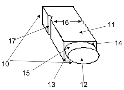
Referring to Figure 1, a particularly preferred embodiment of the flat-based
well 10 to be
used in accordance with the invention includes a main chamber 11 that is
square in cross
section, and a sub-chamber 12 that is circular in cross section. The sub-
chamber 12 extends
from a chamber base 13 of the main chamber 11. As such the opening 14 of the
sub-
chamber is defined by the chamber base 13 of the main chamber. The arc of the
outer wall
15 of the sub-chamber 12 extends to the outer sides of each wall 16 of the
main chamber
11 (best illustrated in Figure 3).
One wall 16 of the main chamber 11 includes a groove 17 for receiving a
cooperating
portion of a well plate (not shown) into which the well 10 is to be placed.
This facilitates
CA 02600412 2007-09-07
WO 2006/094364 PCT/AU2006/000325
-19-
secure locking of the well 10 in the plate and alleviates problems associated
with wells 10
being dislodged from the plate.
The sub-chamber 12 has a predetermined volume so that it receives a liquid
sample and
forms the liquid sample into a droplet advantageously having a convex surface
extending
from the sub-chamber opening 14. This ensures that the depth of the liquid is
substantially
constant through the sub-chamber, as compared with a liquid sample that is
placed in a
well and forms a meniscus up the side walls of the well. In that case, the
depth of liquid on
the sides of the well where the meniscus forms will be substantially greater
than that in the
middle of the well where the meniscus is at its lowest point.
Preferably, the height X of the well is 11mm, including a 10mm height Y for
the main
chamber and a 1mm depth Z for the sub-chamber. The well is generally 8mm
across its
width W, and has a wall thickness T of about lmm. Thus, the internal width Wi
is about
6mm. The main chamber typically defines a voluine of about 360 l and the sub-
chamber a
volume of about 28.3 l.
Figure 4 exemplifies one example of a typical 12 x 8 well plate configuration
(i.e. 2 x 6 x
8) including a listing of viruses to be detected, relevant cell lines and
removal days for
each line. The plate is made up of 96 individual flat-based wells in
accordance with the
present invention.
As will be seen from Figure 4, the plate can be seeded with different cell
lines in the
following order:
Columns 1-3 LLC-MK2
Columns 4,5 MDCK
Column 6 Hep2
Column 7 A549
Column 8 RK13
Columns 9-12 MRC-5
CA 02600412 2007-09-07
WO 2006/094364 PCT/AU2006/000325
-20-
A plate set up in this fashion would allow for example the selection of,
without being
limited to, the respiratory viruses Parainfluenza 1,2,3,4 (PI 1,2,3,4),
Influenza A,B (Inf
A,B) RSV, Adenovirus (AD), Rhinovirus (RH), Cytomegalovirus (CMV), and viruses
from the Enterovirus group (ENT) consisting of Echovirus (Eco), Coxsackievirus
(cox),
Enterovirus (Ent) and Poliovirus (Polio), as well as, but not limited to, the
non-respiratory
viruses Herpes simplex virus (HSV) 1,2, and Varicella zoster virus (VZV).
In a further example, configuration of a 6 x 8 well plate may consist of cell
lines seeded in
the following order:
Columns 1-3 A 549
Columns 4-6 MRC-5
This would allow for example the detection of viruses such as CMV, HSV 1,2,
VZV, AD,
and those from the Enterovirus group.
Finally a configuration of 14x8 wells would ultimately allow for the detection
of pathogens
like PI 1,2,3,4; Inf A,B; RSV; AD; RH; ENT (Echo, cox, Ent, Polio); HSV 1,2;
VZV;
Rubella; Mumps; Measles; Rotavirus; Polyomavirus and also other pathogens-
viruses
using appropriate cell lines.
The removal of the cell lines, due to the individual nature of the wells, can
be selective
depending on the time schedule which is appropriate for the specific viral
detection in
question. In particular, if the detection of PI 1-4 is desired, taking row A
for example,
wells A 1 to A 3 are removed on day two. Similarly, if the detection of Inf A,
B is desired,
wells A 4 and A 5 are removed on day two. However, if the detection of Entero
is required,
then well A 11 is removed on the appropriate day (1-3). The specific nature of
the
individual wells facilitates this selective removal and viral detection.
CA 02600412 2007-09-07
WO 2006/094364 PCT/AU2006/000325
-21-
Reference will now be made to a particular procedure which may be followed
using the kit
of one aspect of the invention, many steps of which may be optional and should
not be
considered to be limiting on the invention in any way.
Using vacuum and sterile glass pasteur pipettes, medium is aspirated from all
wells to be
inoculated. Disposal of pasteur pipettes in a large sharps container is
advantageously
facilitated.
Using a disposable pipette, an appropriate number of wells of the well plate
are inoculated
with approximately 150-200 l specimen per well. The remaining specimen is
stored at -
70 C. The lid is then replaced on the plate and the date written over the
wells inoculated in
the plate.
The plate is then weighed on a digital balance and balanced with balance
plates and cards
until all plates are equivalent weights (+/- 0.5g) and can be balanced in a
centrifuge. The
centrifuge is run at about 37 C and 3500 rpm for a period of 60 min. Using
vacuum and
sterile pasteur pipettes each specimen is then aspirated from each well, and
using a fresh
disposable pipette for each specimen, each well is filled with the virus
recovery medium
BAC.
The specimens are then incubated in a humidified environment at 37 C in a COZ
incubator
(5%) by carefully placing the plates in the COZ incubator and incubating at 37
C for up to
seven days after inoculation of the last specimen.
Immunofluorescent staining is advantageously used for detection of specific
viruses in
single wells, using specific monoclonal antibodies. Generally, the following
procedure is
followed:
Using vacuum suction, the medium is removed from the appropriate well(s) and
the wells
removed from the plate using special forceps and transferred into a different
holder. These
are then air dried for 3 minutes. 300 l of a mixture of cold acetone and
methanol (2:1) is
CA 02600412 2007-09-07
WO 2006/094364 PCT/AU2006/000325
-22-
then added to each well and allowed to fix for 15 minutes at -20 C. The
fixative is then
discarded and the sample again air dried for 2-3 minutes. A specific
monoclonal antibody
(primary) is then added to each well and the cover plate put in place and the
samples
incubated for 30 minutes at 37 C. The samples are then removed from the
incubator and
each well filled with Phosphate Buffered Saline (PBS). The PBS is then
discarded. This
process is repeated four more times. Again, the sample is air dried for 3
minutes, after
which a secondary antibody specific for the primary antibody is added to each
well.
Following this, incubation of the sample again takes place followed by
repeated treatments
with PBS as mentioned above and a final wash with double distilled water. A
small
amount (1 drop) of a specially prepared mounting medium is then added and the
results
observed under fluorescent microscope. One step immunofluorescent staining may
also be
employed.
Tliroughout this specification and the claims which follow, unless the context
requires
otherwise, the word "comprise", and variations such as "comprises" and
"comprising", will
be understood to imply the inclusiori of a stated integer or group of integers
or steps but not
the exclusion of any other integer or group of integers or steps.
Those skilled in the art will appreciate that the invention described herein
is susceptible to
variations and modifications other than those specifically described. It is to
be understood
that the invention includes all such variations and modifications. The
invention also
includes all of the steps, features, compositions and compounds referred to or
indicated in
this specification, individually or collectively, and any and all combinations
of any two or
more of said steps or features.