Note: Descriptions are shown in the official language in which they were submitted.
DEMANDES OU BREVETS VOLUMINEUX
LA PRESENTE PARTIE DE CETTE DEMANDE OU CE BREVETS
COMPREND PLUS D'UN TOME.
CECI EST LE TOME 1 DE 2
NOTE: Pour les tomes additionels, veillez contacter le Bureau Canadien des
Brevets.
JUMBO APPLICATIONS / PATENTS
THIS SECTION OF THE APPLICATION / PATENT CONTAINS MORE
THAN ONE VOLUME.
THIS IS VOLUME 1 OF 2
NOTE: For additional volumes please contact the Canadian Patent Office.
CA 02600794 2013-04-09
1
GENETIC POLYMORPHISMS ASSOCIATED WITH CORONARY H _______________________ PART
DISEASE,
METHODS OF DETECTION AND USES THEREOF
CROSS-REFERENCE TO RELATED APPLICATIONS
This application claims the benefit of U.S. provisional application Serial
No.: 60/660,322,
filed on March 11,2005, and U.S. provisional application Serial No.:
60/711,447, filed on August
24; 2005.
FIELD OF l'HE INVENTION
The present invention is in the field of coronary heart disease (CUD) and in
particular
stenosis and myocardial infarction (MT) diagnosis and therapy. In particular,
the present invention
relates to specific single nucleotide polymorphisms (SNPs) in the human
genome, and their
association with CUD and related pathologies. Based on differences in allele
freqUencies'in the
patient population relative to normal individuals, the naturally-occurring
SNPs disclosed herein can
be used as targets for the design of diagnostic reagents and the development
of therapeutic agents, as
well as for disease association and linkage analysis. In particular, the SNPs
of the present invention
are useful for identifying an individual who is at an increased or decreased
risk of developing CUD
and in particular stenosis and MI, and for early detection of the disease, for
providing clinically
important information for the prevention and/or treatment of MD such as
stenosis and MI, for
screening and selecting therapeutic agents and for predicting a patient's
response to therapeutic
agents. The SNPs disclosed herein are also useful for human identification
applications. Methods,
assays, kits, and reagents for detecting the presence of these polymorphisms
and their encoded
products are provided. =
=
BACKGROUND OF ME INVENTION
is defined in the Framingham study as encompassing angina, ME, coronary
insnffciency (which is manifested as ischemia, that is, impaired oxygen flow
to the heart muscle),
stenosis and coronary heart disease death (Wilson et al., Circulation 97:1837-
1847 (1998)). It is
sometimes recorded throngh clinical records that indicate the following
interventions: coronary
artery bypass graft, angioplasty and stent placement in addition to clinical
records of MI, angina, or
coronary death. This latter definition is used in many population based
studies and clinical trials, but
it probably misses silent MI events and some Unreported angina.
CA 02600794 2007-09-11
WO 2006/099365
PCT/US2006/009016
MI (or heart attack) is the most common cause of mortality in developed
countries. The
incidence of MI is still high despite currently available preventive measures
and therapeutic
intervention. More than 1,500,000 people in the US suffer acute MI each year
(many without
seeking help due to unrecognized MI), and one third of these people die. The
lifetime risk of
coronary artery disease events at age 40 years is 42.4% for men (one in two)
and 24.9% for women
(one in four) (Lloyd-Jones DM; Lancer, 1999 353: 89-92).
MI is a multifactorial disease that involves atherogenesis, thrombus formation
and '
propagation. Thrombosis can result in complete or partial occlusion of
coronary arteries. The
luminal narrowing or blockage of coronary arteries reduces oxygen and nutrient
supply to the
cardiac muscle (cardiac ischemia), leading to myocardial necrosis and/or
stunning. MI, unstable
angina, and sudden ischemic death are clinical manifestations of cardiac
muscle damage. All three
endpoints are part of the Acute Coronary Syndrome since the underlying
mechanisms of acute
complications of atherosclerosis are considered to be the same.
Atherogenesis, the first step of pathogenesis of MI, is a complex interaction
between blood
elements, mechanical forces, disturbed blood flow, and vessel wall abnormality
that results in plaque
accumulation. An unstable (vulnerable) plaque was recognized as an underlying
cause of arterial
thrombotic events and MI. A vulnerable plaque is a plaque, often not stenotic,
that has a high
likelihood of becoming disrupted or eroded, thus forming a thrombogenic focus.
MI due to a
vulnerable plaque is a complex phenomenon that includes: plaque vulnerability,
blood vulnerability
(hypercoagulation, hypothrombolysis), and heart vulnerability (sensitivity of
the heart to ischemia or
propensity for arrhythmia). Recurrent myocardial infarction (RMI) can
generally be viewed as a
severe form of MI progression caused by multiple vulnerable plaques that are
able to undergo pre-
rupture or a pre-erosive state, coupled with extreme blood coagulability.
The current diagnosis of MI is based on the levels of troponin I or T that
indicate the cardiac
muscle progressive necrosis, impaired electrocardiogram (ECG), and detection
of abnoillial
ventricular wall motion or angiographic data (the presence of acute thrombi).
However, due to the
asymptomatic nature of 25% of acute MIs (absence of atypical chest pain, low
ECG sensitivity), a
significant portion of MIs are not diagnosed and therefore not treated
appropriately (e.g., prevention
of recurrent MIs).
MI risk assessment and prognosis is currently done using classic risk factors
or the recently
introduced Framingham Risk Index. Both of these assessments put a significant
weight on LDL
levels to justify preventive treatment. However, it is well established that
half of all MIs occur in
individuals without overt hyperlipidemia.
2
CA 02600794 2007-09-11
WO 2006/099365
PCT/US2006/009016
Other emerging risk factors of MI are inflammatory biomarkers such as C-
reactive protein
(CRP), ICAM-1, SAA, TNF a, homocysteine, impaired fasting glucose, new lipid
markers (ox LDL,
Lp-a, MAD-LDL, etc.) and pro-thrombotic factors (fibrinogen, PAI-1). These
markers have
significant limitations such as low specificity and low positive predictive
value, and the need for
multiple reference intervals to be used for different groups of people (e.g.,
males-females, smokers-
non smokers, hormone replacement therapy users, different age groups). These
limitations diminish
the utility of such markers as independent prognostic markers for MI
screening.
Genetics plays an important role in MI risk. Families with a positive family
history of MI
account for 14% of the general population, 72% of premature MIs, and 48% of
all MIs (Williams R
R, Am J Cardiology, 2001; 87:129). In addition, replicated linkage studies
have revealed evidence
of multiple regions of the genome that are associated with MI and relevant to
MI genetic traits,
including regions on chromosomes 14, 2, 3 and 7 (Broeckel U, Nature Genetics,
2002; 30: 210;
Harrap S, Arterioscler Thromb Vase Biol, 2002; 22: 874-878, Shearman A, Human
Molecular
Genetics, 2000, 9; 9,1315-1320), implying that genetic risk factors influence
the onset,
manifestation, and progression of MI. Recent association studies have
identified allelic variants that
are associated with acute complications of CHD, including allelic variants of
the ApoE, ApoA5,
Lpa, APOCIII, and Klotho genes.
Genetic markers such as single nucleotide polymoiphisms (SNPs) are preferable
to other
types of biomarkers. Genetic markers that are prognostic for MI can be
genotyped early in life and
could predict individual response to various risk factors. The combination of
serum protein levels
and genetic predisposition revealed by genetic analysis of susceptibility
genes can provide an
integrated assessment of the interaction between genotypes and environmental
factors, resulting in
synergistically increased prognostic value of diagnostic tests.
Thus, there is an urgent need for novel genetic markers that are predictive of
predisposition
to CHD such as MI, particularly for individuals who are unrecognized as having
a predisposition to
MI. Such genetic markers may enable prognosis of MI in much larger populations
compared with
the populations that can currently be evaluated by using existing risk factors
and biomarkers. The
availability of a genetic test may allow, for example, appropriate preventive
treatments for acute
coronary events to be provided for susceptible individuals (such preventive
treatments may include,
for example, statin treatments and statin dose escalation, as well as changes
to modifiable risk
factors), lowering of the thresholds for ECG and angiography testing, and
allow adequate monitoring
of infoimative biomarkers. Moreover, the discovery of genetic markers
associated with MI will
3
CA 02600794 2007-09-11
WO 2006/099365
PCT/US2006/009016
provide novel targets for therapeutic intervention or preventive treatments of
MI, and enable the
development of new therapeutic agents for treating MI and other cardiovascular
disorders.
Coronary stenosis is the narrowing of coronary arteries by obstructive
atherosclerotic
plaques. The coronary arteries supply oxygenated blood flow to the myocardium.
Although mild
and moderate coronary stenosis do not impede resting coronary flow, stenosis
>30 ¨45% starts to
restrict maximal coronary flow. Severe coronary stenosis (>70% reduction in
luminal diameter)
causes stable angina (ischemic chest pain upon exertion). Significant stenosis
contributes, along
with plaque rupture and thrombus formation, coronary spasm, or
inflammation/infection, to unstable
angina as well as myocardial infarction. Together with arrhythmia, coronary
stenosis is a major
factor of sudden cardiac deaths, as evidenced by its presence in two or more
major coronary arteries
in 90% of adult sudden cardiac death victims.
Coronary stenosis is a prevalent disease. Each year in the United States,
440,000 new cases
of stable angina and 150,000 new cases of unstable angina occur. This year, an
estimated 1.1
million Americans will have a new or recurrent heart attack. These incidences
result in over six
million individuals in the U.S. living with stable or unstable angina
pectoris, a debilitating condition,
and over seven million individuals in the U.S. living with a history of
myocardial infarction.
Coronary stenosis is frequently a deadly disease. It is a major underlying
cause of CHD, which is
the single largest cause of death in the U.S. Over half a million coronary
deaths, including 250,000
sudden cardiac deaths, occur each year in U.S.
There is, therefore, an unmet need in early diagnosis and prognosis of
asymptomatic
coronary stenosis. This need is particularly significant given that early
diagnosis or prognosis results
can significantly influence the course of disease by influencing treatment
choices (for example, those
with genetic risks can be treated to modify risk factors such as hypertension,
diabetes, inactivity,
dyslipidemia, etc.), thresholds (e.g., lipid levels used to trigger the use of
lipid-lowering drugs), and
goals (e.g., target blood pressure or lipid levels), and possibly enhance
compliance.
Diagnosis of coronary stenosis currently starts by assessing if the risk
profiles (e.g.,
hypertension, dyslipidemia, family history, diabetes, etc.) and symptoms
(e.g., angina) of patients are
consistent with coronary heart disease, followed most commonly by resting and
exercise EKGs.
However, risk assessments and EKGs are imperfect diagnostic tests for stenosis
since they can be
both insensitive (giving false negatives) and non-specific (giving false
positives). Coronary
arteriography is the definitive test for assessing the severity of coronary
stenosis, however, it is not
very sensitive in early detection of mild stenosis. It is also an invasive
procedure with a small risk of
death due to the catheterization procedure and the contrast dye. Because of
this risk, it is typically
4
CA 02600794 2007-09-11
WO 2006/099365
PCT/US2006/009016
only used at a time when coronary stenosis is considered likely from symptoms
or other tests, which
is hardly an ideal time to start intervention.
Coronary stenosis risk is presumed to have a strong genetic component. It is
well known that
several major risk factors of coronary disease are heritable, e.g. serum lipid
levels (Perusse L. et. al.,
Arterioscler Thromb Vase Biol (1997): 17(11) 3263-9) and obesity (Rice T. et.
al., Int J. Obes Relat
Metab Disord (1997):21(11) 1024-31). Indeed, several known genetic defects are
individually
sufficient to cause elevated serum LDL-cholesterol (e.g., familial
hypercholesterolemia) leading to
premature coronary disease (Goldstein and Brown, Science 292 (2001): 1310-12).
In addition,
linkage studies in humans have replicated the findings of the link of several
chromosomal regions
(quantitative trait loci) to coronary heart disease and related diseases and
risk factors (Pajukanta P.
et. al., Am J Hum Genet 67 (2000):1481-93, Francke S. et. al., Human Molecular
Genetics (2001):
10 (24) 2751-65). Finally, a family history of premature coronary disease is a
significant factor in
the risk assessment and diagnosis of coronary disease (Braunwald E., Zipes D.
and Libby P., Heart
Disease, 6th ed. W.B. Saunders Company, 2001, 28).
Although many risk factors for coronary stenosis have been identified,
including age,
diabetes, hypertension, high serum cholesterol, smoking, etc., and genetic
factors play significant
roles in several of these risk factors, significant genetic risk factors are
likely to exist which have not
been identified to date. In addition to the anecdotal coronary disease
patients that exhibit few
traditional risk factors, a study of multiple existing risk factors showed
that only half of the
"population-attributable risk" was attributable to known risk factors (Change
M. et. al:, J Clin
Epidemiol (2001) 54 (6) 634-44). Therefore, the presently known risk factors
are inadequate for
predicting coronary stenosis risk in individuals. Given the magnitude of the
disease, there is an
urgent need for genetic markers that are predictive of coronary stenosis risk.
Such genetic markers
could increase the prognostic ability of existing risk assessment methods and
complement current
diagnostic methods such as exercise EKG, especially in early detection of
disease when intervention
is most effective and should ideally start.
Reduction of coronary and cerebrovascular events and total mortality by
treatment with
HMG-CoA reductase inhibitors (statins) has been demonstrated in a number of
randomized, double.
blinded, placebo controlled prospective trials (Waters, D.D., Clin Cardiol,
2001. 24(8 Suppl): p.
1113-7, Singh, B.K. and J.L. Mehta, Curr. Opin. Cardiol., 2002. 17(5): p. 503-
11). These drugs have
their primary effect through the inhibition of hepatic cholesterol synthesis,
thereby upregalating
LDL receptor in the liver. The resultant increase in LDL catabolism results in
decreased circulating
LDL, a major risk factor for cardiovascular disease.
5
CA 02600794 2007-09-11
WO 2006/099365
PCT/US2006/009016
Statins can be divided into two types according to their physicochemical and
pharmacokinetic properties. Statins such as lovastatin, simvastatin,
atorvastatin, and cerevastatin are
hydrophobic in nature and, as such, diffuse across membranes and thus are
highly cell permeable.
Hydrophilic statins such as pravastatin are more polar, such that they require
specific cell surface
transporters for cellular uptake (Ziegler, K. and W. Stunkel, Biochim Biophys
Acta, 1992. 1139(3):
p. 203-9, Yamazaki, M., et al., Am J PhysioL 1993. 264(1 Pt 1): p. G36-44,
Kornai, T., et aL ,
Biochern Pharmacol, 1992. 43(4): p. 667-70). The latter statin utilizes a
transporter, OATP2, whose
tissue distribution is confined to the liver and, therefore, they are
relatively hepato-specific inhibitors
(Hsiang, B., et al, J Biol Chem, 1999. 274(52): p. 37161-8). The former
statins, not requiring
specific transport mechanisms, are available to all cells and they can
directly impact a much broader
spectrum of cells and tissues. These differences in properties may influence
the spectrum of
activities that each statin possesses. Pravastatin, for instance, has a low
myopathic potential in
animal models and myocyte cultures compared to other hydrophobic statins
(Masters, B.A., et al.,
1998. 152(1): p. 99-106, Reijneveld, J.C., et al., Pediatr Res, 1996. 39(6):
p. 1028-35).
Evidence from gene association studies is accumulating to indicate that
responses to drugs
are, indeed, at least partly under genetic control. As such, pharmaco genetics
- the study of
variability in drug responses attributed to hereditary factors in different
populations - may
significantly assist in providing answers toward meeting this challenge
(Roses, A.D., Nature, 2000.
405(6788): p. 857-65, Mooser, V., et aL , J Thromb Haemost, 2003. 1(7): p.
1398-1402, Humma,
L.M. and S.G. Terra, Am. J. Health Syst Pharrn, 2002. 59(13): p. 1241-52).
Numerous associations
have been reported between selected genotypes, as defined by SNPs and other
sequence variations
and specific responses to cardiovascular drugs. Polymorphisms in several genes
have been
suggested to influence responses to statins including CETP (Kuivenhoven, J.A.,
et al., N Engl J Med,
1998. 338(2): p. 86-93), beta-fibrinogen (de Maat, M.P., et aL, Arterioscler
Thromb Vase Biol, 1998.
18(2): p. 265-71), hepatic lipase (Zambon, A., et al., Circulation, 2001.
103(6): p. 792-8, lipoprotein
lipase (Jukema, J.W., et al., Circulation, 1996. 94(8): p. 1913-8),
glycoprotein Illa (Bray, P.F., et al.,
Am J Cardiol, 2001. 88(4): p. 347-52), stromelysin-1 (de Maat, M.P., et al.,
Am J Cardiol, 1999.
83(6): p. 852-6), and apolipoprotein E (Gerdes, L.U., et al., Circulation,
2000. 101(12): p. 1366-71,
Pedro-Botet, J., et al., Atherosclerosis, 2001. 158(1): p. 183-93). Some of
these variants were shown
to effect clinical events while others were associated with changes in
surrogate endpoints. Thus
there is the need for markers and individuals responsiveness to statins.
6
CA 02600794 2007-09-11
WO 2006/099365
PCT/US2006/009016
SNPs
The genomes of all organisms undergo spontaneous mutation in the course of
their
continuing evolution, generating variant forms of progenitor genetic sequences
(Gusella, Ann. Rev.
Biochem. 55, 831-854 (1986)). A variant form may confer an evolutionary
advantage or
disadvantage relative to a progenitor form or may be neutral. In some
instances, a variant form
confers an evolutionary advantage to the species and is eventually
incorporated into the DNA of
many or most members of the species and effectively becomes the progenitor
form. Additionally,
the effects of a variant form may be both beneficial and detrimental,
depending on the
circumstances. For example, a heterozygous sickle cell mutation confers
resistance to malaria, but a
homozygous sickle cell mutation is usually lethal. In many cases, both
progenitor and variant forms
survive and co-exist in a species population. The coexistence of multiple
forms of a genetic
sequence gives rise to genetic polymorphisms, including SNPs.
Approximately 90% of all genetic polymorphisms in the human genome are SNPs.
SNPs are
single base positions in DNA at which different alleles, or alternative
nucleotides, exist in a
population. The SNP position (interchangeably referred to herein as SNP, SNP
site, SNP locus, SNP
marker, or marker) is usually preceded by and followed by highly conserved
sequences of the allele
(e.g., sequences that vary in less than 1/100 or 1/1000 members of the
populations). An individual
may be homozygous or heterozygous for an allele at each SNP position. A SNP
can, in some
instances, be referred to as a "cSNP" to denote that the nucleotide sequence
containing the SNP is an
amino acid coding sequence.
A SNP may arise from a substitution of one nucleotide for another at the
polymorphic site.
Substitutions can be transitions or transversions. A transition is the
replacement of one purine
nucleotide by another purine nucleotide, or one pyrimidine by another
pyrimidine. A transversion is
the replacement of a purine by a pyrimidine, or vice versa. A SNP may also be
a single base
insertion or deletion variant referred to as an "indel" (Weber et al., "Human
diallelic
insertion/deletion polymorphisms," Am J Hum Genet 2002 Oct.; 71(4):854-62).
A synonymous codon change, or silent mutation/SNP (terms such as "SNP,"
"polymorphism," "mutation," "mutant," "variation," and "variant" are used
herein interchangeably),
is one that does not result in a change of amino acid due to the degeneracy of
the genetic code. A
substitution that changes a codon coding for one amino acid to a codon coding
for a different amino
acid (i.e., a non-synonymous codon change) is referred to as a missense
mutation. A nonsense
mutation results in a type of non-synonymous codon change in which a stop
codon is foiined,
7
CA 02600794 2007-09-11
WO 2006/099365
PCT/US2006/009016
thereby leading to premature termination of a polypeptide chain and a
truncated protein. A read-
through mutation is another type of non-synonymous codon change that causes
the destruction of a
stop codon, thereby resulting in an extended polypeptide product. While SNPs
can be bi-, tri-, or
tetra- allelic, the vast majority of the SNPs are bi-allelic, and are thus
often referred to as "bi-allelic
markers," or "di-allelic markers."
As used herein, references to SNPs and SNP genotypes include individual SNPs
and/or
haplotypes, which are groups of SNPs that are generally inherited together.
Haplotypes can have
stronger correlations with diseases or other phenotypic effects compared with
individual SNPs, and
therefore may provide increased diagnostic accuracy in some cases (Stephens et
al. Science 293,
489-493,20 July 2001).
Causative SNPs are those SNPs that produce alterations in gene expression or
in the
expression, structure, and/or function of a gene product, and therefore are
most predictive of a
possible clinical phenotype. One such class includes SNPs falling within
regions of genes encoding
a polypeptide product; i.e. cSNPs. These SNPs may result in an alteration of
the amino acid
sequence of the polypeptide product (i.e., non-synonymous codon changes) and
give rise to the
expression of a defective or other variant protein. Furthermore, in the case
of nonsense mutations, a
SNP may lead to premature termination of a polypeptide product. Such variant
products can result
in a pathological condition, e.g., genetic disease. Examples of genes in which
a SNP within a coding
sequence causes a genetic disease include sickle cell anemia and cystic
fibrosis.
Causative SNPs do not necessarily have to occur in coding regions; causative
SNPs can
occur in, for example, any genetic region that can ultimately affect the
expression, structure, and/or
activity of the protein encoded by a nucleic acid. Such genetic regions
include, for example, those
involved in transcription, such as SNPs in transcription factor binding
domains, SNPs in promoter
regions, in areas involved in transcript processing, such as SNPs at intron-
exon boundaries that may
cause defective splicing, or SNPs in mRNA processing signal sequences such as
polyadenylation
signal regions. Some SNPs that are not causative SNPs nevertheless are in
close association with,
and therefore segregate with, a disease-causing sequence. In this situation,
the presence of a SNP
correlates with the presence of, or predisposition to, or an increased risk in
developing the disease.
These SNPs, although not causative, are nonetheless also useful for
diagnostics, disease
predisposition screening, and other uses.
An association study of a SNP and a specific disorder involves determining the
presence or
frequency of the SNP allele in biological samples from individuals with the
disorder of interest, such
as stenosis or MI, and comparing the information to that of controls (i.e.,
individuals who do not
8
CA 02600794 2007-09-11
WO 2006/099365
PCT/US2006/009016
have the disorder; controls may be also referred to as "healthy" or "normal"
individuals) who are
preferably of similar age and race. The appropriate selection of patients and
controls is important to
= the success of SNP association studies. Therefore, a pool of individuals
with well-characterized
phenotypes is extremely desirable.
A SNP may be screened in diseased tissue samples or any biological sample
obtained from a
diseased individual, and compared to control samples, and selected for its
increased (or decreased)
occurrence in a specific pathological condition, such as pathologies related
to CHD and in particular,
stenosis and MI. Once a statistically significant association is established
between one or more
SNP(s) and a pathological condition (or other phenotype) of interest, then the
region around the SNP
can optionally be thoroughly screened to identify the causative genetic
locus/sequence(s) (e.g.,
causative SNP/mutation, gene, regulatory region, etc.) that influences the
pathological condition or
phenotype. Association studies may be conducted within the general population
and are not limited
to studies performed on related individuals in affected families (linkage
studies).
Clinical trials have shown that patient response to treatment with
pharmaceuticals is often
heterogeneous. There is a continuing need to improve pharmaceutical agent
design and therapy. In
that regard, SNPs can be used to identify patients most suited to therapy with
particular
pharmaceutical agents (this is often temiled "pharmacogenomics"). Similarly,
SNPs can be used to
exclude patients from certain treatment due to the patient's increased
likelihood of developing toxic
side effects or their likelihood of not responding to the treatment.
Pharmacogenomics can also be
used in pharmaceutical research to assist the drug development and selection
process. (Linder et al.
(1997), Clinical Chemistry, 43, 254; Marshall (1997), Nature Biotechnology,
15, 1249; International
Patent Application WO 97/40462, Spectra Biomedical; and Schafer et al. (1998),
Nature
Biotechnology, 16: 3).
SUMMARY OF THE INVENTION
The present invention relates to the identification of novel SNPs, unique
combinations of
such SNPs, and haplotypes of SNPs that are associated with CHD, and in
particular stenosis and MI,.
The polymorphisms disclosed herein are directly useful as targets for the
design of diagnostic
reagents and the development of therapeutic agents for use in the diagnosis
and treatment of CHD,
and in particular stenosis and MI, as well as predicting a patient's response
to therpeutic agents such
as statins.
Based on the identification of SNPs associated with CHD, the present invention
also provides
methods of detecting these variants as well as the design and preparation of
detection reagents
9
CA 02600794 2013-12-23
=
e
needed 'to accomplish this task. The- invention specifically provides, for
example, novel SNPs in
genetic sequences involved in stenosis and1VII, isolated nucleic acid
molecules (including, for
example, DNA and RNA molecules) containing these SNP1-, variant proteins
encoded by nucleic
acid molecules containing such SNPs, antibodies to the encoded variant
proteins, computer-based
and data storage systems containing the novel SNP information, methods of
detecting these SNPs in
a test sample, methods of identifying individuals who have an altered (i.e.,
increased or decreased)
risk of developing CHD based on the presence or absence of one or more
particular nucleotides
(alleles) at one or more SNP sites disclosed herein or the detection of one or
more encoded variant
products (e.g., variant moRNA transcripts or variant proteins), methods of
identifying individuals who
are more or less likely to respond to a treatment (or more or less likely to
experience undesirable side
effects from a treatment, etc.), methods of screening for compounds useful in
the treatment of a.
disorder associated with a variant gene/protein, compounds identified by these
methods, methods of
. . .
-treating disorders mediated by a variant gene/protein, methods of using the
novel SNPs of the
present invention for human identification, etc.
Various embodiments of the invention provide a method for determining that a
human
has an increased risk for myocardial infarction (MI), comprising testing
nucleic acid from said
human to determine the nucleotide content at a polymorphic position in VAMPS
as represented
by position 101 of SEQ ID NO: 40 or its complement, wherein the presence of C
at position
101 of SEQ ID NO: 40 or G at position 101 of its complement indicates that
said human
has said increased risk for MI.
= Various embodiments of the invention provide a method for determining
that a human
has an increased risk for myocardial infarction (MI), comprising: a) testing
nucleic acid from
said human to determine the nucleotide content at a polymorphism in gene VAMPS
as
represented by position 101 of SEQ ID NO: 40 or its complement; and b)
correlating the
presence of C at position 101 of SEQ ID NO: 40 or G at position 101 of its
complement
with said human having said increased risk for MI, or, correlating the absence
of said C or said
G with said human having no said increased risk for Mi.
Various embodiments of the invention provide an isolated nucleic acid molecule
comprising at least 8 contiguous nucleotides wherein onb of the nucleotides is
a C at a position
corresponding to position 100 of SEQ ID NO: 40 ,or a G corresponding to
position 101 of
its complement.
Various embodiments of the invention provide an amplified polynucleotide
containing
=
=
CA 02600794 2013-12-23
a C at a position corresponding to position 100 of SEQ ID NO: 40 µ, wherein
the
amplified polynucleotide is between 16 and 1,000 nucleotides in length.
Various embodiments of the invention provide an isolated polynucleotide which
specifically hybridizes to a target nucleic acid molecule Containing a C at a
position
corresponding to position 101 of SEQ ID NO: 40 , or a G at position 101 of its
complement,
under hybridizing conditions comprising: a) prehybridizing the target nucleic
acid with a
solution containing 5x standard saline phosphate EDTA, 0.5% NaDodSO4(SDS) at
55 C; b)
incubating the isolated polynucleotide with the target nucleic acid molecule
in the solution of
step a) at 55 C; and c) washing with a solution Containing 2xSSPE, and 0.1%
SDS.
Various embodiments of the invention a kit for detecting a single nticleotide
polymorphism (SNP) in a nucleic acid, comprising the isolated polynucleotide
of any one of
claims 34 to 37, a buffer, and an enzyme.
Various embodiments of the invention an isolated nucleic acid molecule that
encodes
an amino acid sequence, wherein the nucleic acid contains a C at a position
corresponding to
position 101 of SEQ NO: 40.
Various embodiments of the invention a Method for identifying an individual
who is in
need of receiving treatment for myocardial infarction with-a therapeutic
agent, comprising
detecting the presence of a C at a position corresponding to position 101 of
SEQ ID
NO: 40, or a 0 at position 101 of its complement, in a nucleic acid sample
from said individual.
In Tables 1-2, the present invention provides gene information, references to
the
identification of transcript sequences (SEQ ID NOS: 1-8),, encoded amino acid
sequences (SEQ ID
NOS: ,9-16), genomic sequences (SEQ ID NOS: 25-33), transcript-based context
sequences
(SEQ ID NOS: 17-24) and genomic-based context sequences (SEQ BD NOS: 34-42)
that = =
= contain the SNPs of the present Invention, and extensive SNP information
that includes observed
alleles, allele frequencies, populations/ethnic groups in which alleles have
been observed,
Information about the type of SNP and corresponding functional effect, and,
for eSNPs, information
about the encoded polypeptide product The actual transcript sequences (SEQ ID
NOS: 1-8),1
amino acid sequences (SEQ ID NOS: 9-16), genorcio sequences (SEQ ID NOS: 25-
33),
transcript-based SNP context sequences (SEQ ID NOS: 17-24), and genomic-based
SNP context
sequerms (SEQ M NOS: 34-42), together vvitb.primer gequences (SEQ BD NOS: 43-
84) ' are
provided inthe Sequence Listing.
In one embodiment of the invention, applicants teach a method. for identifying
an individnal
= who has an altered risk for developing MD, comprising detecting a singe.
raulleotide polymorphism
(SNP) in any one of the nucleotide sequences of SEQ M NOS: 1-8 and 17-24 in
said
. '
10a .
=
CA 02600794 2013-04-09
10b
individual's nucleic acids, wherein the SNP is as specified in Table 1 and
Table 2, respectively, and
the presence of the SNP is correlated with an altered risk for ME in said
indivirlualin a specific
embodiment of the present invention, SNPs that occur naturally in the hi-misn
gnome are provided
as isolated nucleic acid molecules. These SNPs are associated with CHD, and in
particular stenosis
CA 02600794 2007-09-11
WO 2006/099365
PCT/US2006/009016
and MI such that they can have a variety of uses in the diagnosis and/or
treatment of CHD and
related pathologies. In an alternative embodiment, a nucleic acid of the
invention is an amplified
polynucleotide, which is produced by amplification of a SNP-containing nucleic
acid template. In
another embodiment, the invention provides for a variant protein that is
encoded by a nucleic acid
molecule containing a SNP disclosed herein.
In yet another embodiment of the invention, a reagent for detecting a SNP in
the context of
its naturally-occurring flanking nucleotide sequences (which can be, e.g.,
either DNA or mRNA) is
provided. In particular, such a reagent may be in the form of, for example, a
hybridization probe or
an amplification primer that is useful in the specific detection of a SNP of
interest. In an alternative .
embodiment, a protein detection reagent is used to detect a variant protein
that is encoded by a
nucleic acid molecule containing a SNP disclosed herein. A preferred
embodiment of a protein
detection reagent is an antibody or an antigen-reactive antibody fragment.
Various embodiments of the invention also provide kits comprising SNP
detection reagents,
and methods for detecting the SNPs disclosed herein by employing detection
reagents. In a specific
embodiment, the present invention provides for a method of identifying an
individual having an
increased or decreased risk of developing MI by detecting the presence or
absence of one or more
SNP alleles disclosed herein. In another embodiment, a method for diagnosis of
stenosis or MI by
detecting the presence or absence of one or more SNP alleles disclosed herein
is provided.
The nucleic acid molecules of the invention can be inserted in an expression
vector, such as
to produce a variant protein in a host cell. Thus, the present invention also
provides for a vector
comprising a SNP-containing nucleic acid molecule, genetically-engineered host
cells containing the
vector, and methods for expressing a recombinant variant protein using such
host cells. In another
specific embodiment, the host cells, SNP-containing nucleic acid molecules,
and/or variant proteins
can be used as targets in a method for screening and identifying therapeutic
agents or pharmaceutical
compounds useful in the treatment of CHD, and in particular stenosis and MI.
An aspect of this invention is a method for treating CHD, and in particular
stenosis and MI,
in a human subject wherein said human subject harbors a SNP, gene, transcript,
and/or encoded
protein identified in Tables 1-2, which method comprises administering to said
human subject a
therapeutically or prophylactically effective amount of one or more agents
counteracting the effects
of the disease, such as by inhibiting (or stimulating) the activity of the
gene, transcript, and/or
encoded protein identified in Tables 1-2.
Another aspect of this invention is a method for identifying an agent useful
in therapeutically
or prophylactically treating CHD, and in particular stenosis and MI, in a
human subject wherein said
11
CA 02600794 2013-12-23
human subject harbors a SNP, gene, transcript, and/or encoded protein
identified in Tables 1-2,
which method comprises contacting the gene, transcript, or encoded protein
with a candidate agent
under conditions suitable to allow formation of a binding complex between the
gene, transcript, or
encoded protein and the candidate agent and detecting the formation of the
binding complex,
-5 wherein the presence of the complex identifies said agent.
Another aspect of this invention is a method for treating CHD such as stenosis
and/or MI in a
human subject, which method comprises:
(i) determining that said human subject harbors a SNP, gene, transcript,
and/or encoded
protein identified in Tables 1-2, and
(ii) administering to said subject a therapeutically Or prophylactically
effective amount of one
or more agents counteracting the effects of the disease such as statins.
Many other uses and advantages of the present invention will be apparent to
thOse skilled in
the art upon review of the detailed description of the preferred embodiments
herein. Solely for
clarity of disOussion, the invention is described in the sections below by way
of non-limiting
examples.
25
12
CA 02600794 2013-12-23
=
=
=
DESCRIPTION OF TABLE 1 AND TABLE 2
= Table 1 and Table 2 =disclose' the SNP and associated
gene/transcript/protein information of the present invention. For each gene,
Table 1 provides a
25 header containing gene, transcript and protein information, followed by
a transcript and protein
sequence identifier (SEQ ID), and then SNP=information regarding each SNP
found in that
gene/transcript including the transcript context sequence. For each gene in
Table 2, a header is
provided that contains gene and genomic information, followed by a genomic
sequence identifier
(SEQ ID) and their SNP information regarding each SNP found in that gene,
including the genomic =
30 context sequence.
Note that S'NP markers may be included in both Table 1 and Table 2; Table 1
presents the
SNPs relative to their transcript sequences and encoded protein sequences,
whereas Table 2 presents
the SNPs relative to their genomic sequences. In. some instances Table 2 may
also include, after the
13
CA 02600794 2007-09-11
WO 2006/099365
PCT/US2006/009016
last gene sequence, genomic sequences of one or more intergenic regions, as
well as SNP context
sequences and other SNP information for any SNPs that lie within these
intergenic regions.
Additionally, in either Table 1 or 2 a "Related Interrogated SNP" may be
listed following a SNP
which is determined to be in LD with that interrogated SNP according to the
given Power value.
SNPs can readily be cross-referenced between all Tables based on their Celera
hCV (or, in some
instances, hDV) identification numbers, and to the Sequence Listing based on
their corresponding
SEQ ID NOS.
The gene/transcript/protein information includes:
- a gene number (1 through n, where n = the total number of genes in the
Table)
- a Celera hCG and UID internal identification numbers for the gene
- a Celera hCT and UID internal identification numbers for the transcript
(Table 1 only)
- a public Genbank accession number (e.g., RefSeq NM number) for the
transcript (Table 1
only)
- a Celera hCP and UID internal identification numbers for the protein encoded
by the hCT
transcript (Table 1 only)
- a public Genbank accession number (e.g., RefSeq NP number) for the protein
(Table 1
only)
- an art-known gene symbol
- an art-known gene/protein name
- Celera genomic axis position (indicating start nucleotide position-stop
nucleotide position)
- the chromosome number of the chromosome on which the gene is located
- an OMIM (Online Mendelian Inheritance in Man; Johns Hopkins University/NCBI)
public
reference number for obtaining further information regarding the medical
significance of each gene
- alternative gene/protein name(s) and/or symbol(s) in the OMIM entry
Note that, due to the presence of alternative splice forms, multiple
transcript/protein entries
may be provided for a single gene entry in Table 1; i.e., for a single Gene
Number, multiple entries
may be provided in series that differ in their transcript/protein information
and sequences.
Following the gene/transcript/protein information is a transcript context
sequence and (Table
1), or a genomic context sequence (Table 2), for each SNP within that gene.
After the last gene sequence, Table 2 may include additional genomic sequences
of
intergenic regions (in such instances, these sequences are identified as
"Intergenic region:" followed
by a numerical identification number), as well as SNP context sequences and
other SNP information
14
CA 02600794 2013-12-23
1
for any SNPs that lie within each intergenic region (such SNPs are identified
as "INTERGENIC" for
SNP type).
Note that the transcript, protein, and transcript-based SNP context sequences
are all provided
in the Sequence Listing. The transcript-based SNP context sequences are
provided in both Table 1
and also in the Sequence Listing.. The genomic and genomic-based SNP context
sequences are
provided in t4 Sequence Listing. The genomic-based SNP context sequences are
provided in both
Table 2 and in the Sequence Listing. SEQ ID NOS are indicated in Table 1 for
the transcript-based
context sequences (SEQ ID NOS: 17-24); SEQ ID NOS are indicated in Table 2 for
the genomic-
based context sequences (SEQ ID NOS: 34-42).
The SNP information includes: =
- context sequence (taken from the transcript sequence in Table 1, the genomic
sequence in
Table 2) with the SNP represented by its IUB code, including 100 bp upstream
(5') of the SNP
position plus 100 bp downstream (3') of the SNP position (the transcript-based
SNP context
sequences in Table 1 are provided in the Sequence Listing as SEQ ID NOS: 17-
24; the genomic-
based SNP context sequences in Table 2 are provided in. the Sequence Listing
as SEQ ID NOS: 34-42).
Celera hCV internal identification number for the SNP (in some instances, an
"11DV"
number is given instead of an. "hCV" number).
- The corresponding public identification number for the SNP, the RS number.
- SNP position (position of the SNP within the given transcript sequence
(Table 1) or within
the given genomic sequence (Table 2)).
- "Related Interrogated SNP" is as the interrogated SNP with which the listed
SNP is in LD
at the given value of Power.
= - SNP source (may include any combination of one or more of the following
five codes,
depending on which internal sequencing projects and/or public databases the
SNP has been observed
in: "Applera" = SNP observed during the re-sequencing of genes and regulatory
regions of 39
individuals, "Celera" = SNP observed during shotgun sequencing and assembly of
the Celera human
genome sequences "Celera Diagnostics" = SNP observed during re-sequencing of
nucleic acid
samples from individuals who have a dispose, "dbSNP," = SNP observed in the
dbSNP public
database, "HGBASE" = SNP observed in. the HGBASE public database, "HGMD" = SNP
observed
in the Human Gene Mutation Database (HGMD) public database, "HapMap" = SNP
observed in the
International HapMap Project public database, "CSNP" = SNP observed in an
internal Applied
Biosystems (Foster City, CA) database of coding SNPS (cSNPs)). Note that
multiple "Applera"
CA 02600794 2007-09-11
WO 2006/099365
PCT/US2006/009016
source entries for a single SNP indicate that the same SNP was covered by
multiple overlapping
amplification products and the re-sequencing results (e.g., observed allele
counts) from each of these
amplification products is being provided.
- Population/allele/allele count information in the format of
[populationl
(first_allele,countisecond_allele,count)population2(first_allele,countisecond_a
llele,coun
t) total (first_allele,total countlsecond_allele,total count)]. The
information in this field includes
populations/ethnic groups in which particular SNP alleles have been observed
("cau" = Caucasian,
"his" = Hispanic, "chn" = Chinese, and "afr" = African-American, "jpn" =
Japanese, "id" = Indian,
"mex" = Mexican, "am" = "American Indian, "cra" = Celera donor, "no_pop" = no
population
information available), identified SNP alleles, and observed allele counts
(within each population
group and total allele counts), where available ["-" in the allele field
represents a deletion allele of an
insertion/deletion ("indel") polymorphism (in which case the corresponding
insertion allele, which
may be comprised of one or more nucleotides, is indicated in the allele field
on the opposite side of
the "1"); "-"in the count field indicates that allele count information is not
available]. For certain
SNPs from the public dbSNP database, population/ethnic information is
indicated as follows (this
population information is publicly available in dbSNP): "HISP1" = human
individual DNA
(anonymized samples) from 23 individuals of self-described HISPANIC heritage;
"PAC1" = human
individual DNA (anonymized samples) from 24 individuals of self-described
PACIFIC RIM
heritage; "CAUC1" = human individual DNA (anonymized samples) from 31
individuals of self-
described CAUCASIAN heritage; "AFR1" = human individual DNA (anonymized
samples) from 24
individuals of self-described AFRICAN/AFRICAN AMERICAN heritage; "Pl" = human
individual
DNA (anonymized samples) from 102 individuals of self-described heritage;
"PA130299515";
"SC 12 A" = SANGER 12 DNAs of Asian origin from Corielle cell repositories, 6
of which are
_
male and 6 female; "SC_12_C" = SANGER 12 DNAs of Caucasian origin from
Corielle cell
repositories from the CEPH/UTAH library. Six male and 6 female; "SC_12_AA" =
SANGER 12
DNAs of African-American origin from Corielle cell repositories 6 of which are
male and 6 female;
"SC 95 C" = SANGER 95 DNAs of Caucasian origin from Corielle cell repositories
from the
CEPH/UTAH library; and "SC_12_CA" = Caucasians - 12 DNAs from Corielle cell
repositories that
are from the CEPHTUTAH library. Six male and 6 female.
Note that for SNPs of "Applera" SNP source, genes/regulatory regions of 39
individuals (20
Caucasians and 19 African Americans) were re-sequenced and, since each SNP
position is
represented by two chromosomes in each individual (with the exception of SNPs
on X and Y
chromosomes in males, for which each SNP position is represented by a single
chromosome), up to
16
CA 02600794 2007-09-11
WO 2006/099365
PCT/US2006/009016
78 chromosomes were genotyped for each SNP position. Thus, the sum of the
African-American
("air") allele counts is up to 38, the sum of the Caucasian allele counts
("cau") is up to 40, and the
total sum of all allele counts is up to 78.
Note that semicolons separate population/allele/count information
corresponding to each
indicated SNP source; i.e., if four SNP sources are indicated, such as
"Celera," "dbSNP,"
"HGBASE," and "HGMD," then population/allele/count information is provided in
four groups
which are separated by semicolons and listed in the same order as the listing
of SNP sources, with
each population/allele/count information group corresponding to the respective
SNP source based on
order; thus, in this example, the first population/allele/count information
group would correspond to
- SNP type (e.g., location within gene/transcript and/or predicted functional
effect) ["MIS-
SENSE MUTATION" = SNP causes a change in the encoded amino acid (i.e., a non-
synonymous
coding SNP); "SILENT MUTATION" = SNP does not cause a change in the encoded
amino acid
(i.e., a synonymous coding SNP); "STOP CODON MUTATION" = SNP is located in a
stop codon;
"NONSENSE MUTATION" = SNP creates or destroys a stop codon; "UTR 5" = SNP is
located in a
- Protein coding information (Table 1 only), where relevant, in the format of
[protein SEQ ID
NO:#, amino acid position, (amino acid-1, codonl) (amino acid-2, codon2)]. The
information in this
. within the protein identified by the SEQ ID NO that is encoded by the codon
containing the SNP,
amino acids (represented by one-letter amino acid codes) that are encoded by
the alternative SNP
alleles (in the case of stop codons, "X" is used for the one-letter amino acid
code), and alternative
17
CA 02600794 2013-12-23
codons containing the alternative SNP nucleotides which encode the amino acid
residues (thus, for
example, for missense mutation-type SNPs, at least two different amino acids
and at least .two
different codons are generally indicated; for silent mutation-type SNPs, one
amino acid and at least
two different codons are generally indicated, etc.). In instances where the
SNP is located outside of
a protein-coding region (e.g., in a UTR. region), "None" is indicated
following the protein SEQ ID
NO.
DESCRIPTION OF TABLE 3 .
Table 3 provides sequences (SEQ ID NOS: 43-84) of oligonucleotides that have
been
synthesized and used in the laboratory to assay the SNPs disclosed in Tables.4
-8 during the course
= of association studies to verify the association of these SNPs with
coronary heart disease (CHD).
The experiments ;that were conducted using these primers are explained in
detail in Examples 1 and
2, below.
Table 3 provides the following:
=
- the column labeled "hCV" lists the Celera identifier hCV number for each SNP
marker.
- the column labeled "Alleles" designates the two alternative alleles at the
SNP site identified
by the hCV identification number that are targeted by the allele-specific
oligonucleotides. =
- allele-specific oligonucleotides with their respective SEQ ID numbers are
shown in the next
two columns, "Sequence A (allele-specific primer)" and "Sequence B (allele-
specific primer)."
These two primers were used in conjunction with a common primer in each PCR
assay to genotype
DNA samples for each SNP marker. Note that alleles may be presented in Table 3
based on a
different orientation (i.e., the reverse complement) relative to how the same
alleles are presented in
Tables 1 and 2.
- common oligonucleotides with their respective SEQ ID numbers are shown in
the column,
"Sequence C (common primer)." Each common primer was used in conjunction, with
the two allele-
specific primers to genotype DNA samples for each SNP marker.
All sequences are given in the 5' to 3' direction.
DESCRIPTION OF TABLE 4
Table 4 provides results of statistical analyses for certain SNPs disclosed in
Tables 1 and 2
(SNPs can be cross-referenced between tables based on their hCV identification
numbers), and the
association of these SNPs with CHD. The experiment that provided this data is
explained in detail in
Example 1, below. =
18
=
CA 02600794 2007-09-11
WO 2006/099365
PCT/US2006/009016
The statistical results provided in Table 4 show that the association of these
SNPs with MI is
supported by P values < 0.1 in an allelic or genotypic association test, the
latter based on dominant
or recessive modes of inheritance.
In Table 4, the column labeled "SNP Marker Identifier" presents each SNP as
identified by
its unique Celera hCV identification number. The column "Gene Symbol" presents
the standard
symbol for the gene containing the SNP; i.e., the symbol approved by the Human
Genome
Organization (HUGO) Gene Nomenclature Committee. In the case where a gene
symbol is not
known or the SNP is found in an intergenic region, the word "none" is present.
The column labeled
"Risk Allele" presents a variant nucleotide for each of the identified SNPs.
The allele may be
presented in Table 4 as the reverse complement relative to how the same allele
is presented in Tables
1 and/or 2. "CCF" samples were obtained from patients at the Cleveland Clinic
Foundation Heart
Center. "UCSF" samples were from the University of California at San
Francisco. The columns
labeled "Design" present the coronary endpoints that a particular SNP is
associated with, in the
respective CCF or LTCSF study. Reference is made to the Design Key below the
table for
explanation. For example, in Design A, SNP association with MI was found when
cases with MI
were compared to controls who did not have MI or any other cardiovascular
disease (CVD). In
Design B, younger MI cases were compared to older controls that did not have
any CVD. The
. numbers of cases and controls genotyped for each assay are provided. The
column labeled
"Stratum" lists the subgroups of individuals from cases and controls in which
MI association was
observed. Reference is made to the Stratum Key below the table for an
explanation of symbols used.
"All" indicates that the association was observed in all individuals tested,
"M" or "FM" indicates the
association was observed in males or females, "S+" or "S-" indicates an
association with MI was
observed in smokers or non-smokers, respectively. "BP+" indicates the effect
was observed in
patients with hypertension, etc. The column labeled "Mode" indicates the
genetic model under
which the P value for association was calculated. Under a genotypic analysis
(described in examples
below), when two copies of the SNP are required to see the observed effect,
the mode is recessive, or
"Rec." When one or two copies of the SNP are required to see the association,
the mode is
dominant, or "Dom." When the association is found by simply comparing the
frequency of the allele
in the case population to the control population, the mode is "Allelic." The
allelic mode closely
approximates an additive model. The column labeled "P val" indicates the
results of either the
asymptotic chi-square test for genotypic association (Rec or Dom), or the
Fisher Exact test (Allelic)
to detelilline if the qualitative phenotype is a function of the SNP genotype.
The column labeled
"OR" (odds ratio) indicates an approximation of the relative risk for an
individual for the defined
19
CA 02600794 2013-12-23
=
endpoint associated with the SNP. An OR of less than one indicates that the
allele is protective for
MI, and an OR greater than one indicates the allele increases the risk of MI.
10 =
DESCRIPTION OF TABLE 5
Table 5 provides the results of statistical analyses for certain SNPs
disclosed in Tables 1 and
2; namely, the association of SNP alleles with a risk for MI based on case-
control studies. The
experiment that provided this data is explained in detail in. Example I,
below. Note that SNPs can be
cross-referenced between. tables based on their hCV identification numbers.
The statistical results
provided in Table 5 show that the association of these SNPs with MI is
supported by P values < 0.05
in allelic association tests. The data presented were obtained from
individually genotyped samples
from UCSF. Case samples were limited to patients that had a history of MI,
while controls had no
history of Mi.
In Table 5, the colnmn labeled "Gene Symbol" presents the standard symbol for
the gene
. containing the SNP; i.e., the symbol approved by the Human Genome
Organization (HUGO) Gene
Nomenclature Committee. The column labeled "SNP Marker Identifier" presents
each SNP as
= identified by its unique Celera identifier, the hCV number.. The column
labeled "Risk Allele"
presents the SNP allele for which the odds ratio was > 1.0 for cases vs.
controls. Each allele may be '
presented in Tables I and/or 2 as the complement of the allele presented
iiTable 5; e.g., "a" may be
presented as its complement, "C." The column labeled"? value" indicates the
results of the Fisher
Exact test, to 'determine the association of one allele with risk for MI. The
column labeled "OR"
(odds ratio) shows an approximation of the relative MI risk for individuals
with the risk allele, based
. on the observed frequencies of alleles in cases vs. conthls. An OR less than
one would indicate an
allele is protective for MI, and an OR greater than one indicates the allele
is associated with an
increased risk of Mt. Also shown, is the 90% confidence interval calculated
around each OR
presented ("OR 90% CI").
CA 02600794 2013-12-23
DESCRIPTION OF TABLE 6
Table 6 provides the results of statistical analyses for certain SNPs
disclosed in Tables 1 and
2; namely, the association of SNP genotypes with a risk for MI based on case-
control studies. The
experiment that provided this data is explained in detail in Example 1, below.
The statistical results
provided in Table 6 show that the association of these SNPs with MI is
supported by P values < 0.05 .
in genotypic association tests, when cases are heterozygous or homozygous for
the risk allele,
depending on the SNP. The data presented were obtained from individually
genotyped samples
from UCSF. Case samples were limited to patients that had a history of MI,
while controls had no
history of MI. The numbers of cases and controls of each genotype are provided
in this table. P
values for genotypic association with MI were determined using the asymptotic
chi-square test.
DESCRIPTION OF TABLE 7
Table 7 provides the results of statistical analyses for certain SNPs
disclosed in Tables 1 and
2; namely, the association of SNP genotypes with a risk for MI based on case-
control studies. The
15. SNPs of table 7 were selected from genes in which other SNPs were found
to be accociated with MI '
' (Example 1 below, and Tables 5-6).
The statistical results provided in Table 7 show that the association of these
SNPs with MI is
supported by P values < 0.10 in allelic or genotypic association tests,
depending On the SNP. The :
data presented were obtained from individually genotyped samples from UCSF.
Case samples were
limited to patients that had a history of MI, while controls had no history of
MI. The column "Gene
Symbol" indicates the gene region from which the investigated markers were
obtained. P values for
genotypic association with MI (Dom or Rec) were determined using the
asymptotic chi-square test;
allelic association P value were obtained using the Fisher Exact test. =
DESCRIPTION OF TABLE 8
Table 8 provides the results of a statistical analysis of.SNP
hCV3130332;diselosed in Table
2; namely, the association this SNP with a risk for stenosis based on case-
control studies. The
experiment that provided this data is explained in detail in Example 2, below.
The statistical results provided in Table 8 show that the association of this
SNP with stenosis
is.supportedby P values <0.05 in allelic and genotypic (Dom/Re) association
tests. The data
presented were obtained from individually genotyped samples from CCF and UCSF.
Case samples
were limited to patients with the most severe stenosis, while controls had the
least severe stenosis
and no MI history, as described in Example 2, below. =
21
CA 02600794 2007-09-11
WO 2006/099365
PCT/US2006/009016
In Table 8, the column labeled "Gene Symbol" presents the standard symbol for
the gene
containing the SNP. The column labeled "SNP Marker Identifier" presents the
marker as identified
by its unique Celera identifier, the hCV number. The column labeled "Risk
Allele" presents the
SNP allele for which the odds ratio was > 1.0 for cases vs. controls. The
columns labeled "P value"
indicate the results (in CCF and UCSF samples) of the Fisher Exact test to
determine the association
of one allele with risk for MI, or the results of the asymptotic chi-square
test, in the case of
genotypic (dominant/recessive) association with stenosis. The columns labeled
"OR" (odds ratio)
show an approximation of the relative stenosis risk for individuals with the
risk allele, based on the
observed frequencies of the risk allele in cases vs. controls. An OR less than
one would indicate an
allele is protective for stenosis, and an OR greater than one indicates the
allele is associated with an
increased risk of stenosis. Also shown are the 95% confidence intervals for
the two sample sets,
calculated around each OR presented ("OR 95% CI").
DESCRIPTION OF TABLE 9
Table 9 provides a list of the sample LD SNPs that are related to and derived
from an
interrogated SNP. These LD SNPs are provided as an example of the groups of
SNPs which can
also serve as markers for disease association based on their being in LD with
the interrogated SNP.
The criteria and process of selecting such LD SNPs, including the calculation
of the r 2 value and
the r 2 threshold value, are described in Example 3, below..
In Table 9, the column labeled "Interrogated SNP" presents each marker as
identified by its
unique identifier, the hCV number. The column labeled "Interrogated rs"
presents the publicly
known identifier rs number for the corresponding hCV number. The column
labeled "LD SNP"
presents the hCV numbers of the LD SNPs that are derived from their
corresponding interrogated
SNPs. The column labeled "LD SNP Ts" presents the publicly known rs number for
the
corresponding hCV number. The column labeled "Power" presents the level of
power where the 72
threshold is set. For example, when power is set at 70%, the threshold 7-2
value calculated therefrom
is the minimum 72 thatan LD SNP must have in reference to an interrogated SNP,
in order for the
LD SNP to be classified as a marker capable of being associated with a disease
phenotype at greater
than 70 % probability. The column labeled "Threshold r2" presents the minimum
value of 72 that
an LD SNP must meet in reference to an interrogated SNP in order to qualify as
an LD SNP. The
column labeled " r2" presents the actual r 2 value of the LD SNP in reference
to the interrogated
SNP to which it is related.
22
CA 02600794 2013-12-23
=
DESCRIPTION OF TIM FIGURE
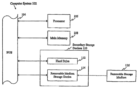
Figure 1 provides a diagrammatic representation of a computer-based discovery
system
containing the SNP information of the present invention in computer readable
form. =
DETAILED DESCRIPTION OF THE INVENTION
The present invention provides SNPs associated with CID, and in particular
stenosis and
MI, nucleic acid molecules containing SNPs, methods and reagents for the
detection of the SNPs
disclosed herein, uses of these SNPs for the development of detection
reagents, and assays or kits
that utilize such reagents. The stenosis or MI-associated SNPs disclosed
herein are useful for
diagnosing, screening for, and evaluating predisposition to stenosis or ME and
related pathologies in
humans,. Furthermore, such SNPs and their encoded products are useful targets
for the development
of therapeutic agents.
=
A large number of SNPs have been identified from re-sequencing DNA from 39
individuals,
and they are indicated as "Applera" SNP source in Tables 1-2. Their allele
frequencies observed in
each of the Caucasian and African-American ethnic groups are provided.
Additional SNPs included
herein were previously identified during shotgun sequencing and assembly of
the human genome,
and they are indicated as "Celera" SNP source in Tables 1-2. Furthermore, the
information provided
in Table 1-2, particularly the allele frequency information obtained from 39
individuals and the
identification of the precise position of each SNP within each
gene/transcript, allows haplotypes
(i.e., groups of SNPs that are co-inherited) to be readily inferred. The
present invention
encompasses SNP haplotypes, as well as individual SNPs.
Thus, the present invention provides individual SNPs associated with CH]), and
in particular
stenosis and MI, as well as combinations of SNPs and haplotypes in genetic
regions associated with
CHD, and in particular stenosis and MI, polymorphic/variant transcript
sequences (SEQ I NOS: 1-8)
and genomic Sequences (SEQ JD NOS: 25-33) containing SNPs, encoded amino acid
sequences (SEQ ID NOS: 9-16), and both transcript-based SNP context sequences
(SEQ ID NOS:
17-24) , and genoMic-based SNP context sequences (SEQ ID NOS: 34-42)
(transcript sequences,
protein sequences, and transcript-based SNP context sequences are provided in
Table 1 and the
Sequence Listing; genomic sequences and genoinic-based SNP context sequences
are provided in
Table 2 and the Sequence Listing), methods of detecting these polymorphisms in
a test sample,
methods of determining the risk of an individual of having or developing
stenosis and/or MI,
methods of screening for compounds useful for treating disorders associated
with a variant
23
CA 02600794 2007-09-11
WO 2006/099365
PCT/US2006/009016
gene/protein such as stenosis or MI, compounds identified by these screening
methods, methods of
using the disclosed SNPs to select a treatment strategy, methods of treating a
disorder associated
with a variant gene/protein (L e., therapeutic methods), methods of
determining if an individual is
likely to respond to a specific treatment such as statins and methods of using
the SNPs of the present
invention for human identification.
The present invention provides novel SNPs associated with stenosis and/or MI,
as well as
SNPs that were previously known in the art, but were not previously known to
be associated with
stenosis or MI. Accordingly, the present invention provides novel compositions
and methods based
on the novel SNPs disclosed herein, and also provides novel methods of using
the known, but
previously unassociated, SNPs in methods relating to stenosis or MI (e.g., for
diagnosing MI, etc.).
In Tables 1-2, known SNPs are identified based on the public database in which
they have been
observed, which is indicated as one or more of the following SNP types:
"dbSNP" = SNP observed
in dbSNP, "HGBASE" = SNP observed in HGBASE, and "HGMD" = SNP observed in the
Human
Gene Mutation Database (HGMD). Novel SNPs for which the SNP source is only
"Applera" and
none other, i.e., those that have not been observed in any public databases
and which were also not
observed during shotgun sequencing and assembly of the Celera human genome
sequence (L e.,
"Celera" SNP source), are also noted in the tables.
Particular SNP alleles of the present invention can be associated with either
an increased risk
of having or developing stenosis or MI, or a decreased risk of having or
developing stenosis or MI.
SNP alleles that are associated with a decreased risk of having or developing
stenosis or MI may be
referred to as "protective" alleles, and SNP alleles that are associated with
an increased risk of
having or developing stenosi or MI may be referred to as "susceptibility"
alleles, "risk" alleles, or .
"risk factors". Thus, whereas certain SNPs (or their encoded products) can be
assayed to determine
whether an individual possesses a SNP allele that is indicative of an
increased risk of having or
developing stenosis or MI (L e., a susceptibility allele), other SNPs (or
their encoded products) can be
assayed to determine whether an individual possesses a SNP allele that is
indicative of a decreased
risk of having or developing stenosis or MI (i.e., a protective allele).
Similarly, particular SNP
alleles of the present invention can be associated with either an increased or
decreased likelihood of
responding to a particular treatment or therapeutic compound, or an increased
or decreased
likelihood of experiencing toxic effects from a particular treatment or
therapeutic compound. The
term "altered" may be used herein to encompass either of these two
possibilities (e.g., an increased
or a decreased risk/likelihood).
24
CA 02600794 2013-12-23
=
Those skilled in the art will readily recognize that nucleic acid moleculesmay
be double-
stranded molecules and that reference to a particular site on one strand
refers, as well, to the
corresponding site on a complementary strand. In dening a SNP position, SNP
allele, or nucleotide
sequence, reference to an adenine, a thymine (midine), a cytosine, or a
guanine at a particular site on '
one strand of a nucleic acid molecule also defines the thymine (uridine),
adenine, guanine, or
cytosine (respectively) at the corresponding site on a complementary strand of
the nucleic acid
molecule. Thus, reference may be made to either strand in order to refer to a
particular SNP
position, SNP allele, or nucleotide sequence. Probes and primers, may be
designed to hybridize to
either strand and SNP genotypin.g methods disclosed herein may generally
target either strand.
Throughout the specification, in identifying a SNP position, reference is
generally made to the
protein-encoding strand, only for the purpose of convenience.
References to variant peptides, polypeptides, or proteins of the present
invention include
peptides, polypeptides, proteins, or fragments thereof, that contain at least
one amino acid residue
that differs from the corresponding amino acid sequence of the art-known
peptide/polypeptide/protein (the art-known protein may be interchangeably
referred to as the "wild-
type," "reference," or. "normal" protein). Such variant
peptides/polypeptides/proteins can result
from a codon change caused by a nonsynonymous nucleotide substitution at a
protein-coding SNP
position (L e., a missense mutation) disclosed by the present invention.
Variant
peptides/polypeptides/proteins of the present invention can also result from a
nonsense mutation, i.e.,
a SNP that creates a premature stop codon, a SNP that generates a read-through
mutation by
1
abolishing a stop codon, or due toan. y SNP disclosed by the present invention
that otherwise alters
. the structure, function/activity, or expression of a protein, such as a
SNP in a regulatory region (e.g.
a promoter or ehhancer) or a SNP that leads to alternative or defective
splicing; such as a SNP in an
intron or a SNP at an exon/intron boundary. As used herein, the terms
"polypeptide," "peptide,". and
"protein" are used interchangeably.
. ISOLATED NUCLEIC ACED MOLECULES AND SNP DETECTION REAGENTS 8z
KITS
Tables 1 and 2 provide a variety of information about each SNP of the present
invention that
is associated with CHD,: and in particular steno sis and MI, including the
transcript sequences (SEQ
ID NOS: 1-8), genomic sequences (SEQ ID NOS: 25-33), and protein sequences
(SEQ ID NOS:
9-16) of the encoded gene products (with. the SNPs indicated by TUB codes in
the nucleic acid
sequences). In addition, Tables 1 and 2 include SNP context sequences, which
generally include 100
. ,
CA 02600794 2013-12-23
=
nucleotide upstream (5') plus 100 nucleotides downstream (3') of each SNP
position (SEQ ID NOS:
17-24 correspond to transcript-based SNP context sequences. disclosed in Table
1, and SEQ ID
NOS: 34-42 correspond to genoinic-based context sequences disclosed in Table
2), the alternative
nucleotides (alleles) at each SNP position, and additional information about
the variant where
relevant, such as SNP type (coding, miss ense, splice site, UTR, etc.), human
populations in which
the SNP was observed, observed allele frequencies, information about the
encoded protein, eto.
Isolated Nucleic Acid Molecules
The present invention. provides isolated nucleic acid molecules that contain
one or more
SNPs disclosed Table 1 and/or Table 2. Isolated nucleic acid molecules
contoining one or more
SNPs disclosed in at least one of Tables 1-2 may be interchangeably referred
to throughout the
present text as `,`SNP-containing nucleic acid molecules". Isolated nucleic
acid molecules may
optionally encode a full-length variant protein or fragment thereof. The
isolated nucleic acid
molgcules of the present invention also include probes and primers. (which are
desbribed in greater
detail below in the section entitled "SNP Detection Reagents"), which may be
used for assaying the
disclosed SNPs, and isolated fun-length genes, transcripts, cDNA molecules,
and fragments thereof, =
which may be used for such purposes as expressing an encoded protein.
As used herein, an "isolated nucleic acid molecule" generally is one that
contains a SNP of the
present invention or one that hybridizes to such molecule such as a nucleic
acid with a complementary
sequence, and is separated from most other nucleic acids present in the
natural source of the nucleic acid
molecule. Moreover, an "isolate& nucleic acid molecule, suchns a cDNA molecule
containing a SNP
of the present invention, can be substantially free of other cellular
material, or culture medium when .
produced by recombinant techniques, or chemical precursors or other chemicals
when chemically
= synthesized. A nucleic acidmolecule can be fused to other coding or
regulatory sequences and still be
considered "isolated". Nucleic acid molecules present in non-human transgenic
animals, which do not
naturally occur in the animal, are also considered "isolated". For example,
recombinant DNA
molecules contained in a vector are considered "isolated". Further examples of
"isolated" DNA
molecules include recombinant DNA molecules maintained in heterologous host
cells, and purified
(partially or substantially) DNA molecules in solution. Isolated RNA molecules
include in -vivo or in =
vitro RNA transcripts of the isolated SNP-containing DNA molecules of the
present invention. Isolated
nucleic acid molecules according to the present invention further include such
molecules produced
synthetically.
26
CA 02600794 2013-12-23
Genertilly, an isolated SNP-containing nucleic acid molecule comprises one or
more SNP
positions disclosed by the present invention with flanking nucleotide
sequences on either side of the
= SNP positions. A flanking sequence can include nucleotide residues that
are naturally associated with
the SNP Site and/or heterologous nucleotide sequences. Preferably the flanking
sequence is up to
about 500, 300, 100, 60, 50, 30, 25, 20, 15, 10, 8, or 4 nucleotides (or any
other length in-.between) on
either side of a SNP position, or as long as the full-length gene or entire
protein-coding sequence (or
any portion thereof such as an exon), especially if the SNP-containing nucleic
acid molecule is to be
used to produce a protein or protein fragment.
For full-length genes and entire protein-coding sequences, a SNP flanking
sequence can be, for
example, up to about 5KB, 4KB, 3KB, 2KB, 1KB on either side of the SNP.
Furthermore, in such
instances, the isolated nucleic acid molecule comprises exonic sequences
(including protein-coding
and/or non-coding exonic sequences), but may also include intronic sequences.
Thus, any protein =
coding sequence may be either contiguous or separated by introns. The
important point is that the
nucleic acid is isolated from remote and unimportant flanking sequences and is
of appropriate length
such that it can be subjected to the specific manipulations or uses described
herein such as recombinant =
protein expression, preparation of probes and primers for assaying the SNP
position, and other uses
specific to the SNP-containing nucleic acid sequences.
An isolated SNP-containing nucleic acid molecule can comprise, for example, a
full-length gene
or transcript, such, as a gene isolated from genomic DNA (e.g., by cloning or
PCR amplification), a
cDNA molecule, or an mRNA transcript molecule. Polymorphic transcript
sequences are referred to in
Table 1 and provided in the Sequence Listing (SEQ ID NOS: 1-8), and
polymorphic genomic
sequences are referred to in Table 2 and provided in the Sequence Listing (SEQ
ID NOS: 25-33).
Furthermore, fragments of such full-length genes Eurd=transcripts that contain
one or more SNPs
disclosed herein are also encompassed by the present invention, and such
fragments may be used, for = =
example, to express any part of a protein, such as a particular functional
domain or an antigenic epitope.
Thus, the present invention also encompasses fragments of the nucleic acid
sequences as
disclosed in Tables 1-2 (transcript sequences are referred to in Table 1 as
SEQ ID NOS: 1-8, genomic
= sequences are referred to in. Table 2 as SEQ ID NOS: 25-33, transcript-
based SNP context sequences
are referred to in Table 1 as SEQ ID NO: 17-24, and genomic-based SNP context
sequences are
referred to in Table 2 as SEQ ID NO: 34-42) and their complements. The actual
sequences referred
to in the tables are provided in. the Sequence Listing. A fragment typically
comprises, a contiguous
nucleotide sequence at least about 8 or more nucleotides, more preferably at
least about 12 or more
nucleotides, and even more preferably at least about 16 or more nucleotides.
Further, a fragment could
27
CA 02600794 2007-09-11
WO 2006/099365
PCT/US2006/009016
=
comprise at least about 18, 20, 22, 25, 30, 40, 50, 60, 80, 100, 150, 200, 250
or 500 (or any other
number in-between) nucleotides in length. The length of the fragment will be
based on its intended use.
For example, the fragment can encode epitope-bearing regions of a variant
peptide or regions of a
variant peptide that differ from the normal/wild-type protein, or can be
useful as a polynucleotide probe
or primer. Such fragments can be isolated using the nucleotide sequences
provided in Table 1 and/or
Table 2 for the synthesis of a polynucleotide probe. A labeled probe can then
be used, for example, to
screen a cDNA library, genomic DNA library, or mRNA to isolate nucleic acid
corresponding to the
coding region. Further, primers can be used in amplification reactions, such
as for purposes of assaying
one or more SNPs sites or for cloning specific regions of a gene.
An isolated nucleic acid molecule of the present invention further encompasses
a SNP-
containing polynucleotide that is the product of any one of a variety of
nucleic acid amplification
methods, which are used to increase the copy numbers of a polynucleotide of
interest in a nucleic
acid sample. Such amplification methods are well known in the art, and they
include but are not
limited to, polymerase chain reaction (PCR) (U.S. Patent Nos. 4,683,195; and
4,683,202; PCR
Technology: Principles and Applications for DNA Amplification, ed. H.A.
Erlich, Freeman Press,
NY, NY, 1992), ligase chain reaction (LCR) (Wu and Wallace, Ge120177iCS 4:560,
1989; Landegren et
al., Science 241:1077, 1988), strand displacement amplification (SDA) (U.S.
Patent Nos. 5,270,184;
and 5,422,252), transcription-mediated amplification (TMA) (U.S. Patent No.
5,399,491), linked
linear amplification (LLA) (U.S. Patent No. 6,027,923), and the like, and
isothermal amplification
methods such as nucleic acid sequence based amplification (NASBA), and self-
sustained sequence
replication (Guatelli et al., Proc. Natl. Acad. Sci. USA 87: 1874, 1990).
Based on such
methodologies, a person skilled in the art can readily design primers in any
suitable regions 5' and 3'
to a SNP disclosed herein. Such primers may be used to amplify DNA of any
length so long that it
contains the. SNP of interest in its sequence.
As used herein, an "amplified polynucleotide" of the invention is a SNP-
containing nucleic
acid molecule whose amount has been increased at least two fold by any nucleic
acid amplification
method performed in vitro as compared to its starting amount in a test sample.
In other preferred
embodiments, an amplified polynucleotide is the result of at least ten fold,
fifty fold, one hundred
fold, one thousand fold, or even ten thousand fold increase as compared to its
starting amount in a
test sample. In a typical PCR amplification, a polynucleotide of interest is
often amplified at least
fifty thousand fold in amount over the unamplified genomic DNA, but the
precise amount of
amplification needed for an assay depends on the sensitivity of the subsequent
detection method
used.
28
CA 02600794 2013-12-23
=
Generally, an amplified polynucleotide is at least about 16 nucleotides in
length. More
typically, an amplified polynucleotide is at least about 20 nucleotides in
length. In a preferred
embodiment of the invention, an amplified polynucleotide is at least about 30
nucleotides in length.
In a more preferred embodiment of the invention, an amplified polynucleotide
is at least about 32,
40, 45, 50, or 60 nucleotides in length. In yet another preferred embodiment
of the invention, an
amplified polynucleotide is at least about 100, 200, 300, 400, or 500
nucleotides in length. While
the total length of an amplified polynucleotide of the invention can be as
long as an exon, an baton
or the entire gene where the SNP of interest resides, an amplified product is
typically up to about
1,000 nucleotides in length (although certain amplification methods may
generate amplified products
greater than 1000 nucleotides in length). More preferably, an amplified
polynucleotide is not greater
than about 600-700 nucleotides in. length. It is understood that irrespective
of the length of an
amplified polynucleotide, a SNP of interest may be located anywhere along its
sequence.
In a specific embodiment of the invention, the amplified product is at least
about 201
nucleotides in length, comprises one of the transcript-based context sequences
or the genomic-based
context sequences shown in Tables 1-2. Such a product may have additional
sequences on its 5' end.
or 3' end or both. In another embodiment, the amplified product is about 101
nucleotides in length,
and it contains a SNP disclosed herein. Preferably, the SNP is located at the
middle of the amplified
product (e.g., at position 101 in an amplified product that is 201 nucleotides
in length, or at position
51 in an amplified product that is 101 nucleotides in length), or within 1,2,
3, 4, 5, 6,7, 8,9, 10, 12,
15, or 20 nucleotides from the middle of the amplified product (however, as
indicated above, the
SNP of interest may be located cmywhere along the length of the amplified
product).
The present invention provides isolated nucleic acid molecules that comprise,
consist of, or
consist essentially of one or more polynucleotide sequences that contain one
or more SNPs disclosed
herein, complements thereof, and SNP-containing fragments thereof
Accordingly, the present invention provides nucleic acid molecules that
consist of any of the
. nucleotide sequences shown in Table 'l and/or Table 2 (transcript sequences
are referred to in Table 1 as
= SEQ ID NOS: 1-8, genornic sequences are referred to in Table 2 as SEQ ID
NOS: 25-33, transcript-
based SNP context sequences are referred to in Table 1 as SEQ ID NO: 17-24,
and genomic-based
SNP context sequences are referred to in Table 2 as SEQ ID NO: 34-42), , or
any nucleic acid
molecule that encodes any of the variant proteins referred to in Table 1 (SEQ
ID NOS: 9-16). , The
actual sequences referred to in the tables are provided in the Sequence
Listing. A nucleic acid molecule
consists of a nucleotide sequence when the nucleotide sequence is the complete
nucleotide sequence of
the nucleic acid molecule.
29
CA 02600794 2013-12-23
The present invention further provides nucleic acid molecules that consist
essentially of any of
the nucleotide sequences referred to in Table 1 and/or Table 2 (transcript
sequences are referred to in
Table 1 as SEQ ID NOS:1-8, gen.omic sequences are referred. to in. Table 2'as
SEQ ID NOS: 25-33,
transcript-based SNP context sequences are referred to in Table 1 as SEQ 3D
NO: 17-24, and
genonaic-based SNP context sequences are referred to in Table 2 as SEQ JD NO:
34-42), or any
nucleic acid molecule that encodes any of the variant proteins referred to in.
Table 1 (SEQ ID NOS:
9-16)- The actual sequences referred to in-the tables are provided in the
Sequence Listing. A nucleic
acid molecule consists essentially of a nucleotide sequence when such a
nucleotide sequence is present
with. only a few additional nucleotide residues in the final nucleic acid
molecule.
The present invention farther provides nucleic acid molecules that comprise
any of the
nucleotide sequences shown in Table 1 and/or Table 2 or a SNP-containing
fragment thereof (transcript
sequences are referred to in Table 1 as SEQ ID NOS:1-8, genomic sequences, are
referred to in Table.
2 as SEQ lD NOS: 25-33, transcript-based SNP context sequences are referred to
in. Table 1 as SEQ
ID NO: 17-24, and genoraic-based SNP context sequences are referred to in
Table 2 as SEQ ID NO:
34-42), or any nucleic acid molecule that encodes any of the variant proteins
provided in Table 1.
The actual sequences referred to in the tables are provided in the Sequence.
Listing. A nucleic acid molecule comprises a nucleotide sequence when the
nucleotide sequence is at
least part of the final nucleotide sequence of the nucleic acid molecule. In
such a fashion, the nucleic
1
acid molecule can be only the nucleotide sequence or have additional
nucleotide residues, such as
residues that are naturally associated with it or heterologous nucleotide
sequences. Such a nucleic acid
molecule can have one to a few additional nucleotides or can comprise many
more additional
nucleotides. A brief description of how various types of these nucleic acid
molecules can be readily
made and isolated is provided below, and such techniques are well known to
those of ordinary skill in
the art (Sambrook and Russell, 2000, Molecular Cloning: A Laboratory Manual,
Cold. Spring Harbor
Press, NY).
The isolated. nucleic acid molecules can encode mature proteins plus
additional amino or
' carboxyl-terminal Funino acids or both, or amino acids interior to the
mature peptide (when the mature
fonn has more than one peptide chain, for instance). Such sequences may play a
role in processing of a
protein from precursor to a mature form, facilitate protein trafficking,
prolong or shorten protein half-
1
3 0 life, or facilitate manipulation of a protein for assay or production.
As generally is the case in situ, the
additional amino acids may be processed away from the mature protein by
cellular enzymes.
Thus, the isolated nucleic acid molecules include, but are not limited to,
nucleic add molecules
having a sequence encoding a peptide alone, a sequence encoding a mature
peptide and additional
CA 02600794 2007-09-11
WO 2006/099365
PCT/US2006/009016
coding sequences such as a leader or secretory sequence (e.g., a pre-pro or
pro-protein sequence), a
sequence encoding a mature peptide with or without additional coding
sequences, plus additional non-
coding sequences, for example introns and non-coding 5' and 3' sequences such
as transcribed but
untranslated sequences that play a role in, for example, transcription, mRNA
processing (including
splicing and polyadenylation signals), ribosome binding, and/or stability of
mRNA. In addition, the
nucleic acid molecules may be fused to heterologous marker sequences encoding,
for example, a
peptide that facilitates purification.
Isolated nucleic acid molecules can be in the form of RNA, such as mRNA, or in
the form
DNA, including cDNA and genomic DNA, which may be obtained, for example, by
molecular
cloning or produced by chemical synthetic techniques or by a combination
thereof (Sambrook and
Russell, 2000, Molecular Cloning: A Laboratory Manual, Cold Spring Harbor
Press, NY).
Furthermore, isolated nucleic acid molecules, particularly SNP detection
reagents such as probes and
primers, can also be partially or completely in the form of one or more types
of nucleic acid analogs,
such as peptide nucleic acid (PNA) (U.S. Patent Nos. 5,539,082; 5,527,675;
5,623,049; 5,714,331).
The nucleic acid, especially DNA, can be double-stranded or single-stranded.
Single-stranded
nucleic acid can be the coding strand (sense strand) or the complementary non-
coding strand (anti-
sense strand). DNA, RNA, or PNA segments can be assembled, for example, from
fragments of the
human genome (in the case of DNA or RNA) or single nucleotides, short
oligonucleotide linkers, or
from a series of oligonucleotides, to provide a synthetic nucleic acid
molecule. Nucleic acid
molecules can be readily synthesized using the sequences provided herein as a
reference;
oligonucleotide and PNA oligomer synthesis techniques are well known in the
art (see, e.g., Corey,
"Peptide nucleic acids: expanding the scope of nucleic acid recognition",
Trends Bioteehnol. 1997
Jun;15(6):224-9, and Hyrup et al., "Peptide nucleic acids (PNA): synthesis,
properties and potential
applications", Bioorg Med Chem. 1996 Jan;4(1):5-23). Furthermore, large-scale
automated
oligonucleotide/PNA synthesis (including synthesis on an array or bead surface
or other solid
support) can readily be accomplished using commercially available nucleic acid
synthesizers, such
as the Applied Biosystems (Foster City, CA) 3900 High-Throughput DNA
Synthesizer or Expedite
8909 Nucleic Acid Synthesis System, and the sequence infoiination provided
herein.
The present invention encompasses nucleic acid analogs that contain modified,
synthetic, or
non-naturally occurring nucleotides or structural elements or other
alternative/modified nucleic acid
chemistries known in the art. Such nucleic acid analogs are useful, for
example, as detection
reagents (e.g., primers/probes) for detecting one or more SNPs identified in
Table 1 and/or Table 2.
Furthermore, kits/systems (such as beads, arrays, etc.) that include these
analogs are also
31
CA 02600794 2007-09-11
WO 2006/099365
PCT/US2006/009016
encompassed by the present invention. For example, PNA oligomers that are
based on the
polymorphic sequences of the present invention are specifically contemplated.
PNA oligomers are
analogs of DNA in which the phosphate backbone is replaced with a peptide-like
backbone
(Lagriffoul et al., Bioorganic & Medicinal Chemistry Letters, 4: 1081-1082
(1994), Petersen et al.,
Bioorganic & Medicinal Chemistry Letters, 6: 793-796 (1996), Kumar et al.,
Organic Letters 3(9):
1269-1272 (2001), W096/04000). PNA hybridizes to complementary RNA or DNA with
higher
affinity and specificity than conventional oligonucleotides and
oligonucleotide analogs. The
properties of PNA enable novel molecular biology and biochemistry applications
unachievable with
traditional oligonucleotides and peptides.
Additional examples of nucleic acid modifications that improve the binding
properties and/or
stability of a nucleic acid include the use of base analogs such as inosine,
intercalators (U.S. Patent
No. 4,835,263) and the minor groove binders (U.S. Patent No. 5,801,115). Thus,
references herein
to nucleic acid molecules, SNP-containing nucleic acid molecules, SNP
detection reagents (e.g.,
probes and primers), oligonucleotides/polynucleotides include PNA oligomers
and other nucleic acid
analogs. Other examples of nucleic acid analogs and alternative/modified
nucleic acid chemistries
known in the art are described in Current Protocols in Nucleic Acid Chemistry,
John Wiley & Sons,
N.Y. (2002).
The present invention further provides nucleic acid molecules that encode
fragments of the
variant polypeptides disclosed herein as well as nucleic acid molecules that
encode obvious variants
of such variant polypeptides. Such nucleic acid molecules may be naturally
occurring, such as
paralogs (different locus) and orthologs (different organism), or may be
constructed by recombinant
DNA methods or by chemical synthesis. Non-naturally occurring variants may be
made by
mutagenesis techniques, including those applied to nucleic acid molecules,
cells, or organisms.
Accordingly, the variants can contain nucleotide substitutions, deletions,
inversions and insertions
(in addition to the SNPs disclosed in Tables 1-2). Variation can occur in
either or both the coding
and non-coding regions. The variations can produce conservative and/or non-
conservative amino
acid substitutions.
Further variants of the nucleic acid molecules disclosed in Tables 1-2, such
as naturally
occurring allelic variants (as well as orthologs and paralogs) and synthetic
variants produced by
mutagenesis techniques, can be identified and/or produced using methods well
known in the art.
Such further variants can comprise a nucleotide sequence that shares at least
70-80%, 80-85%, 85-
90%, 91%, 92%, 93%, 94%, 95%, 96%, 97%, 98%, or 99% sequence identity with a
nucleic acid
sequence disclosed in Table 1 and/or Table 2 (or a fragment thereof) and that
includes a novel SNP
32
CA 02600794 2007-09-11
WO 2006/099365
PCT/US2006/009016
allele disclosed in Table 1 and/or Table 2. Further, variants can comprise a
nucleotide sequence that
encodes a polypeptide that shares at least 70-80%, 80-85%, 85-90%, 91%, 92%,
93%, 94%, 95%,
96%, 97%, 98%, or 99% sequence identity with a polypeptide sequence disclosed
in Table 1 (or a
fragment thereof) and that includes a novel SNP allele disclosed in Table 1
and/or Table 2. Thus, an
aspect of the present invention that is specifically contemplated are isolated
nucleic acid molecules
that have a certain degree of sequence variation compared with the sequences
shown in Tables 1-2,
but that contain a novel SNP allele disclosed herein. In other words, as long
as an isolated nucleic
acid molecule contains a novel SNP allele disalosed herein, other portions of
the nucleic acid
molecule that flank the novel SNP allele can vary to some degree from the
specific transcript,
genomic, and context sequences referred to and shown in Tables 1-2, and can
encode a polypeptide
that varies to some degree from the specific polypeptide sequences referred to
in Table 1.
To determine the percent identity of two amino acid sequences or two
nucleotide sequences
of two molecules that share sequence homology, the sequences are aligned for
optimal comparison
purposes (e.g., gaps can be introduced in one or both of a first and a second
amino acid or nucleic
acid sequence for optimal alignment and non-homologous sequences can be
disregarded for
comparison purposes). In a preferred embodiment, at least 30%, 40%, 50%, 60%,
70%, 80%, or
90% or more of the length of a reference sequence is aligned for comparison
purposes. The amino
acid residues or nucleotides at corresponding amino acid positions or
nucleotide positions are then
compared. When a position in the first sequence is occupied by the same amino
acid residue or
nucleotide as the corresponding position in the second sequence, then the
molecules are identical at
that position (as used herein, amino acid or nucleic acid "identity" is
equivalent to amino acid or
nucleic acid "homology"). The percent identity between the two sequences is a
function of the
number of identical positions shared by the sequences, taking into account the
number of gaps, and
the length of each gap, which need to be introduced for optimal alignment of
the two sequences.
The comparison of sequences and determination of percent identity between two
sequences
can be accomplished using a mathematical algorithm. (Computational Molecular
Biology, Lesk,
A.M., ed., Oxford University Press, New York, 1988; Biocomputing: Informatics
and Genome
Projects, Smith, D.W., ed., Academic Press, New York, 1993; Computer Analysis
of Sequence Data,
Part I, Griffin, A.M., and Griffm, H.G., eds., Humana Press, New Jersey, 1994;
Sequence Analysis in
Molecular Biology, von Heinje, G., Academic Press, 1987; and Sequence Analysis
Primer, Gribskov,
M. and Devereux, J., eds., M Stockton Press, New York, 1991). In a preferred
embodiment, the
percent identity between two amino acid sequences is determined using the
Needleman and Wunsch
algorithm (J. Mol. Biol. (48):444-453 (1970)) which has been incorporated into
the GAP program in
33
CA 02600794 2007-09-11
WO 2006/099365
PCT/US2006/009016
the GCG software package, using either a Blossom 62 matrix or a PAM250 matrix,
and a gap weight
of 16, 14, 12, 10, 8, 6, or 4 and a length weight of 1, 2, 3, 4, 5, or 6.
In yet another preferred embodiment, the percent identity between two
nucleotide sequences
is determined using the GAP program in the GCG software package (Devereux, J.,
et al., Nucleic
Acids Res. 12(1):387 (1984)), using a NWSgapdna.CMP matrix and a gap weight of
40, 50, 60, 70, or
80 and a length weight of 1, 2, 3, 4, 5, or 6. In another embodiment, the
percent identity between
two amino acid or nucleotide sequences is determined using the algorithm of E.
Myers and W.
Miller (CABIOS, 4:11-17 (1989)) which has been incorporated into the ALIGN
program (version
2.0), using a PAM120 weight residue table, a gap length penalty of 12, and
a.gap penalty of 4.
The nucleotide and amino acid sequences of the present invention can further
be used as a
"query sequence" to perform a search against sequence databases to, for
example, identify other
family Members or related sequences. Such searches can be performed using the
NBLAST and
)(BLAST programs (version 2.0) of Altschul, et al. (J. Mol. Biol. 215:403-10
(1990)). BLAST
nucleotide searches can be performed with the NBLAST program, score = 100,
wordlength = 12 to
obtain nucleotide sequences homologous to the nucleic acid molecules of the
invention. BLAST
protein searches can be performed with the XBLAST program, score = 50,
wordlength = 3 to obtain
amino acid sequences homologous to the proteins of the invention. To obtain
gapped alignments for
comparison purposes, Gapped BLAST can be utilized as described in Altschul et
al. (Nucleic Acids
Res. 25(17):3389-3402 (1997)). When utilizing BLAST and gapped BLAST programs,
the default
parameters of the respective programs (e.g., XBLAST and NBLAST) can be used.
In addition to
BLAST, examples of other search and sequence comparison programs used in the
art include, but
are not limited to, FASTA (Pearson, Methods Mol. Biol. 25, 365-389 (1994)) and
KERR (Dufresne
et al., Nat Biotechnol 2002 Dec;20(12):1269-71). For further infoiniation
regarding bioinformatics
techniques, see Current Protocols in Bioinformatics, John Wiley & Sons, Inc.,
N.Y.
The present invention further provides non-coding fragments of the nucleic
acid molecules
disclosed in Table 1 and/or Table 2. Preferred non-coding fragments include,
but are not limited to,
promoter sequences, enhancer sequences, intronic sequences, 5' Imtranslated
regions (UTRs), 3'
untranslated regions, gene modulating sequences and gene teimination
sequences. Such fragments
are useful, for example, in controlling heterologous gene expression and in
developing screens to
identify gene-modulating agents.
34
CA 02600794 2013-12-23
SNP Detection Reagents .
In a specific aspect of the present invention, the SNPs disclosed in Table 1
and/or Table 2, and
. their associated transcript sequences (referred to in Table 1 as SEQ ID
NOS: 1-8), genomic
'sequences (referred to in Table 2 as SEQ ID NOS: 25-33), and context
sequences (transcript-based
context sequences are referred to in Table 1 as SEQ ID NOS: 17-24; genomio-
based context
sequences are provided in Table 2 as SEQ ID NOS: 34-42), can be used for the
design of SNP
detection reagents. The actual sequences referred to in. the tables are
provided iii the Sequence Listing.
As used herein, a "SNP detection reagent" is a reagent that specifically
detects a specific target SNP
position disclosed herein, and that is preferably specific for a particular
nucleotide (allele) of the'target
SNP position (i.e., the detection reagent preferably can differentiate between
different alternative
nucleotides at a target SNP position, thereby allowing the identity of the
nucleotide present at the target
SNP position to be determined). Typically, such detection reagent hybridizes
to a target SNP-
containing nucleic acid molecule by complementary base-pairing in a sequence.
specific manner, and
discriminates the target variant sequence from other nucleic acid sequences
such as an art-known forth. '
in a test sample. An example of a detection reagent is a probe that hybridizes
to a target nucleic acid
=
containing one or more of the SNPs referred to in Table 1 and/or Table 2. In a
preferred embodiment,
such a probe can differentiate between nucleic acids having a particular
nucleotide (allele) at a target
SNP position from other nucleic acids that have a different nucleotide at the
same target SNP position,
In addition, a detection reagent may hybridize to a specific region 5' and/or
3' to a SNP position,
particularly a region corresponding to the context sequences referred to in
Table 1 and/or Table 2
(tranAcript-based context sequences are referred to in. Table 1 as SEQ ID NOS:
17-24; genomio-based
=
context sequences are referred to in Table 2 as SEQ ID NOS: 34-42). Another
example of a
. detection reagent is'a primer that acts as an. initiation point of
nucleotide extension along a
complementary strand of a target polynucleotide. The SNP sequence in.fonnation
provided herein is
also useful for designing primers, e.g. allele-specific primers, to amplify
(e.g., using PCR) any SNP of
the present invention.
In one preferred embodiment of the invention, a SNP detection reagent is an
isolated or
synthetic DNA or RNA polynucleotide probe or primtr or PNA oligomer, or a
combination of DNA,
RNA and/or PNA, that hybridizes to a segment of a target nucleic acid moledule
containing a SNP
identified in Table 1 and/or Table 2. A detection reagent in the form of a
polynucleatide may
= optionally contain modified base analogs, intercalators or minor groove
binders. Multiple detection
reagents such as probes may be, for example, affixed to a solid support (e.g.,
arrays or beads) or
35. =
CA 02600794 2013-12-23
=
supplied in solution (e. g. , probe/primer sets for en7ymatic reactions such
as PCR, RT-PCR, TaqManTM -
assays, or primer-extension, reactions) to form a SNP detection kit.
A. probe or primer typically is a substantially purified oligonucleolide or
PNA oligomer. Such
oligonucleolide typically comprises a region of complementary nucleotide
sequence that hybridizes
under stringent conditions to at least about 8, 10, 12, 16, 18, 20, 22, 25,
30, 40, 50, 55, 60, 65, 70, 80,
90, 100, 120 (or any other number in-between) or more consecutive nucleotides
in a target nucleic acid
molecule. Depending on the particular assay, the consecutive nucleotides can
either include the target
SNP position, or be a specific region in close enough proximity 5' and/or 3'
to the SNP position to carry
out the desired assay.
Other preferred primer and probe sequences can readily be determined using the
transcript
sequences (SEQ ID NOS: 1-8), genomic sequences (SEQ ID NOS: 25-33), and SNP
context
sequences (transcript-based context sequences are referred to in Table 1 as
SEQ ID NOS: 17-24;
gthomic-based context sequences are referred to in Table 2 as SEQ ID NOS: 34-
42) disclosed in the
Sequence Listing and in Tables ,1-2. The actual sequences referred to in the
tables are provided in the
Sequence Listing. It will be apparent to one of skill in the art that such
primers and probes are
directly useful as reagents for genotyping the SNPs of the present invention,
and can be incorporated
into any kit/system format =
In order to produce a probe or primer specific for a target SNP-containing
sequence, the
gene/transcript and/or context sequence surrounding the SNP of interest is
typically examined using
a computer algorithm that starts at the 5' or at the 3' end of the nucleotide
sequence. Typical
algorithms will then identify oligomers of defined length that are unique to
the gene/SNP context
sequence, have a GC content within a range suitable for hybridization, lack
predicted secondary
structure that may interfere with hybridization, and/or possess other desired
characteristics or that
lack other undesired characteristics.
A primer or probe of the present invention is typically at least about 8
nucleotides in length.
In one embodiment of the invention, a primer or a probe is at least about 10
nucleotides in length. In
a preferred embodiment, a primer or a ptobe is at least about 1,2 nucleotides
in length. = In a More
preferred embodiment, a primer or probe is at least about 16, 17, 18, 19,
20,21, 22, 23,24 or 25
nucleotides in length. While the maximal length of a probe can be as long as
the target sequence to
be detected, depending on the type of assay in which it is.employed, it is
typically less than about 50,
60, 65, or 70 nucleotides in length. In the case of a primer, it is typically
less than about 30
nucleotides in length. In a specific preferred embodiment of the invention, a
primer of a probe is
within the length of about 18 and about 28 nucleotides. However, in other
embodiments, such as
36
CA 02600794 2007-09-11
WO 2006/099365
PCT/US2006/009016
nucleic acid arrays and other embodiments in which probes are affixed to a
substrate, the probes can
be longer, such as on the order of 30-70, 75, 80, 90, 100, or more nucleotides
in length (see the
section below entitled "SNP Detection Kits and Systems").
For analyzing SNPs, it may be appropriate to use oligonucleotides specific
for. alternative SNP
alleles. Such oligonucleotides that detect single nucleotide variations in
target sequences may be
referred to by such terms as "allele-specific oligonucleotides", "allele-
specific probes", or "allele-
specific primers". The design and use of allele-specific probes for analyzing
polymorphisms is
described in, e.g., Mutation Detection A Practical Approach, ed. Cotton et al.
Oxford University
Press, 1998; Saiki et al., Nature 324, 163-166 (1986); Dattagupta, EP235,726;
and Saiki, WO
89/11548.
While the design of each allele-specific primer or probe depends on variables
such as the
precise composition of the nucleotide sequences flanking a SNP position in a
target nucleic acid
molecule, and the length of the primer or probe, another factor in the use of
primers and probes is the
stringency of the condition under which the hybridization between the probe or
primer and the target
sequence is performed. Higher stringency conditions utilize buffers with lower
ionic strength and/or
a higher reaction temperature, and tend to require a more perfect match
between probe/primer and a
target sequence in order to form a stable duplex. If the stringency is too
high, however,
hybridization may not occur at all. In contrast, lower stringency conditions
utilize buffers with
higher ionic strength and/or a lower reaction temperature, and permit the
formation of stable
duplexes with more mismatched bases between a probe/primer and a target
sequence. By way of
example and not limitation, exemplary conditions for high stringency
hybridization conditions using
an allele-specific probe are as follows: prehybridization with a solution
containing 5X standard
saline phosphate EDTA (SSPE), 0.5% NaDodSO4 (SDS) at 55 C, and incubating
probe with target
nucleic acid molecules in the same solution at the same temperature, followed
by washing with a
solution containing 2X S SPE, and 0.1%SDS at 55 C or room temperature.
Moderate stringency hybridization conditions may be used for allele-specific
primer
ex'tension reactions with a solution containing, e.g., about 50mM KC1 at about
46 C. Alternatively,
the reaction may be carried out at an elevated temperature such as 60 C. In
another embodiment, a
moderately stringent hybridization condition suitable for oligonucleotide
ligation assay (OLA)
reactions wherein two probes are ligated if they are completely complementary
to the target
sequence may utilize a solution of about 100mM KCI at a temperature of 46 C. =
In a hybridization-based assay, allele-specifia probes can be designed that
hybridize to a
segment of target DNA from one individual but do not hybridize to the
corresponding segment from
37
CA 02600794 2007-09-11
WO 2006/099365
PCT/US2006/009016
another individual due to the presence of different polymorphic forms (e.g.,
alternative SNP
alleles/nucleotides) in the respective DNA segments from the two individuals.
Hybridization
conditions should be sufficiently stringent that there is a significant
detectable difference in
hybridization intensity between alleles, and preferably an essentially binary
response, whereby a =
probe hybridizes ta only one of the alleles or significantly more strongly to
one allele. While a
probe may be designed to hybridize to a target sequence that contains a SNP
site such that the SNP
site aligns anywhere along the sequence of the probe, the probe is preferably
designed to hybridize
to a segment of the target sequence such that the SNP site aligns with a
central position of the probe
(e.g., a position within the probe that is at least three nucleotides from
either end of the probe). This
design of probe generally achieves good discrimination in hybridization
between different allelic
forms.
In another embodiment, a probe or primer may be designed to hybridize to a
segment of
target DNA such that the SNP aligns with either the 5' most end or the 3' most
end of the probe or
primer. In a specific preferred embodiment that is particularly suitable for
use in a oligon.ucleotide
ligation assay (U.S. Patent No. 4,988,617), the 3'most nucleotide of the probe
aligns with the SNP
position in the target sequence.
Oligonucleotide probes and primers may be prepared by methods well known in
the art.
Chemical synthetic methods include, but are limited to, the phosphotriester
method described by
Narang et al., 1979, Methods in Enzymology 68:90; the phosphodiester method
described by Brown
et al., 1979, Methods in Enzymology 68:109, the diethylphosphoamidate method
described by
Beaucage et al., 1981, Tetrahedron Letters 22:1859; and the solid support
method described in U.S.
Patent No. 4,458,066.
Allele-specific probes are often used in pairs (or, less commonly, in sets of
3 or 4, such as if a
SNP position is known to have 3 or 4 alleles, respectively, or to assay both
strands of a nucleic acid
molecule for a target SNP allele), and such pairs may be identical except for
a one nucleotide
mismatch that represents the allelic variants at the SNP position. Commonly,
one member of a pair
perfectly matches a reference form of a target sequence that has a more common
SNP allele (i.e., the
allele that is more frequent in the target population) and the other member of
the pair perfectly
matches a folm of the target sequence that has a less common SNP allele (i.e.,
the allele that is rarer
in the target population). In the case of an array, multiple pairs of probes
can be immobilized on the
same support for simultaneous analysis of multiple different polymorphisms.
In one type of PCR-based assay, an allele-specific primer hybridizes to a
region on a target
nucleic acid molecule that overlaps a SNP position and only primes
amplification of an allelic fowl.
38
CA 02600794 2007-09-11
WO 2006/099365
PCT/US2006/009016
to which the primer exhibits perfect complementarity (Gibbs, 1989, Nucleic
Acid Res. 17 2427-
2448). Typically, the primer's 3'-most nucleotide is aligned with and
complementary to the SNP
position of the target nucleic acid molecule. This primer is used in
conjunction with a second primer
that hybridizes at a distal site. Amplification proceeds from the two primers,
producing a detectable
product that indicates which allelic form is present in the test sample. A
control is usually perfauned
with a second pair of primers, one of which shows a single base mismatch at
the polymorphic site
and the other of which exhibits perfect complementarity to a distal site. The
single-base mismatch
prevents amplification or substantially reduces amplification efficiency, so
that either no detectable
product is fonned or it is formed in lower amounts or at a slower pace. The
method generally works
most effectively when the mismatch is at the 3'-most position of the
oligonucleotide (i.e., the 3'-most
position of the oligonucleotide aligns with the target SNP position) because
this position is most
destabilizing to elongation from the primer (see, e.g., WO 93/22456). This PCR-
based assay can be
utilized as part of the TaqMan assay, described below.
In a specific embodiment of the invention, a primer of the invention contains
a sequence
substantially complementary to a segment of a target SNP-containing nucleic
acid molecule except that
the primer has a mismatched nucleotide in one of the three nucleotide
positions at the 3'-most end of the
primer, such that the mismatched nucleotide does not base pair with a
particular allele at the SNP site.
In a preferred embodiment, the mismatched nucleotide in the primer is the
second from the last
nucleotide at the 3'-most position of the primer. In a more preferred
embodiment, the mismatched
nucleotide in the primer is the last nucleotide at the 3'-most position of the
primer.
In another embodiment of the invention, a SNP detection reagent of the
invention is labeled =
with a fluorogenic reporter dye that emits a detectable signal. While the
preferred reporter dye is a
fluorescent dye, any reporter dye that can be attached to a detection reagent
such as an oligonucleotide
probe or primer is suitable for use in the invention. Such dyes include, but
are not limited to, Acridine,
AMCA, BODIPY, Cascade Blue, Cy2, Cy3, Cy5, Cy7, Dabcyl, Edans, Eosin,
Erythrosin, Fluorescein,
6-Fam, Tet, Joe, Hex, Oregon Green, Rhodamine, Rhodol Green, Tamra, Rox, and
Texas Red.
In yet another embodiment of the invention, the detection reagent may be
further labeled with a
quencher dye such as Tamra, especially when the reagent is used as a self-
quenching probe such as a
TaqMan (U.S. Patent Nos. 5,210,015 and 5,538,848) or Molecular Beacon probe
(U.S. Patent Nos.
5,118,801 and 5,312,728), or other stemless or linear beacon probe (Livak et
al., 1995, PCR Method
Appl. 4:357-362; Tyagi et al., 1996, Nature Biotechnology 14: 303-308;
Nazarenko et al., 1997, Nucl.
Acids Res. 25:2516-2521; U.S. Patent Nos. 5,866,336 and 6,117,635)..
39
CA 02600794 2007-09-11
WO 2006/099365
PCT/US2006/009016
The detection reagents of the invention may also contain other labels,
including but not limited
to, biotin for streptavidin binding, hapten for antibody binding, and
oligonucleotide for binding to
another complementary oligonucleotide such as pairs of zipcodes.
The present invention also contemplates reagents that do not contain (or that
are
complementary to) a SNP nucleotide identified herein but that are used to
assay one or more SNPs
disclosed herein. For example, primers that flank, but do not hybridize
directly to a target SNP
position provided herein are useful in primer extension reactions in which the
primers hybridize to a
region adjacent to the target SNP position (L e., within one or more
nucleotides from the target SNP
site). During the primer extension reaction, a primer is typically not able to
extend past a target SNP
site if a particular nucleotide (allele) is present at that target SNP site,
and the primer extension
product can be detected in order to determine which SNP allele is present at
the target SNP site. For
example, particular ddNTPs are typically used in the primer extension reaction
to terminate primer
extension once a ddNTP is incorporated into the extension product (a primer
extension product
which includes a ddNTP at the 3 '-most end of the primer extension product,
and in which the ddNTP
is a nucleotide of a SNP disclosed herein, is a composition that is
specifically contemplated by the
present invention). Thus, reagents that bind to a nucleic acid molecule in a
region adjacent to a SNP
site and that are used for assaying the SNP site, even though the bound
sequences do not necessarily
include the SNP site itself, are also contemplated by the present invention.
. SNP Detection Kits and Systems
A person skilled in the art will recognize that, based on the SNP and
associated sequence
information disclosed herein, detection reagents can be developed and used to
assay any SNP of the
present invention individually or in combination, and such detection reagents
can be readily
incorporated into one of the established kit or system formats which are well
known in the art. The
terms "kits" and "systems", as used herein in the context of SNP detection
reagents, are intended to
refer to such things as combinations of multiple SNP detection reagents, or
one or more SNP
detection reagents in combination with one or more other types of elements or
components (e.g.,
other types of biochemical reagents, containers, packages such as packaging
intended for
commercial sale, substrates to which SNP detection reagents are attached,
electronic hardware
components, etc.). Accordingly, the present invention further provides SNP
detection kits and
systems, including but not limited to, packaged probe and primer sets (e.g..
TaqMan probe/primer
sets), arrays/microarrays of nucleic acid molecules, and beads that contain
one or more probes,
primers, or other detection reagents for detecting one or more SNPs of the
present invention. The
CA 02600794 2013-04-09
=
41
kits/systems can optionally include various electronic "hardware components;
for example, arrays
("DNA chips") and microfluidic systems ("lab-on-a-chip" systems) provided by
various'
manufacturers typically comprise hardware components. Other kits/systems
(e.g., probe/primer sets)
may not include electronic hardware components, but may be comprised of, for
example, one or
more SNP detection reagents (along with, optionally, other biochemical
reagents) packaged in one or
more containers.
In some embodiments, a SNP detection kit typically contains one or more
detection reagents
and other components (e.g., a buffer, enzymes such as DNA polymerases or
ligases, chain extension
nucleotides such as deoxynucleotide triphosphates, and in the case of Sanger-
type DNA sequencing
reactions, chain terminating nucleotides, positive control sequences, negative
control sequences, and
the like) necessary to carry out an assay or reaction, such as amplification
and/or detection of a SNP-
containing nucleic acid molecule. A kit may further contain means for
determining the amount of a
target nucleic acid, and means for comparing the amount with a standard, and
can comprise
instructions for using the kit to detect the SNP-containing nucleic acid
molecule of interest. In one
embodiment of the present invention, kits are provided which contain the
necessary reagents to carry
out one or more assays to detect one or more SNPs disclosed herein. In a
preferred embodiment of
the present invention, SNP detection kits/systems are in the form of nucleic
acid arrays, or
compaamentali7ed kits, including microfluidic/lab-on-a-chip systems.
SNP detection kits/systems may contain, for example, one or more probes, or
pairs of probes,
that hybridize to a nucleic acid molecule at or near each target SNP position.
Multiple pairs of
allele-specific probes may be included in the kit/system to simultaneously
assay large numbers of
SNPs, at least one of which is a SNP of the Present invention. In some
kits/systems, the allele-
specific probes are immobilized to a substrate such as an array or bead. For
example, the same
substrate can comprise allele-specific probes for detecting at least 1; 10;
100; 1000; 10,000; 100,000
(or any other number in-between) or substantially all of the SNPs shown in
Table 1 and/or Table 2.
The terms "arrays," "microarrays," and "DNA chips" are used herein
interchangeably to refer
to an array of distinct polynucleotides affixed to a substrate, such as glass,
plastic, paper, nylon or
other type of membrane, filter, chip, or any other suitable solid support. The
polynucleotides can be
synthesized directly on the substrate, or synthesized separate from the
substrate and then affixed to
the substrate. In one embodiment, the thicroarray is prepared and used
according to the methods
described in U.S. Patent No. 5,837,832, Chee et al., PCT application
W095/11995 (Chee et al.),
Lockhart, D. I. et al. (1996; Nat. Biotech. 14: 1675-1680) and Schena, M. et
al. (1996; Proc. NatL
Acad Eci. 93: 10614-10619). In
CA 02600794 2007-09-11
WO 2006/099365
PCT/US2006/009016
other embodiments, such arrays are produced by the methods described by Brown
et al., U.S. Patent
No. 5,807,522.
Nucleic acid arrays are reviewed in the following references: Zamrnatteo et
al., "New chips
for molecular biology and diagnostics", Biotechnol Annu Rev. 2002;8:85-101;
Sosnowski et al.,
"Active microelectronic array system for DNA hybridization, genotyping and
pharmacogenomic
applications", Psychiatr Genet. 2002 Dec;12(4):181-92; Heller, "DNA microarray
technology:
devices, systems, and applications", Annu Rev Biomed Eng. 2002;4:129-53. Epub
2002 Mar 22;
Kolchinsky et al., "Analysis of SNPs and other genomic variations using gel-
based chips", Hum
Mutat. 2002 Apr;19(4):343-60; and McGall et al., "High-density genechip
oligonucleotide probe
arrays", Adv Biochem Eng Biotechnol. 2002;77:21-42.
Any number of probes, such as allele-specific probes, may be implemented in an
array, and each
probe or pair of probes can hybridize to a different SNP position. In the case
of polynucleotide probes,
they can be synthesized at designated areas (or synthesized separately and
then affixed to designated
areas) on a substrate using a light-directed chemical process. Each DNA chip
can contain, for
example, thousands to millions of individual synthetic polynucleotide probes
arranged in a grid-like
pattern and miniaturized (e.g., to the size of a dime). Preferably, probes are
attached to a solid
support in an ordered, addressable array.
A microarray can be composed of a large number of unique, single-stranded
polynucleotides,
usually either synthetic antisense polynucleotides or fragments of cDNAs,
fixed to a solid support.
Typical polynucleotides are preferably about 6-60 nucleotides in length, more
preferably about 15-
nucleotides in length, and most preferably about 18-25 nucleotides in length.
For certain types of
microarrays or other detection kits/systems, it may be preferable to use
oligonucleotides that are only
about 7-20 nucleotides in length. In other types of arrays, such as arrays
used in conjunction with
chemiluminescent detection technology, preferred probe lengths can be, for
example, about 15-80
25 nucleotides in length, preferably about 50-70 nucleotides in length,
more preferably about 55-65
nucleotides in length, and most preferably about 60 nucleotides in length. The
microarray or
detection kit can contain polynucleotides that cover the known 5' or 3'
sequence of a gene/transcript
or target SNP site, sequential polynucleotides that cover the full-length
sequence of a
gene/transcript; or unique polynucleotides selected from particular areas
along the length of a target
30 gene/transcript sequence, particularly areas corresponding to one or
more SNPs disclosed in Table 1
and/or Table 2. Polynucleotides used in the microarray or detection kit can be
specific to a SNP or
SNPs of interest (e.g., specific to a particular SNP allele at a target SNP
site, or specific to particular
42
CA 02600794 2013-04-09
43
SNP alleles at multiple different SNP sites), or specific to a polymorphic
gene/transcript or
genes/transcripts of interest.
Hybridization assays based on polynucleotide arrays rely on the differences in
hybridization
stability of the probes to perfectly matched and mismatched target sequence
variants. For SNP
genotyping, it is generally preferable that stringency conditions used in
hybridization assays are high
enough such that nucleic acid molecules that differ from one another at as
little as a single SNP position
can be differentiated (e.g., typical SNP hybridization assays are designed so
that hybridization will
occur only if one particular nucleotide is present at a SNP position, but will
not occur if an alternative
nucleotide is present at tb2t- SNP position). Such high stringency conditions
may be preferable when
using, for example, nucleic acid arrays of allele-specific probes for SNP
detection. Such high
stringency conditions are described in the preceding section, and are well
known to those skilled in the
art and can be found in, for example, Current Protocols in Molecular Biology,
John Wiley & Sons,
N.Y. (1989), 6.3.1-6.3.6.
In other 'embodiments, the arrays are used in conjunction with
chemiluminescent detection
technology. The following patents and patent applications
provide additional information pertaining to chemiluminescent detection: U.S.
patent
applications 10/620332 and 10/620333 describe chemiliiMinescent approaches for
raicroarray
detection; U.S. Patent Nos. 6124478, 6107024, 5994073, 5981768, 5871938,
5843681, 5800999,
and 5773628 describe methods and compositions of dioxetane for performing
chemilriminescent
detection; and U.S. published application US2002/0110828 discloses methods and
compositions for
microarray controls.
In one -embodiment of the invention, a nucleic acid array can comprise an
array of probes of
about 15-25 nucleotides in length. In further embodiments, a nucleic acid
array can comprise any
number of probes, in which at least one probe is capable of detecting one or
more SNPs disclosed in
Table 1 and/or Table 2, and/or at least one probe comprises a fragment of one
of the sequences
selected from the group consisting of those disclosed in Table 1, Table 2, the
Sequence Listing, and
sequences complementary thereto, said fragment comprising at least about 8
consecutive
nucleotides, preferably 10, 12, 15, 16, 18, 20, more preferably 22, 25, 30,40,
47, 50, 55, 60, 65, 70,
80, 90, 100, or more consecutive nucleotides (or any other number in-between)
and containing (or
being complementary to) a novel SNP allele disclosed in Table 1 and/or Table
2. In some
embodiments, the nucleotide conaplementary to the SNP site is within 5,4, 3,2,
or I nucleotide from
the center of the probe, more preferably at the center of said probe.
CA 02600794 2013-04-09
44
A polynucleotide probe can be synthesized on the surface of the substrate by
using a chemical
coupling procedure and an ink jet application apparatus, as described in PCT
application W095/251116
(Baldeschweiler et al.). In another aspect, a
"gridded" may analogous to a dot (or slot) blot may be used to arrange and
link cDNA fragments or
oligonucleotides to the surface of a substrate using a vacuum system, thermal,
UV, mechanical or
chemical bonding procedures. An array, such as those described above, may be
produced by hand or by
using available devices (slot blot or dot blot apparatus), materials (any
suitable solid support), and
machines (including robotic instruments), and may contain 8, 24, 96, 384,
1536, 6144 or more
polynucleotides, or any other number which lends itself to the efficient use
of commercially available
instrumentation.
Using such arrays or other kits/systems, the present invention provides
methods of identifying
the SNPs disclosed herein in a test sample. Such methods typically involve
incubating a test sample of
nucleic acids with an array comprising one or more probes corresponding to at
least one SNP position
of the present invention, and assaying for binding of a nucleic acid from the
test sample with one or
more of the probes. Conditions for incubating a SNP detection reagent (or a
kit/system that employs
one or more such SNP detection reagents) with a test sample vary. Incubation
conditions depend on
such factors as the format employed in the assay, the detection methods
employed, and the type and
nature of the detection reagents used in the assay. One skilled in the art
will recognize that any one of
the conamonly available hybridization, amplification and array assay formats
can readily be adapted to
detect the SNPs disclosed herein.
A SNP detection kit/system of the present invention may include components
that are used to
prepare nucleic acids from a test sample for the subsequent amplification
and/or detection of a SNP-
containing nucleic acid molecule. Such sample preparation components can be
used to produce
nucleic acid extracts (including DNA and/or RNA), proteins or membrane
extracts from any bodily
fluids (such as blood, serum, plasma, urine, saliva, phlegm, gastric juices,
semen, tears, sweat, etc.),
skin, kr,air, cells (especially nucleated cells), biopsies, buccal swabs or
tissue specimens. The test
samples used in the above-described methods will vary based on such factors as
the assay format,
nature of the detection method, and the specific tissues, cells or extracts
used as the test sample to be
assayed. Methods of preparing nucleic acids, proteins, and cell extracts are
well known.in the art
and can be readily adapted to obtnin a sample that is compatible with the
system utili7ed.
Automated sample preparation systems for extracting nucleic acids from a test
sample are
commercially available, and examples are Qiagen's BioRobot 9600, Applied
Biosystems' PRISMTm
6700 sample preparation system, and Roche Molecular Systems' COBAS AmpliPrep
System.
CA 02600794 2007-09-11
WO 2006/099365
PCT/US2006/009016
Another form of kit contemplated by the present invention is a
compartmentalized kit. A
compartmentalized kit includes any kit in which reagents are contained in
separate containers. Such
containers include, for example, small glass containers, plastic containers,
strips of plastic, glass or
paper, or arraying material such as silica. Such containers allow one to
efficiently transfer reagents
from one compartment to another compartment such that the test samples and
reagents are not cross-
contaminated, or from one container to another vessel not included in the kit,
and the agents or
solutions of each container can be added in a quantitative fashion from one
compartment to another
or to another vessel. Such containers may include, for example, one or more
containers which will
accept the test sample, one or more containers which contain at least one
probe or other SNP
detection reagent for detecting one or more SNPs of the present invention, one
or more containers
which contain wash reagents (such as phosphate buffered saline, Tris-buffers,
etc.), and one or more
containers which contain the reagents used to reveal the presence of the bound
probe or other SNP
detection reagents. The kit can optionally further comprise compartments
and/or reagents for, for
example, nucleic acid amplification or other enzymatic reactions such as
primer extension reactions,
hybridization, ligation, electrophoresis (preferably capillary
electrophoresis), mass spectrometry, and/or
laser-induced fluorescent detection. The kit may also include instructions for
using the kit. Exemplary
compartmentalized kits include microfluidic devices known in the art (see,
e.g., Weigl et aL, "Lab-on-a-
chip for drug development", Adv Drug Deliv Rev. 2003 Feb 24;55(3):349-77). In
such microfluidic
devices, the containers may be referred to as, for example, microfluidic
"compartments", "chambers",
or "channels".
Microfluidic devices, which may also be referred to as "lab-on-a-chip"
systems, biomedical
micro-electro-mechanical systems (bioMEMs), or multicomponent integrated
systems, are
exemplary kits/systems of the present invention for analyzing SNPs. Such
systems miniaturize and
compartmentalize processes such as probe/target hybridization, nucleic acid
amplification, and
capillary electrophoresis reactions in a single functional device. Such
microfluidic devices typically
utilize detection reagents in at least one aspect of the system, and such
detection reagents may be
used to detect one or more SNPs of the present invention. One example of a
microfluidic system is
disclosed in U.S. Patent No. 5,589,136, which describes the integration of PCR
amplification and
capillary electrophoresis in chips. Exemplary microfluidic systems comprise a
pattern of
microchannels designed onto a glass, silicon, quartz, or plastic wafer
included on a microchip. The
movements of the samples may be controlled by electric, electroosmotic or
hydrostatic forces
applied across different areas of the microchip to create functional
microscopic valves and pumps
with no moving parts. Varying the voltage can be used as a means to control
the liquid flow at
CA 02600794 2007-09-11
WO 2006/099365
PCT/US2006/009016
intersections between the micro-machined channels and to change the liquid
flow rate for pumping
across different sections of the microchip. See, for example, U.S. Patent Nos.
6,153,073, Dubrow et
al., and 6,156,181, Puce et al.
For genotyping SNPs, an exemplary microfluidic system may integrate, for
example, nucleic
acid amplification, primer extension, capillary electrophoresis, and a
detection method such as laser
induced fluorescence detection. In a first step of an exemplary process for
using such an exemplary
system, nucleic acid samples are amplified, preferably by PCR. Then, the
amplification products are
subjected to automated primer extension reactions using ddNTPs (specific
fluorescence for each
ddNTP) and the appropriate oligonucleotide primers to carry out primer
extension reactions which
hybridize just upstream of the targeted SNP. Once the extension at the 3' end
is completed, the
primers are separated from the unincorporated fluorescent ddNTPs by capillary
electrophoresis. The
separation medium used in capillary electrophoresis can be, for example,
polyacrylamide,
polyethyleneglycol or dextran. The incorporated ddNTPs in the single
nucleotide primer extension
products are identified by laser-induced fluorescence detection. Such an
exemplary microchip can
be used to process, for example, at least 96 to 384 samples, or more, in
parallel.
USES OF NUCLEIC ACID MOLECULES
The nucleic acid molecules of the present invention have a variety of uses,
especially in the
diagnosis and treatment of CHD, and in particular stenosis and MI. For
example, the nucleic acid
molecules are useful as hybridization probes, such as for genotyping SNPs in
messenger RNA,
transcript, cDNA, genomic DNA, amplified DNA or other nucleic acid molecules,
and for isolating
full-length cDNA and genomic clones encoding the variant peptides disclosed in
Table 1 as well as their
orthologs.
A probe can hybridize to any nucleotide sequence along the entire length of a
nucleic acid
molecule referred to in Table 1 and/or Table 2. Preferably, a probe of the
present invention hybridizes
to a region of a target sequence that encompasses a SNP position indicated in
Table 1 and/or Table 2.
More preferably, a probe hybridizes to a SNP-containing target sequence in a
sequence-specific manner
such that it distinguishes the target sequence from other nucleotide sequences
which vary from the
target sequence only by which nucleotide is present at the SNP site. Such a
probe is particularly useful
for detecting the presence of a SNP-containing nucleic acid in a test sample,
or for determining which
nucleotide (allele) is present at a particular SNP site (i.e., genotyping the
SNP site).
A nucleic acid hybridization probe may be used for determining the presence,
level, form,
and/or distribution of nucleic acid expression. The nucleic acid whose level
is determined can be
46
CA 02600794 2007-09-11
WO 2006/099365
PCT/US2006/009016
DNA or RNA. Accordingly, probes specific for the SNPs described herein can be
used to assess the
presence, expression and/or gene copy number in a given cell, tissue, or
organism. These uses are
relevant for diagnosis of disorders involving an increase or decrease in gene
expression relative to
= normal levels. In vitro techniques for detection of mRNA include, for
example, Northern blot
hybridizations and in situ hybridizations. In vitro techniques for detecting
DNA include Southern
blot hybridizations and in situ hybridizations (Sambrook and Russell, 2000,
Molecular Cloning: A
Laboratory Manual, Cold Spring Harbor Press, Cold Spring Harbor, NY).
Probes can be used as part of a diagnostic test kit for identifying cells or
tissues in which a
variant protein is expressed, such as by measuring the level of a variant
protein-encoding nucleic acid
(e.g., mRNA) in a sample of cells from a subject or determining if a
polynucleotide contains a SNP of
interest.
Thus, the nucleic acid molecules of the invention can be used as hybridization
probes to
detect the SNPs disclosed herein, thereby determining whether an individual
with the
polymorphisms is at risk for stenosis or MI or has developed early stage
stenosis or MI. Detection
of a SNP associated with a disease phenotype provides a diagnostic tool for an
active disease and/or
genetic predisposition to the disease.
Furthermore, the nucleic acid molecules of the invention are therefore useful
for detecting a
gene (gene information is disclosed in Table 2, for example) which contains a
SNP disclosed herein
and/or products of such genes, such as expressed mRNA transcript molecules
(transcript information
is disclosed in Table 1, for example), and are thus useful for detecting gene
expression. The nucleic
acid molecules can optionally be implemented in, for example, an array or kit
format for use in
detecting gene expression.
The nucleic acid molecules of the invention are also useful as primers to
amplify any given
region of a nucleic acid molecule, particularly a region containing a SNP
identified in Table 1 and/or
Table 2.
The nucleic acid molecules of the invention are also useful for constructing
recombinant vectors
(described in greater detail below). Such vectors include expression vectors
that express a portion of, or
all of, any of the variant peptide sequences referred to in Table 1. Vectors
also include insertion
vectors, used to integrate into another nucleic acid molecule sequence, such
as into the cellular genome,
to alter in situ expression of a gene and/or gene product. For example, an
endogenous coding sequence
can be replaced via homologous recombination with all or part of the coding
region containing one or
more specifically introduced SNPs.
47
CA 02600794 2007-09-11
WO 2006/099365
PCT/US2006/009016
The nueleic acid molecules of the invention are also useful for expressing
antigenic portions
of the variant proteins, particularly antigenic portions that contain a
variant amino acid sequence
(e.g., an amino acid substitution) caused by a SNP disclosed in Table 1 and/or
Table 2.
The nucleic acid molecules of the invention are also useful for constructing
vectors containing a
gene regulatory region of the nucleic acid molecules of the present invention.
The nucleic acid molecules of the invention are also useful for designing
ribozymes
corresponding to all, or a part, of an mRNA molecule expressed from a SNP-
containing nucleic acid
molecule described herein.
The nucleic acid molecules of the invention are also useful for constructing
host cells expressing
a part, or all, of the nucleic acid molecules and variant peptides.
The nucleic acid molecules of the invention are also useful for constructing
transgenic animals
expressing all, or a part, of the nucleic acid molecules and variant peptides.
The production of
recombinant cells and transgenic animals having nucleic acid molecules which
contain the SNPs
disclosed in Table 1 and/or Table 2 allow, for example, effective clinical
design of treatment
compounds and dosage regimens.
The nucleic acid molecules of the invention are also useful in assays for drug
screening to
identify compounds that, for example, modulate nucleic acid expression.
The nucleic acid_ molecules of the invention are also useful in gene therapy
in patients whose
cells have aberrant gene expression. Thus, recombinant cells, which include a
patient's cells that
have been engineered ex vivo and returned to the patient, can be introduced
into an individual where
the recombinant cells produce the desired protein to treat the individual.
SNP Genotyping Methods
The process of determining which specific nucleotide (i.e., allele) is present
at each of one or
more SNP positions, such as a SNP position in a nucleic acid molecule
disclosed in Table 1 and/or
Table 2, is referred to as SNP genotyping. The present invention provides
methods of SNP genotyping,
such as for use in screening for CHD, and in particular stenosis and MI, or
related pathologies, or
determining predisposition thereto, or deteanining responsiveness to a fouli
of treatment, or in genome
mapping or SNP association analysis, etc.
Nucleic acid samples can be genotyped to determine which allele(s) is/are
present at any
given genetic region (e.g., SNP position) of interest by methods well known in
the art. The
neighboring sequence can be used to design SNP detection reagents such as
oligonucleotide probes,
which may optionally be implemented in a kit format. Exemplary SNP genotyping
methods are
48
CA 02600794 2007-09-11
WO 2006/099365
PCT/US2006/009016
described in Chen et aL, "Single nucleotide polymorphism genotyping:
biochemistry, protocol, cost and
throughput", Phannacogenomics J. 2003;3(2):77-96; Kwok et al., "Detection of
single nucleotide
polymorphisms", Curr Issues Mol Biol. 2003 Apr;5(2):43-60; Shi, "Technologies
for individual
genotyping: detection of genetic polymorphisms in drug targets and disease
genes", Am J
Pharmacogenomics. 2002;2(3):197-205; and Kwok, "Methods for genotyping single
nucleotide
polymorphisms", Annu Rev Genomics Hum Genet 2001;2:235-58. Exemplary
techniques for high-
throughput SNP genotyping are described in Marnellos, "High-throughput SNP
analysis for genetic
association studies", Curr Opin Drug Discov Devel. 2003 May;6(3):317-21.
Common SNP genotyping
methods include, but are not limited to, TaqMan assays, molecular beacon
assays, nucleic acid arrays,
allele-specific primer extension, allele-specific PCR, arrayed primer
extension, homogeneous primer
extension assays, primer extension with detection by mass spectrometry,
pyrosequencing, multiplex
primer extension sorted on genetic arrays, ligation with rolling circle
amplification, homogeneous
ligation, OLA (U.S. Patent No. 4,988,167), multiplex ligation reaction sorted
on genetic arrays,
restriction-fragment length polymorphism, single base extension-tag assays,
and the Invader assay.
Such methods may be used in combination with detection mechanisms such as, for
example,
luminescence or chemiluminescence detection, fluorescence detection, time-
resolved fluorescence
detection, fluorescence resonance energy transfer, fluorescence polarization,
mass spectrometry, and
electrical detection.
Various methods for detecting polymorphisms include, but are not limited to,
methods in which
protection from cleavage agents is used to detect mismatched bases in RNA/RNA
or RNA/DNA
duplexes (Myers et aL, Science 230:1242 (1985); Cotton etal., PNAS 85:4397
(1988); and Saleeba et
al., Meth. Enzyn2ol. 217:286-295 (1992)), comparison of the electrophoretic
mobility of variant and
wild type nucleic acid molecules (Orita et al., PNAS 86:2766 (1989); Cotton et
al., Mutat. Res. 285:1.25-
144 (1993); and Hayashi et al., Genet. Anal. Tech. AppL 9:73-79 (1992)), and
assaying the movement
of polymorphic or wild-type fragments in polyacrylamide gels containing a
gradient of denaturant using
denaturing gradient gel electrophoresis (DGGE) (Myers et al., Nature 313:495
(1985)). Sequence
variations at specific locations can also be assessed by nuclease protection
assays such as RNase and Si
protection or chemical cleavage methods.
In a preferred embodiment, SNP genotyping is performed using the TaqMan assay,
which is
also known as the 5' nuclease assay (U.S. Patent Nos. 5,210,015 and
5,538,848). The TaqMan assay
detects the accumulation of a specific amplified product during PCR. The
TaqMan assay utilizes an
oligonucleotide probe labeled with a fluorescent reporter dye and a quencher
dye. The reporter dye
is excited by irradiation at an appropriate wavelength, it transfers energy to
the quencher dye in the
49
CA 02600794 2007-09-11
WO 2006/099365
PCT/US2006/009016
same probe via a process called fluorescence resonance energy transfer (FRET).
When attached to
the probe, the excited reporter dye does not emit a signal. The proximity of
the quencher dye to the
reporter dye in the intact probe maintains a reduced fluorescence for the
reporter. The reporter dye
and quencher dye may be at the 5' most and the 3' most ends, respectively, or
vice versa.
Alternatively, the reporter dye may be at the 5' or 3' most end while the
quencher dye is attached to
an internal nucleotide, or vice versa. In yet another embodiment, both the
reporter and the quencher
may be attached to internal nucleotides at a distance from each other such
that fluorescence of the
reporter is reduced.
During PCR, the 5' nuclease activity of DNA polymerase cleaves the probe,
thereby
separating the reporter dye and the quencher dye and resulting in increased
fluorescence of the
reporter. Accumulation of PCR product is detected directly by monitoring the
increase in
fluorescence of the reporter dye. The DNA polymerase cleaves the probe between
the reporter dye
and the quencher dye only if the probe hybridizes to the target SNP-containing
template which is
amplified during PCR, and the probe is designed to hybridize to the target SNP
site only if a
particular SNP allele is present.
Preferred TaqMan primer and probe sequences can readily be determined using
the SNP and
associated nucleic acid sequence information provided herein. A number of
computer programs,
such as Primer Express (Applied Biosystems, Foster City, CA), can be used to
rapidly obtain optimal
primer/probe sets. It will be apparent to one of skill in the art that such
primers and probes for
detecting the SNPs of the present invention are useful in diagnostic assays
for CHD, and in
particular stenosis and MI and related pathologies, and can be readily
incorporated into a kit format.
The present invention also includes modifications of the Taqman assay well
known in the art such as
the use of Molecular Beacon probes (U.S. Patent Nos. 5,118,801 and 5,312,728)
and other variant
formats (U.S. Patent Nos. 5,866,336 and 6,117,635).
Another preferred method for genotyping the SNPs of the present invention is
the use of two
oligonucleotide probes in an OLA (see, e.g., U.S. Patent No. 4,988,617). In
this method, one probe
hybridizes to a segment of a target nucleic acid with its 3' most end aligned
with the SNP site. A
second probe hybridizes to an adjacent segment of the target nucleic acid
molecule directly 3' to the
first probe. The two juxtaposed probes hybridize to the target nucleic acid
molecule, and are ligated
in the presence of a linking agent such as a ligase if there is perfect
complementarity between the 3'
most nucleotide of the first probe with the SNP site. If there is a mismatch,
ligation would not occur.
After the reaction, the ligated probes are separated from the target nucleic
acid molecule, and
detected as indicators of the presence of a SNP.
CA 02600794 2013-04-09
51
The following patents, patent applications
provide additional information pertaining to
techniques for carrying out various types of OLA: U.S. Patent Nos.
6027889,6268148, 5494810,
5830711., and 6054564 describe OLA strategies for performing SNP detection; WO
97/31256 and
WO 00/56927 describe OLA strategies for performing SNP detection using
universal arrays,
wherein a zipcode sequence can be introduced into one of the hybridization
probes, and the resulting
product, or amplified product, hybridized to a universal zip code array; U.S.
application US01/17329
(and 09/584,905) describes OLA (or LDR) followed by PCR, wherein zipcodes are
incorporated into
OLA probes, and amplified PCR products are determined by electrophoretic or
universal zipcode
array readout; U.S. applications 60/427818, 60/445636, and 60/445494 describe
SNPlex methods
and software for multiplexed SNP detection using OLA followed by PCR, wherein
zipcodes are
incorporated into OLA probes, and amplified PCR products are hybridized with a
zipchute reagent,
and the identity of the SNP determined from electrophoretic readout of the
zipchute. In some
embodiments, OLA is carried out prior to PCR (or another method of nucleic
acid amplification). In
other embodiments, PCR (or another method of nucleic acid amplification) is
carried out prior to
OLA.
Another method for SNP genotyping is based on mass spectrometry. Mass
spectrometry
takes advantage of the unique mass of each of the four nucleotides of DNA.
SNPs can be
unambiguously genotyped by mass spectrometry by measuring the differences in
the mass of nucleic
acids having alternative SNP alleles. M.ALDI-TOF (Matrix Assisted Laser
Desorption Ionization¨
Time of Flight) mass spectrometry technology is preferred for extremely
precise determinations of .
molecular mass, such as SNPs. Numerous approaches to SNP analysis have been
developed based
on mass spectrometry. Preferred mass spectrometry-based methods of SNP
genotyping include
primer extension assays, which can also be utili7ed in combination with other
approaches, such as
traditional gel-based formats and microarrays.
Typically, the primer extension assay involves designing and annealing a
primer to a
template PCR amplicon upstream (5') from a target SNP position. A mix of
dideoxynucleotide
triphosphates (ddNIPs) and/or deoxynucleotide triphosphates (dNIPs) are added
to a reaction
mixture containing template (e.g., a SNP-containing nucleic acid molecule
which has typically been
amplified, such as by PCR), primer, and DNA polymerase. Extension of the
primer terminates at the
first position in the template where a nucleotide complementary to one of the
ddNTPs in the mix
occurs. The primer can be either immediately adjacent (L e., the nucleotide at
the 3' end of the
primer hybridi7es to the nucleotide next to the target SNP site) or two or
more nucleotides removed
CA 02600794 2007-09-11
WO 2006/099365
PCT/US2006/009016
from the SNP position. If the primer is several nucleotides removed from the
target SNP position,
the only limitation is that the template sequence between the 3' end of the
primer and the SNP
position cannot contain a nucleotide of the same type as the one to be
detected, or this will cause
premature termination of the extension primer. Alternatively, if all four
ddNTPs alone, with no
dNTPs, are added to the reaction mixture, the primer will always be extended
by only one
nucleotide, corresponding to the target SNP position. In this instance,
primers are designed to bind
one nucleotide upstream from the SNP position (i.e., the nucleotide at the 3'
end of the primer
hybridizes to the nucleotide that is immediately adjacent to the target SNP
site on the 5' side of the
target SNP site). Extension by only one nucleotide is preferable, as it
minimizes the overall mass of
The extended primers can then be purified and analyzed by MALDI-TOF mass
spectrometry
to determine the identity of the nucleotide present at the target SNP
position. In one method of
analysis, the products from the primer extension reaction are combined with
light absorbing crystals
that form a matrix. The matrix is then hit with an energy source such as a
laser to ionize and desorb
52
CA 02600794 2007-09-11
WO 2006/099365
PCT/US2006/009016
The following references provide further information describing mass
spectrometry-based
methods for SNP genotyping: Bocker, "SNP and mutation discovery using base-
specific cleavage
and MALDI-TOF mass spectrometry", Bioinformatics. 2003 Jul;19 Suppl 1:144-153;
Storm et al.,
"MALDI-TOF mass spectrometry-based SNP genotyping", Methods Mol Biol.
2003;212:241-62;
Jurinke et al., "The use of Mass ARRAY technology for high throughput
genotyping", Adv Biochem
Eng Biotechnol. 2002;77:57-74; and Jurinke et al., "Automated genotyping using
the DNA
MassArray technology", Methods Mol Biol. 2002;187:179-92.
SNPs can also be scored by direct DNA sequencing. A variety of automated
sequencing
procedures can be utilized ((1995) Biotechniques /9:448), including sequencing
by mass spectrometry
(see, e.g., PCT International Publication No. W094/16101; Cohen et al., Adv.
Chromatogr. 36:127-162
(1996); and Griffin et al., Appl. Biochem. Biotechnol. 38:147-159 (1993)). The
nucleic acid sequences
of the present invention enable one of ordinary skill in the art to readily
design sequencing primers
for such automated sequencing procedures. Commercial instrumentation, such as
the Applied
Biosystems 377, 3100, 3700, 3730, and 3730x1 DNA Analyzers (Foster City, CA),
is commonly
used in the art for automated sequencing.
Other methods that can be used to genotype the SNPs of the present invention
include single-
strand conformational polymorphism (SSCP), and denaturing gradient gel
electrophoresis (DGGE)
(Myers et al., Nature 313:495 (1985)). SSCP identifies base differences by
alteration in
electrophoretic migration of single stranded PCR products, as described in
Orita et al., Proc. Nat.
Acad. Single-stranded PCR products can be generated by heating or otherwise
denaturing double
stranded PCR products. Single-stranded nucleic acids may refold or form
secondary structures that
are partially dependent on the base sequence. The different electrophoretic
mobilities of single-
stranded amplification products are related to base-sequence differences at
SNP positions. DGGE
differentiates SNP alleles based on the different sequence-dependent
stabilities and melting
properties inherent in polymorphic DNA and the corresponding differences in
electrophoretic
migration patterns in a denaturing gradient gel (Erlich, ed., PCR Technology,
Principles and
Applications for DNA Amplification, W.H. Freeman and Co, New York, 1992,
Chapter 7).
Sequence-specific ribozymes (U.S.Patent No. 5,498,531) can also be used to
score SNPs =
based on the development or loss of a ribozyme cleavage site. Perfectly
matched sequences can be
distinguished from mismatched sequences by nuclease cleavage digestion assays
or by differences in
melting temperature. If the SNP affects a restriction enzyme cleavage site,
the SNP can be identified '
by alterations in restriction enzyme digestion patterns, and the corresponding
changes in nucleic acid
fragment lengths determined by gel electrophoresis
53
CA 02600794 2007-09-11
WO 2006/099365
PCT/US2006/009016
SNP genotyping can include the steps of, for example, collecting a biological
sample from a
human subject (e.g., sample of tissues, cells, fluids, secretions, etc.),
isolating nucleic acids (e.g.,
genomic DNA, mRNA or both) from the cells of the sample, contacting the
nucleic acids with one or
more primers which specifically hybridize to a region of the isolated nucleic
acid containing a target
SNP under conditions such that hybridization and amplification of the target
nucleic acid region
occurs, and determining the nucleotide present at the SNP position of
interest, or, in some assays,
detecting the presence or absence of an amplification product (assays can be
designed so that
hybridization and/or amplification will only occur if a particular SNP allele
is present or absent). In
some assays, the size of the amplification product is detected and compared to
the length of a control
sample; for example, deletions and insertions can be detected by a change in
size of the amplified
product compared to a normal genotype.
SNP genotyping is useful for numerous practical applications, as described
below. Examples
of such applications include, but are not limited to, SNP-disease association
analysis, disease
predisposition screening, disease diagnosis, disease prognosis, disease
progression monitoring,
determining therapeutic strategies based on an individual's genotype
("pharmacogenomics"),
developing therapeutic agents based on SNP genotypes associated with a disease
or likelihood of
responding to a drug, stratifying a patient population for clinical trial for
a treatment regimen,
predicting the likelihood that an individual will experience toxic side
effects from a therapeutic
agent, and human identification applications such as forensics.
Analysis of Genetic Association Between SNPs and Phenotypic Traits
SNP genotyping for disease diagnosis, disease predisposition screening,
disease prognosis,
determining drug responsiveness (pharmacogenomics), drug toxicity screening,
and other uses
described herein, typically relies on initially establishing a genetic
association between one or more
specific SNPs and the particular phenotypic traits of interest.
Different study designs may be used for genetic association studies (Modern
Epidemiology,
Lippincott Williams & Wilkins (1998), 609-622). Observational studies are most
frequently carried
out in which the response of the patients is not interfered with. The first
type of observational study
identifies a sample of persons in whom the suspected cause of the disease is
present and another
sample of persons in whom the suspected cause is absent, and then the
frequency of development of
disease in the two samples is compared. These sampled populations are called
cohorts, and the study
is a prospective study. The other type of observational study is case-control
or a retrospective study.
In typical case-control studies, samples are collected from individuals with
the phenotype of interest
54
CA 02600794 2007-09-11
WO 2006/099365
PCT/US2006/009016
(cases) such as certain manifestations of a disease, and from individuals
without the phenotype
(controls) in a population (target population) that conclusions are to be
drawn from. Then-the
possible causes of the disease are investigated retrospectively. As the time
and costs of collecting
samples in case-control studies are considerably less than those for
prospective studies, case-control
studies are the more commonly used study design in genetic association
studies, at least during the
exploration and discovery stage.
In both types of observational studies, there may be potential confounding
factors that should
be taken into consideration. Confounding factors are those that are associated
with both the real
cause(s) of the disease and the disease itself, and they include demographic
information such as age,
gender, ethnicity as well as environmental factors. When confounding factors
are not matched in
cases and controls in a study, and are not controlled properly, spurious
association results can arise.
If potential confounding factors are identified, they should be controlled for
by analysis methods
explained below.
In a genetic association study, the cause of interest to be tested is a
certain allele or a SNP or
a combination of alleles or a haplotype from several SNPs. Thus, tissue
specimens (e.g., whole
blood) from the sampled individuals may be collected and genomic DNA genotyped
for the SNP(s)
of interest. In addition to the phenotypic trait of interest, other
information such as demographic
(e.g., age, gender, ethnicity, etc.), clinical, and environmental information
that may influence the
outcome of the trait can be collected to further characterize and define the
sample set. In many
cases, these factors are known to be associated with diseases and/or SNP
allele frequencies. There
are likely gene-environment and/or gene-gene interactions as well. Analysis
methods to address
gene-environment and gene-gene interactions (for example, the effects of the
presence of both
susceptibility alleles at two different genes can be greater than the effects
of the individual alleles at
two genes combined) are discussed below.
After all the relevant phenotypic and genotypic information has been obtained,
statistical
analyses are carried out to determine if there is any significant correlation
between the presence of
an allele or a genotype with the phenotypic characteristics of an individual.
Preferably, data
inspection and cleaning are first performed before carrying out statistical
tests for genetic
association. Epidemiological and clinical data of the samples can be
summarized by descriptive
statistics with tables and graphs. Data validation is preferably perfoinied to
check for data
completion, inconsistent entries, and outliers. Chi-squared tests and t-tests
(Wilcoxon rank-sum tests
if distributions are not normal) may then be used to check for significant
differences between cases
and controls for discrete and continuous variables, respectively. To ensure
genotyping quality,
CA 02600794 2007-09-11
WO 2006/099365
PCT/US2006/009016
Hardy-Weinberg disequilibrium tests can be performed on cases and controls
separately. Significant
deviation from Hardy-Weinberg equilibrium (HWE) in both cases and controls for
individual
markers can be indicative of genotyping errors. If HWE is violated in a
majority of markers, it is
indicative of population substructure that should be further investigated.
Moreover, Hardy-
Weinberg disequilibrium in cases only can indicate genetic association of the
markers with the
disease (Genetic Data Analysis, Weir B., Sinauer (1990)).
To test whether an allele of a single SNP is associated with the case or
control status of a
phenotypic trait, one skilled in the art can compare allele frequencies in
cases and controls. Standard
chi-squared tests and Fisher exact tests can be carried out on a 2x2 table (2
SNP alleles x 2 outcomes
in the categorical trait of interest). To test whether genotypes of a SNP are
associated, chi-squared
tests can be carried out on a 3x2 table (3 genotypes x 2 outcomes). Score
tests are also carried out
for genotypic association to contrast the three genotypic frequencies (major
homozygotes,
heterozygotes and minor homozygotes) in cases and controls, and to look for
trends using 3 different
modes of inheritance, namely dominant (with contrast coefficients 2, ¨1, ¨1),
additive (with contrast
coefficients 1, 0, ¨1) and recessive (with contrast coefficients 1, 1, ¨2).
Odds ratios for minor versus
major alleles, and odds ratios for heterozygote and homozygote variants versus
the wild type
genotypes are calculated with the desired confidence limits, usually 95%.
In order to control for confounders and to test for interaction and effect
modifiers, stratified
analyses may be perfoinied using stratified factors that are likely to be
confounding, including
demographic information such as age, ethnicity, and gender, or an interacting
element or effect
modifier, such as a known major gene (e.g., APOE for Alzheimer's disease or
HLA genes for
autoimmune diseases), or environmental factors such as smoking in lung cancer.
Stratified
association tests may be carried out using Cochran-Mantel-Haenszel tests that
take into account the
ordinal nature of genotypes with 0, 1, and 2 variant alleles. Exact tests by
StatXact may also be
performed when computationally possible. Another way to adjust for confounding
effects and test
for interactions is to perfoiin stepwise multiple logistic regression analysis
using statistical packages
such as SAS or R. Logistic regression is a model-building technique in which
the best fitting and
most parsimonious model is built to describe the relation between the
dichotomous outcome (for
instance, getting a certain disease or not) and a set of independent variables
(for instance, genotypes
of different associated genes, and the associated demographic and
environmental factors). The most
common model is one in which the logit transformation of the odds ratios is
expressed as a linear
combination of the variables (main effects) and their cross-product terms
(interactions) (Applied
Logistic Regression, Hosmer and Lemeshow, Wiley (2000)). To test whether a
certain variable or
56
CA 02600794 2007-09-11
WO 2006/099365
PCT/US2006/009016
interaction is significantly associated with the outcome, coefficients in the
model are first estimated
and then tested for statistical significance of their departure from zero.
In addition to performing association tests one marker at a time, haplotype
association
analysis may also be performed to study a number of markers that are closely
linked together.
Haplotype association tests can have better power than genotypic or allelic
association tests when the
tested markers are not the disease-causing mutations themselves but are in
linkage disequilibrium
with such mutations. The test will even be more powerful if the disease is
indeed caused by a
combination of alleles on a haplotype (e.g., APOE is a haplotype formed by 2
SNPs that are very
close to each other). In order to perform haplotype association effectively,
marker-marker linkage
disequilibrium measures, both D' and r2 are typically calculated for the
markers within a gene to
elucidate the haplotype structure. Recent studies (Daly et al, Nature
Genetics, 29, 232-235, 2001) in
linkage disequilibrium indicate that SNPs within a gene are organized in block
pattern, and a high
degree of linkage disequilibrium exists within blocks and very little linkage
disequilibrium exists
between blocks. Haplotype association with the disease status can be performed
using such blocks
once they have been elucidated.
Haplotype association tests can be carried out in a similar fashion as the
allelic and genotypic
association tests. Each haplotype in a gene is analogous to an allele in a
multi-allelic marker. One
skilled in the art can either compare the haplotype frequencies in cases and
controls or test genetic
association with different pairs of haplotypes. It has been proposed (Schaid
et al, Am. .1. Hum.
Genet., 70, 425-434, 2002) that score tests can be done on haplotypes using
the program
"haplo.score." In that method, haplotypes are first inferred by EM algorithm
and score tests are
carried out with a generalized linear model (GLM) framework that allows the
adjustment of other
factors.
An important decision in the performance of genetic association tests is the
determination of
the significance level at which significant association can be declared when
the P value of the tests
reaches that level. In an exploratory analysis where positive hits will be
followed up in subsequent
confirmatory testing, an unadjusted P value < 0.2 (a significance level on the
lenient side), for
example, may be used for generating hypotheses for significant association of
a SNP with certain
phenotypic characteristics of a disease. It is preferred that a p-value <0.05
(a significance level
traditionally used in the art) is achieved in order for a SNP to be considered
to have an association
with a disease. It is more preferred that a p-value <0.01 (a significance
level on the stringent side) is
achieved for an association to be declared. When hits are followed up in
confirmatory analyses in
more samples of the same source or in different samples from different
sources, adjustment for
57
CA 02600794 2007-09-11
WO 2006/099365
PCT/US2006/009016
multiple testing will be performed as to avoid excess number of hits while
maintaining the
experiment-wide error rates at 0.05. While there are different methods to
adjust for multiple testing
to control for different kinds of error rates, a commonly used but rather
conservative method is
Bonferroni correction to control the experiment-wise or family-wise error rate
(Multiple
comparisons and multiple tests, Westfall et al, SAS Institute (1999)).
Permutation tests to control
for the false discovery rates, FDR, can be more powerful (Benjamini and
Hochberg, Journal of the
Royal Statistical Society, Series B 57, 1289-1300, 1995, Resampling-based
Multiple Testing,
Westfall and Young, Wiley (1993)). Such methods to control for multiplicity
would be preferred
when the tests are dependent and controlling for false discovery rates is
sufficient as opposed to
controlling for the experiment-wise error rates.
In replication studies using samples from different populations after
statistically significant
markers have been identified in the exploratory stage, meta-analyses can then
be performed by
combining evidence of different studies (Modern Epidemiology, Lippincott
Williams & Wilkins,
1998, 643-673). If available, association results known in the art for the
same SNPs can be included
in the meta-analyses.
Since both genotyping and disease status classification can involve errors,
sensitivity
analyses may be performed to see how odds ratios and p-values would change
upon various
estimates on genotyping and disease classification error rates.
It has been well known that subpopulation-based sampling bias between cases
and controls
can lead to spurious results in case-control association studies (Ewens and
Spielman, Am. J Hum.
Genet. 62, 450-458, 1995) when prevalence of the disease is associated with
different subpopulation
groups. Such bias can also lead to a loss of statistical power in genetic
association studies. To
detect population stratification, Pritchard and Rosenberg (Pritchard et al.
Am. J Hum. Gen. 1999,
65:220-228) suggested typing markers that are unlinked to the disease and
using results of
association tests on those markers to determine whether there is any
population stratification. When
stratification is detected, the genomic control (GC) method as proposed by
Devlin and Roeder
(Devlin et al. Biometrics 1999, 55:997-1004) can be used to adjust for the
inflation of test statistics
due to population stratification. GC method is robust to changes in population
structure levels as
well as being applicable to DNA pooling designs (Devlin et al. Genet. Epidem.
20001, 21:273-284).
While Pritchard's method recommended using 15-20 unlinked microsatellite
markers, it
suggested using more than 30 biallelic markers to get enough power to detect
population
stratification. For the GC method, it has been shown (Bacanu et al. Am. J Hum.
Genet. 2000,
66:1933-1944) that about 60-70 biallelic marker's are sufficient to estimate
the inflation factor for the
58
CA 02600794 2007-09-11
WO 2006/099365
PCT/US2006/009016
test statistics due to population stratification. Hence, 70 intergenic SNPs
can be chosen in unlinked
regions as indicated in a genome scan (Kehoe et al. Hum. Mol. Genet. 1999,
8:237-245).
Once individual risk factors, genetic or non-genetic, have been found for the
predisposition
to disease, the next step is to set up a classification/prediction scheme to
predict the category (for
instance, disease or no-disease) that an individual will be in depending on
his genotypes of
associated SNPs and other non-genetic risk factors. Logistic regression for
discrete trait and linear
regression for continuous trait are standard techniques for such tasks
(Applied Regression Analysis,
Draper and Smith, Wiley (1998)). Moreover, other techniques can also be used
for setting up
classification. Such techniques include, but are not limited to, MART, CART,
neural network, and
discriminant analyses that are suitable for use in comparing the performance
of different methods
(The Elements of Statistical Learning, Hastie, Tibshirani & Friedman, Springer
(2002)).
Disease Diagnosis and Predisposition Screening
Information on association/correlation between genotypes and disease-related
phenotypes
can be exploited in several ways. For example, in the case of a highly
statistically significant
association between one or more SNPs with predisposition to a disease for
which treatment is
available, detection of such a genotype pattern in an individual may justify
immediate administration
of treatment, or at least the institution of regular monitoring of the
individual. Detection of the
susceptibility alleles associated with serious disease in a couple
contemplating having children may
also be valuable to the couple in their reproductive decisions. In the case of
a weaker but still
statistically significant association between a SNP and a human disease,
immediate therapeutic
intervention or monitoring may not be justified after detecting the
susceptibility allele or SNP.
Nevertheless, the subject can be motivated to begin simple life-style changes
(e.g., diet, exercise)
that can be accomplished at little or no cost to the individual but would
confer potential benefits in
reducing the risk of developing conditions for which that individual may have
an increased risk by
virtue of having the risk allele(s).
The SNPs of the invention may contribute to the development of CUD in an
individual in
different ways. Some polymorphisms occur within a protein coding sequence and
contribute to
disease phenotype by affecting protein structure. Other polymorphisms occur in
noncoding regions
but may exert phenotypic effects indirectly via influence on, for example,
replication, transcription,
and/or translation. A single SNP may affect more than one phenotypic trait.
Likewise, a single
phenotypic trait may be affected by multiple SNPs in different genes.
59
CA 02600794 2007-09-11
WO 2006/099365
PCT/US2006/009016
As used herein, the terms "diagnose," "diagnosis," and "diagnostics" include,
but are not
limited to any of the following: detection of CHD such as stenosis and MI that
an individual may
presently have, predisposition/susceptibility screening (i.e., determining the
increased risk of an
individual in developing stenosis or MI in the future, or determining whether
an individual has a
decreased risk of developing CHD in the future), determining a particular type
or subclass of CHD
in an individual known to have CHD, confirming or reinforcing a previously
made diagnosis of
CHD, pharmacogenomic evaluation of an individual to determine which
therapeutic strategy that
individual is most likely to positively respond to or to predict whether a
patient is likely to respond
to a particular treatment such as statins, predicting whether a patient is
likely to experience toxic
effects from a particular treatment or therapeutic compound, and evaluating
the future prognosis of
an individual having CHD. Such diagnostic uses are based on the SNPs
individually or in a unique
combination or SNP haplotypes of the present invention.
Haplotypes are particularly useful in that, for example, fewer SNPs can be
genotyped to
determine if a particular genomic region harbors a locus that influences a
particular phenotype, such
as in linkage disequilibrium-based SNP association analysis.
Linkage disequilibrium (LD) refers to the co-inheritance of alleles (e.g.,
alternative
nucleotides) at two or more different SNP sites at frequencies greater than
would be expected from
the separate frequencies of occurrence of each allele in a given population.
The expected frequency
of co-occurrence of two alleles that are inherited independently is the
frequency of the first allele
multiplied by the frequency of the second allele. Alleles that co-occur at
expected frequencies are
said to be in "linkage equilibrium." In contrast, LD refers to any non-random
genetic association
between allele(s) at two or more different SNP sites, which is generally due
to the physical
proximity of the two loci along a chromosome. LD can occur when two or more
SNPs sites are in
close physical proximity to each other on a given chromosome and therefore
alleles at these SNP
sites will tend to remain unseparated for multiple generations with the
consequence that a particular
nucleotide (allele) at one SNP site will show a non-random association with a
particular nucleotide
(allele) at a different SNP site located nearby. Hence, genotyping one of the
SNP sites will give
almost the same information as genotyping the other SNP site that is in LD.
Various degrees of LD can be encountered between two or more SNPs with the
result being
that some SNPs are more closely associated (L e., in stronger LD) than others.
Furthermore, the
physical distance over which LD extends along a chromosome differs between
different regions of
the genome, and therefore the degree of physical separation between two or
more SNP sites
necessary for LD to occur can differ between different regions of the genome.
CA 02600794 2007-09-11
WO 2006/099365
PCT/US2006/009016
For diagnostic purposes and similar uses, if a particular SNP site is found to
be useful for
diagnosing CHD (e.g., has a significant statistical association with the
condition and/or is recognized
as a causative polymorphism for the condition), then the skilled artisan would
recognize that other
SNP sites which are in LD with this SNP site would also be useful for
diagnosing the condition.
Thus, polymorphisms (e.g., SNPs and/or haplotypes) that are not the actual
disease-causing
(causative) polymorphisms, but are in LD with such causative polymorphisms,
are also useful. In
such instances, the genotype of the polymorphism(s) that is/are in LD with the
causative
polymorphism is predictive of the genotype of the causative polymorphism and,
consequently,
predictive of the phenotype (e.g., CHD) that is influenced by the causative
SNP(s). Therefore,
polymorphic markers that are in LD with causative polymorphisms are useful as
diagnostic markers,
and are particularly useful when the actual causative polymorphism(s) is/are
unknown.
Examples of polymorphisms that can be in LD with one or more causative
polymorphisms
(and/or in LD with one or more polymorphisms that have a significant
statistical association with a
condition) and therefore useful for diagnosing the same condition that the
causative/associated
SNP(s) is used to diagnose, include other SNPs in the same gene, protein-
coding, or mRNA
transcript-coding region as the causative/associated SNP, other SNPs in the
same exon or same
intron as the causative/associated SNP, other SNPs in the same haplotype block
as the
causative/associated SNP, other SNPs in the same intergenic region as the
causative/associated SNP,
SNPs that are outside but near a gene (e.g., within 6kb on either side, 5' or
3', of a gene boundary)
that harbors a causative/associated SNP, etc. Such useful LD SNPs can be
selected from among the
SNPs disclosed in Tables 1-2, for example.
Linkage disequilibrium in the human genome is reviewed in: Wall et al.,
"Haplotype blocks
and linkage disequilibrium in the human genome", Nat Rev Genet. 2003
Aug;4(8):587-97; Gamer et
al., "On selecting markers for association studies: patterns of linkage
disequilibrium between two
and three diallelic loci", Genet Epidemiol. 2003 Jan;24(1):57-67; Ardlie et
al., "Patterns of linkage
disequilibrium in the human genome", Nat Rev Genet. 2002 Apr;3(4):299-309
(erratum in Nat Rev
Genet 2002 Jul;3(7):566); and Remm et al., "High-density genotyping and
linkage disequilibrium in
the human genome using chromosome 22 as a model"; Curr Opin Chem Biol. 2002
Feb;6(1):24-30;
Haldane JBS (1919) The combination of linkage values, and the calculation of
distances between the
loci of linked factors. J Genet 8:299-309; Mendel, G. (1866) Versuche fiber
Pflanzen-Hybriden.
Verhandlungen des naturforschenden Vereines in Briinn [Proceedings of the
Natural History Society
of Brann]; Lewin B (1990) Genes IT/: Oxford University Press, New York, USA;
Haiti DL and
Clark AG (1989) Principles of Population Genetics 2" ed. Sinauer Associates,
Inc. Sunderland,
61
CA 02600794 2007-09-11
WO 2006/099365
PCT/US2006/009016 =
Mass., USA; Gillespie JH (2004) Population Genetics: A Concise Guide.2nd ed.
Johns Hopkins
University Press. USA; Lewontin RC (1964) The interaction of selection and
linkage. I. General
considerations; heterotic models. Genetics 49:49-67; Hoel PG (1954)
Introduction to Mathematical
Statistics 2nd ed. John Wiley & Sons, Inc. New York, USA; Hudson RR (2001) Two-
locus sampling
distributions and their application. Genetics 159:1805-1817; Dempster AP,
Laird NM, Rubin DB
(1977) Maximum likelihood from incomplete data via the EM algorithm. J R Stat
Soc 39:1-38;
Excoffier L, Slatkin M (1995) Maximum-likelihood estimation of molecular
haplotype frequencies
in a diploid population. Mol Biol Evol 12(5):921-927; Tregouet DA, Escolano S,
Tiret L, Mallet A,
Golmard JL (2004) A new algorithm for haplotype-based association analysis:
the Stochastic-EM
algorithm. Ann Hum Genet 68(Pt 2):165-177; Long AD and Langley CH (1999) The
power of
association studies to detect the contribution of candidate genetic loci to
variation in complex traits.
Genome Research 9:720-731; Agresti A (1990) Categorical Data Analysis. John
Wiley & Sons, Inc.
New York, USA; Lange K (1997) Mathematical and Statistical Methods for Genetic
Analysis.
Springer-Verlag New York, Inc. New York, USA; The International HapMap
Consortium (2003)
The International HapMap Project. Nature 426:789-796; The International HapMap
Consortium
(2005) A haplotype map of the human genome. Nature 437:1299-1320; Thorisson
GA, Smith AV,
. Krishnan L, Stein LD (2005), The International HapMap Project Web Site.
Genome Research
15:1591-1593; McVean G, Spencer CCA, Chaix R (2005) Perspectives on human
genetic variation
from the HapMap project. PLoS Genetics 1(4):413-418; Hirschhorn JN, Daly MJ
(2005) Genome-
wide association studies for common diseases and complex traits. Nat Genet
6:95-108; Schrodi SJ
(2005) A probabilistic approach to large-scale association scans: a semi-
Bayesian method to detect
disease-predisposing alleles. SAGMB 4(1):31; Wang WYS, Barratt BJ, Clayton DG,
Todd JA
(2005) Genome-wide association studies: theoretical and practical concerns.
Nat Rev Genet 6:109-
118. Pritchard JK, Przeworski M (2001) Linkage disequilibrium in humans:
models and data. Am J
Hum Genet 69:1-14.
.As discussed above, one aspect of the present invention is the discovery that
SNPs which are
in certain LD distance with the interrogated SNP can also be used as valid
markers for identifying an
increased or decreased risks of having or developing CHD. As used herein, the
term "interrogated
SNP" refers to SNPs that have been found to be associated with an increased or
decreased risk of
disease using genotyping results and analysis, or other appropriate
experimental method as
exemplified in the working examples described in this application. As used
herein, the term "LD
SNP" refers to a SNP that has been characterized as a SNP associating with an
increased or
decreased risk of diseases due to their being in LD with the "interrogated
SNP" under the methods of
62
CA 02600794 2007-09-11
WO 2006/099365
PCT/US2006/009016
calculation described in the application. Below, applicants describe the
methods of calculation with
which one of ordinary skilled in the art may determine if a particular SNP is
in LD with an
interrogated SNP. The parameter r2 is commonly used in the genetics art to
characterize the extent
of linkage disequilibrium between markers (Hudson, 2001). As used herein, the
term "in LD with"
refers to a particular SNP that is measured at above the threshold of a
parameter such as r2 with an
interrogated SNP.
It is now common place to directly observe genetic variants in a sample of
chromosomes
obtained from a population. Suppose one has genotype data at two genetic
markers located on the
same chromosome, for the markers A and B Further suppose that two alleles
segregate at each of
these two markers such that alleles A1 and A2 can be found at marker A and
alleles B1 and B2 at
marker B. Also asSume that these two markers are on a human autosome. If one
is to examine a
specific individual and find that they are heterozygous at both markers, such
that their two-marker
genotype is A1A2B1B2, then there are two possible configurations: the
individual in question could
have the alleles AIB, on one chromosome and A2B2 on the remaining chromosome;
alternatively,
the individual could have alleles A1B2 on one chromosome and A2B/ on the
other. The arrangement
of alleles on a chromosome is called a haplotype. In this illustration, the
individual could have
haplotypes / A2B2 or A1B2/ A2Bi (see Hartl and Clark (1989) for a more
complete description).
The concept of linkage equilibrium relates the frequency of haplotypes to the
allele frequencies.
Assume that a sample of individuals is selected from a larger population.
Considering the
two markers described above, each having two alleles, there are four possible
haplotypes: A1B,
A1B2, A2B/ and A2B2 . Denote the frequencies of these four haplotypes with the
following notation.
freq(AiBi) (1)
Pi2 =freq(A1B2) (2)
P21.¨ freq(A2B1) (3)
P22 =freq(A2B2) (4)
The allele frequencies at the two markers are then the sum of different
haplotype frequencies, it is
straightforward to write down a similar set of equations relating single-
marker allele frequencies to
two-marker haplotype frequencies:
pi freq(10= + P12 (5)
P2 = freq(42)= P21 +P22 (6)
= freq(Bi )= PH+ P2/ (7)
q2= freq(B2)= P12 +P22 (8)
63
CA 02600794 2007-09-11
WO 2006/099365
PCT/US2006/009016
Note that the four haplotype frequencies and the allele frequencies at each
marker must sum to a
frequency of 1.
+ P12 + P21 + P22 -7-- 1 (9)
+ P2 =1 (10)
q2 1 (11)
If there is no correlation between the alleles at the two markers, one would
expect that the frequency
of the haplotypes would be approximately the product of the composite alleles.
Therefore,
P11p1q1 (12)
P12 P1q2 (13)
P21 P2q1 (14)
P22 P2q2 (15)
These approximating equations (12)-(15) represent the concept of linkage
equilibrium where there is
independent assortment between the two markers ¨ the alleles at the two
markers occur together at
random. These are represented as approximations because linkage equilibrium
and linkage
disequilibrium are concepts typically thought of as properties of a sample of
chromosomes; and as
such they are susceptible to stochastic fluctuations due to the sampling
process. Empirically, many
pairs of genetic markers will be in linkage equilibrium, but certainly not all
pairs.
Having established the concept of linkage equilibrium above, applicants can
now describe
the concept of linkage disequilibrium (LD), which is the deviation from
linkage equilibrium. Since
the frequency of the ABI haplotype is approximately the product of the allele
frequencies for A1
and B1 under the assumption of linkage equilibrium as stated mathematically in
(12), a simple
measure for the amount of departure from linkage equilibrium is the difference
in these two
quantities, D,
D = (16)
D = 0 indicates perfect linkage equilibrium. Substantial departures from D = 0
indicates LD in the
sample of chromosomes examined. Many properties of D are discussed in Lewontin
(1964)
including the maximum and minimum values that D can take. Mathematically,
using basic algebra,
it can be shown that D can also be written solely in teims of haplotypes:
64
CA 02600794 2007-09-11
WO 2006/099365
PCT/US2006/009016
D Pi P22 2 P21 (17)
If one transfoims D by squaring it and subsequently dividing by the product of
the allele
frequencies of A1, A2, B1 and B2, the resulting quantity, called r2, is
equivalent to the square of
the Pearson's correlation coefficient commonly used in statistics (e.g. Hoel,
1954).
D2
r 2 = _______________________________________________________________ (18)
PiP2giq2
As with D, values of r2 close to 0 indicate linkage equilibrium between the
two markers
examined in the sample set. As values of r2 increase, the two markers are said
to be in linkage
disequilibrium. The range of values that 72 can take are from 0 to 1. r2 =I
when there is a perfect
correlation between the alleles at the two markers.
In addition, the quantities discussed above are sample-specific. And as such,
it is necessary
to formulate notation specific to the samples studied. In the approach
discussed here, three types of
samples are of primary interest: (i) a sample of chromosomes from individuals
affected by a disease-
related phenotype (cases), (ii) a sample of chromosomes obtained from
individuals not affected by
the disease-related phenotype (controls), and (iii) a standard sample set used
for the construction of
haplotypes and calculation pairwise linkage disequilibrium. For the allele
frequencies used in the
development of the method described below, an additional subscript will be
added to denote either
the case or control sample sets.
= freq(41 in cases) (19)
P2,cs = freq(42 in cases) (20)
ql,õ = freq(Bi in cases) S (21)
q2,cs freq(B2 in cases) (22)
Similarly,
= freq(li in controls) (23)
P2,ct = freq(42 in controls) (24)
ql,ct fi-eq(Bi in controls) (25)
.72,ct freq(B2 in controls) (26)
As a well-accepted sample set is necessary for robust linkage disequilibrium
calculations,
data obtained from the International HapMap project (The International HapMap
Consortium 2003,
CA 02600794 2007-09-11
WO 2006/099365
PCT/US2006/009016
2005; Thorisson et al, 2005; McVean et al, 2005) can be used for the
calculation of pairwise r2
values. Indeed, the samples genotyped for the International HapMap Project
were selected to be
representative examples from various human sub-populations with sufficient
numbers of
chromosomes examined to draw meaningful and robust conclusions from the
patterns of genetic
variation observed. The International HapMap project website (hapmap.org)
contains a description
of the project, methods utilized and samples examined. It is useful to examine
empirical data to get
a sense of the patterns present in such data.
Haplotype frequencies were explicit arguments in equation (18) above. However,
knowing
the 2-marker haplotype frequencies requires that phase to be determined for
doubly heterozygous
samples. When phase is unknown in the data examined, various algorithms can be
used to infer
phase from the genotype data. This issue was discussed earlier where the
doubly heterozygous
individual with a 2-SNP genotype of A1 A2 B1 B2 could have one of two
different sets of
chromosomes: 4,11131/ A2B2 or 2611./32/A2B1 . One such algorithm to estimate
haplotype frequencies is
the expectation-maximization (EM) algorithm first formalized by Dempster et al
(1977). This
algorithm is often used in genetics to infer haplotype frequencies from
genotype data (e.g. Excoffier
and Slatkin, 1995; Tregouet et al, 2004). It should be noted that for the two-
SNP case explored here,
EM algorithms have very little error provided that the allele frequencies and
sample sizes are not too
small. The impact on r2 values is typically negligible.
As correlated genetic markers share information, interrogation of SNP markers
in LD with a
disease-associated SNP marker can also have sufficient power to detect disease
association (Long
and Langley, 1999). The relationship between the power to directly find
disease-associated alleles
and the power to indirectly detect disease-association was investigated by
Pritchard and Przeworski
(2001). In a straight-forward derivation, it can be shown that the power to
detect disease association
indirectly at a marker locus in linkage disequilibrium with a disease-
association locus is
approximately the same as the power to detect disease-association directly at
the disease- association
1
locus if the sample size is increased by a factor of
(the reciprocal of equation 18) at the marker
in comparison with the disease- association locus.
Therefore, if one calculated the power to detect disease-association
indirectly with an
experiment having N samples, then equivalent power to directly detect disease-
association (at the
actual disease-susceptibility locus) would necessitate an experiment using
approximately r2 N
samples. This elementary relationship between power, sample size and linkage
disequilibrium can
66
CA 02600794 2007-09-11
WO 2006/099365
PCT/US2006/009016
be used to derive an r2 threshold value useful in determining whether or not
genotyping markers in
linkage disequilibrium with a SNP marker directly associated with disease
status has enough power
to indirectly detect disease-association.
To commence a derivation of the power to detect disease-associated markers
through an
indirect process, define the effective chromosomal sample size as
4Nõ
Nõ
n = _________________________________________________________________ (27)
+
where Nõ and Nõ are the numbers of diploid cases and controls, respectively.
This is necessary to
handle situations where the numbers of cases and controls are not equivalent.
For equal case and
control sample sizes, Arc,= = N, the value of the effective number of
chromosomes is simply
Net
n = 2N ¨ as expected. Let power be calculated for a significance level a (such
that traditional P-
.
values below a will be deemed statistically significant). Define the standard
Gaussian distribution
function as .0(.). Mathematically,
,
.4).(x)= ____________ e 2d6 (28)
.N/Yr
Alternatively, the following error function notation (Erf) may also be used,
\ 1 (
ci)(x)= ¨ 1+ Eif zL-r_ (29)
2L
For
For example, (l.644854)=0
0.95. The value of r2 may be derived to yield a pre-specified
minimum amount of power to detect disease association though indirect
interrogation. Noting that
the LD SNP marker could be the one that is carrying the disease- association
allele, therefore that
this approach constitutes a lower-bound model where all indirect power results
are expected to be at
least as large as those interrogated.
Denote by p the error rate for not detecting truly disease-associated markers.
Therefore,
1¨ ,6 is the classical definition of statistical power. Substituting the
Pritchard-Pzreworski result into
67
CA 02600794 2007-09-11
WO 2006/099365
PCT/US2006/009016
the sample size, the power to detect disease association at a significance
level of a is given by the
approximation
ct
1- f 3 cD q1, (30)
q1,cs (1¨ ql,c.,)+ q1,ct qi,ct 1-cX ;
r 2n
where Zu is the inverse of the standard normal cumulative distribution
evaluated at u (u E (0,1)).
Zõ = (I)-1 (u) , where (1)(1)-1 (4= 071 (cD(u)) = u. For example, setting a =
0.05, and therefore
1¨ 0,V= 0.975' we obtain Z0.975 = 1.95996. Next, setting power equal to a
threshold of a minimum
2
power of T,
T¨ _______________________________________________________ 31
________________________________________ Z (31)
q1)+ (114(1¨ qi,ct
r 2n
and solving for r2, the following threshold r2 is obtained:
15=
2 191,cs ¨ ql,cs )4- ql,ct ¨ ql,ct [0-
1
rT (32)
n(qi,cs qi,ct)2
Or,
(2-7, + Z, ,/ a
2 -/1,cs (q1,cs ql,ct (ql,ct
rT = (33)
(11,es qi,ct)2
1-
Suppose that r2 is calculated between an interrogated SNP and a number of
other SNPs with
varying levels of LD with the interrogated SNP. The threshold value 1-7?. is
the minimum value of
linkage disequilibrium between the interrogated SNP and the potential LD SNPs
.such that the LD
SNP still retains a power greater or equal to T for detecting disease-
association. For example,
suppose that SNP rs200 is genotyped in a case-control disease-association
study and it is found to be
associated with a disease phenotype. Further suppose that the minor allele
frequency in 1,000 case
chromosomes was found to be 16% in contrast with a minor allele frequency of
10% in 1,000 control
68
CA 02600794 2007-09-11
WO 2006/099365
PCT/US2006/009016
chromosomes. Given those measurements one could have predicted, prior to the
experiment, that
the power to detect disease association at a significance level of 0.05 was
quite high ¨ approximately
98% using a test of allelic association. Applying equation (32) one can
calculate a minimum value
of r2 to indirectly assess disease association assuming that the minor allele
at SNP rs200 is truly
disease-predisposing for a threshold level of power. If one sets the threshold
level of power to .be
80%, then r72 = 0.489 given the same significance level and chromosome numbers
as above. Hence,
any SNP with a pairwise r2 value with rs200 greater than 0.489 is expected to
have greater than
80% power to detect the disease association. Further, this is assuming the
conservative model where
the LD SNP is disease-associated only through linkage disequilibrium with the
interrogated SNP
rs200.
The contribution or association of particular SNPs and/or SNP haplotypes with
disease
phenotypes, such as CHD, enables the SNPs of the present invention to be used
to develop superior
diagnostic tests capable of identifying individuals who express a detectable
trait, such as CHD, as
the result of a specific genotype, or individuals whose genotype places them
at an increased or
decreased risk of developing a detectable trait at a subsequent time as
compared to individuals who
do not have that genotype. As described herein, diagnostics may be based on a
single SNP or a
group of SNPs. Combined detection of a plurality of SNPs (for example, 2, 3,
4, 5, 6, 7, 8, 9, 10, 11,
12, 13, 14, 15, 16, 17, 18, 19, 20, 24, 25, 30, 32, 48, 50, 64, 96, 100, or
any other number in-
= between, or more, of the SNPs provided in Table 1 and/or Table 2)
typically increases the
probability of an accurate diagnosis. For example, the presence of a single
SNP known to correlate
with CHD might indicate a probability of 20% that an individual has or is at
risk of developing
CHD, whereas detection of five SNPs, each of which correlates with CHD, might
indicate a
probability of 80% that an individual has or is at risk of developing CHD. To
further increase the
accuracy of diagnosis or predisposition screening, analysis of the SNPs of the
present invention can
be combined with that of other polymorphisms or other risk factors of CHD,
such as disease
symptoms, pathological characteristics, family history, diet, environmental
factors or lifestyle
factors.
It will, of course, be understood by practitioners skilled in the treatment or
diagnosis of CHD
that the present invention generally does not intend to provide an absolute
identification of
individuals who are at risk (or less at risk) of developing CHD, and/or
pathologies related to CHD,
but rather to indicate a certain increased (or decreased) degree or likelihood
of developing the
disease based on statistically significant association results. However, this
information is extremely
valuable as it can be used to, for example, initiate preventive treatments or
to allow an individual
69
CA 02600794 2007-09-11
WO 2006/099365
PCT/US2006/009016
carrying one or more significant SNPs or SNP haplotypes to foresee warning
signs such as minor
clinical symptoms, or to have regularly scheduled physical exams to monitor
for appearance of a
condition in order to identify and begin treatment of the condition at an
early stage. Particularly with
diseases that are extremely debilitating or fatal if not treated on time, the
knowledge of a potential
predisposition, even if this predisposition is not absolute, would likely
contribute in a very
significant manner to treatment efficacy.
The diagnostic techniques of the present invention may employ a variety of
methodologies to
determine whether a test subject has a SNP or a SNP pattern associated with an
increased or
decreased risk of developing a detectable trait or whether the individual
suffers from a detectable
trait as a result of a particular polymorphism/mutation, including, for
example, methods which
enable the analysis of individual chromosomes for haplotyping, family studies,
single sperm DNA
analysis, or somatic hybrids. The trait analyzed using the diagnostics of the
invention may be any
detectable trait that is commonly observed in pathologies and disorders
related to stenosis or MI.
Another aspect of the present invention relates to a method of determining
whether an
individual is at risk (or less at risk) of developing one or more traits or
whether an individual
expresses one or more traits as a consequence of possessing a particular trait-
causing or trait-
influencing allele. These methods generally involve obtaining a nucleic acid
sample from an
individual and assaying the nucleic acid sample to determine which
nucleotide(s) is/are present at
one or more SNP positions, wherein the assayed nucleotide(s) is/are indicative
of an increased or
decreased risk of developing the trait or indicative that the individual
expresses the trait as a result of
possessing a particular trait-causing or trait-influencing allele.
In another embodiment, the SNP detection reagents of the present invention are
used to
= determine whether an individual has one or more SNP allele(s) affecting
the level (e.g., the
concentration of mRNA or protein in a sample, etc.) or pattern (e.g., the
kinetics of expression, rate
of decomposition, stability profile, Kin, Vmax, etc.) of gene expression
(collectively, the "gene
response" of a cell or bodily fluid). Such a determination can be accomplished
by screening for
mRNA or protein expression (e.g., by using nucleic acid arrays, RT-PCR, TaqMan
assays, or mass
spectrometry), identifying genes having altered expression in an individual,
genotyping SNPs
disclosed in Table 1 and/or Table 2 that could affect the expression of the
genes having altered
expression (e.g., SNPs that are in and/or around the gene(s) having altered
expression, SNPs in
regulatory/control regions, SNPs in and/or around other genes that are
involved in pathways that
could affect the expression of the gene(s) having altered expression, or all
SNPs could be
CA 02600794 2007-09-11
WO 2006/099365
PCT/US2006/009016
genotyped), and correlating SNP genotypes with altered gene expression. In
this manner, specific
SNP alleles at particular SNP sites can be identified that affect gene
expression.
Pharmacogenomics and Therapeutics/Drug Development
The present invention provides methods for assessing the phannacogenomics of a
subject
harboring particular SNP alleles or haplotypes to a particular therapeutic
agent or pharmaceutical
compound, or to a class of such compounds. Pharmacogenomics deals with the
roles which clinically
significant hereditary variations (e.g., SNPs) play in the response to drugs
due to altered drug
disposition and/or abnormal action in affected persons. See, e.g., Roses,
Nature 405, 857-865 (2000);
Gould Rothberg, Nature Biotechnology 19, 209-211 (2001); Eichelbaum, Clin.
Exp. Pharmacol.
Physiol. 23(10-11):983-985 (1996); and Linder, Clin. Chem. 43(2):254-266
(1997). The clinical
outcomes of these variations can result in severe toxicity of therapeutic
drugs in certain individuals or
therapeutic failure of drugs in certain individuals as a result of individual
variation in metabolism.
Thus, the SNP genotype of an individual can determine the way a therapeutic
compound acts on the
body or the way the body metabolizes the compound. For example, SNPs in drug
metabolizing
enzymes can affect the activity of these enzymes, which in turn can affect
both the intensity and
duration of drug action, as well as drug metabolism and clearance.
The discovery of SNPs in drug metabolizing enzymes, drug transporters,
proteins for
pharmaceutical agents, and other drug targets has explained why some patients
do not obtain the
expected drug effects, show an exaggerated drug effect, or experience serious
toxicity from standard
drug dosages. SNPs can be expressed in the phenotype of the extensive
metabolizer and in the
phenotype of the poor metabolizer. Accordingly, SNPs may lead to allelic
variants of a protein in
which one or more of the protein functions in one population are different
from those in another
population. SNPs and the encoded variant peptides thus provide targets to
ascertain a genetic
predisposition that can affect treatment modality. For example, in a ligand-
based treatment, SNPs may
give rise to amino terminal extracellular domains and/or other ligand-binding
regions of a receptor that
are more or less active in ligand binding, thereby affecting subsequent
protein activation. Accordingly,
ligand dosage would necessarily be modified to maximize the therapeutic effect
within a given
population containing particular SNP alleles or haplotypes.
As an alternative to genotyping, specific variant proteins containing variant
amino acid
sequences encoded by alternative SNP alleles could be identified. Thus,
pharmacogenomic
characterization of an individual permits the selection of effective compounds
and effective dosages of
such compounds for prophylactic or therapeutic uses based on the individual's
SNP genotype, thereby
71
CA 02600794 2007-09-11
WO 2006/099365
PCT/US2006/009016
enhancing and optimizing the effectiveness of the therapy. Furthermore, the
production of
recombinant cells and transgenic animals containing particular SNPs/haplotypes
allow effective clinical
design and testing of treatment compounds and dosage regimens. For example,
transgenic animals can
be produced that differ only in specific SNP alleles in a gene that is
orthologous to a human disease
susceptibility gene.
Pharmacogenomic uses of the SNPs of the present invention provide several
significant
advantages for patient care, particularly in treating steno sis or MI.
Pharmacogenomic characterization
of an individual, based on an individual's SNP genotype, can identify those
individuals unlikely to
respond to treatment with a particular medication and thereby allows
physicians to avoid prescribing the
ineffective medication to those individuals. On the other hand, SNP genotyping
of an individual may
enable physicians to select the appropriate medication and dosage regimen that
will be most effective
based on an individual's SNP genotype. This information increases a
physician's confidence in
prescribing medications and motivates patients to comply with their drug
regimens. Furthermore,
pharmacogenomics may identify patients predisposed to toxicity and adverse
reactions to particular
drugs or drug dosages. Adverse drug reactions lead to more than 100,000
avoidable deaths per year in
the United States alone and therefore represent a significant cause of
hospitalization and death, as well
as a significant economic burden on the healthcare system (Pfost et. al.,
Trends in Biotechnology, Aug.
2000.). Thus, pharmacogenomics based on the SNPs disclosed herein has the
potential to both save
lives and reduce healthcare costs substantially.
Pharmacogenomics in general is discussed further in Rose et al.,
"Pharmacogenetic analysis
of clinically relevant genetic polymorphisms", Methods Mol Med. 2003;85:225-
37.
Pharmacogenomics as it relates to Alzheimer's disease and other neuro
degenerative disorders is
discussed in Cacabelos, "Pharmacogenomics for the treatment of dementia", Ann
Med.
2002;34(5):357-79, Maimone et al., "Pharmacogenomics of neurodegenerative
diseases", Eur J
Pharmacol. 2001 Feb 9;413(1):11-29, and Poirier, "Apolipoprotein E: a
pharmacogenetic target for
the treatment of Alzheimer's disease", Mol Diagn. 1999 Dec;4(4):335-41.
Pharmacogenomics as it
relates to cardiovascular disorders is discussed in Siest et al.,
"Pharmacogenomics of drugs affecting
the cardiovascular system", Clin Chem Lab Med. 2003 Apr;41(4):590-9, Mukherjee
et al.,
"Pharmacogenomics in cardiovascular diseases", Prog Cardiovasc Dis. 2002 May-
Jun;44(6):479-98,
and Mooser et al., "Cardiovascular pharmacogenetics in the SNP era", J Thromb
Haemost. 2003
Jul;1(7):1398-402. Pharmacogenomics as it relates to cancer is discussed in
McLeod et al., "Cancer
pharmacogenomics: SNPs, chips, and the individual patient", Cancer Invest.
2003;21(4):630-40 and
72
CA 02600794 2007-09-11
WO 2006/099365
PCT/US2006/009016
Waiters et al., "Cancer pharmacogenomics: current and future applications",
Biochim Biophys Acta.
2003 Mar 17;1603(2):99-111.
The SNPs of the present invention also can be used to identify novel
therapeutic targets for
MI. For example, genes containing the disease-associated variants ("variant
genes") or their
products, as well as genes or their products that are directly or indirectly
regulated by or interacting
with these variant genes or their products, can be targeted for the
development of therapeutics that,
for example, treat the disease or prevent or delay disease onset. The
therapeutics may be composed
of, for example, small molecules, proteins, protein fragments or peptides,
antibodies, nucleic acids,
or their derivatives or mimetics which modulate the functions or levels of the
target genes or gene
products.
The SNP-containing nucleic acid molecules disclosed herein, and their
complementary
nucleic acid molecules, may be used as antisense constructs to control gene
expression in cells,
tissues, and organisms. Antisense technology is well established in the art
and extensively reviewed
in Antisense Drug Technology Principles, Strategies, and Applications, Crooke
(ed.), Marcel
Dekker, Inc.: New York (2001). An antisense nucleic acid molecule is generally
designed to be
complementary to a region of mRNA expressed by a gene so that the antisense
molecule hybridizes
to the mRNA and thereby blocks translation of mRNA into protein. Various
classes of antisense
oligonucleotides are used in the art, two of which are cleavers and blockers.
Cleavers, by binding to
target RNAs, activate intracellular nucleases (e.g., RNaseH or RNase L) that
cleave the target RNA.
Blockers, which also bind to target RNAs, inhibit protein translation through
steric hindrance of
ribosomes. Exemplary blockers include peptide nucleic acids, morpholinos,
locked nucleic acids,
and methylphosphonates (see, e.g., Thompson, Drug Discovery Today, 7 (17): 912-
917 (2002)).
Antisense oligonucleotides are directly useful as therapeutic agents, and are
also useful for
determining and validating gene function (e.g., in gene knock-out or knock-
down experiments).
Antisense technology is further reviewed in: Lavery et al., "Antisense and
RNAi: powerful
tools in drug target discovery and validation", Curr Opin Drug Discov Devel.
2003 Jul;6(4):561-9;
Stephens et al., "Antisense oligonucleotide therapy in cancer", Curr Opin Mol
Ther. 2003
Apr;5(2):118-22; Kurreck, "Antisense technologies. Improvement through novel
chemical
modifications", Eur J Biochem. 2003 Apr;270(8):1628-44; Dias et al.,
"Antisense oligonucleotides:
basic concepts and mechanisms", Mol Cancer Ther. 2002 Mar;1(5):347-55; Chen,
"Clinical
development of antisense oligonucleotides as anti-cancer therapeutics",
Methods Mol Med.
2003;75:621-36; Wang et al., "Antisense anticancer oligonucleotide
therapeutics", Curr Cancer =
73
CA 02600794 2007-09-11
WO 2006/099365
PCT/US2006/009016
Drug Targets. 2001 Nov;1(3):177-96; and Bennett, "Efficiency of antisense
oligonucleotide drug
discovery", Antisense Nucleic Acid Drug Dev. 2002 Jun;12(3):215-24.
The SNPs of the present invention are particularly useful for designing
antisense reagents
that are specific for particular nucleic acid variants. Based on the SNP
information disclosed herein,
antisense oligonucleotides can be produced that specifically target mRNA
molecules that contain
one or more particular SNP nucleotides. In this manner, expression of mRNA
molecules that
contain one or more undesired polymorphisms (e.g., SNP nucleotides that lead
to a defective protein
such as an amino acid substitution in a catalytic dothain) can be inhibited or
completely blocked.
Thus, antisense oligonucleotides can be used to specifically bind a particular
polymorphic form (e.g.,
a SNP allele that encodes a defective protein), thereby inhibiting translation
of this form, but which
do not bind an alternative polymorphic form (e.g., an alternative SNP
nucleotide that encodes a
protein having normal function).
Antisense molecules can be used to inactivate mRNA in order to inhibit gene
expression and
production of defective proteins. Accordingly, these molecules can be used to
treat a disorder, such
as steno sis or MI, characterized by abnormal or undesired gene expression or
expression of certain
defective proteins. This technique can involve cleavage by means of ribozymes
containing
nucleotide sequences complementary to one or more regions in the mRNA that
attenuate the ability
of the mRNA to be translated. Possible mRNA regions include, for example,
protein-coding regions
and particularly protein-coding regions corresponding to catalytic activities,
substrate/ligand
binding, or other functional activities of a protein.
The SNPs of the present invention are also useful for designing RNA
interference reagents
that specifically target nucleic acid molecules having particular SNP
variants. RNA interference
(RNAi), also referred to as gene silencing, is based on using double-stranded
RNA (dsRNA)
molecules to turn genes off. When introduced into a cell, dsRNAs are processed
by the cell into
short fragments (generally about 21, 22, or 23 nucleotides in length) known as
small interfering
RNAs (siRNAs) which the cell uses in a sequence-specific manner to recognize
and destroy
complementary RNAs (Thompson, Drug Discovery Today, 7 (17): 912-917 (2002)).
Accordingly,
an aspect of the present invention specifically contemplates isolated nucleic
acid molecules that are
about 18-26 nucleotides in length, preferably 19-25 nucleotides in length, and
more preferably 20,
21, 22, or 23 nucleotides in length, and the use of these nucleic acid
molecules for RNAi. Because
RNAi molecules, including siRNAs, act in a sequence-specific manner, the SNPs
of the present
invention can be used to design RNAi reagents that recognize and destroy
nucleic acid molecules
having specific SNP alleles/nucleotides (such as deleterious alleles that lead
to the production of
74
CA 02600794 2007-09-11
WO 2006/099365
PCT/US2006/009016
defective proteins), while not affecting nucleic acid molecules having
alternative SNP alleles (such
as alleles that encode proteins having normal function). As with antisense
reagents, RNAi reagents
may be directly useful as therapeutic agents (e.g., for turning off defective,
disease-causing genes),
and are also useful for characterizing and validating gene function (e.g., in
gene knock-out or knock-
down experiments).
The following references provide a further review of RNAi: Reynolds et al.,
"Rational
siRNA design for RNA interference", Nat Biotechnol. 2004 Mar;22(3):326-30.
Epub 2004 Feb 01;
Chi et al., "Genomewide view of gene silencing by small interfering RNAs",
PNAS 100(11):6343-
6346, 2003; Vickers et al., "Efficient Reduction of Target RNAs by Small
Interfering RNA and
RNase H-dependent Antisense Agents", J. Biol. Chem. 278: 7108-7118, 2003;
Agami, "RNAi and
related mechanisms and their potential use for therapy", Curr Opin Chem Biol.
2002 Dec;6(6):829-
34; Lavery et al., "Antisense and RNAi: powerful tools in drug target
discovery and validation",
Curr Opin Drug Discov Devel. 2003 Jul;6(4):561-9; Shi, "Mammalian RNAi for the
masses",
Trends Genet 2003 Jan;19(1):9-12), Shuey et al., "RNAi: gene-silencing in
therapeutic
intervention", Drug Discovery Today 2002 Oct;7(20):1040-1046; McManus et al.,
Nat Rev Genet
2002 Oct;3(10):737-47; Xia et al., Nat Biotechnol 2002 Oct;20(10):1006-10;
Plasterk et al., Curr
Opin Genet Dev 2000 Oct;10(5):562-7; Bosher et al., Nat Cell Biol 2000
Feb;2(2):E31-6; and
Hunter, Curr Biol 1999 Jun 17;9(12):R440-2).
A subject suffering from a pathological condition, such as stenosis or MI,
ascribed to a SNP
may be treated so as to correct the genetic defect (see Kren et al., Proc.
Natl. Acad. Sci. USA
96:10349-10354 (1999)). Such a subject can be identified by any method that
can detect the
polymorphism in a biological sample drawn from the subject. Such a genetic
defect may be
permanently corrected by administering to such a subject a nucleic acid
fragment incorporating a
repair sequence that supplies the normal/wild-type nucleotide at the position
of the SNP. This site-
specific repair sequence can encompass an RNA/DNA oligonucleotide that
operates to promote
endogenous repair of a subject's genomic DNA. The site-specific repair
sequence is administered in
an appropriate vehicle, such as a complex with polyethylenimine, encapsulated
in anionic liposomes,
a viral vector such as an adenovirus, or other pharmaceutical composition that
promotes intracellular
uptake of the administered nucleic acid. A genetic defect leading to an inborn
pathology may then
be overcome, as the chimeric oligonucleotides induce incorporation of the
normal sequence into the
subject's genome. Upon incorporation, the normal gene product is expressed,
and the replacement is
propagated, thereby engendering a permanent repair and therapeutic enhancement
of the clinical
condition of the subject.
CA 02600794 2007-09-11
WO 2006/099365
PCT/US2006/009016
In cases in which a cSNP results in a variant protein that is ascribed to be
the cause of, or a
contributing factor to, a pathological condition, a method of treating such a
condition can include
administering to a subject experiencing the pathology the wild-type/normal
cognate of the variant
protein. Once administered in an effective dosing regimen, the wild-type
cognate provides
complementation or remediation of the pathological condition.
The invention further provides a method for identifying a compound or agent
that can be used to
treat stenosis or MI. The SNPs disclosed herein are useful as targets for the
identification and/or
development of therapeutic agents. A method for identifying a therapeutic
agent or compound typically
includes assaying the ability of the agent or compound to modulate the
activity and/or expression of a
SNP-containing nucleic acid or the encoded product and thus identifying an
agent or a compound that
can be used to treat a disorder characterized by undesired activity or
expression of the SNP-containing
nucleic acid or the encoded product. The assays can be performed in cell-based
and cell-free systems.
Cell-based assays can include cells naturally expressing the nucleic acid
molecules of interest or
recombinant cells genetically engineered to express certain nucleic acid
molecules.
Variant gene expression in a stenosis or MI patient can include, for example,
either expression
of a SNP-containing nucleic acid sequence (for instance, a gene that contains
a SNP can be transcribed
into an mRNA transcript molecule containing the SNP, which can in turn be
translated into a variant
protein) or altered expression of a normal/wild-type nucleic acid sequence due
to one or more SNPs (for
instance, a regulatory/control region can contain a SNP that affects the level
or pattern of expression of
a normal transcript).
Assays for variant gene expression can involve direct assays of nucleic acid
levels (e.g., mRNA
levels), expressed protein levels, or of collateral compounds involved in a
signal pathway. Further, the
expression of genes that are up- or down-regulated in response to the signal
pathway can also be
assayed. In this embodiment, the regulatory regions of these genes can be
operably linked to a reporter
gene such as luciferase.
Modulators of variant gene expression can be identified in a method wherein,
for example, a cell
is contacted with a candidate compound/agent and the expression of mRNA
determined. The level of
expression of mRNA in the presence of the candidate compound is compared to
the level of expression
of mRNA in the absence of the candidate compound. The candidate compound can
then be identified
as a modulator of variant gene expression based on this comparison and be used
to treat a disorder such
as stenosis or MI that is characterized by variant gene expression (e.g.,
either expression of a SNP-
containing nucleic acid or altered expression of a normal/wild-type nucleic
acid molecule due to one or
more SNPs that affect expression of the nucleic acid molecule) due to one or
more SNPs of the present
76
CA 02600794 2007-09-11
WO 2006/099365
PCT/US2006/009016
invention. When expression of mRNA is statistically significantly greater in
the presence of the
candidate compound than in its absence, the candidate compound is identified
as a stimulator of nucleic
acid expression. When nucleic acid expression is statistically significantly
less in the presence of the
candidate compound than in its absence, the candidate compound is identified
as an inhibitor of nucleic
acid expression.
The invention further provides methods of treatment, with the SNP or
associated nucleic acid
domain (e.g., catalytic domain, ligand/substrate-binding domain,
regulatory/control region, etc.) or
gene, or the encoded mRNA transcript, as a target, using a compound identified
through drug screening
as a gene modulator to modulate variant nucleic acid expression. Modulation
can include either up-
regulation (L e., activation or agonization) or down-regulation (L e.,
suppression or antagonization) of
nucleic acid expression.
Expression of mRNA transcripts and encoded proteins, either wild type or
variant, may be
altered in individuals with a particular SNP allele in a regulatory/control
element, such as a promoter or
transcription factor binding domain, that regulates expression. In this
situation, methods of treatment
and compounds can be identified, as discussed herein, that regulate or
overcome the variant
regulatory/control element, thereby generating normal, or healthy, expression
levels of either the wild
type or variant protein.
The SNP-containing nucleic acid molecules of the present invention are also
useful for
monitoring the effectiveness of modulating compounds on the expression or
activity of a variant gene,
or encoded inoduct, in clinical trials or in a treatment regimen. Thus, the
gene expression pattern can
serve as an indicator for the continuing effectiveness of treatment with the
compound, particularly with
compounds to which a patient can develop resistance, as well as an indicator
for toxicities. The gene
expression pattern can also serve as a marker indicative of a physiological
response of the affected cells
to the compound. Accordingly, such monitoring would allow either increased
administration of the
compound or the administration of alternative compounds to which the patient
has not become resistant.
Similarly, if the level of nucleic acid expression falls below a desirable
level, administration of the
compound could be commensurately decreased.
In another aspect of the present invention, there is provided a pharmaceutical
pack
comprising a therapeutic agent (e.g., a small molecule drug, antibody,
peptide, antisense or RNAi
nucleic acid molecule, etc.) and a set of instructions for administration of
the therapeutic agent to
humans diagnostically tested for one or more SNPs or SNP haplotypes provided
by the present
invention.
77
CA 02600794 2007-09-11
WO 2006/099365
PCT/US2006/009016
The SNPs/haplotypes of the present invention are also useful for improving
many different
aspects of the drug development process. For instance, an aspect of the
present invention includes
selecting individuals for clinical trials based on their SNP genotype. For
example, individuals with
SNP genotypes that indicate that they are likely to positively respond to a
drug can be included in the
trials, whereas those individuals whose SNP genotypes indicate that they are
less likely to or would
not respond to the drug, or who are at risk for suffering toxic effects or
other adverse reactions, can
be excluded from the clinical trials. This not only can improve the safety of
clinical trials, but also
can enhance the chances that the trial will demonstrate statistically
significant efficacy.
Furthermore, the SNPs of the present invention may explain why certain
previously developed drugs
performed poorly in clinical trials and may help identify a subset of the
population that would
benefit from a drug that had previously performed poorly in clinical trials,
thereby "rescuing"
previously developed drugs, and enabling the drug to be made available to a
particular stenosis or
MI patient population that can benefit from it.
SNPs have many important uses in drug discovery, screening, and development. A
high
probability exists that, for any gene/protein selected as a potential drug
target, variants of that
gene/protein will exist in a patient population. Thus, determining the impact
of gene/protein variants
on the selection and delivery of a therapeutic agent should be an integral
aspect of the drug
discovery and development process. (Jazwinska, A Trends Guide to Genetic
Variation and Genomic
Medicine, 2002 Mar; S30-S36).
Knowledge of variants (e.g., SNPs and any corresponding amino acid
polymorphisms) of a
particular therapeutic target (e.g., a gene, mRNA transcript, or protein)
enables parallel screening of
the variants in order to identify therapeutic candidates (e.g., small molecule
compounds, antibodies,
antisense or RNAi nucleic acid compounds, etc.) that demonstrate efficacy
across variants
(Rothberg, Nat Biotechnol 2001 Mar;19(3):209-11). Such therapeutic candidates
would be expected
to show equal efficacy across a larger segment of the patient population,
thereby leading to a larger
potential market for the therapeutic candidate.
Furthermore, identifying variants of a potential therapeutic target enables
the most common
form of the target to be used for selection of therapeutic candidates, thereby
helping to ensure that
the experimental activity that is observed for the selected candidates
reflects the real activity
expected in the largest proportion of a patient population (Jazvvinska, A
Trends Guide to Genetic
Variation and Genomic Medicine, 2002 Mar; S30-S36).
Additionally, screening therapeutic candidates against all known variants of a
target can
enable the early identification of potential toxicities and adverse reactions
relating to particular
78
CA 02600794 2007-09-11
WO 2006/099365
PCT/US2006/009016
variants. For example, variability in drug absorption, distribution,
metabolism and excretion
(ADME) caused by, for example, SNPs in therapeutic targets or drug
metabolizing genes, can be
identified, and this information can be utilized during the drug development
process to minimize
variability in drug disposition and develop therapeutic agents that are safer
across a wider range of a
patient population. The SNPs of the present invention, including the variant
proteins and encoding
polymorphic nucleic acid molecules provided in Tables 1-2, are useful in
conjunction with a variety
of toxicology methods established in the art, such as those set forth in
Current Protocols in
Toxicology, John Wiley & Sons, Inc., N.Y.
Furthermore, therapeutic agents that target any art-known proteins (or nucleic
acid
molecules, either RNA or DNA) may cross-react with the variant proteins (or
polymorphic nucleic
acid molecules) disclosed in Table 1, thereby significantly affecting the
pharmacokinetic properties
of the drug. Consequently, the protein variants and the SNP-containing nucleic
acid molecules
disclosed in Tables 1-2 are useful in developing, screening, and evaluating
therapeutic agents that
target corresponding art-known protein forms (or nucleic acid molecules).
Additionally, as
discussed above, knowledge of all polymorphic forms of a particular drug
target enables the design
of therapeutic agents that are effective against most or all such polymorphic
forms of the drug target.
Pharmaceutical Compositions and Administration Thereof
Any of the MI-associated proteins, and encoding nucleic acid molecules,
disclosed herein can
be used as therapeutic targets (or directly used themselves as therapeutic
compounds) for treating
stenosis or MI and related pathologies, and the present disclosure enables
therapeutic compounds
(e.g., small molecules, antibodies, therapeutic proteins, RNAi and antisense
molecules, etc.) to be
developed that target (or are comprised of) any of these therapeutic targets.
In general, a therapeutic compound will be administered in a therapeutically
effective amount
by any of the accepted modes of administration for agents that serve similar
utilities. The actual
= amount of the therapeutic compound of this invention, i.e., the active
ingredient, will depend upon
numerous factors such as the severity of the disease to be treated, the age
and relative health of the
subject, the potency of the compound used, the route and form of
administration, and other factors.
Therapeutically effective amounts of therapeutic compounds may range from, for
example,
approximately 0.01-50 mg per kilogram body weight of the recipient per day;
preferably about 0.1-
20 mg/kg/day. Thus, as an example, for administration to a 70 kg person, the
dosage range would
most preferably be about 7 mg to 1.4 g per day.
79
CA 02600794 2007-09-11
WO 2006/099365
PCT/US2006/009016
In general, therapeutic compounds will be administered as pharmaceutical
compositions by
any one of the following routes: oral, systemic (e.g., transdermal,
intranasal, or by suppository), or
parenteral (e.g., intramuscular, intravenous, or subcutaneous) administration.
The preferred manner
of administration is oral or parenteral using a convenient daily dosage
regimen, which can be
adjusted according to the degree of affliction. Oral compositions can take the
form of tablets, pills,
capsules, semisolids, powders, sustained release formulations, solutions,
suspensions, elixirs,
aerosols, or any other appropriate compositions.
The choice of formulation depends on various factors such as the mode of drug
administration (e.g., for oral administration, formulations in the form of
tablets, pills, or capsules are
preferred) and the bioavailability of the drug substance. Recently,
pharmaceutical formulations have
been developed especially for drugs that show poor bioavailability based upon
the principle that
bioavailability can be increased by increasing the surface area, i.e.,
decreasing particle size. For
example, U.S. Patent No. 4,107,288 describes a pharmaceutical formulation
having particles in the
size range from 10 to 1,000 rim in which the active material is supported on a
cross-linked matrix of
macromolecules. U.S. Patent No. 5,145,684 describes the production of a
pharmaceutical
formulation in which the drug substance is pulverized to nanoparticles
(average particle size of 400
nm) in the presence of a surface modifier and then dispersed in a liquid
medium to give a
pharmaceutical formulation that exhibits remarkably high bioavailability.
Pharmaceutical compositions are comprised of, in general, a therapeutic
compound in
combination with at least one pharmaceutically acceptable excipient.
Acceptable excipients are non-
toxic, aid administration, and do not adversely affect the therapeutic benefit
of the therapeutic
compound. Such excipients may be any solid, liquid, semi-solid or, in the case
of an aerosol
composition, gaseous excipient that is generally available to one skilled in
the art.
Solid pharmaceutical excipients include starch, cellulose, talc, glucose,
lactose, sucrose,
=
gelatin, malt, rice, flour, chalk, silica gel, magnesium stearate, sodium
stearate, glycerol
monostearate, sodium chloride, dried skim milk and the like. Liquid and
semisolid excipients may
be selected from glycerol, propylene glycol, water, ethanol and various oils,
including those of
petroleum, animal, vegetable or synthetic origin, e.g., peanut oil, soybean
oil, mineral oil, sesame oil,
etc. ,Preferred liquid carriers, particularly for injectable solutions,
include water, saline, aqueous
dextrose, and glycols.
Compressed gases may be used to disperse a compound of this invention in
aerosol faun.
Inert gases suitable for this purpose are nitrogen, carbon dioxide, etc.
CA 02600794 2007-09-11
WO 2006/099365
PCT/US2006/009016
Other suitable pharmaceutical excipients and their formulations are described
in Remington's
Pharmaceutical Sciences, edited by E. W. Martin (Mack Publishing Company, 18th
ed., 1990).
The amount of the therapeutic compound in a formulation can vary within the
full range
employed by those skilled in the art. Typically, the formulation will contain,
on a weight percent (wt
%) basis, from about 0.01-99.99 wt % of the therapeutic compound based on the
total formulation,
with the balance being one or more suitable pharmaceutical excipients.
Preferably, the compound is
present at a level of about 1-80 wt %.
Therapeutic compounds can be administered alone or in combination with other
therapeutic
compounds or in combination with one or more other active ingredient(s). For
example, an inhibitor
or stimulator of an stenosis or MI-associated protein can be administered in
combination with
another agent that inhibits or stimulates the activity of the same or a
different MI-associated protein
to thereby counteract the affects of stenosis or MI.
For further information regarding pharmacology, see Current Protocols in
Pharmacology,
John Wiley & Sons, Inc., N.Y.
Human Identification Applications
In addition to their diagnostic and therapeutic uses in stenosis or MI and
related pathologies,
the SNPs provided by the present invention are also useful as human
identification markers for such
applications as forensics, paternity testing, and biometrics (see, e.g., Gill,
"An assessment of the
utility of single nucleotide polymorphisms (SNPs) for forensic purposes", Int
J Legal Med.
2001;114(4-5):204-10). Genetic variations in the nucleic acid sequences
between individuals can be
used as genetic markers to identify individuals and to associate a biological
sample with an
individual. Determination of which nucleotides occupy a set of SNP positions
in an individual
identifies a set of SNP markers that distinguishes the individual. The more
SNP positions that are
analyzed, the lower the probability that the set of SNPs in one individual is
the same as that in an
unrelated individual. Preferably, if multiple sites are analyzed, the sites
are unlinked (i.e., inherited
independently). Thus, preferred sets of SNPs can be selected from among the
SNPs disclosed
herein, which may include SNPs on different chromosomes, SNPs on different
chromosome arms,
and/or SNPs that are dispersed over substantial distances along the same
chromosome arm.
Furthermore, among the SNPs disclosed herein, preferred SNPs for use in
certain
forensic/human identification applications include SNPs located at degenerate
codon positions (L e.,
the third position in certain codons which can be one of two or more
alternative nucleotides and still
encode the same amino acid), since these SNPs do not affect the encoded
protein. SNPs that do not
81
CA 02600794 2007-09-11
WO 2006/099365
PCT/US2006/009016
affect the encoded protein are expected to be under less selective pressure
and are therefore expected
to be more polymorphic in a population, which is typically an advantage for
forensic/human
identification applications. However, for certain forensics/human
identification applications, such as
predicting phenotypic characteristics (e.g., inferring ancestry or inferring
one or more physical
characteristics of an individual) from a DNA sample, it may be desirable to
utilize SNPs that affect
the encoded protein.
For many of the SNPs disclosed in Tables 1-2 (which are identified as
"Applera" SNP =
source), Tables 1-2 provide SNP allele frequencies obtained by re-sequencing
the DNA of
chromosomes from 39 individuals (Tables 1-2 also provide allele frequency
information for "Celera"
source SNPs and, where available, public SNPs from dbEST, HGBASE, and/or
HGMD). The allele
frequencies provided in Tables 1-2 enable these SNPs to be readily used for
human identification
applications. Although any SNP disclosed in Table 1 and/or Table 2 could be
used for human
identification, the closer that the frequency of the minor allele at a
particular SNP site is to 50%, the
greater the ability of that SNP to discriminate between different individuals
in a population since it
becomes increasingly likely that two randomly selected individuals would have
different alleles at
that SNP site. Using the SNP allele frequencies provided in Tables 1-2, one of
ordinary skill in the
art could readily select a subset of SNPs for which the frequency of the minor
allele is, for example,
at least 1%, 2%, 5%, 10%, 20%, 25%, 30%, 40%, 45%, or 50%, or any other
frequency in-between.
Thus, since Tables 1-2 provide allele frequencies based on the re-sequencing
of the chromosomes
from 39 individuals, a subset of SNPs could readily be selected for human
identification in which the
total allele count of the minor allele at a particular SNP site is, for
example, at least 1, 2, 4, 8, 10, 16,
20, 24, 30, 32, 36, 38, 39, 40, or any other number in-between.
Furthermore, Tables 1-2 also provide population group (interchangeably
referred to herein as
ethnic or racial groups) information coupled with the extensive allele
frequency information. For
example, the group of 39 individuals whose DNA was re-sequenced was made-up of
20 Caucasians'
and 19 African-Americans. This population group information enables farther
refinement of SNP
selection for human identification. For example, preferred SNPs for human
identification can be
selected from Tables 1-2 that have similar allele frequencies in both the
Caucasian and African-
American populations; thus, for example, SNPs can be selected that have
equally high
discriminatory power in both populations. Alternatively, SNPs can be selected
for which there is a
statistically significant difference in allele frequencies between the
Caucasian and African-American
populations (as an extreme example, a particular allele may be observed only
in either the Caucasian
or the African-American population group but not observed in the other
population group); such
82
CA 02600794 2007-09-11
WO 2006/099365
PCT/US2006/009016
SNPs are useful, for example, for predicting the race/ethnicity of an unknown
perpetrator from a
biological sample such as a hair or blood stain recovered at a crime scene.
For a discussion of using
= SNPs to predict ancestry from a DNA sample, including statistical
methods, see Frudakis et al., "A
Classifier for the SNP-Based Inference of Ancestry," Journal of Forensic
Sciences 2003; 48(4):771-
.5 782.
SNPs have numerous advantages over other types of polymorphic markers, such as
short
tandem repeats (STRs). For example, SNPs can be easily scored and are amenable
to automation,
making SNPs the markers of choice for large-scale forensic databases. SNPs are
found in much
greater abundance throughout the gen.ome than repeat polymorphisms. Population
frequencies of
two polymorphic forms can usually be determined with greater accuracy than
those of multiple
polymorphic forms at multi-allelic loci. SNPs are mutationaly more stable than
repeat
polymorphisms. SNPs are not susceptible to artefacts such as stutter bands
that can hinder analysis.
Stutter bands are frequently encountered when analyzing repeat polymorphisms,
and are particularly
troublesome when analyzing samples such as crime scene samples that may
contain mixtures of
DNA from multiple sources. Another significant advantage of SNP markers over
STR markers is
the much shorter length of nucleic acid needed to score a SNP. For example,
STR markers are
generally several hundred base pairs in length. A SNP, on the other hand,
comprises a single
nucleotide, and generally a short conserved region on either side of the SNP
position for primer
and/or probe binding. This makes SNPs more amenable to typing in highly
degraded or aged
biological samples that are frequently encountered in forensic casework in
which DNA may be
fragmented into short pieces.
SNPs also are not subject to microvariant and "off-ladder" alleles frequently
encountered
when analyzing STR loci. Microvariants are deletions or insertions within a
repeat unit that change
the size of the amplified DNA product so that the amplified product does not
migrate at the same
rate as reference alleles with normal sized repeat units. When separated by
size, such as by
electrophoresis on a polyacrylamide gel, microvariants do not align with a
reference allelic ladder of
standard sized repeat units, but rather migrate between the reference alleles.
The reference allelic
ladder is used for precise sizing of alleles for allele classification;
therefore alleles that do not align
with the reference allelic ladder lead to substantial analysis problems.
Furthermore, when analyzing
multi-allelic repeat polymorphisms, occasionally an allele is found that
consists of more or less
repeat units than has been previously seen in the population, or more or less
repeat alleles than are
included in a reference allelic ladder. These alleles will migrate outside the
size range of known
alleles in a reference allelic ladder, and therefore are referred to as "off-
ladder" alleles. In extreme
83
CA 02600794 2007-09-11
WO 2006/099365
PCT/US2006/009016
cases, the allele may contain so few or so many repeats that it migrates well
out of the range of the =
reference allelic ladder. In this situation, the allele may not even be
observed, or, with multiplex
analysis, it may migrate within or close to the size range for another locus,
further confounding
analysis.
SNP analysis avoids the problems of microvariants and off-ladder alleles
encountered in STR
analysis. Importantly, microvariants and off-ladder alleles may provide
significant problems, and
may be completely missed, when using analysis methods such as oligonucleotide
hybridization
arrays, which utilize oligonucleotide probes specific for certain known
alleles. Furthermore, off-
ladder alleles and microvariants encountered with STR analysis, even when
correctly typed, may
lead to improper statistical analysis, since their frequencies in the
population are generally unknown
or poorly characterized, and therefore the statistical significance of a
matching genotype may be¨ ¨ = -
questionable. All these advantages of SNP analysis are considerable in light
of the consequences of
most DNA identification cases, which may lead to life imprisonment for an
individual, or re-
association of remains to the family of a deceased individual.
DNA can be isolated from biological samples such as blood, bone, hair, saliva,
or semen, and
compared with the DNA from a reference source at particular SNP positions.
Multiple SNP markers
can be assayed simultaneously in order to increase the power of discrimination
and the statistical
significance of a matching genotype. For example, oligonucleotide arrays can
be used to genotype a
large number of SNPs simultaneously. The SNPs provided by the present
invention can be assayed
in combination with other polymorphic genetic markers, such as other SNPs
known in the art or
STRs, in order to identify an individual or to associate an individual with a
particular biological
sample.
Furtheimore, the SNPs provided by the present invention can be genotyped for
inclusion in a
database of DNA genotypes, for example, a criminal DNA databank such as the
FBI's Combined
DNA Index System (COD'S) database. A genotype obtained from a biological
sample of unknown
source can then be queried against the database to find a matching genotype,
with the SNPs of the
present invention providing nucleotide positions at which to compare the known
and unknown DNA
sequences for identity. Accordingly, the present invention provides a database
comprising novel
SNPs or SNP alleles of the present invention (e.g., the database can comprise
information indicating
which alleles are possessed by individual members of a population at one or
more novel SNP sites of
the present invention), such as for use in forensics, biometrics, or other
human identification
applications. Such a database typically comprises a computer-based system in
which the SNPs or
84
CA 02600794 2007-09-11
WO 2006/099365
PCT/US2006/009016
SNP alleles' of the present invention are recorded on a computer readable
medium (see the section of
the present specification entitled "Computer-Related Embodiments").
The SNPs of the present invention can also be assayed for use in paternity
testing. The object
of paternity testing is usually to determine whether a male is the father of a
child. In most cases, the
mother of the child is known and thus, the mother's contribution to the
child's genotype can be
traced. Paternity testing investigates whether the part of the child's
genotype not attributable to the
mother is consistent with that of the putative father. Paternity testing can
be performed by analyzing
sets of polymorphisms in the putative father and the child, with the SNPs of
the present invention
providing nucleotide positions at which to compare the putative father's and
child's DNA sequences
for identity. If the set of polymorphisms in the child attributable to the
father does not match the set
of polymorphisms of the putative father, it can be concluded, barring
experimental error, that the
putative father is not the father of the child. If the set of polymorphisms in
the child attributable to
the father match the set of polymorphisms of the putative father, a
statistical calculation can be
performed tO determine the probability of coincidental match, and a conclusion
drawn as to the
likelihood that the putative father is the true biological father of the
child.
In addition to paternity testing, SNPs are also useful for other types of
kinship testing, such
as for verifying familial relationships for immigration purposes, or for cases
in which an individual
alleges to be related to a deceased individual in order to claim an
inheritance from the deceased
individual, etc. For further information regarding the utility of SNPs for
paternity testing and other
types of kinship testing, including methods for statistical analysis, see
K_rawczak, "Informativity
assessment for biallelic single nucleotide polymorphisms", Electrophoresis
1999 Jun;20(8):1676-81.
The use of the SNPs of the present invention for human identification further
extends to various
authentication systems, commonly referred to as biometric systems, which
typically convert physical
characteristics of humans (or other organisms) into digital data. Biometric
systems include various
technological devices that measure such unique anatomical or physiological
characteristics as finger,
thumb, or palm prints; hand geometry; vein patterning on the back of the hand;
blood vessel patterning
of the retina and color and texture of the iris; facial characteristics; voice
patterns; signature and typing
dynamics; and DNA. Such physiological measurements can be used to verify
identity and, for example,
restrict or allow access based on the identification. Examples of applications
for biometrics include
physical area security, computer and network security, aircraft passenger
check-in and boarding, =
financial transactions, medical records access, government benefit
distribution, voting, law
enforcement, passports, visas and immigration, prisons, various military
applications, and for restricting
CA 02600794 2013-12-23
Tv, MAWS 1.17.7..PIPJ
access to expensive or dangerous items, such as automobiles or guns (see, for
example, O'Connor,
Stanford Technology Law Review and U.S. Patent No. 6,119,096),
Groups of SNPs, particularly the SNPs provided by the present invention, can
be typed to
-uniquely identify an individual for biometric applications such as those
described above. Such SNP
typing can, readily be accomplished using, for example, DNA chips/ways.
Preferably, a minimally
invasive means for obtaining a DNA sample is utilized: For example, PCR
amplification enables
sufficient quantifies of DNA for analysis to be obtained from buccal swabs or
fingerprints, which
contain DNA-containing skin cells and oils that are naturally transferred
during contact.
Further information, regarding techniques for using SNPs in forensic/human
identification
applications can be found in, for example, Current Protocols in Human
Genetics, John Wiley & Sons,.
'N.Y. (2002), 14.1-14.7.
VARIANT PROTEINS, ANTIBODIES, VECTORS, HOST CELLS, & USES TIIEREOF
Variant Proteins Encoded by SNP-Containing Nucleic Acid Molecules
the present invention provides SNP-containing nucleic acid molecules, many of
which encode
proteins having variant amino acid sequences as compared to the art-known
(Le., wild-type) proteins.
Amino acid sequences encoded by the polymorphic nucleic acid molecules of the
present invention are
referred to as SEQ ID NOS: 9-16 in Table 1 and provided in the Sequence
Listing. These variants
will generally be referred to herein as valiant
proteins/peptides/polypeptides, or polymorphic
proteins/peptides/polypeptides of the Present invention. The terms "protein,"
"peptide," and
"polypeptide" are used herein interchangeably.
=
A variant protein of the present invention may be encoded by, for example, a
nonsynonymous nucleotide substitution at any one of the cSNP positions
disclosed herein. In
%addition, variant proteins may also include proteins whose expression,
structure, and/or function is =
altered by a SNP disclosed herein, such as a SNP that creates or destroys a
stop .codon, a SNP that
affects splicing, and a SNP in control/regulatory elements, e.g. promoters,
enhancers, or transcription
factor binding domains.
As used herein, a protein or peptide is said to be "isolated" or "purified"
when it is =
substantially free of cellular material or chemical precursors or other
chemicals. The variant proteins
of the present invention can be purified to homogeneity or other lower degrees
of purity. The level of
purification will be based on the intended use. The key feature is that the
preparation allows for the
desired function of the variant protein, even if in the presence of
considerable amounts of other
components.
86
CA 02600794 2007-09-11
WO 2006/099365
PCT/US2006/009016
As used herein, "substantially free of cellular material" includes
preparations of the variant
protein having less than about 30% (by dry weight) other proteins e.,
contaminating protein), less than
=
about 20% other proteins, less than about 10% other proteins, or less than
about 5% other proteins.
When the variant protein is recombinantly produced, it can also be
substantially free of culture medium,
i.e., culture medium represents less than about 20% of the volume of the
protein preparation.
The language "substantially free of chemical precursors or other chemicals"
includes
preparations of the variant protein in which it is separated from chemical
precursors or other chemicals
that are involved in its synthesis. In one embodiment, the language
"substantially free of chemical
precursors or other chemicals" includes preparations of the variant protein
having less than about 30%
(by dry weight) chemical precursors or other chemicals, less than about 20%
chemical precursors or
other chemicals, less than about 10% chemical precursors or other chemicals,
or less than about 5%
chemical precursors or other chemicals.
An isolated variant protein may be purified from cells that naturally express
it, purified from
cells that have been altered to express it (recombinant host cells), or
synthesized using known protein
synthesis methods. For example, a nucleic acid molecule containing SNP(s)
encoding the variant
protein can be cloned into an expression vector, the expression vector
introduced into a host cell, and
the variant protein expressed in the host cell. The variant protein can then
be isolated from the cells by
any appropriate purification scheme using standard protein purification
techniques. Examples of these
techniques are described in detail below (Sambrook and Russell, 2000,
Molecular Cloning: A
Laboratory Manual, Cold Spring Harbor Laboratory Press, Cold Spring Harbor,
NY).
The present invention provides isolated variant proteins that comprise,
consist of or consist
essentially of amino acid sequences that contain one or more variant amino
acids encoded by one or
more codons that contain a SNP of the present invention.
Accordingly, the present invention provides variant proteins that consist of
amino acid
sequences that contain one or more amino acid polymorphisms (or truncations or
extensions due to
creation or destruction of a stop codon, respectively) encoded by the SNPs
provided in Table 1 and/or
Table 2. A protein consists of an amino acid sequence when the amino acid
sequence is the entire
amino acid sequence of the protein.
The present invention further provides variant proteins that consist
essentially of amino acid
sequences that contain one or more amino acid polymorphisms (or truncations or
extensions due to
creation or destruction of a stop codon, respectively) encoded by the SNPs
provided in Table 1 and/or
Table 2. A protein consists essentially of an amino acid sequence when such an
amino acid sequence is
present with only a few additional amino acid residues in the final protein.
87
CA 02600794 2007-09-11
WO 2006/099365
PCT/US2006/009016
The present invention further provides variant proteins that comprise amino
acid sequences that
contain one or more amino acid polymorphisms (or truncations or extensions due
to creation or
destruction of a stop codon, respectively) encoded by the SNPs provided in
Table 1 and/or Table 2. A
protein comprises an amino acid sequence when the amino acid sequence is at
least part of the final
amino acid sequence of the protein. In such a fashion, the protein may contain
only the variant amino
acid sequence or have additional amino acid residues, such as a contiguous
encoded sequence that is
naturally associated with it or heterologous amino acid residues. Such a
protein can have a few
additional amino acid residues or can comprise many more additional amino
acids. A brief description
of how various types of these proteins can be made and isolated is provided
below.
The variant proteins of the present invention can be attached to heterologous
sequences to
form chimeric or fusion proteins. Such chimeric and fusion proteins comprise a
variant protein
operatively linked to a heterologous protein having an amino acid sequence not
substantially
homologous to the variant protein. "Operatively linked" indicates that the
coding sequences for the
variant protein and the heterologous protein are ligated in-frame. The
heterologous protein can be
fused to the N-terminus or C-terminus of the variant protein. In another
embodiment, the fusion
protein is encoded by a fusion polynucleotide that is synthesized by
conventional techniques
including automated DNA synthesizers. Alternatively, PCR amplification of gene
fragments can be
carried out using anchor primers which give rise to complementary overhangs
between two
consecutive gene fragments which can subsequently be annealed and re-amplified
to generate a
chimeric gene sequence (see Ausubel et al., Current Protocols in Molecular
Biology, 1992).
Moreover, many expression vectors are commercially available that already
encode a fusion moiety
(e.g., a GST protein). A variant protein-encoding nucleic acid can be cloned
into such an expression
vector such that the fusion moiety is linked in-frame to the variant protein.
In many uses, the fusion protein does not affect the activity of the variant
protein. The fusion
protein can include, but is not limited to, enzymatic fusion proteins, for
example, beta-galactosidase
fusions, yeast two-hybrid GAL fusions, poly-His fusions, MYC-tagged, HI-tagged
and Ig fusions. Such
fusion proteins, particularly poly-His fusions, can facilitate their
purification following recombinant
expression. In certain host cells (e.g., mammalian host cells), expression
and/or secretion of a protein
can be increased by using a heterologous signal sequence. Fusion proteins are
further described in, for
example, Terpe, "Overview of tag protein fusions: from molecular and
biochemical fundamentals to
commercial systems", Appl Microbiol Biotechnol. 2003 Jan;60(5):523-33. Epub
2002 Nov 07; Graddis
et al., "Designing proteins that work using recombinant technologies", Curr
Pharm Biotechnol. 2002
88
CA 02600794 2007-09-11
WO 2006/099365
PCT/US2006/009016
Dec;3(4):285-97; and Nilsson et al., "Affinity fusion strategies for
detection, purification, and
immobilization of recombinant proteins", Protein Expr Purif. 1997 Oct;11(1):1-
16.
The present invention also relates to further obvious variants of the variant
polypeptides of the
present invention, such as naturally-occurring mature forms (e.g., alleleic
variants), non-naturally
occurring recombinantly-derived variants, and orthologs and paralogs of such
proteins that share
sequence homology. Such variants can readily be generated using art-known
techniques in the fields of
recombinant nucleic acid technology and protein biochemistry. It is
understood, however, that variants
exclude those known in the prior art before the present invention.
Further variants of the variant polypeptides disclosed in Table 1 can comprise
an amino acid
sequence that shares at least 70-80%, 80-85%, 85-90%, 91%, 92%, 93%, 94%, 95%,
96%, 97%,
98%, or 99% sequence identity with an amino acid sequence disclosed in Table 1
(or a fragment
thereof) and that includes a novel amino acid residue (allele) disclosed in
Table 1 (which is encoded
by a novel SNP allele). Thus, an aspect of the present invention that is
specifically contemplated are
polypeptides that have a certain degree of sequence variation compared with
the polypeptide
sequences shown in Table 1, but that contain a novel amino acid residue
(allele) encoded by a novel
SNP allele disclosed herein. In other words, as long as a polypeptide contains
a novel amino acid
residue disclosed herein, other portions of the polypeptide that flank the
novel amino acid residue
can vary to some degree from the polypeptide sequences shown in Table 1.
Full-length pre-processed forms, as well as mature processed forms, of
proteins that comprise
one of the amino acid sequences disclosed herein can readily be identified as
having complete
sequence identity to one of the variant proteins of the present invention as
well as being encoded by
the same genetic locus as the variant proteins provided herein.
Orthologs of a variant peptide can readily be identified as having some degree
of significant
sequence homology/identity to at least a portion of a variant peptide as well
as being encoded by a gene
from another organism. Preferred orthologs will be isolated from non-human
mammals, preferably
primates, for the development of human therapeutic targets and agents. Such
orthologs can be encoded
by a nucleic acid sequence that hybridizes to a variant peptide-encoding
nucleic acid molecule under
moderate to stringent conditions depending on the degree of relatedness of the
two organisms
yielding the homologous proteins.
Variant proteins include, but are not limited to, proteins containing
deletions, additions and
substitutions in the amino acid sequence caused by the SNPs of the present
invention. One class of
substitutions is conserved amino acid substitutions in Which a given amino
acid in a polypeptide is
substituted for another amino acid of like characteristics. Typical
conservative substitutions are
89
CA 02600794 2007-09-11
WO 2006/099365
PCT/US2006/009016
replacements, one for another, among the aliphatic amino acids Ala, Val, Leu,
and Ile; interchange of
the hydroxyl residues Ser and Thr; exchange of the acidic residues Asp and
Glu; substitution between
the amide residues Asn and Gin; exchange of the basic residues Lys and Arg;
and replacements among
the aromatic residues Phe and Tyr. Guidance concerning which amino acid
changes are likely to be
phenotypically silent are found in, for example, Bowie et al., Science
247:1306-1310 (1990).
Variant proteins can be fully functional or can lack function in one or more
activities, e.g.
ability to bind another molecule, ability to catalyze a substrate, ability to
mediate signaling, etc.
Fully functional variants typically contain only conservative variations or
variations in non-critical
residues or in non-critical regions. Functional variants can also contain
substitution of similar amino
acids that result in no change or an insignificant change in function.
Alternatively, such
substitutions may positively or negatively affect function to some degree. Non-
functional variants
typically contain one or more non-conservative amino acid substitutions,
deletions, insertions,
inversions, truncations or extensions, or a substitution, insertion,
inversion, or deletion of a critical
residue or in a critical region.
Amino acids that are essential for function of a protein can be identified by
methods known in
the art, such as site-directed mutagenesis or alanine-scanning mutagenesis
(Cunningham et al., Science
244:1081-1085 (1989)), particularly using the amino acid sequence and
polymorphism information
provided in Table 1. The latter procedure introduces single alanine mutations
at every residue in the
molecule. The resulting mutant molecules are then tested for biological
activity such as enzyme activity
or in assays such as an in vitro proliferative activity. Sites that are
critical for binding partner/substrate
binding can also be deteimined by structural analysis such as crystallization,
nuclear magnetic
resonance or photoaffinity labeling (Smith et al., J. Mal. Biol. 224:899-904
(1992); de Vos et al.
Science 255:306-312 (1992)).
Polypeptides can contain amino acids other than the 20 amino acids commonly
referred to as
the 20 naturally occurring amino acids. Further, many amino acids; including
the terminal amino
acids, may be modified by natural processes, such as processing and other post-
translational
modifications, or by chemical modification techniques well known in the art.
Accordingly, the
variant proteins of the present invention also encompass derivatives or
analogs in which a substituted
amino acid residue is not one encoded by the genetic code, in which a
substituent group is included,
in which the mature polypeptide is fused with another compound, such as a
compound to increase
the half-life of the polypeptide (e.g., polyethylene glycol), or in which
additional amino acids are
fused to the mature polypeptide, such as a leader or secretory sequence or a
sequence for purification
of the mature polypeptide or a pro-protein sequence.
CA 02600794 2007-09-11
WO 2006/099365
PCT/US2006/009016
Known protein modifications include, but are not limited to, acetylation,
acylation, ADP-
ribosylation, amidation, covalent attachment of flavin, covalent attachment of
a heme moiety, covalent
attachment of a nucleotide or nucleotide derivative, covalent attachment of a
lipid or lipid derivative,
covalent attachment of phosphotidylinositol, cross-linking, cyclization,
disulfide bond formation,
demethylation, formation of covalent crosslinks, formation of cystine,
formation of pyroglutamate,
formylation, gamma carboxylation, glycosylation, GPI anchor formation,
hydroxylation, iodination,
methylation, myristoylation, oxidation, proteolytic processing,
phosphorylation, prenylation,
racemization, selenoylation, sulfation, transfer-RNA mediated addition of
amino acids to proteins such
as arginylation, and ubiquitination.
Such protein modifications are well known to those of skill in the art and
have been described in
great detail in the scientific literature. Several particularly conunon
modifications, glycosylation, lipid
attachment, sulfation, gamma-carboxylation of glutamic acid residues,
hydroxylation and ADP-.
ribosylation; for instance, are described in most basic texts, such as
Proteins - Structure and Molecular
Properties, 2nd Ed., T.E. Creighton, W. H. Freeman and Company, New York
(1993); Wold, F.,
Posttranslational Covalent Modification of Proteins, B.C. Johnson, Ed.,
Academic Press, New York 1-
12 (1983); Seifter et a, Meth Enzymol. 182: 626-646 (1990); and Rattan et al.,
Ann. NY Acad. Sc!.
663:48-62 (1992).
The present invention further provides fragments of the variant proteins in
which the fragments
contain one or more amino acid sequence variations (e.g., substitutions, or
truncations or extensions due
to creation or destruction of a stop codon) encoded by one or more SNPs
disclosed herein. The
fragments to which the invention pertains, however, are not to be construed as
encompassing fragments
that have been disclosed in the prior art before the present invention.
As used herein, a fragment may comprise at least about 4, 8, 10, 12, 14, 16,
18, 20, 25, 30, 50,
100 (or any other number in-between) or more contiguous amino acid residues
from a variant protein,
wherein at least one amino acid residue is affected by a SNP of the present
invention, e.g., a variant
amino acid residue encoded by a nonsynonymous nucleotide substitution at a
cSNP position provided
. by the present invention. The variant amino acid encoded by a cSNP may
occupy any residue position
along the sequence of the fragment. Such fragments can be chosen based on the
ability to retain one or
more of the biological activities of the variant protein or the ability to
perform a function, e.g., act as an
immunogen. Particularly important fragments are biologically active fragments.
Such fragments will
typically comprise a domain or motif of a variant protein of the present
invention, e.g., active site,
transmembrane domain, or ligand/substrate binding domain. Other fragments
include, but are not
limited to, domain or motif-containing fragments, soluble peptide fragments,
and fragments containing
91
CA 02600794 2013-12-23
T= V sIMUIU.O..7.0*J.J
imm.unogenic structures. Predicted domains and functional sites are readily
identifiable by computer
programs well known to those of skill in the art (e.g., PROSLUE analysis)
(Current Protocols in Protein
SCI071C8, Sohn Wiley & Sons, N.Y. (2002)).
Uses of Variant Proteins
The .variant proteins of the present invention can be used in a variety of
ways, including but
not limited to, in assays to determine the biological activity of a variant
protein, such as in a panel of
multiple proteins for high-Throughput screening; to raise antibodies or to
elicit another type of
immune response; as a reagent (including the labeled reagent) in assays
designed to quantitatively
determine levels of the variant protein (or its binding partner) in biological
fluids; as a marker for
cells or tissues in which it is preferentially expressed (either
constitutively or at a particular stage of
tissue differentiation or development or in a disease state); as a target for
screening for a therapeutic
agent; and as a direct therapeutic agent to be administered into a human
subject. Any of the variant
proteins disclosed herein may be developed into reagent grade or kit format
for commercialization as
research products. Methods for performing the uses listed above are well known
to those skilled in
the art (see, e.g., Molecular Cloning: A Laboratory. Manual, Cold Spring
Harbor Laboratory Press,
Sambrook and Russell, 2000, and Methods in Enzymology: Guide to Molecular
Cloning
Techniques, Academic Press, Berger, S. L. and A. R. 1(imme1 eds., 1987).
. . In a specific embodiment of the invention, the methods of the
present invention include
detection of one or more variant proteins disclosed herein. Variant proteins
are disclosed in Table 1 =
and in the Sequence Listing as SEQ ID NOS: 9-16. Detection of such proteins
can be
accomplished using, for example, antibodies, small molecule compounds,
aptamers,
ligands/substrates, other proteins or protein fragments, or other protein-
binding agents. Preferably,
protein detection agents are specific for a variant protein of the present
invention and can therefore
discriminate between a variant protein of the preSent invention and the wild-
type protein or another
variant form. This can generally be accomplished by, for example, selecting or
designing detection
agents that bind to the region of a protein that differs between the variant
and wild-type protein, such
as a region of a protein that contains one or more amino acid substitutions
that is/are encoded by a
non-synonymous cSNP of the present invention, or a region of a protein that
follows a nonsense
mutation-type SNP that creates a stop codon thereby leading to a shorter
polypeptide, or a region of
. a protein that follows a read-through mutation-type SNP that destroys a
stop codon thereby leading
to a longer polypeptide in which a portion of the polypeptide is present in
one version of the
polypeptide but not the other.
92
CA 02600794 2007-09-11
WO 2006/099365
PCT/US2006/009016
In another specific aspect of the invention, the variant proteins of the
present invention are used
as targets for diagnosing stenosis or MI or for determining predisposition to
stenosis or MI in a human.
Accordingly, the invention provides methods for detecting the presence of, or
levels of, one or more
variant proteins of the present invention in a cell, tissue, or organism. Such
methods typically involve
contacting a test sample with an agent (e.g., an antibody, small molecule
compound, or peptide) capable
of interacting with the variant protein such that specific binding of the
agent to the variant protein can
be detected. Such an assay can be provided in a single detection format or a
multi-detection format
such as an array, for example, an antibody or aptamer array (arrays for
protein detection may also be
referred to as "protein chips"). The variant protein of interest can be
isolated from a test sample and
assayed for the presence of a variant amino acid sequence encoded by one or
more SNPs disclosed by
the present invention. The SNPs may cause changes to the protein and the
corresponding protein
function/activity, such as through non-synonymous substitutions in protein
coding regions that can lead
to amino acid substitutions, deletions, insertions, and/or rearrangements;
formation or destruction of
stop codons; or alteration of control elements such as promoters. SNPs may
also cause inappropriate
post-translational modifications.
One preferred agent for detecting'a variant protein in a sample is an antibody
capable of
selectively binding to a variant form of the protein (antibodies are described
in greater detail in the next
section). Such samples include, for example, tissues, cells, and biological
fluids isolated from a subject,
as well as tissues, cells and fluids present within a subject.
In vitro methods for detection of the variant proteins associated with MI that
are disclosed
herein and fragments thereof include, but are not limited to, enzyme linked
immunosorbent assays
(ELISAs), radioimmunoassays (RTA), Western blots, immunoprecipitations,
immunofluorescence, and
protein arrays/chips (e.g., arrays of antibodies or aptamers). For further
information regarding
immunoassays and related protein detection methods, see Current Protocols in
Immunology, John
Wiley & Sons, N.Y., and Hage, "Immunoassays", Anal Chem. 1999 Jun
15;71(12):294R-304R.
Additional analytic methods of detecting amino acid variants include, but are
not limited to,
altered electrophoretic mobility, altered tryptic peptide digest, altered
protein activity in cell-based or
cell-free assay, alteration in ligand or antibody-binding pattern, altered
isoelectric point, and direct
amino acid sequencing.
Alternatively, variant proteins can be detected in vivo in a subject by
introducing into the subject
a labeled antibody (or other type of detection reagent) specific for a variant
protein. For example, the
antibody can be labeled with a radioactive marker whose presence and location
in a subject can be
detected by standard imaging techniques.
93
CA 02600794 2007-09-11
WO 2006/099365
PCT/US2006/009016
Other uses of the variant peptides of the present invention are based on the
class or action of
the protein. For example, proteins isolated from humans and their mammalian
orthologs serve as
targets for identifying agents (e.g., small molecule drugs or antibodies) for
use in therapeutic
applications, particularly for modulating a biological or pathological
response in a cell or tissue that
expresses the protein. Pharmaceutical agents can be developed that modulate
protein activity.
As an alternative to modulating gene expression, therapeutic compounds can be
developed that
modulate protein function. For example, many SNPs disclosed herein affect the
amino acid sequence of
the encoded protein (e.g., non-synonymous cSNPs and nonsense mutation-type
SNPs). Such alterations
in the encoded amino acid sequence may affect protein function, particularly
if such amino acid
sequence variations occur in functional protein domains, such as catalytic
domains, ATP-binding
domains, or ligand/substrate binding domains. It is well established in the
art that variant proteins
having amino acid sequence variations in functional domains can cause or
influence pathological
conditions. In such instances, compounds (e.g., small molecule drugs or
antibodies) can be developed .
that target the variant protein and modulate (e.g., up- or down-regulate)
protein function/activity.
The therapeutic methods of the present invention further include methods that
target one or
more variant proteins of the present invention. Variant proteins can be
targeted using, for example,
small molecule compounds, antibodies, aptamers, ligands/substrates, other
proteins, or other protein-
binding agents. Additionally, the skilled artisan will recognize that the
novel protein variants (and
polymorphic nucleic acid molecules) disclosed in Table 1 may themselves be
directly used as
. 20 therapeutic agents by acting as competitive inhibitors of corresponding
art-known proteins (or
nucleic acid molecules such as mRNA molecules).
The variant proteins of the present invention are particularly useful in drug
screening assays, in
cell-based or cell-free systems. Cell-based systems can utilize cells that
naturally express the protein, a
biopsy specimen, or cell cultures. In one embodiment, cell-based assays
involve recombinant host cells
expressing the variant protein. Cell-free assays can be used to detect the
ability of a compound to
directly bind to a variant protein or to the corresponding SNP-containing
nucleic acid fragment that
encodes the variant protein.
A variant protein of the present invention, as well as appropriate fragments
thereof, can be used
in high-throughput screening assays to test candidate compounds for the
ability to bind ancVor modulate
the activity of the variant protein. These candidate compounds can be further
screened against a protein
having normal function (e.g., a wild-type/non-variant protein) to further
determine the effect of the
compound on the protein activity. Furthermore, these compounds can be tested
in animal or
invertebrate systems to deteunine in vivo activity/effectiveness. Compounds
can be identified that
94
CA 02600794 2007-09-11
WO 2006/099365
PCT/US2006/009016
activate (agonists) or inactivate (antagonists) the variant protein, and
different compounds can be
identified that cause various degrees of activation or inactivation of the
variant protein.
Further, the variant proteins can be used to screen a compound for the ability
to stimulate or
inhibit interaction between the variant protein and a target molecule that
normally interacts with the
protein. The target can be a ligand, a substrate or a binding partner that the
protein normally interacts
with (for example, epinephrine or norepinephrine). Such assays typically
include the steps of
combining the variant protein with a candidate compound under conditions that
allow the variant
protein, or fragment thereof, to interact with the target molecule, and to
detect the formation of a
complex between the protein and the target or to detect the biochemical
consequence of the interaction
with the variant protein and the target, such as any of the associated effects
of signal transduction.
Candidate compounds include, for example, 1) peptides such as soluble
peptides, including Ig-
tailed fusion peptides and members of random peptide libraries (see, e.g., Lam
et al., Nature 354:82-84
(1991); Houghten et al., Nature 354:84-86 (1991)) and combinatorial chemistry-
derived molecular
libraries made of D- and/or L- configuration amino acids; 2) phosphopeptides
(e.g., members of random
and partially degenerate, directed phosphopeptide libraries, see, e.g.,
Songyang et al., Cell 72:767-778
(1993)); 3) antibodies (e.g., polyclonal, monoclonal, humanized, anti-
idiotypic, chimeric, and single
chain antibodies as well as Fab, F(ab )2, Fab expression library fragments,
and epitope-binding
fragments of antibodies); and 4) small organic and inorganic molecules (e.g.,
molecules obtained from
combinatorial and natural product libraries).
One candidate compound is a soluble fragment of the variant protein that
competes for ligand
binding. Other candidate compounds include mutant proteins or appropriate
fragments containing
mutations that affect variant protein function and thus compete for ligand.
Accordingly, a fragment that
competes for ligand, for example with a higher affinity, or a fragment that
binds ligand but does not
allow release, is encompassed by the invention.
The invention further includes other end point assays to identify compounds
that modulate
(stimulate or inhibit) variant protein activity. The assays typically involve
an assay of events in the
signal transduction pathway that indicate protein activity. Thus, the
expression of genes that are up or
down-regulated in response to the variant protein dependent signal cascade can
be assayed. In one
embodiment,, the regulatory region of such genes can be operably linked to a
marker that is easily
detectable, such as luciferase. Alternatively, phosphorylation of the variant
protein, or a variant protein
target, could also be measured. Any of the biological or biochemical functions
mediated by the variant
protein can be used as an endpoint assay. These include all of the biochemical
or biological events
CA 02600794 2013-04-09
96
described herein, in the references cited herein, for
these endpoint assay
targets, and other functions known to those of ordinary skill in the art.
Binding and/or activating compounds can. also be screened by using chimeric
variant proteins in
which an amino terminal extracellular sin or
parts thereof, an entire transmembrane domain or
subregions, and/or the carboxyl terminal intracellnlar domain or parts
thereof, can be replaced by
heterologous domains or subregions. For example, a substrate-binding region
can be used that interacts
with a different substrate than that which is normally recognized by a variant
protein. Accordingly, a
different set of signal transduction components is available as an end-point
assay for activation. This
allows for assays to be performed in other than the specific host cell from
which the variant protein is
derived.
The variant proteins are also useful in competition binding assays in methods
designed to
discover compounds that interact with the variant protein. Thus, a compound
can be exposed to a
variant protein under conditions that allow the compound to bind or to
otherwise interact with the
variant protein. A binding partner, such as ligand, that normally interacts
with the variant protein is also
added to the mixture. If the test compound interacts with the variant protein
Or its binding partner, it
decreases the amount of complex formed or activity from the variant protein.
This type of assay is
particularly useful in screening for compounds that interact with specific
regions of the variant protein
(Hodgson, Bio/technology, 1992, Sept 10(9), 973-80).
To perform cell-free drug screening assays, it is sometimes desirable to
immobilize either the
variant protein or a fragment thereof, or its target molecule, to facilitate
separation of complexes from
uncomplexed forms of one or both of the proteins, as well as to accommodate
automation of the assay.
Any method for irnmobiliring proteins on matrices can be used in drug
screening assays. In one
embodiment, a fusion protein containing an added domain allows the protein to
be bound to a matrix.
For example, glutathione-S-transferase/125I fusion proteins can be adsorbed
onto glutathione SepharoseTM
beads (Sigma Chemical, St. Louis, MO) or glutathione derivatind microtitre
plates, which are then
combined with the cell lysates (e.g., 35S-labeled) and a candidate compound,
such as a drug candidate,
and the mixture incubated under conditions conducive to complex formation
(e.g:, at physiological.
conditions for salt and pH). Following incubation, the beads can be washed to
remove any unbound
label, and the matrix immobilized and radiolabel determined directly, or in
the supernatant after the
complexes are dissociated. Alternatively, the complexes can be dissociated
from the matrix, separated
by SDS-PAGE, and the level of bound material found in the bead fraction
quantitated from the gel
using standard electrophoretic techniques.
CA 02600794 2007-09-11
WO 2006/099365
PCT/US2006/009016
Either the variant protein or its target molecule can be immobilized utilizing
conjugation of
biotin and streptavidin. Alternatively, antibodies reactive with the variant
protein but which do not
interfere with binding of the variant protein to its target molecule can be
derivatized to the wells of the
plate, and the .variant protein trapped in the wells by antibody conjugation.
Preparations of the target
molecule and a candidate compound are incubated in the variant protein-
presenting wells and the
amount of complex trapped in the well can be quantitated. Methods for
detecting such complexes, in
addition to those described above for the GST-immobilized complexes, include
immunodetection of
complexes using antibodies reactive with the protein target molecule, or which
are reactive with variant
protein and compete with the target molecule, and enzyme-linked assays that
rely on detecting an
enzymatic activity associated with the target molecule.
Modulators of variant protein activity identified according to these drug
screening assays can
be used to treat a subject with a disorder mediated by the protein pathway,
such as MI. These
methods of treatment typically include the steps of administering the
modulators of protein activity .
in a pharmaceutical composition to a subject in need of such treatment.
The variant proteins, or fragments thereof, disclosed herein can themselves be
directly used to
treat a disorder characterized by an absence of, inappropriate, or unwanted
expression or activity of the
variant protein. Accordingly, methods for treatment include the use of a
variant protein disclosed herein
or fragments thereof.
In yet another aspect of the invention, variant proteins can be used as "bait
proteins" in a two-
hybrid assay or three-hybrid assay (see, e.g., U.S. Patent No. 5,283,317;
Zervos et al. (1993) Cell
72:223-232; Madura et al. (1993) J. Biol. Chem. 268:12046-12054; Bartel et al.
(1993)
Biotechniques 14:920-924; Iwabuchi et al. (1993) Oncogene 8:1693-1696; and
Brent W094/10300)
to identify other proteins that bind to or interact with the variant protein
and are involved in variant
protein activity. Such variant protein-binding proteins are also likely to be
involved in the
propagation of signals by the variant proteins or variant protein targets as,
for example, elements of a
protein-mediated signaling pathway. Alternatively, such variant protein-
binding proteins are
inhibitors of the variant protein.
The two-hybrid system is based on the modular nature of most transcription
factors, which
typically consist of separable DNA-binding and activation domains. Briefly,
the assay typically
utilizes two different DNA constructs. In one construct, the gene that codes
for a variant protein is
fused to a gene encoding the DNA binding domain of a known transcription
factor (e.g., GAL-4). In
the other construct, a DNA sequence, from a library of DNA sequences, that
encodes an unidentified
protein ("prey" or "sample") is fused to a gene that codes for the activation
domain of the known
97
CA 02600794 2007-09-11
WO 2006/099365
PCT/US2006/009016
transcription factor. If the "bait" and the "prey" proteins are able to
interact, in vivo, forming a
variant protein-dependent complex, the DNA-binding and activation domains of
the transcription
factor are brought into close proximity. This proximity allows transcription
of a reporter gene (e.g.,
LacZ) that is operably linked to a transcriptional regulatory site responsive
to the transcription
factor. Expression of the reporter gene can be detected, and cell colonies
Containing the functional .
transcription factor can be isolated and used to obtain the cloned gene that
encodes the protein that
interacts with the variant protein.
Antibodies Directed to Variant Proteins
The present invention also provides antibodies that selectively bind to the
variant proteins
disclosed herein and fragments thereof. Such antibodies may be used to
quantitatively or qualitatively
detect the variant proteins of the present invention. As used herein, an
antibody selectively binds a
target variant protein when it binds the variant protein and does not
significantly bind to non-variant
proteins, e., the antibody does not significantly bind to natinal, wild-type,
or art-known proteins that
do not contain a variant amino acid sequence due to one or more SNPs of the
present invention (variant
amino acid sequences may be due to, for example, nonsynonymous cSNPs, nonsense
SNPs that create a
stop codon, thereby causing a truncation of a polypeptide or SNPs that cause
read-through mutations
resulting in an extension of a polypeptide).
As used herein, an antibody is defined in terms consistent with that
recognized in the art: they
are multi-subunit proteins produced by an organism in response to an antigen
challenge. The antibodies
of the present invention include both monoclonal antibodies and polyclonal
antibodies, as well as
antigen-reactive proteolytic fragments of such antibodies, such as Fab,
F(ab)'2, and Fv fragments. In
addition, an antibody of the present invention further includes any of a
variety of engineered antigen-
binding molecules such as,a chimeric antibody (U.S. Patent Nos. 4,816,567 and
4,816,397; Morrison et
al., Proc. Natl. Acad. Sci. USA, 81:6851, 1984; Neuberger et al., Nature
312:604, 1984), a humanized
antibody (U.S. Patent Nos. 5,693,762; 5,585,089; and 5,565,332), a single-
chain Fv (U.S. Patent No.
4,946,778; Ward et at., Nature 334:544, 1989), a bispecific antibody with two
binding specificities
(Segal et al., J ImmunoL Methods 248:1, 2001; Carter, J ImmunoL Methods 248:7,
2001), a diabody, a
triabody, and a tetrabody (Todorovska et at., .1 Immunol. Methods,
248:47,2001), as well as a Fab
conjugate (dimer or trimer), and a minibody.
Many methods are known in the art for generating and/or identifying antibodies
to a given target
antigen (Harlow, Antibodies, Cold Spring Harbor Press, (1989)). In general, an
isolated peptide (e.g., a
variant protein of the present invention) is used as an immunogen and is
administered to a mammalian
98
CA 02600794 2007-09-11
WO 2006/099365
PCT/US2006/009016
=
organism, such as a rat, rabbit, hamster or mouse. Either a Rill-length
protein, an antigenic peptide
fragment (e.g., a peptide fragment containing a region that varies between a
variant protein and a
corresponding wild-type protein), or a fusion protein can be used. A protein
used as an immunogen
may be naturally-occurring, synthetic or recombinantly produced, and may be
administered in
combination with an adjuvant, including but not limited to, Freund's (complete
and incomplete),
mineral gels such as aluminum hydroxide, surface active substance such as
lysolecithin, pluronic
polyols, polyanions, peptides, oil emulsions, keyhole limpet hemocyanin,
dinitrophenol, and the like.
Monoclonal antibodies can be produced by hybridoma technology (Kohler and
Milstein,
Nature, 256:495, 1975), which immortalizes cells secreting a specific
monoclonal antibody. The
immortalized cell lines can be created in vitro by fusing two different cell
types, typically
lymphocytes, and tumor cells. The hybridoma cells may be cultivated in vitro
or in vivo.
Additionally, fully human antibodies can be generated by transgenic animals
(He et Immunol,
169:595, 2002). Pd phage and Pd phagemid technologies may be used to generate
and select
recombinant antibodies in vitro (Hoogenboom and Chames, Immunot Today 21:371,
2000; Liu et
aL, Mol. Biol. 315:1063, 2002). The complementarity-determining regions of an
antibody can be
identified, and synthetic peptides corresponding to such regions may be used
to mediate antigen
binding (U.S. Patent No. 5,637,677).
Antibodies are preferably prepared against regions or discrete fragments of a
variant protein
containing a variant amino acid sequence as compared to the corresponding wild-
type protein (e.g., a
region of a variant protein that includes an amino acid encoded by a
nonsynonymous cSNP, a region
affected by truncation caused by a nonsense SNP that creates a stop codon, or
a region resulting
from the destruction of a stop codon due to read-through mutation caused by a
SNP). Furthetmore,
preferred regions will include those involved in function/activity and/or
protein/binding partner
interaction. Such fragments can be selected on a physical property, such as
fragments corresponding to
regions that are located on the surface of the protein, e.g., hydrophilic
regions, or can be selected based
on sequence uniqueness, or based on the position of the variant amino acid
residue(s) encoded by the
SNPs provided by the present invention. An antigenic fragment will typically
comprise at least about 8-
10 contiguous amino acid residues in which at least one of the amino acid
residues is an amino acid
affected by a SNP disclosed herein. The antigenic peptide can comprise,
however, at least 12, 14, 16,
20, 25, 50, 100 (or any other number in-between) or more amino acid residues,
provided that at least
one amino acid is affected by a SNP disclosed herein.
Detection of an antibody of the present invention can be facilitated by
coupling (i.e., physically
linking) the antibody or an antigen-reactive fragment thereof to a detectable
substance. Detectable
99
CA 02600794 2007-09-11
WO 2006/099365
PCT/US2006/009016
substances include, but are not limited to, various enzymes, prosthetic
groups, fluorescent materials,
luminescent materials, bioluminescent materials, and radioactive materials.
Examples of suitable
enzymes include horseradish peroxidase, alkaline phosphatase, p-galactosidase,
or acetylcholinesterase;
examples of suitable prosthetic group complexes include streptavidin/biotin
and avidin/biotin; examples
of suitable fluorescent materials include umbelliferone, fluorescein,
fluorescein isothiocyanate,
rhodamine, dichlorotriazinylamine fluorescein, dansyl chloride or
phycoerythrin; an example of a
luminescent material includes luminol; examples of bioluminescent materials
include luciferase,
luciferin, and aequorin, and examples of suitable radioactive material include
1251, 1311, 'S or 3H.
Antibodies, particularly the use of antibodies as therapeutic agents, are
reviewed in: Morgan,
"Antibody therapy for Alzheimer's disease," Expert Rev Vaccines. 2003 Feb;
2(1):53-9; Ross et al.,
"Anticancer antibodies," Am J Clin Pathol. 2003 Apr; 119(4):472-85;
Goldenberg, "Advancing role of
radiolabeled antibodies in the therapy of cancer," Cancer Immunol Immunother.
2003 May; 52(5):281-
96. Epub 2003 Mar 11; Ross et al., "Antibody-based therapeutics in oncology,"
Expert Rev Anticancer
Ther. 2003 Feb; 3(1):107-21; Cao et al, "Bispecific antibody conjugates in
therapeutics," Adv Drug
Deity Rev. 2003 Feb 10; 55(2):171-97; von Mehren etal., "Monoclonal antibody
therapy for cancer,"
Annu Rev Med. 2003; 54:343-69. Epub 2001 Dec 03; Hudson et al., "Engineered
antibodies," Nat Med.
2003 Jan; 9(1):129-34; Brekke eta!,, "Therapeutic antibodies for human
diseases at the dawn of the
twenty-first century," Nat Rev Drug Disco-v. 2003 Jan; 2(1):52-62 (Erratum in:
Nat Rev Drug Discov.
2003 Mar; 2(3):240); Houdebine, "Antibody manufacture in transgenic animals
and comparisons with
other systems," Curr Opin Biotechnol. 2002 Dec; 13(6):625-9; Andreakos etal.,
"Monoclonal
antibodies in immune and inflammatory diseases," Curr Opin Biotechnol. 2002
Dec; 13(6):615-20;
Kellermann et al., "Antibody discovery: the use of transgenic mice to generate
human monoclonal
antibodies for therapeutics," Curr Opin Biotechnol. 2002 Dee; 13(6):593-7;
Pith etal., "Phage display
and colony filter screening for high-throughput selection of antibody
libraries," Comb Chem High
Throughput Screen. 2002 Nov; 5(7):503-10; Batra et al., "Pharmacokinetics and
biodistribution of
genetically engineered antibodies," Curr Opin Biotechnol. 2002 Dec; 13(6):603-
8; and Tangri et al.,
"Rationally engineered proteins or antibodies with absent or reduced
immunogenicity," Curr Med
Chem. 2002 Dec; 9(24):2191-9.
Uses of Antibodies =
Antibodies can be used to isolate the variant proteins of the present
invention from a natural cell
source or from recombinant host cells by standard techniques, such as affinity
chromatography or
imrnunoprecipitation. In addition, antibodies are useful for detecting the
presence of a variant protein of
100
CA 02600794 2007-09-11
WO 2006/099365
PCT/US2006/009016
= the present invention in cells or tissues to determine the pattern of
expression of the variant protein
among various tissues in an organism and over the course of nonnal development
or disease
progression. Further, antibodies can be used to detect variant protein in
situ, in vitro, in a bodily fluid,
or in a cell lysate or supernatant in order to evaluate the amount and pattern
of expression. Also,
antibodies can be used to assess abnormal tissue distribution, abnormal
expression during development,
or expression in an abnormal condition, such as ML Additionally, antibody
detection of circulating
fragments of the full-length variant protein can be used to identify turnover.
Antibodies to the variant proteins of the present invention are also useful in
pharmacogenomic
analysis. Thus, antibodies against variant proteins encoded by alternative SNP
alleles can be used to
identify individuals that require modified treatment modalities.
Further, antibodies can be used to assess expression of the variant protein in
disease states such
as in active stages of the disease or in an individual with a predisposition
to a disease related to the
protein's function, particularly stenosis or MI. Antibodies specific for a
variant protein encoded by a
SNP-containing nucleic acid molecule of the present invention can be used to
assay for the presence of
the variant protein, such as to screen for predisposition to MI as indicated
by the presence of the variant
protein.
Antibodies are also useful as diagnostic tools for evaluating the variant
proteins in conjunction
with analysis by electrophoretic mobility, isoelectric point, tiyptic peptide
digest, and other physical
assays well known in the art.
Antibodies are also useful for tissue typing. Thus, where a specific variant
protein has been
correlated with expression in a specific tissue, antibodies that are specific
for this protein can be used to
identify a tissue type.
Antibodies can also be used to assess aberrant subcellular localization of a
variant protein in
cells in various tissues. The diagnostic uses can be applied, not only in
genetic testing, but also in
monitoring a treatment modality. Accordingly, where treatment is ultimately
aimed at correcting the
expression level or the presence of variant protein or aberrant tissue
distribution or developmental
expression of a variant protein, antibodies directed against the variant
protein or relevant fragments can
be used to monitor therapeutic efficacy.
The antibodies are also useful for inhibiting variant protein function, for
example, by blocking
the binding of a variant protein to a binding partner. These uses can also be
applied in a therapeutic
context in which treatment involves inhibiting a variant protein's function.
An antibody can be used,
for example, to block or competitively inhibit binding, thus modulating
(agonizing or antagonizing) the
activity of a variant protein. Antibodies can be prepared against specific
variant protein fragments
101
CA 02600794 2007-09-11
WO 2006/099365
PCT/US2006/009016
containing sites required for function or against an intact variant protein
that is associated with a cell or
cell membrane. For in vivo administration, an antibody may be linked with an
additional therapeutic
payload such as a radionuclide, an enzyme, an immunogenic epitope, or a
cytotoxic agent. Suitable
cytotoxic agents include, but are not limited to, bacterial toxin such as
diphtheria, and plant toxin such
as ricin. The in vivo half-life of an antibody or a fragment thereof may be
lengthened by pegylation
through conjugation to polyethylene glycol (Leong et al., Cytokine
16:106,2001).
The invention also encompasses kits for using antibodies, such as kits for
detecting the presence
of a variant protein in a test sample. An exemplary kit can comprise
antibodies such as a labeled or
labelable antibody and a compound or agent for detecting variant proteins in a
biological sample; means
for determining the amount, or presence/absence of variant protein in the
sample; means for comparing
the amount of variant protein in the sample with a standard; and instructions
for use.
Vectors and Host Cells
The present invention also provides vectors containing the SNP-containing
nucleic acid
molecules described herein. The ten-n "vector" refers to a vehicle, preferably
a nucleic acid molecule,
which can transport a SNP-containing nucleic acid molecule. When the vector is
a nucleic acid
molecule, the SNP-containing nucleic acid molecule can be covalently linked to
the vector nucleic acid.
Such vectors include, but are not limited to, a plasmid, single or double
stranded phage, a single or
double stranded RNA or DNA viral vector, or artificial chromosome, such as a
BAC, PAC, YAC, or
MAC.
A vector can be maintained in a host cell as an extrachromosomal element where
it replicates
and produces additional copies of the SNP-containing nucleic acid molecules.
Alternatively, the vector
may integrate into the hoSt cell genome and produce additional copies of the
SNP-containing nucleic
acid molecules when the host cell replicates.
The invention provides vectors for the maintenance (cloning vectors) or
vectors for expression
(expression vectors) of the SNP-containing nucleic acid molecules. The vectors
can function in
prokaryotic or eukaryotic cells or in both (shuffle vectors).
Expression vectors typically contain cis-acting regulatory regions that are
operably linked in the
vector to the SNP-containing nucleic acid molecules such that transcription of
the SNP-containing
nucleic acid molecules is allowed in a host cell. The SNP-containing nucleic
acid molecules can also be
introduced into the host cell with a separate nucleic acid molecule capable of
affecting transcription.
Thus, the second nucleic acid molecule may provide a trans-acting factor
interacting with the cis-
regulatory control region to allow transcription of the SNP-containing nucleic
acid molecules from the
102
CA 02600794 2007-09-11
WO 2006/099365
PCT/US2006/009016
vector. Alternatively, a trans-acting factor may be supplied by the host cell.
Finally, a trans-acting
factor can be produced from the vector itself. It is understood, however, that
in some embodiments,
transcription and/or translation of the nucleic acid molecules can occur in a
cell-free system.
The regulatory sequences to which the SNP-containing nucleic acid molecules
described herein
can be operably linked include promoters for directing mRNA transcription.
These include, but are not
limited to, the left promoter from bacteriophage the lac, TRP, and TAC
promoters from E. coil, the
early and late promoters from SV40, the CMV immediate early promoter, the
adenovins early and late =
promoters, and retrovirus long-terminal repeats.
In addition to control regions that promote transcription, expression vectors
may also include
regions that modulate transcription, such as repressor binding sites and
enhancers. Examples include
the SV40 enhancer, the cytomegalovirus immediate early enhancer, polyoma
enhancer, adenovirus
enhancers, and retrovirus LTR enhancers.
In addition to containing sites for transcription initiation and control,
expression vectors can also
contain sequences necessary for transcription termination and, in the
transcribed region, a ribosome-
binding site for translation. Other regulatory control elements for expression
include initiation and
termination codons as well as polyadenylation signals. A person of ordinary
skill in the art would be
aware of the numerous regulatory sequences that are useful in expression
vectors (see, e.g., Sambrook
and Russell, 2000, Molecular Cloning: A Laboratory Manual, Cold Spring Harbor
Laboratory Press,
Cold Spring Harbor, NY).
A variety of expression vectors can be used to express a SNP-containing
nucleic acid molecule.
Such vectors include chromosomal, episomal, and virus-derived vectors, for
example, vectors derived
from bacterial plasmids, from bacteriophage, from yeast episomes, from yeast
chromosomal elements,
including yeast artificial chromosomes, from viruses such as baculoviruses,
papovaviruses such as
5V40, Vaccinia viruses, adenoviruses, poxviruses, pseudorabies viruses, and
retroviruses. Vectors can
.25 also be derived from combinations of these sources such as those
derived from plasmid and
bacteriophage genetic elements, e.g, cosmids and phagemids. Appropriate
cloning and expression
vectors for prokaryotic and eukaryotic hosts are described in Sambrook and
Russell, 2000, Molecular
Cloning: A Laboratory Manual, Cold Spring Harbor Laboratory Press, Cold Spring
Harbor, NY.
The regulatory sequence in a vector may provide constitutive expression in one
or more host
cells (e.g., tissue specific expression) or may provide for inducible
expression in one or more cell types
such as by temperature, nutrient additive, or exogenous factor, e.g., a
hounone or other ligand. A
variety of vectors that provide constitutive or inducible expression of a
nucleic acid sequence in
prokaryotic and eukaryotic host cells are well known to those of ordinary
skill in the art.
103
CA 02600794 2007-09-11
WO 2006/099365
PCT/US2006/009016
A SNP-containing nucleic acid molecule can be inserted into the vector by
methodology well-
known in the art. Generally, the SNP-containing nucleic acid molecule that
will ultimately be expressed
is joined to an expression vector by cleaving the SNP-containing nucleic acid
molecule and the
expression vector with one or more restriction enzymes and then ligating the
fragments together.
Procedures for restriction enzyme digestion and ligation are well known to
those of ordinary skill in the
= art.
The vector containing the appropriate nucleic acid molecule can be introduced
into an
appropriate host cell for propagation or expression using well-known
techniques. Bacterial host cells
include, but are not limited to, E. coli, Streptomyces, and Salmonella
typhimurium. E-ukaryotic host
cells include, but are not limited to, yeast, insect cells such as Drosophila,
animal cells such as COS and
CHO cells, and plant cells.
As described herein, it may be desirable to express the variant peptide as a
fusion protein.
Accordingly, the invention provides fusion vectors that allow for the
production of the variant peptides.
Fusion vectors can, for example, increase the expression of a recombinant
protein, increase the
solubility of the recombinant protein, and aid in the purification of the
protein by acting, for example, as
a ligand for affinity purification. A proteolytic cleavage site may be
introduced at the junction of the,
fusion moiety so that the desired variant peptide can ultimately be separated
from the fusion moiety.
Proteolytic enzymes suitable for such use include, but are not limited to,
factor Xa, thrombin, and
enterokinase. Typical fusion expression vectors include pGEX (Smith et al.,
Gene 67:31-40 (1988)),
pMAL (New England Biolabs, Beverly, MA) and pRIT5 (Pharmacia, Piscataway, ND
which fuse
glutathione S-transferase (GST), maltose E binding protein, or protein A,
respectively, to the target
recombinant protein. Examples of suitable inducible non-fusion E. coli
expression vectors include pTrc
(Amann et aL, Gene 69:301-315 (1988)) and pET lid (Studier et al., Gene
Expression Technology:
Methods in Enzymology 185:60-89 (1990)).
Recombinant protein expression can be maximized in a bacterial host by
providing a genetic
background wherein the host cell has an impaired capacity to proteolytically
cleave the recombinant
protein (Gottesman, S., Gene Expression Technology: Methods in Enzymology 185,
Academic Press,
San Diego, California (1990) 119-128). Alternatively, the sequence of the SNP-
containing nucleic acid
molecule of interest can be altered to provide preferential codon usage for a
specific host cell, for
example, E. coli (Wada et al., Nucleic Acids Res. 20:2111-2118 (1992)).
The SNP-containing nucleic acid molecules can also be expressed by expression
vectors that are
operative in yeast. Examples of vectors for expression in yeast (e.g., S.
cerevisiae) include pYepSecl
104
CA 02600794 2007-09-11
WO 2006/099365
PCT/US2006/009016
(Baldari, et al., EMBO J. 6:229-234 (1987)), pMFa (Kurjan et al., Cell 30:933-
943(1982)), pJRY88
(Schultz et al., Gene 54:113-123(1987)), and pYES2 (Invitrogen Corporation,
San Diego, CA).
The SNP-containing nucleic acid molecules can also be expressed in insect
cells using, for
- example, baculovirus expression vectors. Baculovirus vectors available
for expression of proteins in
. 5 cultured insect cells (e.g., Sf 9 Cells) include the pAc series (Smith
et al., MoL Cell Biol. 3:2156-2165
(1983)) and the pVL series (Lucklow etal., Virology 170:31-39 (1989)).
In certain embodiments of the invention, the SNP-containing nucleic acid
molecules described
herein are expressed in mammalian cells using mammalian expression vectors.
Examples of
mammalian expression vectors include pCDM8 (Seed, B. Nature 329:840(1987)) and
pMT2PC
(Kaufman et al., EMBO J. 6:187-195 (1987)).
The invention also encompasses vectors in which the SNP-containing nucleic
acid molecules
described herein are cloned into the vector in reverse orientation, but
operably linked to a regulatory
sequence that permits transcription of antisense RNA. Thus, an antisense
transcript can be produced to
the SNP-containing nucleic acid sequences described herein, including both
coding and non-coding
regions. Expression of this antisense RNA is subject to each of the parameters
described above in
relation to expression of the sense RNA (regulatory sequences, constitutive or
inducible expression,
tissue-specific expression).
The invention also relates to recombinant host cells containing the vectors
described herein.
Host cells therefore include, for example, prokaryotic cells, lower eukaryotic
cells such as yeast, other
eukaryotic cells such as insect cells, and higher eukaryotic cells such as
mammalian cells.
The recombinant host cells can be prepared by introducing the vector
constructs described
herein into the cells by techniques readily available to persons of ordinary
skill in the art. These
include, but are not limited to, calcium phosphate transfection, DEAE-dextran-
mediated transfection,
cationic lipid-mediated transfection, electroporation, transduction,
infection, lipofection, and other
techniques such as those described in Sambrook and Russell, 2000, Molecular
Cloning: A Laboratory
Manual, Cold Spring Harbor Laboratory, Cold Spring Harbor Laboratory Press,
Cold Spring Harbor,
NY).
Host cells can contain more than one vector. Thus, different SNP-containing
nucleotide
sequences can be introduced in different vectors into the same cell.
Similarly, the SNP-containing
nucleic acid molecules can be introduced either alone or with other nucleic
acid molecules that are not
related to the SNP-containing nucleic acid molecules, such as those providing
trans-acting factors for
expression vectors. When more than one vector is introduced into a cell, the
vectors can be introduced
independently, co-introduced, or joined to the nucleic acid molecule vector.
105
CA 02600794 2007-09-11
WO 2006/099365
PCT/US2006/009016
In the case of bacteriophage and viral vectors, these can be introduced into
cells as packaged or
encapsulated virus by standard procedures for infection and transduction.
Viral vectors can be
replication-competent or replication-defective. In the case in which viral
replication is defective,
replication can occur in host cells that provide functions that complement the
defects.
Vectors generally include selectable markers that enable the selection of the
subpopulation of
cells that contain the recombinant vector constructs. The marker can be
inserted in the same vector that
contains the SNP-containing nucleic acid molecules described herein or may be
in a separate vector.
Markers include, for example, tetracycline or ampicillin-resistance genes for
prokaryotic host cells, and
dihydrofolate reductase or neomycin resistance genes for eukaryotic host
cells. However, any marker
that provides selection for a phenotypic trait can be effective.
While the mature variant proteins can be produced in bacteria, yeast,
mammalian cells, and
other cells under the control of the appropriate regulatory sequences, cell-
free transcription and
translation systems can also be used to produce these variant proteins using
RNA derived from the
DNA constructs described herein. =
Where secretion of the variant protein is desired, which is difficult to
achieve with multi-
transmembrane domain containing proteins such as G-protein-coupled receptors
(GPCRs), appropriate
secretion signals can be incorporated into the vector. The signal sequence can
be endogenous to the
peptides or heterologous to these peptides.
Where the variant protein is not secreted into the medium, the protein can be
isolated from the
host cell by standard disruption procedures, including freeze/thaw,
sonication, mechanical disruption,
use of lysing agents, and the like. The variant protein can then be recovered
and purified by well-
known purification methods including, for example, ammonium sulfate
precipitation, acid extraction,
anion or cationic exchange chromatography, phosphocellulose chromatography,
hydrophobic-
interaction chromatography, affinity chromatography, hydroxylapatite
chromatography, lectin
chromatography, or high performance liquid chromatography.
It is also understood that, depending upon the host cell in which recombinant
production of
the variant proteins described herein occurs, they can have various
glycosylation patterns, or may be
non-glycosylated, as when produced in bacteria. In addition, the variant
proteins may include an
initial modified methionine in some cases as a result of a host-mediated
process.
For further information regarding vectors and host cells, see Current
Protocols in Molecular
Biology, John Wiley & Sons, N.Y.
106
CA 02600794 2007-09-11
WO 2006/099365
PCT/US2006/009016
Uses of Vectors and Host Cells, and Transgenie Animals
Recombinant host cells that express the variant proteins described herein have
a variety of uses.
For example, the cells are useful for producing a variant protein that can be
further purified into a
preparation of desired amounts of the variant protein or fragments thereof.
Thus, host cells containing
=
expression vectors are useful for variant protein production.
Host cells are also useful for conducting cell-based assays involving the
variant protein or
variant protein fragments, such as those described above as well as other
formats known in the art.
Thus, a recombinant host cell expressing a variant protein is useful for
assaying compounds that
stimulate or inhibit variant protein function. Such an ability of a compound
to modulate variant
protein function may not be apparent from assays of the compound on the
native/wild-type protein,
or from cell-free assays of the compound. Recombinant host cells are also
useful for assaying
functional alterations in the variant proteins as compared with a known
function.
Genetically-engineered host cells can be further used to produce non-human
transgenic animals.
A transgenic animal is preferably a non-human mammal, for example, a rodent,
such as a rat or mouse,
in which one or more of the cells of the animal include a transgene. A
transgene is exogenous DNA
containing a SNP of the present invention which is integrated into the genome
of a cell from which a
transgenic animal develops and which remains in the genome of the mature
animal in one or more of its
cell types or tissues. Such animals are useful for studying the function of a
variant protein in vivo, and
identifying and evaluating modulators of variant protein activity. Other
examples of transgenic animals
include, but are not limited to, non-human primates, sheep, dogs, cows, goats,
chickens, and
amphibians. Transgenic non-human mammals such as cows and goats can be used to
produce variant
proteins which can be secreted in the animal's milk and then recovered.
A transgenic animal can be produced by introducing a SNP-containing nucleic
acid molecule
into the male pronuclei of a fertilized oocyte, e.g., by rnicroinjection or
retroviral infection, and
allowing the oocyte to develop in a pseudopregnant female foster animal. Any
nucleic acid molecules
that contain one or more SNPs of the present invention can potentially be
introduced as a transgene into
the genome of a non-human animal.
Any of the regulatory or other sequences useful in expression vectors can form
part of the
transgenic sequence. This includes intronic sequences and polyadenylation
signals, if not already
included. A tissue-specific regulatory sequence(s) can be operably linked to
the transgene to direct
expression of the variant protein in particular cells or tissues.
Methods for generating transgenic animals via embryo manipulation and
microinjection,
particularly animals such as mice, have become conventional in the art and are
described in, for
107
CA 02600794 2007-09-11
WO 2006/099365
PCT/US2006/009016
example, U.S. Patent Nos. 4,736,866 and 4,870,009, both by Leder et aL, U.S.
Patent No. 4,873,191 by
Wagner et al., and in Hogan, B., Manipulating the Mouse Embiyo, (Cold Spring
Harbor Laboratory
Press, Cold Spring Harbor, N.Y., 1986). Similar methods are used for
production of other transgenic
animals. A transgenic founder animal can be identified based upon the presence
of the transgene in its
genome and/or expression of transgenic mRNA in tissues or cells of the
animals. A transgenic founder
animal can then be used to breed additional animals carrying the transgene.
Moreover, transgenic
animals carrying a transgene can further be bred to other transgenic animals
carrying other transgenes.
A transgenic animal also includes a non-human animal in which the entire
animal or tissues in the
animal have been produced using the homologously recombinant host cells
described herein.
In another embodiment, transgenic non-human animals can be produced which
contain selected
systems that allow for regulated expression of the transgene. One example of
such a system is the
crefloxP recombinase system of bacteriophage P1 (Lakso et al. PNAS 89:6232-
6236 (1992)). Another
example of a recombinase system is the FLP recombinase system of S. cerevisiae
(O'Gorman et al.
Science 251:1351-1355 (1991)). If a cre/loxP recombinase system is used to
regulate expression of the
transgene, animals containing transgenes encoding both the Cre recombinase and
a selected protein are
generally needed. Such animals can be provided through the construction of
"double" transgenic
animals, e.g., by mating two transgenic animals, one containing a transgene
encoding a selected variant
protein and the other containing a transgene encoding a recombinase.
Clones of the non-human transgenic animals described herein can also be
produced according to =
the methods described in, for example, Wilmut, I. et al. Nature 385:810-813
(1997) and PCT
International Publication Nos. WO 97/07668 and WO 97/07669. In brief, a cell
(e.g, a somatic cell)
from the transgenic animal can be isolated and induced to exit the growth
cycle and enter Go phase.
The quiescent cell can then be fused, e.g., through the use of electrical
pulses, to an enucleated oocyte
from an animal of the same species from which the quiescent cell is isolated.
The reconstructed oocyte
is then cultured such that it develops to morula or blastocyst and then
transferred to pseudopregnant
female foster animal. The offspring born of this female foster animal will be
a clone of the animal from
which the cell (e.g., a somatic cell) is isolated.
Transgenic animals containing recombinant cells that express the variant
proteins described
herein are useful for conducting the assays described herein in an in vivo
context. Accordingly, the
various physiological factors that are present in vivo and that could
influence ligand or substrate
binding, variant protein activation, signal transduction, or other processes
or interactions, may not be
evident from in vitro cell-free or cell-based assays. Thus, non-human
transgenic animals of the present
invention may be used to assay in vivo variant protein function as well as the
activities of a therapeutic
108
CA 02600794 2013-12-23
agent or compound that modulates variant protein function/activity or
expression. Such animals are
also suitable for assessing the effects of null mutations (i.e., mutations
that substantially or completely
eliminate one or more variant protein functions).
For further information regarding Inmsgenic animals, see Houdebine, "Antibody
manufacture in
transgenic animals and comparisons with other systems," Curr Opin Biotechnol.
2002 Dec; 13(6):625-
9; Petters et al., "Transgenic animals as models for human disease,"
Transgenic Res. 2000; 9(4-5):347-
51; discussion 345-6; Wolf etal., "Use of trsnsgenic animals in understanding
molecular mechanisms
of toxicity," .1Pharm Pharmacol. 1998 Sun; 50(6):567-74; Echelard,
"Recombinant protein production =
in transgenic animals," Curr Opin Biotechnol. 1996 Oct; 7(5):536-40;
Houdebine, "Transgenic animal
bioreactors," Ransgenic Res. 2000; 9(4-5):305-20; Pirity etal., "Embryonic
stem cells, creating
transgenic animals;" Methods Cell Biol. 1998; 57:279-93; and Robl et al.,
"Artificial chromosome =
= vectors and expression of complex proteins in trsnsgenic animals,"
Theriogenolov. 2003 San 1;
59(1):107-13.
COMPUTER-RELATED EMBODIMENTS
The SNPs provided in the present invention may be "provided" in a variety of
mediums to
facilitate use thereof. As used in this section, "provided" refers to a
manufacture, other than an
isolated nuclei:0 acid molecule, that contains SNP information of the present
invention. Such a
manufacture provides the SNP information in a form that allows a skilled
artisan to examine the
= manufacture using means not directly applicable to examining the SNPs 'or a
subset thereof as they
exist in nature or in purified form. The SNP information that may be provided
in. such a form . =
includes any of the SNP information provided by the present invention such as,
for example, = =
polymorphic nucleic acid and/or amino acid sequence information such as SEQ ID
NOS: 1-8, SEQ
ID NOS: 9-16, SEQ ID NOS: 25-33, SEQ ID NOS: 17-24, and SEQ1D NOS: 34-42;
information about observed SNP alleles, alternative codons, populations,
allele frequencies, SNP
types, and/or affected proteins; or any other information provided by the
present invention in Tables
1-2 and/or the Sequence Listing.
=
In one application of this embodiment, the SNPs of the present invention can
be recorded on
a computer readable medium. M used herein, "computer readable medium" refers
to any medium
that can be read and accessed directly by' a computer. Such media include, but
are not lirnited.to:
magnetic storage media, such as floppy discs, hard disc storage medium, and
magnetic tape; optical
storage media such as CD-ROM; electrical storage media such as RAM and ROM;
and hybrids of
these categories such as magnetic/optical storage media. A skilled artisan can
readily appreciate
109
CA 02600794 2013-12-23
how any of the presently 1i:flown computer readable media can be used to
create a manufacture
comprising computer readable medium having recorded thereon a nucleotide
sequence of the present
invention, One such medium is provided with the present application, namely,
the present
application contains computer readable medium (CD-R) that has nucleic acid
sequences (and
encoded protein Sequences) conthining SNPs provided/recorded thereon in ASCII
text format in a
Sequence Listing along with accompanying Tables that contain detailed SNP and
related sequence
information (transcript sequences are referred to as SEQ ID NOS: 1-8,. protein
sequences are
provided as SEQ ID NOS: 9-16, genomic sequences are provided as SEQ ID NOS: 25-
33,
transcript-based context sequences are provided as SEQ ID NOS: 17-24, ' and
genomie-based
context sequences are provided as SEQ ID NOS: 34-42).
As used herein, "recorded" refers to a process for storing information on
computer readable
medium. A skilled artisan can. readily adopt any of the presently known
methods for recording
information on computer readable medium to generate manufactures comprising
the SNP
information of the present invention.
A variety of data storage structures are available to a skilled artisan for
creating a computer
readable medium having recorded thereon a nucleotide or amino acid sequence of
the present
invention. The choice of the data storage structure will generally be based on
the means chosen to
access the stored information. In addition, a variety of data processor
programs and formats can, be -
used to store the nucleotide/amino acid sequence information of the present
invention on computer
readable medium. For example, the sequence information can bt.: represented in
a word processing !
text file, formatted in. commercially-available software such as WordPerfect
and. Microsoft Word, _ I
represented in the form of an ASCII file, or stored in. a database
application, such as 0B2, Sybase,
Oracle, or the like. A skilled artisan can readily adapt any number of data
processor structuring
formats (e.g., text file or database) in order to obtain computer readable
medium having recorded
thereon the SNP information of the present invention.
=
By providing the SNPs of the present invention in computer readable form, a
skilled artisan
can routinely access the SNP information for a variety of purposes. Computer
software is publicly
available which allows a skilled artisan to access sequence information
iirovided in a computer
readable medium. Examples of publicly available computer software include
BLAST (Altschul et
at., J. Mol. Biol. 215:403-410 (1990)) and BLAZE (Brutlag etal., Comp. Chem.
17:203-207 (1993)).
search algorithm.s.
The present invention further provides systems, particularly computer-based
systems, which
contain the SNP information described herein. Such systems may be designed to
store and/or
110
CA 02600794 2007-09-11
WO 2006/099365
PCT/US2006/009016
analyze information on, for example, a large number of SNP positions, or
information on SNP
genotypes from a large number of individuals. The SNP information of the
present invention
represents a valuable information source. The SNP information of the present
invention
stored/analyzed in a computer-based system may be used for such computer-
intensive applications
as determining or analyzing SNP allele frequencies in a population, mapping
disease genes,
genotype-phenotype association studies, grouping SNPs into haplotypes,
correlating SNP haplotypes
with response to particular drugs, or for various other bioinformatic,
pharmacogenomic, drug
development, or human identification/forensic applications.
As used herein, "a computer-based system" refers to the hardware means,
software means,
and data storage means used to analyze the SNP information of the present
invention. The minimum
hardware means of the computer-based systems of the present invention
typically comprises a
central processing unit (CPU), input means, output means, and data storage
means. A skilled artisan
can readily appreciate that any one of the currently available computer-based
'systems are suitable for
use in the present invention. Such a system can be changed into a system of
the present invention by
utilizing the SNP information provided on the CD-R, or a subset thereof,
without any
experimentation.
As stated above, the computer-based systems of the present invention comprise
a data storage
means having stored therein SNPs of the present invention and the necessary
hardware means and
software means for supporting and implementing a search means. As used herein,
"data storage
means" refers to memory which can store SNP information of the present
invention, or a memory
access means which can access manufactures having recorded thereon the SNP
information of the
present invention.
As used herein, "search means" refers to one or more programs or algorithms
that are
implemented on the computer-based system to identify or analyze SNPs in a
target sequence based
on the SNP information stored within the data storage means. Search means can
be used to
determine which nucleotide is present at a particular SNP position in the
target sequence. As used
herein, a "target sequence" can be any DNA sequence containing the SNP
position(S) to be searched
or queried.
As used herein, "a target structural motif," or "target motif," refers to any
rationally selected
sequence or combination of sequences containing a SNP position in which the
sequence(s) is chosen
based on a three-dimensional configuration that is formed upon the folding of
the target motif.
There are a variety of target motifs known in the art. Protein target motifs
include, but are not
limited to, enzymatic active sites and signal sequences. Nucleic acid target
motifs include, but are
111
CA 02600794 2007-09-11
WO 2006/099365
PCT/US2006/009016
not limited to, promoter sequences, hairpin structures, and inducible
expression elements (protein
binding sequences).
A variety of structural formats for the input and output means can be used to
input and output
the information in the computer-based systems of the present invention. An
exemplary format for an
output means is a display that depicts the presence or absence of specified
nucleotides (alleles) at
particular SNP positions of interest. Such presentation can provide a rapid,
binary scoring system
for many SNPs simultaneously.
One exemplary embodiment of a computer-based system comprising SNP information
of the
present invention is provided in Figure 1. Figure 1 provides a block diagram
of a computer system
102 that can be used to implement the present invention. The computer system
102 includes a
processor 106 connected to a bus 104. Also connected to the bus 104 are a main
memory 108
(preferably implemented as random access memory, RAM) and a variety of
secondary storage
devices 110, such as a hard drive 112 and a removable medium storage device
114. The removable
medium storage device 114 may represent, for example, a floppy disk drive, a
CD-ROM drive, a
magnetic tape drive, etc. A removable storage medium 116 (such as a floppy
disk, a compact disk, a
magnetic tape, etc.) containing control logic and/or data recorded therein may
be inserted into the
removable medium storage device 114. The computer system 102 includes
appropriate software for
reading the control logic and/or the data from the removable storage medium
116 once inserted in
the removable medium storage device 114.
The SNP information of the present invention may be stored in a well-known
manner in the
main memory 108, any of the secondary storage devices 110, and/or a removable
storage medium
116. Software for accessing and processing the SNP information (such as SNP
scoring tools, search
tools, comparing tools, etc.) preferably resides in main memory 108 during
execution.
=
EXAMPLES
The following examples are offered to illustrate, but not to limit, the
claimed invention.
Example 1: Statistical Analysis of SNP Allelic and Genoqpic Association with
MI Risk
A case-control genetic study was performed to determine the association of
SNPs in the
human genome with coronary heart disease (CHD), and in particular myocardial
infarction (MI),
using genomic DNA extracted from two independently obtained sample sets. One
set was obtained
from the University of California at San Francisco (UCSF). Only individuals
self-described as
112
CA 02600794 2007-09-11
WO 2006/099365
PCT/US2006/009016
Caucasian were used, in order to minimize the likelihood of population
stratification. Individuals
included patients who had undergone diagnostic or interventional cardiac
catheterization, patients at
the UCSF Lipid Clinic, and healthy older individuals. The samples were divided
into cases and
controls based on study design (Table 4). Cases were individuals with a
history of MI, and controls
were individuals with no symptomatic cardiovascular disease (CVD). CVD in this
study refers to
such conditions as unstable angina, coronary aneurysm, CHD such as MI, etc. MI
was defined as a
medical diagnosis of ICD9 code 410 or 411 (World Heath Organization's
International
Classification of Disease, 9th Revision), by a clinical chart review or self-
reported history. Efforts
by UCSF physicians to clinically verify self-reported MI have resulted in
better than 98%
verification. For each particular SNP association study, a subset of the case
and control populations
were investigated (see Table 4, Design Key). All individuals had signed an
informed consent form.
The protocols for obtaining patient samples were approved by the Institutional
Review Board (IRl3).
A second sample set was obtained from the Cleveland Clinic Foundation Heart
Center
(CCF), from individuals who had undergone diagnostic or interventional cardiac
catheterization.
This set consisted of DNA from Caucasian individuals who had had no coronary
artery stenosis, or
stenosis varying in degree from low to severe as evidenced by coronary
angiography. A subset of
individuals also had a history of MI. Samples were divided into cases and
controls: cases were
individuals with a history of MI, while controls had no MI and varying levels
of stenosis. For each
particular SNP association study, a subset of the case and control populations
were investigated (see
Table 4, Design Key). "Low stenosis" was considered less than 50% stenosis by
angiography.
Individuals were between the ages of 18-75 and had signed an informed consent
form. The
protocols for obtaining patient samples were approved by the IRB.
DNA was extracted from blood samples at CCF or UCSF using conventional DNA
extraction methods or commercially available kits, such as the QIA-amp kit
from Qiagen (Valencia,
CA), according to the manufacturer's suggestions. SNP markers in the extracted
DNA were
analyzed by genotyping. Initially, pooling studies were performed in which DNA
samples from
about 50 individuals from each sample set were pooled and the allele
frequencies for specific
markers were obtained using a PRISM 7900HT Sequence Detection System (Applied
Biosystems,
Foster City, CA) by allele-specific PCR, similar to the method described by
Germer et al. Genome
Research 10:258-266 (2000). (PCR primers used in this example and Example 2,
following, are
shown in Table 3 with their corresponding SNP markers by hCV.) Those SNPs with
an observed P
value <0.1 (Fisher Exact test) for association with MI in the pooled samples
were selected as
candidates for validation by individual genotyping.
113
CA 02600794 2007-09-11
WO 2006/099365
PCT/US2006/009016
For validation of SNP association with MI based on the results of the pooling
studies,
samples from CCF and UCSF were individually genotyped by performing
oligonucleotide ligation
assays (OLA). Briefly, in OLA the genomic regions containing the SNPs of
interest were amplified
from sample DNA using upper and lower PCR primers. The resultant amplicons
were then mixed in
a hybridization reaction with the two allele-specific oligonucleotides (AS01
and AS02) and one
common ligation-specific oligonucleotide (LSO) for each marker. Each allele-
specific
oligonucleotide was attached to a Luminex bead that had a specific
fluorescence: One of the two
allele-specific oligonucleotides hybridized with each amplicon, and was then
ligated to the LSO by
the ligase in the reaction mixture. The samples' genotypes were determined
when the ligated
produets were then detected in a Luminex0100Tm fluorimeter (Luminex
Corporation, Austin,
Texas).
Genotype or allele frequencies of SNPs in the UCSF and CCF samples were
analyzed for
association with MI as a clinical endpoint. Results of this analysis are
reported in Table 4, for 44
SNPs showing association with CHD, specifically based on their association
with MI. Allele or
genotype frequencies for the tested SNPs were compared between cases and
controls to determine
MI risk association, according to the various case/control designs (Table 4).
The magnitude of the
allelic or genotypic effect on risk association (effect size) was estimated by
an odds ratio (OR). An
allele or genotype may be under- or overrepresented in cases. An allele or
genotype overrepresented
in cases indicates that the reported allele or genotype is a risk factor for
disease. An allele or
genotype underrepresented in cases indicates that this allele/genotype is
protective against disease.
A SNP was considered a marker for MI risk if the association analyses in the
two sample sets (CCF
and UCSF) showed the same risk allele, and P values less than or equal to 0.1
in both sets. Allelic P
values were calculated using the Fisher Exact test; genotypic (i.e.
dominant/recessive) P values were
calculated using the asymptotic chi-squnre test. No multiple testing
corrections were made. Where
the allele frequencies of cases vs. controls indicated an MI risk association
in a particular population
stratification, this is indicated in the "Stratum" columns of Table 4. For
example, hCV1283127 was
observed to be associated with MI when allele frequencies in male MI cases
were compared with
male non-MI controls. Other population stratifications where a particular SNP
was found to be
associated with MI were age, body mass index (BMI), hypertension, sex, and
smoking status
(smokers and non-smokers):
One example of a SNP marker where the genotype containing two copies of the
risk allele is
associated with an increased risk for MI (i.e. homzygous recessive) is
hCV16189747, a marker in the
SNX19 gene (Table 4). Samples from CCF and UCSF were individually genotyped
for
114
CA 02600794 2013-12-23
1
hCV16189747 (Table 4). In the CCF sample set, 446 cases and 577 controls were
genotyped. Cases
all had a history of MI, while controls had no MI and low (< 50%) or no
stenosis by angiography. In
the UCSF sample set, 740 cases and 951 controls were genotyped. Cases had a
history of MI while
controls had no symptomatic CVD. Individuals with two copies of the C allele
at this SNP position,
i.e. inheritance mode recessive (Rec), in both CCF and UCSF sets showed a
significant association
with increased risk of MI (P values of 0.093 and 0.056, OR of 1.24 and 1.21,
respectively) when
compared to those carrying one or no copies of the risk allele (heterozygous
and homozygous for the
non-risk allele), in case and control populations unstratified by phenotype
(Stratum ".A11").
This and all other SNP markers.described herein may also be found in Tables 1
and 2. In
Table 1, context sequence information is provided regarding.the transcript in
which each SNP is .
found, if it resides in a transcript. For example this SNP marker, Celera SNP
ID hCV16189747, is
found in the transcript sequence of SEQ ID 8 (Table 1). Also provided in Table
1 is other
information regarding the transcript in which the SNP is located: the gene
symbol, SNX19; position
of the SNP in the transcript; chromosome, 11; the Public SNP ID (i.e.. the rs,
or RefSNP, number
from the National Center for Biotechnology Information SNP database if known),
rs2298566; SNP
type, Missense Mutation, etc. In Table 2 is provided genomic sequence
information for all SNP
markers described herein. For example Celera SNP ID hCV16189747 is found in
the genomic =
sequence of SEQ ID 32 (Table 2): In the event that there are SNPs calculated
to be in LD with the
interrogated NP, that information is also provided in Tables 1 and 2,. as a
"Related Interrogated
SNP."
Another example of a SNP marker where the genotype containing two copies of
the risk
allele (homozygous) is associated with an increased risk for MI is hCV7425232,
a marker in the
MYH15 gene. Samples from CCF and UCSF were individually genotyped for this SNP
(Table 4).
In the CCF sample set, genotyping results were obtained from 170 female cases
and 226 female
controls. Cases had a history of MI, while controls had no MI and low (<50%)
or no stenosis. In the
UCSF sample set, genotypin.g results from 300 female cases and 553 female
controls were obtained.
Cases had a history of MI, while controls had no symptomatic CVD. An analysis
of the allele
frequencies obtained from the female cases vs. female controls showed an
association of this SNP
with MI in the female population stratification (Stratum "FM"). Females with
two copies of the C
= allele at this SNP position, i.e. inheritance mode recessive (Rec), in
both CCF and UCSF sets
showed a significant association with increased risk of MI (P values of 0.049
and 0.029, OR. of 1.90
115
CA 02600794 2007-09-11
WO 2006/099365
PCT/US2006/009016
and 1.69, respectively) when compared to females carrying one or no copies of
the risk allele
(heterozygous and homozygous for the non-risk allele).
An example of a SNP marker where the genotype containing a single copy of the
risk allele
(i.e. heterozygous) is associated with an increased risk for MI is hCV9326822,
a marker in the
STX10 gene. Samples from CCF and UCSF were individually genotyped for this SNP
(Table 4). In
the CCF sample set, 398 cases and 233 controls were genotyped. Cases had a
history of MI, while
controls had no MI and no stenosis. In the UCSF sample set, 611 cases and 618
controls were
genotyped. Cases had a history of MI, while controls had no symptomatic CVD.
Individuals with
one or two copies of the T allele of this marker, i.e. inheritance mode
dominant (Dom), in both CCF
and UCSF sets showed a significant association with increased risk of MI (P
values of 0.026 and
0.043, OR of 1.52 and 1.29, respectively) when compared to those carrying no
copies of the risk
allele (homozygous for the non-risk allele), in case and control populations
unstratified by phenotype
(Stratum "All").
An example of a SNP where the risk allele is associated with an increased risk
of MI is
hCV2091644, a marker in the VAMP8 gene. Samples from CCF and UCSF were
individually
genotyped for hCV2091644 (Table 4). In the CCF sample set, 182 cases and 232
controls were
genotyped. Cases had a history of MI and were all <60 years old, while
controls had no MI, low (<
50%) or no stenosis, and were all > 60 years old. In the UCSF sample set, 414
cases and 492
controls were genotyped. Cases had a history of MI and were either females <
60 or males <55
years old, while controls had no symptomatic CVD and were females > 70 or
males > 65 years old.
The C allele of this marker in both CCF and UCSF sets showed a significant
association with
increased risk of MI (P values of 0.007 and 0.017, OR of 1.48 and 1.26,
respectively), in case and
control populations unstratified by phenotype (Stratum "All").
A second case-control study for association with CHD, particularly MI, was
conducted using
the samples obtained from UCSF and CCF as described above. In this study three
sequential
genotyping analyses were performed in order to confirm the markers'
association with MI.
In the first genotyping analysis, UCSF samples were divided into 340 cases and
346 controls.
DNA samples from about 50 individuals were pooled, and the pools genotyped for
SNP markers by
allele-specific PCR, described above. Those SNPs with an observed P value of <
0.05 (Fisher Exact
test) for association with MI in this analysis were selected as candidates for
follow-up genotyping in
a second pooled-sample analysis.
In the subsequent genotyping analysis, DNA from the CCF sample set was divided
into 445
cases and 606 controls. Cases were males under age 66 and females under age 75
who had a history
116
CA 02600794 2007-09-11
WO 2006/099365
PCT/US2006/009016
of MI verified by electrocardiogram, cardiac enzymes or perfusion imaging.
Controls had no history
of MI and had less than 50% coronary luminal narrowing based on clinical
angiography. Of the
controls, 81% had stable angina. SNPs in the pooled samples of this secondary
analysis that
demonstrated the same risk allele as in the first, and had a P value of < 0.05
for association with MI,
were selected as candidates for individual genotyping in order to further
confirm their MI
association.
UCSF DNA samples from 560 cases and 891 controls, males and females, were
evaluated by
individual genotyping. Subjects ranged in age from 52 to 79. Controls had no
history of MI,
symptomatic vascular disease or diabetes, and did not have a known first-
degree relative with a
history of symptomatic coronary disease prior to age 65. Of the controls, 10%
had undergone
cardiac catheterization. Genotyping was performed using OLA, as described
above.
Each SNP was analyzed for association with MI by comparing SNP allele or
genotype
frequencies between cases and controls. To determine association between MI
and allele
frequencies, the two-tailed Fisher Exact test was used for the first two
pooled-sample analyses. For
individually genotyped samples, where the a priori hypothesis was that the
risk allele for the
association matched the risk allele observed in the previous two pooled-sample
analyses, risk
associations were assessed by the one-tailed Fisher Exact test. Genotypic
comparisons of
homozygotes to heterozygotes for the risk allele were performed using logistic
regression, and the P
values from logistic regression were calculated using a Wald test. To correct
for multiple testing, the
values for false discovery rates, denoted here by Qi for the th value in
increasing order, were
calculated using the MULTTEST procedure (SAS Institute, 2002; Benjamini and
Hochberg, 1995)
as follows: given m hypotheses HI, H2, ..., H. and corresponding P values P1,
P25 = = Pm, the P
values are ordered such that P(m) P(n-1)
P. Then let Qon) = P(n), Qm-1)= mill(Q(m) Pon_i) x
[m (in-I)]), min(Q(n_i+i), P(m_i) x [rn. (m-j)]),
, Q0.)= min(Q(2) , m x Pm).
Five SNP markers are shown to be associated with MI in Tables 5 and 6. Table 5
lists each
SNP marker's allelic association with MI, based on the comparison of
individual genotyping results
from 560 cases and 891 controls. Table 6 lists each SNP marker's genotypic
association with MI.
The number of cases and controls of each genotype that was analyzed for each
SNP marker is
indicated. All listed markers demonstrated an association with increased risk
of MI when the sample
genotype was homozygous for the risk allele (Table 6, P values < 0.05). A
subset of these markers
also demonstrated an association with increased MI risk when the genotype was
heterozygous for the
risk allele (Table 6, markers for OR13G1 and ZNF627, P values <0.05).
117
CA 02600794 2007-09-11
WO 2006/099365
PCT/US2006/009016
An example of a SNP marker in which the risk allele provides an increased risk
of
developing MI is hCV1449414, in the OR13G1 gene, with a P value of 0.013
(Table 5). The G
allele is associated with an increased risk of MI regardless of whether the
individual's genotype was
heterozygous or homozygous for the allele, with P values of 0.017 and 0.019,
respectively (Table 6).
A second example of a SNP marker in Which the risk allele provides an
increased risk of,
developing MI is hCV25992024, in the ZNF627 gene, with a P value of 0.0034
(Table 5). The A
allele is associated with an increased risk of MI regardless of whether the
individual's genotype was
heterozygous or homozygous for this allele, with P values of 0.018 and 0.002,
respectively (Table 6).
An example of a SNP marker in which the association with MI risk is greater
for individuals
with two copies of the risk allele than individuals with one (i.e. homozygotes
vs. heterozygotes) is
liCV12107274, in the TAS2R50 gene. Individuals with two copies of the risk
allele C showed =
greater association with MI risk than individuals with one copy, with a P
value of 0.007 for
homozygotes (Table 6).
A second example of a SNP marker in which association with risk of MI is
greater for
individuals with two copies of the risk allele than for individuals with one
is hCV11315168, in the
ROS1 gene. Individuals with two copies of the risk allele C showed greater
association with MI risk
than individuals with one, with a P value of 0.010 for homozygotes (Table 6).
Additional SNPs in five genes in which a SNP was shown to have an association
with MI
(Tables 4-6) were selected for further testing. These selected SNPs were
individually genotyped in
UCSF samples divided into cases and controls (see above for a description of
this sample set).
Genotyping was performed by OLA or allele-specific PCR depending on the assay,
as described
above. Based on the case and control allele frequencies demonstrated in
individual genotyping, the
SNP markers in these five genes that demonstrated an association with MI are
shown in Table 7,
with P values <0.1 and OR> 1Ø Allelic P values were determined using the
Fisher Exact test;
genotypic (dominant/recessive) P values, using the asymptotic chi-square test.
The SNPs presented in Tables 4-7 are shown to be associated with risk for CHD,
specifically
MI. Other SNPs (such as those presented in Tables 1 and 2), including LD SNPs
in the genes listed
in Table 4 would also be expected to be useful for the diagnosis, prognosis,
etc., of CHD, and
particularly MI. All such SNPs associated with CHD may also be useful for
predicting a patient's
response to therapeutic agents such as statins.
Example 2: Statistical Analysis of SNP Allelic and Genotypic Association with
Stenesis
Risk
118
CA 02600794 2007-09-11
WO 2006/099365
PCT/US2006/009016
A case-control genetic analysis was performed to determine the association of
SNPs in the
human genome with coronary heart disease (CHD), and in particular stenosis,
using genomic DNA
from two independently obtained sample sets. One set consisted of individuals
from the Cleveland
Clinic Foundation (CCF), divided into 782 cases and 254 controls. Cases were
individuals with the
most severe stenosis, and controls were individuals with no detectable
stenosis and no history of MI.
The second set of samples were obtained from the University of California at
San Francisco (UCSF).
This set consisted of DNA from 770 Caucasian individuals, divided into 472
cases and 298 controls.
Cases were individuals with the most severe stenosis, and controls were
individuals with the least
severe stenosis and no history of MI. Stenosis severity was determined in both
sample sets by
obtaining angiographic measurements of ten coronary artery segments per
individual, and ranking
the individuals according to these measurements.
DNA was extracted from blood samples at CCF or UCSF, as described above in
Example 1.
SNP markers in the extracted DNA samples were analyzed by genotyping.
Initially, pooling studies
were performed, in which DNA samples from about 50 individuals from each
sample set were
pooled and the allele frequencies for specific markers were obtained by allele-
specific PCR, as
described in Example 1. SNP markers that were determined in pooling studies to
be significantly
associated with stenosis were then individually genotyped by a similar method.
The CCF sample set was genotyped first, using pooled samples. SNPs with an
observed P
value <0.05 for association with stenosis and an odds ratio > 1.3 were
selected for further
genotyping in the second set of pooled samples, from UCSF. Those SNPs with
stenosis association
to the same risk allele in both pooled-sample studies were individually
genotyped in CCF and UCSF
samples.
The SNP markers' association with stenosis was calculated, depending on
whether a
genotype or allelic association was investigated: the Fisher Exact test for
allelic association, and
asymptotic chi-square test for genotypic association of two different modes of
inheritance (dominant
and recessive). The magnitude of the allelic or genotypic effect on risk
association (effect size) was
estimated by the odds ratio (OR).
One validated SNP showing an association with CHD, specifically stenosis, is
shown in
Table 8. The SNP is considered a validated marker because the association
analysis in individual
genotyping indicated the same risk allele as in the previous two pooled
studies, with P values of less
than 0.05. The A risk allele for this SNP marker is associated with stenosis
(P values of 0.04 in both
sample sets). Additionally, the A allele is dominant for stenosis risk; i.e.,
genotypic analysis
119
CA 02600794 2007-09-11
WO 2006/099365
PCT/US2006/009016
indicates assocation with stenosis when an individual is heterozygous for this
allele (Table 8,
"Dom").
The SNP marker presented in Table 8 was shown to be associated with risk for
CHD,
specifically stenosis. Other SNP markers (such as those presented in Tables 1
and 2), including LD
SNPs in the gene listed in Table 8 would also be expected to be useful for the
diagnosis, prognosis,
etc., of CHD, and particularly stenosis. All such SNPs associated with CHD may
also be useful for
predicting a patient's response to therapeutic agents such as statins.
Example 3: Additional SNPs in LD with CHD-Associated Interrogated SNP Markers
An investigation was conducted to identify SNP markers in linkage
disequilibrium (LD) with
SNPs which have been found to be associated with CHD, specifically MI and
stenosis, as shown in
Tables 4-8. Briefly, the power threshold (T) was set at 70% for detecting
disease association using
LD markers. This power threshold is based on equation (31) above, which
incorporates allele
frequency data, from previous disease association studies, the predicted error
rate for not detecting
truly disease-associated markers, and a significance level of 0.05. Using this
power calculation and
the sample size, for each interrogated SNP a threshold level of LD, or r2
value, was derived (r72,
equations (32) and (33)). The threshold value r7-2 is the minimum value of
linkage disequilibrium
between the interrogated SNP and its LD SNPs possible such that the non-
interrogated SNP still
retains a power greater or equal to T for detecting disease-association.
Based on the above methodology, LD SNPs were found for all interrogated SNPs
shown in
Tables 4 - 8. LD SNPs are listed in Table 9, each associated with its
respective interrogated SNP.
Also shown are the public SNP IDs (rs numbers) for interrogated and LD SNPs,
the threshold r2
value and the power used to determine this, and the r2 value of linkage
disequilibrium between the
interrogated SNP and its matching LD SNP. As an example in Table 9, MI-
associated SNP
hCV11315168 was calculated to be in LD with hCV11315156 at an r2 value of
0.71, based on a
70% power calculation.
Two markers from Table 9 that were calculated to be in LD with CHD-associated
SNPs were
also analyzed for their association with MI (see the study description in
Example 1, SNPs in five
genes analyzed for MI association). SNP marker hCV11315171 is in calculated LD
with marker
hCV11315168, at an r2 value of 1.00; hCV11315231 is in LD with hCV8908863, at
an r2 value of
0.63. Data for these two SNP markers are shown in Table 7 of Example 1. Both
SNPs demonstrated
an association with MI, with P values <0.05.
120
CA 02600794 2013-04-09
121
Modifications and variations of the described compositions, methods and
systems of
the invention will be apparent to those skilled in the art without departing
from the scope
of the invention. Although the invention has been described in connection with
specific preferred
embodiments and certain working examples, it should be understood that the
invention as clsimed
should not be unduly limited to such specific embodiments. Indeed, various
modifications of the
above-described modes for carrying out the invention that are obvious to those
skilled in the field of
molecular biology, genetics and relatfx1 fields are intended to be within the
scope of the invention.
=
=
=
CA 02600794 2013-12-23
Gene Number: 3
Celera Gene: hCG1640082 - 62000133120928
Celera Transcript: hCT1640209 - 62000133120929
Public Transcript Accession:
Celera Protein: hCP1625498 - 197000069446812
Public Protein Accession:
Gene Symbol:
Protein Name:
Celera Genomic Axis: GA_x5YUV32W1C6(4594511..4608046)
Chromosome: Chr16
OMIM number: 138253
OMIM Information: GLUTAMATE RECEPTOR, IONOTROPIC, N-METHYL-D-
ASPARTATE, SUBUNIT 2A;GRIN2A
Transcript Sequence SEQ ID NO:1 (WO 2006/099365 SEQ ID NO:3)
Protein Sequence SEQ ID NO:9 (WO 2006/099365 SEQ ID NO:149)
SNP Information
Context SEQ ID NO:17 (WO 2006/099365 SEQ ID NO:329)
Celera SNP ID: hCV1283127
SNP Position Transcript: 622
SNP Source: Celera;dbSNP
Population(Allele,Count): no_pop(G,11C,6) total(G,11C,6);no_pop(G,1IC,6)
total(G,1IC,6)no_pop(G,-IC,-)
SNP Type: MISSENSE MUTATION
Protein Coding: SEQ ID NO:9, 196, (Q,CAG) (E,GAG)
Gene Number: 24
Celera Gene: hCG1811516 - 207000006031760
Celera Transcript: hCT1952983 - 207000006031761
Public Transcript Accession:=
Celera Protein: hCP1766190 - 207000006031752
Public Protein Accession:
Gene Symbol:
Protein Name:
Celera Genomic Axis: GA
x5YUV32VYQG(87109065..87270005)
Chromosome:
OMIM number:
OMIM Information:
Transcript Sequence SEQ ID NO:2 (WO 2006/099365 SEQ ID NO:39)
Protein Sequence SEQ ID NO:10 (WO 2006/099365 SEQ ID NO:185)
SNP Information
Context SEQ ID NO:18 (WO
2006/099365 SEQ ID NO:687)
Celera SNP ID: hCV7425232
SNP Position Transcript: 3430
SNP Source: Applera
Population(Allele,Count): caucasian(G,5IA,25) african american(G,4IA,28)
total(G,9IA,53)
SNP Type: MISSENSE MUTATION
Protein Coding: SEQ ID NO:10, 1125, (T,ACT) (A,GCT)
SNP Source: Celera;HGBASE;dbSNP
Population(Allele,Count): no_pop(G,11A,8) total(G,11A,8);no_pop(G,11A,8)
total(G,11A,8)no_pop(G,-IA,-) ;no_pop(G,1IA,8) total(G,11A,8)no_pop(G,-IA,-
)
SNP Type: MISSENSE MUTATION
Protein Coding: SEQ ID NO:10, 1125, (T,ACT) (A,GCT)
122
CA 02600794 2013-12-23
Gene Number: 53
Celera Gene: hCG26671 - 206000043278849
Celera Transcript: hCT17803 - 206000043278845
Public Transcript Accession: NM 004907
Celera Protein: hd43317 - 206000043278835
Public Protein Accession: NP 004898
Gene Symbol: IETi2
Protein Name: immediate early response 2;ETR101
Celera Genomic Axis: GA_x5YUV32W1A1(4361873..4378375)
Chromosome: Chr19
OMIM number:
OMIM Information:
Transcript Sequence SEQ ID NO:3 (WO 2006/099365 SEQ ID NO:100)
Protein Sequence SEQ ID NO:11 (WO 2006/099365 SEQ ID NO:246)
SNP Information
Context SEQ ID NO:19 (WO 2006/099365 SEQ ID NO:1448)
Celera SNP ID: hCV9326822
SNP Position Transcript: 755
SNP Source: Celera;HGBASE;dbSNP
Population(Allele,Count): no pop(C,5IT,3) total(C,5IT,3);no pop(C,5IT,3)
total(C,5IT,3)no_pop(C,-IT,-) ;no pop(C,5IT,3) total(C,5IT,)no_pop(C,-IT,-
) CEPH(C,51IT,41) total(C,511T,41)
SNP Type: MISSENSE MUTATION
Protein Coding: SEQ ID NO:11, 133, (A,GCT) (V,GTT)
Gene Number: 53
Celera Gene: hCG26671 - 206000043278849
Celera Transcript: hCT2284786 - 206000043278838
Public Transcript Accession: NM 004907
Celera Protein: hCP1895885 - 206000043278837
Public Protein Accession: NP 004898
Gene Symbol:
Protein Name: immediate early response 2;ETR101
Celera Genomic Axis: GA x5YUV32W1A1(4361873..4378375)
Chromosome: Chr19
OMIM number:
OMIM Information:
Transcript Sequence SEQ ID NO:4 (WO 2006/099365 SEQ ID NO:101)
Protein Sequence SEQ ID NO:12 (WO 2006/099365 SEQ ID NO:247)
SNP Information
Context SEQ ID NO:20 (WO 2006/099365 SEQ ID NO:1451)
Celera SNP ID: hCV9326822
SNP Position Transcript: 779
SNP Source: Celera;HGBASE;dbSNP
Population(Allele,Count): no_pop(C,51T,3) total(C,51T,3);no_pop(C,5IT,3)
total(C,5IT,3)no pop(C,-1T,-) ;no pop(C,5IT,3) total(C,5IT,3)no_pop(C,-IT,-
) CEPH(C,511T,41) total(C,511T,41)
SNP Type: MISSENSE MUTATION
Protein Coding: SEQ ID NO:12, 133, (A,GCT) (V,GTT)
123
CA 02600794 2013-12-23
Gene Number: 53
Celera Gene: hCG26671 - 206000043278849
Celera Transcript: hCT2341413 - 206000043278846
Public Transcript Accession: NM 004907
Celera Protein: h6-1907997 - 206000043278836
Public Protein Accession: NP 004898
Gene Symbol: IETR."2
Protein Name: immediate early response 2;ETR101
Celera Genomic Axis: GA_x5YUV32W1A1(4361873-4378375)
Chromosome: Chr19
OMIM number:
OMIM Information:
Transcript Sequence SEQ ID NO:5 (WO 2006/099365 SEQ ID NO:102)
Protein Sequence SEQ ID NO:13 (WO
2006/099365 SEQ ID NO:248)
SNP Information
Context SEQ ID NO:21 (WO
2006/099365 SEQ ID NO:1454)
Celera SNP ID: hCV9326822
SNP Position Transcript: 626
SNP Source: Celera;HGBASE;dbSNP
Population(Allele,Count): no_pop(C,51T,3) total(C,51T,3);no_pop(C,51T,3)
total(C,51T,3)no_pop(C,-IT,-) ;no_pop(C,5IT,3) total(C,51T,3)no_pop(C,-IT,-
) CEPH(C,511T,41) total(C,511T,41)
SNP Type: MISSENSE MUTATION
Protein Coding: SEQ ID NO:13, 133, (A,GCT) (V,GTT)
Gene Number: 60
Celera Gene: hCG34604 - 84000313585903
Celera Transcript: hCT25813 - 84000313585911
Public Transcript Accession: NM 003761
Celera Protein: h6347009 - 197000064957439
Public Protein Accession: NP 003752
Gene Symbol: VARP8
Protein Name: vesicle-associated membrane protein 8
(endobrevin);EDB;MYOBREVIN
Celera Genomic Axis: GA_x5YUV32W5TR(1234662-1262534)
Chromosome: Chr2
OMIM number: 603177
OMIM Information: VESICLE-ASSOCIATED MEMBRANE PROTEIN 8;VAMP8
Transcript Sequence SEQ ID NO:6 (WO 2006/099365 SEQ ID NO:116)
Protein Sequence SEQ ID NO:14 (WO
2006/099365 SEQ ID NO:262)
SNP Information
Context SEQ ID NO:22 (WO
2006/099365 SEQ ID NO:1591)
Celera SNP ID: hCV2091644
SNP Position Transcript: 536
SNP Source: Applera
Population(Allele,Count): caucasian(C,16IT,18) african american(C,121T,14)
total(C,28IT,32)
SNP Type: UTR 3
Protein Coding: SEQ ID NO:14, None
SNP Source: Celera;dbSNP
Population(Allele,Count): no_pop(C,5(T,9) total(C,51T,9);no_pop(C,51T,9)
total(C,51T,9)no_pop(C,-IT,-) CEPH(C,37IT,55) total(C,371T,55)
SNP Type: UTR 3
Protein Coding: SEQ ID NO:14, None
124
CA 02600794 2013-12-23
=
Gene Number: 60
Celera Gene: hCG34604 - 84000313585903
Celera Transcript: hCT1954203 - 84000313585918
Public Transcript Accession: NM 003761
Celera Protein: hC-P-1767701 - 197000064957440
Public Protein Accession: NP 003752
Gene Symbol: VA174-P8
Protein Name: vesicle-associated membrane protein 8
(endobrevin);EDB;MYOBREVIN
Celera Genomic Axis: GA_x5YUV32W5TR(1234662-1262534)
Chromosome: Chr2
OMIM number: 603177
OMIM Information: VESICLE-ASSOCIATED MEMBRANE PROTEIN 8;VAMP8
Transcript Sequence SEQ ID NO:7 (WO 2006/099365 SEQ ID NO:118)
Protein Sequence SEQ ID NO:15 (WO
2006/099365 SEQ ID NO:264)
SNP Information
Context SEQ ID NO:23 (WO
2006/099365 SEQ ID NO:1611)
Celera SNP ID: hCV2091644
SNP Position Transcript: 536
SNP Source: Applera
Population(Allele,Count): caucasian(C,16IT,18) african american(C,12IT,14)
total(0,28IT,32)
SNP Type: MISSENSE MUTATION
Protein Coding: SEQ ID NO:15, 16, (R,CGT) (C,TGT)
SNP Source: Celera;dbSNP
Population(Allele,Count): no_pop(C,5IT,9) total(C,51T,9);no_pop(C,5IT,9)
total(C,5IT,9)no_pop(C,-IT,-) CEPH(C,37IT,55) total(C,37IT,55)
SNP Type: MISSENSE MUTATION
Protein Coding: SEQ ID NO:15, 16, (R,CGT) (C,TGT)
Gene Number: 67
Celera Gene: hCG40416 - 11000595913879
Celera Transcript: hCT31674 - 11000595913880
Public Transcript Accession: NM 014758
Celera Protein: hC1)-50186 - 197000024822485
Public Protein Accession: NP 055573
.Gene Symbol: KIa0254
Protein Name: KIAA0254 gene product
Celera Genomic Axis: GA_x54KRE8WCJ9(11612145-11652731)
Chromosome: 11
OMIM number:
OMIM Information:
Transcript Sequence SEQ ID NO:8 (WO 2006/099365 SEQ ID NO:141)
Protein Sequence SEQ ID NO:16 (WO
2006/099365 SEQ ID NO:287)
SNP Information
Context SEQ ID NO:24 (WO
2006/099365 SEQ ID NO:2132)
Celera SNP ID: hCV16189747
SNP Position Transcript: 3161
SNP Source: dbSNP
Population(Allele,Count): no_pop(T,-IG,-)
SNP Type: MISSENSE MUTATION
Protein Coding: SEQ ID NO:16, 878, (R,CGG) (L,CTG)
125
CA 02600794 2013-12-23
Gene Number: 1
Celera Gene: hCG14641 - 62000133120655
Gene Symbol: GRIN2A
Protein Name: glutamate receptor, ionotropic, N-methyl D-
aspartate 2A;NMDAR2A;NR2A
Celera Genomic Axis: GA x5YUV32W1C6(4524637..4959939)
Chromosome: Chr16
OMIM number: 138253
OMIM Information: GLUTAMATE RECEPTOR, IONOTROPIC, N-METHYL-D-
ASPARTATE, SUBUNIT 2A;GRIN2A
Genomic Sequence SEQ ID NO:25 (WO 2006/099365 SEQ ID NO:2196)
SNP Information
Context SEQ ID NO:34 (WO 2006/099365 SEQ ID NO:3613)
Calera SNP ID: hCV1283127
SNP Position Genomic: 76787
SNP Source: Celera;dbSNP
Population(Allele,Count): no_pop(G,61C,1) tota1(G,61C,1);no_pop(G,61C,1)
total(G,61C,1)no_pop(G,-1C,-)
SNP Type: INTRON;MISSENSE MUTATION
Gene Number: 3
Celera Gene: hCG1640082 - 62000133120926
Gene Symbol:
Protein Name:
Celera Genomic Axis: GA_x5YUV32W1C6(4594511..4608046)
Chromosome: Chr16
OMIM number: 138253
OMIM Information: GLUTAMATE RECEPTOR, IONOTROPIC, N-METHYL-D-
ASPARTATE, SUBUNIT 2A;GRIN2A
Genomic Sequence SEQ ID NO:26 (WO 2006/099365 SEQ ID NO:2198)
SNP Information
Context SEQ ID NO:35 (WO 2006/099365 SEQ ID NO:4208)
Celera SNP ID: hCV1283127
SNP Position Genomic: 6623
SNP Source: Celera;dbSNP
Population(Allele,Count): no_pop(G,11C,6) total(G,11C,6);no_pop(G,11C,6)
total(G,11C,6)no_pop(G,-)C,-)
SNP Type: INTRON;MISSENSE MUTATION
Gene Number: 4
Celera Gene: hCG1640692 - 30000054117165
Gene Symbol:
Protein Name:
Celera Genomic Axis: GA_x5YUV32W1A1(4369772..4382247)
Chromosome: Chr19
OMIM number:
OMIM Information:
Genomic Sequence SEQ ID NO:27 (WO 2006/099365 SEQ ID NO:2199)
SNP Information
Context SEQ ID NO:36 (WO 2006/099365 SEQ ID NO:4302)
Celera SNP ID: hCV9326822
SNP Position Genomic: 11206
126
CA 02600794 2013-12-23
SNP Source: Celera;HGBASE;dbSNP
Population(Allele,Count): no_pop(G,5IA,3) total(G,51A,3);no_pop(G,51A,3)
total(G,51A,3)no_pop(G,-IA,-) ;no_pop(G,5IA,3) total(G,51A,3)no_pop(G,-1A,-
) CEPH(G,51IA,41) total(G,511A,41)
SNP Type: hmCS;MISSENSE MUTATION
Gene Number: 24
Celera Gene: hCG1811516 - 207000006031760
Gene Symbol:
Protein Name:
Celera Genomic Axis: GA x5YUV32VYQG(87109065..87270005)
Chromosome:
OMIM number:
OMIM Information:
Genomic Sequence SEQ ID NO:28 (WO 2006/099365 SEQ ID NO:2219)
SNP Information
Context SEQ ID NO:37 (WO 2006/099365 SEQ ID NO:15161)
Celera SNP ID: hCV7425232
SNP Position Genomic: 106441
SNP Source: Applera
Population(Allele,Count): caucasian(G,5IA,25) african american(G,4IA,28)
total(G,91A,53)
SNP Type: hmCS;MISSENSE MUTATION
SNP Source: Celera;HGBASE;dbSNP
Population(Allele,Count): no_pop(G,11A,8) total(G,11A,8);no_pop(G,11A,8)
total(G,11A,8)no_pop(G,-IA,-) ;no_pop(G,11A,8) total(G,11A,8)no_pop(G,-IA,-
)
SNP Type: hmCS;MISSENSE MUTATION
Gene Number: 53
Celera Gene: hCG26671 - 206000043278849
Gene Symbol: IER2
Protein Name: immediate early response 2;ETR101
Celera Genomic Axis: GA_x5YUV32W1A1(4361873..4378375)
Chromosome: Chr19
OMIM number:
OMIM Information:
Genomic Sequence SEQ ID NO:29 (WO 2006/099365 SEQ ID NO:2248)
SNP Information
Context SEQ ID NO:38 (WO 2006/099365 SEQ ID NO:24882)
Celera SNP ID: hCV9326822
SNP Position Genomic: 9169
SNP Source: Celera;HGBASE;dbSNP
Population(Allele,Count): no_pop(C,5IT,3) total(C,51T,3);no_pop(C,5IT,3)
tota1(C,51T,3)no_pop(C,-IT,-) ;no_pop(C,5IT,3) total(C,51T,3)no_pop(C,-IT,-
) CEPH(C,51IT,41) total(C,51IT,41)
SNP Type: hmCS;MISSENSE MUTATION
Gene Number: 55
Celera Gene: hCG27761 - 67000129310671
Gene Symbol: STX10
127
CA 02600794 2013-12-23
Protein Name: syntaxin 10;SYN10;hsyn10
Celera Genomic Axis: GA_x5YUV32W1A1(4355518..4374242)
Chromosome: Chr19
OMIM number: 603765
OMIM Information: SYNTAXIN 10;STX10
Genomic Sequence SEQ ID NO:30 (WO 2006/099365 SEQ ID NO:2250)
SNP Information
Context SEQ ID NO:39 (WO 2006/099365 SEQ ID NO:24929)
Celera SNP ID: hCV9326822
SNP Position Genomic: 3201
SNP Source: Celera;HGBASE;dbSNP
Population(Allele,Count): no_pop(G,5IA,3) total(G,51A,3);no_pop(G,51A,3)
total(G,51A,3)no_pop(G,-IA,-) ;no_pop(G,5IA,3) total(G,51A,3)no_pop(G,-IA,-
) CEPH(G,51IA,41) total(G,511A,41)
SNP Type: hmCS;MISSENSE MUTATION
Gene Number: 60
Celera Gene: hCG34604 - 84000313585903
Gene Symbol: VAMPS
Protein Name: vesicle-associated membrane protein 8
(endobrevin);EDB;MYOBREVIN
Celera Genomic Axis: GA_x5YUV32W5TR(1234662..1262534)
Chromosome: Chr2
OMIM number: 603177
OMIM Information: VESICLE-ASSOCIATED MEMBRANE PROTEIN 8;VAMP8
Genomic Sequence SEQ ID NO:31 (WO 2006/099365 SEQ ID NO:2255)
SNP Information
Context SEQ ID NO:40 (WO 2006/099365 SEQ ID NO:25510)
Celera SNP ID: hCV2091644
SNP Position Genomic: 10322
SNP Source: Applera
Population(Allele,Count): caucasian(C,16IT,18) african american(C,12IT,14)
total(C,28IT,32)
SNP Type: MISSENSE MUTATION;UTR 3
SNP Source: Celera;dbSNP
Population(Allele,Count): no_pop(C,5IT,9) total(C,51T,9);no_pop(C,51T,9)
total(C,51T,9)no_pop(C,-IT,-) CEPH(C,37IT,55) total(C,37IT,55)
SNP Type: MISSENSE MUTATION;UTR 3
Gene Number: 67
Celera Gene: hCG40416 - 11000595913879
Gene Symbol: KIAA0254
Protein Name: KIAA0254 gene product
Celera Genomic Axis: GA_x54KRE8WCJ9(11606146..11658732)
Chromosome: 11
OMIM number:
OMIM Information:
Genomic Sequence SEQ ID NO:32 (WO 2006/099365 SEQ ID NO:2262)
SNP Information
Context SEQ ID NO:41 (WO 2006/099365 SEQ ID NO:26504)
Celera SNP ID: hCV16189747
SNP Position Genomic: 41720
128
CA 02600794 2013-12-23
SNP Source: dbSNP
Population(Allele,Count): no_pop(T,-1G,-)
SNP Type: SILENT MUTATION;MISSENSE MUTATION
Intergenic Region Number: 5
Celera Axis ID: GA_x5YUV32W46Y - 211000035840223 (217934..257934)
Chromosome: 12
Genomic Sequence SEQ ID NO:33 (WO 2006/099365 SEQ ID NO:2270)
SNP Information
Context SEQ ID NO:42 (WO 2006/099365 SEQ ID NO:27659)
Celera SNP ID: hCV3130332
SNP Position: 19999
SNP Source: Celera;HGBASE;dbSNP
Population(Allele,Count): no_pop(G,7IA,1) total(G,71A,1);no_pop(G,71A,1)
total(G,71A,1)no_pop(G,-1A,-) ;no_pop(G,7IA,1) total(G,71A,l)no_pop(G,-1A,-
)
SNP Type: INTERGENIC
129
-
=
- - -
= =
- - - - -
=
Table 3
hCV11315168 CIG GT7GTTTGCTTCAI t.tttoG (SEQ ID NO:43)
GT1G7TTGCTTCAi t..itota (SEQ ID NO:44) AGTGOTGGGCTCAAGAAC (SEQ
ID N0:45)
(Ni 11M/11315171 C/T
ATTICTS.AATAACTGAAGTIGGTC(SE-Q ID N0:46) ATTICTGAATAACTGAAGTIGGTT (SEQ ID
NO:47) AAACTGGCAATAACTCAGATTCT(SEQ ID NO:48)
(Ni =11315231 NG COC1TCTGCTICACCTCTAT (SEQ ID NO:49)
CCCTTCTGCTTCACGTCTAC (SEQ ID NO:50) CTCCTACAACTCTIGCATTAGCATAAGA
(SEQ ID NO:51)
mI hCV12107274 Cif 7GATGCTAA1 ______ t,t to t 1(4
t.7 (SEQ ID NO:52) TCTGATGCTAAtt../lwIl6it...1 t.IA(SEQ ID
NO:53) ATAACMGAGGAAGGAGATCAGA (SEQ ID NO:54)
WV-1283127 C1G CAGGCATGACATTGAAAC (SEQ ID N0:55)
CAGGCATGACATTGAAAG (SEQ ID NO:56) CAGAATGTAGTCTCGAt a 161
(SEQ ID NO:57)
(Ni ICV1449414 TIC
ACACACATATGGTGGTrCATAAT (SEQ ID NO:58) __ CACACATATGGTGGITCATAAC (SEQ ID
NO:59) CA-MTCCCAGLA Cl it..1 i (SEQ ID N0:60)
en
.3)
115954965 NG ATGCTCAAATGGGAAAGATGAT(SEQ ID NO:61)
TGCTCAAATGGGAAAGATGAC (SEQ ID NO-.62)
CATTACATTGCCTATCAGTGCAGTTAAGT(SEQ ID NO:63)
r-
ICV16189747 AV ATGGACTCCTGAAGAAGCA (SEQ ID NO:64)
TGOACTCGTGAAGAAGCC (SEQ ID NO:65) TCAGTCCGCTGCCA Li tAT(SEQ
ID NO:66)
c\I ICV2091844 OT TTCTGGGGOATACAACG
(SEQ ID NO:67) CTTCTGGGGCATACAACA(SEQ ID NO:68) AGGGADNACCLTCCATAAA (SEQ
ID NO:69)
MI/25992024 G1A ITAGADGGTOTTTCTCCACC(SEQ ID NO:70)
TCTTACAGGGIOTTTCTTOACT (SEQ ID NO:71) AGAGMACCCCATGAAAGTAAG(SEQ
ID NO:72)
fICV3130332. A/G AGTTTATCTGI t.i.:1CCOTAATTAA (SEQ ID NO:73)
AGITrATcrarsarccarAATrAs (SEQ ID NO:74) CC1GCTCCTTGCTGAIG (SEQ
ID NO:75)
=7428282 Cr TCAAA&TrA ______________ L&ACAGG (SEQ ID NO:76)
GTCAAAA1TATTTC.lit..1.;tACAGA(SEQ ID NO:77) TCCTCCAGCCICICATTC
(SEQ ID NO:78)
tCY8908863 OG CAAGCAAG1TTCTCAAAGA1TATTTATTC (SEQ ID
AAGCAAGTTTC1CAAAGATTA77TATTG (SEQ ID NO:80) GGAAAITALTCCACCCTLTATTAGTG(SEQ ID
NO:81)
NO:79)
liCV9326ea2 CIT CTCGOGACCAGTCC.4G (SEQ NO:82)
CTCGGGACCAGTCCAA (SEQ ID NO:83) CCGACAGCCGAGGAGA (SEQ ID N0:84)
=
=
=
Table 4
CCF UCSF
SNP Marker Mak P
Gene Symbol Ageie Design Case Cont Stratum Mode vs/
OR Design Case Cont Stratum Mode via OR
ldenfter
hCV1283127 none c F 287 121
M Allelic 0.08 1.34 A 4E2 402 M Allele
0.07 1-21
hCV16189747 ShM19 C 0 448 577 Al! Rae 0.09 1.24 A 740 951
All Rea, 0.06 1_21
hCV2091844 VAIMP8 C P 182 232 All Affeffc
0.01 1.48 B 414 492 ') All Alleflc 0.02 1.26 -
c\I
hCV742-r-.232 myHis C G 170 228 _ Rae
0.05 1.90 A 300 853 FM Rao 0.03 1_69
c\
hCV9326822 STX10 T F 398 233 Al! Dom
0_03 1.52 A 611 618 All , Dorn 0.04 _ 129
0 lheV15965459 and hCV22274416 are directed to identical
SNP markers.
(.1
2h Vi 801149 and ICV25473208 are directed to identical SNP markers.
3hCV3185278 and hes/28026155 are directed to identical SNP marliprs.
4hCV9626088 and hCV26809148 are directed to identical SNP markers.
0
(.1
0 Stratum Key: Desig' n Key:
BM BMI>= 27kg/m2 A Cases with Mt vs. controls
with no CV])
BP+ Hyzertension Younger cases with Mt
(female <60, male <55) vs.
Older controls withno CV]) (female > 70, male > 65)
BP- No hypertension. C Younger cases with (<60)
vs. Older controls with. no NI (> 60)
FM Female D Younger cases -withME (<60)
TS. Older cases with MI End. low or no stenosis (>60)
M M21e E Cases with vs. Controls
witilno Mt
S+ Smoking F Cases wail/11w. Controls
wit' hno Mt and no stenosis
S- Nonsmoking G Cases will:11\01E17s.
Controls with no MI and low or no stenosis
Y Age < 60
=
= =
= =
=
Table 5
SINT2 Marker =
OR
Gen.e Symbol = Risk Allele P
wine OR
Identifier 90% CI
(.1 ___________________________________________________________________ $
(.1
-
=
ROSI hCV11315168 C
0.0120 123 1.06- 1.42
(.1 TAS2R50 hGV12107274 C
0.0018 1.28 1.11 - 1.46
OR13G1 hCV1449414 G
0.0130 1.19 1.05- 1.36 (-N$
cn
. ZNF627 hCV25992024 A .
0.0034 1.25 1.09 - 1.44
0
0
'
=
=
,=
=
Table 6
SNP.Mark
Gene Sym rer bol Genotype Cases Controls OR P value
Idenidne
GG 298
522
=
ROS1 = hCV11315168 GC 213 324¨ 1.f5 0.110
CC 40 40 1.75 0.010
TT - 54
116
= TAS2R50 hCV12107274 CT 226 396 123 0.140
(.1
= CC 273 372 1.58 0.007
(.1
AA 154
293
OR13G1 hCV1449414 AG 286
416. 1.31 0.017
0
(.1 GG 113 154
1.40 0.019
GG 49
118
0
0 ZNF627
13.6V25992024 AG 224 359 1.50 0.018 =
(.1 AA = , 285
405. 1.69 0.002
0
=
=
= =
.
-
- =
CA 02600794 2013-12-23
Table 7
-SNP Marker - Gene Risk =
Design Cases Controls Mode P value OR
Identifier,. Symbol Allele
hCV11315171 RO81 T A 690 835 = Allelic 0.602 1.8
110V11315231 ROS1 G A 763 971 Aiello 0.020 1.2
h0V8908663 ROS1 = C A 764 971 Aliello 0.000 1.3
hCV15954965 SNX19 A= A 762 976 Reo 0.003 1.4
Design Key:
A Cases with MI vs. controls with no OVD
Younger oases with MI (female <60, male <56) vs.
Older controls with no CVD (female >70, male > 65)
=
=
=
=
134
=
=
=
Table 8 -
,
SNP Marker Gene Risk SampleOR 2
M ode 2 P OR
1 Sample ode 2 OR 1 M P OR 2
= Identifier Symbol Allele Set 1
value 1 95% Cl $et 2 value 2 95% Cl
¨
= CCF Allelic
0.04 _ 1.32 _ 0.98-1.70 = UCSF = Allelic 0.04 t28 0.99-
1.63
hCV3130332 K6IRS4 A CCF Dom 0.03
1.32 0.98-1.70 UCSF Dom _ 0.03 1.28 0.99- 1.63
== CCF Rec 0.30
1.32 0.98-1.70 UCSF Rec 0.42 1.28 0.99 - 1.63
=
(.1
0
C-1-)
0
0
0
=
=
CA 02600794 2013-12-23
=
.
.
Table 9
SNP r2 rs LD SNP Threshold LD SNP re
Power r2
_
- .
hCV11315168 rs819203 h0V11315163 rs13197910 0.7 0.606 011 .
hCV11315168 - rs819203 hCV11316160_ rs511764 0.7 -
0.605 , 1,00
11M/11315188 rs819203 hCV11315180 rs511764 0.8 0.789 1.00 _
hCV11316168 r8819203 hCV11316167
rs664017 _ 0.7 , 0.605 0.71
hCV11315168 rs819203 hCV11315170
rs629158 _ 0.7 0.605 1.00
hCV11315168 rs819203 _ hCV11315170 re629158 0.8 0.769 1.00
hCV11315168 rs619203 hCV11315171 rs629038 0.7
0.606 . 1,00
hCV11315168 m819203 _ hCV11316171 m529038 0,8 0.769 . 1.00
hCV11315168 rs819203 hCV113 i 6172 rs526306 _ 0.7
0.606 0.71
hCV11315168 rs819203 hCV2217381 rs607280 _ 0.7
0.605 033 .
hCV11315168 _ rs819203 hCV2217382 rs687575 _ 0.7
0.605. 0.63
hCV11316168 rs619203 hCV2217394 rs483223
_ 0.7 , 0.605 1.00
,
hCV11315168 rs819203 _ hCV2217394 rs483223 0.8 0.769
1.00
hCV11315168 , rs819203 , hCV850544 rs485768 _ 0.7 0.605
1.00 -
hCV1'1315168 rs819203 hCV850544 rs485768 _ 0.8
0.769 1.00 _
hCV11315171 rs529038 hCV11315158 rs13197910 0.7 0.626 0.71
hCV11316171 rs629038 hCV11315160 rs511764 0.7 0.628 1.00
hCV11315171 rs529038 hCV11316160
rs511764 : 0.8 0.798 1.00
hCV11315171 m529038 hCV11315187 rs554017 , 0.7
, 0.628 = 0.71
hCV11316171 rs529038 _ hCV11315168 rs619203 0.7 0.626 1.00
hCV11315171 rs529038 hCV11315168
rs819203 _ 0:8 0.798 1.00
hCV11316171 rs529038 hCV11315170
re529158 r 0.7 0.826 1.00
hCV11316171 rs529038 . hCV11315170 re629158 0.8 0.798 1.00
hCV11316171 rs529038 hCV11316172
rs526308 , 0.7 0.626 0.71
hCV11316171 rs529038 hCV2217381 rs607280 0.7 0.626 0.63
hCV11315171 r rs529038 . hCV2217382 rs587575 0.7 0.628
0.83
hCV11315171 rs529038 hCV2217394 rs483223 0.7 0.626 1.00
hCV11316171 rs529038 hCV2217394 r8483223 0.8 0.798 1.00
.,
hCV11315171 ' rs529038 hCV850544 rs485768 0.7
0.626 1.00 .
hCV11315171 ,rs529038 hCV850544 rs485768 0.8 0.796 .
1.00
=
.
.
. .
136
_
CA 02600794 2013-12-23
.=
i
,
.
.
=
Table 9 (Cont' d)
hCV16964965 rs2236711 hCV27869000 : rs4436551 0.7 0.780 1.00
hCV15954965 rs2236711 h0V27869000 rs4436551 0.8 0.992 1.00
hCV15954966 rs2286711 hCV27869001 rs4264159 , 0,7 ' 0.780
1.00
hCV16954966 rs223671 I hCV27869001 rs4264169 0.5 0,992 1.00
hCV16964966 rs2236711 hCV27931232 rs4466262 0,7 0.780 1.00
_
hOVi 6964966 rs2236711 hCV27931232 rs4466262 0.8 0.992 1.00
hCV16954965 ra2236711 hCV27996234 rs4467753 0.7 0.780 1.00
..
=
hCV15954966 rs2236711 hCV27996234 r84467763 0.8 0,992 1.00
hCV16954965 rs2236/11 hCV29138820 rs7119426 0.7 0.780 0.83
hCV1595496! ra2238711 hCV29138826 re4936123 0.7 0.780 1.00
hCV16954965 rs2236711- hCV291388.26 rs4936123 0.8 0.992 1.00 -
_
hCV15954966 rs2236711 hGV29138827 rs6590520 0.7 0.780 1.00
hCV15954986 rs2236711 , h0V29138827 --, rs6590520 ' 0.8 0.092 1.00
hCV16954965 , rs2238711 s hCV3108698 rs1054869 0.7 0.780 '
1.00
hCV16954966 rs2236711 hCV3108698 n0054869 0.8 0,992 1.00 =
hCV16964966 , ra2236711 hCV3108690 rsi 0160281 0.7 , 0,780 1.00
hCV16954966 r82238711 hCV3108699 ral 0160281 = 0.8 , 0.992 1.00
_ hCV15954966 r82238711 hCV31268081 rs10791100
' 0.7 0.780 1.00
h0V15954966 rs2236711 , hCV31258081 rs10791100 0,8 0.992 I.00
hCV16964966 r82236711 ' hCV31268087 m10894273 0.7 0.780 1.00
. hCV16964966 . rs2238711 ' hCV31268087 rs10894273 0.8 , 0.992 _
1.00 _
hCV16954966 rs2236711 hCV31268089 rs12303140 0.7 0.780 '
1.00
- hCV16964966 rs2236711 - hCV312513069 r5t2363140 0.8 , 0.992
too
_ hCV15954965 r82236711 hCV31258092 rs11222339
0.7 0.780 1.00 . !
hCV15954966 rs2236711 hCV31258092 rs11222369 ' 0.8 0.992 .1.00
_
hCV15964966 rs2236711 _hCV31268096 rs12365680 0.7 0.780 0,84
'
hCV16954966 rs2236711 hCV31268101 rs4459316 0.7 0.780 1.00
_ h0V15964966 ' ra2236711 hCV31268101 rs4459316 0.8 0.992 1.00
h0V16964966 rs2236711 hCV31258104 m107E31103 0.7 0,780 1.00
' _ hCV16964966 rs2236711 h0V31258104 rs10791103
0.8 0.992 1.00 .
õ
hCV16954965 rs2236711 hCV31268105 rs7106973 0.7 0.780 098
hCV1 6964965 ra2236711 hCV312.58108 ran 07695 0.7 0.780 1.00
_
hCV15964968 rs2236711 ' hCV31268108 rs7107595 - 0.8 0.992 1.00
hCV15954066 ' rs2236711 W/7807666 rs1060078 01 0.780 0.84
- hCV16964986 rs2236711 hDV71141362
rs1376641 0,7 0.780 1.00
- hCV16e64966 rs2236711 hDV71141302
rs876641 00 0.8 0.992 _ 1.00
, .
=
,
..
=
137
CA 02600794 2013-12-23
=
=
Table 9 (Cont'd)
hCV8908863 re1635330 hCV11316231 rs3798381 0.7 0.461 0.63
hCV8908863 rs1536330 hCV11315231 rs3798381 0.8 0.686 0.63
h CV8908863 rs1636330 hCV16989251 rs2364341
0.7 0.461 0.62
hCV8908863 rs1535330 hCV16178867 rs2243383 0.7 0.481 0.60
hCV8908863 rs1536330 hCV18178667 ra2243383 0.8 0.688 0.60
hCV8908863 rs1535330 hCV2100218 rs3798385 0.7 0.461 0.58 *
hCV8908883 rs1535330 hCV2100217 rs3798384 0.7 0.461 0.68
hCV8908863 rs1535330 hCV2100217 re3798384 0.8 0.688 0.60
hCV8908863 rs1535330 hCV2100221 rs9481704 Ø7 0.401 0.68
hCV8908863 rs1535330 hCV2100223 rs9374656 0.7 0.461 1.00
hCV8908863 rs1635330 hCV2100223 re9374655 0.8 0.588 - 1.00
hCV8908863 = rs1535330 h0V2140227 rs3798383 0.7 0.481
0.83 =
hCV8908863 rs1635330 hCV2100227 rs3798383 0.8 0.588 0.63
hCV8908863 rs1636330 hCV2100230 rs9320697 0,7 0.461 = 0.63
hCV8908863 rs1535330 = hCV2100230 rs9320597
0.8 0.586 0.63
hCV8908863 rs1535330 hCV27424308 rs9401001 0.7 0.401 0.63
hCV8908863 rs1535330 hCV27424308 rs9401001 0.8 0.586 0.63
hCV8908863 rs1535330 hCV30502071 rs9401000 0.7 0.461 0.63
hCV8908863 rs1536330 hCV30502071_ rs9401000
0.8 0.588 0.63
hCV8908863 rs1535330 h0V3228481 rs2243384 0.7 0.461 0.58
W8908863 rs1535330 hDV71169900 m2038497 0.7 0.481 0.82
hCV8908863 rs1536330 hDV71109900 _ rs2038497
0.e 0.588 0.62
=
=
=
=
=
.
.
=
138
DEMANDES OU BREVETS VOLUMINEUX
LA PRESENTE PARTIE DE CETTE DEMANDE OU CE BREVETS
COMPREND PLUS D'UN TOME.
CECI EST LE TOME 1 DE 2
NOTE: Pour les tomes additionels, veillez contacter le Bureau Canadien des
Brevets.
JUMBO APPLICATIONS / PATENTS
THIS SECTION OF THE APPLICATION / PATENT CONTAINS MORE
THAN ONE VOLUME.
THIS IS VOLUME 1 OF 2
NOTE: For additional volumes please contact the Canadian Patent Office.