Note: Descriptions are shown in the official language in which they were submitted.
DEMANDE OU BREVET VOLUMINEUX
LA PRESENTE PARTIE DE CETTE DEMANDE OU CE BREVET COMPREND
PLUS D'UN TOME.
CECI EST LE TOME 1 DE 2
CONTENANT LES PAGES 1 A 63
NOTE : Pour les tomes additionels, veuillez contacter le Bureau canadien des
brevets
JUMBO APPLICATIONS/PATENTS
THIS SECTION OF THE APPLICATION/PATENT CONTAINS MORE THAN ONE
VOLUME
THIS IS VOLUME 1 OF 2
CONTAINING PAGES 1 TO 63
NOTE: For additional volumes, please contact the Canadian Patent Office
NOM DU FICHIER / FILE NAME:
NOTE POUR LE TOME / VOLUME NOTE:
CA 02600946 2007-09-07
WO 2006/096690 PCT/US2006/008031
-1-
COMPOSITIONS AND METHODS FOR INHIBITING G PROTEIN SIGNALING
Introduction
This invention was made in the course of research
sponsored by the National Institutes of Health (Grant Nos.
GM60286 and DK46371). The U.S. government may have certain
rights in this invention.
Background of the Invention
Five mammalian isoforms of the G protein (3 subunit (37
kDa) and twelve isoforms of G protein y (7.8 kDa) have been
identified (Offermanns (2003) Prog. Biophys. Mol. Biol
83:101-30). Obligate heterodimers composed of G protein
and y subunits (Gpy) function as regulatory molecules in
various pathways in eukaryotic cells (Neves, et al. (2002)
Science 296:1636-9; Clapham and Neer (1997) Annu. Rev.
Pharmacol. Toxicol. 37:167-203) . First characterized as a
guanine nucleotide dissociation inhibitor (GDI), Gpy
associates tightly with GDP-bound G protein a subunits (Ga)
and thereby constitutes the basal form of the G protein
heterotrimer in which neither Ga nor Gpy are active in
signaling. Agonist-stimulated G protein coupled receptors
(GPCRs) catalyze the exchange of GDP for GTP upon Ga and
release of Gpy from the heterotrimer complex, liberating
two active signaling species: Ga=GTP and GRy. Targets of
Gpy signaling include the G protein-regulated inward-
rectifying potassium channel (GIRK) (Krapivinsky, et al.
(1993) J. Biol. Chem. 273:16946-52); type I, type II, and
type IV isoforms of adenylyl cyclase (Tang and Gilman(1991)
Science 254:1500-3; Sunahara, et al. (1996) Annu. Rev.
Pharmacol. Toxicol. 36:461-80); mitogen-activated protein
kinase (MAPK) (Schwindinger and Robishaw (2001) Oncogene
CA 02600946 2007-09-07
WO 2006/096690 PCT/US2006/008031
-2-
20:1653-60); phosphotidylinositol-3-kinase (P13K)
(Schwindinger and Robishaw (2001) supra) ; phosducin (Schulz
(2001) Pharmacol Res 43:1-10); at least two members of the
G protein receptor kinase (GRK) family (Koch, et al. (1993)
J. Biol. Chem. 268:8256-60; Inglese, et al. (1994) Proc.
Nati. Acad. Sci. USA 91:3637- 41); and other
plextrinhomology (PH) domain-containing proteins including
the dynamins (Lin, et al. (1998) Proc. Natl. Acad. Sci. USA
95:5057-60; Scaife and Margolis (1997) Cell Signal 9:395-
401) and the (31, (32, and (33 isoforms of phospholipase CP
(PLC (3) (Sternweis and Smrcka (1992) Trends Biochem. Sci.
17:502-6; Li, et al. (1998) J. Biol. Chem. 273:16265-72)
and many others.
G(3 is a cone-shaped toroidal structure composed of
seven four-stranded (3-sheets arranged radially about a
central axis (Wall, et al. (1995) Cell 83:1047-58;
Lambright, et al. (1996) Nature 379:311-9). Each (3-sheet is
formed from elements of two consecutive WD-40 repeats,
named for a conserved C-terminal Trp-Asp sequence in each
repeat (Gettemans, et al. (2003) Sci STKE 2003:PE27) . The
Gy subunit, an extended helical molecule, is nested in a
hydrophobic channel that runs across the base of the cone.
The slightly narrower, "top" surface of the GR cone is the
main binding site of Ga (through its switch II region)
(Wall, et al. (1995) supra; Lambright, et al. (1996)
supra), phosducin (Loew, et al. (1998) Structure 6:1007-19;
Gaudet, et al. (1996) Cell 87:577-88), and GRK2 (Lodowski,
et al. (2003) Science 300:1256-62), as shown by the crystal
structures of these complexes. Mutational analysis
indicates that many interaction partners of GpY, including
PLC R2 and adenylyl cyclase, bind to the same surface (Li,
et al. (1998) supra; Ford, et al. (1998) Science 280:1271-
4) . Sites located along the sides of the G(3 torus serve as
CA 02600946 2007-09-07
WO 2006/096690 PCT/US2006/008031
-3-
auxiliary binding surfaces that are specifically recognized
by certain G(3Y targets, exemplified in the crystal
structures of Ga and phosducin bound to G(3Y (Wall, et al.
(1995) supra; Loew, et al. (1998) supra; Gaudet, et al.
(1996) supra; Wall, et al. (1998) Structure 6:1169-83).
Phage display of randomized peptide libraries has been
used to identify sequence requirements for binding and
screen for peptide that bind to GP1Y2 dimers (Scott, et al.
(2001) EMBO J. 20:767-76) . Although billions of individual
clones were screened, most of the peptides that bound GR1Y2
could be classified into four, unrelated groups based on
amino acid sequence. One of these groups included a linear
peptide (the "SIRK" peptide) with the sequence Ser-Ile-Arg-
Lys-Ala-Leu-Asn-Ile-Leu-Gly-Tyr-Pro-Asp-Tyr-Asp (SEQ ID
N0:1). The SIRK peptide inhibited PLC (32 activation by G(31Y2
subunits with an IC50 of 5 M and blocked activation of
P13K. In contrast, the SIRK peptide had little or no effect
on GR1Y2 regulation of type I adenylyl cyclase or voltage-
gated N-type Ca++ channel activity (Scott, et al. (2001)
supra) . This demonstrated that selective inhibition of G(3Y
binding partners could be achieved. Peptides belonging to
all four groups competed with each other with a range of
affinities for binding to GR1Y2, suggesting that all of the
clones isolated from the phage display screen shared a
common binding site on G(31Y2 (Scott, et al. (2001) supra) .
Subsequent experiments have shown that not only does
the SIRK peptide block heterotrimer formation, but it also
displaces Gai1 from a G(31Y2=Gail complex in the absence of
Gali activation and activates G protein-dependent ERK1 and
ERK2 pathways in intact cells (Ghosh, et al. (2003) J.
Biol. Chem. 278:34747-50; Goubaeva, et al. (2003) J. Biol.
Chem. 278:19634-41). In vitro experiments revealed that
SIRK facilitated nucleotide exchange-independent
CA 02600946 2007-09-07
WO 2006/096690 PCT/US2006/008031
-4-
heterotrimer dissociation (Goubaeva, et al. (2003) supra;
Ghosh, et al. (2003) supra) potentially explaining the
activation of ERK in intact cells. Other GpY binding
peptides such as QEHA, derived from adenylyl cyclase II
(Weng, et al. (1996) J. .Siol. Chem. 271:26445-26448; Chen,
et al. (1997) Proc. Natl. Acad. Sci. USA 94:2711-2714) and
amino acids 643-670 from the C-terminal region of
(3ARK(GRK2) (Koch, et al. (1993) supra) could not promote
dissociation of the heterotrimer, despite competing for Ga
subunit binding (Ghosh, et al. (2003) supra). This
indicates that competition for Ga-G(3Y subunit binding is
not sufficient for these peptides to accelerate subunit
dissociation.
Using a doping mutagenesis and rescreening strategy, a
peptide similar to the SIRK peptide was derived that had
higher affinity for G(31YZ. The sequence of this peptide is
Ser-Ile-Gly-Lys-Ala-Phe-Lys-Ile-Leu-Gly-Tyr-Pro-Asp-Tyr-Asp
(SEQ ID NO:2) (SIGK). In vitro studies with the SIGK
peptide indicate that it too can displace Gcxi1 from a
heterotrimeric complex and also effectively prevents
heterotrimer formation (Ghosh, et al. (2003) supra) . The
mechanism by which SIRK/SIGK mediates the dissociation of
Gail=GDP from GR1Y2 is not understood but was suggested to
require a conformational change in G(31YZ subunits to account
for the enhanced Gai1 subunit dissociation rate in the
presence of peptide (Ghosh, et al. (2003) supra).
Sununary of the Invention
The present invention relates to a method for
identifying an agent that modulates at least one activity
of a G protein. This method involves contacting a G protein
(3 subunit with a test agent and determining whether the
agent interacts with at least one amino acid residue of the
CA 02600946 2007-09-07
WO 2006/096690 PCT/US2006/008031
-5-
protein interaction site of the (3 subunit thereby
identifying an agent that modulates at least one activity
of the G protein.
The present invention also relates to a method for
identifying an agent that binds at least one amino acid
residue of the protein interaction site of the (3 subunit.
The method involves the steps of contacting a G protein (3
subunit with a test agent in the presence of a peptide that
binds at least one amino acid residue of the protein
interaction site of P subunit, and determining whether the
agent inhibits the binding of the peptide to the at least
one amino acid residue of the protein interaction site of
the (3 subunit thereby identifying an agent that binds at
least one amino acid residue of the protein interaction
site of the (3 subunit.
The present invention further relates to a method for
modulating at least one activity of a G protein. This
method involves contacting a G protein with an effective
amount of an agent that interacts with at least one amino
acid residue of the protein interaction site of the G
protein (3 subunit so that at least one activity of the G
protein is modulated.
The present invention is also a method for preventing
or treating a disease or condition involving at least one G
protein Ry subunit activity. The method involves
administering to a patient having or at risk of having a
disease or condition involving at least one G protein (3y
subunit activity an effective amount of an agent that
interacts with at least one amino acid residue of the
protein interaction site of the G protein (3 subunit so that
the at least one activity of the G protein is modulated
thereby preventing or treating the disease or condition
involving the at least one G protein (3y subunit activity.
CA 02600946 2007-09-07
WO 2006/096690 PCT/US2006/008031
-6-
Diseases or conditions which involve G protein RY subunit
activities include heart failure, addiction, inflammation,
and opioid tolerance.
A kit for identifying an agent that binds at least one
amino acid residue of the protein interaction site of the P
subunit is also provided. The kit of the invention contains
a SIGK peptide or SIGK peptide derivative.
Agents identified in accordance with the screening
methods of the present invention are further provided,
wherein said agents have a structure of Formula I, II, or
III.
Brief Description of the Drawings
Figure 1 shows that small molecules predicted to bind
to the G(3 protein interaction site can interfere with
peptide interactions at the protein interaction site. 1,
control (DMSO) ; 2, NSC30820; 3, NSC12155; 4, NSC13984; 5,
NSC117079; 6, NSC610930; 7, NSC293161; 8, NSC23128; 9,
NSC402959; 10, NSC109268; 11, NSC125910; 12, SIGK in DMSO.
20 M of SIGK and 200 .M of each small molecule were used
in the assay.
Figure 2 illustrates that NSC119910 binds to Gpy and
interferes with physiologically relevant protein
interactions such as with the Ga subunit.
Figure 3 demonstrates the inhibition of phospholipase
C-G(3y interactions by NSC119910. Phospholipase enzymatic
activity was determined using well-established methods
(Ghosh and Smrcka (2003) Meth. Mol. Biol. 237:67-75).
Figure 4 depicts the peak cytosolic Ca2+ concentrations
for neutrophils activated with fMLP or ATP agonists in the
presence or absence of 10 M NSC119910. fMLP, n = 3; ATP, n
= 2.
CA 02600946 2007-09-07
WO 2006/096690 PCT/US2006/008031
-7-
Figure 5 shows representative experiments
demonstrating peak cytosolic Caa+ concentrations, as well as
the time taken for fluorescence intensity to decline to
half-peak (tl/z) values, for neutrophils activated with fMLP
or ATP in the absence and absence of 10 M NSC119910.
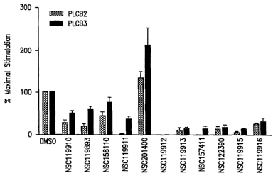
Figure 6 shows inhibition of PLC-(32 and PLC-(33
activation in the presence of exemplary compounds of the
instant invention.
Detailed Description of the Invention
The protein interaction site for G proteins has now
been appreciated. The structure of G(3y bound to SIGK was
elucidated and indicates that SIGK binds to G(3Y as an a
helix across the Ga interaction surface, in a position
occupied by an a helical region of the switch II domain of
Ga in the heterotrimer. The conformations of GRy in the
presence and absence of SIGK are very similar. Thus, the
crystal structure reveals how the peptide blocks Ga-GRY
interactions. The structure further indicates that Gp has
evolved a highly reactive and specialized surface for
interaction with diverse protein partners. This specialized
surface is referred to herein as the "protein interaction
site" or "protein interaction site of G(3' . Analysis of
various characteristics of the protein interaction site led
to the understanding that the basis for this surface as a
preferred interaction surface is not an inherent
conformational flexibility or unusually high surface
accessibility of the site, but rather the prevalence of
multiple types of potential interaction chemistries in this
single binding surface. The specific amino acid
combinations at this surface required for amino acid
sequence recognition at the protein interaction site have
also been determined. Moreover, the specific molecular
CA 02600946 2007-09-07
WO 2006/096690 PCT/US2006/008031
-8-
interactions necessary for either acceleration of
heterotrimer dissociation or inhibition of protein complex
formation have been demonstrated.
Accordingly, the present invention relates to a method
for identifying an agent that modulates (i.e., blocks or
inhibits, or activates or potentiates) at least one
activity of a G protein by contacting a G protein P subunit
with a test agent (e.g., in a high-throughput screen) and
determining whether the test agent interacts with at least
one amino acid residue of the protein interaction site of
the G protein (3 subunit. A G protein (3 subunit is intended
to include any one of the five known mammalian G protein (3
subunit isoforms (Offermanns (2003) supra) . An activity of
a G protein is intended to mean the transduction of signals
through the G protein to one or more downstream proteins
including, but not limited to, G protein-regulated inward-
rectifying potassium channel (GIRK); type I, type II, and
type IV isoforms of adenylyl cyclase; mitogen-activated
protein kinase (MAPK); phosphotidylinositol-3-kinase
(P13K); G protein receptor kinase (GRK) family members; and
other plextrinhomology (PH) domain-containing proteins
including the dynamins and the (31, P2, and R3 isoforms of
phospholipase CP (PLC (3). Modulation of G protein activity
occurs via binding of the agent to at least one amino acid
residue of the protein interaction site thereby blocking
interactions between the Gpy subunits and Ga subunit or the
G(3Y subunits and the downstream proteins described herein.
The crystal structure of G(3y1 bound to SIGK revealed
that the SIGK peptide interacts with residues of G(31 subunit
that are utilized by several Gpy binding proteins (e.g.,
downstream proteins) . For example, Lys57, Tyr59, Trp99,
MetlOl, Leu117, Tyr145, Met188, Asp246, and Trp332 of G(31
are involved in contacts with the GRK2 PH domain in the
CA 02600946 2007-09-07
WO 2006/096690 PCT/US2006/008031
-9-
crystal structure of the GR1Y2=GRK2 complex, and all of
these residues of G(31 are involved in SIGK contacts as well
(Table 1) . This is in spite of the fact that the secondary
structures of the PH domain that contact Gpl (the RH-PH
loop, the aCT region, and P4 strand) are completely
dissimilar to the purely helical SIGK peptide (Lodowski, et
al. (2003) supra). This theme is recapitulated in the
complex of G(3,, with phosducin (Ford, et al. (1998) supra)
where a common subset of GP1 residues contacts a binding
partner with different secondary structure from GRK2.
Notably, the switch II region of Gai1 forms an a-helix that
is bound in almost the same orientation as the SIGK
peptide. However, switch II of Ga;,1 has no sequence
similarity to the SIGK peptide, although it contains a
lysine (Lys210) which is oriented in almost the same
position as Lys4 of SIGK (Goubaeva, et al. (2003) supra).
TABLE 1
Gai1 Phosducin GRK2 SIGK PLC(3 AC GIRK Ca++
42
44
46
47
52
53
55 55 55 55 55
57 57 57 Lys57 57 57
59 59 59 Tyr59 59
75 75 75
76
78 78 78 78 78
80 80 80
88
89 89 89 89
91
92
96
98 98
CA 02600946 2007-09-07
WO 2006/096690 PCT/US2006/008031
-10-
99 99 99 Trp99 99 99 99
Va1100
101 101 101 MetlOl 101 101 101
117 117 117 Leu117 117 117 117
119 119 119 119
132
143 143 143
144
145 145 145 Tyr145
162
182
186 186 Asp186 186 186 186
188 188 188 Met188
204 204 204
228 223 Asp228 228 228 228 228
230 230 Asn230
246 246 246 Asp246 246 246
274
290 290
292
304
310
311
314 314
332 332 332 Trp332 332 332 332
41% 44% 44% --- 54% 67% 43% 60%
Key to column headings: Gai1, the crystal structure of the
Gail=G(31Y2 heterotrimer (Wall, et al. (1995) supra; Wall, et
al. (1998) supra); phosducin, the phosducin=G(31Y2 complex
(Gaudet, et al. (1996) supra); GRK2, the GRK2=GR1Y2 complex
(Lodowski, et al. (2003) supra) ; SIGK, the SIGK=G(31Y2
complex; PLC (3, mutational analysis of the PLC (32/3 =G(31Y2
complexes (Li, et al. (1998) supra; Ford, et al. (1998)
supra); AC, mutational analysis of the adenylyl cyclase
type I/II=G(31Y2 complex (Ford, et al. (1998) supra) ; GIRK,
mutational analysis of GP1Y2 interaction with the GIRK1/4
channels (Ford, et al. (1998) supra) ; Ca++, mutational
analysis of G(31Y2 interaction with N or P/Q type calcium
channels (Ford, et al. (1998) supra; Agler, et al. (2003)
J. Gen. Physiol. 121:495-510). Underlined residues indicate
residues important for the SIGK=G(31Y2 interaction. The last
row indicates the percentage of residues that are shared
between the target and the SIGK interfaces.
When mutational data for G(3Y targets PLC P2, adenylyl
cyclase, and GIRK and CCalB calcium channels are included
in this analysis, the footprint of SIGK upon G(3 is similar
to the footprints of these former targets (Li, et al.
CA 02600946 2007-09-07
WO 2006/096690 PCT/US2006/008031
-11-
(1998) supra; Ford, et al. (1998) supra) . Of the thirteen
residues from G(3 that encompass the protein interaction
site, nine (Lys57, Tyr59, Trp99, MetlOl, Leu117, Tyr145,
Met188, Asp246, and Trp332) are also found as contacting
residues in the Ga, GRK2, and phosducin complexes (Table
1) . These residues reflect a consensus set of residues
utilized by many G(3 binding partners. An additional three
of the thirteen residues (Aspl86, Asp228, and Asn230) are
shared amongst SIGK and two of the other protein complex
structures. One of the thirteen, VallOO, contacts SIGK
through its main chain oxygen and is not involved in
binding interactions in the other complexes. The SIGK
binding residues that are most sensitive to mutational
perturbation are also the most frequently involved in
interactions with other G(3 binding partners. SIGK was
identified from a random peptide phage display where
multiple peptides, unrelated by sequence, appeared to bind
to a common protein interaction site on G(31.
Because of the extensive overlap between the residues
of Gpl that are accessed by SIGK and those involved in the
binding of protein GpY targets, SIGK is a competitive
inhibitor of multiple GpY binding reactions. The closely
related SIRK peptide has effects on several G(3Y-dependent
pathways; it blocks G(3Y-mediated activation of PLC (32, PLC
(33 and P13K in enzyme assays, and induces ERK I/II
activation in a cell-based assay (Scott, et al. (2001)
supra; Goubaeva, et al. (2003) supra) . These effects are
sensitive to mutations of residues in SIGK that interact
with the surface of G(3, as Lys4, Ala5, Phe6, Ile8, Leu9,
and GlylO of SIGK have all been shown by alanine scanning
to be important for inhibition of PLC (32 activation by GR1Y2
(Scott, et al. (2001) supra) . In addition, Leu9 of SIGK is
important for the ability of SIGK to activate MAPK pathways
CA 02600946 2007-09-07
WO 2006/096690 PCT/US2006/008031
-12-
in cell culture (Goubaeva, et al. (2003) supra) . However,
SIRK does not block inhibition of adenylyl cyclase type I
or N-type Ca2 channel regulation, even though their
footprints are quite similar to those of Ga and PLC (32
(Scott, et al. (2001) supra) . Conversely, mutations in Gp
that abrogate SIGK binding do not equally affect
interaction with other G(3Y binding partners. For example,
mutation of Leu1l7 to alanine decreases the ability of GP1Y2
to activate adenylyl cyclase type II and PLC (33 and to bind
GRK2 and SIGK, but has no effect on GIRK1/GIRK4 potassium
channel activation, CCa1B calcium channel activation, or
PLC (32 activation (Table 1) (Li, et al. (1998) supra; Ford,
et 'al . (1998) supra) . Similarly, mutation of Trp332 of GPlY2
to alanine reduces affinity of G(31Y2 for SIGK and impairs
stimulatory activity towards adenylyl cyclase type II,
CCa1B and both PLC P2 and PLC (33, but does not affect
interaction with GRK2, activation of GIRK1/GIRK4, or
inhibition of adenylyl cyclase type I (Li, et al. (1998)
supra; Ford, et al. (1998) supra) . Both Leull7 and Trp332
of G(31Y2 form part of the Gat and Gai,1 binding sites of Gp1
(Wall, et al. (1995) supra; Lambright, et al. (1996) supra;
Wall, et al. (1998) supra) and mutation of Leu117 also
affects Gai1 association with GP1Y2 (Li, et al. (1998) supra;
Ford, et al. (1998) supra).
Unlike other peptides that block heterotrimer
formation (Ghosh, et al. (2003) supra), SIGK promotes
nucleotide exchange-independent dissociation of GR1Y2 from
Gail (Ghosh, et al. (2003) supra; Goubaeva, et al. (2003)
supra) . For example, a peptide derived from the C-terminus
of GRK2 blocks heterotrimer formation (Ghosh, et al. (2003)
supra) but does not promote Gail=G(31Y2 subunit dissociation,
even though the structure of the GRK2=G(31Y2 complex
indicates that this peptide should utilize much the same
CA 02600946 2007-09-07
WO 2006/096690 PCT/US2006/008031
-13-
surface of Gpl as SIGK (Lodowski, et al. (2003) supra) . Not
to be bound by theory, SIGK could promote heterotrimer
dissociation by either of two mechanisms. SIGK may induce
conformational changes on Gpl that propagate beyond the SIGK
binding site and disrupt other interactions between G(31 and
Gail. However, the GR1Y2=SIGK structure shows that SIGK does
not induce substantial conformational change in G(31 outside
of the SIGK binding site itself. The second mechanism
relies on the assumption that Gail can dynamically detach
from and rebind to either of two surfaces on G(3: the switch
II interaction site on the top face of Gp1r where SIGK binds
in a similar orientation, and the N-terminal interaction
surface on blade one of G(31. Transient release from Gail at
the switch II interface would allow SIGK access to GR1.
Complete release of Gccil from Gp could then occur if the
off-rate for SIGK is slower than that for dissociation of
the N-terminus of Gail. Thus the GRK2 peptide, which binds
the top surface of Gp, may dissociate too quickly to
promote dissociation of Ga. This dynamic model of GRY
interactions is biologically relevant, since many G(3Y
binding targets exhibit binding outside of the top surface
of GR and may also transiently sample alternate surfaces on
G.
The ability of the protein interaction site of GPlY2 to
recognize a range of protein ligands with diverse secondary
structures indicates that it may be an example of a
preferential protein binding site (see, e.g., Delano, et
al. (2000) Science 287:1279-1283). Preferential binding
surfaces are characterized as having high solvent
accessibility, low polarity, and a large degree of
conformational flexibility (Scott, et al. (2001) supra; Ma,
et al. (2001) Curr. Opin. Struct. Biol. 11:364-9; Bogan and
Thorn (1998) J. Mol. Biol. 280:1-9; Clackson and Wells
CA 02600946 2007-09-07
WO 2006/096690 PCT/US2006/008031
-14-
(1995) Science 267:383-6; DeLano (2002) Curr. Opin. Struct.
Biol. 12:14-20) . Moreover, preferential binding sites are
likely to contain an unusually high concentration of so-
called "hot spots", i.e., residues that, if mutated to
alanine, reduce binding energy at least ten-fold (DeLano
(2002) supra). Hot spots have been described for both
protein-protein and protein-small molecule interfaces;
often point mutations to any hot spot on a surface
completely abrogate complex formation, even when the
binding interfaces bury several hundred A2 of total surface
area (Bogan and Thorn (1998) supra; Clackson and Wells
(1995) supra; Thanos, et al. (2003) J. Am. Chem. Soc.
125:15280-1; Zhang, et al. (2003) J. Biol. Chem. 278:33097-
104) . These criteria have been used herein to evaluate the
protein interaction site of Gp1 as a protein surface that is
predisposed by its chemical composition and surface
properties to serve as a protein binding site. Of the
twelve residues in the protein interaction site of GR,
eight (Lys57, Tyr59, Leu117, Tyr145, Asp186, Met188,
Asn230, and Trp332) met the energetic criterion for a hot
spot residue. Replacement of any of these residues by
alanine resulted in a 10-fold reduction in the affinity of
GP1y2 for SIGK. It is clear that all of these residues act
as energetically important nodes that contribute favorably
to SIGK binding. The SIGK binding surface of GP1 contains
several residues that have been shown to be enriched in hot
spots (Bogan and Thorn (1998) supra). These include
tyrosine, tryptophan and arginine; bulky residues that are
capable of forming both polar and non-polar interactions.
The protein interaction site of G(3 is significantly more
populated with aromatic residues than the rest of the GR
surface. 38% of the SIGK binding surface versus 8. 5 0 of the
total non-glycine surface accessible G(3 residues is
CA 02600946 2007-09-07
WO 2006/096690 PCT/US2006/008031
-15-
composed of Phe, Tyr, His, or Trp. Therefore, the protein
interaction site of G(3 is more nonpolar; in total, 62% of
the protein interaction site of G(3 is nonpolar compared to
29% of Gp surface accessible residues. Further, asparagine
and aspartic acid, which have a moderately favorable
distribution among hot spot surfaces, account for four of
the thirteen residues in the protein interaction site of
G(.i. This combination of aromatic and charged residues
allows for accommodation of binding partners with diverse
chemical properties at the G(3 protein interaction site.
Preferential binding surfaces are expected to have high
surface accessibility (DeLano, et al. (2000) supra). To
analyze this property of the protein interaction site of
Gp, the total surface accessible area was calculated for
the Gp molecule on a residue, main chain, and side chain
basis. Most amino acids in the protein interaction site of
GR were not significantly more accessible than others of
their type in G. However, five residues showed significant
deviation from the mean: Tyr59, Trp99, MetlOl, Leu117, and
Trp332. In the case of Trp99, side chain surface
accessibility was significantly greater than the type
average; the main chain of Tyr59, Trp99, and MetlOl were
more accessible than the mean. Leull7 had significantly
higher main chain and side chain accessibility than the
mean.
Conformational flexibility or adaptability has been
cited as an important determinant of a preferential binding
surface, since such surfaces are better able to bind to
structurally unrelated protein targets (DeLano, et al.
(2000) supra). Residue flexibility can be quantified in
terms of relative positional variation in the context of
several protein complexes. Histogram analysis of the RMSD
relative to uncomplexed G(31y,_ of all G(3 residues in four
CA 02600946 2007-09-07
WO 2006/096690 PCT/US2006/008031
-16-
crystal structures (G(31Y2 = SIGK; G(3IY2 = Gail; G(31Y2=GRK2 ;
G(31Y1=phosducin) 'shows that the protein interaction site
residues of G(3 exhibit only slightly greater than average
side chain positional dispersity (1.42 A compared to 1.35
A), with the side chains of Trp99, Asp228, andTrp332 having
the largest positive deviation from the average (each
greater than 2 A.). In particular, Arg314 and Trp332 in
blade seven move more than 10 A towards the outside of the
GP1 torus to interact with phosducin. Atomic B factors also
provide a measure of conformational flexibility. In the
structure of uncomplexed GplYz, the B factors for Trp99,
VallOO, and MetlOl exceed the mean value by least one
standard deviation (Trp99 is greater than two standard
deviations from the mean) In complexes with Gail, GRK2,
phosducin, and SIGK complexes, these binding site residues
become more well-ordered with B values close to the mean
and in some cases up to one standard deviation below the
mean. Thus, the capacity of G(3 to recognize structurally
diverse binding partners does not require a high degree of
conformational flexibility for most residues in the protein
interaction site of G. Small structural adaptations in G(31
are sufficient to accommodate a range of co-evolving
binding partners. Structural and mutagenic analysis
demonstrates that the protein interaction site on Gp can be
regarded as a hot surface, co-evolved to promote tight
binding with multiple protein targets. However, the
mechanism by which G(3Y acts as a hot surface is complex.
Trp332 is the only residue which meets all four of the
criteria for a hot spot, although Tyr59 and Trp99 have
three of the four characteristics of hot spot residues that
were tested. There are other residues in the top face of Gp
that are sensitive to mutational perturbation and are
utilized in many binding partner interactions but do not
CA 02600946 2007-09-07
WO 2006/096690 PCT/US2006/008031
-17-
exhibit the characteristics of conformational flexibility,
solvent accessibility, or nonpolarity expected of hot
spots. Especially notable among this group are Lys57 and
Met188; both of these residues are energetically
significant binding determinants in G(3 as shown by
mutational analysis and comparison to known Gpy complex
structures, and yet do not meet any of the additional
statistical criteria for hot spot residues.
Accordingly, an amino acid residue of the protein
interaction site of a Gp is intended to include Lys57,
Tyr59, Trp99, Va1100, Met101, Leu117, Tyr145, Asp186,
Met188, Asp228, Asn230, Asp246, and Trp332. By way of
illustration, the location of these residues is provided in
the rat GR amino acid sequence of:
MGEMEQLKQE AEQLKKQIAD ARKACADITL AELVSGLEVV GRVQMRTRRT
LRGHLAKIYA MHWATDSKLL VSASQDGKLI VWDTYTTNKV HAIPLRSSWV
MTCAYAPSGN FVACGGLDNM CSIYSLKSRE GNVKVSRELS AHTGYLSCCR
FLDDNNIVTS SGDTTCALWD IETGQQKTVF VGHTGDCMSL AVSPDYKLFI
SGACDASAKL WDVREGTCRQ TFTGHESDIN AICFFPNGEA ICTGSDDASC
RLFDLRADQE LTAYSHESII CGITSVAFSL SGRLLFAGYD DFNCNVWDSL
KCERVGVLSG HDNRVSCLGV TADGMAVATG SWDSFLKIWN
(GENBANK Accession No. AAA62620; SEQ ID NO:3), wherein the
protein interaction site residues are underlined.
Likewise, these residues are located in the same
position in a human G(3 having the amino acid sequence of:
MSELEQLRQE AEQLRNQIRD ARKACGDSTL TQITAGLDPV GRIQMRTRRT
LRGHLAKIYA MHWGTDSRLL VSASQDGKLI IWDSYTTNKV HAIPLRSSWV
MTCAYAXSGN FVACGGLDNI CSIYSLKTRE GNVRVSRELP GHTGYLSCCR
FLDDNQIITS SGDTTCALWD IETGQQTVGF AGHSGDVMSL SLAPNGRTFV
SGACDASIKL WDVRDSMCRQ TFIGHESDIN AVAFFPNGYA FTTGSDDATC
RLFDLRADQE LLMYSHDNII CGITSVAFSR SGRLLLAGYD DFNCNIWDAM
KGDRAGVLAG HDNRVSCLGV TDDGMAVATG SWDSFLKIWN
CA 02600946 2007-09-07
WO 2006/096690 PCT/US2006/008031
-18-
(GENBANK Accession No. AAA35922; SEQ ID NO:4), wherein the
protein interaction site residues are underlined.
An agent which interacts with at least one of these
amino acid residues of the protein interaction site of G(3
can bind via various heterogeneous non-bonded interactions
including, but not limited to van der Waals contacts (e.g.,
with methionine or leucine), polar contacts (e.g., with
aspartate or asparagine), or both (e.g., with lysine,
tryptophan, or tyrosine) to contribute to the energy of
binding. In general, it is desirable that the agent
interacts with 2, 3, 4, 5, 6, 7, 8, 9, 10, 11, 12 or 13 of
the amino acid residues of the protein interaction site of
G(3 to enhance the specificity of the agent for one or more
G protein interacting proteins and therefore one or more G
protein-mediated signaling pathways.
Determining whether the agent interacts with at least
one amino acid residue of the protein interaction site of
the (3 subunit can be accomplished using various in vitro or
in vivo assays based on detecting protein-protein
interactions between the Gpy subunits and other peptides or
proteins known to interact with Gpy subunits (e.g., SIGK
peptide, Ga subunit, or downstream proteins). An exemplary
in vitro assay has been disclosed herein. This assay
consists of obtaining an isolated Gpy complex; contacting
the Gpy complex with a test agent in the presence of a
peptide that binds at least one amino acid residue of the
protein interaction site of (3 subunit, (e.g., a SIGK
peptide or SIGK peptide derivative of SEQ ID NO:1, SEQ ID
NO:2, SEQ ID NO:5, SEQ ID NO:6, SEQ ID NO:7, SEQ ID NO:8,
SEQ ID NO:9, SEQ ID NO:10, SEQ ID NO:11, SEQ ID NO:12, or
SEQ ID NO:13); and detecting the ability of the agent to
inhibit the binding of the peptide to the protein
interaction site of the (3 subunit using, for example, an
CA 02600946 2007-09-07
WO 2006/096690 PCT/US2006/008031
-19-
ELISA assay. Other phage displayed peptides identified in
the original screen (Scott, et al. (2001) supra) could also
be used.
Alternatively, an in vivo assay can be used to
determine whether a test agent interacts with at least one
amino acid residue of the protein interaction site of the P
subunit. By way of illustration, a two-hybrid assay is
contemplated where the test agent is contacted with a cell
expressing GRY subunits and a peptide such as SIGK, wherein
the (3 subunit is fused to, e.g., a DNA-binding domain and
the SIGK peptide is fused to an activation domain. When the
SIGK peptide is bound to the protein interaction site of
G(3Y, reporter protein expression is induced. If the test
agent disrupts the binding of the SIGK peptide to the
protein interaction site of G(3y, reporter protein
expression is blocked.
Additional screens such as well-established
computational screens or screens that detect the activity
of G protein-dependent downstream proteins (e.g., PLC (3
enzymatic activity) are also contemplated for use in
conjunction with the assays disclosed herein.
Test agents, also referred to herein as compounds,
which can be screened in accordance with the methods of the
present invention are generally derived from libraries of
agents or compounds. Such libraries can contain either
collections of pure agents or collections of agent
mixtures. Examples of pure agents include, but are not
limited to, proteins, polypeptides, peptides, nucleic
acids, oligonucleotides, carbohydrates, lipids, synthetic
or semi-synthetic chemicals, and purified natural products.
Examples of agent mixtures include, but are not limited to,
extracts of prokaryotic or eukaryotic cells and tissues, as
well as fermentation broths and cell or tissue culture
CA 02600946 2007-09-07
WO 2006/096690 PCT/US2006/008031
-20-
supernates. In the case of agent mixtures, the methods of
this invention are not only used to identify those crude
mixtures that possess the desired activity, but also
provide the means to monitor purification of the active
agent from the mixture for characterization and development
as a therapeutic drug. In particular, the mixture so
identified can be sequentially fractionated by methods
commonly known to those skilled in the art which can
include, but are not limited to, precipitation,
centrifugation, filtration, ultrafiltration, selective
digestion, extraction, chromatography, electrophoresis or
complex formation. Each resulting subfraction can be
assayed for the desired activity using the original assay
until a pure, biologically active agent is obtained.
Library screening can be performed as exemplified
herein or can be performed in any format that allows rapid
preparation and processing of multiple reactions. Stock
solutions of the test agents as well as assay components
are prepared manually and all subsequent pipeting,
diluting, mixing, washing, incubating, sample readout and
data collecting is done using commercially available
robotic pipeting equipment, automated work stations, and
analytical instruments for detecting the signal generated
by the assay. Examples of such detectors include, but are
not limited to, luminometers, spectrophotometers, and
fluorimeters, and devices that measure the decay of
radioisotopes.
To further evaluate the efficacy of a compound
identified using a screening method of the invention, one
of skill will appreciate that a model system of any
particular disease or disorder involving G protein
signaling can be utilized to evaluate the adsorption,
distribution, metabolism and excretion of a compound as
CA 02600946 2007-09-07
WO 2006/096690 PCT/US2006/008031
-21-
well as its potential toxicity in acute, sub-chronic and
chronic studies. For example, overexpression of (3y
inhibitors in NG108-15/D2 cells and rat primary hippocampal
neurons has been shown to block 5-opioid and cannabinoid
receptor-induced PKA Ca translocation and gene expression
by preventing (3Y activation of adenylyl cyclase (Yao, et
al. (2003) Proc. Natl. Acad. Sci. USA 100:14379-84).
Accordingly, to analyze the efficacy of a compound of the
instant invention for treating addiction, NG108-15/D2 cells
and/or rat primary hippocampal neurons are contacted with
said compound and the effect on PKA Ca translocation is
determined. Compounds which block b-opioid and cannabinoid
receptor-induced PKA Ca translocation will be useful in
treating addiction.
Efficacy of compounds of the instant invention for
preventing or treating heart failure can be analyzed in a
genetic model of murine-dilated cardiomyopathy which
involves the ablation of a muscle-restricted gene that
encodes the muscle LIM protein (MLP-/-) (Arber, et al. 1997)
Cell 88:393-403). Using this model, it has been
demonstrated that a beta-adrenergic receptor kinase 1
inhibitor, BARK-ct, which binds to (3y and blocks (3Y-
dependent activation of beta-adrenergic receptor kinase 1
activity, can enhance cardiac contractility in vivo with or
without isoproterenol (Koch, et al. (1995) Science
268:1350-3) and restore left ventricular size and function
(Rockman, eta 1. (1998) Proc. Natl. Acad. Sci. 95:7000-
7005). Similarly, compounds of the instant invention which
block (3y-dependent activation of beta-adrenergic receptor
kinase 1 activity will be useful in preventing or treating
heart failure.
The effectiveness of compounds of the instant to
prevent opioid tolerance can be analyzed in acute (Jiang,
CA 02600946 2007-09-07
WO 2006/096690 PCT/US2006/008031
-22-
et al. (1995) J. Pharmacol. Exp. Ther. 273:680-8) and
chronic (Wells, et al. (2001) J. Pharmacol. Exp. Ther.
297:597-605) dependence model systems, wherein mice are
injected intracerebroventricularly with a compound of the
instant invention and tolerance to a select opioid (e.g.,
morphine) is determined. Compounds which decrease the
amount of opioid necessary to achieve an analgesic effect
will be useful in preventing opioid tolerance.
PLC-(32 and -(33 and PI3Ky have been shown to be
involved in the chemoattractant-mediated signal
transduction pathway. Mice deficient in PI3KY lack
neutrophil production of PtdIns (3, 4, 5) P3, neutrophil
migration, and production of antibodies containing the \
chain when immunized with T cell-independent antigen
hydroxylnitrophenyl-FICOLLTM (Li, et al. (2000) Science
287:1046-1049) . Mice lacking PLC-P2 and -(33 are deficient
in Ca2 release, superoxide production, and MAC-1 up-
regulation in neutrophils (Li, et al. (2000) supra).
Further, PLC-P2 deficient mice exhibit enhanced chemotaxis
of different leukocyte populations and are sensitized to
bacteria, viruses, and immune complexes (Jiang, et al.
(1997) Proc. Natl. Acad. Sci. USA 94(15):7971-5).
Accordingly, to analyze the efficacy of a compound of the
instant invention for modulating an inflammatory response,
mice can be administered said compound and the effect on
neutrophil production of PtdIns (3, 4, 5) P3, neutrophil
migration, Ca2+ efflux, superoxide production, production of
antibodies containing the 1, chain when immunized with T
cell-independent antigen hydroxylnitrophenyl-FICOLLTM is
determined. Compounds which selectively potentiate PLC-(32
and -R3 and/or block PI3KY activation thereby inhibiting
production of PtdIns(3,4,5)P3, neutrophil migration, and
production of TI-IgI\L, will be useful in treating
CA 02600946 2007-09-07
WO 2006/096690 PCT/US2006/008031
-23-
inflammatory conditions such as arthritis, allergies,
Chrohn's Disease and the like. Compounds which selectively
block, e.g., PLC-P2 activation thereby facilitating
neutrophil migration will be useful in facilitating immune
responses to bacterial and viral infections.
Using the screening method of the present invention,
various compounds have now been identified which bind to
the protein interaction site of a Gp subunit to interfere
with or potentiate physiologically relevant protein
interactions (e.g., Ga subunit and PLC P interactions)
thereby modulating the activity of G protein signaling
pathways.
Accordingly, one embodiment of the present invention
is a compound having a structure of Formula I:
R8 R1 R
R I I /R
7 \ 3
R6 R5 R4
FORMULA I
Exemplary compounds having the structure of Formula I
which depict various substituent R groups include, but are
not limited to, the following:
H2NS~
0~
OHO
/ \ \ NH O
HO O o
OH OH HO p
NH2 0
NSC119910 NSC117079
CA 02600946 2007-09-07
WO 2006/096690 PCT/US2006/008031
-24-
H
I N+ N I
NSC402959
HO
O
H2N 0 N / \
\ I \ I (
HO \ /
0~ 0 NH2 0
NSC125910
0
/ I I \
0 I \ I
NH 0
NH / I I \
0
/ I I \
0
NSC23128
and pharmaceutically acceptable salts and complexes
thereof.
Another embodiment of the present invention is a
compound having a structure of Formula II:
CA 02600946 2007-09-07
WO 2006/096690 PCT/US2006/008031
-25-
R
i
I
R4
3
FORMULA II
Exemplary compounds having the structure of Formula II
which depict various substituent R groups include, but are
not limited to, the following:
N HN
N~O \ N\
Br
NHZ
NHZ
0
NSC30820 NSC12155
+ l} oa~N+
Nrnn Cu C
0/ C+ \0
NSC109268
NHz 0' Cu+z 0' 0' Cu+z 0'
03S I
N =N / \ / \ N=N ~
\ ( / - - - %
N
Me
S03
NSC306711
and pharmaceutically acceptable salts and complexes
thereof.
Additional exemplary compounds which bind to the
protein interaction site of GR include, but are not limited
to, the following:
CA 02600946 2007-09-07
WO 2006/096690 PCT/US2006/008031
-26-
H2N
0 0
/ \ ~ I
HO OH / \0
HO
OH 0
NSC201400 NSC19916
and pharmaceutically acceptable salts and complexes
thereof.
Exemplary compounds disclosed herein are intended to
include all enantiomers, isomers or tautomers, as well as
any derivatives of such compounds that retain the same
biological activity as the original compound.
Exemplary compounds of the present invention were
initially selected from a computational screen to identify
ligands that bind to the novel protein interaction site of
Gp. The computational screen involved using SYBYL molecular
modeling software to model the protein interaction site of
Gp as determined in the X-ray structure disclosed herein.
The computational docking screen was performed with the
National Cancer Institute 1900 compound library wherein the
compounds were tested for docking to the protein
interaction site of Gp using FLEXXTM (Tripos, Inc., St.
Louis, MO) for docking and CSCORETM (Tripos, Inc.) to
evaluate the energetics and fitness of the docked
structure. Algorithm-dependent lists of compounds,
predicted to interact with the protein interaction site of
G(3 and the structural model of the interaction, were
generated. Selected compounds were subsequently analyzed in
the phage ELISA binding assay disclosed herein to assess
whether these compounds could bind to the protein
CA 02600946 2007-09-07
WO 2006/096690 PCT/US2006/008031
-27-
interaction site of G(3 and interfere with protein
interactions at this surface. Compounds NSC201400 and
NSC119916 had IC50 values of , 100 nM and 5 M, respectively,
and the remaining compounds were found to bind in the
ELISA-based assay to Gpy with an affinity of at least 50 pM
and interfere with peptide interactions at the protein
interaction site (Figure 1) . These compounds were further
analyzed in the phage ELISA assay and found to have high
affinities for the protein interaction site of GR and
interacted with similar amino acid residues as SIGK.
TABLE 2
SIGK NSC30820 NSC12155 NSC117079 NSC23128 NSC402959 NSC109268
Lys57 Lys57 Lys57 Lys57
Tyr59 Tyr59 Tyr59 Tyr59
G1n75 G1n75
Trp99 Trp99 Trp99 Trp99
Va1100
MetlOl
Leul17 Leu117
Tyr145
Asp186
Met188
Cys204
Asp228 Asp228
Asn230 Asn230 Asn230 Asn230
Asp246 Asp246 Asp246
Thr274
Arg314 Arg314
Trp332
ICSO 100 nM 13 M 43 M 16 .M 2 M 13 M
SIGK NSC125910 NSC119910 NSC30671
CA 02600946 2007-09-07
WO 2006/096690 PCT/US2006/008031
-28-
Lys57 Tyr59 Lys57 Lys57
Tyr59 Tyr59 Tyr59
Trp99 Trp99 Trp99 Trp99
VallOO VallOO
MetlOl MetlOl
Leu117 Leu117 Leu117
Tyrl45
Aspl86
Metl88 Met188
Cys204
Asp228 Asp228
Asn230
Asp246
Thr274
Ser316
Trp332 Trp332 Trp332
ICso 68 M 100 nM 7 M
Underlined residues indicate residues important for the
SIGK=GPlY2 interaction. The last row indicates the IC50 value
for each compound.
To further illustrate the utility of these compounds,
it was demonstrated that NSC119910 blocked interactions of
Ga subunit with Gpy subunits (Figure 2) and inhibited the
ability of Gpy subunits to inhibit interactions with a
physiological target such as PLC (3 in vitro (Figure 3)
based on a decrease in the enzymatic activity of PLC P.
G(3Y-regulated activities of PI3KY and PLC-(32/-R3 are
important in chemoattractant-induced responses and
inflammation. PI3KY is involved in the production of TI-IgAL
and mice deficient in PI3KY, lack neutrophil migration (Li,
et al. (2000) Science 287:1046-9). The PLC pathway is
involved in down-modulation of chemotaxis and in
hyperinflammatory conditions (Li, et al. (2000) supra).
Therefore, it was determined whether NSC119910 could
inhibit the G(3Y/PLC interaction and block PLC activation.
Data from fura-2-based experiments demonstrated that the
abruptly occurring increase in cytosolic CaZ} in fMLP-
stimulated neutrophils, a response which is due to the
release of the cation from intracellular stores (Anderson,
CA 02600946 2007-09-07
WO 2006/096690 PCT/US2006/008031
-29-
and Mahomed (1997) Clin. Exp. Immunol. 110:132-138; Geiszt,
et al. (1997) J. Biol. Chem. 272:26471-26478), was
suppressed by 10 M NSC119910 (Figure 4) . Increases in
[Ca2+] through ATP was not significantly suppressed in the
presence of NSC119910 (Figure 4), indicating that the
effect of the compound on fMLP dependent Ca2+ increases are
specific. Further, the time taken for fluorescence to
decline to half-peak values was not substantially affected
(Figure 5) . The results indicate that NSC119910 inhibits
PLC/G-protein interactions which lead to activation of PLC
in vivo.
Opioid receptors, p, 0, and K, couple to Gi and Go
proteins through a and (3y subunits, and regulate a number
of signaling pathways. In particular, the efficacy of
opioid signal transduction in PLC-p3-deficient mice has
been shown to increase, indicating that PLC suppresses
opioid signaling by modification of opioid-dependent
signaling components (Xie, et al. (1999) Proc. Natl. Acad.
Sci. USA 96:10385-10390). Given that PLC-P3 plays a
significant role as a negative regulator of opioid
responses, it was determined whether NSC119910 could
inhibit PLC-(33 activation thereby enhancing morphine-
induced analgesia. Mice were intracerebroventricularly
injected in accordance with standard protocols (Xu, et al.
(1998) J. Pharmacol. Exp. Therapeut. 284:196-201) with 100
nmol of NSC119910 in combination with varying doses (0.1,
0.3, 1, and 3 nmol) of morphine. Mice were tested 20
minutes after the injection for an analgesic response in a
55 C warm-water tail-flick test (Wells, et al. (2001) J.
Pharmacol. Exp. Therapeut. 297:597-605) . The ED50 value for
morphine alone was 0.74 nmol, while the ED50 value for
NSC119910 plus morphine was 0.065 nmol. The differences in
the ED50 values showed an 11-fold shift to the left in a
CA 02600946 2007-09-07
WO 2006/096690 PCT/US2006/008031
-30-
morphine dose-response curve (Table 3), indicating that
when morphine was administered with NSC119910, less
morphine was required to produce a similar analgesic
effect. Accordingly, administering opioids in combination
with a compound of the instant invention would allow for
the use of a lower dose of opioid in patients thereby
reducing the development of opioid tolerance.
TABLE 3
Dose of Morphine, Percent Antinociception + S.E.M.
nmol Morphine Alone Morphine plus
NSC119910
82.4 11.9 N/A
3 68.0 14.1 100 +_ 0.0
1 55.6 8.3 79.3 +_ 9.1
0.3 41.0 10.2 64.4 + 10.0
0.1 21.0 11.1 55.3 12.7
Having demonstrated that NSC119910 effectively
modulates G-protein interactions, a series of structural
analogs of NSC119910, identified using modeling software,
were analyzed for binding to the protein interaction site
of Gp. These analogs and their corresponding affinities for
GRY were:
OH
O ~ I
HO
HO 0 O HO 0 0
NSC119888 - no binding NSC9037 - no binding
CA 02600946 2007-09-07
WO 2006/096690 PCT/US2006/008031
-31-
OH
HO 0
:::H / \ \ HO 0 0
NSC158109 - no binding NCS119892 - no binding
0 OH
OH
HO ao
HO 0 HO 0 \ O
NSC119891 - no binding NSC2608 - no binding
HO 0 0 OH
I I
HO O 0 HO \ O \ O
OH OH OH OH
NSC119911 - 200 nM NSC119915 - 2.5 M
CA 02600946 2007-09-07
WO 2006/096690 PCT/US2006/008031
-32-
0 OH
H
HO \ \ OC
I
HO / O O
0 0 OH
NSC158112 - no binding NSC5426 - no binding
HO 0
\
~ ~
HO 0 0 I
HO 0 \ 0
NSC158113 - no binding NSC119894 no binding
HO
OH 0 H
O / \ \
HO 0 0 HO 0 O
OH OH OH OH
NSC119893 - 130 nM NSC119912 - 30 M
CA 02600946 2007-09-07
WO 2006/096690 PCT/US2006/008031
-33-
Cl
Cl Cl
HO 1 Cl H
H Cl OH
0
HO 0 0
OH OH HO 0 0
OH OH
NSC119913 - 700 nM NSC158110 - 250 nM
HO
\OH
0 H
O
HO 0 0 HO 0 O
NSC157411 - 7 M NSC122390 - 14 M
From this analysis, a general structure for NSC119910
analogs was identified and is represented as Formula III.
Rl
HO O O
3 R2
FORMULA III
wherein, R,_ can be a substituted or unsubstituted alkyl,
substituted or unsubstituted alkenyl, substituted or
unsubstituted cycloalkyl, or substituted or unsubstituted
cycloalkenyl; and R2 and R3 are independently hydrogen or a
CA 02600946 2007-09-07
WO 2006/096690 PCT/US2006/008031
-34-
hydroxyl group. In particular embodiments, R2 and R3 are
both hydroxyl.
As used herein, alkyl refers to a straight or branched
hydrocarbon chain consisting solely of carbon and hydrogen
atoms, containing no saturation, having from one to eight
carbon atoms.
Alkenyl is intended to mean an aliphatic hydrocarbon
group containing at least one carbon-carbon double bond and
which may be a straight or branched chain having from 2 to
about 10 carbon atoms.
Cycloalkyl denotes a non-aromatic mono or multicyclic
ring system of about 3 to 12 carbon atoms.
As used herein, the term cycloalkenyl refers to a mono
or multicyclic ring system containing in the range of about
3 to 12 carbon atoms with at least one carbon-carbon double
bond.
Substituents in the substituted alkyl, cycloalkyl,
alkenyl or cycloalkenyl groups include, but are not limited
to, hydroxy, carboxyl, halogen (e.g., fluorine, chlorine,
bromine, or iodine), or substituted or unsubstituted alkyl.
With the exception of NSC157411 and NSC122390, analogs of
NSC119910 generally contained hydroxyl groups in the R2 and
R3 positions of Formula III, which appeared to facilitate
binding; and a carboxyl-substituted alkyl, cycloalkyl,
alkenyl or cycloalkenyl substituent at R1 of Formula III,
which appeared to modulate activity, but was not required
for binding.
Accordingly, a further embodiment of the present
invention is a compound having a structure of Formula III
and pharmaceutically acceptable salts and complexes
thereof.
The ability of NSC119910 analogs to selectively
modulate activation PLC-(32 and -(33 was analyzed. In this
CA 02600946 2007-09-07
WO 2006/096690 PCT/US2006/008031
-35-
assay, PLC-(32 and PLC-(33 were purified and PLC enzymatic
activity was measured in the presence or absence of
purified (3y and in the presence or absence of analog. The
results of this analysis indicated that NSC119911 appeared
to block PLC-(32 activation more effectively than PLC-(33
activation and NSC201400 selectively potentiated PLC-(33
activation despite blocking peptide binding to Py (Figure
6). Further, while NSC119910, NSC and analog NSC119893
block Ca2 mobilization, they do so without interfering with
fMLP-dependent ERK activation. Likewise, NSC119911,
NSC158110, and NSC201400 also do not interfere with fMLP-
dependent ERK activation.
The compounds disclosed herein as well as those found
to bind to the protein interaction site of G(3 and interfere
with protein interactions at this surface can be used in a
method for modulating (i.e., blocking or inhibiting, or
enhancing or potentiating) at least one activity of a G
protein. Such a method involves contacting a G protein
either in vitro or in vivo with an effective amount of an
agent that interacts with at least one amino acid residue
of the protein interaction site of the G protein P subunit
so that at least one activity of the G protein is
modulated. An effective amount of an agent is an amount
which reduces or increases the activity of the G protein by
10%, 20%, 30%, 40%, 50%, 60%, 70%, 80%, 90% or 100%. Such
activity can be monitored based on protein-protein
interactions or enzymatic assays detecting activity of
downstream proteins.
As will be appreciated by one of skill in the art,
modulating one or more G protein activities can be useful
in selectively analyzing G protein signaling events in
model systems as well as in preventing or treating diseases
and disorders involving G protein Ry subunit signaling. The
CA 02600946 2007-09-07
WO 2006/096690 PCT/US2006/008031
-36-
selection of the compound for use in preventing or treating
a particular disease or disorder will be dependent upon the
particular G protein-dependent downstream protein involved
in the disease or disorder. For example, a compound which
interacts with Lys57, Trp99, MetlOl, Leu117, Asp186,
Asp228, Asp246 and/or Trp332 of G(3 would be useful in
preventing or treating adenylyl cyclase-associated diseases
or disorders, whereas a compound which interacts with
Lys57, Tyr59, Trp99, MetlOl, Leu117, Tyr146, Met188,
Asp246, and/or Trp332 may be more suitable for GRK2-
associated diseases or disorders. It is contemplated that
this selectivity for specific downstream proteins may
reduce side effects associated with antagonists which
inhibit all activities associated the G protein PY subunit
signaling.
Prevention or treatment typically involves the steps
of first identifying a patient at risk of having or having
a disease or disorder involving at least one G protein (3y
subunit activity (e.g., congestive heart failure,
addiction, hyper- or hypo-inflammation, or opioid
tolerance) . Once such an individual is identified using,
for example, standard clinical practices, said individual
is administered a pharmaceutical composition containing an
effective of a selective compound disclosed herein or
identified in the screening methods of the invention. In
most cases this will be a human being, but treatment of
agricultural animals, e.g., livestock and poultry, and
companion animals, e.g., dogs, cats and horses, is
expressly covered herein. The selection of the dosage or
effective amount of a compound is that which has the
desired outcome of reducing or reversing at least one sign
or symptom of a disease or disorder involving G protein (3y
subunit signaling in a patient. For example, some of the
CA 02600946 2007-09-07
WO 2006/096690 PCT/US2006/008031
-37-
general signs or symptoms associated with congestive heart
failure include increased heart rate, increased respiratory
rate, breathing faster and deeper than normal,
breathlessness, irritability, restlessness, an unexplained
fussiness, swelling, puffiness, edema, sudden weight gain
or poor weight gain, decrease in appetite, diaphoresis,
cough, congestion or wheezing, a decrease in activity
level, fatigue, listlessness, decrease in urine output, or
pale, mottled or grayish appearance in skin color. General
signs or symptoms associated with addiction include, but
are not limited to, changes in attitude, appearance, and
relationships with others, whether at home, school or work
and other behavioral changes.
When preventing or treating an inflammatory condition,
the selective modulation of either the PLC pathway or PI3Ky
will be useful in treating different inflammatory
conditions. For example, to reduce neutrophil migration
into sites of inflammation (e.g., in arthritis) it is
desirable to administer a compound which selectively
inhibits the activation of PI3Ky thereby reducing the
injury to tissues that contribute to the pathophysiology of
the inflammatory diseases. Conversely, to facilitate an
inflammatory response, e.g., to enhance immune responses to
bacterial or viral infection, it is desirable to administer
a compound which selectively inhibits the activation of the
PLC pathway.
Pharmaceutical compositions can be in the form of
pharmaceutically acceptable salts and complexes and can be
provided in a pharmaceutically acceptable carrier and at an
appropriate dose. Such pharmaceutical compositions can be
prepared by methods and contain carriers which are well
known in the art. A generally recognized compendium of such
methods and ingredients is Remington: The Science and
CA 02600946 2007-09-07
WO 2006/096690 PCT/US2006/008031
-38-
Practice of Pharmacy, Alfonso R. Gennaro, editor, 20th ed.
Lippincott Williams & Wilkins: Philadelphia, PA, 2000. A
pharmaceutically-acceptable carrier, composition or
vehicle, such as a liquid or solid filler, diluent,
excipient, or solvent encapsulating material, is involved
in carrying or transporting the subject compound from one
organ, or portion of the body, to another organ, or portion
of the body. Each carrier must be acceptable in the sense
of being compatible with the other ingredients of the
formulation and not injurious to the patient.
Examples of materials which can serve as
pharmaceutically acceptable carriers include sugars, such
as lactose, glucose and sucrose; starches, such as corn
starch and potato starch; cellulose, and its derivatives,
such as sodium carboxymethyl cellulose, ethyl cellulose and
cellulose acetate; powdered tragacanth; malt; gelatin;
talc; excipients, such as cocoa butter and suppository
waxes; oils, such as peanut oil, cottonseed oil, safflower
oil, sesame oil, olive oil, corn oil and soybean oil;
glycols, such as propylene glycol; polyols, such as
glycerin, sorbitol, mannitol and polyethylene glycol;
esters, such as ethyl oleate and ethyl laurate; agar;
buffering agents, such as magnesium hydroxide and aluminum
hydroxide; alginic acid; pyrogen-free water; isotonic
saline; Ringer's solution; ethyl alcohol; pH buffered
solutions; polyesters, polycarbonates and/or
polyanhydrides; and other non-toxic compatible substances
employed in pharmaceutical formulations. Wetting agents,
emulsifiers and lubricants, such as sodium lauryl sulfate
and magnesium stearate, as well as coloring agents, release
agents, coating agents, sweetening, flavoring and perfuming
agents, preservatives and antioxidants can also be present
in the compositions.
CA 02600946 2007-09-07
WO 2006/096690 PCT/US2006/008031
-39-
The compositions of the present invention can be
administered parenterally (for example, by intravenous,
intraperitoneal, subcutaneous or intramuscular injection),
topically (including buccal and sublingual), orally,
intranasally, intravaginally, or rectally according to
standard medical practices.
The selected dosage level will depend upon a variety
of factors including the activity of the particular
compound of the present invention employed, the route of
administration, the time of administration, the rate of
excretion or metabolism of the particular compound being
employed, the duration of the treatment, other drugs,
compounds and/or materials used in combination with the
particular compound employed, the age, sex, weight,
condition, general health and prior medical history of the
patient being treated, and like factors well known in the
medical arts.
A physician or veterinarian having ordinary skill in
the art can readily determine and prescribe the effective
amount of the pharmaceutical composition required. For
example, the physician or veterinarian could start doses of
a compound at levels lower than that required in order to
achieve the desired therapeutic effect and gradually
increase the dosage until the desired effect is achieved.
This is considered to be within the skill of the artisan
and one can review the existing literature on a specific
compound or similar compounds to determine optimal dosing.
As will be understood by those of skill in the art
upon reading this disclosure, additional compounds to those
exemplified herein can be identified routinely in
accordance with the screening methods taught herein.
Additional compounds for screening can be selected randomly
by one skilled in the art, based upon computational
CA 02600946 2007-09-07
WO 2006/096690 PCT/US2006/008031
-40-
prediction, and/or based upon their containing a structure
of Formula I, II or III or a structure similar to that of
the exemplary compounds disclosed herein.
The invention is described in greater detail by the
following non-limiting examples.
Example 1: Materials
Peptides were purchased from Alpha Diagnostic
International (San Antonio, TX) or SIGMA -Genosys (St.
Louis, MO), HPLC purified to greater than 90% and masses
confirmed by mass spectroscopy. Ni-NTA agarose was from
QIAGEN (Valencia, CA). Streptavidin-coated poly-styrene
beads were from Spherotec (Libertyville, IL). HRP-
conjugated anti-M13 antibody was from Amersham Biosciences
(Piscataway, NJ). HRP-conjugated Neutravidin was from
Pierce (Rockford, IL) . All molecular biology reagents were
from INVITROGENTM (Carlsbad, CA) unless otherwise indicated.
Example 2: Expression and Purification of G(31ya and SIGK
Peptide
Baculoviruses harboring cDNA for wild-type bovine G(31
and N-terminally (His)6-tagged bovine GY2 were used to
produce proteins of the same. High 5 cells (INVITROGENTM,
Carlsbad, CA; 2x10⁶ cells/mL) were infected with high titer
GR1 and Gy2 baculoviruses. GP1Y2 was purified according to
standard methods (Kozaza and Gilman (1995) J. Biol. Chem.
270:1734-41), with modifications. All steps were carried
out at 4 C. Cells were harvested 60 hours post-infection by
centrifugation at 2600g, then resuspended in 50 mL of lysis
buffer (20 mM HEPES, pH 8, 150 mM NaCl, 5 mM P-ME, 1 mM
EDTA, 1 mL SIGMA@ protease inhibitor cocktail P-2714) per
liter of cell culture. Cells were lysed by sonication and
centrifuged at 2600g to pellet the membranes. Resuspension
CA 02600946 2007-09-07
WO 2006/096690 PCT/US2006/008031
-41-
and homogenization of membranes was accomplished by
douncing in 100 mL lysis buffer. The membranes were
solubilized by adding 1% Lubrol (C12E10, SIGMA@, St. Louis,
MO) with stirring and the resultant solution clarified by
ultracentrifugation at 125,000g. The supernatant was loaded
onto Ni-NTA agarose (QIAGENO, Valencia, CA) equilibrated
with lysis buffer + 1% Lubrol. The column was washed and
the Lubrol exchanged for sodium cholate using buffers Ni-A
(20 mM HEPES, pH 8, 0.4 M NaCl, 5 mM (3-ME, 0.5% Lubrol,
0.15% cholate) and Ni-B (20 mM HEPES pH 8, 0.1 M NaCl, 5 mM
(3-ME, 0.25% Lubrol, 0.3% cholate). G(31Y2 eluted in Ni-C (20
mM HEPES pH 8, 0.01 M NaCl, 5 mM P-ME, 1% cholate, 200 mM
imidazole). The eluate was loaded onto a HITRAPTM Q
(Amersham Biosciences, Piscataway, NJ) column pre-
equilibrated with QA (20 mM HEPES, pH 8, 5 mM (3-ME, 0.7%
CHAPS, 1 mM EDTA). GP1Y2 eluted in a gradient using QB (QA +
1.0 M NaCl). Fractions containing G(31Y2 were analyzed by
SDS-PAGE and pooled. Gel filtration was performed using a
tandem SEPHADEX 75:SEPHADEX 200 column (Amersham
Biosciences, Piscataway, NJ) equilibrated with buffer GF +
CHAPS (20 mM HEPES, pH 8, 150 mM NaCl, 10 mM (3-ME, 1 mM
EDTA, 0.7% CHAPS) . The purified yield was typically 1 mg
GR1Y2 per liter of cell culture.
SIGK peptide (Ser-Ile-Gly-Lys-Ala-Phe-Lys-Ile-Leu-Gly-
Tyr-Pro-Asp-Tyr-Asp; SEQ ID NO:2) was synthesized using
well-established methods. No modifications were made to the
peptide termini; purification was by reverse phase-HPLC
chromatography on a VYDAC C4 semi-preparative column.
Example 3: Crystallography
SIGK peptide was added to G(31Y2 in 1.5 molar excess,
and the G(31Y2=SIGK complex was used at 7 mg/mL for
crystallization. Crystals were grown by vapor diffusion
CA 02600946 2007-09-07
WO 2006/096690 PCT/US2006/008031
-42-
using equal volumes (2 L) of protein and reservoir
solution (15-17% PEG 4000, 100 mM HEPES, pH 7.5, 0.01-0.05
M Na-Acetate, 10% glycerol) at 20 C. Crystals attained
dimensions of 150 m x 50 m x 20 m within one week.
Crystals were cryoprotected in 15% glycerol and frozen in
liquid nitrogen.
Native crystals of GPlY2=SIGK were screened at Advanced
Light Source (ALS) beamlines 8.2.1 and 8.2.2 (Berkeley, CA)
and at the Advanced Photon Source (APS) beamline BM-19
(Chicago, IL). A dataset from ALS 8.2.2 was used to
determine the structure. Over 100 crystals were screened;
diffraction limits varied from 7A to the 2.7A dataset used
for structure determination. Diffraction data were indexed,
integrated, and scaled using the software package HKL2000
(Otwinowski and Minor (1997) In: Methods in Enzymology,
Vol. 276:307-326) (Table 4). The space-group of the
crystals was P212121.
TABLE 4
Data Collection
Space Group P212121 Unique Reflections 9729
Unit Cell Redundancy' 3.5 (1 . 8)
a (A) 45.468 Completeness (%)' 90.1 (56.2)
b 74.669 <1/0>1 13.5 (1.6)
c 108.023 Rsym 1,2 8.7 (41.4)
a ( ) 90 Mosaicity ( ) 2.3
p 90 Wilson B-factor (A) 61.8
y 90
Dmin (A) 2.7
Refinement
Resolution (A) 45.4-2.7 R.m.s Deviations
Number of atoms3 Bond lengths (A) 0.006
Protein Bond Angles ( ) 1.3
Water
R.m. s . B factors (A )
Rworx o 4 Bonded main chain 1.29
Rfree ( o) 5 Bonded side chain 18.1
Average B-factor (A)6 46.3
CA 02600946 2007-09-07
WO 2006/096690 PCT/US2006/008031
-43-
The final model contains residues 2-340 of Gpl (of 340), 7-
52 of GYa (of 68) , 1-13 of SIGK (of 15) , and 37 water
molecules.
1Numbers in parentheses correspond to the highest resolution
shell, 2.8-2.7 A.
2 Rsyn, = Eh Ei I I;, (h) - <I (h) > Eh Ei I1 (h) , where I;, (h) and
<I(h)> are the ith and mean measurement of the intensity of
reflection h, respectively.
3The final model contains residues 2-340 of GP1 (of 340), 7-
52 of GYZ (of 68) , and 1-13 of SIGK (of 15) .
4Rwork =Zh I I F. (h) ~- I F. (h) I I / Eh I F. (h) I, where Fo (h) and
F,(h) are the observed and calculated structure factors,
respectively. An I/6 cutoff was not used in the final
calculations of R-factors.
5Rfree is the R-factor obtained for a test set of reflections
consisting of a randomly selected 8% of the data.
6B-factors at the N-termini, including G(31 residues 2-41 and
GYZ residues 7-13, are greater than 80 A2.
The structure of the G(31YZ=SIGK complex was solved by
the molecular replacement method using the program PHASER
(Storoni, et al. (2004) Acta Crystallogr. D Biol.
Crystallogr. 60:432-8; Read (2001) Acta Crystallogr. D
Biol. Crystallogr. 57:1373-82). The coordinates of GPIY2 in
the G(31Y2*GRK2 complex (10MW, 100% sequence identity) were
used as the search model. After rigid body refinement using
the maximum likelihood minimization target in CNS version
1.1 (Adams, et al. (1997) Proc. Natl. Acad. Sci. USA
94:5018-23; Brunger, et al. (1998) Acta Crystallographica
Section D 54:905-921), the model was further refined by
using a combination of simulated annealing, Powell
minimization, and B factor refinement. The sigma A-weighted
2Fo-Fc electron density map computed with refined phases
revealed clear main chain density for ten residues of the
SIGK peptide along with identifiable side chain density for
several SIGK residues. Subsequent model building was
performed in O(Jones, et al. (1991) Acta Crystallographica
Section A 47:110-119) followed by simulated annealing,
energy minimization, and 3 factor refinement using CNS.
CA 02600946 2007-09-07
WO 2006/096690 PCT/US2006/008031
-44-
PROCHECK (Laskowski, et al. (1993) J. Appl. Crystallography
26:283-291) analysis indicates that all residues exhibit
main chain conformations in most favored or additional
allowed regions of cp,* space (Table 4) . Calculations of
surface accessibility, G(31Y2=SIGK contacts and RMSD between
structures were carried out using programs in the CNS
suite.
Example 4: Construction and Partial Purification of
Biotinylated G(31y2 (b-(3y) and b-py Mutants
Wild-type G(31 and G(3j_ mutants were made in the
baculovirus vector PDW464 which encodes a biotinylation
site at a lysine upstream of the amino terminus of G(31
(Goubaeva, et al. (2003) supra) . Mutants were generated by
overlap extension PCR using standard protocols. The wild-
type and mutant G(31 constructs consisted of a 20 amino acid
biotin acceptor peptide (BAP) sequence fused in-frame with
the amino-terminus of rat G(31 subunit. When coexpressed with
biotin holoenzyme synthetase (BirA) in Sf9 cells, the G(31
subunit becomes covalently biotinylated in vivo at the
specific lysine acceptor residue in the BAP. Using this
approach, 1-2 mg protein of purified protein can be
obtained per liter of Sf9 insect cells. As 45 ng of protein
is used in the phage ELISA assay, a single purification is
sufficient for 10,000 to 30,000 binding assays.
Baculoviruses were generated via the BAC-TO-BAC
system following the manufacturer's instructions
(GIBCO/BRL, Gaithersburg, MD) . Sf9 cells (200 mL) were
triply infected with 0.5 mL baculovirus encoding (His)6-
Gail, 4 mL of GYZ virus, and 4 mL of either wild-type or
mutated G(31 virus. G(31Y2 dimers were purified 60 hours post-
infection using a well-established method with
modifications as indicated (Kozasa and Gilman (1995)
CA 02600946 2007-09-07
WO 2006/096690 PCT/US2006/008031
-45-
supra) . Cell pellets were lysed in 4 mL lysis buffer (50 mM
HEPES, pH 8.0, 3 mM MgC12, 10 mM P-mercaptoethanol, 1 mM
EDTA, 100 mM NaCl, 10 M GDP, and protease inhibitors) by
four freeze-thaw cycles in liquid nitrogen. Membranes were
solubilized using 1% sodium cholate, clarified by
ultracentrifugation at 100,000g for 20 minutes, diluted
into buffer containing 0.5% lubrol, and mixed with Ni-NTA
resin. After washing thoroughly, G(31Y2 subunits were eluted
from bound Gai1 by mixing beads with buffer containing 50 mM
MgC12, 10 mM NaF, 10 pM A1C13, 1% cholate, and 5 mM
imidazole at room temperature for one hour. The
concentrations of b-(3y and b-(3y mutants were analyzed by
comparative immunoblotting and chemiluminescence. Proteins
were separated by SDS-PAGE, transferred to nitrocellulose,
and probed with HRP-neutravidin (Pierce, Rockford, IL). The
chemiluminescent signal was measured using an EPI-CHEM IITM
darkroom system (UVP Bioimaging Systems, Upland, CA).
Concentrations of eluted b-py dimers were determined by
comparing to a standard curve of fully purified 100%
biotinylated G(31Y2 from at least two separate gels.
Example 5: b-(3y Binding Assay
Phage ELISA assays used to assess peptide binding to
wild-type and mutant b-(3y were performed according to
standard methods (Smrcka and Scott (2002) Methods Enzymol.
344:557-76) . Briefly, 1 g streptavidin was immobilized in
the well of a 96-six well plate overnight at 4 C. The wells
were blocked with 100 L of 2% bovine serum albumin (BSA)
in Tris-buffered saline (TBS) for 1 hour at 4 C followed by
three washes of 1X TBS/0.5% TWEEN . Forty pL of 25 nM bG(31y2
in TBS/0.5% TWEENO was added to each well and incubated at
4 C for 1.5 hour. The wells were washed, followed by the
addition of 1x10⁶ to lxl010 phage particles and incubated at
CA 02600946 2007-09-07
WO 2006/096690 PCT/US2006/008031
-46-
4 C for 3 hours. The wells were then washed six times with
TBS/0.5% TWEENO followed by addition of 40 L of 1:5000
dilution of anti-M13 antibody (Pharmacia, Uppsala, Sweden)
and incubated at room temperature for 1 hour. The wells
were washed, followed by the addition of 40 L of (2,2'-
Azino-bis(3-ethylbenzthiazoline)-6-sulfonic acid (ABTS) and
the colorimetric reaction was monitored at 405 nm. Non-
specific binding was subtracted for each reading.
Signals obtained with partially purified b-(3Y subunits
were similar to signals obtained from fully purified b-(3y
subunits. Blocking of Gai=G(31Y2 binding was assessed by
simultaneously adding 200 pM FITC-Gai with or without SIGK
to 50 pM immobilized b-(3y and measuring the amount of FITC-
Gai bound to the beads by flow cytometry according to
standard methods (Ghosh, et al. (2003) supra; Sarvazyan, et
al. (1998) J. Biol. Chem. 273:7934-40).
Example 6: Architecture of the GP1y2=SIGK Complex
Unless indicated otherwise, amino acid residues having
the prefix "s" are indicative of SIGK residues.
Gp1 is a(3-propeller composed of seven four-stranded (3-
sheets ("blades") and an N-terminal extended helix that
interacts extensively with Gy2. Each sheet is composed of
WD-40 repeats connected by loops of variable length.
Residues 2-340 of GR1 are included in the model. B factors
throughout the core of G(31 are less than 40 Az. Residues
with B factors >60 A2 are found in three loop regions:
Lys127-Ser136 in blade two, Arg214-Met217 in blade four,
and Ser265-Ile269 in the loop connecting blades six and
seven. GY2 forms a helix with a kink made by residues Asn24-
Lys29 and a coil region beginning at residue His44. The
average B factor within the GY2 molecule is 44 A2. No
electron density was observed for the N-terminal seven
CA 02600946 2007-09-07
WO 2006/096690 PCT/US2006/008031
-47-
residues and the C-terminal sixteen residues of GY2 or the
prenyl lipid modification at the C-terminus of Gy2.
SIGK forms an a-helical structure broken by a glycine
at position 10. The C-terminal three residues form an
extended structure that stretches away from the Gpl molecule
and is supported by crystal contacts between sPro12 and
sAsp13 with Thr47 and Lys337 from a symmetry-related G(31
molecule. The B factors for the N- (sSerl, sIle2) and C-
terminal (sGly10-sAsp13) residues of SIGK are greater than
50 A2; those for all other residues are between 30-50 A.2 .
The electron density for the main chain atoms in residues
1-13 is well-defined; three of the SIGK side chains that do
not contact G(31 (sIle2, sLys7, and sAsp13) are disordered.
The peptide binds across the "top" face of G(31 and is buried
970 A2 total solvent-accessible surface area. The peptide
makes no contact with the Gy2 subunit, which is bound to the
"bottom" surface of the G(3I torus.
The SIGK contact surface on Gp1 was separated into two
regions: an acidic region on GR1 that interacts with the N-
terminus of the peptide, and a largely nonpolar region that
interacts with the C-terminus of the peptide. In total,
thirteen GP,, residues directly contact SIGK, contributed by
six of the seven blades of the (3-propeller (Table 5).
TABLE 5
G(31-Interacting SIGK-Interacting Distance Type of
Residues Residues (A) Interaction
Lys57 Cs Leu9 0 3.35 Nonpolar
CE! GlylO Ca 3.99 Nonpolar
Tyr59 OH Leu9 0 2.66 Polar
Cc Ile8 0 3.87 Nonpolar
Trp99 Ne1 Tyrll OH 2.81 Polar
C51 Leu9 C52 3.59 Nonpolar
Va1100 0 Leu9 C52 3.75 Nonpolar
MetlOl Cs Ile8 Cy2 3.46 Nonpolar
Cc Ala5 0 3.52 Nonpolar
CA 02600946 2007-09-07
WO 2006/096690 PCT/US2006/008031
-48-
CE Leu9 C52 3.54 Nonpolar
C61 Ile2 CY2 3.46 Nonpolar
Leul17 C52 Ala5 Cp 3.68 Nonpolar
C52 Leu9 C51 3.80 Nonpolar
CE!2 Ser1 0 3.19 Nonpolar
Tyr145 OH Lys4 Cy 3.45 Nonpolar
C52 AlaS Cp 3.81 Nonpolar
Asp186 052 Serl 0 3.03 Polar
Met188 Cc Ile8 C51 3.31 Nonpolar
CE Lys4 Cc 3.48 Nonpolar
Asp228 062 Lys4 N~ 3.23 Polar
Asn230 N52 Lys4 NC 2.82 Polar
Asp246 0b2 Lys4 N~ 3.05 Polar
Trp332 C~2 Ile8 0 3.12 Nonpolar
CH2 GlylO Ca 3.57 Nonpolar
The N-terminal binding surface is centered on an
electrostatic interaction in which sLys4 projects into a
negatively charged binding pocket on G(31Y2 where it forms
hydrogen-bonded or charge interactions with Asp228, Asn230,
and Asp246. A hydrogen bond between the carbonyl oxygen of
Asp228 and the main chain nitrogen of Asp246 stabilizes the
three acidic residues on Gpl. Met188 participates in van der
Waals interactions with the alkyl chain of sLys4, and
Asp186 forms a polar contact with the carbonyl oxygen of
sSer1 and also makes a hydrogen bond to the amide of
Cys204. Additionally, Tyr145 forms van der Waals
interactions with the main chain oxygen of sSerl, the sLys4
side chain, and the Cp atom of sAla5, and forms a hydrogen
bond with the nearby amide of G1y162. The side chain of
Leu117 is within van der Waals contact distances of the
side chains of sIle2 and sAla5. Together, these nine Gpl
residues form a surface that tethers SIGK to Gp1 using
charged and nonpolar interactions.
Mutational analysis of SIRK and SIGK peptides can now
be interpreted in the context of the SIGK=GR1Y2 structure
(Scott, et al. (2001) supra; Goubaeva, et al. (2003)
CA 02600946 2007-09-07
WO 2006/096690 PCT/US2006/008031
-49-
supra) . Wild-type SIRK peptide inhibits the activation of
PLC (32 by G(31Y2 with an IC50 of 5 M. Substitution of sLys4
with alanine in the SIRK peptide lowers the IC50 of the
peptide 12-fold, and mutation of sAla5 to glycine lowers
the IC50 by 13-fold. Mutation of sIle2 to alanine reduces
IC50 of the peptide by 4-fold, and mutation of sSerl to
alanine has no effect on IC50 (Scott, et al. (2001) supra) .
The SIGK=GPlY2 structure indicates that the main chain of
sSerl and the side chains of sIle2, sLys4, and sAla5
contact multiple resides on GR, thereby explaining this
mutational data.
To measure the contribution of the Gp,, residues
observed at the G(31YZ=SIGK interface to the binding energy
for the complex, two approaches were utilized. First, an
ELISA assay was used to measure binding of immobilized G(31Y2
subunits to phage displaying the SIGK sequence (Table 6).
The ELISA binding data were then correlated with IC50 values
for SIGK as a competitor of GP1Y2 association with Gai1
(Table 7) Both assays were then carried out with GP1Y2
heterodimers containing mutations in the G(31 subunit. In the
N-terminal binding surface, mutation of G(31 Asn230 to
alanine decreased the affinity of GR1Y2 for peptide 10-fold
(Table 6) . Single mutation of G(31 residues Asp186, Met188,
Tyr145, and Leu117 to alanine also resulted in GP1Y2 dimers
with drastically decreased affinity for SIGK (Table 6) . G(31
mutants in which either Asp228 or Asp246 were substituted
with alanine did not dimerize with GY2 and therefore were
not analyzed. However, a mutant in which Asp228 was
substituted with serine caused only a slight loss in
binding affinity for SIGK peptide (Table 6) . Thus, many of
the G(31 residues that create the N-terminal SIGK binding
interface contribute strongly to the energy of binding.
CA 02600946 2007-09-07
WO 2006/096690 PCT/US2006/008031
-50-
TABLE 6
G(31yz Mutation of Wild-Type
Signal (mean SD)
Lys57Ala 18.6 4.6
Tyr59Ala 24.7 15.2
His62Ala 111.2 + 11.3
Trp99Ala 66.0 7.7
MetlOlAla 32.2 + 15.5
Leu117Ala 2.1 + 2.4
Tyr145Ala 0.8 0.9
Asp186Ala 13.0 13.1
Met188Ala 2.5 + 3.7
Asn230Ala 22.4 + 4.2
Asp246Ser 66.5 7.5
Phe292Ala 109.1 + 21.4
His311Ala 94.3 + 18.9
Arg314Ala 50.2 5.0
Trp332Ala 7.1 3.7
Amino acids that contact the SIGK peptide were individually
mutated to alanine (or serine for Asp246) and binding to
peptide was assayed using a phage ELISA. Immobilized b-(3Y
was incubated with phage displaying SIGK peptide. Phage
binding was detected using an a-phage antibody; the raw
data was absorbance at 405nm. Data shown are the mean +_ SD
of triplicate determinations from three independent
experiments.
TABLE 7
Log [SIGK] M o Maximal Fa Binding ( SD)
Wild-Type Met188Ala Trp332Ala Arg314Ala
-7 100.0 0.0 100.0 0.0 100.0 0.0 100.0 0.0
-5.7 55.0 10.8 101.3 7.6 72.7 2.3 65.3 5.8
-5 45.7 16.3 80.7 7.6 57.3 7.2 47.0 11.1
-4.7 17.3 3.8 72.7 11.2 33.0 3.5 30.7 4.2
-4.4 13.3 1.5 40.0 7.1 24.7 3.5 21.0 0.0
-4.1 5.8 4.8 33.0 7.2 16.7 1.2 13.0 3.0
SIGK competition for FITC-Gail(31Y2 interactions with
representative G(31 subunit mutants. SIGK and FITC-ail were
simultaneously added to streptavidin beads coated with
CA 02600946 2007-09-07
WO 2006/096690 PCT/US2006/008031
-51-
wild-type or mutant b-(3Y protein and the amount of FITC-ail
bound to the beads was assayed by flow cytometry. Data are
shown as the mean of triplicate determinants +/- standard
deviation of a representative experiment. The experiment
was repeated two (Met188A) or three (wild-type, Arg314A,
Trp332A) times with similar results. Comparison of the two
assays over a selection of mutants that spanned the range
of SIGK binding affinities indicates that a 50 s loss of
binding translates into a five-fold increase in IC50, a 75%
loss of binding corresponds to a 10-fold increase, a 90%
loss is a 20-fold shift and a 98% loss is a 50-fold shift.
The IC50 values are as follows: wild-type=0.47 M,
Arg314A=1.5 M, Trp332A=9 M, and Metl88A=22 M.
The second area of binding involves most of the C-
terminal residues of SIGK (sAla5-sGly11), which pack
against a largely hydrophobic pocket on G(31. This pocket
extends 11A. from Trp332 on blade seven to Met188 in blade
two. Eight Gp1 residues are in direct contact with the C-
terminal surface of SIGK, and two more G(31 residues support
the residues directly involved in the SIGK interaction.
Met188, which interacts with sLys4 in the N-terminal
interface, is also within contact distance of the side
chain of sLeuB. SIGK residues sAla5, sLeuB and sLeu9 are
complimented by van der Waals interactions with Leu117,
MetlOl, Trp99, Tyr59 and the alkyl chain of Lys57. The main
chain oxygen of VallOO interacts with the side chain of
sLeu9. The indole imine of Trp99 forms a hydrogen bond with
the hydroxyl group of sTyrll and the side chain of Trp332
makes contact with the main chain oxygen of sIle8 and the
Ca of sGlylO. The side chains of Lys57 and Arg314 are
positioned on either side of Trp332 and support its
orientation in the binding site. Arg314 also forms a
hydrogen bond with Trp332, and Lys57 with the nitrogen of
G1n75, further stabilizing this interaction surface on G(31.
Data from alanine scanning of the peptide (Scott, et al.
(2001) supra; Goubaeva, et al. (2003) supra) validate these
CA 02600946 2007-09-07
WO 2006/096690 PCT/US2006/008031
-52-
structural observations. Mutation of sIle8, sLeu9 or sGly10
to alanine increases the ICso for inhibition of PLC
activation by 40-fold (5 M to 200 M), 60-fold and 12-
fold, respectively (Scott, et al. (2001) supra) . The same
mutation of sLeu9 also blocks the ability of SIRK to
enhance ERK1/2 phosphorylation in RASM cells (Goubaeva, et
al. (2003) supra) .
Mutation of amino acids in G(31 that constitute the SIGK
C-terminal binding surface caused a loss in affinity for
the SIGK peptide, although to different extents. Mutation
of Leu117, Met188, or Trp332 to alanine nearly abrogated
SIRK binding; mutants of Lys57, Tyr59, MetlOl, and Arg314
had more modest effects (Table 6 and Table 8) . The Trp99
mutation resulted in a 4-fold decrease in affinity. A
summary of all the Gp1 mutations (i.e., conversions to
alanine) presented herein and their effects on SIGK binding
affinity is listed in Table 8.
TABLE 8
Loss in Affinity for SIGK Peptide
75-100% 50-75% 25-50% 0-25% No
Effect
Lys57 MetlOl Trp99 His311 His62
Tyr59 Arg314 Asn246 Phe292
Leul17
GR1 Tyr145
Residue Asp186
Met188
Asn230
Trp332
Considering all of the data for the N-terminal and C-
terminal SIGK binding interfaces, seven of the fifteen
residues of the SIGK peptide and ten of the twelve G(3
residues tested contribute significant binding energy to
CA 02600946 2007-09-07
WO 2006/096690 PCT/US2006/008031
-53-
the interface, in good correlation with the structural
model.
The binding surface of G(31 in the G(3iy2=SIGK complex is
not significantly changed upon SIGK binding. The RMSD
between the core residues of Gpl in the GPlya=STGK complex
and that in the uncomplexed GpzYi heterodimer (1TBG (Sondek,
et al. (1996) Nature 379:369-74); Val40-Asn340, Ca only) is
0.88 A. However, the side chains of Trp99, Tyr59, Asp228,
Leu117 and MetlOl rotate to accommodate SIGK such that
atoms within these residues undergo maximum displacements
of 4.0 A, 3.6 A, 2.9 A, 2.8 A. and 2.3 A, respectively,
relative to their positions in uncomplexed Gpi. The B
factors for residues in the SIGK binding surface are close
to the overall average for the complex. However, the B
factor for Trp99 is reduced two-fold upon binding to SIGK,
as indicated by comparison of normalized B factors of the
respective structures. In this analysis, there are no large
conformational changes or disorder to order transitions in
GR upon SIGK binding. The SIGK=GPzY2 complex may be compared
to those of five G(31Y2 complexes with protein targets: the
G(31Y2=Gai1 heterotrimer (1GG2) (Wall, et al. (1995) supra;
Wall, et al. (1998) supra) and the GP1y3_= Gat/1 heterotrimer
(1G0T) (Lambright, et al. (1996) supra) , the G(3lYz=phosducin
complex (lAOR and 2TRC) (Loew, et al. (1998) supra; Gaudet,
et al. (1996) supra), and the GRxY2=GRK2 complex
(10MW) (Lodowski, et al. (2003) supra) . Superposition of the
G(31Y2=SIGK complex with each of these structures yields
average RMS deviations for Gpl residues 40-340 of less than
1.0 A (Ca only). With the exception of a few residues
involved in the GplY1=phosducin complex, the GpY heterodimer
does not undergo significant structural rearrangement in
order to bind protein targets, nor does it in the GP,,Y2=SIGK
structure.
CA 02600946 2007-09-07
WO 2006/096690 PCT/US2006/008031
-54-
Example 7: Measurement of a-py Interactions via Flow
Cytometry
Fluorescein-labeled Ga;1 (Fa;.,.) was prepared in
accordance with standard methods (Sarvazyan, et al. (1998)
5' supra) . Assays were used to determine peptide effects on
Ga-GpY interactions included competition and dissociation
assays (Ghosh, et al. (2003) supra). Briefly, for
competition-based assays, 100 pM Fail and indicated
concentrations of peptides were added to 50 pM b-G(3lY2
immobilized on 105 beads per mL and incubated at room
temperature for 30 minutes to reach equilibrium. The bead-
associated fluorescence was then recorded in a BD
Biosciences FACSCALIBURTM flow cytometer. Data was corrected
for background fluorescence and fit with a sigmoid dose
response curve using Graph Pad Prism 4. To measure
dissociation of Fail from b-GPlY2, 100 pM of Fail was
incubated with 50 pM immobilized b-G(31Y2 at room temperature
for 15-20 minutes. The fluorescence of bound Fail subunit
was measured, followed by the addition of a 200-fold excess
of unlabeled Gcxi,, or peptides and the amount of Fail
remaining bound to the beads was measured at the indicated
times.
Example 8: Molecular Recognition at the Protein Interaction
Site
Having demonstrated that the interface for SIGK
peptide binding was divided into two broad interactions; a
C-terminal binding interface, which contacts the
hydrophobic core of the peptide (amino acids 8-10, Ile-Leu-
Gly), and an N-terminal interface, which associates with
the N-terminus (Lys4 primarily) of the peptide, the
molecular basis for recognition of the peptide was
determined. Accordingly, amino acids of the common binding
CA 02600946 2007-09-07
WO 2006/096690 PCT/US2006/008031
-55-
surface of G(31 were individually alanine substituted to
determine which amino acids were most critical for the
interaction of G(31Y2 with nine different SIGK peptide
derivatives (Table 9).
TABLE 9
Phage
Name Sequence* SEQ ID NO: Group
3.14 SIGKALFILGYPDYD 5
2F LCSKAYLLLGQTC 6 I
Cl SCKRTKAQILLAPCT 7
C14 WCPPKAMTQLGIKAC 8 II
3C SCGHGLKVQSTIGACA 9
C4 SCEKRYGIEFCT 10
C5 SCEKRLGVRSCT 11 III
C8 SCARFFGTPGCT 12
C2 WCPPKLEQWYDGCA 13 IV
*Underlined residues denote the lysine residue contacting
the N-terminus, and the hydrophobic core residues.
The nine peptides were selected to represent the
different consensus groups of peptides previously
identified (See Scott et al. (2001) supra; Table 9) and to
compare binding characteristics within and between
consensus groups. Binding of phage displaying these
peptides to wild-type G(31Y2 gave ELISA signals that were
different, but fell within a similar range (25 to 100%
binding relative to phage 3.14). As disclosed herein, the
binding signal obtained in the ELISA assay was correlated
to a loss in affinity by comparing the results to behavior
of the peptide in a solution based assay. For example, a
mutant displaying an 80% loss of binding in an ELISA had a
corresponding 10-fold shift in peptide affinity in
solution. For the purposes of present disclosure, any
substitution that decreased the binding to less than 20% of
the wild-type binding was considered to be a critical
binding contact for that peptide. Data obtained from this
analysis is presented in Table 10.
CA 02600946 2007-09-07
WO 2006/096690 PCT/US2006/008031
-56-
TABLE 10
% of Wild-Type Signal
Peptide C-Terminal Interface Shared
Trp332 Lys57 Tyr59 Trp99 Leu117 MetlOl Met188
Group I
3.14 7.1 3.7 18.6 4.6 24.7 15.2 66.0 7.7 2.1 2.4 26.8 12.5 2.5 3.7
2F -1.5f3.6 -4.5 6.2 -2.0 9.5 32.6 26.3 2.1 5.5 6.0 5.1 8.5 5.2
C1 -0.3 2.5 0.1 1.6 0.3 0.7 1.2 2.7 5.2 3.1 70.5 35.3 4.0 3.4
Group II
C14 1.6 2.4 3.0 5.0 3.6 2.0 10.1 3.3 3.8 9.2 1.1 4.9 9.9 3.8
3C -0.3 1.7 3.6 7.5 8.5 6.4 -0.5 5.9 10 13.3 -4.2 5.5 -5.7}5.6
Group III
C4 1.7 3.0 -0.2 1.9 7.6 9.6 67.0 15.2 18.4 7.1 61.2 30.9 127.5f22.4
C5 3.2 3.7 5.6 5.3 73.8 16.6 39.0 3.8 28.5 5.2 47.8 18.9 97.2 14.2
C8 0.7f2.4 14.2 9.8 4.0 6.2 -0.8 2.7 24.7 12.5 23.6 8.5 122.6 26.2
Group IV
C2 4.7 6.2 -1.2 4.5 -1.7 4.8 -1.0f5.1 1.5 5.5 157.1 51.5 -0.7 2.5
% of Wild-Type Signal
Peptide N-Terminal Interface Indirect
Asn230 Asp246 Tyr145 Asp186 His311 Arg314
Group I
3.14 22.4 4.2 66.5 7.5 0.8 0.9 14.4 13.4 93.4 21.5 50.2 5.0
2F 27.6 19.1 0.6 2.5 0.1 6.5 2.4 3.5 23.6 46.0 2.4 3.9
C1 19.8 12.3 2.3 3.0 88.0 29.9 1.4 1.7 3.0 4.0 2.8 1.6
Group II
C14 60.0 26.5 6.3 13.9 1.9 5.9 3.6 1.9 6.1 3.4 3.9 7.9
3C 4.1 4.4 2.0 2.7 2.7 10.5 35.0 17.2 30.5 18.9 8.3 3.6
Group III
C4 11.5 6.0 35.8 7.4 4.4 3.8 51.3 15.0 36.5 8.3 1.4 1.0
C5 33.5 7.2 56.3 4.1 16.7 4.8 45.7 5.2 58.7 14.4 76.5 6.7
C8 74.8 14.8 60.8 14.4 17.5 7.8 124.9 29.1 51.6 13.2 20.8 11.1
Group IV
C2 0.0 3.3 5.9 5.7 4.5 8.1 267.2 40.6 11.7 8.0 1.3 3.1
Wild-type or alanine-substituted biotinylated GP1Y2 subunits
were immobilized on a streptavidin-coated 96-well plate,
followed by the addition of phage. Phage binding was
assessed as described herein. Data was corrected for non-
specific binding of phage to the plate and is represented
as a percent wild-type binding. Data shown are mean SD of
duplicate determinations from three independent
experiments.
Unexpectedly, each of the peptides utilized unique
combinations of amino acids within the SIGK binding surface
for its particular interaction. A dominant feature amongst
the peptides was a strong requirement for Trp332, within
the C-terminal interface. Lys57, Tyr59, Leu117, also within
this interface, generally contributed significantly to
CA 02600946 2007-09-07
WO 2006/096690 PCT/US2006/008031
-57-
binding the peptides, though there were cases where their
effects were not absolutely required. The remainder of the
amino acids had more variable effects on binding of each
peptide. For example, SIGK has a minimal requirement for
Trp99 while Ser-Cys-Lys-Arg-Thr-Lys-Ala-Gln-Ile-Leu-Leu-
Ala-Pro-Cys-Thr (Cl; SEQ ID NO:7) absolutely requires Trp99
for binding. The reverse is true for Tyr145 where SIGK
binding has an absolute requirement for Tyr145 and Ser-Cys-
Lys-Arg-Thr-Lys-Ala-Gln--Ile-Leu-Leu-Ala-Pro-Cys-Thr (Cl;
SEQ ID NO:7) binding is not affected by this mutation.
The N-terminus of SIGK interacts with the G(3 subunit
through two main contacts: sSerl interactions with (3Asp186
and RTyrl45 residues, and sLys4 interactions with RMetl88
through a Van der Waals interaction and (3Asn230, (3Asp246
and (3Asp228 through hydrogen bonded or charged
interactions. In the expression system utilized herein,
Asp228Ala and Asp246Ala did not dimerize with gamma and
could not be purified; however, Asp246Ser was expressed and
purified. In general, peptides in groups I, II and IV have
a substantial requirement for binding to the N-terminal
region, reflected by an almost complete loss of binding to
the Metl88Ala and Asp246Ser (except SIGK) mutants and
various requirements for Asn230.
Peptides in groups I, II and IV have a conserved motif
where a lysine is spaced three amino acids away from a
hydrophobic core motif (see Table 9) . This motif in SIGK
provides the appropriate spacing in a single alpha-helical
turn between the lysine that interacts with the N-terminal
binding surface and the Ile-Leu-Gly motif that interacts
with the C-terminus. It is believed that some of the other
peptides adopt a similar a-helical structure that may make
this spacing critical. The peptides in group III bind the
C-terminal interaction region, but lack a requirement for
CA 02600946 2007-09-07
WO 2006/096690 PCT/US2006/008031
-58-
Met188 and have minimal requirements for Asn230 and Asp246,
indicating they do not use the N-terminal binding surface
for their interaction with P.
Two amino acids that do not apparently bind directly
to SIGK were also analyzed, Arg314 and His311. Replacement
of Arg314 results in a modest decrease in SIGK binding;
however, for other peptides, Arg314 is absolutely required
indicating that they may directly interact with this amino
acid. His311 lies well outside the SIGK peptide binding
site but was mutated because of its potential involvement
in a conformation change in (3y subunits (Gaudet, et al.
(1996) supra; Loew, et al. (1998) supra). The imidazole
side chain of His311 is 13 A from the guanido nitrogen of
Arg314, the closest amino acid that apparently interacts
with any of the peptides. It is unlikely that His311 could
directly interact with amino acids from the phage display-
derived peptides. Nevertheless, mutation of His311 to
alanine affected binding of various peptides to varying
extents. Peptides whose binding was affected by His311A
also required Arg314 for binding, an effect possibly due to
an alteration in the position of Arg314.
It has been demonstrated that two peptides predicted
to bind at the Ga-G(3y interface, (3ARK-ct peptide (amino
acids 643-670) and QEHA, blocked heterotrimer formation but
could not promote heterotrimer dissociation (Ghosh, et al.
(2003) supra). The crystal structure of the GRK2 ((3ARK) -G(3y
complex reveals that the surface interacting with the PARK-
ct peptide partially overlaps with the SIGK and Ga-switch
II binding site (Lambright, et al. (1996) supra; Wall, et
al. (1995) supra; Lodowski, et al. (2003) supra). In
particular, amino acids Trp99, Trp332, and Try59 within the
hydrophobic pocket are common interaction sites in all
three structures. The SIGK peptide and a switch II have a
CA 02600946 2007-09-07
WO 2006/096690 PCT/US2006/008031
-59-
lysine residue occupying nearly identical positions ori Gp.
Although the PARK-ct peptide has a lysine residue in a
similar position, the geometry and nature of the
interaction is different. PARK interacts only with Asp228
whereas SIGK and Ga interact with Asp228, Asp246, Asn230
and Met188. Based on this difference, it was determined
whether the specific interactions of SIGK at this interface
were critical for promoting dissociation.
To examine subunit dissociation, the SCAR peptide,
another peptide derived from the phage display screen, was
used. Amino acids within the N-terminal interaction
interface, Asn230, Asp246 and Met188, contacting sLys4 of
SIGK, are not important for binding SCAR. SCAR lacks a
lysine residue with the correct positioning relative to the
hydrophobic core motif to reach the lysine-binding N-
terminal surface (Table 9) . Therefore, SCAR would not be
able to promote subunit dissociation. Both SIGK and SCAR
can compete with Ga;, for binding to GP,y2i with IC50' s of 0.5
and 1.7 M, respectively. However, unlike the SIGK peptide,
saturating concentrations of SCAR peptide could not promote
dissociation of a preformed heterotrimer. Concentrations of
up to 160 AM SCAR, (four times the saturating
concentration) did not cause dissociation. The inability of
SCAR to promote heterotrimer dissociation was not due to
its lower binding affinity since SIRK has a similar
affinity and promotes dissociation. These results indicate
that peptide binding to the N-terminal interface is
necessary for acceleration of heterotrimer dissociation.
To more directly assess the importance of peptide
binding to the N-terminal peptide binding interface, the
sLys4 residue of SIRK was mutated to alanine, eliminating
the key contact to the N-terminal binding pocket. This
peptide had a markedly lower affinity than SIRK (IC50= 60 M
CA 02600946 2007-09-07
WO 2006/096690 PCT/US2006/008031
-60-
vs 1.4 }zM) for blocking Gct-GRy interactions; however, at
high concentrations, it blocked to levels near that of
SIRK. Despite blocking Ga-Gpy interactions, SIRK(Lys4Ala)
failed to accelerate heterotrimer dissociation. The
apparent off-rate of Fcx;,, appears slower for SIRK(Lys4Ala)
relative to the intrinsic dissociation rate. This could be
because SIRK(Lys4Ala) is low affinity blocker, and is not
effective at preventing rebinding of Fa;,l. To confirm that
the low affinity of SIRK(Lys4Ala) was not responsible for
the inability to accelerate dissociation, a peptide with
comparable affinity to SIRK(Lys4Ala), SIRK(GlylOAla) (ICs0
-80 AM), was tested. This peptide has Lys4 but Ala is
substituted for Gly at position 10, thus SIRK(GlylOAla)
retains binding to the N-terminal interface but has a
reduced affinity due to decreased interactions with the C-
terminal region. SIRK(GlylOAla) blocked heterotrimer
formation at high peptide concentrations and despite having
a low affinity for GRy, could still accelerate heterotrimer
dissociation.
SIGK binds to GP1 at a region occupied by the switch II
domain of Ga subunits in the heterotrimer. The crystal
structure of the heterotrimer reveals the switch interface
(composed of switch I and switch II) of Ga buries
approximately 1,800 A of Gp through numerous contacts
(Lambright, et al. (1996) supra; Wall, et al. (1995)
supra); however, the effects of mutations of (3 subunit
amino acids at this interface on a subunit binding have not
been measured in direct binding assays near the Kd for Ga-
G(3y interactions. Switch I and switch II undergo large
conformational changes upon GTP binding and it is thought
these changes mediate heterotrimer dissociation.
GP,. subunit mutants disclosed herein were isolated from
insect cells as a complex with Gyz and hexa-histidine-tagged
CA 02600946 2007-09-07
WO 2006/096690 PCT/US2006/008031
-61-
Ga;.1 indicating that many of these contacts between the
subunits predicted from the crystal structures were not
individually critical for Ga subunit binding. To determine
which amino acids were contributing to the ability of
peptides to enhance dissociation rate constants, the
dissociation rate constant (koff) for Fail from each of the
individually substituted b-(3zy2 mutants was measured. The
intrinsic off-rate for wild-type was 0.123 s-1,
corresponding well with previous measurements (Sarvazyan,
et al. (1998) J. Biol. Chem. 273:7934-7940) . Data from all
of these mutants are shown in Table 11.
TABLE 11
Mutat ion Koff * *
Wild-Type 0.123 0.0429 min-'
Lys57Ala 0.144 0.0441 min-l
Tyr59Ala 0.181 0.0726 min-'
Trp99Ala 0.288 0.0547 min-'
MetlOlAla 0.114 0.0175 min-'
Leu117Ala 0.361 0.0258 min-'
Tyr145Ala 0.155 0.0423 m1n-1
Asp186Ala 0.160 -i- 0.0429 min-1
Met188Ala 0.122 + 0.0380 min-'
Asn230A1a 0.148 0.0488 min-'
Asp246Sert ---
Arg314Ala 0.118 + 0.0246 min-'
Trp332Ala 0.301 0.0420 min-'
**Mean -i- SD from four independent experiments.
#Statistically significant as compared to wild-type (p<0.05)
as determined by a one-way ANOVA followed by independent
linear contrasts.
tk,,ff could not be measured because significant stable
binding of F-ai was not detectable.
The results showed that of the 12 mutants tested,
Trp99Ala, Leu117Ala, and Trp332Ala were statistically
different from wild-type with relatively minor increases in
koff. On the other hand, Asp246Ser, despite being able to be
purified based on 6HisGa;, binding (although in low yield
from a large culture), was unable to stably bind F-ail in
CA 02600946 2007-09-07
WO 2006/096690 PCT/US2006/008031
-62-
the flow cytometry assay at the low concentrations used for
this assay. This indicates that interactions with Asp246
are critical for stable Ga subunit interactions, while
individual interactions in the primarily hydrophobic C-
terminal interface are not as important.
Example 9: Small Molecule Library Screen
A phage ELISA assay was used to determine whether
small molecules identified in the computational screen
could interact with the Gpy protein interaction surface.
Phage displaying the SIGK peptide were used in accordance
with established methods (Scott, et al. (2001) supra;
Smrcka and Scott (2002) supra) . The screen was based on a
reduction in the optical density (OD) of wells containing
Gpy subunits and phage. In each plate, three wells
contained positive controls for binding that included b-pY
subunits, SIGK-phage, and the appropriate amount of
vehicle. Three background wells contained no Py subunits.
As disclosed herein, biotinylated Gpy subunits were
immobilized on the surface of a 96-well plate coated with
streptavidin, phage displaying GRy-binding peptides were
subsequently added and binding in the presence and absence
of test compounds detected with an anti-phage antibody.
Example 10: Inhibition of Gpy Signaling in Neutrophils
Ca2+ fluxes were measured using two 35 mL cultures of
differentiated HL-60 neutrophil cultures (0.2 x 10⁶
cells/mL). Cells were cultured for three days with in DMSO
(1.2%), washed in HSS and resuspended in 2 mL HBSS at a
concentration of 7 X 10⁶ cells/mL. Addition of DMSO to the
growth medium induces differentiation of these cells into
morphologically and functionally mature neutrophils
(Collins, et al. (1978) Proc. Natl. Acad. Sci. USA 75:2458;
CA 02600946 2007-09-07
WO 2006/096690 PCT/US2006/008031
-63-
Collins, et al. (1979) J. Exp. Med. 149:969). Neutrophils
were preloaded with fura-2 (1 M), a fluorescent Ca2+-
sensitive indicator (Suh, et al. (1996) J. Biol. Chem.
271:32753), for 45 minutes, washed with HBSS and
resuspended in 2 mL of indicator-free HBSS. An 140 L
aliquote of cells was added to a total of 2 mL HBSS.
Fluorescence ratios were taken by dual excitation at 340 and
380 nm and emission at 510 nm. After a stable baseline was
established, either DMSO or NSC119910 was added and
incubated for 5 minutes. Subsequently, either fMLP or ATP
agonists were added to activate release of Ca2+ from
intracellular stores.
DEMANDE OU BREVET VOLUMINEUX
LA PRESENTE PARTIE DE CETTE DEMANDE OU CE BREVET COMPREND
PLUS D'UN TOME.
CECI EST LE TOME 1 DE 2
CONTENANT LES PAGES 1 A 63
NOTE : Pour les tomes additionels, veuillez contacter le Bureau canadien des
brevets
JUMBO APPLICATIONS/PATENTS
THIS SECTION OF THE APPLICATION/PATENT CONTAINS MORE THAN ONE
VOLUME
THIS IS VOLUME 1 OF 2
CONTAINING PAGES 1 TO 63
NOTE: For additional volumes, please contact the Canadian Patent Office
NOM DU FICHIER / FILE NAME:
NOTE POUR LE TOME / VOLUME NOTE: