Note: Descriptions are shown in the official language in which they were submitted.
CA 02601157 2007-09-13
WO 2006/101925 PCT/US2006/009403
BIOLOGICAL MARKERS PREDICTIVE OF ANTI-CANCER
RESPONSE TO EPIDERMAL GROWTH FACTOR RECEPTOR
KINASE INHIBITORS
BACKGROUND OF THE INVENTION
[1] The present invention is directed to methods for diagnosing and treating
cancer patients. In particular, the present invention is directed to methods
for
determining which patients will most benefit from treatment with an epidermal
growth factor receptor (EGFR) kinase iitliibitor.
[2] Cancer is a generic name for a wide range of cellular malignancies
characterized by unregulated growth, lack of differentiation, and the ability
to invade
local tissues and metastasize. These neoplastic malignancies affect, with
various
degrees of prevalence, every tissue and organ in the body.
[3] A multitude of therapeutic agents have been developed over the past few
decades for the treatment of various types of cancer. The most commonly used
types
of anticancer agents include: DNA-allcylating agents (e.g., cyclophosphamide,
ifosfamide), antimetabolites (e.g., methotrexate, a folate antagonist, and 5-
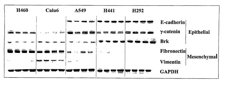
fluorouracil, a pyrimidine antagonist), microtubule disrupters (e.g.,
vincristine,
vinblastine, paclitaxel), DNA intercalators (e.g., doxorubicin, daunomycin,
cisplatin),
and hormone therapy (e.g., tamoxifen, flutamide).
[4] The epidermal growth factor receptor (EGFR) family comprises four closely
related receptors (HER1/EGFR, HER2, HER3 and HER4) involved in cellular
responses such as differentiation and proliferation. Over-expression of the
EGFR
kinase, or its ligand TGF-alpha, is frequently associated with many cancers,
including
breast, lung, colorectal, ovarian, renal cell, bladder, head and neck cancers,
__----
glioblastomas, and astrocytomas, and is believed to contribute to the
malignant
growth of these tumors. A specific deletion-mutation in the EGFR gene
(EGFRvIII)
-1-
CA 02601157 2007-09-13
WO 2006/101925 PCT/US2006/009403
has also been found to increase cellular tumorigenicity. Activation of EGFR
stimulated signaling pathways promote multiple processes that are potentially
cancer-
promoting, e.g. proliferation, angiogenesis, cell motility and invasion,
decreased
apoptosis and induction of drug resistance. Increased HER1/EGFR expression is
frequently linlced to advanced disease, metastases and poor prognosis. For
example, in
NSCLC and gastric cancer, increased HER1/EGFR expression has been shown to
correlate with a high metastatic rate, poor tumor differentiation and
increased tumor
proliferation.
[5] Mutations which activate the receptor's intrinsic protein tyrosine kinase
activity and/or increase downstream signaling have been observed in NSCLC and
glioblastoma. However the role of mutations as a principle mechanism in
conferring
sensitivity to EGF receptor inhibitors, for example erlotinib (TARCEVATM) or
gefitinib (IRESSATM), has been controversial. Recently, a mutant form of the
full
length EGF receptor has been reported to predict responsiveness to the EGF
receptor
tyrosine kinase inhibitor gefitinib (Paez, J. G. et al. (2004) Science
304:1497-1500; Lynch,
T. J. et al. (2004) N. Engl. J. Med. 350:2129-2139). Cell culture studies have
shown that
cell lines which express the nlutant form of the EGF receptor (i.e. H3255)
were more
sensitive to growth inhibition by the EGF receptor tyrosine kinase inliibitor
gefitinib,
and that much higher concentrations of gefitinib was required to inhibit the
tumor cell
lines expressing wild type EGF receptor. These observations suggests that
specific
mutant forms of the EGF receptor may reflect a greater sensitivity to EGF
receptor
inhibitors, but do not identify a completely non-responsive phenotype.
[6] The development for use as anti-tumor agents of compounds that directly
iiihibit the kinase activity of the EGFR, as well as antibodies that reduce
EGFR kinase
activity by blocking EGFR activation, are areas of intense research effort (de
Bono
J.S. and Rowinsky, E.K. (2002) Trends in Mol. Medicine 8:S19-S26; Dancey, J.
and
Sausville, E.A. (2003) Nature Rev. Drug Discovery 2:92-313). Several studies
have
demonstrated, disclosed, or suggested that some EGFR kinase inhibitors might
improve tumor cell or neoplasia killing when used in combination with certain
other
anti-cancer or chemotherapeutic agents or treatments (e.g. Herbst, R.S. et al.
(2001)
Expert Opin. Biol. Ther. 1:719-732; Solomon, B. et al (2003) Int. J. Radiat.
Oncol.
Biol. Phys. 55:713-723; Krishnan, S. et al. (2003) Frontiers in Bioscience 8,
el-13;
-2-
CA 02601157 2007-09-13
WO 2006/101925 PCT/US2006/009403
Grunwald, V. and Hidalgo, M. (2003) J. Nat. Cancer Inst. 95:851-867; Seymour
L.
(2003) Current Opin. Investig. Drugs 4(6):658-666; Khalil, M.Y. et al. (2003)
Expert
Rev. Anticancer Ther.3:367-380; Bulgaru, A.M. et al. (2003) Expert Rev.
Anticancer
Ther.3:269-279; Dancey, J. and Sausville, E.A. (2003) Nature Rev. Drug
Discovery
2:92-313; Ciardiello, F. et al. (2000) Clin. Cancer Res. 6:2053-2063; and
Patent
Publication No: US 2003/0157104).
[7] Erlotinib (e.g. erlotinib HCI, also known as TARCEVATM or OSI-774) is an
orally available inllibitor of EGFR kinase. In vitro, erlotinib has
demonstrated
substantial inhibitory activity against EGFR kinase in a number of huinan
tumor cell
lines, including colorectal and breast cancer (Moyer J.D. et al. (1997) Cancer
Res.
57:4838), and preclinical evaluation has demonstrated activity against a
number of
EGFR-expressing human tumor xenografts (Pollack, V.A. et al (1999) J.
Pharnlacol.
Exp. Ther. 291:739). More recently, erlotinib has demonstrated promising
activity in
phase I and II trials in a number of indications, including head and neck
cancer
(Soulieres, D., et al. (2004) J. Clin. Oncol. 22:77), NSCLC (Perez-Soler R, et
al.
(2001) Proc. Am. Soc. Clin. Oncol. 20:310a, abstract 1235), CRC (Oza, M., et
al.
(2003) Proc. Am. Soc. Clin. Oncol. 22:196a, abstract 785) and MBC (Winer, E.,
et al.
(2002) Breast Cancer Res. Treat. 76:5115a, abstract 445). In a phase III
trial, erlotinib
monotherapy significantly prolonged survival, delayed disease progression and
delayed worsening of lung cancer-related symptoms in patients with advanced,
treatment-refractory NSCLC (Shepherd, F. et al. (2004) J. Clin. Oncology,
22:14S
(July 15 Supplement), Abstract 7022). While most of the clinical trial data
for
erlotinib relate to its use in NSCLC, preliminary results from phase I/II
studies have
demonstrated promising activity for erlotinib and capecitabine/erlotinib
coinbination
therapy in patients with wide range of human solid tumor types, including CRC
(Oza,
M., et al. (2003) Proc. Am. Soc. Clin. Oncol. 22:196a, abstract 785) and MBC
(Jones,
R.J., et al. (2003) Proc. Am. Soc. Clin. Oncol. 22:45a, abstract 180). In
November
2004 the U.S. Food and Drug Administration (FDA) approved TARCEVATM for the
treatment of patients with locally advanced or metastatic non-small cell lung
cancer
(NSCLC) after failure of at least one prior chemotherapy regimen. TARCEVATM is
the only drug in the epidermal growth factor receptor (EGFR) class to
demonstrate in
a Phase III clinical trial an increase in survival in advanced NSCLC patients.
-3-
CA 02601157 2007-09-13
WO 2006/101925 PCT/US2006/009403
[8] An anti-neoplastic drug would ideally kill cancer cells selectively, with
a wide
therapeutic index relative to its toxicity towards non-malignant cells. It
would also
retain its efficacy against malignant cells, even after prolonged exposure to
the drug.
Unfortunately, none of the current chemotherapies possess such an ideal
profile.
Instead, most possess very narrow therapeutic indexes. Furthermore, cancerous
cells
exposed to sliglltly sub-lethal concentrations of a chemotherapeutic agent
will very
often develop resistance to such an agent, and quite often cross-resistance to
several
other antineoplastic agents as well. Additionally, for any given cancer type
one
frequently cannot predict which patient is likely to respond to a particular
treatment,
even with newer gene-targeted therapies, such as EGFR kinase inhibitors, thus
necessitating considerable trial and error, often at considerable risk and
discomfort to
the patient, in order to find the most effective therapy.
[9] Thus, there is a need for more efficacious treatment for neoplasia and
other
proliferative disorders, and for more effective means for determining which
tumors
will respond to which treatment. Strategies for enhancing the therapeutic
efficacy of
existing drugs have involved changes in the schedule for their administration,
and also
their use in combination with other anticancer or biochemical modulating
agents.
Coinbination therapy is well known as a method that can result in greater
efficacy and
diminished side effects relative to the use of the therapeutically relevant
dose of each
agent alone. In some cases, the efficacy of the drug combination is additive
(the
efficacy of the combination is approximately equal to the sum of the effects
of each
drug alone), but in other cases the effect is synergistic (the efficacy of the
combination is greater than the sum of the effects of each drug given alone).
[10] Target-specific therapeutic approaches, such as erlotinib, are generally
associated with reduced toxicity compared with conventional cytotoxic agents,
and
therefore lend themselves to use in combination regimens. Promising results
have
been observed in phase UII studies of erlotinib in combination with
bevacizumab
(Mininberg, E.D., et al. (2003) Proc. Am. Soc. Clin. Oncol. 22:627a, abstract
2521)
and gemcitabine (Dragovich, T., (2003) Proc. Am. Soc. Clin. Oncol. 22:223a,
abstract
895). Recent data in NSCLC phase III trials have shown that first-line
erlotinib or
--------- ----
gefitinib in combination with standard chemotherapy did not improve survival
(Gatzemeier, U., (2004) Proc. Am. Soc. Clin. Oncol. 23:617 (Abstract 7010);
Herbst,
-4-
CA 02601157 2007-09-13
WO 2006/101925 PCT/US2006/009403
R.S., (2004) Proc. Am. Soc. Clin. Oncol. 23:617 (Abstract 7011); Giaccone, G.,
et al.
(2004) J. Clin. Oncol. 22:777; Herbst, R., et al. (2004) J. Clin. Oncol.
22:785).
However, pancreatic cancer phase III trials have shown that first-line
erlotinib in
coinbination with gemcitabine did improve survival (OSI
Pharmaceuticals/Genentech/
Roche Pharmaceuticals Press Release, 9/20/04).
[11] Several groups have investigated potential biomarkers to predict a
patient's
response to EGFR inhibitors (see for example, PCT publications: WO
2004/063709,
WO 2005/017493, WO 2004/111273, WO 2004/071572, WO 2005/117553 and WO
2005/070020; and US published patent applications: US 2005/0019785, and US
2004/0132097). However, no diagnostic or prognostic tests have yet emerged
that can
guide practicing physicians in the treatment of their patients witll EGFR
kinase
inhibitors.
[12] During most cancer metastases, an important change occurs in a tumor cell
known as the epithelial-mesenchymal transition (EMT) (Thiery, J.P. (2002) Nat.
Rev.
Cancer 2:442-454; Savagner, P. (2001) Bioessays 23:912-923; Kang Y. and
Massague, J. (2004) Cell 118:277-279; Julien-Grille, S., et al. Cancer
Research
63:2172-2178; Bates, R.C. et al. (2003) Current Biology 13:1721-1727; Lu Z.,
et al.
(2003) Cancer Cell. 4(6):499-515)). Epithelial cells, which are bound together
tightly
and exhibit polarity, give rise to mesenchymal cells, which are held together
more
loosely, exhibit a loss of polarity, and have the ability to travel. These
mesenchymal
cells can spread into tissues surrounding the original tumor, as well as
separate from
the tumor, invade blood and lymph vessels, and travel to new locations where
they
divide and fonn additional tumors. EMT does not occur in healthy cells except
during embryogenesis. Under normal circumstances TGF-0 acts as a growth
inhibitor. However it is believed that during cancer metastasis, TGF-(3 begins
to
promote EMT.
[13] Thus, there remains a critical need for improved methods for determining
the
----bestmode-~oftreatment-for any-given-cancer-patient and-for-the--
incorporation-of-such------------
-5-
CA 02601157 2007-09-13
WO 2006/101925 PCT/US2006/009403
determinations into more effective treatment regimens for cancer patients,
whether
such inhibitors are used as single agents or combined with other anti-cancer
agents.
SUMMARY OF THE INVENTION
[14] The present invention provides diagnostic and prognostic metllods for
predicting the effectiveness of treatment of a cancer patient with an EGFR
kinase
inhibitor. Based on the surprising discovery that the sensitivity of tumor
cell growth
to iiihibition by EGFR kinase inhibitors is dependent on whether such tumor
cells
have undergone an EMT, methods have been devised for deterinining epitlielial
and/or mesenchymal biomarkers to predict the sensitivity of tumor cells to
EGFR
kinase inhibitors.
[15] Accordingly, the present invention provides a method of predicting the
sensitivity of tumor cell growth to inhibition by an EGFR kinase inhibitor,
comprising: assessing the level of an epithelial biomarker expressed by a
tumor cell;
and predicting the sensitivity of tumor cell growth to inhibition by an EGFR
kinase
ii-Alibitor, wherein high expression levels of tumor cell epithelial
biomarkers correlate
with high sensitivity to inhibition by EGFR kinase iiihibitors.
[16] The present invention also provides a method of predicting the
sensitivity of
tumor cell growth to inhibition by an EGFR kinase inhibitor, comprising:
assessing
the level of a mesenchymal biomarlcer expressed by a tumor cell; and
predicting the
sensitivity of tumor cell growth to inhibition by an EGFR kinase inhibitor,
wherein
high expression levels of tumor cell mesenchymal biomarkers correlate with low
sensitivity to inhibition by EGFR kinase inhibitors.
[17] Improved methods for treating cancer patients with EGFR kinase inhibitors
that incorporate the above metllodology are also provided. Thus, the present
invention further provides a method for treating tumors or tumor metastases in
a
patient, conlprising the steps of diagnosing a patient's likely responsiveness
to an
EGFR kinase inhibitor by assessing whether the tumor cells have undergone an
epithelial-mesenchymal transition, and administering to said patient a
therapeutically
effective amount of an EGFR kinase inhibitor.
-6-
CA 02601157 2007-09-13
WO 2006/101925 PCT/US2006/009403
[18] Additionally, methods are provided for the identification of new
epithelial or
mesenchymal biomarlcers that are predictive of responsiveness of tumors to
EGFR
kinase inhibitors.
[19] Thus, for example, the present invention further provides a method of
identifying an epithelial bioinarlcer that is diagnostic for more effective
treatment of a
neoplastic condition with an EGFR lcinase inhibitor, comprising: measuring the
level
of a candidate epithelial biomarker in neoplastic cell-containing samples from
patients
with a neoplastic condition, and identifying a correlation between the level
of said
candidate epithelial biomarker in the sample from the patient with the
effectiveness of
treatment of the neoplastic condition with an EGFR kinase inhibitor, wherein a
correlation of high levels of the epithelial biomarker with more effective
treatment of
the neoplastic condition with an EGFR kinase inhibitor indicates that said
epithelial
biomarker is diagnostic for more effective treatment of the neoplastic
condition with
an EGFR kinase inhibitor.
[20] The present invention further provides a method of identifying a
mesenchymal
biomarker that is diagnostic for less effective treatment of a neoplastic
condition with
an EGFR kinase inhibitor, comprising: (a) measuring the level of a candidate
mesenchymal biomarker in neoplastic cell-containing samples from patients with
a
neoplastic condition, and (b) identifying a correlation between the level of
said
candidate mesenchymal biomarker in the sample from the patient with the
effectiveness of treatment of the neoplastic condition with an EGFR kinase
inhibitor,
wherein a correlation of high levels of the mesenchymal biomarlcer with less
effective
treatment of the neoplastic condition with an EGFR kinase inhibitor indicates
that said
mesenchylnal biomarker is diagnostic for less effective treatment of the
neoplastic
condition with an EGFR kinase inhibitor.
[21] Furthermore, methods for the identification of agents that restore the
sensitivity of tumor cells that have undergone EMT to inhibition by EGFR
kinase
inhibitors are also provided. Thus, for example, the present invention
provides a _
method for the identification of an agent that enhances sensitivity of the
growth of a
tumor cell to an EGFR kinase inhibitor, sa.id tumor cell having being
characterized as
-7-
CA 02601157 2007-09-13
WO 2006/101925 PCT/US2006/009403
one that has previously undergone an epithelial-mesenchymal transition,
comprising
contacting a sample of said tumor cells with an EGFR kinase inhibitor,
contacting an
identical sample of said tuinor cells with an EGFR lcinase inhibitor in the
presence of
a test agent, comparing the EGFR kinase inhibitor-mediated growth inhibition
in the
presence and absence of the test agent, and detennining whether the test agent
is an
agent that enhances sensitivity of the growth of the tumor cell to an EGFR
kinase
inhibitor.
BRIEF DESCRIPTION OF THE FIGURES
[22] Figure 1: In vivo activity of erlotinib against NSCLC xenografts.
[23] Figure 2: A. Proteomic profiling of NSCLC lines, sensitive or relatively
insensitive to EGFR kinase inhibition in vitro, sliowed markedly increased LC-
MS/MS detection of vimentin and fibronectin peptides in cell lines relatively
insensitive to erlotinib. B. NSCLC lines sensitive to EGF receptor inhibition
express
elevated levels of E-cadllerin, with trends observed for y- and a-catenins. E-
cadherin
immunoblots were performed with two distinct antibodies with similar results
(data
not shown). NSCLC lines relatively insensitive to growth inhibition by
erlotinib
expressed the mesenchymal proteins vimentin and/or fibronectin. No
relationship
between total EGF receptor protein expression and sensitivity was observed,
though
all lines tested expressed detectable EGF receptor. C. Confocal microscopy of
NSCLC lines sensitive to growth inhibition by erlotinib, H292 and H441,
showing
membrane expression of E-cadherin, but not in the cell lines Calu6 and H1703
that
are relatively insensitive to erlotinib. Conversely, the relatively
insensitive lines Calu6
and H1703 expressed intermediate filainent staining for vimentin, while the
erlotinib
sensitive lines H292 and H441 did not.
[24] Figure 3: NSCLC lines were grown as subcutaneous xenografts in SCID mice
to a volume of -500mm3, excised and flash frozen in liquid nitrogen (4 animals
per
cell line). Tumor tissue was pulverized while frozen, subjected to detergent
lysis and
SDS-PAGE as described and iminunoblots probed with antibodies to E-cadherin, y-
- ------
catenin, Brk, fibronectin, vimentin, and GAPDH. Consistent with in vitro
results, E-
-8-
CA 02601157 2007-09-13
WO 2006/101925 PCT/US2006/009403
cadherin expression was restricted to erlotinib sensitive lines and
fibronectin to
relatively insensitive lines.
[25] Figure 4: Immunoblot showing higher Brlc expression levels in NSCLC cell
lines that are most sensitive to EGFR kinase inhibition.
[26] Figure 5: A) Pancreatic cell lines sensitive to EGF receptor inhibition
express
elevated levels of the epithelial cell junction proteins E-cadherin and y-
catenin. The
mesenchymal marker vimentin was most abundant in the insensitive PANC1 cells.
B)
Confocal microscopy of a pancreatic cell line sensitive to growth inhibition
by
erlotinib, BxPC3, showing membrane expression of E-cadherin, but not in the
cell
line MiaPaca2, that is relatively insensitive to erlotinib. Conversely, the
relatively
insensitive line MiaPaca2 expressed interinediate filament staining for
vimentin,
while the erlotinib sensitive line BxPC3 did not.
[27] Figure 6a: Kaplan-Meier curve illustrating time to disease progression
(TTP)
is longer for patients receiving erlotinib in combination with chemotherapy
compared
to patients receiving chemotherapy only whose tumors with E-cadherin staining
intensity of >=2. Figure 6b: Kaplan-Meier curve illustrating time to disease
progression (TTP) is not extended for patients having tumor E-cadherin
staining
intensity of <=1 who are treated with erlotinib in combination with
chemotherapy
compared to patients receiving chemotherapy alone.
DETAILED DESCRIPTION OF THE INVENTION
[28] The term "cancer" in an animal refers to the presence of cells possessing
characteristics typical of cancer-causing cells, such as uncontrolled
proliferation,
immortality, metastatic potential, rapid growth and proliferation rate, and
certain
characteristic morphological features. Often, cancer cells will be in the form
of a
tumor, but such cells may exist alone within an animal, or may circulate in
the blood
stream as independent cells, such as leukemic cells.
-9-
CA 02601157 2007-09-13
WO 2006/101925 PCT/US2006/009403
[29] "Abnonnal cell growth", as used herein, unless otherwise indicated,
refers to
cell growth that is independent of normal regulatory mechanisms (e.g., loss of
contact
inhibition). This includes the abnormal growth of: (1) tumor cells (tuinors)
that
proliferate by expressing a mutated tyrosine kinase or overexpression of a
receptor
tyrosine kinase; (2) benign and malignant cells of other proliferative
diseases in which
aberrant tyrosine kinase activation occurs; (4) any tumors that proliferate by
receptor
tyrosine kinases; (5) any tumors that proliferate by aberrant serine/threonine
kinase
activation; and (6) benign and malignant cells of other proliferative diseases
in which
aberrant serine/threonine kinase activation occurs.
[30] The term "treating" as used herein, unless otherwise indicated, means
reversing, alleviating, inhibiting the progress of, or preventing, either
partially or
completely, the growth of tumors, tumor metastases, or other cancer-causing or
neoplastic cells in a patient. The term "treatinent" as used herein, unless
otherwise
indicated, refers to the act of treating.
[31] The phrase "a method of treating" or its equivalent, when applied to, for
example, cancer refers to a procedure or course of action that is designed to
reduce or
eliminate the number of cancer cells in an animal, or to alleviate the
symptoms of a
cancer. "A method of treating" cancer or another proliferative disorder does
not
necessarily mean that the cancer cells or other disorder will, in fact, be
eliminated,
that the number of cells or disorder will, in fact, be reduced, or that the
symptoms of a
cancer or other disorder will, in fact, be alleviated. Often, a method of
treating cancer
will be performed even with a low likelihood of success, but which, given the
medical
history and estimated survival expectancy of an animal, is nevertheless deemed
an
overall beneficial course of action.
[32] The term "therapeutically effective agent" means a composition that will
elicit
the biological or medical response of a tissue, system, animal or human that
is being
sought by the researcher, veterinarian, medical doctor or other clinician.
_____[33] The term "therapeutically effective amount" or "effective amount"
means the
amount of the subject compound or combination that will elicit the biological
or
-10-
CA 02601157 2007-09-13
WO 2006/101925 PCT/US2006/009403
medical response of a tissue, system, animal or human that is being sought by
the
researcher, veterinarian, medical doctor or other clinician.
[34] The data presented in the Examples herein below demonstrate that tumor
cells,
such as NSCLC or pancreatic cancer cells, containing wild type EGFR, grown
either
in cell culture or in vivo, show a range of sensitivities to inhibition by
EGFR kinase
inhibitors, dependent on whether they have undergone an epithelial to
mesenchymal
transition (EMT). Prior to EMT, tumor cells are very sensitive to inhibition
by EGFR
kinase inliibitors such as erlotinib HCl (TARCEVATM), whereas tumor cells
which
have undergone an EMT are substantially less sensitive to inlzibition by such
compounds. The data indicates that the EMT may be a "general biological
switch"
that determines the level of sensitivity of tumors to EGFR kinase inhibitors.
It is
demonstrated that the level of sensitivity of tumors to EGFR kinase inhibitors
can be
assessed by determining the level of biomarkers expressed by a tumor cell that
are
characteristic for cells either prior to or subsequent to an EMT event. For
example,
high levels of tumor cell expression of epithelial biomarkers such as E-
cadherin,
indicative of a cell that has not yet undergone an EMT, correlate with high
sensitivity
to EGFR kinase inhibitors. Conversely, high levels of tumor cell expression of
mesenchymal biomarkers such as vimentin or fibronectin, indicative of a cell
that has
undergone an EMT, correlate with low sensitivity to EGFR kinase inhibitors.
Thus,
these observations can form the basis of valuable new diagnostic methods for
predicting the effects of EGFR kinase inhibitors on tumor growth, and give
oncologists an additional tool to assist them in choosing the most appropriate
treatinent for their patients.
[35] Accordingly, the present invention provides a method of predicting the
sensitivity of tumor cell growth to iiihibition by an EGFR kinase inhibitor,
comprising: assessing the level of an epithelial biomarker expressed by a
tumor cell;
and predicting the sensitivity of tumor cell growth to inhibition by an EGFR
kinase
inhibitor, wlierein high expression levels of tumor cell epithelial biomarkers
correlate
with high sensitivity to inhibition by EGFR kinase inhibitors. Preferred
examples of
epithelial biomarkers include E-cadherin and Brk (i.e. PTK-6) (see Table 1).
Additional examples of epithelial biomarkers that can be utilized in the
method of this
invention include y-catenin (i.e. junction plakoglobin), a-catenin (i.e. al,
a2, or 0
- 11 -
CA 02601157 2007-09-13
WO 2006/101925 PCT/US2006/009403
catenin), keratin 8, keratin 18, connexin 31, plakophilin 3, stratafin 1,
laminin alpha-5
and ST14 (see Table 1).
[36] The present invention also provides a method of predicting the
sensitivity of
tumor cell growth to inhibition by an EGFR kinase inhibitor, comprising:
assessing
the level of a mesencliymal biomarker expressed by a tumor cell; and
predicting the
sensitivity of tumor cell growth to inhibition by an EGFR kinase inhibitor,
wherein
high expression levels of tumor cell mesenchymal biomarlcers correlate with
low
sensitivity to inhibition by EGFR lcinase inhibitors. Preferred examples of
mesenchymal biomarlcers include vimentin and fibronectin (see Table 1).
Additional
examples of mesenchymal biomarlcers that can be utilized in the method of this
invention include fibrillin-1, fibrillin-2, collagen alpha-2(IV), collagen
alpha-2(V),
LOXL1, nidogen, C11 orfl9, tenascin, N-cadherin, and embryonal EDB+
fibronectin,
tubulin alpha-3 and epimorphin (see Table 1).
[37] In the practice of this invention, with preferred epithelial biomarkers,
the level
of expression in tumor cells that are sensitive to EGFR kinase inhibitors will
generally
be at such a high level that the biomarker will be very readily detectable,
using for
example a specific anti-biomarker antibody for detection. Witli preferred
epithelial
biomarlcers, the level of expression in tumor cells that are relatively
insensitive to
EGFR kinase inhibitors will generally be at such a low level that the
biomarker will
be barely detectable, if at all, using similar procedures (e.g. in the data
presented in
the Examples herein below, compare E-cadherin levels between sensitive and
relatively insensitive tumor cells in Figures 2B, 3 and 5).
[38] However, for other less preferred epithelial biomarkers, the level of
biomarker
expression in tumor cells that are relatively insensitive to EGFR kinase
inhibitors may
be readily detectable, but nevertheless will be at a substantially lower level
of
expression than in tumor cells that are sensitive to EGFR kinase inhibitors
(e.g., in the
data presented in the Examples herein below, compare a-catenin levels for the
relatively insensitive tumor cells H1703 or SW1573 wit11 the sensitive tumor
cells
H441, H358, H322 and H292 in Figure 2B).
-12-
CA 02601157 2007-09-13
WO 2006/101925 PCT/US2006/009403
[39] Similarly, in the practice of this invention, with preferred mesenchymal
biomarlcers, the level of expression in tumor cells that are relatively
insensitive to
EGFR kinase inhibitors will generally be at such a high level that the
biomarker will
be very readily detectable, using for example a specific anti-biomarlcer
antibody for
detection. With preferred mesenchyinal biomarkers, the level of expression in
tumor
cells that are relatively sensitive to EGFR kinase inhibitors will generally
be at such a
low level that the biomarlcer will be barely detectable, if at all, using
similar
procedures (e.g. in the data presented in the Examples herein below, compare
fibronectin or vimentin levels between sensitive and relatively insensitive
tumor cells
in Figures 2B, 3 and 5).
[40] Also, for other less preferred mesenchymal biomarkers, the level of
biomarker
expression in tumor cells that are relatively sensitive to EGFR lcinase
inhibitors may
be readily detectable, but nevertheless will be at a substantially lower level
of
expression than in tumor cells that are relatively insensitive to EGFR kinase
inhibitors.
[41] For any given epithelial or mesenchymal biomarker, the range of
expression
level between tumor cells that are relatively insensitive to EGFR kinase
inhibitors and
those that are sensitive, can readily be assessed by one of skill in the art,
for example
by testing on a panel of tumor cells as described herein (e.g. Figure 2B), or
by testing
in tumor biopsies from patients whose tumors display a range of sensitivities
to an
EGFR kinase inhibitor (e.g. TARCEVATM).
[42] In the context of this invention, for a relatively small percentage of
tumor cells
that are relatively insensitive to EGFR kinase inhibitors, the metllods
described above
for predicting the sensitivity of tumor cell growth to iiihibition by an EGFR
kinase
inhibitor, comprising assessing the level of an epithelial or mesenchymal
biomarker
expressed by a tumor cell, in circumstances where only a single biomarker
level is
assessed, may falsely predict that tumor cell growth is sensitive to
inhibition by an
EGFR kinase inhibitor. For example, in the data presented in the Examples
herein
below, the levels of the epithelial biomarkers y_catenin and a-catenin in H460
tumor
cells, or the mesenchymal biomarker fibronectin in H1703 cells, falsely
predict high
sensitivity to EGFR kinase inhibitors (see Figure 2B). Thus, based on such
false
-13-
CA 02601157 2007-09-13
WO 2006/101925 PCT/US2006/009403
predictions, a physician may be lead to treat a small number of patients with
EGFR
kinase ii-Aiibitors, and the tumor may not be sensitive to the inhibitor.
However, for
the vast majority of tumor cells (e.g. at least 90%, from the data presented
in the
Examples herein below), assessment of a single bioinarlcer expression level
would be
expected to provide an accurate prediction of level of sensitivity to EGFR
kinase
inhibitors.
[43] Furthermore, most importantly in the context of this invention, no tumor
cells
that are sensitive to EGFR kinase inhibitors have been found that when tested
by the
above metliods (where only a single biomarlcer level is assessed) give a false
prediction that tumor cell growth will be insensitive to iiihibition by an
EGFR kinase
inhibitor. Tlius, utilizing the testing methods described herein should never
lead a
physician to withhold treatment with an EGFR kinase inhibitor in cases where
the
patient may benefit from such treatment.
[44] In addition, one of skill in the medical arts, particularly pertaining to
the
application of diagnostic tests and treatment with therapeutics, will
recognize that
biological systems are somewhat variable and not always entirely predictable,
and
thus many good diagnostic tests or therapeutics are occasionally ineffective.
Thus, it
is ultimately up to the judgement of the attending physician to determine the
most
appropriate course of treatment for an individual patient, based upon test
results,
patient condition and history, and his own experience. There may even be
occasions,
for example, when a physician will choose to treat a patient with an EGFR
kinase
inhibitor even when a tumor is not predicted to be particularly sensitive to
EGFR
kinase inhibitors, based on data from diagnostic tests or from other criteria,
particularly if all or most of the other obvious treatment options have
failed, or if
some synergy is anticipated when given with another treatnient. The fact that
the
EGFR kinase inhibitors as a class of drugs are relatively well tolerated
compared to
many other anti-cancer drugs, such as more traditional chemotherapy or
cytotoxic
agents used in the treatment of cancer, makes this a more viable option.
[45] Preferred examples of suitable epitllelial biomarkers for use in this
invention,
such as E-cadherin, do not lead to any false predictions when used in the
methods
described above (where only a single biomarker level is assessed).
-14-
CA 02601157 2007-09-13
WO 2006/101925 PCT/US2006/009403
[46] Furthermore, this invention also provides additional methods wherein
simultaneous assessment of the expression level in tumor cells of more than
one
biomarker level is utilized. In preferred embodiments of these methods
(described
below) there is no level of false prediction, as is the case for some of the
methods
described above where a single biomarker expression level is assessed.
[47] Accordingly, the present invention provides a method of predicting the
sensitivity of tumor cell growth to inhibition by an EGFR kinase inhibitor,
comprising: assessing the level of one or more (or a panel of) epithelial
biomarlcers
expressed by a tuinor cell; and predicting the sensitivity of tumor cell
growth to
inhibition by an EGFR lcinase iiihibitor, wherein simultaneous high expression
levels
of all of the tuinor cell epithelial bioinarkers correlates with high
sensitivity to
inhibition by EGFR kinase inhibitors. In one preferred embodiment of this
method
the epithelial biomarkers comprise E-cadherin and Brk, wlierein simultaneous
high
expression level of the two tumor cell epithelial biomarkers correlates with
high
sensitivity to inhibition by EGFR kinase inhibitor. In another preferred
embodiment
of this method the epithelial biomarkers comprise E-cadherin and y-catenin,
wherein
simultaneous high expression level of the two tumor cell epithelial biomarkers
correlates with high sensitivity to inhibition by EGFR kinase inhibitor. Note
that in
the two latter preferred embodiments a high expression level of both
biomarkers is
required to indicate high sensitivity.
[48] The present invention also provides a method of predicting the
sensitivity of
tumor cell growth to inhibition by an EGFR kinase inhibitor, comprising:
assessing
the level of one or more (or a panel of) mesenchymal biomarkers expressed by a
tumor cell; and predicting the sensitivity of tumor cell growtll to inhibition
by an
EGFR kinase inhibitor, wherein simultaneous low or undetectable expression
levels
of all of the tumor cell mesenchymal biomarkers correlates with high
sensitivity to
inhibition by EGFR kinase inhibitors. In one preferred embodiment of this
method
the mesenchymal biomarkers coinprise vimentin and fibronectin, wherein
simultaneous low or undetectable expression level of the two tumor cell
mesenchymal
biomarkers correlates with high sensitivity to inliibition by EGFR kinase
inhibitor.
-15-
CA 02601157 2007-09-13
WO 2006/101925 PCT/US2006/009403
Note that in the latter preferred embodiment a low or undetectable expression
of both
biomarlcers is required to indicate high sensitivity.
[49] The present invention also provides a method of predicting the
sensitivity of
tumor cell growth to inhibition by an EGFR kinase inhibitor, comprising:
assessing
the level of an epithelial biomarker expressed by a tumor cell; assessing the
level of a
mesenchymal biomarlcer expressed by a tumor cell; and predicting the
sensitivity of
tumor cell growth to inhibition by an EGFR kinase inhibitor, wherein a high
ratio of
epithelial to mesenchymal biomarlcer expression levels correlates with high
sensitivity
to inhibition by EGFR kinase inhibitors. In one preferred embodiment of this
method
the epithelial biomarker comprises E-cadherin and the mesenchymal biomarker
comprises fibronectin. In another preferred einbodiment of this method the
epithelial
biomarker comprises Brk and the mesenchymal biomarker comprises fibronectin.
In
another preferred embodiment of this method the epithelial biomarker comprises
E-
cadherin and the mesenchymal biomarker comprises vimentin. In another
preferred
embodiment of this method the epithelial biomarker comprises y-catenin and the
mesenchymal biomarker comprises fibronectin.
[50] The present invention also provides a method of predicting the
sensitivity of
tumor growth to inhibition by an EGFR kinase inliibitor, comprising: assessing
the
level of one or more (or a panel of) epithelial biomarkers expressed by cells
of the
tumor; and predicting the sensitivity of tumor growth to inhibition by an EGFR
kinase
inhibitor, wherein simultaneous high expression levels of all of the tumor
cell
epithelial biomarkers correlates with hig11 sensitivity to inhibition by EGFR
kinase
inhibitors. In one preferred embodiment of this method the epithelial
biomarkers
comprise E-cadherin and Brk, wherein simultaneous high expression level of the
two
tumor cell epithelial biomarkers correlates with high sensitivity to
inhibition by EGFR
kinase inhibitor. In another preferred embodiment of this method the
epithelial
biomarkers comprise E-cadherin and y-catenin, wherein simultaneous higli
expression
level of the two tumor cell epithelial biomarkers correlates with higli
sensitivity to
inhibition by EGFR kinase inhibitor. Note that in the two latter preferred
embodiments a high expression level of both biomarkers is required to indicate
high
sensitivity.
-16-
CA 02601157 2007-09-13
WO 2006/101925 PCT/US2006/009403
[51] The present invention also provides a method of predicting the
sensitivity of
tuinor growth to inhibition by an EGFR kinase inhibitor, comprising: assessing
the
level of one or more (or a panel of) mesenchymal biomarkers expressed by cells
of
the tumor; and predicting the sensitivity of tumor growth to inhibition by an
EGFR
kinase inhibitor, wherein siinultaneous low or undetectable expression levels
of all of
the tumor cell mesenchymal biomarlcers correlates with high sensitivity to
inliibition
by EGFR kinase inhibitors. In one preferred embodiment of this method the
mesenchymal biomarkers comprise vimentin and fibronectin, wherein simultaneous
low or undetectable expression level of the two tumor cell mesenchymal
biomarkers
correlates with high sensitivity to inhibition by EGFR kinase inhibitor. Note
that in
the latter preferred einbodiment a low or undetectable expression of both
biomarkers
is required to indicate high sensitivity.
[52] The present invention also provides a method of predicting the
sensitivity of
tumor growtl7 to inhibition by an EGFR kinase inhibitor, comprising: assessing
the
level of an epithelial biomarker expressed by cells of the tumor; assessing
the level of
a mesenchymal biomarker expressed by cells of the tuinor; and predicting the
sensitivity of tumor growth to inhibition by an EGFR kinase inhibitor, wherein
a higll
ratio of epithelial to mesenchymal biomarker expression levels correlates with
high
sensitivity to inhibition by EGFR kinase inhibitors. In one preferred
embodiment of
this method the epithelial biomarker comprises E-cadherin and the mesenchymal
biomarker comprises fibronectin. In another preferred embodiment of this
method the
epithelial biomarker comprises Brk and the mesenchymal biomarker comprises
fibronectin. In another preferred embodiment of this method the epithelial
biomarker
comprises E-cadherin and the mesenchymal biomarker comprises vimentin. In
another preferred embodiment of this method the epithelial biomarker comprises
y-
catenin and the mesenchymal biomarker comprises fibronectin.
[53] The present invention also provides a method of predicting whether a
cancer
patient is afflicted with a tumor that will respond effectively to treatment
with an
EGFR kinase inhibitor, comprising: assessing the level of one or more (or a
panel of)
epithelial biomarkers expressed by cells of the tumor;_andpredicting if the
tumor will
respond effectively to treatment with an EGFR kinase inhibitor, wherein
simultaneous
high expression levels of all of the tumor cell epithelial biomarkers
correlates with a
-17-
CA 02601157 2007-09-13
WO 2006/101925 PCT/US2006/009403
tumor that will respond effectively to treatment with an EGFR kinase
inhibitor. In
one preferred embodiment of this method the epithelial biomarlcers comprise E-
cadherin and Brk, wherein simultaneous high expression level of the two tumor
cell
epithelial biomarkers correlates with a tumor that will respond effectively to
treatment
witll an EGFR kinase inhibitor. In another preferred embodiment of this method
the
epithelial biomarkers comprise E-cadherin and 7-catenin, wherein simultaneous
high
expression level of the two tumor cell epithelial biomarlcers correlates with
a tumor
that will respond effectively to treatment with an EGFR kinase inhibitor. Note
that in
the two latter preferred embodiments a high expression level of both
biomarlcers is
required to indicate a tumor that will respond effectively to treatment with
an EGFR
kinase inhibitor.
[54] The present invention also provides a method of predicting whether a
cancer
patient is afflicted with a tumor that will respond effectively to treatment
with an
EGFR kinase inhibitor, comprising: assessing the level of one or more (or a
panel of)
mesenchymal biomarkers expressed by cells of the tumor; and predicting if the
tumor
will respond effectively to treatment with an EGFR kinase inhibitor, wherein
simultaneous low or undetectable expression levels of all of the tumor cell
mesenchymal biomarkers correlates with a tumor that will respond effectively
to
treatment with an EGFR kinase inhibitor. In one preferred einbodiment of this
method the mesenchymal biomarkers comprise vimentin and fibronectin, wherein
simultaneous low or undetectable expression level of the two tuinor cell
mesenchylnal
biomarkers correlates with a tumor that will respond effectively to treatment
with an
EGFR kinase inhibitor. Note that in the latter preferred embodiment a low or
undetectable expression of both biomarkers is required to indicate a tumor
that will
respond effectively to treatment with an EGFR kinase inhibitor.
[55] The present invention also provides a method of predicting whether a
cancer
patient is afflicted with a tumor that will respond effectively to treatment
with an
EGFR kinase inhibitor, coinprising: assessing the level of an epithelial
biomarker
expressed by cells of the tumor; assessing the level of a mesenchymal
biomarker
expressed by cells of the tumor' = and predicting if the tumor will respond
effectively to
treatment with an EGFR kinase inhibitor, wherein a high ratio of epithelial to
mesenchymal biomarker expression levels correlates with a tumor that will
respond
-18-
CA 02601157 2007-09-13
WO 2006/101925 PCT/US2006/009403
effectively to treatment with an EGFR kinase inhibitor. In one preferred
embodiment
of this method the epithelial biomarlcer comprises E-cadherin and the
mesenchymal
biomarker comprises fibronectin. In another preferred embodiment of this
method the
epithelial biomarlcer comprises Brk and the mesenchymal biomarker comprises
fibronectin. In another preferred embodiment of this method the epithelial
bioinarlcer
comprises E-cadherin and the mesenchymal bioinarlcer comprises vimentin. In
another preferred embodiment of this method the epithelial biomarlcer
comprises y-
catenin and the mesenchymal biomarker comprises fibronectin.
[56] In the context of the methods of this invention, biomarlcers expressed by
a
tumor cell can include molecular and cellular markers that indicate the
transition state
of the tumor cell. In a preferred einbodiment the biomarker is an individual
marker
protein, or its encoding mRNA, characteristic of the particular transition
state of the
tumor, i.e. a tumor exhibiting epithelial or mesencliymal characteristics. In
an
alternative embodiment, in certain circumstances the biomarker may be a
characteristic morphological pattern produced in the tumor cell by cellular
macromolecules that is characteristic of either an epithelial or mesenchymal
condition.
[57] Table 1: Molecular Biomarker Gene Identification
-------------
Human Biomarker NCBI GenelDl NCBI RefSeq2
-19-
CA 02601157 2007-09-13
WO 2006/101925 PCT/US2006/009403
E-cadherin 999 NP 004351
Brk 5753 NP 005966
y-catenin 3728 NP 002221
al-catenin 1495 NP 001894
a2-catenin 1496 NP 004380
a3-catenin 29119 NP 037398
keratin 8 3856 NP 002264
keratin 18 3875 NP 000215
connexin 31 2707 NP 076872
plakophilin 3 11187 NP_009114
stratifin 1 2810 NP 006133
laminin al ha-5 3911 NP 005551
ST14 19143 NP_035306
vimentin 7431 NP 003371
fibronectin 1 2335 NP 002017
fibrillin-1 2200 NP000129
fibrillin-2 2201 NP001990
collagen alpha2(IV) 1284 NP 001837
collagen alpha2(V) 1290 NP 000384
LOXL1 4016 NP_005567
nidogen 4811 NP_002499
Cllorf9 745 NP 037411
tenascin 3371 NP002151
N-cadherin 1000 NP 001783
tubulin alpha-3 7846 NP_006009
epimorphin 2054 NP_919337
1 The NCBI GeneID number is a unique identifier of the biomarker gene from the
NCBI Entrez Gene
database record (National Center for Biotechnology Information (NCBI), U.S.
National Library of
Medicine, 8600 Rockville Pike, Building 38A, Bethesda, MD 20894; Internet
address
http://www.iicbi.nlm.iiih.gov/),
2 The NCBI RefSeq (Reference Sequence) is an example of a sequence expressed
by the biomarker
gene.
[58] Table 1 lists the genes coding for examples of molecular biomarkers that
can
be used in the practice of the methods of the invention described herein. The
molecular biomarkers can include any product expressed by these genes,
including
variants thereof, e.g. expressed mRNA or protein, splice variants, co- and
post-
translationally modified proteins, polymorphic variants etc. In one embodiment
the
biomarker is the embryonal EDB+ fibronectin, a splice variant expressed by the
fibronectin 1 gene (Kilian, O. et al. (2004) Bone 35(6):1334-1345). A possible
advantage of determining this fetal form of fibronectin is that one could
readily
__distinguish_mes_enchymal-like tumors fromsurrounding stromal_tissue,
In._an_____
-20-
CA 02601157 2007-09-13
WO 2006/101925 PCT/US2006/009403
additional embodiment the biomarker can be an animal homologue of the human
gene
product (e.g. from dog, mouse, rat, rabbit, cat, monlcey, ape, etc.).
[59] In the methods described herein the tumor cell will typically be from a
patient
diagnosed with cancer, a precancerous condition, or another form of abnormal
cell
growth, and in need of treatment. The cancer may be lung cancer (e.g. non-
small cell
lung cancer (NSCLC)), pancreatic cancer, head and neck cancer, gastric cancer,
breast
cancer, colon cancer, ovarian cancer, or any of a variety of otller cancers
described
herein below. The cancer is preferably one known to be potentially treatable
with an
EGFR kinase inhibitor.
[60] In the methods of this invention, biomarlcer expression level can be
assessed
relative to a control molecule whose expression level remains constant
throughout
EMT, or when comparing tumor cells expressing either epithelial or mesenchymal
transition states as indicated by molecular biomarkers (e.g. a "housekeeping"
gene,
such as GAPDH, (3-actin, tubulin, or the like). Biomarker expression level can
also be
assessed relative to the other type of tumor cell biomarker (i.e. epithelial
compared to
mesenchymal), or to the biomarker level in non-tumor cells of the same tissue,
or
another cell or tissue source used as an assay reference.
[61] In the methods of this invention, the level of an epithelial or
mesenchymal
biomarker expressed by a tumor cell can be assessed by using any of the
standard
bioassay procedures known in the art for determination of the level of
expression of a
gene, including for example ELISA, RIA, immunopreciptation, immunoblotting,
immunofluorescence microscopy, RT-PCR, in situ hybridization, cDNA microarray,
or the like, as described in more detail below.
[62] In the methods of this invention, the expression level of a tumor cell
epithelial
or mesenchymal biomarker is preferably assessed by assaying a tumor biopsy.
However, in an alternative embodiment, expression level of the tumor cell
biomarker
can be assessed in bodily fluids or excretions containing detectable levels of
biomarkers originatingfrom the tumor or tumor cells. Bodily fluids or
excretions
useful in the present invention include blood, urine, saliva, stool, pleural
fluid,
lylnphatic fluid, sputum, ascites, prostatic fluid, cerebrospinal fluid (CSF),
or any
-21-
CA 02601157 2007-09-13
WO 2006/101925 PCT/US2006/009403
other bodily secretion or derivative tllereof. By blood it is meant to include
whole
blood, plasma, serum or any derivative of blood. Assessment of tumor
epithelial or
mesenchymal biomarlcers in such bodily fluids or excretions can sometimes be
preferred in circumstances where an invasive sampling method is inappropriate
or
inconvenient.
[63] In the methods of this invention, the tumor cell can be a lung cancer
tumor cell
(e.g. non-small cell lung cancer (NSCLC)), a pancreatic cancer tumor cell, a
breast
cancer tumor cell, a head and neck cancer tumor cell, a gastric cancer tumor
cell, a
colon cancer tumor cell, an ovarian cancer tumor cell, or a tunlor cell from
any of a
variety of other cancers as described herein below. The tumor cell is
preferably of a
type known to or expected to express EGFR kinase, as do all tumor cells from
solid
tumors. The EGFR kinase can be wild type or a mutant form.
[64] In the methods of this invention, the EGFR kinase inhibitor can be any
EGFR
kinase inliibitor as described herein below, but is preferably 6,7-bis(2-
methoxyethoxy)-4-quinazolin-4-yl]-(3-ethynylphenyl) amine (also known as
erlotinib,
OSI-774, or TARCEVATM (erlotinib HCl), including pharmacologically acceptable
salts or polymorphs thereof.
[65] The following methods represent additional specific embodiments of the
method of the invention.
[66] The present invention provides a method of predicting the sensitivity of
tumor
growth to inhibition by an EGFR kinase inhibitor, comprising: assessing the
level of
an epithelial biomarker expressed by cells of the tumor; and predicting the
sensitivity
of tumor growth to inhibition by an EGFR kinase inhibitor, wherein high
expression
levels of tumor cell epithelial biomarkers correlate with high sensitivity of
tumor
growth to inhibition by EGFR kinase inhibitors.
[67] The present invention provides a method of predicting the sensitivity of
tumor
growth to inhibition by an EGFR kinase inhibitor, comprising: assessing the
level of a
mesenchymal biomarker expressed by cells of the tumor; and predicting the
sensitivity of tumor growth to inhibition by an EGFR kinase inhibitor, wherein
high
-22-
CA 02601157 2007-09-13
WO 2006/101925 PCT/US2006/009403
expression levels of tumor cell mesenchymal biomarkers correlate with low
sensitivity of tumor growth to inhibition by EGFR kinase inhibitors.
[68] The present invention provides a method of predicting whether a cancer
patient is afflicted with a tumor that will respond effectively to treatment
with an
EGFR kinase inhibitor, comprising: assessing the level of an epithelial
biomarker
expressed by cells of the tumor; and predicting if the tumor will respond
effectively to
treatnlent with an EGFR kinase inhibitor, wherein high expression levels of
tumor cell
epithelial biomarkers correlate with a tumor that will respond effectively to
treatinent
with an EGFR kinase iiihibitor.
[69] In the methods of this invention, the tumor can be a lung cancer tumor
(e.g.
non-small cell lung cancer (NSCLC)), a pancreatic cancer tumor, a breast
cancer
tumor, a head and neck cancer tuinor, a gastric cancer tumor, a colon cancer
tumor, an
ovarian cancer tumor, or a tumor from any of a variety of other cancers as
described
herein below. The tuinor is preferably of a type whose cells are known to or
expected
to express EGFR kinase, as do all solid tumors. The EGFR kinase can be wild
type or
a mutant form.
[70] The present invention provides a method of predicting whether a cancer
patient is afflicted with a tumor that will respond effectively to treatment
with an
EGFR kinase inhibitor, comprising: assessing the level of a mesenchymal
biomarker
expressed by cells of the tumor; and predicting if the tumor will respond
effectively
to treatment with an EGFR kinase inhibitor, wllerein high expression levels of
tumor
cell mesenchymal biomarkers correlate with a tumor that will respond less
effectively
to treatment with an EGFR kinase iiiliibitor.
[71] The present invention provides a method of predicting the sensitivity of
tumor
cell growth to inhibition by an EGFR kinase inhibitor comprising: determining
the
tumor cell level of at least one epithelial biomarker polypeptide; determining
the
tumor cell level of at least one control polypeptide; comparing the tumor cell
level of
at least one epithelial biomarker polypeptide to the tumor cell level of at
least one
control polypeptide; wherein a high ratio of tumor cell biomarker polypeptide
to
tumor cell control polypeptide indicates a high predicted sensitivity of tumor
cell
- 23 -
CA 02601157 2007-09-13
WO 2006/101925 PCT/US2006/009403
growth to inhibition by an EGFR kinase inhibitor. For this method, examples of
useful epithelial biomarlcer polypeptides include E-cadherin, y-catenin,
keratin 8,
keratin 18, connexin 31, plakophilin 3, stratafin 1, laminin alpha-5, ST14 and
Brk.
[72] The present invention provides a method of predicting the sensitivity of
tumor
cell growth to inhibition by an EGFR kinase inhibitor comprising: determining
the
tumor cell level of at least one epithelial biomarlcer polynucleotide that
encodes an
polypeptide; determining the tumor cell level of at least one control
polynucleotide;
comparing the tuinor cell level of at least one epithelial bioinarker
polynucleotide that
encodes a polypeptide to the tumor cell level of at least one control
polynucleotide;
wherein a high ratio of tumor cell biomarker polynucleotide to tumor cell
control
polynucleotide indicates a high predicted sensitivity of tumor cell growth to
inhibition
by an EGFR kinase inhibitor. For this method examples of polypeptides encoded
by
the epithelial biomarker polynucleotide include E-cadherin, y-catenin, keratin
8,
keratin 18, connexin 31, plakophilin 3, stratafin 1, laminin alpha-5, ST14 and
Brk.
[73] The present invention provides a method of predicting the sensitivity of
tumor
cell growth to inhibition by an EGFR kinase ii-A7ibitor comprising:
determining the
tumor cell level of at least one mesenchymal biomarker polypeptide;
determining the
tuinor cell level of at least one control polypeptide; comparing the tumor
cell level of
at least one mesenchymal biomarker polypeptide to the tumor cell level of at
least one
control polypeptide; wherein a low ratio of tumor cell biomarker polypeptide
to tumor
cell control polypeptide indicates a high predicted sensitivity of tumor cell
growth to
inhibition by an EGFR kinase inhibitor. For this method, exainples of useful
mesenchymal biomarker polypeptides include vimentin and fibronectin.
[74] The present invention provides a method of predicting the sensitivity of
tumor
cell growth to inhibition by an EGFR kinase inhibitor comprising: determining
the
tumor cell level of at least one mesenchymal biomarker polynucleotide that
encodes
an polypeptide; detennining the tumor cell level of at least one control
polynucleotide; comparing the tumor cell level of at least one mesenchymal
biomarker polynucleotide that encodes an polypeptide to the tumor cell level
of at
least one control polynucleotide; wherein a low ratio of tumor cell biomarker
polynucleotide to tuinor cell control polynucleotide indicates a high
predicted
-24-
CA 02601157 2007-09-13
WO 2006/101925 PCT/US2006/009403
sensitivity of tumor cell growth to inhibition by an EGFR kinase inhibitor.
For this
metliod, examples of useful polypeptides encoded by the biomarlcer
polynucleotide
include vimentin and fibronectin.
[75] The present invention provides a method of predicting the sensitivity of
tumor
cell growth to inhibition by an EGFR kinase inhibitor coinprising: determining
the
tuinor cell level of at least one epithelial biomarker polypeptide;
determining a non-
tumor cell level of at least one epithelial biomarlcer polypeptide; comparing
the tumor
cell level of at least one epithelial biomarker polypeptide to the non-tumor
cell level
of at least one epithelial biomarlcer polypeptide; wherein a high ratio of
tumor cell
biomarlcer polypeptide to non-tumor cell biomarker polypeptide indicates a
high
predicted sensitivity of tumor cell growth to inhibition by an EGFR kinase
inhibitor.
For this method, examples of useful epithelial biomarker polypeptide include E-
cadherin, y-catenin, keratin 8, keratin 18, comlexin 31, plakophilin 3,
stratafin 1,
laminin alpha-5, ST14 and Brk.
[76] The present invention provides a method of predicting the sensitivity of
tumor
cell growth to inllibition by an EGFR kinase inhibitor comprising: determining
the
tumor cell level of at least one epithelial biomarker polynucleotide that
encodes an
polypeptide; detennining a non-tumor cell level of at least one epithelial
biomarker
polynucleotide that encodes an polypeptide; coinparing the tumor cell level of
at least
one epithelial biomarker polynucleotide that encodes an polypeptide to the non-
tumor
cell level of at least one epithelial biomarker polynucleotide that encodes an
polypeptide; wherein a high ratio of tumor cell biomarker polynucleotide to
non-
tumor cell biomarker polynucleotide indicates a high predicted sensitivity of
tuinor
cell growth to inhibition by an EGFR kinase inhibitor. For this method,
exainples of
useful polypeptides encoded by the epithelial biomarker polynucleotide include
E-
cadherin, y-catenin, keratin 8, keratin 18, connexin 31, plakophilin 3,
stratafin 1,
laminin alpha-5, ST14 and Brk.
[77] The present invention provides a method of predicting the sensitivity of
tumor
_____ _______._cell_growthto inhibition by an EGFR kinase inhibitor
comprising: determining the
tumor cell level of at least one mesenchymal biomarker polypeptide;
determining a
non-tumor cell level of at least one mesenchymal biomarker polypeptide;
comparing
- 25 -
CA 02601157 2007-09-13
WO 2006/101925 PCT/US2006/009403
the tumor cell level of at least one mesenchymal biomarker polypeptide to the
non-
tuinor cell level of at least one mesenchymal biomarker polypeptide; wherein a
low
ratio of tumor cell biomarker polypeptide to non-tumor cell biomarlcer
polypeptide
indicates a high predicted sensitivity of tumor cell growth to inhibition by
an EGFR
kinase inhibitor. For this method, examples of useful mesenchylnal biomarlcer
polypeptides include vimentin and fibronectin.
[78] The present invention provides a method of predicting the sensitivity of
tumor
cell growth to inhibition by an EGFR kinase inhibitor comprising: determining
the
tumor cell level of at least one mesenchyinal biomarlcer polynucleotide that
encodes
an polypeptide; deteimining a non-tuinor cell level of at least one
mesenchymal
biomarker polynucleotide that encodes an polypeptide; comparing the tumor cell
level
of at least one mesenchyinal biomarker polynucleotide that encodes an
polypeptide to
the non-tuinor cell level of at least one mesenchymal biomarker polynucleotide
that
encodes an polypeptide; wherein a low ratio of tumor cell biomarlcer
polynucleotide
to non-tumor cell biomarker polynucleotide indicates a higli predicted
sensitivity of
tumor cell growth to inhibition by an EGFR kinase inhibitor. For this method,
examples of useful polypeptides encoded by the biomarker polynucleotide
include
vimentin and fibronectin.
[79] The present invention provides a method of predicting the sensitivity of
tumor
cell growth to inhibition by an EGFR kinase inhibitor comprising: determining
the
tumor cell level of at least one epithelial biomarker polypeptide; determining
the
tumor cell level of at least one mesenchymal biomarker polypeptide; comparing
the
level of at least one epithelial biomarker polypeptide to the level of at
least one
mesenchymal biomarker polypeptide; wherein a high ratio of epithelial
biomarker
polypeptide to mesenchymal biomarker polypeptide indicates a high predicted
sensitivity of tumor cell growth to inhibition by an EGFR kinase inhibitor.
For this
method, examples of useful epithelial biomarker polypeptides include E-
cadherin, y-
catenin, keratin 8, keratin 18, connexin 31, plakophilin 3, stratafin 1,
laminin alpha-5,
ST14 and Brk. For this method, examples of useful mesenchymal biomarker
popeptides include vimentin and fibronectin.
-26-
CA 02601157 2007-09-13
WO 2006/101925 PCT/US2006/009403
[80] The present invention provides a method of predicting the sensitivity of
tumor
cell growth to inhibition by an EGFR kinase inhibitor comprising: determining
the
tumor cell level of at least one epithelial biomarker polynucleotide that
encodes a
polypeptide; determining the tumor cell level of at least one mesenchymal
biomarker
polynucleotide that encodes a polypeptide; (c) comparing the level of at least
one
epithelial biomarker polynucleotide to the level of at least one mesenchymal
bioinarlcer polynucleotide; wherein a high ratio of epithelial biomarlcer
polynucleotide
to mesenchymal biomarker polynucleotide indicates a predicted high sensitivity
of
tumor cell growth to inhibition by an EGFR kinase inhibitor. For this method,
examples of useful polypeptides encoded by the epithelial biomarlcer
polynucleotide
include E-cadherin, y-catenin, keratin 8, keratin 18, connexin 31, plakophilin
3,
stratafin 1, laminin alpha-5, ST14 and Brk. For this method, examples of
useful
polypeptides encoded by the mesenchymal biomarker polynucleotide include
vimentin and fibronectin.
[81] The present invention provides a method of assessing whether a cancer
patient
is afflicted with a cancer that will respond effectively to treatment with an
EGFR
kinase inhibitor, the method comprising coinparing: the level of expression of
a
mesenchymal biomarker in a patient sample; and the normal level of expression
of the
biomarker in a control non-cancer sample, wherein a significant increase in
the level
of expression of the mesenchymal biomarker in the patient sainple over the
normal
level is an indication that the patient is afflicted with a cancer which is
less likely to
respond effectively to treatment with an EGFR kinase inhibitor. For this
method,
examples of useful mesenchymal biomarkers include vimentin and fibronectin,
and
nucleic acids encoding for these proteins.
[82] The present invention provides a method of assessing whether a cancer
patient
is afflicted with a cancer that will respond effectively to treatment with an
EGFR
kinase inhibitor, the method comprising comparing: the level of expression of
an
epithelial biomarker in a patient sample; and the normal level of expression
of the
biomarker in a control non-cancer sample, wherein a significant decrease in
the level
of expression of the epithelial biomarker in the patient sample over the
normal level is
an indication that the patient is afflicted with a cancer which is less likely
to respond
effectively to treatment with an EGFR kinase inhibitor. For this method,
examples of
-27-
CA 02601157 2007-09-13
WO 2006/101925 PCT/US2006/009403
useful epithelial biomarkers include E-cadherin, y-catenin, keratin 8, keratin
18,
connexin 31, plakophilin 3, stratafin 1, laminin alpha-5, ST14 and Brlc, and
nucleic
acids encoding for these proteins.
[83] The present invention provides a method of assessing whether a cancer
patient
is afflicted with a cancer that will respond effectively to treatment with an
EGFR
kinase inliibitor, the method coinprising comparing: the level of expression
of an
epithelial biomarker in a patient sample; and the level of expression of a
mesenchymal biomarlcer in a patient sample, wherein a high ratio of the level
of
expression of the epithelial biomarlcer to the level of expression of the
mesenchymal
biomarlcer is an indication that the patient is afflicted with a cancer which
is likely to
respond effectively to treatment with an EGFR kinase inhibitor. For this
method,
examples of useful epithelial biomarleers include E-cadlzerin, y-catenin,
keratin 8,
keratin 18, connexin 31, plakophilin 3, stratafin 1, laminin alpha-5, ST14 and
Brk,
and nucleic acids encoding for these proteins. For this method, exainples of
useful
mesenchymal biomarkers include vimentin and fibronectin, and nucleic acids
encoding for these proteins.
[84] In any of the above metliods referring to a patient sample, an example of
such
a sample can be a tumor biopsy.
[85] The present invention provides a method of deterinining whether in a
human
subject a tumor will be responsive to treatment with an EGFR kinase inhibitor,
comprising: (a) collecting a sainple of a bodily substance containing human
nucleic
acid or protein, said nucleic acid or protein having originated from cells of
the human
subject, (b) determining quantitatively or semi-quantitatively in the sample a
level of
expression for one or more epithelial cell biomarker proteins or one or more
epithelial
cell biomarker protein-specific mRNAs; and (c) comparing the expression level
in (b)
to the level of biomarker expression in a normal control, or to the level of a
control
polypeptide or nucleic acid in the sample, wherein reduced expression of one
or more
epithelial cell biomarker proteins or one or more epithelial cell biomarker
protein-
specific mRNAs, with respect to the control level, indicates the presence in
the human
subject of a tumor which is less likely to respond effectively to treatment
with an
EGFR kinase inhibitor.
-28-
CA 02601157 2007-09-13
WO 2006/101925 PCT/US2006/009403
[86] The present invention provides a method of determining whether in a
lluman
subject a tumor will be responsive to treatment with an EGFR kinase inhibitor,
comprising: (a) collecting a sample of a bodily substance containing huinan
nucleic
acid or protein, said nucleic acid or protein having originated from cells of
the human
subject, (b) determining quantitatively or semi-quantitatively in the sainple
a level of
expression for one or more mesenchymal cell biomarlcer proteins or one or more
mesenchymal cell biomarlcer protein-specific mRNAs; and (c) comparing the
expression level in (b) to the level of biomarlcer expression in a normal
control, or to
the level of a control polypeptide or nucleic acid in the sainple, wherein
increased
expression of one or more mesenchymal cell biomarker proteins or one or more
mesenchymal cell biomarlcer protein-specific mRNAs, with respect to the
control
level, indicates the presence in the human subject of a tumor which is less
likely to
respond effectively to treatment with an EGFR kinase inhibitor.
[87] The present invention provides a method of determining the likelihood
that a
patient with a tumor will show relatively long survival benefit from therapy
with an
EGFR kinase inhibitor, comprising determining the level of one or more
epithelial
biomarkers in the cells of the tumor, coinparing said level with the level of
epithelial
biomarker expression in a non-tumor control, or to the level of a control
polypeptide
or nucleic acid in the tumor sample, and determining whether the cells of the
tumor
contain a relatively high level of one or more epithelial biomarkers, a high
level being
indicative that a patient with a tumor will show relatively long survival
benefit from
therapy with an EGFR kinase inhibitor.
[88] The present invention provides a method of determining the likelihood
that a
patient with a tumor will show relatively long survival benefit from therapy
with an
EGFR kinase inhibitor, comprising determining the level of one or more
inesenchymal biomarkers in the cells of the tumor, comparing said level with
the level
of inesenchyinal biomarker expression in a non-tumor control, or to the level
of a
control polypeptide or nucleic acid in the tumor sample, and determining
whether the
cells of the tumor contain a relatively low level of one or more mesenchymal
biomarkers, a low level being indicative that a patient with a tumor will show
relatively long survival benefit from therapy with an EGFR kinase inhibitor.
-29-
CA 02601157 2007-09-13
WO 2006/101925 PCT/US2006/009403
[89] The present invention provides a method for determining for a patient
with a
tumor the likelihood that said patient will show relatively long survival
benefit from
therapy with an EGFR kinase inhibitor, comprising: determining the level of
one or
more epithelial biomarlcers in the cells of the tumor, comparing said level
with the
level of epithelial biomarker expression in a non-tumor control, or to the
level of a
control polypeptide or nucleic acid in the tumor sample, and determining
whether the
cells of the tumor contain a relatively high level of one or more epithelial
biomarkers;
determining the level of one or more mesenchymal biomarlcers in the cells of
the
tumor, comparing said level with the level of mesenchymal biomarlcer
expression in a
non-tumor control, or to the level of a control polypeptide or nucleic acid in
the tumor
sample, and determining whether the cells of the tumor contain a relatively
low level
of one or more mesenchymal biomarlcers, wherein a high level of one or more
epithelial biomarkers and a low level of one or more mesenchymal biomarkers is
indicative that a patient with a tunlor will show relatively long survival
benefit from
therapy with an EGFR kinase iiihibitor.
[90] The present invention provides a method of determining a prognosis for
survival for a patient with a neoplastic condition in response to therapy with
an EGFR
kinase inhibitor, comprising: measuring the level of an epithelial biomarker
associated
witli neoplastic cells, and comparing said level of epithelial biomarker to a
non-
neoplastic epithelial biomarker reference level, or to the level of a control
polypeptide
or nucleic acid associated with the neoplastic cells, wherein a decreased
level of
epithelial biomarker associated with the neoplastic cells correlates with
decreased
survival of said patient.
[91] The present invention provides a method of determining a prognosis for
survival for a patient with a neoplastic condition in response to therapy with
an EGFR
kinase inhibitor, comprising: measuring the level of an mesenchymal biomarker
associated with neoplastic cells, and comparing said level of mesenchymal
biomarker
to a non-neoplastic mesenchymal biomarker reference level, or to the level of
a
control polypeptide or nucleic acid associated witli the neoplastic cells,
wherein an
increased level of mesenchymal biomarker associated with the neoplastic cells
correlates with decreased survival of said patient.
-30-
CA 02601157 2007-09-13
WO 2006/101925 PCT/US2006/009403
[92] For assessment of tumor cell epithelial or mesenchymal biomarker
expression,
patient samples containing tumor cells, or proteins or nucleic acids produced
by these
tumor cells, may be used in the methods of the present invention. In these
embodiments, the level of expression of the biomarker can be assessed by
assessing
the amount (e.g. absolute amount or concentration) of the marlcer in a tumor
cell
sample, e.g., a tumor biopsy obtained from a patient, or other patient sample
containing material derived from the tumor (e.g. blood, serum, urine, or other
bodily
fluids or excretions as described herein above). The cell sample can, of
course, be
subjected to a variety of well-known post-collection preparative and storage
techniques (e.g., nucleic acid and/or protein extraction, fixation, storage,
freezing,
ultrafiltration, concentration, evaporation, centrifugation, etc.) prior to
assessing the
amount of the marker in the sample. Likewise, tumor biopsies may also be
subjected
to post-collection preparative and storage techniques, e.g., fixation.
[93] In the methods of the invention, one can detect expression of biomarker
proteins having at least one portion which is displayed on the surface of
tumor cells
which express it. It is a simple matter for the skilled artisan to determine
whether a
marker protein, or a portion thereof, is exposed on the cell surface. For
example,
immunological methods may be used to detect such proteins on whole cells, or
well
known computer-based sequence analysis methods may be used to predict the
presence of at least one extracellular domain (i.e. including both secreted
proteins and
proteins having at least one cell-surface domain). Expression of a marker
protein
having at least one portion which is displayed on the surface of a cell which
expresses
it may be detected without necessarily lysing the tumor cell (e.g. using a
labeled
antibody which binds specifically with a cell-surface domain of the protein).
[94] Expression of a biomarkers described in this invention may be assessed by
any
of a wide variety of well known methods for detecting expression of a
transcribed
nucleic acid or protein. Non-limiting examples of such methods include
innnunological methods for detection of secreted, cell-surface, cytoplasmic,
or
nuclear proteins, protein purification methods, protein function or activity
assays,
nucleic acid liybridization methods, nucleic acid reverse transcription
methods, and
nucleic acid amplification methods.
-31-
CA 02601157 2007-09-13
WO 2006/101925 PCT/US2006/009403
[95] In one embodiment, expression of a biomarker is assessed using an
antibody
(e.g. a radio-labeled, chromophore-labeled, fluorophore-labeled, or enzyme-
labeled
antibody), an antibody derivative (e.g. an antibody conjugated with a
substrate or with
the protein or ligand of a protein-ligand pair {e.g. biotin-streptavidin} ),
or an
antibody fragment (e.g. a single-chain antibody, an isolated antibody
hypervariable
domain, etc.) which binds specifically with a biomarlcer protein or fragment
thereof,
including a biomarker protein which has undergone either all or a portion of
post-
translational modifications to which it is normally subjected in the tumor
cell (e.g.
glycosylation, phosphorylation, methylation etc.).
[96] In another embodiment, expression of a biomarker is assessed by preparing
mRNA/cDNA (i.e. a transcribed polynucleotide) from cells in a patient sample,
and
by hybridizing the mRNA/cDNA with a reference polynucleotide which is a
complement of a biomarker nucleic acid, or a fragment thereof. cDNA can,
optionally, be amplified using any of a variety of polymerase chain reaction
methods
prior to hybridization witli the reference polynucleotide. Expression of one
or more
biomarkers can likewise be detected using quantitative PCR to assess the level
of
expression of the biomarker(s). Alternatively, any of the many known methods
of
detecting mutations or variants (e.g. single nucleotide polymorphisms,
deletions, etc.)
of a biomarker of the invention may be used to detect occurrence of a
biomarker in a
patient.
[97] In a related embodiment, a mixture of transcribed polynucleotides
obtained
from the sample is contacted with a substrate having fixed thereto a
polynucleotide
complementary to or homologous with at least a portion (e.g. at least 7, 10,
15, 20, 25,
30, 40, 50, 100, 500, or more nucleotide residues) of a biomarker nucleic
acid. If
polynucleotides complementary to or homologous with are differentially
detectable
on the substrate (e.g. detectable using different chromophores or
fluorophores, or
fixed to different selected positions), then the levels of expression of a
plurality of
biomarkers can be assessed simultaneously using a single substrate (e.g. a
"gene chip"
microarray of polynucleotides fixed at selected positions). When a method of
assessing biomarker expression is used which involves hybridization of one
nucleic
-32-
CA 02601157 2007-09-13
WO 2006/101925 PCT/US2006/009403
acid with another, it is preferred that the hybridization be performed under
stringent
hybridization conditions.
[98] When a plurality of biomarkers of the invention are used in the methods
of the
invention, the level of expression of each biomarlcer in a patient sample can
be
compared with the nonnal level of expression of each of the plurality of
biomarlcers in
non-cancerous samples of the same type, either in a single reaction mixture
(i.e. using
reagents, such as different fluorescent probes, for each biomarlcer) or in
individual
reaction mixtures corresponding to one or more of the biomarkers.
[99] The level of expression of a biomarker in normal (i.e. non-cancerous)
human
tissue can be assessed in a variety of ways. In one embodiment, this normal
level of
expression is assessed by assessing the level of expression of the biomarker
in a
portion of cells wlzich appears to be non-cancerous, and then comparing this
normal
level of expression with the level of expression in a portion of the tuinor
cells.
Alternately, and particularly as further information becomes available as a
result of
routine perfonnance of the methods described herein, population-average values
for
normal expression of the biomarlcers of the invention may be used. In other
embodiments, the 'normal' level of expression of a biomarker may be determined
by
assessing expression of the biomarker in a patient sample obtained from a non-
cancer-
afflicted patient, from a patient sample obtained from a patient before the
suspected
onset of cancer in the patient, from archived patient samples, and the like.
[100] An exemplary method for detecting the presence or absence of a biomarker
protein or nucleic acid in a biological sample involves obtaining a biological
sample
(e.g. a tumor-associated body fluid) from a test subject and contacting the
biological
sample with a compound or an agent capable of detecting the polypeptide or
nucleic
acid (e.g., inRNA, genomic DNA, or cDNA). The detection methods of the
invention
can thus be used to detect mRNA, protein, cDNA, or genomic DNA, for example,
in a
biological sample in vitro as well as in vivo. For example, in vitro
teclzniques for
detection of inRNA include Northern hybridizations and in situ hybridizations.
In
vitro techniques for detection of a biomarker protein include enzyme linked
immunosorbent assays (ELISAs), Western blots, immunoprecipitations and
immunofluorescence. In vitro techniques for detection of genomic DNA include
-33-
CA 02601157 2007-09-13
WO 2006/101925 PCT/US2006/009403
Southern hybridizations. In vivo techniques for detection of mRNA include
polymerase chain reaction (PCR), Northern hybridizations and in situ
hybridizations.
Furthermore, in vivo tecllniques for detection of a biomarker protein include
introducing into a subject a labeled antibody directed against the protein or
fragment
thereof. For example, the antibody can be labeled with a radioactive marker
whose
presence and location in a subject can be detected by standard imaging
techniques.
[101] A general principle of such diagnostic and prognostic assays involves
preparing a sample or reaction mixture that may contain a biomarker, and a
probe,
under appropriate conditions and for a time sufficient to allow the biomarker
and
probe to interact and bind, thus forming a complex that can be removed and/or
detected in the reaction mixture. These assays can be conducted in a variety
of ways.
[102] For example, one method to conduct such an assay would involve anchoring
the biomarker or probe onto a solid phase support, also referred to as a
substrate, and
detecting target biomarker/probe complexes anchored on the solid phase at the
end of
the reaction. In one embodiment of such a method, a sainple from a subject,
which is
to be assayed for presence and/or concentration of biomarker, can be anchored
onto a
carrier or solid phase support. In another embodiment, the reverse situation
is
possible, in wliich the probe can be anchored to a solid phase and a sample
from a
subject can be allowed to react as an unanchored component of the assay.
[103] There are many established methods for anchoring assay components to a
solid phase. These include, without limitation, biomarker or probe molecules
which
are immobilized through conjugation of biotin and streptavidin. Such
biotinylated
assay components can be prepared from biotin-NHS (N-hydroxy-succinimide) using
techniques known in the art (e.g., biotinylation kit, Pierce Chemicals,
Rockford, Ill.),
and inunobilized in the wells of streptavidin-coated 96 well plates (Pierce
Chemical).
In certain embodiments, the surfaces with immobilized assay components can be
prepared in advance and stored.
[104] Otlier suitable carriers or solid phase supports for such assays include
any
material capable of binding the class of molecule to which the biomarker or
probe
belongs. Well-known supports or carriers include, but are not limited to,
glass,
-34-
CA 02601157 2007-09-13
WO 2006/101925 PCT/US2006/009403
polystyrene, nylon, polypropylene, nylon, polyethylene, dextran, amylases,
natural
and modified celluloses, polyacrylamides, gabbros, and magnetite.
[105] In order to conduct assays with the above mentioned approaches, the non-
immobilized component is added to the solid phase upon which the second
component is anchored. After the reaction is complete, uncomplexed components
may
be removed (e.g., by washing) under conditions such that any complexes formed
will
remain immobilized upon the solid phase. The detection of bioinarlcer/probe
complexes anchored to the solid phase can be accomplished in a number of
methods
outlined herein.
[106] In one embodiment, the probe, when it is the unanchored assay component,
can be labeled for the purpose of detection and readout of the assay, either
directly or
indirectly, with detectable labels discussed herein and which are well-known
to one
skilled in the art.
[107] It is also possible to directly detect biomarker/probe complex formation
without further manipulation or labeling of either component (biomarker or
probe),
for example by utilizing the tecllnique of fluorescence energy transfer (i.e.
FET, see
for example, Lakowicz et al., U.S. Pat. No. 5,631,169; Stavrianopoulos, et
al., U.S.
Pat. No. 4,868,103). A fluorophore label on the first, 'donor' molecule is
selected
such that, upon excitation with incident light of appropriate wavelength, its
emitted
fluorescent energy will be absorbed by a fluorescent label on a second
'acceptor'
molecule, which in turn is able to fluoresce due to the absorbed energy.
Alternately,
the 'donor' protein molecule may simply utilize the natural fluorescent energy
of
tryptophan residues. Labels are chosen that emit different wavelengths of
ligllt, such
that the 'acceptor' molecule label may be differentiated from that of the
'donor'.
Since the efficiency of energy transfer between the labels is related to the
distance
separating the molecules, spatial relationships between the molecules can be
assessed.
In a situation in which binding occurs between the molecules, the fluorescent
emission of the 'acceptor' molecule label in the assay should be maximal. An
FET
binding event can be conveniently measured through standard fluorometric
detection
means well known in the art (e.g., using a fluorimeter).
-35-
CA 02601157 2007-09-13
WO 2006/101925 PCT/US2006/009403
[108] In another embodiment, determination of the ability of a probe to
recognize a
biomarlcer can be accomplished without labeling either assay component (probe
or
biomarker) by utilizing a technology such as real-time Biomolecular
Interaction
Analysis (BIA) (see, e.g., Sjolander, S. and Urbaniczky, C., 1991, Anal. Chem.
63:2338-2345 and Szabo et al., 1995, Curr. Opin. Struct. Biol. 5:699-705). As
used
herein, "BIA" or "surface plasmon resonance" is a technology for studying
biospecific
interactions in real time, without labeling any of the interactants (e.g.,
BlAcore).
Changes in the mass at the binding surface (indicative of a binding event)
result in
alterations of the refractive index of light near the surface (the optical
phenomenon of
surface plasmon resonance (SPR)), resulting in a detectable signal which can
be used
as an indication of real-time reactions between biological molecules.
[109] Alternatively, in another embodiment, analogous diagnostic and
prognostic
assays can be conducted with biomarker and probe as solutes in a liquid phase.
In
such an assay, the complexed biomarker and probe are separated from
uncomplexed
components by any of a number of standard techniques, including but not
limited to:
differential centrifugation, chromatography, electrophoresis and
immunoprecipitation.
In differential centrifugation, biomarker/probe complexes may be separated
from
uncomplexed assay components through a series of centrifugal steps, due to the
different sedimentation equilibria of complexes based on their different sizes
and
densities (see, for example, Rivas, G., and Minton, A. P., 1993, Trends
Biochem Sci.
18(8):284-7). Standard chroinatographic techniques may also be utilized to
separate
complexed molecules from uncomplexed ones. For example, gel filtration
chromatography separates molecules based on size, and through the utilization
of an
appropriate gel filtration resin in a column format, for example, the
relatively larger
complex may be separated from the relatively smaller uncomplexed components.
Similarly, the relatively different charge properties of the biomarker/probe
complex as
compared to the uncomplexed components may be exploited to differentiate the
complex from uncomplexed components, for example through the utilization of
ion-
exchange chromatography resins. Such resins and chromatographic techniques are
well known to one skilled in the art (see, e.g., Heegaard, N. H., 1998, J.
Mol.
Recognit. Winter 11(1-6):141-8; Hage, D. S., and Tweed, S. A. J. Chromatogr B
--------- -- ---- ----
Biomed Sci Appl 1997 Oct 10;699(1-2):499-525). Gel electrophoresis may also be
employed to separate conlplexed assay components from unbound components (see,
-36-
CA 02601157 2007-09-13
WO 2006/101925 PCT/US2006/009403
e.g., Ausubel et al., ed., Current Protocols in Molecular Biology, John Wiley
& Sons,
New Yorlc, 1987-1999). In this technique, protein or nucleic acid complexes
are
separated based on size or charge, for example. In order to maintain the
binding
interaction during the electrophoretic process, non-denaturing gel matrix
materials
and conditions in the absence of reducing agent are typically preferred.
Appropriate
conditions to the particular assay and components thereof will be well known
to one
skilled in the art.
[110] In a particular embodiment, the level of biomarlcer mRNA can be
determined
both by in situ and by in vitro formats in a biological sanlple using methods
lcnown in
the art. The term "biological sample" is intended to include tissues, cells,
biological
fluids and isolates thereof, isolated from a subject, as well as tissues,
cells and fluids
present within a subject. Many expression detection methods use isolated RNA.
For in
vitro methods, any RNA isolation technique that does not select against the
isolation
of iuRNA can be utilized for the purification of RNA from tumor cells (see,
e.g.,
Ausubel et al., ed., Current Protocols in Molecular Biology, John Wiley &
Sons, New
York 1987-1999). Additionally, large numbers of tissue samples can readily be
processed using techniques well known to those of skill in the art, such as,
for
exainple, the single-step RNA isolation process of Chomczynski (1989, U.S.
Pat. No.
4,843,155).
[111] The isolated mRNA can be used in hybridization or amplification assays
that
include, but are not limited to, Southern or Northern analyses, polymerase
chain
reaction analyses and probe arrays. One preferred diagnostic method for the
detection
of mRNA levels involves contacting the isolated mRNA with a nucleic acid
molecule
(probe) that can hybridize to the mRNA encoded by the gene being detected. The
nucleic acid probe can be, for example, a full-length cDNA, or a portion
thereof, such
as an oligonucleotide of at least 7, 15, 30, 50, 100, 250 or 500 nucleotides
in length
and sufficient to specifically hybridize under stringent conditions to a mRNA
or
genomic DNA encoding a biomarker of the present invention. Other suitable
probes
for use in the diagnostic assays of the invention are described herein.
Hybridization of
an A. with the probe indicates that the biomarker in question is being
expressed_
-37-
CA 02601157 2007-09-13
WO 2006/101925 PCT/US2006/009403
[112] In one format, the mRNA is immobilized on a solid surface and contacted
with
a probe, for example by running the isolated mRNA on an agarose gel and
transferring the mRNA from the gel to a membrane, such as nitrocellulose. In
an
alternative format, the probe(s) are immobilized on a solid surface and the
mRNA is
contacted with the probe(s), for example, in an Affymetrix gene chip array. A
skilled
artisan can readily adapt known mRNA detection metllods for use in detecting
the
level of mRNA encoded by the biomarlcers of the present invention.
[113] An alternative method for determining the level of mRNA biomarker in a
sample involves the process of nucleic acid amplification, e.g., by RT-PCR
(the
experimental embodiinent set forth in Mullis, 1987, U.S. Pat. No. 4,683,202),
ligase
chain reaction (Barany, 1991, Proc. Natl. Acad. Sci. USA, 88:189-193), self
sustained
sequence replication (Guatelli et al., 1990, Proc. Natl. Acad. Sci. USA
87:1874-1878),
transcriptional amplification system (Kwoh et al., 1989, Proc. Natl. Acad.
Sci. USA
86:1173-1177), Q-Beta Replicase (Lizardi et al., 1988, Bio/Technology 6:1197),
rolling circle replication (Lizardi et al., U.S. Pat. No. 5,854,033) or any
other nucleic
acid amplification method, followed by the detection of the amplified
molecules using
techniques well known to those of skill in the art. These detection schemes
are
especially useful for the detection of nucleic acid molecules if such
molecules are
present in very low numbers. As used herein, amplification primers are defined
as
being a pair of nucleic acid molecules that can aimeal to 5' or 3' regions of
a gene
(plus and minus strands, respectively, or vice-versa) and contain a short
region in
between. In general, amplification primers are from about 10 to 30 nucleotides
in
length and flank a region from about 50 to 200 nucleotides in length. Under
appropriate conditions and with appropriate reagents, such primers permit the
amplification of a nucleic acid molecule comprising the nucleotide sequence
flanked
by the primers.
[114] For in situ methods, mRNA does not need to be isolated from the tumor
cells
prior to detection. In such methods, a cell or tissue sample is
prepared/processed using
known histological methods. The sample is then immobilized on a support,
typically a
glass slide, and then contacted with a probe that can hybridize to mRNA that
encodes
-------
the biomarker.
-38-
CA 02601157 2007-09-13
WO 2006/101925 PCT/US2006/009403
[115] As an alternative to making determinations based on the absolute
expression
level of the biomarlcer, determinations may be based on the normalized
expression
level of the biomarlcer. Expression levels are normalized by correcting the
absolute
expression level of a biomarker by comparing its expression to the expression
of a
gene that is not a biomarker, e.g., a housekeeping gene that is constitutively
expressed. Suitable genes for normalization include housekeeping genes such as
the
actin gene, or epithelial cell-specific genes. This normalization allows the
comparison
of the expression level in one sample, e.g., a patient sample, to another
sample, e.g., a
non-tumor sample, or between samples from different sources.
[116] Alternatively, the expression level can be provided as a relative
expression
level. To determine a relative expression level of a biomarker (e.g. a
mesenchymal
biomarker), the level of expression of the biomarlcer is determined for 10 or
more
samples of normal versus cancer cell isolates, preferably 50 or more samples,
prior to
the determination of the expression level for the sample in question. The mean
expression level of each of the genes assayed in the larger number of samples
is
determined and this is used as a baseline expression level for the biomarker.
The
expression level of the biomarker determined for the test sample (absolute
level of
expression) is then divided by the mean expression value obtained for that
biomarker.
This provides a relative expression level.
[117] In another embodiment of the present invention, a biomarker protein is
detected. A preferred agent for detecting biomarker protein of the invention
is an
antibody capable of binding to such a protein or a fragment thereof,
preferably an
antibody with a detectable label. Antibodies can be polyclonal, or more
preferably,
monoclonal. An intact antibody, or a fragment or derivative thereof (e.g., Fab
or
F(ab')2) can be used. The term "labeled", with regard to the probe or
antibody, is
intended to encompass direct labeling of the probe or antibody by coupling
(i.e.,
physically linking) a detectable substance to the probe or antibody, as well
as indirect
labeling of the probe or antibody by reactivity with another reagent that is
directly
labeled. Examples of indirect labeling include detection of a primary antibody
using a
fluorescently labeled secondary antibody and end-labeling of a DNA probe with
biotin such that it can be detected with fluorescently labeled streptavidin.
-39-
CA 02601157 2007-09-13
WO 2006/101925 PCT/US2006/009403
[118] Proteins from tumor cells can be isolated using techniques that are well
known
to those of skill in the art. The protein isolation methods employed can, for
example,
be such as those described in Harlow and Lane (Harlow and Lane, 1988,
Antibodies:
A Laboratory Manual, Cold Spring Harbor Laboratory Press, Cold Spring Harbor,
N.Y.).
[119] A variety of formats can be employed to determine whether a sainple
contains
a protein that binds to a given antibody. Examples of such formats include,
but are not
limited to, enzyme immunoassay (EIA), radioimmunoassay (RIA), Western blot
analysis and enzyme linked iinmunoabsorbant assay (ELISA). A skilled artisan
can
readily adapt known protein/antibody detection methods for use in determining
whether tumor cells express a biomarker of the present invention.
[120] In one format, antibodies, or antibody fragments or derivatives, can be
used in
methods such as Western blots or immunofluorescence techniques to detect the
expressed proteins. In such uses, it is generally preferable to immobilize
either the
antibody or proteins on a solid support. Suitable solid phase supports or
carriers
include any support capable of binding an antigen or an antibody. Well-known
supports or carriers include glass, polystyrene, polypropylene, polyethylene,
dextran,
nylon, amylases, natural and modified celluloses, polyacrylamides, gabbros,
and
magnetite.
[121] One skilled in the art will know many other suitable carriers for
binding
antibody or antigen, and will be able to adapt such support for use with the
present
invention. For example, protein isolated from tumor cells can be run on a
polyacrylamide gel electrophoresis and immobilized onto a solid phase support
such
as nitrocellulose. The support can then be washed with suitable buffers
followed by
treatment witll the detectably labeled antibody. The solid phase support can
then be
washed wit11 the buffer a second time to remove unbound antibody. The amount
of
bound label on the solid support can then be detected by conventional means.
[122] For ELISA assays, specific binding pairs can be of the immune or non-
immune type. Immune specific binding pairs are exemplified by antigen-antibody
systems or hapten/anti-hapten systems. There can be mentioned fluorescein/anti-
-40-
CA 02601157 2007-09-13
WO 2006/101925 PCT/US2006/009403
fluorescein, dinitrophenyl/anti-dinitrophenyl, biotin/anti-biotin,
peptide/anti-peptide
and the like. The antibody member of the specific binding pair can be produced
by
customary methods familiar to those skilled in the art. Such methods involve
inununizing an animal with the antigen member of the specific binding pair. If
the
antigen member of the specific binding pair is not immunogenic, e.g., a
hapten, it can
be covalently coupled to a carrier protein to render it immunogenic. Non-
immune
binding pairs include systems wherein the two coinponents share a natural
affinity for
each other but are not antibodies. Exemplary non-immune pairs are biotin-
streptavidin, intrinsic factor-vitamin B12, folic acid-folate binding protein
and the like.
[123] A variety of methods are available to covalently label antibodies witll
members of specific binding pairs. Methods are selected based upon the nature
of the
member of the specific binding pair, the type of linkage desired, and the
tolerance of
the antibody to various conjugation chemistries. Biotin can be covalently
coupled to
antibodies by utilizing commercially available active derivatives. Some of
these are
biotin-N-hydroxy-succinimide which binds to amine groups on proteins; biotin
hydrazide which binds to carbollydrate moieties, aldehydes and carboxyl groups
via a
carbodiimide coupling; and biotin maleimide and iodoacetyl biotin which bind
to
sulfhydryl groups. Fluorescein can be coupled to protein amine groups using
fluorescein isothiocyanate. Dinitrophenyl groups can be coupled to protein
amine
groups using 2,4-dinitrobenzene sulfate or 2,4-dinitrofluorobenzene. Other
standard
methods of conjugation can be einployed to couple monoclonal antibodies to a
member of a specific binding pair including dialdel7yde, carbodiimide
coupling,
homofunctional crosslinking, and heterobifunctional crosslinlcing.
Carbodiimide
coupling is an effective method of coupling carboxyl groups on one substance
to
amine groups on another. Carbodiimide coupling is facilitated by using the
commercially available reagent 1-ethyl-3-(dimethyl-aminopropyl)-carbodiiinide
(EDAC).
[124] Homobifunctional crosslinkers, including the bifunctional imidoesters
and
bifunctional N-hydroxysuccinimide esters, are commercially available and are
employed forcoupling amine groups on one substance to amine groups on another.
Heterobifunctional crosslinkers are reagents which possess different
functional
groups. The most common commercially available heterobifunctional crosslinkers
-41-
CA 02601157 2007-09-13
WO 2006/101925 PCT/US2006/009403
have an amine reactive N-hydroxysuccinimide ester as one functional group, and
a
sulfhydryl reactive group as the second functional group. The most common
sulfllydryl reactive groups are maleimides, pyridyl disulfides and active
halogens.
One of the functional groups can be a photoactive aryl nitrene, which upon
irradiation
reacts with a variety of groups.
[125] The detectably-labeled antibody or detectably-labeled member of the
specific
binding pair is prepared by coupling to a reporter, which can be a radioactive
isotope,
enzyme, fluorogenic, chemiluminescent or electrochemical materials. Two
commonly
used radioactive isotopes are 125I and 3H. Standard radioactive isotopic
labeling
procedures include the chloramine T, lactoperoxidase and Bolton-Hunter methods
for
125 I and reductive methylation for 3H. The term "detectably-labeled" refers
to a
molecule labeled in such a way that it can be readily detected by the
intrinsic enzymic
activity of the label or by the binding to the label of another component,
which can
itself be readily detected.
[126] Enzymes suitable for use in this invention include, but are not limited
to,
horseradish peroxidase, alkaline phosphatase, p-galactosidase, glucose
oxidase,
luciferases, including firefly and renilla, R-lactamase, urease, green
fluorescent protein
(GFP) and lysozyme. Enzyme labeling is facilitated by using dialdehyde,
carbodiimide coupling, homobifunctional crosslinkers and heterobifunctional
crosslinkers as described above for coupling an antibody with a member of a
specific
binding pair.
[127] The labeling method chosen depends on the functional groups available on
the
enzyme and the material to be labeled, and the tolerance of both to the
conjugation
conditions. The labeling method used in the present invention can be one of,
but not
limited to, any conventional methods currently employed including those
described
by Engvall and Pearlmann, Immunochemistry 8, 871 (1971), Avrameas and
Ternynck, Immunochemistry 8, 1175 (1975), Ishikawa et al., J. Immunoassay
4(3):209-327 (1983) and Jablonski, Anal. Biochem. 148:199 (1985).
[128] Labeling can be accoinplished by indirect methods such as using spacers
or
other members of specific binding pairs. An example of this is the detection
of a
-42-
CA 02601157 2007-09-13
WO 2006/101925 PCT/US2006/009403
biotinylated antibody with unlabeled streptavidin and biotinylated enzyme,
with
streptavidin and biotinylated enzyine being added either sequentially or
simultaneously. Thus, according to the present invention, the antibody used to
detect
can be detectably-labeled directly with a reporter or indirectly with a first
member of
a specific binding pair. When the antibody is coupled to a first member of a
specific
binding pair, then detection is effected by reacting the antibody-first
meinber of a
specific binding complex with the second member of the binding pair that is
labeled
or unlabeled as mentioned above.
[129] Moreover, the unlabeled detector antibody can be detected by reacting
the
unlabeled antibody with a labeled antibody specific for the unlabeled
antibody. In this
instance "detectably-labeled" as used above is taken to mean containing an
epitope by
which ail antibody specific for the unlabeled antibody can bind. Such an anti-
antibody
can be labeled directly or indirectly using any of the approaches discussed
above. For
example, the anti-antibody can be coupled to biotin wliich is detected by
reacting with
the streptavidin-horseradish peroxidase system discussed above.
[130] In one einbodiment of this invention biotin is utilized. The
biotinylated
antibody is in turn reacted with streptavidin-horseradish peroxidase complex.
Orthophenylenediamine, 4-chloro-naphthol, tetrametlzylbenzidine (TMB), ABTS,
BTS or ASA can be used to effect chromogenic detection.
[131] In one iminunoassay fonnat for practicing this invention, a forward
sandwich
assay is used in which the capture reagent has been immobilized, using
conventional
techniques, on the surface of a support. Suitable supports used in assays
include
synthetic polymer supports, such as polypropylene, polystyrene, substituted
polystyrene, e.g. aminated or carboxylated polystyrene, polyacrylamides,
polyamides,
polyvinylchloride, glass beads, agarose, or nitrocellulose.
[132] The invention also encompasses kits for detecting the presence of a
biomarker
protein or nucleic acid in a biological sample. Such kits can be used to
determine if a
subject is suffering from or is at increased risk of developing a tuinor that
is less
susceptible to inhibition by EGFR kinase inhibitors. For example, the kit can
comprise a labeled compound or agent capable of detecting a biomarker protein
or
- 43 -
CA 02601157 2007-09-13
WO 2006/101925 PCT/US2006/009403
nucleic acid in a biological sample and means for determining the anzount of
the
protein or mRNA in the sample (e.g., an antibody which binds the protein or a
fragment thereof, or an oligonucleotide probe which binds to DNA or mRNA
encoding the protein). Kits can also include instructions for interpreting the
results
obtained using the kit.
[133] For antibody-based kits, the kit can comprise, for example: (1) a first
antibody
(e.g., attached to a solid support) which binds to a biomarker protein; and,
optionally,
(2) a second, different antibody which binds to either the protein or the
first antibody
and is conjugated to a detectable label.
[134] For oligonucleotide-based kits, the kit can comprise, for example: (1)
an
oligonucleotide, e.g., a detectably labeled oligonucleotide, which hybridizes
to a
nucleic acid sequence encoding a biomarker protein or (2) a pair of primers
useful for
amplifying a biomarker nucleic acid molecule. The kit can also comprise, e.g.,
a
buffering agent, a preservative, or a protein stabilizing agent. The kit can
further
comprise components necessary for detecting the detectable label (e.g., an
enzyme or
a substrate). The kit can also contain a control sample or a series of control
samples
which can be assayed and compared to the test sainple. Each component of the
kit can
be enclosed within an individual container and all of the various containers
can be
within a single package, along with instructions for interpreting the results
of the
assays performed using the kit.
[135] The present invention further provides a method for treating tumors or
tumor
metastases in a patient, comprising the steps of diagnosing a patient's likely
responsiveness to an EGFR kinase inhibitor by assessing whether the tumor
cells have
undergone an epithelial-mesenchymal transition, by for example any of the
methods
described herein for determining the expression level of tumor cell epithelial
and/or
mesenchymal biomarkers, and adininistering to said patient a therapeutically
effective
amount of an EGFR kinase inhibitor. For this method, an example of a preferred
EGFR kinase inhibitor would be erlotinib, including phannacologically
acceptable
salts or polymorphs thereof. In this method one or more additional anti-cancer
agents
or treatments can be co-administered siinultaneously or sequentially with the
EGFR
kinase inhibitor, as judged to be appropriate by the administering physician
given the
-44-
CA 02601157 2007-09-13
WO 2006/101925 PCT/US2006/009403
prediction of the likely responsiveness of the patient to an EGFR kinase
inhibitor, in
combination with any additional circuinstances pertaining to the individual
patient.
[136] It will be appreciated by one of skill in the medical arts that the
exact manner
of administering to said patient of a therapeutically effective amount of an
EGFR
lcinase inhibitor following a diagnosis of a patient's likely responsiveness
to an EGFR
kinase inhibitor will be at the discretion of the attending physician. The
mode of
administration, including dosage, combination witll other anti-cancer agents,
timing
and frequency of administration, and the like, may be affected by the
diagnosis of a
patient's likely responsiveness to an EGFR kinase inhibitor, as well as the
patient's
condition and history. Thus, even patients diagnosed with tumors predicted to
be
relatively insensitive to EGFR kinase inhibitors may still benefit from
treatment with
such iiAzibitors, particularly in combination with other anti-cancer agents,
or agents
that may alter a tumor's sensitivity to EGFR kinase inhibitors.
[137] The present invention further provides a method for treating tumors or
tumor
inetastases in a patient, comprising the steps of diagnosing a patient's
lilcely
responsiveness to an EGFR kinase inhibitor by assessing whetl7er the tumor
cells have
undergone an epithelial-mesenchymal transition, by for example any of the
methods
described herein for determining the expression level of tuinor cell
epithelial and/or
mesencliymal biomarkers, identifying the patient as one who is likely to
demonstrate
an effective response to treatment with an EGFR kinase inhibitor, and
administering
to said patient a tlierapeutically effective amount of an EGFR kinase
inhibitor.
[138] The present invention further provides a method for treating tumors or
tumor
metastases in a patient, comprising the steps of diagnosing a patient's likely
responsiveness to an EGFR kinase inhibitor by assessing whether the tumor
cells have
undergone an epithelial-mesenchymal transition, by for example any of the
methods
described herein for determining the expression level of tunior cell
epithelial and/or
mesenchymal biomarkers, identifying the patient as one who is less likely or
not
likely to demonstrate an effective response to treatment with an EGFR kinase
inhibitor, and treating said_patient with an anti-cancer therapy other than an
EGFR kinase inhibitor.
-45-
CA 02601157 2007-09-13
WO 2006/101925 PCT/US2006/009403
[139] The present invention further provides a method of identifying an
epithelial
biomarker whose expression level is predictive of the sensitivity of tumor
cell growth
to inhibition by an EGFR kinase inllibitor, comprising: (a) measuring the
expression
level of a candidate epithelial biomarlcer in a panel of tumor cells that
displays a range
of sensitivities to an EGFR kinase inhibitor, and (b) identifying a
correlation between
the expression level of said candidate epithelial biomarlcer in the tumor
cells and the
sensitivity of tumor cell growth to inliibition by the EGFR lcinase inhibitor,
wherein a
correlation of high levels of the epithelial biomarlcer with high sensitivity
of tumor
cell growtll to inhibition by the EGFR kinase inhibitor indicates that the
expression
level of said epithelial biomarker is predictive of the sensitivity of tumor
cell growth
to inhibition by an EGFR kinase inhibitor. In one embodiment of this method
the
panel of tumor cells is a panel of tuinor cell lines. In an altenlative
embodiment the
panel of tumor cells is a panel of primary tuinor cells, prepared from tumor
samples
derived from patients or experimental animal models. In an additional
embodiment
the panel of tumor cells is a panel of tumor cell lines in mouse xenografts,
wherein
tumor cell growth can for example be determined by monitoring a molecular
marlcer
of growth or a gross measurement of tumor growth, e.g. tumor dimensions or
weight.
[140] The present invention further provides a metllod of identifying a
mesenchymal
biomarker whose expression level is predictive of the sensitivity of tumor
cell growth
to inhibition by an EGFR kinase inhibitor, comprising: (a) measuring the
expression
level of a candidate mesenchymal biomarker in a panel of tuinor cells that
displays a
range of sensitivities to an EGFR kinase inhibitor, and (b) identifying a
correlation
between the expression level of said candidate mesenchymal biomarker in the
tumor
cells and the sensitivity of tumor cell growth to inhibition by the EGFR
kinase
inhibitor, wherein a correlation of high levels of the mesenchymal biomarker
with low
sensitivity of tumor cell growth to inh.ibition by the EGFR kinase inhibitor
indicates
that the expression level of said mesenchymal biomarker is predictive of the
lack of
sensitivity of tumor cell growth to inhibition by an EGFR kinase inhibitor. In
one
embodiment of this method the panel of tumor cells is a panel of tumor cell
lines. In
an alternative einbodiment the panel of tumor cells is a panel of primary
tumor cells,
prepared from tumor samples derived frompatients or experimental animal
models. In
an additional embodiment the panel of tumor cells is a panel of tumor cell
lines in
mouse xenografts, wherein tumor cell growth can for example be determined by
-46-
CA 02601157 2007-09-13
WO 2006/101925 PCT/US2006/009403
monitoring a molecular marker of growth or a gross measurement of tumor
growth,
e.g. tumor dimensions or weight.
[141] The present invention further provides a method of identifying an
epithelial
biomarker that is diagnostic for more effective treatment of a neoplastic
condition
with an EGFR kinase inhibitor, comprising: (a) measuring the level of a
candidate
epithelial biomarlcer in neoplastic cell-containing samples from patients with
a
neoplastic condition, and (b) identifying a correlation between the level of
said
candidate epithelial biomarlcer in the sample from the patient with the
effectiveness of
treatment of the neoplastic condition wit11 an EGFR kinase inhibitor, wherein
a
correlation of high levels of the epithelial biomarlcer with more effective
treatment of
the neoplastic condition with an EGFR kinase inhibitor indicates that said
epithelial
biomarker is diagnostic for more effective treatment of the neoplastic
condition with
an EGFR kinase inl7ibitor.
[142] The present invention further provides a method of identifying a
mesenchyrnal
biomarlcer that is diagnostic for less effective treatment of a neoplastic
condition with
an EGFR kinase inhibitor, comprising: (a) ineasuring the level of a candidate
mesenchymal biomarker in neoplastic cell-containing samples from patients
witlz a
neoplastic condition, and (b) identifying a correlation between the level of
said
candidate mesenchymal biomarker in the sample from the patient with the
effectiveness of treatment of the neoplastic condition with an EGFR kinase
iiihibitor,
wherein a correlation of high levels of the mesenchylnal biomarker with less
effective
treatment of the neoplastic condition with an EGFR kinase inhibitor indicates
that said
mesenchyinal biomarker is diagnostic for less effective treatment of the
neoplastic
condition with an EGFR kinase inhibitor.
[143] The effectiveness of treatment in the preceding methods can for example
be
determined by measuring the decrease in size of tumors present in the patients
with
the neoplastic condition, or by assaying a molecular determinant of the degree
of
proliferation of the tumor cells.
[144] The present invention provides a method of identifying an epithelial
biomarker
that is diagnostic for increased survival of a patient with a neoplastic
condition when
-47-
CA 02601157 2007-09-13
WO 2006/101925 PCT/US2006/009403
treated with an EGFR kinase iiihibitor, comprising: (a) measuring the level of
the
candidate epithelial biomarker in neoplastic cell-containing samples from
patients
with a neoplastic condition, and (b) identifying a correlation between the
level of said
candidate epithelial biomarlcer in the sample from the patient with the
survival of that
patient when treated with an EGFR kinase inhibitor, wherein the correlation of
an
epithelial biomarlcer with survival in said patients indicates said epithelial
biomarker
is diagnostic for increased survival of a patient with said neoplastic
condition when
treated with an EGFR kinase inhibitor.
[145] The present invention provides a method of identifying a mesenchymal
biomarker that is diagnostic for decreased survival of a patient with a
neoplastic
condition when treated with an EGFR kinase inhibitor, comprising: (a)
measuring the
level of the candidate mesenchymal biomarker in neoplastic cell-containing
samples
from patients with a neoplastic condition, and (b) identifying an inverse
correlation
between the level of said candidate mesenchymal biomarker in the sample from
the
patient with the survival of that patient when treated with an EGFR kinase
inhibitor,
wherein the inverse correlation of a mesenchymal biomarker with survival in
said
patients indicates said mesenchymal biomarker is diagnostic for decreased
survival of
a patient with said neoplastic condition when treated with an EGFR kinase
inhibitor.
[146] The present invention provides a method for the identification of an
agent that
enhances sensitivity of the growth of a tumor cell to an EGFR kinase
inhibitor, said
tumor cell having being characterized as one that has previously undergone an
epithelial-mesenchymal transition, comprising contacting a sample of said
tumor cells
with an EGFR kinase ii-Aiibitor, contacting an identical sample of said tumor
cells with
an EGFR kinase inhibitor in the presence of a test agent, comparing the EGFR
kinase
inhibitor-mediated growth inhibition in the presence and absence of the test
agent, and
determining whetller the test agent is an agent that enhances sensitivity of
the growth
of the tumor cell to an EGFR kinase inhibitor. For this method, an example of
a
preferred EGFR kinase inhibitor would be erlotinib, including
pharmacologically
acceptable salts or polymorphs thereof. In one embodiment of this method the
sample
of tumor cells can be cells in vitro, such as a tumor cell line or a primary
tumor cell
culture. In an alternative embodiment the sample of tumor cells can be cells
in vivo,
such as tumor cells in a mouse xenograft. In the latter embodiment, tumor cell
growth
-48-
CA 02601157 2007-09-13
WO 2006/101925 PCT/US2006/009403
can for example be determined by monitoring a molecular marker of growth or a
gross measurement of tumor growth, e.g. tumor dimensions or weight.
[147] Suitable test agents which can be tested in the preceding method include
combinatorial libraries, defined chemical entities, peptide and peptide
mimetics,
oligonucleotides and natural product libraries, such as display (e.g. phage
display
libraries) and antibody products. Test agents may be used in an initial screen
of, for
example, 10 substances per reaction, and the substances of these batches which
show
inhibition or activation tested individually. Test agents may be used at a
concentration
of from lnM to 1000 M, preferably from 1 M to 100 M, more preferably from 1
M to 10 M.
[148] Agents which enhances sensitivity of the growth of a tumor cell to an
EGFR
kinase inhibitor which have been identified by the preceding methods can be
used in
the treatment of patients with cancers which are predicted to be less
responsive to
inhibition by EGFR kinase inhibitors (including lung cancer, pancreatic
cancer, or any
of the other cancer types described herein), and are an additional embodiment
of this
invention. Thus the present invention further provides a composition of matter
comprising such an agent, which may be formulated and adininistered by any of
the
methods known in the art, including those described herein in relation to EGFR
kinase
inhibitors. Such agents that enhances sensitivity of the growth of a tumor
cell to an
EGFR kinase inllibitor may for example be agents that induce a mesenchymal to
epithelial transition (MET), or that inhibit a specific cellular activity
responsible for
reduced sensitivity to EGFR kinase iiihibitors, or induce a specific cellular
activity
that enhances sensitivity to EGFR kinase inhibitors. Examples of suitable
agents
include antagonists of EMT inducing agents, TGF-beta antagonists or TGF-beta
receptor antagonists (for example: anti-TGF-beta and anti-TGF-beta receptor
antibodies, 4-(4-Fluorophenyl)-2-(4-methylsulfinylphenyl)-5-(4-pyridyl)-1H-
imidazole (SB 203580); 4-[4-(3,4-Methylenedioxyphenyl)-5-(2-pyridyl)-1H-
imidazol-2-yl]-benzamide (SB431542); and similarly or more active analogues or
homologues of such compounds), inhibitors of FAK, ILK, SRC, FYN or YES protein-
tyrosine kinases, and calpain inhibitors.
-49-
CA 02601157 2007-09-13
WO 2006/101925 PCT/US2006/009403
[149] The present invention further provides a method of treating tumors or
tumor
metastases in a patient, comprising administering to the patient a
therapetucially
effective amount of an EGFR kinase inhibitor and in addition, simultaneously
or
sequentially, one or more antagonists of an EMT inducing agent. In a preferred
embodiment said tumor is first determined to have epithelial phenotype by the
presence one or more epithelial biomarkers. In a particular embodiment, said
EMT
inducing agent is an anti-TGF-beta antibody, an anti-TGF-beta receptor
antibody, 4-
(4-Fluorophenyl)-2-(4-methylsulfinylphenyl)-5 -(4-pyridyl)-1 H-imidazole (SB
203580); or 4-[4-(3,4-Methylenedioxyphenyl)-5-(2-pyridyl)-1H-imidazol-2-yl]-
benzamide (SB431542). In a particular embodiment, said EGFR antagonist is
erlotinib.
[150] The present invention further provides the preceding methods for
treating
tumors or tumor metastases in a patient, comprising adininistering to the
patient a
therapeutically effective amount of an EGFR kinase inhibitor and in addition,
simultaneously or sequentially, one or more other cytotoxic, chemotherapeutic
or anti-
cancer agents, or compounds that enhance the effects of such agents.
[151] In the context of this invention, additional other cytotoxic,
chemotherapeutic
or anti-cancer agents, or compounds that enhance the effects of such agents,
include,
for example: alkylating agents or agents with an alkylating action, such as
cyclophosphamide (CTX; e.g. CYTOXANO), chlorainbucil (CHL; e.g.
LEUKERANO), cisplatin (CisP; e.g. PLATINOLO) busulfan (e.g. MYLERANO),
melphalan, carmustine (BCNU), streptozotocin, triethylenemelamine (TEM),
mitomycin C, and the like; anti-metabolites, such as methotrexate (MTX),
etoposide
(VP16; e.g. VEPESIDO), 6-mercaptopurine (6MP), 6-thiocguanine (6TG),
cytarabine
(Ara-C), 5-fluorouracil (5-FU), capecitabine (e.g.XELODAO), dacarbazine
(DTIC),
and the like; antibiotics, such as actinomycin D, doxorubicin (DXR; e.g.
ADRIAMYCINO), daunorubicin (daunomycin), bleomycin, mithramycin and the
like; alkaloids, such as vinca alkaloids such as vincristine (VCR),
vinblastine, and the
like; and other antitumor agents, such as paclitaxel (e.g. TAXOLO) and
pactitaxel
derivatives, the cytostatic agents, glucocorticoids such as dexamethasone
(DEX; e.g.
DECADRONO) and corticosteroids such as prednisone, nucleoside enzyme
inhibitors
such as hydroxyurea, amino acid depleting enzyines such as asparaginase,
leucovorin
-50-
CA 02601157 2007-09-13
WO 2006/101925 PCT/US2006/009403
and other folic acid derivatives, and similar, diverse antitumor agents. The
following
agents may also be used as additional agents: arnifostine (e.g. ETHYOL ),
dactinomycin, mechlorethamine (nitrogen mustard), streptozocin,
cyclophosphamide,
lomustine (CCNLT), doxorubicin lipo (e.g. DOXIL ), gemcitabine (e.g. GEMZAR ),
daunorubicin lipo (e.g. DAUNOXOME ), procarbazine, mitomycin, docetaxel (e.g.
TAXOTERE ), aldesleukin, carboplatin, oxaliplatin, cladribine, camptothecin,
CPT
11 (irinotecan), 1 0-hydroxy 7-ethyl-camptothecin (SN3 8), floxuridine,
fludarabine,
ifosfamide, idarubicin, mesna, interferon beta, interferon alpha,
mitoxantrone,
topotecan, leuprolide, megestrol, inelphalan, mercaptopurine, plicamycin,
mitotane,
pegaspargase, pentostatin, pipobroman, plicamycin, tamoxifen, teniposide,
testolactone, thioguanine, thiotepa, uracil mustard, vinorelbine,
chlorambucil.
[152] The present invention further provides the preceding methods for
treating
tumors or tumor metastases in a patient, comprising administering to the
patient a
therapeutically effective amount of an EGFR kinase inhibitor and in addition,
simultaneously or sequentially, one or more anti-hormonal agents. As used
herein, the
terin "anti-hormonal agent" includes natural or synthetic organic or peptidic
compounds that act to regulate or inhibit hormone action on tumors.
[153] Antihonnonal agents include, for example: steroid receptor antagonists,
anti-
estrogens such as tamoxifen, raloxifene, aromatase inhibiting 4(5)-imidazoles,
other
aroinatase inliibitors, 42-hydroxytainoxifen, trioxifene, keoxifene, LY
117018,
onapristone, and toremifene (e.g. FARESTON(b); anti-androgens such as
flutamide,
nilutainide, bicalutamide, leuprolide, and goserelin; and pharnnaceutically
acceptable
salts, acids or derivatives of any of the above; agonists and/or antagonists
of
glycoprotein hormones such as follicle stimulating hormone (FSH), thyroid
stimulating hormone (TSH), and luteinizing hormone (LH) and LHRH (leuteinizing
hormone-releasing hormone); the LHRH agonist goserelin acetate, commercially
available as ZOLADEX (AstraZeneca); the LHRH antagonist D-alaninamide N-
acetyl-3 -(2-naphthalenyl)-D-alanyl-4-chloro-D-phenylalanyl-3 -(3 -pyridinyl)-
D-
alanyl-L-seryl-N6-( 3-pyridinylcarbonyl)-L-lysyl-N6-(3-pyridinylcarbonyl)-D-
lysyl-
L-leucyl-N6- (1-methylethyl)-L-lysyl -L-proline (e.g ANTIDE , Ares-Serono);
the
LHRH antagonist ganirelix acetate; the steroidal anti-androgens cyproterone
acetate
(CPA) and megestrol acetate, commercially available as MEGACE (Bristol-Myers
-51-
CA 02601157 2007-09-13
WO 2006/101925 PCT/US2006/009403
Oncology); the nonsteroidal anti-androgen flutamide (2-methyl-N-[4, 20-nitro-3-
(trifluoromethyl) phenylpropanamide), commercially available as EULEXIN
(Schering Corp.); the non-steroidal anti-androgen nilutamide, (5,5-dimethyl-3-
[4-
nitro-3 -(trifluoromethyl-4' -nitrophenyl)-4,4-dimethyl-imidazolidine-dione);
and
antagonists for otlier non-permissive receptors, such as antagonists for RAR,
RXR,
TR, VDR, and the like.
[154] The use of the cytotoxic and other anticancer agents described above in
chemotherapeutic regimens is generally well characterized in the cancer
therapy arts,
and their use herein falls under the same considerations for monitoring
tolerance and
effectiveness and for controlling administration routes and dosages, with some
adjustments. For example, the actual dosages of the cytotoxic agents may vary
depending upon the patient's cultured cell response determined by using
histoculture
methods. Generally, the dosage will be reduced compared to the amount used in
the
absence of additional other agents.
[155] Typical dosages of an effective cytotoxic agent can be in the ranges
recommended by the manufacturer, and where indicated by in vitro responses or
responses in animal models, can be reduced by up to about one order of
magnitude
concentration or ainount. Thus, the actual dosage will depend upon the
judgment of
the physician, the condition of the patient, and the effectiveness of the
therapeutic
method based on the in vitro responsiveness of the primary cultured malignant
cells or
histocultured tissue sample, or the responses observed in the appropriate
animal
models.
[156] The present invention further provides the preceding methods for
treating
tumors or tumor metastases in a patient, comprising administering to the
patient a
therapeutically effective amount of an EGFR kinase inhibitor and in addition,
simultaneously or sequentially,one or more angiogenesis inhibitors.
[157] Anti-angiogenic agents include, for example: VEGFR inhibitors, such as
SU-
5416 and SU-6668 (Sugen Inc. of South San Francisco, Calif., USA), or as
described
in, for example International Application Nos. WO 99/24440, WO 99/62890, WO
95/21613, WO 99/61422, WO 98/50356, WO 99/10349, WO 97/32856, WO
-52-
CA 02601157 2007-09-13
WO 2006/101925 PCT/US2006/009403
97/22596, WO 98/54093, WO 98/02438, WO 99/16755, and WO 98/02437, and U.S.
Patent Nos. 5,883,113, 5,886,020, 5,792,783, 5,834,504 and 6,235,764; VEGF
inhibitors such as IM862 (Cytran Inc. of Kirkland, Wash., USA); angiozyme, a
synthetic ribozyme from Ribozytne (Boulder, Colo.) and Chiron (Emeryville,
Calif.);
and antibodies to VEGF, such as bevacizumab (e.g. AVASTINTM, Genentech, South
San Francisco, CA), a recombinant humanized antibody to VEGF; integrin
receptor
antagonists and integrin antagonists, such as to av(33, a,,(35 and a,,(36
integrins, and
subtypes thereof, e.g. cilengitide (EMD 121974), or the anti-integrin
antibodies, such
as for example a,,(33 specific humanized antibodies (e.g. VITAXIN ); factors
such as
IFN-alpha (U.S. Patent Nos. 41530,901, 4,503,035, and 5,231,176); angiostatin
and
plasminogen fragments (e.g. kringle 1-4, lcringle 5, kringle 1-3 (O'Reilly, M.
S. et al.
(1994) Ce1179:315-328; Cao et al. (1996) J. Biol. Chem. 271: 29461-29467; Cao
et
al. (1997) J. Biol. Chem. 272:22924-22928); endostatin (O'Reilly, M. S. et al.
(1997)
Ce1188:277; and International Patent Publication No. WO 97/15666);
thrombospondin (TSP-1; Frazier, (1991) Curr. Opin. Cell Biol. 3:792); platelet
factor
4 (PF4); plasminogen activator/urokinase inhibitors; urokinase receptor
antagonists;
heparinases; fumagillin analogs such as TNP-4701; suramin and suramin analogs;
angiostatic steroids; bFGF antagonists; flk-1 and flt-1 antagonists; anti-
angiogenesis
agents such as MMP-2 (inatrix-metalloproteinase 2) inhibitors and MMP-9
(matrix-
metalloproteinase 9) inhibitors. Examples of useful matrix inetalloproteinase
inhibitors are described in International Patent Publication Nos. WO 96/33172,
WO
96/27583, WO 98/07697, WO 98/03516, WO 98/34918, WO 98/34915, WO
98/33768, WO 98/30566, WO 90/05719, WO 99/52910, WO 99/52889, WO
99/29667, and WO 99/07675, European Patent Publication Nos. 818,442, 780,386,
1,004,578, 606,046, and 931,788; Great Britain Patent Publication No. 9912961,
and
U.S. patent Nos. 5,863,949 and 5,861,510. Preferred MMP-2 and MMP-9 inhibitors
are those that have little or no activity inhibiting MMP-1. More preferred,
are those
that selectively inhibit MMP-2 and/or MMP-9 relative to the other matrix-
inetalloproteinases (i.e. MMP-1, MMP-3, MMP-4, MMP-5, MMP-6, MMP-7, MMP-
8, MMP-10, MMP-11, MMP-12, and MMP-13).
[158] The present invention further provides the preceding methods for
treating
tumors or tuinor metastases in a patient, comprising administering to the
patient a
therapeutically effective ainount of an EGFR kinase inllibitor and in
addition,
-53-
CA 02601157 2007-09-13
WO 2006/101925 PCT/US2006/009403
simultaneously or sequentially, one or more tumor cell pro-apoptotic or
apoptosis-
stimulating agents.
[159] The present invention further provides the preceding methods for
treating
tumors or tumor metastases in a patient, comprising administering to the
patient a
therapeutically effective amount of an EGFR kinase inhibitor and in addition,
simultaneously or sequentially, one or more signal transduction inhibitors.
[160] Signal transduction inhibitors include, for exainple: erbB2 receptor
iiihibitors,
such as organic molecules, or antibodies that bind to the erbB2 receptor, for
exainple,
trastuzumab (e.g. HERCEPTINO); inhibitors of other protein tyrosine-kinases,
e.g.
imitinib (e.g. GLEEVECOO); ras inhibitors; raf inhibitors (e.g. BAY 43-9006,
Onyx
PhannaceuticalsBayer Pharmaceuticals); MEK inhibitors; mTOR inhibitors; cyclin
dependent kinase inhibitors; protein kinase C inhibitors; and PDK-1 inhibitors
(see
Dancey, J. and Sausville, E.A. (2003) Nature Rev. Drug Discovery 2:92-313, for
a
description of several examples of such inllibitors, and their use in clinical
trials for
the treatment of cancer).
[161] ErbB2 receptor inhibitors include, for exainple: ErbB2 receptor
inhibitors,
such as GW-282974 (Glaxo Wellcome plc), monoclonal antibodies such as AR-209
(Aronex Pharmaceuticals Inc. of The Woodlands, Tex., USA) and 2B-1 (Chiron),
and
erbB2 inhibitors such as those described in International Publication Nos. WO
98/02434, WO 99/35146, WO 99/35132, WO 98/02437, WO 97/13760, and WO
95/19970, and U.S. Patent Nos. 5,587,458, 5,877,305, 6,465,449 and 6,541,481.
[162] The present invention further provides the preceding methods for
treating
tumors or tumor nletastases in a patient, comprising administering to the
patient a
therapeutically effective amount of an EGFR kinase inhibitor and in addition,
simultaneously or sequentially, an anti-HER2 antibody or an
immunotherapeutically
active fragment thereof.
_[163] The present invention further provides the preceding methods for
treating
tumors or tumor metastases in a patient, comprising administering to the
patient a
-54-
CA 02601157 2007-09-13
WO 2006/101925 PCT/US2006/009403
therapeutically effective amount of an EGFR kinase inhibitor and in addition,
simultaneously or sequentially, one or more additional anti-proliferative
agents.
[164] Additional antiproliferative agents include, for example: Tnhibitors of
the
enzyme farnesyl protein transferase and inhibitors of the receptor tyrosine
kinase
PDGFR, including the compounds disclosed and claimed in U.S. patent Nos.
6,080,769, 6,194,438, 6,258,824, 6,586,447, 6,071,935, 6,495,564, 6,150,377,
6,596,735 and 6,479,513, and International Patent Publication WO 01/40217.
[165] The present invention further provides the preceding methods for
treating
tumors or tumor metastases in a patient, comprising adininistering to the
patient a
tlierapeutically effective amount of an EGFR lcinase inhibitor and in
addition,
simultaneously or sequentially, a COX II (cyclooxygenase II ) inhibitor.
Examples of
useful COX-II inhibitors include alecoxib (e.g. CELEBREXTM), valdecoxib, and
rofecoxib.
[166] The present invention further provides the preceding methods for
treating
tumors or tumor metastases in a patient, comprising administering to the
patient a
therapeutically effective amount of an EGFR kinase inhibitor and in addition,
simultaneously or sequentially, treatment with radiation or a
radiopharmaceutical.
[167] The source of radiation can be either external or internal to the
patient being
treated. When the source is external to the patient, the therapy is known as
external
beam radiation therapy (EBRT). When the source of radiation is internal to the
patient, the treatment is called brachytherapy (BT). Radioactive atoms for use
in the
context of this invention can be selected from the group including, but not
limited to,
radium, cesium-137, iridium-192, americium-241, gold-198, cobalt-57, copper-
67,
techn.etium-99, iodine-123, iodine-131, and indium-111. Where the EGFR kinase
inhibitor according to this invention is an antibody, it is also possible to
label the
antibody with such radioactive isotopes.
[168] Radiation therapy is a standard treatment for controlling unresectable
or
inoperable tumors and/or tumor metastases. Improved results have been seen
when
radiation therapy has been combined with chemotherapy. Radiation therapy is
based
-55-
CA 02601157 2007-09-13
WO 2006/101925 PCT/US2006/009403
on the principle that high-dose radiation delivered to a target area will
result in the
death of reproductive cells in both tumor and normal tissues. The radiation
dosage
regimen is generally defined in terms of radiation absorbed dose (Gy), time
and
fractionation, and must be carefully defined by the oncologist. The amount of
radiation a patient receives will depend on various considerations, but the
two most
important are the location of the tumor in relation to other critical
structures or organs
of the body, and the extent to which the tumor has spread. A typical course of
treatment for a patient undergoing radiation therapy will be a treatment
schedule over
a 1 to 6 weelc period, with a total dose of between 10 and 80 Gy administered
to the
patient in a single daily fraction of about 1.8 to 2.0 Gy, 5 days a week. In a
preferred
embodiment of this invention there is synergy when tumors in human patients
are
treated with the coinbination treatment of the invention and radiation. In
other words,
the inhibition of tuinor growth by means of the agents comprising the
combination of
the invention is enhanced when combined with radiation, optionally witli
additional
chemotherapeutic or anticancer agents. Parameters of adjuvant radiation
therapies are,
for example, contained in International Patent Publication WO 99/60023.
[169] The present invention further provides the preceding methods for
treating
tumors or tumor metastases in a patient, comprising administering to the
patient a
therapeutically effective amount of an EGFR kinase inhibitor and in addition,
simultaneously or sequentially, treatment with one or more agents capable of
enhancing antitumor immune responses.
[170] Agents capable of enhancing antitumor immune responses include, for
example: CTLA4 (cytotoxic lymphocyte antigen 4) antibodies (e.g. MDX-CTLA4),
and other agents capable of blocking CTLA4. Specific CTLA4 antibodies that can
be
used in the present invention include those described in U.S. Patent No.
6,682,736.
[171] In the context of this invention, an "effective amount" of an agent or
therapy is
as defined above. A "sub-therapeutic amount" of an agent or therapy is an
amount
less than the effective amount for that agent or therapy, but when combined
with an
effective or sub-therapeutic amount of another agent or therapy can produce a
result
desired by the physician, due to, for example, synergy in the resulting
efficacious
effects, or reduced side effects.
-56-
CA 02601157 2007-09-13
WO 2006/101925 PCT/US2006/009403
[172] As used herein, the term "patient" preferably refers to a human in need
of
treatment with an EGFR kinase inhibitor for any purpose, and more preferably a
human in need of such a treatment to treat cancer, or a precancerous condition
or
lesion. However, the term "patient" can also refer to non-human animals,
preferably
inanunals such as dogs, cats, horses, cows, pigs, sheep and non-human
primates,
among others, that are in need of treatment with an EGFR lcinase inhibitor.
[173] In a preferred embodiment, the patient is a human in need of treatment
for
cancer, a precancerous condition or lesion, or other forms of abnormal cell
growth.
The cancer is preferably any cancer treatable, either partially or completely,
by
administration of an EGFR kinase inhibitor. The cancer may be, for example,
lung
cancer, non small cell lung (NSCL) cancer, bronchioloalviolar cell lung
cancer, bone
cancer, pancreatic cancer, skin cancer, cancer of the head or neclc, cutaneous
or
intraocular melanoma, uterine cancer, ovarian cancer, rectal cancer, cancer of
the anal
region, stomach cancer, gastric cancer, colon cancer, breast cancer, uterine
cancer,
carcinoma of the fallopian tubes, carcinoma of the endometrium, carcinoma of
the
cervix, carcinoma of the vagina, carcinoma of the vulva, Hodgkin's Disease,
cancer of
the esophagus, cancer of the small intestine, cancer of the endocrine system,
cancer of
the thyroid gland, cancer of the parathyroid gland, cancer of the adrenal
gland,
sarcoma of soft tissue, cancer of the urethra, cancer of the penis, prostate
cancer,
cancer of the bladder, cancer of the kidney or ureter, renal cell carcinoma,
carcinoma
of the renal pelvis, mesothelioma, hepatocellular cancer, biliary cancer,
chronic or
acute leukemia, lymphocytic lymphomas, neoplasms of the central nervous system
(CNS), spinal axis tumors, brain stem glioma, glioblastoma multiforme,
astrocytomas,
schwannomas, ependymomas, medulloblastomas, meningiomas, squamous cell
carcinomas, pituitary adenomas, including refractory versions of any of the
above
cancers, or a combination of one or more of the above cancers. The
precancerous
condition or lesion includes, for example, the group consisting of oral
leukoplakia,
actinic keratosis (solar keratosis), precancerous polyps of the colon or
rectum, gastric
epithelial dysplasia, adenomatous dysplasia, hereditary nonpolyposis colon
cancer
syndrome (HNPCC), Barrett's esophagus, bladder dysplasia, and precancerous
cervical conditions.
-57-
CA 02601157 2007-09-13
WO 2006/101925 PCT/US2006/009403
[174] For purposes of the present invention, "co-administration of' and "co-
administering" an EGFR kinase inhibitor with an additional anti-cancer agent
(botli
components referred to hereinafter as the "two active agents") refer to any
administration of the two active agents, either separately or together, where
the two
active agents are administered as part of an appropriate dose regimen designed
to
obtain the benefit of the combination therapy. Thus, the two active agents can
be
administered either as part of the same pharmaceutical composition or in
separate
pharmaceutical compositions. The additional agent can be administered prior
to, at the
same time as, or subsequent to administration of the EGFR kinase inhibitor, or
in
some combination thereof. Where the EGFR kinase inhibitor is administered to
the
patient at repeated intervals, e.g., during a standard course of treatment,
the additional
agent can be administered prior to, at the same time as, or subsequent to,
each
adininistration of the EGFR kinase inhibitor, or some combination thereof, or
at
different intervals in relation to the EGFR kinase inhibitor treatment, or in
a single
dose prior to, at any time during, or subsequent to the course of treatment
with the
EGFR kinase inhibitor.
[175] The EGFR kinase inhibitor will typically be administered to the patient
in a
dose regimen that provides for the most effective treatment of the cancer
(from both
efficacy and safety perspectives) for which the patient is being treated, as
known in
the art, and as disclosed, e.g. in International Patent Publication No. WO
01/34574. In
conducting the treatment method of the present invention, the EGFR kinase
inhibitor
can be administered in any effective manner lcnown in the art, such as by
oral, topical,
intravenous, intra-peritoneal, intramuscular, intra-articular, subcutaneous,
intranasal,
intra-ocular, vaginal, rectal, or intradermal routes, depending upon the type
of cancer
being treated, the type of EGFR kinase inhibitor being used (for example,
small
molecule, antibody, RNAi, ribozyme or antisense construct), and the medical
judgement of the prescribing physician as based, e.g., on the results of
published
clinical studies.
[176] The amount of EGFR kinase inhibitor administered and the timing of EGFR
kinase inhibitor administration will depend on the type (species, gender, age,
weight,
- ----------- ------ ----
etc.) and condition of the patient being treated, the severity of the disease
or condition
being treated, and on the route of administration. For example, small molecule
EGFR
-58-
CA 02601157 2007-09-13
WO 2006/101925 PCT/US2006/009403
kinase inhibitors can be administered to a patient in doses ranging from 0.001
to 100
mg/kg of body weight per day or per week in single or divided doses, or by
continuous infusion (see for example, International Patent Publication No. WO
01/34574). In particular, erlotinib HCl can be administered to a patient in
doses
ranging from 5-200 mg per day, or 100-1600 mg per week, in single or divided
doses,
or by continuous infusion. A preferred dose is 150 mg/day. Antibody-based EGFR
kinase inhibitors, or antisense, RNAi or ribozyme constructs, can be
administered to a
patient in doses ranging from 0.1 to 100 mg/lcg of body weight per day or per
week in
single or divided doses, or by continuous infusion. In some instances, dosage
levels
below the lower limit of the aforesaid range may be more than adequate, while
in
other cases still larger doses may be employed without causing any harmful
side
effect, provided that such larger doses are first divided into several small
doses for
administration throughout the day.
[177] The EGFR kinase inlzibitors and other additional agents can be
administered
either separately or together by the same or different routes, and in a wide
variety of
different dosage forms. For exainple, the EGFR kinase inhibitor is preferably
administered orally or parenterally. Where the EGFR kinase inhibitor is
erlotinib HCl
(TARCEVATM), oral administration is preferable. Both the EGFR kinase inhibitor
and
other additional agents can be administered in single or inultiple doses.
[178] The EGFR kinase inhibitor can be administered with various
phannaceutically
acceptable inert carriers in the form of tablets, capsules, lozenges, troches,
hard
candies, powders, sprays, creams, salves, suppositories, jellies, gels,
pastes, lotions,
ointments, elixirs, syrups, and the like. Administration of such dosage forms
can be
carried out in single or multiple doses. Carriers include solid diluents or
fillers, sterile
aqueous media and various non-toxic organic solvents, etc. Oral pharmaceutical
compositions can be suitably sweetened and/or flavored.
[179] The EGFR kinase inhibitor can be combined together with various
pharmaceutically acceptable inert carriers in the form of sprays, creams,
salves,
suppositories, jellies, gels, pastes, lotions, ointments, and the like.
Administration of
- - ---- - -- such dosage forms can be carried out in single or multiple
doses. Carriers include
-59-
CA 02601157 2007-09-13
WO 2006/101925 PCT/US2006/009403
solid diluents or fillers, sterile aqueous media, and various non-toxic
organic solvents,
etc.
[180] All fonnulations comprising proteinaceous EGFR kinase inhibitors should
be
selected so as to avoid denaturation and/or degradation and loss of biological
activity
of the inhibitor.
[181] Methods of preparing pharmaceutical compositions comprising an EGFR
kinase inhibitor are lcnown in the art, and are described, e.g. in
International Patent
Publication No. WO 01/34574. In view of the teaching of the present invention,
methods of preparing pharmaceutical compositions comprising an EGFR kinase
inhibitor will be apparent from the above-cited publications and from other
known
references, such as Remington's Pharmaceutical Sciences, Mack Publishing
Company, Easton, Pa., 18th edition (1990).
[182] For oral administration of EGFR kinase inhibitors, tablets containing
one or
bot11 of the active agents are combined with any of various excipients such
as, for
exainple, micro-crystalline cellulose, sodium citrate, calcium carbonate,
dicalcium
phosphate and glycine, along with various disintegrants such as starch (and
preferably
corn, potato or tapioca starch), alginic acid and certain complex silicates,
together
with granulation binders like polyvinyl pyrrolidone, sucrose, gelatin and
acacia.
Additionally, lubricating agents such as magnesium stearate, sodium lauryl
sulfate
and talc are often very useful for tableting purposes. Solid compositions of a
similar
type may also be employed as fillers in gelatin capsules; preferred materials
in this
connection also include lactose or milk sugar as well as high molecular weight
polyethylene glycols. When aqueous suspensions and/or elixirs are desired for
oral
administration, the EGFR kinase inhibitor may be combined with various
sweetening
or flavoring agents, coloring matter or dyes, and, if so desired, emulsifying
and/or
suspending agents as well, together with such diluents as water, ethanol,
propylene
glycol, glycerin and various like combinations thereof.
[183] For parenteral administration of either or both of the active agents,
solutions in
either sesame or peanut oil or in aqueous propylene glycol may be employed, as
well
as sterile aqueous solutions comprising the active agent or a corresponding
water-
-60-
CA 02601157 2007-09-13
WO 2006/101925 PCT/US2006/009403
soluble salt thereof. Such sterile aqueous solutions are preferably suitably
buffered,
and are also preferably rendered isotonic, e.g., with sufficient saline or
glucose. These
particular aqueous solutions are especially suitable for intravenous,
intramuscular,
subcutaneous and intraperitoneal injection purposes. The oily solutions are
suitable
for intra-articular, intranluscular and subcutaneous injection purposes. The
preparation of all these solutions under sterile conditions is readily
accomplished by
standard pharmaceutical techniques well known to those skilled in the art. Any
parenteral formulation selected for administration of proteinaceous EGFR
kinase
inhibitors should be selected so as to avoid denaturation and loss of
biological activity
of the inhibitor.
[184] Additionally, it is possible to topically administer either or both of
the active
agents, by way of, for example, creams, lotions, jellies, gels, pastes,
ointments, salves
and the like, in accordance with standard pharmaceutical practice. For
example, a
topical formulation comprising an EGFR kinase inhibitor in about 0.1 1% (w/vto
about 5% (w/v) concentration can be prepared.
[185] For veterinary purposes, the active agents can be administered
separately or
together to animals using any of the fonns and by any of the routes described
above.
In a preferred embodiment, the EGFR kinase inhibitor is administered in the
form of a
capsule, bolus, tablet, liquid drench, by injection or as an implant. As an
alternative,
the EGFR kinase inhibitor can be administered with the animal feedstuff, and
for this
purpose a concentrated feed additive or premix may be prepared for a normal
animal
feed. Such fonnulations are prepared in a conventional manner in accordance
with
standard veterinary practice.
[186] As used herein, the term "EGFR kinase inhibitor" refers to any EGFR
kinase
inhibitor that is currently known in the art or that will be identified in the
future, and
includes any chemical entity that, upon administration to a patient, results
in
inhibition of a biological activity associated with activation of the EGF
receptor in the
patient, including any of the downstream biological effects otherwise
resulting from
the bindiilgto EGFR of its natural ligand. Such EGFR kinase inhibitors include
any
agent that can block EGFR activation or any of the downstream biological
effects of
EGFR activation that are relevant to treating cancer in a patient. Sucli an
inhibitor can
-61-
CA 02601157 2007-09-13
WO 2006/101925 PCT/US2006/009403
act by binding directly to the intracellular domain of the receptor and
inhibiting its
kinase activity. Alternatively, such an inhibitor can act by occupying the
ligand
binding site or a portion thereof of the EGF receptor, thereby making the
receptor
inaccessible to its natural ligand so that its normal biological activity is
prevented or
reduced. Alternatively, such an inhibitor can act by modulating the
dimerization of
EGFR polypeptides, or interaction of EGFR polypeptide with other proteins, or
enhance ubiquitination and endocytotic degradation of EGFR. EGFR kinase
inhibitors
include but are not limited to low molecular weight inhibitors, antibodies or
antibody
fragments, antisense constructs, small inhibitory RNAs (i.e. RNA interference
by
dsRNA; RNAi), and ribozymes. In a preferred embodiment, the EGFR kinase
inhibitor is a small organic molecule or an antibody that binds specifically
to the
human EGFR.
[187] EGFR kinase inhibitors that include, for example quinazoline EGFR kinase
iiihibitors, pyrido-pyrimidine EGFR kinase inhibitors, pyrimido-pyrimidine
EGFR
kinase inhibitors, pyrrolo-pyrimidine EGFR kinase inhibitors, pyrazolo-
pyrimidine
EGFR kinase inhibitors, phenylamino-pyrimidine EGFR kinase inhibitors,
oxindole
EGFR kinase inhibitors, indolocarbazole EGFR kinase inhibitors, phthalazine
EGFR
kinase inhibitors, isoflavone EGFR kinase inhibitors, quinalone EGFR kinase
inhibitors, and tyrphostin EGFR kinase inhibitors, such as those described in
the
following patent publications, and all pharmaceutically acceptable salts and
solvates
of said EGFR kinase inhibitors: International Patent Publication Nos. WO
96/33980,
WO 96/30347, WO 97/30034, WO 97/30044, WO 97/38994, WO 97/49688, WO
98/02434, WO 97/38983, WO 95/19774, WO 95/19970, WO 97/13771, WO
98/02437, WO 98/02438, WO 97/32881, WO 98/33798, WO 97/32880, WO 97/3288,
WO 97/02266, WO 97/27199, WO 98/07726, WO 97/34895, WO 96/31510, WO
98/14449, WO 98/14450, WO 98/14451, WO 95/09847, WO 97/19065, WO
98/17662, WO 99/35146, WO 99/35132, WO 99/07701, and WO 92/20642; European
Patent Application Nos. EP 520722, EP 566226, EP 787772, EP 837063, and EP
682027; U.S. Patent Nos. 5,747,498, 5,789,427, 5,650,415, and 5,656,643; and
German Patent Application No. DE 19629652. Additional non-limiting examples of
low molecular weight EGFR kinase inhibitors include any of the EGFR kinase
inhibitors described in Traxler, P., 1998, Exp. Opin. Ther. Patents 8(12):1599-
1625.
-62-
CA 02601157 2007-09-13
WO 2006/101925 PCT/US2006/009403
[188] Specific preferred examples of low molecular weight EGFR kinase
inhibitors
that can be used according to the present invention include [6,7-bis(2-
methoxyethoxy)-4-quinazolin-4-yl]-(3-ethynylphenyl) amine (also known as OSI-
774, erlotinib, or TARCEVATM (erlotinib HC1); OSI
Pharmaceuticals/Genentech/Roche) (U.S. Pat. No. 5,747,498; International
Patent
Publication No. WO 01/34574, and Moyer, J.D. et al. (1997) Cancer Res. 57:4838-
4848); CI-1033 (formerly known as PD183805; Pfizer) (Sherwood et al., 1999,
Proc.
Am. Assoc. Cancer Res. 40:723); PD-158780 (Pfizer); AG-1478 (University of
California); CGP-59326 (Novartis); PKI-166 (Novartis); EKB-569 (Wyeth); GW-
2016 (also known as GW-572016 or lapatinib ditosylate ; GSK); and gefitinib
(also
lcnown as ZD1839 or IRESSATM; Astrazeneca) (Woodburn et al., 1997, Proc. Am.
Assoc. Cancer Res. 38:633). A particularly preferred low molecular weight EGFR
kinase inhibitor that can be used according to the present invention is [6,7-
bis(2-
methoxyethoxy)-4-quinazolin-4-yl]-(3-ethynylphenyl) amine (i.e. erlotinib),
its
hydrochloride salt (i.e. erlotinib HCI, TARCEVATM), or other salt forms (e.g.
erlotinib mesylate).
[189] Antibody-based EGFR kinase inhibitors include any anti-EGFR antibody or
antibody fragment that can partially or completely block EGFR activation by
its
natural ligand. Non-limiting examples of antibody-based EGFR kinase inhibitors
include those described in Modjtahedi, H., et al., 1993, Br. J. Cancer 67:247-
253;
Teramoto, T., et al., 1996, Cancer 77:639-645; Goldstein et al., 1995, Clin.
Cancer
Res. 1:1311-1318; Huang, S. M., et al., 1999, Cancer Res. 15:59(8):1935-40;
and
Yang, X., et al., 1999, Cancer Res. 59:1236-1243. Thus, the EGFR kinase
inhibitor
can be the monoclonal antibody Mab E7.6.3 (Yang, X.D. et al. (1999) Cancer
Res.
59:1236-43), or Mab C225 (ATCC Accession No. HB-8508), or an antibody or
antibody fragment having the binding specificity thereof. Suitable monoclonal
antibody EGFR kinase inhibitors include, but are not limited to, IMC-C225
(also
known as cetuximab or ERBITUXTM; Imclone Systems), ABX-EGF (Abgenix), EMD
72000 (Merck KgaA, Darmstadt), RH3 (York Medical Bioscience Inc.), and MDX-
447 (Medarex/ Merck KgaA).
[190] Additional antibody-based EGFR kinase inhibitors can be raised according
to
known methods by administering the appropriate antigen or epitope to a host
animal
-63-
CA 02601157 2007-09-13
WO 2006/101925 PCT/US2006/009403
selected, e.g., from pigs, cows, horses, rabbits, goats, sheep, and mice,
among others.
Various adjuvants lcnown in the art can be used to enhance antibody
production.
[191] Although antibodies useful in practicing the invention can be
polyclonal,
monoclonal antibodies are preferred. Monoclonal antibodies against EGFR can be
prepared and isolated using any technique that provides for the production of
antibody
molecules by continuous cell lines in culture. Techniques for production and
isolation
include but are not limited to the hybridoma technique originally described by
Kohler
and Milstein (Nature, 1975, 256: 495-497); the human B-cell hybridoma
technique
(Kosbor et al., 1983, Iinmunology Today 4:72; Cote et al., 1983, Proc. Nati.
Acad.
Sci. USA 80: 2026-2030); and the EBV-hybridoma technique (Cole et al, 1985,
Monoclonal Antibodies and Cancer Therapy, Alan R. Liss, Inc., pp. 77-96).
[192] Alternatively, techniques described for the production of single chain
antibodies (see, e.g., U.S. Patent No. 4,946,778) can be adapted to produce
anti-EGFR
single chain antibodies. Antibody-based EGFR kinase inllibitors useful in
practicing
the present invention also include anti-EGFR antibody fragments including but
not
limited to F(ab')2 fragments, which can be generated by pepsin digestion
of an
intact antibody molecule, and Fab fragments, which can be generated by
reducing the
disulfide bridges of the F(ab')2 fragments. Alternatively, Fab and/or
scFv
expression libraries can be constructed (see, e.g., Huse et al., 1989, Science
246:
1275-1281) to allow rapid identification of fragments having the desired
specificity to
EGFR.
[193] Techniques for the production and isolation of monoclonal antibodies and
antibody fragments are well-known in the art, and are described in Harlow and
Lane,
1988, Antibodies: A Laboratory Manual, Cold Spring Harbor Laboratory, and in
J. W.
Goding, 1986, Monoclonal Antibodies: Principles and Practice, Academic Press,
London. Humanized anti-EGFR antibodies and antibody fragments can also be
prepared according to known techniques such as those described in Vaughn, T.
J. et
al., 1998, Nature Biotech. 16:535-539 and references cited therein, and such
antibodies or fragments thereof are also useful in practicing the present
invention.
-64-
CA 02601157 2007-09-13
WO 2006/101925 PCT/US2006/009403
[194] EGFR kinase inhibitors for use in the present invention can
alternatively be
based on antisense oligonucleotide constructs. Anti-sense oligonucleotides,
including
anti-sense RNA molecules and anti-sense DNA molecules, would act to directly
block
the translation of EGFR inRNA by binding thereto and thus preventing protein
translation or increasing mRNA degradation, thus decreasing the level of EGFR
kinase protein, and thus activity, in a cell. For example, antisense
oligonucleotides of
at least about 15 bases and complementary to unique regions of the inRNA
transcript
sequence encoding EGFR can be synthesized, e.g., by conventional
phosphodiester
techniques and administered by e.g., intravenous injection or infusion.
Methods for
using antisense techniques for specifically inhibiting gene expression of
genes whose
sequence is known are well lcnown in the art (e.g. see U.S. Patent Nos.
6,566,135;
6,566,131; 6,365,354; 6,410,323; 6,107,091; 6,046,321; and 5,981,732).
[195] Small inllibitory RNAs (siRNAs) can also function as EGFR kinase
inhibitors
for use in the present invention. EGFR gene expression can be reduced by
contacting
the tumor, subject or cell with a small double stranded RNA (dsRNA), or a
vector or
construct causing the production of a small double stranded RNA, such that
expression of EGFR is specifically iiiliibited (i.e. RNA interference or
RNAi).
Methods for selecting an appropriate dsRNA or dsRNA-encoding vector are well
known in the art for genes whose sequence is known (e.g. see Tuschi, T., et
al. (1999)
Genes Dev. 13(24):3191-3197; Elbashir, S.M. et al. (2001) Nature 411:494-498;
Hannon, G.J. (2002) Nature 418:244-25 1; McManus, M.T. and Sharp, P. A. (2002)
Nature Reviews Genetics 3:737-747; Bremmelkamp, T.R. et al. (2002) Science
296:550-553; U.S. Patent Nos. 6,573,099 and 6,506,559; and International
Patent
Publication Nos. WO 01/36646, WO 99/32619, and WO 01/68836).
[196] Ribozymes can also function as EGFR kinase inhibitors for use in the
present
invention. Ribozymes are enzymatic RNA molecules capable of catalyzing the
specific cleavage of RNA. The mechanism of ribozyme action involves sequence
specific hybridization of the ribozylne molecule to complementary target RNA,
followed by endonucleolytic cleavage. Engineered hairpin or hammerhead motif
ribozyme molecules that specifically and efficiently catalyze endonucleolytic
cleavage of EGFR mRNA sequences are thereby useful within the scope of the
present invention. Specific ribozyme cleavage sites within any potential RNA
target
-65-
CA 02601157 2007-09-13
WO 2006/101925 PCT/US2006/009403
are initially identified by scanning the target molecule for ribozyme cleavage
sites,
which typically include the following sequences, GUA, GUU, and GUC. Once
identified, short RNA sequences of between about 15 and 20 ribonucleotides
corresponding to the region of the target gene containing the cleavage site
can be
evaluated for predicted structural features, such as secondary structure, that
can render
the oligonucleotide sequence unsuitable. The suitability of candidate targets
can also
be evaluated by testing their accessibility to hybridization with
complementary
oligonucleotides, using, e.g., ribonuclease protection assays.
[197] Both antisense oligonucleotides and ribozymes useful as EGFR kinase
inhibitors can be prepared by known methods. These include techniques for
chemical
synthesis such as, e.g., by solid phase phosphorainadite chemical synthesis.
Alternatively, anti-sense RNA molecules can be generated by in vitro or in
vivo
transcription of DNA sequences encoding the RNA molecule. Such DNA sequences
can be incorporated into a wide variety of vectors that incorporate suitable
RNA
polyinerase promoters such as the T7 or SP6 polymerase promoters. Various
modifications to the oligonucleotides of the invention can be introduced as a
means of
increasing intracellular stability and half-life. Possible modifications
include but are
not limited to the addition of flanking sequences of ribonucleotides or
deoxyribonucleotides to the 5' and/or 3' ends of the molecule, or the use of
phosphorothioate or 2'-O-methyl rather than phosphodiesterase linkages within
the
oligonucleotide backbone.
[198] In the context of the methods of treatment of this invention, EGFR
kinase
inhibitors are used as a composition comprised of a pharmaceutically
acceptable
carrier and a non-toxic therapeutically effective amount of an EGFR kinase
inhibitor
compound (including pharmaceutically acceptable salts thereof).
[199] The term "pharmaceutically acceptable salts" refers to salts prepared
from
pharmaceutically acceptable non-toxic bases or acids. When a compound of the
present invention is acidic, its corresponding salt can be conveniently
prepared from
pharmaceutically acceptable non-toxic bases, including inorganic bases and
organic
bases. Salts derived from such inorganic bases include aluminum, ammonium,
calcium, copper (cupric and cuprous), ferric, ferrous, lithium, magnesium,
manganese
-66-
CA 02601157 2007-09-13
WO 2006/101925 PCT/US2006/009403
(manganic and manganous), potassium, sodium, zinc and the like salts.
Particularly
preferred are the ammonium, calcium, magnesium, potassium and sodium salts.
Salts
derived from pharmaceutically acceptable organic non-toxic bases include salts
of
primary, secondary, and tertiary amines, as well as cyclic amines and
substituted
amines such as naturally occurring and syntllesized substituted amines. Other
pharmaceutically acceptable organic non-toxic bases from which salts can be
fonned
include ion exchange resins such as, for example, arginine, betaine, caffeine,
choline,
N',N'-dibenzylethylenediamine, diethylamine, 2-diethylaminoethanol, 2-
dimethylaminoethanol, ethanolamine, etllylenediamine, N-ethylmorpholine, N-
ethylpiperidine, glucamine, glucosamine, histidine, hydrabamine,
isopropylamine,
lysine, methylglucamine, morpholine, piperazine, piperidine, polyamine resins,
procaine, purines, theobromine, triethylameine, trimethylamine,
tripropylamine,
tromethamine and the like.
[200] When a compound used in the present invention is basic, its
corresponding salt
can be conveniently prepared from pharmaceutically acceptable non-toxic acids,
including inorganic and organic acids. Such acids include, for example,
acetic,
benzenesulfonic, benzoic, camphorsulfonic, citric, ethanesulfonic, fumaric,
gluconic,
glutainic, hydrobromic, hydrochloric, isethionic, lactic, maleic, malic,
mandelic,
methanesulfonic, mucic, nitric, pamoic, pantotllenic, phosplloric, succinic,
sulfuric,
tartaric, p-toluenesulfonic acid and the like. Particularly preferred are
citric,
hydrobromic, hydrochloric, maleic, phosphoric, sulfuric and tartaric acids.
[201] Pharmaceutical compositions used in the present invention comprising an
EGFR kinase inhibitor compound (including pharmaceutically acceptable salts
thereof) as active ingredient, can include a pharmaceutically acceptable
carrier and
optionally other therapeutic ingredients or adjuvants. Other therapeutic
agents may
include those cytotoxic, chemotherapeutic or anti-cancer agents, or agents
which
enhance the effects of such agents, as listed above. The compositions include
compositions suitable for oral, rectal, topical, and parenteral (including
subcutaneous,
intramuscular, and intravenous) administration, although the most suitable
route in
any given case will depend on the particular host, and nature and severity of
the
conditions for which the active ingredient is being administered. The
pharmaceutical
-67-
CA 02601157 2007-09-13
WO 2006/101925 PCT/US2006/009403
compositions may be conveniently presented in unit dosage form and prepared by
any
of the methods well known in the art of pharmacy.
[202] In practice, the EGFR kinase inhibitor compounds (including
pharmaceutically acceptable salts thereof) of this invention can be combined
as the
active ingredient in intimate admixture with a pharmaceutical carrier
according to
conventional pharmaceutical compounding techniques. The carrier may talce a
wide
variety of forms depending on the form of preparation desired for
adininistration, e.g.
oral or parenteral (including intravenous). Thus, the pharmaceutical
compositions of
the present invention can be presented as discrete units suitable for oral
administration
such as capsules, cachets or tablets each containing a predetennined ainount
of the
active ingredient. Further, the compositions can be presented as a powder, as
granules, as a solution, as a suspension in an aqueous liquid, as a non-
aqueous liquid,
as an oil-in-water emulsion, or as a water-in-oil liquid emulsion. In addition
to the
common dosage forms set out above, an EGFR kinase inhibitor compound
(including
pharmaceutically acceptable salts of each component thereof) may also be
administered by controlled release means and/or delivery devices. The
combination
compositions may be prepared by any of the methods of pharmacy. In general,
such
methods include a step ~of bringing into association the active ingredients
with the
carrier that constitutes one or more necessary ingredients. In general, the
compositions are prepared by uniformly and intimately admixing the active
ingredient
with liquid carriers or finely divided solid carriers or both. The product can
then be
conveniently shaped into the desired presentation.
[203] An EGFR kinase inhibitor compound (including pharmaceutically acceptable
salts thereof) used in this invention, can also be included in pharmaceutical
compositions in combination with one or more other therapeutically active
compounds. Other therapeutically active compounds may include those cytotoxic,
chemotherapeutic or anti-cancer agents, or agents which enhance the effects of
such
agents, as listed above.
[204] Thus in one embodiment of this invention, the pharmaceutical composition
can comprise an EGFR kinase inhibitor coinpound in combination with an
anticancer
agent, wherein said anti-cancer agent is a member selected from the group
consisting
-68-
CA 02601157 2007-09-13
WO 2006/101925 PCT/US2006/009403
of alkylating drugs, antimetabolites, microtubule inhibitors,
podophyllotoxins,
antibiotics, nitrosoureas, hormone therapies, kinase inhibitors, activators of
tumor cell
apoptosis, and antiangiogenic agents.
[205] The pharmaceutical carrier employed can be, for exainple, a solid,
liquid, or
gas. Examples of solid carriers include lactose, terra alba, sucrose, talc,
gelatin, agar,
pectin, acacia, magnesium stearate, and stearic acid. Examples of liquid
carriers are
sugar syrup, peanut oil, olive oil, and water. Examples of gaseous caiTiers
include
carbon dioxide and nitrogen.
[206] In preparing the compositions for oral dosage form, any convenient
pharmaceutical media may be employed. For example, water, glycols, oils,
alcohols,
flavoring agents, preservatives, coloring agents, and the like may be used to
form oral
liquid preparations such as suspensions, elixirs and solutions; while carriers
such as
starches, sugars, microcrystalline cellulose, diluents, granulating agents,
lubricants,
binders, disintegrating agents, and the like may be used to form oral solid
preparations
such as powders, capsules and tablets. Because of their ease of
administration, tablets
and capsules are the preferred oral dosage units whereby solid pharmaceutical
carriers
are employed. Optionally, tablets may be coated by standard aqueous or
nonaqueous
techniques.
[207] A tablet containing the composition used fot this invention may be
prepared
by compression or molding, optionally with one or more accessory ingredients
or
adjuvants. Compressed tablets may be prepared by compressing, in a suitable
machine, the active ingredient in a free-flowing form such as powder or
granules,
optionally mixed with a binder, lubricant, inert diluent, surface active or
dispersing
agent. Molded tablets may be made by molding in a suitable machine, a mixture
of
the powdered compound moistened with an inert liquid diluent. Each tablet
preferably contains from about 0.05mg to about 5g of the active ingredient and
each
cachet or capsule preferably contains from about 0.05mg to about 5g of the
active
ingredient.
[208] For example, a formulation intended for the oral administration to
huinans
may contain from about 0.5mg to about 5g of active agent, compounded with an
-69-
CA 02601157 2007-09-13
WO 2006/101925 PCT/US2006/009403
appropriate and convenient amount of carrier material that may vary from about
5 to
about 95 percent of the total composition. Unit dosage foims will generally
contain
between from about 1mg to about 2g of the active ingredient, typically 25mg,
50mg,
100mg, 200mg, 300mg, 400mg, 500mg, 600mg, 800mg, or 1000mg.
[209] Pharmaceutical coinpositions used in the present invention suitable for
parenteral administration may be prepared as solutions or suspensions of the
active
compounds in water. A suitable surfactant can be included such as, for
example,
hydroxypropylcellulose. Dispersions can also be prepared in glycerol, liquid
polyethylene glycols, and mixtures thereof in oils. Further, a preservative
can be
included to prevent the detrimental growth of microorganisms.
[210] Pharmaceutical compositions used in the present invention suitable for
injectable use include sterile aqueous solutions or dispersions. Furthermore,
the
compositions can be in the form of sterile powders for the exteinporaneous
preparation of such sterile injectable solutions or dispersions. In all cases,
the final
injectable fonn must be sterile and must be effectively fluid for easy
syringability.
The pharmaceutical compositions inust be stable under the conditions of
manufacture
and storage; thus, preferably should be preserved against the contaminating
action of
microorganisms such as bacteria and fungi. The carrier can be a solvent or
dispersion
medium containing, for example, water, ethanol, polyol (e.g., glycerol,
propylene
glycol and liquid polyethylene glycol), vegetable oils, and suitable mixtures
thereof.
[211] Pharmaceutical compositions for the present invention can be in a form
suitable for topical sue such as, for example, an aerosol, cream, ointment,
lotion,
dusting powder, or the like. Further, the compositions can be in a form
suitable for
use in transdermal devices. These formulations may be prepared, utilizing an
EGFR
kinase iiihibitor compound (including pharmaceutically acceptable salts
thereof), via
conventional processing methods. As an example, a cream or ointinent is
prepared by
admixing hydrophilic material and water, together with about 5wt% to about
10wt%
of the compound, to produce a cream or ointment having a desired consistency.
-- ----------
[212] Pharmaceutical compositions for this invention can be in a form suitable
for
rectal administration wherein the carrier is a solid. It is preferable that
the mixture
-70-
CA 02601157 2007-09-13
WO 2006/101925 PCT/US2006/009403
forms unit dose suppositories. Suitable carriers include cocoa butter and
other
materials commonly used in the art. The suppositories may be conveniently
formed
by first admixing the cotnposition with the softened or melted carrier(s)
followed by
chilling and shaping in molds.
[213] In addition to the aforementioned carrier ingredients, the
pharmaceutical
formulations described above may include, as appropriate, one or more
additional
carrier ingredients such as diluents, buffers, flavoring agents, binders,
surface-active
agents, tllickeners, lubricants, preservatives (including anti-oxidants) and
the like.
Furthermore, other adjuvants can be included to render the formulation
isotonic with
the blood of the intended recipient. Compositions containing an EGFR kinase
iiihibitor compound (including pharmaceutically acceptable salts tllereof) may
also be
prepared in powder or liquid concentrate form.
[214] Dosage levels for the compounds used for practicing this invention will
be
approximately as described herein, or as described in the art for these
compounds. It is
understood, however, that the specific dose level for any particular patient
will depend
upon a variety of factors including the age, body weight, general health, sex,
diet,
time of administration, route of administration, rate of excretion, drug
coinbination
and the severity of the particular disease undergoing therapy.
[215] Many alternative experimental methods known in the art may be
successfully
substituted for those specifically described herein in the practice of this
invention, as
for example described in many of the excellent manuals and textbooks available
in the
areas of technology relevant to this invention (e.g. Using Antibodies, A
Laboratory
Manual, edited by Harlow, E. and Lane, D., 1999, Cold Spring Harbor Laboratory
Press, (e.g. ISBN 0-87969-544-7); Roe B.A. et. al. 1996, DNA Isolation and
Sequencing (Essential Techniques Series), John Wiley & Sons.(e.g. ISBN 0-471-
97324-0); Methods in Enzymology: Chimeric Genes and Proteins", 2000, ed.
J.Abelson, M.Simon, S.Emr, J.Thorner. Academic Press; Molecular Cloning: a
Laboratory Manual, 2001, 3ra Edition, by Joseph Sambrook and Peter MacCallum,
(the_ former Maniatis Cloning manual) (e.g_ISBN 087969-577-3); Current
Protocols
in Molecular Biology, Ed. Fred M. Ausubel, et. al. John Wiley & Sons (e.g.
ISBN 0-
471-50338-X); Current Protocols in Protein Science, Ed. John E. Coligan, John
Wiley
-71-
CA 02601157 2007-09-13
WO 2006/101925 PCT/US2006/009403
& Sons (e.g. ISBN 0-471-11184-8); and Methods in Enzymology: Guide to protein
Purification, 1990, Vol. 182, Ed. Deutscher, M.P., Acedemic Press, Inc. (e.g.
ISBN 0-
12-213585-7)), or as described in the many university and commercial websites
devoted to describing experimental methods in molecular biology.
[216] This invention will be better understood from the Experimental Details
that
follow. However, one skilled in the art will readily appreciate that the
specific
methods and results discussed are merely illustrative of the invention as
described
more fully in the claims which follow thereafter, and are not to be considered
in any
way limited thereto.
[217] Experimental Details:
[218] Introduction
[219] Inhibitors of EGF receptor function have shown clinical utility and the
definition of key EGF receptor signaling pathways which describe patient
subsets
most likely to benefit from therapy has become an important area of
investigation.
Mutations which activate the receptor's intrinsic protein tyrosine kinase
activity
and/or increase downstream signaling have been observed in NSCLC and
glioblastoma. However the role of mutations as a principle mechanism in
conferring
sensitivity to EGF receptor inhibitors has been controversial. In vitro and
clinical
studies have shown considerable variability between wt EGF receptor cell lines
and
tumors in their cellular responses to EGF receptor inhibition, which in part
has been
shown to derive from EGF receptor independent activation of the phosphatidyl
inositol 3-kinase pathway, leading to the continued phosphorylation of the
anti-
apoptotic serine-threonine kinase Akt. The molecular determinants to
alternative
routes of PI3-kinase activation and consequent EGF receptor inhibitor
insensitivity
are an active area of investigation. For example the insulin-like growth
factor-1
receptor (IGF-1 receptor), which strongly activates the PI3-kinase pathway,
has been
implicated in cellular resistance to EGF inhibitors. The roles of cell-cell
and cell-
adhesion networks, which can also exert survival signals through the P13-
kinase
pathway in mediating insensitivity to selective EGF receptor inhibition are
less clear
and would be postulated to impact cell sensitivity to EGF receptor blockade.
The
-72-
CA 02601157 2007-09-13
WO 2006/101925 PCT/US2006/009403
ability of tumor cells to maintain growth and survival signals in the absence
of
adhesion to extracellular matrix or cell-cell contacts is iinportant not only
in the
context of cell migration and metastasis but also in inaintaining cell
proliferation and
survival in wound-like tumor environments where extracellular matrix is being
remodeled and cell contact inhibition is diminished. Here we demonstrate that
sensitivity of NSCLC and pancreatic cells to EGF receptor inhibition is
conferred by
an E-cadherin epithelial cell phenotype in which ErbB family member signaling
was
active. Conversely insensitivity to EGF receptor inhibition was mediated
through an
epithelial-mesenchymal transition (EMT) associated with the expression of
vimentin
and/or fibronectin.
[220] Materials and methods
[221] Cell culture and preparation of cell extracts
[222] The NSCLC lines with wt EGFR, H292, H358, H322, H441, A549, Calu6,
H460, H1703 and SW1573 were cultured in the appropriate ATCC recommended
supplemented media. Cell extracts were prepared by detergent lysis ((50mM Tris-
HCI, pH8, 150mM NaCI, 1% NP-40, 0.5% NaDeoxycholate, 0.1% SDS) containing
protease and phosphatase inhibitors. The soluble protein concentration was
determined by micro-BSA assay (Pierce, Rockford IL).
[223] Protein identification and quantitation by LC-MS/MS peptide sequencing
[224] Anti-phosphotyrosine immunoaffinity resins were prepared by covalent
coupling to a solid support by standard methods . Freshly prepared
immunoaffinity
resins were used for each biological experiment to maximize binding and reduce
carryover. Briefly, anti-phosphotyrosine antibodies were crosslinked to solid-
support
and non-covalently bound IgG removed by low pH elution. Fresh affinity resins
were
prepared for each biological experiment to avoid cross-contamination. Proteins
isolated by anti-phosphotyrosine affinity selection were measured by iTRAQ
labeling
of tryptic peptides as previously described (Ross et a1, 2004; Haley et al.,
2004).
Peptide masses and sequence information were determined by electrospray LC-
MS/MS and database searching. Peptides with confidence levels of >=90% with
-73-
CA 02601157 2007-09-13
WO 2006/101925 PCT/US2006/009403
scores of >=20 were considered, after which spectra were inspected manually.
Peptide
expression ratios were converted to log2 values and averaged to yield a single
protein
expression value for each time point (1, 4 and 24 hours) after erlotinib
exposure
(luM). Proteins were clustered by temporal log2 protein expression ratios
using
Euclidian hierarchical methods and self-organizing maps.
[225] Immunoblot analysis of NSCLC and pancreatic cell line extracts
[226] Protein immunodetection was performed by electrophoretic transfer of SDS-
PAGE separated proteins to nitrocellulose, incubation with antibody and
chemiluminescent second step detection (PicoWest; Pierce, Roclcford, IL). The
antibodies included: E-Cadherin (Santa Cruz Biotechnology, Santa Cruz, CA;
sc21791), a-catenin (sc9988), (3-catenin (sc7963), y-catenin (sc8415) and Brk
(scl 188); Vimentin (BD Biosciences, San Jose, CA; BD550513) and Fibronectin
(BD610077); GAPDH (AbCam, Cambridge, UK); Phospho-Akt (Cell Signaling,
Beverly, MA #9271), Akt (CS, #9272), Phospho-p44/42 Map kinaseT202iY204
(Erkl/2;CS #9101), Phospho-Src familyY416 (CS #2101), Phospho-STAT3Y705 (CS,
#9131) and Phospho-S6s235i236 (CS, #2211); P-actin (Sigma, Saint Louis, MO
#A5441). Antibodies further included: Phospho-Shc (Cell Signaling, #2434,
Beverly,
MA), Phospho-Paxillin (Cell Signaling, #2541), Phospho-Akt (Ser473 and Thr308)
(Cell Signaling, #9271 and 9275), Phospho-HER2/ErbB2 (Cell Signaling, #2245),
Phospho-Her3 (Tyr1289) (Cell Signaling #4791), Phospho-p44/42 Map kinase (Cell
Signaling, #9101), Phospho-EGFR (Tyr845) (Cell Signaling, #223 1), Phospho-
EGFR
(Tyr992) (Cell Signaling, #2235), Phospho-EGFR (Tyr1045) (Cell Signaling,
#2237),
EGFR (Cell Signaling, #2232), Phospho-p70 S6 kinase (Cell Signaling, #9205),
Phospho-GSK-3alpha/beta (Cell Signaling, #9331), Phospho-EGFR (Tyr1068) (Cell
Signaling, #2236), Phospho-Src family (Tyr416) (Cell Signaling #2101), phospho-
SAPK/JNK (Thr183/Tyr185)(Cell Signaling #9251), phospho-STAT3 (Tyr705) (Cell
Signaling #913 1), ErbB2 (Cell Signaling #2242); ErbB4 (Cell Signaling 4795),
PY20
(Exalpha Biologicals Inc.), Brk (Santa Cruz Biochemicals).
_ In_vitropharma_cology__
-74-
CA 02601157 2007-09-13
WO 2006/101925 PCT/US2006/009403
[228] On day 1, NSCLC cells were plated 3-5 x 10⁴ cells/ well in 96 well
plates in
their normal serwn-containing media. After 24h, erlotinib was added to the
plates at a
10X concentration in a 10% DMSO/ water solution to achieve a final assay
concentration range from 20 M to 8nM. Dilutions were made in 3-fold steps.
Final
DMSO concentrations in each well was constant and did not exceed 1%. Following
erlotinib addition, cells were replaced in the incubator and left for 72h. On
day 5,
Cell-Titer Glo (Promega) was used to assess the effects on cell viability.
Manufacturers instructions were followed for the assay. Experiments were
conducted
in triplicate to at least an n=3. Data was normalized as a percentage
inhibition
compared to DMSO only control wells and concentration-response analysis was
performed using Prizm graphing software.
[229] In vivo pharmacology
[230] Female CD-1 nu/nu mice (Charles River Laboratories) were implanted with
harvested NSCLC tumor cells in a single subcutaneous site on the flank of the
mice in
the axillary region. Tumors were allowed to grow to 200 + 50 mm3, at which
time the
animals were sorted into treatment groups of 8 animals per group based on
weiglit (+
1 g body weight) and tattooed on the tail for permanent identification. Tumor
volumes and body weights were determined twice weekly. The tumor volume was
determined by measuring in two directions with vernier calipers and calculated
using
the formula: Tumor volume = (length x width2)/2. The data were plotted as the
%
change in mean values of tumor volume and body weight for each group. The
tumor
growth inhibition (%TGI) was determined as %TGI =100(1-Wt-WJ: where Wt is the
median tumor volume of the treated group at time x and Wc is the median tumor
volume of the control group at time x. TARCEVATM was dosed in a 6% Captisol
(CyDex, Inc) in WFI (Water for Injection) solution and all control animals
were dosed
with an equal volunie of the vehicle. Tumor growth inhibition studies were
dosed by
oral gavage once a day for 14 days. Pharmacodynamic studies were dosed by oral
gavage for 1-3 days with tumors from 4 control and 4 TARCEVATM treated animals
harvested and snap frozen in liquid nitrogen 4 hours after dosing on Days 1, 2
and 3.
---- ------ ---- - ---- -- -- -- -- -- ---- - --- - -- --- -- -
[231 ] Confocal microscopy
-75-
CA 02601157 2007-09-13
WO 2006/101925 PCT/US2006/009403
[232] Cells grown on glass coverslips for 24 hours were washed and fixed with
3.7%
formaldehyde in PBS followed by permeabilization in 0.5% NP-40. The cells were
washed, blocked with 5% BSA and incubated with primary antibody for 2 hours at
room temperature and with diluted FITC-conjugated secondary antibody for 1
hour.
Nuclei were stained with DAPI (300nM for 5 min). The confocal images were
captured using a spinning objective confocal microscope at 60X magnification.
[233] Results
[234] TABLE 2 Growth inhibition of wt EGF receptor tumor cell lines sensitive
or
relatively insensitive to erlotinib expressed as concentration ( M) required
for half-
maximal efficacy (EC50) and maximum inhibition (%) by erlotinib. Tumor growth
inhibition (TGI) is given for day 15 after xenograft exposure to erlotinib.
Cell line Max. Inhibition (%) %TGI Day 15 EC50 Half Maximal Classification
H292 69 85 0.1 Sensitive
H322 80 nd 0.4 Sensitive
H358 72 25 0.6 Sensitive
H441 55 60 2 Sensitive
A549 30 49 5 I ntermed iate
H460 30 6 5 Insensitive
Calu6 46 0 >10 Insensitive
H1703 30 nd 7 Insensitive
SW1573 25 nd 9 Insensitive
[235] NSCLC lines containing wt EGF receptor display a range of sensitivities
to erlotinib in vitro
[236] NSCLC cell lines containing mutations in the catalytic domain of EGFR
displayed hypersensitivity to treatment with the selective EGFR inhibitors
erlotinib
and gefitinib. It has been suggested that only those patients bearing such
mutations
would respond and/or show survival benefit from treatment with EGFR tyrosine
kinase inhibitors. However, a randomized placebo controlled clinical trial
conducted
with erlotinib indicated that the survival rate of patients exposed to the
drug was well
in excess of the predicted occurrence of such mutations in the patient
population. This
suggested that, although mutations were an indicator of patient response,
other factors
were undoubtedly involved in conferring survival benefit____________
-76-
CA 02601157 2007-09-13
WO 2006/101925 PCT/US2006/009403
[237] Initially the receptor for epidermal growth factor (EGFR) was sequenced
in 14
NSCLC cell lines. Sequence analysis demonstrated that the EGFR expressed in
all of
the cell lines of this study was wild-type with respect to two recently
identified
mutations (deletion and point mutations; data not shown). Having determined
that the
receptors were wild-type, the sensitivity of the panel of non- small cell lung
cancer
cell lines to erlotinib was assessed using a cell viability assay.
[238] Analysis of erlotinib sensitivity in a range of human NSCLC cell lines,
which
are wild type for EGFR, indicated a wide range of sensitivity (Table 2;
Griffin et al.,
2005). We have thus broadly classified these cell lines into those that are
relatively
insensitive (H1703, SW1573, H460 and Calu6), those which show an intermediate
sensitivity (A549) and those which are sensitive (H441, H358, H322 and H292)
to
erlotinib-mediated growth inl7ibition in vitro and in xenografts. These
differences can
be correlated in part to a failure of the relatively insensitive cell lines to
show
erlotinib-mediated inhibition of Akt/PKB phosphorylation (Griffin et al.,
2005). A
range of sensitivities of the cells to erlotinib was observed from cells lines
ranging
from the most sensitive (H292) through the least sensitive (H460). There were
few
correlations between tumor type and erlotinib sensitivity, although it is
interesting to
note that both of the bronchioalveolar carcinoma (BAC) derived cell lines
(H358 and
H322) showed a level of sensitivity to EGFR iiihibition. Previous reports from
clinical
trials have suggested that of the population of NSCLC patients, those with BAC
histologies tended to have a greater treatment benefit than other NSCLC
patients.
However, more BAC derived cell lines should be tested prior to making any
conclusions. The data from the in vitro pharniacology experiments is
summarized in
Table 2. The concentration response curves were analyzed in two ways. Firstly
in
order to define the more traditionally accepted IC50 values (not shown), the
curves
have been fit in a 0-100% range. However, since erlotinib and other EGFR
inhibitors
may be described as cytostatic rather than cytotoxic, and therefore would
therefore
never be expected to achieve complete cell kill, it is questionable how
relevant an
IC50 value is. Indeed, even in the most sensitive lines a maximal efficacy of
about 70-
80% was the most observed. Therefore, an EC50 constraining the curves from 0-
80%
is a more relevant potency comparison.
-77-
CA 02601157 2007-09-13
WO 2006/101925 PCT/US2006/009403
[239] In order to determine the relevance of the in vitro cell viability assay
to in vivo
efficacy, a selection of cell lines ranging from sensitive through insensitive
in vitro
were tested in mouse xenograft models. The data from these experiments are
shown in
Figure 1 and Table 2. The correlation between in vitro sensitivity and in vivo
sensitivity to erlotinib was striking. Those cells that were most sensitive in
vitro, were
also the most sensitive in vivo, with the rank order of sensitivities of all
cell lines
being identical between the two assays. Such a finding strongly supports the
use of
the in vitro assay as an initial guide for assessing erlotinib sensitivity in
xenograft
models. The cell lines chosen were picked for their range of sensitivities
based on in
vitro and in vivo activities. Although a somewhat subjective classification,
two
sensitive lines (H292 and H358), two intermediate (H441 and A549) and two
insensitive (H460 and Calu-6) were selected. Despite its low sensitivity in
vitro, A549
were classed as an intermediate cell line due to a low level of response in
vivo. The
principle aim of further study was to determine the molecular determinants of
erlotinib sensitivity in these NSCLC cell lines.
[240] Changes in epithelial and mesenchymal cell markers correlate with
sensitivity of NSCLC cell lines to erlotinib
[241] Initially differences in protein tyrosine phosphorylation and complex
formation between NSCLC lines sensitive or relatively insensitive to erlotinib
in vitro
and in xenograft models were measured. These experiments involved anti-
phosphotyrosine affinity selection of cell lysates, tryptic digestion and
protein
identification based on LC-MS/MS fragment ion spectra. We observed a striking
difference between the erlotinib sensitive and relatively insensitive NSCLC
lines in
the abnonnal expression vimentin and or fibronectin (Figure 2A). Typically
vimentin
and fibronectin expression are characteristic of inesenchylnal cells and are
only
weakly or unexpressed in epithelial cell lineages. Vimentin expression was
primarily
found in H1703 and Calu6, while fibronectin expression was observed in H460
cells.
These three NSCLC lines were relatively insensitive to growth iiihibition by
erlotinib
in vitro (>10 uM EC50) and in vivo (at 200mg/kg orally qd). Little or no
vimentin or
fibronectin expression was found in the the erlotinib sensitive NSCLC lines
H292 and
H358, the intermediate line A549 or in the two mutant EGF receptor cell lines
H1650
and H1975.
-78-
CA 02601157 2007-09-13
WO 2006/101925 PCT/US2006/009403
[242] Based on the expression of mesenchymal proteins in NSCLC lines
relatively
insensitive to erlotinib, we analyzed protein extracts from the same panel of
relatively
insensitive and sensitive NSCLC cell lines for the presence or absence of
marlcers
characteristic of either epithelial or mesenchymal phenotypes (Figure 2B).
Strikingly,
E-cadherin was detected in the sensitive cell lines (H441, H358, H322 and
H292) but
was absent in the relatively insensitive cell lines (H1703, SW1573, H460 and
Calu6).
The intermediately sensitive cell line A549 showed low but detectable
expression. A
similar loss of y-catenin was observed in cells relatively insensitive to
erlotinib, with
the exception of H460. Therefore, the relatively insensitive cell lines appear
to have
lost expression of epithelial cell marker proteins. Next we asked whether
these cell
lines expressed the mesenchymal markers fibronectin and/or vimentin. The
relatively
insensitive cell lines clearly expressed either one or both of fibronectin and
vimentin
(Figure 2B), whereas neither protein was detectable in cell lines sensitive to
erlotinib.
Interestingly the intermediately sensitive cell line A549 again showed low but
detectable levels of both proteins. However, confocal microcopy experiments
(results
not shown) using immunostaining with antibodies specific for E-cadherin and
vimentin indicated that the A549 cell culture used appears to be a mixed
population of
cells since no dual staining of cells was observed. This could also explain
the
somewhat variable results obtained wit11 this cell line, and is consistent
with its
intemlediate sensitivity to erlotinib.
[243] The changes in cell-lineage markers were further analyzed in two
relatively
insensitive and two sensitive cell lines by confocal microscopy after
iinmunostaining
witll antibodies toward E-cadherin and vimentin (Figure 2C). No E-cadherin
staining
could be detected in either H 1703 or Calu6 cells (Figure 2C, panels 1 and 2),
whereas
all of these cells could be stained for vimentin (Figure 2C, panels 5 and 6).
The
reverse was true for the sensitive cell lines H441 and H292, with clear E-
cadllerin
staining on the membrane of these cells (Figure 2C, panels 3 and 4) but no
visible
vimentin staining (Figure 2C, panels 7 and 8). Taken together these data
indicate that
NSCLC cells which were relatively insensitive to growtl7 inhibition by
erlotinib
_. appeared to_ have undergone transition to a more mesenchymal cell type and
expressed
-79-
CA 02601157 2007-09-13
WO 2006/101925 PCT/US2006/009403
either vimentin or fibronectin. In contrast cell lines that were sensitive to
growth
inhibition by erlotinib maintained an epithelial phenotype and expressed E-
cadherin.
[244] Erlotinib sensitivity correlates with maintenance of epithelial markers
during tumor growth in vivo
[245] Tumors xenografts derived from NSCLC cell lines grown in mice displayed
a
similar degree of erlotinib sensitivity to that observed for the respective
cell line in
vitro. We therefore wished to examine whether the protein markers identified
in vitro
were also predictive of erlotinib sensitivity in vivo. Protein extracts were
prepared
from 3 independent tumor xenografts grown from H460, Calu6, A549, H441 and
H292 cells. Immunoblotting of extracts indicated that E-cadherin was not
detectably
expressed in the xenografts derived from the H460 and Calu6 cells that are
relatively
insensitive to erlotinib, was expressed at low levels in xenografts derived
from the
A549 cells of intermediate sensitivity and expressed at high levels in H441
and H292
cell lines sensitive to erlotinib (Figure 3). A similar result was observed on
analysis of
y-catenin levels. In contrast xenograft samples derived from Calu6 expressed
fibronectin and vimentin (Calu6) or fibronectin alone (H460), a result
consistent with
that obtained from in vitro cell cultures (Figure 2B). H441 and H292 derived
xenograft extracts showed little or no expression of either fibronectin or
vimentin.
These in vivo results further support the in vitro data and indicate that the
presence of
these protein markers is not an artifact of cell culture. Further, they
support the
hypothesis that erlotinib sensitivity may be restricted to cells with an
epithelial
phoenotype and that cells which have undergone EMT become less dependent upon
EGFR signaling for cell proliferation and survival.
[246] Expression of Brk in NSCLC cell lines that are relatively insensitive or
sensitive to EGF receptor inhibition
[247] The results of these experiments led to the working hypothesis that
erlotinib
sensitivity is determined by the ability of the compound to inllibit Akt
signaling.
Followingthis hy_potllesis the_question arises as to what is unique about
these cells
that allows the EGFR pathway to so significantly impact cellular Akt
signaling.
-80-
CA 02601157 2007-09-13
WO 2006/101925 PCT/US2006/009403
Recent papers by (REFS) have suggested an interesting potential link between
EGFR
and Akt signaling, which may or may not involve heterodimerization witli other
Her
members such as ErbB3, involving the non receptor tyrosine kinase Brlc (also
known
as PTK6). It was of interest therefore to determine whether there may be any
relationship between Brk expression in sensitive and insensitive erlotinib
lines, thus
providing a rationale for why EGFR inhibition is so intricately linlced to
Alct in
sensitive compared to insensitive. Figure 4 shows Western blot analysis of a
number
of lines from the NSCLC panel, and their respective expression of Brk protein.
Interestingly there is a very good correlation between Brk levels and
erlotinib
sensitivity in so far as high Brlc expression equates to higher erlotinib
sensitivity and
absence, or lower expression, of Brk tends to characterize insensitive lines.
[248] Analysis of EMT markers is predictive of erlotinib sensitivity of
pancreatic cell lines in culture
[249] We next extended these studies to ask whether these observations would
be
applicable to other cancer cell types. As erlotinib has shown efficacy in
Phase III
combination studies with gemcitabine in pancreatic cancer, we examined the
sensitivity of pancreatic cell lines to growth inhibition by erlotinib in
vitro and their
expression of epithelial and mesenchymal lineage markers. Consistent with data
in
NSCLC, pancreatic cell lines sensitive to erlotinib expressed E-cadherin but
not
vimentin or fibronectin, while pancreatic lines that are relatively
insensitive to
erlotinib had lost E-cadherin expression and gained vimentin and/or
fibronectin
expression (Figure 5). These results were observed both by immunoblot (Figure
5A)
and confocal fluorescence microscopy studies (Figure 5B).
[250] Patients with tumors expressing high levels of E-cadherin have greater
time to disease progression when treated with erlotinib + chemotherapy
compared to chemotherapy treatment alone
[251] Samples from patients who participated in a randomized, double-blinded
phase III_ clinical trial referredto as Tribute were analyzed for E-cadherin
expression
by Immunohistochemistry (IHC). Tribute studied 1,079 patients at approximately
150
centers in the United States having histological confirmed NSCLC who had not
-81-
CA 02601157 2007-09-13
WO 2006/101925 PCT/US2006/009403
received prior chemotherapy comparing erlotinib + chemotherapy
(carboplatin/paclitaxel) with chemotherapy alone. Patients received paclitaxel
(200
mg/mz 3 hour i.v. infusion) followed by carboplatin (AUC = 6 mg/ml x minute
infused over 15-30 minutes using Calvert formula) with or without erlotinib
(100
mg/day p.o. escalated to 150mg/day for tolerant patients). Tumor samples,
formalin-
fixed paraffin-embedded blocks or unstained slides, from 87 patients in the
Tribute
trial were immunostained to detect E-cadherin expression. Staining intensity
was
scored as 0, 1+, 2+ and 3+ with 65 of of the 87 sainples having >=2 staining
intensity
and 22 had <=1 staining intensity.
[252] Immunohistochemistry for E-cadherin was performed on formalin-fixed
paraffin embedded tissue sections assembled in a tissue microarray. Following
deparaffinization, antigen retrieval was performed by pretreating with Target
Retrieval Solution at 110 degrees C for 20 min (DakoCytomation, Carpenteria
CA).
The pretreated sections were then incubated with primary mouse monoclonal IgG2
antibody against E-cadherin (clone 36, Pharmingen) at a concentration of 1
inicrogram/ml for 60 min at ambient temperature. Primary antibody bound to the
sections was detected using biotinylated horse anti-mouse IgG, and visualized
using
the avidin-biotin peroxidase complex technique (Vectastain ABC Elite, Vector
Laboratories) and diaminobenzidine as chromagen.
[253] It was determined that patients whose tumors stained for high levels of
membrane and cytoplasmic E-cadherin exhibited significantly longer time to
disease
progression (TTP) when treated with the combination of erlotinib and
chemotherapy
compared to chemotherapy alone (34.0 weeks v. 19.3 weeks, p=0.0028). The
results
are provided in table 2 and are illustrated by the Kaplan-Meier curve in
Figure 6a.
Conversely, patients whose tuinors had low membrane and cytoplasmic E-cadherin
expression (staining intensity of <=1) did not have a significant difference
in TTP for
the two treatment groups which is illustrated by the Kaplan-Meier curve in
figure 6b.
Table 2 Time to Progression by E-cadherin staining for erlotinib +
chemotherapy and chemotherapy alone treatinent groups
-82-
CA 02601157 2007-09-13
WO 2006/101925 PCT/US2006/009403
Intensity >= 2 Intensity <= I
Chemo Erlotinib + chemo Chemo Erlotinib + chemo
N 37 28 14 8
Patients who 31(83.8%) 16 (57.1 %) 8(57.1%) 5(62.5%)
progressed
Censored patients 6(16.2%) 12 (42.9%) 6(42.9%) 3(37.5%)
Median time (wk) 19.3 34.0 30.0 19.1
95% CI (12.9, 25.7) (13.1, 42.1) (19.1, .) (8.6,.)
P-Value (Logrank) 0.0028 0.3976
Hazard ratio (HR) 0.37 1.63
95% CI for HR (0.19, 0.73) (0.50, 5.33)
[254] Conclusion
[255] The loss of E-cadherin expression and the acquisition of a more
mesenchymal
phenotype has been shown to correlate with poor prognosis in multiple
epithelial-
derived solid tumors. The loss of E-cadherin and to a lesser extent y-catenin
and Brk
correlated witll cellular and xenograft insensitivity to EGF receptor
inhibition.
Conversely the cellular acquisition of mesenchymal markers, vimentin,
fibronectin or
fibrillin correlates with a loss of sensitivity to EGF receptor inhibitors. We
clearly
show that a partial or complete epitllelial to mesenchymal transition
negatively
impacts cellular responses to EGF receptor inhibitors in vitro and in
xenografts and
serves a diagnostic for patients most likely to benefit from EGF receptor
kinase
inhibitors and anti-EGF receptor antibody therapies.
[256] Abbreviations
EGF, epidermal growth factor; EMT, epithelial to mesenchymal transition;
NSCLC,
non-small cell lung carcinoma; HNSCC, head and neck squamous cell carcinoma;
CRC, colorectal cancer; MBC, metastatic breast cancer; EGFR, epidermal growth
factor receptor; Brk, Breast tumor kinase (also known as protein tyrosine
kinase 6
(PTK6)); LC, liquid chromatography; MS, mass spectrometry; IGF-1, insulin-like
-83-
CA 02601157 2007-09-13
WO 2006/101925 PCT/US2006/009403
growth factor-1; TGFa, transforming growth factor alpha; HB-EGF, heparin-
binding
epidermal growth factor; LPA, lysophosphatidic acid; TGFa, transforming growth
factor alpha; IC50, half maximal inhibitory concentration; pY,
phosphotyrosine; wt,
wild-type; P13K, phosphatidyl inositol-3 kinase; GAPDH, Glyceraldehyde 3-
phosphate dehydrogenase.
[257] Incorporation by Reference
[258] All patents, published patent applications and other references
disclosed herein
are hereby expressly incorporated herein by reference.
[259] Equivalents
Those skilled in the art will recognize, or be able to ascertain, using no
more than
routine experimentation, many equivalents to specific embodiments of the
invention
described specifically herein. Such equivalents are intended to be encompassed
in the
scope of the following claims.
-84-