Note: Descriptions are shown in the official language in which they were submitted.
CA 02601698 2007-08-28
WO 2006/092791 PCT/IL2006/000279
Endoscope With Protective Sleeve
Field of the Invention
The present invention relates generally to the field of endoscopy and
specifically to flexible
endoscopes used for medical examination of an intestinal system during which
an insertion tube
provided with an optical head is put through the mouth and down the esophagus.
An example of
possible implementation of the present invention could be a gastroscope for
viewing the stomach
or duodenum and for removing tissues from these organs. It should be kept in
mind, however that
the present invention is not limited to gastroscopes and can be implemented in
any other
endoscopes employed for medical examination of the intestinal system, e.g. in
duodenoscopes,
sigmoidoscopes, etc.
Background of the Invention
The main components of a modern gastroscopic apparatus comprise a flexible
insertion tube
fitted at its distal end with an optical head, an. operating handle for
manipulating the insertion
tube during its advancement within the body organ and a system control unit
provided with a
source of compressed air, water and vacuum to be supplied to the body organ
during the
gastroscopic procedure. The insertion tube is introduced within a patient's
mouth through a
dedicated mouthpiece lield by the patient's teeth. The mouthpiece guides the
insertion tube
during the gastroscopic procedure.
Flexible endoscopes in general and gastroscopes in particular are notoriously
difficult to
clean and disinfect thoroughly, leading to problems of cross-contamination
between patients and
between patients and staff. These problems can be partially avoided by
covering the endoscope
with a single-use sleeve, which is discarded after use. The use of a
disposable sleeve (also
referred to as a sheath) to cover an endoscope is well known in the art.
Endoscopes commonly have working channels, running from a proximal port
outside the body to
a distal port at the distal end of the endoscope. When the distal end of the
endoscope is inserted
into the body organ, the working channel may be used to pass a surgical
instrument through to
the distal end of the endoscope in order to perform a surgical procedure, such
as a biopsy.
CA 02601698 2007-08-28
WO 2006/092791 PCT/IL2006/000279
Instruments that are used in this manner become contaminated with biological
matter from inside
the_ patient's body. As the instrument is withdrawn from the body, it spreads
the contamination
to the interior of the working channel and to the proximal port of the
endoscope and eventually to
the operator's hands.
Therefore it would be very beneficial to prevent spread of contamination
originating from the
endoscope itself or froin the surgical instrument
Ouchi (US Publication 2003/0097043) describes a cover for preventing
contamination of an
operating portion of an endoscope. The cover is formed in a bag-like shape for
enveloping a total
of the operating portion. The cover can prevent exposure to contamination
during the endoscopic
procedure when the insertion tube is inside the patient. However, during the
procedure and in the
end of the procedure, the insertion tube is withdrawn from the patient and,
since it remains
uncovered, the spread of contamination originating from the insertion tube
would not be
prevented.
Chinese patent CN 1,486,666 describes an endoscope system fitted with a
disposable sheath,
which at least at its distal end is made of transparent material. The sheath
seals all the inserted
portion of the endoscope. Various hard-to-clean, open-ended channels of the
endoscope,
including working channel for surgical instrument, are arranged outside the
insertion tube and are
disposed of together with the sheath after single use.
Still further methods for sheathing an endoscope for protecting it from
contamination are
described, for example, in Silverstein (U.S. Patent 4,646,722) and Sidall (US
Patent 4,741,326),
whose disclosures are incorporated herein by reference. These methods attempt
to prevent
contamination of the endoscope, either by adding disposable working channels
external to the
endoscope itself (Silverstein) or by adding a disposable liner inside a
working channel of the
endoscope (Sidall).
Voloshin (US Patent 6,485,409), whose disclosure is incorporated herein by
reference, describes
an endoscope, which comprises an endoscopic probe, a bending section for
directing the probe
within the colon (steering unit), an insertion tube and a flexible covering
sleeve or a sheath,
which is coupled proximally to the probe. The sleeve is attached to the
endoscope in such a
manner that its folded section is retained between a cap and an internal
spindle located between
the insertion tube and the probe head. When inflated, the folded section
unfolds over a flange of
2
CA 02601698 2007-08-28
WO 2006/092791 PCT/IL2006/000279
the internal spindle and an inner portion of the sleeve is pulled in a distal
direction. The sleeve at
the same time covers the insertion tube and prevents its contamination during
the endoscopic
procedure.
Eizenfeld (WO 2004/016299; PCT/IL03/000661), whose disclosure is incorporated
herein by
reference, discloses an endoscope which employs a flexible inflatable sleeve
assisting propulsion
of the insertion tube within the body organ. The sleeve is retained in folded
condition within a
dedicated dispenser. The insertion tube is inserted into a dispenser and is
advanced within the
body organ. The insertion tube engages the sleeve, which covers the insertion
tube and protects it
from contamination.
lo Bar-Or (WO 2005/110185; PCT/IL05/000426), whose disclosure is incorporated
herein by
reference, discloses a disposable set which comprises a dispenser for
retaining a folded
disposable sleeve, which upon inflation unfolds and protects the insertion
tube from
contamination.
The above-mentioned references teach how the principle of a disposable
covering sleeve can be
realized essentially in a colonoscopic apparatus, however they do not disclose
how to implement
this approach for preventing contamination in a gastroscopic apparatus as
well.
Summary of the Invention
The object of the present invention is to provide a new and improved
gastroscopic apparatus,
which minimizes the risk of cross contamination to both patient and staff
during the gastroscopic
procedure.
Still a further object of the invention is to provide a new and improved
gastroscopic apparatus,
wherein an insertion tube can be advanced along the esophagus while being
protected by a
disposable covering sleeve.
Still a further object of the invention is to provide a new and improved
gastroscopic apparatus
fitted with a simple arrangement for deployment of a covering sleeve around
the insertion tube
when it is being inserted in the esophagus and displaced along the esophagus.
3
CA 02601698 2007-08-28
WO 2006/092791 PCT/IL2006/000279
Various embodiments of the present invention can be implemented as a
gastroscopic apparatus
and as a method for propelling a gastroscope in the esophagus.
For a better understanding of the present invention as well of its benefits
and advantages,
reference will now be made to the following description of its embodiments,
taken in
combination with the accompanying drawings.
Brief Description of the Drawings
Fig. 1 is a perspective view of an operation handle of a gastroscopic
apparatus of the present
invention.
Fig. 2 is a longitudinal view, partially broken away, of a fragment of the
insertion tube coupled to
a dispenser.
Fig. 3A is a longitudinal view, partially broken away, of the dispenser just
before entry into the
mouthpiece; and, Fig. 3B is an enlarged sectional view of area A of the
mouthpiece.
Fig. 3C is a longitudinal view, partially broken away, of the dispenser upon
entry into the
mouthpiece and arresting of the dispenser within the mouthpiece; and, Fig. 3D
is an enlarged
sectional view of area A of the mouthpiece encircled in Fig. 3C.
Fig. 4 is an isometric view of a mouthpiece employed in the gastroscopic
apparatus of the present
invention.
Fig. 5 shows the insertion tube before insertion into the mouthpiece.
Fig. 6 shows the insertion tube inserted into the mouthpiece and at the
beginning of the
gastroscopic procedure.
Fig. 7 shows the insertion tube at the beginning of the advancement through
the mouthpiece into
the esophagus and Fig. 8 shows the entire gastroscopic apparatus and the
patient during the
gastroscopic procedure.
4
CA 02601698 2007-08-28
WO 2006/092791 - PCT/IL2006/000279
Detailed Description of the Invention
With reference to Fig. 1 an operation handle 10 of a gastroscopic apparatus of
the present
invention is shown. The operation handle is of a conventional design and is
identical with the
operation handle of a standard gastroscopic apparatus in all its functionality
for the operator. The
gastroscopic apparatus comprises also a system control unit (SCU) and a
monitor. Since these
components are similar with those of the standard gastroscopic apparatus they
are not shown in
Fig. 1, but are seen in Fig. 8.
The operation handle includes an angulation control knob or wheel 11, suction
and air/water
buttons 12, 14 for admitting fluid medium into the esophagus and a Y-connector
16 provided
with an inlet port 18 and a channel for connecting to respective sources of
fluid medium (usually
air, water and vacuum). These sources are available at the SCU.
The operation handle is operatively connected to a proximal end of an
insertion tube 20, through
which a guide channel 22 extends. In a preferred embodiment of the invention
the guide channel
is designed as an integral conduit suitable for receiving a disposable tube
with separate lumens
for supplying air, water and vacuum. This tube will be referred-to further as
multilumen tubing.
An example of such multilumen tubing and its description can be found in Bar-
Or (WO
2005/110200; PCT/IL05/000428), whose disclosure is incorporated herein by
reference. The
operation handle is connected also to a system control unit (SCU, shown in
Fig. 8) via an
umbi.lical cord 24, through wllich extend electrical cables connecting the SCU
with the optical
head deployed in the distal end of the insertion tube. All above elements are
similar to those
employed in the conventional gastroscopic apparatuses.
It is not shown in detail, but one should keep in mind that within the
insertion tube are provided
various devices, which are necessary for proper functioning of the endoscope.
These devices and
their arrangeinent within the insertion tube are known in the art. Among such
devices one can
mention vertebrae and strings, which can be manipulated by the angulation knob
provided at the
operation handle.
5
CA 02601698 2007-08-28
WO 2006/092791 PCT/IL2006/000279
Referring now to Fig. 2 it is shown that at the distal end of the insertion
tube 20 an optical head
26 covered by a plastic cap 28 is provided. The plastic cap is defined by a
forward butt end and
by a cylindrical periphery surface. The butt end is conveniently provided with
windows, which
are aligned with an optical camera located within the optical head and with a
light source of the
optical camera. In accordance with a preferred embodiment a CCD camera should
be used. The
optical head is fitted also with an integrated light source, preferably white
LED light source. By
virtue of.the CCD camera and the LED light source, visualization is much more
efficient since
there is no need for fiber optics, and thus it is possible to significantly
reduce maintenance and
repair costs. An example of a suitable optical head provided with the CCD
camera and the LED
light source can be found in our patent application PCT/IL05/000929 whose
disclosure is
incoiporated herein by reference.
One should also keep in mind that adjacent the distal section of the insertion
tube a bending
section is provided. This section enables maneuvering of the distal end of the
insertion tube
during the endoscopic procedure. The bending section comprises a plurality of
vertebrae
connected to the strings, which are pullable upon rotation of the angulation
knob. By virtue of
this provision it is possible to change the vertebrae's position and thus to
control the ctuvature of
the distal end and to navigate the insertion tube during its advancement
within the patient.
Fig. 2 shows still a further component of the gastroscopic apparatus of the
invention. This
component is a dispenser 30, which is provided at the distal end of the
insertion tube and is used
during the gastroscopic procedure. The dispenser is configured as a tubular
body through which
the insertion tube can pass. The dispenser has a rear end 32, an elastically
flexible forward end 34
and an intermediate portion 36 confined therebetween. At the beginning of the
gastroscopic
procedure the dispenser is coupled to the insertion tube in such a manner that
its rear end 32 is
located at the bending section and its forward end 34 elastically embraces the
cap. The dispenser
is coupled to the insertion tube at the forward end with a possibility that
during the gastroscopic
procedure the insertion tube can be protracted into or retraced from the
dispenser. Tightly put on
the cap a snap ring 38 is provided, which allows detachable snap connection
between the forward
end 34 and the insertion tube. The snap connection at the same time ensures
that the insertion
tube would be only distally displaceable with respect to the dispenser. The
intermediate portion
6
CA 02601698 2007-08-28
WO 2006/092791 PCT/IL2006/000279
of the dispenser is formed with a couple of diametrically disposed througli
going openings 40,
42, which function will be explained later on with reference to Figs. 3C and
3D.
Deployed within the dispenser a fixation bushing 44 is provided. This bushing
has a flaring
entrance opening 46 to pass the insertion tube there through and a conical
outside surface 48,
which snugly fits with a mating inwardly facing conical surface 50 at the rear
end of the
dispenser. The remainder of the bushing is configured as a thin cylindrical
tube, which is put on
the insertion tube with possibility for relative displacement there between.
An annular space is defined between intermediate portion 36 of the dispenser
and the bushing
and a flexible protection sleeve or sheath 52 is deployed in this space. The
sleeve is folded within
the dispenser as shown in Fig. 2, while its proximal end is firmly anchored
between conical
surfaces 48,50. The sleeve's distal end passes between the cap and the forward
end 34 and is
firmly anchored between the cap's periphery and the snap ring.
Now with reference to Figs. 3A, 3B, 3C, 3D and 4 still a further component of
the gastroscopic
apparatus of the invention will be described. This component is a mouthpiece
54 depicted in
Figs. 3A and 4.
During the gastroscopic procedure the mouthpiece is held within the patient's
mouth by her or
his teeth and the insertion tube is advanced along the esophagus through the
mouthpiece, while
the dispenser is arrested within the mouthpiece.
Fig. 3A also schematically shows the cross-section of the integral guide
channel or multilumen
tubing with separate lumens 56, 58, 60 extending there along. Lumens 56,58 are
of a smaller
diameter and they are respectively intended for supplying water for irrigation
and air for
insufflation. Lumen 60 is of a larger diameter and is intended for introducing
surgical tools or for
suction.
As shown in Fig. 3A and 3B the mouthpiece is provided with a central body
portion 62, which
has a tubular shape. An inwardly facing cylindrical surface 64 of the central
body portion defines
an opening 66 through which the dispenser can be brought in the mouthpiece. To
allow this, the
inside diameter of the opening slightly exceeds the outside diameter of the
intermediate portion
7
CA 02601698 2007-08-28
WO 2006/092791 PCT/IL2006/000279
36 of the dispenser. As will be explained later on, by virtue of this
provision the dispenser also
can be rotated along its longitudinal axis when it is brought within the
mouthpiece.
One end of the central body portion of the mouthpiece terminates by a flange
portion 68, while
the opposite end terminates by a distal, bite portion 70. The flange portion
is directed essentially
perpendicularly to the central body portion and has a.n overall dimension D.
The inside diameter
of the opening 66 is less than the outside diameter of the rear end 32 of the
dispenser. By virtue
of this provision when the dispenser is fully inserted within the mouthpiece,
the flange portion 68
of the mouthpiece abuts the rear end 32 of the dispenser to limit its axial
displacement within the
moutlipiece as seen in Fig. 3C.
The bite portion is configured with a rim, which slightly widens laterally to
ensure reliable
holding of the bite portion by the patient's teeth when the mouthpiece is in
the patient's mouth.
Fig. 4 shows the flange portion 68 of the mouthpiece in more detail. One can
see that it has
nearly elliptical configuration defined by two rounded wings 72, 74. The wings
are situated
diametrically at both sides of the central body portion 62. To reduce weight
of the mouthpiece,
respective openings 76, 78 are provided within the rounded wings. It is seen
also that respective
ends 80, 82 of an elastic strap are attached to each wing. During the
gastroscopic procedure the
strap is worn behind the back of patient's head and llolds the mouthpiece more
reliably in place
within the patient's mouth.
Attention is now called again to Figs. 3A, 3B, 3C and 3D. These figures show
that a protrusion
84 is formed on the inwardly facing surface 64 of the mouthpiece. The
protrusion is made of
elastically deformable material and has a saw tooth shape defined by a long
inclined surface 86
and by a short inclined surface 88. The inclination angle of the surface 86 is
less than inclination
angle of the surfaces 88. The surface 86 is inclined to allow advancement of
the dispenser within
the mouthpiece only in the distal direction. Dispenser 30 is allowed to
advance along the
mouthpiece until protrusion 84 enters in one of the through-going opening 40
or 42, depending
on the relative angular disposition of the dispenser within the mouthpiece. As
soon as protrusion
84 is aligned with one of the openings, it elastically snaps and the dispenser
becomes arrested
within the mouthpiece. This situation is shown in Figs. 3C and 3D. To separate
the dispenser
from the mouthpiece one should rotate the dispenser so as to elastically
deform the protrusion
s
CA 02601698 2007-08-28
WO 2006/092791 PCT/IL2006/000279
and to remove it from the opening. Rigidity of dispenser material as well as
configuration and
dimension of the protrusion are selected to allow elastic deformation of the
protrusion resulting
in snapping engagement and disengagement between protrusion and openings
depending whether
dispenser is advanced within the mouthpiece, or rotated.
In practice the dispenser, the sleeve and the mouthpiece are cheap disposable
items, which are
discarded at the end of the endoscopic procedure after evacuating the
insertion tube from the
esophagus. The dispenser and the mouthpiece could be made from a rigid or semi
rigid plastic
material, e.g. polypropylene, polyethylene, ABS, etc.
The sleeve could be typically made from a flexible biocompatible plastic, such
as polyamide,
having a thickness of about several tens of microns.
In Figs. 5-8 are shown the consecutive stages of a gastroscopic procedure
during which the
gastroscopic apparatus of the present invention is used in practice.
Fig. 5 presents an initial stage of the gastroscopic procedure when the
mouthpiece 54 is held
within the patient's mouth being ready to receive the distal end of the
insertion tube along with
the dispenser 30 coupled thereto. For the sake of simplicity the other
components of the
gastroscopic apparatus are not shown.
Fig. 6 shows the next stage of the gastroscopic procedure, during which the
distal end of the
insertion tube has been brought towards the opening of the mouthpiece and then
advanced there
into. As explained earlier by rotating the dispenser within the mouthpiece one
of the openings 40,
42 can be brought in alignment with the protrusion 84 to allow snapping
thereof on the
intermediate portion 36 of the dispenser and arresting within the mouthpiece.
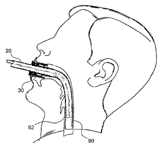
Still fiu-ther stage of the gastroscopic procedure is depicted in Fig. 7,
showing how the insertion
tube has been pushed further through the dispenser 30. The dispenser is
arrested within the
mouthpiece and remains stationary. The proximal end of the sleeve remains
stationary too, since
it is firmly secured at the rear end of the dispenser. Insertion tube 20 is
pushed distally through
the dispenser along the esophagus 90 and urges the distal end of the sleeve to
move distally
together with the insertion tube, since it is firmly secured between the cap
and the insertion tube.
Distal end of the sleeve extends automatically from the dispenser, unfolds and
covers a portion
9
CA 02601698 2007-08-28
WO 2006/092791 PCT/IL2006/000279
92 of the insertion tube, which has passed through the dispenser. It can be
appreciated that the
unfolded sleeve deploys around the insertion tube and reliably protects it
from any contamination
matter originating from the esophagus.
Fig. 8 depicts a still further stage of the advancement of the insertion tube.
It is seen that at this
stage a major portion of the insertion tube has passed the dispenser and is
located within the
esophagus. This portion is covered by protective sleeve 52. Distal end of the
insertion tube is
aproaching the stomach 94, which interior can be observed by the optical head.
Other
components of the gastroscopic apparatus remain outside the patient. Among
these components
are proximal portion 96 of the insertion tube 20 and operation handle 10 with
Y-connector 16,
working channe122, umbilical cord 24, the SCU and the monitor.
It is seen that proximal portion 96 of the insertion tube still has not been
advanced in the
dispenser. This portion would be available if it is required to advance the
insertion tube along the
esophagus still further, through the stomach 94 within a duodenum 98.
At the end of gastroscopic procedure the insertion tube and the sleeve are
withdrawn from the
patient.
According to one embodiment of the invention the dispenser remains arrested
within the
mouthpiece during the withdrawal. When the insertion tube is being withdrawn,
the sleeve
bunches and remains in the patient's mouth outside the dispenser. At the end
of withdrawal the
compactly bunched sleeve is located in the mouth adjacent the bite portion.
Now the dispenser is
disengaged from the mouthpiece and the insertion tube together with the
bunched sleeve is taken
out. Thereafter the insertion tube is separated from the sleeve and the sleeve
along with the
dispenser is disposed of. The mouthpiece is removed from the patient's mouth
and is disposed
of too.
According to an alternative embodiment the dispenser is disengaged from the
mouthpiece before
the withdrawal and the insertion tube is evacuated from the esophagus together
with the sleeve
being extended and deployed there along. The insertion tube is removed from
the mouthpiece
together with the dispenser and with the covering sleeve. Thereafter the
insertion tube is
CA 02601698 2007-08-28
WO 2006/092791 PCT/IL2006/000279
separated from the sleeve and the sleeve along with the dispenser is disposed
of. The mouthpiece
is removed from the patient's mouth and is disposed of too.
It can be readily appreciated that by virtue of the present invention it is
possible to minimize the
risk of cross contaminations to both patients and staff during the
gastroscopic procedure and to
make the gastroscopic procedure cleaner and safer.
Furthermore, the present invention eliminates the need for reprocessing
equipment and helps the
facility to save on capital expenses (purchase of additional scopes), labor
costs and costs of
disinfectants, cleaning tools and scope maintenance.
The present invention allows for increased patient throughput as a result of
faster procedures
with shorter down time for the scope. This, in its turn, allows to schedule
more procedures and
therefore to increase revenues for both physician and facility.
The gastroscopic apparatus of the present invention has very simple
construction, which is
reliable and at the same time remains very similar to a standard apparatus,
thus enabling an
extremely short learning time to the physician to achieve optimal performance.
It should be appreciated that the present invention is not limited to the
above-described
embodiments and that one ordinarily skilled in the art can make modifications
without deviation
from the scope of the invention, as will be defined in the appended claims.
For example, more that two openings could be made in the dispenser and more
than one tooth
could be made in the mouthpiece.
The openings could be made in the mouthpiece instead of the dispenser and the
teeth can be
arranged in the dispenser instead of the mouthpiece.
Other types of detachable connections between the mouthpiece and the dispenser
could be
employed instead of a snap connection.
When used in the following claims, the meaning of terms "comprise", "include",
"have" and
their conjugates is "including but not limited to".
11
CA 02601698 2007-08-28
WO 2006/092791 PCT/IL2006/000279
It should also be appreciated that the features disclosed in the foregoing
description,
andlor in the following claims, and/or in the accompanying drawings may, both
separately and in
any combination thereof, be material for realizing the present invention in
diverse forms thereof.
12