Note: Descriptions are shown in the official language in which they were submitted.
CA 02602048 2007-09-10
WO 2006/098831 PCT/US2006/004641
INTRAVASCULAR FILTER WITH CENTERING MEMBER
Teclulical Field
The present invention relates generally to the field of medical devices. More
specifically, the present invention pertains to systems and methods for
centering
intravascular filters within the body.
Background
Intravascular filters are typically used in combination witli other
thrombolytic
agents to treat pulmonary embolism occurring within a patient. Such devices
are
generally implanted within a vessel such as the inferior or superior vena
cava, and
function by capturing blood clots (emboli) contained in the blood stream
before they
can reach the lungs and cause permanent damage to the body. To trap emboli
contained within the blood, many conventional filters include an apical head
operatively coupled to a plurality of elongated filter legs that can be
expanded within
the body to form a conical-shaped surface that captures blood clots without
disturbing
the flow of blood. Once collected, a natural clot lysing process occurs within
the
body to dissolve the blood clots collected by the filter.
Delivery of the intravascular filter within the body is generally accomplished
via an introducer sheath percutaneously inserted through the femoral (groin)
or
jugular (neck) veins. Such introducer sheaths are typically tubular in shape,
and
include an interior lumen configured to transport the filter in a collapsed
position
through the body. Once transported to a desired location in the body, the
filter can
then be removed from within the introducer sheath, allowing the filter legs to
spring
open and engage the vessel wall. A needle, hook, barb, prong, wedge or other
attachment means disposed on the free end of each filter leg can be used to
secure the
filter to the vessel wall.
The efficacy of the intravascular filter to capture blood clots is dependent
in
part on the ability of the filter to center when deployed within the blood
vessel.
Tilting of the filter may result if the apical head is not aligned centrally
within the
vessel, causing the filter legs to asymmetrically engage the vessel wall.
Tilting of the
filter may also result if the introducer sheath used to deploy the filter is
off-centered
-1-
CA 02602048 2007-09-10
WO 2006/098831 PCT/US2006/004641
within the blood vessel. In certain circumstances, tilting of the filter may
affect the
ability of the device to efficiently capture blood clots contained in the
blood.
Summarv
The present invention pertains to systems and methods for centering
intravascular filters within the body. A filter system in accordance with an
illustrative
embodiment of the present invention may include an intravascular filter, a
filter
sheath having an interior lumen adapted to contain the intravascular filter,
and a
centering meinber adapted to radially expand when deployed within a blood
vessel.
The centering member may comprise an elongated wire that, when unconstrained
radially, assumes a preset shape having a radial section and a hoop section.
The radial
section may comprise a portion of the elongated wire extending outwardly in a
direction substantially orthogonal to the interior wall of the blood vessel.
The hoop
section, in turn, may comprise a portion of the elongated wire that radially
expands
against the inner wall of the blood vessel. In some embodiments, a tubular
member
having an interior lumen can be configured to radially constrain the centering
member
in a substantially straight position to facilitate delivery and/or retrieval
of the filter
assembly through the body.
In certain einbodiments, the filter system may include multiple centering
members that can be used in centering the intravascular filter and/or filter
sheath at
multiple locations within the blood vessel. In one illustrative embodiment,
for
example, a second centering member may be provided at or near the distal end
of the
filter sheath to center the filter sheath within the blood vessel, if
necessary. The
second centering member may similarly comprise an elongated wire that, when
unconstrained radially within a second interior lumen of the filter sheath,
can be
configured to assume a preset shape within the blood vessel. As with the other
embodiments described herein, the second centering meniber may include a
radial
section adapted to extend outwardly in a direction substantially orthogonal to
the
interior wall of the blood vessel, and a hoop section adapted to radially
expand against
the inner wall of the blood vessel.
An illustrative method of centering an intravascular filter within a patient's
blood vessel may include the steps of providing an intravascular filter and
centering
member within an interior lumen of a filter sheath, inserting the filter
sheath into the
patient and advancing the filter sheath to a desired location within the blood
vessel,
-2-
CA 02602048 2007-09-10
WO 2006/098831 PCT/US2006/004641
deploying the centering member within the blood vessel, and then deploying the
intravascular filter within the blood vessel. Other methods and techniques are
also
described herein. As used herein proximal end distal refer to the orientation
of the
system as delivered by a femoral approach to the vena cava. It is understood
that the
system could be use in other vessels, and from other approaches.
Brief Description of the Drawings
Figure 1 is perspective view of a filter system in accordance with an
illustrative embodiment of the present invention employing a single centering
meinber;
Figure 2 is a partial cross-sectional view showing' the centering member
disposed througli the apical head of the intravascular filter of Figure 1;
Figure 3 is a partial cross-sectional view showing an alternative embodiment
employing a tubular member movably disposed relative to the intravascular
filter;
Figure 4 is a perspective view of a filter system in accordance with an
illustrative embodiment of the present invention employing multiple centering
members;
Figure 5 is a transverse cross-sectional view of the filter sheath along line
5-5
in Figure 4;
Figure 6 is a partial cross-sectional view showing the illustrative filter
system
of Figure 1 in a first position within a blood vessel;
Figure 7 is a partial cross-sectional view showing the illustrative filter
system
of Figure 1 in a second position within the blood vessel, wherein the
centering
member is shown engaged against the vessel wall;
Figure 8 is a partial cross-sectional view showing the illustrative filter
system
of Figure 1 in a third position within the blood vessel, wherein the
intravascular filter
is shown deployed within the blood vessel;
Figure 9 is a partial cross-sectional view showing the illustrative filter
system
of Figure 1 in a fourth position within the blood vessel, wherein the
centering member
and delivery catheter are shown removed from the blood vessel;
Figure 10 is a partial cross-sectional view showing the illustrative filter
system
of Figure 4 in a first position within a blood vessel;
-3-
CA 02602048 2007-09-10
WO 2006/098831 PCT/US2006/004641
Figure 11 is a partial cross-sectional view showing the illustrative filter
system
of Figure 4 in a second position within the blood vessel, wherein the second
centering
member is shown engaged against the vessel wall;
Figure 12 is a partial cross-sectional view showing the illustrative filter
system
of Figure 4 in a third position within the blood vessel, wherein the first and
second
centering members are shown engaged against the vessel wall; and
Figure 13 is a partial cross-sectional view showing the illustrative filter
system
of Figure 4 in a fourth position within the blood vessel, wherein the
intravascular
filter is shown deployed within the blood vessel.
Detailed Description
The following description should be read with reference to the drawings, in
which like elements in different drawings are numbered in like fashion. The
drawings, which are not necessarily to scale, depict selected embodiments and
are not
intended to limit the scope of the invention. Although examples of
construction,
dimensions, and materials are illustrated for the various elements, those
skilled in the
art will recognize that many of the examples provided have suitable
alternatives that
may be utilized.
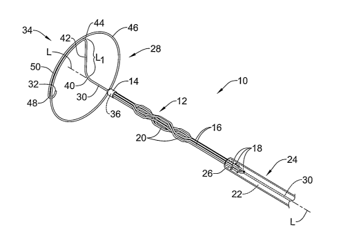
Figure 1 is perspective view of a filter system 10 in accordance with an
illustrative embodiment of the present invention. Filter system 10,
illustratively a
filter system for use in the inferior and/or superior vena cava, can include
an
intravascular filter 12 having an apical head 14 and a plurality of elongated
filter legs
16 adapted to expand and secure the intravascular filter 12 to the inner wall
of a blood
vessel. The free end 18 of each filter leg 16 may include a needle, hook,
barb, prong,
wedge or other suitable attaclunent means for securing the intravascular
filter 12 to
the inner wall of the blood vessel. A number of bend regions 20 located along
the
length of one or more of the filter legs 16 can also be provided to increase
the surface
area of the intravascular filter 12, if desired.
The filter legs 16 can be configured to radially collapse within an interior
lumen 22 of a filter sheath 24 to delivery and/or receive the intravascular
filter 12
through the patient's body. The filter sheath 24 may comprise a tubular member
having a distal end 26 and a proximal end (not shown). For sake of clarity in
Figure
1, the intravascular filter 12 is shown withdrawn at least in part from within
the
interior lumen 22 of the filter sheath 24, exposing all but the free end 18 of
each filter
-4-
CA 02602048 2007-09-10
WO 2006/098831 PCT/US2006/004641
leg 16. It should be understood, however, that all or a portion of the
intravascular
filter 12 can be loaded within the interior lumen 22 of the filter sheath 24,
if desired.
As can be further seen in Figure 1, the filter system 10 may also include a
centering member 28 that can be used to aid in centering the intravascular
filter 12
witliin the interior of the blood vessel. The centering member 28 may comprise
an
elongated wire 30 having a distal end 32, a proximal end (not shown), and a
distal
section 34 adapted to assume a preset shape when deployed within the blood
vessel.
Prior to insertion within the patient's body, the elongated wire 30 can be
inserted
through an interior lumen 36 formed through the apical head 14 and through the
interior lumen 22 of the filter sheath 24.
The distal section 34 may comprise a portion of the elongated wire 30
extending distally from a first bend region 40 of the elongated wire 30 to the
distal
end 32 thereof. A radial section 42 of the elongated wire 30 extending
distally from
the first bend region 40 can be adapted to extend outwardly in a direction
substantially
orthogonal to the interior wall of the blood vessel, when deployed. The length
LI in
which the radial section 42 extends outwardly may vary depending on the
particular
vessel the intravascular filter 12 is to be inserted into. In applications
involving the
superior or inferior vena cava, for exainple, the length L1 of the radial
section 42 may
be in the range of about 6 mm to 15 mm, which is sufficient for blood vessels
having
a diameter of about 12 mm to 30 mm. It should be understood, however, that the
length Lt of the radial section 42 may vary to permit the centering member 28
to be
used in other regions of the body and/or to accommodate for anatomical
differences
among patients.
At a second bend region 44 of the distal section 34, the radial section 42 may
transition to a hoop section 46 of the elongated wire 30 extending
circumferentially
about a general longitudinal axis L of the intravascular filter 12 and filter
sheath 24.
The shape of the hoop section 46 can be selected to approximate the general
shape of
the blood vessel, allowing the hoop section 46 to radially expand and fully
appose the
inner wall of the blood vessel. In certain embodiments, for example, the hoop
section
46 of the elongated wire 30 may have a substantially elliptical shape to
facilitate
centering of the intravascular filter 12 in blood vessels having an oblique or
non-
symmetrical shape. In other embodiments, the hoop section 46 may have a
substantially circular shape to facilitate centering of the intravascular
filter 12 in blood
vessels having a substantially symmetrical shape.
-5-
CA 02602048 2007-09-10
WO 2006/098831 PCT/US2006/004641
In the illustrative embodiment of Figure 1, the hoop section 46 is configured
to
lie in a single plane that is oriented substantially orthogonal to the length
of the blood
vessel. In an alternative embodiment, the hoop section 46 can be configured to
spiral
in multiple planes along the longitudinal axis L. In the latter case, for
example, the
hoop section 46 may have the general shape of a helix that tapers distally
towards the
distal end 32. The hoop section 46 may assume other desired shapes, however,
to
facilitate centering of the intravascular filter 12 at other locations within
the body
such as at a branching vessel.
At a third bend region 48 of the distal section 34, the distal end 32 of the
elongated wire 30 may curl inwardly towards the longitudinal axis L. In use,
the third
bend region 48 orients the distal end 32 away from the inner wall of the blood
vessel,
preventing the distal end 32 from contacting the blood vessel. If desired, an
overlapping portion 50 of the hoop section 46 wherein the elongated wire 30 is
wound
adjacent itself can be used to space the distal end 32 away from the second
bend
region 44. In some embodiments, the distal end 32 may also be rounded to
further
prevent trauma to the vessel wall. Also, the bend region 48 may be
diametrically
tapered to further prevent trauma to the vessel wall.
Figure 2 is a partial cross-sectional view showing the centering member 28
disposed through the apical head 14 of the intravascular filter 12 of Figure
1. As
shown in Figure 2, the apical head 14 may include a tubular member 52 having a
distal end 54 and a proximal end 56. The tubular member 52 may comprise a
member
separate from the apical head 14 (e.g. a h.ypotube) that is then subsequently
attached
to the apical head 14, or, in the alternative, may be formed integral with the
apical
head 14. In certain embodiments, for example, the tubular member 52 and joined
end
58 of each filter leg 16 can be soldered together using a solder bead, forming
an apical
head 14 having a generally bulbous shape. In an alternative technique, the
tubular
member 52, filter legs 16, and apical head 14 may each be formed as a single
piece
using a suitable process such as insert molding.
The length of the tubular member 52 can be made sufficient to permit the
distal section 34 of the elongated wire 30 to be loaded into the interior
lumen 36. The
inner diameter of the tubular member 52, in turn, can be made slightly larger
than the
outer diameter of the elongated wire 30, allowing the elongated wire 30 to
move
within the interior lumen 36. In use, the tubular member 52 acts to maintain
the
elongated wire 30 in a substantially straight position within the interior
lumen 36 prior
-6-
CA 02602048 2007-09-10
WO 2006/098831 PCT/US2006/004641
to deployment within the blood vessel. The tubular member 52 also acts to
straighten
the elongated wire 30 when it is pulled back into the filter sheath 24 for
subsequent
removal from the body.
The elongated wire 30 may be formed from a flexible material that permits it
to maintain its preset shape when disposed within the interior lumen 36 of the
tubular
member 52. Examples of suitable flexible materials may include certain metals,
polymers, or metal-polyiner compounds. In some embodiments, the elongated wire
30 may include a layer or coating of lubricious material such as HYRDOPASS to
facilitate movement of the elongated wire 30 through the tubular member 52 and
filter
sheath 24, and to reduce trauma to the body caused during deployment of the
centering member 28 within the blood vessel. The elongated wire 30 as well as
other
portions of the filter system 10 may also include an anti-thrombogenic coating
such as
herapin (or its derivatives), urokinase, or PPack (dextrophenylalanine proline
arginine
chloromethylketone) to prevent insertion site thrombosis from occurring. An
anti-
inflammatory agent such as dexamethasone, prednisolone, corticosterone,
budesonide,
estrogen, sulfasalazine, mesalamine, or any suitable combination or mixture
thereof
may also be applied to the elongated wire 30, intravascular filter 12 as well
as other
components of the filter system 10 to prevent inflammation within the blood
vessel.
In some embodiments, the elongated wire 30 may be formed from a linear
elastic material such as a nickel-titanium alloy, which exhibits the ability
to undergo
significant bending or flexion without imparting a residual stress to the
material.
Examples of other suitable linear elastic materials may include, but are not
limited to,
silver-cadmium (Ag-Cd), gold-cadmium (Au-Cd), gold-copper-zinc (Au-Cu-Zn),
copper-aluminuni-nickel (Cu-Al-Ni), copper-gold-zinc (Cu-Au-Zn), copper-zinc
(Cu-
Zn), copper-zinc-aluminum (Cu-Zn-Al), copper-zinc-tin (Cu-Zn-Sn), copper-zinc-
silicon (Cu-Zn-Si), iron-beryllium (Fe-Be), iron-nickel-titanium-cobalt (Fe-Ni-
Ti-Co),
iron-platinum (Fe-Pt), indium-thallium (In-Tl), iron-manganese (Fe-Mn), nickel-
titanium-cobalt (Ni-Ti-Co), and copper-tin (Cu-Sn). In certain embodiments,
the
elongated wire 30 may be combined with other materials such as stainless
steel,
platinum, titanium, etc. to form a composite material exhibiting certain
desirable
characteristics within the body. In certain applications, for example, the
linear elastic
material may be joined together with a relatively radiopaque material such as
platinum (Pt) to increase the radiopacity of the composite member, allowing
the
centering member 28 to be viewed radiographically with the aid of a
fluoroscope.
-7-
CA 02602048 2007-09-10
WO 2006/098831 PCT/US2006/004641
In another aspect of the present invention, the elongated wire 30 may be
formed from a shape-memory material that has been heat treated to inipart a
shape
memory effect to distal section 34, allowing the centering member 28 to be
transformed from a substantially straight position to an expanded (i.e.
centering)
position when withdrawn from within the tubular member 52. In certain
embodiments, for example, the elongated wire 30 may be formed of or otherwise
include a shape-memory alloy such as a nickel-titanium alloy (Nitinol)
configured to
transform from a martensite state to an austenite state at or about body
temperature,
allowing the centering member 28 to assume a preset shape when exposed to
blood
within the blood vessel.
Figure 3 is a partial cross-sectional view showing an alternative embodiment
employing a tubular member 60 movably disposed relative to the intravascular
filter
12. As shown in Figure 3, the tubular member 60 has a distal end 62, a
proximal end
(not shown), and an interior lumen 64 therethrough adapted slidably receive
the distal
section 34 of centering member 28 in a manner similar to that described above
with
respect to Figure 2. As indicated by the arrow 66, however, the tubular member
60
can be configured to move independently of the intravascular filter 12,
allowing the
physician to further remove the tubular member 60 from the body once the
intravascular filter 12 has been deployed within the blood vessel. The tubular
member 60 can be either connected to the filter sheath 24, or can be
configured to
independently move within the interior lumen 22 of the filter sheath 24.
The interior lumen 36 of the apical head 14 can be sized to slidably receive
the
tubular member 60 to facilitate advancement of the centering member 28
distally
beyond the distal end 68 of the apical head 14. If desired, a tapered inner
portion 70
of the apical head 14 extending inwardly into the interior lumen 36 can be
configured
to prevent the physician from overextending the distal end 62 of the tubular
member
60 beyond the distal end 68 of the apical head 14. In use, the tapered inner
portion 70
acts as a distal stop as the physician advances the tubular member 60 through
the
interior lumen 36, preventing the tubular member 60 from being advanced
distally
beyond the distal end 68 of the apical head 14. In some cases, the tapered
inner
portion 70 may also provide the physician with tactile feedback that the
centering
member 28 is in the proper position within the interior lumen 36 for
deployment.
Figure 4 is a perspective view of a filter system 72 in accordance with
another illustrative embodiment of the present invention employing two
centering
-8-
CA 02602048 2007-09-10
WO 2006/098831 PCT/US2006/004641
members. Filter system 72 may be configured similar to filter system 10
described
above with respect to Figures 1-2, with like elements being labeled in like
fashion. In
the illustrative embodiment of Figure 4, however, the filter system 72 may
further
include a second centering member 74 that can be used to aid in centering the
base of
the intravascular filter 12 within the blood vessel.
The second centering member 74 may comprise an elongated wire 76 having a
distal end 78, a proximal end (not shown), and a distal section 80 adapted to
assume a
preset shape when deployed within the blood vessel. In a generally deployed
position
illustrated in Figure 4, the distal section 80 may comprise a portion of the
elongated
wire 76 extending distally from a first bend region 82 to the distal end 78
thereof. A
radial section 84 of the elongated wire 76 extending distally from the first
bend region
82 can be adapted to extend outwardly in a direction substantially orthogonal
to the
interior wall of the blood vessel, when deployed. As with the first centering
member
28, the length L2 of the radial section 84 may vary depending on the size of
the blood
vessel. The length L2 of the radial section 84 may be made similar to the
length LI of
radial section 42, or may be made grater or lesser than length Li.
At a second bend region 86 of the distal section 80, the elongated wire 76 may
transition to a hoop section 88 of the elongated wire 76 extending
circumferentially
about the longitudinal axis L. The shape of the hoop section 88 can be
selected to
approximate the general shape of the blood vessel, similar to that described
above
witll respect to the other centering member 28. Other features such as a third
bend
region 90 forming a curled (i.e. atraumatic) distal end 78 may also be
provided, if
desired.
The filter system 72 may further include a filter sheath 92 having a distal
end
94, a proximal end (not shown), and an interior lumen 96 therethrough adapted
to
slidably receive the intravascular filter 12 and a portion of the elongated
wire 30. A
second interior lumen 98 of the filter sheath 92, in turn, can be adapted to
slidably
receive the second elongated wire 76, allowing the physician to deploy the
second
centering member 74 within the blood vessel at a location at or near the
distal end 94
of the filter sheath 92. As can be seen in further detail in Figure 5, a lumen
opening
100 provided in the wall 102 of the filter sheath 92 may form an exit port,
allowing
the physician to advance the second elongated wire 76 distally out from within
the
second interior lumen 98 to deploy the second centering member 74 within the
blood
vessel.
-9-
CA 02602048 2007-09-10
WO 2006/098831 PCT/US2006/004641
Referring now to Figures 6-9, an illustrative method of centering an
intravascular filter in accordance with the present invention will now be
described
with respect to filter system 10 described above. In a first position
illustrated in
Figure 6, the intravascular filter 12 and centering member 28 are shown loaded
into
the interior lumen 22 of the filter sheath 24 and advanced to a desired
location within
a blood vessel V (e.g. the superior or inferior vena cava). As shown in Figure
6, the
centering member 28 can be configured to maintain a substantially straight
shape
when radially constrained within the interior lumen 36 of the tubular member
52.
Such straight shape permits the filter system 10 to assume a relatively small
profile
when transported through the vasculature, allowing the physician to employ a
smaller
sized filter sheatli 24.
Once the filter system 10 is advanced to a desired location within the blood
vessel V, the physician may next advance the elongated wire 30 distally out
from
within the interior lumen 36, causing the distal section 34 of the elongated
wire 30 to
assume its preset shape within the blood vessel V. The elongated wire 30 can
be
deployed within the blood vessel V by holding the filter sheath 24 and
intravascular
filter 12 stationary while advancing the elongated wire 30 distally, or, in
the
alternative, by holding the elongated wire 30 stationary and retracting the
filter sheath
24 and intravascular filter 12 proximally. A combination of the two techniques
may
also be performed to deploy the centering member 28, if desired.
Once the centering member 28 is withdrawn from the tubular member 52, the
hoop section 46 can be configured to radially expand and fully appose the
vessel wall,
as shown, for example, in Figure 7. When this occurs, a centering force is
exerted
against the apical head 14 by the elongated wire 30, causing the intravascular
filer 12
to align centrally within the blood vessel V. If, for example, the filter
system 10 is
off-centered within the blood vessel V (see Figure 6), the general alignment
of the
elongated wire 30 centrally within the blood vessel V produces a centering
force that
re-aligns the intravascular filter 12 within the blood vessel V.
To deploy the intravascular filter 12 within the blood vessel V, the
physician,
while holding the elongated wire 30 stationary, may next retract the filter
sheath 24 in
the proximal direction to expose the filter legs 16, as shown, for example, in
Figure 8.
If desired, a pusher tube 104 can be provided within the interior lumen 22 of
the filter
sheath 24 to hold the intravascular filter 12 stationary as the filter sheath
24 is being
retracted proximally. Once the intravascular filter 12 is deployed within the
blood
-10-
CA 02602048 2007-09-10
WO 2006/098831 PCT/US2006/004641
vessel V, the physician may next pull the elongated wire 30 proximally through
the
tubular member 52 and out of the body, if desired. As shown in a subsequent
view in
Figure 9, the filter sheath 24 and centering member 28 can then be removed
from the
body, leaving the centered intravascular filter 12 within the blood vessel V.
Turning now to Figures 10-14, another illustrative method of centering an
intravascular filter in accordance with the present invention will now be
described
with respect to filter system 72 described above. In a first position
illustrated in
Figure 10, the intravascular filter 12, first centering member 28, and second
centering
member 74 are shown loaded into the filter sheath 92 and advanced to a desired
location within a blood vessel V (e.g. the inferior vena cava).
Once the filter system 72 is advanced to a desired location within the blood
vessel V, the physician may next advance the second elongated wire 76 distally
out
from the second interior lumen 98 through the lumen opening 100. Once the
centering member 74 is deployed within the blood vessel V, the hoop section 88
can
be configured to radially expand and fully appose the vessel wall, as shown,
for
example, in Figure 11. When this occurs, the centering force of the elongated
wire 76
exerted against the filter sheath 92 causes the filter sheath 92 to align
centrally within
the blood vessel V.
In addition to deploying the second centering member 74 within the blood
vessel V, the physician may further advance the first elongated wire 30
distally out
from within the interior lumen 36, causing the distal section 34 of the
elongated wire
30 to assume its preset shape within the blood vessel V, as shown, for
example, in
Figure 12.
To deploy the intravascular filter 12 within the blood vessel V, the physician
may next retract the second elongated wire 76 proximally within the filter
sheath 92,
causing the distal section 80 to straighten within the second interior lumen
98. The
physician, while holding the first elongated wire 30 stationary, may also
retract the
filter sheath 92 proximally to expose the filter legs 16, as shown, for
example, in
Figure 13. If desired, a pusher tube 104 can be provided within the interior
lumen 96
of the filter sheath 92 to hold the intravascular filter 12 stationary as the
filter sheatll
92 is being retracted. Once the intravascular filter 12 is deployed within the
blood
vessel V, the physician may next pull the elongated wire 30 proximally
througli the
tubular member 52. The filter sheath 92 and centering members 28,74 can then
be
- 11 -
CA 02602048 2007-09-10
WO 2006/098831 PCT/US2006/004641
removed from the body, leaving the centered intravascular filter 12 within the
blood
vessel V.
While the illustrative steps depicted in Figures 11-12 show the deployment of
the second centeririg member 74 prior to the first centering member 28, otlier
embodiments have been envisioned wherein the first centering member 28 is
deployed prior to the second centering member 74, or wherein both centering
members 28,74 are deployed at or about the same time.
Having thus described the several embodiments of the present invention, those
of skill in the art will readily appreciate that other embodiments may be made
and
used which fall within the scope of the claims attached hereto. Numerous
advantages
of the invention covered by this document have been set forth in the foregoing
description. It will be understood that this disclosure is, in many respects,
only
illustrative. Changes can be made with respect to various elements described
herein
without exceeding the scope of the invention.
-12-