Note: Descriptions are shown in the official language in which they were submitted.
CA 02602385 2007-09-18
WO 2006/105234 PCT/US2006/011531
HYBRID INORGANIC NANOPARTICLES, METHODS OF USING AND
METHODS OF MAKING
The present application claims priority to U.S. Provisional
Application No. 60/666,114, filed March 29, 2005, which is
hereby incorporated by reference herein.
FIELD OF THE INVENTION
The subject invention is directed generally to hybrid
inorganic nanoparticles, methods of making hybrid inorganic
nanoparticles and methods of using the hybrid inorganic
nanoparticles.
BRIEF DESCRIPTION OF THE DRAWINGS
These and other features and advantages of this invention
will be evident from the following detailed description of
preferred embodiments when read in conjunction with the
accompanying drawings in which:
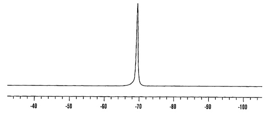
Figure 1 is a representative 19F spectra of silica based
TFMPTS nanoparticles at 376.3 MHz.
Figure 2 illustrates a typical 19F spectra obtained from
neat silica based TFPTMS 19F containing nanoparticles immediately
before imaging at 188.34 MHz.
Figure 3 illustrates 19F spectra obtained from neat silica
based TFPTMS 19F containing nanoparticles as compared to 1000 mM
sodium fluoride (NaF) in aqueous solution at 188.34 MHz.
Figure 4 depicts representative 1H and 19F MR images
following administration of TFMPTS nanoparticles in a mouse. iH
MR images obtained at 200 MHz. (A, axial; B, coronal) and 19F MR
images obtained at 188 MHz. (C, axial; D, coronal) obtained
immediately after oral administration of silica based TFPTMS
nanoparticles. As shown, arrows denote location of stomach
(A,B,E and F,) spinal canal (A); lung (B); lobe of liver (B).
Figure 5 is a z QF MR image of semi-solid crystalline
aggregates of TFPTMS 19F nanoparticles (left panel, scale denotes
1 cm) and corresponding micrograph (right panel) of
nanoparticles in a glass vial photographed using a surgical
microscope with attached Nikon 1.2 Mb digital camera (Nikon
CA 02602385 2007-09-18
WO 2006/105234 PCT/US2006/011531
2 -
DETAILED DESCRIPTION OF THE INVENTION
Throughout this application various publications are
referenced, many in parenthesis. Full citations for each of
these publications are provided at the end of the Detailed
Description. The disclosures of each of these publications in
their entireties are hereby incorporated by reference in this
application.
The subject invention provides hybrid inorganic
nanoparticles, methods of making the hybrid inorganic
nanoparticles and methods of using the hybrid inorganic
nanoparticles.
As used herein, "hybrid inorganic" nanoparticles refer to
nanoparticles which contain both organic and inorganic groups.
Although not meaning to be bound by theory, the nanoparticles of
the invention have the desirable physical properties of both
ceramic materials and the functional groups associated with the
nanoparticles.
Further, as used herein, the hybrid inorganic nanoparticles
of the present invention are used in spectroscopic and image
based acquisitions, including, but not limited to, magnetic
resonance, fluorescence, bioluminescence spectroscopy and other
imaging techniques and other biomedical applications.
The nanoparticles of the present invention are hybrid
inorganic nanoparticles which include 19F nuclei. In one
embodiment of the invention, the nanoparticles are silica based
hybrid inorganic nanoparticles. The nanoparticles are
constructed having various diameters and distribution ranging
from about 20 nanometers to about 200 nanometers, and all ranges
therein. For example, in one embodiment of the invention, the
hybrid inorganic nanoparticles are from about 50 to about 200
nanometers in diameter. In alternative embodiments of the
present invention, the nanoparticles are from about 100 to about
200 nanometers, from about 150 to about 200 nanometers or from
CA 02602385 2007-09-18
WO 2006/105234 PCT/US2006/011531
- 3 -
about 75 to about 200 nanometers. In another embodiment, the
nanoparticles are from about 20 nanometers to less than about
200 nanometers, for example from about 20 nanometers, up to
about 50, 75, 100 or 150 nanometers.
The nanoparticles of the present invention have a high
number of 19F nuclei per nanoparticle. As used herein, a high
number is defined as having up to about 600,000 19F nuclei per
nanoparticle. In one embodiment, the nanoparticles of the
present invention include from about 2000 to about 600,000 19F
nuclei per nanoparticle. In one embodiment, the nanoparticles
have from 10,000 to about 600,000 19F nuclei per nanoparticle.
In one embodiment, the nanoparticles include from about 30,000
to about 600,000 19F nuclei per nanoparticle, or from about
100,000 to about 600,000 19F nuclei per nanoparticle.
Therefore, the nanoparticles of the present invention
include a quantity of 19F nuclei to be used in the methods of the.
present invention, for example, in imaging, spectroscopic
acquisitions and biomedical applications.
Although not meaning to be bound by theory, the number of
19F nuclei per nanoparticle may be calculated by first
determining the size of each nanoparticle. For each size of
nanoparticle, the mass of the nanoparticle can be determined,
and, accordingly, because the mass of each molecule present in
each nanoparticle is known, the resultant number of molecules
present in the nanoparticle can be calculated by one skilled in
the art. For example, a nanoparticle of the present invention
having a diameter of about 40 nanometers has approximately
105,000 molecules present in the nanoparticle. Each molecule of
the nanoparticle has about three fluorine atoms contained
therein. Assuming approximately 30%-40% incorporation of 19F
nuclei per nanoparticle, a nanoparticle having a diameter of
approximately 40 nanometers would have about 105,000 19F nuclei
per nanoparticle. Using these calculations, a nanoparticle
having about a 20 nanometer diameter would have approximately
13,000 19F nuclei per nanoparticle. Likewise, a nanoparticle
CA 02602385 2007-09-18
WO 2006/105234 PCT/US2006/011531
- 4 -
having about a 100 nanometer diameter would have approximately
273,000 19F nuclei per nanoparticle and a nanoparticle having
about a 200 nanometer diameter would have approximately 600,000
19F nuclei per nanoparticle.
In one aspect of the invention, the 19F nuclei are contained
in the inner core of the nanoparticles. In an alternative
embodiment, the 19F nuclei are contained at the outer surface of
the nanoparticles. In another alternative embodiment, the 19F is
contained both in the inner core and at the outer surface of the
nanoparticles.
In another aspect of the invention, the nanoparticles
additionally include.a biomarker, such as a fluorescent dye,
bioluminescent marker and/or near infrared (NIR) marker.
In another aspect of the invention, the nanoparticles
include a therapeutic or diagnostic agent, or both. The
therapeutic and diagnostic agents are either hydrophilic or
hydrophobic. Therapeutic or diagnostic agents include
substances capable of treating or preventing an infection
systemically or locally, as, for example, antibacterial agents
such as penicillin, cephalosporins, bacitracin, tetracycline,
doxycycline, quinolines, clindamycin, and metronidazole;
antiparasitic agents such as quinacrine, chloroquine and
vidarabine; antifungal agents such as nystatin; antiviral agents
such as acyclovir, ribarivin and interferons; anti-inflammatory
agents such as hydrocortisone and prednisone; analgesic agents
such as salicylic acid, acetaminophen, ibuprofen, naproxen,
piroxicam, flurbiprofen and morphine; local anesthetics such as
lidocaine, bupivacaine, benzocaine, and the like; immunogens
(vacc=ines) for stimulating antibodies against hepatitis,
influenza, measles, rubella, tetanus, polio and rabies; peptides
such as leuprolide acetate (an LH-RH agonist), nafarelin and
ganirelix. Also useful is a substance or metabolic precursor
thereof, which is capable of promoting growth and survival of
cells and tissues or augmenting the functioning of cells, as for
example, a nerve growth promoting substance such as a
CA 02602385 2007-09-18
WO 2006/105234 PCT/US2006/011531
- 5 -
ganglioside, a nerve growth factor, and the like; a hard or soft
tissue growth promoting agent such as fibronectin (FN), human
growth hormone (HGH), a colony stimulating factor, bone
morphogenetic protein, platelet-derived growth factor (PDGF),
insulin-derived growth factor (IGF-I, IGF-II), transforming
growth factor-alpha, transforming growth factor-beta, epidermal
growth factor (EGF), fibroblast growth factor (FGF) and
interleukin-1 (IL-1); an osteoinductive agent or bone growth
promoting substance such as bone chips and demineralized freeze-
dried bone material; and antineoplastic agents such as
methotrexate, 5-fluoroacil, adriamycin, vinblastine, cisplatin,
tumor-specific antibodies conjugated to toxins and tumor
necrosis factor. Other useful substances include hormones such
as progesterone, testosterone, and follicle stimulating hormone
(FSH) (birth control, fertility-enhancement), insulin metal
complexes and somatotropins; antihistamines such as
diphenhydramine and chlorpheneramine; cardiovascular agents such
as digitalis glycosides, papaverine and streptokinase; anti-
ulcer agents such as cimetidine, famotidine and isopropamide
iodide; vasodilators such as theophylline, B-adrenergic blocking
agents and minoxidil; central nervous system agents such as
dopamine; antipsychotic agents such as risperidone, olanzapine;
narcotic antagonists such as naltrexone, maloxone and
buprenorphine. Other examples of therapeutic and diagnostic
agents are water insoluble anticancer drugs such as carmustine
(BCNU), antiviral drugs such as azidothymidine (AZT) and other
nucleosides, HIV Protease inhibitors such as saquinavir and
retinovir immune-modulating agents such as cyclosporine, natural
and synthetic hormones and hormone regulators such as
contraceptives. Other therapeutic agents are steroidal and non-
steroidal anti-inflammatory agents such as hydrocortisone,
prednisolone, ketoprofen, celecoxib and ibuprofen, centrally
acting medicines such as antiseptics, antidepressants and
sedatives and cardiovascular drugs such as anti-hypertensives
and blood lipid lowering agents.
CA 02602385 2007-09-18
WO 2006/105234 PCT/US2006/011531
- 6 -
In another embodiment of the invention, the surfaces of the
nanoparticles are modified, such as, for example by attaching a
ligand to which a targeting agent is attached. Such ligands,
and their attachment via standard conjugation chemistry, are
known in the art [6].. For example, ligands, such as typical
functional groups such as amino groups, carboxyl groups and
sulfhydryl groups, are used. The targeting agent is an agent
which is specific for an intended target. Such targeting agents
include, for example, leutinizing hormone releasing hormone,
growth hormone release hormone, epithelial growth factor, folic
acid, antibodies specific for tumor markers, tumor specific
drugs, and other targeting agents.
In another embodiment of the invention, additional
paramagnetic MR contrast enhancing agents such as Gd-DTPA
commonly used for H-1 MR imaging, can be incorporated into the
nanoparticles to increase signal-to-noise-characteristics of the
nanoparticles. Examples of such agents are included in U.S.
Patent No. 6,869,591, which is incorporated herein by reference.
Another aspect of the invention relates to the manufacture
of the nanoparticles of the present invention. In this
embodiment, the method includes providing a first liquid
component of an emulsion system, providing a second liquid
component of an emulsion system, providing a precursor, where
the precursor is an a=lkoxy silane precursor which includes 19F,
mixing the first liquid component, the second liquid component
and the precursor, applying mechanical force to produce an
emulsion which includes a dispersed phase and a continuous phase
and separating the dispersed phase from the continuous phase to
produce hybrid inorganic nanoparticles.
In one embodiment, the first liquid component is a
surfactant. In one embodiment, the second liquid component is
an acid.
Typical compounds which are used as the precursor in the
method of the invention include all 19F alkoxy silane precursors.
CA 02602385 2007-09-18
WO 2006/105234 PCT/US2006/011531
_ 7 - '
In one embodiment the precursor is 3,3,3-trifluoropropyl-
trimethoxysilane (TFPTMS).
Typical surfactants include, for example, reaction products
of natural or hydrogenated vegetable oils, and ethylene glycol;
i.e., polyoxyethylene glycolated natural or hydrogenated
vegetable oils, polyoxyethylene glycolated natural or
hydrogenated castor oils, Cremophor RH-40, Cremophor RH60,
Cremophor EL, Nikkol HCO-40, Nikkol HCo-60; Polyoxyethylene
sorbitan fatty acid esters, e.g., mono- and tri-lauryl,
palmityl, stearyl and oleyl esters; e.g. products of the trade
name "Tween," which includes polyoxyethylene sorbitan mono-
.laurate (Tween), polyoxyethylene sorbitan mono-palmitate (Tween
40), polyoxyethylene sorbitan mono-oleate (Tween 80);
Polyoxyethylene fatty acid esters, for example, polyoxyethylene
stearic acid esters of the type known and commercially available
under the trade name Myrj as well as polyoxyethylene fatty acid
esters known and commercially available under the trade name
Cetiol HE; Polyoxyethylene-polyoxypropylene co-polymers: e.g. of
the type known and commercially available under the trade names
Pluronic and Emkalyx; Polyoxyethylene-polyoxypropylene block
co-polymers, of the type known and commercially available under
the trade name Poloxamer; Dioctylsuccinate,
dioctylsodiumsulfosuccinate, di-[2-ethylhexyl]-succinate, sodium
lauryl sulfate; and Phospholipids, such as lecithins, for
example, soybean lecithin; non-ionic polyoxyethylene fatty acid
derivatives, such as polyoxyethylene sorbitan fatty acid esters
(spans) such as sorbitan sesquiolate.
The mechanical force applied to the mixture includes any
mechanical force known in the art to produce an emulsion, such
as stirring. Separation of the dispersed phase and continuous
phase is achieved by methods known to those skilled in the art,
such as centrifugation. General methods for producing an
emulsion system are described in [4], [6], and [12]=
Optionally, the applying mechanical force step may be
performed a number of times, for example, the method may include
CA 02602385 2007-09-18
WO 2006/105234 PCT/US2006/011531
- 8 -
mixing the first liquid component with the precursor, followed
by applying mechanical force, followed by adding the second
liquid component, followed by, optionally, applying a second
mechanical force step.
Mechanical force is applied for a period of from about 30
minutes up to about 15 hours, and all ranges in between, for
example, from about 1 hour to about 6 hours, from about 2 hours
to about 12 hours, from about 5 hours to about 15 hours. The
mixing and applying mechanical force steps take place at about
room temperature. The separation step takes place at about 20
to about 6 C.
Nanoparticles produced by the above method include an inner
core and a surface and the 19F nuclei will be in the inner core
of the nanoparticles.
In another embodiment of the invention, a second compound
is added to the mixture. The addition of this compound results
in additional amounts of 19F nuclei included in the nanoparticles
of the invention. The additional amounts of 19F are provided by
providing a second component, such as a perfluorocarbon, to
incorporate additional amounts of 19F nuclei into the
nanoparticles. In one embodiment a perfluorocarbon, such as
zinc 1,2,3,4,8,8,9,10,11,15,16,17,18,22,23,24,25-hexadecafluoro-
29H, 31H-phthalocyanine (ZnFP) is used.
In another embodiment of the invention, the 19F nuclei will
be found either at the surface of the nanoparticles or at both
the surface and in the inner core of the nanoparticles. For
example, by preparing the nanoparticles by a reverse micellar
method (using an organic solvent (like hexane, toluene etc.) as
a bulk medium), the 19F nuclei will be on the outer surface of
the nanoparticles.
The method of the present invention results in the
production of nanoparticles having a size distribution of from
about 20 to about 200 nanometers in diameter.
Another aspect of the invention relates to a method of
imaging using the nanoparticles of the present invention. In
CA 02602385 2007-09-18
WO 2006/105234 PCT/US2006/011531
- 9 -
the method, the nanoparticles of the present invention are
administered to a subject and the subject is imaged. Using the
nanoparticles of the present invention, an image, such as an MR
image, having sufficient specificity and sensitivity is
obtained.
Another aspect of the invention relates to a method of
acquiring a.spectroscopic acquisition of a subject. The method
includes administering the nanoparticles of the present
invention to the subject and obtaining a spectroscopic
acquisition of the subject.
Another aspect of the invention relates to using the
nanoparticles of the invention in other biomedical applications,
such as a coating for medical devices, such as implantable
medical devices such as, for example, stents, breast implants
(to determine leakage or integrity of the implant), cardiac
pacemakers, catheters or other implantable medical devices.
Implantable medical devices refers to medical devices which are
inserted into a subject.
Examples
Magnetic resonance (MR) imaging is a noninvasive technique
that has been applied to the detection, characterization and
subsequent assessment of tumors and other soft tissue lesions
following therapy. As it is commonly used, MR imaging utilizes
the principles of nuclear magnetic resonance to obtain and
decipher spectral patterns of 'H (proton) magnetic resonance
signals of body fluids and/or tissues. Typical 'H images depict
the distribution of water versus fat in a patient or sample.
While 'H MR imaging is arguably the best clinical diagnostic
imaging modality available for non-invasive detection and
characterization of in vivo tumors, several major drawbacks
exist resulting in data yielding high resolution anatomic
(structural) images of soft tissue but little physiologic
(functional) information. In a similar fashion, other standard
CA 02602385 2007-09-18
WO 2006/105234 PCT/US2006/011531
- 10 -
clinical diagnostic modalities suffer from the same drawback
including computed tomography (CT), positron emission tomography
(PET), X-ray, single photon emission computed tomography (SPECT)
and ultrasound (US). Each modality can yield a plethora of
either structural or functional information (albeit each with
distinct advantages/disadvantages), but not a high degree of
both during a single examination. The ability to readily provide
researchers/clinicians with both structural and functional
information during a single examination would significantly
advance the field.
An alternative method of in vivo MR imaging is based on
analysis of the spectral patterns of fluorine (19F) magnetic
resonance signals, a non-radioactive species that is > 99%
naturally abundant and 83% as sensitive as 'H. 19F MR imaging
differs from 'H MR imaging in that 19F nuclei are not naturally
found in solution in living mammalian systems. Clinical
applications of 19F MR imaging therefore will require specialized
agents specifically designed for this purpose. However, in most
other aspects, 19F MR is similar to standard 'H techniques in
terms of the imaging physics involved. Moreover, in vivo 19F MR
imaging offers several advantages compared to 'H based MR imaging
methods. First, 19F containing compounds can be directly imaged
by MR without background contamination from other molecules or
anatomical structures. Secondly, 19F MR acquisitions yield images
of the three-dimensional distribution of 19F containing molecules
and therefore enable direct quantitative measurements of the
biodistribution, pharmacokinetics and pharmacodynamics of
administrated agents. Thirdly, for high resolution localization
of 19F signals, images can subsequently be registered with high
resolution 1H MR images and/or acquired directly with arbitrarily
high spatial resolution dependent only upon signal-to-noise
(S/N) per unit time considerations (approx. 17% lower 19F S/N
compared to 1H S/N per molar concentration). Lastly, '9F MR T1
relaxation rates of many perfluorocarbon emulsions have been
shown to correlate to p02 concentrations in solution and in
CA 02602385 2007-09-18
WO 2006/105234 PCT/US2006/011531
- 11 -
preliminary in vivo studies [1, 2]. This ability might allow for
non-invasive measurement of tissue oxygenation before, during
and after therapeutic intervention for assessing delivery of
radiation, chemotherapy and/or photodynamic therapy (PDT)
resulting in improved patient outcome.
Currently, the main limitation of 19F MR imaging is the
paucity of available fluorine-containing compounds which can be
administered in sufficient quantities for in vivo imaging while
remaining non-toxic. To fill this void, silica nanoparticles
containing an abundance of 7-9F molecules were specifically
designed and synthesized as a platform for developing/optimizing
19F MR image acquisitions and for agent assessments to be used in
a variety of biomedical applications including diagnostic
applications, delivery of targeted therapies, as biomarkers or
probes of tissue p02 concentration, fiduciary markers for 3D
registration, localization and visualization, molecular imaging
of specific metabolic pathways, etc. Preliminary experiments
have demonstrated the validity of this approach. Additionally,
nanoparticles can encapsulate photosensitizing agents such as
those typically used in photodynamic therapy (PDT) (e.g., 2-
devinyl-2-(1-hexyloxyehtyl)pyropheophorbide commonly known as
HPPH). Thus, the nanoparticle approach also represents a
platform for the development of a new class of bifunctional
agents that can be used for both therapy (e.g., PDT) and
diagnostic assessment (e.g., 19F MR imaging) or as multimodality
imaging probes to be used in fluorescence/bioluminescence and MR
imaging exams. In vitro fluorescence imaging by confocal
microscopy of HPPH doped silica nanoparticles has demonstrated
that our nanoparticles are taken up by cancer cells in
sufficient quantities so as to be imaged. Moreover, 19F
nanoparticles can be concentrated and made to aggregate so as to
yield a semi-solid crystalline or "slurry" containing little
free water. In preliminary studies, strong 19F MR signal
intensities were observed from these slurries that could be
applied as biomedical "coatings" for assessing stent placement
CA 02602385 2007-09-18
WO 2006/105234 PCT/US2006/011531
- 12 -
or as implantable "beads" for use in 19F - 'H MR image
registration and/or as fiduciary markers for localization in 3D
space and/or time. 19F MR imaging of "solid state" 19F containing
materials has not been reported due to the generally short T2
relaxation times known for other 19F containing solids [3] (e.g.,
Teflon ). For example, if T2 relaxation times occur in time-
frames shorter than what can be observed using MR pulse
acquisition sequences commonly employed for imaging, then no MR
image can be constructed from the raw data. In summary, the 19F
nanoparticles of the present invention could have an impact on
medical imaging and facilitate the development of new
multimodality based imaging methods. In a manner analogous to
the introduction of iodinated contrast media originally
developed over 100 yrs. ago and still in use today to enhance X-
ray image contrast in clinical practice, silica based 19F
nanoparticles could significantly impact medical imaging and
change the manner in which clinical medicine is currently
practiced.
Example 1. Synthesis and Characterization of Dye Loaded Silica
Based TFPTMS Nanoparticles.
Silica based nanoparticles containing 19F nuclei using a
precursor 3,3,3-trifluoropropyl-trimethoxysilane (TFPTMS) were
synthesized. Silica based 19F nanoparticles loaded, with a
porphyrin based zinc compound (zinc
1,2,3,4,8,8,9,10,11,15,16,17,18,22,23,24,25-hexadecafluoro -
29H, 31H - phthalocyanine) containing 60 19F nuclei, were
synthesized either in-polar core of Aerosol-OT/DMSO/water
microemulsions or Tween-80/DMSO/water microemulsion. The loaded
and unloaded nanoparticles were prepared by using the following
methods:
A) Preparation of void TFPTMS nanoparticles
CA 02602385 2007-09-18
WO 2006/105234 PCT/US2006/011531
- 13 -
In a typical experiment, the micelles were prepared by
mixing 3.0m1 of butanol-1 and 500 ul DMSO to 100 ml of 2% Tween
-80 solution in double distilled water with the help of a
magnetic stirrer. After half an hour stirring, 1 ml of the neat
TFPTMS was added and stirred vigorously for 3-5 hrs. Finally, 2
mL hydrochloric acid (-6.0 N) solution was added and stirred
overnight. At the end of the process, a white translucency
indicating the formation of nanoparticles was observed. The next
day the nanopart.zcles were separated out by centrifugation at
11000 rpm (at 40 C) for one hour. Further, the centrifuged
nanoparticles were washed at least three times with double
distilled water to remove the unreacted materials.
B) Preparation of Loaded TFPTMS nanoparticles
In case of Zinc 1,2,3,4,8,8,9,10,11,15,16,17,18,22,23,24,25-
hexadecafluoro - 29H, 31H - phthalocyanine (ZnFP) loaded
nanoparticles, the micelles were prepared by dissolving a 2.2 g
of AOT (sodium bis-2-ethylhexyl-sulfosuccinate) and 4.0 ml 1-
butanol in 100 ml of double distilled water by vigorous magnetic
stirring. A 500 l sample of zinc
1,2,3,4,8,8,9,10,11,15,16,17,18,22, 23,24,25 - hexadecafluoro-
29H,31H-phthalocyanine in dimethyl sulfoxide (DMSO) (10 mM) was
dissolved in the above solution by magnetic stirring. After
that, 1.0 ml of neat 3,3,3-trifluoropropyltrimethoxysilane
(TFPTMS) was added to the micellar system, and the resulting
solution was stirred for about 3-5 hours. Next, nanoparticles
were precipitated by adding 1.5 ml of hydrochloric acid (-6N)
solution stirring for about 72 hours. The entire reaction was
carried out at room temperature. The nanoparticles were
separated out by centrifuging at 11,000 rpm (4 C) for at least
one hour. The main object of doping the zinc
1,2,3,4,8,8,9,10,11,15,16,17,18,22,23,24,25-hexadecafluoro-
29H,31H-phthalo -cyanine is to increase the concentration and
subsequent 19F signal-to-noise in MR imaging experiments.
CA 02602385 2007-09-18
WO 2006/105234 PCT/US2006/011531
- 14 -
Example 2. Determination of the Size and Morphology of the
Nanoparticles.
Size and the morphology of TFPTMS nanoparticles as produced
in Example 1 were examined by using Transmission Electron
Microscope (TEM). After completion of the synthesis process, one
drop of this TFPTMS (at least 5 times dilutes) was mounted on a
thin film of pure carbon deposited on a copper grid. The grid
was then examined under an electron microscope (model JEOL 2010
microscope). Nanoparticles size distribution was found to be
approx. 10-20 nm in diameter and generally spherical in shape
(not shown).
Example 3. 19F NMR Spectra.
Silica based TFPTMS nanoparticles as produced in Example 1
were characterized by 19F-NMR spectroscopy by suspending a small
quantity in 90o D20 and acquiring 19F-NMR spectra using a Varian
Inova-400 NMR Spectrometer (Varian, Palo Alto, CA) operating at
376.3 MHz for 19F nucleus. The data were fourier transformed (FT)
with an exponential function and expressed to 'H at 0.0 ppm
relative to tetramethoxy silane (TMS) at room temperature. The
results are as shown in Figure 1.
Example 4. In Vitro Fluorescence Imaging_
For in vitro fluorescence imaging, the photosensitizer, (2-
devinyl-2-(1-hexyloxyehtyl)pyropheophorbide, (HPPH), was used.
Although any appropriate hydrophobic fluorescence dye could be
incorporated in nanoparticles of the present invention, HPPH was
chosen for demonstration purpose only. HPPH doped nanoparticles
were prepared by the technique described above in Example 1
except here 50 1 of HPPH (8 mg/ml DMSO) was added and in a
smaller scale. Thus, in a typical experiment, 0.22g of AOT was
dissolved by adding 10 ml of distilled water and 400 l of
butanol-1 by vigorous stirring. Fifty l of HPPH (8 mg/ml DMSO)
was added, followed by the addition of 100 l of 3,3,3-
CA 02602385 2007-09-18
WO 2006/105234 PCT/US2006/011531
- 15 -
trifluoropropyl-trimethoxysilane, and the whole mixture was
stirred for at least two hours. Then, 150 l of HC1 (-6N) was
added for the hydrolysis of 3,3,3-trifluoropropyl-
trimethoxysilane for at least 72 hours resulting in the
formation of silica based TFPTMS '19F nanoparticles. Next, the
surfactant and free dye were removed by dialysis against water
for 50 hours. The dialyzed solution was filtered though 0.22 m
filters membrane for use in imaging experiments. It was also
seen that by using Tween-80 as a surfactant instead of AOT
hydrophilic dye, hydrophobic agents like HPPH, can be
incorporated. For demonstrating imaged based nanoparticle uptake
into cells, three different tumor cell lines were employed and
studied using cell culture protocols. The cell lines used were
UCI-107 (Uterine Carcinoma), MCF-7 (Human breast cancer) and
HepG2 (human hepatocarcinoma). For in vitro fluorescence
imaging, cells were first trypsinized and resuspended in
suitable culture medium at a concentration of 7.5 x10⁵ per ml.
Approximately 0.10 ml of this cell suspension was combined with
ml of medium on 60 mm culture plates followed by overnight
incubation at 37 C with 5% COz in an incubator (VWR Scientific
model 2400, Bridgeport, NJ). After overnight incubation, the
cells were rinsed with Phopshate-Buffered Saline (PBS) and 5 ml
of fresh medium was added to it. Subsequently, 100 l of the
dialysed HPPH doped silica based TFMPTS nanoparticles which were
filtered through 0.22 m syringe filter membrane were added to
each plate and thoroughly mixed. Then, the HPPH doped silica
based TFMPTS nanoparticles treated cells were again incubated in
the same incubator (37 C with 5% C02) for at least one hour. The
incubated cells were again rinsed with PBS and 5 ml of fresh
medium was added to prepare the cells ready for imaging. The
cells were then directly imaged using a confocal laser scanning
microscope (MRC-1024, Bio-Rad, Richmond, CA), which was attached
to an upright (Nikon model Eclipse E800) camera. Further,
localized spectroflurometry on the cells [4] ensured that the
observed fluorescence was from HPPH doped silica based TFMPTS
CA 02602385 2007-09-18
WO 2006/105234 PCT/US2006/011531
- 16 -
nanoparticles. Thus, from in vitro fluorescence results, it is
clear that HPPH containing nanoparticles entered tumor cells in
sufficient quantities so as to be imaged in all cases (HepG2,
MCF-7 and UCI-107).
Example 5. In Vitro 19F MR Imaging and Spectroscopy.
High resolution in vitro 19F MR spectra of the silica based
TFPTMS nanoparticles were acquired using a General Electric (GE)
CSI 4.7T/33 cm horizontal bore magnet (GE NMR Instruments,
Fremont, CA) operating at 188.342705 MHz for 19F using radio-
frequency (RF) and computer systems incorporating AVANCE digital
electronics (Bruker BioSpec platform with Paravision Version
3.01 Operating System; Bruker BioSpin MRI, Billerica, MA). MR
data (spectra and images) were acquired using a G060 removable
gradient coil insert generating a maximum field strength of 950
mT/zn and a custom-designed 35 mm RF transceiver coil serially
tuned to 1H or 19F resonances (Bruker Biospin, Billerica, MA)
1-9F MR spectra were acquired from neat nanoparticle
preparations immediately before imaging by first frequency
tuning and impedance matching our RF transceiver coil to the
resonance frequency of 19F nuclei. A RF, non-slice selective 90
block pulse was applied and magnetic field shimming performed to
optimize magnetic field homogeneity over the entire sample.
Transmit and receiver gains were then determined for slice
selective 90 to 180 and results used to optimize S/N
relationships in resultant data sets. 19F MR spectra were
obtained using a RF non-slice selective 90 block pulse or a
slice selective 90 sinc3 RF pulse. Typical acquisition
parameters consisted of 1-16 NEX (number of excitations) and
were acquired in 1-2 min. A typical MR spectra is shown in
Figure 2.
19F MR images were acquired using standard 2D or 3D spin
echo (SE), rapid acquisition with refocused echoes (RARE) SE or
gradient recalled echo (GRE) MR imaging pulse sequences. A
typical MR image acquisition consisted of a series of scans in
CA 02602385 2007-09-18
WO 2006/105234 PCT/US2006/011531
_ 17 -
the axial, sagittal and/or coronal plane including a localizer,
T1-weighted SE (or proton-density-weighted) and T1-weighted RARE
SE MR images. Typical acquisition parameters consisted of 6-30
mm thick slices with a 3.2 X 4.8 cm field of view (FOV), 64 X 64
matrix, 1-16 NEX, 1-16 slices using TR/TE (time for
repetition/time for echo) = 1200/14 ms for T1-weighted SE
acquisitions, TR/TE = 2000/20 - 41 ms with an echo train = 4 or
8 for more proton-density-weighted RARE acquisitions. A
representative 19F MR image of silica based TFPTMS nanoparticles
was obtained (not shown). The composite 19F MR image of two
separate MR acquisitions clearly demonstrated a direct
relationship between 19F MR signal intensity and 19F
concentration. A sagittal acquisition depicted seven 200 ul
wells containing increasing amounts of neat silica based TFPTMS
nanoparticles. A coronal acquisition fully encompassing the 200
pl wells in the sagittal image were acquired using identical MR
parameters. Results from a line profile through coronal image
demonstrated that a linear increase in signal intensity as
concentration of neat silica based TFPTMS nanoparticles is
linearly increased. Unlike 'H MR images, this demonstrates that
19 F contrast agents offer an easily quantifiable metric of 19F
concentration of labeled agents. 'H MR acquisitions obtained
using FDA approved MR contrast enhancing agents employ
paramagnetic metal ions to induce non-linearly increased 1H S/N
per unit time in regions containing the ions on T1-weighted MR
acquisitions [5]. Because the paramagnetic metal ion's effect on
proton relaxation is measured indirectly (i.e., proton
relaxation, not Gd concentration, is measured), absolute
measurement of Gd-labeled contrast enhancing agent concentration
is complex, often ambiguous and confounded by physiologic
processes. 19F MR images employing 19F labeled agents do not
suffer from these disadvantages.
CA 02602385 2007-09-18
WO 2006/105234 PCT/US2006/011531
- 18 -
Example 6.19F spectra obtained from neat silica based TFPTMS 19F
containing nanoparticles and NaF in aqueous solution.
19F spectra obtained from two vials (placed symmetrically
around magnetic field isocenter) containing equal volumes of
either neat silica based TFPTMS 19F nanoparticles or 1000 mM NaF
in aqueous solution is shown in Figure 3. Clearly shown is the
dramatically increased S/N per unit volume per unit time from
the 19F labeled nanoparticles compared to NaF acquired using a
90 block pulse with a center frequency approx. midway between
their resonant frequencies. Integrated peak intensities as shown
were 92.45 versus 7.55 relative units. Similarly, when
subsequent spectral acquisitions were obtained by shifting the
center frequency of the 90 block pulse to each of the resonance
peaks in separate data acquisitions maintaining all other MR
parameters identical, results for signal to noise measurements
were as follows: S/N = 783 for silica based TFPTMS 19F
nanoparticle versus S1N = 27.3 for 1000 mM NaF in aqueous
solution. This represents a 28.8 fold relative increase in MR
sensitivity for the silica based nanoparticles as compared to
1000 mM NaF compared on an equal volume basis. Moreover, this
figure deznonstrates the significant increase in dynamic range in
19F chemical shift for 19F labeled agents (6,000 - 12,000 Hz at
4.7 T) that can be used as a sensitive probe to study specific
19F species (metabolic, catabolic processes) as compared to 1H
chemical shifts (typically 200 - 800 Hz at 4.7 T).
Example 7. Ti, T2 MR Relaxation Time Experiments.
Tl and T2 relaxation times are phenomenologically defined
time constants commonly used in MR to describe the regrowth of
longitudinal magnetization (T1) along the z axis or the decay of
magnetization of the transverse components (T2) along the x-y
plane after application of a RF pulse. Knowledge of T1 and T2
relaxation times can be used to determine and optimize signal-
to-noise characteristics and image contrast in MR data
CA 02602385 2007-09-18
WO 2006/105234 PCT/US2006/011531
- 19 -
acquisitions. Tl relaxation rates (1/T1 relaxation time = Rl
relaxivity) were acquired for a range of contrast agent
concentrations using a saturation recovery SE sequence with a
fixed TE = 10 ms and TR times ranging from 52 to 6000 ms (FOV =
32 X 32 mm, slice thickness = 8 mm, slices = 1, matrix = 64 X
64, NEX = 2. Signal intensities at each repetition time were
obtained by taking the mean intensity within a region of
interest (ROI) and Rl and SDs determined by nonlinear fitting of
the equation: Y = A(1-exp(-TR/Ti)) using software provided by
the manufacturer. Similarly, T2 relaxation rates (R2) were
acquired using a multi-echo, CPMG SE sequence with a fixed TR of
2760 ms and TE times ranging from 8.21 to 164.2 ms. R2 and SDs
were determined as described above using the equation: Y =
A+C*exp(-TE/T2). Ti relaxation time for void nanoparticles
preparation at 188.342705 MHz for 19F was determined to be
approx. 482.9 ms while T2 relaxation time was determined to be
approx. 14.7 ms. In general, short T1 relaxation times with
moderately short T2 relaxation times similar to those obtained
herein yield high MR signal intensities per unit time on T1-
weighted MR acquisitions (i.e., short TE, short to moderate TR
MR acquisition times).
Example 8. In Vivo 19F MR imaging.
High resolution in vivo 19F MR images of the silica
based TFPTMS nanoparticles were acquired using a General
Electric (GE) CSI 4.7T/33 cm horizontal bore magnet (GE NMR
Instruments, Fremont, CA) operating at 188.342705 MHz for 19F
using radio-frequency (RF) and computer systems incorporating
AVANCE digital electronics (Bruker BioSpec platform with
Paravision Version 3.01 Operating System; Bruker BioSpin,
Billerica, MA). MR data (spectra and images) were acquired using
a G060 removable gradient coil insert generating a maximum field
strength of 950 mT/m, a custom-designed 35 mm RF transceiver
coil serially tuned to 1H or 19F resonances (Bruker BioSpin,
Billerica, MA), for standard spin echo (SE), and rapid
CA 02602385 2007-09-18
WO 2006/105234 PCT/US2006/011531
- 20 -
acquisition with relaxation enhancement (RARE) SE MR imaging
pulse sequences. A typical acquisition consisted of a series of
scans including 1H and 19F localizer images, T1-weighted SE
and/or RARE SE MR images spanning the entire liver, upper and
lower abdomen. Coronal and axial 1H and 19F images were routinely
acquired for murine imaging. Briefly, mice were administered the
nanoparticle preparation orally (po) by gavage and anesthetized
for imaging by injection of 100 mg/kg ketamine HC1 + 10 mg/kg
xylazine via intraperitoneal (ip) injection. Typical MR
acquisition parameters consisted of 3 mm thick slice(s) for 1H or
15-30 mm thick slice(s) for 19 F acquisitions with a 32 mm X 32
mm field of view (FOV) for axial acquisitions or 64 mm X 32 mm
FOV for coronal acquisitions, 128 X 128 matrix for 'H or 64 X 64
matrix for 19F acquisitions, 1-4 NEX, 1-12 slices using TR/TE =
424/10 ms for T1-weighted 'H SE acquisitions or TR/TE = 1400/8.5
ms for T1-weighted 19F SE acquisitions. A series of 'H and 19F MR
murine images (Figure 4) were obtained immediately after oral
administration of silica based TFPTMS nanoparticles. Note: 19F
MR signal intensities in images C and D were obtained only from
regions containing nanoparticles (stomach). 'H images (A and B)
were 1 mm thick slices acquired approximately midline through
mouse in either the axial or coronal plane while 19F MR images (C
and D) were approximately 30 mm thick (analogous to an X-ray
image or projection through the mouse) acquired with identical
spatial registration parameters, but with a 64 X 64 matrix (19F)
versus 256 X 256 matrix ('H) . Panels E and F depict a summary of
1H and 19F data demonstrating the spatial localization of the '9F
MR signal obtained from the nanoparticles. Briefly, the look-up-
table (LUT) for the grey scale images (as shown in A and B) were
inverted and fused with 19F acquired data (as shown in panels C
and D).19F signal intensity values were then modified to a grey-
scale value of 255 for increased conspicuity (0-255 level 8 bit
image).
CA 02602385 2007-09-18
WO 2006/105234 PCT/US2006/011531
- 21 -
Example 9. "Solid State" 19 F MR Imaging of Semi-Solid Crystalline
Aggregates.
High resolution in vivo 19F MR images of the silica based
TFPTMS nanoparticles doped with ZnPF were acquired as previously
described for in vitro and in vivo MR acquisitions using
standard SE and RARE SE MR imaging pulse sequences. A typical
acquisition consisted of a series of scans including 'H and 1gF
localizer images, T1-weighted SE and/or RARE SE MR images in the
coronal and axial 1H and -19F images. Typical MR acquisition
parameters consisted of 3 mm thick slice(s) for IH or 15-30 mm
thick slice(s) for 19F acquisitions with a 32 mm X 32 mm field
of view (FOV) for axial acquisitions or 64 mm X 32 mm FOV for
coronal acquisitions, 128 X 128 matrix for 1H or 32 X 32 matrix
for 19E' acquisitions, 32 NEX, 1-12 slices using TR/TE = 424/10
ms for Tl-weighted 1H SE acquisitions or TR/TEeft = 2045/22.5 ms
for moderately T1-weighted 19F SE acquisitions.
19F MR images (Figure 5) (a) of semi-solid crystalline
aggregates of silica based TFPTMS 19F containing
nanoparticles doped with ZnFP obtained from the same
sample photographed in (b) and shown in the same general
orientation. The nanoparticles in the bottom of the
glass tube were photographed using a surgical microscope
with attached Nikon 1.2 Mb digital camera (Nikon CoolPix
950 camera, Nikon USA).
Example 10. Toxicity.
In preliminary studies, no significant acute toxicity due
to the silica based TFPTMS 19F containing nanoparticles
was observed when administered to a small animal model of
disease.
Discussion
A number of researchers and manufadturers have been trying to
develop image based agents to improve the sensitivity and
CA 02602385 2007-09-18
WO 2006/105234 PCT/US2006/011531
- 22 -
specificity of MR and other imaging modalities such as CT, PET,
SPECT, US while maintaining high spatial and temporal resolution
as well as structural, functional relationships [7, 8, 9]. To
date, this has not been feasible, demonstrated or proposed. The
ultimate goal is to obtain the specificity and sensitivity
already demonstrated from optical based methods including
bioluminescence, fluorescence and near infrared (near IR)
imaging typically used in cell culture studies employing a gamut
of available probes such as green fluorescent protein (GFP), red
fluorescent protein (RFP) and other fluorophores [10]. However,
the major inherent limitation of optical based methods at the
present time appears to be inherent light scattering artifacts
which severely limit the depth of penetration of the excitation
and/or transmission of light in biological systems [11]. Due to
the inherent physics of the problem, overcoming these
limitations may not be possible.
In theory,19F MR imaging techniques coupled to current aH MR
methods can overcome these barriers and could significantly
impact current practices. The major drawback currently facing
the commercialization and clinical application of 19F MR
techniques concerns the lack of a suitable 19F containing probe
that can be administered in sufficient quantities without
subsequent toxicity. In this regard, the synthesis, application
and further development of silica based TFPTMS 19F containing
nanoparticles and other similarly labeled nanoparticles as a
platform for delivering 19F nuclei in sufficient quantities
represents a significant advance that could facilitate
additional novel applications and discoveries. Additional
increases in S/N are possible and expected in the near future
using improved MR hardware and software instrumentation as well
as modification and optimization of our nanoparticles.
Presently, non-invasive image based methods to accurately
assess p02 values in tissue do not exist. While some recent
developments appear promising (e.g., near IR tomographic imaging
of fluorescent probes designed for this purpose), a clear void
CA 02602385 2007-09-18
WO 2006/105234 PCT/US2006/011531
- 23 -
in this area currently exists. The ability to non-invasively
assess p02 in tumors and other tissues in near real-time would
permit near real-time optimization of radiation, chemo and/or
photodynamic therapy dose delivery leading to improved
prognostic indicators of treatment.Silica based TFPTMS 19F
containing nanoparticles as a semi-solid crystalline aggregate
can be readily imaged and used as a "surface coating" or,
embedded within other materials for 2D, 3D spatial localization
of medical devices or as a fudiciary marker for image
registration or potentially as a calibration standard for
quality assurance testing. Currently no solid state calibration
standard exists for MR and only "relative" changes in MR signal
intensity at specific magnetic field strengths and pulse
sequences are used. This limitation represents another major
disadvantage of current MR instrumentation, i.e., it is
difficult or impossible to compare absolute MR signal
intensities acquired on one MR system to those obtained on a.
different MR system or the same system at a different points in
time.
Although preferred embodiments have been depicted and
described in detail herein, it wi11 be apparent to those skilled
in the relevant art that various modifications, additions,
substitutions and the like can be made without departing from
the spirit of the invention and these are therefore considered
to be within the scope of the invention as defined in the claims
which follow.
CA 02602385 2007-09-18
WO 2006/105234 PCT/US2006/011531
- 24 -
REFERENCES
1. Krafft, MP. Fluorocarbons and fluorinated amphiphiles in
drug delivery and biomedical research. Advanced Drug
Delivery Reviews. Vol. 47. 209-228, 2001.
2. McIntyre, DJO., McCoy, CL, and Griffiths, JR. Tumour
oxygenation measurements by 19F magnetic resonance imaging
of perfluorocarbons.
3. Randall, EW. xH and 19F magnetic resonance imaging of solid
paramagnetic compounds using large magnetic field
gradients and Hahn echoes. Solid State Nucl Magn Reson.
1997 May; 8(3):173-8.
4. Roy I, Ohulchansky TY, Pudavar HE, Bergey, JE, Oseroff AR,
Morgan J, Dougherty TJ, Prasad, PN. Ceramic based
nanoparticles entrapping water insoluble photosensitizing
anticancer drugs: A novel drug carrier system for
photodynamic therapy. J. Am. Chem. Soc. 125, 7860-7865,
2003.
5. Weinmann HJ, Brasch RC, Press WR, Wesbey GE. Characteristics
of gadolinium-DTPA complex: a potential NMR contrast
agent. AJR Am J Roentgenol. 1984 Mar; 142(3):619-24.
6. Roy I, Ohulchanskyy TY, Bharali DJ, Pudavar HE, Mistretta
RA, Kaur N, Prasad PN. Optical tracking of organically
modified silica nanoparticles as DNA carriers: a nonviral,
nanomedicine approach for gene delivery. Proc Natl Acad
Sci SA 102 (2):279-284 2005.
7. Blasberg, RG. Molecular imaging and cancer. Molecular Cancer
Therapeutics. Vol. 2, 335-343. 2003.
8. Neeman M, and Dafni, H. Structural, functional and molecular
MR imaging of the microvasculature. Annu. Rev. Biomed.
Eng. Vol. 5, 29-56, 2003.
9. Weissleder R, and Mahmood, U. Molecular imaging/ Radiology.
Vol. 219: 316-333, 2001.
10.Carrington, C. Optical imaging shed light on cancer's
signature. Diagnostic Imaging. June 2004.
11.Choy G, 0'Connor S, Diehn FE, Costouros N, Alexander HR,
Choyke P, Libutti SK. Comparison of noninvasive
fluorescent and bioluminescent small animal optical
imaging. Biotechniques. 2003 Nov; 35(5):1022-6, 1028-30.
12.Bharali DJ, Klejbor I, Stachowiak EK, Dutta P, Roy I, Kaur
N, Bergey EJ, Prasad PN, Stachowiak MK. Organically
modified silica nanoparticles: a nonviral vector for in
vivo gene delivery and expression in the brain. Proc Nat1
Acad Sci USA 2005 Aug 9: 102(32): 279-84.