Note: Descriptions are shown in the official language in which they were submitted.
CA 02602577 2007-09-27
WO 2007/047607 PCT/US2006/040429
OCULAR THERAPY USING GLUCOCORTICOID DERIVATIVES SELECTIVELY
PENETRATING POSTERIOR SEGMENT TISSUES
The mammalian eye is a complex organ comprising an
outer covering including the sclera (the tough white portion
of the exterior of the eye) and the cornea, the clear outer
portion covering the pupil and iris. In a medial cross
section, from anterior to posterior, the eye comprises
features including, without limitation: the cornea, the
anterior chamber (a hollow feature filled with a watery
clear fluid called the aqueous humor and bounded by the
cornea in the front and the lens in the posterior
direction), the iris (a curtain-like feature that can open
and close in response to ambient light) the lens, the
posterior chamber (filled with a viscous fluid called the
vitreous humor), the retina (the innermost coating of the
back of the eye comprised of light-sensitive neurons), the
choroid (and intermediate layer providing blood vessels to
the cells of the eye), and the sclera. The posterior
chamber comprises approximately 2/3 of the inner volume of
the eye, while the anterior chamber and its associated
features (lens, iris etc.) comprise about 1/3 of the eye's
volume.
The delivery of therapeutic agents to the anterior
surface of the eye is relatively routinely accomplished by
topical means such as eye drops. However, the delivery of
such therapeutic agents to the interior or back of the eye,
even the inner portions of the cornea, presents unique
challenges. Drugs are available that may be of use in
treating diseases of the posterior segment of the eye,
including pathologies of the posterior sclera, the uveal
tract, the vitreous, the choroid, retina and optic nerve
head (ONH).
1
CA 02602577 2007-09-27
WO 2007/047607 PCT/US2006/040429
However, a major limiting factor in the effective use
of such agents is actually getting the agent to the affected
tissue. The urgency to develop such methods can be inferred
from the fact that the leading causes of vision impairment
and blindness are posterior segment-linked diseases. These
diseases include, without limitation, age-related macular
degeneration (ARMD), proliferative vitreoretinopathy (PVR),
diabetic macular edema (DME), and endophthalmitis.
Glaucoma, which is often thought of as a condition of the
anterior chamber affecting the flow (and thus the
intraocular pressure (I0P)) of aqueous humor, also has a
posterior segment component; indeed, certain forms of
glaucoma are not characterized by high IOP, but mainly by
retinal degeneration alone.
The present invention relates to the use of
Glucocorticoid Derivatives (GDs) that are either selectively
designed to possess the ability to be directed to tissue of
the posterior segment of the eye, or which possess the
ability, when administered to the posterior segment of the
eye, to preferentially penetrate, be taken up by, and remain
within the posterior segment of the eye, as compared to the
anterior segment of the eye. More specifically, the
invention is drawn to ophthalmic compositions and drug
delivery systems that provide extended release of the
Glucocorticoid Derivatives to the posterior segment (or
tissue comprising within the posterior segment) of an eye to
which the agents are administered, and to methods of making
and using such compositions and systems, for example, to
treat or reduce one or more symptoms of an ocular condition
to improve or maintain vision of a patient.
Glucocorticoids are one of the three major classes of
steroid hormones, the other two being the sex hormones and
the mineralcorticoids. The naturally occurring
glucocoricoids include cortisol (hydrocortisone), which is
2
CA 02602577 2007-09-27
WO 2007/047607 PCT/US2006/040429
essential for the maintenance of life. Cortisol is a
natural ligand to the glucocorticoid nuclear receptor, a
member of the steroid superfamily of nuclear receptors, a
very large family of receptors that also includes the
retinoid receptors RAR and RXR, the peroxisome proliferator-
activated receptor (PPAR), the thyroid receptor and the
androgen receptor. Among other activities, cortisol
stimulates gluconeogenesis from amino acids and lipids,
stimulates fat breakdown and inhibits glucose uptake from
muscle and adipose tissue.
Glucocorticoids can therefore be distinguished by their
activity, which is associated with glucose metabolism, and
by their structure. All steroid hormones derive their core
structure from cholesterol, which has the following
structure and numbering scheme,
21
2
2
18
23 24
12
19 11 13 116 25 27
14
15 15
1 9 26
2 8
3 57
HO
20 Glucocorticoids are large multiringed derivatives of
cholesterol; the characteristics comprising a hydroxyl group
at C, and/or a double bond between C4 and C5. The double
bond between carbons 5 & 6 is not an essential part of a
glucocorticoid, nor is the identity of any particular R
group at C17.
3
CA 02602577 2007-09-27
WO 2007/047607 PCT/US2006/040429
Corticosteroids are steroid hormones released by the
adrenal cortex; they comprise the mineralcorticoids (the
only naturally occurring mineralcorticoid is aldosterone)
and the glucocorticoids. The term "corticosteroid" is
sometimes used to mean glucocorticoid, and unless
specifically indicated otherwise, this will be the meaning
in this patent application. Exemplary glucocorticoids
include, without limitation, dexamethasone, betamethasone,
triamcinolone, triamcinolone acetonide, triamcinolone
diacetate, triamcinolone hexacetonide, beclomethasone,
dipropionate, beclomethasone dipropionate monohydrate,
flumethasone pivalate, diflorasone diacetate, fluocinolone
acetonide, fluorometholone, fluorometholone acetate,
clobetasol propionate, desoximethasone, fluoxymesterone,
fluprednisolone, hydrocortisone, hydrocortisone acetate,
hydrocortisone butyrate, hydrocortisone sodium phosphate,
hydrocortisone sodium succinate, hydrocortisone cypionate,
hydrocortisone probutate, hydrocortisone valerate, cortisone
acetate, paramethasone acetate, methylprednisolone,
methylprednisolone acetate, methylprednisolone sodium
succinate, prednisolone, prednisolone acetate, prednisolone
sodium phosphate, prednisolone tebutate, clocortolone
pivalate, flucinolone, dexamethasone 21-acetate,
betamethasone 17-valerate, isoflupredone, 9-fluorocortisone,
6-hydroxydexamethasone, dichlorisone, meclorisone,
flupredidene, doxibetasol, halopredone, halometasone,
clobetasone, diflucortolone, isoflupredone acetate,
fluorohydroxyandrostenedione, beclomethasone, flumethasone,
diflorasone, clobetasol, cortisone, paramethasone,
clocortolone, prednisolone 21-hemisuccinate free acid,
prednisolone metasulphobenzoate, prednisolone terbutate,
triamcinolone acetonide 21-palmitate, prednisolone,
flurometholone, medrysone, loteprednol, fluazacort,
betamethasone, prednisone, methylprednisolone, triamcinolone
4
CA 02602577 2007-09-27
WO 2007/047607
PCT/US2006/040429
hexacatonide, paramethasone acetate, diflorasone,
fluocinolone and fluocinonide, derivatives thereof, salts
thereof, and mixtures thereof. Some of these compounds are
GDs, as defined in this patent application, and others are
prospective parents of such GDs.
In 1950 the Nobel Prize for Medicine was awarded to
Hench, Kendall and Richenstein for their work concerning
adrenal (naturally occurring) and synthetic glucocorticoids.
Since that time these compounds including, without
limitation, hydrocortisone and the synthetic glucocorticoids
dexamethasone and prenisolone have been a valuable part of
the physician's arsenal of weapons to fight inflammation,
inflammatory diseases and conditions such as acute asthma.
The glucocorticoid receptor (GR) is found in almost all
tissues of the mammalian body. The nuclear receptors,
including the glucocorticoid receptor, are ligand-dependent
transcription factors that, when activated, bind to-
chromosomal DNA and initiate or inhibit the transcription of
particular genes. As a result, steroids have myriad effects
on various systems of the body.
Historically, the short-term systemic or topical use of
glucocorticoids has been largely free of serious side
effects, and the therapeutic effects of such use are
sometimes quite miraculous, particularly in treating
diseases related to inflammation, such as arthritis and the
like. However, because of the diverse and somewhat poorly
characterized effects these compounds have, prolonged use of
glucocorticoids, particularly prolonged systemic exposure to
these agents, can give rise to a variety of sometimes
serious side effects such as glucose intolerance, diabetes,
weight gain, osteoporosis, and fat redistribution, as well
as frailty and skin thinning.
The topical use of steroids in the treatment of
ophthalmic conditions (particularly ocular inflammation) is
5
CA 02602577 2013-07-05
WO 2007/047607
PCT/US2006/040429
also well known. Clinicians have found topical
administration of steroids to be safe and effective for
short-term use in the treatment of conditions of the
anterior chamber of the eye. For moderate to severe
inflammation loteprednol etabonate 0.5% (Lotemax ),
prednisolone acetate (Pred Forte), prednisolone sodium
phosphate (Inflamase Forte ) and rimexolone (Vexol ) have
been used with success, while the fluorometholones are
prescribed for mild to moderate inflammation - additionally,
dexamethosone and hydrocortosone are also used for topical
ocular use. Triamcinolone (Kenalog 50 - approved for
dermatological use) has been successfully used as an off-
label medication for intravitreal injection for the
treatment of macular edema. =
All of the above-mentioned topical steroid preparations
are designed and/or used mainly for superficial or anterior
segment inflammation. However, topical application of
steroid drugs does not result in significant concentrations
of the drug entering the posterior segment. Indeed, only a
minute fraction of the drug topically applied to the surface
of the eye ends up within the eye, with the majority of what
drug does enter the eye remaining contained within the
anterior segment. Retisert , is a non-biodegradable implant
for delivery to the posterior segment. It comprises
fluocinolone acetonide, and has been approved for the
treatment of chronic noninfective posterior uveitis.
Retisert has also been associated with 90.3% of study eyes
developing cataracts, necessitating surgical removal. See
Hudson, Henry L., Retinal Physician July 2005. Some
ophthalmologists have recently made use of the
triamcinolone acetonide suspension Kenaloge 40 by
injecting into the vitreous of patients suffering from
conditions including, without
6
CA 02602577 2007-09-27
WO 2007/047607
PCT/US2006/040429
limitation, cystic macular edema, diabetic macular edema,
and wet macular degeneration. The few steroids, such as
dexamethasone and triamcinolone acetonide that have been
reported to be used intravitreally tend to migrate by
diffusion to anterior segment tissues, which can cause
serious and unwanted side effects.
Additionally, in May 2003 Oculex Pharmaceuticals
announced that preliminary findings from a clinical trial
testing a biodegradable intravitreal implant containing 700
lag of the corticosteroid dexamethasone showed that the
implant, having the trade name Posurdex , was highly
effective in improving vision in patients suffering from
persistent macular edema.
When treating conditions of the posterior segment with
steroids it is particularly preferable to reduce the
exposure of anterior segment tissues to steroids - long term
use of steroids can lead to extremely high incidence of lens
cataracts, ocular hypertension, and steroid-induced
glaucoma.
In part, the present invention is drawn to methods of
treating a variety of conditions of the posterior segment
including (without limitation): cystic macular edema,
diabetic macular edema, diabetic retinopathy, uveitis, and
wet macular degeneration, by the administration of Gps,
including C17- and/or On-substituted GDs, to specifically
target the tissue of the posterior segment of the eye, and
to resist migration to the anterior segment. In other
embodiments the invention is drawn to compositions
comprising such glucocorticoid components and to methods of
administrating such glucocorticoids.
In a particularly preferred embodiment a composition
comprising one or more GD is administered directly to the
posterior segment by, for example, injection or surgical
incision. In a further embodiment the composition is
7
CA 02602577 2007-09-27
WO 2007/047607 PCT/US2006/040429
injected directly into the vitreous humor in a fluid
solution or suspension of crystals or amorphous particles
comprising a GD compound. In another embodiment the
composition is comprised within an intravitreal implant.
The GD may, without limitation, be comprised in a reservoir
of such implant, may be joined to a biodegradable implant
matrix in such a manner that it is released as the matrix is
degraded, or may be physically blended with the
biodegradable polymeric matrix.
Additionally, while less preferred, a GD of the present
invention may be administered to the posterior segment
indirectly, such as (without limitation) by topical ocular
administration, by subconjunctival or subscleral injection.
The GDs of the present invention all possess certain
properties in accord with the present invention. First, the
GD should possess a relatively slow dissolution rate. By
"relatively low dissolution rate" is mean a dissolution rate
from the solid to the vitreous liquid phase, which is less
than that of triamcinolone acetonide preferably 50% or less
of the dissolution rate of triamcinolone acetonide, even
more preferably 25% or less than the dissolution rate of
triamcinolone acetonide, 10% or less than that of
triamcinolone acetonide.
Secondly, the GD should possess a relatively low
solubility in the vitreous humor. By "relatively low
solubility" is mean a solubility which is less than that of
triamcinolone acetonide, preferably 50% or less of the
dissolution rate of triamcinolone acetonide, even more
preferably 25% or less than the dissolution rate of
triamcinolone acetonide, or 10% or less than that of
triamcinolone acetonide.
In another measurement of solubility, the GD used in
the present invention has an aqueous solubility less than
about 21
8
CA 02602577 2013-07-05
WO 2007/047607 PCT/US2006/040429
ug/ml, preferably less than about 10 pg/ml, even more
preferably less than about 5 pg/ml, or less than about
2 ug/ml, or less than about 1 pg/ml, or less than about
0.5 pg/ml or less than about 0.2 pg/ml or less than about
0.14 pg/ml at room temperature and atmospheric pressure
(sea level).
Finally, the GD should be highly lipophilic so as to
partition well into the membranes of retinal tissue and
quickly achieve a high local concentration of GD in retinal
tissue. This means that a GD has a lipophilicity (log P,
where P is the octanol/water partition coefficient) of
greater than 2.53, or greater than 3.00, or greater than
about 3.5 or greater than about 4.00, or greater than about
4.20 at room temperature and atmospheric pressure (sea
level).
While a most preferred GD possesses all of these
properties, a GD may possess less than all such properties
so long as it possesses the property of remaining
therapeutically active in the posterior chamber when
delivered intravitreally, while not being present in
therapeutically effective concentrations in the anterior
chamber.
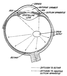
The vitreous chamber bathes the posterior surface of
the lens and is connected to the anterior chamber via a
fluid channel that encircles the lens and continues through
the pupil. Solutes (including solublized glucocorticoids)
in the vitreous may diffuse anteriorly to the lens, or
around the lens to the anterior chamber outflow apparatus
(the trabecular meshwork, Sclemm's canal), thereby causing
steroid-induced cataracts, ocular hypertension or glaucoma.
The present inventors have found that steroids that are
only sparingly soluble in vitreal fluid and that have a slow
dissolution rate from the solid to the soluble form do not
migrate well to the anterior segment. While not wishing to
9
CA 02602577 2007-09-27
WO 2007/047607 PCT/US2006/040429
limit the scope of the invention by theory, and only as an
illustration, the Applicants believe that the Gps of the
present invention lack sufficient diffusional force due to
their lack of solubility in the vitreous to move the soluble
steroid through the indicated path to the anterior chamber.
The lipophilicity of the GDs of the present invention, at
the same time, encourages their partition from the aqueous
vitreous fluid to the lipid bilayer of the retinal cell
membranes. This is thought to create a low-level
intravitreal flow of the GD from vitreous to retina, at a.
concentration sufficient to provide therapeutic benefit to
the retinal tissue, but at a low enough level to confer
substantially reduced exposure to the lens and anterior
segment tissues.
Brief Description of the Drawings
Figure 1 is a view of the human eye, showing the
anterior and posterior segments.
Figure 2 shows scanning ocular fluorophotemetry traces
of fluorescein leakage (arbitrary fluorescence units) from
rabbit retina and iris in a single eye two days after
intravitreal VEGF injection in that eye, and 50 minutes
after intravenous fluorescein injection (12 mg/kg).
Figure 3 shows scanning ocular fluorophotemetry traces
of fluorescein leakage (arbitrary fluorescence units) from
rabbit retina and iris in a single eye treated with 1 mg
(100 L) crystalline dexamethasone suspended in PBS, two
days after intravitreal VEGF injection in that eye, and 50
minutes after intravenous fluorescein injection (12 mg/kg).
The results indicate that intravitreally-administered
dexamethasone is present in both posterior and anterior
segments to inhibit BRB and BAB breakdown, respectively.
CA 02602577 2007-09-27
WO 2007/047607 PCT/US2006/040429
Figure 4 shows scanning ocular fluorophotemetry traces
of fluorescein leakage (arbitrary fluorescence units) from
rabbit retina and iris in a single eye treated with 1 mg of
triamcinolone acetonide contained in 100 I, of an aqueous
suspension and injected into the vitreous under the same
conditions described for Figure 3. This also completely
inhibited VEGF-stimulated BRB and BAB breakdown.
Figure 5 shows scanning ocular fluorophotemetry traces
of fluorescein leakage (arbitrary fluorescence units) from
rabbit retina and iris in a single eye treated with 100 1
(1 mg) of an aqueous suspension of beclomethasone was
injected into the vitreous of a rabbit eye, followed by VEGF
as described above. As with dexamethasone and
triamcinolone, beclomethasone inhibited the VEGF-induced BRB
and BAB breakdown.
Figure 6 shows scanning ocular fluorophotemetry traces
of fluorescein leakage in rabbit eye injected with VEGF, and
indicates that 1 mg (100 1) fluticasone propionate followed
by intravitreal administration of VEGF, completely blocks
BRB breakdown but has no effect on BAB breakdown.
Figure 7 shows scanning ocular fluorophotemetry traces
of fluorescein leakage in rabbit eye injected with VEGF, and
indicates that 1 mg (100 1) beclomethasone 17,21-
dipropionate followed by intravitreal administration of
VEGF, completely blocks BRB breakdown but has no effect on
BAB breakdown.
The GDs of the present materials refer to agents that
bind or interact with and activate the glucocorticoid
receptor. Preferably, the agents bind or interact with the
GR to a greater extent than to the mineralcorticoid
receptor, even more preferably to an extent at least twice
as great, or at least 5 times as great, or at least 10 times
11
CA 02602577 2007-09-27
WO 2007/047607 PCT/US2006/040429
as great, or at least 50 times as great, or at least 100
times as great or at least 1000 times as great as the
mineralcorticoid receptor. The GDs of the therapeutic
component have a greater vitreous humor/aqueous humor
concentration ratio and greater vitreal half-life than other
steroids identically administered, such as dexamethasone and
triamcinolone acetonide.
Posteriorly-directed GDs can be screened, for example,
by injecting the potential GD into a rabbit vitreous. The
vitreous humor and aqueous humor can be sampled as a
function of time, and the amount of the potential GD in the
vitreous and aqueous humor can be measured. The vitreous
concentration of the potential GD can be plotted as a
function of time, and using standard pharmacokinetic
techniques, the vitreous half-life for the GD and clearance
of the potential GD can be calculated.
Similarly, the aqueous concentration of the GD can be
plotted as a function of time, and standard pharmacokinetic
techniques can be used to determine the anterior clearance
of the potential GD. Agents with desired vitreal half-lives
and/or that are selectively present in the vitreous humor
rather than the aqueous humor are used in the present
materials. For example, agents that have vitreous half-
lives greater than about three hours can be selected for the
present ophthalmically therapeutic materials.
Pathological Conditions of the Posterior Segment
In part, the present invention is generally drawn to
methods for treating the posterior segment of the eye.
Preferably, the posterior segment of the eye comprises,
without limitation, the uveal tract, vitreous, retina,
choroid, optic nerve, and the retinal pigmented epithelium
(RPE). The disease or condition related to this invention
12
CA 02602577 2007-09-27
WO 2007/047607 PCT/US2006/040429
may comprise any disease or condition that can be prevented
or treated by the action of a glucocorticoid, especially a
GD, upon a posterior part of the eye. While not intending
to limit the scope of this invention in any way, some
examples of diseases or conditions that can be prevented or
treated by the action of an active drug upon the posterior
part of the eye in accordance with the present invention may
include maculopathies/retinal degeneration such as macular
edema, anterior uveitis, retinal vein occlusion, non-
exudative age related macular degeneration, exudative age
related macular degeneration (ARMD), choroidal
neovascularization, diabetic retinopathy, acute macular
neuroretinopathy, central serous chorioretinopathy, cystoid
macular edema, and diabetic macular edema;
uveitis/retinitis/choroiditis such as acute multifocal
placoid pigment epitheliopathy, Behcet's disease, birdshot
retinochoroidopathy, infections (syphilis, lyme,
tuberculosis, toxoplasmosis), intermediate uveitis (pars
planitis), multifocal choroiditis, multiple evanescent
white dot syndrome (mewds), ocular sarcoidosis, posterior
scleritis, serpiginous choroiditis, subretinal fibrosis and
uveitis syndrome, Vogt-Koyanagi-and Harada syndrome;
vascular diseases/exudative diseases such as retinal
arterial occlusive disease, central retinal vein occlusion,
disseminated intravascular coagulopathy, branch retinal
vein occlusion, hypertensive fundus changes, ocular
ischemic syndrome, retinal arterial microaneurysms, Coat's
disease, parafoveal telangiectasis, hemiretinal vein
occlusion, papillophlebitis, central retinal artery
occlusion, branch retinal artery occlusion, carotid artery
disease (CAD), frosted branch angiitis, sickle cell
retinopathy and other hemoglobinopathies, angioid streaks,
familial exudative vitreoretinopathy, and Eales disease;
traumatic/surgical conditions such as sympathetic
13
CA 02602577 2007-09-27
WO 2007/047607 PCT/US2006/040429
ophthalmia, uveitic retinal disease, retinal detachment,
trauma, conditions caused by laser, conditions caused by
photodynamic therapy, photocoagulation, hypoperfusion during
surgery, radiation retinopathy, and bone marrow transplant
retinopathy; proliferative disorders such as proliferative
vitreal retinopathy and epiretinal membranes, and
proliferative diabetic retinopathy; infectious disorders
such as ocular histoplasmosis, ocular toxocariasis, presumed
ocular histoplasmosis syndrome (POHS), endophthalmitis,
toxoplasmosis, retinal diseases associated with HIV
infection, choroidal disease associate with HIV infection,
uveitic disease associate with HIV infection, viral
retinitis, acute retinal necrosis, progressive outer
retinal necrosis, fungal retinal diseases, ocular syphilis,
ocular tuberculosis, diffuse unilateral subacute
neuroretinitis, and myiasis; genetic disorders such as
retinitis pigmentosa, systemic disorders with accosiated
retinal dystrophies, congenital stationary night blindness,
cone dystrophies, Stargardt's disease and fundus
flavimaculatus, Best's disease, pattern dystrophy of the
retinal pigmented epithelium, X-linked retinoschisis,
Sorsby's fundus dystrophy, benign concentric maculopathy,
Bietti's crystalline dystrophy, and pseudoxanthoma
elasticum; retinal tears/holes such as retinal detachment,
macular hole, and giant retinal tear; tumors such as
retinal disease associated with tumors, congenital
hypertrophy of the retinal pigmented epithelium, posterior
uveal melanoma, choroidal hemangioma, choroidal osteoma,
choroidal metastasis, combined hamartoma of the retina and
retinal pigmented epithelium, retinoblastoma,
vasoproliferative tumors of the ocular fundus, retinal
astrocytoma, and intraocular lymphoid tumors; and
miscellaneous other diseases affecting the posterior part
of the eye such as punctate inner choroidopathy, acute
14
CA 02602577 2007-09-27
WO 2007/047607 PCT/US2006/040429
posterior multifocal placoid pigment epitheliopathy, myopic
retinal degeneration, and acute retinal pigement
epitheliitis. Preferably, the disease or condition is
retinitis pigmentosa, proliferative vitreal retinopathy
(PVR), age-related macular degeneration (ARMD), diabetic
retinopathy, diabetic macular edema, retinal detachment,
retinal tear, uveitus, or cytomegalovirus retinitis.
Glaucoma can also be considered a posterior ocular condition
because the therapeutic goal is to prevent the loss of or
reduce the occurrence of loss of vision due to damage to or
loss of retinal cells or optic nerve cells (i.e.
neuroprotection).
The present materials claimed, and used in the methods
claimed, herein include, without limitation, liquid-
containing compositions (such as formulations) and polymeric
drug delivery systems. The present compositions may be
understood to include solutions, suspensions, emulsions, and
the like, such as other liquid-containing compositions used
in ophthalmic therapies. Polymeric drug delivery systems
comprise a polymeric component, and may be understood to
include biodegradable implants, nonbiodegradable implants,
biodegradable microparticles, such as biodegradable
microspheres, and the like. The present drug delivery
systems may also be understood to encompass elements in the
form of tablets, wafers, rods, sheets, and the like. The
polymeric drug delivery systems may be solid, semisolid, or
viscoelastic.
As used herein, "periocular" administration refers to
delivery of the therapeutic component to a retrobulbar
region, a subconjunctival region, a subtenon region, a
suprachoroidal region or space, and/or an intrascleral
region or space. For example, a posterior directed GD may
be associated with water, saline, a polymeric liquid or
semisolid carrier, phosphate buffer, or other ophthalmically
CA 02602577 2013-07-05
WO 2007/047607
PCT/IIS2006/040429
acceptable liquid carrier. The present liquid-containing
compositions are preferably in an injectable form. In other
words, the compositions may be intraocularly administered,
such as by intravitreal injection, using a syringe and
needle or other similar device (e.g., see U.S. Patent
Publication No. 2003/0060763, or the compositions
can be periocularly administered using an injection
device.
Also as used herein the term a "therapeutically
effective" amount or concentration means an amount or
concentration of a GD or a GD-containing composition
sufficient, when applied to the posterior segment of the
eye, to improve at least one symptom of a disease, condition
or disorder affecting said posterior seyment, as compared to
an untreated eye.
A "biologically significant amount" means an amount of
a GD or other steroid present in the anterior segment of an
eye sufficient to cause a statistically significant increase
in either or both a) intraocular pressure or b) cataract
formation as compared to an untreated eye.
The GD of the present methods and compositions may be
present in an amount in the range of about 0.05% or less, or
about 0.1% or about 0.2% or about 0.5% to about 5% or about
10% or about 20% or about 30% or more (w/v) of the
composition. While the GD may be contained in solution
(including, without limitation, a supersaturated solution),
in a preferred embodiment the GD is present, at least in
part, as crystals or particles in a suspension.
For intravitreally administered compositions, providing
relatively high concentrations of the GD (for example, in
the form of crystals) may be beneficial in that reduced
amounts of the composition may be required to be placed or
injected into the posterior segment of the eye in order to
provide the same amount or more of the therapeutic component
16
CA 02602577 2007-09-27
WO 2007/047607 PCT/US2006/040429
in the posterior segment of the eye relative to other
compositions.
In certain embodiments, the material further Comprises
a GD and an excipient component. The excipient component
may be understood to include solubilizing agents, viscosity
inducing agents, buffer agents, tonicity agents,
preservative agents, and the like.
In some embodiments of the present compositions, a
solubilizing agent may be a cyclodextrin. In other words,
the present materials may comprise a cyclodextrin component
provided in an amount from about 0.1% (w/v) to about 5%
(w/v) of the composition. In further embodiments, the
cyclodextrin comprises up to about 10% (w/v) of certain
cyclodextrins, as discussed herein. In further embodiments,
the cyclodextrin comprises up to about 60% (w/v) of certain
cyclodextrins, as discussed herein. The excipient component
of the present compositions may comprise one or more types
of cyclodextrins or cyclodextrin derivatives, such as alpha-
cyclodextrins, beta-cyclodextrins, gamma-cyclodextrins, and
derivatives thereof. As understood by persons of ordinary
skill in the art, cyclodextrin derivatives refer to any
substituted or otherwise modified compound that has the
characteristic chemical structure of a cyclodextrin
sufficiently to function as a cyclodextrin, for example, to
enhance the solubility and/or stability of therapeutic
agents and/or reduce unwanted side effects of the
therapeutic agents and/or to form inclusive complexes with
the therapeutic agents.
Viscosity inducing agents of the present materials,
include without limitation, polymers that are effective in
stabilizing the therapeutic component in the composition.
The viscosity-inducing component is present in an effective
amount in increasing, advantageously substantially
increasing, the viscosity of the composition. Increased
17
CA 02602577 2007-09-27
WO 2007/047607 PCT/US2006/040429
viscosities of the present compositions may enhance the
ability of the present compositions to maintain the GD,
including GD-containing particles, in substantially uniform
suspension in the compositions for prolonged periods of
time, for example, for at least about one week, without
requiring resuspension processing. The relatively high
viscosity of certain of the present compositions may also
have an additional benefit of at least assisting the
compositions to have the ability to have an increased amount
or concentration of the GD, as discussed elsewhere herein,
for example, while maintaining such GD in substantially
uniform suspension for prolonged periods of time.
Direct Intraocular Administration
Preferably, the GDs of the present invention are
administered directly to the vitreous chamber of the eye, by
means including adminiostration of a solution, suspension,
or other means of carrying of crystals or particles of the
GD, or as part of an intravitreal implant, by, for example,
incision or injection.
The vitreous humor contained in the posterior chamber
of the eye is a viscous aqueous substance. Injection of a
fluid or suspension of substantially lower viscosity into
the posterior segment could therefore result in the presence
of two phases or layers of different density within the eye,
which in turn can lead to either "pooling" of GD particles
or floating of the less dense solution. If the injected or
inserted material contains a drug in the form of a solid
(for example as crystals, particles or an unsutured implant
or reservoir), the solid material will fall to the bottom of
the eye and remain there until it dissolves. Additionally, a
substantially different refractive index between vitreous
18
CA 02602577 2007-09-27
WO 2007/047607 PCT/US2006/040429
and the injected or inserted GD-containing composition may
impair vision.
The therapeutic compositions, including the GDs
described as part of the present invention, may be suspended
in a viscous formulation having a relatively high viscosity,
such as one approximating that of the vitreous humor. Such
viscous formulation comprises a viscosity-inducing
component. The therapeutic agent of the present invention
may be administered intravitreally as, without limitation,
an aqueous injection, a suspension, an emulsion, a solution,
a, gel or inserted in a sustained release or extended release
implant, either biodegradable or non-biodegradable.
The viscosity-inducing component preferably comprises a
polymeric component and/or at least one viscoelastic agent,
such as those materials that are useful in ophthalmic
surgical procedures.
Examples of useful viscosity-inducing components
include, but are not limited to, hyaluronic acid, carbomers,
polyacrylic acid, cellulosic derivatives, polycarbophil,
polyvinylpyrrolidone, gelatin, dextrin, polysaccharides,
polyacrylamide, polyvinyl alcohol, polyvinyl acetate,
derivatives thereof and mixtures thereof.
The molecular weight of the viscosity-inducing
components may be in a range up to about 2 million Daltons,
such as of about 10,000 Daltons or less to about 2 million
Daltons or more. In one particularly useful embodiment, the
molecular weight of the viscosity-inducing component is in a
range of about 100,000 Daltons or about 200,000 Daltons to
about 1 million Daltons or about 1.5 million Daltons.
In one very useful embodiment, a viscosity-inducing
component is a polymeric hyaluronate component, for example,
a metal hyaluronate component, preferably selected from
alkali metal hyaluronates, alkaline earth metal hyaluronates
and mixtures thereof, and still more preferably selected
19
CA 02602577 2007-09-27
WO 2007/047607 PCT/US2006/040429
from sodium hyaluronates, and mixtures thereof. The
molecular weight of such hyaluronate component preferably is
in a range of about 50,000 Daltons or about 100,000 Daltons
to about 1.3 million Daltons or about 2 million Daltons.
In one embodiment, the GDs of the present invention may
be comprised in a polymeric hyaluronate component in an
amount in a range about 0.01% to about 0.5% (w/v) or more.
In a further useful embodiment, the hyaluronate component is
present in an amount in a range of about 1% to about 4%
(w/v) of the composition. In this latter case, the very high
polymer viscosity forms a gel that slows the sedimentation
rate of any suspended drug, and prevents pooling of injected
GD.
The GD of this aspect of the claimed invention may
include any or all salts, prodrugs, conjugates, or
precursors of such therapeutically useful GDs, including
those specifically identified herein.
In certain embodiments, the compositions of the present
invention may comprise more than one therapeutic agent, so
long as at least one such therapeutic agent is a GD having
one or more of the properties described herein as important
to preventing migration of the GD into the anterior segment
and/or penetration of the GD into tissue of the posterior
segment, which tissue may include, without limitation,
retinal tissue. In other words, a therapeutic composition
of the present invention, however administered, may include
a first therapeutic agent, and one or more additional
therapeutic agent, or a combination of therapeutic agents,
so long as at least one of such therapeutic agents is a GD.
One or more of the therapeutic agents in such compositions
may be formed as or present in particles or crystals.
In these aspects of the present invention, the
viscosity-inducing component is present in an effective
amount to increase, advantageously substantially increase,
CA 02602577 2007-09-27
WO 2007/047607 PCT/US2006/040429
the viscosity of the composition. Without wishing to limit
the invention to any particular theory of operation, it is
believed that increasing the viscosity of the compositions
to values well in excess of the viscosity of water, for
example, at least about 100 cps at a shear rate of
0.1/second, compositions which are highly effective for
placement, e.g., injection, into the posterior segment of an
eye of a human or animal are obtained. Along with the
advantageous placement or injectability of the these GD-
containing compositions into the posterior segment, the
relatively high viscosity of the present compositions are
believed to enhance the ability of such compositions to
maintain the therapeutic component (for example, comprising
GD-containing particles) in substantially uniform suspension
in the compositions for prolonged periods of time, and may
aid in the storage stability of the composition.
Advantageously, the compositions of this aspect of the
invention may have viscosities of at least about 10 cps or
at least about 100 cps or at least about 1000 cps, more
preferably at least about 10,000 cps and still more
preferably at least about 70,000 cps or more, for example up
to about 200,000 cps or about 250,000 cps, or about 300,000
cps or more, at a shear rate of 0.1/second. In particular
embodiments the present compositions not only have the
relatively high viscosity noted above but also have the
ability or are structured or made up so as to be effectively
able to be placed, e.g., injected, into a posterior segment
of an eye of a human or animal, preferably through a 27
gauge needle, or even through a 30 gauge needle.
The viscosity inducing components preferably are shear
thinning components such that as the viscous formulation is
passed through or injected into the posterior segment of an
eye, for example, through a narrow aperture, such as 27
gauge needle, under high shear conditions the viscosity of
21
CA 02602577 2007-09-27
WO 2007/047607
PCT/US2006/040429
the composition is substantially reduced during such
passage. After such passage, the composition regains
substantially its pre-injection viscosity so as to maintain
any GD-containing particles in suspension in the eye.
Any ophthalmically acceptable viscosity-inducing
component may be employed in accordance with the GDs in the
present invention. Many such viscosity-inducing components
have been proposed and/or used in ophthalmic compositions
used on or in the eye. The viscosity-inducing component is
present in an amount effective in providing the desired
viscosity to the composition. Advantageously, the
viscosity-inducing component is present in an amount in a
range of about 0.5% or about 1.0% to about 5% or about 10%
or about 20% (w/v) of the composition. The specific amount
of the viscosity inducing component employed depends upon a
number of factors including, for example and without
limitation, the specific viscosity inducing component being
employed, the molecular weight of the viscosity inducing
component being employed, the viscosity desired for the GD-
containing composition being produced and/or used and
similar factors.
Biocompatible Polymers
In another embodiment of the invention, the therapeutic
agents (including at least one GD) may be delivered
intraocularly in a composition that comprises, consists
essentially of, or consists of, a therapeutic agent
comprising a GD and a biocompatible polymer suitable for
administration to the posterior segment of an eye. For
example, the composition may, without limitation, comprise
an intraocular implant or a liquid or semisolid polymer.
Some intraocular implants are described in publications
including U.S. Patents No. 6,726,918; 6,699,493; 6,369,116;
22
CA 02602577 2013-07-05
WO 2007/047607
PCT/US2006/040429
6,331,313; 5,869,079; 5,824,072; 5,766,242; 5,632,984;
and 5,443,505. These are only examples of particular
preferred implants, and others will be available to the
person of ordinary skill in the art.
The polymer in combination with the GD-containing
therapeutic agent may be understood to be a polymeric
component. In some embodiments, the particles may comprise
D,L-polylactide (PLA) or latex (carboxylate modified
polystyrene beads). In other embodiments the particles may
comprise materials other than D,L-polylactide (PLA) or latex
(carboxylate modified polystyrene beads). In certain
embodiments, the polymer component may comprise a
polysaccharide. For example, the polymer component may
comprise a mucopolysaccharide. In at least one specific
embodiment, the polymer component is hyaluronic acid.
However, in additional embodiments, and regardless of
the method of GD administration, the polymeric component may
comprise any polymeric material useful in a body of a
mammal, whether derived from a natural source or synthetic:
Some additional examples of useful polymeric materials for
the purposes of this invention include carbohydrate based
polymers such as methylcellulose, carboxymethylcellulose,
hydroxymethylcellulose hydroxypropylcellulose,
hydroxyethylcellulose, ethyl cellulose, dextrin,
cyclodextrins, alginate, hyaluronic acid and chitosan,
protein based polymers such as gelatin, collagen and
glycolproteins, and hydroxy acid polyesters such as
bioerodable polylactide-coglycolide (PLGA), polylactic acid
(PLA), polyglycolide, polyhydroxybutyric acid,
polycaprolactone, polyvalerolactone, polyphosphazene, and
polyorthoesters. Polymers can also be crosslinked, blended
23
CA 02602577 2007-09-27
WO 2007/047607 PCT/US2006/040429
or used as copolymers in the invention. Other polymer
carriers include albumin, polyanhydrides, polyethylene
glycols, polyvinyl polyhydroxyalkyl methacrylates,
pyrrolidone and polyvinyl alcohol.
Some examples of non-erodible polymers include
silicone, polycarbonates, polyvinyl chlorides, polyamides,
polysulfones, polyvinyl acetates, polyurethane, ethylvinyl
acetate derivatives, acrylic resins, crosslinked polyvinyl
alcohol and crosslinked polyvinylpyrrolidone, polystyrene
and cellulose acetate derivatives.
These additional polymeric materials may be useful in a
composition comprising the therapeutically useful GD agents
disclosed herein, or for use in any of the methods,
including those involving the intravitreal administration of
such methods. For example, and without limitation, PLA or
PLGA may be coupled to a GD for use in the present
invention, either as particles in suspension or as part of
an implant. This insoluble conjugate will slowly erode over
time, thereby continuously releasing the GD.
The term "biodegradable polymer" refers to a polymer or
polymers which degrade in vivo, and wherein erosion of the
polymer or polymers over time occurs concurrent with or
subsequent to release of the therapeutic GD agent. The
terms "biodegradable" and "bioerodible" are equivalent and
are used interchangeably herein. A biodegradable polymer
may be a homopolymer, a copolymer, or a polymer comprising
more than two different polymeric units.
The term "therapeutically effective amount" as used
herein, refers to the level or amount of GD agent needed to
treat a condition of the posterior segment, or reduce or
prevent ocular injury or damage without causing significant
negative or adverse side effects to the anterior segment of
the eye.
24
CA 02602577 2007-09-27
WO 2007/047607 PCT/US2006/040429
Formulation Vehicles
Regardless of the mode of administration or form (e.g.,
in solution, suspension, as a topical, injectable or
implantable agent), the GD-containing therapeutic
compositions of the present invention will be administered
in a pharmaceutically acceptable vehicle component. The
therapeutic agent or agents may also be combined with a
pharmaceutically acceptable vehicle component in the
manufacture of a composition. In other words, a
composition, as disclosed herein, may comprise a therapeutic
component and an effective amount of a pharmaceutically
acceptable vehicle component. In at least one embodiment,
the vehicle component is aqueous-based. For example, the
composition may comprise water.
In certain embodiments, the GD-containing therapeutic
agent is administered in a vehicle component, and may also
include an effective amount of at least one of a viscosity
inducing component, a resuspension component, a preservative
component, a tonicity component and a buffer component. In
some embodiments, the compositions disclosed herein include
no added preservative component. In other embodiments, a
composition may optionally include an added preservative
component. In addition, the composition may be included with
no resuspension component.
Formulations for topical or intraocular administration
of the GD-containing thereapeutic agents (including, without
limitation, implants or particles containing such agents)
will preferably include a major amount of liquid water. Such
compositions are preferably formulated in a sterile form,
for example, prior to being used in the eye. The above-
mentioned buffer component, if present in the intraocular
CA 02602577 2007-09-27
WO 2007/047607 PC T/US2006/040429
formulations, is present in an amount effective to control
the pH of the composition. The formulations may contain,
either in addition to, or instead of the buffer component at
least one tonicity component in an amount effective to
control the tonicity or osmolality of the compositions.
Indeed, the same component may serve as both a buffer
component and a tonicity component. More preferably, the
present compositions include both a buffer component and a
tonicity component.
The buffer component and/or tonicity component, if
either is present, may be chosen from those that are
conventional and well known in the ophthalmic art. Examples
of such buffer components include, but are not limited to,
acetate buffers, citrate buffers, phosphate buffers, borate
buffers and the like and mixtures thereof. Phosphate buffers
are particularly useful. Useful tonicity components
include, but are not limited to, salts, particularly sodium
chloride, potassium chloride, any other suitable
ophthalmically acceptably tonicity component and mixtures
thereof. Non-ionic tonicity components may comprise polyols
derived from sugars, such as xylitol, sorbitol, mannitol,
glycerol and the like.
The amount of buffer component employed preferably is
sufficient to maintain the pH of the composition in a range
of about 6 to about 8, more preferably about 7 to about 7.5.
The amount of tonicity component employed preferably is
sufficient to provide an osmolality to the present
compositions in a range of about 200 to about 400, more
preferably about 250 to about 350, mOsmol/kg respectively.
Advantageously, the present compositions are substantially
isotonic.
The compositions of, or used in, the present invention
may include one or more other components in amounts
effective to provide one or more useful properties and/or
26
CA 02602577 2007-09-27
WO 2007/047607 PCT/US2006/040429
benefits to the present compositions. For example, although
the present compositions may be substantially free of added
preservative components, in other embodiments, the present
compositions include effective amounts of preservative
components, preferably such components that are more
compatible with or friendly to the tissue in the posterior
segment of the eye into which the composition is placed than
benzyl alcohol. Examples of such preservative components
include, without limitation, quaternary ammonium
preservatives such as benzalkonium chloride ("BAC" or "BAK")
and polyoxamer; bigunanide preservatives such as
polyhexamethylene biguandide (PHMB); methyl and ethyl
parabens; hexetidine; chlorite components, such as
stabilized chlorine dioxide, metal chlorites and the like;
other ophthalmically acceptable preservatives and the like
and mixtures thereof. The concentration of the preservative
component, if any, in the present compositions is a
concentration effective to preserve the composition, and
(depending on the nature of the particular preservative
used) is often and generally used in a range of about
0.00001% to about 0.05% (w/v) or about 0.1% (w/v) of the
composition.
Intravitreal delivery of therapeutic agents can be
achieved by injecting a liquid-containing composition into
the vitreous, or by placing polymeric drug delivery systems,
such as implants and microparticles, such as microspheres,
into the vitreous. Examples of biocompatible implants for
placement in the eye have been disclosed in a number of
patents, such as U.S. Pat. Nos. 4,521,210; 4,853,224;
4,997,652; 5,164,188; 5,443,505; 5,501,856; 5,766,242;
5,824,072; 5,869,079; 6,074,661; 6,331,313; 6,369,116; and
6,699,493.
Other route of administering the GD-containing
therapeutic agents of the present invention to the interior
27
CA 02602577 2007-09-27
WO 2007/047607
PCT/US2006/040429
of the eye may include periocular delivery of drugs to a
patient. Penetration of drugs directly into the posterior
segment of the eye is restricted by the blood-retinal
barriers. The blood-retinal barrier is anatomically
separated into inner and outer blood barriers. Movement of
solutes or drugs into the internal ocular structures from
the periocular space is restricted by the retinal pigment
epithelium (RPE), the outer blood-retinal barrier. The
cells of this structure are joined by zonulae oclludentae
intercellular junctions. The RPE is a tight ion
transporting barrier that restricts paracellular transport
of solutes across the RPE. The permeability of most
compounds across the blood-retinal barriers is very low.
Lipophilic compounds, however, such as chloramphenical and
benzyl penicillin, can penetrate the blood-retinal barrier
achieving appreciable concentrations in the vitreous humor
after systemic administration. The lipophilicity of the
compound correlates with its rate of penetration and is
consistent with passive cellular diffusion. The blood
retinal barrier, however, is impermeable to polar or charged
compounds in the absence of a transport mechanism.
Structure of Exemplary GDs
The GDs of the present invention are compounds that 1)
selectively bind to and activate the glucocorticoid receptor
(glucocorticoids), 2) have an aqueous solubility less than
that of triamcinolone acetonide (21 g/ml) and/or a
lipophilicity (log P) greater than that of triamcinolone
acetonide (2.53). Log P is the lipophilicity coefficient,
where P is the octonol/water partition coefficient.
According to the present patent application, the basic
steroid ring structure is as follows
28
CA 02602577 2007-09-27
WO 2007/047607
PCT/US2006/040429
18
CH3
19 11 12 13 17
CH3 H 16
14= 13 a,
,
1 9
2 10 8
3 5 7
4 6
For example, the phosphate salt of the glucocorticoid
dexamethosone has the following structure:
OH
P¨OH
0
0
CH3
HO ah
CH3
=..ffilicH3
5 0
IMP
11
Similarly, the glucocorticoid triamcinolone acetonide
has the structure:
OH
0 CH
CH3
HO
11 11011,
CH3
CH3 H
0
29
CA 02602577 2007-09-27
WO 2007/047607 PCT/US2006/040429
The Glucocorticoid Derivatives (GDs) used in the
compositions and methods of the present invention also
selectively bind to and activate the glucocorticoid
receptor, have an aqueous solubility less than that of
triamcinolone acetonide (21 g/ml) and/or a lipophilicity
(log P) greater than that of triamcinolone acetonide (2.53).
In a useful embodiment, the GDs of the present
invention comprise an acyl group linked via an ester linkage
to a glucocorticoid at the C17 position and/or the C21
position (if the latter carbon atom is present). Preferably
the ester is a monoester linkage. However, in another
embodiment the ester is a diester linkage. Useful acyl
groups include, without limitation, the acetyl, butyryl,
valeryl, propionyl, or furoyl groups. Additional
potentially useful groups would include the benzoyl group
and/or other substituted or unsubstituted cyclic or aromatic
acyl groups. Ideally, the acyl group(s) should have high
hydrophobicity; thus alkyl or aromatic acyl groups are
particularly preferred in the present application, while
those containing polar substituents are less preferred, and
in some embodiments of the invention are absent. In certain
of the embodiments of the present invention acyl group is
linked to the steroid by a thiol ester.
Certain C17 and/or C21 acyl ester-substituted
glucocorticoids are used for treatment of inflammatory and
other conditions by routes including, without limitation,
such as topical skin or systemic administration. For
example, beclomethasone dipropionate is used in the
treatment of bronchial asthma and to shrink nasal polyps. It
is formulated in a powder form, and is administered by
inhalation. It has the following structure:
CA 02602577 2007-09-27
WO 2007/047607
PCT/US2006/040429
0
o
CH3 0
HO
= CH3 1010
0
OSA
While beclomethasone dipropionate is sometimes called
simply "beclomethasone", this is an incorrect use of the
chemical nomenclature. Unsubstituted beclomethasone has the
following structure:
OH
0
CH3
HO
CH3
0
IMO"
Another compound comprises fluticasone propionate,
having the following structure:
31
CA 02602577 2007-09-27
WO 2007/047607
PCT/US2006/040429
0
CH3
HO
GH,
..... 0
0 00 0.13
Relative to a "parent" glucocorticoid lacking
hydrophobic substitutions(for example, an identical compound
lacking the indicated substitutions of a hydrophoblic
(preferably acyl ester) group at positions Cri and/or Cn),
the addition of such substitutions in accordance with the
present invention tends to result in a decreased solubility
in aqueous medium and increased lipophilicity coefficient
(log P. where P is the octanol/water partition coefficient),
and slow the compound's dissolution rate from the crystal to
the solublized phase. These physiochemical attributes
experimentally reduce the amount of compound migrating from
the posterior segment to the anterior segment, thereby
resulting in reduced anterior-segment related side effects.
At the same time, these compounds are better able to migrate
into the tissues of the posterior segment, such as the
retina, the RPE, etc.), thereby selectively being directed
to such tissue. When the GDs are administered to the
vitreous in crystalline or particulate form, the GDs possess
an extended duration of action with intravitreal delivery
compared to the parent glucocorticoid.
A non-exclusive list of currently preferred GDs
includes, without limitation, dexamethasone 17-acetate,
dexamethasone 17, 21-acetate, dexamethasone 21-acetate,
32
CA 02602577 2007-09-27
WO 2007/047607 PCT/US2006/040429
clobetasone 17-butyrate, beclomethasone 17, 21-dipropionate,
fluticasone 17-propionate, clobetasol 17-propionate,
betamethasone 17, 21-dipropionate, alclometasone 17,21-
dipropionate, dexamethasone 17,21-dipropionate,
dexamethasone 17-propionate, halobetasol 17-propionate, and
betamethasone 17-valerate. The use of these compounds for
treatment of conditions of the posterior segment of the eye,
particularly by ocular administration, such as intravitreal,
subconjunctival, subscleral or topical ocular administration
will confer a significant therapeutic improvement compared
to existing therapies in the treatment of posterior eye
diseases such as those listed above, which include, without
limitation, dry and wet ARMO, diabetic macular edema,
proliferate diabetic retinopathy, uveitis, and ocular
tumors.
If desired, buffering agents may be provided in an
amount effective to control the pH of the composition.
Tonicity agents may be provided in an amount effective to
control the tonicity or osmolality of the compositions.
Certain of the present compositions include both a buffer
component and a tonicity component, which may include one or
more sugar alcohols, such as mannitol, or salts, such as
sodium chloride, as discussed herein. The buffer component
and tonicity component may be chosen from those that are
conventional and well known in the ophthalmic art. Examples
of such buffer components include, but are not limited to,
acetate buffers, citrate buffers, phosphate buffers, borate
buffers and the like and mixtures thereof. Phosphate
buffers are particularly useful. Useful tonicity components
include, but are not limited to, salts, particularly sodium
chloride, potassium chloride, any other suitable
ophthalmically acceptably tonicity component and mixtures
thereof.
33
CA 02602577 2007-09-27
WO 2007/047607 PCT/US2006/040429
The amount of buffer component employed preferably is
sufficient to maintain the pH of the composition in a range
of about 6 to about 8, more preferably about 7 to about 7.5.
The amount of tonicity component employed preferably is
sufficient to provide an osmolality to the present
compositions in a range of about 200 to about 400, more
preferably about 250 to about 350, mOsmol/kg respectively.
Advantageously, the present compositions are substantially
isotonic.
Preservative agents that may be used in the present
materials include benzyl alcohol, benzalkonium chloride,
methyl and ethyl parabens, hexetidine, chlorite components,
such as stabilized chlorine dioxide, metal chlorites and the
like, other ophthalmically acceptable preservatives and the
like and mixtures thereof. The concentration of the
preservative component, if any, in the present compositions
is a concentration effective to preserve the composition,
and is often in a range of about 0.00001% to about 0.05% or
about 0.1% (w/v) of the composition.
The present compositions can be produced using
conventional techniques routinely known by persons of
ordinary skill in the art. For example, a GD-containing
therapeutic component can be combined with a liquid carrier.
The composition can be sterilized. In certain embodiments,
such as preservative-free embodiments, the compositions can
be sterilized and packaged in single-dose amounts. The
compositions may be prepackaged in intraocular dispensers
which can be disposed of after a single administration of
the unit dose of the compositions.
The present compositions can be prepared using suitable
blending/processing techniques, for example, one or more
conventional blending techniques. The preparation
processing should be chosen to provide the present
compositions in forms which are useful for intravitreal or
34
CA 02602577 2007-09-27
WO 2007/047607 PCT/US2006/040429
periocular placement or injection into eyes of humans or
animals. In one useful embodiment a concentrated
therapeutic component dispersion is made by combining the
GD-containing therapeutic component with water, and the
excipients (other than the viscosity inducing component) to
be included in the final composition. The ingredients are
mixed to disperse the therapeutic component and then
autoclaved. The viscosity inducing component may be
purchased sterile or sterilized by conventional processing,
for example, by filtering a dilute solution followed by
lyophylization to yield a sterile powder. The sterile
viscosity inducing component is combined with water to make
an aqueous concentrate. The concentrated therapeutic
component dispersion is mixed and added as a slurry to the
viscosity inducing component concentrate. Water is added in
a quantity sufficient (q.s.) to provide the desired
composition and the composition is mixed until homogenous.
In one embodiment, a sterile, viscous suspension
suitable for administration is made using an GD. A process
for producing such a composition may comprise sterile
suspension bulk compounding and aseptic filling.
Other embodiments of the present materials are in the
form of a polymeric drug delivery system that is capable of
providing sustained drug delivery for extended periods of
time after a single administration. For example, the
present drug delivery systems can release the GD for at
least about 1 month, or about 3 months, or about 6 months,
or about 1 year, or about 5 years or more. Thus, such
embodiments of the present materials may comprise a
polymeric component associated with the therapeutic
component in the form of a polymeric drug delivery system
suitable for administration to a patient by at least one of
intravitreal administration and periocular administration.
CA 02602577 2007-09-27
WO 2007/047607 PCT/US2006/040429
The polymeric drug delivery system may be in the form
of biodegradable polymeric implants, non-biodegradable
polymeric implants, biodegradable polymeric microparticles,
and combinations thereof. Implants may be in the form of
rods, wafers, sheets, filaments, spheres, and the like.
Particles are generally smaller than the implants disclosed
herein, and may vary in shape. For example, certain
embodiments of the present invention utilize substantially
spherical particles. These particles may be understood to
be microspheres. Other embodiments may utilize randomly
configured particles, such as particles that have one or
more flat or planar surfaces. The drug delivery system may
comprise a population of such particles with a predetermined
size distribution. For example, a major portion of the
population may comprise particles having a desired diameter
measurement.
As discussed herein, the polymeric component of the
present drug delivery systems can comprise a polymer
selected from the group consisting of biodegradable
polymers, non-biodegradable polymers, biodegradable
copolymers, non-biodegradable copolymers, and combinations
thereof. In certain embodiments, the polymeric component
comprises a poly (lactide-co-glycolide) polymer (PLGA). In
other embodiments, the polymeric component comprises a
polymer selected from the group consisting of poly-lactic
acid (PLA), poly-glycolic acid (PGA), poly-lactide-co-
glycolide (PLGA), polyesters, poly (ortho ester),
poly(phosphazine), poly (phosphate ester),
polycaprolactones, gelatin, collagen, derivatives thereof,
and combinations thereof. The polymeric component may be
associated with the therapeutic component to form an implant
selected from the group consisting of solid implants,
semisolid implants, and viscoelastic implants.
36
CA 02602577 2007-09-27
WO 2007/047607
PCT/US2006/040429
The GD may be in a particulate or powder form and
entrapped by a biodegradable polymer matrix. Usually, GD
particles in intraocular implants will have an effective
average size measuring less than about 3000 nanometers.
However, in other embodiments, the particles may have an
average maximum size greater than about 3000 nanometers. In
certain implants, the particles may have an effective
average particle size about an order of magnitude smaller
than 3000 nanometers. For example, the particles may have
an effective average particle size of less than about 500
nanometers. In additional implants, the particles may have
an effective average particle size of less than about 400
nanometers, and in still further embodiments, a size less
than about 200 nanometers. In addition, when such particles
are combined with a polymeric component, the resulting
polymeric intraocular particles may be used to provide a
desired therapeutic effect.
If formulated as part of an implant or other drug
delivery system, the GD of the present systems is preferably
from about 1% to 90% by weight of the drug delivery system.
More preferably, the GD is from about 20% to about 80% by
weight of the system. In a preferred embodiment, the GD
comprises about 40% by weight of the system (e.g., 30%-50%).
In another embodiment, the GD comprises about 60% by weight
of the system.
Suitable polymeric materials or compositions for use in
the drug delivery systems include those materials which are
compatible, that is biocompatible, with the eye so as to
cause no substantial interference with the functioning or
physiology of the eye. Such materials preferably include
polymers that are at least partially and more preferably
substantially completely biodegradable or bioerodible.
In addition to the foregoing, examples of useful
polymeric materials include, without limitation, such
37
CA 02602577 2007-09-27
WO 2007/047607 PCT/US2006/040429
materials derived from and/or including organic esters and
organic ethers, which when degraded result in
physiologically acceptable degradation products, including
the monomers. Also, polymeric materials derived from and/or
including, anhydrides, amides, orthoesters and the like, by
themselves or in combination with other monomers, may also
find use. The polymeric materials may be addition or
condensation polymers, advantageously condensation polymers.
The polymeric materials may be cross-linked or non-cross-
linked, for example not more than lightly cross-linked, such
as less than about 5%, or less than about 1% of the
polymeric material being cross-linked. For the most part,
besides carbon and hydrogen, the polymers will include at
least one of oxygen and nitrogen, advantageously oxygen.
The oxygen may be present as oxy, e.g. hydroxy or ether,
carbonyl, e.g. non-oxo-carbonyl, such as carboxylic acid
ester, and the like. The nitrogen may be present as amide,
cyano and amino. The polymers set forth in Heller,
Biodegradable Polymers in Controlled Drug Delivery, In: CRC
Critical Reviews in Therapeutic Drug Carrier Systems, Vol.
1, CRC Press, Boca Raton, FL 1987, pp 39-90, which describes
encapsulation for controlled drug delivery, may find use in
the present drug delivery systems.
Of additional interest are polymers of hydroxyaliphatic
carboxylic acids, either homopolymers or copolymers, and
polysaccharides. Polyesters of interest include polymers of
D-lactic acid, L-lactic acid, racemic lactic acid, glycolic
acid, polycaprolactone, and combinations thereof.
Generally, by employing the L-lactate or D-lactate, a slowly
eroding polymer or polymeric material is achieved, while
erosion is substantially enhanced with the lactate racemate.
Among the useful polysaccharides are, without
limitation, calcium alginate, and functionalized celluloses,
particularly carboxymethylcellulose esters characterized by
38
CA 02602577 2007-09-27
WO 2007/047607 PCT/US2006/040429
being water insoluble, a molecular weight of about 5 kD to
500 kD, for example.
Other polymers of interest include, without limitation,
polyesters, polyethers and combinations thereof which are
biocompatible and may be biodegradable and/or bioerodible.
Some preferred characteristics of the polymers or
polymeric materials for use in the present systems may
include biocompatibility, compatibility with the therapeutic
component, ease of use of the polymer in making the drug
delivery systems of the present invention, a half-life in
the physiological environment of at least about 6 hours,
preferably greater than about one day, not significantly
increasing the viscosity of the vitreous, and water
insolubility.
The biodegradable polymeric materials which are
included to form the matrix are desirably subject to
enzymatic or hydrolytic instability. Water soluble polymers
may be cross-linked with hydrolytic or biodegradable
unstable cross-links to provide useful water insoluble
polymers. The degree of stability can be varied widely,
depending upon the choice of monomer, whether a homopolymer
or copolymer is employed, employing mixtures of polymers,
*and whether the polymer includes terminal acid groups.
Also important to controlling the biodegradation of the
polymer and hence the extended release profile of the drug
delivery systems is the relative average molecular weight of
the polymeric composition employed in the present systems.
Different molecular weights of the same or different
polymeric compositions may be included in the systems to
modulate the release profile. In certain systems, the
relative average molecular weight of the polymer will range
from about 9 to about 64 kD, usually from about 10 to about
54 kD, and more usually from about 12 to about 45 kD.
39
CA 02602577 2007-09-27
WO 2007/047607 PCT/US2006/040429
In some drug delivery systems, copolymers of glycolic
acid and lactic acid are used, where the rate of
biodegradation is controlled by the ratio of glycolic acid
to lactic acid. The most rapidly degraded copolymer has
roughly equal amounts of glycolic acid and lactic acid.
Homopolymers, or copolymers having ratios other than equal,
are more resistant to degradation. The ratio of glycolic
acid to lactic acid will also affect the brittleness of the
system, where a more flexible system or implant is desirable
for larger geometries. The % of polylactic acid in the
polylactic acid polyglycolic acid (PLGA) copolymer can be 0-
100%, preferably about 15-85%, more preferably about 35-65%.
In some systems, a 50/50 PLGA copolymer is used.
The biodegradable polymer matrix of the present systems
may comprise a mixture of two or more biodegradable
polymers. For example, the system may comprise a mixture of
a first biodegradable polymer and a different second
biodegradable polymer. One or more of the biodegradable
polymers may have terminal acid groups.
Release of a drug from an erodible polymer is the
consequence of several mechanisms or combinations of
mechanisms. Some of these mechanisms include desorption
from the implants surface, dissolution, diffusion through
porous channels of the hydrated polymer and erosion.
Erosion can be bulk or surface or a combination of both. It
may be understood that the polymeric component of the
present systems is associated with the therapeutic component
so that the release of the therapeutic component into the
eye is by ane or more of diffusion, erosion, dissolution,
and osmosis. As discussed herein, the matrix of an
intraocular drug delivery system may release drug at a rate
effective to sustain release of an amount of the GD for more
than one week after implantation into an eye. In certain
systems, therapeutic amounts of the GD are released for more
CA 02602577 2007-09-27
WO 2007/047607
PCT/US2006/040429
than about one month, and even for about twelve months or
more. For example, the therapeutic component can be
released into the eye for a time period from about ninety
days to about one year after the system is placed in the
interior of an eye.
The release of the GD from the drug delivery systems
comprising a biodegradable polymer matrix may include an
initial burst of release followed by a gradual increase in
the amount of the GD released, or the release may include an
initial delay in release of the GD followed by an increase
in release. When the system is substantially completely
degraded, the percent of the GD that has been released is
about one hundred.
It may be desirable to provide a relatively constant
rate of release of the therapeutic agent from the drug
delivery system over the life of the system. For example,
it may be desirable for the GD to be released in amounts
from about 0.01 gg to about 2 gg per day for the life of the
system. However, the release rate may change to either
increase or decrease depending on the formulation of the
biodegradable polymer matrix. In addition, the release
profile of the GD may include one or more linear portions
and/or one or more non-linear portions. Preferably, the
release rate is greater than zero once the system has begun
to degrade or erode.
The drug delivery systems, such as the intraocular
implants, may be monolithic, i.e. having the active agent or
agents homogenously distributed through the polymeric
matrix, or encapsulated, where a reservoir of active agent
is encapsulated by the polymeric matrix. Due to ease of
manufacture, monolithic implants are usually preferred over
encapsulated forms. However, the greater control afforded by
the encapsulated, reservoir-type implant may be of benefit
41
CA 02602577 2007-09-27
WO 2007/047607
PCT/US2006/040429
in some circumstances, where the therapeutic level of the GD
falls within a narrow window. In addition, the therapeutic
component, including the therapeutic agent(s) described
herein, may be distributed in a non-homogenous pattern in
the matrix. For example, the drug delivery system may
include a portion that has a greater concentration of the GD
relative to a second portion of the system.
The polymeric implants disclosed herein may have a size
of between about 5 gm and about 2 mm, or between about 10 pm
and about 1 mm for administration with a needle, greater
than 1 mm, or greater than 2 mm, such as 3 mm or up to 10
mm, for administration by surgical implantation. The
vitreous chamber in humans is able to accommodate relatively
large implants of varying geometries, having lengths of, for
example, 1 to 10 mm. The implant may be a cylindrical
pellet (e. g., rod) with dimensions of about 2 mm x 0.75 mm
diameter. Or the implant may be a cylindrical pellet with a
length of about 7 mm to about 10 mm, and a diameter of about
0.75 mm to about 1.5 mm.
The implants may also be at least somewhat flexible so
as to facilitate both insertion of the implant in the eye,
such as. in the vitreous, and accommodation of the implant.
The total weight of the implant is usually about 250-5000
g, more preferably about 500-1000 g. For example, an
implant may be about 500 g, or about 1000 g. However,
larger implants may also be formed and further processed
before administration to an eye. In addition, larger
implants may be desirable where relatively greater amounts
of the GD are provided in the implant. For non-human
individuals, the dimensions and total weight of the
implant(s) may be larger or smaller, depending on the type
of individual. For example, humans have a vitreous volume
of approximately 3.8 ml, compared with approximately 30 ml
for horses, and approximately 60-100 ml for elephants. An
42
CA 02602577 2007-09-27
WO 2007/047607
PCT/US2006/040429
implant sized for use in a human may be scaled up or down
accordingly for other animals, for example, about 8 times
larger for an implant for a horse, or about, for example, 26
times larger for an implant for an elephant.
Drug delivery systems can be prepared where the center
may be of one material and the surface may have one or more
layers of the same or a different composition, where the
layers may be cross-linked, or of a different molecular
weight, different density or porosity, or the like. For
example, where it is desirable to quickly release an initial
bolus of GD, the center may be a polylactate coated with a
polylactate-polyglycolate copolymer, so as to enhance the
rate of initial degradation. Alternatively, the center may
be polyvinyl alcohol coated with polylactate, so that upon
degradation of the polylactate exterior the center would
dissolve and be rapidly washed out of the eye.
The drug delivery systems may be of any geometry
including fibers, sheets, films, microspheres, spheres,
circular discs, plaques and the like. The upper limit for
the system size will be determined by factors such as
toleration for the system, size limitations on insertion,
ease of handling, etc. Where sheets or films are employed,
the sheets or films will be in the range of at least about
0.5 mm x 0.5 mm, usually about 3-10 mm x 5-10 mm with a
thickness of about 0.1-1.0 mm for ease of handling. Where
fibers are employed, the fiber diameter will generally be in
the range of about 0.05 to 3 mm and the fiber length will
generally be in the range of about 0.5-10 mm. Spheres may
be in the range of about 0.5 gm to 4 mm in diameter, with
comparable volumes for other shaped particles.
The size and form of the system can also be used to
control the rate of release, period of treatment, and drug
concentration at the site of implantation. For example,
larger implants will deliver a proportionately larger dose,
43
CA 02602577 2007-09-27
WO 2007/047607
PCT/US2006/040429
but depending on the surface to mass ratio, may have a
slower release rate. The particular size and geometry of
the system are chosen to suit the site of implantation.
The proportions of GD-containing therapeutic agent,
polymer, and any other modifiers may be empirically
determined by formulating several implants, for example,
with varying proportions of such ingredients. A USP
approved method for dissolution or release test can be used
to measure the rate of release (USP 23; NF 18 (1995) pp.
1790-1798). For example, using the infinite sink method, a
weighed sample of the implant is added to a measured volume
of a solution containing 0.9% NaCl in water, where the
solution volume will be such that the drug concentration is
after release is less than 5% of saturation. The mixture is
maintained at 37 C and stirred slowly to maintain the
implants in suspension. The appearance of the dissolved
drug as a function of time may be followed by various
methods known in the art, such as by spectrophotometry,
HPLC, mass spectroscopy, etc. until the absorbance becomes
constant or until greater than 90% of the drug has been
released.
In addition to the GD-containing therapeutic component,
and similar to the compositions described herein, the
polymeric drug delivery systems disclosed herein may include
an excipient component. The excipient component may be
understood to include solubilizing agents, viscosity
inducing agents, buffer agents, tonicity agents,
preservative agents, and the like.
Additionally, release modulators such as those
described in U. S. Patent No. 5,869,079 may be included in
the drug delivery systems. The amount of release modulator
employed will be dependent on the desired release profile,
the activity of the modulator, and on the release profile of
44
CA 02602577 2007-09-27
WO 2007/047607
PCT/US2006/040429
the therapeutic agent in the absence of modulator.
Electrolytes such as sodium chloride and potassium chloride
may also be included in the systems. Where the buffering
agent or enhancer is hydrophilic, it may also act as a
release accelerator. Hydrophilic additives act to increase
the release rates through faster dissolution of the material
surrounding the drug particles, which increases the surface
area of the drug exposed, thereby increasing the rate of
drug bioerosion. Similarly, a hydrophobic buffering agent
or enhancer dissolve more slowly, slowing the exposure of
drug particles, and thereby slowing the rate of drug
bioerosion.
Various techniques may be employed to produce such drug
delivery systems. Useful techniques include, but are not
necessarily limited to, solvent evaporation methods, phase
separation methods, interfacial methods, molding methods,
injection molding methods, extrusion methods, co-extrusion
methods, carver press method, die cutting methods, heat
compression, combinations thereof and the like.
Specific methods are discussed in U.S. Pat. No.
4,997,652. Extrusion methods may be used to avoid the need
for solvents in manufacturing. When using extrusion
methods, the polymer and drug are chosen so as to be stable
at the temperatures required for manufacturing, usually at
least about 85 degrees Celsius. Extrusion methods use
temperatures of about 25 degrees C to about 150 degrees C,
more preferably about 65 degrees C to about 130 degrees C.
An implant may be produced by bringing the temperature to
about 60 degrees C to about 150 degrees C for drug/polymer
mixing, such as about 130 degrees C, for a time period of
about 0 to 1 hour, 0 to 30 minutes, or 5-15 minutes. For
example, a time period may be about 10 minutes, preferably
about 0 to 5 min. The implants are then extruded at a
CA 02602577 2007-09-27
WO 2007/047607 PCT/US2006/040429
temperature of about 60 degrees C to about 130 degrees C,
such as about 75 degrees C.
In addition, the implant may be coextruded so that a
coating is formed over a core region during the manufacture
of the implant.
Compression methods may be used to make the drug
delivery systems, and typically yield elements with faster
release rates than extrusion methods. Compression methods
may use pressures of about 50-150 psi, more preferably about
70-80 psi, even more preferably about 76 psi, and use
temperatures of about 0 degrees C to about 115 degrees C,
more preferably about 25 degrees C.
In certain embodiments of the present invention, a
method of producing a sustained-release intraocular drug
delivery system, comprises combining an GD and a polymeric
material to form a drug delivery system suitable for
placement in an eye of an individual. The resulting drug
delivery system is effective in releasing the GD into the
eye for extended periods of time. The method may comprise a
step of extruding a particulate mixture of the GD and the
polymeric material to form an extruded composition; such as
a filament, sheet, and the like.
When polymeric particles are desired, the method may
comprise forming the extruded composition into a population
of polymeric particles or a population of implants, as
described herein. Such methods may include one or more
steps of cutting the extruded composition, milling the
extruded composition, and the like.
As discussed herein, the polymeric material may
comprise a biodegradable polymer, a non-biodegradable
polymer, or a combination thereof. Examples of polymers
include each and every one of the polymers and agents
identified above.
46
CA 02602577 2007-09-27
WO 2007/047607 PCT/US2006/040429
Embodiments of the present invention also relate to
compositions comprising the present drug delivery systems.
For example, and in one embodiment, a composition may
comprise the present drug delivery system and an
ophthalmically acceptable carrier component. Such a carrier
component may be an aqueous composition, for example saline
or a phosphate buffered liquid.
Another embodiment relates to a method of producing an
ophthalmically therapeutic material which comprises an GD.
In a broad aspect, the method comprises the steps of
selecting an GD and combining the selected GD with a liquid
carrier component or a polymeric component to form a
material suitable for administration to an eye. Or stated
differently, a method of producing the present materials may
comprise a step of selecting GDs having a low aqueous
humor/vitreous humor concentration ratio and long
intravitreal half-life.
The method may further comprise one or more of the
following steps, which will typically be used to select the
GD: administering an GD to an eye of a subject and
determining the concentration of the GD in at least one of
the vitreous humor and aqueous humor as a function of time;
and administering a GD to an eye of a subject and
determining at least one of the vitreous half-life and
clearance of the GD from the posterior chamber of the eye.
The material formed in the method may be a liquid-
containing composition, a biodegradable polymeric implant, a
non-biodegradable polymeric implant, polymeric
microparticles, or combinations thereof. As discussed
herein, the material may be in the form of solid implants,
semisolid implants, and viscoelastic implants. In certain
embodiments, the GD is combined with a polymeric component
to form a mixture, and the method further comprises
extruding the mixture.
47
CA 02602577 2007-09-27
WO 2007/047607 PCT/US2006/040429
Additional embodiments of the present invention related
to methods of improving or maintaining vision of an eye of a
patient. In general, the methods comprise a step of
administering the present ophthalmically therapeutic
material to an eye of an individual in need thereof.
Administration, such as intravitreal or periocular (or less
preferably, topical) administration of the present materials
can be effective in treating posterior ocular conditions
without significantly affecting the anterior chamber. The
present materials may be particularly useful in treating
inflammation and edema of the retina. Administration of the
present materials are effective in delivering the GD to one
or more posterior structures of the eye including the uveal
tract, the vitreous, the retina, the choroid, the retinal
pigment epithelium.
When a syringe apparatus is used to administer the
present materials, the apparatus can include an
appropriately sized needle, for example, a 27-gauge needle
or a 30-gauge needle. Such apparatus can be effectively
used to inject the materials into the posterior segment or a
periocular region of an eye of a human or animal. The
needles may be sufficiently small to provide an opening that
self seals after removal of the needle.
The present methods may comprise a single injection
into the posterior segment of an eye or may involve repeated
injections, for example over periods of time ranging from
about one week or about 1 month or about 3 months to about 6
months or about 1 year or longer.
The present materials are preferably administered to
patients in a sterile form. For example, the present
materials may be sterile when stored. Any routine suitable
method of sterilization may be employed to sterilize the
materials. For example, the present materials may be
sterilized using radiation. Preferably, the sterilization
48
CA 02602577 2007-09-27
WO 2007/047607 PCT/US2006/040429
method does not reduce the activity or biological or
therapeutic activity of the therapeutic agents of the
present systems.
The materials can be sterilized by gamma irradiation.
As an example, the drug delivery systems can be sterilized
by 2.5 to 4.0 mrad of gamma irradiation. The drug delivery
systems can be terminally sterilized in their final primary
packaging system including administration device e.g.
syringe applicator. Alternatively, the drug delivery
systems can be sterilized alone and then aseptically
packaged into an applicator system. In this case the
applicator system can be sterilized by gamma irradiation,
ethylene oxide (ETO), heat or other means. The drug
delivery systems can be steriliZed by gamma irradiation at
low temperatures to improve stability or blanketed with
argon, nitrogen or other means to remove oxygen. Beta
irradiation or e-beam may also be used to sterilize the
implants as well as UV irradiation. The dose of irradiation
from any source can be lowered depending on the initial
bioburden of the drug delivery systems such that it may be
much less than 2.5 to 4.0 mrad. The drug delivery systems
may be manufactured under aseptic conditions from sterile
starting components. The starting components may be
sterilized by heat, irradiation (gamma, beta, UV), ETO or
sterile filtration. Semi-solid polymers or solutions of
polymers may be sterilized prior to drug delivery system
fabrication and GD incorporation by sterile filtration of
heat. The sterilized polymers can then be used to
aseptically produce sterile drug delivery systems.
In another aspect of the invention, kits for treating
an ocular condition of the eye are provided, comprising: a)
a container, such as a syringe or other applicator,
comprising an GD as herein described; and b) instructions
for use. Instructions may include steps of how to handle
49
CA 02602577 2007-09-27
WO 2007/047607 PCT/US2006/040429
the material, how to insert the material into an ocular
region, and what to expect from using the material. The
container may contain a single dose of the GD.
EXAMPLES
Example 1:
Recombinant vascular endothelial growth factor (VEGF)
was obtained from a supplier (R&D Systems). Female Dutch
Belt rabbits were anaesthetized with isoflurane inhalation
and topical 0.5% proparacaine hydrochloride, and
intravitreal injection of one eye with 500 ng VEGF in
sterile phosphate buffered saline (PBS) containing 0.1%
bovine serum albumin was performed using a 28 guage inch
needle. The other eye is given the same volume of the
vehicle, without the VEGF.
The extent of VEGF-induced BRB and BAB breakdown of the
blood retinal barrier and the blood aqueous barrier was
measured by scanning ocular fluorophotemetry (Fluorotron
Master, Ocumetrics Inc.); at various times following
intravitreal injection. In this model a fluorescent label
is administered intravenously, following by determination of
the amount of fluorescenin in the anterior and posterior
segment and an indication of iridial and retinal leakage,
respectively.
Under normal conditions the blood retinal and blood
aqueous barrier prevents solutes in the blood from
infiltrating the vitreous (and to a somewhat lesser but very
significant extent, the aqueous). By contrast, in the
presence of retinal disease such as macular degeneration,
retinopathy, macular edema, retinal neovascularization etc.,
there is leakage of blood into retinal tissue, and the
fluorescent tracer will be visible in the vitreous and
CA 02602577 2013-07-05
WO 2007/047607 PCT/US2006/040429
aqueous of the eye. VEGF injection mimics this pathological
condition.
Figure 2 shows representative traces of fluorescein
leakage (arbitrary fluorescence units) from rabbit retina
and iris from a single eye two days (48 hours) after
intravitreal VEGF injection. Sodium fluorescein in 1 ma
saline was injected via the marginal ear vein at a
concentration of 50 mg/kg, and ocular fluorescein levels in
the vitreoretinal chamber and the anterior chamber was
determined 50 minutes later.
Compared to normal untreated rabbit eyes, VEGF caused
an approximately 18-fold increase in fluorescein contained
in the vitreous, and an approximately 6-fold increase in
fluorescein contained in the aqueous, which reflects
breakdown in the blood retinal barrier (BRB) causing retinal
leakage, and the blood aqueous barrier (BAB) iris leakage),.
respectively.
Both of these responses were completely blocked-by the
corticosteroids dexamethasone, triamcinolone and
beclomethasone, when these corticosteroids were either
administered systemically or intravitreally. See infra and
Edelman et al., EXP. EYE RES. 80:249-258 (2005).
Thus, when, after steroid treatment
both the anterior and posterior chambers are free of
fluorescein leakage following VEGF challenge, this indicates
that the steroid is able to infiltrate both chambers
effectively.
Five corticosteroids (dexamethasone, triamcinolone,
fluticasone propionate, beclomethasone dipropionate and
beclomethasone) were purchased from. Sigma-Aldrich Co. and
evaluated in this model system. In combination these
compounds define a solubility range of nearly three log
units (1000 fold) from the most water soluble to the least
51
CA 02602577 2013-07-05
WO 2007/047607
PCT/US2006/040429
water soluble, and a range of lipophilicity coefficients,
log P, from 1.95 to 4.4.
Ten milligrams of each compound is added to 1 ml of
sterile phosphate-buffered saline (PBS; ph 7.4). At day 0,
100 ml of a 10 mg/ml suspension of each steroid is injected
into the vitreous of a rabbit eye. The PBS vehicle is
injected into the other eye. VEGF is then injected at a
pre-determined time (one month) thereafter, and BRB and BAB
breakdown were measured by scanning ocular fluorophotometry
48 hrs later as described in Edelman et al., EXP. ErEREs.
80:249-259 (2005).
Compound Water Solubility-
Lipophicity (log P)
Dexamethasone 100 pg/m1 1.95
(Sigma cat.#
D1756)
. Triamcinolone 21.0 ug/rnj 2.53
acetonide
(Sigma cat.#
T6501)
Fluticasone 0.14 pgyrni 4.20
propionate
(Sigma cat.#
F9428)
Beclomethasone 0.13 pg/m1 4.40
dipropionate
(Sigma cat.*
83022)
As can be seen, of the compounds tested dexamethasone
(M) had the highest water solubility (100 pg/m1)and
lowest lipophilicity (log P = 1.95) of the five compounds
tested. After intravitreal injection of 1 mg crystalline
dexamethasone suspended in 100 AL PBS, dexamethasone
completely inhibited VEGF-induced leakage of intravenous
fluorescein into both the posterior segment and the anterior
segment, indicating that intravitreally administered
52
=
CA 02602577 2013-07-05
WO 2007/047607 PCT/US2006/040429
dexamethasone is present in both posterior and anterior
segments to inhibit BRB and BAB breakdown, respectively
(Figure 3). Since the BAB is normally relatively leaky
compared to the BRB (see Fig 1), there is some residual
fluorescence observed in the anterior chamber of rabbit eyes
treated with dexamethasone.
This result indicated that intravitreally administered
dexamethasone readily diffuses from the crystal depot within
the vitreous in both directions: in the posterior direction
to the retinal vasculature and in the anterior direction to
the iris. These characteristics result in pharmacologically
active levels within both tissues.
Similar to the result with dexamethasone, 1 mg of
triamcinolone acetonide contained in 100 I.LL of an aqueous
suspension and injected into the vitreous also completely
inhibited VEGF-stimulated BRB and BAB breakdown (Figure 4).
As a final example of the effect of unsubstituted
glucocorticoids, 100 Al of a 10mg/m1 suspension of aqueous
beclomethasone was injected into the vitreous of a rabbit
eye, followed by VEGF as described above. As with
dexamethasone and triamcinolone, beclomethasone inhibited
the VEGF-induced breakdown of the BRB and the BAB (Figure
5).
In contrast, intravitreal injection of rabbit eye with
100111 of a 10mg/m1 suspension of fluticasone propionate
(water solubility 0.14 pg/ml; log P = 4.2), followed by
intravitreal administration of VEGF, completely blocked BRB
breakdown but had no effect on BAB breakdown (Figure 6).
This result indicates that the intravitreally placed drug is
able to diffuse in therapeutically effective concentrations
from the vitreous posteriorly to the retina, but is unable
to diffuse from the posterior chamber to the anterior
chamber in such concentration.
53
CA 02602577 2013-07-05
WO 2007/047607
PCT/US2006/040429
Similarly, another sparingly water soluble compound,
beclomethasone 17,21-dipropionate (0.13 pg/m1; log P = 4.4),
appears to completely block VEGF-induced BRB breakdown, but
has no effect on BAB breakdown (Figure 7). Moreover, 100 gl
of 10mg/ma intravitreal beclomethasone 17,21-dipropionate
completely inhibited VEGF-mediated responses for greater
than 3 months.
These results indicate that GDs that possessing one or
more hydrophobic Cr and/or C21 substitution (in this case,
an acyl monoester functional group, such as propionate) have
reduced water solubility, increased lipophilicity, and are
superior pharmacophores for intravitreal delivery to treat
ocular diseases that largely or solely involve the posterior
segment or have little or no anterior chamber components.
Intravitreal administration of these compounds therefore
display few, reduced, or abrogated anterior segment side
effects such as cataracts, high IOP, and steroid inducted
glaucoma. Specific examples of these coutpounds include
dexamethasone 17-acetate, dexamethasone 17, 21-acetate,
dexamethasone 21-acetate, clobetasone 17-butyrate,
beclomethasone 17, 21-dipropionate, fluticasone 17-
propionate, clobetasol 17-propionate, betamethasone 17, 21-
dipropionate, alclometasone 17,21-dipropionate,
dexamethasone 17,21-dipropionate, dexamethasone 17-
propionate, halobetasol 17-propionate, betamethasone 17-
valerate. These compounds will be a significant
improvement compared to existing therapies in the treatment
of posterior eye diseases including, without limitation, dry
and wet ABM, diabetic macular edema, proliferate diabetic
retinopathy, uveitis, and ocular tumors.
Example 2: GD Implant
54
CA 02602577 2007-09-27
WO 2007/047607
PCT/US2006/040429
Biodegradable drug delivery systems can be made by
combining a GD with a biodegradable polymer composition in a
stainless steel mortar.
The combination is mixed via a
Turbula shaker set at 96 RPM for 15 minutes. The powder
blend is scraped off the wall of the mortar and then remixed
for an additional 15 minutes. The mixed powder blend is
heated to a semi-molten state at specified temperature for a
total of 30 minutes, forming a polymer/drug melt.
Rods are manufactured by pelletizing the polymer/drug
melt using a 9 gauge polytetrafluoroethylene (PTFE) tubing,
loading the pellet into the barrel and extruding the
material at the specified core extrusion temperature into
filaffients. The filaments are then cut into about 1 mg size
implants or drug delivery systems. The rods have dimensions
of about 2 mm long x 0.72 mm diameter. The rod implants
weigh between about 900 jig and 1100 jig.
Wafers are formed by flattening the polymer melt with a
Carver press at a specified temperature and cutting the
flattened material into wafers, each weighing about 1 mg.
The wafers have a diameter of about 2.5 mm and a thickness
of about 0.13 mm. The wafer implants weigh between about
900 jig and 1100 jig.
In-vitro release testing can be performed on each lot
of implant (rod or wafer). Each implant may be placed into
a 24 mL screw cap vial with 10 mL of Phosphate Buffered
Saline solution at 37 C and 1 mL aliquots are removed and
replaced with equal volume of fresh medium on day 1, 4, 7,
14, 28, and every two weeks thereafter.
Drug assays may be performed by HPLC, which consists of
a Waters 2690 Separation Module (or 2696), and a Waters 2996
CA 02602577 2013-07-05
WO 2007/047607
PCT/U52006/040429
Photodiode Array Detector. An Ultrasphere, C-18 (2), 5 Dm:
4.6 x 150 mm column heated at 30 C can be used for
separation and the detector can be set at 264 nm. The
mobile phase can be (10:90) Me0H - buffered mobile phase
with a flow rate of 1 mL/min and a total run time of 12 min
per sample. The buffered mobile phase may comprise
(68:0.75:0.25:31) 13 mM 1-Heptane Sulfonic Acid, sodium salt
- glacial acetic acid - triethylamine - Methanol. The
release rates can be determined by calculating the amount of
drug being released in a given volume of medium over time in
Og/day.
The polymers chosen for the implants can be obtained
from Boehringer Ingelheim or Purac America, for example.
Examples of polymers include: RG502, RG752, R202H, R203 and
R206, and Purac PDLG (50/50). RG502 is (50:50) poly(D,L-
lactide-co-glycolide), RG752 is (75:25) poly(D,L-lactide-co-
glycolide), R202H is 100% poly(D, L-lactide) with acid end
group or teLminal acid groups, R203 and R206 are both 100%
poly(D, L-lactide). Purac PDLG (50/50) is (50:50) poly(D,L-
lactide-co-glycolide). The inherent viscosity of RG502,
RG752, R202H, R203, R206 , and Purac PDLG are 0.2, 0.2, 0.2,
0.3, 1.0, and 0.2 dL/g, respectively. The average molecular
weight of RG502, RG752, R202H, R203, R206, and Purac PDLG
are, 11700, 11200, 6500, 14000, 63300, and 9700 daltons,
respectively.
Example 3: Manufacture of Double Extrusion GD implant
Double extrusion methods may also be used for the
manufacture of GD implants. Such iniplants can be made as
follows, and as set forth in as set forth in U.S. Patent
application serial number 10/918597.
56
CA 02602577 2007-09-27
WO 2007/047607 PCT/US2006/040429
Thirty grams of RG502 were milled using the Jet-Mill (a
vibratory feeder) at milling pressures of 60 psi, 80 psi and
80 psi for the pusher nozzle, grinding nozzle, and grinding
nozzle, respectively. Next, 60 grams of RG502H were milled
using the Jet-Mill at milling pressure of 20 psi, 40 psi and
40 psi for the pusher nozzle, grinding nozzle, and grinding
nozzle, respectively. The mean particle size of both RG502
and RG502H is measured using a TSI 3225 Aerosizer DSP
Particle Size Analyzer. Both milled polymers have a mean
particle size of no greater than 20 um.
(b) Blending of GD and PLGA
48 grams of beclamethasone diproprionate ("DP"), 24
grams of milled RG502H and 8 grams of milled RG502 are
blended using the Turbula Shaker set at 96 RPM for 60
minutes. For the first extrusion, all 80 grams of the
blended DP/RG502H/RG502 mixture are added to the hopper of a
Haake Twin Screw Extruder. The Haake extruder is then
turned on and the following parameters are set:
Barrel Temperature: 105 degrees C.
Nozzle Temperature: 102 degrees C.
Screw Speed: 120 RPM
Feed Rate Setting: 250
Guide Plate Temperature: 50 - 55 degrees C.
Circulating water bath: 10 degrees C.
The extruded filament is collected. The first filament
begins extruding about 15-25 minutes after the addition of
the powder blend. The filaments extruded in the first five
minutes at these settings are discarded. The remaining
filaments are collected until exhaustion of extrudates; this
normally takes from 3 to 5 hours.
57
CA 02602577 2007-09-27
WO 2007/047607 PCT/US2006/040429
The resulting filaments are pelletized using the
Turbula Shaker and one 19 mm stainless steel ball set at 96
RPM for 5 minutes.
In the second extrusion all the pellets from the last
step are added into the same hopper and the Haake extruder
turned on.
The extruder is set as follows:
Barrel Temperature: 107 C.
Nozzle temperature: 90 C.
Screw speed: 100 RPM
Guide Plate Temperature: 60-65 C.
Circulation water bath: 10 C.
All extruded filaments are collected until exhaustion
of extrudates. This normally takes about 3 hours. The bulk
filaments are cut to an appropriate length to give the
desired dosage strengths, for example 350 jig and 700 jig. The
single and double extruded implants have the characteristics
shown by the following Tables 1 and 2, respectively.
58
CA 02602577 2007-09-27
WO 2007/047607 PCT/US2006/040429
Table 1 In Process Controls for the First Extrusion
Batch Number 03J001 0311004 03M001
Batch size 80g 80g 80g
Parameter Specifications
Filament 0.85 to 1.14 g/cm3 1.03 1.01 1.04
density
UnifoLwity 85.0 to 115.0%m 99.3 100.5 98.7
Potency 97.0 to 103.0% 100.1 100.0 99.8
label strength
Degradation 1.5% total 0.2 0.2 0.2
products 0.75% acid ND ND ND
^ 0.75% 0.10
ketone 0.10 0.12
^ 0.75%
aldehyde
(1) Percentage of target weight
Table 2: In Process Controls for the second extrusion
Batch number 03J001 03H004 03M001
Batch size 80g 80g 80g
Parameter Specifications
Appearance White to off white pass pass
pass
Filament 1.10 to 1.30 g/crr 1.18 1.13
1.19
density
Diameter _80% within 0.0175 to 0.0185 inch 100 100 100
Fracture 2g 9.88 9.39
9.52
force 0.9 J 5.88 4.54
4.64
Fracture
energy
Moisture 1.0% 0.4 0.4 0.4
Foreign No visible foreign materials Pass Pass
Pass
particulate
Insoluble Particle count
mater Diameter :C. 10 m 17 26 2.6
(for Diameter 25 m 0.5 1 0
information
only)
GD identity Positive for GD positi positi positi
ve ve ve
Potency 95.0 to 105.0 % label strength 98.5 101.2
99.9
Degradation 2% total 1.1 0.6 1.0
59
CA 02602577 2007-09-27
WO 2007/047607 PCT/US2006/040429
Batch number 03J001 03H004 03M001
Batch size 80g 80g 80g
Parameter Specifications
products 0.5% acid ND ND ND
^ 1.0% ketone 0.4
0.2 0.4
= 1.0% aldehyde 0.7
0.4 0.5
GD release Pass Pass
Pass
Uniformity 85.0 - 115.0% Label Strength (LS) 97.0% 97.1%
98.0%
Stage 1 (n=10): If one unit is all all all
outside the range and between 75% values values values
and 125% LS or RSD 6.0%, test within within within
20 more units. range range range
Stage 2 (n=20): pass if no more
than 1 unit is outside the range,
and is between 75% and 125% LS,
and the RSD 7.8%.
Example 4:
Treatment of macular edema with a GD implant
A 58 year old man diagnosed with cystic macular edema
treated by administration of a biodegradable drug delivery
system administered to each eye of the patient. A 2 mg
intravitreal implant containing about 1000 gg of PLGA and
about 1000 gg of beclomethasone dipropionate is placed in
his left eye at a location that does not interfere with the
man's vision. A similar implant is administered
subconjunctivally to the patient's right eye. A more rapid
reduction in retinal thickness in the right eye appears to
be due to the location of the implant and the activity of
the steriod. After about 3 months from the surgery, the
man's retinal appears normal, and degeneration of the optic
nerve appears to be reduced. No increase in intraocular
pressure is seen one week after administration.
CA 02602577 2013-07-05
=
WO 2007/047607
PCT/US2006/040429
Example 5: Treatment of ARMD with a GD composition
A 62 year old woman with wet age-related macular
degeneration is treated with an intravitreal injection of
100 gl of a hyaluronic acid solution containing about 1000
4g of fluticasone propionate crystals in suspension. Within
one month following administration the patient exhibits an
acceptable reduction in the rate of neovascularization and
related inflammation. The patient reports an overall
improvement in quality of life.
While this invention has been described with respect to
various specific examples and embodiments, it is to be
understood that the invention is not limited thereto and
that it can be variously practiced within the scope of the
following claims.
61