Note: Descriptions are shown in the official language in which they were submitted.
CA 02603079 2007-09-28
WO 2006/104881 PCT/US2006/010763
ARTICULATING RETRIEVAL DEVICE
Field
The present invention relates generally to the field of medical devices. More
specifically, the present invention pertains to devices for removing foreign
objects
within a body lumen.
Background
Embolectomy devices such as inflatable catheters and clot pullers are used in
a
variety of applications to remove blood clots or other foreign objects from a
blood
vessel. In applications involving the cerebrovasculature, for example, such
devices
may be used to remove a blood clot from an intracranial artery for the
treatment of
ischemic stroke. The formation of thrombus within the artery may partially
block or
totally occlude the flow of blood through the artery, preventing blood from
reaching
the brain or other vital organs. Such thrombolytic events may also be
exacerbated by
atherosclerosis, a vascular disease that causes the vessels to become tortuous
and
narrowed. The tortuosity or narrowncss of the vessel may, in certain
circumstances,
lead to the formation of atherosclerotic plaque, which can cause further
complications
to the body if not treated.
In embolectomy procedures for removing blood clots, a delivery catheter or
sheath is typically inserted percutaneously into the body (e.g. via the
femoral, jugular
or antecubital veins) and advanced to a target site within the body containing
the clot.
In some applications, for example, a Fogarty catheter or other such delivery
device
can be used to transport the embolectomy device in a collapsed position to the
site of
the clot. To ascertain the precise location of the clot witlzin the vessel, a
radiopaque
die can be injected into the body to permit the occluded vessel to be
radiographically
visualized with the aid of a fluoroscope. Once positioned, the embolectomy
device is
then deployed out from within the delivery device, causing the embolectomy
device
to expand in the vessel. The embolectomy device can then be manipulated within
the
vessel to remove the clot from the vessel wall, if necessary. A wire basket,
coil,
membrane or other collector element can be used to capture the clot as it is
dislodged
from the vessel wall. Once captured, the embolectomy device is then loaded
into a
retrieval catheter and withdrawn from the patient's body.
The ability of many embolectomy devices to capture blood clots or other
foreign objects may be limited by the ability of the collector element to
expand and
positively engage the blood clot surface. In those enibodiments employing an
CA 02603079 2007-09-28
WO 2006/104881 PCT/US2006/010763
articulating wire coil, for example, the efficacy of the device to ensnare the
foreign
object may be limited by the ability of the wire coil to adequately expand
about the
surface of the object. In some cases, the shape of the coil turns may affect
the ability
of the embolectomy device to dislodge and grip the blot clot. Other factors
such as
the mechanical strength and/or size of the collector element may also reduce
the
effectiveness of the device in capturing blood clots in certain applications.
Summary
The present invention pertains to devices for removing foreign objects within
a body lumen. A retrieval device in accordance with an exemplary embodiment of
the present invention can include an elongated member having a flexible coil
section
actuatable between a collapsed shape and an expanded shape within the body.
The
coil section can include a coiled flat ribbon that, when expanded using a core
wire
operatively coupled to an optional actuation mechanism, causes the coiled flat
ribbon
to assunie an expanded shape having one or more helically oriented loops. A
distal
section of the core wire can be configured to yield under tension at a force
lower than
that of a proximal section thereof, causing the coil section to articulate
when a tensile
force is applied to the core wire. A textured surface formed on one or more of
the coil
turns can be used in certain embodiments to facilitate gripping of the blood
clot as the
retrieval device is manipulated within the blood vessel.
The size and number of loops can be varied to permit the retrieval device to
be
utilized in a variety of applications, as desired. In some embodiments, the
expanded
loops may have a distally tapering shape with a closed configuration at one
end that
prevents the blood clot from slipping through the structure as the retrieval
device is
engaged proximally within the blood vessel, or when the device is loaded
within the
interior of a retrieval catheter. In certain embodiments, a number of polymer
fibers
can be attached to various locations of the coil section to limit the amount
of
longitudinal stretching that occurs to the coil section as the retrieval
device is engaged
within the body. In some applications, the polymer fibers also function by
increasing
the total surface area of the retrieval device.
In another illustrative embodiment, the retrieval device can include a pusher
wire, a filter basket operatively coupled to the pusher wire and including a
plurality of
filter struts that form a number of expandable basket cells for capturing the
blood clot,
and a core wire operatively coupled to one or more of the filter struts. The
filter
2
CA 02603079 2007-09-28
WO 2006/104881 PCT/US2006/010763
basket can be configured to expand from a collapsed position to an expanded
position
in response to a tensile force applied to the core wire, allowing the
structure to assume
a relatively low profile within a delivery catheter or sheath.
Brief Description of the Drawings
Figure 1 is perspective view showing a retrieval device in accordance with an
exemplary embodiment of the present invention;
Figure 2 is a perspective view showing the illustrative retrieval device of
Figure 1 in a second position;
Figure 3 is a cross-sectional view showing the distal coil section of Figure 1
in
greater detail;
Figure 4 is an expanded view showing the coil turns of Figure 3 having a
textured surface;
Figure 5 is a cross-sectional view showing the distal coil section of Figure 1
in
a second position;
Figure 6 is a partial cross-sectional view showing the retrieval device of
Figure 1 advanced to a target site within a blood vessel;
Figure 7 is a partial cross-sectional view showing the retrieval device of
Figure 1 in a second position engaged along the wall of the blood vessel;
Figure 8 is a partial cross-sectional view showing the retrieval device of
Figure 1 in a third position collapsed about the blood clot;
Figure 9 is a partial cross-sectional view showing the retrieval device of
Figure 1 in a fourth position loaded into a catheter;
Figure 10 is a perspective view showing the distal portion of a retrieval
device
in accordance with another exemplary embodiment of the present invention;
Figure 11 is a perspective view showing the distal portion of a retrieval
device
in accordance with another exemplary embodiment of the present invention;
Figure 12 is a perspective view showing the distal portion of a retrieval
device
in accordance with another exemplary embodiment of the present invention;
Figure 13 is a top view of the filter basket of Figure 12, showing the filter
basket prior to assembly on the pusher wire; and
Figure 14 is another top view of the filter basket of Figure 12, showing the
filter basket with a polymeric web covering.
3
CA 02603079 2007-09-28
WO 2006/104881 PCT/US2006/010763
Detailed Description
The following description should be read with reference to the drawings, in
which like elements in different drawings are numbered in like fashion. The
drawings, which are not necessarily to scale, depict selected embodiments and
are not
inteilded to limit the scope of the invention. Although examples of
construction,
dimensions, and materials are illustrated for the various elements, those
skilled in the
art will recognize that many of the examples provided have suitable
alternatives that
may be utilized.
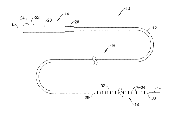
Figure 1 is perspective view showing a retrieval device 10 in accordance with
an exemplary embodiment of the present invention. As shown in a first (i.e.
collapsed) position in Figure 1, the retrieval device 10 can include an
elongated
member 12 having a proximal section 14, a longitudinally extending support
body 16,
and a distal coil section 18. As is described in greater detail below, the
retrieval
device 10 can be actuated between a collapsed position wherein the distal coil
section
18 assumes a substantially straight shape having a relatively low profile for
transport
of the retrieval device 10 through the vasculature, and an expanded position
wherein
the distal coil section 18 articulates in the general shape of a helix for
removal of a
blood clot within the body.
The proximal section 14 of the elongated member 12 can include a handle 20
that can be used by the physician to manipulate the retrieval device 10 from a
position
outside of the patient's body. The handle 20 may include a slidable thumbpiece
actuator 22 that can be engaged by the physician's thumb between a first (i.e.
retracted) position and a second (i.e. forward) position to actuate the
retrieval device
between the collapsed and expanded positions. The thumbpiece actuator 22 can
be
configured to slide back and forth within a slot disposed along the length of
the
handle 20, allowing the physician to actuate the retrieval device 10 by moving
the
thumbpiece actuator 22 forward with the thumb while gripping the handle 20. In
certain embodiments, the retrieval device 10 may include an internal spring
mechanism that can be used to releasably lock the thumbpiece actuator 22 in
position
within the slot. A button 24 or other suitable mechanism can be provided to
subsequently release the thumbpiece actuator 22 within the slot, allowing the
physician to reposition the thumbpiece actuator 22 to another position, if
desired.
The support body 16 of the elongated member 12 can have a tubular
construction adapted to transmit axial and rotational forces exerted on the
handle 20
4
CA 02603079 2007-09-28
WO 2006/104881 PCT/US2006/010763
to the distal coil section 18. In contrast to the flexible distal coil section
18, the
support body 16 may have a relatively stiff construction with sufficient
column
strength and rigidity to withstand buckling or bulging as the retrieval device
10 is
engaged within the patient's body. The wall thickness of the support body 16
may be
generally uniform along its length, or may vary along its length to alter the
flexibility
or bending characteristics of the retrieval device 10, as desired. A strain
relief 26 can
be provided in certain embodiments to reduce stress buildup at the transition
between
the proximal section 14 and the support body 16. While the illustrative
support body
16 depicted in Figure 1 is formed from a substantially solid tubular
structure, it should
be understood that other suitable structures such as a spring coil or braid
could be
employed.
The materials used in forming the support body 16 can be selected to impart a
desired mechanical characteristic to the retrieval device 10. Typically, the
support
body 16 will be formed of a material or materials having a sufficient
stiffness or
rigidity to permit the retrieval device 10 to be manipulated within the
patient's body
without buckling or bulging. Examples of suitable materials that can be used
in
forming the support body 16 may include, but are not limited to, metals such
as
stainless steel (e.g. 304V, 316L, etc.), polymers such as polyether block
amide
(PEBA), polyethylene terapthalate (PET), polytetrafluoroethylene (PTFE), or
metal-
polymer composites such as stainless steel reinforced hypotube. In certain
embodiments, a superelastic material such as nickel-titanium alloy (Nitinol)
can be
utilized, allowing the retrieval device 10 to undergo significant bending or
flexion
within the body without imparting a residual strain to the material.
The distal coil section 18 of the retrieval device 10 may have a proximal end
28 and a distal end 30. At the proximal end 28 of the distal coil section 18,
the
elongated member 12 may transition from the distal end of the support body 16
to a
flexible wire coil 32 having a number of individual coil turns 34 that can be
articulated in a path away from the general longitudinal axis L of the
retrieval device
10. The distal end 30 of the distal coil section 18 may have a rounded or
bulbous
shape to reduce trauma to the vessel wall as the retrieval device 10 is
traversed
through the vasculature.
To permit visualization within the body, at least a portion of the distal coil
section 18 can be loaded with or otherwise formed of a radiopaque material.
Examples of suitable radiopaque materials can include, but are not limited to,
gold
CA 02603079 2007-09-28
WO 2006/104881 PCT/US2006/010763
(Ag), iridium (Ir), platinum (Pt), silver (Au), tantalum (Ta), tungsten (W),
bismuth
subcarbonate ((BiO)2CO3), and barium sulfate (BaSO4). In certain embodiments,
the
distal coil section 18 can be made of a coilable metal, polymer, or metal-
polymer
material, and then coated with a radiopaque layer or coating to enhance
radiopacity.
In addition, and in some embodiments, radiopaque marker bands can be placed on
one
or more of the coil turns 34, if desired.
Figure 2 is a perspective view showing the illustrative retrieval device 10 of
Figure 1 in a second (i.e. expanded) position. As can be seen in Figure 2, the
distal
coil section 18 can be configured to articulate into an expanded position in
response to
forward movement of the thumbpiece actuator 22 within the handle 20. In an
expanded position, the coil turns 32 can be configured to bend and orient to a
pre-
defined (i.e. equilibrium) helical shape, forming a number of helically
oriented Ioops
that align circumferentially with the inner wall of the blood vessel.
In the illustrative embodiment depicted in Figure 2, for example, the distal
coil
section 18 is shown having three individual loops 36,38,40 in the expanded
position,
each loop 36,38,40 having a radius R similar to the radius of the blood vessel
in which
the retrieval device 10 is to be inserted into. The distal coil section 18 can
have a
greater or lesser number of loops than that depicted in Figure 2, however,
depending
on the particular application, the size of the blood vessel, the size of the
blood clot, as
well as other factors. If, for example, the blood clot to be excised from the
vessel wall
is relatively long, or is located at the juncture of multiple lumens, a
retrieval device
having a greater number of loops can be employed. Conversely, if the blood
clot to
be excised from the vessel wall is relatively short, or is located in a vessel
having a
relatively short length, a retrieval device having a lesser number of loops
can be
employed.
The size and shape of the loops 36,38,40 can be furtlzer customized to treat
any number of pathologies and/or to facilitate insertion of the retrieval
device 10 in
hard-to-reach regions of the vasculature (e.g. at a bifurcation branch).
Typically, the
loops 36,38,40 will be selected to expand to a size that encloses a volume
slightly
larger than the anticipated volume of the blood clot, although other sizes may
be
desired in certain applications. Collectively, the loops 36,38,40 may define
an interior
space that receives the incoming blood clot as it is dislodged from the vessel
wall.
Figure 3 is a cross-sectional view showing the distal coil section 18 of
Figure
1 in greater detail. As shown in Figure 3, the retrieval device 10 may further
include
6
CA 02603079 2007-09-28
WO 2006/104881 PCT/US2006/010763
a core wire 42 operatively coupled at a proximal end (not shown) to the
thumbpiece
actuator 22, and at a distal end 44 thereof to the distal end 30 of the distal
coil section
18. The core wire 42 may have a proximal section 46 extending through an
interior
lumen 48 of support body 16, and a distal section 50 that extends through an
interior
lumen 52 of the distal coil section 18.
The distal section 50 of the core wire 42 can be configured to yield under
tension at a force lower than that of the proximal section 48, causing the
distal section
50 to displace and assume a coiled shape when the core wire 42 is advanced
distally
using the thumbpiece actuator 22. The distal section 50 can be configured to
displace
only when a certain threshold tensile force is applied to the core wire 42, at
which
point the core wire 42 material readily responds to each addition unit of
force applied
thereto by displacing into the coiled state.
The ability of the distal section 50 of the core wire 42 to yield at a rate
greater
than the proximal section 48 thereof can be accomplished by altering the cross-
sectional area of each section 48,50. In the illustrative embodiment of Figure
3, for
example, the distal section 50 of the core wire 42 may have a transverse cross-
sectional area that is smaller than that of the proximal section 48, imparting
greater
bendability and flexibility to the distal section 50. A tapered region 54 of
the core
wire 42 located at the juncture of the proximal and distal sections 48,50 can
be
configured to gradually transition the profile of the core wire 42. In other
embodiments, the core wire 42 may continuously change in cross-section along
its
length, or, alternatively, may transition in cross-section at multiple regions
along its
length, if desired.
The materials used in forming the proximal and distal sections 48,50 can be
further selected to permit the distal section 50 of the core wire 42 to yield
under
tension at a rate greater than the proximal section thereof 48. In certain
embodiments,
for example, the proximal section 48 may be formed from a stiff or rigid
material
having a relatively high modulus of elasticity, whereas the distal section 50
may be
formed from a bendable or flexible material having a relatively low modulus of
elasticity that is capable of bending appreciably in response to the same
applied stress.
By way of example and not limitation, the proximal section 48 may comprise a
relatively stiff material such as stainless steel whereas the distal section
50 may
comprise a relatively flexible, superelastic material such as nickel-titanium
alloy
(Nitinol). In such case, the proximal and distal sections 48,50 of the core
wire 42
7
CA 02603079 2007-09-28
WO 2006/104881 PCT/US2006/010763
could have the same cross-sectional area while still exhibiting the desired
yielding
characteristics, as described above.
The types of material or materials used in forming the proximal and distal
sections 48,50 of the core wire 42 will typically depend on the desired
mechanical
characteristics of the retrieval device 10, the materials used in fabricating
the support
body 16 and distal coil section 18, the size and shape of the coil turns 34,
as well as
other factors. In those embodiments wherein the distal section 50 comprises a
superelastic material, a desired shape can be imparted to the core wire 42 by
heating
the material beyond its final austenitic temperature A f, and then bending the
material
to a desired shape. Once cooled, and when subjected to further deformation
during
use, the distal section 50 can be configured to revert to its heat-induced
(i.e. coiled)
state.
As can be further seen in Figure 3, each of the coil turns 34 may be formed
from a coiled flat ribbon having a rectangular transverse cross-sectional
area. The
coiled flat ribbon may have either a smooth surface or a textured surface
depending
on the amount the amount of force necessary to excise the blood clot from the
vessel
wall, the amount of gripping and/or tackiness required to positively engage
the blood
clot, as well as otller factors. In use, the edges of the coil turns 34 act to
positively
engage the surface of the blood clot, improving the ability of the coil turns
34 to
mechanically grip the blood clot as the retrieval device 10 is manipulated
within the
blood vessel. The coil turns 34 may be tightly wound together, as shown, or
may be
loosely wound to impart greater flexibility to the distal coil section 18, as
desired.
Other factors such as the pitch and the number of the coil turns 34 can be
selected to
accommodate blood clots of different size, or to permit the retrieval device
10 to be
inserted into variously sized vessels of the body. In some embodiments, the
coil turns
34 of the coiled flat ribbon can formed by helically wrapping a flat piece of
ribbon
about a mandrel, and then applying heat to the material to set the desired
shape.
While the illustrative coil turns 34 are shown having a rectangular transverse
cross-
sectional area in Figure 3, it should be understood that the coil turns 34 may
assume
other shapes (e.g. circular, oval, triangular, etc.), as desired.
One or more of the coil turns 34 may have a textured surface that can be
further utilized to grip the blood clot as the retrieval device 10 is
manipulated within
the blood vessel. As shown in greater detail in Figure 4, for example, a
number of
bumps or protrusions 36 formed on the edges and/or sides of the coil turns 34
can be
8
CA 02603079 2007-09-28
WO 2006/104881 PCT/US2006/010763
provided to facilitate gripping of the coil turns 34 to the blood clot
surface. The
textured surface can be formed by applying a metal or polymer nanoporous
coating to
the surface of each coil turn 34 by sputter deposition, electroplating,
epitaxial growth,
or other suitable technique. A nanoporous coating, as used herein, is
understood to be
a material having a pore size in the range of about 1 nm to 500 nm, and more
specifically, 1 nm to 200 nm. In use, the nanoporous coating provides an open
cell
surface that enhances the ability of the retrieval device 10 to grip the blood
clot by
increasing the overall surface area of the coil turns 34. The nanoporous
further
provides additional tackiness that facilitates adherence of the blood clot to
the coil
turns 32 once contacted therewith.
Figure 5 is a cross-sectional view showing the distal coil section 18 of
Figure
1 in a second (i.e. coiled) position. As indicated generally by arrow 58 in
Figure 5,
advancement of the core wire 42 in the distal direction relative to the
elongated
member 12 increases the tensile force exerted on the distal coil section 18,
inducing
stress at each point along the length of the core wire 42. Because the distal
section 50
of the core wire 42 has a smaller cross-sectional area than the proximal
section 48, the
stress induced within the distal section 50 is greater than that experienced
by the
proximal section 48. This increase in stress within the distal section 50
causes the
distal section 50 to undergo a greater strain than at the proximal section 48,
thus
becoming significaiitly longer in length. A similar effect occurs in those
embodiments wherein the distal section comprises a material having a modulus
of
elasticity smaller than the proximal section 48 thereof. The increased amount
of
strain induced in the distal section 50 from either the decrease in cross-
sectional area
and/or the selection of certain types of materials causes the distal coil
section 18 to
revert to its equilibrium coiled state, as shown in Figure 5.
Referring now to Figures 6-9, an illustrative method of retrieving a foreign
object within a blood vessel will now be described with respect to the
illustrative
retrieval device 10 of Figure 1. In preparation for insertion within the body,
and if
necessary, the thumbpiece actuator 22 can be retracted proximally, causing the
core
wire 42 to release the tension on the distal coil section 18 and allowing the
coil turns
34 to assume their low profile (i.e. collapsed) position. In a collapsed
position, the
physician may insert the retrieval device 10 percutaneously into the body and
advance
the device 10 through the vasculature to a desired location adjacent a blood
clot C, as
9
CA 02603079 2007-09-28
WO 2006/104881 PCT/US2006/010763
shown in Figure 6. If desired, a guide catheter or other suitable guiding
instrument
may be utilized to help guide the retrieval device 10 within the body.
Once positioned at the site of the blood clot C, the distal coil section 18 of
the
retrieval device 10 can then be actuated within the blood vessel V, causing
the coil
tiu-ns 34 to expand and assume their coiled state. Actuation of the distal
coil section
18 may be accomplished, for example, by sliding the thumbpiece actuator 22
forward
within the handle 20 (see Figure 2), causing the core wire 42 to tension and
strain,
thereby permitting the coil turns 34 to revert to their coiled position.
With the distal coil section 18 expanded within the blood vessel V, the
physician can then manipulate the retrieval device 10 to excise the blood clot
C from
the inner wall of the blood vessel V, as shown in a second position in Figure
7. In
certain techniques, for example, removal of the blood clot C from the wall of
the
blood vessel V may be accomplished by positioning one or more of the expanded
loops 36,38,40 distally of the blood clot C, and then pulling the elongated
member 12
proximally a distance to dislodge the blood clot C from the vessel wall. The
engagement of the distal coil section 18 against the wall of the blood vessel
V in this
manner acts to shear the blood clot C from the vessel wall, forcing it into
the interior
space defined by the loops 36,38,40.
Once the blood clot C has been excised from the vessel wall, the physician
may then retract the thumbpiece actuator 22 proximally within the handle 20,
causing
the distal coil section 18 to revert to its collapsed position, as shown in a
third position
in Figure 8. As shown in Figure 8, a catheter 60 having an interior lumen 62
adapted
to receive the collapsed retrieval device 10 and captured blood clot C can
then be
inserted into the body and advanced to the target site. Once positioned at the
target
site, the retrieval device 10 can then be loaded into the interior lumen 64,
as shown in
a fourtli position in Figure 9. Loading of the retrieval device 10 into the
interior
lumen 62 can be accomplished by withdrawing the retrieval device 10 proximally
while holding the catheter 60 stationaiy within the blood vessel V, or,
alternatively,
by holding the retrieval device 10 stationary within the blood vessel V while
advancing the catheter 60 distally. Once loaded, the catheter 60 and
accompanying
retrieval device 10 can then be removed from the body.
Figure 10 is a perspective view showing the distal portion of a retrieval
device
66 in accordance with another exemplary embodiment of the present invention.
As
shown in Figure 10, the retrieval device 66 can include a coil section 68
having a
CA 02603079 2007-09-28
WO 2006/104881 PCT/US2006/010763
proximal end 70 and a distal end 72. In the illustrative embodiment of Figure
10, the
proximal end 72 of the retrieval device 70 can be connected directly to a core
wire 74
having a proximal end (not shown) and a distal end 76. The distal end 72 of
the coil
section 68 can be connected to the distal end 76 of the core wire 74, and can
have a
rounded or bulbous shape to reduce trauma to the vessel wall as the retrieval
device
66 is manipulated within the body. In some embodiments, the coil section 68
can be
loaded with or otherwise formed of a radiopaque material, and/or can include
radiopaque marker bands on one or more of its coil turns 78, if desired.
The coil section 68 of the retrieval device 66 can be configured to articulate
from a collapsed position to an expanded position in response to axial
movement of
the core wire 74 by the physician. In an expanded position depicted in Figure
10, the
coil turns 78 can be configured to bend and orient to a pre-defined helical
shape,
forining a number of helically oriented loops 80,82,84,86 that align
circumferentially
with the inner wall of the blood vessel. The loops 80,82,84,86 can each be
configured
to radially expand the same amount within the blood vessel, or can radially
expand by
varying amounts depending on the application. In the illustrative embodiment
of
Figure 10, for example, the distal-most loop 86 is shown having a smaller
radius than
that of the other loops 80,82,84. In use, the smaller radius on the distal-
most loop 86
acts to close-off the distal portion of the coil section 68 to prevent the
blood clot from
slipping through the structure as the retrieval device 10 is manipulated
proximally
within the blood vessel, or when the device 10 is loaded into a retrieval
catheter.
The coil turns 78 may be formed from a coiled flat ribbon having a rectangular
cross-sectional area, or can comprise some other cross-sectional shape, as
desired. In
some embodiments, one or more of the coil turns 78 may have a textured surface
88
thereon, which as described above, can be formed by applying a metal or
polymer
nanoporous coating to the surface of each coil turn 78. Alternatively, and in
other
embodiments, the coil turns 78 may have a relatively smooth surface 88.
Actuation of the coil section 68 between the collapsed position and the
expanded position can be accomplished by pulling the core wire 74 proximally,
releasing the tension provided on the distal end 76 by the core wire 74 and
allowing
the coil turns 78 to assume their equilibrium coiled shape, as shown. A number
of
polymer fibers 90,92 attached to various locations of the coil section 68 can
be
provided to limit the amount of longitudinal stretching that occurs to the
coil section
11
CA 02603079 2007-09-28
WO 2006/104881 PCT/US2006/010763
68 as the retrieval device 66 is engaged within the body. The polymer fibers
also
function by increasing the total surface area of the retrieval device 10.
Figure 11 is a perspective view showing the distal portion of a retrieval
device
94 in accordance with another exemplary embodiment of the present invention.
As
shown in Figure 11, the retrieval device 94 can include a coil section 96
having a
proximal end 98 and a distal end 100. As with the embodiment of Figure 10, the
proximal end 98 of the retrieval device 94 can be connected directly to a core
wire
102 having a proximal end (iiot shown) and a distal end 104. The distal end
100 of
the coil section 96 can be connected to the distal end 104 of the core wire
102, and
can have a rounded or bulbous shape to reduce trauma to the vessel wall as the
retrieval device 94 is manipulated within the body. As with other embodiments
herein, the coil section 96 can be loaded with or otherwise formed of a
radiopaque
material, and/or can include radiopaque marker bands on one or more of its
coil turns
106, if desired.
The coil section 96 of the retrieval device 94 can be configured to articulate
from a collapsed position to an expanded position in a manner similar to that
described above with respect to Figure 10. In the illustrative embodiment of
Figure
11, however, the expanded loops 108,110,112,114 may have a tapered shape
wherein
each successive loop in the distal direction 108,110,112,114 is reduced in
size. Such
reduction in size of the loops 108,110,112,114 in the distal direction acts to
close-off
the distal portion of the coil section 96 to prevent the blood clot from
slipping through
the structure as the retrieval device 94 is manipulated proximally within the
blood
vessel, or wlien the device 94 is loaded into a retrieval catheter and/or
guide catlleter.
The coil turns 106 can be formed from a coiled flat ribbon having a
rectangular cross-sectional area, or can comprise some other cross-sectional
shape, as
desired. In some embodiments, one or more of the coil turns 106 may have a
textured
surface 116 thereon, which as described above, can be formed by applying a
metal or
polymer nanoporous coating to the surface of each coil turn 106.
Actuation of the coil section 96 between the collapsed position and the
expanded position can be accomplished in a manner similar to that described
above
with respect to Figure 10, by pulling the core wire 102 proximally. A number
of
polymer fibers 118,120 attached to various locations of the coil section 96
can be
provided to limit the amount of longitudinal stretching that occurs to the
coil section
96 as the retrieval device 94 is engaged within the body. In certain
embodiments, a
12
CA 02603079 2007-09-28
WO 2006/104881 PCT/US2006/010763
portion of the polymer fiber 118 located furthest away from the core wire 102
may
extend a distance proximally of the proximal-most loop 108, and can be looped
around to form a mouth 122 of the retrieval device 94.
Figure 12 is a perspective view showing the distal portion of a retrieval
device
124 in accordance with another exemplary embodiment of the present invention.
As
shown in Figure 12, the retrieval device 124 can include a filter basket 126
operatively coupled to a pusher wire 128 that can be manipulated by the
physician
from a position outside of the patient's body to engage the retrieval device
124 within
a blood vessel. The pusher wire 128 can have a proximal section (not shown)
adapted
to lie outside of the patient's body, and a distal section 130 adapted to
support the
filter basket 126 within a blood vessel. The pusher wire 128 can be configured
similar to other guiding members used in the art (e.g. guidewires), having the
ability
to transmit axial and rotational motion from the proximal section of the
pusher wire
128 to the distal end 130 thereof. A radiopaque spring coil 132 disposed about
the
distal section 130 may provide additional stiffness to the pusher wire 128
while
providing a visual reference point when used in conjunction with a
fluoroscope. An
atrauniatic distal tip 134 having a rounded or bulbous shape may also be
employed to
reduce trauma to the body, if desired.
The filter basket 126 can include several filter struts 136 and connecting
junctures 138 forming a number of basket cells 140 adapted to radially
surround and
capture the blood clot therein. The filter basket 126 can include an opening
142 in a
proximal section 144 thereof, which receives the incoming blood clot as it is
dislodged from the vessel wall. The basket cells 140 located on the proximal
section
144 of the filter basket 126 can be arranged in a circumferential manner,
forming an
inner lumen 146 that receives the incoming blood clot. Several basket cells
148
located at a distal section 150 of the filter basket 126 can have a closed
configuration,
preventing the blood clot or other emboli from escaping the filter basket 126
once
captured therein. The profile of the filter basket 126 can be generally
cylindrical,
conical, or other desired shape.
The filter struts 136 forming the basket cells 140 can be made flexible to
permit the filter basket 126 to move and expand in multiple directions,
including both
radially and longitudinally within the blood vessel. In certain embodiments,
the filter
struts 136 may comprise a superelastic and/or shape memory material such as
nickel-
titanium alloy (Nitinol), allowing the filter struts 136 to bend and flex
significantly
13
CA 02603079 2007-09-28
WO 2006/104881 PCT/US2006/010763
without permanently detornung. Other suitable metals, polymers, or metal-
polymer
composites may be employed, however, depending on the application.
A core wire 152 extending through the inner lumen 146 of the filter basket 126
can be used to actuate the filter basket 126 between a collapsed position and
an
expanded position within the body. The core wire 152 may have a proximal
section
(not shown) that can be manipulated by the physician at a location outside of
the
patient's body, and a distal section 154 that is attached to the closed basket
cells 148
located at the distal section 150 of the filter basket 126. The distal section
154 of the
core wire 152 can be connected to each of the closed basket cells 148 via a
number of
wire segments 156,158, which can be formed integrally with or otherwise
attached to
the core wire 152. A number of collars 160,162,164,166 coupled to the filter
struts
136 allow the filter basket 126 to slide and rotate on the pusher wire 128.
The basket cells 140 forming the filter basket 126 can be configured to expand
between a collapsed position and an expanded position within the body. To
retrieve a
blood clot within a blood vessel, the retrieval device 124 can be loaded into
the inner
lumen of a delivery device in its unexpanded state, inserted into the
patient's body,
and then advanced through the vasculature to a target site using the pusher
wire 126.
Once positioned at or near the blood clot, the retrieval device 124 can then
be
withdrawn from the delivery device, causing the filter basket 124 to radially
expand
within the blood vessel.
Once withdrawn from the delivery device, the physician may next pull the
core wire 152 proximally while holding the pusher wire 128 stationary within
the
blood vessel, causing the filter basket 126 to move proximally along the
pusher wire
128. A proximal stop 168 attached to the pusher wire 128 can be configured to
limit
proximal movement of the filter basket 126 along the pusher wire 128. Once in
contact with the proximal stop 168, continued pulling of the core wire 152 in
the
proximal direction causes the proximal-most collar 160 to compress against the
proximal stop 168, which, in turn, compresses the filter basket 126 axially
along its
length. When compressed in this manner, the basket cells 140 of the filter
basket 126
radially expand within the blood vessel. To vary the size that the expanded
filter
basket 126 assumes within the blood vessel, the physician may vary the
proximal
force exerted on the core wire 152, as desired.
Figure 13 is a top view of the filter basket 124 of Figure 12, showing the
filter
basket 126 prior to assembly on the pusher wire. As shown in Figure 13, the
filter
14
CA 02603079 2007-09-28
WO 2006/104881 PCT/US2006/010763
basket 126 may have a unitary construction formed from a single unitary
workpiece
such as a flat sheet or a tubular structure. In some fabrication methods, a
laser
machining, laser etching, chemical etching, or photochemical etching process
can be
used to cut the workpiece to form the various elements of the device. The
filter
basket 126 can then be attached to the collars 160,162,164,166 (see Figure 12)
using a
suitable bonding technique such as soldering, crimping, brazing, adhesion,
etc. In
some embodiments, all or a portion of the filter basket 126 may have a
textured
surface thereon formed, for example, by applying a nanoporous coating to all
or
selective portions of the filter struts 136. Other features such as radiopaque
markers
can also be placed on selective filter struts 136 to enhance radiographic
visualization
of the device within the body.
The filter basket 126 may further include a polymeric web covering to further
capture the blood clot or any other emboli therein. As shown in Figure 14, for
example, a polymeric web 170 can be coupled to selective filter struts 142 on
the filter
basket 126. The polymeric web 170 can include a number of openings or pores
172
of sufficient size to capture the blood clot and any emboli while maintaining
the
perfusion of blood through the filter basket 126.
Having tllus described the several embodiments of the present invention, those
of skill in the art will readily appreciate that other embodiments may be made
and
used which fall within the scope of the claims attached hereto. Numerous
advantages
of the invention covered by this document have been set forth in the foregoing
description. Changes may be made in details, particular in matters of size,
shape, and
arrangement of parts without exceeding the scope of the invention. It will be
understood that this disclosure is, in many respects, only illustrative.