Note: Descriptions are shown in the official language in which they were submitted.
DEMANDE OU BREVET VOLUMINEUX
LA PRESENTE PARTIE DE CETTE DEMANDE OU CE BREVET COMPREND
PLUS D'UN TOME.
CECI EST LE TOME 1 DE 2
CONTENANT LES PAGES 1 A 33
NOTE : Pour les tomes additionels, veuillez contacter le Bureau canadien des
brevets
JUMBO APPLICATIONS/PATENTS
THIS SECTION OF THE APPLICATION/PATENT CONTAINS MORE THAN ONE
VOLUME
THIS IS VOLUME 1 OF 2
CONTAINING PAGES 1 TO 33
NOTE: For additional volumes, please contact the Canadian Patent Office
NOM DU FICHIER / FILE NAME:
NOTE POUR LE TOME / VOLUME NOTE:
CA 02603391 2007-09-28
WO 2006/102902 PCT/DK2006/000185
1
A human immortalised neural precursor cell line
The present invention relates to an immortalised human neural precursor cell
line, NGC-407.
The cell line has been established from human foetal tissue.
Background
The efficacy of treating neurodegerative disorders with transplantation of
human fetal tissue
has been shown in animal models [Brundin, et al, Behavioural effects of human
fetal dopamine
neurons grafted in a rat model of Parkinson's disease, Exp Brain Res, 65
(1986) 235-40.;
Wictorin et al, Reformation of long axon pathways in aduit rat central nervous
system by
human forebrain neuroblasts, Nature, 347 (1990) 556-8.] as well as in patients
with
Parkinson's disease (PD) and Huntington's disease [Bachoud-Levi et al, Motor
and cognitive
improvements in patients with Huntington's disease after neural
transplantation, Lancet, 356
(2000) 1975-9.; Freed et al, Transplantation of embryonic dopamine neurons for
severe
Parkinson's disease, N Engl J Med, 344 (2001) 710-9.; Hagell et al, Sequential
bilateral
transplantation in Parkinson's disease: effects of the second graft, Brain,
122 ( Pt 6) (1999)
1121-32.; Kordower et al, Neuropathological evidence of graft survival and
striatal
reinnervation after the transplantation of fetal mesencephalic tissue in a
patient with
Parkinson's disease, N Engl J Med, 332 (1995) 1118-24.; Lindvall et al, Grafts
of fetal
dopamine neurons survive and improve motor function in Parkinson's disease,
Science, 247
(1990) 574-7; Olanow et al, Fetal nigral transplantation as a therapy for
Parkinson's disease,
Trends Neurosci, 19 (1996) 102-9.]. However, human-derived fetal donor cells
gives rise to
both ethical and practical dilemmas, and therefore, aiternative cell sources
for future
transplantations have to be developed. Implantation of cells genetically
modified to express
therapeutic genes into the brain has been proposed as a potential treatment
for
neurodegenerative disorders [Villa, A., Navarro, B. and Martinez-Serrano, A.,
Genetic
perpetuation of in vitro expanded human neural stem cells: cellular properties
and therapeutic
potential, Brain Res Bull, 57 (2002) 789-94.]. Thus, when combining genetic
engineering and
cell transplantation, an important issue is to find a suitable cell vehicle.
Tumour cells modified to express a Thymidine Kinase (TK) gene acquire the
ability to
convert the non-toxic nucleoside analog ganciclovir (GCV) to its cytotoxic
metabolite
ganciclovir-triphosphate. Cells genetically engineered to express this
"suicide" gene are
eliminated if exposed to ganciclovir. Experimental tissue culture of tumour
cells as well as
brain tumour implants, consisting of a mixture of TK-expressing cells and
unmodified "native"
tumour cells also regress following ganciclovir treatment without harm to
adjacent normal
tissue. This phenomenon, where a minority of TK-expressing cells lead to the
death and
CA 02603391 2007-09-28
WO 2006/102902 PCT/DK2006/000185
2
elimination of adjacent native tumour cells not expressing TK, has been termed
the "bystander
effect".
Malignant brain tumours are an appealing target for suicide gene delivery,
since the
entire malignancy is confined to the brain and amenable to eradication by the
bystander effect.
Key components for the success of this strategy are the genetic vector from
which the suicide
gene is expressed and its delivery vehicle. As it is impossible to target all
individual tumours in
e.g. glioblastoma multiforme with separate injections of a gene therapy vector
another delivery
strategy is needed. Migrating cells that are capable of tracking down glioma
cells and that have
been engineered to deliver a therapeutic molecule represent an ideal solution
to the problem of
gfioma cells invading normal brain tissue. It has been demonstrated that the
migratory capacity
of neural stem cells (NSCs) is ideally suited to therapy in neurodegenerative
disease models
that require brain-wide cell replacement and gene expression. It has been
hypothesized that
NSCs may specifically home to sites of disease within the brain. Studies have
also yielded the
intriguing observation that transplanted NSCs are able to home into a primary
tumour mass
when injected at a distance from the tumour itself; furthermore, NSCs were
observed to
distribute themselves throughout the tumour bed, even migrating in
juxtaposition to advancing
single tumour cells (Dunn & Black, Neurosurgery 2003, 52:1411-1424; Aboody et
al, PNAS,
2000, 97:12846-12851). These authors showed that NSCs were capable of tracking
infiltrating
glioma cells in the brain tissue peripheral to the tumour mass, and "piggy
back" single tumour
cells to make cell-to-cell-contact.
The present invention addresses several problems in the area of treatment of
neurodegenerative disorders and in the treatment of cancer It is thus one
object of the
invention to provide sufficient material for replacement cell therapy
obviating the need for large
amounts of foetal tissue. It is another object to provide cells capable of
stably expressing
transgenes after transplantation into the CNS. It is a further object to
provide cells capable of
forming gap junctions with cancer cells. It is also an object to provide cells
capable of tracing
cancer cells in the CNS. Finally, such cells should be able to proliferated
such that they can be
passaged enough to be expanded, transfected with therapeutic genes and banked.
Summary of the invention
The present invention in one aspect relates to a human cell line obtainabie
from or
derived from or constituted by NGC-407 cells. The cell line has been deposited
under the
Budapest Treaty with Deutsche Sammlung von Mikroorganismen und Zellkulturen
GmbH,
Mascheroder Weg 1 b, D-38124 Braunschweig, Germany on the 315t of March, 2005
under
accession number DSM ACC2718.
The cell line of the invention has several advantages. It is a stable,
immortalised cell
line which has been expanded and has remained stable during more than 130
population
doublings. The cell line is a neural progenitor cell line, which can
differentiate into neurons,
CA 02603391 2007-09-28
WO 2006/102902 PCT/DK2006/000185
3
astrocytes and dopaminergic neurons depending on the differentiation
conditions. The NGC-
407 cell line can be used for transplantation. It has been shown that the cell
line can survive
transplantation for at least 3 weeks in rats. It is therefore expected that
the NGC-407 cell line
can survive for an even longer time in human brains. During the
transplantation period, the cell
line can stably express a heterologous gene. The NGC-407 cell line therefore
can be used
both for replacement therapy (replacement of lost or damaged cells of the
nervous system)
and for protective therapy (as a vehicle to deliver a biological function such
as a secreted
growth factor, neurotrophic factor or neurotransmitter).
The cell line has also been transduced to express a heterologous thymidine
kinase.
Monoclonal cell lines expressing high levels of this heterologous kinase have
been selected.
These cell lines can be used as vehicles for delivery of thymidine kinase to
tumour cells in the
nervous system. It has also been shown that the NGC-407 cell line can migrate
towards
cancer cells in the central nervous system, and that the NGC-407 cell line can
form gap
junctions with cancer cells and transfer low molecular weight compounds from
the cell line to
the cancer cells. The NGC-407 cell line can therefore be used as a delivery
vehicle to activate
prodrugs (e.g. AZT, ganciclovir) after the cell line has migrated to cancer
cells and formed gap
junctions with these. The activated prodrugs will then be transferred to the
cancer cells and kill
both these and the delivery cell line. This is a feasible and promising way of
treating
glioblastoma multiforme.
In a further aspect, the invention relates to use of the NGC-407 cell line for
experimental purposes such as in vitro drug screening and characterisation.
This could e.g. be
part of a safety and toxicity study. Compared to a known cell line of
mesencephalic origin
(MES-II(1)-C2, described in WO 00/09669; also known as MESC2.10 described in
Lotharius et
al J Biol Chem 2002, 277:38884-38894) NGC-407 expresses the stem cell marker,
Nestin, and
is capable of differentiating into both neurons and astrocytes. MESC2.10 on
the other hand
does not express nestin and can only be induced to differentiate into neurons.
NGC-407 thus
represents an earlier developmental stage and has a broader potential compared
to
MESC2.10. Astrocytes secrete a number of growth factors (including GDNF) and
hormones
that are of importance for maintaining the functionality of neurons. In terms
of identification of
potential biologics drugs for treatment of e.g. Parkinson's Disease, a cell
line containing a
significant proportion of astrocytes therefore represents a better model
system compared to a
neuronal cell line.
In another aspect, the invention relates to use of the NGC-407 cell line for
therapeutic
applications and in a further aspect for replacement therapy. In a
particularly preferred
embodiment, the cell line is used for cancer therapy.
In a further aspect, the invention relates to a biocompatible capsule
comprising a core
comprising a composition of cells derived from NGC-407 cell line, said cells
being capable of
secreting a compound delivering a biological function to an individual; and
semi-permeable
CA 02603391 2007-09-28
WO 2006/102902 PCT/DK2006/000185
4
membrane surrounding the composition of cells and allowing the passage of a
compound
secreted by the composition of cells.
In one embodiment of the present invention, "treatment", "therapy", and
"medical use"
is intended to cover prophylaxis. "Treatment", "therapy" and "medical use" may
also cover
inhibition of a disease or disorder, protection against a disease or disorder,
and/or prevention
(not absolute) of a disease or disorder. "Treatment", "therapy" and "medical
use" may also
comprise curative, ameliorative, and/or symptomatic treatment, therapy and
medical use.
Brief description of the drawings
Figure 1 shows photographs of differentiated immunolabelled NGC-407 cells. In
figure IA TH
labelled neurons are marked with arrows. In figure 1 B(3-III-tubulin labelled
cells are marked
with arrows and GFAP labelled cells are marked with arrowheads.
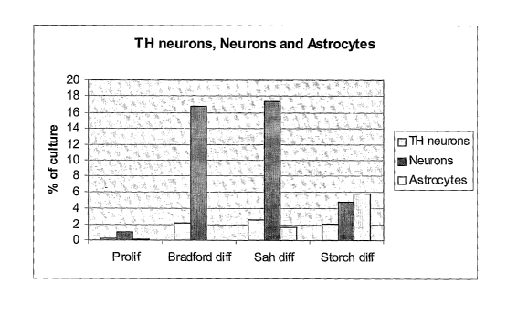
Figure 2 shows a diagram of the percentage of differentiated NGC-407 cells
labelled with the
different markers. TH neurons are cells labelled with the TH antibody, neurons
are cells
labelled with the (3-III-tubulin antibody and astrocytes are cells labelled
with the GFAP
antibody. The diagram shows four different groups; proliferating cells
(Prolif) and three groups
differentiated with the three differentiation protocols according to Riaz et
al (Bradford diff), Sah
et al (Sah diff) and Storch et al (Storch diff).
Figure 3 shows immunohistochemical visualization of hNuc- and GFP-expressing
NGC-407
cells at 1 and 3 weeks following transplantation to the rat striatum. At 1
week, a majority of the
hNuc-positive cells were found around the site of injection, however, some
cells had migrated
away (B). GFP-staining revealed that approximately 50% of the hNuc-positive
cells co-
expressed GFP, and that the cells exhibit an astrocytic morphology (A, C and
A', C'). A'-C'
are close ups of the cell-populations represented in white boxes in A-C. At 3
weeks, a high
percentage (> 35%) of the hNuc-positive cells were still expressing the
transgene
(arrowheads) and displayed a more differentiated morphology (D-F). Arrows in D-
F
demonstrate a hNuc-positive cell, negative for GFP. Scale bars in C, C' and F
represents 50
pm.
Figure. 4. A. Control cells Gap junction mediated transfer of calcein.
Unlabeled U343MGa-cl
2:6 cells have become green (straight arrows) after receiving transferable
calcein dye from the
yellow (dotted arrows) double labeled (red Dil & green Calcein) NGC-407 cells.
Both cell types
are in physical contact with each other through extended processes. B. PB
treated cells. The
processes have become more prominent by PB treatment (triple arrow), and the
number of
green recipient cells has also been significantly increased in this group.
CA 02603391 2007-09-28
WO 2006/102902 PCT/DK2006/000185
Figure 5. Vector map of the TDI-2 immortalisation vector used for
immortalising NGC-407 cell
line.
Figure 6: IC50 values for NGC-407 cell line (Figure 6A) and Tomato thymidine
kinase
5 expressing NGC-407 cell line (Figure 6B) with respect to AZT. For details,
see Example 6.
Detailed description
Transfection or transduction of NGC-407 cell line
In a preferred embodiment, cells derived from the NGC-407 cell line of the
invention
comprise, integrated into the genome and replicated together with the
chromosome(s) into
which it has been integrated, the heterologous DNA elements, in operable
combination, of a
eukaryotic promoter, a heterologous therapeutic gene, a polyadenylation signal
(pA).
The heterologous DNA elements may be of any suitable origin, but preferably
selected among those described herein.
In a preferred embodiment, the heterologous therapeutic gene may be expressed
under the transcriptional control of the human ubiquitin (UbC) promoter.
A possible down-regulation of expression may be circumvented by procedures
that
direct a site specific integration of the transgene and its accompanying
promoter.
According to one embodiment of the invention, the promoter is a constitutive
promoter selected from the group consisting of: ubiquitin promoter, CMV
promoter, JeT
promoter (US 6,555,674), SV40 promoter, Elongation Factor 1 alpha promoter
(EF1-alpha),
RSV, and Mo-MLV-LTR.
Examples of inducible/repressible promoters include: Tet-On, Tet-Off,
Rapamycin-
inducible promoter, Mxl.
Suitable expression control sequences include promoters, enhancers,
transcription
terminators, start codons, splicing signals for introns, and stop codons, all
maintained in the
correct reading frame of the polynucleotide of the invention so as to permit
proper translation
of mRNA. Expression control sequences may also include additional components
such as
leader sequences and fusion partner sequences.
Suitable expression vectors may be a viral vector derived from Herpes simplex,
alphavirus, adenovirus, adeno associated virus, baculovirus, HSV, coronavirus,
Bovine
papilloma virus, Mo-MLV, preferably adeno associated virus, or from various
bacterially
produced plasmids.
Other transfection methods include, but are not limited to, liposome
transfection,
electroporation, and transfection with carrier peptides containing nuclear or
other localising
signals.
CA 02603391 2007-09-28
WO 2006/102902 PCT/DK2006/000185
6
Other suitable expression vectors include general purpose mammalian vectors
which are also obtained from commercial sources (invitrogen Inc., Clontech,
Promega, BD
Biosecences, etc) and contain selection for Geneticin/neomycin (G418),
hygromycin B,
puromycin, Zeocin/bleomycin, blasticidin SI, mycophenolic acid or histidinol.
The vectors include the following classes of vectors: general eukaryotic
expression
vectors, vectors for stable and transient expression and epitag vectors as
well as their TOPO
derivatives for fast cloning of desired inserts (see list below for non-
limiting examples of
vectors).
Ecdysone-Inducible Expression: pIND(SP1) Vector; pINDN5-His Tag Vector Set;
pIND(SP1)N5-His Tag Vector Set; EcR Cell Lines; Muristerone A.
Stable Expression: pcDNA3.1/Hygro; PCI; PSI; pSecTag A, B & C; pcDNA3.1(-
)/MycHis A, B & C; pcDNA3.1 +/-; pcDNA3.1/Zeo (+) and pcDNA3.1/Zeo (-);
pcDNA3.1/His A,
B, & C; pRc/CMV2; pZeoSV2 (+) and pZeoSV2 (-); pRc/RSV; pTracerTM-CMV;
pTracerTM-
SV40.
Transient Expression: pCDM8; pcDNA1.1; pcDNA1.1/Amp.
Epitag Vectors: pcDNA3.1/MycHis A, B & C; pcDNA3.1N5-His A, B, & C.
Heterologous Therapeutic Genes
The heterologous therapeutic gene is a gene encoding a therapeutically active
polypeptide or proteins (also designated a therapeutic factor). Preferred
therapeutically active
polypeptides or proteins are polypeptides or proteins that are capab{e of
ameliorating or
treating neurological disorders.
. In a preferred embodiment, the heterologous therapeutic gene is encoding a
neurotrophic factor. In a more preferred embodiment, the neurotrophic factor
is a Nerve Growth
Factor (NGF); an Insulin-like Growth Factor (IGF), in particular IGF I or IGF
II; a member of the
Transforming Growth Factor (TGF) superfamily, including a Transforming Growth
Factor-a and
-0 (TGFa and TGFP), Transforming Growth Factor-P2 (TGF-(i2), Neurturin (NTN),
Persephin
(PSP); a Glial cell-line Derived Neurotrophic Factor (GDNF); Neublastin (NBN);
a Ciliary
Neurotrophic Factor (CNTF); a Brain Derived Neurotrophic Factor (BDNF); a
Neurotrophin
(NT), in particular NT 3 to 9; a Tumor Necrosis Factor (TNF), in particular
TNF-a.
In another preferred embodiment, the heterologous therapeutic gene is encoding
a
neuronal survivai factor. In a more preferred embodiment, the neuronal
survival factor is a
soluble or secreted Super Oxide Dismutase (SOD), Bc12, BCIXL, or a Hedgehog
protein.
In a third preferred embodiment, the heterologous therapeutic gene is encoding
a
nerve growth factor. In a more preferred embodiment, the nerve growth factor
is a Fibroblast
Growth Factor (FGF), in particular an acidic or a basic Fibroblast Growth
Factor (aFGF or
bFGF); an Endothelial Growth Factor (EGF), in particular a Vascular
Endothelial Growth and
CA 02603391 2007-09-28
WO 2006/102902 PCT/DK2006/000185
7
Permeability Factor (VEGPF); an interferon, in particular Interferon-a,
Interferon-(3 or
InterFeron-y; an interieukin (IL), in particular IL-1, IL-1P, GMCSF, and IL 2
to 14.
In a fourth preferred embodiment, the heterologous therapeutic gene is
encoding a
biologically active molecule that participates in the synthesis of a
neurotransmitter substance.
1n a more preferred embodiment, the neurotransmitter substance is
acetylcholine,
noradrenaline, adrenaline, 3,4-dihydroxyphenylalanine (L-DOPA), dopamine,
octopamine,
glutamate, aspartate, glycine, proline, x-aminobutyric acid (GABA), tyrosine,
taurine, alanine,
cystathione, histamine, serotonine (5-hydroxytryptamine), substance P,
Neuropeptid Y (NPY),
Cholecystokinin, neurotensin, enkephalins, or somatostatin. In another
preferred embodiment,
the biologically active molecule that functions in the synthesis of a
neurotransmitter substance
is a choline acetyl transferase; a Tyrosine Hydroxylase (TH); a tyrosine
decarboxylase; a
thymidine kinase, a cytosine deamidase, a monoamine oxidase, a L-DOPA
decarboxylase, a
histidine decarboxylase, a glutamate decarboxylase, an Ornithine
Transcarbamylase (OTC).
In a fifth preferred embodiment, the heterologous therapeutic gene is encoding
a
receptor. In a more preferred embodiment, the receptor is a receptor which
binds
acetylcholine, rioradrenaline, adrenaline, 3,4-dihydroxyphenylaianine (L-
DOPA), dopamine,
octopamine, glutamate, aspartate, glycine, proline, x-aminobutyric acid
(GABA), tyrosine,
taurine, aianine, cystathione, histamine, serotonine (5-hydroxytryptamine),
substance P,
Neuropeptid Y (NPY), Cholecystokinin, neurotensin, enkephalins, or
somatostatin.
Replacement Therapy
The cell lines of, the invention may or may not be manipulated so as to
contain
additional heterologous DNA encoding specific therapeutic factors. In case the
cell line of the
invention does not contain additional heterologous DNA encoding specific
therapeutic factors it
may be particularly suited for restorative therapy.
As defined herein, replacement therapy relates to the transplantation of cells
of
origin in the nervous system which, after engraftment, replace defective,
absent or lost cells
and their functions, at specific locations, or globally in the CNS and/or PNS.
In a further aspect the invention provide provides methods and compositions
for use
in replacement therapy within the CNS or outside the CNS. Replacement therapy
of the
invention may in particular be applied to cell replacement, delivery of cell-
secreted endogenous
substance produced by the cells, therapy for the hematopoeitic system,
neurological diseases in
mammals, including humans.
The neurological deficits contemplated according to the invention inciude any
neuro-
degenerative disease, disorder or condition. The neurological deficit may in
particular be a
neurodegenerative disease involving lesioned and traumatic neurons, in
particular traumatic
lesions of peripheral nerves, the medulla, and/or the spinal cord, cerebral
ischaemic neuronal
damage, neuropathy and especially peripheral neuropathy, Alzheimer's disease,
Huntington's
CA 02603391 2007-09-28
WO 2006/102902 PCT/DK2006/000185
8
disease, Parkinson's disease, glioblastoma, amyotrophic lateral sclerosis or
any other
neurodegenerative disease, and memory impairment connected to dementia.
Protective Therapy
While an immortalised cell line of the invention not ho{ding additional
heterologous
DNA encoding specific therapeutic factors may be particularly well suited for
replacement
therapy, the immortalised cell line of the invention that has been subjected
to the introduction of
additional heterologous DNA encoding specific therapeutic factors may be
particular well suited
for protective therapy.
As defined herein, protective therapy relates to the transplantation of cells
of origin
in the nervous system which, after engraftment, produce either endogenous or
exogenous
therapeutic factors that will prevent, or protect cell death or dysfunction in
the nervous system
of the recipient individual, or stimulate function or regenerative and re-
innervation capacity of
these cells, at specific locations or globally in the CNS and/or PNS.
In particular the invention provides methods and compositions for use in
protective
therapy. More specifically the invention provides methods and compositions for
use by
implantation with therapeutic and/or preventive intent into the brains of
normal or immune-
suppressed mammals, including humans. In particular the invention provides
methods and
compositions useful for sustainable and safe remediation of neurological
deficits.
The neurological deficits contemplated according to the invention include any
neuro-
degenerative disease, disorder or condition. The neurological deficit may in
particular be a
neurodegenerative disease involving lesioned and traumatic neurons, in
particular traumatic
lesions of peripheral nerves, the medulla, and/or the spinal cord, cerebral
ischaemic neuronal
damage, neuropathy and especially peripheral neuropathy, Alzheimer's disease,
Huntington's
disease, Parkinson's disease, glioblastoma, amyotrophic lateral sclerosis or
any other
neurodegenerative disease, and memory.impairment connected to dementia.
Differentiation
The NGC-407 cell line may be subjected to known differentiation treatments in
vitro such
as' those described in Example 1(Bardford differentiation; Sah
differentiation; Storch
differentiation) in addition to other known differentiation methods such as
the TH induction
method described in WO 02/086106 (NsGene). Such differentiation may be
performed prior to
repiacement therapy or as part of an in vitro assay or gene expression
profiling.
A further illustrative example of differentiation protocols include the two
protocols
described in Example 8 (differentiation in N2 medium without EGF and bFGF (N2
differentiation);
differentiation in N2 medium without EGF and bFGF and with cAMP and GDNF (DA
differentiation medium)).
CA 02603391 2007-09-28
WO 2006/102902 PCT/DK2006/000185
9
Furthermore, NGC-407 cells may be differentiated by transducing or
transfecting with an
expression vector coding for transcription factors responsible for or involved
in dopaminergic
differentiation such as Nurr1, Pitx3, En and Lmx1b. A preferred transcription
factor is Lmx1a as
described by Andersson et al (Andersson et al 2006, "Identification of
intrinsic determinants of
midbrain dopamine neurons", Cell 124: 393-405).
In vitro assays
The NGC-407 ceil line can be used to test potential drugs (both low molecular
weight and
proteins, genes or iRNA) in various in vitro assays. Briefly, the ceil line is
exposed to a
compound of interest and the response is compared to a control treatment. The
response may
be survival, differentiation, metabolic activity, signalling, receptor
activation etc.
Gene profiling
The NGC-407 cell line has been established from human foetal ventral midbrain
at
approximately the time when the ventral midbrain develops dopaminergic
neurons. Genes, the
regulation of which is specific to NGC-407, may thus be used as markers of
cells from the ventral
midbrain, as markers of dopaminergic neurons, or as markers of stem
cells/progenitor cells from
the ventral midbrain. Genes identified using NGC-407 may also be tested for
therapeutic
potential.
Suicide gene therapy
As described in the Background part of the present application, neural stem
cells can
be used as a delivery vehicle to deliver the product of a suicide gene to
cancer cells. As
evidenced by the examples herein NGC-407 is indeed capable of migrating to
gliobastoma
tumours while maintaining expression of a marker gene (GFP). The cell line may
therefore be
used as a vehicle to deliver a heterologous suicide gene to tumours. It has
been observed that
administration of 4-PB increases the number of GFP positive cells around the
implanted
tumours. Thus in a preferred embodiment, 4-PB is administered to a patient to
whom suicide
gene expressing NGC-407 cells have been implanted. Methods and dosages for
administration of 4-PB and analogs in connection with suicide gene therapy are
described in
WO 2005/079849.
Deoxyribonucleoside kinases
In a preferred embodiment of bystander mediated suicide gene therapy, the cell
line of
the invention has been genetically engineered to overexpress a heterologous
deoxyribonucleoside kinase. Deoxyribonucleoside kinases (dNK) from various
organisms differ
in their substrate specificity, regulation of gene expression and cellular
localisation. In
mammalian cells there are four enzymes with overlapping specificities, the
thymidine kinases 1
CA 02603391 2007-09-28
WO 2006/102902 PCT/DK2006/000185
(TK1) and 2 (TK2), deoxycytidine kinase (dCK) and deoxyguanosine kinase (dGK)
phosphorylate purine and pyrimidine deoxyribonucleosides. TKI and TK2 are
pyrimidine
specific and phosphorylate deoxyuridine (dUrd) and thymidine (dThd), and TK2
also
phosphorylates deoxycytidine (dCyd). dCK phosphorylates dCyd, deoxyadenosine
(dAdo) and
5 deoxyguanosine (dGuo), but not dThd. dGK phosphorylates dGuo and dAdo. In
mammals,
TKI is cytosolic, and TK2 and dGK are localised in the mitochondria, although
recent reports
indicate a cytoplasmic localisation of TK2 as well.
The best known and most studied example of suicide gene therapy is
Herpes,simplex
virus (HSV) thymidine kinase (tk) gene (Karreman, 1998, A new set of
positive/negative
10 selectable markers for mammalian cells. Gene. 218: 57-61). The HSV tk gene
leads to cell
death when growing cells are exposed to antiherpetic nucleoside analogs such
as ganciclovir
(GCV), as this and other prodrugs are metabolised by HSV TK to toxic
metabolites.
A Drosophila melanogaster deoxyribonucleoside kinase (Dm-dNK) phosphorylates
all
four natural deoxyribonucleosides as well as several nucleoside analogs (Munch-
Petersen et
al., 1998, Four deoxynucleoside kinase activities from Drosophila melanogaster
are contained
within a single monomeric enzyme, a new multifunctional deoxynucleoside
kinase. J Biol
Chem. 273: 3926-31; Munch-Petersen et al 2000, Functional expression of a
multisubstrate
deoxyribonucleoside kinase from Drosophila melanogaster and its C-terminal
deletion mutants.
J Biol Chem. 275: 6673-9; WO 00/36099 "New medical use of gene and vector
encoding a
multisubstrate deoxyribonucleoside kinase (dNK)"). The broad substrate
specificity of this
enzyme together with a high catalytic rate makes it unique among the
nucleoside kinases for
use as a suicide gene in combined gene/chemotherapy of cancer.
Mutant forms of the Drosophila melanogaster Dm dNK have been developed, which
have broad substrate specificities (WO 01/88106 "Multi-substrate insect
deoxynucleoside
kinase variants"). A particularly preferred variant is the variant B5 because
its degree of
activation is approximately 50 times better than wild type Dm dNK for
gemcitabine. The degree
of activation is defined as the ratio of the IC50 of the prodrug in the
nontransfected cell line to
the IC50 of the nucleoside analogue in the transfected cell line.
These and other recombinant kinases in a gene therapy approach can be
overexpressed in NGC-407 cells by placing them under the control of a strong
constitutive
promoter, such as the CMV promoter, human UbiC promoter, JeT promoter (US
6,555,674),
SV40 promoter, and Elongation Factor 1 alpha promoter (EF1-alpha).
Non-limiting examples of specific known sequences of deoxyribonucleoside
kinases
comprise for example the following:
HSV-tk wild type ACCESSION V00470 (SEQ ID NO 1)
MASYPGHQHASAFDQAARSRGHSNRRTALRPRRQQEATEVRPEQKMPTLLRVYIDGPHGMGKTTTTQLLVALGSRD
DIVYVPEPMTYfnTRVLGASETIANIYTTQHRLDQGEISAGDAAVVMTSAQITMGMPYAVTDAVLAPHIGGEAGSSHA
PPPALTLIFDRHPIAALLCYPAARYLMGSMTPQAVLAFVALIPPTLPGTNIVLGALPEDRHIDRLAKRQRPGERLD
CA 02603391 2007-09-28
WO 2006/102902 PCT/DK2006/000185
11
LAMLAAIRRVYGLLANTVRYLQCGGSWREDWGQLSGTAVPPQGAEPQSNAGPRPHIGDTLFTLFRAPELLAPNGDL
YNVFAWALDVLAKRLRSMHVFILDYDQSPAGCRDALLQLTSGMVQTHVTTPGSIPTICDLARTFAREMGEAN
Drosophila melanogaster wildtype kirnase GenBanK ACCN Y18048 (SEQ ID NO 2)
MAEAASCARKGTKYAEGTQPFTVLIEGNIGSGKTTYLNHFEKYKNDICLLTEPVEKWRNVNGVNLLELMYKDPKKW
AMPFQSYVTLTMLQSHTAPTNKKLKIMERSIFSARYCFVENMRRNGSLEQGMYNTLEEWYKFIEESIHVQADLIIY
LRTSPEVAYERIRQRARSEESCVPLKYLQELHELHEDWLIHQRRPQSCKVLVLDADLNLENIGTEYQRSESSIFDA
ISSNQQPSPVLVSPSKRQRVAR
Tomato TK (SEQ ID NO 3)
MAFSSSARNPVDLRNGSKNSFCPVGEIHVIVGPMFAGKTTALLRRVNLESNDGRNVVLIKSSKDARYAVDAWTHD
GTRFPCWSLPDLSSFKQRFGKDAYEKVDVIGIDEAQFFGDLYEFCCNAADFDGKIIVVAGLDGDYLRKSFGSVLDI
IPLADTVTKLTARCELCNRRAFFTFRKTNETETELIGGADIYMPVCRQHYVNGQSVNESAKMVLESHKVSNELILE
SPLVDP
Arabidopsis thaliana dNK (SEQ ID NO 4)
MVDYLRSSVGIIHRNHAESITTFIKESVDDELKDSGPEPNLNVKKRLTFCVEGNISVGKSTFLQRIANETVELQDL
VEIVPEPVDKWQDVGPDHFNILDAFYSEPQRYAYTFQNYVFVTRLMQEKESASGVKPLRLMERSVFSDRMVFVRAV
HEAKWMNEMEISIYDSWFDPVVSSLPGLVPDGFIYLRASPDTCHKRMMLRKRAEEGGVSLKYLQDLHEKHESWLLP
FESGNHGVLSVSRPSLHMDNSLHPDIKDRVFYLEGNHMHSSIQKVPALVLDCEPNIDFSRDIEAKTQYARQVAEFF
EFVKKKQETSTEKSNSQSPVLLPHQNGGLWMGPAGNHVPGLDLPPLDLKSLLTRPSA
Drosophila melanogaster, mutant B5 (SEQ ID NO 5)
MAEAASCARKGTKYAEGTQPFTVLIEGNIGSGKTTYLNHFEKYKNDICLLTEPVEKWRNVNGVNLLELMYKDPKKW
AMPFQSYATLTMLQSHTAPTNKKLKIMERSIFSARYCFVENMRRNGSLEQGMYNTLEEWYKFIEESIHVQADLIIY
LRTSPEVAYERIRQRARSEESCVPLKYLQELHELHEDWLIHQRRPQSCKVLVLDADLDLENIGTEYQRSESSIFDA
ISSNQQPSPVPVSPSKRQRVAR
>Arabidopsis thaliana dCGK NP_565032 (SEQ ID NO 6)
1 mqkilckstt sstpvlstpv nslaagfisl gfktpvknlp pcsttkplst cffstsampt
61 ttasvssggv gfsaylqrtv hkpapasvrf stagyrtcre sidgtnrawv grtgswralf
121 csdstggltp vnatagavve seeesdgede deekdekpvr mnrrnrsssg sgefvgnpdl
181 lkipgvglrn qrklvdngig dvaelkklyk dkfwkasqkm vdylrssvgi ihrnhaesit
241 tfikesvdde lkdsgpepnl nvkkrltfcv egnisvgkst flqrianetv elqdlveivp
301 epvdkwqdvg pdhfnildaf ysepqryayt fqnyvfvtrl mqekesasgv kplrlmersv
361 fsdrmvfvra vheakwmnem eisiydswfd pvvsslpglv pdgfiylras pdtchkrmml
421 rkraeeggvs lkylqdlhek heswllpfes gnhgvlsvsr pslhmdnslh pdikdrvfyl
481 egnhmhssiq kvpalvldce pnidfsrdie aktqyarqva effefvkkkq etsteksnsq
541 spvllphqng glwmgpagnh vpgldlppld lkslltrpsa
>Oryza sativa dCGK BAB86213 (SEQ ID NO 7)
1 mveflqssvg iihknhaesi tlfikesvde elkgtdspnv sknkrltfcv egnisvgktt
61 flqrianeti elrdlveivp epiakwqdvg pdhfnildaf yaepqryayt fqnyvfvtrv
121 mqekesssgi kplrlmersv fsdrmvvkfl kvfvravhea nwmnemeisi ydswfdpvvs
181 slpglipdgf iylraspdtc hkrmmvrkrs eeggvtldyl rglhekhesw llpskgqgpg
241 vlsvsqvpvh megslppdir ervfylegdh mhssiqkvpa lvldcehdid fnkdieakrq
>H. sapiens dCK XP_003471 (SEQ ID NO 8)
MATPPKRSCPSFSASSEGTRIKKISIEGNIAAGKSTFVNILKQLCEDWEVVPEPVARWCNVQSTQDEFEELTMSQK
NGGNVLQMMYEKPERWSFTFQTYACLSRIRAQLASLNGKLKDAEKPVLFFERSVYSDRYIFASNLYESECMNETEW
TIYQDWHDWMNNQFGQSLELDGIIYLQATPETCLHRIYLRGRNEEQGIPLEYLEKLHYKHESWLLHRTLKTNFDYL
QEVPILTLDVNEDFKDKYESLVEKVKEFLSTL
>H. sapiens dGK XP_002341 (SEQ ID NO 9)
MAAGRLFLSRLRAPFSSMAKSPLEGVSSSRGLHAGRGPRRLSIEGNIAVGKSTFVKLLTKTYPEWHVATEPVATWQ
NIQAAGNQKACTAQSLGNLLDMMYREPARWSYTFQTFSFLSRLKVQLEPFPEKLLQARKPVQIFERSVYSDRYIFA
KNLFENGSLSDIEWHIYQDWHSFLLWEFASRITLHGFIYLQASPQVCLKRLYQRAREEEKGIELAYLEQLHGQHEA
WLIHKTTKLHFEALMNIPVLVLDVNDDFSEEVTKQEDLMREVNTFVKNL
>H. sapiens TK2 NP_004605 (SEQ ID NO 10)
MGAFCQRPSSDKEQEKEKKSVICVEGNIAGGKTTCLEFFSNATDVEVLTEPVSKWRNVRGHNPLGLMYHDASRWGL
TLQTYVQLTMLDRHTRPQVSSVRLMERSIHSARYIFVENLYRSGKMPEVDYVVLSEWFDWILRNMDVSVDLIVYLR
CA 02603391 2007-09-28
WO 2006/102902 PCT/DK2006/000185
12
TNPETCYQRLKKRCREEEKVIPLEYLEAIHHLHEEWLIKGSLFPMAAPVLVIEADHHMERMLELFEQNRDRILTPE
NRKHCP
>H. sapiens TK1 XP_037195 (SEQ ID NO 11)
MSCINLPTVLPGSPSKTRGQIQVILGPMFSGKSTELMRRVRRFQIAQYKCLVIKYAKDTRYSSSFCTHDRNTMEAL
PACLLRDVAQEALGVAVIGIDEGQFFPDIMEFCEAMANAGKTVIVAALDGTFQRKPFGAILNLVPLAESWKLTAV
CMECFREAAYTKRLGTEKEVEVIGGADKYHSVCRLCYFKKASGQPAGPDNKENCPVPGKPGEAVAARKLFAPQQIL
QCSPAN
>Sombys mori dNK AAK28318 (SEQ ID NO 12)
1 msannvkpft vfvegnigsg kttflehfrq feditlltep vemwrdlkgc nllelmykdp
61 ekwamtfqsy vsltmldmhr rpaptpvklm erslfsaryc fvehimrnnt lhpaqfavld
121 ewfrfiqhni pidadlivyl ktspsivyqr ikkrarseeq cvplsyieel hrlhedwlin
181 rihaecpapv lvldadldls qitdeykrse hqi.lrkavnv vmsspnkhsp kkpisttpik
241 itphmril
>Anopheles dNK AAO49462 (SEQ ID NO 13)
MPPIASEKLGASGKKPFTVFVEGNIGSGKTTFLNHFQKFNDICLLTEPVEKWRNCGGVNL
LDLMYKESHRWAMPFQTYVTLTMLDMHTCQTDKSVKLMERSLFSARNCFVESMLASGSLH
QGMYNVLQEWYDFICCNIHIQADLIVYLQTSPEVVYERMKQRARSEESCVPLEYLKELHE
LHENWLIHGASPRPAPVLVLNADLDLNTIGAEYERSETSILKPILIENTNQHAILTSPAK
RAKTDF
>Rice TK1 (SEQ ID NO 14)
MSSICAMRSLLAASTFLRSGASPLLRPLSRPLPSRLNLSRFGPVRPVSAAAAAADKSRGGGG
SAMEAQPSYPGEIHVIVGPMFAGKTTALLRRVQVEAGTGRNVALIKSDKDNRYGLDSVVTHD
GTKMPCWALPELSSFQDKLGTEAYDKVDVIGIDEAQFFDDLHDFCCKAADRDGKIVWAGLD
GDYKRNKFGSVLDIIPLADSVTKLTARCELCGRRAFFTLRKTRETKTELIGGADVYMPVCRQ
HYLDGQIVIEATRIVLDLEKSKVIHAFK
>A. thaliana TK1 AAF13097 (SEQ ID NO 15)
MATLKASFLIKTLDSDVTGDFLSDLERRGSGAVHVIMGPMFSGKSTSLLRRIKSEISDGRS
VAMLKSSKDTRYAKDSVVTHDGIGFPCWALPDLMSFPEKFGLDAYNKLDVIGIDEAQFFG
DLYEFCCKVADDDGKIVIVAGLDGDYLRRSFGAVLDIIPIADSVTKLTARCEVCGHKAFF
TLRKNCDTRTELIGGADVYMPVCRKHYITNHIVIKASKKVLEDSDKARAESCVAATI
>A. thaliana TK1b (SEQ ID NO 16)
MRTLISPSLAPFSLHLHKPSLFSTALRFSFSINNITPTNSPPST
ISTRKLQTKATRVTSSSSSQPLSSSSPGEIHVVVGPMFSGKTTTLLRRILAERETGKR
IAIIKSNKDTRYCTESIVTHDGEKYPCWSLPDLSSFKERFGFDDYENRLDVIGIDEAQ
FFGDLYEFCREAADKEGKTVIVAGLDGDFMRRRFGSVLDLIPIADTVTKLTSRCEVCG
KRALFTMRKTEEKETELIGGAEVYMPVCRSITYVCGQNVLETARAVLDSSNNHSVVASS
L
>Tomato dCGK (SEQ ID NO 17)
MVEFLQSSIGIIHRNHAESITTYIRKSVDEELKENNSDS
NVKSTQKKRLTFCVEGNISVGKTTFLQRIANETLELQDLVEIVPEPIAKWQDIGPDHFNI
LDAFYAEPQRYAYTFQNYVFVTRVMQERESSGGIRPLRLMERSVFSDRMVFVRAVHEANW
MNEMEISIYDSWFDPVVSTLPGLIPDGFIYLRASPDTCHKRMMLRKRTEEGGVSLEYLRG
LHEKHESWLFPFESGNHGVLSVSELPLNFDKFCVPPEIRDRVFYLEGNHMHPSIQKVPAL
VLDCEPNIDFNRDIEAKRQYARQVADFFEFVKKKQEVMPGAGEEQPKGNQAPVMLPQNGG
LWVPGGKFSESTLNLDFRRNMSFMSH
The corresponding nucleotide sequences can be found in Genbank using the
accession numbers given above, in the references given above and for the plant
kinases in
WO 03/100045 (thymidine kinases), and WO 2004/003185 (dCK/dGK).
In a preferred embodiment, the deoxyribonucleoside kinase is selected from the
group
consisting of
CA 02603391 2007-09-28
WO 2006/102902 PCT/DK2006/000185
13
a) a deoxyribonucleoside kinase having the amino acid sequence of any of SEQ
ID No 1
to 17;
b) a deoxyribonucleoside kinase variant comprising an amino acid sequence
having at
least 50% sequence identity to any of SEQ ID No 1 to 17;
c) a deoxyribonucleoside kinase encoded by a nucleotide sequence capable of
hybridising under conditions of high stringency to a nucleotide sequence
encoding any
ofSEQIDNo1to17.
In the context of this invention, the term kinase variant is a polypeptide (or
protein) having
an amino acid sequence that differs from the sequence presented as SEQ ID NO:
1, as SEQ
ID NO:2, as SEQ ID NO: 3, as SEQ ID NO: 4, as SEQ ID NO: 5, as SEQ ID NO: 6,
as SEQ ID
NO: 7, as SEQ ID NO: 8, as SEQ ID NO: 9, as SEQ ID NO: 10, as SEQ ID NO: 11,
as SEQ ID
NO: 12, as SEQ ID NO: 13, as SEQ ID NO: 14, as SEQ ID NO: 15, as SEQ ID NO:
16, as
SEQ ID NO: 17, at one or more amino acid positions and has dNK activity. Such
analogous
polypeptides include polypeptides comprising conservative substitutions,
spiice variants,
isoforms, homologues from other species, and polymorphisms.
As defined herein, the term "conservative substitutions" denotes the
replacement of an
amino acid residue by another, biologically similar residue. Examples of
conservative
substitutions include
(i) the substitution of one non-polar or hydrophobic residue such as alanine,
leucine,
isoleucine, valine, proline, methionine, phenylalanine or tryptophan for
another, in particular
the substitution of alanine, leucine, isoleucine, valine or proline for
another; or
(ii) the substitution of one neutral (uncharged) polar residue such as serine,
threonine,
tyrosine, asparagine, giutamine, or cysteine for another, in particular the
substitution of
arginine for lysine, glutamic for aspartic acid, or glutamine for asparagine;
or
(iii) the substitution of a positively charged residue such as lysine,
arginine or histidine for
another; or
(iv) the substitution of a negatively charged residue such as aspartic acid or
glutamic acid
for another.
Modifications of this primary amino acid sequence may result in proteins which
have
substantially equivalent activity as compared to the unmodified counterpart
polypeptide, and
thus may be considered functional analogous of the parent proteins. Such
modifications may
be deliberate, e.g. as by site-directed mutagenesis, or they may occur
spontaneous, and
include splice variants, isoforms, homologues from other species, and
polymorphisms. Such
functional analogous are also contemplated according to the invention.
It has been found that deoxyribonucleoside kinase enzymes that are C- and/or N-
terminally altered significantly change their properties in particular in
respect of kinetic
properties such as turnover and substrate specificity. So from having a more
restricted
specificity, usually deoxycytidine kinase (dCK) and deoxyguanosine kinase
(dGK) activity, the
CA 02603391 2007-09-28
WO 2006/102902 PCT/DK2006/000185
14
deoxyribonucleoside kinase enzymes of the invention may be converted into
essentially multi-
substrate enzymes, having ability to phosphorylate all four
deoxyribonucleosides.
A variant deoxyribonucleoside kinase can be defined with reference- to the
amino acid
sequence of a known deoxyribonucleoside kinase, such as any of the kinases
disclosed
above. In a preferred embodiment, the variant kinase has at least 50% sequence
identity to a
reference sequence, more preferably at least 60% sequence identity, more
preferably at least
70% sequence identity, more preferably at least 75% sequence identity, more
preferably at
least 80% sequence identity, more preferably at least 85% sequence identity,
more preferably
at least 90% sequence identity, more preferably at least 95% sequence
identity. The individual
reference sequence may be either of SEQ ID NO: 1, SEQ ID NO:2, SEQ ID NO: 3,
SEQ ID
NO: 4, SEQ ID NO: 5, SEQ ID NO: 6, SEQ ID NO: 7, SEQ ID NO: 8, SEQ ID NO: 9,
SEQ ID
NO: 10, SEQ ID NO: 11, SEQ ID NO: 12, SEQ ID NO: 13, SEQ ID NO: 14, SEQ ID NO:
15,
SEQ ID NO: 16, and SEQ ID NO: 17.
In a more preferred embodiment, the deoxyribonucleoside kinases comprise a
deoxyribonucleoside kinase selected from the group consisting of
a) a deoxyribonucleoside kinase having the amino acid sequence of any of SEQ
ID
NO 1 to 5; and
b) a deoxyribonucleoside kinase variant comprising an amino acid sequence
having at
least 70% sequence identity to any of SEQ ID No 1 to 5 and having dNK
activity.
It is also possible to administer two or more deoxyribonucleoside kinases to
the same
individual.
Without being limiting the following combinations of kinase and nucleoside
analogues are
preferred:
HSV-tk - GCV, ACV, penciclovir
Drosophila melanogaster dNK or B5- gemcitabine, CdA, FaraA, araC, ddC
Plant TKs including Tomato TK- AZT, D4T, ddT, fluorouridine
Plant dNKs including Arabidopsis thaliana dNK- gemcitabine, CdA, FaraA, araC,
ddC.
A preferred, non-limiting example of a mathematical algorithm utilized for the
comparison of sequences and calculation of sequence identities is the
algorithm of Myers and
Miller, CABIOS (1989). Such an algorithm is incorporated into the ALIGN
program (version
2.0) which is part of the FASTA sequence alignment software package (Pearson
WR, Methods
MoI Biol, 2000, 132:185-219). Align calculates sequence identities based on a
global
alignment. AlignO does not penalise to gaps in the end of the sequences. When
utilizing the
ALIGN or AlignO program for comparing amino acid sequences, a BLOSUM50
substitution
matrix with gap opening/extension penalties of -12/-2 is preferably used.
Encapsulation
CA 02603391 2007-09-28
WO 2006/102902 PCT/DK2006/000185
Encapsulated cell therapy is based on the concept of isolating cells from the
recipient
host's immune system by surrounding the cells with a semipermeable
biocompatible material
before implantation within the host. The invention includes a device in which
cells of the
invention are encapsulated in an immunoisolatory capsule. An "immunoisolatory
capsule"
5 means that the capsule, upon implantation into a recipient host, minimises
the deleterious
effects of the host's immune system on the cells in the core of the device.
Cells are
immunoisolated from the host by enclosing them within implantable polymeric
capsules formed
by a microporous membrane. This approach prevents the cell-to-cell contact
between host and
implanted tissues, eliminating antigen recognition through direct
presentation. The membranes
10 used can also be tailored to control the diffusion of molecules, such as
antibody and
complement, based on their molecular weight while allowing for the diffusion
of a therapeutic
protein. Using encapsulation techniques, cells can be transplanted into a host
without immune
rejection, either with or without use of immunosuppressive drugs. Useful
biocompatible
polymer capsules usually contain a core that contains cells, either suspended
in a liquid
15 medium or immobilised within an immobilising matrix, and a surrounding or
peripheral region of
permselective matrix or membrane ("jacket") that does not contain isolated
cells, that is
biocompatible, and that is sufficient to protect cells in the core from
detrimental immunological
attack. Encapsulation hinders elements of the immune system from entering the
capsule,
thereby protecting the encapsulated cells from immune destruction. The
semipermeable nature
of the capsule membrane also permits the biologically active molecule of
interest to easily
diffuse from the capsule into the surrounding host tissue.
The capsule can be made from a biocompatible material. A "biocompatible
material" is
a material that, after implantation in a host, does not elicit a detrimental
host response
sufficient to result in the rejection of the capsule or to render it
inoperable, for example through
degradation. The biocompatible material is relatively impermeable to large
molecules, such as
components of the host's immune system, but is permeable to small molecules,
such as
insulin, growth factors, and nutrients, while allowing metabolic waste to be
removed. A variety
of biocompatible materials are suitable for delivery of growth factors by the
composition of the
invention. Numerous blocompatible materials are known, having various outer
surface
morphologies and other mechanical and structural characteristics. Preferably
the capsule of
this invention will be similar to those described by WO 92119195 or WO
95/05452,
incorporated by reference; or U.S. Pat. Nos. 5,639,275; 5,653,975; 4,892,538;
5,156,844;
5,283,187; or U.S. Pat. No. 5,550,050, incorporated by reference. Such
capsules allow for the
passage of metabolites, nutrients and therapeutic substances while minimizing
the detrimental
effects of the host immune system. Components of the biocompatible material
may include a
surrounding semipermeable membrane and the internal cell-supporting
scaffolding. Preferably,
the recombinant cells are seeded onto the scaffolding, which is encapsulated
by the
permselective membrane. The filamentous cell-supporting scaffold may be made
from any
CA 02603391 2007-09-28
WO 2006/102902 PCT/DK2006/000185
16
biocompatible material selected from the group consisting of acrylic,
polyester, polyethylene,
polypropylene polyacetonitrile, polyethylene teraphthalate, nylon, polyamides,
polyurethanes,
polybutester, silk, cotton, chitin, carbon, or biocompatible metals. Also,
bonded fiber structures
can be used for cell implantation (U.S. Pat. No. 5,512,600, incorporated by
reference).
Biodegradable polymers include those comprised of poly(lactic acid) PLA,
poly(lactic-coglycolic
acid) PLGA, and poly(glycolic acid) PGA and their equivalents. Foam scaffolds
have been
used to provide surfaces onto which transplanted cells may adhere (WO
98/05304,
incorporated by reference). Woven mesh tubes have been used as vascular grafts
(WO
99/52573, incorporated by reference). Additionally, the core can be composed
of an
immobilizing matrix formed from a hydrogel, which stabilizes the position of
the cells. A
hydrogel is a 3-dimensional network of cross-linked hydrophilic polymers in
the form of a gel,
substantially composed of water.
The jacket preferably has a molecular weight cutoff of less than 1000 kD, more
preferably between 50-700 kD, most preferably between 70-300 kD. The molecular
weight
cutoff should be selected to ensure that the bioactive therapeutic protein can
escape from the
capsule while protecting the encapsulated cells from the immune system of the
patient.
The thickness of the jacket typically lies in the range of 2 to 200 microns,
more
preferably from 50 to 150 microns. The jacket should have a thickness to give
the capsule
sufficient strength to keep the cells encapsulated and should with this in
mind be kept as thin
as possible to take up as little space as possible.
Various polymers and polymer blends can be used to manufacture the surrounding
semipermeable membrane, including polyacrylates (including acrylic
copolymers),
polyvinylidenes, polyvinyl chloride copolymers, polyurethanes, polystyrenes,
polyamides,
cellulose acetates, cellulose nitrates, polysulfones (including polyether
sulfones),
polyphosphazenes, polyacrylonitriles, poly(acrylonitrile/covinyl chloride), as
well as derivatives,
copolymers and mixtures thereof. Preferably, the surrounding semipermeable
membrane is a
biocompatible semipermeable hollow fiber membrane. Such membranes, and methods
of
making them are disclosed by U.S. Pat. Nos. 5,284,761 and 5,158,881,
incorporated by
reference. The surrounding semipermeable membrane may be formed from a
polyether
sulfone hollow fiber, such as those described by U.S. Pat. No. 4,976,859 or
U.S. Pat. No.
4,968,733, incorporated by reference. An alternate surrounding semipermeable
membrane
material is poly(acrylonitrile/covinyl chloride).
The capsule can be any configuration appropriate for maintaining biological
activity and
providing access for delivery of the product or function, including for
example, cylindrical,
rectangular, disk-shaped, patch-shaped, ovoid, stellate, or spherical.
Moreover, the capsule
can be coiled or wrapped into a mesh-like or nested structure. If the capsule
is to be retrieved
after it is implanted, configurations, which tend to lead to migration of the
capsules from the
site of implantation, such as spherical capsules small enough to travel in the
recipient host's
CA 02603391 2007-09-28
WO 2006/102902 PCT/DK2006/000185
17
blood vessels, are not preferred. Certain shapes, such as rectangles, patches,
disks, cylinders,
and flat sheets offer greater structural integrity and are preferable where
retrieval is desired. A
particularly preferred shape is cylinder-shaped as such a shape is easily
produced from hollow
fibers which can produces industrially.
When macrocapsules are used, preferably at least 103 cells are encapsulated,
such as
between 103 and 108 cells are encapsulated, most preferably 105 to 10' cells
are encapsulated
in each device. Of course, the number of cells in each capsule depends on the
size of the
capsule. As a rule of thumb, in a capsule with foam (described below) the
present inventors
have found that loading between 10,000 and 100,000 cells per pL of capsule
(volume
calculated as the internal volume including foam), more preferably from 25,000
to 50,000 cells
per pL, more preferably from 30,000 to 40,000 cells per pL. The number of
cells to be loaded
also depends on the size of the cells.
Dosage may be controlled by implanting a fewer or greater number of capsules,
preferably between 1 and 10 capsules per patient.
A macrocapsule in the present context is a capsule having a volume of at least
1 pL,
such as from 1 to 10 pL.
The scaffolding may be coated with extracellular matrix (ECM) molecules.
Suitable
examples of extracellular matrix molecules include, for example, collagen,
laminin, and
fibronectin. The surface of the scaffolding may also be modified by treating
with plasma
irradiation to impart charge to enhance adhesion of cells.
Any suitable method of sealing the capsules may be used, including the use of
polymer
adhesives or crimping, knotting and heat sealing. In addition, any suitable
"dry" sealing method
can also be used, as described, e.g., in U.S. Pat. No. 5,653,687, incorporated
by reference.
The encapsulated cell devices are implanted according to known techniques.
Many 25 implantation sites are contemplated for the devices and methods of
this invention. These
implantation sites include, but are not limited to, the central nervous
system, including the
brain, spinal cord (see, U.S. Pat. Nos. 5,106,627, 5,156,844, and 5,554,148,
incorporated by
reference), and the aqueous and vitreous humors of the eye (see WO 97/34586,
incorporated
by reference).
Foam scaffolds:
The foam scaffold may be formed from any suitable material that forms a
biocompatible
foam with an open cell or macroporous structure with a network of pores. An
open-cell foam is
a reticulate structure of interconnected pores. The foam scaffold provides a
non-
biodegradable, stable scaffold material that allows attachment of adherent
cells. Among the
polymers that are useful in forming the foam scaffolds for the devices of this
invention are
thermoplastics and thermoplastic elastomers.
Some examples of materials useful in forming suitable foam scaffolds are
listed in
Table 1.
CA 02603391 2007-09-28
WO 2006/102902 PCT/DK2006/000185
18
TABLE I
Thermoplastic
Thermoplastics: Elastomers:
Acrylic Polyamide
Modacrylic Polyester
Polyamide Polyethylene
Polycarbonate Polypropylene
Polyester Polystyrene .
Polyethylene Polyurethane
Polypropylene Polyvinyl Alcohol
Polystyrene Silicone
Polysulfone
Polyethersulfone
Polyvinylidene fluoride
Thermoplastic foam scaffolds made from polysulfone and polyethersulfone, and
thermoplastic elastomer foam scaffolds made from polyurethane and polyvinyl
alcohol are
preferred.
The foam must have some (but not necessarily all) pores of a size that permits
cells to
attach to the walls or surfaces within the pores. The pore size, pore density
and void volume of
the foam scaffold may vary. The pore shape may be circular, elliptical or
irregular. Because the
pore shape can vary considerably, its dimensions may vary according to the
axis being
measured. For the purposes of this invention, at least some pores in the foam
should have a
pore diameter of between 20-500 pm, preferably between 50-150 pm. Preferably
the foregoing
dimensions represent the mean pore size of the foam. If non-circular, the pore
may have
variable dimensions, so long as its size is sufficient to permit adherent
cells to attach to the
walls or surfaces within the pore. In one embodiment, foams are contemplated
having some
elliptical pores that have a diameter of 20-500 pm along the minor axis and a
diameter of up to
1500 pm along the major axis.
In addition to the foregoing cell permissive pores sizes, preferably a least a
fraction of
the pores in the foam should be less than 10 pm to be cell impermissive but
still provide
channels for transport of nutrients and biologically active molecules
throughout the foam.
Pore density of the foam (i.e., the number per volume of pores that can
accommodate
cells, as described above) can vary between 20-90%, preferably between 50-70%.
Similarly, the void volume of the foam may vary between 20-90%, preferably
between
30-70%.
CA 02603391 2007-09-28
WO 2006/102902 PCT/DK2006/000185
19
The walls or surfaces of the pores are typically coated with an extracellular
matrix
molecule or molecules, or other suitable molecule. This coating can be used to
facilitate
adherence of the cells to the walls of the pores, to hold cells in a
particular phenotype and/or to
induce cellular differentiation.
Preferred examples of extracellular matrix molecules (ECM) that can be adhered
to the
surfaces within the pores of the foams include: coliagen, laminin,
vitronectin, polyornithine and
fibronectin. Other suitable ECM molecules include glycosaminoglycans and
proteoglycans;
such as chrondroitin sulfate, heparin sulfate, hyaluron, dermatan sulfate,
keratin sulfate,
heparan sulfate proteoglycan (HSPG) and elastin.
The ECM may be obtained by culturing cells known to deposit ECM, inciuding
cells of
mesenchymal or astrocyte origin. Schwann cells can be induced to synthesize
ECM when
treated with ascorbate and cAMP. See, e.g., Baron-Van Evercooren et al.,
"Schwann Cell
Differentiation in vitro: Extracellular Matrix Deposition and Interaction,"
Dev. Neurosci., 8, pp.
182-96 (1986).
In addition, adhesion peptide fragments, e.g., RGD containing sequences
(ArgGlyAsp),
YIGSR-containing sequences (TyrlleGlySerArg), as well as IKVAV containing
sequences
(IleLysValAlaVal), have been found to be useful in promoting cellular
attachment. Some RGD-
containing molecules are commercially availabie--e.g., PepTite-2000.TM.
(Telios).
The foam scaffolds of this invention may also be treated with other materials
that
enhance ceilular distribution within the device. For example, the pores of the
foam may be
filled with a non-permissive hydrogel that inhibits cell proliferation or
migration. Such
modification can improve attachment of adherent cells to the foam scaffold.
Suitable hydrogels
include anionic hydrogels (e.g., alginate or carageenan) that may repel cells
due to charge.
Alternately, "solid" hydrogels (e.g., agarose or polyethylene oxide) may also
be used to inhibit
cell proliferation by discouraging binding of extracellular matrix molecules
secreted by the
cells.
Treatment of the foam scaffold with regions of a non-permissive material
allows
encapsulation of two or more distinct cell populations within the device
without having one
population overgrow the other. Thus non-permissive materials may be used
within the foam
scaffoid to segregate separate populations of encapsulated cells. The distinct
populations of
cells may be the same or different cell types, and may produce the same or
different
biologically active molecules. In one embodiment, one cell population produces
a substance
that augments the growth and/or survival of the other cell population. In
another embodiment,
multiple cell types producing multiple biologically active molecules are
encapsulated. This
provides the recipient with a mixture or "cocktail" of therapeutic substances.
The devices of this invention may be formed according to any suitable method.
In one
embodiment, the foam scaffold may be pre-formed and inserted into a pre-
fabricated jacket,
e.g., a hollow fiber membrane, as a discrete component.
CA 02603391 2007-09-28
WO 2006/102902 PCT/DK2006/000185
Any suitable thermoplastic or thermoplastic elastomer foam scaffold material
may be
preformed for insertion into a pre-fabricated jacket. In one embodiment we
prefer polyvinyl
alcohol (PVA) sponges for use as the foam scaffold. Several PVA sponges are
commercially
available. For example, PVA foam sponges #D-3, 60 pm pore size are suitable
(Rippey Corp,
5 Kanebo). Similarly, PVA sponges are commercially available from Ivalon Inc.
(San Diego,
Caiif.). PVA sponges are water-insoluble foams formed by the reaction of
aerated Poly(vinyl
alcohol) solution with formaidehyde vapor as the crosslinker. The hydroxyl
groups on the PVA
covalently crosslink with the aldehyde groups to form the poiymer network. The
foams are
flexible and elastic when wetted and semi-rigid when dried.
10 As an alternative, support a mesh or yarn may be used as described in US
6,627,422.
For easy retrieval the capsule may be equipped with a tether anchor.
NGC-407 or NGC-407 derived cells of the invention can be expanded as described
in
the examples and be loaded into the immunoisolatory capsule. After loading,
the capsules can
be kept for a number of weeks in vitro. During this period the growth medium
may be one that
15 allows continued proliferation within the capsule - to fill the capsuie
more completely - or the
medium may be replaced by a differentiation medium to differentiate the
encapsulated cells
and thereby stop the proliferation.
Support matrix for cells of the invention
20 The method of the present invention further comprises the culturing of the
cells of the
invention in vitro on a support matrix prior to implantation into the
mammalian brain. The
preadhesion of cells to microcarriers prior to implantation in the brain is
designed to enhance
the long-term viability of the transplanted cells and provide long term
functional benefit.
Methods for culturing cells on a support matrix and methods for implanting
said cells into the
brain are described in US 5,750,103 (Cherksey).
To increase the long term viability of the transplanted cells, the cells to be
transplanted
can be attached in vitro to a support matrix prior to transplantation.
Materials of which the
support matrix can be comprised include those materials to which cells adhere
following in
vitro incubation, and on which cells can grow, and which can be implanted into
the mammalian
body without producing a toxic reaction, or an inflammatory reaction which
would destroy the
implanted cells or otherwise interfere with their biological or therapeutic
activity. Such materials
may be synthetic or natural chemical substances, or substances having a
biological origin.
The matrix materials include, but are not limited to, glass and other silicon
oxides,
polystyrene, polypropylene, polyethylene, polyvinylidene fluoride,
poiyurethane, polyalginate,
polysulphone, polyvinyl alcohol, acrylonitrile polymers, polyacrylamide,
polycarbonate,
polypentent, nylon, amylases, natural and modified gelatin and natural and
codified collagen,
natural and modified polysaccharides, including dextrans and celluloses (e.g.,
nitrocellulose),
agar, and magnetite. Either resorbable or non-resorbable materials may be
used. Also
CA 02603391 2007-09-28
WO 2006/102902 PCT/DK2006/000185
21
intended are extracellular matrix materials, which are well-known in the art.
Extracellular matrix
materials may be obtained commercially or prepared by growing cells which
secrete such a
matrix, removing the secreting celis, and allowing the cells which are to be
transplanted to
interact with and adhere to the matrix. The matrix material on which the cells
to be implanted
grow, or with which the cells are mixed, may be an indigenous product of the
cells. Thus, for
example, the matrix material may be extracellular matrix or basement membrane
material,
which is produced and secreted by cells to be implanted.
To improve ceil adhesion, survival and function, the solid matrix may
optionally be
coated on its external surface with factors known in the art to promote cell
adhesion, growth or
survival. Such factors include cell adhesion molecules, extracellular matrix,
such as, for
example, fibronectin, laminin, collagen, elastin, glycosaminoglycans, or
proteoglycans or
growth factors.
Alternatively, if the solid matrix to which the implanted cells are attached
is constructed
of porous material, the growth- or survival promoting factor or factors may be
incorporated into
the matrix material, from which they would be slowly released after
implantation in vivo.
When attached to the support according to the present invention, the cells
used for
transplantation are generally on the "outer surface" of the support. The
support may be solid or
porous. However, even in a porous support, the cells are in direct contact
with the external
milieu without an intervening membrane or other barrier. Thus, according to
the present
invention, the cells are considered to be on the "outer surface" of the
support even though the
surface to which they adhere may be in the form of internal folds or
convolutions of the porous
support material which are not at the exterior of the particle or bead itself.
The configuration of the support is preferably spherical, as in a bead, but
may be
cylindrical, elliptical, a flat sheet or strip, a needle or pin shape, and the
like. A preferred form
of support matrix is a glass bead. Another preferred bead is a polystyrene
bead.
Bead sizes may range from about 10 pm to 1 mm in diameter, preferably from
about 90
pm to about 150 pm. For a description of various microcarrier beads, see, for
example, Fisher
Biotech Source 87-88, Fisher Scientific Co., 1987, pp. 72-75; Sigma Cell
Culture Catalog,
Sigma Chemical Co., St, Louis, 1991, pp. 162-163; Ventrex Product Catalog,
Ventrex
Laboratories, 1989; these references are hereby incorporated by reference. The
upper limit of
the bead's size may be dictated by the bead's stimulation of undesired host
reactions, which
may interfere with the function of the transplanted cells or cause damage to
the surrounding
tissue. The upper limit of the bead's size may also be dictated by the method
of administration.
Such limitations are readily determinable by one of skill in the art.
Examples
Example 1 Generation and characterisation of the NGC-407 cell line
CA 02603391 2007-09-28
WO 2006/102902 PCT/DK2006/000185
22
This example illustrates the generation and in vitro characterisation of the
NGC-407 cell
line.
Human midbrain cells in primary culture were prepared from first trimester
human fetal
brain (obtained through Lund University, Sweden). The tissue was procured in
compliance with
Swedish laws and regulations. Immediately after dissection of the ventral
midbrain, the tissue
was cut into <0.5mm3 pieces by placing the tissue in a drop of Cell
dissociation solution
(Sigma #C5914) in a petri dish. The tissue was then cut by moving two scalpeis
against each
other. Tissue was transferred to 2ml culture medium consisting of DMEM/F12
(Gibco # 31331-
028), N2 supplement (Gibco # 17502-048) supplemented with 0.5% HSA (Sigma # A
1653),
0.6% Glucose (Sigma # G8769), 5mM HEPES (Gibco # 15630-056), B27 supplement
(Gibco
#17504-044), 40 g/m1 bFGF (R&D Systems # 233-FB) 20ug/ml EGF (R&D Systems #
236-
EG), centrifugated at 1,000rpm for 5 minutes and respuspended in fresh culture
medium.
Tissue pieces were seeded into 1-4 wells in PLL/Fibronectin coated 4w
chamberslides or 24w
plates. Standard cell culture plastic was used and PLL/fibronectin coating
performed according
to:
Coating of culture vessels:
1. Incubate with 0.01 % Poly-L-lysine solution (Sigma #P4832) for I h at 37 C
2. Remove PLL, rinse with dH2O (Gibco #15230-071) and let the surface dry for
2h.
3. Just before seeding the cells, add enough solution of fibronectin (Sigma #
F0895)
diluted to 50 pg/mI in dH2O to cover surface, aspirate immediately, and let
the surface
dry for 45 min before adding the tissue/cells.
At 4 days after seeding the cell cultures were transduced with the TD1-2
retroviral vector
by adding viral stock at an MOI of 1, incubated overnight and removed by
changing culture
medium. The vector TD1-2 was made by cloning v-myc (as a gag-vmyc fragment)
(GenBank
Acc. #: AF033809) as an EcoRi/Dral fragment between EcoRl and Hpal sites of
pLXSN (BD
Clontech, cat #: 631509, GenBank Acc. #: M28248) (Figure 5). A gene conferring
neomycin
resitance is also present in the vector. The culture was expanded in culture
medium and
PLL/fibronectin coated vessels to a confluent T25 fiask. Geneticin at a
concentration of 800
pg/mI was added for selection of transduced cells. After selection, cultures
were maintained
growing as monoiayer in the culture medium described above. Cultures
approaching
confluence were passaged using a cell scraper and split 1:4 every 3-7 days.
Removal of growth factors bFGF and EGF results in differentiation of the cell
line,. Addition
of different factor will also influence the composition of cell phenotypes
after differentiation
(Figure 1 and 2). Three different differentiation media were used:
1. Culture medium with mitogenic growth factors bFGF and EGF replaced by 50
ng/mI BDNF, 20 ng/mi CNTF, 100 ng/ml IGF-1 and 50 uM-Forskolin (Sah et al,
1997).
2. Culture medium with mitogenic growth factors bFGF and EGF replaced by 50
ng/mI BDNF, 50 uM Forskolin and 50 uM Dopamine (Bradford; Riaz et al, 2002).
CA 02603391 2007-09-28
WO 2006/102902 PCT/DK2006/000185
23
3. Culture medium with mitogenic growth factors bFGF and EGF replaced by 10%
FCS, 100 pg/mI IL-lb 1 ng/ml IL-11, 1 ng/ml LIF, 10 ng/ml GDNF (Storch et al,
2001).
Following differentiation, TH (tyrosine hydroxylase), (3-III-tubulin, and GFAP
(Glial fibrillary
acidic protein) immunoreactive cells were observed. For such
immunocytochemistry, cells
were fixed in 4% paraformaldehyde, and were incubated with primary antibody
(TH antibody
Chemicon # AB152, GFAP antibody DAKO #Z0334, P-Ill-tubulin antibody Sigma #T-
8660) in
blocking buffer for 2 hours at room temperature, rinsed and then incubated
with a fluorophor-
conjugated (FITC or Cy3, Jackson lmmunoResearch Laboratories Inc) species-
specific
secondary antibody in blocking buffer for another hour at room temperature.
Cultures were
then rinsed with PBS and coverslipped with DAKO mounting medium before cell
counting and
photographing representative fieids. The percentage of different cell types
varied depending on
the type of factors used for differentiation the numbers of TH positive cells
were around 2% of
total cell number in the differentiation media. The number neurons (Beta-Ill-
tubulin positive
cells) varied between 4-17% and the GFAP positive astrocytes between 0-6%. The
proliferating cell culture consistently had below 1% of cells immunolabeled
with any of the
markers (Figure 1 and 2).
These results indicate that the NGC-407 is a neural progenitor cell line that
can
differentiate into neurons, astrocytes and dopaminergic neurons.
REFERENCES
Riaz SS, Jauniaux E, Stern GM, Bradford HF, The controlled conversion of human
neural progenitor cells derived from foetal ventral mesencephalon into
dopaminergic neurons
in vitro, Brain Res Dev Brain Res., 2002 May 30;136(1):27-34.
Sah DW, Ray J, Gage FH, Bipotent progenitor cell lines from the human CNS, Nat
Biotechnol 1997 Jun;15(6):574-80
Storch A, Paul G, Csete M, Boehm BO, Carvey PM, Kupsch A, Schwarz J., Long-
term
proliferation and dopaminergic differentiation of human mesencephalic neural
precursor cells,
Exp Neurol. 2001 Aug;170(2):317-25.
Example 2 Transplantation and in vivo transgene expression of the NGC-407 cell
line
This example illustrates lentiviral transduction, stability of transgene
expression and
integration of the NGC-407 cell line after experimental transplantation into
the rat brain.
NGC-407 cells were expanded according to the methods described in Example 1.
Forty-eight hrs prior to transplantation, NGC-407 cells were transduced with a
self-inactivated
lentiviral vector expressing GFP (LV-GFP-SIN; Zufferey R et al, 1998). The
multiple of infection
of I was used, resulting in a 60-70% transduction rate. At the day for
transplantation, the cells
were washed three times, trypsinized, centrifuged for 5 min at 600 rpm and the
cell pellet was
CA 02603391 2007-09-28
WO 2006/102902 PCT/DK2006/000185
24
resuspended in Hank's Balanced Salt Solution (HBSS; Gibco, Sweden). The cell
number was
estimated in a hemocytometer and prepared into a single ceii suspension with a
density of
50.000 cells/NI in HBSS.
A total of 40 adult female Sprague-Dawley rats (B&K Universal, Stockholm,
Sweden)
were housed three per cage with free access to food and water under a 12 hour
light:dark
cycle. All surgical procedures were approved by and performed according to the
guidelines of
the Ethical Committee for Use of Laboratory Animals at Lund University,
Sweden. The animals
were anaesthetized with halothane (2% in 02) and placed into a stereotaxic
frame (Kopf
Instruments, Tujunga, CA, USA). 100.000 cells were injected biiaterally into
the striatum, using
a 10 pl Hamilton syringe, at the following coordinates from the bregma
according to Paxinos
and Watson (Paxinos and Watson, 1986); anterior-posterior: 1.2; medial-
lateral: 3.0 and
dorso-ventral: -4.0 and -5.0, with the tooth bar set at 0.0 mm. For evaluation
if GFP was
transferred from the grafted cells to the host cells after transplantation,
one animal received
bilateral injections of cells, killed by repeated cycles of freeze-thawing. No
GFP- or human
nuclei (hNuc)-positive cells were found in these grafts (data not shown).
The first set of 12 animals, got immuno-suppression one and two days before
surgery
with betamethasone (20 mg/kg, BetapredT"', Defiante Farmaceutica) and
cyclosporine A (10
mg/kg, Sandimmun Neoral , Novartis), respectively, while the second round of
17 animals only
received cyclosporine A (15 mg/kg), started 2 days before surgery. The
substances were given
orally with a silicon-tipped plastic tube down the esophagus, every day until
sacrifice.
At 3 days, 1 week, 2 weeks, and 3 weeks following transplantation the rats
were deeply
anaesthetized with pentobarbital and perfused as previously described (Ericson
C et al, 2002)
before sectioning on a freezing-stage microtome at 40 pm in 6 series. Light-
field and
fluorescent stainings were preformed as described elsewhere (Ericson C et al,
2002), and the
primary antibodies used were: chicken-anti-GFP (1:5000; Chemicon), mouse-anti-
hNuc (1:100;
Chemicon), two glial markers; rabbit-anti-glial fibrillary acidic protein
(GFAP; 1:5000; DAKO
A/S) and mouse-anti-S100 (1:500; Sigma), the glial progenitor marker rabbit-
anti-NG2 (1:200;
Chemicon), the neuronal marker mouse-anti-NeuN (1:1000; Chemicon), two markers
for
immature neural cells and reactive astrocytes; rabbit-anti-nestin (1:200;
Chemicon) and
mouse-anti-vimentin (1:50; DAKO A/S) and the early neuronal marker mouse-anti-
f3111-tubulin
(1:333; Sigma). t
Total number of hNuc- and GFP-positive cells in the grafts were estimated by
stereology using the optical fractionator formula (West MJ, 1999) or by the
Abercrombie
formula (Abercrombie M, 1946), as previously described (Ericson C et al,
2002).
The grafted cells survived up to 3 weeks following transplantation, the
longest time-
point studied, detected by hNuc. Stereological estimations revealed a decrease
of hNuc-
expressing cells over the first 2 weeks, but the cell number was maintained at
3 weeks (NGC-
407: 3d, 3271 1866; 1w, 3729 3715; 2w, 1159 865; 3w, 1207 1225). Moreover, at
least 35 /
CA 02603391 2007-09-28
WO 2006/102902 PCT/DK2006/000185
of the hNuc positive cells co-expressed GFP at all time-points (3d, 2358 1070
(72%); 1w,
1487 821 (40%); 2w, 560 403 (48%); 3w, 425 275 (35%)).
In 12 animals, a higher number of GFP-expressing cells than hNuc-positive
cells was
detected, suggesting that hNuc did not detect all grafted cells. Thus, the low
number of
5 estimated hNuc-positive cells found in all grafts might be due to a down-
regulation of hNuc
expression, resulting in an unknown number of undetectable cells, and as a
consequence an
underestimation of the total number of grafted cells.
In conclusion, lentivirally transduced NGC-407 cells survive and integrate
well following
transplantation to the rat brain, without any signs of inflammation or tumor
formation. A large
10 percentage of the grafted cells expressed the transgene for up to at least
3 weeks.
References
Abercrombie, M., Estimation of nuclear populations from microtome sections.,
Anat.
Rec., 94 (1946) 239-247.
Ericson, C., Wictorin, K. and Lundberg, C., Ex vivo and in vitro studies of
transgene
15 expression in rat astrocytes transduced with lentiviral vectors, Exp
Neurol, 173 (2002) 22-30.
Paxinos, G. and Watson, C., The rat brain in stereotxic coordinates., Academic
Press, San
Diego, 1986.
West, M.J., Stereological methods for estimating the total number of neurons
and synapses:
issues of precision and bias, Trends Neurosci, 22 (1999) 51-61.
20 Zufferey, R., Dull, T., Mandel, R.J., Bukovsky, A., Quiroz, D., Naldini, L.
and Trono, D., Self-
inactivating lentivirus vector for safe and efficient in vivo gene delivery, J
Virol, 72 (1998) 9873-
80.
Example 3. Gap-junction communication between NGC-407 & U343MGa-ci 2:6 cells
and
25 its enhancement by 4-Phenyl Butyrate, analyzed by fluorescent dye transfer.
Methods
Donor human embryonic neural stem cells (NGC-407) and recipient human
glioblastoma cells (U343MGa-cl 2:6) were grown each in 2 separate 35mm Petri
dishes
(1x10⁵/dish). For NGC-407 cells the plates were poly-L-lysine (Sigma) coated
and the medium
was DMEM/F12 1:1 with glutamax I(Invitrogen) containing 40 ng/ml bFGF2 (R&D
Systems),
20 ng/ml rhEGF, lx N2 supplement, lx non-essential amino acid, 5 mM HEPES
buffer solution
(from Invitrogen), 0.5% human serum albumin and 6 g/L D-glucose (from Sigma).
The recipient
cells were grown in DMEM containing 10% FBS, 100 units/mi penicillin and 100
g/mi
streptomycin (from Invitrogen). When the cells were approx. 60% confluent,
treatment with 0 &
0.5 mM 4-Phenyl Butyrate (PB) for donor cells and 0 & 4 mM PB for recipient
cells was started
and continued for 72 hours. The donor cells were then double labelled by
incubating with I ml
of cell medium containing 10 pM Dil and 5 pM Calcein-AM (Molecular Probes) for
20 min. After
CA 02603391 2007-09-28
WO 2006/102902 PCT/DK2006/000185
26
aspiration of the dye containing medium the cells were washed 4-5 times with
pure medium
and then with PBS to get rid of free dye. After a very brief trypsinization
the cells were
centrifuged and resuspended in co-culture medium (donor cell medium including
0.5% FBS).
The medium of recipient cells was replaced by co-culture medium and 5x10⁴
donor cells were
added to one recipient plate. The PB treated donor cells were mixed with PB
treated recipient
cells and the treatment was continued in a 0.5 mM PB concentration. After 4
hours of co-
culturing calcein dye transfer from donor to recipient cells was observed
under an Olympus
fluorescent microscope.
Results
It was found that NGC-407 cells functionally communicated with U343MGa-cl 2:6
cells
by forming gap junctional cell coupling and that the communication was
significantly enhanced
by 4-PB. See figure 4.
In another similar experiment, it was shown that the HDAC inhibitor 4-PB
enhances the
gap junction communication (GJC) between NGC-407 cells, as well as between NGC-
407 cells
and U87 giobiastoma cells. This was anaiyzed semi-quantitatively/qualitatively
by using
fluorescent dye transfer techniques. It is evident that the GJC is enhanced by
4-PB both under
differentiating and proliferating conditions in vitro. This is relevant since
the in vivo environment
may present transplanted cells with conditions that stimulate neither
differentiation nor
proliferation.
Example 4: Migration of BrdU labeled NGC-407-cells towards U87MG glioblastoma
cell
xenograft in vivo.
Method
U87 cells (200.000 in 2 l) were injected into the brain of nude rats, 3mm
right lateral, 2
mm caudal from the bregma and 5 mm deep. U87-MG cells are available from ATCC
under
accession number HTB-14. After one week, when a tumour had been established,
NGC-407
cells were prelabeled with 2 M BrdU for 72 hours, and injected at a place 3mm
right lateral,
1 mm frontal from the bregma and 5mm deep.
Two weeks after the stem cells injection, the animals were sacrificed, and the
brain was
sectioned (14 m) and subjected to immunofluorescent staining using antibodies
against BrdU
and human nestin.
Results
A smaller number of BrdU labeled cells were found around the tumor border and
inside
the tumor. These ceils were also positive for human nestin.
The results indicate a tropic migration of this neural progenitor cell line
from the site of
injection to the U87 cell tumor.
CA 02603391 2007-09-28
WO 2006/102902 PCT/DK2006/000185
27
Example 5: Cloning tomato TK1 kinases into retroviral expression vector for
stem cells.
Retroviral expression vector pLHCX (obtained from BD Biosciences Clontech,
Catalog
# K1061-1) which contains elements derived from Moloney murine leukemia virus
(MoMuLV)
and Moloney murine sarcoma virus (MoMuSV), and is designed for retroviral gene
delivery and
expression, was used for expression of the tomato TK1 kinase. The multiple
cloning site of
pLHCX was changed from Hindlll-Hpal-Cial to Hindlli-Xhol-Sall-BamHI-Sphl-Mfel-
Clal. The
change was obtained by two oligonucleotides
5'-AGCTTCTCGAGGTCGACGGATCCGCATGCCAATTGAT-'3 and
5'- CGATCAATTGGCATGCGGATCCGTCGACCTCGAGA-'3
that were ligated into the Hindil/Cial cut pLHCX vector. The new poiylinker of
modified vector
was named as pLHCXZ and was confirmed by DNA sequencing.
The tomato TK1 wild type and tomato TK1 deltaC gene (coding for ~a Tomato
Thymidine kinase I with a C-terminal deletion of 26 amino acids, WO 03/100045)
were cut out
from pZG69 (TomTK1) and pZG59 (TomTK1 C26) (both described in WO 03/100045)
respectively as Xhol/Bgill fragments and cloned into the Xhol/BamHl site of
the pLHCXZ
vector. The construct containing the tomato TK1 wild type gene in pLHCXZ was
named
pZG556 and tomato TK1 deltaC gene in pLHCXZ was named pZG561.
Example 6. Transduction of NGC-407 cells with pZG556 and pZG561
Cell culture The human neuronal progenitor cell line NGC-407 was cultured in
DMEM/F12 (Gibco #31331-028) conditioned with Human Serum Albunim (HSA) (Sigma
#A
1653), N2 (Gibco #17502-048), B27 (Gibco #17504-044), Glucose (Sigma #G8769),
bFGF
(R&D Systems #233-FB), EGF (R& Systems #236-EG), MEM NEAA, xlOO (Gibco #11140-
035), HEPES (Gibco #15630-056), in poly-L-lysine (Sigma #P4832) coated
flasks/Plates (CM-
Lab, Denmark). Cells were grown at 37 C and 5% CO2 in a humidified incubator.
Construction of retrovirus vectors and transduction procedure
Mid scale production and concentration of Moloney Murine Leukemea Virus (MMLV)-
derived repiication defective VSV-G pseudo-typed retroviruses, typically
yielding a total of 10'
transducing units, was performed in 293 T cells. The 293T packaging cells
(ATTC CRL-1 1268)
were cultured at 37 C, 5% CO2 in OPTIMEM 1 medium (Life Technologies, Inc.).
The constructed pLHCXZ (Clontech) plasmid vector pZG561 (coding for a Tomato
Thymidine kinase I with a C-terminal deletion of 26 amino acids, WO 03/100045)
and pVPack-
GP (Stratagene) plus pVPack-VSV-G (Stratagene) were transfected into the
packaging cells
using LipofectAMINE PLUS (Invitrogen - Life Technologies, Inc.) according to
the protocol
provided by the supplier. The medium from the transfected cells, cultured in
DMEM
(Invitrogen), was collected 48 and 72 hours post transfection, filtered
through a 0.45 pm fiiter,
CA 02603391 2007-09-28
WO 2006/102902 PCT/DK2006/000185
28
pelleted by ultracentrifugation (50.000g, 90 min at 4 C) and dissolved in DMEM
(Invitrogen).
The titer of the virus was determined by reverse transcriptase assay. The
virus was
subsequently used to transduce the NGC-407 cell line.
Retroviral transduction
The day before transduction, 1 x10⁶ cells/well were seeded in 6-well plates.
On the day
of transduction, viruses were added with a MOI of 1. Cells incubated for 3
hours and thereafter
media was renewed and cells were expanded and selected by addition of
Hygromycin (Sigma
#H3274) for 14 day. Subsequently cells were ready for experiments.
Cell killing effect of AZT in U87MG/tomato kinase positive cells
Exponentially growing NGC-407 wt and tomato kinase (ZG561) expressing cells
were
plated at a density of 5.000 cells/well in poly-L-lysine coated 96-well plates
in lOOpI
conditioned medium and incubated 37 C in a humidified incubator with a gas
phase of 5%
CO2. After 48 hours, medium was replaced with medium containing varying
concentrations of
AZT starting at 20 mM and down. Hereafter, cells were exposed to drug
conditioned media for
120 constitutive hours. The chemo resistance of cultured cells was monitored
by the surviving
cell fraction as a function of the drug concentration. Viability of cells was
determined via the
colorimetric XTT assay (XTT kit II - Roche, cat no. 1465015). Briefly, cell
media was carefully
removed and 100 pl fresh media and 50 pl XTT mix was added to each well. The
absorbance
at 450 and 690 nm was determined using an ELISA plate reader (Ascent,
ThermoLab). The
IC50 value (50% inhibition concentration) of the investigated compound was
calculated as the
mean value of each experiment using SigmaPlot (Dyrberg Trading, DK).
Results
The Hygromycin selected ZG561 and parental cells were tested for AZT
activation.
There was a significant sensitivity increase (IC50 decrease), compared to the
parental cell line
(0.05105 mM and 11.781 mM, respectively), see Figure 6A and 6B.
Conclusion
The Tomato Thymidine kinase clearly sensitised the NGC-407 cells towards the
nucleoside analogue AZT by a 230-fold order of magnitude, thus indicating a
clinical relevance
in a setting for glioma multiforme treatment in humans.
Example 7: Selection of monociones of tomato TK1 expressing NGC-407 cells.
Monoclonal cell lines of tomato TK1 expressing NGC-407 cell lines were
isolated and
tested for enzymatic activity using thymidine and AZT as substrates. The best
of the
monoclonal cell lines possessed AZT activities twice as high as the activity
of the polyclonal
cell line expressing tomato TK1 (Figure 6B).
Selection for monoclones
CA 02603391 2007-09-28
WO 2006/102902 PCT/DK2006/000185
29
Retro virally transduced NGC-407 cells expressing Tomato TK AC pLHCXZ (ZG561)
(see exampie 6) were selected by addition of 100 pg Hygromycin/mi media for 14
day (Sigma
#H3274). Hereafter cells were seeded in 4 poly-L-lysine coated 24-well plates
at a cells density
of 30 cells/well and placed in humidified incubator at 37 C, 5% CO2. Media was
changed
regularly and after 21 days 32 clones were picked by pipeting and transferred
to poly-L-lysine
coated 6-well plates.
After expansion cells were cryo preserved and a pellet was prepared for kinase
activity
assay.
Thymidine kinase assay
The cells for activity assay were harvested and stored at -80 C until activity
testing. Cells
were submitted to brief sonification in extraction buffer (50 mM Tris/HCI pH
7.5, 1 mM DTT,
10% (v/v) glycerol, 1%(v/v) Triton X-100, protease inhibitor cocktail
(CompleteTM from Roche
Diagnostics) and thereafter thymidine kinase activity was determined in the
cell extracts by
initial velocity measurements based on four time samples by the DE-81 filter
paper assay
using tritium-labelled nucleoside substrates. App. 20 pg extracts were used in
the assays. The
assay was done as described by Munch-Petersen et al. [Munch-Petersen, B.,
Knecht, W.,
Lenz, C., Sondergaard, L. & Piskur, J: Functional expression of a
multisubstrate
deoxyribonucleoside kinase from Drosophila melanogaster and its C-terminal
deletion mutants;
J.Biol.Chem. 2000 275 6673-6679]. The deoxyribonucleosides were tested at a
fixed
concentration of 200 pM. One unit of deoxyribonucleoside kinase activity is
defined as 1 nmol
of the corresponding monophosphate formed per minute per mg of protein.
The protein concentration was determined according to Bradford with BSA as
standard
protein [Bradford M M: A rapid and sensitive method for the quantitation of
microgram
quantities of protein utilizing the principle of protein-dye binding; Anal.
Biochem. 1976 72 248-
254].
The results of these experiments are presented in Table 2 below.
Table 2. Thymidine kinase activity in crude extracts of NGC-407 monoclonal
cells
unit unit
Thd AZT
Thd AZT ratio ratio
CI.1 0,075 0,053 1,3 1,2
CI.2 0,059 0,046 1,0 1,0
Cl. 3 0,086 0,071 1,5 1,6
Cl. 4 0,114 0,085 1,9 1,9
Cl. 5 0,057 0,043 1,0 1,0
CI.6 0,069 0,050 1,2 1,1
CA 02603391 2007-09-28
WO 2006/102902 PCT/DK2006/000185
CI. 7 0,081 0,061 1,4 1,4
CI.8 0,046 0,037 0,8 0,8
C1.9 0,111 0,098 1,9 2,2
CI.10 0,042 0,024 0,7 0,5
CI.11 0,082 0,056 1,4 1,2
CI.12 0,047 0,032 0,8 0,7
CI.13 0,068 0,050 1,2 1,1
CI.14 0,038 0,027 0,7 0,6
CI.15 0,056 0,037 1,0 0,8
CI.16 0,077 0,054 1,3 1,2
CI.17 0,070 0,057 1,2 1,3
CI.18 0,075 0,044 1,3 1,0
CI.19 0,079 0,044 1,4 1,0
CI.20 0,073 0,051 1,2 1,1
CI. 21 0,061 0,045 1,0 1,0
CI.22 0,061 0,033 1,0 0,7
Cl. 23 0,091 0,058 1,6 1,3
CI. 24 0,075 0,042 1,3 0,9
NGC-407, ZG651 0,059 0,045 1,0 1,0
NGC-407, parental 0,016 0,014 0,3 0,3
Two monoclones (nr. 4 and nr. 9) exhibited higher phosphorylation activity of
both
thymidine and AZT compared to parental cells and cells transduced with ZG651.
5 Example 8, Comparison of NGC-407 to MESC2.10
MATERIALS AND METHODS
The MESC2.10 human mesencephalic cell line (Lund University and Signal
Pharmaceuticals Inc, LA Jolla, CA, USA [Lotharius, J., et al., Effect of
mutant alpha-synuclein
on dopamine homeostasis in a new human mesencephalic cell line. J Biol Chem,
2002.
10 277(41): p. 38884-94]) was generated by dissecting the ventral
mesencephalon from a 8-
week-old human embryo and immortalized using a retroviral vector containing
the v-myc
oncogene under control of the tet-off system. MESC2.10 cells are cultured in
DMEM/F12
containing N2 supplement (N2 medium) and basic fibroblast growth factor (bFGF)
and
differentiated by plating cells in differentiation medium consisting of N2
medium containing
15 tetracycline ("differentiation in N2 medium"), or for dopaminergic
differentiation in N2 medium
containing tetracycline, dibutyryl cAMP (dbcAMP, Sigma), and glial cell line-
derived
neurotrophic factor (GDNF, RDsystems) ("DA differentiation medium").
The NGC-407 cell line was developed by dissecting the ventral mesencephalon
from a
7-week-old human embryo, and immortalizing cells using a retroviral vector
containing the v-
CA 02603391 2007-09-28
WO 2006/102902 PCT/DK2006/000185
31
myc oncogene. In this cell line the immortalizing gene is expressed
constitutively in regular
growth medium, and proliferation is controlled by changing the cell culture
conditions. Thus,
NGC-407 cells grow as an adherent monolayer in medium containing DMEM-F12 with
N2
supplement + epidermal growth factor (EGF) and bFGF. When the cells are
transferred into N2
medium without bFGF and EGF ("differentiation in N2 medium"), or for
dopaminergic
differentiation into N2 medium without bFGF and EGF and with GDNF and dbcAMP
("DA
differentiation medium"), they start differentiate into neurons and astrocytes
(see Table 3 for
details).
Immunocytochemistry and cell counting
MESC2.10 and NGC-407 cell cultures differentiated for 4 days in parallel were
fixed
with 4% paraformaidehyde for 20 min at room temperature before
immunocytochemical
staining. Fixed cultures were pre-incubated with blocking buffer containing 5%
normal goat
serum and 0.3% Triton-X-100 and then incubated with one of the following
antibodies, diiuted
in PBS containing 2% normal goat serum and 0.3% TritonX-100: mouse anti-
tubulin P-III
(1:750; Sigma), rabbit anti-GFAP (1:200; DAKO), rabbit anti-TH (1:400,
Chemicon), or rabbit
anti-nestin (1:200; chemicon). Incubations were carried out at 4 C overnight.
After washing,
cultures were incubated with a fluorescin isothiocyanate (FITC)- or Texas red-
conjugated
species-specific secondary antibody, rinsed again and nuclear counterstained
with 4,6-
Diamidino-2-Phenylindole (DAPI, 1 pg/ml, Sigma). In order to quantify the
percentage of TH-
immunopositive cells out of the total population, cells were counted at 200x
magnification,
three fields were randomly chosen in each culture and the percentage of TH-
immunopositive
cells was calculated with respect to the total number of cells indicated by
DAPI-positive nuclei.
Data are from three independent experiments. On average, 400 cells were
examined in each
experiment.
Results of Cell line differentiation
Using immunocytochemistry, 2 human cell lines derived from the embryonic human
VM
at different stages of neuronal and dopaminergic differentiation were
characterised. These
immortalized cell lines can be expanded and cultured during long periods
without uncontrolled
transformation and will represent a homogenous, stable, reproducible source of
cells. The
percentage of TH-positive cells under the different differentiation protocois
are shown in Table
3. TH-immunopositive neurons are detected in 19.1+/-0.2% of the differentiated
MESC2.10
cells and 3.5+1-1.2% of the differentiated NGC-407 cells after 4 days in "DA
differentiation
medium", whereas only 0.6+/-0.1 % of the MESC2.10 cells and 0.5+/- Io of the
NGC-407 cells
were TH-positive after 4 days of "differentiation in N2 medium". NGC-407 has
the capacity to
differentiate into both RIII-tubulin-positive neurons (18.1+/_4.9%) and GFAP-
positive astrocytes
(26.5}1_4.0%), whereas MESC2.10 cells only give rise to (illl-tubulin-positive
neurons (>90%).
CA 02603391 2007-09-28
WO 2006/102902 PCT/DK2006/000185
32
Thus, the polyclonal NGC-407 cell line can be described as a neural
stem/progenitor cell line,
and the MESC2.10 cell line as a unipotent neuronal progenitor cell line.
Table 3 Features of the two human mesencephalic cell lines employed in the
study.
MESC2.90 NGC-407
Donor Age 8 weeks 7 weeks
Immortalization construct Tet-off v-myc v-myc
Nestin/Vimentin -/+ +/+
Neurons (Plll-tubulin+) -91 % 18.1+/-4.9%
Putative DA neurons 19.1 %+/- 0.2% 3.5+/-1.2%
(TH+)
Astrocytes (GFAP+) -.0% 26.5+/_4.0%
Example 9 In vivo migration studies of NGC-407 cells in a nude rat model of
human
glioblastoma multiforme (using U87MG cell line)
Materials & Methods
8-9 weeks old athymic nude male rats (rnu/rnu; Harlan, Germany) with an
average
weight of 200g were used in our human glioblastoma multiforme xenograft model.
They were
housed in a group of 3 in standardized big cages in 50-70% relative humidity
and 20 -24 C
temperature with 12/12 day/night variation. Food (Ad libitum), water and other
materials used
for these immunocompromised animals were autoclaved before use. Animals were
anesthesitized by isoflurane inhalation and the head was fixed in a stereo
tactic apparatus.
Under microscopic guidance a burr hole of 1 mm diameter was made over the
right
hemisphere, 2 mm right lateral to the bregma. Using a Hamilton syringe of 5
lai volume 1.5x10⁵
U87MG cells in 3pl volume were slowly injected through the burr hole at a
depth of 3.5 mm
from the surface to reach the corpus callosum.
2x10⁶ neural progenitors (NGC-407) recombinantly expressing green fluorescent
'protein (GFP), were plated in a 100mm Petri dish and were grown as
neurospheres for 48
hours. They were collected, centrifuged and resuspended in 50pl of growth
medium. After one
week of tumor cell implantation (to allow tumor growth) 3 l of neurospheres
were injected into
each rat brain just contralateral to U87MG cells implantation. Half of the
animals were treated
intraperitoneally with PB 250mg/kg body weight twice daily from the time of
NSC inoculation
and the rest got phosphate buffer saline as vehicle. All animals were observed
twice daily for
significant weight loss, abnormal behaviour or other neurological symptoms
which were set as
the end point of experiment. Otherwise, the treatment was continued for 2
weeks and the
animals were then sacrificed by decapitation. Collected brain was immediately
frozen in dry ice
CA 02603391 2007-09-28
WO 2006/102902 PCT/DK2006/000185
33
cooled 2-methyl butane and then kept in -75 C freezer until they were
sectioned by cryostat at
14pm thickness.
Histopathological analysis of the sections was carried out by haematoxylin and
eosin
staining to determine the size and location of the implants. NGC-407 cells in
the rat brain were
tracked down by immunofluorescence studies using chicken anti-GFP antibody
(Chemicon #
AB16901). Immunohistochemically the tumor location was determined by
correlating with the
histological analysis and by the cell density while the sections were counter-
stained with
hoechst.
Results
The current results confirm previous in vitro results, and extend them to
include in vivo
migration of human NGC-407 neural stem cells.
The suicide gene therapeutic paradigm using NGC-407 ceils recombinantly
expressing
a suicide gene, relies on their migration through the brain to the site of
tumor, and the efficient
transfer of activated prodrug to neighbouring tumor cells.
NGC-407 cells expressing green fluorescent protein, (GFP) implanted
contralaterally of
a formed xenograft tumor in nude rats were able to migrate through the corpus
callosum to the
tumor bed, and even inside the tumor. The treatment of the rats with
intraperitoneally added 4-
PB, during severai days, enhanced the GFP staining around an inside the tumor.
The
interpretation of this is that more GFP-expressing cells were present around
and in the tumor,
emphasizing the future usefulness of these cells for the transfer of a suicide
gene to the site of
brain tumors. It can not be ruled however, that the enhanced GFP staining is
due to induction
by 4-PB of the CMV promoter driving the GFP gene.
DEMANDE OU BREVET VOLUMINEUX
LA PRESENTE PARTIE DE CETTE DEMANDE OU CE BREVET COMPREND
PLUS D'UN TOME.
CECI EST LE TOME 1 DE 2
CONTENANT LES PAGES 1 A 33
NOTE : Pour les tomes additionels, veuillez contacter le Bureau canadien des
brevets
JUMBO APPLICATIONS/PATENTS
THIS SECTION OF THE APPLICATION/PATENT CONTAINS MORE THAN ONE
VOLUME
THIS IS VOLUME 1 OF 2
CONTAINING PAGES 1 TO 33
NOTE: For additional volumes, please contact the Canadian Patent Office
NOM DU FICHIER / FILE NAME:
NOTE POUR LE TOME / VOLUME NOTE: