Note: Descriptions are shown in the official language in which they were submitted.
CA 02603414 2007-10-01
1
Anti-CD20 monoclonal antibody
Technical Field
The present invention relates to a monoclonal antibody
directed to the human CD20 antigen. The present invention
further relates to a chimeric anti-CD20 monoclonal antibody
and a humanized anti-CD20 monoclonal antibody produced by
gene recombination, as well as a therapeutic agent for a B
cell-mediated tumor or an immunological disease containing
either of these antibodies as an active ingredient.
Background Art
As monoclonal antibodies that recognize the CD20
antigen, B1, 2B8 (chimeric antibody name is rituximab), 1F5,
2H7 and so forth are known. Above all, rituximab, a chimeric
anti-CD20 monoclonal antibody developed by IDEC
Pharmaceuticals Corporation, U.S., has been established as a
standard therapeutic agent for low malignancy non-Hodgkin's
lymphoma (NHL), and found to have a therapeutic effect on
many B cell-mediated immunological diseases. For example, it
is said to be effective for, in addition to malignant tumors
such as chronic lymphatic leukemia, autoimmune diseases in
which a pathogenic autoantibody appears to be involved such
as autoimmune hemolytic anemia and idiopathic
thrombocytopenia purpura, and inflammatory diseases such as
chronic rheumatoid arthritis and multiple sclerosis (Non-
patent documents 14 to 17).
CD20 is a molecule present on the B lymph cell surface
and expression thereof is seen in normal B cells in
peripheral blood, spleen, tonsil and bone marrow and so forth
as well as B cells in most of malignant tumors. This
molecule comprises 297 amino acid residues, penetrates a cell
membrane four times, and has both the C-terminus and N-
CA 02603414 2007-10-01
2
terminus inside the cell, and has the only extracellularly
exposed loop with no sugar chain consisting of 43 amino acid
residues between the third and fourth transmembrane domains
(Non-patent documents 1 and 9). The CD20 molecule is thought
to usually exist as a tetramer, and further form a
heterocomplex with other minor components (Non-patent
document 18). Since the CD20 protein is not secreted out of
the cell or cleaved, and in addition, it is hardly taken up
into the cell by antibody binding,, it can be expected that a
cytotoxic mechanism based on an antibody directed to it
against a target cell effectively works (Non-patent documents
1 to 3).
In spite of the small molecular size thereof, CD20
shows diversity of epitope partly due to the effect of the
expression form thereof as a complex outside the cell, and
antibodies binding to it mediate variously different
biological responses. For example, activities such as down-
regulation of B cell receptors, increase of expressions of
MHC class II antigens and adhesion molecules, activation of
Ca 2+ release in the presence of hyper-cross-linking,
inhibition of lymphocyte function-associated antigen 1 non-
dependent homotypic adhesion, induction of apoptosis and the
opposite activity, promotion of cell growth, vary
significantly (Non-patent documents 4 to 13) . The typical
examples of anti-CD20 antibody, rituximab, B1, 1F5 and 2H7,
also have different characteristics and biological functions,
and a reference to a "monoclonal antibody binding to CD20"
alone cannot specify the biological properties thereof.
The molecule that constitutes the extracellular domain
of CD20 is insoluble. Although the CD20 molecule derived
from a cell lysate or as a gene recombinant protein can be
solubilized by using a surfactant or strong alkali, it is
difficult to maintain the natural three-dimensional structure
CA 02603414 2007-10-01
3
under such a treatment condition. Therefore, a CD20 positive
B cell strain is used as an immunogen for obtaining
antibodies. However, immunostimulating property thereof is
weak, and it is not easy to obtain clones of mature antibody-
producing cells.
As of 2005, rituximab, a mouse/human chimeric antibody,
is the only anti-CD20 monoclonal antibody approved as a
therapeutic agent. Since chimeric molecules with
heterologous molecules have antigenicity, they are not
generally preferred as therapeutic agents. However, anti-
CD20 antibodies have a property of targeting and eliminating
all B cells including normal cells, and therefore they are
said to have substantially no antigenicity. However,
examples have been reported in which a neutralizing antibody
is induced during the treatment period, although they account
for only several percents, and it would become more likely to
be induced depending on the dose and dosing period.
Therefore, development of a humanized antibody having a
sequence closer to that of human or a human antibody is
desired. Another disadvantage of chimeric antibodies is the
short blood half-life, and R half-life is only 3 or 4 days.
The effective rate of rituximab alone against recurrence of
low malignancy NHL was a little lower than 50% in a clinical
study in the United States, indicating that 50% or more
patients do not respond or poorly respond to rituximab. The
response rate in patients with moderate malignancy NHL is
even lower, being only about 30% (Non-patent document 14).
Therefore, it is necessary to investigate the factors and
background of the different responses in patients, and
development of an antibody having a superior effect is
desired at the same time.
Non-patent document 1: Leukocyte Fact Book 2nd Edition,
Academic Press
CA 02603414 2007-10-01
4
Non-patent document 2: Stashenko P et al., J. Immunol., 1980,
125: 1678-85
Non-patent document 3: Anderson KC et al., Blood, 1984, 63:
1424-33
Non-patent document 4: Shan D et al., Blood, 1998, 91: 1644-
52
Non-patent document 5: Flieger D et al., Cell Immunol., 2000,
204: 55-63
Non-patent document 6: Mathas S et al., Cancer Res., 2000,
60: 7170-6
Non-patent document 7: Cardarelli PM et al., Cancer Immunol.
Immunother., 2002, 51: 15-24
Non-patent document 8: Pedersen IM et al., Blood, 2002, 99:
1314-9
Non-patent document 9: Deans JP et al., Imminol., 2002, 107:
176-82
Non-patent document 10: Golay JT et al., J. Immunol., 1992,
149: 300-8
Non-patent document 11: Bourger I et al., Eur. J. Immunol.,
1993, 23: 768-71
Non-patent document 12: White MW et al., J. Immunol., 1991,
146: 846-53
Non-patent document 13: Shan D et al., Cancer Immunol.
Immnother., 2000, 48: 673-83
Non-patent document 14: Coiffier B et al., Blood, 1998, 92:
1927-32
Non-patent document 15: Edward JC et al., Rheumatology
(Oxford), 2001, 40: 205-11
Non-patent document 16: Zaja F et al., Heamatologica, 2002,
87: 189-95
Non-patent document 17: Perrotta S et al., Br. J. Haematol.,
2002, 116: 465-7
Non-patent document 18: Polyak MJ et al., Blood, 2002, 99:
CA 02603414 2007-10-01
3256-62
Disclosure of the Invention
An object of the present invention is to provide a
monoclonal antibody having biological functions superior to
those of conventional anti-CD20 monoclonal antibody
therapeutic agents.
The inventors of the present invention obtained murine
anti-CD20 monoclonal antibodies that specifically bind to the
human CD20 antigen by using two or more CD20 antigen positive
B cell strains, mammalian cells biotechnologically made to
express the human CD20 antigen on cell membranes thereof, and
the human CD20 protein fused with glutathione S-transferase
(GST) protein in an arbitrary combination as an immunogen.
Some of them had direct cell growth inhibitory activities
including apoptosis in an in vitro CD20 expressing cell
culture without effector cells. Further, irrespective of the
presence or absence of the cell growth inhibitory activities
such as apoptosis, these antibodies, including other selected
murine anti-CD20 monoclonal antibodies, were imparted with
effective complement- or antibody-dependent cell-mediated
cytotoxicity by chimerization. By humanizing the amino acid
sequences of the antibodies determined to have the most
desirable biological activities among them, anti-CD20
monoclonal antibodies that could be used as a therapeutic
agent were prepared. The present invention was thus
accomplished.
The present invention provides the followings.
(1) A murine anti-CD20 monoclonal antibody having cell growth
inhibitory activities including apoptosis against human CD20
antigen expressing cells in culture of the CD20 antigen
expressing cells without effector cells.
(2) The anti-CD20 monoclonal antibody according to (1),
CA 02603414 2007-10-01
6
wherein the amino acid sequences of the H chain variable
region and the L chain variable region are SEQ ID NOS: 1 and
7, SEQ ID NOS: 2 and 8, or SEQ ID NOS: 15 and 17.
(3) A hybridoma producing the anti-CD20 monoclonal antibody
according to (1) or (2).
(4) A chimeric anti-CD20 monoclonal antibody, wherein the
amino acid sequence of the variable region of the anti-CD20
monoclonal antibody according to (2) and the amino acid
sequence of the constant region of human immunoglobulin are
fused.
(5) An anti-CD20 monoclonal antibody humanized by using the
amino acid sequence of the complementarity determining region
(CDR) of the variable region of the anti-CD20 monoclonal
antibody according to (2) and an amino acid sequence of human
immunoglobulin.
(6) The humanized anti-CD20 monoclonal antibody according to
(5), wherein the combination of the amino acid sequences of
the H chain variable region and the L chain variable region
is a combination of SEQ ID NOS: 19 and 23, SEQ ID NOS: 19 and
24, SEQ ID NOS: 19 and 25, SEQ ID NOS: 19 and 26, SEQ ID NOS:
20 and 23, SEQ ID NOS: 20 and 24, SEQ ID NOS: 20 and 25, SEQ
ID NOS: 20 and 26, SEQ ID NOS: 21 and 23, SEQ ID NOS: 21 and
24, SEQ ID NOS: 21 and 25, SEQ ID NOS: 21 and 26, SEQ ID NOS:
22 and 23, SEQ ID NOS: 22 and 24, SEQ ID NOS: 22 and 25, or
SEQ ID NOS: 22 and 26.
(7) The anti-CD20 monoclonal antibody according to any one of
(4) to (6), which has cytotoxicity against CD20 antigen
expressing cells in the presence of a human complement.
(8) A mammalian cell incorporated with a nucleotide sequence
encoding the amino acid sequence of the anti-CD20 monoclonal
antibody according to any one of (4) to (7).
(9) The mammalian cell according to (8), which is a Chinese
hamster ovary (CHO) cell.
CA 02603414 2007-10-01
7
(10) A murine anti-CD20 monoclonal antibody, wherein the
combination of the amino acid sequences of the H chain
variable region and the L chain variable region is a
combination of SEQ ID NOS: 3 and 9, SEQ ID NOS: 4 and 10, SEQ
ID NOS: 5 and 11, SEQ ID NOS: 6 and 12, or SEQ ID NOS: 16 and
18.
(11) A hybridoma producing the anti-CD20 monoclonal antibody
according to (10).
(12) A chimeric anti-CD20 monoclonal antibody, wherein the
amino acid sequence of the variable region of the anti-CD20
monoclonal antibody according to (10) and the amino acid
sequence of the constant region of human immunoglobulin are
fused.
(13) An anti-CD20 monoclonal antibody humanized by using the
amino acid sequence of the variable region CDR of the anti-
CD20 monoclonal antibody according to (10) and an amino acid
sequence of human immunoglobulin.
(14) The anti-CD20 monoclonal antibody according to (12) or
(13), which has cytotoxicity against CD20 antigen expressing
cells in the presence of a human complement.
(15) A mammalian cell incorporated with a nucleotide sequence
encoding the amino acid sequence of the anti-CD20 monoclonal
antibody according to any one of (12) to (14).
(16) The mammalian cell according to (15), which is a CHO
cell.
(17) A diagnostic agent comprising the anti-CD20 monoclonal
antibody according to any one of (2), (4) to (7), (10) and
(12) to (14) as an active ingredient.
(18) A therapeutic agent comprising the anti-CD20 monoclonal
antibody according to any one of (4) to (7) and (12) to (14)
as an active ingredient.
Amino acid residues in the amino acid sequences of the
monoclonal antibodies defined above may be replaced with
CA 02603414 2007-10-01
8
other amino acid residues so long as the secondary structures
and biological properties thereof are not significantly
altered, and such monoclonal antibodies of which amino acid
sequences are changed as mentioned above also fall in the
scope of the present invention.
Brief Description of the Drawings
[Fig. 1] Structure of a vector for expressing a recombinant
antibody, pNOW-Ab.
[Fig. 2] Structure of a vector for expressing a protein,
pNOW-Ag.
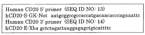
[Fig. 3] Sequences of primers for cloning human CD20 gene.
[Fig. 4] Amino acid sequences of H chain and L chain variable
regions of murine anti-CD20 monoclonal antibodies.
[Fig. 5] Results of apoptosis,test using murine anti-CD20
monoclonal antibodies.
[Fig. 6] Results of cell growth inhibition test using murine
anti-CD20 monoclonal antibodies.
[Fig. 7] Results of complement-dependent cytotoxicity test
using chimeric anti-CD20 monoclonal antibodies.
[Fig. 8A] Amino acid sequences of H chain and L chain
variable regions of humanized anti-CD20 monoclonal antibodies
and nucleotide sequences corresponding to them.
[Fig. 8B] Amino acid sequences of H chain and L chain
variable regions of humanized anti-CD20 monoclonal antibodies
and nucleotide sequences corresponding to them.
[Fig. 9] Results of cell growth inhibition test using
humanized anti-CD20 monoclonal antibodies.
Best Mode for Carrying out the Invention
In the present invention, the term "antibody" is used
in a meaning that encompasses antibody in the general meaning,
H chain and L chain constituting it, and fragments thereof.
CA 02603414 2007-10-01
9
The present invention relates to an anti-CD20
monoclonal antibody that binds to the human CD20 antigen on a
cell membrane and has biological activities desirable for
inducing a therapeutic effect.
The antibody according to a first embodiment of the
present invention is a monoclonal antibody that specifically
binds to the human CD20 antigen on a cell membrane and has
cell growth inhibitory activities including apoptosis against
human CD20 antigen expressing cells in culture of the CD20
antigen expressing cells without the aid of effector cells.
This is originally a murine anti-CD20 monoclonal antibody,
and further includes an anti-CD20 monoclonal antibody
obtained by chimerizing or humanizing that antibody. These
antibodies have direct cell growth inhibitory activities
including apoptosis against human CD20 antigen expressing
cells in in vitro culture of the CD20 antigen expressing
cells without the aid of effector cells. These chimerized or
humanized anti-CD20 monoclonal antibodies have a complement-
and/or antibody-dependent cell-mediated cytotoxicity.
The binding property to a CD20 antigen on a cell
membrane can be examined by cell-ELISA, in which CD20
expressing cells such as SB cells and Raji cells are adhered
to a plate and reacted with a monoclonal antibody to be
tested. However, since expression levels of the CD20 antigen
of these cells are insufficient, the reactivity is not high.
Therefore, in the present invention, a method of cell-ELISA
was developed, in which CHO cells in which CD20 is expressed
in a large amount by gene recombination (CD20/CHO cells) are
adhered to a plate and reacted with a monoclonal antibody to
be tested. In a preliminary test of the present invention,
it was confirmed that cell-ELISA using the CD20/CHO cells
showed a pattern similar to that observed in cell-ELISA using
the SB cells or Raji cells in a reactivity test of a
CA 02603414 2007-10-01
monoclonal antibody, and showed high sensitivity (see the
example, Establishment of CD20/CHO cell ELISA screening
method, Table 1).
The direct cell growth inhibitory activities in an in
vitro culture of human CD20 antigen expressing cells without
effector cells can be determined by a usual method (Miyamoto
T et al., Avian Dis., Vol. 46(1), 10-16). Further, the
apoptosis inducing ability can be determined by a test using
flow cytometry (annexin V/propidium iodide (PI) staining).
Examples of the antibody according to the first ,
embodiment include mouse anti-CD20 monoclonal antibodies
having a combination of the amino acid sequences of SEQ ID
NOS: 1 and 7, SEQ ID NOS: 2 and 8, or SEQ ID NOS: 15 and 17
for the H chain variable region and the L chain variable
region, as well as anti-CD20 monoclonal antibodies obtained
by chimerizing or humanizing those antibodies. These
antibodies exhibit direct cell growth inhibitory activities
including apoptosis against human CD20 antigen expressing
cells in in vitro culture of the CD20 antigen expressing
cells without the aid of effector cells. These antibodies
also have complement- and/or antibody-dependent cell-mediated
cytotoxicity. The present invention also includes a
hybridoma producing a murine antibody, and a mammalian cell
(CHO cell in the examples) incorporated with a nucleotide
sequence corresponding to any one of the amino acid sequences
of the chimeric or humanized antibodies.
Chimerization is carried out by fusing the amino acid
sequence of the H chain variable region of a murine
monoclonal antibody and the amino acid sequence of the H
chain constant region of human immunoglobulin, and the amino
acid sequence of the L chain variable region and the amino
acid sequence of the L chain constant region of human
immunoglobulin (Ishida T et al., Nippon Rinsho, Vol. 60, No 3,
CA 02603414 2007-10-01
11
439-444). Humanized antibodies are designed by using an
amino acid sequence of the variable region CDR of a murine
monoclonal antibody and an amino acid sequence of human
immunoglobulin. Humanized anti-CD20 monoclonal antibodies
preferred as therapeutic agents are selected by comparing
characteristics of variously designed antibodies (Padlan EA,
Mol. Immunol., Vol. 28, No 4/5, 489-498; Wu TT and Kabat EA,
Mol. Immunol., Vol. 29, No 9, 1141-1146; Padlan EA et al.,
FASEB J., Vol. 9, 133-139).
Chimerized or humanized anti-CD20 monoclonal
antibodies further have complement-dependent cytotoxicity
(CDC), and antibody-dependent cell-mediated cytotoxicity
(ADCC) in the presence of effector cells. As for test
methods for these CDC and ADCC, commonly used methods can be
referred to (Manches 0 et al., Blood, 2003, 101(3), 949-54;
Idusogie EE et al., J. Immunol., 2000, 164, 4178-4184).
Specific examples of the humanized anti-CD20
monoclonal antibodies include those having a combination of
the amino acid sequences of SEQ ID NOS: 19 and 23, SEQ ID
NOS: 19 and 24, SEQ ID NOS: 19 and 25, SEQ ID NOS: 19 and 26,
SEQ ID NOS: 20 and 23, SEQ ID NOS: 20 and 24, SEQ ID NOS: 20
and 25, SEQ ID NOS: 20 and 26, SEQ ID NOS: 21 and 23, SEQ ID
NOS: 21 and 24, SEQ ID NOS: 21 and 25, SEQ ID NOS: 21 and 26,
SEQ ID NOS: 22 and 23, SEQ ID NOS: 22 and 24, SEQ ID NOS: 22
and 25, or SEQ ID NOS: 22 and 26 for the H chain variable
region and the L chain variable region.
The antibody according to a second embodiment of the
present invention is a murine monoclonal antibody that
specifically binds to the human CD20 antigen on a cell
membrane, and does not exhibit cell growth inhibitory
activities including apoptosis or exhibit such activities at
a level not so high. However, these murine antibodies can be
imparted with CDC or ADCC activity by chimerization or
CA 02603414 2007-10-01
12
humanization. These cytotoxic activities are also important
biological activities, and therefore the anti-CD20 monoclonal
antibody of the second embodiment can also be a promising
candidate of therapeutic agent.
Examples of the antibody according to the second
embodiment include murine anti-CD20 monoclonal antibodies
having a combination of the amino acid sequences of SEQ ID
NOS: 3 and 9, SEQ ID NOS: 4 and 10, SEQ ID NOS: 5 and 11, SEQ
ID NOS: 6 and 12, or SEQ ID NOS: 16 and 18 for the H chain
variable region and the L chain variable region, as well as
anti-CD20 monoclonal antibodies obtained by chimerizing or
humanizing those antibodies. The present invention also
includes a hybridoma producing the murine antibody, and a
mammalian cell (CHO cells in the example) incorporated with a
nucleotide sequence corresponding to any one of the amino
acid sequences of the chimeric or humanized antibodies.
The method for determining binding property to the
CD20 antigen on a cell membrane, various test methods for
determining cell growth inhibition, apoptosis, ADCC, CDC, and
so forth, and the preparation method for chimeric or
humanized antibodies are similar to those mentioned for the
antibody of the first embodiment.
Both the chimeric anti-CD20 monoclonal antibody and
humanized anti-CD20 monoclonal antibody described as the
antibodies of the first embodiment and the second embodiment
can be expected to exhibit superior effect as a therapeutic
agent for B cell-mediated malignant tumors and immunological
diseases in which B cells or antibodies produced by B cells
are involved, and an object of the present invention is to
use them in development of a therapeutic agent containing
either a chimeric or humanized anti-CD20 monoclonal antibody,
desirably a humanized anti-CD20 monoclonal antibody, as an
active ingredient. Examples of the objective diseases
CA 02603414 2007-10-01
13
include non-Hodgkin's lymphoma, Hodgkin's lymphoma, chronic
lymphatic leukemia, acute lymphatic leukemia, chronic
rheumatoid arthritis, autoimmune hemolytic anemia, idiopathic
thrombocytopenia purpura, systemic lupus erythematosus, anti-
phospholipid antibody syndrome, Sjogren's syndrome, Crohn's
disease, scleroderma, multiple sclerosis, type I diabetes,
and so forth.
The murine monoclonal antibody of the present
invention can be prepared by the following method.
As an immunogen for sensitization, the SB cell or Raji
cell as a cell strain that expresses the human CD20 antigen,
and CHO cell made to express the human CD20 antigen can be
used in combination. Further, a human CD20 protein fused
with GST (GST-CD20) may be used as a complementary
sensitizing antigen.
A hybridoma producing a monoclonal antibody can be
prepared by a series of procedures including (1) immunization
of an animal to be immunized (mouse), (2) preparation of
lymphocytes from the immunized animal, (3) preparation of
parent cells, (4) cell fusion of the lymphocytes and the
parent cells, (5) screening and (6) cloning (Biochemistry
Experiment Method: Monoclonal antibody, written by Ailsa M.
Campbell, translated by Toshiaki Osawa, Tokyo Kagaku Dozin
Co., Ltd., 1989). An anti-CD20 monoclonal antibody that
specifically binds to the CD20 antigen on a cell surface can
be cloned by reacting a monoclonal antibody to be tested with
a cell-ELISA system in which CD20/CHO cells are immobilized
on a plate. Commercially available expression vectors can
also be used. However, since the CD20 antigen needs to be
expressed on the CHO cell at a high density, a mammalian cell
high expression vector, pNOW (Japanese Patent No. 3582965)
may be used. A selection criterion of the monoclonal
antibody is exhibition of reactivity comparable to or higher
CA 02603414 2007-10-01
14
than that of a positive control.
A chimerized or humanized antibody can be prepared
according to a usual gene recombination method. For example,
pNOW-Ab, which contains 2 sets of multicloning sites
positioned in tandem for producing the antibody, and is
incorporated beforehand with the genes encoding human H chain
and the L chain constant regions, can be used as an
expression vector (Fig. 1).
[Example 1]
Preparation, chimerization and humanization of
monoclonal antibodies directed to the CD20 antigen as well as
test for characteristics of the obtained antibodies will be
explained below with reference to the examples.
(1) Preparation of immunogen for sensitizing mouse
The human CD20 gene was obtained from a cDNA library
by using a 5' primer of SEQ ID NO: 13 and a 3' primer of SEQ
ID NO: 14, which are specific to the gene encoding the total
molecule of human CD20 (Multiple Choice cDNA human spleen,
Origene Technologies, Inc., 6 Taft Court, Suite 100,
Rockville, MD 20850). Specifically, the primers shown in Fig.
3 were used. The CD20 gene was incorporated into pNOW-Ag
(Fig. 2) as a high expression vector for mammalian cells, and
transfected into CEO cells as the host cells. Recombinant
CHO cells (CD20/CHO cells) expressing CD20 molecules at a
high level on their cell surfaces were established by FACS
analysis. Cells showing 5 or more times higher fluorescence
intensity compared with the SB cell in staining with FITC-
labeled anti-CD20 monoclonal antibodies were defined as those
showing high expression. GST-CD20, the complementary
immunogen, was prepared by fusing GST at the N terminus of 43
amino acid residues of the CD20 extracellular domain by, using
the pGEX-4T2 vector (G et al. AM, Electrophoresis, Vol.
CA 02603414 2007-10-01
20(2): 344-348).
(2) Preparation of immunogen
The SB cells or Raji cells were cultured in 10% FCS-
added RPMI 1640 medium. The CD20/CHO cells were cultured in
CHO-S-SFM II medium (GIBCO, Cat. No. 12052-098) added with
800 ug/ml of G418. These cultures were centrifuged (1100 rpm,
5 minutes), then the cells were added with Dulbecco's PBS(-)
and suspended, and the suspension was centrifuged again.
This washing procedure was repeated once again, and a
suspension prepared by adding physiological saline to the
cells (cell count: 1 to 3 x 10⁷/ml) was used for immunization.
pGEX-4T2 incorporated with GST-CD20 was introduced into E.
coli competent cells. The competent cells were lysed after
culture, and GST-CD20 was crudely purified from the lysed
cells, and then solubilized by addition of 0.1 N sodium
hydroxide.
(3) Immunization and cell fusion
As animals to be immunized, 7- to 11-week old Balb/c
female mice were used. The SB cells, Raji cells or CD20/CHO
cells were repeatedly administered twice or three times at
intervals of various numbers of days, then a different cell
antigen (SB cells, Raji cells or CD20/CHO cells) was selected
and used for the final immunization. The count of cells
administered was 1 to 3 x 10⁷ per mouse regardless of the
cell type. Further, complementary immunization was performed
by using GST-CD20 for a part of the mice. Three days after
the final immunization, spleen cells were extracted from the
mice, and suspended in the RPMI medium, and a fusion reaction
with mouse myeloma (NS-1) was carried out in the presence of
PEG-1500 (Oi, VT et. Herzenberg, 1980, in: Selected Methods
in Cellular Immunology; Mishell B et al. (Freeman and Co.,
CA 02603414 2007-10-01
16
San Francisco, CA) p.351).
(4) Establishment of CD20/CHO cell-ELISA screening method
Several murine anti-CD20 monoclonal antibodies and 2B8
were reacted by using 96-well plates to which the SB cells,
Raji cells, CD20/CHO cells and CD20 CHO parent cell line were
adhered. It was confirmed that in these cell-ELISA tests,
similar tendencies were observed for the antibody
concentrations, and it was found that relative comparisons
between the antibodies and with a control were also possible.
Because of the high density of the surface cell antigens
adhered to the plate in the CD20/CHO cell-ELISA, an
absorbance was observed at a level sufficiently enabling the
detection even with a relatively low concentration of the
test antibody sample, and it was found to be a sensitive
measurement system. The specific measurement results are
shown in Table 1.
CA 02603414 2007-10-01
17
U) r- r-i -I Ln m m U) r o o O
in in u) lD lO a) N N l0 l0 l0 l0 l0
M z c O O O O O 0l M z O O O C) O O
O O O O O O O O O O O O
N Cl l0 l0 co [- [- : m [- O LO O M
za) C) m r-i l0 M ~ lO M N O M M [- M a) C a
^ r1 O r1 O O O O O C rl O O O O O O O
0) C) O O O O O O r1 O O O O O O O
H C
FL' FC 0 lO if) 0) m O m ri 0 CZV 6l (n r- l0 M LO
f/) -- 0) Cl M C) N M N =.-1 M M l0 d' Ln mot' M
H a-) CO -1 N C) O ri r1 C) U 4 C) O O (D O O O
C) O O O O O O p O O (D C) O O O
F-4 N O N r=-I N '' LI) )-i W 00 OD v) '.0 ,-I N LI)
U O U) [- Ln r a) lfl ,zv ro U O M M m a' rn r C'
U _j O -i' u) ri o N N O 0 C O O O O O O O O
0 N O r-i O 0 0 0 0 0 0 0 rt O C) O C) O O O
U U U)
H M O P Ol LO H H >1 O M O OD M M C)
-0 o M d' o a o a' m '0 o Ln r- '.0 -1 Ln v
o
04 ~ G 0 (N 0-) H -Y' ri u) O W 0 (N O O -i O N r1 O
A H O ri O O O O O H O O C) C) O O O
rl
C O LO Cl M M ri 0) r- m U < O rl iI) L) iI) lD 0)
l0 O l0 O H LO M N m 0) m l0 lO r- [n N
O C) (N LI) r- zT '--1 m M O 0 o0 O O M H u) M O O
i o o H o 0 o o 0 0 0 0 0 0 0
H
l0 0) Cl r- 00 C0 LI) H -zv m m l0 0l
rn d' d' 4f) to ,zj' l0 r- r- l0 m m m
M C) O O O O O M N N r-I O N N O
O
r) O O O O O O O CD* O O CD* c;
Q1 H
U m [- rl l0 P- m c) N N C' 01 in M dl ~w
' l~ lfl V' l0 LO M dl tT C- C) co O l0 LO [-
'~ N OH -1 r1 O C) O C) O if) l0 M r-i d' to r1
04 m y 0 0 0 0 0 0 0 si v 0 0 0 C), 0*
0
C
0 l0 LI) N [~ v' u) m O u) M -::v m r- rl vI
r-i N ~' O u) O O M m '.4 P N N M l0 O l0
H FC -W N M I' H O rt H O H N N N m N 01 r) u)
C/) (a M a (U M
W H O ] rri O O O 4 G4 ~--I O
H W H
m ~--I l0 N O m O
{ U r-i U O N m ' C) [rn
U r-i C U O O Cl d m rl N O
CA U OU O O O O O O O O U N r1 r1 O N N rl
E
y..) = r 1 ?i O M u) r a' l0 r-I U >1 m
0 -n T3 O l- l0 N [- r-i [- N - -0 O O r N d' l0 l0
a 0 N H N Ln (N Ln M ri ON (Ny) [[ Ln II) M a M lO
M
0 H 1--1 rl O O O O O U' N N rl r-i N N rl
fl < O M Ln m co l0 1- c) U) O l0 d' t` O N M O
m r- N 141 dl m N M in co r- M d' M m CD l0 P- N (- r--I m M O OO (D ri m N
Li) (N O
0 r= rl rl r1 O ri O O O ) M rl N N N N O
U
rf
N >1 >1
r i a m r- N N m '0 m r- N N m kD
O N Ln O N N (,.) m 0 (N Ln o N (,,) m
A N N ~' d a' q;;I, U .0 N (N C' C -, W U
H =.-1 rl ri r1 1-1 rr Z r I rl rl ri rl rl Z
x x x x x N x x x `t, x N
41
r-1 rt H r= rl rt r-i rI r1 H
CA 02603414 2011-12-05
18
(5) Screening by cell-ELISA
Cell-ELISA was performed by using a 96-well plate to
which the CD20/CHO cells or CHO cells (CD20 parent cell line)
were adhered, and wells were selected in which antibodies
specifically reactive to CD20 were produced. 2B8 was used as
a positive control, and a mouse monoclonal antibody directed
to the human CD3 antigen (BD PharMingen) was used as a
negative control. Specifically, the CD20/CHO cells or CHO
cells (parent cell line) adhered to a poly-L-lysine coated
96-well plate (Asahi Techno Glass Corporation, Cat. No. 11-
023-018) were used for cell-ELISA. A blocking solution (0.2%
gelatin and 0.5% BSA solution in PBS) was added in a volume
of 150 pl to each well and left standing at 37 C for 1 hour.
Then, the plate was washed 5 times with 150 mM NaCl and 0.05%
Tween 20 aqueous solution, and 100 pl of each sample (diluted
solution of culture supernatant) was added to each well to
perform the primary reaction at 37 C for 1 hour. After
washing, 100 pl of a diluted solution of a labeled antibody
[HRP-labeled anti-mouse IgG(H+L) rabbit antibody (Jackson
Lab., Code No. 315-035-003) or HRP-labeled anti-mouse
IgG(Fcy) rabbit antibody (Jackson Lab., Code No. 315-035-
008)] was added to each well to perform the secondary
reaction at 37 C for 1 hour. For the preparation of the
reaction mixtures for the primary and secondary reactions, a
solution the same as the blocking solution was used. After
washing, 100 pl of a color development solution (OPD) was
added to each well, 30 minutes later, 50 pl of 4 N H2SO4 was
added to terminate the reaction, and absorbance was measured
at 492 nm (A492) . Then, wells showing reactivity comparable
to or significantly higher than that of 2B8 were selected.
(6) Cloning
Cloning was carried out by the limited dilution method.
*Trade-mark
CA 02603414 2007-10-01
19
Cells were seeded on a 96-well plate and cultured, then cell-
ELISA for CD20/CHO cells was performed for culture
supernatant of a well containing 1 colony to select a clone
producing a specific antibody.
(7) Preparation of purified antibody
The clone producing a specific antibody was cultured
in 10% FCS-added RPMI 1640 medium. When the cell density
became about 5 x 10⁵/ml, the medium was replaced with a
serum-free medium, ASF-104N (Ajinomoto Co. Inc.), and culture
were continued. Then, 2 to 4 days later, the culture medium
was centrifuged, and the culture supernatant was collected
and subjected to purification using a protein G column. The
eluted monoclonal antibody solution was dialyzed against 150
mM NaCl. The solution was subjected to filtration
sterilization using a filter having a pore size of 0.2 pm and
used as a test antibody (anti-human CD20 mouse monoclonal
antibody).
Monoclonal antibody clones showing binding affinity
comparable to that of the positive control were selected by
the CD20/CHO cell-ELISA. The gene sequences of variable
regions of these antibodies were determined, and the amino
acid sequences thereof were determined as a result. The
sequences of the H chain variable region and the L chain
variable region of typical antibodies are shown in SEQ ID
NOS: 1 and 7, SEQ ID NOS: 2 and 8, SEQ ID NOS: 3 and 9, SEQ
ID NOS: 4 and 10, SEQ ID NOS: 5 and 11, SEQ ID NOS: 6 and 12,
SEQ ID NOS: 15 and 17, and SEQ ID NOS: 16 and 18 (Fig. 4).
Further, biological characteristics of these clones were
investigated.
Biological characteristic test (1): Apoptosis induction test
The apoptosis inducing ability of the test antibodies
CA 02603414 2007-10-01
was determined by flow cytometry (annexin V/PI staining).
2B8 was used as a positive control, and the mouse monoclonal
antibody directed to the human CD3 (BD PharMingen) was used
as a negative control. The procedures were as follows.
MEBCYTO Apoptosis Kit (MBL, Cat. No. 4700, Lot. 20)
was used.
The Raji cells were centrifuged, and then suspended in
a fresh RPMI 1640 medium (Sigma, Cat. No. R8758, Lot 44K2416)
containing 10% FCS (inactivated) (ICN, Cat. No. 2916754, Lot
8005C), and 1 ml of the suspension at a density of 5 x 10⁵
cells/ml was added to each well of a 12-well plate. Twelve
wells were used for each antibody, and each antibody was
added at a final concentration of 2 pg/ml or 4 pg/ml (3 wells
x 2 different concentrations x 2 time points, 12 wells in
total).
One day and two days after the start of the culture,
the culture medium containing about 2 x 10⁵ cells was
collected, and centrifuged, and then the cells were washed
once with PBS. Subsequently, 85 pl of a binding buffer was
added to the cells to suspend the cells in the buffer.
Further, to the suspension was added 10 pl of annexin V-FITC
and 5 pl of PI, mixed sufficiently, and allowed to react at
room temperature for 15 minutes with light shielding.
Measurement was performed by flow cytometry (FACS Calibur,
Becton Dickinson), and the results were analyzed by using
CellQuest (Becton Dickinson).
The measurement results of 8 kinds of the typical
murine anti-CD20 monoclonal antibodies, positive control
(2B8), and negative control (anti-CD3 antibody) are shown in
Fig. 5. In general, the apoptosis inducing ability of 2B8 is
said to be high. Even compared with this, the clone of which
amino acid sequences of the H chain variable region and the L
chain variable region were those of SEQ ID NOS: 2 and 8
CA 02603414 2007-10-01
21
(1K1791) showed a markedly higher apoptosis inducing activity.
Cell death clearly due to apoptosis was also observed with
the clones of which amino acid sequence of the H chain
variable region and the L chain variable region were those of
SEQ ID NOS: 1 and 7 (1K1422) and SEQ ID NOS: 15 and 17
(lK0924).
Biological characteristic test (2): Cell growth inhibition
test
A 5 x 10⁹ cells/ml Raji cell suspension was prepared
with 10% FCS-added RPMI 1640 medium, and added to a 96-well
plate in a volume of 100 pl/well, and culture was performed.
After 24 hours, 50 p1/well of each antibody solution was
added at an antibody concentration of 0.01 pg/ml, 0.1 pg/ml
or 1 pg/ml, and culture was continued. Seventy two hours
after the addition of the antibody, 10 p1/well of a color
development solution, Cell Counting Kit-8 (Dojindo
Laboratories, Cat. No. 343-07623, Lot SG076) was added, the
cells were cultured for further 4 hours, and then absorbance
was measured at 492 nm. The living cell counts of the
typical 8 kinds of murine anti-CD20 monoclonal antibodies and
the positive control (288) are shown in Fig. 6 as percentages
based on that of the negative control (100%) . The cell
growth inhibitory effect can be estimated on the basis of the
rate of the decreased living cell count compared with that of
the negative control. Clear cell growth inhibition was
observed with 1K0924, lH1422, 1K1791 and 2B8 as the positive
control, and the inhibition was particularly marked with
1K1791. This tendency was consistent with the results of the
apoptosis induction test.
Preparation of chimeric antibodies
The genes encoding the H chain and L chain variable
CA 02603414 2007-10-01
22
regions of each murine antibody were incorporated into pNOW-
Ab, a high expression vector for CHO cell already containing
the genes encoding human immunoglobulin H chain and L chain
(K) constant regions as a cassette. Each expression vector
was transfected into CHO cells, and clones showing high
productivity were selected for each antibody.
Test for binding property to CD20 antigen of chimeric
antibodies
The prepared 8 kinds of chimeric anti-CD20 monoclonal
antibodies were examined for reactivity to the human CD20
antigen by the CD20/CHO cell-ELISA. Rituximab (c2B8) was
used as a positive control. The test results are shown in
Table 2. The values measured in the cell-ELISA (A492)
reflect intensity of the binding property. These antibodies
showed affinity substantially comparable to or higher than
that of the control except that clK0924 and clK1422 tended to
show slightly lower affinity than that of the control.
CA 02603414 2007-10-01
23
Table 2 CD20/CHO cell-ELISA test of anti-CD20
chimeric antibodies
CD20/CHO cell-ELISA (A492)
Antibody Antibody concentration (ng/ml)
100 32 10 3 1 0
c1K0924 1.423 0.724 0.391 0.186 0.094 0.032
c1K1228 2.226 1.580 0.701 0.289 0.120 0.032
c1K1402 2.449 1.621 0.737 0.349 0.116 0.032
c1K1422 1.919 0.912 0.357 0.151 0.077
clKl712 2.292 1.683 0.793 0.359 0.145
c1K1736 2.428 1.548 0.748 0.320 0.122
c1K1782 2.101 1.017 0.505 0.169 0.074
c1K1791 2.231 1.458 0.745 0.276 0.108
c2B8 2.147 1.143 0.536 0.226 0.088
CDC test of chimeric antibodies
The prepared 8 kinds of chimeric anti-CD20 monoclonal
antibodies were examined for the CDC activity. Rituximab
(c2B8) was used as a positive control. RC-K8 (obtained from
Kochi Medical School) was used as the target cells. As a
medium for use, RHB (basal medium: RPMI-1640, additives: 0.1%
BSA, 20 mM HEPES (pH 7.2), 2 mM glutamine, 100 units/ml of
penicillin G, 100 pg/ml of streptomycin) was prepared and
used. The target cells were washed with RHB and resuspended
at 106 cells/ml. In a volume of 50 pl of each of the
solutions of the test chimeric antibodies and rituximab
having different concentrations, 50 pl of 4-fold diluted
solution of a commercially available human complement (Quidel,
San Diego, CA, Cat. A113), and 50 pl of a cell suspension
containing 106 cells/ml were added to each well of a flat
bottom 96-well tissue culture plate (black). The antibody
concentration in the mixture of 150 p1/well was set at 0.1, 1
CA 02603414 2011-12-05
24
and 10 pg/ml. To promote complement-mediated cell lysis, the
mixture was incubated under the conditions of 37 C and 5% CO2
for 2 hours. To the mixture was added 50 pl of alamar blue
(undiluted, prescription of AccuMed International, Biosource,
Cat. DAL1100), and the reaction was further allowed overnight
under the same conditions. The plate was left at room
temperature for 10 minutes to cool, and fluorescence was
measured at 590 nm for emission with excitation at 530 nm by
using a fluorescence microplate reader. The results were
represented in terms of fluorescence intensity (RFU). The
rate of CDC activity was calculated in accordance with the
following equation:
%CDC activity = 100 x {RFU (antibody not added) - RFU
(antibody added)}/{RFU (antibody not added] - RFU (Triton X-
100 added)}
The results are shown in Fig. 7. Except for c1K1712,
the antibodies showed CDC activities substantially comparable
to or higher than that of C2B2 as the positive control.
Preparation of humanized antibodies
Humanized antibodies were designed based on the
variable region CDR of the murine anti-CD20 monoclonal
antibody 1K1791. By performing structural analysis based on
the amino acid sequences and further changing the designing
method, 4 kinds of humanized sequences were prepared for each
of the H chain and the L chain (Padlan EA, Mol. Immunol., Vol.
28, No 4/5, 489-498; Wu TT and Kabat EA, Mol. Immunol., Vol.
29, No 9, 1141-1146; Padlan EA et al., FASEB J., Vol. 9, 133-
139). Antibodies were prepared with all possible
combinations of the four types for each of the H chain and
the L chain. These amino acid sequences are shown in SEQ ID
NOS: 19 and 23, SEQ ID NOS: 19 and 24, SEQ ID NOS: 19 and 25,
SEQ ID NOS: 19 and 26, SEQ ID NOS: 20 and 23, SEQ ID NOS: 20
*Trade-mark
CA 02603414 2007-10-01
and 24, SEQ ID NOS: 20 and 25, SEQ ID NOS: 20 and 26, SEQ ID
NOS: 21 and 23, SEQ ID NOS: 21 and 24, SEQ ID NOS: 21 and 25,
SEQ ID NOS: 21 and 26, SEQ ID NOS: 22 and 23, SEQ ID NOS: 22
and 24, SEQ ID NOS: 22 and 25, and SEQ ID NOS: 22 and 26
(Figs. 8A and 8B).
The amino acid sequences of these 4 kinds of H chain
variable regions and 4 kinds of L chain variable regions were
converted into DNA (nucleotide) sequences with codons most
frequently used in human gene sequences, and some of these
nucleotides were changed considering suitability in the host
CHO cells without changing the original amino acid residues
(Kim CH et al., Gene, 1997, 15; 199 (1-2): 293-301).
Specifically, used as the nucleotide sequences corresponding
to the amino acid sequences were those of SEQ ID NO: 27
corresponding to SEQ ID NO: 19, SEQ ID NO: 28 corresponding
to SEQ ID NO: 20, SEQ ID NO: 29 corresponding to SEQ ID NO:
21, SEQ ID NO: 30 corresponding to SEQ ID NO: 22, SEQ ID NO:
31 corresponding to SEQ ID NO: 23, SEQ ID NO: 32
corresponding to SEQ ID NO: 24, SEQ ID NO: 33 corresponding
to SEQ ID NO: 25, and SEQ ID NO: 34 corresponding to SEQ ID
NO: 26 (Fig. 8A and Fig. 8B). In these nucleotide sequences,
nucleotides may be replaced with other nucleotides so long as
the corresponding amino acid sequences are not altered.
Total 8 kinds of the nucleotide sequences of H chain
variable regions (SEQ ID NOS: 27 to 30) and L chain variable
regions (SEQ ID NOS: 31 to 34) were synthesized (Takara Shuzo
Co., Ltd.) and incorporated into pNOW-Ab, an expression
vector for mammalian cells containing a multicloning site.
The expression vectors incorporated with each of these
humanized antibody genes were transfected into CHO cells, and
clones showing high productivity were selected for each
antibody.
CA 02603414 2007-10-01
26
Biological characteristic and cell growth inhibition tests
for humanized antibodies
A suspension containing 5 x 10⁴/ml of the Raji cells
was prepared with 10% FCS-added RPMI 1640 medium, and.added
to a 96-well plate in a volume of 100 p1/well, and culture
was performed. After 24 hours, 50 p1/well of each antibody
solution was added at an antibody concentration of 0.5 pg/ml,
and culture was continued. Seventy two hours after the
addition of the antibody, 10 p1/well of a color development
solution, Cell Counting Kit-8 (Dojindo Laboratories, Cat. No.
343-07623, Lot SG076) was added, culture was performed for
further 4 hours, and then absorbance was measured at 492 nm.
The living cell counts of 15 kinds (27 clones) out of 16
kinds of humanized antibodies derived from 1K1791 and c2B8
(other name of rituximab) as the positive control are shown
in Fig. 9 as rates based on that of the negative control
(1000). The cell growth inhibitory effect can be estimated
on the basis of rate of decreased living cell count compared
with that of the negative control, and the growth inhibitory
effect was observed for all the clones in this test.
The names of monoclonal antibodies and the sequence
numbers described in this specification and the appended
drawings are summarized as follows.
CA 02603414 2007-10-01
27
M LU l0 M zzr L lC M LU l0 M L ,O
- N N N N N N N N N N N N N N N N
z3 z3 ~S LS z3 t3
ro ro ro ro 10 10 ro ro ro ro ro ro ro ro ro ro
al m 61 Ol C) CD O O '--I r-I r-I 1-1 N N N N
(a r-i r-i r-1 H N N N N N N N N N N (N N
-r-i
U~ Cn co co (n U~ va UW U) (n v~ (n u~ U) (n U
Q)U a
0000000000000000
N rI Z Z Z Z Z Z Z Z Z Z Z Z Z Z Z Z Z
v Q Q Q Q Q Q Q Q Q Q Q (~ Q Q (~ (~
ro O H H H H H H H H H H H H H H H H
a a a a a a a a a a a a aaaa
x ~, w w w w w w w w w w w w w w w w
~~(n~~+ncncncn~+nv~U1(n(n(n
U i N r-I N N Q0 N CO
0 O N C rH CD M OD N N
O -Q N
.(::., r--I c--I r~ c--I r--I r-I .--I '--I
O ro u u 0 0 0 0 0 0
I- co m O r1 N I- CO
r--I r1 r= r-i rI
O
-H -i r- 0 0 0 0 0 0 0 0
A 0 2 Z 2 Z Z Z Z Z
Q Q Q 0 Q 0 0
Q
O
ro ~i H H H H H
as 0aaaa
~~ ~ w w w w w
U) U) U) co U)
r1 N M ~ Ln <O Ln r-! rl
1U Q 0 z z O O O O z z
U ro -t3) Q Q Q Q 0 Q Q Q
Si O H H H H H H H H
~4
aa aaaa
w w w w w w a a
CO U) U) (] U) w w U) U)
O
ro
r-
O N r-I (N N lO N 'T co
-Q N O> r--I O M 00 N N
-H ~t N N v N N C N
4-) r-4 1-4 r-I r-i r-I rI O r-i
~ x x x x~ x x x
ro r~ r-I r~ r~ r~ rI
M O
4) =H
rl ~-1
ro ~
H
CA 02603414 2007-10-01
28
The hybridomas producing these monoclonal antibodies
were named on the basis of the names of the antibodies
produced thereby, and internationally deposited at the
National Institute of Advanced Industrial Science and
Technology, International Patent Organism Depositary (Tsukuba
Central 6, 1-1, Higashi 1-Chome, Tsukuba-shi, Ibaraki-ken,
305-8566, Japan) on March 28, 2006 under the provisions of
the Budapest Treaty, and assigned accession numbers of FERM
BP-10587 (1K1422), FERM BP-10591 (1K1791), FERM BP-10588
(1K1712), FERM BP-10586 (1K1402), FERM BP-10589 (1K1736),
FERM BP-10590 (1K1782), FERM BP-10584 (1K0924), and FERM BP-
10585 (1K1228).
Industrial Applicability
The present invention provides a monoclonal antibody
having biological activities suitable for use as a
therapeutic agent.