Note: Descriptions are shown in the official language in which they were submitted.
CA 02603415 2007-09-28
WO 2006/105476 PCT/US2006/012255
ULTRA,SONIC PERIODONTAL DEVICE, SYSTEM AND METHOD OF
USING
FIELD OF THE INVENTION
[0001] The present invention is generally directed to the field of periodontal
medicine and in
particular to the application of ultrasonic technology to periodontal medicine
and to general
dentistry.
BACKGROUND OF THE INVENTION
[0002] Periodontal gum disease is a serious infection of the mouth that, if
left untreated, can lead
to tooth loss and has been associated with, and is suspected of contributing
to heart attacks,
strokes, diabetes, respiratory diseases, premature/underweight babies and even
death.
[0003] Periodontal disease can affect one tooth or many teeth. It begins when
the bacteria in
plaque (the sticky, colorless film that constantly forms on everyone's teeth)
causes the gums to
become infected and inflamed.
[0004] In the mildest form of the disease, gingivitis, the gums redden, swell
and bleed easily.
There is usually little or no discomfort. Gingivitis is often caused by
inadequate oral hygiene,
especially lack of flossing. Gingivitis is reversible with professional
treatment and good oral
home care.
[0005] Untreated gingivitis can advance to periodontitis. With time, plaque
can harden into
calculus and spread and grow below the gum line where it can become a breeding
ground for
bacteria below the gurn line. Toxins produced by the bacteria in plaque and
calculus continue to
irritate and inflame the gums and surrounding tissue. As the infection becomes
more severe, the
toxins stimulate a chronic inflammatory response in which the body in essence
turns on itself and
the tissues (ligaments) and bone that support the teeth are broken down and
destroyed.
[0006] Periodontal soft tissue (gums or gingiva and the periodontal ligament)
detach from the
teeth, formin.g periodontal pockets (spaces between the teeth and periodontal
tissue) that become
infected. As the disease progresses, more and more destructive toxins are
produced and as a
CA 02603415 2007-09-28
WO 2006/105476 PCT/US2006/012255
result, the periodontal pockets deepen and more periodontal tissue and bone
are broken down
and destroyed. Initially this destructive process may be asymptomatic.
Eventaally, teeth can
become loose and may be lost or have to be removed. More than 300 different
types of bacteria
can exist in the human mouth, either alone or in combination. This makes
treating periodontal
disease difficult, time consuming and expensive as the periodontist tries
various antibiotics and
treatment modalities until an effective treatment plan is developed. Like any
other serious
infection, if not promptly treated with the proper types and amounts of
antibiotics, periodontitis
can result in severe systemic infection that can lead to many other serious
diseases and even
become life-threatening. As the patient's immune system fights this chronic
and perhaps serious
infection, it can create an opportunity for other serious diseases, such as
heart disease, stroke and
diabetes, to develop.
[0007] The current methodology used by dentists and dental hygienists to
detect and measure
periodontal pocket depth is a primitive methodology that consists of a sharp
metal probe that is
inserted between the tooth and gum and which is manually pressed down until it
encounters
resistance of the ligament. The depth to the ligament is thereby measured and
indicates the
amount of clinical attachment lost (loss of ligament), which can be an
indication of the amount
of periodontal disease that may be present. This method is often painful for
the patient, and is
invasive, bloody, inaccurate and subjective. It is especially inaccurate and
subjective because of
the difficultly in applying the same amount of force with each measurement,
resulting in high
intra-examiner and inter-examiner variation in measurement. The difficulty is
increased because
the examiner does not know the type of tissue present below the gum line and
if the probe is
touching or piercing this tissue. Additionally, exposure to the patient's
blood by dental
professionals increases risk of exposure to hepatitis, HIV and other
infectious diseases.
[0008] Further, the current methodology is limited in its effectiveness as a
tool for diagnosing
periodontal disease in its earliest stages as it is a retrospective analysis
and can only measure
significant amounts of tissue already lost. In addition, this method typically
calls for two people
to perform this test, an examiner who actually makes the measurements and a
scribe who usually
writes down the measurements by hand. The examiner is generally a dental
health professional,
such as a dentist, dental hygienist or periodontist. The scribe may also be a
dental health
professional but may also be a lesser skilled individual such as an office
assistant. Another
2
CA 02603415 2007-09-28
WO 2006/105476 PCT/US2006/012255
problem facing dentists is the difficulty in determining long term trends of
the patients' condition
because all of the information is contained in numerous paper (i.e., analog)
records that usually
span many years. As a result, usually only the last one or two records are
reviewed for
comparison with the current test results and these may not be sufficient to
accurately reflect a
very gradual deterioration of the patient's periodontal condition.
[0009] An additional problem with the existing manual probe methodology is
that it is typically
can be disruptive to the healing process. The trial and error approach can
tear newly healed
tissue and can cause recovery to be extended for weeks or months. Further, it
can allow bacteria
into the wound and the patient's blood stream, which can lead to infection
(i.e., bacteremia).
Indeed more than 300 different types of bacteria can exist in the human mouth,
either alone or in
combination. This makes treating periodontal disease difficult, time consuming
and expensive as
the periodontist tries various antibiotics and treatment modalities until an
effective treatment plan
is developed.
[00101 Figure 1 is a schematic diagram comparing a healthy tooth 100 on the
left and a tooth 106
with periodontal disease on the right. The healthy tooth 100 has a fi.i1l,
healthy bone level 104,
healthy periodontal ligament 103, and a healthy gum/gingiva 102. The diseased
tooth 106
exhibits gum/gingiva loss 116, loss of periodontal ligament attachment
(clinical attachment loss)
11-5 and resorption of avelor bone level 114, resulting in the formation of a
periodontal pocket
112. The diseased tooth 106 also exhibits a build up of plaque 108 and
tartar/calculus 110. If
the periodontal condition is not diagnosed and corrected, the diseased tooth
106 may be lost or
have to be removed.
[0011] Figure 2 is a more detailed schematic diagram of the teeth 100, 106
illustrated in Figure
1. The teeth 100, 106 have an enamel portion 118 and a root portion 120. The
root portion 120
is connected to the gum 122 by the periodontal ligament 126. The top of the
gum 122 is known
as the gum line 124. As illustrated in Figure 2, the gum line 124 has receded.
In some cases,
however, the gum 122 may be irritated, resulting in the gum line 124 rising
due to edema.
[0012] At the top of the periodontal ligament 126 is the upper boundary 130 of
the periodontal
ligament 126. Between the upper boundary 130 of the periodontal ligament 126
and the enamel
portion 118 is the junction epithelium 128. In a healthy tooth 100, the upper
boundary 130 of the
3
CA 02603415 2007-09-28
WO 2006/105476 PCT/US2006/012255
periodontal ligament 126, the bottom of the junction epithelium 128 and the
enamel portion 120
meet at the cemento-enamel junction 132. In a diseased tooth 106, tartar or
calculus 110 and
polymorphonuclear leukocytes 138 spread into the junction epitheliurn 128 and
the periodontal
ligament 126 opening a periodontal pocket 112. If the periodontal pocket
1121ies between the
gum line 124 and the cemento-enamel junction 132, the patient has a condition
known as
gingivitis. If the periodontal pocket 112 extends below the cemento-enamel
junction 132, the
patient has a condition known as periodontitis. Additionally, the growth of
the periodontal
pocket 112 may be irregular and result in intennediate features 136.
[0013] Frequently, prior measurements of pocket depth were made relative to
the gum line 124.
As discussed above, however, the gum line 124 may vary due to recession or
edema. Therefore,
use of the gum line (or free margin of gingiva) 124 in measuring pocket depth
may lead to
inaccurate and widely varying measures of pocket depth. In contrast to the gum
line 124, the
location of the cemento-enamel junction 132 remains constant. Therefore, use
of the cemento-
enamel junction 132 in measuring pocket depth provides a better and more
consistent method of
measurement over time and is preferable. Manual probing is also used to
determine if, and on
which teeth and exactly where, calculus is present below the gum line. This
method can be
inaccurate.
[0014] It would therefore be desirable to have a painless, noninvasive,
accurate and reproducible
method of measuring periodontal attachment loss capable of using both the gum
line and the
cemento-enamel junction 132 as a reference. It would also be desirable to have
an accurate
method of determining if, and on which teeth calculus is present below the gam
line.
SUMMARY OF THE INVENTION
[0015] This invention relates to a system for detecting and measuring
attachment loss, an
indicator of periodontal disease. The invention incorporates the use of
ultrasonic technology to
measure the differential depth between both the gum line and the cemento-
enamel junction of a
tooth and the bottom of a periodontal pocket. In contrast to conventional
methods that require
inserting a sharp metal probe between the teeth and the gum, the present
invention provides a
system and method that is painless and noninvasive, painless, bloodless,
accurate, fast, objective
and digital.
4
CA 02603415 2007-09-28
WO 2006/105476 PCT/US2006/012255
[0016] The present invention provides a system for detecting and measuring
periodontal tissue
destruction related to periodontal disease comprising a hand piece having an
ultrasonic
transducer, and an acoustic lens; a controller unit having discrimination
analysis software, the
discrimination analysis software comprising a wavelet algorithm; and a fluid
supply.
[0017] The present invention also provides a hand piece for detecting and
measuring periodontal
disease comprising a permanent handle having a cavity and an alignment slot in
a first end; an
ultrasonic transducer located in the cavity in the first end of the permanent
handle; a fluid supply;
and a disposable cover, the disposable cover having a protrusion in the
interior of the disposable
cover, the protrusion adapted to fit into the alignment slot.
[0018] The present invention also provides a hand piece for detecting and
measuring periodontal
disease comprising a continuously curved handle; an ultrasonic transducer; and
a fluid supply.
[0019] The present invention also provides a method of detecting and measuring
periodontal
disease comprising filling a periodontal pocket with a fluid capable of
propagating sound waves;
transmitting at least one sound wave into the periodontal pocket; sensing at
least one return
sound wave from the periodontal pocket; and determining the depth of the
pocket by measuxing
the time it takes the at least one transmitted sound wave to traverse the
periodontal pocket and
return.
[0020] The present invention also provides a method of performing periodontal
examinations
comprising providing dentists or dental hygienists with at least one
ultrasonic periodontal
system; and charging the dentist or dental hygienist per visit of a patient.
[0021] The present invention also provides a discrimination analysis algorithm
to analyze
ultrasonic echoes comprising processing waveforms; detecting peaks; and
discriminating peaks,
wherein the discrimination analysis algorithm uses a continuous wavelet
transformation.
BRIEF DESCRIPTION OF THE DRAWINGS
[0022] Figure 1 is a schematic diagram showing a healthy tooth on the left and
one with
periodontal disease on the right.
[0023] Figure 2 is a more detailed schematic diagram of the teeth illustrated
in Figure 1.
CA 02603415 2007-09-28
WO 2006/105476 PCT/US2006/012255
[0024] Figure 3 is a perspective view of a periodontal system according'to a
first embodiment of
the invention.
[0025] Figure 4 is alternative a periodontal system in accordance with the
principles of the
invention.
[0026] Figure 5 is a perspective view of a system illustrating one aspect of
the invention.
[0027] Figure 6 is a perspective view of a system illustrating another aspect
of the invention.
[0028] Figure 7 is a perspective view of a system illustrating another aspect
of the invention.
[0029] Figure 8 is a schematic illustration of a disassembled hand piece
according to one
embodiment of the invention.
[0030] Figure 9 is a schematic illustration of an assembled hand piece
illustrated in Figure 8.
[0031] Figure 10 is a schematic illustration of an embodiment of the invention
having an
acoustic lens.
[0032] Figure 11 is a funetional layout of an embodiment of the invention
having foot pedal
controls.
[0033] Figure 12 is a schematic illustration of an embodiment of the invention
illustrating the
use of an enterprise portal.
[0034] Figure 13 is a schematic illustration of a disassembled hand piece
according to one
embodiment of the invention.
[0035] Figure 14 is a schematic illustration of an assembled hand piece
illustrated in Figure 13.
[0036] Figure 15 is a schematic illustration of a software layout of the
invention.
[0037] Figure 16 is flow diagram illustrating operational modes of an
embodiment of the
invention.
[0038] Figure 17 is flow diagram illustrating screen flows of an embodiment of
the invention.
6
CA 02603415 2007-09-28
WO 2006/105476 PCT/US2006/012255
[0039] Figure 18 is a screen shot of an embodiment of the invention, initial
login screen.
[0040] Figure 19 is a screen shot of an embodiment of the invention, main
screen.
[0041] Figure 20 is a screen shot of an embodiment of the invention, account
screen.
[0042] Figure 21 is a screen shot of an embodiment of the invention, options
screen.
[0043] Figure 22 is a screen shot of an embodiment of the invention, patient
records screen.
[0044] Figure 23 is a screen shot of an embodiment of the invention, patient
chart screen.
[0045] Figure 24 is a screen shot of an embodiment of the invention, enter
tooth condition data
screen.
[0046] Figure 25 is a screen shot of an embodiment of the invention, measure
pocket depth
screen.
[0047] Figure 26 is a screen shot of an embodiment of the invention,
calibration screen.
[0048] Figure 27 is a screen shot of an embodiment of the invention, view
tooth history screen.
[0049] Figure 28 is a screen shot of an embodiment of the invention, view full
patient chart
screen.
[0050] Figure 29 is a screen shot of an embodiment of the invention, patient
history selection
screen.
[0051] Figure 30 is a screen shot of an enibodiment of the invention, help
screen.
[0052] Figure 31 is a flow chart illustrating data acquisition and analysis.
[0053] Figure 32 is a flow chart illustrating data entry operator options.
[0054] Figure 33 is a schematic illustration of the external interface
arrangement.
[0055] Figure 34 is a schematic illustration of a head and tip portion of a
hand piece of an
embodiment of the invention.
7
CA 02603415 2007-09-28
WO 2006/105476 PCT/US2006/012255
[0056] Figure 35 is a plot illustrating the effect of flow rate on ultrasound
measurements.
[0057] Figure 36 is a plot illustrating a Mexican Hat wavelet.
[0058] Figure 37 is a plot illustrating a Morlet wavelet.
[0059] Figure 38 is a plot illustrating wavelet scale to signal frequency.
[0060] Figure 39 is a plot illustrating peak discrimination.
DETAILED DESCRIPTION OF THE INVENTION
[0061] In contrast to the conventional method of detecting and measuring
periodontal attachment
loss, the systems and methods according to the present invention incorporate
ultrasound
technology. The systems and methods provides dentists and their patients with
a painless, non-
invasive, bloodless, extremely accurate, objective, automated, rapid, digital
and inexpensive
method to effectively diagnose, detect, and evaluate attachment loss related
to periodontal
disease, create a digital dental record and monitor treatment via a sequence
of measurements.
The system takes analog measurements, converts the analog ultrasonic
measurements to digital
data and calculates the periodontal pocket depth (preferably, the distance
from both the gain line
and the cemento-enamel junction to the upper boundary of the periodontal
ligament). The
methods according to the present invention enable dentists to detect
periodontal disease in its
earliest stage when it is easy and inexpensive to treat and before the body's
immune system is
weakened and susceptible to other diseases. It also permits dentists to more
easily and
effectively clean their patient's teeth by providing qualitative information
regarding the presence
of calculus (i.e., hardened plaque) present on tooth surfaces below the gum
line, before and after
cleaning. Additionally, the methods are essentially examiner independent as
inter-examiner and
intra-examiner variation has been essentially eliminated.
[0062] In contrast to prior art methods of diagnosing periodontal disease, the
periodontal system
of the present invention allows the dentist to digitally overlay the patients'
current test and easily
and quickly compare it with some, many or all of the prior tests contained in
the patients'
electronic medical record. Additional benefits of the periodontal system of
the present invention
8
CA 02603415 2007-09-28
WO 2006/105476 PCT/US2006/012255
include that its test may be perfortned by only one person (compared to two
people) and it
typically only takes about four minutes to complete (compared to about ten
minutes).
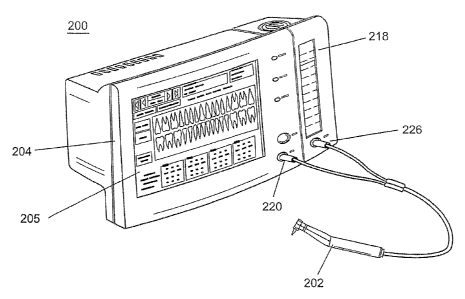
[0063] One preferred embodiment of the invention is illustrated in Figure 3.
In this embodiment,
the periodontal system 200 includes a hand piece 202 and a display/controller
unit 204 (Figure
4). The display/controller unit 204 includes, circuit boards (not shown) and
software to control,
acquire, and process the signals, data storage (not shown), a liquid reservoir
236 to hold the
liquid used as the signal coupler, at least one liquid flow connector 240,
electrical connectors
238, and software and data storage. The display/controller unit 204 is
preferably compact, yet
includes a large touch-screen 205. Because the display/controller unit 204
contains its own
liquid reservoir 236, it can be used in locations without a water supply.
Additionally, the liquid
reservoir 236 need not be located on the side of the display/controller unit
204 as illustrated in
the figures. It may be located, for example, on the bottom or in the back of
the display/controller
unit 204. Further, for purposes of this disclosure, the word "liquid"
encompasses gels.
[0064] The software includes an advanced discrimination analysis algorithm.
Optionally, it may
also include diagnostic medical imaging ability. The periodontal system 200
uses ultrasonic
signals (i.e., sonar waves) to detect, quantify and profile the upper boundary
130 of the
periodontal ligament 126 (i.e., the depth of each tooth's periodontal pocket
112) below the gum
line 124 and from the cemento-enamel junction 132 while also providing
qualitative information
regarding the presence of calculus or plaque 110, 108 above or below the gum
line 124. The
periodontal system 200 converts analog ultrasonic signals to digital signals
and digitally"stores
the pocket depths 112 of each tooth 106 and their variation over time. This
greatly assists
dentists in the diagnosis, and as an indicator of the extent and severity of
periodontal disease and
the effectiveness of their treatment plan. Preferably, the entire test is
fully computerized and all
patient information may be digitally recorded by the person performing the
test. Preferably, the
dentist inputs essential data about each tooth once (e.g., the location of a
missing tooth or a
bridge), and it will appear on all subsequent screens.
Algorithm
[0065] The discrimination analysis algorithm of the periodontal system 200
converts the
ultrasonic waveform it receives from the transducer to a pocket depth reading
using a
9
CA 02603415 2007-09-28
WO 2006/105476 PCT/US2006/012255
transformation algorithm. This algorithm uses signal processing techniques
that are commonly
used in telecommunications to detect low level signals and isolate them from
background noise.
[0066] The algorithm is performed in three steps: waveform processing, peak
detection, and
peak discrimination.
Wavelet Transformation
[0067] In one embodiment a Continuous Wavelet Transform (CWT) is performed on
the raw
signal using the Mexican Hat Wavelet,'IJS(x):
T(X) = 2aw-1/2 [1-27G(X/W)2]e (/W)"2
and
Ts(X) - S-1/2 Ts(X/3)
Where:
= w, the width of the wavelet, set to 1.2 in one aspect of the invention and
= s, the scale of the wavelet, set to the following values: 3.5, 4.21, 5.26,
6.58, and 7.89
The width and scale of the wavelet were chosen to target the frequency range
of the l OMHz
transducer. The wavelet transform of the function f is then equal to:
W(s,t) = I:f(x) 'I's(x-t)
Wavelet Selection
[0068] The Mexican Hat wavelet (see Figure 36) was chosen because, with only
1.5
"oscillations" in the wavelet, it provides better time resolution than
wavelets that contain more
oscillations, such as the Morlet wavelet (Figure 37). The trade off is reduced
frequency
resolution. For the present embodiment of the invention, time resolution is
typically more
important than frequency, the range of which is fixed by the natural frequency
of the transducer
crystal.
[0069] Unlike many other wavelets used in CWT, the Mexican Hat wavelet does
not have an
imaginary component. Therefore, to determine the out of phase frequencies
present in the raw
signal, the wavelet transform is also performed with the Hilbert transform of
the Mexican Hat
wavelet.
CA 02603415 2007-09-28
WO 2006/105476 PCT/US2006/012255
[0070] Performing a Hilbert transform on a time based signal generates a7r/2
phase shift in the
signal. Given a signal g(t), Hilbert transform of this signal is defined as:
g(t) = 1 f g(z) dz
rc -Mt - z
[0071] Another way to write this definition is to recognize that Hilbert
transform is also the
convolution of function 1/ nt with the signal g(t). The convolution of two
functions is the
inverse Fourier transfonn of the product of the Fourier transfonns of the two
functions:
[0072] So we can write the above equation as:
g(t) = 7c t * g(t)
[0073] The Fourier transform of 1/ Tct is:
FC ~ctJ -J sgn(.f )
where:
1 f?0
sgn(f) = 0 f=0
-1 f < 0
[00741 Therefore, to calculate the Hilbert transform of the Mexican Hat
wavelets, first the
Fourier transform of the wavelet is calculated. Second, the DC component and
the Nyquist
frequency component are set to zero. Then the positive harmonics are
multiplied by -j and the
negative harmonics are multiplied by +j. Finally the inverse Fourier transform
is performed on
the result to obtain the Hilbert transform of the Mexican Hat wavelets (see
Figure 36).
[0075] To increase the processing speed at run-time the wavelet coefficients
and transfonned
wavelet coefficients have been calculated and are preferably coded into the
algorithm as
constants.
11
CA 02603415 2007-09-28
WO 2006/105476 PCT/US2006/012255
Scale Selection
[0076] To determine the relationship between the wavelet scale and the signal
frequency, sine
waves with known frequencies were analyzed to determine the scales that
produced the highest
wavelet amplitudes. The optimum scale is inversely proportional to the
frequency (Figure 3 g).
A range of scales within the transducer's natural frequency band was selected
and the
corresponding scales determined (see Table 1).
Table 1
Frequency (MHz) Mexican Hat Scale
6.66 3.5
8 4.21
5.26
12.5 6.58
7.89
[0077] The periodontal system 200 (Figure 3) may include a digital imaging and
diagnostic tool
for effectively detecting, mapping, characterizing and evaluating the
presence, and monitoring
the treatment of periodontal disease. Preferably, it also provides important
and useful
information regarding calculus (i.e., hardened plaque) 110 (Figure 2) which
harbor bacteria and
interferes with dental hygiene present on tooth surfaces below the gum line
124.
[0078] In use, the hand piece 202 (Figure 9) directs a steady drip or a gentle
stream of water onto
the gums between the periodontal tissue and the tooth 100, 106. In one
embodiment, the dentist
then uses one of three buttons on a foot pedal to activate each burst of
signals. This permits the
dentist to perform the three standard probes on the facial side and the
lingual (tongue) side of
each tooth. One button advances to the next location, one button activates the
signal and if
necessary, one button permits the dentist to go back and test the last spot.
The dentist is
informed with audible tones if a signal was properly obtained or with a
different sound if the
signal was not properly received (the software recognizes an aberrant
reading). The transducer
227 (see Figures 10, 34) in the tip of the hand piece 202 transmits ultrasonic
signals (i.e., sonar
waves) below the gum line 124 (along each tooth's 100, 106 surface and into
the periodontal
pockets 112), using an anti-bacterial or germicidal gel, applied to the gums,
or water (or other
liquid such as solution containing an anti-bacterial agent or an germicidal
agent) dripped onto the
12
CA 02603415 2007-09-28
WO 2006/105476 PCT/US2006/012255
gums as the signal coupler. The signal may also use saliva below the gum line
as the signal
coupler. The transducer 227 captures the corresponding echoes resulting from
their collision
with normal and/or abnormal anatomical features below the gum line 124. The
time each signal
takes to make the round trip is measured. From this measurement, the distance
the signal
traveled to the feature causing the reflection can be determined. With this
information, the
system's 200 advanced discrimination analysis algorithm can provide healthcare
professionals
with a painless, non-invasive, extremely accurate, fast, automated, digital
and user friendly
method to provide important information regarding the true condition of the
patient's periodontal
anatomy and on each tooth's surface below the gum-line 124.
[0079] In other embodiments of the invention, the display/controller unit 204
(Figure 3) can be
directly connected to an existing water supply. Optionally, the circuit boards
can convert the
signals into a series of user-friendly images. In one aspect of the invention,
the
display/controller unit 204 includes a keyboard and mouse rather than a touch
screen. In another
embodiment of the invention, the system 300 (Figure 4) includes a controller
unit 304. Similar
to display/controller unit 204, controller unit 304 includes data storage (not
shown), a water
reservoir 236, water flow connectors 240, electrical connectors 238 and
circuit boards (not
shown) to control, acquire and process the signals. In contrast to
display/controller unit 204,
controller unit 304 does not include a display. In this embodiment of the
invention, controller
unit 304, is connected to the healthcare professional's existing computer
monitor. The
connection may be accomplished through a hard wire connection such as through
a USB port or
wirelessly such as BlueTooth.
[0080] Embedded software within the computer converts the signal from an
analog to a digital
format and uses algorithms to interpret and convert the echoes corresponding
to the depth of the
outer boundary of the periodontal Iigament into a dimension (e.g., pocket
depth in millimeters)
and to detect the presence of calculus 110 on the tooth's surface, above or
below the gum line
124 so it can be more easily and effectively removed.
[0081] The display/controller unit 204 receives the analog information
generated by the hand
piece 202, converts the data into a digital format, and processes it using
analysis algorithms.
Preferably, the periodontal system 200 also includes dental imaging software
to and creates user
13
CA 02603415 2007-09-28
WO 2006/105476 PCT/US2006/012255
friendly images of the applicable tooth 100, 106 (as shown on the screens in
Figures 5-7). The
images can be displayed on the display/controller unit's 204 large, color
touch-screen. If the
dentist wants to view the test results on another screen in his operatory, the
display/controller
unit 204 will transmit the images to the dentist's screen on a wireless or
wired basis. The dentist
can input all patient information using the display/controller 204 unit's
large, user-friendly
touch-screen, or a keypad. In another aspect of the invention, the dentist can
input all patient
information using voice recognition software. Further, the system is easily
and quickly moved
between operatories using its quick connect/disconnect water and electrical
fittings.
100821 The software preferably included in system 200 (Figure 3) preferably
allows the
display/controller unit 204 to display periodontal measurement 206 (Figure 5),
the charting of
results 208 (Figure 6), and patient management 210 (Figure 7). In one
embodiment, the soflware '
may generate an image of a tooth with surrounding periodontal tissue and
illustrate the data on
the image of the tooth. In this way, healthcare professionals and patient's
can visually monitor
the progress and/or treatment of periodontal disease and/or the removal of
calculus below the
gum line.
[0083] Preferably, the periodontal system 200 is calibrated before examining
each patient.
Calibration may include testing the software, calibrating the head, testing
the transducer and/or
testing the acoustic lens. Calibration may be accomplished, for example by
measuring the depth
of a known cavity built into the unit 204. Should the periodontal system 200
fail such that
recalibration in the healthcare provider's office is not possible, the system
200, may optionally
be provided with automatic messaging that can transmit a request for a new
system 200, or part
thereof, from the supplier.
[0084] In one embodiment of the invention, all tests will be performed after
the dentist or dental
hygienist connects to a third party web site via the Internet. This will
perrnit the third party to
confirm the periodontal system. 200 is properly calibrated and working
perfectly prior to each test
and that the dentist's account has been properly charged the test fee. Patient
information may be
securely stored in a HIPAA compliant centralized back-up database maintained
by the third party
at its website. In this embodiment, dentists will have controlled access to
the website and be able
to:
14
CA 02603415 2007-09-28
WO 2006/105476 PCT/US2006/012255
= Review their patient records;
= Review and update their account information;
= Review and update their disposable inventory and ordering information; and
= Review the status of the system(s) in their office.
[0085] In one embodiment of the invention, illustrated in Figure 12, the
connection to the third
party is accomplished with enterprise portal software 500. In this embodiment,
dental records
can be stored at the third party site for backup purposes. Further, dental
records (including their
digital images) can be forwarded to periodontal experts for online
consultation. The periodontal
expert can evaluate the patient's condition and send his evaluation and
recommendations either
to the third party or directly to the dentist or dental hygienist the patient
is seeing. Further, the
enterprise software is capable of monitoring the dentist's activities. The
system can be used to
deternine which dentists are successful in treating minor periodontal problems
and those who
are not. Further, it can be used to identify dentists who are treating
patients that should be under
the care of a periodontist due to the severity of the patient's condition.
Additionally, software for
the periodontal system 200 can be easily upgraded via a simple download by
request by the user
upon notification that an update is available or automatically by the provider
if a service
agreement is in place. In still another embodiment, the dental records may be
encrypted.
[0086] The system permits dentists to show the test results on their computer
screens (rather than
on paper records from multiple years) to their patients and also provides them
with a printout of
the test. This permits patients to confirm what their dentists have told them
and to monitor the
effectiveness of their treatment plan. This active patient involvement is
expected to result in
more patients following their dentist's instructions because they will be able
to see that their
periodontal disease treatment program is worlcing. It also provides patients
(and their payers)
with objective proof of the presence of periodontal disease and the necessity
of treatment. This
is expected to reduce the number of "walkaways" (i.e., patients that do not
believe their dentist
or the severity of their periodontal condition).
CA 02603415 2007-09-28
WO 2006/105476 PCT/US2006/012255
[00871 If a patient does not currently have periodontal disease, by comparing
the base line digital
images with those taken over a period of time, both the dentist and the
patient can see that
periodontal disease is not present.
[0088] According to one embodiment of the invention, periodontal examiners
establish a
baseline of their patient's periodontal pocket depths 112 (Figure 2) during an
initial exam. After
their first examination, each time the patient has a new examination, the data
from the prior
examinations may be digitally and automatically compared to the current data
and illustrated
with the periodontal system's 200 dental display software. This permits
dentists and their
patients to identify even relatively minor changes in periodontal pocket
depths 112 not otherwise
detectable using the current manual probe method. These minor changes may be
illustrated with
color trend lines that reflect improving, deteriorating or unchanged pocket
conditions. In one
aspect of the invention, changes of approximately 0.2-0.5 mrn may be detected.
In another
aspect of the invention, changes of approximately 0.1-0.2mm may be detected.
This permits
treatment to start while the periodontal disease is in its earliest stages and
easily and
inexpensively treated.
100891 The periodontal system 200 provides the following considerable benefits
to patients,
dental healthcare professionals and payers:
= For patients, the test is objective, non-invasive, painless, bloodless and
inexpensive.
= For dentists, the test is fast, accurate, objective and digital. Dentists
can
immediately provide patients and payers with a paper or electronic copy of the
test results and are expected to generate substantial additional practice
revenues
from additional periodontal testing and early-stage periodontal disease
treatment fees. Proof that treatment is necessary can be shown to the patient
on-screen and e-mailed to payers. A back-up copy of each patient's digital
records may be stored in a HIPAA compliant manner at a remote Web-Site.
= For payers, including third party payers, the test can accurately,
objectively and
digitally confirm the presence and extent of periodontal disease. More and
more dental insurance companies and other payers are requiring digital proof
of
the patient's condition from dentists to confirm that treatment was necessary.
[0090] If periodontal disease is present, the dentist can quickly and
accurately detect and
diagnose the type and extent of the periodontal disease in its earliest stage,
prescribe preventative
16
CA 02603415 2007-09-28
WO 2006/105476 PCT/US2006/012255
treatment and perform on-going periodontal disease management to prevent its
spread, the loss
of diseased teeth 106 and the onset of other serious diseases and reduce
healthcare costs. By
using the digitized data and generating images taken every'several months
during the treatment
period, both the dentist and the patient can readily confirrn the treatment
plan's effectiveness.
[0091] This type of preventative dentistry program results in better dental
care for the patient. In
fact, most dentists are expected to perform more examinations and treat those
patients with mild
periodontal disease that would otherwise have developed into more serious
periodontal disease
and then been referred to a periodontist for treatment. Those patients
currently with more serious
periodontal disease would still be referred to a periodontist.
[0092] The periodontal system 200 provides considerably more accurate and
detailed
information than the standard manual probe and analog method currently used by
dentists and
dental hygienists for periodontal tests, which requires the repeated,
frequently painful insertion
of a sharp metal probe into the crevice between the tooth and the gum. The
manual probe and
analog method is very inaccurate and can over- or under- estimate the
patient's true condition by
1mm or more. As a result, the ability of current manual probe method to
diagnose periodontal
disease in its very early stages is very difficult. Even the same dentist, or
different dentists
performing periodontal examinations on the same patient, can derive
significantly different
measurements. This happens for many reasons, including the probe not always
being placed in
the exact same location, the amount of pressure applied, the presence of
granulation tissue due to
infection, the skill and experience of the dentist or dental hygienist,
patient movement, etc.
[0093] By contrast, the periodontal system's 200 dental imaging technology
provides a
significantly more accurate, consistent, reproducible measurement and
diagnosis of periodontal
disease and therefore earlier disease treatment opportunities because its
margin for error is only
+/- 0.1 to 0.5mm and the smallest changes can be easily and quickly recognized
and treated.
Preferably, the margin of error is 0.1-0.3mm. More preferably, the margin of
error is
approximately 0.1 to 0.2mm.
[0094] In addition to diagnosis, the periodontal system 200 may be used to
monitor the progress
of healing during treatment. Monitoring the progress of healing during
treatment is possible
because the system and method of the present invention is noninvasive and
hence, does not tear
17
CA 02603415 2007-09-28
WO 2006/105476 PCT/US2006/012255
or disrupt soft, healing tissue during use. This is in contrast to the
conventional method of
measuring periodontal disease, which requires insertion of a sharp probe
between the tooth 106
and gum 102, which can result in tearing of the healing tissue.
[0095] In another embodiment of the invention, the periodontal system 200 may
be used to assist
in the treatment of periodontal disease. In this embodiment, medication is
added to unit's liquid
reservoir 236 or to the fluid from the hand piece 202. In still another
embodiment, the system is
able to detect the presence of calculus 110 on the tooth's 106 surface below
the gum line 124 so
it can be more easily and effectively removed. Further, the completeiness of
calculus 110
removal of calculus 110 can be monitored by subsequent use of the system 200.
[0096] Another embodiment of the invention permits dentists or dental
hygienists to determine
how many rneasurements they want to be obtained on each tooth. This embodiment
includes
software that allows the handling of the large amount of digital data
collected and stored. The
software will enable dentists to obtain and store their patients' data on
their office computers. In
one aspect of this invention, the dentists or dental hygienists can operate
the periodontal system
200 in continuous mode. In this mode, once triggered, the hand piece 202
automatically
repeatedly emits pulses at regular intervals. The dental examiner sweeps the
probe tip from one
interdental space across the surface of the tooth to the adjacent interdental
space. Preferably, the
dental examiner performs a first continuous scan along the facial surface of
the tooth and a
second scan along the lingual surface of the tooth. In this manner, a profile
of the bottom of the
pocket can be generated rather than only gathering data from a few
representative points. The
total number of data points taken in this embodiment depends on the frequency
of the transducer
and the rate the dental examiner drags the hand piece 202 across the tooth
100, 106. Dozens,
hundreds, even thousands of data points may be taken. In this manner, focal
disease in the
periodontal pocket 112 may be detected. In one aspect of this embodiment, all
of the teeth 100,
106 may be scanned by the dentist or dental hygienist. In another aspect of
this embodiment,
only those teeth 106 that have previously identified as exhibiting periodontal
disease are scanned
in continuous mode, the remaining teeth 100 scanned with discretely triggered
pulses. In still
another aspect of this embodiment of the invention, geopositional technology
may be used in
combination with a fixed reference in the mouth to assist in defining the
location and profile of
the periodontal pocket 112.
18
CA 02603415 2007-09-28
WO 2006/105476 PCT/US2006/012255
[0097] Another embodiment of the invention can obtain complete and highly
accurate readings
and 3-D images of alY of the patient's teeth and may be able to eliminate the
need for dentists to
obtain dental x-rays.
[0098] One embodiment of the invention is illustrated in Figures 8 and 9. This
embodiment
provides a hand piece 202 having a straight handle 214. One end of the handle
214 includes a
cavity 216 adapted to hold a transducer (not shown). Adjacent to cavity 216 is
an alignment slot
218. The alignment slot 218 mates with a protrusion in a disposable cover 212.
The
combination of the alignment slot 218 and the protrusion, greatly improve the
reliability of
alignment of the when placing a new disposable cover 212 on the handle 214.
Located in a
central portion of the handle 214, is a first circumferential slot 220. In one
embodiment of the
invention, the first circumferential slot 220 is provided with an helical
spring (not shown). When
a disposable cover 212 is pushed in place on the handle 214, the helical
spring mates with a slot
in the interior of the disposable cover 212, providing a snap fit. Also
located in a central portion
of the handle 214, is a second circumferential slot 222. Preferably, an 0-ring
is inserted in the
second circumferential slot 222 to provide a seal. The disposable cover 212
includes a head
portion 224 that covers the transducer and a probe tip 226 from which the
ultrasonic waves are
emitted. In one aspect of the invention, the disposable cover 212 may be
provided with a safety
feature that renders the disposable cover unusable after initial use. In
another aspect, the
disposable cover 212 includes an identification feature such as a serial
nu.mber. The periodontal
system 200 may be provided with a sensor to read the identification feature
and determine if the
disposable cover 212 has already been used. If the disposable cover 212 has
already been used,
the periodontal system 200 may refuse to allow further examination until a new
disposable cover
212 is provided.
[0099] In one embodiment of the invention, the probe tip 226 is sized to fit
snugly in the
interdental space between teeth. As the location of this space does not vary,
it provides a fixed
reference point for taking periodontal measurements. The hand piece 202 is
particularly
advantageous because the probe tip 226 can be located behind the papilla. In
this configuration,
the hand piece 202 can be used to measure the deterioration of periodontal
tissue (gum 102,
periodontal ligament 103, 126, and the specula of bone between the teeth) due
to periodontal
disease.
19
CA 02603415 2007-09-28
WO 2006/105476 PCT/US2006/012255
[00100] In an alternative embodiment of the invention, the periodontal system
200
includes hand piece 402, illustrated in Figures 13 and 14. The present
inventors have recognized
that the efficiency of the ultrasonic probe is significantly enhanced if the
transducer 227 is
located close to the probe tip 426. On the other hand, due to the concern of
the spread of disease,
it is necessary to sterilize that portion,of the probe that enters the
patient's mouth. The inventors
have determined, unfortunately, that all current methods of sterilization,
such as autoclaving and
chemical washing, can damage the transducer 227, adversely affecting the
useful life of the hand
piece and its accuracy over time.
[00101] The inventors have discovered that the hand piece 402 of the present
embodiment
can be fitted with an easily removable cover 412. With this arrangement, the
transducer 227 may
be located in the head 424 of the hand piece 202, close to the probe tip 426.
After a periodontal
examination in one embodiment of the invention, the removable cover 412 can be
removed and
thrown away and a new removable cover 412 placed over the head 424. In still
another
embodiment of the invention, the removable cover 412 may be reused after
sterilization. That is,
the removable cover 412 may be removed from the hand piece 202, separately
sterilized, and
reattached to the handle 414. In another aspect of the invention, the hand
piece 202 is connected
to the display/controller unit 204 (Figure 3) using quick-connect/disconnect
electrical 238 and
liquid flow connectors 240 that make it easy to quickly move the system
between operatories.
[00102] , Figure 10 illustrates another embodiment of the invention. In this
embodiment,
the hand piece 202 includes an acoustic Iens 228. The inventors have
discovered that the
efficiency of the hand piece 202 can be significantly increased by focusing
the sound wave from
the transducer 227 with an acoustic lens 228. Typically, the transducer 227
has an area much
larger than the area of the exit opening of the probe tip 226 of the hand
piece 202. Without an
acoustic lens 228, much of the sound wave from the transducer 227 bounces off
the inside walls
of the probe head 224 as the probe head 224 narrows towards the tip 226.
However, with an
acoustic lens 228, the sound may be focused to the size of the exit opening of
the probe tip 226.
[00103] In still another embodiment of the invention, the transducer 227 of
the hand piece
202 is operated at intermediate frequencies. It is known that high frequency
sound waves yield
higher resolution, while low frequency sound waves have higher penetration.
Typically, prior art
CA 02603415 2007-09-28
WO 2006/105476 PCT/US2006/012255
ultrasonic devices have been designed to operate at frequencies of 2-5 MHz
when high
penetration was required and 15-20 MHz when higher resolution was required. In
one aspect of
the present invention, the inventors have discovered that a transducer 227
that uses frequencies
between 5 and 15 NIHz can yield both high resolution and high penetration. In
one preferred
embodiment of the invention, the transducer frequency is approximately 10 MHz.
Tip Shape Determination
Design Constraints
[00104] The shape of the tip 226 is preferably designed to ensure patient
comfort and ease
of use. It should also be compatible with the selected transducer, i.e.
placing the focal point of
the transducer in the region of interest.
Length
[00105] In one embodiment of the invention, the general length was determined
to be
approximately 10mm to allow enough room for the medical professional to
properly position the
probe, but still provide enough length for the medical professional to
visually determine the
angular position. With tliis distance as a guide, an available transducer with
an appropriate focal
length (13.25 mm) was identified. The final length of the tip was then
determined to place the
focal point 244 approximately 2nun beyond the end of the tip.
Diameter and Profile
[00106] In this embodiment, the inner diameter and profile of the tip 226 was
determined
from the beam diameter of the ultrasound pulse. The tip 226 surrounds but does
not encroach
upon the ultrasonic beam 242, ensuring that the pulse will not be reflected by
the tip 226.
Therefore, when properly aligned, the tip should not be visible in the
ultrasound echoes. Figure
34 illustrates the ultrasonic beam and the tip geometry.
Water Path Design
Design Constraints
[00107] The flow rate should be low enough to ensure patient comfort, but high
enough to
provide adequate acoustic coupling between the transducer and the patient.
21
CA 02603415 2007-09-28
WO 2006/105476 PCT/US2006/012255
Average Flow Rate
[00108] Theoretically, the water flow rate should have a negligible effect on
the time of
flight measurements of the ultrasound echoes. While the speed of the outgoing
pulse is increased
by the velocity of the water, the speed of the incoming echo is decreased by
the same amount.
However, turbulent flow could cause noise or distortions in the signal;
therefore the velocity is
preferably limited to ensure laminar flow (Reynolds Number < 1000) through the
tip. With a
minimum tip diameter of 0.5 in, the maximum laminar velocity is approximately
3.1 inches per
second:
v=(Rev)/d
where:
v is the velocity of the flow
Re is the Reynolds Number (1000)
v is the kinematic viscosity of water (1.01 x 10⁻⁵ m2/s)
d is the minimum inner diameter of the tip (0.5 in)
[00109] The maximum laminar flow rate is 604 mL per minute:
Q=vA
where:
q is the flow rate
v is the velocity of the flow (3.1 in/s)
A is the cross sectional area of the tip (0.196 in2)
[00110] Tests were also performed to verify the effects of flow rate on the
measurements.
This study gathered waveform data for reflections from a nominal 4.763 mm
thick (4.752 mm
measured) flat aluminum plate containing 1 mm diameter circular through hole
that is placed on
top of a second ahuninum plate containing no holes. An immersion transducer
(Xactex, 10
MHz, 13.25 mm focal length, 0.67 mm beam diameter) with an 11.055 mm tip was
used. Data
was collected at nine separate flow rates with the reflector positioned
approximately 1 mm from
the end of the tip. Flow rate was adjusted using a Harvard Apparatus PHD 2000
programmable
22
CA 02603415 2007-09-28
WO 2006/105476 PCT/US2006/012255
syringe pump with a 10 mL Hamilton gas-tight syringe. For each flow rate, the
three
consecutive waveforms measurements were recorded.
[00111] The results of this study are shown in Figure 35. Flow rate had little
effect on the
determination of the distance to the flat plate. However, the signal produced
from the hole did
show variation with flow, with a change in measurement between 5 and 7.5
mL/min. From this
data, a target range of flow rates from 10 to 15 mL/min was selected. This
range provides
consistent measurements throughout the range and provides sufficient flow for
acoustic coupling.
Flow Pulsations
[00112] Preferably, the periodontal system 200 uses a diaphragm pump with a
running
speed of approximately 30Hz. This equates to a cycle period of 30 ms.
Preferably, the data
acquisition time of an entire scan is 30us, or 1/1000 of a pump cycle.
Therefore, even though the
diaphragm produces observable pulsations in flow rate, the flow can reasonably
be assumed to
be stable during the duration of the scan acquisition.
Air Bubble Elimination
[00113] Air bubbles, including microbubbles, dramatically increase the
attenuation of the
signal and reduce signal strength. The flow path is preferably designed to
facilitate the flushing
of air bubbles out of the system. Areas in the flow path where air could get
trapped (i.e. local
high points) are preferably minimized. Additionally, the water is preferably
deaerated upstream
of the hand piece. This is preferably accomplished by pulling a vacuum on one
side of a PTFE
filter. The surface tension of water prevents liquid from flowing through this
filter, but air and
other gases can flow freely through it.
[00114] A preferred embodiment of the invention provides a completely
noninvasive
method of measuring the gum line 124 (Figure 2) to the cemento-enamel junction
132, and the
deterznination of the depth of the periodontal pocket 112 extending from the
cemento-enannel
junction 132 to the bottom of the periodontal pocket 112. In this embodiment,
a sound wave is
transmitted along the tooth 106 starting from the gum line 124. Returning
echoes are analyzed
by the discrimination analysis algorithm. Echoes from the cemento-enamel
junction 132 f x its
location relative to the gum line 124, while echoes from the bottom of the
periodontal pocket 112
23
CA 02603415 2007-09-28
WO 2006/105476 PCT/US2006/012255
fix the location of the bottom of the periodontal pocket 112 relative to the
gum line 124. The
depth of the periodontal pocket is determined by subtracting the distance from
the gum line 124
to the cemento-enamel junction 132 from the distance from the gum line 124 to
the bottom of the
pocket 112. Prior art ultrasonic periodontal devices, in contrast, either used
an invasive probe to
determine the location of the cemento-enamel junction 132 or measured the
periodontal pocket
112 through the gum line 124, completely ignoring the cemento-enamel junction
132.
[001151 In one aspect of the invention, the return pulses are amplified and
transformed to
separate peaks from noise. In one preferred embodiment of the invention, a
wavelet algorithm is
used in the transformation process. In still another embodiment of the
invention, a,
discrimination analysis algorithm is used to aid in determining the
identification of the various
peaks. In still another embodiment, both a wavelet and a discrimination
analysis algorithm are
used.
[00116] In one embodiment, the display/controller unit 204 (Figure 3) will
provide a series
of audible tones and/or visual signals to guide the dentist through the test
thereby permitting the
dentist to advance to the next tooth 100, 106 or to reverse back to the last
tooth and re-test it if
the image was not properly captured. These signals can also alert the dentist
to the presence of
unusually deep periodontal pockets 112 that may signify significant
periodontal disease or other
conditions that require attention or treatment.
[001171 The display/controller unit 204 is fully self-contained and will
provide the
necessary images on its own screen even if the sigtial cannot be transmitted
outside of the room.
In addition, all of the patient's information can be sent to the dentist's
office computer wirelessly
or via a cable connection so that patient information does not have to be re-
entered.
[00118] In one embodiment of the invention, the hand piece 202 includes a
disposable
cover 212. The disposable hand piece cover 212 will be contained inside of a
sterile, tamper-
resistant package that also contains a disposable stylus that can be used on
the system's touch
screen for data entry purposes, an alcohol soaked gauze pad in a sterile pouch
to wipe off the
hand piece between patient tests and a see-through disposable plastic cover
for the touch screen
in the event of splatters. The package, all disposables and technology
(including all
24
CA 02603415 2007-09-28
WO 2006/105476 PCT/US2006/012255
enhancements) may be provided to dentists without charge in consideration of
their paying a test
fee.
[00119] Figure 11 illustrates still another embodiment of the invention. This
embodiment
of periodontal system 200 includes a hand piece 202 and a display/controller
204. This
embodiment further includes a triggering device 229. In this embodiment, the
triggering device
229 includes three foot pedals 230, 232, 234. The triggering device 229
activates the transducer
227 (Figure 10) and initiates fluid flow. In another embodiment, the
triggering device 229 is on
the hand piece 204. In still another embodiment the triggering device 229
includes software that
allows voice activation.
[00120] The following summarizes and describes various features of the
software of the
periodontal system 200 (Figure 11). This summary describes how the software:
= Controls the device's electronic components,
= Interfaces with the end-user,
= Executes the data acquisition algorithm, including initiation and receipt of
the acoustic
signal and the subsequent calculation of periodontal pocket depth,
= Displays collected data,
= Stores and protects patient information,
= Calibrates the device, and
= Communicates with external devices.
[00121] The periodontal system 200 is an ultrasonic probe system used in the
measurement of a patient's periodontal condition. The periodontal system 200
consists of a
handheld probe 202, a triggering mechanism and a compact display/controller
unit 204. The
probe 202 transmits an ultrasonic pulse into the periodontal pocket 112 of the
patient through a
stream of water, or other liquid (typically, required for acoustic coupling)
and captures the
echoes resulting from collision of the ultrasonic wave with anatornical
features in the periodontal
pocket 112. Embedded software running within the display/controller unit 204
uses an analysis
algorithm to correlate the acoustic echo with the depth of the outer boundary
of the periodontal
ligament 130 (e.g. pocket depth in millimeters).
CA 02603415 2007-09-28
WO 2006/105476 PCT/US2006/012255
[00122] The software application is preferably supported by an embedded
operating
system running on the display/controller unit 204. The software application
controls the
periodontal system 200. Control features include:
= Sending a signal to external hardware to emit an ultrasonic pulse upon
receipt of a trigger
signal from an external trigger mechanism (e.g., a foot pedals 230, 232, 234),
= Controlling water flow to the handheld probe 202,
= Acquiring the ultrasonic echo as a digitized electrical signal,
= Performing the pocket depth calculation,
= Calibrating the periodontal system 200, as necessary, between scans,
= Providing audible prompts to the user denoting the end of a scan and
readiness to perform
a new scan,
= Driving a touch screen and LCD display 205,
= Reacting to user input via the triggering mechanism,
= Storing data relating to the patient and all exams performed in a patient
database,
= Is capable of dr.iving an external video display unit,
= Is capable of receiving software upgrades,
= Supporting network communications to transmit and receive patient data,
= Communicating to a remote Internet portal.
[001231 Figure 15 illustrates an embodiment of the functional layout of the
periodontal
system 200. Table 2 contains a list of the major components and external
systems that interface
with the periodontal system's 200 software application, and indicates their
main function in the
device.
Table 2
Component Description
Ultrasonic Transducer Emits and receives ultrasonic signals
Solenoid Valve Relay Opens and closes water supply
26
CA 02603415 2007-09-28
WO 2006/105476 PCT/US2006/012255
Trigger Device A trigger device accepts operator commands to
1) Sequence forward and backward to reach the desired
tooth location for a given scan, and
2) Acquire a pocket depth measurement
Pulser/Receiver Sends an electrical pulse to the transducer and receives the
returning electrical signal created by the ultrasonic echo
Analog-to-Digital Converter Samples the signal received by the pulser/receiver
and
(A/D board) converts sam les to digital values
Single Board Cornputer (SBC) Supports the operating system and runs the
software
application. Executes the data analysis algorithm and control
commands and/or si als
Flat Panel Display Video screen that is the main user interface medium
Touch-Sensitive Display Accepts user input through a touch-sensitive screen
integrated
(Touch Screen) into the device flat panel display
Audio Speaker & Amplifier Emits audio feedback to the user concerning data
acquisition
events and the state of device readiness
Compact Flash Memory Provides data storage capacity
External Network Connection Allows the user, via the software application, to
upload or
download patient visit data to or from a central data
repository, and/or from device to device, and to download
software upgrades.
Software Environment
Operating System
[00124] In one preferred embodiment of the invention, the periodontal system
200
includes a microprocessor that runs using the Windows XP Embedded (XPe)
Operating System,
which is a componentized form of the Windows XP Professional Operating System.
The
componentization enables the operating system of the periodontal system 200 to
be customized
to include only those features of Windows XP necessary to the operation of the
periodontal
system 200, and the exclusion of those that are not.
Software Development Tools
[00125] The application is preferably an object-oriented Windows application
written in
the C++ coding language using Microsoft's Visual Studio 6.0 IDE (integrated
development
environment). The application may, however, be implemented using other
computer languages
and with other tools. Software modules including graphics tools, device driver
programs for the
A/D card, the touch screen control electronics, the trigger device, and the
audio speaker are
27
CA 02603415 2007-09-28
WO 2006/105476 PCT/US2006/012255
preferably included in the application or are accessed by the application via
dynamically linked
library (DLL) files.
Hardware Environment
Single Board Computer and Processor
[00126] The periodontal system's 200 software application preferably runs on a
single-
board computer that supports and contains all of the interface hardware and
software
components. This computer preferably has a 1 -GHz VIA EdenTm ESP 10000
processor with a
VIA Technologies, Inc. Twister-T chipset (VT8606 and VT82C686B chips).
Further, it
preferably has 256 MB of RAM, connections for a keyboard and mouse, cathode-
ray tube (CRT)
and liquid crystal display (LCD) video interface connections, four universal
serial bus (USB)
ports, two Ethernet ports, one paralleUfloppy port, one General Purpose
Input/Output (GPIO)
port, and four serial ports. It has PC/104 and PC/104+ interfaces, and a
Compact Flash adapter.
Other combinations input and output connections are also possible and within
the scope of the
invention.
Touchscreen Controller
[00127] A 4-wire resistive touch-sensitive touchscreen, mounted in front of an
LCD is the
preferred way for operator interaction with the periodontal system's 200
software. The touch
screen is preferably used in the same manner as a one-button mouse. A
controller board
preferably converts the analog signals coming from the touchscreen into X and
Y coordinates
and selection events, and communicates this data over a USB interface to the
computer. Driver
software is typically required for the controller board to operate. This
driver application
preferably includes touchscreen calibration software that initially correlates
LCD X and Y
coordinates with touchscreen X and Y coordinates to account for misalignment
between the two
reference frames. In one embodiment, the driver application is not part of the
periodontal
system's 200 software application, but is used by the operating system to
allow it to receive and
use the mouse-like inputs coming from the controller board. The touchscreen is
preferably
calibrated before the device is delivered to the user. Under normal
circumstances, the user will
not calibrate the touchscreen.
28
CA 02603415 2007-09-28
WO 2006/105476 PCT/US2006/012255
Trigger Device
[00128] The periodontal system 200 is preferably controlled by the operator
during patient
examinations by a trigger device. The trigger device sends commands to the
periodontal system
200 to begin an acquisition or to move to the next tooth location. Commands
given by the
trigger device are preferably mapped to unique keyboard sequences, meaning the
operating
system interprets each type of command received from the trigger device as a
certain keyboard
sequence. The periodontal system's 200 software application waits for these
keyboard
sequences (generated by the trigger device), and takes specific actions in
response to each
reported sequence. The keyboard-trigger device mapping is shown below in Table
3.
[00129] The trigger device for the rapid prototype and investigational
periodontal system
200 is preferably a three-position foot pedal that is connected to the
periodontal system's 200
microprocessor through a USB interface. The Savant USB driver for Windows, for
example,
may be used to accornmodate the USB communication between the operating system
and the
foot pedal. The trigger device can also be activated using buttons in the hand
piece or using
voice recognition software.
Table 3
Trigger Device Keyboard Sequence Function During Examinations
Left Button ALT+l Move to previous tooth location
Middle Button ALT+2 Perform periodontal depth
acquisition
Right Button ALT+3 Move to next tooth location
Input/Output
[00130] Preferably, the software application interfaces with three components,
a water
control solenoid valve, the Pulser/Receiver and the A/D card via the
display/controller's 204
parallel port. Preferably, all three are triggered when they receive a digital
HIGH signal from the
parallel port channel to which they are connected. Typically, the parallel
port is conimanded to
send these signals when the data acquisition software receives an appropriate
command from the
operator interface. A parallel port software module may be written into the
periodontal system's
200 software application and implement the functions necessary to configure
and use this
interface. The pulser/receiver and A/D card may be activated by the same
parallel port channel.
29
CA 02603415 2007-09-28
WO 2006/105476 PCT/US2006/012255
[00131] When the solenoid valve opens, water preferably passes through the
valve to the
periodontal system's 200 hand piece 202. The Pulser/Receiver preferably sends
a negative
voltage pulse to the ultrasonic transducer 227, which converts that pulse into
acoustic energy.
The transducer 227 then receives and converts the acoustic echo returning to
it back into an
electrical signal, which is then sent back to the Pulser/Receiver, and sampled
by the A!D board.
Analog/Digital Board
[00132] Preferably, the analog/digital (A/D) board communicates over the PCI
bus on the
periodontal system's 200 microprocessor. The periodontal system's 200 software
application
preferably includes a software module that contains all of the driver
functions and variables
necessary to initialize, trigger, and retrieve data from this board. The
functions included in this
module are preferably supplied by the manufacturer of the board. In one aspect
of the invention,
12-bit digital samples of the returning echo waveform are acquired at a rate
of 100 megasamples
per second. In other aspects, the digital samples may comprise more or less
than 12 bits. In still
other aspects, the sample rate may be more or less than 100 megasamples per
second.
Flat Panel Display
[00133] The transflective TFT LCD flat-panel display preferably does not
require any
additional software or drivers to operate, and is controlled by drivers
resident in the operating
system. Preferably, the BIOS is configured to support both an LCD and an
external CRT
monitor. The LCD connection is preferably internal to the periodontal system
200, while a
connection to an external CRT may be provided at the rear of the device.
Audio Speaker
[00134] The audio speaker is preferably supported by drivers resident in the
operating
system. Software commands to play selected audio files (e.g., WAV) are
preferably issued
using platform (Windows XP Embedded) functions.
Compact Flash
[00135] Preferably, the compact flash card functions as the storage medium of
the
periodontal system's 200. Preferably, it contains the operating system (XPe),
the periodontal
system 200 software application, and a database of patient records. The
operating system is
CA 02603415 2007-09-28
WO 2006/105476 PCT/US2006/012255
configured to boot from the compact flash card. Preferably, the compact flash
card is type II,
1GB in size, and formatted as fixed media. Any suitable type and size,
however, may be used.
External Network Interface
[00136] Preferably, the periodontal system 200 can communicate to external
devices
through wired (Ethernet) or wireless (802.11x) connections. Preferably, the
Ethernet hardware is
integrated into the computer and drivers are supplied by the manufacturer. The
wireless
hardware is an optional module that may be added to the computer. The make and
model of the
wireless Ethernet module are not critical to the invention.
[00137] Figure 15 provides a graphical summary of the software application's
four main
tasks. These include the operator interface, data acquisition and calibration,
the maintenance of a
database of patient information and data, and external interface.
[0013$] The operator interface forms the backbone of the application, and all
other
functions of the application are preferably controlled from commands received
through this
interface. The operator may enter commands or data through a touch-sensitive
screen and trigger
device, and receive information back via a series of interface screens. The
operator may also be
given audio feedback via the audio speaker.
[00139] Software elements that control the acquisition and analysis of data
typically
receive their instructions from the operator interface, and then execute the
necessary software
and hardware procedures to perform those tasks. Similarly, these elements may
also control the
task of calibration of the Data Acquisition system.
[00140] Preferably, patient data is stored in a database created and
maintained by the
application. This application preferably controls the entry, modification and
protection of patient
data.
[00141] External interface software preferable to communicate over USB and
Ethernet
ports with external devices such as the trigger device or an external monitor.
Preferably, the
external interface creates the ability to cornmunicate with a web-based
repository where
periodontal data can be. stored or retrieved. Finally, it preferably allows
the passing of patient
31
CA 02603415 2007-09-28
WO 2006/105476 PCT/US2006/012255
data and information from one periodontal system 200 device to another within
the dentist's
office, and to and from external computers over a secure line of
communication.
[00142] The typical process of performing an examination is shown in Figure
16. An
operator typically logs into the device through a login screen. The operator
can then load an
existing patient from the patient database or create a new one that is then
stored in the patient
database. At this point, if the operator creates a new visit they are
functioning in Examination
Mode and can perform an examination and enter in tooth condition data. Usage
of this mode is
preferably considered a usage towards business accounting purposes.
Preferably, if the operator
simply views an existing patient visit they are functioning in Review Mode and
can view patient
data and standard reports on the patients periodontal and tooth condition
data.
Graphical User Interface Screens
[00143] The operator interface consists of several screens with specific
functions as shown
in Figure 17. A commercial sofl.ware library may be used to graphically
enhance the visual
appearance of the dialog boxes and operator interface screens.
[00144] The entryway into the application is preferably through the Login
Screen, which
allows operators to login using a password. Once logged in, the operator is at
the Main Screen
where they can view account information through the Account Screen, setup
preferences in the
Operator Screen, or review patient information through the Patient Records
Screen. This screen
allows the operator to select current patients to load or to create new
patients.
[00145] Once a patient is loaded the operator can view or edit the record
through the
Chart Screen. The chart screen shows inforrn.ation about the patient including
pocket depth and
tooth condition data including any overlays selected through the Patient
History Selection
Screen. The operator can edit tooth condition data for the patient through the
Enter Tooth.
Condition Data Screen, or can perform a periodontal examination through the
Measure Pocket
Depth Screen. The operator can view reports on the patient through the View
Complete Chart
Screen and the View Tooth History Screen.
32
CA 02603415 2007-09-28
WO 2006/105476 PCT/US2006/012255
Common Screen Information
[00146] The Chart Screen, Enter Tooth Condition Data Screen, Measure Pocket
Depth
Screen, View Complete Claart Screen, View Tooth History Screen preferably have
the following
fields at the top and bottom of the screen:
= Operator name text.
= Patient name text.
= The date and time of the currently loaded visit, which is the current date
and time if it is a
new visit.
= Status text, which is used to prompt the operator with status infonnation
relative to events
on the screen.
= Current date and time, updated once per minute in the status bar.
= Help button that takes the operator to the Help Screen and loads the
relevant section in
the help topics index for the screen.
Tooth Condition Tables
[00147] Preferably, several screens contain charts with tooth condition data
presented in a
tabular fonn. In one embodiment, due to size constraints and in order to
maximize readability,
the chart presents data from only eight teeth at a time (one quadrant of the
mouth). In other
embodiments of the invention, any number of teeth may be presented up to and
including all of
the teeth. For example, the dentist may choose to view a single tooth with
periodontal disease, or
one or two teeth on each side of the diseased tooth (a total of three or five
teeth, respectively).
[00148] Certain data is preferably displayed for each location on a tooth. The
locations
may be denoted as:
= D for the location on the Distal side of the tooth.
= M for the location on the Mesial side of the tooth.
= C for the Central location on the tooth (i.e. between the Mesial and Distal
location).
In other aspects of the invention data may taken in the interdental space and
denoted as I.
[00149] Preferably, the data displayed for each of the three locations on a
side of a tooth
(and in the interdental space) is:
= Pocket Depth (0.0-9.9)
= Furcation (0-3)
33
CA 02603415 2007-09-28
WO 2006/105476 PCT/US2006/012255
= Recession (0-3, or using the Miller Classification which is determined in
the
Options screen).
[00150] The tooth condition data displayed for each tooth may include:
= Suppuration (S for yes, blank for no)
= Mobility (0-3)
= Edema (0-3)
= Missing (Missing for yes, blank for no).
Login Screen
[00151] This screen is preferably the initial screen displayed when the device
is powered
on. Preferably, when this screen is present no other screens or functions of
the device can be
activated except to login to the device or to shutdown (power down) the
device. When the Login
button is clicked the operator can login and begin using the device. This way,
any data taken is
associated with that operator. Preferably, when the operator is finished with
any examination
they should log off of the device from the Main Screen. When that occurs this
screen preferably
becomes the only screen visible. In one aspect of the invention, if the device
is inactive for more
than a configurable amount of time, this screen becomes active again. The
login provides a layer
of security for patients' medical records. Figure 18 shows a notional view of
the Login Screen.
[00152] The operator encounters the following fields and buttons in the Login
Screen:
A. Login Button: This button opens a keyboard dialog that allows a person to
login to the
device. A person can login only as one of the selectable operators and with
that
operator's password. There is always a login available for "Administratof',
and when
that person is logged on they have extra functions available to them (e.g.
they can choose
to add new operators in the Account screen).
B. Shutdown Button: This button allows the operator to "soft" shutdown the
system. This
is the preferred manner to shutdown the periodontal system 200 device.
C. Periodontal system 200 Image: This image is available to show the device
and the PII
logo.
Main Screen
[00153] The Main Screen is preferably used as a gateway to the functions of
the
periodontal system 200 device. On this screen the operator can also logout or
shutdown the
device. Figure 19 shows a notional view of the Main Screen. The Main Screen
can also be used
34
CA 02603415 2007-09-28
WO 2006/105476 PCT/US2006/012255
to promote products and services and provide dentist with access to a database
containing the
standard of care and recommended course of treatment applicable to the
patient's periodontal
condition.
[00154] The operator encounters the following fields and options in the Main
Screen:
A. Menu Description: Textaal information on the-menu option
B. Menu Buttons: These buttons take the operator to various screens (Patient
Records,
Account, Options, Help, Login) or allow the current operator to logout.
Account Screen
[00155] The Account Screen is used to display information about the system's
manufacturer, to provide statistics about the usage of the device, and account
in.for.mation. A
user will go to this screen to get information on current account status and
an explanation on how
to update their account. Figure 20 shows a notional view of the Account
Screen.
[00156] The operator encounters the following fields and options in the
Account Screen:
A. Account Summary: This section preferably lists information on the dentist's
account
such as account status and the number of uses left on the device. If the
number of
permitted uses left in the account is low, the number displayed may be red in
color and
warning text appear below the number. If the number of permitted uses left is
not low
then the color of the number is preferably the normal text color.
B. Account Functions: This region preferably contains buttons that the
"Administrator"
operator can use perform account operations such as:
= Add a new operator
= Order more supplies from The Supplier
= Order more device usages from The Supplier
C. Information on Registered Dental Office: Textual display of the current
owner of the
device such as registration number and dentist office name and address.
D. Perio Imaging Inc. Information: Contains textual infornzation on how to
contact P1T.
E. Periodontal system 200 Information: Shows the current device's serial
number, as well
as software and hardware version.
F. Return Button: This button takes the operator back to the Main Screen.
CA 02603415 2007-09-28
WO 2006/105476 PCT/US2006/012255
Options Screen
[00157] The Options Screen preferably allows the operator to set up and store
his or her
preferences for the device and for the process of measurement. Each option can
be selected from
a list. Once an option is selected, its specific controls are preferably
displayed on the screen.
Figure 21 shows a notional view of the Options Screen.
[00158] Options that can be specified through the screen include:
= Preference options for tooth measurement scan order
= Speaker and microphone volume settings
= Login screen timeout
= Quick notes definitions.
[00159] The operator encounters the following fields and buttons in the
Options Screen:
A. Option List: This presents the operator with a tree view of options to
select for
modification. Once selected, the controls for those options preferably become
visible in
the Option View.
B. Option View: The View area preferably houses all controls associated with
the option set
selected in the Option List.
C. Reset: This button preferably resets all options back to the defaults.
D. Apply: This button preferably applies all modified options to the device.
E. Return Button: This button preferably takes the operator back to the Main
Screen.
Patient Records Screen
[00160] The Patient Records Screen allows the operator to add and edit patient
records
stored in the internal database. Each patient is preferably assigned a unique
identification
number (ID) that is stored internal to the device and is used when
transferring data to external
devices (such as the PII Internet Portal). Figure 22 shows a notional view of
the Patient Records
Screen.
36
CA 02603415 2007-09-28
WO 2006/105476 PCT/US2006/012255
[00161] The operator encounters the, following fields and options in the
Patient Records
Screen:
A. Patient Record Selection: This list preferably allows the operator to
select a patient and
a particular record. When the operator changes the patient selection the Visit
Selection
field preferably updates with the list of all past visits for that patient. In
addition, the
patient name, displayed at the top of the screen, is preferably updated. This
field
preferably has several functions:
a. Add New: This button preferably opens a series of Keyboard dialogs that
allows
the entry of a new patient including (patient name, dentist office patient ID,
date
of birth, gender, and address). After entry the new patient name is preferably
displayed in the Patient Record Selection field and is highlighted.
b. Edit: This button preferably opens a series of Keyboard dialogs that allows
the
operator to edit the patient information (patient name, dentist office patient
ID,
date of birth, gender, and address).
c. Delete: This button preferably brings up a confirmation dialog to confirm
if the
operator wants to delete the patient. Note: a patient can only be deleted if
that
patient does not have any visits.
d. Find: This buttons preferably opens a dialog that allows the operator to
enter the
first few letters of the patients last name to allow for quickly finding a
patient in
the Patient Record Selection list.
B. Visit Selection: This list preferably allows the operator to view all of
the visits that have
been performed for the currently selected patient. If the patient is newly
created and
there are no visits, this list is preferably empty. When a visit is selected,
the visit date,
displayed at the top of the screen, is preferably updated. This field
preferably has several
fi.inctions:
a. Add New: This button preferably allows the operator to create a new visit
record
for the currently selected patient. A new entry in the Visit Record Selection
list
for the current patient is preferably created, displayed, and selected. The
new
entry preferably references the current time and date. A warning dialog is
preferably displayed that asks the operator to confirm that they want to
proceed
with adding a new visit.
b. View: This button takes the Operator to the Claart. ,Screen in Review mode
(see
Figure 16). This button is preferably enabled only after a visit has been
selected
in the Visit Selection list, or after a new visit has been added.
c. Edit: This button preferalby takes the Operator to the Chart Screen in
Examination mode (see Figure 16). This button is preferably enabled only after
a
visit has been selected in the Visit Selection list, or after a new visit has
been
added.
37
CA 02603415 2007-09-28
WO 2006/105476 PCT/US2006/012255
C. Return Button: This button preferably takes the operator back to the Main
Screen.
Patient Chart Screen
[00162] The Patient Chart Screen is used to display all the periodontal
measurement data
from a patient visit overlaid on images of teeth and tabular presentation of
tooth condition data.
In addition, the operator can choose to display patient data from past visits.
All data displayed is
retrieved from the internal database. Figure 23 shows a notional view of the
Patient Chart
Screen.
[00163] The operator encounters the following fields and options in the
Patient Chart
Screen:
A. Teeth Chart: Periodontal data is preferably presented as points for each
measurement
and lines between the points overlaid on an image of teeth. The specific data
displayed is
preferably controlled by the history selections and the Tooth Selection
Arrows.
Preferably, a region of eight teeth is highlighted and the data for these
teeth is tabulated
in the Tooth Condition Data Table.
B. Teeth Name: Textual name of the currently selected tooth.
C. Tooth Condition Data Table: Tabulated tooth condition data for the
currently selected
quadrant according to Tooth Condition Tables.
D. Option Buttons:
a. Measure Pocket Depth: This button preferably takes the operator to the
Measure
PocketDeptla Screen. Preferably this button is only enabled when the operator
is
in Examination Mode.
b. Enter Tooth Condition Data: This button preferably takes the operator to
the
Enter Tooth Condition Data Screen. Preferably this button is only enabled when
the operator is in Examination Mode.
c. View Full Chart: This button preferably takes the operator to the View Full
Chart Screen.
d. View Tooth History: This button preferably takes the operator to the Vzew
Tooth
History Screen.
E. Navigation buttons:
38
CA 02603415 2007-09-28
WO 2006/105476 PCT/US2006/012255
a. Switch to Facial/Lingual Button: These buttons preferably select between
facial
and lingual data to be presented on the Teeth Chart and the Tooth Condition
Data
Table.
b. Tooth Selection Arrows: The operator can use these buttons to cycle which
set
of teeth is being highlighted in the Teeth Chart and therefore also tabulated
in the
Tooth Condition Data Table. The operator can cycle by location on tooth, by
tooth, or by quadrant.
F. Select History Button: This button preferably takes the operator to the
Patient History
Selection Screeia. It allows the operator to select which past examinations to
overlay on
the Teeth Chart, and which past examinations to display on the View Tooth
History
Screen.
G. Today & Dates 1-3 Buttons: These buttons preferably control which overlay
is
currently highlighted on the Teeth Chart. The histories are selected through
the Select
History button. Dates 1-3 are appropriately disabled if there are less than
three history
selections.
H. Return Button: This button takes the operator back to the Patient Records
Screen.
Enter Tooth Condition Data Screen
[001641 The Enter Tooth Condition Data Screen is used to enter tooth condition
data for
the patient according to the Tooth Condition Tables. All data displayed is
retrieved from the
internal database, and any data entered is stored in the interna.l database.
Figure 24 shows a
notional view of the Enter Tooth Condition Data Screen.
[00165] The operator encounters the following fields and options in the Enter
Tooth
Condition Screen:
A. Overhead Tooth View: This is preferably a top-down image of teeth and is
used to show
to the operator the current tooth that is selected.
B. Teeth Name: Textual name of the currently selected tooth.
C. Tooth Notes: This field preferably shows the textual notes currently
entered for this
patient for this tooth.
a. Standard Notes: In one aspect there are six buttons for entry of quick
notes.
These may be defined in the options screen.
b. Custom Notes: The operator can enter in custom notes by clicking the Custom
Notes button which takes them to a Keyboard Dialog.
39
CA 02603415 2007-09-28
WO 2006/105476 PCT/US2006/012255
c. Clear Notes: This is preferably used to clear the notes field.
D. Tooth Condition Data Table: Tabulated tooth condition data for the
currently selected
quadrant according to tooth Condition Tables.
E. Navigation buttons:
a. Switch to Facial/Lingual Button: These buttons preferably select between
facial
and lingual data to be presented on the Teeth Chart and the Tooth Condition
Data
Table.
b. Tooth Selection Arrows: The operator can use these buttons to cycle which
set
of teeth is being highlighted in the Teeth Chart and therefore also tabulated
in the
Tooth Condition Data Table. The operator can cycle by location on tooth, by
tooth, or by quadrant.
F. Return Button: Tlais button preferably takes the operator back to the
Chart,Screen.
Measure Pocket Depth Screen
[00166] The Measure Pocket Depth Screen facilitates the collection of
periodontal
measurements. An operator can perform an automated scan examination, which
uses the trigger
device to trigger a pocket depth measurement and automatically proceed to the
next tooth. An
operator can also manually enter in data for a patient, either before, during,
or after an automated
examination. Figure 25 shows a notional view of the Measure Pocket Depth
Screen.
[00167] The operator encounters the following fields and options in the
Measure Pocket
Depth Screen:
_A. Overhead Tooth View: This is preferably a top-down image of teeth and is
used to show
to the operator the current tooth that is selected.
B. Teeth Name: Textual name of the currently selected tooth.
C. Tooth Status: This static text preferably displays whether the currently
selected tooth is
missing.
D. Teeth Image: This is preferably a pictorial image of the mouth with the
current tooth
highlighted. This image is preferably used to help guide the operator during
an
examination.
E. Pocket Depth Data Table: This table preferably displays the pocket depth
measurement
results for each of the three locations (Mesial, Central, Distal), on a single
side
(Facial/Lingual), of the eight teeth of a quadrant.
CA 02603415 2007-09-28
WO 2006/105476 PCT/US2006/012255
F. Option Buttons:
= Start Exaniination: This button preferably toggles to start or stop an
automated
scan sequence: When starting the Calibration Screen is shown, this preferably
guides the operator through calibration of the device. After calibration the
operator preferably performs an automated scan.
= Manual Entry: This button preferably takes the operator to a keyboard entry
dialog. The user can enter a depth manually for the current location. This is
intended for entry of traditional probe measurements and preferably allows
only a
single unit (0-9) of data.
G. Navigation buttons:
= Switch to Facial/Lingual Button: These buttons preferably change the side of
the tooth for the currently selected tooth.
= Tooth Selection Arrows: The operator can use these buttons to cycle the
current
selected tooth and location. The operator can cycle by location on tooth, by
tooth,
or by quadrant.
H. Return Button: This button preferably takes the operator back to the Chart
Screen.
Calibration Screen
[00168] The disposable shroud covering the ultrasonic transducer is intended
to be
replaced before each patient exam. Differences in individual shrouds due to
manufacturing
tolerances may cause slight differences in water flow and acoustic performance
from exam to
.exam. Therefore, device calibration preferably occurs between data collection
sessions.
[00169] To assist the operator in calibrating the system, the application
provides
instructions to the operator via the Calibration Screen. Preferably, this
screen is used to guide
the operator through the attachment of a new hand piece 202 and the
calibration of the
periodontal system 200. The process may be broken down into distinct steps and
the operator
preferably guided through the steps by a calibration wizard. To navigate
between steps in one
aspect of the invention there is a Next and a Back button. Figure 26 shows a
notional view of the
Calibration Screen.
41
CA 02603415 2007-09-28
WO 2006/105476 PCT/US2006/012255
[00170] In one embodiment of the invention, there are four steps in the
calibration
process. An instructional image for each step will appear in turn in the
Procedure Display field.
The steps in the calibration process are:
1. Attachment of New Hand piece 202: This step preferably explains that the
operator is
to remove a hand piece from the sterile packaging and how to attach it to the
base piece.
2. Placement of Hand piece on Calibration Fixture: This step preferably shows
the
operator where to place the hand piece 202 on the device body to perform the
calibration. In
one aspect, there is an explanation that when the user presses the Next button
that the device
will take measurements to perform the calibration.
3. Perform Calibration: This step preferably shows the result of the
calibration.
Preferably, there is a statement whether the calibration was successful or not
successful. If
the calibration is not successful preferably there is a description of
possible problems and
remediation. In this case, the Next button is preferably disabled and the
operator is typically
required to return to the last step to redo the calibration.
4. Calibration Complete: This step preferably displays that the calibration is
done and the
operator can perform measurements on the patient.
[00171) The operator typically encounters the following fields and buttons in
the
Calibration Screen:
A. Procedure Diagram: This is the display for images to explain calibration
steps.
B. Procedure Instruction: Textual information preferably with instructions for
each step of
the procedure.
C. Next & Back Button: These buttons preferably allow navigation through the
calibration
steps.
D. Cancel Button: This button preferably allows the operator to cancel out of
the calibration
if they have already performed a calibration on the current hand piece 202.
View Tooth History Screen
[00172] The View Tooth History Screen is a report screen that preferably
presents to the
operator data about a specific tooth, including current and past visit data.
The operator on this
screen can change between teeth. The past visits preferably are those that are
selected on the
Patient History Selection Screen. Figure 27 shows a notional view of the View
Tooth History
SCYeeil.
42
CA 02603415 2007-09-28
WO 2006/105476 PCT/US2006/012255
[00173] The operator encounters the following fields and options in the View
Tooth
Histor,y Screen:
A. Overhead Tooth View: Preferably, this is a top-down image of teeth and is
used to show
to the operator the current tooth that is selected.
B. Teeth Name: Textual name of the currently selected tooth.
C. Tooth Notes: This field preferably shows the textual notes entered for the
currently
selected tooth for the current and past visits.
D. Tooth Condition Data Table: Tabulated tooth condition data for the
currently selected
quadrant according to Tooth condition Tables.
E. Navigation buttons:
a. Switch to Facial/Lingual Button: These buttons preferably change the side
of
tooth for the currently selected tooth.
b. Tooth Selection Arrows: The operator can use these buttons to cycle the
current
selected tooth. The operator can cycle by location on tooth, by tooth, or by
quadrant.
F. Return Button: This button preferably takes the operator back to the Chart
Screen.
View Full Patient Chart Screen
[00174] The View Full Patient Chart Screen is a report screen that preferably
presents to
the operator to view a full patient chart including periodontal and tooth
condition data. Figure 28
shows a notional view of the View Full Patient Chart Screen.
[00175] The operator encounters the following fields and options in the View
Full Patient
Chart Screen:
A. Full Chart: This image preferably displays a patient chart including
periodontal depth
measurements, tooth condition data, and graphical plots of depth overlays on
teeth
(similar to those of the Teeth Chart on the Chart Screen). In one aspect of
the invention,
only the upper or lower teeth are displayed at any one moment. In this aspect,
the
Navigation Buttons are used to switch between each teeth set. In another
aspect of the
invention, both the upper and lower teeth are displayed at the same time.
B. Navigation buttons:
43
CA 02603415 2007-09-28
WO 2006/105476 PCT/US2006/012255
a. Upper/Lower Button: These buttons preferably switch the Full Chart between
display of upperteeth and lower teeth.
C. Return Button: This button preferably takes the operator back to the Chart
Screen.
Patient History Selection Screen
[00176] The Patient History Selection Screen allows the operator to select
other patient
visits. Preferably, these visits are those that are displayed as overlays on
the Chart Screen, and
in the View Tooth History Screen. Figure 29 shows a notional view of the
Patient History
Selection Screen.
[00177] The operator encou.pters the following fields and options in the
Patient History
Selection Screen:
A. Patient Visit History List: This list box preferably shows a list of all
visits for the
currently selected patient. An operator clicks on individual visits in the
field.
B. Data History Selection: This list box preferably contains options for
quickly selecting
patient visit histories. Options may include:
= Select, for example, the first three visit records. In other aspects of the
invention, the
number of visits selected may be more or less than three.
= Select, for example, the last three visit records. In other aspects of the
invention, the
number of visits selected may be more or less than three.
= Select the last visit.
= Select the first visit.
= Select a date range of prior tests.
C. Select Visit Button: Once a visit is selected in the Patient Visit History
List, this button
preferably adds the visit to the history selection.
D. Deselect Visit Button: This button preferably deselects the currently
selected visit in the
Patient Visit History List.
E. Clear All Selected Button: This button preferably deselects all selected
visit in the
Patient Visit History List.
F. Return Button: This button preferably takes the operator back to the Clzart
Screen.
44
CA 02603415 2007-09-28
WO 2006/105476 PCT/US2006/012255
Help Screen
[00178] This screen has a list of topics that the operator can click on to get
topic specific
help. The information displayed preferably includes help on the device and
educational material
about the periodontal exam. Figure 30 shows a notional view of the Help
Screen.
[00179] The operator encounters the following fields and buttons in the Help
Screen:
A. Help Topics: Preferably, this tree list displays the topics of interest
that an operator can
select.
B. Help Display: This field is preferably an HTML viewer that is used to
display a
combination of text and images that give specific help on a topic. The help is
preferably
stored internally as HTML code and graphics. The field need not be HTML, any
suitable
language may be used.
C. Return Button: This button preferably takes the operator back to the
previous screen,
where the operator initially called up the Help Screen.
Additional Dialogs
[00180] Dialogs are typically used for specific user interface, both for data
entry and to
announ.ce/warn the operator of a condition. Dialogs are generally considered
modal, which
means that once they are displayed the program preferably cannot continue
until the user does
some specific action to close the dialog such as clicking "OK" to a warning.
Keyboard Dialog
[00181] This dialog is preferably used throughout the program for text and
numeric entry.
It preferably contains all ten numbers (0-9), letters, a caps lock key, and a
variety of special
characters for entry. Preferably, there is a display for the data entered, an
Enter button to accept
the entry, and a Cancel button to cancel the entry.
Add/Edit Patient Dialogs
[00182] These dialogs preferably allow the operator to enter or edit
information about a
new or existing patient. The information that can be entered is:
= Patient name
= Dentist office
CA 02603415 2007-09-28
WO 2006/105476 PCT/US2006/012255
= Dentist office patient ID
= Date of birth
= Gender
= Address
= Patient dental and medical history
= Family history.
Error Dialog
[00183] This dialog is preferably displayed if there is an error in the
program. It contains
a description of actions the operator can perform to remedy the error.
Find Patient Dialog
[00184] This dialog preferably contains a keyboard and is displayed to help
the operator
quickly find a patient in the list of patients by entering in the first few
letters in the patient's
name.
Data Acquisition
[00185] The data acquisition hardware includes the ultrasonic transducer 227,
the pulser-
receiver and the analog-to-digital converter (A/D card). Because water flow is
preferable for
recording of data, the relay that controls the water control solenoid valve
preferably is also
considered a data acquisition element.
[00186] When the operator of the periodontal system 200 enters calibration or
clinical data
acquisition commands through the user interface devices (the trigger device
and the touchscreen
205), the user interface software preferably passes them on to the data
acquisition software
elements, which in turn issues appropriate commands to the hardware. The
pulser-receiver, A/D
card, and solenoid valve relay are preferably triggered or activated by logic
level signals sent to
thexn, by the SBC through the parallel port. The pulser-receiver and the A/D
card are preferably
wired to the same parallel port channel, and are thus preferably triggered
simultaneously.
46
CA 02603415 2007-09-28
WO 2006/105476 PCT/US2006/012255
Sampled data is preferably passed from memory on the AJD card to the SBC
memory, where it is
accessible to the periodontal system 200 software application, over the A/D
card's PCI header.
Data Acquisition and Analysis Sequence
[00187] Figure 31 is a diagram illustrating the sequence of the data
acquisition and
0
analysis events controlled by the software. This process is preferably
executed each time the
operator initiates a scan by pressing the trigger device.
[00188] Preferably, the data acquisition sequence of events is nearly
identical for
calibration and human in-use data acquisition. Once the operator initiates a
scan for either via
the trigger mechanism, the solenoid valve relay preferably opens the valve to
begin acoustic
couplant water flow. Preferably, after a pause to allow flow stabilization,
the application
commands the data acquisition hardware to acquire N acoustic signals.
Preferably each
measurement acquisition transpires as follows:
= the pulser/receiver is triggered and preferably "rings" the ultrasonic
transducer with an
electric pulse,
= the ultrasonic transducer emits ultrasonic waves,
= the ultrasonic transducer converts returning sonic echoes into an electric
signal,
= the pulser/receiver receives the signal from the transducer and conditions
it for output ,
= the A/D card (preferably triggered with the pulser/receiver) records the
returning signal to
its memory, and
= when N signals have been recorded, the software application preferably
transfers all
signals from A/1? memory to system (SBC) memory for processing.
[00189] Preferably, once all N signals are in system memory, they are pre-
processed
twice. First they are all preferably compared to required criteria, and
invalid signals are rejected.
Second, the remaining signals are preferably compared to each other and the
highest quality
signal is selected for analysis. The analysis algorithm processes the signal
to find the periodontal
pocket depth which is then stored in the database.
[00190] The calibration acquisition sequence is nearly identical. Instead of N
signals all
being transferred at once, each signal is transferred and processed
immediately after it is
47
CA 02603415 2007-09-28
WO 2006/105476 PCT/US2006/012255
acquired (not shown in Figure 31). The calibration signals are preferably all
processed, and are
not compared to each other. Preferably, the calibration signals are processed
to determine the
distance from transducer to the end of the hand piece tip. The average tip
distance from the N
signals is preferably used as the tip distance calibration parameter.
[00191] In both calibration and measurement acquisition cases, if data is
collected and
analyzed successfully a "successful" chime is preferably emitted from the
periodontal system's
200 speaker. If an error occurs an "unsuccessful" chime preferably sounds and
a message
indicating the cause of the scan failure is shown on the display. Depending on
the cause of the
scan failure, the operator may then be prompted to repeat the scan.
[00192] The acquisition signal processing algorithm preferably converts the
digital
representation of the ultrasonic echo returning from the anatoniical structure
into a single
measurement of the depth of the periodontal pocket, in units of millimeters
with a resolution of
0.1 millimeters (0.0 mm - 9.9 mm). Preferably, the calibration signal
processing algorithm
converts the digital representation of the ultrasonic echo returning from the
calibration target into
a single measurement of the distance from the transducer to the end of the
hand piece tip in units
of millimeters with a resolution of 0.1 millimeters.
Automatic Data Entry
[00193] During a periodontal exam, the operator typically follows a pre-
planned
measurement route, collecting data as prompted by the device. The operator may
skip
measurement locations, or re-take a previously taken measurement using the
controls on the
Measure Pocket Depth Screen and the trigger device. Figure 32 is a diagram of
operator options
during data collection, assuming a three-position foot-pedal is used as the
triggering device. The
operator can navigate to desired locations using the left and right foot
pedals, and by using the
location, tooth and quadrant navigation buttons on the Measure Pocket Deptlz
Screen. In one
aspect of the invention, pressing the middle foot pedal button initiates a
scan. The operator can
choose to measure the pocket depth manually and record the depth into the
database using the
Manual Entry button. The operator can click the Help button to enter the help
area, and then
return to the Measure Pocket Depth Screen. Finally, the operator can choose to
end the
examination by clicking the Stop Examination button.
48
CA 02603415 2007-09-28
WO 2006/105476 PCT/US2006/012255
Periodontal system 200 Database
[00194] All operator, patient and visit information used or collected by the
periodontal
system 200 is preferably contained in the periodontal system's 200 Database,
which resides in
the periodontal system 200. This database preferably contains both personal
information for
operators and patients (name, address, etc.) as well as clinical data
(periodontal pocket depths,
tooth condition notes, etc.). The periodontal system's 200 Database preferably
includes six
distinct tables, each discussed in detail below.
[00195] Because the periodontal system's 200 Database contains electronic
personally
identifiable health information (EPHI), privacy and security laws are
preferably taken into
account when this information is transmitted. A brief discussion of these
issues is also contained
below.
Operator Table
[00196] Table 4 summarizes the information stored in the periodontal system
200
Database for each operator. The OpID number is preferably assigned at the time
of record
creation, and is preferably sequential. Preferably, all fields are updated
automatically as they are
changed through the graphical user interface. Preferably, each time a new
operator is given
authorization to use a periodontal system 200 his or her information will
become a new row
record in the Operator Table. Although the data type of OpDOB is Date/Time,
the time portion
of OpDOB is typically never entered or accessed in the application, and can
thus be ignored.
Table 4
Field Name Data Type Description
OpID AutoNumber A unique record identifier, the Operator's ID
Number
LastName Text The Operator's Last Name
OpFirstName Text The Operator's First Name
OpMiddleName Text The Operator's Middle Name
OpAddress Text The Operator's Mailing Address
OpDOB Date/Time The Operator's DOB
OpPassword Text The Operator's Password
OpPreferencel Number The Operator's Data Collection Route Preference
RecordCreated Date(Time The Date and Time the record was created
RecordModified Date/Time The Date and Time the record was modified
CreatorlD Number The Operator ID of the record creator
49
CA 02603415 2007-09-28
WO 2006/105476 PCT/US2006/012255
ModifierlD Number The Operator ID of the record modifier
Patient Table
[00197] Table 5 summarizes the information preferably stored in the
periodontal system
200 Database for each patient. The PatientID number is preferably assigned at
the time of record
creation, and is preferably sequential. Preferably, all fields are updated
automatically as they are
changed through the graphical user interface. The optional PatientFilelD field
may be used, for
instance, to store the patient's ID number from the dental office's practice
management software.
Preferably, each time a patient is examined by a dental office using a
periodontal system 200 for
the first time, his or her information will become a new row record in the
Patient Table.
Typically the data type of PatientDOB is Date/Time, the time portion of
PatientDOB is never
entered or accessed in the application, and can thus be ignored.
Table 5
Field Name Data Type Description
PatientlD AutoNumber A unique record identifier, the Patient's ID
Number
PatientLastName Text The Patient's Last Name
PatientFirstName Text The Patient's First Name
Patientlnitial Text The Patient's Middle Name
PatientGender Number Patient Gender: O=Male, 1=Female
PatientAddress Text The Patient's Mailing Address
PatientDOB Date/Time The Patient's DOB
PatientFilelD Text An optional additional patient identifier for use in
the office
RecordCreated Date/Time The Date and Time the record was created
RecordModified Date/Time The Date and Time the record was modified
CreatorlD Number The Operator ID of the record creator
ModifierlD Number The Operator ID of the record modifier
Visit Table
[00198J Table 6 summarizes the information stored in the periodontal system
200
Database for each patient visit. The VisitID number is preferably assigned at
the time of record
creation, and is preferably sequential. Preferably, all fields are updated
automatically as they are
changed through the graphical user interface. The VisitDataFileName field
preferably contains
the directory pathway to a file containing the raw periodontal data for the
visit. Preferably, the
data is not itself stored in the database to prevent the database from growing
too large.
CA 02603415 2007-09-28
WO 2006/105476 PCT/US2006/012255
Preferably, each time a periodontal exam is begun it will result in the
creation of a new visit row
record in the Visit Table.
Table 6
Field Name Data Type Description
VisitID AutoNumber A unique record identifier, the Visit ID Number
PatientID Number The Patient's ID Number
OpID Number The Operator's ID Number
VisitDate Date/Time The Date of the Visit
VisitDataFileName Text The directory path to the raw periodontal data file
RecordCreated Date/Time The Date and Time the record was created
RecordModified Date/Time The Date and Time the record was created
CreatorlD Number The Operator ID of the record creator
ModifierlD Number The Operator ID of the record modifier
VisitNotes Text The Notes entered by the examiner
VisitStatus Number The overall exam status, default of 0 means
untaken
Visit Tooth Condition Table
[00199] Table 7 summarizes the Tooth Condition information stored in the
periodontal
system 200 Database for each visit. The DataID number is preferably assigned
at the time of
record creation, and is preferably sequential. The VisitID field preferably
contains the Visit ID
number of the periodontal exam during which the Tooth Condition was recorded.
Preferably, all
fields are updated automatically as they are changed through the graphical
user interface. In one
aspect of the invention, the fields T1C through T32C each contain five comma-
separated digits,
each with a value of 0 or 1. Each digit preferably refers to the presence
(value of 1) or absence
(value of 0) of a certain tooth condition. In this aspect of the invention,
the five digits refer in
order to: missing tooth, suppuration, bleeding, mobility, and edema. In
another aspect of the
invention, additional digits may be assigned to refer to a tooth with a cap or
a re-implanted tooth.
Preferably, each time a periodontal exam is begun it will result in the
creation of a new tooth
condition row record in the Tooth Condition Table.
Table 7
Field Name Data Type Description
DatalD AutoNumber A unique record identifier, the data ID Number
Visit ID Number Related VisitID record
RecordCreated Date/Time The Date and Time the record was created
51
CA 02603415 2007-09-28
WO 2006/105476 PCT/US2006/012255
RecordModified Date/Time The Date and Time the record was created
CreatorID Number The Operator ID of the record creator
ModifierID Number The Operator ID of the record modifier
T1C Text A series of five comma-separated digits with values of
one or zero
T32C Text A series of five comma-separated digits with values of
one or zero, re resentin tooth condition
Visit Depth Table
[00200] Table 8 summarizes the Depth measurement information stored in the
periodontal
system's 200 Database for each visit. The DatalD number is preferably assigned
at the time of
record creation, and is preferably sequential. The VisitID field preferably
contains the Visit ID
number of the periodontal exam during which the Depth measurements were
recorded.
Preferably all fields are updated automatically as they are changed through
the graphical user
interface. In one aspect of the invention, the fields T1D through T32D each
contain 24 comma-
separated numbers. These 24 numbers preferably represent four values, measured
or assessed by
the operator at the time of the exam, for each of six locations around a
tooth. The four values
preferably refer in order to: periodontal pocket depth, level of furcation,
level of recession, and
status. Preferably each time a periodontal exam is begun it will result in the
creation of a new
depth row record in the Depth Table. In another embodiment of the invention,
measurement are
taken in the interdental space between teeth rather than in six locations
around a tooth. In this
embodixnent, the numbers represent values taken behind the papilla and are
associated with the
deterioration periodontal tissue (gum 102, periodontal Iigament 103, 126, and
the specula of
bone between the teeth) due to periodontal disease. Further, additional
numbers may be assigned
to record measurements of teeth that have been capped or re-implanted.
Table 8
Field Name Data Type Description
DatalD AutoNurnber A unique record identifier, the data ID Number
Visit ID Nurn.ber Related VisitID record
RecordCreated Date/Time The Date and Time the record was created
RecordModified Date/Time The Date and Time the record was created
CreatorlD Number The Operator ID of the record creator
ModifierID Number The Operator ID of the record modifier
T1D Text A series of 24 comma-separated numbers representing
measured values at that tooth
52
CA 02603415 2007-09-28
WO 2006/105476 PCT/US2006/012255
T32D Text A series of 24 comma-separated numbers representing
imeasured values at that tooth
Tooth Notes Table
[00201] Table 9 summarizes the information stored in the periodontal system's
200
Database for each tooth note recorded during a periodontal visit. The NotelD
number is
preferably assigned at the time of record creation, and is preferably
sequential. The VisitID field
preferably contains the Visit ID number of the periodontal exam during which
the note was
recorded. The TNum field preferably contains the tooth number for which the
note was created.
Preferably, all fields are updated automatically as they are changed through
the graphical user
interface. Preferably the Note field is simply a text string containing the
note recorded by the
operator. Preferably each time a note is recorded it will result in the
creation of a new note row
record in the Tooth Note Table.
Table 9
Field Name Data Type Description
NoteID AutoNumber A unique record identifier, the data ID Number
VisitID Number Related VisitID record
RecordCreated Date/Time The Date and Time the record was created
RecordModified Date/Time The Date and Time the record was modified
CreatorID Number The Operator ID of the record creator
ModifierlD Number The Operator ID of the record modifier
Tnum Number The Tooth to which the note attaches
Note Text The Text of the Notes
HIPAA Conformance
[00202] Preferably because the periodontal system 200 collects, stores and
transmits
personal health information, it should confom to HIPAA (Health Insurance
Portability and
Accountability Act) regulations. These regulations require that measures be
taken to ensure the
privacy and security of personal health information. That information must
also be available for
authorized transmission in an industry-standard format to facilitate
administrative simplification.
[00203] To comply with HIPAA, any personally-identifiable health information
that is
transmitted by the periodontal system 200 is preferably encrypted. Typically,
encryption should
53
CA 02603415 2007-09-28
WO 2006/105476 PCT/US2006/012255
be limited where possible by sending clinical data only, and omitting personal
identifiers such as
dates of birth, addresses, and names of patients and operators. Data will may
also be available
for transmission in a format compliant with HIPAA standards. Flexibility
exists in the strategy
employed to meet these evolving standards.
External Interface
[00204] The periodontal system 200 can communicate with the external world
over the
Internet. This is preferable in order to synchronize the database in each
periodontal system 200
in an office, to allow online software upgrades, to print reports of
periodontal data, and perform
other activities. Figure 33 below depicts the primary software and hardware
components
involved in the periodontal system 200 external communications.
[00205] The preferred conununications strategy is to have each periodontal
system 200
cornmunicate individually with and only with the Portal. The Internet Portal
preferably has
access to a central database which, for each client office, stores all
previously measured data and
all recorded patient visit and operator information that originated from that
office. At start-up
and/or prior to an exam, the periodontal system 200 preferably requests an
update to its local
database from the Internet Portal. This preferably synchronizes its database
with the office
central database on the Internet Portal. Preferably, this allows a patient to
be examined using any
periodontal system 200 device in a dental office. It also preferably gives
operators access to all
patient periodontal information, regardless of which device(s) have been used
to examine the
patient in the past. Preferably any dental office healthcare provider will be
able to view or print
periodontal reports, originating from their office, from a standard web
browser by logging into
the Internet Portal.
[00206] The periodontal system 200 preferably comes standard with a Category-5
cable
Ethernet adapter installed. An optional wireless Ethernet adapter is also
available.
[00207] The periodontal system's 200 software application preferably
communicates with
the Internet Portal via a third-party interface application. This interface
application and the
Internet Portal and are described below.
54
CA 02603415 2007-09-28
WO 2006/105476 PCT/US2006/012255
Internet Portal
[00208] Communication with the Internet Portal preferably accommodate the
transferal
and synchronization of database information (operator, patient and visit
information) between the
local (device) and central databases. This also is preferably the pathway for
the transferal of
usage and account information and software updates. Patient and operator
information can be
entered directly into the periodontal system's 200 devices, or, through a web
browser, directly
into the dental office's central database for subsequent download by the
individual devices.
Entering infozmation for a new patient into the periodontal system's 200
Database via web
browser and the Internet Portal is convenient, for instance, while entering
the same information
into the dental office practice management software application. All
information transmission to
and from the Internet Portal is preferably done in compliance with HIPAA
privacy and security
rules.
Internet Portal Interface Application
[00209] The Internet Portal Interface application facilitates two way
communications
between the periodontal system's 200 software application and the Internet
Portal. It is
preferably HTTP/XiVIL based. Coordination between dental office practice
management
software packages and the periodontal system's 200 software application, if
any, occurs via the
Portal Interface Application.
Illustrative Example
Peak Detection
[00210] Peaks were detected by first selecting a signal threshold. The
threshold selected
was the value of the 90a' percentile of the waveform values. In other words,
the level where only
10% of the signal is greater than the threshold. Then, local maxima were found
by comparing
each value in the waveform with values up to one noniinal wavelength on either
side, i.e. five
points before and after. If the value was greater than the values to either
side, it was designated
as a maxima. Finally, the significance of the peak was determined by
integrating over the same
range. If the average value of the range was greater than the threshold value,
then the maxima
was determined to be a significant peak.
CA 02603415 2007-09-28
WO 2006/105476 PCT/US2006/012255
Peak Discrimination
[002111 The largest significant peak after the end of the tip was determined
to correspond
to the reflection off of the gum. The last significant and consistent peak
(i.e. the peak is present
in 60% of the replicate scans) within the measurement range (0 to 10 mm) was
determined to be
the bottom of the pocket. (see Figure 39). Additional data, not shown,
establish the distance
from the gum line to the cemento-enamel junction. Thus, it is possible to
determine the
attachment loss by subtracting the distance from the gum line to the cemento-
enamel junction
from the distance of the gum line to the bottom of the pocket.
[00212] The foregoing description of the invention has been presented for
purposes of
illustration and description. It is not intended to be exhaustive or to limit
the invention to the
precise form disclosed, and modifications and variations are possible in light
of the above
teachings or may be acquired from practice of the invention. The drawings and
description were
chosen in order to explain the principles of the embodiments of the invention
and its practical
application. It is intended that the claims and terms thereof be interpreted
to protect the
invention to the broadest extent permitted by the prior art and the applicable
law. Moreover,
features described in connection with one embodiment of the invention may be
used in
conjunction with other embodiments, even if not explicitly stated above.
56