Note: Descriptions are shown in the official language in which they were submitted.
CA 02603773 2007-10-03
WO 2006/113984 PCT/CA2006/000533
PRO-ANGIOGENIC POLYMER SCAFFOLDS
FIELD OF THE INVENTION
[0001] The present invention relates to a novel porous polymer scaffold,
useful for
generating a vascularized tissue construct for tissue engineering/regeneration
applications.
BACKGROUND OF THE INVENTION
[0002] The emerging fields of tissue engineering and tissue regeneration
typically
require the intimate interaction of tissue or tissue components and synthetic
materials to
produce a desired therapeutic effect (e.g. formation of artificial skin to
treat extensively
burned patients). Synthetic polymers, formed into porous constructs, are often
used to
encourage tissue ingrowth upon implantation or are seeded with relevant cells
prior to
implantation to promote new tissue formation. Ideal tissue engineering
construct
materials must have both appropriate mechanical/physical and biological
properties.
Appropriate mechanical/physical properties may be attained through the careful
selection
of polymer chemical composition as well as methods for porous construct
formation.
[0003] Porous construct formation may be attained in a number of ways. For
example, solvent casting/salt leaching is a well-documented technique used to
prepare
porous, polymeric constructs for tissue engineering applications (Lin, H.R.,
Kuo, C.J.,
Yang, C.Y. and Wu, Y.J., "Preparation of macroporous biodegradable PLGA
scaffolds for
cell attachment with the use of mixed salts as porogen additives", Journal of
Biomedical
Materials Research 63(3) 271-279 (2002).; and Murphy, W.L., Dennis, R.G.,
Kileny, J.L.
and Mooney, D.J., "Salt fusion: An approach to improve pore interconnectivity
within
tissue engineering scaffolds" Tissue Engineering 8(1) 43-52 (2002)). In this
technique, a
porogen, such as NaCI crystals, is added to a polymer solution and cast into a
mold. The
solvent is evaporated, resulting in a solid polymer/porogen mixture. Removal
of the
porogen (e.g. by dissolution in water) results in the formation of a porous
polymeric
construct.
[0004] Porous polymer constructs may be produced in either biodegradable or
biostable forms in accordance with the needs of the particular application.
Polymers may
be rendered degradable through the introduction of readily hydrolysable
linkages (e.g.
ester, anhydride, amide) to the backbone. Cleavage of the hydrolysable
linkages
liberates soluble products that, if of the appropriate molecular weight, may
be eliminated
via normal biological processes. The rate of degradation can be modified by
alteration of
the polymer chemistry and amount of degradable linkages present in the
polymer. In
1
CA 02603773 2007-10-03
WO 2006/113984 PCT/CA2006/000533
contrast, biostable constructs may be produced by the incorporation of non-
degradable
linkages (e.g. alkane, ether).
[0005] One of the limitations of tissue engineering constructs is that the
cells
contained within the structure cannot survive unless an oxygen source is
within close
proximity. Therefore, to prepare functionally useful tissue replacements, new
blood
vessels must penetrate the scaffold allowing the transport of oxygen and
nutrients,
preserving viability. New blood vessel ingrowth, also known as
vascularization, may be
promoted through the local delivery of pro-angiogenic growth factors (e.g.
VEGF, FGF).
However, these compounds are typically expensive, have short in vivo half-
lives and
often do not promote the formation of functional blood vessels, at least as
individual
molecules (Kumar, R., Yoneda, J., Bucana C.D. and Fidler, I.J., "Regulation of
distinct
steps of angiogenesis by different angiogenic molecules", International
Journal of
Oncology, 12(4) 749-757 (1998); and Zisch, A.H., Lutolf, M.P. and Hubbell,
J.A.,
"Biopolymeric delivery matrices for angiogenic growth factors", Cardiovascular
Pathology,
12(6), 295-310 (2003)). Thus, there exists a need for scaffolds which promote
vascularization without the addition of pro-angiogenic growth factors.
[0006] Pro-angiogenic polymers are known; however, these are not suitable as
scaffolds. US Patent No. 6,641,832 (November 4, 2003 to Sefton et al)
describes
polyacrylates for use in promoting localized, functional angiogenesis. The
polymers were
prepared by polymerizing 90 mol-% methyl methacrylate (CH2=CH(CH3)COOCH3) with
10
mol-% methacrylic acid (CH2=CH(CH3)COOH) in solution. The resulting polymers
were
used to make microcapsules (polymeric membranes encapsulating cell(s)) and
microspheres (polymeric sphere, typically 10 to 200 microns in diameter). The
polymers
have pro-angiogenic characteristics but are not suitable as pro-angiogenic
scaffolds due
to various factors, including their lack of pores, their low acid content
(which makes less
angiogenic), and they are too brittle.
[0007] Acid-containing scaffolds are known (for example Baier Leach J. et al.
"Photocrosslinked hyaluronic acid hydrogels: natural, biodegradable tissue
engineering
scaffolds" Biotechnol. Bioeng. 2003 82:578-89). However, these are not
suitable to due
their lack of pores.
SUMMARY OF THE INVENTION
[0008] Accordingly, it is an object of the present invention to provide
scaffolds,
capable of promoting a localized angiogenic response in tissue in the absence
of
exogenous growth factors. The scaffolds may be degradable or biostable.
2
CA 02603773 2007-10-03
WO 2006/113984 PCT/CA2006/000533
[0009] Thus, in one aspect, the invention provides a pro-angiogenic porous
polymer
scaffold. The polymer comprises at least 20 mol-% monomeric subunits
containing acidic
functional groups, is optionally crosslinked, has a porosity of at least 40%,
and has
interconnected pores.
[00010] In another aspect, the invention provides a method for making a pro-
angiogenic porous polymer scaffold, wherein said polymer comprises acidic
functional
groups grafted to or incorporated into the polymer, said scaffold having a
porosity of at
least 40% and said pores being interconnected. The method comprises mixing one
or
more types of monomers and an initiator together in a solvent, wherein at
least one of
said monomers contains an acidic functional group; pouring the mixture over a
fused salt
bed having a pore size range of 10 to 800 microns; allowing the mixture to
polymerize;
and leaching the salt out, to yield the porous scaffold.
[00011] Other objects of the present invention will become apparent to those
ordinarily
skilled in the art upon review of the following description of specific
embodiments of the
invention.
BRIEF DESCRIPTION OF THE DRAWINGS
[00012] Figure 1 is an illustration of a network pro-angiogenic polymer.
[00013] Figure 2 is an illustration of a grafted polymer, where the grafts
contain acidic
functionality making the polymer pro-angiogenic.
[00014] Figure 3 shows a schematic illustrating a salt-bed polymerization
method for
obtaining porous constructs.
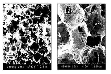
[00015] Figure 4 shows scanning electron micrographs of a poly(MAA-BMA)
scaffold
(0.10 monomer to salt ratio, 24 h fusion time) cross-sections at two
magnifications (40x
and 150x).
[00016] Figure 5 shows scanning electron micrographs for poly(MAA-BMA)
scaffolds
produced using varying salt fusion times: A) 0 h, B) 24 h, C) 48 h and D) 96h.
[00017] Figure 6 shows the relationship between salt fusion time and the
compressive
modulus for poly(MAA-BMA) scaffolds (10% monomer to salt ratio).
[00018] Figure 7 shows the relationship between salt fusion time and the yield
strength
for poly(MAA-BMA) scaffolds (10% monomer to salt ratio).
[00019] Figure 8 shows the effect of monomer to salt ratio on poly(MAA-BMA)
scaffold
porosity (24 h fusion time).
[00020] Figure 9 shows the relationship between monomer to salt ratio and
compressive modulus for poly(MAA-BMA) scaffolds (24 h salt fusion time).
3
CA 02603773 2007-10-03
WO 2006/113984 PCT/CA2006/000533
[00021] Figure 10 shows the relationship between monomer to salt ratio and
yield
strength for poly(MAA-BMA) scaffolds (24 h salt fusion time).
[00022] Figure 11 illustrates the sites of implantation for the test and
control scaffold
disks.
[00023] Figure 12 shows tissue ingrowth into control and test scaffolds (H+E
stained)
at 7, 21 and 30 days post-implantation. Poly(MAA-BMA) at 7 days (a), 21 days
(c) and
30 days (e). Poly(BMA) at 7 days (b), 21 days (d) and 30 days (f). Scale bars
represent
250 pm.
[00024] Figure 13 shows H+E stained scaffold explants at 30 days post-
implantation
that indicate differences in the inflammatory response for test and control
implants. More
foreign-body giant cells shown (by arrows) in the poly(BMA) explants (b and d)
in
comparison to poly(MAA-BMA) (a and c). For figures a and b, scale bar
represents 200
pm and figures c and d, scale bar represents 100 pm.
[00025] Figure 14 shows microvessel density counts at 21 and 30 days post-
implantation in the pores of test poly(MAA-BMA) and control poly(BMA) scaffold
explants.
Values represent means standard deviations and * represents statistical
significance
relative to the poly(BMA) control.
[00026] Figure 15 shows fVlll-stained explant samples at 7, 21 and 30 days
post-
implantation indicating greater vascularisation of the poly(MAA-BMA) scaffolds
(a,c and
e) in comparison to the control poly(BMA) scaffolds (b,d and f). 7 day samples
(a and b),
21 day (c and d) and 30 day (e and f). P denotes areas occupied by polymer
scaffold.
Scale bars represent 100 pm.
DETAILED DESCRIPTION
[00027] Generally, the present invention provides a new type of porous,
polymeric
scaffolds containing pro-angiogenic components that can be used for tissue
engineering/regeneration applications, a method for making the scaffolds,
methods of
using the scaffolds, and systems formed from, or incorporating, the scaffolds.
Both
biostable and biodegradable polymer constructs are contemplated. The scaffold
is
formed from a pro-angiogenic polymer by incorporating pores.
The Polymer
[00028] The polymer that composes the scaffold is a biocompatible polymer.
Biocompatible polymers are defined herein as polymers that induce, when
implanted, an
appropriate host response given the application. For the purposes herein, they
are
essentially non-toxic, non-inflammatory, non-immunogenic, and non-
carcinogenic.
4
CA 02603773 2007-10-03
WO 2006/113984 PCT/CA2006/000533
[00029] Furthermore, the polymer encourages vascularization. The term
"vascularization" refers to the blood vessel network in and around an
implanted scaffold,
or the formation of such a blood vessel network.
[00030] In order to function as a scaffold, the polymer must be insoluble in
aqueous
solution at 37 C (i.e. body temperature).
[00031] The polymer is made from polymerizable monomeric subunits or monomers
which are polymerized together. The monomers once incorporated into the
polymer are
referred to herein as mers or monomeric (sub)units. The polymer comprisesof
the
scaffold comprises at least 20 mol-% monomeric units (i.e. mers) contain
acidic functional
groups. The polymer may contain at least 30, at least 40, at least 45, or at
least 50 mol-%
of acidic mers. Preferably, the polymer contains at least 45 or at least 50
mol-% of acidic
mers. The polymer may comprise 100 mol-% acidic mers, and may be a homopolymer
of
one type of such acidic mers. However, the polymer will typically contain
other
biocompatible mers to give the scaffold the desired structural and physical
properties,
such as solubility, flexibility, strength, etc. These other mers are referred
to herein as the
backbone mers (though the majority or the entirity of the polymer may consist
of acidic
mers). Furthermore, the polymer optionally contains crosslinks.
1000321 The polymer is preferably a polyacrylate.
[00033] The polymer may be biodegradable or biostable.
[00034] Examples of suitable copolymer structures are random, block, and graft
copolymers.
[00035] In the case of a graft copolymer the polymer comprises a backbone and
arms
grafted onto the backbone. Preferably, the arms contain the at least 20 mol-%
monomeric
subunits containing acidic functional groups. Methods of making graft
copolymers are
known in the art. As an example of a graft copolymer, the acidic mers may be
grafted to
a biocompatible polymer. In this way, a pro-angiogenic effect is conferred to
the existing
biocompatible polymer. This may be accomplished through the inclusion of
grafting sites
(e.g. unsaturated carbon bonds, acids, amines, amides, hydroxyls) in the
biocompatible
polymer.
[00036] However, this invention is not meant to include scaffolds which are
surface-
modified or polymers which are derivativatized post-scaffold formation.
[00037] Figure 1 shows a schematic example of a polymer in accordance with
invention with both the acidic and backbone co-monomers used to form the main
chain.
Degradable cross-links are used to join the various main chains. Figure 2
shows a
schematic representation of a type of graft copolymer in accordance with the
invention
5
CA 02603773 2007-10-03
WO 2006/113984 PCT/CA2006/000533
with the backbone co-monomers joining together to form the main chain and the
acidic
co-monomers used to make polymers which are grafted onto the main chain.
Acidic Mers
[00038] At least 20 mol-% of the monomeric units (i.e. mers) in the polymer
contain
acidic functional groups that, upon implantation, bind and stabilize
endogenous pro-
angiogenic growth factors (such as VEGF and FGF). This provides a sustained,
localized
angiogenic effect by stabilizing the growth factors (in analogy to
extracellular matrix
components) and slowly releasing them over a prolonged period of time.
Examples of
suitable acidic functional groups include any biocompatible acids, such as
carboxylic
acids (-COOH), sulfonic acids (-SO3H), and phosphoric acids (-OP(OH3), and
their
corresponding salts (i.e. carboxylates (-COO-), sulfonates(-S03 ), and
phosphates).
Examples of polymerizable groups (i.e. monomers or polymerizable monomeric
(sub)units) containing acidic functional groups that may be used to produce
the pro-
angiogenic polymer of the invention include: acrylates (CH2CR'COOR2) (such as
methacrylic acid (CH2C(CH3)COOH) and acrylic acid (CH2CHCOOH)), 2-propene-1-
sulfonic acid (CH2C(CH3)CH2SO2OH), 4-vinyl benzoic acid (CH2-CH-C6H4-COOH),
crotonic acid (CH3CHCHCO2H), itaconic acid (CH2C(CH2CO2H)CO2H), vinylsulfonic
acid
(CH2CHSO3H), vinyl acetic acid (CH2CHCHCOOH), citric acid
(C(OH)(CO2H)(CH2CO2H)2, and styrene sulfonic acid (CH2-CH-C6H4-SO3H), and
their
salts, such as sodium styrene sulfonate (CH2-CH-C6H4-SO3Na) and
monoacryloxyethyl
phosphate. Combinations of the above may also be used. In one aspect, the
acidic mers
are methacrylic acid. These polymerizable groups may be incorporated directly
into the
polymer backbone or grafted to the backbone.
Backbone Mer
[00039] In addition to the acidic mer or mers, the polymer may comprise one or
more
additional non-acidic mers. Any mers may be used so long as the resulting
polymer is
biocompatible and so long as the starting monomer is polymerizable with the
selected
starting acidic monomer (i.e. the polymerizable groups (i.e. monomers)
containing acidic
functional groups). Generally, the mers will be chosen as a function of the
desired
physicochemical properties (e.g. mechanical, aqueous swelling, etc.), as a
function of
desired physical properties (such as mechanical strength), and as a function
of desired
solubility properties, i.e. they may help render the polymer insoluble in
aqueous solution
at 37 C. Such co-monomers are known in the art.
[00040] Examples of backbone co-monomers for forming the polymers of the
present
invention include acrylates (such as hydroxyethyl methacrylate, methyl
methacrylate,
6
CA 02603773 2007-10-03
WO 2006/113984 PCT/CA2006/000533
butylmethacrylate, hexylmethacrylate, and butylacrylate), phosphazenes,
various vinyl co-
monomers including vinyl chloride, acrylonitrile, vinyl acetate, ethylene
vinyl acetate, vinyl
alcohols, vinyl amines, imides, ether ketones, sulphones, siloxanes, urethanes
and
amides, carbonates, esters and bioresorbables such as anhydrides, orthoesters,
caprolactones, amino acids, lactic/glycolic acid co-monomers and
hydroxybutyrates.
Combinations of the above may also be used.
[00041] As a matter of practicality, if the acidic mer is an acrylate, such as
methacrylic
acid, the backbone co-monomer may be chosen to be an acrylate, such as butyl
methacrylate (BMA). The acrylates provide a diverse range of monomers, and are
readily
available making it possible to tailor material properties to a variety of
applications.
Crosslinkers
[00042] The polymer forming the scaffold is optionally crosslinked.
Crosslinking is
used to render the polymer insoluble in aqueous solution at 37 C. The
crosslinks may be
biodegradable or biostable. The crosslinking agent is generally incorporated
into the
polymer comprising the scaffold during polymerization, in an amount of about
0.001 to
about 5 mol-% based on the total number of mols of monomers comprising the
polymer,
preferably about 0.01 to about 1 mol-%. The amount of crosslinker chosen will
depend
on the desired physicochemical properties of the resultant scaffold including,
in the case
of the degradable linkers, the rate of degradation desired.
[00043] Biostable crosslinking agents: Biostable crosslinking agents are known
in
the art. Examples of biostable crosslinking agents are biocompatible divinyl
benzenes
and bifunctional acrylates, such as (poly)ethylene glycol dimethacrylates,
e.g. ethylene
glycol dimethacrylate (EGDMA). An advantage of polyethylene glycol
dimethacrylates is
that the length of the polyether chain can be modified to suit the
application.
[00044] Degradable linkages: In many cases it may be desirable to have the
constructs degrade in vivo over time. Degradable constructs can be produced
through
the incorporation of crosslinkers that contain hydrolysable linkages (i.e.
ester, amide,
anhydride). Cleavage of these crosslinks by simple chemical or enzyme-mediated
hydrolysis breaks down the polymer network, liberating soluble polymer chains,
which
eventually leads to the elimination of the solid construct. The rate of
polymer degradation
may be modified through the selection of monomer chemistry, crosslinker
chemistry and
crosslink density. Crosslinker molecules containing internal hydrolysable
linkages (e.g.
ester, amide, anhydride) and polymerizable functional groups, yielding an
overall
functionality greater than 2, introduce degradable branch points in the
formation of
insoluble, network polymers. These crosslinkers are obtained by covalently
attaching
7
CA 02603773 2007-10-03
WO 2006/113984 PCT/CA2006/000533
polymerizable functional groups to the ends of molecules containing degradable
linkages.
The attached polymerizable functional groups may include: methacrylate,
acrylate,
isocyanate, carboxylic acid, acid chloride, vinyl, amine, and hydroxyl. An
example of
commonly used degradable linkers is methacrylated polyesters, such as
polycaprolactone, which liberates non-toxic degradation products.
The Scaffold
[00045] The scaffold must have a porosity of at least 40%. For many
applications it is
preferred to have a porosity of at least 70%, preferably at least 80%. A
porosity of at
least 90% may also be desirable. The porosity (po) is calculated as: po = 1-
(d/dP), were
dP is the density of the non-porous scaffold, and d is the density of the
porous scaffold.
The density of the scaffolds (d) is calculated as d=m/v (where m is the mass
and v the
volume); alternatively, literature values for the density of non-porous
scaffolds may be
used.
[00046] The pore diameter (primary pores) will generally be between 10 to 800
microns, with the average pore diameter being between 200 to 350 microns;
though for
certain applications a range of 25 to 250 microns may be preferred.
[00047] The pores of the scaffold are interconnected. The diameter of the
interconnections is significantly smaller than the pore diameter, typically
less than about
100 microns. The pores must be sufficiently interconnected to permit
vascularization.
[00048] In one particular embodiment, the invention provides a pro-angiogenic
porous
polymer scaffold, said polymer being a polyacrylate comprising at least 20 mol-
%
monomeric subunits containing acidic functional groups, said polymer being
optionally
crosslinked, having a porosity of at least 40%, and having interconnected
pores. The
monomeric subunits containing acidic functional groups may be methacrylic
acid. The
mol-% of monomeric subunits containing acidic functional groups may be at
least 45 mol-
%. The backbone mers may be one or more types of methacrylates, such as
butylmethacrylate.
Methods of making the scaffold
[00049] A novel method for making scaffolds is disclosed, using a modified
porogen
technique, as described in more detail in Example 1. Generally, the monomers,
optionally the crosslinker, and the initiator are dissolved in a solvent,
poured into a bed of
fused particles (such as a salt) and polymerized. As the polymerization and
optionally
crosslinking reaction proceeds, the polymer precipitates out of solution. The
solvent is
removed. Removal of the included fused particles (such as salt crystals)
results in a
highly porous polymer construct. The method is ilustrated in Figure 3.
8
CA 02603773 2007-10-03
WO 2006/113984 PCT/CA2006/000533
[00050] More specifically, the particles are fused by exposing them to a humid
environment for a predetermined length of time. As is discussed in Example 3,
longer
fusion times result in progressively less organized pore structures and
increasing
frequency of holes in the primary pore walls of the scaffold.
[00051] Examples of suitable particles include sugars, such as glucose, and
organic
and inorganic salts, such as NaCI. NaCI is preferred.
[00052] Particles having a diameter corresponding to the desired diameter of
the pores
in the scaffold are suitable. For instance, the particles may have a particle
size of about
to 800 microns, with the average diameter being between 200 to 350 microns;
though
10 for certain applications a range of 25 to 250 microns may be preferred. The
particles can
be sorted by size prior to fusion depending on the desired average pore size
and size
ranges.
[00053] The monomers, initiator, and optionally crosslinking agent are
combined in a
suitable solvent, such as methylene chloride, ethyl acetate, chloroform,
acetone,
benzene, 2-butanone, carbon tetrachloride, n-heptane, n-hexane, and n-pentane.
For
polyacrylates, chloroform is often suitable. The mixture is poured over the
fused particle
bed and is allowed to polymerize under conditions suitable for the particular
polymer
chosen.
[00054] The monomer to particle ratio is selected to achieve the desired
porosity. For
instance, it may range from 7 to 16 % wt:wt expressed as a percentage.
[00055] Once the polymerization is complete the solvent is removed, such as by
evaporation (such as by air drying).
[00056] The scaffold is then subjected to one or more washes with a solvent in
which
the particles are soluble, but the scaffold is not, such as water.
[00057] Thus, in one aspect, the invention provides a method for making a pro-
angiogenic porous polymer scaffold, wherein said polymer comprises acidic
functional
groups grafted to or incorporated into the polymer, said scaffold having a
porosity of at
least 40% and said pores being interconnected, said method comprising: mixing
one or
more types of monomers and an initiator together in a solvent, wherein at
least one of
said monomers contains an acidic functional group; pouring the mixture over a
fused salt
bed having a pore size range of 10 to 800 microns; allowing the mixture to
polymerize;
and leaching the salt out, to yield the porous scaffold.
[00058] Other methods for making porous scaffolds are known in the art
(Sachlos
E. Czernuszka J.T., "Making Tissue Engineering Scaffold Work. Review on the
Application of Solid Freeform Fabrication Technology to the Production of
Tissue
9
CA 02603773 2007-10-03
WO 2006/113984 PCT/CA2006/000533
Engineering Scaffolds" European Cells and Materials Vol. 5 2003, 29-40) and
could be
used to make scaffolds of the present invention using the pro-angiogenic
polymers
described herein. These include gas foaming, fibre meshes/fibre bonding, phase
separation, melt moulding, emulsion freeze drying, solution casting, freeze
drying, and
solid freeform fabrication.
[00059] The method of making the scaffold and the monomeric units chosen to be
included in the scaffold can vary and will depend on the particular
application. These and
other methods may be used, so long as the scaffold produced is porous and the
pores
are interconnected.
Uses of the Scaffold
[00060] There are different approaches to implanting the scaffolds known in
the art.
These include implantation of the scaffolds alone (known as guided tissue
regeneration);
seeding the scaffolds with cells in vitro and then implanting them
immediately; or seeding
the scaffolds with cells in vitro allowing the cells to grow, and then
implanting the
scaffolds. The target tissues for use with these scaffolds are principally
vascularized
tissues, such as the skin, the blood, the organs...etc. Tissue with little
vascularization,
such as cartilage, is not preferred.
[00061] The scaffold may also be used as a bioreactor, by implanting the
scaffold with
cells and allowing the cells to produce a given protein; examples of proteins
include
growth factors. The scaffold has the ability to provide a unique environment
for the
maintenance of such cells.
[00062] The scaffold could also be used to generate artificial organs by
placing several
cell types into the scaffold and providing organizational cues (i.e.
mechanical and/or
biochemical stimuli) to promote complex 3-D tissue formation.
Examples
Example I - Scaffold Fabrication
[00063] A novel adaptation to the traditional solvent casting/particulate
leaching
technique was used to prepare the porous scaffolds. The monomers were
dissolved in
solvent and polymerized in situ on a bed of fused salt (NaCI) particles.
Subsequent to
polymerization, the reaction solvent was evaporated off leaving a polymer-salt
composite.
Sequential washes in various solutions removed the salt, yielding a porous
polymer
scaffold.
[00064] Salt Fusion: A salt fusion technique was used to generate pore
interconnectivity in the fabricated scaffolds (Figure 3). Pore
interconnectivity is essential
CA 02603773 2007-10-03
WO 2006/113984 PCT/CA2006/000533
to allow tissue ingrowth and vacularization upon implantation. The fusion
technique
involves exposing salt particles to a humid environment prior to scaffold
formation. When
exposed to the humid environment, adjacent salt crystals fuse in a process
called
"caking". The surfaces of contacting salt particles coalesce, forming bridges
between
particles thereby increasing scaffold pore interconnectivity upon salt
dissolution.
[00065] Unsieved NaCI (20 g) was added to a PTFE mold and agitated until
level. The
mold was then placed in a large beaker containing distilled water (1 cm
depth). The top
of the beaker was sealed with Parafilm and placed in an oven (37 C) to create
a humid
environment. After the desired fusion time (24 to 96 h), the mold containing
the fused salt
particles was removed from the beaker and dried for 24 h in an oven (37 C).
The degree
of salt particle fusion was varied by altering the fusion time.
[00066] In Situ Polymerization: The monomers and initiator, namely 45 mol%
methacrylic acid, 54 mol% comonomer (meth)acrylate, 1 mol% ethylene glycol
dimethacrylate (EGDMA) (the biostable crosslinker), and benzoyl peroxide (an
initiator)
were dissolved in chloroform. Comonomer (meth)acrylates employed were
methylmethacrylate (MMA), butylmethacrylate (BMA), hexylmethacrylate (HMA) and
butylacrylate (BA). Chloroform was used as a solvent (at 2:1 chloroform to
total monomer
volume ratio) to increase the volume of reactant solution to allow complete
coverage of
the salt bed. The reaction mixture was poured over the bed of fused salt
particles. The
polymerization reaction proceeded for 5 h at 67 C under nitrogen gas (Figure
3). A reflux
condenser was attached to the reaction vessel to limit evaporation of the
solvent during
polymerization. Upon completion of the reaction, the polymer-salt composite
was air
dried overnight to remove chloroform. A poly(butylmethacrylate) control
scaffold was
synthesized as above to directly assess the effect of methacrylic acid
incorporation on the
in vivo response to the scaffolds.
[00067] Salt Removal and Scaffold Purification: The salt-containing scaffolds
were
subjected to a series of water washes to remove the embedded porogen.
Scaffolds were
placed in deionized water for 5 days, replacing the water at least 3 times per
day for a
total of 15 washes. Upon salt removal, the scaffolds were dried under vacuum
for 24 h.
Residual monomers and solvent were removed through a series of acid, base and
solvent
washes. The scaffold was placed sequentially in the following solutions for 3
h each at
room temperature:
1. 0.1 M HCI 9. Water
2. Water 10. DMF
3. Acetone 11. Water
11
CA 02603773 2007-10-03
WO 2006/113984 PCT/CA2006/000533
4. Acetone 12. 0.1 M NaOH
5. Acetone 13. Water
6. Water 14. 0.5 M HCI
7. 0.1 M NaOH 15. Water
8. 0.5 M HCI 16. Water
[00068] The scaffolds were cut into disks (6 mm diameter x 2 mm thick) and
washed
with 95% ethanol to remove endotoxin (lipopolysaccharide fragments of gram-
negative
bacterial cell walls, which are found as contaminants almost everywhere) (EU).
Scaffold
pieces (1-2 g) were placed in a 50 mL polystyrene tube and 40 mL of ethanol
was added.
The tubes were sonicated for 20 min., the ethanol was removed and a fresh 40
mL of
ethanol was added to the tube. This washing procedure was repeated 10 times.
Following the ethanol washes, the scaffolds were washed with endotoxin-free
water to
remove residual ethanol. The scaffolds were then dried under vacuum and stored
in a
desiccator. Endotoxin testing (LAL Pyrochrome Kit, Cape Cod, USA) was
performed to
ensure the scaffolds contained less than 0.25 EU/mL. Any scaffolds that
contained >0.25
EU/mL were rewashed as above until the endotoxin level was below the cut-off
value.
[00069] Scaffold Characteristics: The scaffolds were visualized using scanning
electron microscopy (SEM) to assess the pore size range and pore
interconnectivity.
Specimens were frozen in liquid nitrogen for 5 min and cut with a razor blade.
Cross-
sections of the scaffolds were sputter coated with gold and visualized on a
Hitachi S800
scanning electron microscope. Figure 4 shows scanning electron micrographs of
a
poly(BMA-MAA) scaffold made with 24h salt fusion and a 10% weight ratio of
monomer to
salt. Pore interconnectivity can be seen at higher magnification. Diameters of
the
primary pores range from approximately 100-600 pm, with the majority falling
within the
200-350 pm range. The interconnecting pores resulting from salt fusion were
significantly
smaller in size (<100 pm).
Example 2- Effect of Comonomer Chemistry on Scaffold Properties
[00070] MAA-containing scaffold copolymer formulation was examined using four
different acrylate comonomers, methylmethacrylate (MMA), butylmethacrylate
(BMA),
hexylmethacrylate (HMA) and butylacrylate (BA). The mechanical stability of
the various
copolymer scaffolds was assessed by visual observation during the salt
leaching phase of
the fabrication process and/or quantitatively evaluated by compression
testing. All
scaffolds were produced using the following monomer feed ratios: 50 mol% MAA,
49
mol% comonomer and 1 mol% crosslinker (EGDMA).
12
CA 02603773 2007-10-03
WO 2006/113984 PCT/CA2006/000533
[00071] Qualitative Visual Assessment: Porous scaffolds fabricated with MMA as
the
comonomer were brittle and crumbled easily with handling during the salt
leaching phase.
Poly(MAA-MMA) scaffolds fabricated with a monomer to salt ratio of 12.5% or
lower
disintegrated into small fragments. Poly(MAA-BMA) scaffolds were found to be
much
less brittle than the poly(MAA-MMA) scaffolds. Mechanically stable
(qualitatively
assessed) scaffolds were produced down to a monomer-salt ratio of 10%. In
comparison,
MAA-containing scaffolds produced by copolymerization with hexylmethacrylate
and butyl
acrylate were much softer and less brittle than either the BMA or MMA
versions, as
expected. These differences were examined in more detail by compression
testing.
[00072] Compression Testing: Compressive mechanical properties were measured
in
a phosphate-buffered saline (PBS) solution at 37 C on a Mach-1
T"'Micromechanical
System equipped with a 0.01 kN load cell according to ASTM F541-99a standard
specifications for testing acrylic bone cement. Four cylindrical samples (6 mm
diameter,
12 mm thick) for each scaffold formulation were preconditioned in PBS at 37 C
for 24 h
prior to testing. The specimens were compressed at a rate of 1.0 mm/min up to
a strain
level of approximately 0.7 mm/mm. Young's modulus (E) was calculated from the
stress-
strain curve as the slope of the initial linear portion of the curve,
neglecting any toe region
due to the initial settling of the specimen. The compressive strength at yield
(6y) was
defined as the intersection of the stress-strain curve with the modulus slope
at an offset of
1.0% strain. A Student's t-test was performed in comparing means from two
independent
sample groups. A significance level of p<0.05 was used in all the statistical
tests
performed.
[00073] Table I shows the effect of comonomer type on scaffold compressive
mechanical properties. Poly(MAA-MMA) scaffolds were not tested since they were
too
brittle and friable to easily prepare test specimens. Both poly(MMA) and
poly(MAA) have
glass transitions over 100 C, making the copolymer composed of these monomers
rigid.
This rigidity combined with the high porosity necessary for a tissue
engineering scaffold
likely led to the brittle quality of this formulation. All other specimens
were produced
using a salt fusion time of 24 h and a monomer to salt ratio of 10%. Scaffold
stiffness, as
indicated by Young's modulus (E), decreases dramatically with comonomer type
from
BMA to HMA to BA. In addition, compressive strength at yield was only
measurable for
the BMA-containing copolymer scaffold. HMA has a longer pendant group than BMA
which serves to limit chain packing and increase the free volume of the
polymer,
effectively lowering the glass transition temperature (Tg). This results in a
weaker, softer
copolymer as shown in Table I. BA has a similar chemical structure to BMA,
only lacking
13
CA 02603773 2007-10-03
WO 2006/113984 PCT/CA2006/000533
a methyl substituent group. The absence of this methyl substituent in BA
permits greater
chain mobility, reducing the T. of the copolymer. This results in a weaker,
softer
copolymer than both the BMA and HMA-containing ones. This data shows that
modifying
the comonomer chemistry is a relatively simple method for generating MAA-
containing
scaffolds with a broad range of physical properties that may be tailored to
suit a variety of
applications.
Table I - Effect of comonomer chemistry on compressive properties for MAA-
containing
scaffolds
Comonomer Monomer:Salt (%) Fusion Time (h) E (MPa) ay (MPa)
BMA 10 24 1.9t0.3 0.15t0.03
HMA 10 24 0.7 t 0.1 ND
BA 10 24 0.04 0.01 ND
Example 3- Modifying Scaffold Porosity and Pore Structure
[00074] Copolymer scaffold pore structure and porosity were systematically
modified
by altering the salt fusion time and monomer to salt ratio (wt/wt, expressed
as a
percentage) in the reaction mold.
[00075] Incubation of NaCI crystals in a humidified environment resulted in
fusion of
the crystals, creating a highly interconnected salt matrix. Salt fusion times
were varied
from 0 to 96 h and the resulting scaffolds were visualized by SEM to assess
pore
morphology. In addition, the effect of salt fusion time on scaffold mechanical
properties
was determined by compressive testing (done as described in Example 2). All
scaffolds
tested were poly(MAA-BMA) with a monomer to salt ratio of 10%.
[00076] Figure 5 shows the pore structure of scaffold cross-sections as a
function of
salt fusion time. The unfused salt scaffold (A) has a well-defined pore
structure that
appears to be poorly interconnected. In contrast, for the salt fused scaffolds
a highly
porous and interconnected pore structure is evident. For the 24 h fusion
scaffold (B),
clearly defined primary pores are seen with holes in the pore walls. Longer
salt fusion
times (48h (C) and 96h (D)) resulted in progressively less organized pore
structures and
increasing frequency of holes in the primary pore walls. In addition, the
holes in the
primary pore walls increased in size with salt fusion time. Finally, the pore
walls are
appreciably thicker in the 24 h salt fusion scaffold, likely a result of
larger interstitial space
between less fused salt particles that was filled with the copolymer.
[00077] Salt fusion had a pronounced effect on the mechanical properties of
the
scaffolds. As seen in Figure 6, scaffolds fabricated with 24 or 48 h salt
fusion time were
14
CA 02603773 2007-10-03
WO 2006/113984 PCT/CA2006/000533
found to have a significantly higher compressive modulus (E) compared with the
unfused
scaffold. Scaffolds produced with 48 and 96 h salt fusion times were found to
have
significantly lower moduli compared to the 24 h scaffold. The dependence of
yield
strength (6y) on salt fusion time followed a similar trend (Figure 7). The 24
h salt fusion
time scaffold produced a significantly higher yield strength than the unfused
scaffold but
increasing fusion time resulted in reduced yield strengths. The inter-particle
space is
larger upon short salt fusion time (i.e. 24 h) due to a small amount of
particle erosion that
results in a"rounding-off' of the salt particles. The increased inter-particle
space is filled
during polymerization leading to thicker pore walls and stronger scaffolds.
However, as
the salt fusion time is increased to 48 and 96 h, the salt particles become
increasingly
connected; reducing the inter-particle space leads to thinner pore walls and a
more
disorganized pore structure (seen in Figure 5). These factors combine to
produce the
decreasing modulus and yield strength values at the longer salt fusion times
seen.
[00078] Scaffold porosity was modified by varying the monomer to salt ratio
(wt/wt)
used in the reaction mold. For this study, poly(MAA-BMA) scaffolds were
produced using
a salt fusion time of 24 h and the monomer to salt ratio was varied from 7.5
to 15%. The
density and porosity of the scaffolds were determined in triplicate by
measuring their
dimensions and masses. The density of the scaffolds (d) was calculated as
follows:
d=m/v (where m is the mass and v the volume). The porosity (po) was calculated
as: po =
1- (d/dP), were dP is the density of the non-porous polymer (dP =1.1 g/cm3
based on
literature values).
[00079] The porosities of the poly(MAA-BMA) scaffolds produced as a function
of
monomer to salt ratio are shown in Figure 8. Increasing monomer to salt ratio
resulted in
decreasing scaffold porosity, as expected. Compressive testing showed that
both
modulus and yield strength increased with increasing monomer to salt ratio
(Figures 9
and 10). As expected, increasing scaffold porosity (with decreasing monomer to
salt
ratio) resulted in decreasing mechanical properties as a result of thicker or
more
numerous pore walls.
Example 4- Scaffold Cytotoxicity
[00080] Scaffold cytotoxicity was evaluated prior to implantation studies to
assess the
effectiveness of the washing method used to remove residual monomers and
solvent
post-polymerization. An alamarBlueTM cell viability assay (Biosource, USA) was
conducted on cells after direct contact with poly(MAA-BMA) scaffolds and
contact with a
scaffold extract. The alamarBlueTM assay incorporates an oxidation-reduction
indicator
that changes in color in response to the chemical reduction of the growth
medium
CA 02603773 2007-10-03
WO 2006/113984 PCT/CA2006/000533
resulting from metabolic activity. The color change of the cell culture medium
is
measured spectrophotometrically at two wavelengths.
[00081] Scaffold Extract Test: THP-1 monocytes cultured in RPMI medium
supplemented with 10% fetal bovine serum were seeded into wells in a tissue
culture
polystyrene (TCPS) 96-well plate at 3 cell densities (100,000, 150,000 and
250,000
cells/well) and evaluated in triplicate. The cells were differentiated
overnight into
macrophage-like cells with the addition of phorbol myristate acetate (PMA).
The next day
the cells were rinsed twice with 150 pL media per well to remove the PMA.
Media (150
pL/well), previously incubated with poly(MAA-BMA) scaffold for 48 h (40 mg
scaffold/10
mL medium), was then added to each test well while a fresh 150 pL of medium
was
added to each control well. The cells were incubated for 24 h, then 150 pL of
fresh
medium and 16.65 pL of alamarBlueTM solution was added to each well. The cells
were
incubated for a further 4 h, then 100 pL of solution was transferred from each
well to a
new plate and the solution absorbance was read at 570 and 600 nm to quantify
viability.
Cell viability by alamarBlueTM assay, when exposed to the poly(MAA-BMA)
extracts, was
determined to be >100% 5% compared to cells cultured with fresh media.
[00082] Direct Contact Test: THP-1 monocytes were differentiated into
macrophage-
like cells and seeded in a TCPS plate, as for the extract test. Medium (150
pL/well),
containing crushed scaffold (1 mg scaffold/mL medium), was then added to each
test
well containing activated cells while 150 pL of fresh medium was added to each
control
well. The cells were incubated for 24 h, then 150 pL of fresh medium and 16.65
pL
alamarBlueTM was added to each well and incubated for 4 h. The absorbance of
each
well was measured directly. Cells cultured directly with the crushed poly(MAA-
BMA)
scaffolds exhibited a high level of viability (91 7%) compared to cells
cultured in fresh
media. This result, in conjunction with the scaffold extract result, suggests
that the
scaffold washing procedure was effective in removing residual monomers and
solvent
post-polymerization. The slight decrease in viability for cells in direct
contact with the
scaffold pieces may be attributed to a difference in adherence to the pieces
compared to
TCPS or a mild inhibitory (non-toxic) effect on cell metabolism by the
scaffold fragments.
Example 5- In Vivo Evaluation of Scaffolds
[00083] The angiogenic potential of the scaffolds was evaluated in a murine
subcutaneous implant model. The test scaffolds were all poly(MAA-BMA) produced
using
a monomer to salt ratio of 10% and 24 h salt fusion time because these
conditions
produced a well interconnected, highly porous scaffold that was easily
handled. Since
MAA is the pro-angiogenic component of the copolymer, homopolymer poly(BMA)
16
CA 02603773 2007-10-03
WO 2006/113984 PCT/CA2006/000533
scaffolds were prepared and used as the negative control in this study.
Scaffolds were
implanted subcutaneously on the dorsum of male CD31 mice for 7, 21 and 30 days
and
the levels of tissue invasion, host tissue reaction and vascularization were
evaluated
histologically.
[00084] Sample Preparation: Washed poly(MAA-BMA) and poly(BMA) scaffolds were
cut into disks 6 mm in diameter and 2 mm thick using a biopsy punch and razor
blade.
Endotoxin was removed (as described in Example 1) from the scaffolds and
tested to be
<0.25 EU/mL. Prior to implantation, the scaffolds were hydrated in sterile
saline overnight
(0.9% NaCI).
[00085] Implantation Procedure: Subcutaneous pockets were created in the right
and
left dorsal upper quandrants of male CD31 mice by blunt dissection. A poly(MAA-
BMA)
disk was then placed in the left quadrant pocket while a poly(BMA) control
disk was
placed in the right quadrant pocket for each mouse (Figure 11). Surgical
staples were
removed 10 days after surgery upon complete closure of the incision wound. For
each
study time, 4 animals were implanted with both a poly(MAA-BMA) test and
poly(BMA)
control scaffold disk. At 7, 21 and 30 days post-implantation, the mice were
sacrificed
and the scaffold disks were explanted and fixed in 10% neutral buffered
formalin for 24-
48 h prior to tissue processing.
[00086] Histology and Immunohistochemistry Preparation: Specimens were
prepared,
cut and stained for hematoxylin and eosin (H+E) and vonWillebrand factor
(factor VIII) by
the clinical research pathology lab at Toronto General Hospital. Implants were
removed
from the formalin solution, embedded in paraffin and sectioned by cutting
along the
longitudinal axis at several points along the thickness of the disk. Samples
from these
sections were cut to a thickness of 4 pm prior to histological or
immunohistochemical
staining.
[00087] For H+E staining, sections were first dewaxed in 4 changes of xylene,
then
rehydrated with sequential dips in decreasing graded alcohol, followed by a
water wash
for 1 min. The sections were then placed in filtered hematoxylin for 5 min
followed by a 2
min water wash. The sections were then decolorized in 1% acid alcohol and
washed with
water for 15 sec. Next, the samples were dipped 3 times in ammonia water,
followed by
a water wash for 1 min, placement in eosin for 10-15 sec and another quick
rinse in
water. The samples were dehydrated by sequential dips in increasing graded
alcohol.
Finally, the sections were dipped into 4 changes of xylene and mounted in
Permount .
[00088] For anti-vonWillebrand factor staining, the initial steps of dewaxing
in xylene
and rehydrating in sequential dips of decreasing graded alcohol were the same
as
described above. Then endogenous peroxidase activity was blocked with 3%
aqueous
17
CA 02603773 2007-10-03
WO 2006/113984 PCT/CA2006/000533
hydrogen peroxide for 15 min, followed by a tap water wash. Pre-treatment was
achieved
with 1% pepsin for 15 min, followed by treatment with 10% normal goat serum.
Next, the
sections were incubated with an anti-vonWillebrand primary antibody (also
referred to as
factor VIII, rabbit anti-human polyclonal) at a dilution of 1/8000 for 1 h.
The sections were
then incubated with the secondary linking antibody, a goat-anti-rabbit
antibody, for 30
min. Sections were then incubated for 30 min in Signet USA Level 2 labeling
reagent,
diluted %4 with DAKO antibody diluting buffer. The sections were developed
with
NovaRed for 5 min and a counterstain with Mayer's hematoxylin was added.
Dehydration
was performed via increasing graded alcohol dips, followed by clearance with
xylene and
mounting in Permount .
[00089] Microvessel Counting Method: The level of vascularization in the
tissue
invading the porous poly(MAA-BMA) and poly(BMA) scaffold explants was
quantified
using a microvessel density (MVD) count technique adapted from the tumour
research
literature. At low power (50x magnification), the three areas of the sample
with the most
abundant staining ("hotspots") per section were identified with the scaffold.
At high power
(200x magnification), the number of factor VIII stained structures was counted
for each
"hot spot". Any brown-staining endothelial cell or cluster of cells was
counted as an
individual microvessel if it was clearly separated from adjacent microvessels
by other
non-staining cells or connective tissue. The presence of a patent lumen or
erythrocytes
was not a requirement for the definition of a microvessel. MVD counts were
expressed
as microvessels per mmZ with a mean MVD count per section calculated by
averaging the
three counts. The mean MVD counts were used to make a statistical comparison
between the poly(MAA-BMA) test and poly (BMA) control scaffolds.
[00090] Characterization of Tissue Invasion into Scaffolds: Both the poly(MAA-
BMA)
test and poly(BMA) control scaffolds elicited a similar progression of tissue
invasion over
days, as seen in Figure 12. At 7 days ((a) and (b)), tissue penetration at the
periphery
of the scaffold was observed with minimal progression into the inner pores of
the
scaffolds. At 21 days ((c) and (d)) post-implantation, tissue had penetrated
from the
periphery to deeper sections of the scaffold. By 30 days((e) and (f)),
complete tissue
30 infiltration throughout the scaffolds was apparent. Tissue penetrating from
opposite sides
of the scaffold merged to create a continuous bridge across the cross-section
of the
scaffold. However, even at 30 days there were regions of all scaffolds that
appeared to
be devoid of tissue indicating the presence of a fraction of closed pores in
the scaffolds.
[00091] The inflammatory/foreign body response to the implanted scaffolds was
also
evaluated histologically. In all animals, after 7 days both test and control
scaffolds were
surrounded by a thin capsule containing proliferating fibroblasts, collagen
fibers, capillary
18
CA 02603773 2007-10-03
WO 2006/113984 PCT/CA2006/000533
sprouts and some inflammatory cells. From this capsule, endothelial cells,
fibroblasts and
inflammatory cells penetrated into the porous cavities at the periphery of the
scaffold.
Very few giant cells (multinucleated macrophages) were observed at the border
of the
scaffold. There was however, a difference in the invading tissue of the test
and control
scaffolds at 21 and 30 days post-implantation. In the poly(MAA-BMA) scaffold
explants,
the invading tissue consisted mainly of fibroblasts, collagen and newly formed
capillaries
with some macrophages and a few giant cells. In contrast, the poly(BMA)
control scaffold
presented a more inflammatory response (Figure 13). Along with fibroblasts,
collagen
and newly formed capillaries in the invading tissue, a larger number of
neutrophils and
foreign body giant cells were observed.
[00092] Characterization of Scaffold-Induced Vascularization: The microvessel
density
counting technique was used to quantify the level of histological
vascularization in tissue
penetrating the pores of the poly(MAA-BMA) and poly(BMA) scaffold explants.
MVD
counts in the tissue penetrating the pores of the poly(MAA-BMA) scaffolds at
21 and 30
days post-implantation were significantly higher than in the poly(BMA)
scaffold (Figure
14). There was no significant difference in MVD counts at 21 and 30 days.
[00093] Photomicrographs of fVlll-stained sections of poly(MAA-BMA) scaffold
explants show an increased level of brown-staining blood vessels compared with
the
poly(BMA) control scaffolds at all time points investigated (Figure 15). MVD
counts were
not performed on sections at 7 days post-implantation as there was limited
tissue
ingrowth at this time. However, a large number of stained blood vessels can be
seen at
the periphery of the poly(MAA-BMA) scaffold at day 7, suggesting angiogenic
activity
soon after implantation.
[00094] In this study a poly(MAA-BMA) tissue engineering scaffold was
fabricated and
evaluated for its ability to enhance vascularization in the invading host
tissue. Scaffolds
implanted subcutaneously in mice revealed a higher number of fVIIl stained
blood vessels
in tissue with close proximity to the copolymer. Microvessel density counts
revealed a
higher number of vessels in the tissue invading the pores of the poly(MAA-BMA)
scaffolds
compared to a poly(BMA) control. These results suggest that poly(MAA-BMA) is a
pro-
angiogenic biomaterial that may serve as a tissue engineering scaffold.
[00095] The above-described embodiments of the present invention are intended
to be
examples only. Alterations, modifications and variations may be affected to
the particular
embodiments by those of skill in the art without departing from the scope of
the invention,
which is defined solely by the claims appended hereto.
19