Note: Descriptions are shown in the official language in which they were submitted.
CA 02603775 2007-10-02
WO 2007/053176
PCT/US2006/012969
METHOD OF PROTECTING AGAINST STAPHYLOCOCCAL INFECTION
Technical Field
The invention relates generally to the use of staphylococcal vaccines in
preventing bacterial infection in an individual.
Background Art
Staphylococci and Enterococci rarely cause systemic infections in
otherwise healthy individuals, and therefore are considered opportunistic
pathogens. Through various mechanisms, normal adult humans and animals with
a competent immune system attain an innate natural resistance to these
bacterial
infections. These include mucosal and epidermal barriers, in addition to
possible
immunological mechanisms. Interruption of these natural barriers as a result
of
injuries such as burns, traumas, or surgical procedures involving indwelling
medical devices, increases the risk for staphylococcal and enterococcal
infections.
In addition, individuals with a compromised immune response such as cancer
patients undergoing chemotherapy and radiation therapy, diabetes, AIDS,
alcoholics, drug abuse patients, post organ transplantation patients and
infants
are at an increased risk for staphylococcal and enterococcal infections.
Staphylococci are commensal bacteria of the anterior nares, skin, and the
gastrointestinal tract of humans. It is estimated that staphylococcal
infections
account for >50% of all hospital acquired infections. S. aureus alone is
responsible for 15-25% of such infections and is surpassed only by S.
epidermidis, which accounts for 35% of these infections. Staphylococcal
infections, especially those caused by S. aureus are associated with high
morbidity and mortality.
Staphylococcus and Enterococcus are a major cause of. nosocomial and
community-acquired infections, including bacteremia, metastatic abscesses,
septic arthritis, endocarditis, osteomyelitis, and wound infections. For
example,
the bacteremia-associated overall mortality for S. aureus is approximately 25
percent. A study of hospitalized patients in 1995 found that death rate,
length of
stay, and medical costs were twice as high for S. aureus-associated
CA 02603775 2007-10-02
WO 2007/053176
PCT/US2006/012969
hospitalizafibris compared with other hospitalizations. S. aureus bacteremia
is a
prominent cause of morbidity and mortality in hemodialysis patients with an
annual incidence of three to four percent. Contributing to the seriousness of
S.
aureus infections is the increasing percentage of isolates resistant to
methicillin,
and early reports of resistance to vancomycin. Hence, immunoprophylaxis
against S. aureus is highly desired.
The capsular polysaccharides (CPS) of S. aureus are virulence factors in
systemic infections caused by this opportunistic pathogen. S. aureus CPS
confer
invasiveness by inhibiting opsonophagocytic killing by polymorphonuclear
neutrophils (PMN), similar to other encapsulated bacteria, such as
Streptococcus
pneumoniae. This enables the bacteria to persist in the blood, where they
elaborate several different virulence factors, including toxins and
extracellular
enzymes. Of the 13 known types of S. aureus, Types 5 and 8 account for
approximately 88 percent of all clinical isolates. Nearly all of the remaining
isolates are of Type 336 that carries a more recently identified
polysaccharide
(PS) antigen known as 336PS. Antibodies to Types 5 and 8 capsular
polysaccharides ("T5CPS" and "T8CPS") and 336PS induce type-specific
opsonophagocytic killing by human PMNs in vitro, and confer protection against
the homologous strain in animal infection models.
S. aureus causes several diseases by various pathogenic mechanisms.
The most frequent and serious of these diseases are bacteremia and its
complications in hospitalized patients. In particular, S. aureus can cause
wound
infections and infections associated with catheters and prosthetic devices.
Serious
infections associated with S. aureus bacteremia include osteomyelitis,
invasive
endocarditis and septicemia. Staphylococci have developed very sophisticated
mechanisms for inducing diseases in humans, including both intracellular and
extracellular factors. For instance, S. aureus possesses other surface
antigens
that facilitate its survival in the blood stream by helping the bacteria to
evade
phagocytic killing by the host leukocytes. These surface antigens include cell
wall
components such as teichoic acid, protein A, and capsular polysaccharides
(CPS). Due in part to the versatility of these bacteria and their ability to
produce
extracellular products that enhance virulence and pathogenicity,
staphylococcal
2
CA 02603775 2007-10-02
WO 2007/053176
PCT/US2006/012969
bacteremia and its complications continue to be serious and frequently
observed
nosocomial infections.
Antibiotics such as penicillin have been used successfully against both
staphylococcal and enterococcal infections in humans, but more recently the
effectiveness of such antibiotics has been thwarted by the ability of bacteria
to
develop resistance. For example, shortly after the introduction of
methicillin, the
first semisynthetic penicillin, strains of methicillin-resistant S. aureus
(MRSA) were
isolated. Antibiotic resistance among staphylococcal isolates from nosocomial
infections continues to increase in frequency, and resistant S. aureus strains
continue to cause epidemics in hospitals in spite of developed preventive
procedures and extensive research into bacterial epidemiology and antibiotic
development. Enterococci resistant to vancomycin started to appear in 1988 and
have now become commonplace among hospital-acquired infections. Although
methicillin-resistant S. aureus organisms with intermediate resistance to
vancomycin have been identified in some centers, it was only recently that
three
S. aureus strains with complete resistance to vancomycin were reported. This
suggests that the probable conjugal transfer of vancomycin resistance from
Enterococci to Staphylococci has become a reality, and dissemination of these
strains could eventually lead to the widespread development of organisms that
are
more difficult to eradicate. The problem is compounded by multiple antibiotic
resistance in hospital strains, which severely limits the choice of therapy.
The initial efficacy of antibiotics in treating and curing staphylococcal
infections drew attention away from immunological approaches for dealing with
these infections. Although multiple antibiotic-resistant strains of S. aureus
have
emerged, other strategies such as vaccines have not been developed. In
addition, passive immunization has been tested for use in immune-compromised
individuals, such as neonates, who are at increased risk for contracting these
bacterial infections. The data failed to support a solid conclusion in
recommending the use of passive immunization in this population. Baker et al.,
New EngL J. Med. 35:213-219 (1992); Fanaroff et al., New Engl. J. Med.
330:1107-1113 (1994).
3
CA 02603775 2012-11-26
While polysaccharide vaccines have been developed for some primary
bacterial pathogens that induce acute diseases in normal individuals, namely,
Streptococcus pneumoniae, Neisseria meningitidis and Hemophilus
influenzae, prior to development of StaphVAX (Nabi Biopharmaceuticals,
Rockville, MD), none had been described specifically for protection against
opportunistic bacteria. This vaccine against S. aureus infections is currently
in
a confirmatory Phase III clinical trial in end-stage renal (kidney) disease
patients.
StaphVAX is a conjugate vaccine against two serotypes of S. aureus:
Type 5 and Type 8. In the 1980s, eight different serotypes of S. aureus were
identified using polyclonal and monoclonal antibodies to capsular
polysaccharide (CPS). Karakawa et al., J. Clin. Microbiol. 22:445 (1985).
Surveys have shown that approximately 85% of isolates are capsular
polysaccharide Type 5 or Type 8. More recently, Nabi Biopharmaceuticals
has identified and patented an antigen, 336PS, which is found on newly
discovered serotype Type 336 of Staphylococcus aureus. This serotype
accounts for approximately 10-12 percent of all clinically significant S.
aureus
infections. In the present context, a "clinically-significant" bacterial
strain is
one that is pathogenic in humans. The antigen was identified, purified and
characterized, and a prototype conjugate vaccine based on the antigen
demonstrated the ability to protect animals from challenge with clinical
isolates of the homologous serotype. Nabi Biopharmaceuticals is developing a
second generation of StaphVAX vaccine that will contain 336PS antigen in
addition to S. aureus Types 5 and 8 antigens. These second-generation
vaccines are expected to provide coverage for nearly 100% of all clinically
significant S. aureus infections.
In addition to S. aureus, Staphylococcus epidermidis is another
clinically significant Gram-positive bacterium that causes hospital-acquired
infections. S. epidermidis Conjugate Vaccine is an investigational vaccine in
preclinical development for the prevention of S. epidermidis infections. This
vaccine has been shown to induce antibodies that are protective in animal
models and facilitate elimination of bacteria by the same type of immune
system response as
4
CA 02603775 2007-10-02
WO 2007/053176
PCT/US2006/012969
StaphVAXu. To date, none of these vaccines has been shown to provide
protection against non-homologous strains of bacteria.
Nabi's S. aureus vaccine provides a solution for the problem of antibiotic
resistance in Type 5 and Type 8 strains, and proposed next-generation vaccines
address the same issue for other strains. However, there was no reason to
expect that a vaccine based on 336PS would be effective in protecting
individuals
against infection by non-homologous strains of bacteria.
Disclosure of the Invention
The present inventors have found that conjugates of 336PS are effective in
protecting against bacterial infection by strains of bacteria other than those
that
are classified as Type 336 when serotyped. More particularly, a conjugate
vaccine comprising 336PS confers protection against infection by other S.
aureus
strains and against S. epidermidis. In particular, it confers protection
against
infection by both Type 336/5 and Type 336/8 strains of S. aureus that are
described herein, as well as infection by S. epidermidis. This was entirely
unexpected as it was not known that conjugates of 336PS could stimulate the
production of antibodies that combat bacterial infection by strains other than
Type
336 strains. Absent such a teaching, the scope of protection offered by 336PS
conjugate vaccines could not have been expected.
Based on the inventors' discovery, a method now is provided for preventing
infection in a population of patients at risk for infection by various species
of
Staphylococcus or various types of Staphylococcus aureus, comprising
administering to a patient in the population a composition comprising a
conjugate
of an isolated S. aureus antigen that contains N-acetylglucosamime linked to
ribitol, wherein the antigen binds with antibodies to S. aureus Type 336
deposited
under ATCC 55804. The conjugate of the isolated S. aureus antigen produces
antibodies that protect against the homologous serotype and species or
serotype
of Staphylococcus other than S. aureus Type 336. The present invention further
provides a method for preventing infection in a population of patients at risk
for
infection by Staphylococcus epidermidis, comprising administering to a patient
in
the population a composition comprising a conjugate of an isolated S. aureus
antigen that contains N-acetylglucosamime linked to ribitol, wherein the
antigen
5
CA 02603775 2007-10-02
WO 2007/053176
PCT/US2006/012969
binds with antibodies to S. aureus Type 336 deposited under ATCC 55804.
Conjugates of the isolated S. aureus antigen produce antibodies that protect
against S. epidermidis. The antigen comprises a 1,5-poly(ribitol phosphate)
polymer chain in which the 3-position of the ribitol is substituted by N-
acetyl-p-D-
glucosaminyl residues.
Also provided is a method for treating infection in a population of patients
at
risk for developing infection by various species of Staphylococcus or various
types
of Staphylococcus aureus, comprising administering to a patient in the
population
a composition comprising antibodies to a conjugate of an isolated S. aureus
antigen that contains N-acetylglucosamime linked to ribitol, wherein the
antigen
binds with antibodies to S. aureus Type 336 deposited under ATCC 55804. The
conjugate of the isolated S. aureus antigen produces antibodies that protect
against various species of Staphylococcus or various types of S. aureus other
than Type 336. The present invention also provides a method for treating
infection in a patient diagnosed as having a S. epidermidis infection,
comprising
administering to the patient a composition comprising antibodies to a
conjugate of
an isolated S. aureus antigen that contains N-acetylglucosamime linked to
ribitol,
wherein the antigen binds with antibodies to S. aureus Type 336 deposited
under
ATCC 55804.
Brief Description Of The Drawings
The foregoing advantages and features of the invention will become
apparent upon reference to the following detailed description and the
accompanying drawings, of which:
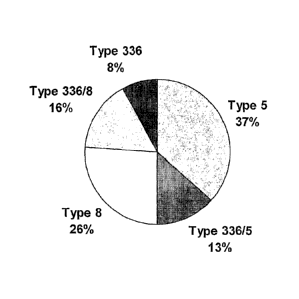
Figure 1 is a pie chart that shows the distribution of surface and capsular
polysaccharide serotypes of 234 S. aureus clinical isolates from bacteremic
patients
Figure 2 is a bar graph demonstrating opsonic killing of mixed serotype S.
aureus isolates by purified 336PS specific 336 conjugate rabbit IgG ("336-
IgG").
Figures 3 is a bar graph of S. epidermidis bacteremia clearance in a mouse
model.
6
CA 02603775 2007-10-02
WO 2007/053176
PCT/US2006/012969
Best Mode for Carrying Out the Invention
It surprisingly has been discovered that vaccines based on conjugates of
336PS can effectively protect individuals against bacterial infection not only
by
homologous strains of bacteria that type as S. aureus 336 strains, but also by
strains of S. aureus that type as other than Type 336, as well as by strains
of S.
epidermidis. There are very few polysaccharide-based vaccines that provide
protection against bacterial infection, and protection against non-homologous
strains of bacteria has not been reported for any of these. Accordingly, it
was
quite surprising to discover that a conjugate vaccine based on antigen
isolated
from the 336 serotype of S. aureus provided protection against some non-
homologous Type 5 and Type 8 strains of S. aureus and against strains of S.
epidermidis.
It appears that in some strains that type as Type 5 and Type 8 S. aureus,
the capsule is discontinuous which allows exposure of an antigen that is
serologically cross-reactive with antibodies that are raised against 336PS
conjugate (336PS covalently bound to protein) vaccine. These strains therefore
type serologically as both Type 336 and one of Type 5 or Type 8. They are
denoted herein as "mixed Type 336/5" and "mixed Type 336/8," and account for
approximately 29% of clinically significant isolates. The distribution of
surface and
capsular polysaccharide serotypes of 234 S. aureus clinical isolates from
bacteremic patients is shown in Figure 1. The isolates were serotyped using
antibodies generated by immunizations of rabbits with Type 5, Type 8 or Type
336
polysaccharide conjugated to Pseudomonas aeruginosa exoprotein A (rEPA). The
336 phenotype was found to be present on 37% of all the clinical isolates,
which
include 8% 336, 13% 336/5, and 16% 336/8. As a surprising correlate of this
discovery, it has been shown that IgG generated in response to a 336PS
conjugate vaccine is able to mediate opsonophagocytosis of serotype 336, 336/5
and 336/8 strains.
Quite unexpectedly, antibodies generated in response to a 336PS
conjugate vaccine also possess the ability to protect against infections in
which S.
epidermidis is the causative organism. IgG derived from 336PS conjugate
vaccine shows cross-reactivity with a S. epidermidis polysaccharide antigen
that is
7
CA 02603775 2012-11-26
found on clinical isolates. Furthermore, immunoglobulin raised in response to
336PS conjugate vaccine efficiently cross-clears S. epidermidis bacteremia in
a mouse model.
Antigen for the preparation of a conjugate vaccine according to the
present invention is described in U.S. 5,770,208. This patent describes that
virtually all clinical isolates strains of S. aureus that do not serotype as
Type 5
or Type 8 serotype as Type 336. In U.S. 5,770,208, the "336PS antigen" is
combined with Type 5 and Type 8 CPS antigens, to produce a vaccine that
provides almost complete protection against infection by clinically
significant
S. aureus isolates. In this regard, a "clinically significant" isolate is an
isolate
that is pathogenic. More particularly, typing of isolates obtained from
various
sources has shown that approximately 60% of isolates are Type 8,
approximately 30% are Type 5 and that nearly all of the remaining 10% of
isolates are Type 336. Less than 1% of the isolates do not type as one of
these three types.
U.S. 5,770,208 reports that antibodies to S. aureus 336 do not cross-
react with serotype specific polysaccharides isolated from any of S. aureus
Type 5, Type 8, Type 4, K73 (a Type 5 variant strain) or S. epidermidis. The
antibodies in this case were a whole cell antiserum raised against Type 336
cells. The results for whole cell antiserum contrast with the results obtained
with antiserum derived from 336PS conjugate (336PS covalently bound to
protein), as the latter do react with Type 336/5, Type 336/8 and S.
epidermidis. Indeed, immunodiffusion studies demonstrate a broad reactivity
of 336PS conjugate antiserum, e.g., towards S. aureus 336PS, S. aureus
teichoic acid (SA TA) and S. epidermidis PS1. In contrast, immunodiffusion
studies with anti-336 whole cell serum demonstrate a specificity towards
336PS, i.e., 336PS isolated from a Type 336 isolate gives a positive reaction
with homologous type whole cell antiserum by immunodiffusion test and
therefore was stated to be type-specific. It is postulated that conjugation of
336PS in the form of conjugate with protein induces significant amounts of
antibodies that recognize epitopes not only on Type 336 cells, but also
epitopes on Type 336/5, Type 336/8 and S. epidermidis.
8
CA 02603775 2007-10-02
WO 2007/053176
PCT/US2006/012969
The antigen can be obtained in recoverable amounts, from certain S.
aureus isolates cultured pursuant to the protocols described herein, in
substantially pure form. In particular, purified antigen acceptable for human
use
contains minimal amounts of other materials such as proteins and nucleic
acids,
and is of vaccine-grade quality as defined by the FDA. A "recoverable" amount
in
this regard means that the isolated amount of the antigen is detectable by a
methodology less sensitive than radiolabeling, such as immunoassay, and can be
subjected to further manipulations involving transfer of the antigen per se
into
solution.
To obtain 336PS, a 336 isolate according to the invention can be grown, for
example, in Columbia Broth supplemented with 2% NaCI, although other media
can be substituted. Following fermentation, cells are killed, and then
harvested by
centrifugation. Antigen preferably is extracted from cell paste.
Enzyme treatments of cell paste with lysostaphin, DNase, RNase and
optionally protease, followed by sequential precipitation with 25-75% cold
ethanol/CaCl2, results in a crude antigen extract. The crude antigen extract
is
treated with lysozyme and purified by size on a suitable size exclusion matrix
and
the 336PS positive fractions are then pooled, concentrated, dialyzed and
lyophilized. The lyophilized material is dissolved in buffer and loaded onto
an ion-
exchange column equilibrated with the same buffer. The column is washed with
NaCI loading buffer and then eluted with a NaCl gradient. Fractions containing
antigen are pooled, dialyzed, concentrated, and lyophilized. The separation
can
be repeated to obtain better purification. The foregoing protocol is
exemplary;
various protocols can be followed to extract and purify 336PS in accordance
with
the present invention.
Analysis of purified 336PS shows that it comprises N-acetyl glucosamine
and ribitol. The antigen comprises a 1,5-poly(ribitol phosphate) polymer chain
in
which the 3-position of the ribitol is substituted by N-acetyl-p-D-
glucosaminyl
residues.
9
CA 02603775 2007-10-02
WO 2007/053176
PCT/US2006/012969
This structure is distinct from that of the S. aureus poly(ribitol phosphate)
teichoic acid where the N-acetyl-6-D-glucosaminyl residues are attached to the
4-
position of the ribitol.
HO
OH
HO
0
HO 0 __ 4 0
HO
NH
o(
OH
CH3
Although 336PS is by chemical composition similar to S. aureus teichoic
acid, structurally it is different. What appear to be slight differences in
their
primary structure, L e., GIcNAc binds in the C3 of ribitol instead of C4 of
ribitol in a
polymer, apparently results in dramatically different effects of periodate
oxidation
on these compounds. The structural difference likely also accounts for the
differences in serological reactivities. The seemingly slight difference in
primary
structure might have considerable consequences in terms of folding of the
polymer and epitope configuration and conformation, leading to the
distinctiveness
of the antigen by serologic tests, e.g., Ouchterlony assay, ELISA and
inhibition
ELISA.
The antigen also is chemically distinct from both the Type 5 and Type 8 S.
aureus antigens. The structures of Types 5 and 8 polysaccharide antigens have
been elucidated by Moreau etal., Carbohydr. Res. 201:285 (1990); and Fournier
etal., Infect. Imm. 45:87 (1984). Both have N-acetylfucosamine in their repeat
unit
as well as N-acetylmannosamine. Their structures were reported as:
Type 5:
->4)43-D-ManpNAcA(30Ac)-(1-->4)-a-L-FucpNAc-(1-->3)-P-D-FucpNAc-(1->
Type 8:
->3)-P-D-ManpNAcA(40Ac)-(1-->3)-a-L-FucpNAc-(1-->3)-13-D-FucpNAc-(1->
CA 02603775 2007-10-02
WO 2007/053176
PCT/US2006/012969
Induction of bacteremia in mammals requires extremely high numbers of
organisms or some previous maneuver to lower the host resistance. In vitro
phagocytosis mediated by specific antibodies to bacterial polysaccharide,
however, can be used as a correlate of protective immunity in vivo. In this
model,
the ability of 336PS-specific monoclonal and polyclonal antibodies to opsonize
S.
aureus in vitro is measured by phagocytosis, according to the method described
in
Kojima et al., Infect. Dis. lmmun. 58: 2367-2374 (1990).
As reported in U.S. 5,770,208, antibodies induced by a type 336PS vaccine
facilitate type-specific phagocytosis, and it was also reported that the in
vitro
phagocytosis assays indicated that antibodies to 336PS are protective against
infection by S. aureus strains that carry 336PS. There was no suggestion that
antibodies to the conjugate of 336PS would be protective against S. aureus
strains that react serologically with antiserum raised against Type 5 or Type
8
strains.
Preferably, a composition of the antigen/immunocarrier conjugate
according to the present invention "consists essentially of" the conjugate. In
this
context, the phrase "consists essentially of" means that the composition does
not
contain any material that negatively impacts the elicitation of an immune
response
to the antigen (and to other antigens, if present) when the composition is
administered to a subject as a vaccine. Preferably the composition does not
contain a substantial amount of unconjugated antigen.
Bacterial capsular polysaccharides are generally poor immunogens.
Polysaccharide antigens normally generate a T-cell independent immune
response and they induce humoral antibodies with no boost of the immune
response observed upon reinjection. To generate a complete immune response,
conjugation of polysaccharide to protein carriers can alter bacterial CPS
antigens
to make them T-cell dependent immunogens, thus increasing their
immunogenicity and potentiating their use in infants and immune-compromised
patients. Therefore, for use in a vaccine, it is preferable to conjugate the
antigen
to an immunocarrier, usually a polypeptide or protein, thereby to improve
qualitatively and quantitatively the host humoral immune response specific to
the
PS antigen by recruiting T cells and interaction between T and B cells for the
11
CA 02603775 2007-10-02
WO 2007/053176
PCT/US2006/012969
induction of an immune response against the PS antigen. This is particularly
important for vaccines intended for use in patients with reduced resistance.
An immunocarrier thus enhances immunogenicity both for active
immunization and for preparing high-titered antisera in volunteers for passive
immunization. Suitable immunocarriers according to the present invention
include
tetanus toxoid, diphtheria toxoid, Pseudomonas aeruginosa Exotoxin A or its
derivatives, recombinantly-produced non-toxic mutants of exotoxin A, as
described, for example, in Fattom et al., Inf. and Imm. 61: 1023-1032 (1993),
as
well as other proteins commonly used as immunocarriers.
Hydroxyl groups on the antigen can be activated using cyanogen bromide
or 1-cyano-4-dimethylamino-pyridinium tetrafluoroborate and bound, through a
linker containing nucleophilic group(s) or without a linker, to a suitable
immunocarrier such as a protein, e.g., diphtheria toxoid (DTd), recombinant
exoprotein A from Pseudomonas aeruginosa (rEPA), or tetanus toxoid (TTd).
See, for example, Kohn etal. FEBS Lett. 154: 209:210 (1993); Schneerson,
etal.,
J. Exp. Med 152:361-376 (1980); Chu et al. Infect. Immun. 40:245-256 (1983);
Kossaczka, et al., Infect. Immun. 68:5037-5043 (2000). The resulting
conjugates
are separated from unconjugated antigen.
There are other conjugation methods known in the art, e.g., periodate
oxidation followed with reductive amination, carbodiimide treatment, and other
methods and/or their different combinations that can provide direct or
indirect
(through a linker) covalent binding of 336PS and carrier protein and thus
yield the
conjugate. Regardless of the method used to conjugate the antigen to the
carrier
protein, the covalent binding of 336PS to carrier protein converts 336PS from
a T
cell independent antigen to a T cell dependent antigen. As a result, 336PS-
protein conjugate elicited 336PS-specific antibody response in immunized
animals
in contrast to no such response observed upon administering 336PS alone.
Preferably the conjugate is administered without an adjuvant in order to
avoid adjuvant-induced toxicity. If an adjuvant is used, it is preferred to
use one
that promotes humoral immune response and is acceptable for human use, e.g.,
aluminum hydroxide, aluminum phosphate, QS-21. Efficient adjuvants to be used
12
CA 02603775 2007-10-02
WO 2007/053176
PCT/US2006/012969
experimentally include complete Freund's adjuvant (CFA) and incomplete
Freund's adjuvant (IFA). A vaccine according the invention additionally may
comprise a yeast or a fungal derived p-glucan or its derivatives, in
particular, a
baker yeast p-glucan as described in U.S. Patent no. 6,355,625.
The 336PS conjugate according to the present invention is the active
ingredient in a composition, which additionally may comprise a
pharmaceutically
acceptable excipient for the active ingredient. In this regard, a
pharmaceutically
acceptable excipient is a material that can be used as a vehicle for
administering
a medicament because the material is inert or otherwise medically acceptable,
as
well as compatible with the active agent, in the context of vaccine
administration.
In addition to a suitable excipient, the composition can contain conventional
vaccine additives like diluents, adjuvants, antioxidants, preservatives and
solubilizing agents. The vaccine can induce production in vivo of antibodies
that
combat S. aureus infection.
The present invention is particularly based on the ability of antibodies
specific to 336PS, that are elicited in response to 336PS conjugate, to
mediate
protection against not only homologous strains of bacteria but also against
heterologous strains. This results from the heretofore unrealized cross-
reactive
capacity of antibodies elicited by 336PS conjugate to other surface
polysaccharides of other staphylococcal species, strains and serotypes.
The present invention also relates to the use of the 336PS conjugate to
produce polyclonal antibodies or monoclonal antibodies (mouse or human) that
bind to S. aureus strains that carry 336PS and/or antigens that cross-react
with
antibodies to 336PS, thereby mediating their clearance. Protocols for
producing
these antibodies are described in Ausubel, et al. (eds.), Molecular Cloning: A
Laboratory Manual, Cold Spring Harbor Laboratory, Cold Spring Harbor, N.Y.).,
Chapter 11; in METHODS OF HYBRIDOMA FORMATION 257-271, Bartal &
Hirshaut (eds.), Humana Press, Clifton, N.J. (1988); in Vitetta et al.,
Immunol.
Rev. 62:159-83 (1982); and in Raso, Immunol. Rev. 62:93-117 (1982).
lnoculum for polyclonal antibody production typically is prepared by
dispersing the conjugate in a physiologically-tolerable diluent such as
phosphate
13
CA 02603775 2007-10-02
WO 2007/053176
PCT/US2006/012969
buffered saline (PBS). An immunostimulatory amount of inoculum, with or
without
adjuvant, is administered to a mammal and the inoculated mammal is then
maintained for a time period sufficient for the antigen to induce protecting
336PS
specific antibodies. Boosting doses of the conjugate may be used in
individuals
that are not already primed to respond to the antigen.
Antibodies can include antibody preparations from a variety of commonly
used animals, e.g., goats, primates, donkeys, swine, rabbits, horses, hens,
guinea
pigs, rats, and mice, and even human antibodies after appropriate selection,
fractionation and purification. Animal antisera may also be raised by
inoculating
the animals with formalin-killed 336 strains of S. aureus, by conventional
methods,
bleeding the animals and recovering serum or plasma for further processing.
The antibodies induced in this fashion can be harvested and isolated to the
extent desired by well known techniques, such as by alcohol fractionation and
column chromatography, or by immunoaffinity chromatography; that is, by
binding
antigen to a chromatographic column, passing the antiserum through the column,
thereby retaining specific antibodies and separating out other immunoglobulins
(IgGs) and contaminants, and then recovering purified antibodies by elution
with a
chaotropic agent, optionally followed by further purification, for example, by
passage through a column of bound blood group antigens or other non-pathogen
species. This procedure may be preferred when isolating the desired antibodies
from the sera or plasma of humans that have developed an antibody titer
against
the pathogen in question, thus assuring the retention of antibodies that are
capable of binding to the antigen. They can then be used in preparations for
passive immunization against 336 strains of S. aureus as well as against
heterologous strains of S. aureus, and even against other species of
Staphylococcus.
A monoclonal 336PS specific antibody composition contains, within
detectable limits, only one antibody specificity capable of binding to an
epitope on
336PS or an epitope of a cross-reactive antigen. Suitable antibodies in
monoclonal form can be prepared using conventional hybridoma technology.
14
CA 02603775 2007-10-02
WO 2007/053176
PCT/US2006/012969
To form hybridomas from which a monoclonal antibody composition of the
present invention is produced, a myeloma or other self-perpetuating cell line
is
fused with lymphocytes obtained from peripheral blood, lymph nodes or the
spleen of a mammal hyperimmunized with 336PS conjugate. It is preferred that
the myeloma cell line be from the same species as the lymphocytes. Splenocytes
are typically fused with myeloma cells using polyethylene glycol 1500. Fused
hybrids are selected by their sensitivity to HAT. Hybridomas secreting the
antibody molecules of this invention can be identified using an ELISA.
A BALM mouse spleen, human peripheral blood, lymph nodes or
splenocytes are the preferred materials for use in preparing murine or human
hybridomas. Suitable mouse myelomas for use in the present invention include
the hypoxanthine-aminopterin-thymidine-sensitive (HAT) cell lines, a preferred
myeloma being P3X63-Ag8.653. The preferred fusion partner for human
monoclonal antibody production is SHM-D33, a heteromyeloma available from
ATCC, Manassas, Va. under the designation CRL 1668.
A monoclonal antibody composition of the present invention can be
produced by initiating a monoclonal hybridoma culture comprising a nutrient
medium containing a hybridoma that secretes antibody molecules of the
appropriate specificity. The culture is maintained under conditions and for a
time
period sufficient for the hybridoma to secrete the antibody molecules into the
medium. The antibody-containing medium is then collected. The antibody
molecules then can be isolated further by well known techniques.
Media useful for the preparation of these compositions are both well known
in the art and commercially available, and include synthetic culture media,
inbred
mice and the like. An exemplary synthetic medium is Dulbecco's Minimal
essential
medium supplemented with 20% fetal calf serum. An exemplary inbred mouse
strain is the BALB/c.
Other methods of preparing monoclonal antibody compositions are also
contemplated, such as interspecies fusions, since it is primarily the antigen
specificity of the antibodies that affects their utility in the present
invention. Human
lymphocytes obtained from infected individuals can be fused with a human
CA 02603775 2007-10-02
WO 2007/053176
PCT/US2006/012969
myeloma cell line to produce hybridomas that can be screened for the
production
of antibodies that recognize 336PS. More preferable in this regard, however,
is a
process that does not entail the use of a biological sample from an infected
human subject. For example, a subject immunized with a vaccine as described
herein can serve as a source for antibodies suitably used in an antibody
composition within the present invention.
In a particularly preferred embodiment, monoclonal antibodies are
produced to 336PS using methods similar to those described for type-specific
antibodies to S. aureus Type 5 and Type 8. The purified monoclonal antibodies
are characterized by bacterial agglutination assays using a collection of
clinical
isolates.
The monoclonal and polyclonal antibody compositions produced according
to the present description can be used in passive immunization to introduce
antibodies that mediate opsonophagocytosis for the treatment of infection by
strains of Staphylococcus that carry 336PS and/or an antigen that cross-reacts
with antibodies raised to 336PS conjugate. Such strains include, but are not
necessarily limited to, Type336/5, Type 336/8 and S. epidermidis. In this
regard,
the antibody preparation can be a polyclonal composition. Such a polyclonal
composition may include, in addition to the antibodies that bind to 336PS
and/or
antigens that cross-react with antibodies raised to the 336PS conjugate,
antibodies that bind to the antigens that characterize Type 5 and Type 8
strains of
S. aureus. Such a composition can be obtained by immunizing a population with
a
multivalent vaccine or by mixing antibodies raised in separate populations in
response to univalent vaccines. Thus, the polyclonal antibody component can be
a polyclonal antiserum, preferably affinity purified, from an animal that has
been
immunized with the 336PS conjugate, and preferably also with Type 5 and Type 8
antigen conjugates. Alternatively, an "engineered oligoclonal" mixture may be
used, such as a mixture of monoclonal antibodies to 336PS, and monoclonal
antibodies to the Type 5 and/or Type 8 antigens.
In both types of mixtures, it can be advantageous to link antibodies
together chemically to form a single polyspecific molecule capable of binding
to
336PS or to a cross-reactive antigen, and to one or both of Type 5 and Type 8
16
CA 02603775 2007-10-02
WO 2007/053176
PCT/US2006/012969
antigens. One way of effecting such a linkage is to make bivalent F(ab')2
hybrid
fragments by mixing two different F(ab')2 fragments produced, e.g., by pepsin
digestion of two different antibodies, reductive cleavage to form a mixture of
Fab'
fragments, followed by oxidative reformation of the disulfide linkages to
produce a
mixture of F(ab')2 fragments including hybrid fragments containing a Fab'
portion
specific to each of the original antigens. Methods of preparing such hybrid
antibody fragments are disclosed in Feteanu, Labeled Antibodies In Biology And
Medicine 321-23, McGraw-Hill Intl Book Co. (1978); Nisonoff, et al., Arch
Biochem. Biophys. 93: 470 (1961); and Hammerling, et al., J. Exp. Med. 128:
1461 (1968); and in U.S. Pat. No. 4,331,647.
Other methods are known in the art to make bivalent fragments that are
entirely heterospecific, e.g., use of bifunctional linkers to join cleaved
fragments.
Recombinant molecules are known that incorporate the light and heavy chains of
an antibody, e.g., according to the method of Boss et al., U.S. Pat. No.
4,816,397.
Analogous methods of producing recombinant or synthetic binding molecules
having the characteristics of antibodies are included in the present
invention. More
than two different monospecific antibodies or antibody fragments can be linked
using various linkers known in the art.
An antibody component produced in accordance with the present invention
can include whole antibodies, antibody fragments, or subfragments. Antibodies
can be whole immunoglobulin of any class, e.g., IgG, IgM, IgA, IgD, IgE,
chimeric
antibodies or hybrid antibodies with dual or multiple antigen or epitope
specificities, or fragments, e.g., F(ab')2, Fab', Fab and the like, including
hybrid
fragments, and additionally includes any immunoglobulin or any natural,
synthetic
or genetically engineered protein that acts like an antibody by binding to a
specific
antigen to form a complex. In particular, Fab molecules can be expressed and
assembled in a genetically transformed host like E. coll. A lambda vector
system
is available thus to express a population of Fab's with a potential diversity
equal to
or exceeding that of subject generating the predecessor antibody. See Huse, W.
D. etal., Science 246: 1275-81 (1989).
The present invention comprehends the protecting of a human at risk for
infection by various species of Staphylococcus or various types of
Staphylococcus
17
CA 02603775 2007-10-02
WO 2007/053176
PCT/US2006/012969
aureus. The method comprises administering to a patient in such a population a
composition comprising a conjugate of 336PS. The 336PS conjugate produces
antibodies that protect against a species or type of Staphylococcus other than
S.
aureus Type 336. The vaccine is administered in a dose that produces a
serotype-specific antibody level in the individual that is sufficient to
provide
immunity against challenge.
The method can be used to protect against bacterial infection in immune-
compromised individuals, and produces in immune-compromised individuals a
level of serotype-specific antibody to the antigens contained in the vaccines
that is
the same, within the limits of expected experimental variation, to the level
that is
achieved in normal healthy subjects when they are immunized. This was entirely
unexpected in light of conventional theory to the effect that immune-
compromised
individuals cannot be expected to mount an effective immune response against
poorly immunogenic antigens such as polysaccharide antigens, which are known
for their generally low immunogenicity. There are a large number of immune-
compromised populations that benefit from the administration of vaccines
according to the present invention. Immune-compromised individuals include end
stage renal disease (ESRD) patients; cancer patients on immunosuppressive
therapy, AIDS patients, diabetic patients, the elderly in extended care
facilities,
patients with autoimmune disease on immunosuppressive therapy, transplant
patients, and burn patients.
Preferably the 336PS-conjugate vaccine or adjuvanted vaccine is
formulated to contain a target dose of at least about 5 p.g of Type 336PS and
up
to about 500 Ag of Type 336PS. Preferably at least 25 vtg of Type 336PS, and
more preferably 50, 75 or 100 pig of Type 336PS is used. A higher initial dose
and/or a second dose of the vaccine given after the first dose may be used,
particularly in immune-compromised populations because of the anticipated
weaker immune response in this chronically-ill population. The vaccine
provides a
concentration of antibody of at least 15-20 tig/mL and a level that is at
least two
fold greater, and preferably four fold greater, than the prevaccination level.
The vaccine can be used for active protection in immune-compromised
individuals that are about to be subjected to conditions that place them at
18
CA 02603775 2007-10-02
WO 2007/053176
PCT/US2006/012969
immediate risk of developing a bacterial infection. These conditions would
include, for example, catheterization or a surgical procedure. Notably, the
present
inventors found that even immune-compromised individuals mounted an effective
immune response when vaccinated with a vaccine according to the present
invention.
Pursuant to the present invention, such a vaccine can be administered to a
subject not already infected with Staphylococcus, thereby to induce a
staphylococcal-protective immune response in that subject. Alternatively, a
vaccine within the present invention can be administered to a subject in whom
staphylococcal infection already has occurred but is at a sufficiently early
stage
that the immune response produced to the vaccine effectively inhibits further
spread of infection.
Notably, the 336PS conjugate vaccine can prevent
bacteremia from developing.
By another approach, a vaccine of the present invention can be
administered to a subject who then acts as a source for globulin, produced in
response to challenge from the specific vaccine ("hyperimmune globulin"), that
contains antibodies directed against S. aureus. A subject thus treated would
donate plasma from which hyperimmune globulin would then be obtained, via
conventional plasma-fractionation methodology, and administered to another
subject in order to impart resistance against or to treat staphylococca/
infection.
Hyperimmune globulins according to the invention are particularly useful for
immune-compromised individuals, for individuals undergoing invasive procedures
or where time does not permit the individual to produce his own antibodies in
response to vaccination.
Similarly, monoclonal or polyclonal antibodies to 336PS of S. aureus
produced according to the present invention can be conjugated to an
immunotoxin, and administered to a subject in whom S. aureus infection has
already occurred but has not become widely spread. To this end, antibody
material produced pursuant to the present description would be administered in
a
pharmaceutically acceptable carrier, as defined herein.
19
CA 02603775 2007-10-02
WO 2007/053176 PCT/US2006/012969
The present invention is further described by reference to the following,
illustrative examples.
Example 1. Fermentation of S. aureus
A S. aureus 336 isolate according to the invention first is grown on a
Columbia Broth agar plate supplemented with 2% MgCl2 and 0.5% CaCl2. A
single colony is inoculated into starter culture of Columbia broth containing
2%
NaCl and grown overnight with shaking at 37 C. The cells are grown in a 50-
liter
fermentor that contains the same medium and fermented at 37 C with agitation
at
200 rpm for 24 hours, to an A650nm of 20Ø
Cells for purification of antigen were killed by adding phenol-ethanol (1:1,
vol/vol) to the fermentor to a final concentration of 2%, and mixing slowly
for 2
hours at 15-20 C. No viable cells were detected after this treatment. The
cells
then were harvested by centrifugation at 14,500 x g and stored at -70 C until
use.
Approximately 800-900 grams of cell paste (net weight) were obtained from a 50-
liter fermentation.
Example 2. Purification of Antigen
The cell paste was suspended at 0.5 g (wet weight) per ml in 0.05 M Tris-2
mM MgSo4, pH 7.5. Lysostaphin (100 to 150 pg/ml) was added and
incubated at 37 C for 3 hours with mixing. Thereafter, DNase and RNase were
added to final concentrations of 50 pg/ml each, and the incubation was
continued
for an additional 4 hours. The reaction mixture was precipitated sequentially
with
and 75% ethanol in the presence of 10 mM CaCl2. .
The 75% ethanol precipitate was pelleted by centrifugation at
25 12,000×g for 30 minutes, or at a lower rpm for a longer time. The
supernatant was transferred to dialysis tubing. The reaction mixture was
filtered
through a 0.45 pm pore-size membrane and precipitated sequentially with 25 and
75% ethanol in the presence of 10 mM CaCl2. The 75% ethanol precipitate was
dialyzed extensively against water at 3 to 8 C. and freeze-dried. The powder
was
dissolved in 0.2 M NaCl/0.05 M Tris HCI, pH 7Ø The resulting crude material
was
loaded onto a Q Sepharose column in 0.2 M NaCl/0.05 M Tris HCI, pH 7.0, and
eluted with a 0.2-0.4 M NaCl linear gradient. Fractions that contained
antigen, as
CA 02603775 2007-10-02
WO 2007/053176
PCT/US2006/012969
detected by capillary precipitation with antiserum from Example 2, were
pooled,
dialyzed, and freeze-dried. Most of the antigen eluted at 0.32-0,35 M
NaCl/0.05 M
Tris HCI.
The crude antigen thus obtained was treated with 1 mg lysozyme per 10
mg crude antigen in 10 mM CaCl2 to digest residual peptidoglycan
contamination.
The lysozyme-treated crude antigen then was further purified on a Sephacryl S-
300 gel filtration column in 0.2 M NaCl/PBS lx to obtain substantially pure
antigen. All reactive material was screened using whole antiserum.
Example 3. Characterization of Antigen
Chemical and physicochemical analysis of purified antigen. Purified
336PS showed Kd on Superose 12 HR of 0.30-0.36. The antigen itself was
almost free of protein, but typically is found in combination with about 3-18%
peptidoglycan, less than 1% nucleic acids, and contains about 5% phosphorus.
No 0-acetyl groups were detected by colorimetric assay (Hestrin (1949) Biol.
Chem. 189:249). lmmunoelectrophoresis of purified antigen and elution pattern
on ion-exchange column during purification process indicate a negatively-
charged
molecule.
Analysis of the carbohydrate composition of the antigen by HPAEC (high
pH anion exchange chromatography) after its adequate complete hydrolysis
showed that it is composed of N-acetyl-glucosamine and ribitol, typically in
about
a 1:1 ratio. A phosphorus assay indicated the presence of phosphorus as a
phosphodiester function, clarifying the origin of the negative charge. The
composition of this phosphorylated polymer is the same as that of the known S.
aureus teichoic acid (from S. aureus Wood strain). Indeed, a comparison of the
proton nuclear magnetic resonance spectra of this teichoic acid and 336PS
showed a strong similarity between the two structures, but it also revealed a
major
difference in the chemical shifts of their respective single anomeric proton
(4.75
ppm in 336PS versus 4.87 ppm in teichoic acid). The comparison of the 13C-
nuclear magnetic resonance spectra of the two compounds confirms this
difference. Analysis of the 1H-1H homonuclear correlation (COSY) and the 1H-
13C
heteronuclear multiple quantum correlation (HMQC) nuclear magnetic resonance
spectra of the antigen allowed the establishment of its structure without
ambiguity.
21
CA 02603775 2007-10-02
WO 2007/053176
PCT/US2006/012969
The antigen comprises a 1,5-poly(ribitol phosphate) polymer chain in which the
3-
position of the ribitol is substituted by N-acetyl-p-D-glucosaminyl residues.
Both S. aureus 336PS and S. aureus teichoic acid were subjected to
periodate oxidation treatment. Unlike 336PS, S. aureus teichoic acid was
severely degraded upon periodate oxidation, clearly indicating a critical
structural
distinction between the two.
Structural analysis of purified polysaccharide. 1H NMR and 13C NMR
spectroscopy confirmed the presence of one glycoside, as indicated by the
presence of one anomeric signal at 4.75 ppm and 102.4 ppm respectively. This
confirms the presence of monosaccharide as a component. The large value of
JfitH2 (8.98 Hz) demonstrated that this residue is in the 3-configuration.
Signals at
23.2 ppm (NAc-methyl) in 1H NMR and 175 ppm (NAc-carbonyl) in 13C NMR
spectrum suggested that it was N -acetylated.
The mobility of purified antigen in immunoelectrophoresis (IEF) indicates
the presence of negatively-charged groups. The purified antigen does not
contain
neutral sugars as detected by the phenol sulfuric assay. The Kd of purified
antigen
was 0.34 on Superose 12 HR column, which is a smaller molecular size material
in comparison with Type 5 (Kd of 0.017), Type 8 (Kd of 0.061) and teichoic
acid (Kd
of 0.18).
lmmunochemical analysis of S. aureus 336PS. Purified 336PS reacted
with a single precipitin band with whole cell antisera to the prototype 336
strain in
a double immunodiffusion assay, while teichoic acid isolated from S. aureus
Wood
strain or S. epidermidis (ATCC 55254) did not cross-react with the antiserum
raised against the prototype strain in this assay.
Example 4. Preparation of Antigen-lmmunocarrier Conjugates
Immunization of ICR mice with purified polysaccharide induced no
detectable antibody response by ELISA. To increase immunogenicity of the
polysaccharide, S. aureus 336PS was conjugated to a recombinantly-produced,
non-toxic Pseudomonas aeruginosa exotoxin A (rEPA) using adipic acid
dihydrazide (ADH) as the linker. CNBr-activated 336PS was covalently bound to
22
CA 02603775 2007-10-02
WO 2007/053176
PCT/US2006/012969
the protein using adipic acid dihydrazide as the linker. Carbodiimide was
employed to bind a linker to protein carboxyls. Resultant conjugate was
purified
further to separate it from unconjugated reactants and reagents. Conjugate was
characterized for 336PS to protein ratio, size, and the amount of unconjugated
336PS, if any, and then was formulated in saline or other suitable diluent for
immunogenicity testing.
Example 5. Immunogenicity of S. aureus 336 Conjugate Vaccine.
The 336PS conjugate vaccine was injected into ICR mice three times in
two weeks intervals. Immune response to 336PS was tested one week after each
injection. Results showed that three injections were needed to elicit a
significant
rise in 336PS antibodies. Conjugated 336PS also was used to generate
hyperimmune 336PS specific antisera.
Rabbit antibodies from rabbit immunized with 336PS conjugate vaccine
formed a precipitin line with both S. aureus 336PS and teichoic acid from S.
aureus Wood strain and also S. epidermidis (ATCC 55254). This indicates that
336PS conjugate vaccine generates cross-reactive antibodies with
polysaccharides isolated from other staphylococcal species.
The 336PS conjugate vaccine was injected into rabbits with adjuvant (CFA
followed by IFA) at a 1:1 ratio. Positive bleeds were combined and IgGs were
purified on a protein G column. Conjugate-raised IgG and S. aureus 336 whole
cell IgG recognized 336PS as an identical antigen in an immunodiffusion assay
against the antigen. Purified anti-conjugate serum IgG was shown to contain
12.2
mg/ml total IgG by a 280 nm UV scan and 0.7 mg/m1 antigen-specific IgG by
ELISA. Whole cell antiserum, anti-whole cell IgG, and anti-conjugate IgG were
used in opsonophagocytosis assays and animal models.
Example 6. In vitro Opsonophagocytic Activity of S. aureus 336 Conjugate
Vaccine in Homologous and Non-Homologous strains of S. aureus.
Frozen beads of S. aureus 336 strains were inoculated in 5 ml of Columbia
MgC12/CaCl2 broth and were incubated at 37'C with 200 rpm for 16 hours. Cells
were adjusted in saline to 0.02 O.D. at 540 nm to yield an approximate
concentration of 2x10⁶ CFU/ml. Meanwhile, freshly prepared HL-60 cells that
had
23
CA 02603775 2007-10-02
WO 2007/053176
PCT/US2006/012969
prior been induced by dimethyl sulfoxide (DMSO) were spun at 1200 rpm. The
pelleted cells were resuspended in 1 ml opsonization media (lx MEM [Minimum
Essential Medium, with Earle's salt, w/o glutamate], supplemented with 0.1%
gelatin) to yield a cell concentration of 1 x 10⁷ cells/ml. Simultaneously,
human
complement (plasma) was prepared by diluting human plasma to 1:80 dilution in
opsonization media.
To initiate the assay, 50111 of bacterial suspension, 50111 of diluted
complement, 50 I of the induced HL-60 cells suspension and 50 1 of buffer or
diluted rabbit antibodies (normal rabbit serum, 336 whole cell antiserum, Wood
whole cell antiserum, and 336PS conjugate antiserum) were added per individual
wells of polystyrene round bottom microtiter plates (Corning Glass Works).
After
mixing, a 25 jtl aliquot was taken and plated on Tryptic Soy Agar (TSA) plates
at
1:10, 1:100, 1:500, 1:1,000 and 1:2,000 dilution in distilled water for 0 time
measurement. Simultaneously the reaction plate was spun at 37 C for 5 minutes
at 1200 rpm and was incubated for an hour at 37 C in 5% CO2 atmosphere. At
time 1 hour, samples were plated in the same fashion as at the 0 hour time
point.
The TSA plates were incubated for 16-24 hours and the emerging colonies were
enumerated and used to calculate percent survival by following formula:
[CFU/mL
(1 hour counts) / CFU/ml (Time 0 counts)] x 100.
The capability of conjugate-raised antibodies to mediate opsonophagocytic
killing of multiple S. aureus strains specifically serotyped as being Type 336
was
evaluated on randomly selected isolates and vancomycin-intermediate S. aureus
14358. The results showed that 336PS conjugate elicited antibodies that
mediated opsonophagocytic killing of these isolates, with more than 80%
reduction of bacterial cell counts.
The 336PS conjugate-raised antibodies were tested for the ability to
mediate opsonophagocytic killing of S. aureus Type 336/5 and Type 336/8
strains.
Opsonic killing of mixed serotype S. aureus isolates by purified 336PS
conjugate
rabbit IgG ("336-IgG") is shown in Figure 2. 336-IgG was able to mediate
opsonophagocytosis of serotype 336, 336/5 and 336/8 strains. As controls,
normal
rabbit IgG (Nr-IgG), T5 congugate human hyperimmune 1gG (T5/T8 IGIV) and
24
CA 02603775 2007-10-02
WO 2007/053176
PCT/US2006/012969
purified standard human IgG (Std 1GIV) were also evaluated for opsonization of
S.
aureus isolates.
The role of 336PS-specific antibodies in opsonophagocytic killing of S.
aureus Type 336 was evaluated by absorption with free 336PS and S. aureus
teichoic acid. Samples of antibodies (normal rabbit serum, 336 whole cell
antiserum, and 336P5 conjugate antiserum) were absorbed by overnight
incubation with S. aureus 336PS and Wood teichoic acid at 4 C. The mixtures
were clarified by microcentrifugation and the resulting supernatants were
evaluated for opsonic activity.
Opsonophagocytic killing was inhibited by
preincubation of antibodies with native 336 polysaccharide, but not with
teichoic
acid from Wood strain. This confirmed the importance of the structural
difference
between S. aureus 336PS and S. aureus teichoic acid and subsequently the role
of 336PS specific antibodies in killing of homologous bacterial serotype.
Example 7. Cross-reactivity of antibodies raised against 336PS conjugate or
336 whole cell vaccine and conjugate with S. epidermidis antigen and S.
aureus teichoic acid.
The cross-reactivity of 336PS antibodies with S. epidermidis
polysaccharide (PSI) antigen and S. aureus teichoic acid (SA TA) was measured
using an inhibition-ELISA. Both anti-336 whole cell (WC) rabbit serum and
murine
antiserum raised against 336P5 conjugate vaccine were evaluated.
Tested antiserum (anti-336PS conjugate or anti-336 whole cell) was diluted
in PBB (1% BSA, 0.3% Brij in 1XPBS) to achieve a concentration that is double
the concentration that gives an 0D450 of ¨2Ø S. aureus 336PS or S.
epidermidis
polysaccharide (PS1) as disclosed in U.S. 5,866,140 was 2-fold serially
diluted
and 200 I of each dilution in separate Eppendorf tubes were mixed with 200 I
of
antiserum or IgG. The antiserum/inhibitor mixtures were incubated at 37 C for
one
hour and tested using ELISA procedure as follows.
Microplates were coated with 100 l/well of 4 g/m1 solution of either SA
336PS or SE PSI or SA TA in PBS from columns 2 - 12 and incubated overnight
at room temperature. Plates were aspirated and blocked with 200 l/well of
1%6SA in PBS for 1h at 37 C. Plates were washed 5 times with 0.9% NaCl
CA 02603775 2007-10-02
WO 2007/053176
PCT/US2006/012969
containing 0.1% Brij. To each well from column 2-12 and the rows B-H, 100
PBB was added. Wells A2 and A3 received 200 I of antiserum 2-fold diluted
with
PBS to reach 01D450 2.0 (no inhibitor) and the rest of the wells in row A
received
200 d of the pre-incubated serum/inhibitor mixtures. Specimens in A2-Al2 were
2-fold serially diluted down to H2-H12. Plates were incubated for 1 hour at 37
C,
then washed and filled with 100 pi /well of goat horse-radish peroxidase anti-
rabbit
IgG (Fc) of relevant animal species. Plates were incubated for 1 hour at 37 C,
washed and filled with 100 l/well of H202/1"MB substrate. The reaction was
developed for 10 minutes and then stopped with 100 l/well of 1M phosphoric
acid. Intensity of the color developed in the wells was monitored at 450 nm
using
a microplate reader.
Table 1A shows the inhibition of binding 336 conjugate antiserum or 336
whole whole cell antiserum to 336PS (coating antigen) with homologous PS
inhibitor SA 336PS (from S. aureus 336) and heterologous PS inhibitors SE PS1
(from S. epidermidis ATCC 55254) and SA TA (from S. aureus Wood strain).
Table 1B shows inhibition of binding 336PS conjugate antiserum to different
coating antigens (SA 336PS, SE PSI or SA TA) with homologous and
heterologous PS inhibitors. The term "homologous" here refers to 336PS
because each antiserum (anti-WC or anti-conjugate) was raised against either
Type 336 bacterium or Type 336 derived PS. SE PS1 and SA TA are in this
sense "heterologous" polysaccharides.
Table 'IA
Coating antigen Inhibitors, and their concentration (pg/ml)
conferring 50%
336PS inhibition of serum binding
SA 336PS SE PS1 SA TA
(ATCC 55254)
336PS-conjugate 0.51 2.5 28
antiserum (Ratio) (1) . (5) (60)
336-whole cell antiserum 17 NA 197
(Ratio) (1) (c ) (12)
NA: 50% inhibition was not reached at the highest tested inhibitor
concentration of 250
pg/ml
Ratio stands for the ratio of the concentrations of heterologous inhibitor to
homologous
inhibitor "336PS" needed to render 50% inhibition of binding antiserum to
coating antigen.
26
CA 02603775 2007-10-02
WO 2007/053176
PCT/US2006/012969
00 shows that it could not be estimated since 50% inhibition was not reached
Table 1B
336PS-conjugate Inhibitors, and their concentration (pg/ml)
conferring 50%
antiserum inhibition of serum binding
SA 336PS SE PSI SA TA
(ATCC 55254)
Antigen: SA 336PS 0.51 2.5 28
(Ratio) (1) (5) (60)
Antigen: SE PSI 0.15 0.5 7
(Ratio) (1) _ (3) (47)
Antigen: SA TA 0.25 1.3 3
(Ratio) (1) (5) (12)
Results in Table 1A suggest that 336 conjugate antiserum contains
antibodies to 336 PS that can cross-react with SE PSI and SA TA. The ratios of
50% inhibitor concentrations of heterologous to homologous inhibitor reflect
comparative cross-reactivity powers by heterologous versus homologous PS to
336 conjugate antiserum. SE PSI and SA TA are about 5 and 12 times,
respectively, weaker inhibitors than 336PS of binding 336PS conjugate
antiserum
to 336PS. SA 336PS is also the strongest inhibitor of 336 whole cell
antiserum.
PSI does not confer a 50% inhibition of 336 WC antiserum, indicating a very
low
cross-reactivity, if any, of PSI with 336 WC antiserum. Table 1B compares
inhibition powers of heterologous polysaccharides (SE PS1 and SA TA) and 336
PS towards binding 336PS-conjugate antiserum with either 336PS, SE PSI or SA
TA. It is shown that 336PS is the best inhibitor of anti-336PS conjugate
antiserum
regardless to what polysaccharide this antiserum binds. These results confirm
that 336PS conjugate elicits antibodies that carry high specificity to 336PS,
yet
also cross-react with other antigens due to shared similarities of some
antigenic
determinants.
Example 8. Efficacy of 336 conjugate-derived antibodies in clearing S.
epidermidis bacteremia.
The ability of 336PS conjugate to clear Staphylococcal bacteremia was
assessed. ICR mice were passively immunized SQ with purified rabbit 336-rEPA
conjugate derived immunoglobulin or with purified rabbit PSI-rEPA conjugate
derived immunoglobulin.
Twenty-four hours later mice were challenged
27
CA 02603775 2007-10-02
WO 2007/053176 PCT/US2006/012969
intraperitoneally at 5 x 10⁷ CFU/ 5% mucin-saline with a S. epidermidis
prototype
strain that expresses S. epidermidis PSI. At 24 hours, 30 hours and 48 hours
post-challenge, 10 mice per group were exsanguinated, and blood samples were
streaked onto tryptic soy agar (TSA) agar plates for S. epidermidis blood
cultures.
Data from this study demonstrated that S. epidermidis bacteremia was cleared
by
336PS conjugate vaccine derived IgGs, indicating that 336 conjugate-derived
imrnunoglobulin efficiently cross-clears S. epidermidis bacteremia. The
results
are shown in Figures 3A and 3B.
Example 9. Efficacy of 336PS monoclonal antibodies in S. aureus lethal
challenge.
BALB/c mice were immunized s.c. with 500 pg of appropriate 336
monoclonal antibody 48 hours prior to challenge. On following day, mice were
intraperitoneally primed with phosphate buffered saline and challenged the
next
day with different S. aureus 336 prototype isolate. The monoclonal antibodies
provided specific protection against S. aureus challenge. The results are
shown
in Table 2.
Table 2.
Immunization w/ MAb Bacterial Challenge Post-Challenge Survival
(500pg Dose s.c.) (IP) (Percent Survival)
(Day -2) ( Day 0) 24 40 5-7 Days
S. aureus 336-119 19/28 19/28 19/28
(67.8 %)
S. aureus 336-560 ¨2.5x10⁵ CFU/500 1._ 25/28 25/28
25/28
of S. aureus 336, (89.3 %)
5 % Hog Mucin/PBS.
E. coli 400 3/28 3/28 3/28
Serotype: (10.7 %)
336
PBS 0/28 0/28 0/28
(0%)
Example 10. Protective efficacy of 336PS conjugate vaccine in Type 336/5
and Type 336 lethal challenge.
BALB/c mice were immunized s.c. with 2.5 pg of either Type 5 or Type 336
vaccine and adjuvant on days 0, 14, 28 and 42. On day 48, the mice were
intraperitoneally primed with phosphate buffered saline and challenged the
next
day with 2x10⁵ CFU of either S. aureus 14538 or S. aureus 5836, which are S.
28
CA 02603775 2012-11-26
aureus 336 vancomycin intermediate resistant isolates (VISA) that express 336
antigen. The former strain is serotype 336, whereas the latter is a mixed
336/T5
strain. Challenged mice were monitored for morbidity 'and mortality at 24
hours,
40 hours and 5-7 days after bacterial challenge.
At the conclusion of the study, mice that had been immunized with 336PS
conjugate vaccine showed 100 % protection against both the challenge isolates.
The results are shown in Table 3.
Table 3.
s.c. Immunization Bacterial Challenge Post-Challenge Survival
(IP) (percent Survival)
(Day 0 and 14) ( Day 28 and 42) ( Day 49) 24 40
5-7 Days
(2,5 pg
vaccine+100 pg
adjuvant)
S. aureus S. aureus 336PS 14/14 14/14 14/14
336 conjugate (100%)
-1 x 10 CFU
S. aureus
PBS PBS+adjuvant 14358/4% Mucin- 4/10 4/10 3/10
PBS (30%)
Serotype:
336
S. aureus S. aureus 336 15/15 15/15 15/15
336 conjugate 1 x 10⁵ CFU (100 /0)
__________________________ S. aureus 5836/4% __
Mucin-PBS.
PBS PBS 4/10 4/10 4(10
Serotype: (40 %)
336/T5 __________________________________________________
The scope of the claims should not be limited by the preferred
embodiments and examples, but should be given the broadest interpretation
consistent with the description as a whole.
29