Note: Descriptions are shown in the official language in which they were submitted.
DEMANDES OU BREVETS VOLUMINEUX
LA PRESENTE PARTIE DE CETTE DEMANDE OU CE BREVETS
COMPREND PLUS D'UN TOME.
CECI EST LE TOME 1 DE 2
NOTE: Pour les tomes additionels, veillez contacter le Bureau Canadien des
Brevets.
JUMBO APPLICATIONS / PATENTS
THIS SECTION OF THE APPLICATION / PATENT CONTAINS MORE
THAN ONE VOLUME.
THIS IS VOLUME 1 OF 2
NOTE: For additional volumes please contact the Canadian Patent Office.
CA 02603815 2013-07-29
A METHOD VOA-PROVIDING DNA FRAGMENTS DERIVED
FROM A REMOTE SAMPLE
FIELD OF THE INVENTION
The invention relates generally to dovel and substantially
improved compositions and methods for providing DNA fragments
derived from a remote sample, and for analyses of same.
=
=
15
SEQUENCE LISTING
A Sequence Listing, comprising SEQ ID NOS:1-15, in paper
form is included and attached hereto as part of this
applicatiOn.
BACKGROUND OF ASPECTS OF THE INVENTION
Development of a medical test. The probability of curing a
disease (e.g. a cancer disease) is many times predominantly
dependent from an early as possible detection of the disease. It
is also often advantageous to detect a predisposition for a
disease or if for example the disease is already advanced to
make an 'estimation for the most promising treatment foi the
disease. Such an early as Possible detection, prediction or
estimation reduces the costs for direct and associated medical
treatment. It ensures also a higher quality of life for the
affected patient.
This leads to the situation that a lot of samples derived
from individuals with a suspected disease have to be tested, the
majority may not be affected by the disease. Or, in case of
CA 02603815 2007-10-03
WO 2006/113770
PCT/US2006/014667
patients witn a diagnosed disease, a lot of samples have to be
tested, and only a small percentage will respond to a certain
treatment.
In general, it is desirable that a test should have a high
as possible sensitivity, a high as possible specificity and a
high as possible accuracy. Sensitivity is a measure of a test's
ability to correctly detect the target disease in an individual
being tested. A test having poor sensitivity produces a high
rate of false negatives, i.e., individuals who have the disease
but are falsely identified as being free of that particular
disease. The potential danger of a false negative is that the
diseased individual will remain undiagnosed and untreated for
some period of time, during which the disease may progress to a
later stage wherein treatments, if any, may be less effective.
Mathematical it can be described as: Sensitivity = TP/(TP-I-FN).
Thereby TP represents a true positive result and FN a false
negative result. A true positive result means that the test is
positive and the condition is present while a false negative
result is where the test is negative but the condition is not
present.
An example of a test that has low sensitivity is a protein-
based blood test for HIV.
This type of test exhibits poor
sensitivity because it fails to detect the presence of the virus
until the disease is well established and the virus has invaded
the bloodstream in substantial numbers. In contrast, an example
of a test that has high sensitivity is viral-load detection
using the polymerase chain reaction (PCR). High sensitivity is
achieved because this type of test can detect very small
quantities of the virus.
High sensitivity is particularly
important when the consequences of missing a diagnosis are high.
Specificity, on the other hand, is a measure of a test's
ability to identify accurately patients who are free of the
disease state. A test having poor specificity produces a high
rate of false positives, i.e., individuals who are falsely
identified as having the disease. A drawback of false positives
-2-
CA 02603815 2007-10-03
WO 2006/113770
PCT/US2006/014667
is that they torce patients to undergo unnecessary medical
procedures or treatments with their attendant risks, emotional
and financial stresses, and which could have adverse effects on
the patient's health.
A feature of diseases which makes it
difficult to develop diagnostic tests with high specificity is
that disease mechanisms, particularly in cancer, often involve a
plurality of genes and proteins. Additionally, certain proteins
may be elevated for reasons unrelated to a disease state.
Mathematical specificity can be described as: Specificity =
TN/(FP+TN). Thereby TN represents a true negative result and FP
a false positive result. A true negative result is where the
test is negative and the condition is not present.
A false
positive result is where the test is positive but the condition
is not present.
An example of a test that has high specificity is a gene-
based test that can detect a p53 mutation. Specificity is
important when the cost or risk associated with further
diagnostic procedures or further medical intervention are very
high.
Accuracy is a measure of a test's ability on one hand to
correctly detect the target disease in an individual being
tested and simultaneously on the other to identify accurately
patients who are free of the disease state. So accuracy
describes a test's sensitivity and specificity simultaneously.
Mathematical it is defined as: Accuracy = (TP+TN)/N, wherein TP
represents true positive results, TN true negative results and N
the number of patients tested.
In general, because of self-evident reasons, a test of
choice would be further characterized by at least one of the
following criteria, but of course preferably by all of them: (i)
high degree of standardization, (ii) large capability for
automatization, (iii) avoidance of cross-contaminations of
samples, (iv) low handling effort, (v) low cost, (vi) ease of
handling, (vii) high reproducibility, (viii) high reliability.
-3-
CA 02603815 2007-10-03
WO 2006/113770
PCT/US2006/014667
Of course, all of the above described specifications apply
not only for the test itself. They also apply to the workflow
from collecting a sample to the actual start of the test. In
other words a suitable workflow should enable a test with said
specifications.
Starting material for a test. It is advantageous for a test
with regard to cost reduction and to a high quality of life of
the patient that it can be performed non-invasively. If this is
not possible, it is desirably to perform it by invasive means
which affect as less as possible the patient, which are easy to
perform, which cause low costs or combinations thereof. Because
of that, remote samples like for example blood, sputum, stool or
body fluids are the starting material of choice for a test.
However, the use of remote samples is quite limited by the
low amount of DNA, in particular by the low amount of DNA which
originates by the diseased cell or tissue. Therefore the
workflow from the sample collecting to the start of the test has
to be characterized by high yields of DNA.
In most cases the DNA of interest is very diluted in the
sample. Typically less than 1 % is relevant for the test
underlying question. This emphasis that a workflow for
collecting, providing, and processing DNA prior the test has to
be characterized by high yields of DNA.
A further difficulty, for the use of remote samples is that
the samples can be contaminated by a large amount of cells and
therewith DNA. The contamination is thereby completely unrelated
to the question on which the test is based on. For example such
contaminations are bacteria like E.coli in stool samples or red
blood cells in plasma or serum samples. These contaminations are
especially critical if they are interfere with the detection of
the DNA of interest or if they are present in large amounts. In
last case, the percentage of the DNA of interest becomes so
small that it can no more be detected. Because of that a
workflow for collecting, providing and processing DNA prior a
test has to be sure to efficiently remove such contaminations.
-4-
CA 02603815 2007-10-03
WO 2006/113770
PCT/US2006/014667
Furthermore, the DNA of interest might be partially
degraded in a remote sample. This depends on the type of the
remote sample and also on the way of collecting and handling the
remote sample. A fragmentation of DNA in remote sample down to a
fragment size of 100 bp and under it is possible. Therefore a
workflow from collecting a sample to the start of a test should
ensure that small DNA fragments as well as large DNA fragments
are provided and that the DNA does not get further fragmented.
Numerous documents exist which address these problems.
Exemplary only the following are cited herein: Diehl F., et al.
(2005) PNAS 102(45), 16368-16373; and Li J., et al. (2006)
Journal of Molecular Diagnostics, 8(1), 22-30.
Methylation analysis. As revealed in recent years, one of
the most powerful and promising approaches for detecting a
disease, the predispostion for a disease or for estimating a
probable response with respect to a certain disease treatment is
the methylation analysis of the patient's genomic DNA. '
Many diseases, in particular cancer diseases, , are
accompanied by modified gene expression. This may be a mutation
of the genes themselves, which leads to an expression of
modified proteins or to an inhibition or over-expression of the
proteins or enzymes. A modulation of the expression may hover
also occur by epigenetic modifications, in particular by changes
in the DNA methylation pattern. Such epigenetic modifications do
not affect the actual DNA coding sequence. It has been found
that DNA methylation processes have substantial implications for
health, and it seems to be clear that knowledge about
methylation processes and modifications of the methyl metabolism
and DNA methylation are essential for understanding diseases,
for the prophylaxis, diagnosis and therapy of diseases.
The precise control of genes, which representwa small part
only of the complete genome of mammals, involves regulation in
consideration of the fact that the main part of the DNA in the
genome is not coding. The presence of such 'trunk' DNA
containing introns, repetitive elements and potentially actively
-5-
CA 02603815 2007-10-03
WO 2006/113770
PCT/US2006/014667
transposable elements, requires effective mechanisms for their
durable suppression (silencing). Apparently, the methylation of
cytosine by S-adenosylmethionine (SAM) dependent DNA methyl
transferases, which form 5-methylcytosine, represents such a
mechanism for the modification of DNA-protein interactions.
Genes can be transcribed by methylation-free promoters, even
when adjacent transcribed or not-transcribed regions are widely
methylated. This permits the use and regulation of promoters of
functional genes, whereas the trunk DNA including the
transposable elements is suppressed. Methylation also takes
place for the long-term suppression of X-linked genes and may
lead to either a reduction or an increase of the degree of
transcription, depending on where the methylation in the
transcription units occurs.
Nearly the complete natural DNA methylation in mammals is
restricted to cytosine-guanosine (CpG) dinucleotide palindrome
sequences, which are controlled by DNA methyl transferases. CpG
dinucleotides are about 1 to 2% of all dinucleotides and are
concentrated in CpG islands. According to an art-recognized
definition, a region is considered as a CpG island when the C+G
content over 200 bp is at least 50 % and the percentage of the
observed CG dinucleotides in comparison to the expected CG
dinucleotides is larger than 0.6 (Gardiner-Garden, M., Frommer,
M. (1987) J. Mol. Biol. 196, 261-282). Typically, CpG islands
have at least 4 CpG dinucleotides in a sequence of a length of
100 bp.
CpG islands located in promotor regions frequently have a
regulatory function for the expression of the corresponding
gene. For example, in case the CpG island is hypomethylated, the
gene can be expressed. On the other hand, hypermethylation
frequently leads to a suppression of the expression. Normally
tumour suppressor genes are hypomethylated. But if they become
hypermethylated, their expression becomes suppressed. This is
observed many times in tumour tissues. By contrast, oncogenes
-6-
CA 02603815 2007-10-03
WO 2006/113770
PCT/US2006/014667
are hypermethylated in healthy tissue, whereas they are
hypomethylated in many times in tumour tissues.
The methylation of cytosine has the effect that the binding
of proteins is normally prohibited which regulate the
transcription of genes. This leads to an alteration of the
expression of the gene. Relating to cancer, the expression of
genes regulating cell division are thereby alterated, for
example, the expression of an apoptotic gene is down regulated,
while the expression of an oncogene is up regulated.
Additionally, hypermethylation may have a long term influence on
regulation. Proteins, which deacetylate histones, are able to
bind via their 5-methylcytosine binding domain to the DNA when
the cytosines get methylated. This results in a deacetylation of
the histones, which itself leads to a tighter package of the
DNA. Because of that, regulatory proteins are not precluded from
binding to the DNA.
The efficient detection of DNA methylation patterns
consequently is an important tool for developing new approaches
to understand diseases, for the prevention, diagnosis and
treatment of diseases and for the screening for disease
associated targets. But on the other hand, methods for an
efficient detection of DNA methylation require high quality
standards in regard to the starting material the genomic DNA.
Preferably, the standards are:
I) A sufficient amount of DNA characterized by a
methylation pattern specific for a defined condition is
comprised in the employed DNA sample. This sufficient amount of
DNA is dependent on the method for detecting the methylation
pattern as well as on the methylation pattern itself. Typical
values are in the range of about 20 pg to about 10 ng. But it
has to be considered that the actual amount of this DNA in a
sample taken from a patient has to be much higher, at least by a
factor of 4-8 times. The reason for this is the loss of DNA
during sample providing and sample processing for example DNA
isolation;
-7-
CA 02603815 2007-10-03
WO 2006/113770
PCT/US2006/014667
II) The employed DNA sample has to be free of DNA which
might interfere with a choosen method for detecting a desired
methylation pattern;
III) The employed DNA sample should preferably also not
contain large contamination of DNA which is unrelated to the
underlying problem. This is for example E. colt DNA in stool
samples or DNA of red blood cells in plasma or serum samples;
and
IV) The employed DNA should be preferably free of
associated or linked proteins, peptides, amino acids, RNA as
well as of nucleotides or bases, which are not part of the DNA
backbone. These may sterically hinder the detection of
methylation.
Pronounced need in the art. At the moment the applicant is
not aware of any relevant prior art method. Thereby relevant
means that it fulfills the criteria as specified above for
providing DNA from remote samples, for providing DNA suitable
for methylation analysis, and for medical tests in general.
As the closest prior art, the following documents may be
considered: Utting M., et al. (2002) Clinical Cancer Research 8,
35-40. This study indicates that microsatellite marker analysis
using free-floating DNA of urine or blood could be relevant for
diagnosis and screening of bladder cancer. The sample providing
as well as the providing of DNA from the samples is carried out
according to standard procedures.
Wong I.H.N., et al. (2003) Clinical Cancer Research 9, 047-
1052 describe a new method named RTQ-MSP which is a combination
of MSP (methylation sensitive PCR) and real-time PCR. The
authors demonstrate that a detection of a particular tumor-
derived DNA sequence in plasma, serum and blood cells of already
diagnosed hepatocellular carcinoma patients is possible.
US 6,927,028 teaches a method for differentiating DNA
species originating form cells of different individuals in
biological samples by means of methylation specific FOR. The
-8-
CA 02603815 2015-01-15
sample providing as well as the providing of DNA from the samples is
carried out according to standard procedures.
Lecomte T., et al. (2002) Int. J. Cancer 100, 542-548 tested
free-circulating DNA derived from plasma of colorectal cancer
patients for the presence of KRAS2 mutations, for p16 gene promotor
methylation, or both. The authors suggest, patients with free-
circulating tumor-associated DNA in the blood have a lower
probability of a 2-year recurrence-free survival than patients for
who no free-circulating tumor-associated DNA in the blood is
detected.
SUMMARY
The present disclosure relates to compositions and methods for
providing DNA fragments from a remote sample.
Particular aspects provide compositions and methods for
providing DNA fragments derived from a remote sample, wherein amongst
others a remote sample comprising DNA is provided, DNA is isolated
from the remote sample, and the isolated DNA is treated in a way
which allows differentiation of methylated and unmethylated cytosine.
Particular aspects provide compositions and methods for providing a
remote sample, the remote sample being characterized in that only a
subset of DNA is of interest and the DNA concentration is less about
100 ng/ml. Particular aspects provide compositions and methods for
minimizing loss of DNA. Particular aspects provide compositions and
methods for isolating as much as possible DNA from a remote sample.
Preferably these aspects comprise a subdivision step, a concentration
step, or combinations thereof.
Additional, particular embodiments provide compositions and
methods for methylation analysis of DNA derived from 'a remote
sample. Particular embodiments provide compositions and methods for
identification of a marker. Particular embodiments provide methods
for use of a marker.
Other aspects provide for compositions and methods of whole
genome amplification of bisulfite treated DNA.
-9-
CA 02603815 2015-01-15
Various embodiments of the claimed invention relate to a method
for preparing DNA of a blood sample, a plasma sample, a serum sample
or a urine sample from an individual for determination of a
methylation status of at least one cytosine in the DNA, wherein the
sample comprises less than 100 ng/ml DNA, the method comprising (a)
isolating DNA from said sample and (b) treating the isolated DNA with
a bisulfite reagent in presence of a chromane derivative and without
desulfonation.
Further aspects provide a kit for carrying out such a method.
-10-
CA 02603815 2015-01-15
BRIEF DESCRIPTION OP THE FIGURES
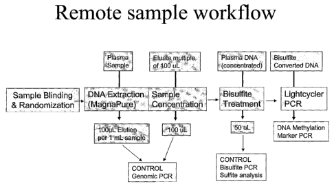
Figure 1 shows, an overview over one embodiment of the
invention.
Figure 2 shows an overview of an exemplary pooling and
concentrating strategy.
DETAILED DESCRIPTION
For achieving various technical objects, the disclosure provides
compositions and methods for providing DNA fragments derived from a
remote sample. Said compositions and methods comprise providing a
remote sample comprising DNA, isolating DNA from the remote sample,
and treating the isolated DNA with a reagent or enzyme which allow
differentiation of methylated and unmethylated cytosine.
Particular aspects provide methods to find amongst an enormous
plurality of known methods for remote sample providing, DNA isolation
and treatments which allowing a differentiation between methylated
and unmethylated DNA those methods, which in principle can be used to
solve the technical object. Particular aspects provide further
methods to find amongst an enormous plurality of known methods for
methylation analysis, marker identification and use of identified
markers those methods, which in principle can be used to solve the
technical object of the invention. Particular aspects provide
suitable combinations and adjustments of these methods with each
other in a manner that actually meets the technical object(s).
Advantages
CA 02603815 2015-03-03
CA 2603815
The exemplary methods disclosed herein can provide one or more of the
following advantages:
- It is characterized by high yields of provided DNA. This is
achieved although remote samples are characterized in that they
comprise only low levels of DNA, especially low levels of DNA of
interest. On the other hand, in many cases the amount of a remote
sample is not limited. Therefore large amounts of remote samples are
preferably processed. In particular, a DNA isolation method was
selected amongst the enormous number of possible DNA isolation
methods which allow the use of large volumes of starting samples. The
use of even larger volumes could be achieved according to the
invention by dividing the remote sample into subsamples, performing
the DNA isolation in parallel, pooling the isolated DNA and
concentrating the DNA into a volume suitable for further processing.
The high yields of provided DNA are further determinated by
selection of a method for discrimination between methylated and
unmethylated cytosine which can allow for a complete and reliable
discrimination and simultaneously minimizes further DNA fragmentation
amongst the enormous number of discrimination methods.
In addition, the high yields of DNA after the DNA isolation step
and a bisulfite treatment step can even be further raised by applying
an optional step of whole genome amplification of bisulfite treated
DNA.
The exemplary method disclosed herein is further characterized
in that contaminations can be furthermost avoided. This is based in
embodiments of sample collection which efficiently remove components
of the sample taken from an individual which are not for interest.
For example, the removal of red blood cells from blood to provide a
plasma sample. Thereby it is of particular importance on one hand to
efficiently remove all red blood cells but minimizing damage to them.
Because this will lead to a release of red blood cell DNA, the reason
for the removal of the
-11-
CA 02603815 2015-03-03
CA 2603815
red blood cells. Furthermore, according to the disclosure, a DNA
isolation method was selected amongst the enormous number of possible
DNA isolation methods which excludes the possibility of sample cross
contaminations. Taken together, the provided DNA according to the
disclosure is free of DNA contaminations which might interfere with a
choosen method for detecting a desired methylation pattern.
The exemplary method is further characterized in that small DNA
fragments as well as long DNA fragments can be provided. First, this
is enabled by selection of a DNA isolation method which isolates DNA
fragments of at least 100 bp with high efficiency amongst a enormous
number of DNA isolation methods. Second, this is enabled by selecting
methods for DNA concentration, for bisulfite treatment, in particular
for purification and/or desulfonation of bisulfite treated DNA, and
for whole genome amplification amongst the enormous number of other
possible methods.
The exemplary method is further characterized in that the
provided DNA comprises small DNA fragments as well as long DNA
fragments as they are present in the starting remote sample. This can
be achieved i) by selecting a DNA isolation method which isolates
small DNA fragments at least as small as 100 bp as well as large DNA
fragments amongst the enormous number of possible DNA isolation
methods; ii) by selecting devices for DNA concentration and
purification of bisulfite treated DNA which retain small DNA
fragments as well as large DNA fragments amongst the enormous number
of possible devices; and iii) by efficiently amplifying small
bisulfite treated DNA fragments as well as large ones by the optional
step of whole genome amplification of bisulfite treated DNA.
The exemplary methods are further characterized in that the
provided DNA can be free of associated or linked proteins, peptides,
amino acids, RNA, nucleotides or bases as well as
-12-
CA 02603815 2015-03-03
1
CA 2603815
,
interfering chemical reagents. According to the invention, this is
based therein, that i) a DNA isolation method is selected which is
characterized by a efficient removal of associated or linked
proteins, peptides, amino acids, RNA, nucleotides or bases amongst
the enormous number of possible DNA isolation methods; ii) devices
for DNA concentration and purification of bisulfite treated DNA which
efficiently remove associated nucleotides or bases amongst the
enormous number of possible devices; and iii) a method for
discrimination between methylated and unmethylated cytosine is
selected which minimizes further DNA fragmentation amongst the
enormous number of discrimination methods. The removal of such
components is of particular importance because the may sterically
hinder the methylation analysis.
Taken together, the exemplary methods allow the use of remote
samples for methylation analysis. In particular, said use is
characterized in that it is reliable and reproducible. These are two
necessary requirements for a medical test.
But, of course, the exemplary methods can also be characterized
by other preferred criteria of a medical test. According to the
exemplary methods, large amount of samples can be processed. For
example, it is possible to carry out the exemplary methods in a plate
scale. Moreover the different steps can be automated and standardized
and therefore robotics can also be used. The different steps are
further characterized by a low handling effort. The execution in
plate scale, the suitability of the method for automatization and
standardization, and the low handling effort also lead to a reduction
in costs. In addition the costs are further reduced by the use of
devices and solutions which are already available at low expenses.
Another advantage of the exemplary methods disclosed herein can be
that every step can easily be performed because only standard
laboratory equipment is necessary for its execution. Because of its
simpleness, its suitability for automatization, its low
-13-
CA 02603815 2015-03-03
CA 2603815
handling effort as well as its easy handling, the exemplary methods
disclosed herein can have a high reliability and reproducibility.
Thus the exemplary method makes remote samples available for
methylation based medical test. In other words, it can enable a
methylation based medical test which is based on non-invasive means
or on invasive means which affects as less as possible the patient,
which are easy to perform, and which cause low costs.
The exemplary method makes also remote samples available for
methylation based discovery of markers. In particular, it can allow
for the identification of markers, characterized by a high
sensitivity, a high specificity, or both.
Method of aspects of the invention.
The method disclosed is a method for providing DNA fragments
derived from a remote sample. According to the disclosure, the method
comprises the following steps: providing a remote sample comprising
DNA, isolating DNA from the remote sample, and treating the isolated
DNA with a reagent or enzyme which allows differentiation of
methylated and unmethylated cytosine. In realizing these steps, DNA
fragments are provided from a remote sample.
In brief, in particular aspects, the method disclosed is a
method for providing DNA fragments derived from a remote sample,
comprising:
providing a remote sample comprising DNA,
isolating DNA from the remote sample, and
treating the isolated DNA with a reagent or enzyme which allows
differentiation of methylated and unmethylated cytosine.
In particular aspects, the method disclosed is a method which
comprises the collecting and preprocessing of a remote sample. The
remote sample is thereby characterized in that it comprises genomic
DNA. The method
-14-
CA 02603815 2015-03-03
CA 2603815
further comprises the extraction of DNA from the collected and
preprocessed remote sample. The extracted DNA can be subject to a
treatment which allows to differentiate if the DNA is methylated or
not at a certain position. Such a treatment can be any kind of
treatment. Preferably the treatment comprises the use of an enzyme or
reagent. Thereby the enzyme can be any kind of enzyme, but preferably
the enzyme is protein or RNA molecule. Said reagent can also be any
kind of reagent for example but not limited to it a chemical reagent,
a pharmaceutical reagent, a biological reagent or a medical reagent.
In an embodiment, the method disclosed is a method, wherein the
DNA of the remote sample is characterized in that less than about 5
%, less than about 3 %, less than about 1 %, or less than about 0.1 %
of the DNA is derived from a defined cell, group of cells, tissue or
organ. In a preferred embodiment, the DNA of the remote sample is
characterized in that less than about 1 % of the DNA is derived from
a defined cell, group of cells, tissue or organ.
According to an embodiment, the provided remote sample comprises
less than about 5 %, less than about 3 %, less than about 1 %, or
less than about 0.1 % DNA which originates from the same defined
cell, group of cells, tissue or organ. Preferably, less than about 1
% of the DNA is derived from the same defined cell, group of cells,
tissue or organ. Thereby said DNA is characterized in having the same
methylation pattern at a defined allele or genomic locus.
In an embodiment, the presence or absence of the said DNA can be
detected with more than about 99 % confidence interval, more than
about 95 % confidence interval, more than about 90 % confidence
interval, more than about 80 % confidence interval, more than about
70 % confidence interval, or more than about 60 % confidence
interval. Particularly preferred is a confidence interval of more
than about 95%.
-15-
CA 02603815 2015-03-03
CA 2603815
, .
According to an embodiment, the percentage of said DNA can be
determined within a confidence interval of more than about 99 %, more
than about 95 %, more than about 90 %, more than about 80 %, more
than about 70 %, or more than about 60 %. Preferably a confidence
interval of more than about 95 % is applied.
In an embodiment, the method disclosed is a method, wherein the
remote sample is characterized in that it comprises less than about
100 ng DNA in 1 ml, less than about 60 ng DNA in 1 ml or less than
about 10 ng DNA in 1 ml. In a preferred embodiment, the remote sample
comprises less than about 10 ng of DNA in 1 ml remote sample.
According to an embodiment, a remote sample is considered which
comprises less than about 1,000 ng, less than about 500 ng, less than
about 100 ng, less than about 80 ng, less than about 60 ng DNA, less
than bout 40 ng, less than about 20 ng, less than about 10 ng, less
than about 1 ng, or less than about 0.1 ng per milliliter remote
sample. Preferably, the DNA concentration of a remote sample is less
than 10 ng/ml.
In an embodiment the method disclosed is a method, wherein loss
of DNA can be minimized by at least one selected from the group
comprising: selection of a DNA isolation method characterized by high
yields of DNA, selection of a method for differentiation of
unmethylated and methylated cytosine characterized by high accuracy
and high reliability, high accuracy of pipetting, reuse of pipetting
device, reuse of device contacted with DNA.
According to an embodiment, it is very important that as much as
possible of the DNA of interest is provided for methylation analysis.
This importance is based thereon that the DNA of a remote sample
comprises only a small percentage of DNA which is relevant for the
underlying question. This has been already specified above. Therefore
preferably, it is possible to minimize the loss of DNA by at least
one of the following provisions: a) selection of a suitable DNA
extraction method; b) selection of a suitable method for
differentiation if a genomic
-16-
CA 02603815 2013-07-29
locus or allel is methylated or not; c) ensureing a high
accuracy of pipetting; d) reuse of pipetting devices; and e)
reuse of devices brought into contact with DNA of a remote
sample.
A suitable DNA extraction method is a method which enables
and ensures high yield of DNA. It is further characterized in
that it has the possibility of standardization, the possibility
of automatization, a high reliability, a high reproducibility,
and the exclusion of contamination with for example but not
limited to it DNA of other remote samples. Of course, a suitable
method should also fulfill as good as possible as much as
possible the above specified citeria for a medical test, for
processing of a remote sample and for methylation analysis.
Therefore in a particular preferred embodiment, the DNA is
extracted by means of at least one component of the MagNA Pure
Compact Nucleic .Acid Isolaton Kit (I) Large Volume (Roche
Diagnostics GmbH) or at least a thereto related device.
A suitable method for differentiation between a methylated
and a unmethylated genomic locus or allel is characterized in
that it allows or, ensures a high as possible rate. of
differentiation with high reliablility and high accuracy.
Preferably the differentiation is possible for nearly every
single site which is capable of being methylated. Furthermore a
suitable method should not lead to a fragmentation of DNA. Of
/5 course, a suitable method should also fulfill as good as
possible as much as possible of the above specified citeria for
a medical test, for processing of remote samples and for
methylation analysis. Therefore in a particular preferred
embodinient, the DNA is treated with bisulfite as essentially
carried out as described in W005/038051.
A'high accuracy of pipetting characterized in that only the
necessary amount of DNA is transferred to subsidiary steps which
enables to exploit the as much as possible of the remote sample
DNA. It further assures that the optimum amount of DNA and other
-17-
CA 02603815 2015-03-03
CA 2603815
reagents is used. This results in optimum reactions and therewith in
high quality DNA. Loss of DNA for example but not limited to it by
degradation is therewith minimized.
The reuse of pipetting devices is also advantageous. As known in
the art, DNA is binding to plastics surfaces as for example pipet
tips. But less DNA is bound to a surface which was already brought
into contact once with the said DNA. Of course the same holds true
for DNA containers for example but not limited to microtiter plates,
tubes, columns. However the reuse is also limited by the risk of
contaminations. Therefore, only devices brought into contact with DNA
of a remote sample are re-used for samples or DNA which were derived
from the same patient or from the same sample collected from a
patient. In another preferred embodiment, the use of devices brought
into contact with remote sample DNA is minimized.
In an embodiment the method disclosed is a method, wherein the
volume of the remote sample is at least about 1.5 ml, about 2 ml,
about 3 ml, about 4 ml, about 5 ml, about 6 ml, about 7 ml, about 8
ml, about 9 ml, about 10 ml, about 11 ml, about 12 ml, about 15 ml,
about 20 ml, about 25 ml, about 30 ml, about 40 ml, or about 50 ml.
In a preferred embodiment, the volume of the remote sample is at
least about 36 ml, about 38 ml, about 40 ml, about 42 ml, or about 45
ml. In a particularly preferred embodiment, the volume of the remote
sample is at least about 40 ml. In another preferred embodiment, the
volume of the remote sample is at least about 15 ml, about 18 ml,
about 20 ml, about 23 ml, or about 25 ml. In a particularly preferred
embodiment, the volume of the remote sample is at least about 20 ml.
In a further preferred embodiment, the volume of the remote sample is
at least about at least about 4 ml, about 5 ml, about 6 ml, about 7
ml, about 8 ml, or about 9 ml. In a particularly preferred
embodiment, the volume of the remote sample is at least about 6 ml or
about 8 ml.
According to an embodiment, a remote sample is taken or
collected from an individual. Said remote sample has a volume of
-18-
CA 02603815 2015-03-03
CA 2603815
at least about 1.5 ml, about 2 ml, about 3 ml, about 4 ml, about 5
ml, about 6 ml, about 7 ml, about 8 ml, about 9 ml, about 10 ml,
about 11 ml, about 12 ml, about 15 ml, about 20 ml, about 25 ml,
about 30 ml, about 40 ml, or about 50 ml. Preferably, the volume is
at least about 36 ml, about 38 ml, about 40 ml, about 42 ml, or about
45 ml. Most preferably, the volume is at least about 40 ml. Also
preferably, the volume is at least about 15 ml, about 18 ml, about 20
ml, about 23 ml, or about 25 ml, and most preferably the volume is at
least about 20 ml. Also preferably, the volume is at least about 4
ml, about 5 ml, about 6 ml, about 7 ml, about 8 ml, or about 9 ml.
Most preferably, the volume is at least about 6 ml or about 8 ml.
In an embodiment the method disclosed is a method, wherein the
remote sample is at least one selected from the group comprising:
blood sample, plasma sample, serum sample, body fluid sample, saliva
sample, urine sample, semen sample, sample of the fluid from the
pleural cavity, sample from the fluid from the peritoneal cavity,
sample of the cerebrospinal fluid, smear from a epithelial surface,
sputum sample, stool sample, ejaculate sample, tears sample, sweat
sample, lymph fluid sample, bronchial lavage sample, pleural effusion
sample, meningal fluid sample, glandular fluid sample, fine needle
aspirates sample, nipple aspirates fluid sample, spinal fluid sample,
conjunctival fluid sample, vaginal fluid sample, duodenal fluid
sample, prancreatic juice sample, or bile sample.
According to an embodiment, the remote sample can be any kind of
a sample. Preferably, the remote sample is a sample which is
characterized in that it comprises at least one component which is
mainly located distantly from the other components of the said
sample. For example blood is not a remote sample with regard to a red
blood cell, but it is a remote sample with regard to a DNA fragment
which is derived from a tumor located in the lung. According to a
preferred embodiment, a remote sample is a sample of blood, plasma,
serum, body fluid, saliva, urine, semen, fluid from the pleural
cavity, fluid from
-19-
CA 02603815 2015-03-03
CA 2603815
the peritoneal cavity, cerebrospinal fluid, smear from a epithelial
surface, sputum, stool, ejaculate, tears, sweat, lymph fluid,
bronchial lavage, pleural effusion, meningal fluid, glandular fluid,
fine needle aspirates, nipple aspirates fluid, spinal fluid,
conjunctival fluid, vaginal fluid, duodenal fluid, prancreatic juice,
or bile. A person skilled in the art probably knows of additional
remote samples. Of course, these samples may also be used according
to the method disclosed.
In an embodiment the method disclosed is a method, wherein the
remote sample is plasma and the providing of the remote sample
comprises one or more of the following:
obtaining at least about 5 ml, about 10 ml, about 15 ml, about
ml, about 25 ml, about 30 ml, about 35 ml, about 40 ml, about 45
ml, about 50 ml of blood from a individual;
15 adding EDTA (ethylene-diamine-tetra-acetic acid) to the blood
comprising gentle mixing;
adjusting the blood to a final concentration of about 2.2
pmo1/1, about 3.2 pmo1/1, about 3.7 pmo1/1, about 4.0 pmo1/1, about
4.5 pmo1/1, about 4.9 pmo1/1, about 5.4 pmo1/1, about 5.9 pmo1/1, or
20 about 6.9 }Imola, dipotassium EDTA (dipotassium ethylene-diamine-
tetra-acetic acid) comprising gently mixing;
centrifuging the blood-EDTA mixture at about 750 x g, about 1000
x g, about 1500 x g, or about 2000 x g for about 4 min, about 8 min,
about 10 min, about 12 min, or about 20 min at about 1 C, about 4
C, about 7 C, about 10 C, about 15 C, about 21 C, or about 27 C
;
transferring the plasma into a new container;
centrifuging the plasma at about 750 x g, about 1000 x g, about
1500 x g, or about 2000 x g for about 4 min, about 8 min, about 10
min, about 12 min, or about 20 min at about 1 C, about 4 C, about 7
or about 10 C ;
transferring the re-centrifuged plasma into a new container;
cooling a plasma comprising sample at about 0 C, about 2 C,
about 4 C, about 6 C, or about 10 C;
-20-
CA 02603815 2007-10-03
WO 2006/113770
PCT/US2006/014667
freezing, storing or transporting a plasma comprising
sample at least at about -10 C, about -20 C, about -50 C, about
-60 C, about -70 C, about -80 C, about -90 C, or about -196 C;
and
performing the providing of the remote sample from
obtaining blood from a individual to freezing the corresponding
re-centrifuged plasma within about 1, about 2, about 3, about 4,
about 5, about 6, or about 8 hours.
According to an embodiment, a remote sample is plasma. According
to an preferred embodiment the providing of plasma comprises one
or more of the following steps:
obtaining at least about 5 ml, about 10 ml, about 15 ml,
about 20 ml, about 25 ml, about 30 ml, about 35 ml, about 40 ml,
about 45 ml, about 50 ml of blood from a individual;
adding EDTA (ethylene-diamine-tetra-acetic acid) to the
blood comprising gentle mixing;
adjusting the blood to a final concentration of about 2.2
pmo1/1, about 3.2 pmo1/1, about 3.7 pmo1/1, about 4.0 pmo1/1,
about 4.5 pmo1/1, about 4.9 1=01/1, about 5.4 pmo1/1, about 5.9
pmo1/1, or about 6.9 umo1/1, dipotassium EDTA (dipotassium
ethylene-diamine-tetra-acetic acid) comprising gently mixing by
immediately inversion for at least about 2 times, about 4 times,
about 6 times, about 8 times, about 10 times, about 12 times,
about 14 times, or about 18 times;
centrifuging the blood-EDTA mixture at about 750 x g, about
1000 x g, about 1500 x g, or about 2000 x g for about 4 min,
about 8 min, about 10 min, about 12 min, or about 20 min at
about 1 C, about 4 C, about 7 C, about 10 C, about 15 C,
about 21 C, or about 27 C ;
transferring the cleared upper phase into a new container,
therein the centrifuged container is held upright and the pipet
is tilt to touch the edge of the centrifuged container and the
surface of the cleared upper phase, transferring only so much of
the cleared upper phase until its surface is more than about 20
-21-
=
CA 02603815 2015-03-03
CA 2603815
mm, about 10 mm, about 7 mm, about 5 mm, or about 4 mm distant from
the surface of the next layer the buffy coat layer;
centrifuging the plasma sample at about 750 x g, about 1000 x g,
about 1500 x g, or about 2000 x g for about 4 min, about 8 min, about
min, about 12 min, or about 20 min at about 1 C, about 4 C,
about 7 C, or about 10 C ;
transferring the re-centrifuged plasma sample into a new
container, therein more than the about 20 ml, about 12 ml, about 8
10 ml, about 5 ml, or about 4 ml of the lowest re-centrifuged plasma
sample remain in the centrifugation container;
cooling a blood sample, plasma sample or intermediate sample at
about 0 C, about 2 C, about 4 C, about 6 C, or about 10 C;
freezing, storing or transporting a plasma sample or an
intermediate sample at least at about -10 C, about -20 C, about -
50 C, about -60 C, about -70 C, about -80 C, about -90 C, or about -
196 C; and
performing the providing of the remote sample starting from
obtaining blood from a individual and ending at freezing the
corresponding re-centrifuged plasma sample within about 1, about 2,
about 3, about 4, about 5, about 6, or about 8 hours.
In a preferred embodiment, the method disclosed is a method, wherein
the remote sample is a plasma sample and the providing of the remote
sample comprises one or more of the following:
obtaining at least about 35 ml, about 40 ml, about 45 ml, or
about 50 ml of blood from a individual;
adjusting the blood to a final concentration of about 3.7
pmo1/1, about 4.0 pmo1/1, about 4.5 pmo1/1, about 4.9 pmo1/1, or
about 5.4 pmo1/1 dipotassium EDTA (dipotassium ethylene-diamine-
tetra-acetic acid) comprising gently mixing;
centrifuging the blood-EDTA mixture at about about 1500 x g for
about 10 min at about 4 C, preferably no brakes are used for
stopping the centrifuge;
-22-
CA 02603815 2007-10-03
WO 2006/113770
PCT/US2006/014667
transferring the plasma into a new container, ;
centrifuging the plasma at about 1500 x g for about 10 min
at about 4 C, preferably no brakes are used for stopping the
centrifuge;
transferring the re-centrifuged plasma into a new
container;
cooling a plasma comprising sample at about 0 C, about 2 C,
or about 4 C;
freezing, storing or transporting a plasma comprising
sample at least at about -70 C, about -80 C, or about -90 C; and
performing the providing of the remote sample from
obtaining blood from a individual to freezing the corresponding
re-centrifuged plasma within about 4 hours.
According to a preferred embodiment, the remote sample is a
plasma sample and the providing of the plasma sample comprises
one or more of the following steps:
obtaining at least about 35 ml, about 40 ml, about 45 ml,
or about 50 ml of blood from a individual;
adjusting the blood to a final concentration of about 3.7
pmo1/1, about 4.0 pmo1/1, about 4.5 pmo1/1, about 4.9 pmo1/1, or
about 5.4 pmo1/1 dipotassium EDTA (dipotassium ethylene-diamine-
tetra-acetic acid) comprising gently mixing by immediately
inversion for about 10 times;
centrifuging the blood-EDTA mixture at about about 1500 x g
for about 10 min at about 4 C, preferably no brakes are used
for stopping the centrifuge;
transferring the cleared upper phase into a new container,
therein the centrifuged container is held upright and the pipet
is tilt to touch the edge of the centrifuged container and the
surface of the cleared upper phase, transferring only so much of
the cleared upper phase until its surface is more than about 5
mm distant from the surface of the next layer the buffy coat
layer ;
- 23 -
CA 02603815 2015-03-03
CA 2603815
centrifuging the plasma sample at about 1500 x g for about 10
min at about 4 C, preferably no brakes are used for stopping the
centrifuge;
transferring the re-centrifuged plasma sample into a new
container, therein more than the about 5 ml of the lowest re-
centrifuged plasma sample remain in the centrifugation container;
cooling a blood sample, plasma sample or intermediate sample at
about 0 C, about 2 C, or about 4 C;
freezing, storing or transporting a plasma sample or
intermediate sample at least at about -70 C, about -80 C, or about -
90 C; and
performing the providing of the remote sample starting from
obtaining blood from a individual ending at freezing the
corresponding re-centrifuged plasma sample within about 4 hours.
In an embodiment the method disclosed is a method, wherein the remote
sample is urine and the providing of the remote sample comprises one
or more of the following:
performing prostatic palpation, prostatic massage, or both from
the middle of the prostate to the left side of the prostate, to the
right side of the prostate or both for about 10 s, about 30 s, about
50 s, about 60 s, about 75 s, or about 120 s;
collecting the first about 5 ml, about 10 ml about 15 ml, about
20 ml, about 25 ml, about 30 ml, about 40 ml of voided urine;
adding EDTA to the urine;
adjusting the urine to a final concentration of about 3 mmo1/1,
about 6 mmo1/1, about 7 mmo1/1, about 8 mmo1/1, about 9 mmo1/1, about
9.80 mmo1/1, about 10 mmo1/1, about 11 mmo1/1, about 12 mmo1/1, about
13 mmo1/1, about 14 mmo1/1, about 18 mmo1/1, or about 25 mmo1/1 EDTA
(ethylene-diamine-tetra-acetic acid) with a pH of about 5.0, about
6.0, about 7.0, about 7.5, about 8.0, about 8.5, about 9.0, about 10;
-24-
CA 02603815 2007-10-03
WO 2006/113770
PCT/US2006/014667
cooling the urine comprising sample at about 0 C, about
2 C, about 4 C, about 6 C, or about 10 C;
freezing, storing or transporting the urine comprising '
sample at least at about -20 C, about -50 C, about -60 C, about
-70 C, about -80 C, about -90 C, or about -196 C; and
performing the providing of the urine sample from
collecting the first ml of voided urine to freezing the
corresponding urine-EDTA mixture within about 15, about 30,
about 45, about 60, about 75, about 90, or about 120 min.
According to an embodiment, the remote sample is urine.
According to an embodiment, the providing of a urine sample
comprises at least one of the following steps:
performing prostatic palpation, prostatic massage, or both
from the middle of the prostate to the left side of the
prostate, to the right side of the prostate or both for about 10
s, about 30 s, about 50 s, about 60 s, about 75 s, or about 120
s;
collecting the first about 5 ml, about 10 ml about 15 ml,
about 20 ml, about 25 ml, about 30 ml, about 40 ml of voided
urine immediately after the prostatic palpation, the prostatic
massage, or both;
adding dipotassium EDTA to the urine immediately;
adjusting the urine to a final concentration of about 3
mmo1/1, about 6 mmo1/1, about 7 mmo1/1, about 8 mmo1/1, about 9
mmo1/1, about 9.80 mmo1/1, about 10 mmo1/1, about 11 mmo1/1,
about 12 mmo1/1, about 13 mmo1/1, about 14 mmo1/1, about 18
mmo1/1, or about 25 mmo1/1 EDTA (ethylene-di-amine-tetra-acetic
acid) with a pH of about 5.0, about 6.0, about 7.0, about 7.5,
about 8.0, about 8.5, about 9.0, about 10 comprising gently
mixing by inversion immediately after collection;
cooling the urine sample at about 0 C, about 2 C, about
4 C, about 6 C, or about 10 C;
-25-
CA 02603815 2015-03-03
CA2603815
freezing, storing or transporting the urine sample at least at
about -20 C, about -50 C, about -60 C, about -70 C, about -80 C,
about -90 C, or about -196 C; and
performing the providing of the urine sample from collecting the
first milliliter of voided urine to freezing the corresponding urine
sample within about 15, about 30, about 45, about 60, about 75, about
90, or about 120 min.
In a preferred embodiment, the method disclosed is a method, wherein
the providing of the urine remote sample comprises one or more of the
following:
performing prostatic palpation, prostatic massage, or both from
the middle of the prostate to the left side of the prostate, to the
right side of the prostate or both for about 60 s;
collecting the first about 20 ml of voided urine;
adjusting the urine to a final concentration of about 9 mmo1/1,
about 9.80 mmo1/1, about 10 mmo1/1, or about 11 mmo1/1, EDTA
(ethylene-diamine-tetra-acetic acid) with a pH of about 7.5, about
8.0, or about 8.5;
cooling the urine comprising sample at about 0 C, about 2 C, or
about 4 C;
freezing, storing or transporting the urine comprising sample at
least at about -70 C, about -80 C, or about -90 C; and
performing the providing of the urine sample from collecting the
first ml of voided urine to freezing the corresponding urine-EDTA
mixture within about 60 min.
According to a preferred embodiment, the remote sample is a urine
sample and the providing of the urine sample comprises at least one
of the following steps:
performing prostatic palpation, prostatic massage, or both from
the middle of the prostate to the left side of the prostate, to the
right side of the prostate or both for about 60 s;
-26-
CA 02603815 2007-10-03
WO 2006/113770
PCT/US2006/014667
collecting tne tirst about 20 ml of voided urine
immediately after the prostatic palpation, the prostatic
massage, or both;
adjusting the urine to a final concentration of about 9
mmo1/1, about 9.80 mmo1/1, about 10 mmo1/1, or about 11 mmo1/1,
EDTA (ethylene-diamine-tetra-acetic acid) with a pH of about
7.5, about 8.0, or about 8.5 immediately after collection
comprising gently mixing by inversion immediately after
collection;
cooling the urine sample at about 0 C, about 2 C, or about
4 C;
freezing, storing or transporting the urine sample at least
at about -70 C, about -80 C, or about -90 C; and
performing the providing of the urine sample from
collecting the first milliliter of voided urine to freezing the
corresponding urine sample within about 60 min.
In an embodiment, the providing of a remote sample comprises the
processing of a checklist, a standardized protocol, or both.
According to an embodiment, the providing of a remote sample
comprises the use of a checklist, of a protocol, or both.
According to an preferred embodiment, a checklist used for
providing a remote sample comprises a step by step description
of actions which are necessary, which have only to be performed,
or both. It may further comprise a note about a precaution.
According to an embodiment, a protocol used for providing a
remote sample comprises i) the providing and use of at least one
remote sample identification number, preferable a combination of
numbers and letters or preferable a computer-readable code like
a bar code, ii) the recordation of characteristic data about the
sample, iii) the recordation of characteristic blinded data of
the individual the sample is taken from, iv) or combinations
thereof.
- 27 -
CA 02603815 2015-03-03
CA 2603815
In an embodiment the method disclosed is a method, wherein the remote
sample is divided into different subsamples subsequent to providing
the remote sample.
According to an embodiment, a remote sample is split into different
subsamples. This is particularly done, in order to obtain high yields
of DNA from a sample collected from a patient. According to an
embodiment, the volume of a remote sample collected from a single
patient can be larger than the volume suitable for further
processing. Therefore the collected remote sample is split into
subsamples. These subsamples are then further considered as remote
samples. Preferably these remote samples are processed in parallel.
The splitting of remote samples is done in particular with regard to
the DNA extraction step.
In an embodiment, the method disclosed is a method, wherein the
remote sample or at least one component of the remote sample is
concentrated subsequent to providing the remote sample.
According to an embodiment, a remote sample is concentrated.
According to an embodiment, at least one component of a remote sample
is concentrated. Preferably this component is a DNA comprising
component. The concentration of a remote sample or at least one
component of it is particularly done, in order to obtain high yields
of DNA from a sample collected from a patient. According to an
embodiment, the volume of a remote sample collected from a single
patient can be larger than the volume suitable for further
processing. Therefore the collected remote sample or at least one
component of the remote sample is concentrated. In a preferred
embodiment, the High Pure Viral Nucleic Acid Kit or at least one
component of it is used i) for providing the remote sample, ii) for
isolating DNA, iii) for treating DNA with a reagent or enzyme
allowing the
-28-
CA 02603815 2015-03-03
CA 2603815
differentiation between methylated or non-methylated cytosine, iv)
or combinations thereof.
In a preferred embodiment, the method disclosed is a method, wherein
the concentration comprises ultrafiltration, volume reduction, or
both. In a preferred embodiment, the method disclosed is a method,
wherein the concentration comprises protein digestion. Said preferred
embodiments are either carried out independently of the DNA isolation
or as a substep of it.
According to an embodiment, the concentration of a remote sample or
at least one component of the remote sample comprises
ultrafiltration, volume reduction, or both. Preferably the
concentration comprises digestion of protein. Said embodiments are
either part of the providing of a remote sample or they are part of
the isolation of DNA. According to a preferred embodiment, the
concentration of a remote sample or at least one component of it
comprises at least one selected from the group comprising: protease,
serine protease, thiol protease, carboxy protease, metalloprotease,
proteinase K, ultrafiltration device, Microcon filter device for
example but not limited to it Y-30 Microcon column, filter device,
silica surface, silica membrane, magnetic particle, polystyrol
particle, polystyrol surface, positively charged surface, and
positively charged membrane, charged membrane, charged surface,
charged switch membrane, charged switched surface, column of the ZR
DNA Clean & Concentrator-5 Kit, column of the Wizard Genomic DNA
Purification Kit, column of the QIAamp DNA Micro Kit, a component of
the MagNA Pure Compact Nucleic Acid Isolation Kit (I) Large Volume, a
component of the QIAamp UltraSens Virus Kit, a component of the RTP
DNA/RNA Virus Supersense Kit, a component of the chemagic Viral
DNA/RNA Kit special, a component of the chemagic DNA Blood Kit
special, a component of the High Pure Viral Nucleic Acid Kit, a
component of the Puregene DNA
-29-
CA 02603815 2015-03-03
CA 2603815
Isolation Kit, a component of the NasterPureTM Complete DNA and RNA
Purification Kit, or a component of the NucliSens Isolation Kit,
ethanol precipitation, propanol precipitation, or vacuum
concentration amongst others by means of a centrifuge. A person
skilled in the art knows to select other suitable devices or kits in
considering the above specifications and named kits. The said devices
or kits are well known in the art, for a list of current
manufacturers please see below.
In an embodiment, the method disclosed is a method, wherein the
isolation of DNA comprises one or more of the following:
treating the remote sample with a protease,
treating the remote sample with at least one protein
degenerating reagent or solution,
bringing the DNA of the remote sample in contact with a DNA-
purifying device,
washing the DNA on the DNA-purifying device, and
recovering the DNA from the DNA-purifying device.
According to an embodiment, the remote sample is subjected to at
least one of the following steps: i) treating the remote sample with
a protease or a protein degrading reagent; ii) treating the remote
sample with at least protein degenerating reagent or solution;
purifying the DNA by bringing into contact with a DNA purifying
device; washing the DNA; and eluting the DNA from the DNA purifying
device.
According to an embodiment, the isolation of DNA from a remote sample
comprises the treatment with a protein degrading reagent. Such a
reagent can be any kind of reagent as known by those skilled in the
art. For example, but not limited to it, the protein degrading
reagent is cyanogen bromide.
According to an embodiment, the isolation of DNA from a remote sample
comprises the treatment with a protein degenerating
CA 02603815 2015-03-03
CA 2603815
. .
reagent. Such a reagent can be any kind of reagent as known by those
skilled in the art. For example, but not limited to it, the protein
degenerating reagent is a chaotropic salt like guanidine
hydrochloride or urea; or a detergent like sodium dodecyl sulphate
(SDS).
According to an embodiment, the isolation of DNA from a remote sample
comprises the washing of DNA, in particular if it is in contact with
the DNA purifying device. Suitable solutions and reagents are well
known in the art. For example, but not limited to it, the washing
solution can be any mixture of a short-chain alcohol with water like
70% ethanol in water.
According to an embodiment, the isolation of DNA from a remote sample
comprises the elution of DNA from a DNA purifying device. Such a
reagent can be any kind of reagent as known by those skilled in the
art. For example, but not limited to it, the eluting solution is
water or any elution buffer supplied with the DNA purifying device.
In an embodiment, the method disclosed is a method, comprising the
isolation of DNA by means of the treatment of the remote sample with
a protease, wherein the protease is at least one selected from the
group comprising: serine protease, thiol protease, carboxy protease,
metalloprotease, and proteinase K.
According to an embodiment, the extraction of DNA from a remote
sample comprises the use of at least one protease selected from the
group comprising: serine protease, thiol protease, carboxy protease,
metalloprotease, and proteinase K.
In an embodiment, the method disclosed is a method, comprising the
isolation of DNA by means of bringing the DNA of the remote sample
into contact with a DNA-purifying device, wherein the DNA purifying
device is at least one selected from
-31-
cp, 02603815 2007-10-03
WO 2006/113770
PCT/US2006/014667
the group comprising: ultrafiltration, Microcon filter device
for example but not limited to it Y-30 Microcon column, filter
device, silica surface, silica membrane, magnetic particle,
polystyrol particle, polystyrol surface, positively charged
surface, and positively charged membrane, charged membrane,
charged surface, charged switch membrane, charged switched
surface, column of the ZR DNA Clean & Concentrator-5 Kit, column
of the Wizard Genomic DNA Purification Kit, column of the QIAamp
DNA Micro Kit, a component of the MagNA Pure Compact Nucleic
Acid Isolation Kit (I) Large Volume, a component of the QIAamp
UltraSens Virus Kit, a component of the RTP DNA/RNA Virus
Supersense Kit, a component of the chemagic Viral DNA/RNA Kit
special, a component of the chemagic DNA Blood Kit special, a
component of the High Pure Viral Nucleic Acid Kit, a component
of the Puregene DNA Isolation Kit, a component of the
MasterPurem Complete DNA and RNA Purification Kit, or a
component of the NucliSensa Isolation Kit. A person skilled in
the art may also think of other possibilities like for example
but not limited to it ethanol precipitation or propanol
precipitation, vacuum concentration amongst others by means of a
centrifuge. A person skilled in the art knows to select other
suitable devices or kits in considering the above specifications
and named kits. The said devices or kits are well known in the
art. The current manufacturers are: Roche Diagnostics GmbH for
the MagNA Pure Compact Nucleic Acid Isolation Kit (I) Large
Volume or the High Pure Viral Nucleic Acid Kit; Quiagen, Inc.
for the QIAamp UltraSens Virus Kit, QIAamp DNA Micro Kit or for
the QIAamp DNA Blood Maxi Kit; Invitek Gesellschaft fur
Biotechnik & Biodesign mbH for the RTP DNA/RNA Virus Supersense
Kit; chemagen AG for the chemagic Viral DNA/RNA Kit special or
the chemagic DNA Blood Kit special; Gentra Systems, Inc. for the
Puregene DNA Isolation Kit; Epicentre Technologies for the
MasterPureTM Complete DNA and RNA Purification Kit, Millipore
Inc. for the Microcon filter device, Zymo Research Corporation
for the ZR DNA Clean & Concentrator-5 Kit, Promega U.S. for the
-32-
CA 02603815 2015-03-03
CA 2603815
Wizard Genomic DNA Purification Kit, and bioMerieux SA for the
NucliSens Isolation Kit. Of course, other devices or kits may be used
as long as they are based on these devices or kits equal if they are
available at the time the invention was made or in the future.
According to an embodiment, the DNA-purifying device which is used
DNA isolation or extraction is characterized by at least one criteria
selected from the group comprising ultrafiltration, Microcon filter
device for example but not limited to it Y-30 Microcon column, filter
device, silica surface, silica membrane, magnetic particle,
polystyrol particle, polystyrol surface, positively charged surface,
and positively charged membrane, charged membrane, charged surface,
charged switch membrane, charged switched surface, column of the ZR
DNA Clean & Concentrator-5 Kit, column of the Wizard Genomic DNA
Purification Kit, column of the QIAamp DNA Micro Kit, a component of
the MagNA Pure Compact Nucleic Acid Isolation Kit (I) Large Volume, a
component of the QIAamp UltraSens Virus Kit, a component of the RTP
DNA/RNA Virus Supersense Kit, a component of the chemagic Viral
DNA/RNA Kit special, a component of the chemagic DNA Blood Kit
special, a component of the High Pure Viral Nucleic Acid Kit, a
component of the Puregene DNA Isolation Kit, a component of the
MasterPureTM Complete DNA and RNA Purification Kit, a component of the
NucliSens Isolation Kit, ethanol precipitation, propanol
precipitation, or vacuum concentration amongst others by means of a
centrifuge. Of course, other suitable devices or kits may be used
insofar as their use is obvious for a person skilled in the art while
reading the above specifications and named kits.
In an embodiment, the method disclosed is a method, wherein the
isolation of DNA is carried out by use of at least one kit selected
from the group comprising: MagNA Pure Compact
-33-
cp, 02603815 2007-10-03
WO 2006/113770
PCT/US2006/014667
Nucleic Acid Isolation Kit (I) Large Volume, QIAamp UltraSens
Virus Kit, QIAamp DNA Blood Maxi Kit, RTP DNA/RNA Virus
Supersense Kit, chemagic Viral DNA/RNA Kit special, chemagic DNA
Blood Kit special, High Pure Viral Nucleic Acid Kit, Puregene
DNA Isolation Kit, MasterPurelm Complete DNA and RNA Purification
Kit, or NucliSens Isolation Kit. A person skilled in the art
knows to select other suitable kits in considering the above
named kits. The said kits are well known in the art. For the
name of the correspondent manufacturers, please refer above. Of
course, other kits may be used as long as they are based on
these kits equal if they are available at the time the invention
was made or in the future.
According to an embodiment, at least one of the following kits
is used for DNA extraction: MagNA Pure Compact Nucleic Acid
Isolation Kit (I) Large Volume, QIAamp UltraSens Virus Kit,
QIAamp DNA Blood Maxi Kit, RTP DNA/RNA Virus Supersense Kit,
chemagic Viral DNA/RNA Kit special, chemagic DNA Blood Kit
special, High Pure Viral Nucleic Acid Kit, Puregene DNA
Isolation Kit, MasterPureTM Complete DNA and RNA Purification
Kit, or NucliSens Isolation Kit. A person skilled in the art
might think of other kits while reading the above named kits. Of
course those might also be used according to the invention. This
includes in particular also kits which are based on the same
technology as the above specified kits, but have different or
similar name or might be produced by a different manufacturer.
The use of said kits for isolating DNA is preferred because each
of them fulfills the following criteria: i) high yields of DNA;
.30 ii) avoidance of cross-contaminations; iii) high degree of
standardization; iv) high degree of automatization; v) low
handling effort; vi) low cost; vii) ease of handling; viii)
small fragments as well as large fragments are purified as
present in the sample; ix) high reproducibility; x) high
- 34 -
CA 02603815 2015-03-03
CA 2603815
reliability; xi) efficient removal of proteins, peptides, amino
acids, RNA, nucleotide or bases.
According to a preferred embodiment, the MagNA Pure Compact Nucleic
Acid Isolation Kit (I) Large Volume or the QIAamp UltraSens Virus Kit
are used for DNA isolation because they may be suitable to fulfill
the above specified criterion.
According to an particular preferred embodiment, the MagNA Pure
Compact Nucleic Acid Isolation Kit (I) Large Volume is used for DNA
isolation because it can have high reproducibility and a high
reliability.
In an embodiment, the method disclosed is a method, wherein isolated
DNA derived from different samples is pooled, concentrated or pooled
and concentrated.
According to an embodiment, the extracted DNA derived from the same
individual is pooled. According to an embodiment, the extracted DNA
derived from the same individual is enriched.
According to an embodiment, the extracted DNA derived from the same
individual is pooled and enriched simultaneously.
In an embodiment, the method disclosed is a method, wherein the
isolated DNA is concentrated and the concentration of isolated DNA
comprises at least one selected from the group comprising
ultrafiltration, Microcon filter device for example but not limited
to it Y-30 Microcon column, filter device, ethanol precipitation,
propanol precipitation, silica surface, silica membrane, magnetic
particle, polystyrol particle, positively charged surface, and
positively charged membrane, charged membrane, charged surface,
charged switch membrane, charged switched surface, vacuum
concentration, vacuum concentration by means of a centrifuge, column
of the ZR DNA Clean & Concentrator-5 Kit, column of the Wizard
Genomic DNA
-35-
ak 02603815 2007-10-03
WO 2006/113770
PCT/US2006/014667
Euritication Kit, column oi the QIAamp DNA Micro Kit, a
component of the MagNA Pure Compact Nucleic Acid Isolation Kit
(I) Large Volume, a component of the QIAamp UltraSens Virus Kit,
a component of the RTP DNA/RNA Virus Supersense Kit, a component
of the chemagic Viral DNA/RNA Kit special, a component of the
chemagic DNA Blood Kit special, a component of the High Pure
Viral Nucleic Acid Kit, a component of the Puregene DNA
Isolation Kit, a component of the MasterpureTM Complete DNA and
RNA Purification Kit, or a component of the NucliSens Isolation
Kit. A person skilled in the art knows to select other suitable
devices or kits in considering the, above specifications and
named kits. The said kits are well known in the art. Regarding
the current manufacturers please refer to the said above. Of
course, other devices or kits may be used as long as they are
based on said devices or kits equal if they are available at the
time the invention was made or in the future.
According to an embodiment, the enrichment of DNA is carried out
by means of at least one of the following or combinations
thereof: ultrafiltration, Microcon filter device for example but
not limited to it Y-30 Microcon column, filter device, ethanol
precipitation, propanol precipitation, silica surface, silica
membrane, magnetic particle, polystyrol particle, positively
charged surface, and positively charged membrane, charged
membrane, charged surface, charged switch membrane, charged
switched surface, vacuum concentration, vacuum concentration
by means of a centrifuge, column of the ZR DNA Clean &
Concentrator-5 Kit, column of the Wizard Genomic DNA
Purification Kit, column of the QIAamp DNA Micro Kit, a
component of the MagNA Pure Compact Nucleic Acid Isolation Kit
(I) Large Volume, a component of the QIAamp UltraSens Virus Kit,
a component of the RTP DNA/RNA Virus Supersense Kit, a component
of the chemagic Viral DNA/RNA Kit special, a component of the
chemagic DNA Blood Kit special, a component of the High Pure
Viral Nucleic Acid Kit, a component of the Puregene DNA
-36-
CA 02603815 2015-03-03
CA 2603815
Isolation Kit, a component of the MasterPureTM Complete DNA and RNA
Purification Kit, or a component of the NucliSens Isolation Kit.
The use of said devices is particularly preferred because they
fulfill best the following criteria for a medical test based on a
remote sample: i) high yields of DNA; ii) avoidance of cross-
contaminations; iii) high degree of standardization; iv) high degree
of automatization; v) low handling effort; vi) low cost; vii) ease of
handling; viii) small fragments as well as large fragments are
purified as present in the sample; ix) high reproducibility; x) high
reliability; xi) efficient removal of proteins, peptides, amino
acids, RNA, nucleotide or bases.
According to an particularly preferred embodiment, ultrafiltration
devices, in particular Microron filter devices are used for
enrichment or concentration because they have the highest yield of
DNA and allow a recovery of small fragments as well as large
fragments as present in the sample.
In an embodiment, the method disclosed is a method, wherein the
reagent which allows differentiation of methylated and unmethylated
cytosine is a reagent that converts unmethylated cytosine to uracil
and leaves methylated cytosine unchanged.
According to an embodiment, treatment which allows to differentiate
if DNA is methylated or not at a certain position is a treatment that
leads to a conversion of unmethylated cytosine to uracil while
methylated cytosines remain unchanged. Such a treatment can be any
kind of treatment. Preferably the treatment comprises the use of an
enzyme or reagent. Thereby the enzyme can be any kind of enzyme, but
preferably the enzyme is a protein or RNA molecule. Said reagent can
also be any kind of reagent for example but not limited to it a
chemical reagent, a
CA 02603815 2015-03-03
CA 2603815
pharmaceutical reagent, a biological reagent or a medical reagent.
In an embodiment, the method disclosed is a method comprising a
converting reagent as a differentiation allowing reagent, wherein the
reagent that converts unmethylated cytosine to uracil and leaves
methylated cytosine unchanged is a bisulfite reagent. A person
skilled in the art knows how to apply a bisulfite reagent. Suitable
kits for application of bisulfite reagent on DNA are available, for
example but not limited to it: EZ DNA Methylation-Gold Kit (Zymo
Research Corporation), Methylamp DNA Modification Kit (Epigentek
Inc.), MethylEasy DNA Bisulphite Modification Kit (Human Genetic
Signatures Pty Ltd).
According to an embodiment, the treatment that leads to a conversion
of unmethylated cytosine to uracil while methylated cytosines remain
unchanged comprises the use of a bisulfite reagent. A person skilled
in the art knows applicable methods or kits for bisulfite treatment.
For example, but not limited to it, the kits may be: EZ DNA
Methylation-Gold Kit (Zymo Research Corporation), Methylamp DNA
Modification Kit (Epigentek Inc.), MethylEasy DNA Bisulphite
Modification Kit (Human Genetic Signatures Pty Ltd).
According to a preferred embodiment, a bisulfite treatment is
essentially carried out as described in W005/038051. According to
this, in one embodiment DNA is reacted with a bisulfite reagent,
characterized in that said reaction is carried out in the presence of
a compound out of the group of dioxane, one of its derivatives and a
similar aliphatic cyclic ether.
-38-
CA 02603815 2007-10-03
WO 2006/113770
PCT/US2006/014667
In an embodiment DNA is reacted with a bisulfite reagent,
characterized in that said reaction is carried out in the
presence of a compound of the following formula:
R10 R2
CH 2- m 0
n = 1-35000
m = 1-3
R1 = H, Me, Et, Pr, Bu
R2 - H, Me, Et, Pr, Bu
Preferred are thus n-alkylene glycol compounds, particularly
their dialkyl ethers, and especially diethylene glycol dimethyl
ether (DME).
The bisulfite conversion may take place both in solution as well
as also on DNA bound to a solid phase. Preferably sodium
disulfite (= sodium bisulfite/sodium metabisulfite) is used,
since it is more soluble in water than sodium sulfite. The
disulfite salt disproportionates in aqueous solution to the
hydrogen sulfite anions necessary for the cytosine conversion.
When bisulfite concentration is discussed below, this refers to
the concentration of hydrogen sulfite and sulfite anions in the
reaction solution. For the method according to the invention,
concentration ranges of 0.1 to 6 mo1/1 are possible.
Particularly preferred is a concentration range of 1 to 6 mo1/1,
and most particularly preferred, 2-4 mo1/1. However, when
dioxane is used, the maximal concentration of bisulfite that can
be used is smaller (see below). In selecting the bisulfite
concentration, one must consider that a high concentration of
bisulfite leads to a high conversion, but also leads to a high
decomposition rate due to the lower pH.
-39-
CA 02603815 2013-07-29
Dicxane can be utilizea in different concentrations. Preferably,
the dioxane concentration amounts to 10 to 35% (vol/vol),
particularly preferred is 20 to 30%, and most particularly
preferred is 22 to 28% , especially 25%. A dioxane concentration
higher than 35% is problematic, since this results in a
formation of two phases within the reaction solution. In the
particularly preferred embodiments with a dioxane concentration
of 22-28%, the final preferred bisulfite concentration amounts
to 3.3 to 3.6 mo1/1, and in the most particularly preferred
embodiment with a dioxane concentration of 25%, it amounts to
3.5 mo1/1 (see Examples).
The n-alkylene glycol compounds according to the invention can
be utilized in a different concentration range. DME is
preferably used in concentrations between 1-35% (vol/vol). There
is preferably between 5 and 25%, and most preferably 10% DME.
The preferred scavengers utilized according to the invention are
chromane derivatives, e.g., 6-hydroxy-2,5,7,8,- '
tetramethylchromane 2-carboxylic acid (also known as: Trolox-
CI"). Further scavengers are listed in the patent application WO
01/98528 (= DE 100 29 915; = US application 10/311,661.
The bisulfite conversion can be conducted in a wide temperature
range from 0 to 95 C. However, as at higher temperatures the
rates of both the conversion and decomposition of the DNA
increase, in a preferred embodiment the reaction temperature
lies between 0-80 C, preferably between 30-80 C. Particularly
preferred is a range between 50-70 C; most particularly
preferred between 57-65 C. The optimal reaction time of the
bisulfite treatment depends on the reaction temperature. The
reaction time normally amounts to between 1 and 18 hours (see:
Grunau et al. 2001, Nucleic Acids Res. 2001, 29(13):E65-5.
-40-
CA 02603815 2015-03-03
CA 2603815
The reaction time is ordinarily 4-6 hours for a reaction temperature
of 60 C.
In a particularly preferred embodiment, the bisulfite conversion is
conducted at mild reaction temperatures, wherein the reaction
temperature is then clearly increased for a short time at least once
during the course of the conversion. In this way, the effectiveness
of the bisulfite conversion can be surprisingly clearly be increased.
The temperature increases of short duration are named "thermospikes"
below. The "standard" reaction temperature outside the thermospikes
is denoted as the basic reaction temperature. The basic reaction
temperature amounts to between 0 and 80 C, preferably between 30-
80 C, more preferably between 50-70 C, most preferably between 57-
65 C , as described above.
The reaction temperature during a thermospike is increased to over 85
C by at least one thermospike. The optimal number of thermospikes is
a function of the basic reaction temperature. The higher the optimal
number of thermospikes is, the lower is the basic reaction
temperature. At least one thermospike is necessary in each case. And,
on the other hand, in principle, any number of thermospikes is
conceivable. Of course, it must be considered that with a large
number of temperature increases, the decomposition rate of the DNA
also increases, and an optimal conversion is no longer assured. The
preferred number of thermospikes is thus between 1 and 10
thermospikes each time, depending on the basic reaction temperature.
A number of two to 5 thermospikes is thus particularly preferred. The
thermospikes increase the reaction temperature preferably to 85 to
100 C, particularly preferably to 90-100 C, and most preferably to
94 C-100 C.
The duration in time of the thermospikes also depends on the volume
of the reaction batch. It must be assured that the
-41-
CA 02603815 2015-03-03
CA 2603815
temperature is increased uniformly throughout the total reaction
solution. For a 20 pl reaction batch when using a thermocycler a
duration between 15 seconds and 1.5 minutes, especially a duration
between 20 and 50 seconds is preferred. In a particular preferred
embodiment the duration is 30 seconds. Operating on a volume of 100
pl the preferred range lies between 30 seconds and 5 minutes,
especially between 1 and 3 minutes. Particularly preferred are 1.5-3
minutes. For a volume of 600 pl, a duration of 1 to 6 minutes, is
preferred, especially between 2 and 4 minutes. Particularly preferred
is a duration of 3 minutes. A person skilled in the art will easily
be able to determine suitable durations of thermospikes in relation
to a variety of reaction volumes. The above-described use of
thermospikes leads to a significantly better conversion rates in the
bisulfite conversion reaction, even when the above-described
denaturing solvents are not utilized.
According to an embodiment, the said treatment of DNA with bisulfite
is particularly preferred because it can have several important
advantages in comparison to other known methods or kits of the state
of the art. These advantages are: i) higher yield of converted DNA;
ii) a nearly complete conversion of unmethylated cytosine while
methylated cytosine remain unchanged; and iii) almost no further
fragmentation of DNA. These advantages are based in milder reaction
conditions because of i) a thermal denaturation of DNA; ii) a
comparably lower bisulfite concentration; iii) a slightly more
alkalic pH; and iv) the use of a more efficient and more effective
radical scavenger.
In a preferred embodiment, the method disclosed is a method, wherein
treating DNA with a bisulfite reagent comprises:
mixing of about 10 to about 250 pl of a solution comprising DNA
with about 45 to about 750 pl of bisulfite solution, the bisulfite
solution having a pH in the range of about 5.45 to
-42-
CA 02603815 2007-10-03
WO 2006/113770
PCT/US2006/014667
about 5.50 comprising about 4.83 to about 4.93 mo1/1
hydrogensulfite;
adding about 5 to about 500 pl of an organic radical scavenger
solution, the organic radical scavenger solution comprising an
organic solvent and about 10 to about 750 mmo1/1 of 6-hydroxy-
2,5,7,8-tetramethyl-chroman-2-carboxylic acid; and
applying a temperature protocol for about 2 to about 18 h,
wherein the reaction is conducted in a temperature range of
about 0 to about 80 C with about 2 to about 5 additional
temperature increases, in each case for about 0.5 to about 10
min, to a temperature of about 85 to about 100 C including an
initial temperature increase to a temperature of about 85 to
about 100 C.
According to a preferred embodiment, the treatment comprising
the use of a bisulfite reagent comprises further:
mixing of about 10 to about 250 pl of a solution comprising
DNA with about 45 to about 750 pl of bisulfite solution, the
bisulfite solution having a pH in the range of about 5.45 to
about 5.50 comprising about 4.83 to about 4.93 mo1/1
hydrogensulfite;
adding about 5 to about 500 p1 of an organic radical scavenger
solution, the organic radical scavenger solution comprising an
organic solvent and about 10 to about 750 mmo1/1 of 6-hydroxy-
2,5,7,8-tetramethyl-chroman-2-carboxylic acid; and
applying a temperature protocol for about 2 to about 18 h,
wherein the reaction is conducted in a temperature range of
about 0 to about 80 C with about 2 to about 5 additional
temperature increases, in each case for about 0.5 to about 10
min, to a temperature of about 85 to about 100 C including an
initial temperature increase to a temperature of about 85 to
about 100 C.
- 43 -
CA 02603815 2007-10-03
WO 2006/113770
PCT/US2006/014667
In a particular preterred embodiment, the method of the
invention is a method wherein treating DNA with a bisulfite
reagent comprises:
mixing about 50 to about 150 pl of solution comprising DNA
with about 177 to about 531 pl of the bisulfite solution;
adding about 73 to about 219 pl of dioxane solution, the
dioxane solution comprising about 157 mmo1/1 of 6-hydroxy-
2,5,7,8-tetramethyl-chroman-2-carboxylic acid dissolved in 1,4-
dioxane; and
applying a temperature protocol for about 3 to about 16 h,
wherein the reaction is conducted in a temperature range of
about 57 to about 65 C with about 2 to about 5 additional
temperature increases, in each case for about 3 to about 5 min,
to a temperature of about 94 to about 100 C including an initial
temperature increase to a temperature of about 94 to about
100 C.
According to a particular preferred embodiment, the bisulfite
treatment of DNA comprises:
mixing about 50 to about 150 pl of solution comprising DNA
with about 177 to about 531 pl of the bisulfite solution;
adding about 73 to about 219 pl of dioxane solution, the
dioxane solution comprising about 157 mmo1/1 of 6-hydroxy-
2,5,7,8-tetramethyl-chroman-2-carboxylic acid dissolved in 1,4-
dioxane; and
applying a temperature protocol for about 3 to about 16 h,
wherein the reaction is conducted in a temperature range of
about 57 to about 65 C with about 2 to about 5 additional
temperature increases, in each case for about 3 to about 5 min,
to a temperature of about 94 to about 100 C including an initial
temperature increase to a temperature of about 94 to about
100 C.
-44-
CA 02603815 2015-03-03
CA 2603815
In a particular preferred embodiment, the method of the invention is
a method wherein treating DNA with a bisulfite reagent comprises:
mixing of about 50 to about 150 pl of a solution containing the DNA
with about 95 to about 285 pl of the bisulfite solution;
adding about 15 to about 45 pl of DME solution, the DME solution
comprising about 500 mmo1/1 of 6-hydroxy-2,5,7,8-tetramethyl-chroman-
2-carboxylic acid dissolved in diethyleneglycoldimethylether; and
applying a temperature protocol for about 3 to about 16 h, wherein
the reaction is conducted in a temperature range of about 57 to about
65 C with about 2 to about 5 additional temperature increases, in
each case for about 3 to about 5 min, to a temperature of about 94 to
about 100 C including an initial temperature increase to a
temperature of about 94 to about 100 C.
According to a particular preferred embodiment, the bisulfite
treatment of DNA comprises:
mixing of about 50 to about 150 pl of a solution containing the DNA
with about 95 to about 285 pl of the bisulfite solution;
adding about 15 to about 45 pl of DME solution, the DME solution
comprising about 500 mmo1/1 of 6-hydroxy-2,5,7,8-tetramethyl-chroman-
2-carboxylic acid dissolved in diethyleneglycoldimethylether; and
applying a temperature protocol for about 3 to about 16 h, wherein
the reaction is conducted in a temperature range of about 57 to about
65 C with about 2 to about 5 additional temperature increases, in
each case for about 3 to about 5 min, to a temperature of about 94 to
about 100 C including an initial temperature increase to a
temperature of about 94 to about 100 C.
According to an embodiment, the method disclosed is a method,
wherein bisulfite treated DNA is subjected directly to
CA 02603815 2013-07-29
me-mods ror metnylation analysis. This is especially preferred
in view of the avoidance of cross-contaminations in PCR based
methods. This embodiment is basically carried out as described
in US 11/248,721.
According to this, decontaminated DNA are
provided which are suitable for DNA methylation analysis. This
embodiment is characterized in that DNA is incubated with a
bisulfite reagent comprising solution as described above. This
leads to a sulfonation, a deamination, or both of unmethylated
cytosine. Deamination is a spontaneous process in an aqueous
solution and leads to sulfonated uracil comprising DNA. No
desulfonation occurs yet.
In a separate step, the DNA comprising sulfonated uracil is
brought into contact and incubated with an enzyme which
specifically degrades non-sulfonated uracil containing nucleic
acids. Such an enzyme is for example Uracil-DNA-Glycosylase
(UNG).
In a preferred embodiment for providing a decontaminated
template DNA for polymerase based amplification reactions, the
sulfonated and/or deaminated template DNA are mixed with an UNG
activity and components required for a polymerase mediated
amplification reaction or an amplification based detection
assay. After degradation of non-sulfonated uracil containing
nucleic acids by use of UNG, the UNG activity is terminated and
=
the template DNA is desulfonated by increased temperature.
Subsequently the template DNA is ready to be amplified.
In a preferred embodiment, degradation, termination,
desulfonation and amplification occur in a single tube during a
polymerase based amplification reaction and/or an amplification
based assay. Preferably such an amplification is performed in
the presence of dUTP instead of dTTP.
-46-
CA 02603815 2015-03-03
CA 2603815
In a preferred embodiment, sulfonated and partially or completely
deaminated DNA after bisulfite treatment is subjected directly to a
polymerase based amplification reaction and/or an amplification based
assay without any prior desulfonation. The desulfonation occurs
during the initial temperature increase of the amplification
reaction.
These particular embodiments can have the advantage in comparison to
known methods of bisulfite treatment that the purification step after
bisulfite treatment becomes dispensable. This is a simplification
which results in reduction of costs and handling effort, minimizes
loss of bisulfite treated DNA and is also time saving.
In an embodiment, the method disclosed is a method, wherein treating
DNA with a reagent or enzyme allowing differentiation of the
methylation status comprises purifying the treated DNA.
According to an embodiment, the treatment that leads to a conversion
of unmethylated cytosine to uracil while methylated cytosines remain
unchanged comprises the purification of the bisulfite treated DNA.
According to an embodiment, such a purification comprises a
desulfonation of the bisulfite treated DNA by bringing the said into
contact with an alkaline reagent or solution.
In a preferred embodiment, the method disclosed is a method, wherein
purifying the treated DNA comprises the use of at least one selected
from the group comprising: ultrafiltration, Microcon filter device,
filter device, ethanol, propanol, silica surface, silica membrane,
magnetic particle, polystyrol particle, positively charged surface,
and positively charged membrane, charged membrane, charged surface,
charged switch membrane, charged switched surface, column of the ZR
DNA
ak 02603815 2007-10-03
WO 2006/113770
PCT/US2006/014667
Clean & Concentrator-5 Kit, column of the Wizard Genomic DNA
Purification Kit, column of the QIAamp DNA Micro Kit, a
component of the MagNA Pure Compact Nucleic Acid Isolation Kit
(I) Large Volume, a component of the QIAamp UltraSens Virus Kit,
a component of the RTP DNA/RNA Virus Supersense Kit, a component
of the chemagic Viral DNA/RNA Kit special, a component of the
chemagic DNA Blood Kit special, a component of the High Pure
Viral Nucleic Acid Kit, a component of the Puregene DNA
Isolation Kit, a component of the MasterPure' Complete DNA and
RNA Purification Kit, or a component of the NucliSens Isolation
Kit. A person skilled in the art knows to select other suitable
devices or kits in considering the above specifications and
named kits. The said kits are well known in the art. Regarding
the current manufacturers please refer to the said above. Of
course, other devices or kits may be used as long as they are
based on said devices or kits equal if they are available at the
time the invention was made or in the future.
According to an embodiment, the purification of bisulfite
treated DNA comprises the use of at least one of the following
or combinations thereof: ultrafiltration, Microcon filter
device, filter device, ethanol, propanol, silica surface, silica
membrane, magnetic particle, polystyrol particle, positively
charged surface, and positively charged membrane, charged
membrane, charged surface, charged switch membrane, charged
switched surface, column of the ZR DNA Clean & Concentrator-5
Kit, column of the Wizard Genomic DNA Purification Kit, column
of the QIAamp DNA Micro Kit, a component of the MagNA Pure
Compact Nucleic Acid Isolation Kit (I) Large Volume, a component
of the QIAamp UltraSens Virus Kit, a component of the RTP
DNA/RNA Virus Supersense Kit, a component of the chemagic Viral
DNA/RNA Kit special, a component of the chemagic DNA Blood Kit
special, a component of the High Pure Viral Nucleic Acid Kit, a
component of the Puregene DNA Isolation Kit, a component of the
-48-
CA 02603815 2015-03-03
CA 2603815
MasterPureTM Complete DNA and RNA Purification Kit, or a component of
the NucliSens Isolation Kit.
According to a preferred embodiment, ultrafiltration devices, in
particular Microron filter devices are used for purification,
desulfonation, or purification and desulfonation of bisulfite treated
DNA because they have the highest yield of bisulfite treated DNA and
allow a recovery of small bisulfite treated fragments as well as
large bisulfite treated fragments as present in the sample after
bisulfite treatment.
In a particular preferred embodiment, the method disclosed is a
method, wherein purifying the treated DNA comprises:
adding of about 50 to about 1000 pl of water to the sample after
the bisulfite reaction;
applying the mixture onto a Microcon filter device subsequently
centrifuging at about 10,000 to about 18,000 x g for about 10 to
about 30 min;
washing with about 100 to about 800 pl of about 0.2 mo1/1 sodium
hydroxide, and subsequent centrifuging at about 10,000 to about
18,000 x g for about 6 to about 25 min;
applying of about 100 to about 800 pl of about 0.1 mo1/1 sodium
hydroxide, and subsequent centrifuging at about 10,000 to about
18,000 x g for about 6 to about 25 min;
applying, in 1 to about 8 repetitions, the following: applying of
about 100 to about 400 pl water or TE buffer and subsequent
centrifuging at about 10,000 to about 18,000 x g for about 6 to about
25 min; and
eluting by application of about 25 to about 200 pl TE buffer
preheated to about 15 to about 65 C, incubation for about 1 to about
30 min at a temperature of about 15 to about 65 C, and subsequent
inversion of the Microcon filter device and centrifugation at about
500 to about 5,000 x g for about 0.5 to about 30 min.
CA 02603815 2015-03-03
CA 2603815
According to a preferred embodiment, the purification and
desulfonation of bisulfite treated DNA comprises:
adding of about 50 to about 1000 pl of water to the sample after
the bisulfite reaction;
applying the mixture onto a Microcon filter device subsequently
centrifuging at about 10,000 to about 18,000 x g for about 10 to
about 30 min;
washing with about 100 to about 800 pl of about 0.2 mo1/1 sodium
hydroxide, and subsequent centrifuging at about 10,000 to about
18,000 x g for about 6 to about 25 min;
applying of about 100 to about 800 pl of about 0.1 mo1/1 sodium
hydroxide, and subsequent centrifuging at about 10,000 to about
18,000 x g for about 6 to about 25 min;
applying, in 1 to about 8 repetitions, the following: applying of
about 100 to about 400 pl water or TE buffer and subsequent
centrifuging at about 10,000 to about 18,000 x g for about 6 to about
min; and
eluting by application of about 25 to about 200 pl TE buffer
preheated to about 15 to about 65 C, incubation for about 1 to about
20 30 min at a temperature of about 15 to about 65 C, and subsequent
inversion of the Microcon filter device and centrifugation at about
500 to about 5,000 x g for about 0.5 to about 30 min.
In a particular preferred embodiment, the method disclosed is a
25 method, wherein purifying the treated DNA comprises:
a) adding of 200 pl water to the sample after the bisulfite
reaction,
b) applying the mixture onto a Microcon filter device subsequently
centrifuging at about 14,000 x g for about 20 min,
c) washing with about 400 pl of about 0.2 mo1/1 sodium hydroxide,
and subsequent centrifuging at about 14,000 x g for about 10 to about
14 min,
-50-
CA 02603815 2007-10-03
WO 2006/113770
PCT/US2006/014667
d) applying of about 400 pl of about 0.1 mo1/1 sodium
hydroxide, and subsequent centrifuging at about 14,000 x g for
about 10 to about 14 min,
e) applying, in 1 to about 4 repetitions, the following:
applying of about 400 pl water or TE buffer and subsequent
centrifuging at about 14,000 x g for about 12 min; and
f) eluting by application of about 45 to about 70 pl TE buffer
preheated to about 50 C, incubation for about 10 min at a
temperature of about 50 C, and subsequent inversion of the
Microcon filter device and centrifugation at about 1,000 x g for
about 7 min.
According to a particular preferred embodiment, the purification
and desulfonation of bisulfite treated DNA comprises:
a) adding of 200 pl water to the sample after the bisulfite
reaction,
b) applying the mixture onto a Microcon filter device
subsequently centrifuging at about 14,000 x g for about 20 min,
c) washing with about 400 pl of about 0.2 mo1/1 sodium
hydrOxide, and subsequent centrifuging at about 14,000 x g for
about 10 to about 14 min,
d) applying of about 400 pl of about 0.1 mo1/1 sodium
hydroxide, and subsequent centrifuging at about 14,000 x g for
about 10 to about 14 min,
e) applying, in 1 to about 4 repetitions, the following:
applying of about 400 pl water or TE buffer and subsequent
centrifuging at about 14,000 x g for about 12 min; and
f) eluting by application of about 45 to about 70 pl TE buffer
preheated to about 50 C, incubation for about 10 min at a
temperature of about 50 C, and subsequent inversion of the
Microcon filter device and centrifugation at about 1,000 x g for
about 7 min.
-51-
CA 02603815 2015-03-03
CA 2603815
In a particular preferred embodiment, the method disclosed is a
method, wherein purifying the treated DNA further comprises at least
one of the following:
in step b, applying the mixture in portions onto the Microcon
filter device in step b,
subsequent to step b, applying of about 400 pl TE buffer, the TE
buffer pH 8 containing about 10 mmo1/1 tris-hydroxymethyl-amino-
methan and about 0.1 mmo1/1 EDTA, subsequent centrifuging at about
14,000 x g for about 12 min,
in step c, incubating the about 0.2 mo1/1 sodium hydroxide for
about 10 min at room temperature,
in step d, incubating the about 0.1 mo1/1 sodium hydroxide for
about 10 min at room temperature,
According to a particular preferred embodiment, the purification and
desulfonation of bisulfite treated DNA comprises further in addition
to the specified above at least one of the following:
in step b, applying the mixture in portions onto the Microcon
filter device in step b,
subsequent to step b, applying of about 400 pl TE buffer, the TE
buffer pH 8 containing about 10 mmo1/1 tris-hydroxymethyl-amino-
methan and about 0.1 mmo1/1 EDTA, subsequent centrifuging at about
14,000 x g for about 12 min,
in step c, incubating the about 0.2 mo1/1 sodium hydroxide for
about 10 min at room temperature,
in step d, incubating the about 0.1 mo1/1 sodium hydroxide for
about 10 min at room temperature,
According to a preferred embodiment, bisulfite treated DNA or
bisulfite treated and purified DNA is subjected to a whole genome
amplification prior to any further analysis.
In a preferred embodiment, the method disclosed is a method for
amplification of at least one nucleic acid, comprising:
-52-
CA 02603815 2007-10-03
WO 2006/113770
PCT/US2006/014667
providing a nucleic acid sample comprising at least one
nucleic acid molecule,
treating at least one nucleic acid molecule derived from
said sample with an enzyme or reagent which differentiates
between methylated bases within said nucleic acid molecule and
unmethylated bases within said nucleic acid molecule,
extending at least one strand of at least one nucleic acid
molecule derived from said sample by at least one nucleotide or
PNA-monomer, and
amplifying the at least one extended nucleic acid molecule.
Thereby the steps of treating at least one nucleic acid molecule
derived from said sample and the step of extending at least one
strand of at least one nucleic acid molecule derived from said
sample can be carried out in arbitrary order.
According to a preferred embodiment, at least one nucleic acid
is amplified. The amplification thereby comprises the following
steps which may be carried out in any arbitrary order: Providing
at least one nucleic acid molecule by providing a nucleic acid
sample. Extending at least one strand of at least one nucleic
acid molecule by at least one nucleotide or PNA-monomer.
Treating at least one nucleic acid molecule with an enzyme or
reagent which differentiates between methylated bases within
said nucleic acid molecule and unmethylated bases within said
nucleic acid molecule. Amplifying at least one nucleic acid
molecule. Thereby, preferably, the extended or the treated and
extended portions of at least one nucleic acid molecule are used
for amplification of said at least one nucleic acid molecules.
In a preferred embodiment, the extension is characterized in
that the at least one strand of at least one nucleic acid is
extended
by one or more single nucleotides or PNA-monomers,
by one ore more oligonucleotides or PNA-oligomers,
-53-
cp, 02603815 2007-10-03
WO 2006/113770
PCT/US2006/014667
by a second nucleic acid derived from the provided nucleic
acid sample, or
by combinations thereof.
According to a preferred embodiment, the said at least one
strand is elongated be either one or more single nucleotides or
PNA monomers, by one or more oligonucleotides or PNA-oligomers,
by a second nucleic acid preferably derived from the same
provided nucleic acid sample as specified above, or by
combinations thereof. The nucleotides, oligonucleotides or
second nucleic acid can be of any type of nucleotides or
nucleotide analog suitable for elongation and as known to those
skilled in the art. Preferably, but not limited to it the
nucleotides are deoxyribonucleotides, ribonucleotides, locked
ribonucleotides or PNA-monomers. Preferably, but not limited to
it, the oligonucleotides are oligodeoxyribonucleotides,
oligoribonucleotides, or PNA-oligomers, more preferably the PNA-
oligomers are arbitrary chimeric oligomers of nucleotides and
PNA-monomers, wherein at least one nucleotide is located at the
5' or the 3' end of the chimeric oligomer. The said second
nucleic acid can be any nucleic acid either comprised by the
provided sample or added during the method of the invention.
This second nucleic acid can be of known or unknown sequence. It
can be endogenous or artificial. Preferably, the second nucleic
acid is a bisulfite treated endogeneous nucleic acid provided
with the nucleic acid sample.
In a preferred embodiment, the extension is catalyzed template
independently.
According to a preferred embodiment, no template is used for
extension. This means that the extension occurs randomly or as
specified by the used one or more enzymes or further reaction
conditions (e.g. but not limited to it, by the provided
nucleotides).
- 54 -
CA 02603815 2007-10-03
WO 2006/113770
PCT/US2006/014667
In a preferred embodiment, the extension is catalyzed by means
of at least one enzyme selected from the group comprising: a
transferase, a transferase transferring phosphorus-containing
groups, a nucleotidyltransferase, a DNA
nucleotidylexotransferase, terminal deoxynucleotidyl transferase
(TdT), a enzyme with ribonucleotide transferase activity, a
polyribonucleotide nucleotidyltransferase, a tRNA
nucleotidyltransferase, RNA uridylyltransferase, a ligase, a
ligase forming phosphoric ester bonds, a DNA ligase, a ATP
dependent DNA ligase, a single stranded DNA ligase, a ATP
dependent single stranded DNA ligase catalyzing intramolecular
circularization, CircLigase ssDNA Ligase.
According to a preferred embodiment, the extension reaction is
catalyzed by means of at least one enzyme. Said enzyme(s) having
at least an activity selected from the group comprising: a
transferase activity, a transferase transferring phosphorus-
containing groups activity, a nucleotidyltransferase activity, a
DNA nucleotidylexotransferase activity, terminal
deoxynucleotidyl transferase (TdT) activity, a enzyme with
ribonucleotide transferase activity, a polyribonucleotide
nucleotidyltransferase activity, a tRNA nucleotidyltransferase
activity, RNA uridylyltransferase activity, a ligase activity, a
ligase forming phosphoric ester bonds activity, a DNA ligase
activity, a ATP dependent DNA ligase activity, a single stranded
DNA ligase activity, a ATP dependent single stranded DNA ligase
catalyzing intramolecular circularization activity, CircLigase
ssDNA Ligase activity. Suitable enzymes are known to those
skilled in the art. According to a particular preferred
embodiment, the catalyzing enzyme is Terminal Transferase TdT
(New England Biolabs Cat# M0252S/L). According to another
particular preferred embodiment, the catalyzing enzyme is
CircLigasen4 ssDNA Ligase (Epicentre Biotechnologies Cat# CL4111K
/ CL4115K).
-55-
cp, 02603815 2007-10-03
WO 2006/113770
PCT/US2006/014667
In a preferred embodiment, the enzyme or reagent differentiating
between methylated bases and unmethylated bases is a bisulfite
reagent.
According to a preferred embodiment, the enzyme or reagent
differentiating between methylated and unmethylated cytosines is
a bisulfite reagent. Suitable reagents as well as suitable
methods for differentiation are described above. A person
skilled in the art knows how to adjust the use of the said
reagents or how to adjust said methods for the amplification of
bisulfite treated DNA if case may be.
In a preferred embodiment, the provided nucleic acid is at least
in parts DNA, RNA or PNA.
According to a preferred embodiment, the nucleic acid provided
with nucleic acid sample is a deoxyribonucleic acid (DNA), a
ribonucleic acid (RNA), a peptide nucleic acid (PNA) or
modifications thereof, for example but not limited to it locked
ribonucleic acid (LNA). Of course the provided nucleic acid can
also be a combination of said types of nucleic acids.
In a preferred embodiment, the providing of a nucleic acid
sample comprises at least one of the following: fragmentation,
random fragmentation, fragmentation by mechanical stress,
fragmentation by means of an reagent, fragmentation by means of
an enzyme, fragmentation by means of an nuclease, fragmentation
by means of an restriction endonuclease.
According to a preferred embodiment, the providing of a nucleic
acid sample comprises also a fragmentation of the comprised
nucleic acids. Suitable methods for fragmentation are known to
those skilled in the art. Preferable, the methods of
fragmentation are characterized by one or more of the following:
-56-
cp, 02603815 2007-10-03
WO 2006/113770
PCT/US2006/014667
Tandom fragmentation, fragmentation by mechanical stress,
fragmentation by means of an reagent, fragmentation by means of
an enzyme, fragmentation by means of an nuclease, fragmentation
by means of an restriction enzyme.
In a preferred embodiment, the amplifying of at least one
extended nucleic acid molecule comprises at least one of the
following: a polymerase, a heatstable polymerase, a nucleotide,
oligonucleotide, a ligase, a reverse transcriptase, a RNA
polymerase, a RNase.
According to a preferred embodiment, the extended nucleic acid
is amplified by means of one or more enzymes or reagent selected
from the group comprising: a polymerase, a heatstable
polymerase, a nucleotide, oligonuclectide, a ligase, a reverse
transcriptase, a RNA polymerase, a RNase.
In a preferred embodiment, the amplifying of at least one
extended nucleic acid molecule comprises the use of at least one
method selected from the group comprising: amplification method,
PCR method, isothermal amplification method, NASBA method, LCR
method or combinations thereof.
According to a preferred embodiment, the extended nucleic acid
is amplified according to an amplification method, a PCR method,
a isothermal amplification method, a NASBA method, a RACE PCR
method, a LCR method or combinations thereof. Suitable methods
for amplification are already described herein with exception of
the RACE PCR method. A person skilled in the art knows how to
adjust said suitable methods for the amplification of bisulfite
treated DNA if case may be.
In a preferred embodiment, the methylation of the provided
nucleic acid molecule is analyzed by comprising at least one
method selected from the group comprising: amplification method,
-57-
CA 02603815 2007-10-03
WO 2006/113770
PCT/US2006/014667
FOR method, isothermal amplification method, NASBA method, LCR
method, methylation specific amplification method, MSP
(Methylation Specific PCR) method, nested MSP method,
HeavyMethylTm method, detection method, methylation specific
detection method, bisulfite sequencing method, detection by
means of DNA-arrays, detection by means of oligonucleotide
microarrays, detection by means of CpG-island-microarrays,
detection by means of restriction enzymes, simultaneous
methylation specific amplification and detection method, COBRA
method, real-time PCR, HeavyMethylTm real time FOR method, MSP
MethyLightTm method, MethyLightTM method, MethyLightTmAlgoTm
method, QM method, Headloop MethyLightTM method, HeavyMethylTm
MethyLightTM method, HeavyMethylTm ScorpionTM method, MSP
Scorp ionTm method, Headloop ScorpionTM method, methylation
sensitive primer extension, and Ms-SNuPE (Methylation-sensitive
Single Nucleotide Primer Extension) method or combinations
thereof.
According to a preferred embodiment, the provided nucleic acid
molecule is analyzed with regard to it methylation. Preferably
with regard to its cytosine methylation. Suitable methods are
for example, but not limited to, amplification method, FOR
method, isothermal amplification method, NASBA method, LCR
method, methylation specific amplification method, MSP
(Methylation Specific FOR) method, nested MSP method,
HeavyMethylTm method, detection method, methylation specific
detection method, bisulfite sequencing method, detection by
means of DNA-arrays, detection by means of oligonucleotide
microarrays, detection by means of CpG-island-microarrays,
detection by means of restriction enzymes, simultaneous
methylation specific amplification and detection method, COBRA
method, real-time FOR, HeavyMethylTm real time FOR method, MSP
MethyLightTM method, MethyLightTM method, MethyLightTmAlgoTm
method, QM method, Headloop MethyLightTM method, HeavyMethylTm
MethyLightTM method, HeavyMethylTm ScorpionTM method, MSP
-58-
cp, 02603815 2007-10-03
WO 2006/113770
PCT/US2006/014667
Scorpion method, Meadloop ScorpionTM method, methylation
sensitive primer extension, and Ms-SNuPE (Methylation-sensitive
Single Nucleotide Primer Extension) method or combinations
thereof. The said methods are described in detail below.
A particular preferred embodiment comprises
providing a DNA sample comprising at least one DNA
molecule,
extending at least one strand of the said provided at least
one DNA molecule by at least one single nucleotide or PNA-
monomer,
treating the extended at least one DNA strand with an
enzyme or reagent which differentiates between methylated
cytosine within the said DNA molecule and unmethylated cytosine
within said DNA molecule, and
amplifying at least one treated DNA molecule comprising at
least one extended strand.
According to a particular preferred embodiment, the said
comprised steps for amplification of bisulfite treated are
carried out in the following order: i) providing a DNA sample
comprising at least one DNA molecule; ii) extending at least one
strand of the said provided at least one DNA molecule by at
least one single nucleotide or PNA-monomer; iii) treating the
extended at least one DNA strand with an enzyme or reagent which
differentiates between methylated cytosine within the said DNA
molecule and unmethylated cytosine within said DNA molecule; and
iv) amplifying at least one treated DNA molecule comprising at
least one extended strand. Of course, additional steps may also
be included before, in-between, or after the said steps.
In a particular preferred embodiment, the extending of at least
one strand of the provided at least one DNA molecule comprises
terminal deoxynucleotidyl transferase and one or more
nucleotides.
-59-
=
CA 02603815 2007-10-03
WO 2006/113770
PCT/US2006/014667
According to a particular preferred embodiment, at least one
strand of a provided DNA molecule is extended by means of a
terminal deoxynucleotidyl transferase, preferably by means of
the Terminal deoxynucleotidyl Transferase TdT (New England
Biolabs Cat if M0252S/L). In addition, the extension is carried
out in the presence of ribonucleotides, preferably in the
presence of either only adenosintriphoshate; in the presence of
only thymidintriphosphate; in the presence of only
guanosintriphosphate; in the presence of only
cytidintriphosphate; or in the presence of only
uraciltriphosphate. More preferably the extension is carried out
in the presence of deoxynucleotides, preferably in the presence
of either only deoxyadenosintriphosphate; in the presence of
only deoxythymidintriphosphate; in the presence of only
deoxyguanosintriphosphate; in the presence of only
deoxycytidintriphosphate; or in the presence of only
deoxyuraciltriphophate. The TdT catalyzes the elongation of
said at least one single strands by polymerizing the respective
nucleotides onto the 3' hydroxyl group of the terminal
nucleoside of the single strand. The TdT adds 300-400
nucleotides within 30 min for addition of a dA-tail or for
addition of a dT-tail and about 10-100 nucleotides for a
dC-tailing or a dC -tailing.
In a particular preferred embodiment, the amplifying of the
treated DNA molecule is characterized in that an oligonucleotide
or oligomer is at least in parts hybridized to the extended
portion of the said DNA molecule.
According to a particular preferred embodiment, a extended
bisulfite treated single stranded DNA molecule is amplified by
means of at least one oligonucleotide or PNA-oligomer. Thereby
said oligonucleotide or oligomer hybridizes completely or in
parts onto the extended portion of the extended bisulfite
-60-
cp, 02603815 2007-10-03
WO 2006/113770
PCT/US2006/014667
treated single stranded DNA molecule. According to a particular
preferred embodiment, the oligonucleotide or oligomer hybridizes
completely onto the extended portion. Thereby an amplification
of the whole genome provided with the nucleic acid sample is
achieved. Furthermore, this embodiment is characterized in that
a representative amplification of the whole genome provided in
the nucleic acid sample is amplified in large amounts. According
to another particular preferred embodiment, the oligonucleotide _
or oligomer hybridzes only in parts to the extended portion.
Thereby a specific amplification of regions of interest is
achieved.
According to a preferred embodiment, the at least one
oligonucleotide or oligomer for amplification hybridizes
completely onto the treated DNA strand. This preferred
embodiment is already part of another embodiment, in which the
extension step is dispensable. According to this embodiment, at
least one nucleic acid is provided in form of a nucleic acid
sample, the provided nucleic acid sample is treated with an
enzyme or reagent which differentiates between methylated and
unmethylated bases within said provided nucleic acid, and the
treated nucleic acid is amplified by means of at least one
oligonucleotide or PNA-oligomer which hybridizes onto said
treated nucleic acid. In a preferred embodiment, the said at
least one oligonucleotide or PNA-oligomer is guanine-poor and
rich in adenine, thymine and cytosine.
Another particular preferred embodiment comprises
providing a DNA sample comprising at least one double
stranded DNA molecule or at least two single stranded DNA
molecules,
treating the provided DNA with an enzyme or reagent which
differentiates between methylated cytosine within the said DNA
and unmethylated cytosine within said DNA, wherein treated
single stranded DNA molecules are provided,
-61-
ak 02603815 2007-10-03
WO 2006/113770
PCT/US2006/014667
extending at least one of the said treated single stranded
DNA molecules by at least one oligonucleotide or PNA-oligomer or
by at least one additional treated single stranded DNA molecule,
and
amplifying at least one single stranded DNA molecule after
treatment and extension.
According to a particular preferred embodiment, the said
comprised steps for amplification of bisulfite treated are
carried out in the following order: i) providing a DNA sample
comprising at least one DNA molecule; ii) treating the provided
DNA with an enzyme or reagent which differentiates between
methylated cytosine within the said DNA molecule and
unmethylated cytosine within said DNA molecule; iii) extending
at least one strand of the provided and treated DNA by at least
one single nucleotide or PNA-monomer; and iv) amplifying at
least one treated DNA molecule comprising at least one extended
strand. Of course, additional steps may also be included before,
in-between, or after the said steps.
According to a particular preferred embodiment, the at least one
strand of treated DNA is extended by ligation of a
oligonucleotide or a chimeric oligomer. The chimeric oligomer
being characterized in that it comprises nucleotides and PNA-
monomers, wherein at least one nucleotide is located at the 5'
or the 3' end of the chimeric oligomer.
According to a particular preferred embodiment, the at least one
strand of treated DNA is extended by ligation of a second DNA
strand. This second DNA strand can be derived as well by the
provided sample or it can be added during the method of the
invention. This second DNA strand can be of known or unknown
sequence. It can further be endogenous (sequence of a genome for
example but not limited to it, the human genome) or it can be
artificial. Preferably, the said second DNA strand is a
-62-
CA 02603815 2007-10-03
WO 2006/113770
PCT/US2006/014667
bisulfite treated single DNA strand derived as the first
bisulfite treated single DNA strand from the provided nucleic
acid sample.
In a preferred embodiment, the extending of at least one treated
single stranded DNA molecule comprises a single stranded DNA
ligase.
According to a preferred embodiment, the extension reaction is
carried out by use of a single stranded DNA ligase. This is in
particular preferred, in case the extension reaction is a
ligation reaction of a end of a bisulfite treated single strand
with another end. Preferably, the single stranded DNA ligase is
the CircLigaseTmssDNA Ligase (Epicentre Biotechnologies). But,
of course, other ligases might be used according to the
invention as long as they are able to ligate bisulfite treated
DNA.
In a preferred embodiment, the amplifying of the said DNA
molecule is characterized in that at least one oligonucleotide
or oligomer is at least in parts hybridized on the extended #
portion of the treated single stranded DNA molecule.
According to a particular preferred embodiment, a extended
bisulfite treated single stranded DNA molecule is amplified by
means of at least one oligonucleotide or PNA-oligomer. Thereby
said oligonucleotide or oligomer hybridizes completely or in
parts onto the extended portion of the extended bisulfite
treated single stranded DNA molecule. According to a particular
preferred embodiment, the oligonucleotide or oligomer hybridzes
completely onto the extended portion. Thereby an amplification
of the whole genome provided with the nucleic acid sample is
achieved. According to another particular preferred embodiment,
the oligonucleotide or oligomer hybridzes only in parts to the
extended portion. Thereby a specific amplification of regions of
- 63 -
CA 02603815 2013-07-29
interest is achieved. The specificity is then determined by the
sequence of the used oligonucleotides or oligomers and the
amplification condition.
According to a preferred embodiment, the at least one
oligonucleotide or oligomer for amplification hybridizes
completely onto the bisulfite treated single DNA strand. This is
in particular preferred for an embodiment, in which the 5' end
of the bisulfite treated single DNA strand is ligated to its 3'
end resulting into a intramolecular circularization. The
oligonucleotide or oligomer hybridization can thereby occur at
any site within the bisulfite treated circularisized single DNA
strand.
In a preferred embodiment, the treated single stranded DNA
molecule is intramolecular ligated during the extension step,
and the amplifying is characterized in that at least one
oligonucleotide or oligomer hybridizes at an arbitrary site of
the circularisized treated single stranded DNA molecule. This
embodiment is characterized in that a representative
amplification of the whole genome provided in the nucleic acid
sample is amplified in large amounts.
A survey for whole genome amplification can be gathered from
Hawkins et al.: Whole genome amplification - applications and
advances. Curr ppin Biotechnol. 2002 Feb; 13(1):65-7.
According to these
methods, fragments are amplified by means of a DNA polymerase
and primers. The primers may be linker-specific primers, random
primers or degenerated primers. Up to. now, different WGA methods
are described. In the so-called primer extension pre-
amplification (PEP), the amplification is performed by means of
a random mixture of oligonucleotide primers having a length of
approx. 15 nucleotides (Zhang et al.: Whole genome amplification
from a single cell: implications for genetic analysis. Proc Natl
-64-
CA 02603815 2013-07-29
Aced Sci USA 89:5847-51, 1992.
In the DOP-PCR (degenerate
oligonucleotide primed polymerase chain reaction), however, only
a degenerate primer is used (cf: Telenius et al.: Degenerate
oligonucleotide-primed PCR: general amplification of target DNA
by a single degenerate primer; Genomics 13: 718-25, 1992.
Another
WGA method is the so-called linker/adaptor-PCR. Therein,
linkers are ligated to fragments. In the subsequent
amplification, primers are used, which specifically bind to the
linkers (survey in: Cheung and Nelson: Whole genome
amplification using a degenerate oligonucleotide primer allows
hundreds of genotypes to be performed on less than one nanogram
of genomic DNA. Proc Nat1 Aced Sci USA 93:14676-9, 1996.
The above
WGA methods based on PCR have several drawbacks, however. For
instance a generation of unspecific amplification artifacts may
occur. Further, often an incomplete coverage only of all genome
regions will take place. Further, in part short DNA fragments
with lengths of less than 1 kB only are generated. (cf: Dean et
al.: Comprehensive human genome amplification using multiple
displacement amplification. Proc Nat1 Acad Sci USA: 99:5261-6,
2002,
The
most powerful method for a whole genome amplification is
therefore at present the isothermal "Multiple Displacement
Amplification" (MDA, cf: Dean et al. 2002 as above; US Patent
6,124,120). The DNA is reacted with random primers and a DNA
polymerase. Polymerases are used here, which are capable to
displace the non-template strand of the DNA double strand during
the amplification (e.g. a (p29 polymerase). The displaced
strands in turn serve as a matrix for the extension of further
primers. By using this method, an amplification by more than
5,000 is possible. The average product length is more than 10
kB, and the amplification is distributed rather uniformly over
the complete pool of fragments. Commercial kits for the MDA are
-65-
CA 02603815 2013-07-29
=
at present available from two suppliers ("GenomiPhi" from
Amersham Biosciences and "Repli-g"
from Molecular Staging,
According to a particular preferred embodiment, the whole
genome amplification is achieved by means of a linker/adapter
PCR. According to another particular preferred embodiment, the
whole genome amplification is achieved by means of multiple
displacement amplification.
The herein specified embodiments have the advantage that a whole
genome amplification of DNA after bisulfite treatment is
enabled. The underlying problem is that a bisulfite treatment,
even a mild one, has a negative effect on the integrity of the
treated DNA. In other words the DNA molecule treated with
bisulfite is fragmented into subfragments. These subfragments
are hard to be amplified because of the small size and the
property of random primers (oligonucleotides or oligomers)
usually used for whole genome amplification to bind on genomic
DNA only in large distances. An ever better whole genome
amplification characterized in being more representative and
resulting in larger amounts of amplified DNA is achieved by two
particular preferred embodiments. According to the first
particular preferred embodiment, nucleotides are added to one or
both of the single strands of a double stranded DNA molecule
before bisulfite treatment, preferably by means of terminal
deoxynucleotidyl transferase (TdT) activity. After bisulfite
treatment, the DNA is amplified using primers which are specific
for the added nucleotides. For example in case a poly dA-tail
was added, poly dT primers are used. According to the second
particular embodiment, a single strand bisulfite converted DNA
molecule is provided by bisulfite treatment of a double strand
DNA molecule. This single DNA strand is then a) ligated
intermolecular to other (at least one) also in the same manner
provided single bisulfite treated DNA strands, resulting in a
-66-
CA 02603815 2015-03-03
CA 2603815
extended single DNA strand; b) it is ligated intramolecular by
ligation of its 5' end with its 3' end, resulting in a circularized
single strand DNA molecule; or c) combinations of a) and b) wherein a
extended single DNA strand is circularisized. After ligation the
single stranded DNA is amplified by random primers. Because of the
elongation of the single stranded DNA molecules the polymerase can
longer bind and amplify to the bisulfite treated DNA. In other words,
the polymerase has a higher processivity as compared to just
bisulfite treated non-extended DNA. In addition the ligation to
intermolecular chains has also the advantage that fragments are
efficiently amplified on which only a few or even no random primer
are hybridized.
In an embodiment, the method disclosed is a method as specified
above for determining the methylation status of at least one
cytosine, a methylation pattern, or both in the DNA of the remote
sample, comprising at least one of the following:
determining the methylation status of at least one cytosine in
the DNA of the remote sample, each cytosine located at a defined
position,
determining a methylation pattern in the DNA of the remote
sample.
According to an embodiment, at least one of the above specified
embodiments is used for determining the methylation status of at
least one CpG position in the DNA of the remote sample, a methylation
pattern within the DNA of the remote sample, or both, further
comprising at least one of the following:
determining the methylation status of at least one CpG position
in the DNA of the remote sample, each CpG position located at a
defined position,
determining a methylation pattern within the DNA of the remote
sample.
-67-
CA 02603815 2015-03-03
CA 2603815
In an embodiment, the method disclosed is a method for determining a
methylation status, a methylation pattern, or both, wherein
determining of the methylation status, the methylation pattern, or
both comprises the use of at least one method selected from the group
comprising: amplification method, PCR method, isothermal
amplification method, NASBA method, LCR method, methylation specific
amplification method, MSP (Methylation Specific PCR) method, nested
MSP method, HeavyMethylTm method, detection method, agarose gel,
staining of an agarose gel, methylation specific detection method,
bisulfite sequencing method, detection by means of DNA-arrays,
detection by means of oligonucleotide microarrays, detection by means
of CpG-island-microarrays, detection by means of restriction enzymes,
simultaneous methylation specific amplification and detection method,
COBRA method, real-time PCR, HeavyNethylTM real time PCR method, MSP
MethyLightTM method, MethyLightTM method, MethyLightTM AlgoTM method, QM
method, Headloop MethyLightTM method, HeavyMethylTm MethyLightTM
method, HeavyMethylm ScorpionTM method, MSP ScorpionTM method,
Headloop ScorpionTM method, methylation sensitive primer extension,
and Ms-SNuPE (Methylation-sensitive Single Nucleotide Primer
Extension) method.
According to an embodiment, the determining of a methylation
status of at least one CpG position, determining of at least one
methylation pattern, or both comprises the use of at least one of the
following methods or combinations thereof: amplification method, PCR
method, isothermal amplification method, NASBA method, LCR method,
methylation specific amplification method, MSP (Methylation Specific
PCR) method, nested MSP method, HeavyMethylTm method, detection
method, agarose gel, staining of an agarose gel, methylation specific
detection method, bisulfite sequencing method, detection by means of
DNA-arrays, detection by means of oligonucleotide microarrays,
detection by means of CpG-island-microarrays, detection by means of
restriction
-68-
CA 02603815 2015-03-03
CA 2603815
enzymes, simultaneous methylation specific amplification and
detection method, COBRA method, real-time PCR, HeavyMethylTm real
time PCR method, MSP MethyLightTM method, MethyLightTM method,
MethyLightTM Al goTm method, QM method, Headloop MethyLightTM method,
HeavyMethylTm MethyLightTM method, HeavyMethylTm ScorpionTM method, MSP
ScorpionTM method, Headloop ScorpionTM method, methylation sensitive
primer extension, and Ms-SNuPE (Methylation-sensitive Single
Nucleotide Primer Extension) method.
According to an embodiment, the amplification method can be any kind
of amplification method. A person skilled in the art is in knowledge
of suitable amplification methods. According to a preferred
embodiment, the amplification method is a PCR method. A person
skilled in the art knows suitable PCR methods which can be used.
According to a preferred embodiment, the amplification method is an
isothermal amplification. Suitable amplification methods for use are
well known in the art. Such a method can be for example but not
limited to it the Primer Extension method. According to a preferred
embodiment, the amplification method is a NASBA method. NASBA methods
are RNA-DNA based amplification methods which comprise the use of a
Reverse Transcriptase, a RNA polymerase and a RNase. A person skilled
in the art is aware of NASBA methods which can be used. According to
a preferred embodiment, the amplification method is a Ligase Chain
Reaction method. In general, these are amplification methods which
are based on the use of a ligase. A person skilled in the art knows
suitable LCR which can be used.
According to an embodiment, the amplification method is a
methylation specific amplification. Suitable methylation specific
amplification methods are known to those skilled in the art.
According to a preferred embodiment, the methylation
-69-
CA 02603815 2013-07-29
specific amplification method is the Methylation Specific PCR
(MSP) method. The MSP method allows the assessing of the
methylation status of virtually any group of CpG sites within a
CpG island, independent of the use of methylation-sensitive
restriction enzymes (Herman et al. Proc. Natl. Acad. Sal. USA
93:9821-9826, 1996; US Patent No. 5,786,146.
Briefly, DNA
is modified by sodium bisulfite converting all unmethylated, but
not methylated cytosines to uracil, and subsequently amplified
with primers specific for methylated versus unmethylated DNA.
MSP primer pairs contain at least one primer, which hybridizes
to a bisulfite treated CpG dinucleotide. Therefore, the
sequence of said primers comprises at least one CpG
dinucleotide. MSP primers specific for non-methylated DNA
contain a "T" at the 3' position of the C position in the CpG.
Preferably, therefore, the base sequence of said primers is
required to comprise a sequence having a length of at least 9
nucleotides which hybridizes to the bisulfite converted nucleic
acid sequence, wherein the base sequence of said oligomers
comprises at least one CpG dinucleotide. MSP requires only small
quantities of DNA and is sensitive to 0.1% methylated alleles of
a given CpG island locus. Bisulfite treatments and amplification
method described herein may be used in combination with this
detection method.
According to a preferred embodiment, the amplification is a
nested MSP method. The nested MSP method is essentially carried
out as described in WO 02/18649 and US 20040038245
This MSP method considers the apparent conflict of requiring
high specificity of the MSP primer to sufficiently differentiate
between CG and TG positions and of allowing a mismatch in order
to create a unique restriction site.
It comprises the expanding of copy numbers of the genetic region
of interest. Therefore a polymerase chain reaction is used to
- 70 -
CA 02603815 2013-07-29
amplify a portion of said region wherein the methylation of
interest resides. Thereby an amplification product is generated.
An aliquot of said product is then used in a second,
methylation-specific, polymerase chain reaction to detect the
presence of methylation. In other words a non methylation
specific PCR is performed prior to the methylation specific PCR.
According to a preferred embodiment, the amplification method is
the HeavyMethylPm method. The HeavyMethylrm method is essentially
carried out as described in WO 02/072880 and Cottrell SE et al.
Nucleic Acids Res. 2004 Jan 13;32(1);e10.
This method
comprises the use of blocking probe oligonucleotides which may
be hybridized to the bisulfite treated template nucleic acid
concurrently with the FOR primers. Preferably, the blocking
oligonucleotides are characterized in that their base sequence
comprises a sequence having a length of at least 9 nucleotides
which hybridizes to the chemically treated nucleic acid
sequence. Thereby the base sequence of said blocker
oligonucleotides comprises at least one CpG, TpG or CpA
dinucleotide. The amplification of the template nucleic acid is
suppressed in case the complementary sequence of the blocking
probe is present in the template. In such a case the
amplification is terminated at the 5 position of the blocking
probe. The blocking probe may be designed to hybridize to the
bisulfite treated nucleic acid in a methylation status specific
manner. For example, methylated nucleic acids within a
population of unmethylated nucleic acids can be detected by
suppressing the amplification of nucleic acids which are
unmethylated at a position in question. Therefore a blocking
probe would comprise a 'CpA' or ITpA' at the position in
question, as opposed to a 'CpG' if the suppression of
amplification of methylated nucleic acids is desired. The use of
blocker oligonucleotides requires for a efficient disruption of
polymerase-mediated amplification that the blocker
-71-
ak 02603815 2007-10-03
WO 2006/113770
PCT/US2006/014667
oligonucleotides can not be elongated by the polymerase.
According to the HeavyMethylTm method, this is achieved through
the use of blockers that are 3'-deoxyoligonucleotides, or
oligonucleotides derivatized at the 3' position with other than
a "free" hydroxyl group. For example, but not limited to it, 3'-
0-acetyl oligonucleotides are representative of a preferred
class of blocker molecules.
Additionally, polymerase-mediated degradation of the blocker
oligonucleotides should be precluded. Preferably, such
preclusion comprises either i) the use of a polymerase lacking
5'-3' exonuclease activity, or ii) the use of modified blacker
oligonucleotides. These modified blocker oligonucleotides are
characterized in having, for example, thioate bridges at the 5f-
terminii. This renders the blocker molecule nuclease-resistant.
Particular applications may not require such 5' modifications of
the blocker oligonucleotide. For example, degradation of the
blacker oligonucleotide will be substantially precluded if the
blocker- and primer-binding sites overlap. Thereby the binding
of the primer is precluded (e,g., in case of excess blocker
oligonucleotide). Therefore the polymerase can not bind on the
primer and elongated it. Because no polymerase is extending the
primer, the blocking oligonucleotide will not be degraded. A
particularly preferred embodiment of the HeavyMethylTm method,
for purposes of the present invention and as implemented herein,
comprises the use of peptide nucleic acid (PNA) oligomers as
blocking oligonucleotides. Such PNA blacker oligomers are
ideally suited because they are neither degraded nor extended by
the polymerase.
According to an embodiment, the detection method can be any kind
of detection method. A person skilled in the art is in knowledge
of suitable detection methods. Preferably, a detection method
can be any kind of detection method which comprises the use of a
fluorescent dye, a non-fluorescent dye, a mass label, a
separation by size, or a separation by weight. For example, but
- 72 -
CA 02603815 2013-07-29
not limited to it, the detection method is a separation by size
in an agarose gel followed by a staining of DNA by means of a
fluorescent dye. According to a preferred embodiment, the
detection method is a methylation specific detection. A person
skilled in the art knows suitable methylation specific detection
methods. According to a preferred embodiment, the methylation
specific detection method is a bisulfite sequencing method. The
bisulfite sequencing method is essentially carried out as
described in Frommer et al. Proc. Natl. Acad. Sci. USA 89:1827-
1831, 1992. The bisulfite sequencing method is a method wherein
the sequencing of a previously amplified fragment of the
bisulfite treated genomic DNA is carried out. As the bisulfite
treated DNA is amplified before sequencing, an amplification
method as described herein may be used in combination with this
detection method. It is further especially preferred that the
"results of a bisulfite sequencing are essentially analyzed as
described in EP 02090203.7.
In brief, according to this
. method the degree of methylation of a cytosine is determined by
means of an electropherogram of one or more bases. Thereby the
area underneath the electropherogram of a detected base is
calculated. The degree of methylation is then deduced by
comparison this value for a cytosine position to be analyzed
with the value obtained for an unmethylated cytosine. For better
results, the determination and the consideration of the
conversion rate of cytosine to uracil of the bisulfite treatment
and/or a standardization of electropherogram signals is
favorable.
According to a preferred embodiment, the detection method is a
method of detection by means of a DNA-array. A person skilled in
the art knows at lot of suitable DNA-arrays. Preferably, a DNA
array comprises DNA molecules which are bound to or elsewise
associated with a solid phase. The array can be characterized,
for example but not limited to it, in that the DNA molecules are
- 73 -
CA 02603815 2013-07-29
arranged on the solid phase in the form of a rectangular or
hexagonal lattice. Thereby the solid phase is at least one phase
selected from the group comprising: silicon, glass, polystyrene,
aluminum, steel, iron, copper, nickel, silver, gold,
nitrocellulose, or plastics such as but not limited to it nylon.
But also combinations of the said materials are thinkable. For
detection, the DNA hybridized on the array is labeled,
preferably with a fluorescent dye. Such labelling is for
example, but not limited to it, the simple attachment of Cy3 and
Cy5 dyes to the 5'-OH of the DNA fragment. The detection of the
fluorescence of the hybridized DNA may be carried out, for
example, but not limited to it, via a confocal microscope.
According to a particular preferred embodiment, the detection
method is a method of detection by means of a oligonucleotide
microarray. An overview of the prior art in oligomer array
manufacturing can be gathered from a special edition of Nature
Genetics (Nature Genetics Supplement, Volume 21, January 1999,
and from the literature cited therein,
According to a particular preferred embodiment, the detection
method is a method of detection by means of a CpG-island-
microarray. Thereby the immobilized or associated DNA of the
array comprises sequences which were derived from CpG islands.
According to a particular preferred embodiment, the detection
method is a method of detection by means of a DNA-array as
essentially described in WO 99/28498, WO 01/38565, or in WO
02/18632.
- 74 -
CA 02603815 2013-07-29
According to a preferred embodiment, the detection method is a
method of detection by means of restriction enzymes. A person
skilled in the art is in knowledge of suitable methods.
According to a preferred embodiment, the methylation specific
amplification and the detection are carried out simultaneously.
Suitable methods are known to those skilled in the art.
According to a particular preferred embodiment, the method for
simultaneous methylation specific amplification and detection is
the COBRA method. The COBRA method is a quantitative
methylation method useful for determining DNA methylation levels
at specific gene loci in small amounts of genomic DNA (Xiong &
Laird, Nucleic Acids Res. 25:2532-2534, 1997.
According to the
COBRA method, restriction enzyme digestion is used to reveal
methylation-dependent sequence differences in PCR products of
bisulfite-treated DNA. Methylation-dependent sequence
differences are first introduced into the genomic DNA by
bisulfite treatment. PCR amplification of the bisulfite
converted DNA is then performed using methylation unspecific
primers followed by restriction endonuclease digestion, gel
electrophoresis, and detection using specific, labeled
hybridization probes. Methylation levels in the original DNA
sample are represented by the relative amounts of digested and
undigested PCR product in a linearly quantitative fashion across
a wide spectrum of DNA methylation levels. Additionally,
restriction enzyme digestion of PCR products amplified from
bisulfite-converted DNA is also used, in the method described by
Sadri & Hornsby (Nucl. Acids Res. 24:5058-5059, 1996.
Bisulfite treatments and amplification methods described herein
may be used in combination with this detection method.
According to a particular preferred embodiment, the method for
simultaneous methylation specific amplification and detection is
-75-
cp, 02603815 2007-10-03
WO 2006/113770
PCT/US2006/014667
a real-time FOR method. A person skilled in the art knows
suitable real-time FOR methods. According to a particular
preferred embodiment, the real-time FOR method is a HeavyMethylTm
method. The HeavyMethylTm method is thereby performed as
described above by means' of a real-time FOR machine.
According to a particular preferred embodiment, the real-time
PCR method is a MethyLightTM method. The MethyLight' method is a
high-throughput quantitative methylation method that utilizes
fluorescence-based real-time FOR (TaqManTm) technology that
requires no further manipulations after the PCR step (Eads et
al., Cancer Res. 59:2302-2306, 1999). Briefly, the NethyLightTM
process begins with a mixed sample of genomic DNA that is
converted, in a bisulfite reaction, to a mixed pool of
methylation-dependent sequence differences according to standard
procedures. Fluorescence-based FOR is then performed either in
an "unbiased" (with primers that do not overlap known CpG
methylation sites) FOR reaction, or in a "biased" (with PCR
primers that overlap known CpG dinucleotides) reaction.
Sequence discrimination can occur either at the level of the
amplification process or at the level of the fluorescence
detection process, or both.
The MethyLight2m method may be used as a quantitative test for
methylation patterns in the genomic DNA sample, wherein sequence
discrimination occurs at the level of probe hybridization. In
this quantitative version, the FOR reaction provides for
unbiased amplification in the presence of a fluorescent probe
that overlaps a particular putative methylation site. An
unbiased control for the amount of input DNA is provided by a
reaction in which neither the primers, nor the probe overlie any
CpG dinucleotides. Alternatively, a qualitative test for
genomic methylation is achieved by probing of the biased FOR
pool with either control oligonucleotides that do not "cover"
known methylation sites (a fluorescence-based version of the
-76-
CA 02603815 2007-10-03
WO 2006/113770
PCT/US2006/014667
"MSP" technique also named MSP MethyLightTM method), or with
oligonucleotides covering potential methylation sites.
The MethyLightTM process can be used with a "TaqMan(D" probe in
the amplification process. For example, double-stranded genomic
DNA is treated with bisulfite and subjected to one of two sets
of FOR reactions using TagMan0 probes; e.g., with either biased
primers and TaqMan0 probe, or unbiased primers and TaqMan
probe. The TaqManC) probe is dual-labeled with fluorescent
"reporter" and "quencher" molecules, and is designed to be
specific for a relatively high GC content region so that it
melts out at about 10 C higher temperature in the FOR cycle than
the forward or reverse primers. This allows the TaqMan,0 probe
to remain fully hybridized during the FOR annealing/extension
step. As the Taq polymerase enzymatically synthesizes a new
strand during FOR, it will eventually reach the annealed TagManC)
probe. The Taq polymerase 5' to 3' endonuclease activity will
then displace the TaqManD probe by digesting it to release the
fluorescent reporter molecule for quantitative detection of its
now unquenched signal using a real-time fluorescent detection
system.
Variations on the TaqManC) detection technology that are also
suitable include the use of dual-probe technology
(LightCycleflm), fluorescent amplification primers (SunriseTM
technology), Molecular Beacon Probes (Tyagi S., and Kramer F.R.,
Nature Biotechnology 14, 303-308, 1996), Sorpion primers
(Whitcombe et al., Nature and Biotechnology, 17, 804-807, 1999),
or LNA (Locked Nucleid Acid) Double-Dye Oligonucleotide probes
(Exiqon A/S). All of these techniques may be adapted in a manner
suitable for use with bisulfite treated DNA, and moreover for
methylation analysis within CpG dinucleotides.
-77-
CA 02603815 2013-07-29
bisultite treatments .and amplification methods described herein
may be used in combination with the MethyLightTM method or its
variants.
According to a particular preferred embodiment, the real-time
PCR method is the MethyLightTm ALGOTH method. The MethyLightTM
ALGOTM method is an improved method of the MethyLightTM method as
essentially described in EP 04090255.3.
According to this
improved method, the degree of methylation is calculated from
the signal intensities of probes using different algorithms.
According to a particular preferred embodiment, the real-time
PCR method is the QM (quantitative methylation) assay. This
assay is a methylation unspecific and therefore unbiased real-
time PCR amplification. It is accompanied by the use of two
methylation specific probes (MethyLightm) one for the methylated
amplificate and a second for the unmethylated amplificate. In
this way, two signals are generated which can be used a) to
determine the ratio of methylated (CG) to qnmethylated (TG)
nucleic acids, and at the same time b) to determine the absolute
amount of methylated nucleic acids. For the later, a calibration
of the assay is necessary with a known amount of control DNA.
According to preferred embodiment, the method for simultaneous
methylation specific amplification and detection is a Headloop
PCR method. The Headloop PCR method is a suppression PCR method.
It essentially carried out as described in Rand K.N., et al.,
Nucleic Acid Research, 33(14), e127.
It is a PCR method
for distinguishing related sequences in which the selectivity of
amplification is dependent from the amplicon's sequence. A 5'
extension is included in one (or both) primer(s) that
corresponds to sequences within one of the related amplicons.
After copying and incorporation into the amplificate this
- 78 -
CA 02603815 2013-07-29
sequence is then able to loop back, anneal to the internal
sequences and prime to form a hairpin structure. This structure
prevents then further amplification. Thus, amplification of
sequences containing a perfect match to the 5' extension is
suppressed while amplification of sequences containing
mismatches or lacking the sequence is unaffected.
According to a particular preferred embodiment, the method for
simultaneous methylation specific amplification and detection is
a combination of the Headloop PCR method and the MethyLightTM
method, also named Headloop MethyLightTM method.
According to preferred embodiment, the method for simultaneous
methylation specific amplification and detection is a ScorpionTM
method. This method was first described by Whitcombe et al.:
Detection of PCR products using self-probing amplicons and
fluorescence. Nat Biotechnol. 1999; 17(8):804-7; Thelwell et
al.: Mode of action and application of ScorpionTM primers to
mutation detection. Nucleic Acids Res. 2000 Oct 1;28(19):3752-
61; US 6,326,145; US 6,365,729; US 20030087240 Al.
Several embodiments of this method are known to those skilled in
the art. All of this methods have the intramolecular probing in
common. According to the so-called Hairloop variant, ScorpionTM
primers posses a specific probe sequence at their 5' end. This
sequence is present in a hairloop like configuration. A
fluorescent dye and a quencher are located in spatial proximity
at the end of the probing sequence. After denaturation
subsequent to an amplification cycle, the probe hybridizes
intramolecularly onto the elongated primer sequence of the same
strand. Thereby the hairloop is opened, the dye and the quencher
are separated and thus the dye's signal can be detected.
Other ScorpionTM method variants are for example the Duplex
variant (Solinas et al.: Duplex ScorpionTm primers in SNP
-79-
CA 02603815 2013-07-29
analysis and FRET applications. Nucleic Acids Res. 2001 Oct
15;29(20):E96), or the variants as described in US 6,326,145 and
US 20030087240,
According to a particular preferred embodiment, the ScorpionTM
method is a method as essentially described in WO 05/024056.
According to a particular preferred embodiment, the method for
simultaneous methylation specific amplification and detection is
a combination of the HeavyMethylTm method and the ScorpionTM
method, also named HeavyMethylTm ScorpionTmmethod.
According to a particular preferred embodiment, the method for
simultaneous methylation specific amplification and detection is
a combination of the HeavyMethylTm method and the MethyLightTM
method, also named HeavyMethylTm MethyLightTM method.
According to a particular preferred embodiment, the method for
simultaneous methylation specific amplification and detection is
a combination of the MSP method and the Scorpion Tm method, also
, named MSP ScorpionTM method.
According to a particular preferred embodiment, the method for
simultaneous methylation specific amplification and detection is
a combination of the Headloop method and the ScorpionTM method,
also named Headloop ScorpionTM method.
According to a preferred embodiment, the method for simultaneous
methylation specific amplification and detection is a method of
methylation specific primer extension. A person skilled in the
art knows several methods which can be used according to the
invention.
- 80 -
CA 02603815 2013-07-29
According to a particular preferred embodiment, the method qf
methylation specific primer extension is the Ms-SNuPE
(methylation-sensitive Single Nucleotide Primer Extension)
method. The Ms-SNuPE method is a method as essentially carried
out as described in Gonzalgo et al., Nucleic Acids Research
25(12), 2529-2531, 1997 and US Patent 6,251,594.
According to the Ms-SNuPE method, regions of interest are
amplified by PCR from bisulfite treated DNA. After purification
of the PCR products, primers are proximately hybridized in front
of the position to be analyzed. The primer is then elongated by
a single nucleotide either with labeled dCTP or with differently
labeled dTTP. In case the cytosine in the original DA was
methylated, then dCTP will be incorporated because methylated
cytosines remain unchanged during bisulfite treatment. In the
other case, the cytosine in the original DNA was unmethylated,
then dTTP will be incorporated because unmethylated cytosine is
converted to uracil by bisulfite treatment and subsequent PCR
will substitute uracil by thymine. By detection of the different
labels, it can be distinguished if a cytosine of a CpG position
was methylated or unmethylated. The MS-SNuPE method can also be
performed in a quantitative manner.
According to a particular preferred embodiment, the method of
methylation specific primer extension is a method as essentially
described in WO 01/062960, WO 01/062064, or WO 01/62961.
All
of these methods can be performed in a quantitative manner.
According to WO 01/062960, the primer to be extended hybridizes
with its 3' terminus complete or only partially onto the .
positions of interest. A extension of at least one nucleotide
occurs only if the primer hybridizes completely. WO 01/062064
discloses a method in which the primer to be extended hybridizes
proximately adjacent or at a distance of up to ten bases to the
position to be analyzed. The primer is then extended by at least
- 81 -
CA 02603815 2015-03-03
CA 2603815
a single nucleotide. The third method is described in WO 01/62961.
According to this method, two set of oligonucleotides are hybridized
to the amplified DNA after bisulfite treatment. The first type of
oligonucleotide hybridizes 5' proximately adjacent or at a distance
of up to 10 bases to the position to be analyzed. The second type of
oligonucleotide hybridizes on the amplified DNA so that its 5'
terminus hybridizes 3' proximately adjacent to said position to be
analyzed. Through this, the two oligonucleotide are separated from
each other by a gap of in the range of 1 to 10 nucleotides. The first
type of oligonucleotide is then extended by means of a polymerase,
wherein not more than the number of nucleotides lying between the two
oligonucleotides are added. Thereby nucleotides are used which
comprise differentially labeled dCTP and/or dTTP. The two
oligonucleotides are then linked to each other by means of a ligase
enzyme. In case the cytosine in the original DNA was methylated, then
dCTP will be incorporated. In case the cytosine in the original DNA
was unmethylated, then dTTP will be incorporated.
Of course other similar methods, which are further developed methods
of the named methods or combinations thereof are also useable.
In an embodiment, the method disclosed is a method as specified above
for identification of a marker, further comprising:
identification of at least one methylation pattern comprising
the methylation status of at least two CpG positions, said CpG
positions are comprised by one DNA fragment and are localized in cis,
and wherein the methylation pattern differs between DNA derived from
a cell, group of cells, tissue, organ or individual characterized by
a condition A and DNA derived from a cell, group of cells, tissue,
organ or individual characterized by a condition B; and
-82-
cp, 02603815 2007-10-03
WO 2006/113770
PCT/US2006/014667
selecting a cut off value for the percentage of DNA
fragments characterized by a identified methylation pattern
within a mixture of DNA fragments, wherein a percentage value
equal to or larger than the cut off value is indicative for
condition A and a percentage value smaller than the cut off
value is indicative for condition B, or wherein a percentage
value smaller than the cut off value is indicative for condition
A and a percentage value equal to or larger than the cut off
value is indicative for condition B.
According to an embodiment, a marker is identified, whereby at
least one of the herein described embodiments is comprised. The
marker identification further comprises at least the following
additional steps: i) identifying at least one methylation
pattern, and ii) selecting a threshold value of the fraction of
DNA fragments comprising said at least one methylation pattern
in comparison to all DNA fragments within a group of DNA
fragments. Thereby step i) is characterized in that in two
aspects. First, the methylation pattern comprises at least the
methylation status of two CpG position, said CpG positions are
comprised by one DNA fragment and are localized in cis. As a
person skilled in the art knows, a localization in cis means
that the corresponding CG dinucleotides have the same
orientation on the same DNA strand. The second aspect refers to
the said at least one methylation pattern in that DNA obtained
from a cell, group of cells, tissue, organ or individual
characterized by a condition A can be distinguished from DNA
obtained from a cell, group of cells, tissue, organ or
individual characterized by a condition B by one of the said
methylation patterns or a combination of them.
According to a preferred embodiment, step i) is characterized in
that the marker is a pre-identified methylation pattern. Thereby
said pattern might be known to be indicative for a condition
which is similar to the condition of interest.
- 83 -
ak 02603815 2007-10-03
WO 2006/113770
PCT/US2006/014667
According to a preferred embodiment, step ii) is realizied in
that a) a value representing the fraction of DNA fragments
comprising said at least one methylation pattern in comparison
to all DNA fragments within a group of DNA fragments is equal to
or larger than the threshold value is indicative for a condition
A, and b) a value representing said fraction of DNA fragments is
smaller than the threshold value is indicative for condition B.
According to preferred embodiment, step ii) is realizied in that
a) a value representing the fraction of DNA fragments comprising
said at least one methylation pattern in comparison to all DNA
fragments within a group of DNA fragments is larger than the
threshold value is indicative for a condition A, and b) a-value
representing said fraction of DNA fragments is equal to or
smaller than the threshold value is indicative for condition B.
According to a preferred embodiment, step ii) is realizied in
that a) a value representing the fraction of DNA fragments
comprising said at least one methylation pattern in comparison
to all DNA fragments within a group of DNA fragments is smaller
than the threshold value is indicative for a condition A, and b)
a value representing said fraction of DNA fragments is equal to
or larger than the threshold value is indicative for condition
B.
According to preferred embodiment, step ii) is realizied in that
a) a value representing the fraction of DNA fragments comprising
said at least one methylation pattern in comparison to all DNA
fragments within a group of DNA fragments is equal to or smaller
than the threshold value is indicative for a condition A, and b)
a value representing said fraction of DNA fragments is larger
than the threshold value is indicative for condition B.
- 84 -
CA 02603815 2015-03-03
CA 2603815
According to these embodiments of step ii), condition A, condition B,
or both can be any condition as described herein.
According to a preferred embodiment, the herein described embodiments
for indentifying a marker are characterized in that they allow the
indentification of a marker with at least one criteria selected from
the group comprising: more than about 20 % sensitivity, more than
about 30 % sensitivity, more than about 35 % sensitivity, more than
about 40% sensitivity, more than about 50 % sensitivity, more than
about 60 % sensitivity, more than about 70 % sensitivity, more than
about 80 % sensitivity, more than about 90 % sensitivity, more than
about 95 % sensitivity, more than about 99 % sensitivity, more than
about 40 % specificity, more than about 50 % specificity, more than
about 60 % specificity, more than about 70 % specificity, more than
about 80 % specificity, more than about 85 % specificity, more than
about 90 % specificity, more than about 95 % specificity, or more
than about 99 % specificity.
In a preferred embodiment, the method disclosed is a method for
identifying of a marker, wherein the identification of a marker is
enabled with at least one of the following:
a sensitivity of more than about 20 %, about 30 %, about 35 %,
about 40% %, about 50 %, about 60 %, about 70 %, about 80 %, about 90
%, about 95 %, or about 99 %;
a specificity of more than about 40 %, about 50 %, about 60 %,
about 70 %, about 80 %, about 85 %, about 90 %, about 95 %, or about
99 %.
Said particularly preferred embodiment was applied by the applicant
in several studies for identifying a marker. One of these studies led
to the identification of a colon cancer marker, which became the
subject matter of the US 60/672,242; US 60/676,997; US 60/697,521 and
US 60/723,602. Said marker is specified by a sensitivity of 57 % at a
specificity of 96% in a
-85-
CA 02603815 2013-07-29
WO 2006/113770
PCTTUS2006/014667
set of 233 samples obtained from healthy individuals and 127
samples obtained from colorectal cancer patients or by a
sensitivity of 50 % at a specificity of 95 % in a set of 83
samples obtained from healthy individuals and 209 samples
obtained form colorectal cancer patients. Explicit reference is
made to US 60/723,602
US 60/723,602 demonstrates that said
particular preferred embddiment of the invention enables the
identification of a marker with at least a sensitivity of more
than about 20 %, about 30 %, about 35 %, about 40% %, about 50
%, about 60 %, about 70 %, about 80 %, about 90 %, about 95 %,
or about 99 %; a specificity of more than about 40 %, about 50
%, about 60 %, about 70 %, about 80 %, about 85 %, about 90 %,
about 95 %, or about 99 %; or both- As a person skilled in the
art knows, the values for sensitivity and specificity are
specific for a performed study. Further, they are dependent from
each other and it is possible to rise the value for one by
lowering the value for the other.
According to a preferred embodiment, the method of the invention
is a method for identifying of a marker, wherein the
identification of a marker is enabled with
a sensitivity of more than about 20 %, about 30 %, about 35
%, about 40% %, about 50 %, about 60 %, about 70 %, about 80 %,
about 90 %, about 95 %, or about 99 %; and
a specificity of more than about 40 %, about 50 %, about 60
%, about 70 %, about 80 %, about 85 %, about 90 %, about 95 %,
or about 99 %.
According to a preferred embodiment, the herein described
embodiments for indentifying a marker are characterized in that
they allow the indentification of a marker with a sensitivity of
more than about 20 %, about 30 %, about 35 %, about 40% %, about
50 %, about 60 %, about 70 %, about 80 %, about 90 %, about 95
%, or about 99 % at a specificity of more than about 40 %, about
-86-
=
CA 02603815 2007-10-03
WO 2006/113770
PCT/US2006/014667
50 %, about 60 %, about 70 %, about 80 %, about 85 %, about 90
%, about 95 %, or about 99 %.
In a particularly preferred embodiment, the method of the
invention is a method for identification of a marker, wherein a
colon cancer marker is identified characterized by at least one
of the following:
a sensitivity of at least about 25 %, about 35 %, about 40
%, about 50 %, about 55 %, about 60 %, about 70 %, about 80 %,
about 85 %, about 90%, about 95%, or about 99%; and
a specificity of at least about 65 %, about 75 %, about 80
%, about 85 %, about 90 %, about 95%, or about 99 %.
According to a preferred embodiment, a colon cancer marker is
identified, the marker having at least one of the following
characteristics:
a sensitivity of at least about 25 %, about 35 %, about 40
%, about 50 %, about 55 %, about 60 %, about 70 %, about 80 %,
about 85 %, about 90%, about 95%, or about 99%; and
a specificity of at least about 65 %, about 75 %, about 80
%, about 85 %, about 90 %, about 95%, or about 99 %.
In a particularly preferred embodiment, the method of the
invention is a method for identification of a marker, wherein a
= colon cancer marker is identified characterized by
a sensitivity of at least about 25 %, about 35 %, about 40
%, about 50 %, about 55 %, about 60 %, about 70 %, about 80 %,
about 85 %, about 90%, about 95%, or about 99%; and
a specificity of at least about 65 %, about 75 %, about 80
%, about 85 %, about 90 %, about 95%, or about 99 %.
According to a preferred embodiment, a colon cancer marker is
identified, the marker having a sensitivity of at least about 25
%, about 35 %, about 40 %, about 50 %, about 55 %, about 60 %,
about 70 %, about 80 %, about 85 %, about 90%, about 95%, or
- 87 -
CA 02603815 2015-03-03
CA 2603815
about 99% at a specificity of at least about 65 %, about 75 %, about
80 %, about 85 %, about 90 %, about 95%, or about 99 %.
In a preferred embodiment, the method disclosed is a method for
identification of a marker characterized by selecting a cut off
value, wherein the cut off value is selected according to at least
one of the following criteria:
a sensitivity of more than about 15 %, about 25 %, about 35 %,
about 40 %, about 50 %, about 55 %, about 60 %, about 70 %, about 80
%, about 85 %, about 90%, or about 95%; and
a specificity of more than about 20 %, about 40 %, about 50 %,
about 60 %, about 65 %, about 70 %, about 75 %, about 80 %, about 85
%, about 90 %, about 95 % or about 99 %.
According to a preferred embodiment, a marker is identified by
selecting a threshold value according to at least one of the
following:
a sensitivity of more than about 15 %, about 25 %, about 35 %,
about 40 %, about 50 %, about 55 %, about 60 %, about 70 %, about 80
%, about 85 %, about 90%, or about 95%; and
a specificity of more than about 20 %, about 40 %, about 50 %,
about 60 %, about 65 %, about 70 %, about 75 %, about 80 %, about 85
%, about 90 %, about 95 % or about 99 %.
According to a preferred embodiment, a marker is identified by
selecting a threshold value according to a sensitivity of more than
about 15 %, about 25 %, about 35 %, about 40 %, about 50 %, about 55
%, about 60 %, about 70 %, about 80 %, about 85 %, about 90%, or
about 95% at a specificity of more than about 20 %, about 40 %, about
50 %, about 60 %, about 65 %, about 70 %, about 75 %, about 80 %,
about 85 %, about 90 %, about 95 % or about 99 %.
In a preferred embodiment, the method disclosed is a method for
identification of a marker for at least one of the
-88-
CA 02603815 2007-10-03
WO 2006/113770
PCT/US2006/014667
following: diagnosing a condition, providing a prognosis of a
condition, predicting treatment response of a condition,
determining a predisposition for a condition, predicting a
predisposition for a condition, determining a progression of a
condition, predicting a progression of a condition, grading a
condition, staging a condition, classification of a condition,
characterization of a condition, or combinations thereof,
wherein the condition is a healthy condition or an adverse
event, wherein one of the said is deduced from the percentage
value for DNA fragments characterized by pre-identified
methylation pattern within a mixture of DNA fragments, and
wherein the corresponding methylation status of CpG positions
are measured according to an embodiment described herein,
further comprising at least one of the following:
deducing one of the said for a condition A in case the
percentage value is equal to or larger than the selected cut off
value;
deducing one of the.said for condition B in case the
percentage value is smaller than the selected cut off value;
deducing one of the said for condition B in case the
percentage value is larger than the selected cut off value; and
deducing one of the said for condition A in case the
percentage value is equal to or smaller than the selected cut
off value.
According to a preferred embodiment, a marker is identified for
at least one application or use selected from the group
comprising: diagnosing a condition, providing a prognosis of a
condition, predicting treatment response of a condition,
determining a predisposition for a condition, predicting a
predisposition for a condition, determining a progression of a
condition, predicting a progression of a condition, grading a
condition, staging a condition, classification of a condition,
characterization of a condition, or combinations thereof. This
embodiment is characterized in that
-89-
CA 02603815 2015-03-03
CA 2603815
i) the condition is a healthy condition or an adverse event;
ii) the methylation status of the CpG positions of at least one pre-
identified methylation pattern are determined for a DNA sample
derived from an individual, thereby the determination is preferably
carried out according to an embodiment described herein;
iii) determining the proportion of DNA fragments, the fragments are
characterized in that they comprise the pre-identified methylation
pattern; and
iv) diagnosing of a condition, providing a prognosis of a condition,
predicting treatment response of a condition, determining of a
predisposition for a condition, predicting of a predisposition for a
condition, determining of a progression of a condition, predicting of
a progression of a condition, grading of a condition, staging of a
condition, classification of a condition, characterization of a
condition, or combinations thereof by comparing the determined
proportion of DNA fragments with a pre-selected threshold value.
According to a particular preferred embodiment, the diagnosing of a
condition, the providing a prognosis of a condition, the predicting
treatment response of a condition, the determining of a
predisposition for a condition, the predicting of a predisposition
for a condition, the determining of a progression of a condition,
predicting of a progression of a condition, the grading of a
condition, the staging of a condition, the classification of a
condition, the characterization of a condition, or combinations
thereof is made through the determination that said proportion of DNA
fragments is greater than, greater than or equal to, equal to, equal
to or less than, or less than the pre-selected threshold value.
In a preferred embodiment, the method disclosed is a method for
identification of a marker, wherein condition A, condition B, or both
are a healthy condition or at least one adverse event, the adverse
event comprises at least one category selected from the group
comprising: undesired drug interactions;
-90-
CA 02603815 2015-03-03
CA 2603815
cancer diseases, proliferative diseases or therewith associated
diseases; CNS malfunctions; damage or disease; symptoms of aggression
or behavioral disturbances; clinical; psychological and social
consequences of brain damages; psychotic disturbances and personality
disorders; dementia and/or associated syndromes; cardiovascular
disease of the gastrointestinal tract; malfunction, damage or disease
of the respiratory system; lesion, inflammation , infection, immunity
and/or convalescence; malfunction, damage or disease of the body as
an abnormality in the development process; malfunction, damage or
disease of the skin, of the muscles, of the connective tissue or of
the bones; endocrine and metabolic malfunction, damage or disease;
and headaches or sexual malfunction.
According to a preferred embodiment for identification of a marker, a
condition A, a condition B, or both or a condition in general is a
healthy condition or at least one adverse event. Thereby the adverse
event comprises at least one category selected from the group
comprising: undesired drug interactions; cancer diseases,
proliferative diseases or therewith associated diseases; CNS
malfunctions; damage or disease; symptoms of aggression or behavioral
disturbances; clinical; psychological and social consequences of
brain damages; psychotic disturbances and personality disorders;
dementia and/or associated syndromes; cardiovascular disease of the
gastrointestinal tract; malfunction, damage or disease of the
respiratory system; lesion, inflammation , infection, immunity and/or
convalescence; malfunction, damage or disease of the body as an
abnormality in the development process; malfunction, damage or
disease of the skin, of the muscles, of the connective tissue or of
the bones; endocrine and metabolic malfunction, damage or disease;
and headaches or sexual malfunction.
According to a preferred embodiment, the method disclosed comprises
controls.
-91-
CA 02603815 2007-10-03
WO 2006/113770
PCT/US2006/014667
According to a particular preferred embodiment, the collecting
of a remote samples comprises the selection and pre-
determination of control criterias. Preferably at least one of
the control criterias are:
1. Sample selection criteria: Samples selected from
patients of a defined disease, samples selected from healthy
individuals, samples selected from patients with a similar
disease as the defined disease, and samples selected from
patients with a non-similar disease as the defined disease.
2. Criterias further specifying samples derived from
patient with the defined disease. Such criterias are for
example, but not limited to it, stage, grade, class,
classification, characteristics, symptoms, previous medical
treatment, presence or absence of disease history, availability
of histological analysis.
3. General criteria: a) Samples are excluded if the samples
are derived from patients or individuals known to have a
infectious diseae for example but not limited to it HIV (Human
Immunodeficiency Virus), HBV (Hepatitis B Virus) or HCV
(Hepatitis C Virus). B) Samples are only included
- if they are derived from an individual of a pre-defined
minimum age,
- if they are derived from a patient for whom a medical record
is availalble,
- or both.
4. Criterias further specifying samples derived from a
healthy individual: Only samples are included which were derived
from individuals
- with no histological abnormalities of the organ or the defined
disease usually affects,
- with no history regarding the defined disease within a to be
determined time frame.
5. Criterias further specifying samples selected from
patients with a similar disease as the defined disease: Hereby
- 92 -
CA 02603815 2015-03-03
CA 2603815
it has to be pre-determined what a similar disease is. Only samples
of patients are included which are characterized by pre-defined
criteria for example but not limited to stage, grade, class,
classification, characteristics, symptoms, previous medical
treatment, presence or absence of disease history, availability of
histological analysis.
6. Criterias further specifying samples selected from patients
with a non-similar disease as the defined disease: Only samples of
patients are included
- who's disease is active at the time of the analysis,
- who have no history of the defined disease within a pre-defined
time frame. If the non-similar disease is affecting the same tissue
or organ than the defined disease, a further selection of samples is
preferred according to pre-defined criterias for example but not
limited to stage, grade, class, classification, characteristics,
symptoms, previous medical treatment, presence or absence of disease
history, availability of histological analysis.
According to a particular preferred embodiment, the DNA isolation,
bisulfite treatment and methylation analysis of a remote samples
comprises the selection and pre-determination of control criterias.
Preferably at least one of the control criterias as listed in Table 1
is used.
Table 1: Controls suitable for the disclosed methods
-Positive Controls:
A known amount of DNA is subjected to embodiments of the disclosed
methods. The concentration of the provided DNA is analyzed. These
controls are a measure of the variation in-between batches of remote
samples.
*Control for isolating DNA:
A solution comprising fully methylated DNA and BSA (bovine
-93-
CA 02603815 2007-10-03
WO 2006/113770
PCT/US2006/014667
serum albumin) is subjected to embodiments of the DNA
isolation step. This control gives a measure of the variation
of the DNA isolation in-between batches of remote samples. It
is further a control for the correct operation of the DNA
isolation step.
= Control for isolating DNA and for bisulfite treatment
a) A solution comprising fully methylated DNA and BSA is
subjected to DNA isolation and bisulfite treatment.
b) A solution comprising fully methylated DNA and genomic DNA
is introduced at the bisulfite treatment step.
These two type of controls give a measure of the variability
and the correct operation of the bisulfite treatment step.
= Run of a calibration study
Upfront calibration by use of only controls as specified above
for the DNA isolation and bisulfite treatment. Such a
calibration study gives a range of process variability against
which the study is calibrated.
=Batch inclusion controls
Sample set are excluded in case the controls (see above)
within a batch are not within the process variability range of
3 standard deviations (STDev) of the process calibration mean.
0Negative Controls - Contamination measured by PCR
DNA isolation: a solution comprising BSA and no DNA. Bisulfite
treatment: a solution comprising elution buffer and no DNA.
Methylation analysis: a solution comprising no DNA, for
example but not limited to it water.
0Contamination or mishandling control
In case a negative control named above is positive, the
-94-
CA 02603815 2015-03-03
CA 2603815
'
batches will be excluded from further investigation. The rate of
contamination or sample mishandling is determined and is a measure
for the quality of the method.
.Prior study inclusion criteria
A set of rules is established for inclusion or exclusion of samples
based on the performance of the batch controls prior to the study.
The term batch controls may refer herewith to any control as
specified in table 1 and which is included into a set of remote
samples, said set of samples being processed in parallel.
A person with ordinary skills in the art knows how to apply the
specified controls of the above embodiments.
Kit.
Also disclsoed is a kit, comprising at least one of the following:
a container;
one or more solutions, substances, devices or combinations
thereof for collecting a urine comprising sample;
one or more solutions, substances, devices or combinations
thereof for collecting a plasma comprising sample;
one or more solutions, substances, devices or combinations
thereof for DNA isolation;
one or more solutions, substances, devices or combinations
thereof for bisulfite treatment of DNA;
one or more solutions, substances, devices or combinations
thereof for methylation status or methylation pattern determination;
a description for carrying out an embodiment of the disclosed
methods.
-95-
CA 02603815 2015-03-03
CA 2603815
A preferred kit comprises a container;
one or more solutions, substances, devices or combinations
thereof for DNA isolation;
one or more solutions, substances, devices or combinations
thereof for bisulfite treatment of DNA;
one or more solutions, substances, devices or combinations
thereof for methylation status or methylation pattern determination;
a description for carrying out an embodiment of the disclosed
methods.
A particular preferred kit comprises in addition at least one of the
following: i) one or more solutions, substances, devices or
combinations thereof for collecting a urine comprising sample; ii)
one or more solutions, substances, devices or combinations thereof
for collecting a plasma comprising sample; and iii) one or more
solutions, substances, devices or combinations thereof for
amplification of bisulfite converted DNA.
A particular preferred kit comprises a container; one or more
solutions, substances, devices or combinations thereof for collecting
a plasma or a urine comprising sample; one or more solutions,
substances, devices or combinations thereof for DNA isolation; one or
more solutions, substances, devices or combinations thereof for
bisulfite treatment of DNA; one or more solutions, substances,
devices or combinations thereof for amplification of bisulfite
converted DNA; one or more solutions, substances, devices or
combinations thereof for methylation status or methylation pattern
determination; and
a description for carrying out an embodiment of the disclosed
methods.
Another particular preferred kit comprises a container and one
or more solutions, substances, devices or combinations thereof for
amplification of bisulfite converted DNA. Thereby bisulfite
CA 02603815 2007-10-03
WO 2006/113770
PCT/US2006/014667
converted DNA can be just bisulfite treated or bisulfite treated
and purified (desulfonated) DNA.
According to the specified kits herein, the one or more
solutions, substances, devices or combinations thereof for
amplification of bisulfite converted DNA comprise i) a ligase
activity, a terminal transferase activity, or both; ii) a
polymerase activity; iii) at least one primer; and iv) at least
one nucleotide, at least one oligomer, or both.
Preferably, according to the specified kits herein i) the ligase
activity is any ligase as specified herein in particular a
single stranded DNA ligase; ii) the terminal transferase
activity is a transferase activity as specified herein in
particular a terminal deoxynucleotidyl transferase; iii) the
polymerase activity is an enzyme useful for amplification in
particular it is a DNA polymerase, a heatstable DNA polymerase,
a RNA transcriptase, a RNA transcriptase in combination with a
RNase as an additional enzyme, or a ligase; iv) the primer or
primers are primers as specified herein in particular random
primers, guanin-poor random primers, specific primers, gene
specific primers, or extension specific primers; v) the oligomer
is an oligomer as specified herein in particular an
oligonucleotide or a chimeric oligomer of at least one PNA-
monomer and a 5' or 3' terminal nucleotide. A gene specific
primer is any primer which is able to hybridize under stringent
or moderately stringent conditions onto a DNA molecule which was
derived from the initially provided DNA sample. In contrast
thereto, an extension specific primer is any primer which is
able to hybridize under stringent or moderately stringent
conditions onto a extended portion of a DNA molecule, whereby
the extension is realized as described herein. Of course also
primers which are in part specific for the extended portion and
in part specific for a provided bisulfite treated DNA molecule
-97-
CA 02603815 2007-10-03
WO 2006/113770
PCT/US2006/014667
are also comprised. Of course a preferred kit may only comprise
one or more but not all of the said components.
In a preferred kit either
the ligase activity is a single stranded DNA ligase;
the terminal transferase activity is a terminal
deoxynucleotidyl transferase;
the polymerase activity is a DNA polymerase, a heatstable
DNA polymerase, a RNA transcriptase, a RNA transcriptase, in
combination with a RNase as an additional enzyme, or a ligase;
the primer or primers are random primers, guanin-poor
random primers, specific primers, gene specific primers, or
extension specific primers;
the oligomer is a oligonucleotide or a chimeric oligomer of
at least one PNA-monomer and a 5' or 3' terminal nucleotide; or
combinations thereof.
A particular preferred kit also comprises a description or
manual for carrying out an amplification of bisulfite treated
DNA according to a method specified herein.
A preferred kit comprising a container and
one or more solutions, substances, devices or combinations
thereof for amplification of bisulfite converted DNA comprises
in addition one or more solutions, substances, devices or
combinations thereof for bisulfite treatment of DNA. Preferably,
this also includes one or more solutions, substances, devices or
combinations thereof for purifying especially for desulfonation
of bisulfite treated DNA.
According to a particular preferred kit, such one or more
solutions, substances, devices or combinations thereof for
bisulfite treatment of DNA comprise a bisulfite reagent as
specified herein and a radical scavenger or radical scavenger
solution as specified herein. Preferably, a particular preferred
-98-
CA 02603815 2015-03-03
CA 2603815
kit comprises in addition a purification device as described herein
for example a MicroconTM filter device, a basic reagent or solution
like sodium hydroxide as specified herein, or both.
A preferred kit comprises further one or more of the following:
a description for carrying out a disclosed method for providing
a plasma sample; and
a description for carrying out a disclosed method for providing
a urine sample.
A preferred kit, which comprises one or more solutions, substances,
devices or combinations thereof for collecting a plasma comprising
sample, comprises in addition at least one of the following:
a container comprising EDTA;
a container comprising negative pressure;
a syringe;
one or more container suitable for centrifugation;
one or more pipets;
one or more container suitable for cooling, freezing, storing,
transporting, or combinations thereof of the plasma comprising
sample;
a case report form; and
a process checklist.
A preferred kit, which comprises one or more solutions, substances,
devices or combinations thereof for collecting a urine comprising
sample, comprises in addition at least one of the following:
a urine collection cup;
a pipet;
one or more container comprising EDTA suitable for cooling,
freezing, storing, transporting, or combinations thereof of the urine
comprising sample;
-99-
CA 02603815 2015-03-03
CA 2603815
=
a case report form; and
a process checklist.
Also disclosed is a kit as specified above, further comprising at
least one of the following:
one or more solutions, substances, devices or combinations
thereof for concentrating a remote sample or at least one component
of a remote sample;
one or more solutions, substances, devices or combinations
thereof for concentrating a isolated DNA of a remote sample; or
one or more solutions, substances, devices or combinations
thereof for purifying bisulfite treated DNA.
A kit is preferred comprising one or more of the following:
A) a container;
B) One or more solutions, substances, devices or combinations thereof
for collecting a urine comprising sample such as, but not limited to
it, at least one urine collection cup, at least one pipet, at least
one container comprising EDTA suitable for cooling freezing, storing,
transporting, or combinations thereof, at least one case report form,
a least one process check list;
C) One or more solutions, substances, devices or combinations thereof
for collecting a plasma comprising sample such as, but not limited to
it, at least one container comprising EDTA preferably comprising
negative pressure or the possibility of applying negative pressure
for a example a syringe, at least one container suitable for
centrifugation, at least one pipet, at least one container comprising
EDTA suitable for cooling freezing, storing, transporting, or
combinations thereof, at least one case report form, a least one
process check list;
D) One or more solutions, substances, devices or combinations thereof
for concentrating a remote sample or at least one component of a
remote sample as described herein;
-100-
CA 02603815 2015-03-03
CA 2603815
E) One or more solutions, substances, devices or combinations thereof
for DNA isolation as specified herein;
F) One or more solutions, substances, devices or combinations thereof
for concentrating a isolated DNA of a remote sample as described
herein;
G) One or more solutions, substances, devices or combinations thereof
for bisulfite treatment of DNA as specified herein;
H) One or more solutions, substances, devices or combinations thereof
for purifying bisulfite treated DNA.
1) One or more solutions, substances, devices or combinations thereof
for methylation status or methylation pattern determination as
specified herein;
J) A description for carrying out at least one embodiment described
herein.
Use of a method or a kit
The methods and kits disclosed herein are preferably used for the
analysis of at least one DNA methylation status, at least one DNA
methylation level, or of at least one DNA methylation pattern. Of
course also combinations of the said are preferred.
Preferably, the embodiments and kits described herein are used for
DNA methylation analysis. In particular such analysis comprises the
detection and quantification of the methylation or the non-
methylation of at least one CpG position. Further, it comprises the
identification of at least one CpG position, the methylation of said
position or positions is indicative for a condition described herein.
Preferably, such analysis comprises the identification of a
methylation status, a methylation level, or a methylation pattern.
Particularly preferred, such analysis comprises the identification of
at least one methylation pattern which is indicative for a condition
described herein. Preferably, such analysis comprises the
determination of a methylation status at a CpG position, the
determination of a
-101-
ak 02603815 2007-10-03
WO 2006/113770
PCT/US2006/014667
methylation level at a CpG position, the quantification of a
methylation pattern, or combinations thereof. Particularly
preferred, such analysis comprises the quantification of a
methylation pattern.
The methods and test kits disclosed herein are further
preferably used for identifying an indication-specific target,
comprising
a) detecting the percentage of DNA fragments characterized by
a defined methylation pattern within a mixture of DNA fragments
which are derived from a diseased cell, group of cells, tissue
or organ;
b) detecting the percentage of DNA fragments characterized by
a defined methylation pattern within a mixture of DNA fragments
which are derived from a healthy cell, group of cells, tissue or
organ; and
c) defining a indication-specific target based on differences
in the percentages of the DNA derived from the diseased cell,
group of cells, tissue or organ in comparison to the DNA derived
from the healthy cell, group of cell, tissue or organ.
Preferably, the embodiments and kits described herein are used
for the identification of an indication specific target. This
embodiment comprises the detection and quantification of a
fraction of DNA fragments within a group of DNA fragments. The
group of DNA fragments is thereby characterized in that it is
obtained from a diseased cell, group of cells, tissue or organ.
The fraction of DNA fragments is thereby characterized in that
each DNA fragment comprises at least one methylation pattern
which is specific or indicative for disease associated with the
cell, group of cells, tissue or organ. This embodiment
comprises also a second detection and quantification of a
fraction of DNA fragments within a group of DNA fragments. The
group of DNA fragments is thereby characterized in that is
obtained from a healthy cell, group of cells, tissue or organ.
- 102 -
CA 02603815 2007-10-03
WO 2006/113770
PCT/US2006/014667
The fraction of DNA fragments is thereby characterized in that
each DNA fragment comprises at least one methylation pattern
which is specific or indicative for said disease. In addition,
this embodiment comprises the identification of an indication-
specific target. The identification is thereby determinated by
quantitative differences of said fractions of DNA fragments
obtained from diseased cell, group of cells, tissue or organ and
obtained from healthy cell, group of cell, tissue or organ.
The use of the methods and kits described herein is especially
preferred for identifying an indication-specific target, wherein
the indication-specific target is a DNA section, a RNA molecule,
a protein, a peptide or metabolic compound.
In particular preferred is the use of embodiments and kits
described herein for the identification of an indication-
specific target. According to this embodiment, the indication-
specific target is a DNA section, a RNA molecule, a protein, a
peptide or metabolic compounds.
The use of the methods and kits described herein is further
especially preferred, wherein a per se known modulator of said
DNA section, said RNA molecule, said protein, said peptide or
said metabolic compound is assigned to the specific indication
of the diseased cell, group of cell or tissue.
In particular, the use of an embodiment or of a kit described
herein is preferred in case a per se known modulator of said DNA
section, said RNA molecule, said protein, said peptide or said
metabolic compound is assigned to the specific indication of the
diseased cell, group of cell or tissue.
Preferably, the use of a said assigned modulator is preferred
for preparing a pharmaceutical composition in case of a specific
- 103 -
ak 0260315 2007-10-03
WO 2006/113770
PCT/US2006/014667
indication, or a specific cancer indication. This is in
particular preferred if the indication is a cancer indication.
The use of the methods and kits described herein is further
especially preferred for at least one of the following with
regard to a patient or individual: diagnosing a condition,
prognosing a condition, predicting a treatment response,
diagnosing a predisposition for a condition, diagnosing a
progression of a condition, grading a condition, staging a
condition, classification of a condition, characterization of a
condition, or combinations thereof, wherein the condition is a
healthy condition or an adverse event, the adverse event
comprises at least one category selected from the group
comprising: undesired drug interactions; cancer diseases,
proliferative diseases or therewith associated diseases; CNS
malfunctions; damage or disease; symptoms of aggression or
behavioral disturbances; clinical; psychological and social
consequences of brain damages; psychotic disturbances and
personality disorders; dementia and/or associated syndromes;
cardiovascular disease of the gastrointestinal tract;
malfunction, damage or disease of the respiratory system;
lesion, inflammation, infection, immunity and/or convalescence;
malfunction, damage or disease of the body as an abnormality in
the development process; malfunction, damage or disease of the
skin, of the muscles, of the connective tissue or of the bones;
endocrine and metabolic malfunction, damage or disease; and
headaches or sexual malfunction.
In particular preferred is the use of embodiments or kits
disclosed herein for at least one of the applications or uses
selected from the group comprising: diagnosing a condition,
prognosing a condition, predicting a treatment response,
diagnosing a predisposition for a condition, diagnosing a
progression of a condition, grading a condition, staging a
condition, classification of a condition, characterization of a
- 104 -
CA 02603815 2013-07-29
WO 2006/113770
PCT/US2006/014667
condition, or combinations thereof. According to this
embodiment, the condition is a healthy condition or an adverse
event. Said adverse event comprises at least one category
selected from the group comprising: undesired drug interactions;
cancer diseases, proliferative diseases or therewith associated
diseases; CNS malfunctions; damage or disease; symptoms of
aggression or behavioral disturbances; clinical; psychological
and social consequences of brain damages; psychotic disturbances
and personality disorders; dementia and/or associated syndromes;
cardiovascular disease of the gastrointestinal tract;
malfunction, damage or disease of the respiratory system;
lesion, inflammation, infection, immunity and/or convalescence;
malfunction, damage or disease of the body as an abnormality in
the development process; malfunction, damage or disease of the
skin, of the muscles, of the connective tissue or of the bones;
endocrine and metabolic malfunction, damage or disease; and
headaches or sexual malfunction.
The use of the methods and kits described herein is further
especially preferred for distinguishing cell types or tissue, or
for investigating cell differentiation, wherein condition A and
condition B are different cell conditions.
The embodiments and kits disclosed herein are also preferable
used for distinguishing cell types, tissues or for investigating
cell differentiation. This serves in a particularly preferred
manner for analyzing the response of a patient to a drug
treatment.
DEFINITIONS.
-105-
CA 02603815 2007-10-03
WO 2006/113770
PCT/US2006/014667
In particular aspects, the term "methylation status" refers to,
but is not limited to, the presence or absence of methylation of
a single nucleotide in a single DNA molecule, said nucleotide
being capable of being methylated.
In particular aspects, the term "methylation level" refers to,
but is not limited to, the average methylation occupancy at a
single nucleotide in a plurality of DNA molecules, said
nucleotide being capable of being methylated.
In particular aspects, the term "methylation pattern" refers to,
but is not limited to, the methylation status of a series of
nucleotides located in cis on a single DNA molecule, said
nucleotides being capable of being methylated.
In particular aspects, the term "remote sample" includes, but is
not limited to, a sample having genomic DNA, wherein the sample
is taken from a site (e.g., organ, tissue, body fluid, group of
cells, cell, etc.) that is remote with respect to or that is
distinct from the site of the cell, group of cells, tissue, or
organ from which said genomic DNA originated.
In particular aspects, the term treatment also comprises, but is
not limited to, the prophylaxis and the follow-up treatment
(e.g. of a tumor not detectable anymore or of a stable tumor).
The term prophylaxis comprises in conjunction with the detection
the medical check-up, too. In particular aspects, the terms
detection or diagnosis and/or treatment or therapy of a cancer
disease comprise, but is not limited to, as an option also the
detection and/or treatment of metastases of primary tumors in
other tissues.
In particular aspects, the term prognosis comprises, but is not
limited to, herein statements about the probability of a therapy
success or treatment success, and/or statements about the
aggressiveness of a disease, and/or statements about the assumed
-106-
CA 02603815 2007-10-03
WO 2006/113770
PCT/US2006/014667
life time without the occurrence of further disease symptoms or
metastases and/or about the probability of the necessity of an
additional treatment, and/or about the compatibility of
undesired side effects.
In particular aspects, a DNA microarray is, but is not limited
to, an arbitrary construct with a substrate or carrier, on which
or in which different nucleic acid species, such as genes, gene
fragments or other oligonucleotides or polynucleotides are
arranged, respectively at different defined places assigned to
the respective nucleic acid species. At respectively one place
one nucleic acid species is arranged, there may however a
defined mixture of different nucleic acid species also be
arranged at respectively one place, and then every place carries
a different mixture. The nucleic acids may be immobilized, this
is however not necessarily required, depending on the used
substrate or carrier. Not limiting examples for microarrays are:
nucleic acid microarrays, gene microarrays, microtiter plates
with nucleic acid solutions in the wells, the nucleic acids
being immobilized or not immobilized, membranes with nucleic
acids immobilized thereupon, and oligonucleotide arrays,
microarrays or chips, characterized by that oligonucleotides
having a length of up to under 200 bp are immobilized on a sur-
face.
In particular aspects, a modulator of a target is, but is not
limited to, a compound or substance, which either inhibits or
induces the generation of the target, or reduces or increases
the activity of the generated target, referred to the in vitro
or in vivo activity in absence of the substance. In so far, a
modulator may on the one hand be a substance, modulatingly
affecting the development cascade of the target. On the other
hand, a modulator may be a substance, which forms a bond with
the generated target, and that such that further physiological
interactions with endogenous substances are at least reduced or
increased. Modulators may also be molecules, which affect and
-107-
c.A. 02603815 2007-10-03
WO 2006/113770
PCT/US2006/014667
inhibit or activate the transcription of the target gene. Such
molecules may for instance be polyamides or zinc finger
proteins, which prevent, by binding to DNA regions of the basal
transcription machinery, the transcription. The transcription
may also take place indirectly by the inhibition of
transcription factors, which are essential for the transcription
of the target gene. The inhibition of such transcription factors
may be guaranteed by binding to so-called decoy aptamers.
Modulators may be natural or synthetic molecules that
specifically bind to a target or target forerunner or target
successor. They may also be target-specific antibodies, for
instance human, humanized and non-humanized polyclonal or
monoclonal antibodies. The term antibodies further includes
phage display antibodies, ribozyme display antibodies (covalent
fusion between RNA and protein) and RNA display antibodies (pro-
duced in vitro). The term also includes antibodies, which are
modified by chimerization, humanization or deimmunization, and
specific fragments of the light and/or heavy chain of the
variable region of basic antibodies of the above type. The pro-
duction or extraction of such antibodies with given immunogenes
is well known to the average person skilled in the art and needs
not to be explained in detail. Further are included bispecific
antibodies, which on the one hand bind to a trigger molecule of
an immune effector cell (e.g. CD3, CD16, CD64), and on the other
hand to an antigen of the tumor target cell. This will cause in
the case of a binding that for instance a tumor cell is killed.
Modulators may for instance also be suitable target-specific
anticalins and affibodies mimicrying an antibody.
In particular aspects, a cancer disease is, but is not limited
to, an organ-specific cancer disease, such as lung cancer, ovary
cancer, scrotal cancer, prostate cancer, pancreas cancer, breast
cancer, cancer of an organ of the digestive tract etc. Suitable
sequences with regard to all aspects of the present invention
are for instance described in the documents DE 20121979 Ul, DE
- 108 -
CA 02603815 2015-03-03
CA 2603815
20121978 Ul, DE 20121977 Ul, DE 20121975 Ul, DE 20121974 Ul, DE
20121973 Ul, DE 20121972 Ul, DE 20121971 Ul, DE 20121970 Ul, DE
20121969 Ul, DE 20121968 Ul, DE 20121967 Ul, DE 20121966 Ul, DE
20121965 Ul, DE 20121964 Ul, DE 20121963 Ul, DE 20121961 Ul, DE
20121960 Ul, DE 10019173 Al, DE 10019058 Al, DE 10013847 Al, DE
10032529 Al, DE 10054974 Al, DE 10043826 Al, DE 10054972 Al, DE
10037769 Al, DE 10061338 Al, DE 10245779 Al, DE 10164501 Al, DE
10161625 Al, DE 10230692, DE 10255104, EP 1268855, EP 1283905, EP
1268857, EP 1294947, EP 1370685, EP 1395686, EP 1421220, EP 1451354,
EP 1458893, EP 1340818, EP 1399589, EP 1478784, WO 2004/035803, and
WO 2005/001141, to which explicitly reference is made herewith.
In particular aspects, a pharmaceutical composition may be prepared,
but is not limited to, in a usual way. As counter-ions for ionic
compounds can for instance be used Nat, Kt, Li or cyclohexyl ammonium.
Suitable solid or liquid galenic preparation forms are for instance
granulates, powders, dragees, tablets, (micro) capsules,
suppositories, syrups, juices, suspensions, emulsions, drops or
injectable solutions (IV, IP, IM, SC) or fine dispersions (aerosols),
transdermal systems, and preparations with protracted release of
active substance, for the production of which usual means are used,
such as carrier substances, explosives, binding, coating, swelling,
sliding or lubricating agents, tasting agents, sweeteners and
solution mediators. As auxiliary substances are named here magnesium
carbonate, titanium dioxide, lactose, mannite and other sugars,
talcum powder, milk protein, gelatin, starch, cellulose and
derivatives, animal and vegetable oils such as cod-liver oil,
sunflower oil, peanut oil or sesame oil, polyethylene glycols and
solvents, such as sterile water and mono or multi-valent alcohols,
for instance glycerin. A pharmaceutical composition can be produced
by that at least one modulator used is mixed in a defined
-109-
CA 02603815 2007-10-03
WO 2006/113770
PCT/US2006/014667
dose with a pharmaceutically suitable and physiologically well
tolerated carrier and possibly further suitable active,
additional or auxiliary substances with a defined inhibitor
dose, and is prepared in the desired form of administration.
In particular aspects, Response markers are, but are not limited
to, proteins or RNA molecules or modifications of a nucleic acid
(such as SNP or methylation), which are correlated with the cel-
lular response of a cell to an exogenous substance, in
particular a therapeutic substance. Different patients react in
different ways to a specific therapy. This is based on the
patient-individual cellular responses to a therapeutic
substance. By a differential analysis of identical tissues of
different persons, the persons suffering from the same disease
and being treated with the same therapy, however reacting in
different ways to the therapy (e.g. by healing processes of
different speeds or different disadvantageous effects such as
side effects), such response markers can be identified, and on
the one hand the (differential) existence of a protein or enzyme
or a modification of the nucleic acid, but also its absence will
qualify it as a response marker.
In particular aspects, the term "confidence interval" refers to,
but is not limited to, quantification of uncertainty in
measurement. It is usually reported as percentage of confidence
interval, which is the range of values within which one can be
sure a certain percentage of likelihood that the true value for
the whole population lies.
EXAMPLES.
Example 1. Sample Collection
Example la. Collection of Plasma Samples
- 110 -
CA 02603815 2007-10-03
WO 2006/113770
PCT/US2006/014667
Plasma samples were collected from several Providers located in
the US, Russia, Hungary, and Germany according to the following
specifications:
Plasma samples from patients with stages 1-III (AJCC) of
colorectal cancer and various controls according to the
following groups were collected:
= CRC-Group: Patients with colorectal cancer (pathologically
confirmed)
= Healthy controls: Patients without pathological findings in
colonoscopy and no signs of acute or exacerbated chronic
disease
= Cancer Controls: Patients with carcinomas other than
colorectal cancer, e.g. breast or prostate carcinoma
= Non-Cancer Controls: Patients with non-cancerous diseases
Table 2 and 3 give an overview of two collected sample sets:
Diagnosis Sample Number of Remarks
Group volume samples
Colorectal 175 Stage 1,11,111
cancer regardless of symptoms
Non-cancer 175 Symptomatic patients
16 ml of
controls with non-acute
plasma
conditions
Cancer 50 Predominantly prostate
controls and breast cancer
Table 2 overview of a set of samples.
Diagnosis Sample Number of Remarks
Group Volume samples
Colorectal 16 ml of 200 Stage 1,11,111 and no
cancer plasma CRC-specific symptoms
-111-
CA 02603815 2007-10-03
WO 2006/113770
PCT/US2006/014667
."" --- 125 Asymptomatic patients
controls
Non-cancer 175 Symptomatic patients
controls with non-acute
conditions
Cancer 50 Predominantly prostate
controls and breast cancer
Table 3 overview of a second set of samples.
In-/exclusion criteria for samples:
An equivalent number of samples at minimum 30 samples were
collected for each group. To enroll a plasma remote sample the
following criteria must be fulfilled (Table 4-8):
General criteria - applied to all samples
Consent Consent form explained and signed by patient
(-45ml of blood)
Infectious Patient not known to have HIV, HBV or HCV
Age Patient was preferably 50 years or older (40 years
minimum)
Med. Record Med. record available,
Enrollment Disease group still eligible for study enrollment
Table 4 General criteria for plasma sample enrollment.
CRC-Group - Samples derived from patients with Colorectal
Cancer (CRC)
Timing Patient still PRE-treatment, i.e. has not received
any treatment including neoadjuvant and
colonoscopy tumor removal
Pathology Histological type: adenocarcinoma
Staging Stage I-III according to AJCC
-112-
CA 02603815 2007-10-03
WO 2006/113770
PCT/US2006/014667
History 't:t3:"eriV¨A7i no history of colon cancer
Colonoscopy Report including histological analysis from
colonoscopy exists
Colonoscopy not done within last 7 days or more
than 6 months ago
Table 5 Criteria for enrollment of plasma samples in the
colorectal cancer group.
Cancer Controls - samples derived from patients with e.g.
breast and prostate cancer
Timing Patient still pre-treatment, i.e. before any cancer-
related therapy
History Patient has no history of colon cancer
Pathology Histological diagnosis available
Staging TNM classification data
Info
Symptoms Patients not colonoscopied for any of the following
CRC-specific symptoms were the preferred target
population for this group:
- anorectal bleeding (hematochezia)
- altered bowel habits
- obvious/known anemia with hemoglobin <10g/d1
- unexplained weight loss (10% of weight in 6
months)
- signs of bowel obstruction (change in stool
shape)
- altered bowel habits
Table 6 Criteria for enrollment of plasma samples in the cancer
control group.
Healthy controls - no sign of acute disease
Timing Patient in 'normal' situation (e.g. no general
- 113 -
CA 02603815 2007-10-03
WO 2006/113770
PCT/US2006/014667
anesthesia, surgery etc.)
Pathology Histological type: normal mucosa, no inflammation
or other findings
History No history of cancer in last 5 years (beside basal
cell skin)
Colonoscopy Report including histological analysis from
colonoscopy exists
Colonoscopy not done within last 7 days or more
than 6 months ago
Symptoms No signs or symptoms of acute disease
No change in symptoms of existent chronic disease
(exacerbation)
Table 7 Criteria for enrollment of plasma samples in the healthy
control group.
Non-cancer controls - acute infectious, inflammatory, systemic
disease
Timing Disease is currently active,
History No history of cancer in last 5 years (beside basal
cell skin)
Colonoscopy Colonoscopy only if disease was located in colon,
e.g. diverticulitis
Colonoscopy not done within last 7 days or more
than 6 months ago
Table 8 Criteria for enrollment of plasma samples in the non-
cancer control group.
Each principal investigator provided clinical patient
information as specified by the Case Report Form included in the
sample collection kit supplied for every patient. This included,
but is not limited to:
= General patient data: age, gender, race
= Symptoms
-114-
CA 02603815 2007-10-03
WO 2006/113770
PCT/US2006/014667
= Diagnosis info: current diseases including pathology report
details
= Treatment information
Furthermore, each provider was provided a study protocol with a
description of the collection process and sample collection kits
to ensure the use of same material for blood draw, plasma
extraction and storage of samples.
For each patient a sample collection kit with pre-labelled tubes
and printed forms is used. In one large bag it contains the Case
Report Form (CRF) for the clinical information and a Process
Checklist to record processing steps. The material required for
the blood draw (needles and blood containers), the plasma
extraction (Falcon Tubes and pipettes) and the storage of
samples (cryovials) is bundled in smaller bags.
After the primary investigator decided to enroll a patient,
blood was drawn and the CRF was completed. Blood collection
tubes were need to be filled above the mark printed on the label
to ensure correct EDTA concentration in the sample.
The blood tubes were either processed immediately or were kept
on cold packs for up to three hours, if needed to be shipped to
the laboratory.
Plasma was extracted from the drawn blood by a two step
centrifugation process. Both steps were performed at 1,500 x g
at 4 degree Celsius. Blood containers were used for the first
spin. The supernatant was then carefully pipetted from the 4-5
blood collection tubes in two 15 ml Falcon tubes stopping 5 mm
above the buffy coat.
After the second spin the plasma from the small Falcon tubes was
transferred to a large 50 ml Falcon tube for pooling before
being aliquoted in 4-5 4.5 ml cryovials. At this step a residual
-115-
CA 02603815 2007-10-03
WO 2006/113770
PCT/US2006/014667
volume of 0.5 to 1 ml remained in each small Falcon tube to
ensure optimum separation of white blood cells from supernatant.
Cryovials were frozen upright immediately and in no more than 4
hours time from blood draw. Freezing and storage occurred at -70
to -80 degrees Celsius.
Each step was recorded on the Process Checklist provided with
the collection kit.
Samples were shipped on dry ice upon request.
Information recorded was entered in an electronic spreadsheet
and was provided as the CRFs and the process checklists for
subsequent analysis of the plasma samples. The clinical data
provided in the electronic spreadsheet was reviewed to ensure:
= Plausibility
= Completeness
= Pseudonymization and
- = compliance with the in- and exclusion criteria.
Example lb. Collection of Urine samples:
Urine samples were collected by several providers located in the
US, and Germany.
Each Provider provided a study protocol with a description of
the collection process and sample collection kits to ensure the
use of same material for urine collection and storage of
samples.
Prior to starting the collection in-/exclusion criteria were
determined.
-116-
CA 02603815 2007-10-03
WO 2006/113770
PCT/US2006/014667
For each patient a sample collection kit with pre-labelled tubes
and printed forms is used. In one large bag it contains the Case
Report Form (CRF) for the clinical information and a: Process
Checklist to record processing steps. The material required for
the urine collection (urine containers, pipette) and the storage
of samples (cryovials) is bundled in smaller bags.
After the primary investigator decided to enroll a patient, the
patient's prostate was massaged and first 20 ml of urine was
collected and the CRF completed. The urine collection cup was
needed to be filled to the mark on the collection container to
ensure correct EDTA concentration in the sample.
The urine is pipetted into two 10 ml cryovials and frozen at -70
to -80 degree Celsius within one hour after collection.
Each step was recorded on the Process Checklist provided with
the collection kit.
Samples were shipped on dry ice upon request.
Information recorded was entered in an electronic spreadsheet
and was provided as the CRFs and the process checklists for
subsequent analysis of the urine samples.
Example 2. Process Controls.
The following process controls were prepared and used.
MagNA Pure negative control: 5% BSA (Bovine Serum Albumin
Fraction V (Roche Cat # 03 117 375 001) diluted in lx Phosphate
Buffered Saline (10X PBS Buffer pH 7.4 (Ambion Inc. Cat 4 9625).
- 117 -
CA 02603815 2007-10-03
WO 2006/113770
PCT/US2006/014667
MagNA Pure positive Control: methylated DNA (Chemicon Cat #
S7821) spiked into 5% BSA solution, final concentration 25
ng/ml.
The said controls were prepared in bulk prior to each study, and
stored in 4 ml aliquots which is the sufficient volume for one
run of the MagNA Pure LC instrument (Roche).
Example 3. DNA isolation from plasma samples.
DNA isolation from 895 plasma samples was performed using the
MagNA Pure Compact Nucleic Acid Isolation Kit (I) Large Volume
(Roche). Plasma samples were thawed. DNA was extracted in
parallel from eight 1 ml aliquots of each plasma sample. The
samples and controls were filled into a microtiter plate,
handled and pooled according to figure 2. Each run of the MagNA
Pure LC instrument includes 3 three samples each of 8 ml divided
into 1 ml aliquots; a negative control of 4 ml divided into 1 ml
aliquots; and a positive control of 4 ml divided into 1 ml
aliquots. The position on the microtiter plate of the negative
and positive controls was randomly assigned for each run at the
outset of the study. An elution volume of 100 Al per well was
selected. Using 4 MagNA Pure LC instruments, runs were set up
and completed in pairs. The provided DNA in elution buffer was
then subjected to the pooling and concentration step.
Example 4. Pooling and concentration.
The objective of this step was to pool the 8 DNA extractions
performed in parallel for each remote sample and to concentrate
the 800 Al eluate to a volume of 100 Al (see figure 2).
According to the MagNA Pure Extraction protocol 100 Al of eluate
were obtained for each 1 ml aliquote. Two Microcon YM-30
columns (Millipore) were used per remote sample. In other
words, 4 eluates resulting in a volume of 400 Al were pooled on
- 118 -
CA 02603815 2007-10-03
WO 2006/113770
PCT/US2006/014667
each filter. Subsequently the filters were centrifuged with a
microcentrifuge until the volume is 50 pl. The two 50 pl
concentrates were pooled, to provide a 100 pl sample comprising
the DNA extracted from the 8 ml of plasma sample.
A single Microcon YM-30 column (Millipore) was used for the
positive and negative controls. The resulting 50 pl of
respective control was brought to 100 pl by addition of MagNA
Pure Elution buffer provided by the MagNA Pure Compact Nucleic
Acid Isolation Kit (I) Large Volume.
Example 5. Quality Control for DNA Extraction.
5 pl were removed from each 100 pl of concentrated sample,
positive control and negative control and were diluted in 45 pl
of MagNA Pure Elution buffer provided by the MagNA Pure Compact
Nucleic Acid Isolation Kit (I) Large Volume. 12.5 pl of the
diluted DNA were subjected to the CFF1 genomic DNA assay for
determination of the concentration of the total DNA. The
concentration of recovered DNA in the positive control samples
was used as quality control measure to calibrate the DNA
extraction step. The median DNA recovery for the positive
controls was 2.8 ng/ml. The median DNA recovery from 895 plasma
samples was 3.86 ng/ml, with a range of 0 to 1086 ng/ml.
CFF1 genomic DNA assay
CFF1 forward primer SEQ ID NO: 1
5'TAAGAGTAATAATGGATGGATGATG3'
CFF1 reverse primer SEQ ID NO: 2
5'CCTCCCATCTCCCTTCC3'
CFF1 TaqMan probe SEQ ID NO: 3
5'-6FAM-ATGGATGAAGAAAGAAAGGATGAGT-BHQ-1-3'
- 119-
CA 02603815 2007-10-03
WO 2006/113770
PCT/US2006/014667
The following solutions were pipetted together and mixed
according to Table 9.
solution Concen- volume final
tration of concentration
stock
Hybprobe 10x 2 pl lx
Master Mix
MgC12 25 mmo1/1 1.2 pl 2.500 mmo1/1
Primer mixture 10 pmo1/1 1.25 pl 0.625 pmo1/1
(each) (each)
TaqMan probe 10 pmo1/1 0.4 pl 0.200 pmo1/1
(each)
water 2.65 pl --
Diluted DNA 12.5 pl --
Total react. 20 p1
volume
Table 9: FOR mix preparation for CFF1 genomic DNA assay.
(Hybprobe Master Mix stands for the LightCycler FastStart DNA
Master Hybridization Probes (Roche Cat # 2 239 272).)
The PCR was carried out in a LightCycler 2.0 PCR Machine (Roche)
according to the conditions specified in Table 10.
1 Activation 95 C 10 min
2 Denaturation 95 C 15 s
3 Annealing/ 58 C 60 3
extension and
detection
- 120 -
CA 02603815 2007-10-03
WO 2006/113770
PCT/US2006/014667
Cycling Steps 2 to 3 were
repeated 45 times
4 Cooling 40 C 30 s
Table 10: FOR cycling conditions for CFF1 genomic DNA assay at a
LightCycler 2.0 PCR Machine (Roche).
Example 6. Bisulfite treatment and purification of bisulfite
5 treated DNA.
The following devices were prepared: 50 C water bath, 60 C
thermomixers, boiling water bath.
The following reagents were prepared: Prior to the start of the
study, all dry bisulfite and radical scavenger reagents were
weighed and aliquoted in each case into 75 tubes to provide 75
= sets of aliquoted bisulfite treatment reagent. Each of the 75
sets is used for a batch of bisulfite treatment.
The bisulfite solution as well as the dioxane-radical scavenger
solution was prepared fresh for each procedure. For the
bisulfite solution, 10.36 g of sodium bisulfite and 2.49 g of
sodium sulfite were dissolved by adding 22 ml of nuclease-free
water. The solution was repeatedly rigorously mixed and
incubated at 50 C until all bisulfite particles were dissolved.
For the dioxane-radical scavenger solution, 323 mg of 6-hydroxy-
2, 5,7,8-tetramethyl-chroman-2carboxylic acid (radical
scavenger) were dissolved by adding 8.2m1 of 1,4-dioxane. The
solution was rigorously mixed until all particles were
dissolved. In addition, a 500 ml solution of 0.1 mo1/1 tris-
(hydroxymethyl)-aminomethane 0.1 mmo1/1 EDTA, 50 ml of a 0.2
mo1/1 NaOH solution and 50 ml of a 0.1 mo1/1 NaOH solution were
prepared.
Storage of samples: All samples were stored at 4 C.
Used materials and equipment:
- 121 -
CA 02603815 2013-07-29
WO 2006/113770
PCT/US2006/014667
EppendorfmThermomixer 5355 (Brinkmann #022670107)
Eppendorfr-mThermamixer 2.0m1 block (Brinkmann #022670549)
VWR Water bath 1225 (VWR #13309-375)
VWR hotplate with stir 620, 7" x7" (VWR #12365-382)
EppendorfmMicrocentrifuge 5417C
Mettler-Toledo Analytical Balance AG64
Millipore Y-30 Microcon Filter Devices (Millipore #42411)
Corning 4L glass beaker (Corning #1003-4L)
Nalgene Graduated Cylinder 500m1 (NNI #3663-0500)
Nalgene Sterile 0.2pM Filter unit, 500m1 (Nalgene #166-0020)
Nalgene floating microtube rack, 16 position (VWR #60986-098)
Nalgene floating microtube rack, 8 position (VWR #60986-099)
50m1 Falcon conical tube (Falcon 352070)
15m1 Falcon conical tube (Falcon 352096)
Nuclease free water (Ambion #9932)
Sodium Bisulfite (Sigma #S-9000)
Sodium Sulfite (Sigma #4672)
1,4-stabilized Dioxane (Sigma #33147)
Radical Scavenger - 6-hydroxy-2, 5,7,8-tetramethyl-chroman-2
carboxylic acid (Sigma #238813-5G)
0.5 mo1/1 EDTA (Ambion #92,60G)
0.2 mo1/1 NaOH (Fisher # AC349685000)
1 mo1/1 tris-(hydroxymethyl)-aminomethane (Ambion #9855G)
Tube cap locks (ISC Bioexpress #C-3271-2)
EppendorfmSafe-Lock 2.0m1 Tubes (Eppendorf #22 60 004-4)
Column Collection Tube (Millipore #1065601)
Quik-Spin Minifuge (ISC Bioexpress #C-1301-P)
Portable Pipet-Aid (Drummond #4-000-100)
1.7 ml low-retention microcentrifuge tubes (ISC Bioexpress #C-
3228-1)
0.65 ml low-retention microcentrifuge tubes (ISC Bioexpress #C-
3226-1)
-122-
CA 02603815 2007-10-03
WO 2006/113770
PCT/US2006/014667
a) Bisulfite treatment of isolated DNA
Sample preparation: The 2 ml tubes containing the solution of
isolated DNA of example 4 were removed from 4 C. Along with 20
samples of isolated DNA and process controls one positive
control and one negative control for the bisulfite treatment
were included in each batch of bisulfite treatment. The
bisulfite negative control was MagNA Pure Elution buffer, while
the bisulfite positive control contained 0.1 pg Chemicon
Methylated DNA and 0,9 pg of Roche Human Genomic DNA per 1 ml of
MagNA Pure Elution buffer.
The tubes were briefly centrifuged at 6,000 rpm. 354 pl of
bisulfite solution and 146 pl of dioxane solution were added to
each tube consecutively. The tubes were mixed rigorously for 10
s and centrifuged briefly at 6,000 rpm.
Bisulfite reaction:
Tubes were locked and placed into a boiling water bath for 3
minutes to denature the DNA. Thereafter the tubes were
transferred to the preheated thermomixer and incubated at 60 C
while mixing at 1,000 rpm for 30 min. After this the tubes were
placed back into the boiling water bath for 3 min, before they
were again incubated in the thermomixer for 1.5 h at 60 C and
1,000 rpm. Subsequently, the tubes were placed back into the
boiling water bath for 3 min, and incubated again in the
thermomixer for 3 h at 60 C and 1,000 rpm.
Desalting of bisulfite reaction mixture:
The tubes of the bisulfite reaction were mixed and briefly
centrifuged at 6,000 rpm. Precipitates formed during the heating
were dissolved by repeatedly mixing and by the addition of 200
pl of water. Subsequently, the tubes were briefly centrifuged
for 10 S. 400 pl of solution were removed from each tube an
- 123 -
CA 02603815 2007-10-03
WO 2006/113770
PCT/US2006/014667
transferred to the corresponding appropriately labelled Micron
YM-30 Microcon column. 1 column was used for each original
remote sample, which was placed into a collection tube. The
column assemblies were centrifuged for 20 min at 14,000 x g.
After centrifugation the column was transferred to a new
collection tube and the remainder of about 400 pl of bisulfite
reaction was transferred onto the corresponding Micron YM-30
Microcon column. Again, the column assemblies were centrifuged
for 20 min at 14,000 x g and the column was transferred to a new
tube. After this second centrifugation, the filter membrane of
the columns should look moist but there should be no visible
volume of fluid. Columns with remained liquid, were centrifuged
repeatedly for 5 min at 14,000 x g until the filter was just
moist. Finally the columns are transferred into a new tube.
b) Purification of bisulfite treated DNA
Washing and Desulfonation of bisulfite treated DNA:
400 pl of 0.2 mo1/1 NaOH were transferred onto each just moist
YM-30 Microcon column comprising a bisulfite treated DNA. The
columns were centrifuged at 14,000 x g for 12 min and then
placed into a new tube. 400 pl of 0.1 mo1/1 NaOH were
transferred onto each column and again centrifuged at 14,000 x g
for 12 min. After this second centrifugation, the filter
membrane of the columns should look moist but there should be no
visible volume of fluid. Columns with remained liquid, were
centrifuged repeatedly for 5 min at 14,000 x g until the filter
was just moist. Finally the columns are transferred into a new
tube. After this desulfonation step, columns were washed twice
with 400 pl of water and subsequent centrifugation at 14,000 x g
for 12 min. A new tube for collecting the flow through was used
for the second washing step. After the second washing, the
filter membrane of the columns should look moist but there
should be no visible volume of fluid. Columns with remained
liquid, were centrifuged repeatedly for 5 min at 14,000 x g
-124-
CA 02603815 2007-10-03
WO 2006/113770
PCT/US2006/014667
until the filter was just moist. Columns with just moist were
placed into a new tube for elution.
Elution of bisulfite treated DNA:
50 to 65 pl of pre-warmed 50 C 0.1 mo1/1 Iris 0.1 mmo1/1 EDTA
were transferred onto each column comprising the desulfonated
DNA. Subsequently, the column assembly was placed in a
thermomixer and incubated at 50 C for 10 min while shaking at
1,000 rpm. Thereafter the columns were inverted and placed into
a new labeled tube. The DNA is eluted from the respective column
by centrifugation at 1,000 x g for 7 min. Samples of eluted
bisulfite treated DNA with a volume smaller than 50 pl were
adjusted to 50 pl by the addition of the appropriate amount of
0.1 mo1/1 Iris 0.1mmo1/1 EDTA.
For control purposes, 5 pl of the 50 pl of eluted DNA were
diluted with 45 pl water. Thereof 12.5 pl were subjected to the
HB14 assay for determination of the amount of bisulfite
converted DNA and 20 pl for ulfite analysis. The median DNA
recovery for 887 plasma samples was 3.32 ng/ml ranging from 0 to
1109 ng/ml.
HB14 assay (determination of the amount of bisulfite converted
DNA):
=
HB14 forward primer SEQ ID NO: 4
5'-TGGTGATGGAGGAGGTTTAGTAAGT-3'
HB14 reverse primer SEQ ID NO: 5
5'-AACCAATAAAACCTACTCCTCCCTTAA-3'
HB14 TaqMan probe SEQ ID NO: 6
5'-FAM-ACCACCACCCAACACACAATAACAAACACA-BHQ1a-3'
- 125 -
CA 02603815 2007-10-03
WO 2006/113770
PCT/US2006/014667
The following solutions were pipetted together and mixed
according to Table 11.
solution Concen- volume final
tration of concentration
stock
Hybprobe 10x 2 pl lx
Master Mix
MgC12 25 mmo1/1 1.6 pl 3.000 mmo1/1
Primer mixture 10 pmo1/1 1.8 pl 0.900 pmo1/1
(each) (each)
TaqMan probe 10 pmo1/1 0.6 pl 0.300 pmo1/1
(each)
water -- 1.5 pl --
bisulfite -- 12.5 pl --
converted DNA
Total react. 20 pl
volume
Table 11: PCR mix preparation for HB14 assay.
(Hybprobe Master Mix stands for the LightCycler FastStart DNA
Master Hybridization Probes (Roche Ca-tit 2 239 272).)
The PCR was carried out in a LightCycler 2.0 PCR machine (Roche)
according to the conditions specified in Table 12.
1 Activation 95 C 10 min
2 Denaturation 95 C 15 s
3 Annealing/ 60 C 45 s
extension and
detection
- 126 -
CA 02603815 2007-10-03
WO 2006/113770
PCT/US2006/014667
õ _________________________________________________________________________
Cycling Steps 2 to 3 were
repeated 45 times
4 Cooling 40 C 30 s
Table 12: PCR cycling conditions for 1-1314 assay at a LightCycler
2.0 PCR machine (Roche).
Sulfite analysis:
5 Residual sulfite were measured in diluted bisulfite converted
DNA samples using the Sulfite Cell Test (Merck Cat#
1.14394.0001). A sulfite standard curve was prepared ranging
from 100 mg/1 to 0.78 mg/1 sodium sulfite anhydrous (Na2S03, M =
126.04 g/mol) in 0.1 mo1/1 Tris 0.1 mmo1/1 EDTA. The detection-
agent was prepared by placing one level grey micro-spoon (in the
cap of the S03-1K bottle) of reagent into a reaction cell, close
tightly and shake vigorously (or gently vortex 3 times for 5 s)
until the reagent is completely dissolved.
Sulfite measurements were done in a 96 well plate. The final
volume was 100 pl. To 30 pl of water in wells of a clear-bottom-
plate, 20 pl of the diluted bisulfite converted DNA aliquot were
added. For a standards, 50 pl of the sulfite-standards were
pipetted into the clear-bottom-plate. Thereafter, 50 pl of the
Merck sulfite-reagent were added to each well. For blank
samples, 50 pl of water were added to 50 pl of the Merck
sulfite-reagent. The plate was read at 412 nm on a Spectramax
Plus Plate Reader (Molecular Devices). The data was analysed
with SOFTMax PRO 4.0 software.
Example 7. Whole genome amplification of bisulfite DNA
Example 7a. Whole genome amplification of bisulfite DNA by use
of CiroLigase ssDNA Ligase.
15 pl of the bisulfite treated and purified DNA of example 6
are mixed with 2 pl of CircLigase 10x reaction buffer
-127-
CA 02603815 2007-10-03
WO 2006/113770
PCT/US2006/014667
(Epicenter@ Biotechnologies), 1 pl of 1 mmo1/1 ATP solution and
2 pl of 5 U/pl CircLigase ssDNA Ligase (Epicenter
Biotechnologies). The ligation mixture is incubated for 1 h at
60 C, before it is heated to 80 C for 10 min to inactivate the
ligase enzyme. After heating to 95 C for 3 min it is stored on
ice.
For amplification, 10 pl sample buffer of the TempliPhimm DNA
Sequencing Template Amplification Kit / Templinim 100/500
Amplification Kit (GE Healthcare) and a freshly prepared mixture
of 18 pl reaction buffer of the TempliPhilm Kit and 2 pl Phi29
DNA polymerase of the TempliPhiTm Kit are added to 10 pl of the
ligation mixture. After incubation for 16 h at 30 C, the Ph129
DNA polymerase is inactivated by heating for 10 min at 65 C.
The amount of amplified bisulfite converted DNA can be
determined according to the HB14 assay as described in example
6b. Therefore 10 pl of the inactivated amplification reaction
mixture are mixed with 2.5 pl of water. This mixture is then
applied into the HB14 assay. The efficiency of the whole genome
amplification of bisulfite treated DNA can be determined by
calculating the ratio of the determined concentration of
bisulfite converted DNA after whole genome amplification to 15
times the determined concentration of bisulfite converted DNA
before whole genome amplification (see example 6b). The factor
15 is thereby determined by the equivalent amount of eluted
bisulfite converted DNA applied to the HB14 assay before the
whole genome amplification and the equivalent amount of eluted
bisulfite converted DNA applied to the HB14 assay after the
whole genome amplification. If other volumes are used, a person
skilled in the art knows how to calculate a corresponding
factor.
The amount of total DNA is determined accordingly. Instead of
the HB14 assay the CFF1 genomic DNA assay of example 5 is used.
A person skilled in the art knows how to adjust the procedure
- 128 -
CA 02603815 2007-10-03
WO 2006/113770
PCT/US2006/014667
for determination of the bisulfite converted DNA for the CFF1
genomic DNA assay.
To verify that the ratio of original methylated to unmethylated
DNA has not changed, an assay is performed which is specific for
a defined methylation pattern. Such an assay is for example the
HM 17378.71LC assay in example 8. Therefore equivalent amounts
of bisulfite converted DNA before and after amplification are
subject to a said assay. Here again, a person skilled in the art
knows how to adjust accordingly the above specified procedure of
the determination of the ratio of bisulfite converted DNA before
and after amplification for determining the ratio of DNA
representing a defined methylation pattern before and after
whole genome amplification.
Example 7b. Whole genome amplification of bisulfite DNA by use
of terminal nucleotidyl transferase.
Overview:
For whole genome amplification of bisulfite converted DNA the
following steps are carried out:
1. Fragmentation of the human DNA using DNA restriction
enzymes.
2. Tailing (adding a poly-dA sequence) the 3' ends of the DNA
fragments with terminal nucleotidyl transferase (TdT).
3. Conversion of all unmethylated cytosines into uracils by
treatment with bisulfite and subsequent purification
(including desulfonation).
4. Linear amplification of the bisulfite converted DNA by
means of a primer extension reaction with primers
complementary to the attached tail (poly-dT primers)
5. Quantification of the resulting DNA amount by means of real
time PCR (CFFI assay total amount DNA, H514 assay amount of
bisulfite converted DNA)
- 129 -
CA 02603815 2007-10-03
WO 2006/113770
PCT/US2006/014667
Realization:
The detailed experimental setup is depicted in Table 13 . Six
different reactions are performed comprising the use of the two
different DNA restriction endonucleases StuI and BseRI as well
as respective controls.
Reaction I II III IV V VI
no.
Step
Fragmentation StuI StuI BseRI BseRI
Tailing TdT + dATP TdT + dATP TdT +
dATP
dATP dATP dATP
DNA conversion bisulfite treatment and purification
Amplification primer extension reaction
Quantification real time PCR quantification
Table 13 Experimental setup.
The single steps are performed according to the following
conditions:
1. Fragmentation:
The enzymatic restriction of human genomic DNA (Roche) takes
place in a total volume of 40 pl consisting of 1 pg DNA, 4 pl
10x NE Buffer 2 (New England Biolabs) and 5 units of the
restriction enzymes StuI or BseRI, respectively. The negative
controls (reactions V and VI) contain no restriction enzyme. The
reactions are incubated for 2 hours at 37 C.
2. Tailing:
The tailing of the fragmented DNA with a poly-dA sequence is
achieved using terminal nucleotidyl transferase (TdT, New
England Biolabs) in the presence of dATP. Negative controls are
- 130 -
CA 02603815 2007-10-03
WO 2006/113770
PCT/US2006/014667
not treated with TdT. The reaction takes place in a volume of 50
pl consisting of 40 ul of the fragmentation reaction, 1 pl 10x
NE Buffer 4 (New England Biolabs), 0.25 mmo1/1 00012, 50 pmo1/1
dATP (Fermentas) and 2 units TdT (omitted in the respective
negative controls (reactions II, IV and VI)). The mixtures are
incubated 30 min at 37 C and finally inactivated by heating to
70 C for 10 min.
3. Bisulfite conversion of DNA:
Unmethylated cytosines are converted to uracils according to
embodiments and methods described herein. Whole reaction
mixtures (each 50 pl) of the tailing reaction are subjected to
the bisulfite treatment and purification (including
desulfonation). After the bisulfite conversion the DNA is
recovered in a volume of 50 pl.
4. Linear amplification:
Whole genome amplification of the bisulfite DNA by a primer
extension reaction is carried out in a volume of 50 pl
containing 25 pl of the bisulfite converted DNA (as described in
step 3.), 2 U Hotstar Taq polymerase (Qiagen), 25 pmol primer
(dT25), lx FOR buffer (Qiagen), 0.2 mmo1/1 of each dNTP
(Fermentas). Cycling is done using a Mastercycler (Eppendorf)
with the following conditions: 15 min at 95 C and 15 cycles at
96 C for 1 min, 45 C for 1 min and 72 C for 5 min.
5. Real time FOR quantification:
The amplified bisulfite converted DNA (1 pl each) is subjected
to quantitative real time FOR (GSTP1 gene assay) using a
LightCycler 2.0 FOR machine (Roche). Reactions are performed in
20 pl volume using the LightCycler FastStart DNA Master
Hybridization kit (Roche) containing 4 mmo1/1 Mg012, 0.15 pmo1/1
of each detection probe (SEQ ID NO: 7
5'-
GTTTAAGGTTAAGTTTGGGTGTTTGTA-Fluo-3' and SEQ ID NO: 8 5'-Red640-
TTTTGTTTTGTGTTAGGTTGTTTTTTAGG-Phosphate-3' and 0.3 pmo1/1 of
- 131 -
CA 02603815 2007-10-03
WO 2006/113770
PCT/US2006/014667
each primer (forward primer SEQ ID NO:
9
5'-GGAGTGGAGGAAATTGAGAT-3', reverse primer SEQ ID NO: 10
5'-CCACACAACAAATACTCAAAAC-3'). 40 cycles at 95 C for 10 s, 56 00
for 30 s and 72 00 for 10 s are performed after initial
incubation for 10 min at 95 C. Quantification is done in
triplicates.
Results:
All reactions without TdT (reactions II, IV and VI) are expected
to show no significant amplification of bisulfite converted DNA
due to the absence of the tail which acts as the primer binding
site. Due to a loss of approximately 20% during the bisulfite
treatment and subsequent purification, these reactions yield 0.8
pg of bisulfite converted DNA. A slight amplification is
expected in reaction mixture V comprising no restriction
endonuclease but the terminal transferase. This is due to random
fragmentation of the used human genomic DNA. These fragments act
also as templates for the TdT and can subsequently be amplified.
Assuming a loss of 20% during the bisulfite conversion and 90%
efficiency of the linear amplification in each amplification
cycle, the reaction mixtures I and III are expected to yield
10.8 pg of amplified bisulfite converted DNA.
Discussion:
The method for whole genome amplification by means of the
terminal transferase is expected to be valuable to amplify
bisulfite converted DNA up to approximately 10 fold. A higher
amplification can be achieved by increasing the number of
amplification cycles during the primer extension reactions.
Using poly-dT primers, this is limited due to the presence of
several poly-dA and poly-dT sites within the human bisulfite
genome. These sites might be amplified in an exponential manner
(FOR), therefore hampering the linear amplification. This
limitation can be circumvented by applying 5'-methylated dCTP
(d5meCTP) in the tailing reaction and a poly-G primer in the
- 132 -
CA 02603815 2007-10-03
WO 2006/113770
PCT/US2006/014667
aMplification reaction. Since the resulting poly-5meC tails are
unaffected by the bisulfite reaction, these tails represent the
only possible primer binding sites for poly-G primers in the
human bisulfite genome. Accordingly, a FOR amplification can be
avoided. Another improvement of this method can be achieved
using primer comprising the residual bases of the restriction
endonuclease recognition site. This leads to an increased
specificity of the primer again avoiding an unwanted FOR
amplification.
Example 8. Quantification of the methylation pattern defined by
the HM 17378.71L0 assay.
For the quantification of a defined methylation pattern an assay
suitable for measuring said methylation pattern was used. For
example, such an assay is the HM 17378.71L0 assay.
Therefore 12.5 pl of the eluted bisulfite converted DNA of
example 6 were subjected to the HM 17378.71L0 assay. The 12.5 pl
of eluted DNA of example 6 correspond the equivalent of 1.9 ml
of original remote sample as subjected to example 2.
Alternatively, also an equivalent amount of amplified bisulfite
converted DNA of example 7 can be used. The assay was carried
out in triplicates. ,
HM 17378.71L0 assay
HM 17378.71LC TagMan Flour LC Probe SEQ ID NO: 11
GTtCGAAATGATtttATttAGtTGC-FL -3'
HM 17378.71L0 TaqMan LC 640 Red Probe SEQ ID NO: 12
5'- LCred640-CGTTGAtCGCGGGGTtC-PH -3'
HM 17378.71LC forward primer SEQ ID NO: 13
5'-GtAGtAGttAGtttAGtAtttAttTT -3'
- 133 -
CA 02603815 2007-10-03
WO 2006/113770
PCT/US2006/014667
HM 17378.71LC reverse primer SEQ ID NO: 14
5'- CCCACCAaCCATCATaT -3'
HM 17378.71L0 blocker oligonucleotide SEQ ID NO: 15
5'- CATCATaTCAaACCCCACAaTCAACACACAaC-INV -3'
(INV represents a inverted 3'end)
A small capital letter represents a bisulfite converted cytosine
in the sequence of the named primers, probes and the blocker
oligonucleotide.
The following solutions were pipetted together and mixed
according to Table 14.
solution Concen- volume final
tration of concentration
stock
Hybprobe Master 10x 2 pl lx
Mix
mgC12 25 mmo1/1 2 pl 3.50 mmo1/1
Primer mixture 10 pmo1/1 0.6 pl 0.30 pmo1/1
(each) (each)
Blocker 100 pmo1/1 0.8 pl 4.00 pmo1/1
oligonucleotide
detection probe 10 pmo1/1 0.3 pl 0.15 pmo1/1
mixture (each) (each)
water 1.8 pl
bisulfite 12.5 pl
converted DNA
Total react. 20 pl
volume
-134-
CA 02603815 2007-10-03
WO 2006/113770
PCT/US2006/014667
Table 14: PCR mix preparation for HM 17378.71LC assay.
(Hybprobe Master Mix stands for the LightCycler FastStart DNA
Master Hybridization Probes (Roche Cat # 2 239 272).)
The PCR was carried out in a LightCycler 2.0 PCR machine (Roche)
according to the conditions specified in Table 15.
1 Activation 95 C 10 min
2 Denaturation 95 C 10 s
3 Annealing and 56 C 30 s
detection
4 Extension 72 C 10 s
Cycling Steps 2 to 4 were
5 repeated 50 times
6 Cooling 40 C 30 s
Table 15: PCR cycling conditions for HM 17378.71LC assay at a
LightCycler 2.0 PCR machine (Roche).
Example 9. Realization of a study
Overview:
895 plasma samples were collected according to example la and
analyzed according to the following: A workflow of examples 2 to
6 and 8 was performed in two studies. DNA was isolated from
plasma samples, pooled and concentrated before it was bisulfite
treated and purified. Subsequently, the bisulfite converted DNA
was quantified according to the HB14 assay described in example
6. The methylation pattern defined by the HM17378.71LC assay was
quantified (see example 7). The 90% limit of detection of the
HM17278.71LC assay was estimated as 21 pg by a dilution series
of methylated (SSS1 treated) DNA in a background of 50 ng blood
DNA (Roche human genomic DNA). In the first study a 1.6 ml
- 135 -
CA 02603815 2007-10-03
WO 2006/113770
PCT/US2006/014667
plasma equivalent of DNA was added per PCR reaction and each
plasma sample run in duplicate. In the second study a 1.9 ml
plasma equivalent of DNA was added per PCR reaction and run in
triplicate. The complete workflow was carried out in batch
format in parallel. Positive and negative control samples were
run in each process step to determine fluctuations per process
batch. Based on a process calibration phase, the MagNaPure
extraction and bisulfite treatment steps were calibrated and
batches in which control DNA concentrations were outside the
range of 3 standard deviations were excluded from analysis.
Realization:
Each remote sample was processed within three days. On the first
day DNA was isolated and concentrated. On the second day DNA was
bisulfite treated and purified. Finally, on the third day a HM
17378.71LC assay in triplicates, a CFF1 genomic DNA assay, and a
HB14 assay are carried out for a respective sample. Every day i)
12 MagNA Pure LC instrument runs were carried out with 4
instruments runned three times in parallel and plates with
eluted DNA were concentrated in pairs; ii) three batches of
samples were subjected to bisulfite treatment and purification
after bisulfite treatment, each batch comprising 20 samples of
the MagNA Pure DNA isolation with one additional positive and
one additional negative control (in total 66 samples including
controls); and iii) five set of real-time PCR LightCycler runs
were performed (1 set of CFF1 genomic DNA assay, 1 set of HB14
assay, and 3 sets of HM 17378.71LC assay).
Statistical analysis:
Exemplary in the first study, the median DNA recovery for the
positive controls was 2.8 ng/ml for DNA isolation. The
corresponding median DNA recovery for 895 plasma samples was
3.86 ng/ml, with a range of 0 to 1086 ng/ml. For bisulfite
treatment and purification, the median DNA recovery for 887
plasma samples was 3.32 ng/ml ranging from 0 to 1109 ng/ml.
- 136 -
CA 02603815 2007-10-03
WO 2006/113770
PCT/US2006/014667
The first study was designed to determine the optimal method of
replicate aggregation and the threshold value (cut off value)
for positive/negative classification. The second study was
designed to validate the assay and classification rule using an
independent sample set. Sample numbers were pre-determined to
provide acceptable confidence intervals. In the first study
duplicate analysis of methylation pattern defined by the
HM17378.71LC assay was performed on each plasma sample. A sample
was considered positive if both replicates were positive.
Sensitivity and specificity on the data derived in the second
study were computed by applying the threshold value (cut of
value) determined in the first study. Because of the high
specificity of the marker (treshold or cut off value for the
HM17378.71L0 assay) found in the first study, a qualitative
threshold of 0 pg DNA comprising the methylation pattern defined
by the HM17378.71LC assay was determined. In the second study,
triplicate analysis was performed on each patient plasma sample.
A sample was considered positive if at least 2 of the 3
replicates were positive. Amplification curves were analyzed
automatically and also by two independent reviewers to validate
true curves. Discrepancies were resolved by a third, independent
reviewer.
Results:
The sensitivity was first determined in the first study and then
in the second study. The results of the two studies are
summerized by Table 16 and 17. Sensitivity in both studies
ranged from 50 to 57% for detection of colorectal cancer. This
results indicate that the marker defined by the HM17378.71LC
assay and the selected threshold value (cut off value) is also
highly specific (94-95%) in asymptomatic individuals over 50
years of age. Specificity was also high (92%) when patients with
conditions such as gastritis, arthritis, respiratory infection
and early stage cancers other than colorectal cancers were
included. The marker was shown to detect colorectal cancer with
-137-
CA 02603815 2007-10-03
WO 2006/113770
PCT/US2006/014667
similar sensitivity regardless of stage of progression or
location of the lesion in the colon unlike fecal tests such as
FOBT, and iFOBT that have been shown to have a decreased
sensitivity for both proximal colorectal cancers and early stage
cancers.
Patient compliance and performance of current screening
strategies limit the effectiveness of tests available on the
market today. An easily administered blood-based test for early
detection of colorectal cancer followed by colonoscopy for
positive individuals has the potential to be a very effective
tool for reducing mortality from this disease.
Group Positives/ Total % [95% CI]
Tested
CRC 72/127 57 [47,66]
CRC Stage I 3/11 27 [6,61]
CRC Stage II 4/15 27 [8,55]
CRC Stage III 35/59 59 [46,77]
CRC Stage IV 27/36 75 [58,88]
Healthy 10/233 4 [2,8]
All Controls 28/365 8 [-]
=
Table 16: Results of the first study.
Group # Positives/Total % [95% CI]
Tested
CRC 104/209 ' 50 [43,57]
CRC Stage I 24/51 47 [33,62]
CRC Stage II 29/65 45 [32,57]
CRC Stage III 30/52 58 [43,71]
CRC Stage IV 14/26 54 [33,73]
Healthy 5/83 '6 [2,14]
All Controls 19/239 '8 [5,12]
- 138 -
CA 02603815 2007-10-03
WO 2006/113770
PCT/US2006/014667
Table 17: Results of the second study.
-139-
DEMANDES OU BREVETS VOLUMINEUX
LA PRESENTE PARTIE DE CETTE DEMANDE OU CE BREVETS
= COMPREND PLUS D'UN TOME.
CECI EST LE TOME 1 DE 2
NOTE: Pour les tomes additionels, veillez contacter le Bureau Canadien des
Brevets.
JUMBO APPLICATIONS / PATENTS
THIS SECTION OF THE APPLICATION / PATENT CONTAINS MORE
THAN ONE VOLUME.
THIS IS VOLUME 1 OF 2
NOTE: For additional volumes please contact the Canadian Patent Office.
_