Note: Descriptions are shown in the official language in which they were submitted.
CA 02604128 2007-10-05
WO 2006/105603 PCT/AU2006/000457
A PROSTHESIS
Field of the Invention
The present invention relates to a prosthesis
primarily for use as an artificial invertebral disk,
predominantly, but not exclusively, for use in human
spines.
Background of the Invention
A human invertebral disk maintains a linkage
between adjacent vertebrae of the vertebral column. it
must fulfil a number of important functions including load
bearing and dampening of impact forces. Furthermore, it
must permit a complex pattern of movements and resist
various stresses, pure or combined, in the sagittal,
coronal and axial planes. Assisted by musco-ligamentous
structures surrounding the spine, the invertebral disk
must also help to maintain the normal alignment of the
vertebrae of the spinal column.
An ideal artificial disk replacement will
accurately reproduce all the functions of the invertebral
disk. However although there have been many different
artificial disks which have been described and tested, at
this time they have all failed to reproduce the abilities
of an invertebral disk.
Typical failings of previous artificial disks
have included loosening or dislodgement of vertebral
fixation, premature materials wear or structural failure,
poor replication of normal or physiological spinal
segmental motion and predisposition to the loss of normal
neutral vertebral alignment.
An important aspect of the normal motion of the
spinal column and the kinematics of the various
invertebral motion segments is the behaviour of the motion
segments during flexion and extension movements in the
sagittal plane. Fundamental to the kinematics is the
location of the instantaneous axis of rotation (IAR). The
IAR varies from level to level within the spinal column
and throughout flexion and extension movements for any
CA 02604128 2007-10-05
WO 2006/105603 PCT/AU2006/000457
- 2 -
given motion segment (level).
One type of spinal disk prosthesis is described
in US patent 5674296. The endoprosthesis described
consists of a resilient body having a generally elliptical
shape. The endoprosthesis is affixed between adjacent
upper and lower vertebrae through L-shaped supports each
having confronting concave-convex legs for engaging the
adjacent bone sectional thickness on one surface and
retaining the resilient endoprosthesis therebetween. The
endoprosthesis is centrally located between the upper and
lower vertebrae to allow central pivoting of the upper
vertebrae relative to the lower vertebrae.
In addition to the above a gasket and seal are
located at the anterior and posterior regions between the
vertebrae to seal the endoprosthesis in its position
between the upper and lower vertebrae.
US Patent 5556431 describes another type of
invertebral disk endoprosthesis in which top and bottom
plates are used instead of the L-shaped supports of the
above identified US patent. The endoprosthesis described
includes a core which has spherical upper and lower
surfaces which from drawings shown appear to be aligned
with a central vertical axis through the upper and lower
vertebrae.
In contrast to US 5674296 the prosthesis core of
this patent has an edge rim which limits the range of
movement of the core and ensures even under extreme
conditions cohesion of the prosthesis.
This patent also discloses displacement of the
centre of articulation of the prosthesis towards the rear
relative to the centre of the vertebral end plates so as
to provide sufficient space in the ventral edge area of
the prosthesis upper and lower plates so as to enable
receipt of bone screws.
Other artificial prostheses have sought to
reproduce normal variation in the location of the IAR
using various mechanisms including the use of visco-
CA 02604128 2007-10-05
WO 2006/105603 PCT/AU2006/000457
- 3 -
elastic deformable cores. An example of this is shown in
US Patent No. 5824094. Unfortunately these type of
artificial disks are subject to premature materials wear
and stress failure. Furthermore, artificial disks with
metallic springs have not yet found their way into
clinical use.
All of the artificial disks described above have
inherent problems which ultimately create unnatural
stresses and resultant pain for an artificial disk implant
recipient. The present invention provides an alternative
prosthesis which is aimed at mitigating at least some of
the problems associated with prior art prosthesis.
Summary of the Invention
It should be noted that definitions for
abbreviations,are provided at the beginning of the details
description of the drawings.
According to one embodiment of the present
invention there is provided a vertebral disk prosthesis
which reproduces substantially similar kinematics of a
human invertebral disk.
According to another embodiment of the invention
a process for analysing prosthesis performance is provided
using a unique modelling method to describe motion of an
artificial disk with a mobile core.
According to a further embodiment of the present
invention the process of analysis involves a combination
of linear algebra and matrix transformations.
It is preferred that the process of analysis
enables optimum design of an invertebral disk
endoprosthesis.
According to another embodiment of the present
invention a prosthesis for a vertebral disk is provided
with a mobile core in which the axis of rotation is able
to vary, but which can more closely approximate the normal
anatomical centre of rotation (ACR) of an existing
prosthesis with a mobile core.
According to another embodiment of the invention
CA 02604128 2007-10-05
WO 2006/105603 PCT/AU2006/000457
- 4 -
a disk prosthesis is provided which minimises the adverse
effects of abnormal tension in adjacent ligamentis
structures.
According to an object of one embodiment of the
invention there is provided a disk prosthesis which
resists a tendency to adopt an abnormal position or
orientation at rest.
It is preferred that a prosthetic disk is
provided which has a long life expectancy.
According to one aspect of the present invention
there is provided a prosthesis for a vertebral column
comprising an upper part for attachment to an upper
vertebrae, a lower part for attachment to a lower
vertebrae and a middle part located between the upper and
lower parts, wherein the upper part has a lower surface
portion with a first radius of curvature, the middle part
has an upper surface portion with a second radius of
curvature and a lower surface portion with a third radius
of curvature and the lower part has an upper surface
portion with a fourth radius of curvature, wherein the
centre of the radius of curvature for at least two
surfaces is offset rearwardly with respect to a central
vertical axis through the upper and lower vertebrae.
Preferably the centre of the fourth radius of
curvature and/or the first radius of curvature is offset
rearwardly of the central vertical axis.
It is preferred that the centre of the radius of
curvature of all of the surfaces is offset rearwardly with
respect to the central vertical axis.
The centre of the radius of curvature for each of
the surfaces is preferably located in the posterior third
of the prosthesis.
The middle part may have a minor central axis and
a major central axis, the minor central axis being located
through the centre of the radius of curvature of the
second and third surfaces.
The minor central axis may be inclined with
CA 02604128 2007-10-05
WO 2006/105603 PCT/AU2006/000457
- 5 -
respect to the vertical central axis.
It is preferred that the major axis is located
through the centre of the posterior and anterior ends of
the middle part.
The second and third surfaces may have a
substantially similar radius of curvature.
At least one of the second and third surfaces may
have one of a convex, concave, cylindrical surface.
The posterior and anterior ends may comprise flat
surfaces.
Preferably the middle part has a convex upper
surface and a concave lower surface.
Preferably the upper surface of the middle part
is concave and the lower surface of the middle part is
concave.
Preferably the radius of curvature of the upper
surface of the middle part is greater than the radius of
curvature of the lower surface.
The flat surfaces may be vertically oriented or
slightly skewed in accordance with normal angulation of
vertebrae.
It is preferred that the flat surfaces are
vertically oriented parallel to the vertical axis plus or
minus an angular offset.
According to one embodiment the flat surfaces are
parallel to the minor axis.
It is preferred that the centre of the radius of
curvature for the third surface is offset rearwardly with
respect to the centre of the radius of curvature for the
second surface.
The radius of curvature of the third surface
according to one embodiment has a centre on a line
perpendicular to the major axis.
According to another embodiment the radius of
curvature of the third surface has a centre on a line
coincident with the minor axis.
According to a further embodiment the radius of
CA 02604128 2007-10-05
WO 2006/105603 PCT/AU2006/000457
- 6 -
curvature of the second surface has a centre on a line at
right angles/normal to the major axis.
According to a further embodiment the second
surface has a radius of curvature with a centre on a line
coincident with the minor axis.
According to a further embodiment the first and
fourth surfaces have radii of curvature with a centre
similar to that for the third and second surfaces
respectively.
It is preferred that the centre of the radius of
curvature of the second and/or third surfaces is
substantially coincident with a vertical axis through the
anatomical centre of rotation.
The length of the second and third surfaces may
be substantially the same.
Preferably the length of the end surfaces of the
posterior and anterior ends is different.
The posterior end surface may be larger than the
anterior end surface if the second and third surfaces are
convex.
Preferably if the second and third surfaces are
concave then the posterior end surface is smaller than the
anterior end surface.
According to one embodiment the second surface
has a major portion located forward of the anatomical
centre of rotation.
The third surface may have a major portion
located forward of the anatomical centre of rotation.
It is preferred that each of the surfaces have a
major portion located forward of the anatomical centre of
rotation and the minor portion located rearwardly of it.
The middle part may be asymmetric.
Preferably a major portion of the middle part is
located forward of the anatomical centre of rotation when
the upper and lower vertebrae are substantially vertically
aligned.
According to one embodiment the minor axis of the
CA 02604128 2007-10-05
WO 2006/105603 PCT/AU2006/000457
- 7 -
middle part when in a vertical orientation close to its
point of rest (equilibrium with the upper and lower
vertebrae) is as close as possible if not coincident with
a vertical axis through the anatomical centre of rotation.
The upper part may comprise an axis of symmetry
which is offset to the posterior end.
The axis of symmetry may coincide with the centre
of radius of curvature of the first surface.
The axis of symmetry preferably passes through
the anatomical centre of rotation.
The lower part may comprise an axis of symmetry
which passes through the anatomical centre of rotation.
Preferably the first and second surfaces have
substantially matching radii of curvature.
Preferably the third and fourth surfaces have
substantially matching radii of curvature.
The upper part may comprise an anterior portion
which is larger than a posterior portion relative to the
axis of symmetry.
The lower part may comprise an anterior portion
which is larger than a posterior portion relative to the
axis of symmetry.
It is preferred that the middle part is movable
relative to the upper and lower parts.
Movement of the middle part is preferably limited
by stopping means located behind and in front of the
middle part.
The stopping means may include end portions of
the upper and lower parts.
The upper and lower parts may be fixed to the
upper and lower vertebrae and configured to form a small
gap between respective anterior end portions and a larger
gap between respective posterior end portions.
Preferably the second and/or third surfaces
include a curved surface portion.
The curved surface portion preferably has a
substantially spherical profile with a radius of .
CA 02604128 2007-10-05
WO 2006/105603 PCT/AU2006/000457
- 8 -
curvature.
It is preferred that the second and third
surfaces have centres of radius of curvature which are
vertically offset.
Preferably the first and second surfaces have
substantially similar radii of curvature of opposite sign.
The third and fourth surfaces may have
substantially similar radii of curvature of opposite sine.
According to one embodiment the second radius of
curvature is different than the third radius of curvature.
According to an alternative embodiment the third
radius of curvature is greater than the first or less than
the first.
The third surface may be offset more than the
second from the central vertical axis of the vertebrae.
It is preferred according to one embodiment that
the parts of the prosthesis are designed asymmetrically to
correspond to the asymmetry of upper and lower vertebrae
with which they are to be used.
It is to be understood that any of the
embodiments or preferred options described previously
include variations in which all surfaces are tilted or
skewed.
It is preferred that the lower part and upper
part include a stop surface at a rearward part to limit
rearward movement of the middle part.
The length of one of the second/third surfaces
may be greater than the other when measured front to back.
The fourth surface preferably includes a flat forward
portion extending from a front end of a curved portion.
The curved portion preferably has a spherical
cylindrical profile.
It is preferred that the top and bottom surfaces
are convex.
According to another aspect of the present
invention there is provided a device for linking bones
comprising a band having first and second ends each with
CA 02604128 2007-10-05
WO 2006/105603 PCT/AU2006/000457
- 9 -
attachment portions for attachment to upper and lower
bones and a plurality of filaments configured to provide a
plurality of zones conducive to cellular growth.
It is preferred that the plurality of zones
comprise spaces.
The plurality of filaments may be configured to
form a matrix.
According to one embodiment the plurality of
zones comprise a plurality of interwoven portions.
The filaments may be woven together.
The band preferably comprises a gauze or mesh.
The band may have inherent stiffness.
Preferably the band is resiliently deformable.
It is preferred that the band is extendible and
compressible.
The zones may comprise spaces between filaments.
The zones according to one embodiment include
overlapping regions of filaments.
Preferably the spaces are formed by filaments.
According to another embodiment the filaments are
configured in parallel and perpendicular rows forming an
intersecting grid pattern.
It is preferred that the device is used for
linking upper and lower vertebrae.
It is preferred that the band is connected to an
anterior portion of upper and lower vertebrae.
The band may be generally flat.
The band may be in the form of a flat strap.
The band may be composed of fabric, metal or a
polymeric substance.
It is preferred that the band is made from a
substance which dissolves in use.
The band preferably can concertina or lozenge.
According to one embodiment the band provides
axial support against a predetermined level of
compression.
According to a further embodiment the band
CA 02604128 2007-10-05
WO 2006/105603 PCT/AU2006/000457
- 10 -
provides a predetermined level of resilient extension.
Each attachment portion may comprise a plate or
strap with holes to allow fixing elements to be inserted
therethrough.
According to another aspect of the present
invention there is provided a prosthesis for vertebrae
having one or more of the features of the previously
described prosthesis wherein the upper part when the
prosthesis is attached to upper and lower vertebrae,
closely simulates rotational and translational movements
possible with an invertebral disk.
According to another aspect of the present
invention there is provided a method of producing a
prosthesis for vertebrae comprising providing a model for
designing a prosthesis used to simulate kinematics of an
invertebral disk, using the model to produce a prosthesis
comprising an upper part, a lower part and a middle part,
which prosthesis simulates kinematics of an invertebral
disk and wherein the upper part when the prosthesis is
attached to upper and lower vertebrae simulates rotational
and translational movements possible with an invertebral
disk,
Preferably the simulation provided by the
prosthesis includes tilting of the upper part relative to
the anatomical centre of rotation of the lower vertebral
disk.
The simulation provided by the prosthesis may
include movement during rotation along an arc permissible
with an invertebral disk.
The simulation provided by the prosthesis may
include translational movement forward and back to an
extent permissible for an upper vertebrae with an
invertebral disk.
It is to be noted that the anatomical centre of
rotation may vary for adjacent pairs of upper and lower
vertebrae in a vertebral column.
According to one embodiment the radius of
CA 02604128 2007-10-05
WO 2006/105603 PCT/AU2006/000457
- 11 -
curvature for the first and second surfaces is selected
based on rotational movement possible for an upper
vertebrae with respect to a lower vertebrae.
According to another embodiment the third and
fourth surfaces have a radius of curvature which is
selected to simulate the amount of tilting possible for
the upper vertebrae.
It is preferred that the angle of tilting
permissible for the upper vertebrae and the angle
indicative of the rotational movement of the upper
vertebrae together closely approximate the angular
displacement of an upper vertebrae with respect to a lower
vertebrae with an invertebral disk between the upper and
lower vertebrae.
According to another aspect of the present
invention there is provided a process for analysing
performance of a prosthesis for use between upper and
lower vertebrae, the process comprising determining a
first centre of radius of curvature for a lower surface of
a middle part of a prosthesis, determining a second centre
of radius of curvature for an upper surface of the middle
part of the prosthesis, providing a link between the first
centre of radius of curvature and second centre of radius
of curvature, rotating the second centre of radius of
curvature with respect to the first centre of radius of
curvature by a degrees representing tilting of the upper
vertebrae, rotating a portion of the first link by (3
degrees whereby the length of the portion corresponds to
the length from the second centre of rotation of curvature
to the centre of the lower surface of the upper vertebrae
or upper surface of the upper part whereby (3 corresponds
to angular movement of the upper part over the upper
surface of the middle part, determining the anatomical
centre of rotation, determining an angle y corresponding
to the desired angle of rotation of an invertebral disk
relative to the anatomical centre of rotation, comparing
the angle y with the angles a + R and designing a
CA 02604128 2007-10-05
WO 2006/105603 PCT/AU2006/000457
- 12 -
prosthesis with values for the upper and lower centre of
radius of curvature which minimises the value of y - (a +
R) .
According to a further aspect of the present
invention there is provided a process similar to the
process described above except that the first two
determining steps are replaced by the steps of determining
an upper centre of radius of curvature for an upper
surface of a lower part of a prosthesis and determining a
lower centre of radius of curvature for a lower surface of
an upper part of the prosthesis.
According to one embodiment the link passes
through the minor axis of the middle part.
According to another embodiment the angle a
corresponds to the angle between the upper centre of
radius of curvature relative to a central vertical axis of
the upper and lower vertebrae (prosthesis axis).
According to one embodiment the angle p
corresponds to the angle formed by moving the first link
through an angle whereby the link coincides with a central
point on the lower surface of the upper vertebrae when
moved a maximum permissible amount relative to the
anatomical centre of rotation.
According to different embodiments of the present
invention the second and third surfaces may be any one of
the following combinations:
convex/convex;
concave/concave;
concave/convex;
convex/concave;
convex/cylindrical;
concave/cylindrical.
It is preferred that the process includes
determining the length of the first link and the length of
a second link between the lower centre of radius of
curvature and the centre point on the lower surface of the
upper vertebrae.
CA 02604128 2007-10-05
WO 2006/105603 PCT/AU2006/000457
- 13 -
According to a further embodiment of the present
invention the method involves converting a frame located
at the anatomical centre of rotation to a global co-
ordinate system and moving the frame by translational and
rotational transformations to relocate the frame at either
the centre of the lower surface of the upper vertebrae or
a point on the lower surface of the upper vertebrae that
lies on a vertical axis through the anatomical centre of
rotation when the upper vertebrae is in rest above the
lower vertebrae.
It is preferred that the transformations involved
include the algebraic and matrix transformations described
in the preferred embodiment.
According to one embodiment the process involves
designing the prosthesis so that the maximal change in
ligament length due to prosthesis malplacement is
minimised. Prosthesis malplacement can be defined by the
value of the horizontal distance between the prosthesis
axis and the patients centre of rotation ( value Ldsk in
figure 5a, 5c and value L in figures 19A and 19B).
According to another embodiment the process
involves designing a mechanism such that the ligament is
stretched in such a way as to be under more tension in
flexion and extension and be under the least tension in
the neutral position. Such a mechanism will provide a
restoring force that will tend to move the prosthesis back
to a neutral position.
According to a further aspect of the present
invention there is provided a modelling method for a
prosthesis comprising:
determining a frame matrix FR1 in at least 2D for
a prosthesis in situ between upper and lower vertebrae
representing a co-ordinate system for a reference point at
the ACR of a linear vertebrae.
determining a reference frame B1 for a point at
the CUPR expressed in terms relative to the frame FR1 at
the ACR;
CA 02604128 2007-10-05
WO 2006/105603 PCT/AU2006/000457
- 14 -
1 0 l
whef=eB1 0 1 p where
0 0 1
1= the distance of the CUPR from the ACR along an x axis;
or
p = the distance of the CUPR from the ACR along a y axis.
Rotating the frame Bl by a to produce a new
frame B2 = B1 X T
where a is the angle of rotation of the CLPR in
relation to the CUPR; and
T is a transformation matrix:
cos a- sin a Ax
sin a cos a Ay
0 0 1
translating t-he frame B2 by the distance b of the
CUPR to the CLPR along the y-axes to produce a frame B3:
where the translation matrix
1 0 0
= 0 1 -b
0 0 1
rotating the frame B3 by p degrees using T to
produce a new frame
B4 = B3 X T
where P is the angle of rotation of a point B on
an upper vertebrae relative to the CLPR,
translating the frame B4 by the distance C of the
CLPR to point B along the y axes to produce a new frame
B5.
' 1 0 0
where B5 = 0 1 C
0 0 1
CA 02604128 2007-10-05
WO 2006/105603 PCT/AU2006/000457
- 15 -
translating the frame B5 by the distance 1 of the
point B along the x-axis to a point E co-axial with a
vertical axis through the ACR, to produce a new frame B6
where the translation matrix is
1 0 l
0 1 0
0 0 1
rotating frame Al by y using T to produce a new
frame A2.
y = normal rotation of an upper vertebrae
relating to the ACR.
Translating A2 by a distance h of the ACR to
point E along the y-axis to produce a frame A3 where the
translation matrix is
1 0 0
0 1 h
0 0 1
Comparing B6 and A3 to determine how clearly the
prosthesis simulates kinematics of an invertebral disk.
According to one embodiment frames B6 and A3 are
rotated by y about global reference frame Al to produce
new frames A4 and B7.
Preferably the step of comparing includes solving
at least one of the following equations for a minimum
value.
A3 (1, 3) - B6 (1, 3) = 0 A4 (1, 3) - B7 (1, 3) - 0
A3 (2, 3) - B6 (2, 3) = 0 or A4 (2, 3) - B7 (2, 3) = 0
where the numbers in brackets represent rows and
columns respectively of the applicable matrix.
According to another embodiment the step of
comparing includes solving simultaneous equations for
equivalent rows and columns of A4 and B7.
CA 02604128 2007-10-05
WO 2006/105603 PCT/AU2006/000457
- 16 -
It is preferred that reference frame Al is a
global reference frame.
It is to be understood that use of the word
simulation is intended to be interpreted broadly to cover
similar and not just exact reproductions.
The word "prosthesis" is intended to cover any
artificial insert having any number of components.
The modelling method used for analysing
performance of a prosthesis preferably describes motion of
an artificial disk that has a mobile core and is
constrained by adjacent ligamentous structures.
The modelling method preferably can be used to
optimise the various design parameters of a mobile core
prosthesis so as to more accurately reproduce the location
of the IAR of a normal disk and minimise the tendency to
follow or adopt an abnormal path of motion during
flexion/extension movements and/or an abnormal neutral
alignment in the sagittal plane at rest.
Using the modelling method it can be shown that
for a prosthesis with a mobile core possessing upper and
lower plates with articulating surfaces, according to a
preferred embodiment of the present invention the
following applies:
1. The larger the radius, the more the core
will need to translate for a given change in
orientation.
2. The smaller the radius, the less the core
will need to translate for a given change in
orientation.
3. For a given change in position and
orientation:
(a) The closer the axis of rotation of the
prosthesis is to the normal anatomical
CA 02604128 2007-10-05
WO 2006/105603 PCT/AU2006/000457
- 17 -
centre of rotation, the less the LLS need'to
change length.
(c) The more the radii of the upper
and lower articulating surfaces of a bi-
convex or bi-concave prosthesis are unequal,
the more the LLS need to stretch, if the
axis of rotation of the prosthesis is
displaced anterior to the anatomical centre
of rotation.
(d) If the axis of rotation of the
disc prosthesis is displaced anterior to the
normal anatomical axis of rotation, during
flexion, the final position and orientation
of the upper vertebra will be determined by
the ability of both the PLL and the LLS to
stretch. It follows that there four
possibilities:
(i) PLL can't stretch & LLS can't
stretch - the upper vertebra cannot
move,
(ii) PLL can stretch & LLS can't
stretch - the upper vertebra will
adopt a position of kyphosis,
(iii) PLL can stretch and LLS can
stretch - the upper vertebra will be
unstable and may adopt a non-
anatomical position / orientation
(iv) PLL can't stretch and LLS can
stretch - unlikely to occur in
clinical practice
It follows that for the upper vertebra to
adopt a given orientation during flexion,
the LLS must stretch and therefore the
CA 02604128 2007-10-05
WO 2006/105603 PCT/AU2006/000457
- 18 -
final vertebral position will not be
normal.
(e) If the axis of rotation of the
disc prosthesis is displaced anterior to
the normal anatomical axis of rotation,
during extension, the final position and
orientation of the upper vertebra will be
entirely determined by the ability of the
LLS to stretch. This is because the ALL
has been resected during the surgical
approach. It follows that there are two
possibilities:
i) The LLS can't stretch - the upper
vertebra will adopt a position of less
lordosis than normal.
(ii) The LLS can stretch - the upper
vertebra can adopt the normal
orientation but will have an abnormal
position which is permitted by stretch
of the LLS.
4. Movement of the prosthesis axis of rotation
close to the normal ACR will:
(i) Minimize the need for the
ligaments to stretch or shorten during
normal flexionand extension movements
(ii) Optimize the ability of the
vertebra to adopt normal orientation
and position during flexion and
extension movements
5. Movement of the prosthesis axis towards the
normal anatomical position for the ACR lying
below the posterior half of the disc space
introduces two new problems:
(i) Posterior translation of the core
on flexion, with an existing bi-convex
CA 02604128 2007-10-05
WO 2006/105603 PCT/AU2006/000457
- 19 -
design, causing neural compression. A
solution is to use a bi-concave core.
A bi-concave core mechanism will cause
the core to move anteriorly with
flexion and posteriorly with
extension.
(ii) In some embodiments the core
becomes asymmetrical around the
prosthetic axis. Rotation around this
axis would therefore produce neural
compression. One solution to prevent
rotation around the prosthetic axis is
by making one of the two prosthetic
articulations cylindrical rather than
spherical. A further solution is to
make one of the two prosthetic
articulations an ellipsoid shape. Yet
another solution is to have both
surfaces spherical but placing
mechanical stops or guide fins.
6. In another embodiment, the Mathematical
Process can be used to optimize a disc
mechanism consisting of curved upper and
lower articulations where the arc centres
are below the disc base but where the radii
are unequal. This may permit variation in
the vertical location of the prosthesis axis
of rotation but restrict it to below the
disc base. Such a prosthesis would not have
the ability to achieve certain undesirable
positions that would be readily apparent to
someone skilled in the art.
7. It follows that following resection of the
anatomical ALL for anterior insertion of an
internally unconstrained disc prosthesis,
that the prosthesis may not function
correctly without appropriate tension in the
CA 02604128 2007-10-05
WO 2006/105603 PCT/AU2006/000457
- 20 -
adjacent ligamentous structures. The
placement of constraints within the disc
prosthesis will strain the
prosthesis/vertebral interface and may
predispose to loosening of the prosthesis.
However under some circumstances it may be
desirable to allow the placement of material
that is attached to the lower non
articulating surface of the upper part and
to the upper non articulating surface of the
lower part. Such material could be made out
of any appropriate elastic material (such as
, but not restricted to, a polymer) that
could increase the stiffness of the
construct in a desirable way.
While the Mathematical Process may be used
to design a prosthesis which will minimize
the effect of abnormal tension in the
adjacent ligamentous structures, the
prosthesis may optimally be further
supported by the placement of an artificial
ALL, attached to anterior aspect of the
vertebral bodies and separate from the disc
prosthesis.
It is preferred that following resection of the
anatomical LLL for anterior insertion of an internally
unconstrained disk prosthesis, that the prosthesis may not
function correctly without appropriate tension in the
adjacent ligamentous structures. The placement of
constraints within the disk prosthesis will strain the
prosthesis/vertebral interface and may predispose to
loosening of the prosthesis. While the mathematical
process may be used to design a prosthesis which will
minimise the effect of abnormal tension in the adjacent
ligamentous structures, the prosthesis may optimally be
CA 02604128 2007-10-05
WO 2006/105603 PCT/AU2006/000457
- 21 -
further supported by the placement of an artificial ALL,
attached to anterior aspect of the vertebral bodies and
separate from the disk prosthesis.
In the claims which follow and in the preceding
description of the invention, except where the context
requires otherwise due to express language or necessary
implication, the word "comprise" or variations such as
"comprises" or "comprising" is used in an inclusive sense,
i.e. to specify the presence of the stated features but
not to preclude the presence or addition of further
features in various embodiments of the invention.
Brief Description of the Drawings
Preferred embodiments of the present invention
will now be described by way of example only with
reference to the accompanying drawings in which:
Figure 1 shows a schematic diagram of a prior art
prosthesis between upper and lower vertebrae;
Figure 2 shows a dual linkage model of a
prosthesis in accordance with an embodiment of the present
invention;
Figure 3 shows a schematic of motion of a normal
invertebral disk about an anatomical centre of rotation;
Figure 4 shows a schematic diagram of upper and
lower vertebrae with attached global reference frame in
accordance with a preferred embodiment of the present
invention;
Figures 5A and 5C show a schematic diagram of a
prosthesis (convex/concave and bi-concave core
respectively) and upper and lower vertebrae showing
translational characteristics of a model according to the
preferred embodiment of the invention;
Figures 5B and 5D show rotational characteristics
of the model shown in Figures 5A and 5C;
Figure 6 shows a schematic of a bi-convex core
prosthesis with an upper vertebrae in kyphosis;
Figure 7 shows a schematic of a convex/concave
CA 02604128 2007-10-05
WO 2006/105603 PCT/AU2006/000457
- 22 -
core prosthesis with the upper vertebrae in kyphosis;
Figure 8 shows a schematic diagram of a biconvex
prosthesis with the upper vertebrae under the constraint
of maximum ligament stretch (bbS);
Figure 9 shows a schematic diagram of a
prosthesis with a core having a convex upper surface and
concave lower surface, with the upper vertebrae under the
constraint of maximum ligament stretch (MLS);
Figure 10A shows a prosthesis according to
another embodiment with upper and lower vertebrae in rest
positions;
Figure 10B shows the prosthesis shown in Figure
l0A with the upper vertebrae rotated by 10 ;
Figure 11 shows a prosthesis according to another
embodiment of the present invention with the upper
vertebrae and lower vertebrae at rest;
Figure 12 shows the prosthesis shown in Figure 11
with the upper vertebrae rotated by 10 ;
Figure 13A shows a top view of a prosthesis
according to another embodiment of the present invention;
Figure 13B shows a cross-sectional view of the
prosthesis of Figure 13A taken along sectional lines A-A;
Figure 13C shows a cross-sectional view of the
prosthesis shown in Figure 13A taken along sectional lines
B-B;
Figure 13D shows a top view of the prosthesis
shown in Figure 13A;
Figure 13E shows a rear view of the prosthesis
shown in Figure 13A;
Figure 13F shows a side view of the prosthesis
shown in Figure 13A with the left hand side representing
the posterior end;
Figure 14 shows an angled view of a prosthesis
according to another embodiment of the present invention;
Figure 15A shows a side schematic view of a
prosthesis according to another embodiment of the
invention with upper and lower vertebrae in a rest
CA 02604128 2007-10-05
WO 2006/105603 PCT/AU2006/000457
- 23 -
position;
Figure 15B shows the prosthesis in Figure 15A
with the upper vertebrae rotated 10 ;
Figure 16 shows a schematic side view of a
prosthesis according to another embodiment of the present
invention;
Figure 17 shows a schematic side view of a
prosthesis according to a further embodiment of the
present invention; and
Figure 18 shows a front view of a ligament band
of the present invention according to one embodiment;
Figure 19A shows a schematic cross-sectional end
view of a prosthesis in an equilibrium position according
to another embodiment of the invention;
Figure 19B shows the prosthesis of Figure 19A in
an unstable position;
Figures 20A and 20B show a three dimensional
graphical analysis of different positions of a prosthesis
having a core with a convex upper surface and convex lower
surface in accordance with an embodiment of the present
invention;
Figure 21 shows a 2D graphical representation of
a the prosthesis analysed in Figure 20;
Figure 22 shows a 3D graphical analysis of a bi-
convex prosthesis;
Figure 23 shows a 2D graph of ligament length vs
angular movement for a dual convex prosthesis; and
Figure 24A and 24B show a 3D graphical analysis
of a Bi-concave prosthesis according to different
embodiments of the present invention.
Detailed Description of the Drawings
To assist with an understanding of the invention
terminology used is set out below.
Terminology:
a. Centre of Rotation (COR): A point around which an
object is rotated to achieved a desired position
and orientation with zero translation.
CA 02604128 2007-10-05
WO 2006/105603 PCT/AU2006/000457
- 24 -
(Translation is defined as a pure linear movement
in any direction without change in orientation).
b. Instantaneous Axis of Rotation (IAR): The
location of the COR at any instant in time as it
varies in exact location during the course of
movement (such as flexion and extension) between
two end points.
C. Anatomical Centre of Rotation (ACR): The centre
of rotation of an undiseased cervical motion
segment between two end points (such as flexion
and extension).
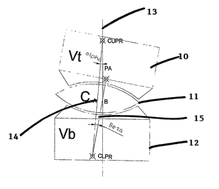
d. Upper and lower Prosthesis Radii (UPR & LPR): The
upper and lower radii of curvature of the disc
prosthesis.
e. Centre of Upper and Lower Prosthesis Radii (CUPR
& CLPR): The centre point of the upper and lower
disc prosthesis radii. For a bi-convex disc
prosthesis core, the CUPR lies inferior and the
CLPR lies superior.
f. Prosthetic axis (PA): The line joining the CUPR
and LUPR.
g. Lateral ligament structure (LLS): The ligaments
taking origin from the supero-lateral edge of the
lower vertebra and attached to the infero-lateral
edge of the upper vertebra, along lines radiating
upwards and forwards from the ACR and which
stretch the least during vertebral segmental
flexion and extension around the ACR.
h. Simplified lateral ligament structure (SLLS): A
single line or intervertebral linkage which
describes the mathematical behaviour of the LLS.
i. Anterior longitudinal ligament (ALL): The
anterior ligamentous structures.
j. Posterior longitudinal ligament (PLL): The
posterior ligamentous structures.
k. The Mathematical Process: A mathematical process
involving linear algebra and matrix
CA 02604128 2007-10-05
WO 2006/105603 PCT/AU2006/000457
- 25 -
transformations which can be used to describe the
motion of an artificial disc that has a mobile
core
Figure 1 shows a prosthesis with a bi-convex core
representing a prior art prosthesis as shown for example
in US 5674296 to Bryan.
From Figure 1 it should be apparent that the
upper vertebrae 10 can rotate relative to the core 11 and
the core 11 can rotate relative to the lower vertebrae 12.
It has been assumed in the past that because
there is in effect two angles of rotation, that the
prosthesis can adopt whatever position is needed to
simulate normal rotation. However an analysis in
accordance with a preferred embodiment of the invention
shows that exact simulation of normal rotation is not
possible but it is possible to design a prosthesis with
near normal motion.
Incremental normal rotation in the sagittal plane
occurs around an instantaneous centre of rotation. When
measured over larger angles this ICR moves somewhat,
although in both the lumber and cervical spines it is
always in the posterior one half of the lower vertebrae.
In accordance with one embodiment of the
invention, motion of the upper vertebrae 10 can be
described by analysing it as a dual linkage with links 14
and 15 as shown in Figure 2. Point CUPR remains fixed in
global co-ordinates. The motion can be considered as
sequential movements of the links 14 and 15. Initially
upper vertebrae 10, the core 11 and the point CLPR rotate
by a degrees around the point CUPR. The lower vertebrae
12 then rotate by (3 degrees around the newly rotated
position of CLPR (CLPRl) .
The minor axis (not shown) of the core 11 remains
at right angles to link A which itself passes through the
minor axis of the core 11. Core 11 therefore moves in the
same direction to upper vertebrae 10. In flexion core 11
will anteriorly, in extension core 11 will move
CA 02604128 2007-10-05
WO 2006/105603 PCT/AU2006/000457
- 26 -
posteriorly.
In designing a prosthesis as previously outlined
it is desirable to simulate as closely as possible
movement of vertebrae in normal operation with an
invertebral disk between upper and lower vertebrae.
Therefore to provide a frame of reference of this normal
motion reference is made to Figure 3 which shows motion of
a normal disk, (invertebral disk) with the approximation
of a fixed centre of rotation (ACR). All points on
vertebrae 10 move to corresponding points on vertebrae 18
and the tran.sformation that describes the movement of any
arbitrary point from the position of upper vertebrae 10 to
upper vertebrae 16 is rotation by angle y around ACR.
Lines 17 and 18 both exhibit positional information and
angular information. These characteristics are defined as
position and orientation.
It follows that for any artificial disk mechanism
to reproduce the behaviour of the movement shown in Figure
3 that it must be able to move line segment 17 to line
segment 18 and at the end of the movement both the
position and orientation of line segment C1-D1 with the
artificial disk mechanism (prosthesis) must match the line
segment 18 in Figure 3.
Referring back to Figure 2 it follows that the
position and orientation of the vertebrae are fully
described by angles a and (3 and the lengths of the links
14 and 15. It follows that if the mechanism in Figure 2
is able to mimic the mechanism in Figure 3 (normal) then
there must exist a combination of values for variables
a,R,14,15 that will make both the position and orientation
of both vertebrae the same.
Position and orientation of objects in two
dimensional space are conveniently describe by the use of
linear algebra. To fully describe the position and
orientation of a two dimensional structure in two
dimensional space, a coordinate system can be attached to
the object. This coordinate system is called a frame. All
CA 02604128 2007-10-05
WO 2006/105603 PCT/AU2006/000457
- 27 -
points on the moving object have fixed coordinates in the
new frame and the frame is considered to move within
another coordinate system - usually the global or 'world'
coordinate system . Figure 4 shows a Frame FR1 attached to
the moving vertebrae in figure 3. The origin of this frame
is displaced from the origin of the global frame G by
position vector p. The orientation of frame FR1 is given
by the unit vectors n for the x axis and o for the y axis
of FR1.
In matrix notation the frame FR1 can be described
as
n.r o, P.,
FR1 = n,, o,,
0 0 1
Where nY= x coordinate of unit vector Fz
n,,= y coordinate of unit vector n
o,.= x coordinate of unit vector o
o,,= y coordinate of unit vector o
p,.= x coordinate of position vector p
p,,= y coordinate of position vector p
Any point with coordinates x,y attached to frame
FR1 can be converted to global coordinates by
premultiplying matrix FR1 by the vector of the coordinates
of the point in FR1
x xÃiobed
FRI ~ Y = Yglõhi,l
1 1
Any frame such as FR1 can be transformed by
multiplying by a transformation matrix T with the
following characteristics.
cos a- sin a Ax
T= sina cos a Ay
0 0 1
Where a = angle of rotation
dx and Ay = change in x and y position.
CA 02604128 2007-10-05
WO 2006/105603 PCT/AU2006/000457
- 28 -
If matrix M is premultiplied by Frame FR1 frame
FR1 will be rotated around the fixed global reference
frame origin and translated in the direction of the global
reference frames axes. If Matrix M is postmultilpied by
FR1, FR1 is rotated around the origin of the moving frame
(FR1) and translated in the direction of the moving (FR1)
frames axes.
Figures 5A to SD show a hypothetical prosthesis
with a convex upper surface and a concave lower surface.
For analysis purposes there is a mechanical linkage
consisting of line segment AD rotating around point A and
a further link consisting of line segment DB. DB is
rigidly attached to the upper vertebrae and upper
prosthetic end plate. A reference frame has been attached
at point ACR. A further reference frame has been attached
at point A.
Considering the variables in Figure 5A and 5C it
should be apparent that BFR1 should have the following
value - expressed in the global reference frame AFR1.
1 0 Ldsk
BFRI= 0 1 Pdsk
0 0 1
In order for the reference frame BFR1 to be
transformed to be attached to the top vertebrae at point
B, it must undergo the following transformations shown in
Figures SB and 5D.
1. Rotation by alpha degrees to produce new
frame BFR1R
cos a- sin a 0
BFRI R= BFR1 * sina cosa 0
0 0 1
in figure 1 alpha is negative considering the normal
convention of positive rotation being anticlockwise.
CA 02604128 2007-10-05
WO 2006/105603 PCT/AU2006/000457
- 29 -
2. Inferior translation by Bdsk in the frame
of reference of BFR1R to produce new frame BFR2
1 0 0
BFR2=BFR1R* 0 1 -Bdsk
0 0 1
3. Rotation of BFR2 by Beta degrees in its own
frame of reference to produce new frame BFR2R
cos,8 - sin,8 0
BFR2R = BFR2 * sin,Q cos,8 0
0 0 1
4. Translation by Cdsk in the frame of
reference BFR2R to produce a new frame BFR3
1 0 0
BFR3 = BFR2R * 0 1 Cdsk
0 0 1
BFR3 is now attached to the upper vertebrae at
point B and has the orientation of the upper vertebrae.
BFR3 (1,3) (row 1, coluinn 3) contains a function
f(alpha,Beta) that represents the x coordinate of point B
and BFR3 (2 , 3) contains a function g(alpha, Beta) that
represents the y coordinate of point B. BFR3(1,1) contains
a function k(alpha,Beta) that represents the cosine of the
angle made by the top vertebrae with the global reference
frame.
Consider a further linear translation of -Ldsk in
the frame of reference of BFR3 (the upper vertebrae. This
will create at new frame BFR4 at point E
CA 02604128 2007-10-05
WO 2006/105603 PCT/AU2006/000457
- 30 -
1 0 - Ldsk
BFR4=BFR3* 0 1 0
0 0 1
The equivalent functions f, g and k now represent
the coordinates of point E and the (unchanged) angle of
orientation of the upper vertebrae.
By performing the matrix calculations It can be
shown that
f(a,Q) =-(cosa=cos,Q-sina=sin,l3)=Ldsk+(-cosa=sin/3 -sin(x
=cosfl)=C'clck+sin(x =Ba!ck+I,dck)
........................ . (1)
Where f x coordinate of point E
g(aõ6) =-(sina=cos)C3+cosa=sin)6)=Ldsk+(cosa=cos,(3-sina=sin)6)=Cdsk-
cosa=BcGsk+Pdsk)
..................... . . ( 2 )
Where g = y coordinate of point E
And
k(a,,6) =cosa=cos,6 -sina=sin/3 ............... (3)
Where k = cosine of angle between upper vertebrae and
global reference frame.
From Figure 5 it can be seen that as AFR1 is the
global reference frame it's value is
1 0 0
AFR1= 0 1 0
0 0 1
a frame AFR2 can be derived by rotation b'y angle gamma
(the desired rotation of the normal disc) of frame AFR1 to
produce AFR1R
CA 02604128 2007-10-05
WO 2006/105603 PCT/AU2006/000457
- 31 -
cosy -sin0
AFR1 R= AFR1 * sin y cos y 0
0 0 1
Frame AFR1R can now be translated by value Adsk
along the y axis of AFR1R to produce frame AFR2
1 0 0
AFR2 = AFR1R * 0 1 Adsk
0 0 1
cos y- sin y- sin y= Adsk
AFR2 = sin y cos y. cos y= Adsk
0 0 1
AFR2(1,3) should now contain the x coordinates of
point E and AFR2(2,3) should now contain the y coordinates
of point E.
, Let function s(y)= AFR2(1,3) (x coordinate) ......................... .(4)
Let Function t(),,)= AFR2(2,3) (y coordinate) ......................... .(5)
It follows that as both frame AFR2 and BFR4 are
at the same point (E) that from equations 1 and 2 that
s'(y)=.f(a,)Y) .............................. (6)
t(y) g(a,,6) ............................... (7)
Equations 6 and 7 represent 2 simultaneous
equations with two variables. In order for the mechanism
to exactly simulate the movement of the normal disc, it
also follows the AFR2 and BFR4 must be equal.
AFR2 = BFR4 ........................ . . ... . (8)
It can be shown that for this to occur that as
well as equations 6 and 7 holding true. It also follows
that.
CA 02604128 2007-10-05
WO 2006/105603 PCT/AU2006/000457
- 32 -
A -c~+/3 ..................................... (9)
It can be shown by numerical means that there are
no real solutions that satisfy equations 6, 7 and B. For a
given angle y that a normal disc will flex, the solutions
for angles a and 8 are such that the prosthesis will be
positioned in relative Kyphosis or Lordosis . Figure 6
represents the effect of an attempt to flex an existing
prosthesis with a biconvex core by 10 degrees with the
constraint (constraint 1) being that point E is the same
as the normal prosthesis. The solution to equations 6, 7
and 8 result in a equalling -10.72 and P equalling -
18.26 . The dashed line represents a real disk rotating by
10 about ACR, this position represents the kyphotic
solution to keep points E with the same co-ordinates.
This position is the position of 0 ligament stretch (ZLS).
Figure 7 represents the effect of an attempt to flex a
prosthesis with a core with a convex uppersurface and a
concave lower surface by 10 degrees with the constraint
(constraint 1) being that point E is the same as the
normal prosthesis. The solution to equations 6, 7 and 8
result in a equalling 5.71 and (3 equalling -7.71 . The
dashed line represents a real disk rotating by 10 about
ACR, this position represents the kyphotic solution to
keep points E with the same co-ordinates, Though the
position of kyphosis is significantly less than the
biconvex core prosthesis. This position is the position
of zero ligament stretch (ZLS).
In Figure 7 the effect of attempting to extend a
prosthesis by 10 is shown. Solutions to equations 6, 7
and 8 result in a equalling -1.55 and P equalling 4.75 .
The dashed representation of the upper vertebrae again
represents a real disk rotating by 100 about ACR. This
position represents the Lordotic solution to keep points E
with the same co-ordinates. This position represents the
0 ligament stretch position (ZLS).
CA 02604128 2007-10-05
WO 2006/105603 PCT/AU2006/000457
- 33 -
There are other ways of adding a constraint to
the assembly. The other useful constraint is to constrain
the lower end plate of the upper vertebrae to be parallel
with the lower end plate of the upper vertebrae in the
'normal' situation and to minimize the distance between
them. This can be achieved by rotating frames BFR4 and
AFR2 by gamma degrees about the global reference frame
AFR1 to produce two new frames AFR3 and BFRS
cos y- sin y 0
AFR3 = sin y cos y 0= AFR2
0 0 1
cos y- sin y 0
BFR5 = sin y cos y 0= BFR4
0 0 1
For both end plates to be parallel AFR3(1,1)=1
(cos (0) = 1) ..........10
and
BFR5(1,3) = AFR3(1,3) = 0 ........................... . . (11)
as both x coordinates must be the same (zero)
Figure 8 shows the effect of adding this
constraint (constraint 2) to an existing prosthesis with a
biconvex core and attempting to match a 10 degree of
flexion from a'normal' motion segment. It can be seen
that with this constraint that the two upper vertebrae
cannot superimpose and that a ligament joining points ACR
to E must be stretched beyond its normal length. With the
constraint that the end plates are parallel, solutions to
equation 6, 7 and 8 result in a equalling -1.61 and (3
equalling -8.39 . The dashed lines represent the real disk
rotating by 100 about ACR and the resultant position
represents the solution to keep points with the end plates
CA 02604128 2007-10-05
WO 2006/105603 PCT/AU2006/000457
- 34 -
parallel and with minimum distance between them. This
constraint is therefore termed Maximal Ligament Stretch
(MLS). Figure 9 shows the effect of adding this
constraint (constraint 2) to a prostheis with a core with
a convex upper surface and a concave lower surface and
attempting to match a 10 degree of flexion from a'noxmal'
motion segment. It can be seen that with this constraint
that the two upper vertebrae cannot superimpose and that a
ligament joining points ACR to E must be stretched beyond
its normal length. With the constraint that the end
plates are parallel, solutions to equation 6, 7 and 8
result in a equalling -12 . 68 and (3 equalling -2 . 68 . The
dashed lines represent the real disk rotating by 10 about
.ACR and the resultant position represents the solution to
keep points with the end plates parallel and with minimum
distance between them. This constraint is therefore
termed Maximal Ligament Stretch (MLS).
In Figures 6 and 7 the Ligament joining ACR to E
has no stretch and instead the prosthesis rotates at point
E to cause a degree of Kyphosis or Lordosis. This
constraint is defined a Zero ligament stretch (ZLS)
In the cervical spine there is good anatomical
evidence that there is only a weak posterior longitudinal
ligament and the main lateral ligaments diverge from near
the normal Anatomical centre of rotation (ACR) for that
vertebrae. As the anterior longitudinal ligament has, by
necessity, been destroyed by the surgical approach, The
main ligamentous constraint in the cervical spine is
approximated by Ligament ACR-E. In the absence of an
effective posterior longitudinal ligament, there is reason
to believe that a cervical disc prosthesis of the type
shown in Figure 6 would behave as if the constraint to
movement was that of the ZLS variety, and there should be
a tendency to kyphosis with flexion and
lordosis/retrolisthesis in extension.
In the lumbar spine the posterior longitudinal
ligament is much tougher. The lumbar spine therefore would
CA 02604128 2007-10-05
WO 2006/105603 PCT/AU2006/000457
- 35 -
preferentially attempteto stretch the ligament ACR-E by
using the constraint MLS. The annulus fibrosis would
rarely allow this and the theory would suggest that
flexion would be limited.
Whatever the particular case the real constraints
in a given disc space will be a combination of the
constraints ZLS and MLS. The difference in the angle
achieved by the vertebrae and the desired angle (Gamma -
(alpha+beta)) in the ZLS situation (Delta A) will be a
measure of the prostheses inability to match the normal
motion required. The difference between the length of
ligament ACR-E and the desired length (Delta L) will also
be a measure of the prostheses inability to match the
normal motion required.
The mathematical equations developed above will
enable design variables in a 2 articulation prosthesis to
be optimized so as to minimize either Delta A or Delta L
or both. By minimizing Delta A or Delta L the prosthesis
will have a better chance of optimally simulating normal.
By the use of simulations using the above
mathematical analysis the following holds.
Delta A is minimized to virtually nil by reducing
variable Ldsk to zero. This has the effect of moving the
prosthetic axis posteriorly so that the ACR lies on the
Prosthetic Axis. In this position Delta A remains very
small for all positions of the ACR that lie at or below
the disk space on the prosthetic axis.
Delta A is minimized when the radii of the upper
and lower prosthetic articulations are approximately
equal.
Delta A is minimized when the radii of the
articulations are larger and Delta A gets larger with
smaller radii.
It is preferred that Delta A is between 3 and 5 .
The translation of the Core is larger when the
Prosthetic Radii are larger and the translation is smaller
when the radii are smaller,
CA 02604128 2007-10-05
WO 2006/105603 PCT/AU2006/000457
- 36 -
The disclosed prosthesis therefore seeks to.
Move the prosthetic axis to the posterior one
third of the disc.
Select optimal radii of the upper and lower
joints.
In making these changes two problems are created.
In some embodiments the core of the prosthesis is
no longer symmetrical and was it to rotate, it
may impinge on the spinal canal.
Because of the posterior positioning of the
prosthetic axis the core is at risk of spinal
cord impingement.
Based upon the Mathematical Process described
above the prosthesis consists, briefly, of two end plates,
an intermediate mobile core and a separate anterior band
for attachment to the upper and lower vertebrae.
Figures 13A to 13E show another embodiment of the
invention in which the prosthesis consists of a core 50
having concave upper and lower surfaces 51, 52. An upper
plate 53 has a convex lower surface 54 and lower plate 55
has an upper convex surface 56.
The lower surfaces 52, 56 are cylindrical from
one side to the other (rotational and translational
movement) rather than completely spherical, whereas the
top surfaces 51 and 54 are completely spherical allowing
for universal movement as opposed to backwards and forward
movement as with the lower surfaces.
An additional feature of the prosthesis 49 shown
in these figures is the provision of upper and lower
vertical ridges 57, 58 which are centrally located and
adapted to fit into grooves created in the bottom surface
of the upper vertebrae and the upper surface of the lower
vertebrae. As shown more clearly in Figure 11 the core 50
and upper plate 53 and lower plate 55 have the prosthetic
axis 60 moved to the posterior 1-3 of the prosthesis so that
the centre of the upper radius of curvature (CUPR) A and
the centre of the lower radius of curvature (CLPR) D are
CA 02604128 2007-10-05
WO 2006/105603 PCT/AU2006/000457
- 37 -
aligned on the vertical axis through the ACR. As with the
previous embodiment a major portion 61 is located forward
of the axis 60 and a minor portion 62 is located behind
it. Furthermore, the minor axis of the core 50 is aligned
with the vertical axis 61. In addition the anterior and
posterior vertical edges of the core 50 are flat and
aligned in parallel with the minor axis 64.
The effect of attempting to flex the prosthesis
49 by 10 with the constraint being parallel end plates and
full ligament stretch results in solutions to equations 6,
7 and 8 providing a with an angle of -6.87 and (3 with an
angle of 3.130.
In Figure 12 an upper vertebrae 65 rotated
through angles a and (3 are almost coincident with
vertebrae 66 represented in dash line and corresponding to
rotation by 10 (y) about the ACR. This position
represents the solution to keep points with the end plates
parallel and with minimum distance between them. This
corresponds to the position of maximum ligament stretch
(MLS). The core of a bio-concave prosthesis as shown in
Figures 11 and 12 move anteriorly in flexion. The amount
of ligament stretch required to do this is less than when
the prosthetic axis is at the mid point of the prosthesis
and has therefore a design as shown in Figure l0A and
Figure 10B. In this configuration the effect of
attempting to flex a prosthesis by 10 ~ with the constraint
being parallel end plates and full ligament stretch
results in solutions to equations 6, 7 and 8 providing a
with an angle of -6.94 and P with an angle -3.06 . The
prosthesis shown in this example represented by item 70 is
symmetric about its minor axis which also in a state of
rest coincides with the vertical axis of the upper and
lower vertebrae 71, 72. Figure 10B again shows the effect
of moving upper vertebrae 71 through angles a and (3
compared to an upper vertebrae rotating by 10 relative to
the ACR. It can be seen that movement possible by upper
vertebrae 71 does not approximate movement of a real
CA 02604128 2007-10-05
WO 2006/105603 PCT/AU2006/000457
- 38 -
vertebrae 74 as well as prosthesis as designed with a
prosthetic axis/minor axis coincident with the vertical
axis through the ACR.
Figure 14 shows an angled view of the prosthetic
device 49 with core 50, upper plate 53 and lower plate 55.
Figures 15A and 15B show an alternative
embodiment of the invention in which a prosthesis is
provided with a core 75 with upper plate 76 and lower
plate 77. The core 75 has an upper convex surface 78 and
a lower concave surface 79. As with the embodiments
described in relation to Figures 12 and 13, the minor axis
80, the prosthetic axis coincides with the vertical axis
through the ACR of the lower vertebrae 81. Because the
lower surface 79 is convex it is significantly smaller
than the upper convex surface 78. Likewise the lower
surface of the upper plate 76 is concave and has a
matching configuration to surface 78. The lower plate 77
has a convex upper surface which is longer than the
matching concave surface 79 to allow movement by the core
75 there over backwards or forwards.
Figure 15B shows how rotation of the upper
vertebrae 82 results in relative movement between upper
plate 76 and core 75 as well as relative movement between
core 75 and lower plate 77.
As with the embodiments shown in Figure 13 the
prosthetic axis is asymmetric and a major portion of the
core 75 is located forward of the prosthetic axis.
Figure 16 shows a side view of another prosthesis
83 consisting of a core 84 having an upper convex surface
85 which has a lower radius of curvature compared to a
lower concave surface 86. In this embodiment both the
upper and lower surfaces 85, 86 have centres of radius of
curvature which are located below the core 84.
Upper plate 87 has a].ower concave surface
matching that of surface 85 and lower plate 88 has an
upper convex surface 89 which is much longer than the
length of the surface 86 to allow reasonable travel
CA 02604128 2007-10-05
WO 2006/105603 PCT/AU2006/000457
- 39 -
backwards and forwards. In addition the convex surface 89
of the lower plate 88 extends into a straight horizontal
flat surface 90. This effectively prevents forward travel
of the core 84 beyond the end of the convex surface 89.
Figure 17 shows a prosthesis 91 which is similar
to prosthesis 83 except that the upper surface 92 has a
greater radius of curvature than the lower surface 93. In
addition therefore the lower surface of the upper plate 93
is concave and longer in length than its co acting upper
surface 91. Lower plate 95 has a convex surface which is
longer in length than the co-acting concave surface 92.
In addition at a rearward end of the convex surface 96, an
upwardly angled straight section 98 is provided as a
method of stopping movement of the core 99 beyond the end
of the convex surface 96.
The forward end of convex surface 96 also extends
into a horizontal straight section 97 which serves to
prevent the core 99 moving beyond the front end of the
curved surface 96.
It should be noted that the prostheses 83, 91 are
more realistically represented in Figures 16 and 17 as
being interposed between upper and lower vertebrae which
have a more trapezoidal shape rather than a rectangular
shape. Thus although surfaces 90 and 97 and previously
described surfaces have been described as being
horizontal, in fact they are slanted and instead are
generally parallel to the general orientation of the upper
and lower faces of the upper and lower vertebrae. It
should also be noted that surfaces 90 and 97 can be angled
upwardly or even downwardly as long as they prevent
forward movement of the core 84, 99.
The different prosthesis which have been thus far
described have concentrated on characteristics which
emulate an invertebral disk. An additional component
useful for a prosthesis designed to emulate
characteristics of an invertebral disk include a band 100
shown in Figure 18 which is designed to closely simulate
CA 02604128 2007-10-05
WO 2006/105603 PCT/AU2006/000457
- 40 -
actions of ligature and in one embodiment also provides a
stop for forward movement of a prosthetic core.
The band 100 consists of a woven fabric 101
consisting of filaments of wafts and wefts creating a
weave with a grid like pattern. Upper and lower ends 102,
103 are provided with connecting plates 104, 105 each with
holes 106 for screws to be inserted through for attachment
to upper and lower vertebrae respectively.
The woven fabric 101 is preferably designed to
encourage cellular growth in the interstitial spaces
between the threads/filaments and to ultimately result in
ligatures growing between the upper and lower vertebrae.
According to one embodiment the band is in the
form of a prosthetic ligament made from a woven and
absorbable material of appropriate stiffness. The woven
material is designed to allow ingrowth of fibrous tissue
to replace the function of the prosthetic ligament as it
is reabsorbed.
According to one embodiment the band is in the
form of a gauze made of wire or polymeric material.
It is preferred that the band is able to elongate
or contract in a similar fashion to a ligament.
With regard to materials used for the different
prosthesis described above, the end plates may be made
from a metal such as titanium, cobalt-chromium steel or a
ceramic composite. Typically they have a roughened planar
surface which abuts against the adjacent surface of the
vertebrae. To assist with fixing the plates to the
vertebrae, they may be provided with a fin or ridge as
described in the embodiment shown in Figures 13 and 14 or
they may be provided with curved surfaces for bearing on
an adjacent vertebral body end plate.
The upper and lower surfaces of the core as well
as the adjacent curved surfaces of the upper and lower end
plates are preferably smooth to enhance articulation. The
central core may be made from similar materials to those
used for the end plates, but may also be made from a
CA 02604128 2007-10-05
WO 2006/105603 PCT/AU2006/000457
- 41 -
plastic such as UHMW polyethylene or polyurethane
composite.
It is preferred that the radius of curvature of
each of the curved surfaces of the prosthesis is in the
range of 5 to 35mm.
The foot print of the prosthesis end plates may
be of a variety of shapes but will be optimised to
minimise the risk of subsidence into the adjacent
vertebral bone.
Although the various articulation surfaces of the
core and upper and lower plates have been described in
relation to concave and convex surfaces, it should be
noted that other surface profiles are also included in the
invention.
For example the co acting surfaces of the core
and the lower plate could be ellipsoid instead of
cylindrical to provide restricted relative movement
therebetween.
Previously a mathematical explanation has been
provided of the behaviour of an artificial disk prosthesis
having dual articulation. Different embodiments of the
prosthesis have been described covering each of the
permutations of possible upper and lower surface profiles.
These have included biconvex, biconcave as well as convex
upper and concave lower and concave upper and convex
lower. The equations previously outlined described the
position and orientation of a moving upper vertebrae on a
fixed lower vertebrae with the dual articulating
prosthesis located therebetween. Movement of the upper
vertebrae relative to the upper surface of the prosthesis
and movement of the lower surface of the prosthesis
relative to the lower vertebrae have been described with
reference to constants and by variable angle of rotation
of variables a and P. The orientation of the upper
vertebrae is described by:
cos-' (cos a= cos,Q - sin a= sin )6)
The position of a point E immediately above the
CA 02604128 2007-10-05
WO 2006/105603 PCT/AU2006/000457
- 42 -
centre of rotation of the disc space and on the lower edge
of the upper vertebrae is given by the following
equations:
x(a,/3)=-(sina=cosB+cosa=sin/3)=Ldsk+(cosa=cos/3-sina=sin/3)=Cdsk-
cosa=Bdsk+Pdsk
y(a,/3)=-(sina=cos,<3+cosa=sin,6)=Ldsk+(cosa=cos)6-sina=sin/3)=Cdsk-
cosa=Bdsk+Pdsk)
Where Constants define the size and functional
type of the prosthesis.
Depending on the relative sizes of parameters
Ldsk, Cdsk , Bdsk and Pdsk there are 4 distinct types of
prosthesis that are described:
These are: (described by the core shape)
1. Biconvex
2. Biconcave
3. Convex top concave bottom with the top
radius greater than bottom radius
4. Convex top concave bottom with the bottom
radius greater than the top radius
Equations 1-3 describe the kinematics of these 4
prosthesis.
The length of a line joining the COR to point E
is
l(a,/j)= x(a,+y(a,)6)'
Wherein a is the angular displacement of the
upper part relative to the middle part, (3 is the angular
displacement of the middle part relative to the lower part
and 1 is the ligature joining a part of the upper part
with the centre of rotation of the skeletal structure (or
prosthesis) when in use and where x(a, p) and y(a, (3) are
different functions.
Preferably "ligature" includes any elongate
member particularly one with a degree of extension of
stretch and contraction or compression.
The values of alpha and beta can be calculated
that produce a minimum value for 1. 1 can be considered
CA 02604128 2007-10-05
WO 2006/105603 PCT/AU2006/000457
- 43 -
to be the lateral ligament of the spinal motion segment.
As this is elastic it can be seen that it will behave as a
spring and consequently will have the lowest elastic
potential energy when 1 is smallest. An equilibrium
position can be calculated when 1 is either a minimum or a
maximum. Mathematically this can be defined as the
gradient vector being zero:
Dl(a,b) = ~~ = O 0
918
Under the circumstances of a zero gradient vector
the prosthesis will have a zero change in elastic
potential energy for infinitesimal changes in a and ,8 and
the prosthesis will be in an equilibrium position. However
when this equilibrium position is a maximum value for 1 it
can be seen that small perturbations in a or ~'iwill tend
to cause 1 to decrease and the equilibrium position is
unstable. Mathematically this can be described as:
~a -ve _ +ve -ve
~2~' -ve or S2~' -ve or 2 +ve
918 1519 Under these circumstances it would be possible
for the prosthesis to be precisely balanced and be in
equilibrium, though small perturbations would cause it to
rapidly adopt a position of maximum flexion or extension.
It can be shown that prostheses 1) and 4) have
unstable equilibrium positions. This situation occurs
when matching an on axis or off axis COR. By increasing
the radii of the upper and lower articulations the value
of the gradient vector will be less negative and the
CA 02604128 2007-10-05
WO 2006/105603 PCT/AU2006/000457
- 44 -
tendency to adopt a position of maximum flexion or
extension will be diminished.
Prostheses 2) and 3) however, have the property
of having a positive second partial derivative of 1.
(5'e
& + ve
S?~ +ve
S16
At the equilibrium position defined by
c5a
OZ(aõQ) = S~ - 0
0
S6
In other words there exist values of a and ,(3
that cause an equilibrium when 1 is a minimum. This
equilibrium is stable and self correcting or self
centering because any tendency to perturb the prosthesis
from the equilibrium position will have a tendency to
cause 1 to lengthen and therefore increase the elastic
potential energy of the system.
Prostheses 2) and 3) therefore have stable
equilibrium positions and are self correcting or self
centering.
When attempting to match a COR that is on axis
with the prosthesis the equilibrium position is in the
neutral position (when a=/3 = 0). When matching an off
axis COR the equilibrium position will now be located away
from the neutral position (when a#,8:#0) - when matching a
CR that is anterior to the prosthesis axis the equilibrium
position will be in extension and when matching a CR that
is posterior to the prosthetic axis the equilibrium
position will be in flexion.
CA 02604128 2007-10-05
WO 2006/105603 PCT/AU2006/000457
- 45 -
When the values of the radii of the upper and
lower articulations are large the equilibrium position
changes due to matching an offset will be lessened.
Figure 20A shows a graphical plot in three
dimensions of the ligament length 1 versus (3 and a for a
prosthesis core with a convex upper surface and a concave
lower surface where the radius of curvature of the upper
surface of greater than that of the lower surface. As an
example the upper radius is 36mm compared to 12mm for the
lower radius.
It can be seen from Figure 20A that the three
dimensional graph shown indicates a minimum ligament
length as represented by a trough in the graph.
In this Figure there is zero, X and Y offset,
which means the prosthesis axis is aligned with the
patient's centre of rotation and corresponds to L in
Figures 19A and 19B.
Figure 20B shows the effect of introducing a
value for L of lmm for the same type of prosthesis shown
in Figure 20A. The equilibrium position moves in the
opposite direction to L. The mathematical method can be
used to optimise this change in equilibrium position and
make the prosthesis less sensitive to changes in Y offset
(L) or X offset (moving the patient's CR inferiorly).
Figure 21 shows a two dimensional view of the
change in ligament length with angle of flexion for the
prosthesis referred to in Figure 20A. In this Figure it
can be seen that there is a clear trough around the centre
of rotation represented by angle of flexion extension 0.
This graph clearly shows that any movement of the
prosthesis away from the centre of rotation results in
extension of the ligament length and therefore a natural
tendency for the ligament to want to return to its minimum
length at the centre of rotation.
The above contrasts with a prosthesis with a
biconvex core. A graphical solution to the mathematical
equation outlined above is shown in Figure 22 for a
CA 02604128 2007-10-05
WO 2006/105603 PCT/AU2006/000457
- 46 -
biconvex core. It should be apparent from this graphical
analysis that there is no minimum ligament length which
provides a point of equilibrium. In fact the two
dimensional graphical representation shown in Figure 23
shows how the point of equilibrium is located about the
centre of rotation of the biconvex core and shows that any
movement of the core away from the centre of rotation
results in a decrease in the ligaments length and
therefore a tendency for the core to move away from the
point of equilibrium.
Figure 24A shows another embodiment of the
invention in which the prosthesis has a biconcave core.
As with the embodiment shown in Figures 20A, 20B and 21 in
this embodiment there is a point of equilibrium about the
centre of rotation for the core. This point of
equilibrium corresponds to the minimum ligature length and
hence provides a natural tendency for the core to return
to the point of equilibrium if there is movement away from
the centre of rotation.
Figure 24B shows the variation in ligament length
for a biconcave prosthesis with the upper radius equal to
36mm and the lower radius equal to 36mm with Y offset and
X offset being zero. It can thus be seen that with equal
radius the graphical representation of the mathematical
model shows there is no tendency for movement of the
prosthesis away from the equilibrium position to result in
a movement back to the equilibri.um position.
The two dimensional graphical representation
which is not shown has a similar appearance to Figure 21.
From the above it should be clear that if it is
desired to produce a prosthesis with a self correcting
ability vihich results in a tendency of any movement away
from a point of equilibrium to result in a natural urging
of the prosthesis back towards the point of equilibrium,
then the embodiments of the invention described in
relation to Figures 20A, 20B, 21 and 24A provide suitable
solutions. It is also noted however that the other
CA 02604128 2007-10-05
WO 2006/105603 PCT/AU2006/000457
- 47 -
embodiments of the invention which have been described may
still be used even though they may not have this self-
centering ability. This is because other alterations may
be made to the overall prosthesis in order to keep
movement of the prosthesis within predetermined
boundaries.
All prostheses are able to match a COR that is on
axis. When the COR is off axis they can only match by
either stretching or shortening the lateral ligament
(Delta L) or by adopting abnormal orientation (Delta A).
Delta A and Delta L, for a given offset can be reduced by
making the Radii Larger.
All prostheses are capable of pure translation.
Prosthesis 2) does so with loss of disc height. Prosthesis
3) does so with a gain in disc height.
The Ideal prosthesis has
1 Stable equilibrium position
2 The best capacity to handle off axis CR
This is 2) or 3)
The preferred embodiment is 3) with as large
radii as possible. This has the added advantage of
resistance to pure translation (because the soft tissues
would need to lengthen)
The second preferred embodiment is 2) this has
the advantage of relatively unrestricted translation.
There may be clinical situations where either 2)
or 3) are preferable.
The ratio of radii for prosthesis 3 can be set so
that under a COR match to a physiologically normal centre
of rotation there is equal arc travel between the top and
bottom articulation. This can be achieved by
R1* alpha = R2* beta
Where alpha and beta can be calculated for the CR
match.
This will match wear for upper and lower
articulations and is the most efficient use of the surface
area of contact.
CA 02604128 2007-10-05
WO 2006/105603 PCT/AU2006/000457
- 48 -
A capacity to calculate alpha and beta allows the
prosthesis to be drafted.
An ideal range for of ratio for prostheses in the
lumbar spine and cervical spines to achieve this is 3:1 -
10:1.
In the lumbar and cervical spines the preferred
radii are 5mm and 50mm.
Prosthesis 2 does not allow the travel on each
articulating surface to be equal. Preferably the
mathematical model will be used for this prosthesis to
allow an optimum choice of ratio of radii based on any
desirable parameter such as the desired ratio of angles a
and 6 . In the lumbar and cervical spines the ideal ratio
is 2:1 - 1:2. In the Lumbar spine the preferred radii are
between 8 - 40 mm for each radii. In the cervical spine
the preferred radii are between 6 - 30 mm
Figures 10A to 11B and 13A to 13F show a
prosthesis having a biconcave profile. Figures 15A, 15D,
16 and 17 show a prosthesis with an upper convex surface
and a lower concave surface. It is preferred that the
version of the prosthesis shown in Figure 17 is utilised
as the radius of curvature of the upper surface is larger
than that of the lower concave surface. It is also
preferred that the lower surface of the upper plate has a
matching profile to the upper surface of the prosthesis
and the upper surface of the lower plate has a matching
profile to the lower surface of the prosthesis. It should
be understood however that this does not mean that the
entire lower surface of the upper plate and upper surface
of the lower plate have the matching profiles. Thus a
reference to Figure 19A shows one preferred configuration
of a prosthesis having the preferred upper and lower
surface profiles identified above. The prosthesis 110
shown is symmetrical about a central vertical axis and has
smooth curved outer upper and lower edges. The prosthesis
is shown offset rearwardly and hence with its core 113
retained within a rearward region of the upper and lower
CA 02604128 2007-10-05
WO 2006/105603 PCT/AU2006/000457
- 49 -
plates 111, 112.
The upper surface of the lower plate 112 has a
rearward portion having a convex shape of matching
configuration to the opposing concave profile of the core
113.
The apex of the convex region is offset
rearwardly with respect to the centre of prosthesis . The
convex region is symmetrical about its offset central
vertical axis and on either side extends into a concave
trough with the result that the overall profile of this
region has the appearance of part of a sinusoidal curve.
Each of the troughs extend into upwardly curved
surfaces on either side of the convex region and provide
rearward and forward detents to limit forward and backward
movement of the prosthesis relative to the lower plate.
The upper plate 111 has a rearwardly offset
concave lower surface with a rearmost downwardly extending
edge which is configured to limit rearward movement of the
core 113 relative to the upper plate 111.
As can be seen from Figure 19A the radius of
curvature of the lower surface of the upper plate is
larger than the radius of curvature of the upper surface
of the lower plate. In each case the radius of curvature
has a common origin which is located on the central
vertical axis of the prosthesis at a virtual point below
the prosthesis.
Figure 19A also shows the core 113 in a stable
equilibrium position aligned with the central vertical
axis 114 which is rearwardly offset to the anatomical
central axis.
A lateral ligament 115 is shown connected between
upper and lower vertebrae 116, 117. The ligament 115 is
offset by a distance L from the axis 114.
Figure 19B shows how the prosthesis 112 is
effected by backward rotational movement of the upper
vertebrae 116 with respect to the lower vertebral disk
117.
CA 02604128 2007-10-05
WO 2006/105603 PCT/AU2006/000457
- 50 -
As shown the lateral ligament 115 rotates about
the centre of rotation (CR) and the upper plate 111 and
core 113 rotate to an unstable position. Because of the
difference in radius of curvature of the upper surface of
core 113 and the lower surface the ligament 115 is
stretched and there is a natural tendency for it to return
to the equilibrium position shown in Figure 19A. The
difference in radius of curvature also means that the
upper vertebral disk 116 will rotate and translate with
respect to the lower vertebral disk 117.
By increasing the radius of curvature of the
lower surface of the upper plate and hence the upper
surface of the prosthesis it is possible to increase the
stability of the prosthesis when in use if the COR of the
prosthesis is offset from the ACR.
Desirably increasing the radius of curvature of
the upper surface of the prosthesis and hence the lower
surface of the upper plate enhances the ease with which
the prosthesis is able to return to its equilibrium point
centered about its central vertical axis.
According to one embodiment the further the
central vertical axis of the prothesis is offset from the
centre of rotation of the skeletal structure, the larger
the radius of curvature of the upper surface of the
prosthesis.
It is preferred that the radius of curvature of
the upper surface of the prosthesis is between 30 and 50
mm in the lumbar spine and 20 and 40 mm in the cervical
spine. As it is preferred that the ratio of the radius
of curvature of the upper surface of the prosthesis
compared to the lower surface of the prosthesis is within
a predetermined range an increase in the radius of
curvature of the upper surface of the prosthesis will
result in a corresponding increase in radius of curvature
of the lower surface of the prosthesis.
It is preferred that the length of the convex
region of the upper surface of the lower plate (when
CA 02604128 2007-10-05
WO 2006/105603 PCT/AU2006/000457
- 51 -
measured from front to rear) is determined in accordance
with typical travel allowed for a vertebral disk in a
typical vertebral column.
It is preferred that all embodiments of the
prostheses have a prosthetic axis that is set in the
posterior 1% of the disc to as closely as possible match
the normal physiological centre of rotation.
It is to be understood that, if any prior art
publication is referred to herein, such reference does not
constitute an admission that the publication forms a part
of the common general knowledge in the art, in Australia
or in any other country.