Note: Descriptions are shown in the official language in which they were submitted.
CA 02604337 2007-09-27
WO 2006/106439 PCT/IB2006/001479
1
SYSTEM AND METHOD FOR NON-INVASIVE CARDIOVASCULAR
ASSESSMENT FROM SUPRA-SYSTOLIC SIGNALS OBTAINED
WITH A WIDEBAND EXTERNAL PULSE TRANSDUCER IN A BLOOD
PRESSURE CUFF
FIELD OF THE INVENTION
This invention relates to non-invasive cardiovascular assessnient of a patient
based on
the evaluation of pressure wave signals obtained by means of a low frequency,
wideband electrical transducer or sensor disposed in, on or under the
Korotkoff arm
cuff of a sphygmomanometer. More particularly, the invention relates to the
non-
invasive assessment of aortic compliance and other cardiovascular parameters
by
analyzing signals obtained from a sensor of this type.
BACKGROUND OF THE INVENTION
The signals recorded with a sensor placed beneath a blood pressure cuff are
termed
"supra-systolic" signals if the cuff pressure is above the subject's systolic
blood
pressure. In addition, signals can be recorded when the cuff pressure is below
systolic
pressure. In all cases, the signals result from pressure energy transmissions
and are
dependent upon the subject's physiology.
When the heart pumps, a pressure gradient is generated within the
cardiovascular
system. This results in pulse pressure waves traveling peripherally from the
heart
through the arteries. Like any wave, they reflect back off a surface or other
change in
impedance. Arterial pulse waves reflect back from both the peripheral
circulation and
from the distal aorta when it becomes less compliant (Murgo, Westerhof et al.
1980;
Lathain, Westerhof et al. 1985). These reflection waves are identifiable in
arterial
pressure tracings, but the exact timing and magnitude of the waves are
difficult to
discern. Nevertheless, they have been the basis of several commercial systems
to
assess reflectance waves. These systems measure arterial contours using
applanation
tonometry from the radial artery.
CA 02604337 2007-09-27
WO 2006/106439 PCT/IB2006/001479
2
If a low frequency sensor is placed over the brachial artery beneath a blood
pressure
cuff and the cuff is inflated above systole, supra-systolic signals can be
recorded
(Blank, West et al. 1988; Hirai, Sasayama et al. 1989; Denby, Mallows et al.
1994).
An idealized supra-systolic signal for one heart beat is shown in Figure 1.
These
signals contain frequency components of less than 20 Hertz, which are non-
audible.
Supra-systolic low frequency signals provide clear definition of three
distinct waves:
an incident wave corresponding to the pulse wave and two subsequent waves.
Blank
(Blank 1996) proposed that the second wave emanated from the periphery and the
relative amplitude of this wave to the incident wave (K1R) was a measure of
peripheral vascular resistance (PVR). He proposed a constant such that PVR
could be
measured from the ratio of the incident to the first reflectance wave. See,
also, U.S.
Pat. No. 5,913,826, which is incorporated herein by reference in its entirety.
The second supra-systolic wave is, in fact, a reflectance wave from the distal
abdominal aorta--most likely originating from the bifurcation of the aorta and
not
from the peripheral circulation as proposed by Blank. This has been verified
in human
experiments (Murgo, Westerhof et al. 1980; Latham, Westerhof et al. 1985) and
in
studies using pulse wave velocity (PWV) measurements. The relative ainplitude
of the
first reflectance wave is now believed to be a measure of the stiffness,
compliance, or
elasticity of the abdominal aorta rather than peripheral resistance.
In the clinical experiments upon which Blank relied to formulate his
hypothesis,
changes in compliance were induced with epinephrine and epidural anesthesia.
The
changes in compliance were accompanied by changes in peripheral resistance.
Thus,
he saw a relationship between his KIR and PVR, but it was a co-variable and
not a
true association.
The third wave occurs at the beginning of diastole and is believed to be a
reflection
wave from the peripheral circulation. As such, it is a measure of peripheral
vasoconstriction with superimposed secondary reflections. Supra-systolic
signals can
be utilized to measure compliance by relating the amplitude of the first wave
(incident
or SS1) to the amplitude of the second (aortic reflection or SS2) wave. The
degree of
vasoconstriction can be assessed by measuring the amplitude of the diastolic
or third
CA 02604337 2007-09-27
WO 2006/106439 PCT/IB2006/001479
3
wave (SS3 wave) and relating it to the SS1 wave. Amplitudes, areas under the
curves,
or other values calculated from the waves can be utilized. Data has been
analyzed by
measuring amplitudes, ratios of amplitudes and time delays between waves.
Augmentation Index (AI) has become recognized as an important marker of
cardiovascular disease. It increases with age, hypertension and
atherosclerosis.
Through ventricular-vascular coupling, AI is a marker of ventricular (cardiac)
hypertrophy - stiffness or diastolic dysfunction. Thus, this single measure
gives an
indication of the health of the whole cardiovascular system.
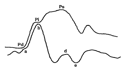
Al is measured from an aortic pressure tracing (Figure 8) as follows: The
amplitude
of the augmentation wave (Ps-Pi) is divided by the ainplitude of the incident
plus
reflection wave (Ps-Pd). The ratio is multiplied by 100 to give a percentage.
Aortic Augmentation Index (AAI) =(Ps-Pi) / (Ps-Pd) x 100 (1)
Measurements of aortic pressure can only be made in the cardiac
catheterization
laboratory so other non-invasive means of assessing it have been developed.
Two
have been described. Firstly, using tonometry on the carotid artery, a
waveform can be
measured which identifies the initial and late systolic peaks. A carotid
augmentation
index (CAI) is measured. Secondly, tonometry of the radial artery likewise
provides a
signal, which can be transformed to provide a measure of aortic augmentation
index
(AAI)=
SUMMARY OF THE INVENTION
The relationship between the aortic pressure and brachial arterial wideband
supra-
systolic pressure trace can be understood and a correction formula derived
from a
comparison between the two, both on an individual and/or on a population
basis,
enabling a Brachial Artery Augmentation Index (AAI) and a brachial artery
derived
AAI to be measured.
The present invention therefore provides a system for measuring peripheral
arterial
signals, e.g. of the brachial artery, using a wideband external pulse
transducer
CA 02604337 2007-09-27
WO 2006/106439 PCT/IB2006/001479
4
disposed in, on or under a blood pressure cuff, and a processor, receiving the
signals
from the transducer, and processing these signals to determine distortions
present in
the transducer waveform with respect to an inferred original aortic waveform.
A cuff is inflated to a supra-systolic pressure, such as 15-150 mm Hg above a
systolic
pressure, preferably about 30 mm above the systolic pressure, measuring with a
pressure transducer having sufficient bandwidth to capture detailed waveform
information, for example from 0.1 to 1000 Hz, and analyzing the wavefonn to
infer an
aortic pressure waveform. Various corrections may be applied to the inference,
both
personal to the subject, and based on population studies, to correct for
aberrations. In
a preferred embodiment, a model of the patient is formulated, wherein a set of
parameters, which may be generally orthogonal (e.g., parameters having low
interactivity) or correlated to available clinical measurements, describe
elements of
the model. These parameters may then be used to populate the model, or the
model
used to estimate the parameters. By employing a physiological model, and
analyzing
the values of the parameters, as well as their responsivity to various
factors, clinical
conclusions are facilitated.
This inferred waveform may then be used for a number of purposes, including
analyzing cardiac function, analyzing the central and/or peripheral arterial
system, or
for analyzing the cardiovascular system as a whole.
Another embodiment of the invention employs an algorithm for extracting
features
from the pressure waveform (or, for example, the model constructed from the
data),
which may be multivariate or complex. In any case, the parameter(s) or
features may
be used as diagnostic, prognostic, or therapeutic indices. Thus, if the
parameter
corresponds to a therapeutic target of a drug, the parameter may be monitored,
and
drug use titrated for its desired effect on the cardiovascular system.
Stimuli may also be used to excite various responses in the system, for
example a cold
pressor stimulus, which may allow more accurate or detailed analysis of the
pressure
data.
CA 02604337 2007-09-27
WO 2006/106439 PCT/IB2006/001479
Thus, the present invention provides means for extracting useful parameters of
central
and peripheral cardiovascular system performance, without requiring a direct
measurement of waveforms from the heart or aorta.
A reliable system may therefore be provided to acquire supra-systolic signals
from
patients, a method to analyze the signals, and clinical applications for the
signals. The
system consists of a low frequency transducer placed in, on or beneath a blood
pressure cuff or similar device, placed around a patient's arm. The signals
are
conditioned and, if necessary, amplified, passed through an analog to digital
converter
and transferred to a computer or processor for analysis. Analyzed signals will
be
stored, presented on a screen numerically or graphically. Data can be stored
or
transmitted to databases or other health care facilities.
A variety of vibration transducers can be used. The transducer must be able to
sense
dynamic signals as low as about 0.1 Hertz and be sturdy enough to withstand
repeated
use under external pressures of about 300 mm Hg. For example, a suitable
commercially available piezoelectric transducer consists of two adjacent
sensors
approximately 1.5 cm in diameter. The transducer is placed along the axis of
the
brachial artery providing proximal (closer to the heart) and distal signals.
Preferably
only one sensor is used. However, an alternative is to use an array of sensors
to aid in
noise elimination or other signal processing in certain clinical environments.
Another
possibility is to incorporate inexpensive sensors into a disposable blood
pressure cuff
to create a disposable product suitable for critical care environments where
infection
control is important.
According to the invention, it is possible to simplify the assessment of
stroke volume
and/or blood volume and/or other indicators of cardiovascular status and/or to
improve the accuracy of such indicators.
For a full understanding of the present invention, reference should now be
made to the
following detailed description of the preferred embodiments of the invention
as
illustrated in the accompanying drawings.
CA 02604337 2007-09-27
WO 2006/106439 PCT/IB2006/001479
6
BRIEF DESCRIPTION OF THE DRAWINGS
Figure 1 is a graph of idealized supra-systolic signal for one heartbeat
obtained from a
patient.
Figure 2 is a diagram showing the supra-systolic pulse wave transit paths
resulting in
the signal of Figure 1.
Figure 3a is a diagra.in illustrating the positioning of blood pressure cuff
with a
wideband external pressure (WEP) transducer arranged on a patient's arm to
obtain the
signal of Figure 1.
Figure 3b is a cross-sectional view of the blood pressure cuff of Figure 3a.
Figure 4 is a graph showing a sample determination of area under the SS1 peak
of a
supra-systolic signal from a patient.
Figure 5 is an example graph of supra-systolic signal versus time over an
inspiratory/expiratory cycle of a patient breathing normally.
Figure 6 is a graph of supra-systolic signal versus time over an
inspiratory/expiratory
cycle of a patient during labored breathing.
Figure 7 is a schematic block diagram of apparatus in accordance with a
preferred
embodiment of the present invention.
Figure 8 shows a pressure trace from the ascending aorta using the apparatus
of the
present invention.
Figure 9 shows a supra-systolic signal with designations of its inflection
points.
Figure 10 shows overlaid traces of a pressure trace from the ascending aorta
and the
supra-systolic signal, using a wideband external pressure (WEP) transducer.
CA 02604337 2007-09-27
WO 2006/106439 PCT/IB2006/001479
7
Figure 11 shows a WEP transducer signal and cuff pressure on an upper axis,
and an
expanded WEP tracing on a lower axis, evidencing a medium Augmentation Index.
Figure 12 is a diagram similar to Figure 11, with an expanded WEP tracing
evidencing a low Augmentation Index.
Figure 13 is a diagram similar to Figure 11, with an expanded WEP tracing
evidencing a high Augmentation Index..
Figure 14 is a diagram similar to Figure 11, with an expanded WEP tracing
obtained
before a hand is cooled with ice.
Figure 15 is a diagram similar to Figure 11, with an expanded WEP tracing
obtained
after a hand is cooled with ice.
Figure 16 is a diagram similar to Figure 11, with an expanded WEP tracing with
dropped heartbeats.
Figure 17 is a diagram similar to Figure 11, with an expanded WEP tracing
evidencing varying beat-to-beat rates.
Figure 18 is a diagram similar to Figure 11, with an expanded WEP tracing
wherein
both the beat-to-beat rate and the configuration of the waves vary.
Figure 19 is a diagram similar to Figure 11, with an expanded WEP tracing
showing
large variations in the wave configuration.
Figures 20-23 are diagrams similar to Figure 11, with expanded WEP tracings
obtained from a succession of patients with progressively deteriorating,
diastolic heart
failure.
Figures 24-27 are diagrams similar to Figure 11, with expended WEP tracings
obtained from a young patient, a middle-aged patient and two older patients,
respectively, illustrating the importance of dt1-2.
CA 02604337 2007-09-27
WO 2006/106439 PCT/IB2006/001479
8
DETAILED DESCRIPTION OF THE PREFERRED EMBODIMENTS
The preferred embodiments of the present invention will now be described with
reference to Figures 1-27 of the drawings. Identical elements in the various
Figures
are designated with the same reference numerals.
Back ound
With reference to the drawings and in particular Figure 1 initially, an
idealized supra-
systolic signal 1 is shown which has been obtained utilizing the arrangements
shown
in Figures 2 and 3. The signal shown in Figure 1 is characteristic of the
transduced
signal within a patient's brachial artery 3 in the upper arm as a result of
applying
supra-systolic pressure to the brachial artery utilizing a blood pressure cuff
2 (Figures
2 and 3) which has been inflated above the patient's systolic blood pressure
(subsequent to a determination being made of the patient's systolic blood
pressure).
When the blood flow in the brachial artery 3 is occluded, flow related
pressure
changes are effectively filtered out so that a sensor 4 positioned proximate
to the
patient's proximal artery may purely measure pressure-induced energy
transmissions
generated within the cardiovascular system as a result of the heart pumping.
As the heart puinps, pulse pressure waves travel peripherally from the heart
through
the arteries. These pressure waves reflect back off a surface or other change
in
impedance. As shown in Figure 2, the signals sensed at the brachial artery
will
include the result of a pressure wave traveling directly from the heart (shown
as peak
or. pulse S S 1 in Figure 1) as well as a pressure signal resulting in a
reflection of energy
traveling from the heart to the distal aorta 5 and back up to the brachial
artery (shown
as peak or pulse SS2 in Figure 1). A further peak or pulse or wave (SS3)
results from
a reflection of the pressure wave off the peripheral circulation and secondary
reflections from the distal aorta.
Because the large majority of the energy within the supra-systolic signal of
Figure lis
outside the frequency range of normal human hearing it is necessary to use a
specialized low frequency transducer or sensor 4 (Figure 3) to obtain the
signal of
Figure 1. For example, "wideband" transducers are suitable and in the present
CA 02604337 2007-09-27
WO 2006/106439 PCT/IB2006/001479
9
application these transducers are often referred to as wideband external pulse
(or
"WEP") transducers. WEP transducers may, for example, include piezo-electric
sensors capable of converting low frequency mechanical pressure vibrations or
fluctuations to an electrical output (voltage) signal. WEP transducer 4 is
preferably
positioned close to (1.5 to 2cm) the distal (further from the heart) edge of
the blood
pressure cuff 2 and aligned with the brachial artery 3 as shown in Figure 3a.
Figure 3b illustrates a patient's arm 6 with a blood pressure cuff (Korotkoff
cuff) 2 in
cross-section. The arm 6 is shown as being surrounded by the partially
inflated
pressure cuff 2 which comprises an inflatable bladder 8 formed of flexible
material.
One end 9 of the bladder is wrapped around and secured to itself by means of
Velcro
or the like.
A piezoelectric transducer 4 is retained against the surface of the bladder by
means of
a thin film 10 of synthetic material such as nylon, rayon or the like. The
transducer 4
which is retained by the film 10 is positioned such that the transducer
receives
pressure waves or vibrations from the brachial artery 3.
The previously mentioned WO0205726A and U.S. 5,193,826B both describe methods
of determining particular cardiovascular parameters from the output signal of
a
wideband external pulse transducer. It is known that for example, the
magnitude of
the SS2 wave is a measure of large arterial tone best assessed by the ratio of
the
magnitude of the SS1 to SS2 waves. Changes in the SS1:SS2 ratio therefore
represent
changes in large arterial tone.
Stroke Volume
"Stroke Volume" (SV) is the amount of blood ejected by the heart in a single
heartbeat.
As previously mentioned, W00205726A includes an empirical equation utilizing
experimentally determined SS 1 and SS2 peak values to calculate stroke volume.
"Cardiac Output" is a related cardiovascular parameter indicating the amount
of blood
pumped by the heart per unit time and is the product of Heart Rate (HR) x
Stroke
CA 02604337 2007-09-27
WO 2006/106439 PCT/IB2006/001479
Volume and hence cardiac output may be easily determined once Stroke Volume is
known.
It has been discovered and confirmed, according to the invention, that the
area beneath
the SS 1 peak or pulse or portion of the signal as exemplified in Figure 1 is
positively
correlated with stroke volume and improvements in cardiac performance. By
"positively correlated", it is meant that stroke volume can be approximated as
a
function of the area beneath the SS 1 pulse. Changes in the area under the SS1
peak or
pulse or curve in an individual over time therefore reflect changes in stroke
volume
and thus the S S 1 signal can be used as a monitor of change in stroke volume
of an
individual or patient over time. As an alternative to area beneath the SS 1
peak, it has
also been shown that the area beneath a function of the SS 1 peak can also
provide a
good indication of stroke volume. For example, the area beneath a curve which
is the
square (or other function) of the SS 1 peak curve, could be utilized as an
indicator of
stroke volume.
By utilizing flow probes, it has been determined that the majority of forward
flow
during a cardiac cycle occurs during the initial stage of systole (the regular
contraction
of the heart and arteries that drives the blood outward). Analysis of the
timing of
supra-systolic signals (as shown in Figure 1) demonstrates that the S S 1
signal
corresponds to the timing of the peak and forward flow noted with the flow
probes.
Furthermore, studies have demonstrated that changes in the amplitude of the SS
1
signals are consistent with changes in stroke volume.
Preferably, the duration of the S S 1 curve for the area calculation is the
time from the
inflection of the SS 1 signal (that is, the transition from concave to convex)
to the
onset of the SS21 signal. Figure 4 demonstrates the area 7 which must be
calculated in
which a base line 6 has been inserted at a selected amplitude level through
the initial
point 8 in the SS 1 wave at which it is inflected.
As a result of this discovery of the relationship between the area under the
SS 1 signal
and stroke volume, an empirical equation can be determined or, alternatively,
changes
in calculated area values in a particular patient over time can be recorded to
provide
CA 02604337 2007-09-27
WO 2006/106439 PCT/IB2006/001479
11
an indication of changes in stroke volume (in comparison to a base value) for
that
patient. Alternatively, a model of the cardiovascular system may be developed
which
explains this relationship and serves to predict stroke volume based on SS1
signal
data.
Blood Volume
Blood volume is a cardiovascular parameter indicating the amount of blood in a
patient's circulatory system. Changes in arterial pressure with breathing
(either
spontaneous or with a ventilator) are used in clinical practice as a measure
of blood
volume such that large declines in pressure with ventilation represent volume
depletion. Volume depletion leads to less blood returning to the heart and
therefore a
decline in cardiac output.
Changes in the magnitude or amplitude of the SS 1 signal occur with breathing.
It is
known that more labored breathing produces a larger decline in the magnitude
of the
SS 1 signal during a breathing cycle (an inhalation followed by an exhalation
or vice
versa). Figure 5 shows the effect of normal respiration on the supra-systolic
waveform during a typical breathing cycle. It can be seen that SS 1 peak 9 is
a
maximum from start of exhalation and subsequent SS1 peaks 10 and 11 show a
gradual reduction in SS1 amplitude whereas peaks 12 and 13 show a gradual
increase
in SS1 peak amplitude as the patient iiihales.
In contrast, Figure 6 shows a supra-systolic blood pressure signal from a
patient
whose breathing is labored (for example, the patient may be suffering an
asthma
attack or be breathing via a ventilator). It can be seen in Figure 6 that the
change in
magnitude of the SS 1 peak between the maximum peak 14 and minimum amplitude
peak 15 is much greater than the example shown in Figure 5.
It has been discovered that the size of the change in amplitude of the SS 1
peak over a
respiratory cycle is negatively correlated with blood volume. By "negatively
correlate", it is meant that blood volume can be approximated by a decreasing
function of the change in magnitude of the SS 1 peak over a breathing cycle.
Accordingly, this discovery can be used to empirically determine a
relationship or
CA 02604337 2007-09-27
WO 2006/106439 PCT/IB2006/001479
12
equation which equates the change in amplitude to change in blood volume
during the
breathing cycle. Alternatively, the measured change in atnplitude during a
breathing
cycle can itself be recorded for comparison with previous or future changes in
amplitude for that same patient to generate a trend of changes in blood volume
for that
patient over time.
Although the examples shown with reference to Figures 5 and 6 both utilize
supra-
systolic blood pressure signals, it should be noted that changes in the
magnitude of the
SS1 signal with ventilation can also be detected at subsystolic (but greater
than
diastolic) pressure and even subdiastolic pressure can be used as a measure of
change
in blood voluine.
Both of the above-mentioned discoveries require the obtaining of a signal
associated
witll pressure fluctuations from a peripheral artery of the patient (for
example,
brachial artery) and the measurement of a feature of that signal. While the
obtaining
and measurement of the feature of the signal may be carried out manually in
the case
of measuring the change in amplitude of the SS 1 peak, the measurement of
these
features may be automated. For example, signals from sensor 4 may be
amplified,
passed through an analog-to-digital converter and input to a computer via data
acquisition hardware and analyzed utilizing software such as National
Instruments'
LabVIEWTM software which provides the ability to not only easily measure the
changes in amplitude required for the above blood volume calculation, but also
easily
enables the selection of a suitable baseline and measurement of the area
beneath SS 1
to determine stroke volume. It is known that heart rate can also be determined
from
the SS 1 curve and therefore cardiac output may be determined from stroke
volume
once heart rate has been established. The method for calculating area beneath
the SS1
peak may, for example, comprise integrating a determined function between
start and
end times; components of the SS 1 signal -- e.g., amplitude and time to
achieve peak
amplitude -- can also be determined.
As shown in Figure 7, it is possible to automate the process of determining
cardiac
output or blood volume by utilizing a controller 16 which may comprise
hardwired
electronic devices or may comprise, for example, a microprocessor running
suitable
CA 02604337 2007-09-27
WO 2006/106439 PCT/IB2006/001479
13
software which receives the output of the WEP transducer 4 and controls
inflation/deflation of blood pressure cuff 2 via a controllable air pump 17.
For fast inflation the controller may be programmed (1) to inflate the cuff 2
while
monitoring the output of the WEP transducer to deterniine when the patient's
systolic
blood pressure is reached, and then (2) to continue to inflate the cuff to
between about
25 to 30mm Hg above the thus determined systolic pressure in order for the
controller
to obtain the supra-systolic blood pressure signal exemplified in Figure 1.
The software or hardware within controller 16 (shown as box 19) may then
analyze
the captured supra-systolic signal to determine such parameters as the peak
amplitudes
of the various SS 1 signals and the area beneath the SS 1 signal as well as
determining
the positioning of the base line for area determination. Software or hardware
19 may
then determine the stroke volume and/or blood volume based on the respective
measured parameters. For example, software may incorporate an equation
correlating
the measured parameter to blood volume or stroke volume. Once the appropriate
parameter or value has been measured or determined by the software or hardware
19
within or associated with controller 16, the calculated value may be output to
an
output device such as a display screen or printer 18. Alternatively or in
addition, the
output device 18 may include storage means for recording the various
parameters
gleaned from a particular patient's blood pressure signal (and/or the
calculated values
of stroke volume or blood volume) and software may input the recorded values
to
detennine trends or changes in the parameters or values over time to aid in
assessing
changes in circulatory physiology.
It is lrnown that an estimate of arterial softness in a patient may be
determined based
on such cardiovascular parameters as stroke volume and blood volume.
Accordingly,
the various measurements derived from the suprasystolic waveform (such as area
under SS1, the change in peak SS1 value during a breathing cycle, the SSI.-SS2
time
delay between respective adjacent peaks of SS1 and SS2 and/or ratio of SS1:SS2
peak
values) and/or a series of readings taken over time from the same patient may
be fed
into an appropriately trained neural network which would output a value for
arterial
softness in the patient under analysis.
CA 02604337 2007-09-27
WO 2006/106439 PCT/IB2006/001479
14
Accordingly, at least in its preferred form, the present invention provides a
method
and apparatus for efficiently and simply measuring cardiac performance in a
patient
non-invasively.
Arterial Compliance
Arterial compliance refers to the stiffness of arteries. In young healthy
people, arteries
are compliant so that a volume of blood ejected causes them to distend more
for a
given pressure. By contrast, stiff arteries (arteries with a low compliance)
distend less.
Compliance (C) is measured by the change in volume (dV) per unit increase in
pressure (dp) (Brinton, Cotter et al. 1997; de Simone, Roman et al. 1999):
C=dV/dp (2) True compliance
Compliance can be measured fairly accurately by stroke volume (SV) divided by
pulse
pressure (PP) even though the arterial circuit is not a totally closed system
(Chemla,
Hebert et al. 1998):
C = SV/PP (3) Estimated compliance
Arterial coinpliance, although important, is not commonly measured in clinical
practice, as the measurement, up until now, has been difficult to perform. The
aforementioned US Patent Application No. 10/221,530, which has been
incorporated
herein by reference, discloses a technique for obtaining this information non-
invasively using a Korotlcoff blood pressure cuff.
According to the present invention, it has been found that by measuring aortic
waveforms and brachial artery signals concurrently, the relationship
therebetween can
be understood and a correction function derived. This enables a Brachial
Artery
Augmentation Index (AAI) and a brachial artery derived AAI to be measured.
The problem with the existing methodologies are that they are technically
difficult to
use and not easy to readily repeat. The blood pressure cuff/sensor combination
is
simple to use, provides clear, repeatable data that is easy to analyze, can be
cheap to
CA 02604337 2007-09-27
WO 2006/106439 PCT/IB2006/001479
manufacture, and generally will not require trained personnel. It can also be
used as a
monitor, as it can be left in place wrapped around the patient's arm.
Modeling the Cardiovascular System
The present invention provides a system for measuring peripheral arterial
signals, e.g.
of the brachial artery, such as the aforementioned occlusive cuff and
transducer, for
reading pressure fluctuations over the occluded artery, and a processor,
receiving the
signals from the transducer, and processing these signals to determine
distortions
present in the waveform transducer waveform with respect to the inferred
original
aortic waveform.
The method proceeds by occluding a peripheral artery by, for example,
inflating a cuff
to a supra-systolic pressure, such as 30 mm Hg above a systolic pressure,
measuring
with an extracorporeal wideband (WEP) transducer a pressure waveform of the
peripheral artery, and analyzing the waveform with respect to a model of at
least a
portion of the cardiovascular system to infer an aortic pressure waveform.
This
inferred wavefonn may then be used for a number of purposes, including
analyzing
cardiac function, analyzing the central and/or peripheral arterial system, or
for
analyzing the cardiovascular system as a whole.
In order to infer the aortic waveform, it is preferred to model the
cardiovascular
system to extract features from the waveform having separate meaning or
interpretation. These may be orthogonal features or mildly interacting. These
features
may then be processed with respect to population statistics, in order to
normalize the
values to obtain an accurate estimate. While it may be possible to avoid the
feature
extraction, this method potentially results in an improved ability to account
for
population variability and therefore may provide increased accuracy for a
similar
number of clinical samples. Likewise, a proper model may allow known pathology
of
a particular patient to be accounted for, or may allow a proposed diagnosis to
be tested
with respect to its presumed affect on the cardiovascular system.
Further, extracting a useful low dimensionality parameter (that is, a
parameter which
has a close correlation to a measurable intrinsic mechanical attribute of the
CA 02604337 2007-09-27
WO 2006/106439 PCT/IB2006/001479
16
cardiovascular system) from the transducer output, facilitates the use of this
parameter
as a diagnostic, prognostic, or therapeutic index. Thus, if the parameter
corresponds
to a therapeutic target of a drug, the parameter may be monitored, and drug
use titrated
for its desired effect on the cardiovascular system.
It has also been found that various stimuli or stresses can dynamically change
the
cardiovascular system. For example, a cold stimulus on the hand may produce a
peripheral arterial vasoconstriction. Therefore, another optional aspect of
the present
invention is to measure the response of the cardiovascular system to one or
more
stiinuli or stressors, to produce a characteristic change in the
cardiovascular system.
The measurements of cardiovascular system are then synchronized with the onset
and/or relaxation of the stimulus or stressor. Thus, this provides an
additional
variable to allow elucidation of parameters of the cardiovascular system (or
model
thereof), which may be directly useful, and/or useful wlien analyzed in
context. The
application of a stressor or stimulus pennits distinction between functional
parameters
(those which vary over time based on extrinsic factors) and fixed parameters
(those
which are not subject to change over periods of time of interest). Thus,
atherosclerosis maybe distinguished from stress induced vasoconstriction, even
though in a single measurement, these may produce the same waveform, since
they
may present the same iinpedance characteristics (e.g., arterial compliance).
Once
these types of distinctions are made, it is then possible to monitor changes
in these
responses over time, for example as a result of treatment.
Augmentation Index
Supra-systolic brachial artery signals derived from a wideband sensor placed
beneath
the distal edge of a blood pressure cuff in apposition with the skin, likewise
produce
an early and late systolic wave (Figure 9). The sensor records signals
directly from an
occluded brachial artery with the blood pressure cuff inflated to 30 mm Hg
above
systole. See U.S. Pat. 5,913,826, W002/05726, and U.S. Pub. Pat. App.
2003/040675
each of which is expressly incorporated herein by reference.
Studies in the cardiac catheterization lab (Figure 10) demonstrated that the
first
systolic wave (SS1) corresponds to the first phase of the aortic pressure
trace such that
CA 02604337 2007-09-27
WO 2006/106439 PCT/IB2006/001479
17
the peak of the SS 1 (b) corresponds to the Pi of the aortic pressure trace.
The late
systolic wave SS2 (d) corresponds to the augmentation wave Ps of the aortic
pressure
trace. From this, it follows that the Augmentation Index can be directly
measured by
using "de" as the augmentation wave (equivalent to "Ps-Pi") and using the sum
of
"ab+de" to be equivalent to "Ps-Pd".
Thus, the brachial Artery Augmentation Index (AAI) is given by:
AAI = de / (ab+de) x 100 (4) Augmentation Index
In a sample of 66 people aged 30-75, Augmentation Index measured in this way
provided a value ranging from 5-66%. This range is typical of Augmentation
Index
measured by other investigators.
Figures 11-15 are screen shots from a computer display showing, in the upper
half of
the diagratn, the pressure wave signal from a wideband external pressure (WEP)
transducer and, superimposed thereon, the cuff pressure applied to the
Korotkoff arm
cuff of a sphygmomanometer along a time axis which is measured in seconds.
Thus,
in Figure 11, the time starts at 0.0 seconds and continues to about 76
seconds. As may
be seen, the Korotkoff cuff is inflated twice; a first time to determine the
approximate
systolic pressure and a second time to obtain a supra-systolic signal when the
pressure
cuff is inflated to a pressure of about 25 to 30mm Hg above the patient's
systolic
blood pressure.
The lower part of the diagram shows an expanded view of the WEP transducer
signal
during the time period indicated by the rectangular box surrounding a portion
of the
supra-systolic signal along the upper axis. In this case, the box surrounds
the portion
of the supra-systolic signal which occurs during the 3 second time interval,
commencing at approximately the 65 second point along the time scale.
Considering now the formula (4) given above for the brachial artery
Augmentation
Index, it may be seen that the time distance between the pealc of the first
reflected
wave (SS2) and the following trough (the distance d to e) is approximately
0.58
seconds. Similarly, the distance from the initial trough to the initial peak
of the
CA 02604337 2007-09-27
WO 2006/106439 PCT/IB2006/001479
18
incident wave (SS1) is about.105 seconds. Using the formula (4), the
augmentation
index is calculated to be 36%, which is about average for a healthy, middle
aged adult.
Figures 12 and 13 are similar diagrams illustrating a low Augmentation Index
of 4.6%
and a high Augmentation Index of 50%, respectively.
Figures 14 and 15 illustrate what happens to the supra-systolic signal when
the hand
of the arm, to which the Korotkoff cuff has been applied, is placed in ice. In
Figure
14, the supra-systolic signal follows the normal pattern wherein the second
reflected
wave (SS3) is substantially attenuated from the first reflected wave (SS2).
Figure 15
illustrates that when the hand is placed in ice, causing stress to the
adjacent artery, the
second reflected wave (SS3) is markedly pronounced. It may be seen, therefore,
that
the supra-systolic signal reveals useful information relating to a patient's
central and
peripheral cardiovascular system.
In summary, the present invention provides means for extracting useful
parameters of
the central and peripheral cardiovascular system performance, without
requiring a
direct measurement of the pressure waveforms from the heart or aorta.
It is noted that Blank et al., US 5,913,826 refers to use of a modified
Windkessel
model of circulation, with respect to analysis of the so-called K3 signal.
(See also US
5,211,177, expressly incorporated herein by reference). However, these
references do
not address analysis of external stimuli or stressors, and, for example,
Blanlc et al.
suggest that a solution for "white coat hypertension" is to provide a home
monitor,
and thus to avoid the stress itself, rather than advantageously employ it to
perform
differential testing.
Cardiac Arrhythmia
When a piezoelectric (WEP) sensor is placed beneath the distal edge of a blood
pressure cuff, distinct vascular signals can be detected with the cuff
inflated to 30 mm
Hg above systolic pressure (supra-systolic signals). These signals have
characteristic
appearances refiecting the incidence (SS1) and reflective waves (SS2 and SS3).
If the
cuff is left inflated for 10-12 seconds, a series of pulse signals can be
obtained and
CA 02604337 2007-09-27
WO 2006/106439 PCT/IB2006/001479
19
recorded. This simple non-invasive maneuver provides the equivalence of a
rhythm
strip used to diagnose arrhythmias on an EKG.
When a typical cardiac arrhythmia occurs, the beat or beats are less effective
resulting
in an abnormal pulse signal or abnormal interval between beats.
An example of "dropped beats" is shown in Figure 16. Note the normal
characteristic
of all beats but the amplitude of the beat following the pause is increased -
so called
post-ectopic potentiation (Figure 16 marked "x").
Examples of arterial fibrillation are shown in Figures 17-19. Note in Figure
17 that all
beats are siinilar but beat-to-beat rates vary. In Figures 18 and 19, both
beat-to-beat
rates vary as do the configuration of the waves. This is due to variation in
stroke
volume/ventricular filling.
Normal beat-to-beat variation occurs and is typical of a healtliy heart (so
called sinus
arrhythmia). Absence of beat-to-beat arrhythmia can be a predictor of heart
disease.
Beat-to-beat variation in heart rate is measured with software using supra-
systolic
signals.
The method according to the present invention is not meant to displace the
EKG.
Rather it is a useful component of the utility of supra-systolic signal
analysis as a
screening tool for cardiovascular disease in primary care setting. It augments
the use
of an EKG as this provides a functional analysis of the pulse wave itself.
Heart Failure
Heart failure exists in at least 500,000 people in the United States; these
numbers are
increasing due to better treatment of ischemic heart disease, aging
population, etc. The
condition is under diagnosed, under treated and places a huge burden on the
health
care industry.
Diagnosis and management of treatment often entails expensive cardiac
technology.
The most frequently used is echocardiography. These machines cost $200,000
each,
require expert technician to use and physicians to interpret the studies. More
CA 02604337 2007-09-27
WO 2006/106439 PCT/IB2006/001479
expensive or invasive tests are also used. There is thus a need for a simple
technology
to assess heart failure in the primary care environment or for routine
management by
cardiologists. The use of supra-systolic signal analysis can provide cheap
simple non-
invasive assessment of cardio-vascular function including insight into the
existence of
heart failure or the propensity to develop heart failure.
There are several forms of heart failure:
1. Systolic heart failure wherein the left ventricle loses contractile
strength. The
heart doesn't pump well and cardiac output falls.
2. The other common category is diastolic heart failure wherein the heart
becomes stiff. It doesn't relax well and is subject to fluid overload,
pulmonary
edema and acute heart failure.
Evidence of both forms of heart failure can be detected or assessed with supra-
systolic
wave analysis.
When the heart ejects blood into the aorta, a pulse wave enters the large
vessels and is
reflected back off the distal aorta. The reflectance wave becomes more
prominent and
returns more rapidly with aging or degenerative diseases of the large
arteries. This
results in a resistance to forward flow of blood.
As has been described above, the amplitude of the forward and reflective waves
and
the duration between them can be accurately determined by analyzing signals
obtained
from a sensor placed over the skin adjacent to the brachial artery. The sensor
is
positioned beneath the distal edge of a blood pressure cuff wrapped around the
arm.
With the blood pressure cuff inflated 30 mm Hg above systolic pressure for 10-
12
seconds, a series of pulse recordings are obtained. The average of these beats
provides
a mean value for the SS 1 and SS2 waves. The characteristics of these waves
can be
used to diagnose systolic heart failure and the propensity to develop heart
failure.
CA 02604337 2007-09-27
WO 2006/106439 PCT/IB2006/001479
21
Systolic Heart Failure
Typical tracings shown in Figures 20-23 illustrate supra-systolic signals from
patients
with systolic heart failure.
The pumping strength of the heart decreases thus producing a less intense SS1
and the
reflection wave (SS2) is either absent or incorporated into the descending
portion of
the SSl (Figures 21-23). Typically, this SS2 is incorporated into the down
slope
approximately halfway down the slope with a dtl-2 of 0.8-0.11 second. (dtl2 is
the
delay between the peak of SS1 and SS2). The duration of the upstroke of SS1
(dtl)
may be prolonged and the amplitude of the SS 1 wave decreased. The SS 1 wave
may
be biphasic (Figure 21).
Diastolic Heart Failure
As large arteries harden, the pulse wave velocity increases and the amplitude
of the
reflection wave increases. This results in a shortening of the SS l-SS2 period
- the
period during which the majority of blood is ejected from the ventricle. As
the period
for ventricular emptying shortens, this places additional strain on the left
heart
resulting in left ventricular hypertrophy. Eventually, the duration of
ventricular
emptying gets so limited that the heart fails. Thus, the duration dtl-2 can be
used as a
predictor of the likelihood of developing heart failure or secondly, as a
marker that the
patient has heart failure. When the dt1-2 is 0.10 seconds or less, it is
likely that the
heart will fail or heart failure is already established. In young patients,
dt12 may be
0.15-0.2 seconds.
Two factors in ventriculo-vascular coupling which adversely affect ventricular
emptying are the duration of the waves and the amplitude of the SS2. The
greater the
amplitude of the SS2, the greater the impediment to forward flow. The shorter
the
dtl2, the less time there is for ventricular emptying. Thus, a short dtl2 and
high
amplitude SS2 foretell adverse ventricular emptying, ventricular strain and
impending
diastolic heart failure.
CA 02604337 2007-09-27
WO 2006/106439 PCT/IB2006/001479
22
Importance of dtl-2
The duration or time lapse between the peaks of the two supra-systolic peaks
SS1 and
SS2 is an important measurement for two reasons: first, as a measure of pulse
wave
velocity, and second, as a measure of the adverse effect of the reflection
wave on
ventriculo-vascular coupling and ventricular emptying.
Four tracings are shown for comparison (Figures 24-27). Figure 24 shows the
expasided WEP tracing for a young patient having good ventricular function. In
this
case, the period dtl-2, between the peak of the incident wave SS 1 and the
first
reflected wave SS2 is a prolonged 0.185 seconds. In contrast, a middle-aged
patient
with increased Augmentation Index (hardening of the arteries) may have a dtl-2
of
about 0.15 seconds (Figure 25). An increase in the Augmentation Index and a
shortening of dt1-2, but with preserved ventricular function (i.e., a normal
SSl peak in
the supra-systolic wave) is illustrated in Figure 26 (dtl -2 = 0.11 sec.) and
in Figure 27
(dtl-2 = 0.12 sec.).
Changes in supra-systolic signals with exercise can be used in two ways.
First, the
patient's response to acute exercise can be assessed. Normal individuals
exhibit an
increase in the ainplitude of the SS 1 signal consistent with an increase in
stroke
volume, a decrease in the time to generate the SS1 (dtl) consistent with
increased
cardiac contractility and a decrease in their Augmentation Index (AI)
representing
arterial dilatation. Second, physical training with conditioning results in an
improvement in arterial compliance which manifests as a reduction in AI. These
changes can be used to assess (1) cardiovascular fitness and (2) the
cardiovascular
benefits of an exercise prescription.
The preceding preferred embodiments are illustrative of the practice of the
invention.
It is to be understood, however, that other expedients known to those skilled
in the art,
or disclosed herein, may be employed without departing from the spirit of the
invention or the scope of the claims.