Note: Descriptions are shown in the official language in which they were submitted.
CA 02604424 2007-10-05
WO 2006/119032 PCT/US2006/016283
METHOD OF TREATING MULTIPLE MYELOMA USING 17-AAG OR 17-AG OR A
PRODRUG OF EITHER IN COMBINATION WITH A PROTEASOME INHIBITOR
TECHNICAL FIELD OF THE INVENTION
This invention relates to a method of treating multiple myeloma using 17-
allylamino-
17-demethoxy-geldanamycin or 17-amino geldanamycin, or a prodrug of either 17-
AAG or
17-AG, in combination with a proteasome inhibitor.
BACKGROUND OF THE INVENTION
Multiple myeloma ("MM", also known as myeloma or plasma cell myeloma) is an
1o incurable but treatable cancer of the plasma cell. Plasma cells are an
important part of the
immune system, producing immunoglobulins (antibodies) that help fight
infection and
disease. MM is characterized by excessive numbers of abnormal plasma cells in
the bone
marrow ("BM") and overproduction of intact monoclonal irnmunoglobulins (IgG,
IgA, IgD,
or IgE; "M-proteins") or Bence-Jones protein (free monoclonal light chains).
Hypercalcemia,
anemia, renal damage, increased susceptibility to bacterial infection, and
impaired
production of normal immunoglobulin are common clinical manifestations of MM.
MM is
often also characterized by diffuse osteoporosis, usually in the pelvis,
spine, ribs, and skull.
Therapies for MM include chemotherapy, stem cell transplantation, high-dose
chemotherapy with stem cell transplantation, and salvage therapy.
Chemotherapies include
treatment with Thalomid (thalidomide), bortezomib, Aredia R(pamidronate),
steroids, and
Zometa ' (zoledronic acid). However many chemotherapy drugs are toxic to
actively
dividing non-cancerous cells, such as of the BM, the lining of the stomach and
intestines,
and the hair follicles. Therefore, chemotherapy may result in a decrease in
blood cell counts,
nausea, vomiting, diarrhea, and loss of hair.
Conventional chemotherapy, or standard-dose chemotherapy, is typically the
primary
or initial treatment for patients with MM. Patients also may receive receive
chemotherapy in
preparation for high-dose chemotherapy and stem cell transplant. Induction
therapy
(conventional chemotherapy prior to a stem cell transplant) can be used to
reduce the tumor
burden prior to transplant. Certain chemotherapy drugs are more suitable for
induction
therapy than others, because they are less toxic to BM cells and result in a
greater yield of
-1-
CA 02604424 2007-10-05
WO 2006/119032 PCT/US2006/016283
stem cells from the BM. Examples of chemotherapy drugs suitable for induction
therapy
include dexamethasone, thalidomide/dexamethasone, VAD (vincristine,
Adriamycin0
(doxorubicin), and dexamethasone in combination), and DVd (pegylated liposomal
doxoru-
bicin (DoxilO, Caelyx ), vincristine, and reduced schedule dexamethasone in
combination).
The standard treatment for MM is melphalan in combination with prednisone (a
corticosteroid drug), achieving a response rate of 50%. Unfortunately,
melphalan is an
alkylating agent and is less suitable for induction therapy. Corticosteroids
(especially
dexamethasome) are sometimes used alone as MM therapy, especially in older
patients and
those who cannot tolerate chemotherapy. Dexamethasone is also used in
induction therapy,
1 o alone or in combination with other agents. VAD is the most commonly used
induction
therapy, but DVd has recently been shown to be effective in induction therapy.
Bortezomib
has been approved recently for the treatment of MM, but it is very toxic.
However, none of
the existing therapies offer a significant potential for a cure.
17-Allylamino-17-demethoxygeldanamycin ("17-AAG", also sometimes referred to
as 17-allylaminogeldanamycin) is a semi-synthetic analog of the naturally
occurring
compound geldanamycin (Sasaki et al., 198 1). Geldanamycin is obtainable by
culturing a
producing organism, such as Streptomyces hygroscopicus var. geldanus NRRL
3602.
Another biologically active geldanamycin derivative is 17-aminogeldanamycin
("17-AG"),
which is produced in the human body by metabolism of 17-AAG. 17-AG can also be
made
from geldanamycin (Sasaki et al. 1979). While geldanamycin and its analogs
have been
studied intensively as anti-cancer agents in the 1990s (e.g., Sasaki et al.,
1981; Schnur, 1995;
Schnur et al., 1999), none of them has been approved for anti-cancer use.
O O O
O)~NH2 O~NH2 O~NH2
7 . 7 7
MeO,, 11 OH Me0 I MeO,, 11 OH Me0 MeO,, 11 OH Me0 I
O O O
O O 1 O 1
NH NH NH
~ I17 I Me0 I17 H2N I17 I
~H
O O
17-AAG Geldanamycin 17-AG
-2-
CA 02604424 2007-10-05
WO 2006/119032 PCT/US2006/016283
17-AAG and geldanamycin are believed to act by binding to and inhibiting the
activity of heat shock protein-90 ("Hsp90") (Schulte and Neckers, 1998). Hsp90
acts as a
chaperone for the normal processing of many cellular proteins ("client
proteins") and is
found in all mammalian cells. Stress (hypoxia, heat, etc.) induces a several-
fold increase in
its expression. There exist other stress-induced proteins (co-chaperones),
such as heat shock
protein-70 ("Hsp70"), which also play a role in cellular response to and
recovery from stress.
In cancer cells, Hsp90 inhibition leads to disruption of the interaction
between Hsp90
and its client proteins, such as erbB2, steroid receptors, raf-1, cdk4, and
Akt. For example,
exposure to 17-AAG results in depletion of erbB2 and destabilization of Raf-1
and mutant
p53 in SKBr3 breast cancer cells (Schulte and Neckers, 1998), depletion of
steroid receptors
in breast cancer cells (Bagatell et al., 2001), depletion of Hsp90 and down-
regulation of Raf-
1 and erbB2 in MEXF 276L melanoma cells (Burger et al., 2004), depletion of
Raf-1, c-
Akt, and Erk1/2 in colon adenocarcinoma cells (Hostein et al., 2001), down-
regulation of
intracellular Bcr-Abl and c-Raf proteins and reduction of Akt kinase activity
in leukemia
cells (Nimmanapalli et al., 2001), degradation of cdk4, cdk6, and cyclin E in
lung cancer
cells with wild-type Rb (Jiang and Shapiro, 2002), and depletion of erbB
1(EGFR) and
erbB2 (p185) levels in NSCLC cells (Nguyen et al., 2000).
Because of the activity of 17-AAG relative to Hsp90 and other proteins
involved in
oncogenesis and metastasis of cancer cells, a number of clinical investigators
have evaluated
its effectiveness as an anti-cancer agent in human clinical trials. From these
various trials,
the Cancer Therapy Evaluation Program (CTEP) of the National Cancer Institute
recommended these Phase 2 dose/schedule regimens for further study: 220 mg/m2
(mg per
square meter of body surface area of the patient or subject) administered
twice weekly for 2
out of 3 weeks, 450 mg/m2 administered once a week continuously or with a rest
or break,
and 300 mg/m2 once a week for 3 weeks out of 4 weeks. Results of various
clinical trials -
almost exclusively with patients having solid tumors - with 17-AAG generally
showed
limited clinical activity and are summarized below:
(a) A Phase 1 trial in adult patients with solid tumors was conducted in which
patients
received 17-AAG daily for 5 days every 3 weeks. The starting dose was 10 mg/m2
and was escalated to 56 mg/m2, with a maximum tolerated dose ("MTD") and recom-
mended Phase 2 dose defined as 40 mg/m2. The protocol was amended to exclude
patients with significant pre-existing liver disease, after which patients
were treated
-3-
CA 02604424 2007-10-05
WO 2006/119032 PCT/US2006/016283
at doses up to 110 mg/m2 on the same schedule. No objective tumor responses
were
observed. Due to dose limiting reversible hepatotoxicity, the protocol was
further
amended to dose patients on a twice weekly schedule every other week starting
at a
dose of 40 mg/m2 per day. At daily doses of 40 and 56 mg/m2 for 5 days, the
peak
plasma concentrations were 1,860 660 and 3,170 1,310 nM, respectively. For
patients treated at 56 mg/m2 average AUC values for 17-AAG and 17-AG were
6,708 and 5,558 nM*h, respectively, and average t~i2 3.8 and 8.6 hours,
respectively.
Clearances of 17-AAG and 17-AG were 19.9 and 30.8 L/h/m2, respectively, and VZ
values were 93 and 203 L/m2, respectively (Grem et al., 2005).
(b) In a second Phase 1 trial, patients with advanced solid tumors received 17-
AAG on a
daily x 5 schedule at a starting dose of 5 mg/m2. At the 80 mg/m2, dose
limiting
toxicities (hepatitis, abdominal pain, nausea, dyspnea) were observed but dose
escalations nevertheless were continued until the dose reached 157 mg/m2/day.
Further dose schedule modifications were implemented to allow twice weekly
dosing. At the 80 mg/m2 dose level, the t12 was 1.5 hours and the plasma Cmax
was
2,700 nM. Similarly, for 17-AG the t,~T was 1.75 hours and the Cmax was 607
nM.
Plasma concentrations exceeded those needed to achieve cell kill (10-500 nM)
in in
vitro and in vivo xenograft models (Munster et al., 2001).
(c) A Phase 1 trial of 17-AAG was conducted in which patients with advanced
solid
tumors were treated weekly for 3 out of every 4 weeks at a starting dose of 10
mg/m2, with a recommended Phase 2 dose of 295 mg/m2. Dose escalations reached
a
dose of 395 mg/m2, at which nausea and vomiting secondary to pancreatitis and
grade 3 fatigue were observed. The dosing schedule was amended to allow dosing
twice weekly for 3 out of every 4 weeks and twice weekly for 2 out of every 3
weeks.
A population pharmacokinetic (PK) analysis was performed on data obtained from
this trial. The Vd (volume of distribution) for 17-AAG was 24.2 L for the
central
compartment and 89.6 L for the peripheral compartment. Clearance values were
26.7
L/h and 21.3 L/h for 17-AAG and 17-AG, respectively. Metabolic clearance
indicated that 46.4% of 17-AAG was metabolized to 17-AG. No objective tumor
responses have been observed in this trial to date. (Chen et al., 2005).
(d) Another Phase 1 trial in patients with solid tumors and lymphomas was
conducted
using a weekly dosing for 3 weeks out of a 4 week cycle. The starting dose was
15
-4-
CA 02604424 2007-10-05
WO 2006/119032 PCT/US2006/016283
mg/m2 . Dose escalation reached 112 mg/m2 without significant toxicity and was
continued with an objective of reaching a dose range of "biological" activity.
The
MTD for weekly 17-AAG was reached at 308 mg/m2. No objective tumor responses
have been observed to date in this trial, and the levels of Hsp90 client
proteins
measured were unchanged during therapy. No correlation between chaperone or
client protein levels and 17-AAG or 17-AG PK was seen. There was also no
correlation between the 17-AAG PK and its clinical toxicity (Goetz et al.,
2005).
(e) Another Phase 1 trial was conducted using a once weekly administration
schedule,
including 11 patients with metastatic melanoma. The starting dose was 10
mg/m2,
and dose limiting toxicity was observed at 450 mg/m2/week (grade 3/4 elevation
of
AST). At higher doses (16-450 mg/m2/week) the 17-AAG formulation employed
contained 10-40 mL dimethylsulfoxide (DMSO) in a single infusion, which likely
contributed to the gastrointestinal toxicity that was observed in the trial.
Among the
patients treated at 320-450 mg/m2, two showed radiologically documented long
term
stable disease. No complete or partial responses were recorded. At the highest
dose
level (450 mg/m2) the plasma 17-AAG concentrations exceeded 10 gM and remained
above 120 nM for periods in excess of 24 hours. At the highest dose level of
450
mg/m2, the mean volume of distribution was 142.6 L, mean clearance was 32.2
L/h,
and the mean peak plasma level was 8,998 g/L. There was a linear correlation
between dose and area under the curve (AUC) for the dose levels studied.
Pharmacodynamic (PD) parameters were also measured and induction of the co-
chaperone protein Hsp70 was observed in 8 of 9 patients treated at 320-
450mg/m2/week. Depletion of client proteins was also observed in tumor
biopsies:
CDK4 in 8 out of 9 patients and Raf-1 depletion in 4 out of 6 patients at 24
hours.
These data indicated that Hsp90 in tumors is inhibited for between 1 and 5
days.
(Banerji et al., 2005).
The in vivo anti-MM activity of 17-AAG has been studied using a model of
diffuse
GFP positive MM lesions in SCID/NOD mice (Mitsiades et al., 2006). Survival
analysis
showed that treatment significantly prolonged median overall survival, but non-
clinical data
3o are frequently not predictive of clinical activity. As discussed above,
this has particularly
been the case for 17-AAG in solid tumors, where the promise of pre-clinical
data has not
been borne out in Phase 1 clinical trials.
-5-
CA 02604424 2007-10-05
WO 2006/119032 PCT/US2006/016283
Thus, despite intensive efforts to develop 17-AAG as an anti-cancer agent, no
regulatory agency has approved it for the treatment of any cancer. There
remains a need for
methods of dosing and administering 17-AAG and prodrugs of 17-AAG (and its
metabolic
counterpart 17-AG) so that its potential therapeutic benefits can be realized.
The present
invention provides such methods that are efficacious in the treatment of MM
using 17-AAG.
Recently, preclinical and clinical studies have shown that bortezomib
(Velcade0,
BZ, PS-341) can overcome resistance of MM cells to conventional or high-dose
cytotoxic
chemotherapy (Hideshima et al., 2001; Mitsiades et al., 2001; Mitsiades et
al., 2003) and
improve patient outcome in MM. Bortezomib has recently been approved for
treatment of
relapsed and refractory MM (Richardson et al., 2003a). Pre-clinical studies
have also shown
that treatment of MM cells with bortezomib triggers significant Hsp90 up-
regulation as a
major stress response in MM cells. While bortezomib is capable of improving
patient
outcome, it is however highly toxic.
The present invention provides combination treatments of 17-AAG or 17-AG or a
prodrug of either with bortezomib that are efficacious in the treatment of
multiple myeloma.
A list references cited herein is provided at the end of this specification.
All
documents cited herein are incorporated herein by reference as if each such
publication or
document were specifically and individually incorporated herein by reference.
BRIEF SUMMARY OF THE INVENTION
The present invention provides methods for treating multiple myeloma (MM) in a
subject in need of such treatment, said methods comprising the step of
administering to said
subject a therapeutically effective dose of 17-AAG or 17-AG or a prodrug of
either 17-AAG
or 17-AG and a therapeutically effective dose of a proteasome inhibitor, and
optionally
repeating said step until no further therapeutic benefit is obtained.
In one embodiment, the method comprises the administration of multiple doses
of
17-AAG or a prodrug thereof to a subject with MM over a time period of at
least 2 weeks,
wherein each such dose is in the range of about 100 mg/m2 to about 340 mg/m2
of 17-AAG
or an equivalent amount of a 17-AAG or 17-AG prodrug. In one embodiment, the
dose is
about 340 mg/m2 of 17-AAG or an equivalent amount of a 17-AAG or 17-AG
prodrug. In
one embodiment, this dose is administered twice weekly for at least two weeks.
In one
embodiment, this dose is administered twice weekly for at least two weeks in a
three week
-6-
CA 02604424 2007-10-05
WO 2006/119032 PCT/US2006/016283
period, which rate of dosing per three week period is called a cycle, and
multiple cycles of
such treatment are administered to the MM patient.
In one embodiment, the therapeutically effective dose of 17-AAG or a prodrug
of 17-
AAG is a dose that results in an AUCtotal of 17-AAG per dose in the range of
about 2,300 to
19,000 ng/mL*h. In one embodiment, this dose is administered at a rate and
frequency such
that the Cma,, of 17-AAG (or the prodrug) does not exceed 9,600 ng/mL (or the
molar
equivalent of the prodrug). In one embodiment, this dose is administered at a
rate and
frequency such that the CmaX of 17-AAG is greater than 1,300 ng/mL. In one
embodiment,
this dose is administered at a rate and frequency such that the C.ax of 17-AAG
is greater
than 1,800 ng/mL. In one embodiment, this dose is administered at a rate and
frequency such
that the CmaX of 17-AAG is greater than 1,300 but does not exceed 9,600 ng/mL.
In one
embodiment, this dose is administered at a rate and frequency such that the
CmaX of 17-AAG
is greater than 1,800 but does not exceed 9,600 ng/mL.
In one embodiment, the therapeutically effective dose of 17-AG or a prodrug of
17-
AG (which prodrug includes 17-AAG) is a dose that results in an AUCtotal of 17-
AG per
dose in the range of about 800 to about 17,000 ng/mL*h. In one embodiment,
this dose is
administered at a rate and frequency such that the CIõaX of 17-AG does not
exceed 1,400
ng/mL. In one embodiment, this dose is administered at a rate and frequency
such that the
C,r,ax of 17-AG is greater than 140 ng/mL. In one embodiment, this dose is
administered at a
2o rate and frequency such that the C.X of 17-AG is greater than 230 ng/mL. In
one embodi-
ment, this dose is administered at a rate and frequency such that the C.,t of
17-AG is greater
than 140 but does not exceed 1,400 ng/mL. In one embodiment, this dose is
administered at
a rate and frequency such that the Cmax of 17-AG is greater than 230 but does
not exceed
1,400 ng/mL.
In one embodiment, the therapeutically effective dose of 17-AAG, a prodrug of
17-
AAG, 17-AG, or a prodrug of 17-AG is a dose that results in a combined
AUCtota1 of 17-
AAG and 17-AG per dose in the range of about 3,500 to 35,000 ng/mL*h. In one
embodiment, this dose is administered at rate and frequency such that the CmaX
of 17-AAG
does not exceed 9,600 ng/mL and/or the CmaX of 17-AG does not exceed 1,400
ng/mL. In
one embodiment, this dose is administered at a rate and frequency such that
the C.aX of 17-
AAG is greater than 1,300 ng/mL and/or the Cma. of 17-AG is greater than 140
ng/mL. In
-7-
CA 02604424 2007-10-05
WO 2006/119032 PCT/US2006/016283
one embodiment, this dose is administered at a rate and frequency such that
the Cma, of 17-
AAG is greater than 1,800 ng/mL and/or the Cmax of 17-AG is greater than 230
ng/mL. In
one embodiment, this dose is administered at a rate and frequency such that
the CmaX of 17-
AAG is greater than 1,300 but does not exceed 9,600 ng/mL and/or the CmaX of
17-AG is
greater than 140 but does not exceed 1,400 ng/mL. In one embodiment, this dose
is
administered at a rate and frequency such that the Cma,, of 17-AAG is greater
than 1,800 but
does not exceed 9,600 ng/mL and/or the Cm~,x of 17-AG is greater than 230 but
does not
exceed 1,400 ng/mL.
In one embodiment, the therapeutically effective dose of 17-AAG or a prodrug
of 17-
1 o AAG is a dose that results in a Terminal t-h of 17-AAG in the range of 1.6
to 5.6 h. In one
embodiment, the therapeutically effective dose of 17-AAG or a prodrug of 17-
AAG is a dose
that results in a Terminal t~~Z of 17-AAG in the foregoing range and an
AUCtota1 of 17-AAG
per dose in the range of about 2,300 to about 19,000 ng/xnL*h.
In one embodiment, the therapeutically effective dose of 17-AG or a prodrug of
17-
AG is a dose that results in a Terminal t./, of 17-AG in the range of 3.7 to
9.1 h. In one
embodiment, the therapeutically effective dose of 17-AG or a prodrug of 17-AG
is a dose
that results in a Terminal t/, of 17-AG in the foregoing range and an AUCtotal
of 17-AG per
dose in the range of about 800 to about 17,000 ng/mL*h.
In one embodiment, the therapeutically effective dose of 17-AAG or a prodrug
of 17-
2o AAG is a dose that results in a Volume of distribution V, of 17-AAG in the
range of 56 to
250 L. In one embodiment, the therapeutically effective dose of 17-AAG or a
prodrug of 17-
AAG is a dose that results in a Volume of distribution VZ of 17-AAG in the
foregoing range
and an AUCtotal of 17-AAG per dose in the range of about 2,300 to 19,000
ng/mL*h.
In one embodiment, the therapeutically effective dose of 17-AAG or a prodrug
of 17-
AAG is a dose that results in a Clearance in the range of 13 to 85 L/h. In one
embodiment,
the therapeutically effective dose of 17-AAG or a prodrug of 17-AAG is a dose
that results
in a Clearance of 17-AAG in the foregoing range and an AUCtotal of 17-AAG per
dose in the
range of about 2,300 to about 19,000 ng/mL*h.
In one embodiment, the therapeutically effective dose of 17-AAG or a prodrug
of 17-
3o AAG is a dose that results in a Vss in the range of 96 to 250 L. In one
embodiment, the
therapeutically effective dose of 17-AAG or a prodrug of 17-AAG is a dose that
results in a
-8-
CA 02604424 2007-10-05
WO 2006/119032 PCT/US2006/016283
VSS of 17-AAG in the foregoing range and an AUCtotal of 17-AAG per dose in the
range of
about 2,300 to about 19,000 ng/mL*h.
In one embodiment, the 17-AAG, 17-AG, or a prodrug of either 17-AAG or 17-AG,
and the proteasome inhibitor are each administered in separate pharmaceutical
formulations.
In another embodiment, the 17-AAG, 17-AG, or prodrug of either 17-AAG or 17-
AG, and
proteasome inhibitor are in the same pharmaceutical formulation. The
pharmaceutical for-
mulations each optionally further comprise a pharmaceutically acceptable
carrier or diluent.
In one embodiment, the proteasome inhibitor is bortezomib. In one embodiment,
each dose of 17-AAG, 17-AG, or prodrug of either 17-AAG or 17-AG, is
administered over
90 or 120 minutes as an infusion, and each dose of the bortezomib is
administered as an
intravenous rapid bolus of 3 to 5 seconds. In one embodiment, each dose of the
bortezomib
is administered prior to each dose of 17-AAG, 17-AG, or a prodrug of either 17-
AAG or 17-
AG. In one embodiment, the method comprises the administration of multiple
doses of
bortezomib to a patient with MM over a time period of at least 2 weeks,
wherein each such
dose is at least 1 mg/m2 or in the range of about 1 mg/m2 to about 1.3 mg/m2
of bortezomib.
In one embodiment, the method comprises the administration of multiple doses
of
bortezomib and 17-AAG, 17-AG, or prodrug of either 17-AAG or 17-AG to a
subject with
MM over a time period of at least 2 weeks, wherein each such dose of
bortezomib is at least
1 mg/m2 or in the range of about 1 to about 1.3 mg/m2 of bortezomib, and each
dose of 17-
2o AAG is at least 100 mg/m2 of 17-AAG (or an equivalent amount of 17-AG or
prodrug of
either 17-AAG or 17-AG) or in the range of about 100 to about 340 mg/m2 of 17-
AAG (or
an equivalent amount of 17-AG or prodrug of either 17-AAG or 17-AG). In a
preferred
embodiment, the method comprises administering multiple doses of bortezomib
and 17-
AAG, 17-AG, or prodrug of either 17-AAG or 17-AG to a subject with MM over at
least 2
weeks, wherein each such dose of bortezomib is at least 1 mg/m2 or in the
range of about 1
to about 1.3 mg/m2, and each dose of 17-AAG, 17-AG, or prodrug of either 17-
AAG or 17-
AG is at least 150 mg/m2 of 17-AAG (or an equivalent amount of 17-AG or
prodrug of
either 17-AAG or 17-AG) or in the range of about 150 to about 340 mg/m2 of 17-
AAG (or
an equivalent amount of 17-AG or prodrug of either 17-AAG or 17-AG).
-9-
CA 02604424 2007-10-05
WO 2006/119032 PCT/US2006/016283
BRIEF DESCRIPTION OF THE DRAWING(S)
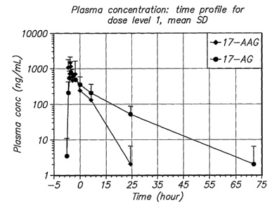
Figure 1 shows the plasma concentration of 17-AAG and 17-AG versus time for
dose
level 1 (0.7 mg/m2 bortezomib and 100 mg/m2 17-AAG), with mean and standard
deviation
(SD) for Day 1 and Day 11 combined.
Figure 2 shows the plasma concentration of 17-AAG and 17-AG versus time for
dose
level 2 (1.0 mg/m2 bortezomib and 100 rng/m2 17-AAG), with mean and SD for Day
1 and
Day 11 combined.
Figure 3 shows the plasma concentration of 17-AAG and 17-AG versus time for
dose
level 3 (1.0 mg/m2 bortezomib and 150 mg/m2 17-AAG), with mean and SD for Day
1 and
1 o Day 11 combined.
Figure 4 shows the plasma concentration of 17-AAG and 17-AG versus time for
dose
level 4 (1.3 mg/m2 bortezomib and 150 mg/m2 17-AAG), with mean and SD for Day
1 and
Day 11 combined.
Figure 5 shows the AUCtotai of 17-AAG and 17-AG for individual patients.
Figure 6 shows the total exposure (the sum of AUCtotal (17-AAG) and AUCtotal
(17-
AG)) for individual patients.
Figure 7 shows the percent reduction of serum M-spike, total IgA, and urine M-
protein in a patient (Patient 201).
Figure 8 shows the percent reduction of serum M-spike and total IgG in a
patient
(Patient 204).
Figure 9 shows the percent reduction of serum M-spike in a patient (Patient
307).
Figure 10 shows the percent reduction of serum M-spike and urine M-protein in
a
patient (Patient 308).
Figure 11 shows the percent reduction of 20S proteasome activity following
doses of
0.7 mg/m2 bortezomib and 100 mg/m2 17-AAG; 1.0 mg/m2 bortezomib and 100 mg/m2
17-
AAG; 1.0 mg/m2 bortezomib and 150 mg/m217-AAG; and 1.3 mg/m2 bortezomib and
150
mg/m2 17-AAG (Treatment Cycle 1, Day 11).
Figures 12A and 12B show the induction of apoptosis and reduction in AKT
levels in
CD138+ myeloma cells after four infusions of 17-AAG.
-10-
CA 02604424 2007-10-05
WO 2006/119032 PCT/US2006/016283
DETAILED DESCRIPTION OF THE INVENTION
Definitions
To aid in understanding and practice of the present invention, definitions for
certain
terms used herein are provided below.
In describing the invention, a concentration of 17-AAG is defined to include a
molar
equivalent concentration of a prodrug of 17-AAG.
In describing the invention, a concentration of 17-AG is defined to include a
molar
equivalent concentration of a prodrug of 17-AG.
"Adverse effects" are as defined in National Cancer Institute (2003).
A "dose limiting toxicity" (DLT) is defined as any of the following clinical
toxicities,
referencing National Cancer Institute (2003). Hematologic toxicities comprise:
(1) Grade 4
neutropenia (absolute neutrophil count (ANC) <0.5 x 10⁹/L) for more than 5
consecutive
days, or febrile neutropenia (ANC < 1.0 x 10⁹/L, fever>_ 38.5 C), (2) Grade 4
thrombocytopenia (platelets < 25.0 x 10⁹/L or bleeding episode requiring
platelets
transfusion), and/or Grade 4 anemia (Hemoglobin < 6.5 g/dl). Non-Hematologic
toxicities
comprise: (1) any _ Grade 3 non-hematologic toxicity (except Grade 3 injection
site
reaction, alopecia, anorexia, fatigue), (2) nausea, diarrhea and/or vomiting
of Grade>_ 3
despite the use of maximal medical intervention. and/or prophylaxis, and/or
(3) treatment
delay of more than 4 weeks due to prolonged recovery from a drug-related
toxicity.
"Complete response (CR)" is defined on the basis of negative immunofixation
("IF")
on both serum and urine, maintained for at least 6 weeks. A bone marrow
aspirate (iiBNIAõ)
containing <5% plasma cells can be used to confirm a CR. A trephine biopsy is
performed,
and the results indicate <5% plasma cells. In non-secretory myeloma, the
marrow biopsy is
repeated after a 6-week interval to confirm a CR. No increase in the size or
number of lytic
lesions should occur (development of a compression fracture does not exclude
response),
with disappearance of soft tissue plasmacytomas.
"KPS performance status" is as defined in Table 1, which also provides a
comparison
against the ECOG Scale.
-11-
CA 02604424 2007-10-05
WO 2006/119032 PCT/US2006/016283
Table 1 - KPS Performance Status
Kamofsky Scale ECOG Scale
Normal, no complaints 100 Fully active, able to carry on all pre- 0
disease performance without
restriction
Able to carry on normal activity, 90
minor signs or symptons of disease
Normal activity with effort 80 Restricted in physically strenuous 1
activity but ambulatory and able to
carry out work of a light or
sedentary nature (e.g., office work
or light house work)
Unable to carry on normal activity 70
or perform active work; cares for
self
Requires occasional assistance but 60 Ambulatory and capable of all self- 2
is able to care for most own needs care but unable to carry out any
work activities; up and about more
than 50% of waking hours
Requires considerable assistance 50
and frequent medical care
Disabled; requires special medical 40 Capable of only limited self-care, 3
care and assistance confined to bed or chair more than
50% of waking hours
Severely disabled; hospitalization 30
indicated although death not
imminent
Very sick; hospitalized and active 20 Completely disabled; cannot 4
perform any self-care; totally
confined to bed or chair
Moribund; fatal processes 10
progressing rapidly
Dead 0
"Minimal response" is defined as one or more of the following: between 25-49%
reduction in serum M-protein, maintained for at least six weeks; between 50-
89% reduction
in urinary light chain excretion which still exceeds 200 mg/24 hours,
maintained for at least
s 6 weeks; for patients with non-secretory myeloma only, between 25-49%
reduction in
plasma cells in a BMA or a bone trephine biopsy, if biopsy is performed,
maintained for at
-12-
CA 02604424 2007-10-05
WO 2006/119032 PCT/US2006/016283
least 6 weelcs; between 25-49% reduction in the size of soft tissue
plasmacytomas (by radio-
graphy or clinical examination); and no increase in the size or number of
lytic lesions (deve-
lopment of a compression fracture does not exclude response). (Blade et al.,
1998.)
"No Change" is defined as not meeting the criteria of either minimal response
or
progressive disease. (Blade et al., 1998.)
"Partial response (PR)" is defined as occurring in patients in whom some, but
not all,
of the criteria for CR have been met, including those in whom routine
electrophoresis is
negative but on whom IF has not been performed. See Blade et al. (1998) for
examples.
"Plateau phase" is defined on the basis of stable paraprotein levels for a
minimum of
1 o 3 months. Plateau will require observations to be within 25% of the value
when response is
assessed, a rise above 25% being one of the criteria for disease progression.
(Blade et al.,
1998.)
"Progression of disease," for patients not in CR, is defined as a definite
increase in
disease activity in patients in partial remission or plateau phase, whereas
the term relapse
applies to a recurrence of evident disease in patients previously in CR. See
Blade et al.
(1998) for examples.
"Refractory cancer" means a cancer that has not responded to one or more
previous
treatment.
"Relapse" means the return of signs and symptoms of cancer after a period of
improvement from one or more previous treatment. "Relapse from CR" is defined
as one or
more of the following: a reappearance of serum or urinary paraprotein on IF or
routine
electrophoresis, confirmed by at least one further investigation and excluding
oligoclonal
reconstitution; a greater than 5% plasma cells in a BMA or on trephine bone
biopsy;
development of new lytic bone lesions or soft tissue plasmacytomas or definite
increase in
the size of residual bone lesions (development of a compression fracture does
not exclude
continued response and may not indicate progression); and development of
hypercalcemia
(corrected serum calcium greater than 11.5 mg/dL) not attributable to any
other cause.
"Therapeutically effective dose" means, otherwise indicated, the amount of
drug that
is required to be administered to achieve the desired therapeutic result.
-13-
CA 02604424 2007-10-05
WO 2006/119032 PCT/US2006/016283
Embodiments
The present invention provides important new methods for using 17-AAG or 17-AG
and prodrugs that exert their anti-cancer effect through the in vivo formation
of 17-AAG or
17-AG to treat MM. The present invention arose in part from the discovery of
new methods
for dosing and administering 17-AAG to achieve and maintain therapeutically
effective
blood levels of 17-AAG or its major metabolite 17-AG (or blood levels of 17-
AAG added
together with 17-AG, as these moieties are equipotent in cellular assays),
expressed as
AUCtotaz, Cma,,, Terminal t~27 Clearance, Volume of distribution, and/or Vss,
without reaching
blood levels likely to cause unmanageable toxicity.
In one embodiment, the method of the present invention comprises administering
multiple doses of 17-AAG, or a prodrug of 17-AAG, and multiple doses of the
proteasome
inhibitor, over a period of three weeks. Collectively, these doses over the
three week period
are called a cycle. A patient may be treated with multiple cycles of therapy.
Different cycles,
including cycles of longer or shorter duration or involving greater or fewer
doses than
described specifically herein, can be used to practice the present invention,
so long as the
therapeutically effective doses described herein are achieved. In one
embodiment, four doses
are administered per cycle, and a pei7od of 3 to 4 days between each dose. In
another
embodiment, four doses are administered per cycle, with two doses per week
administered
for the first two weeks of the three week cycle.
In one embodiment, the therapeutically effective dose is achieved by the
administration of multiple doses of 17-AAG, or a prodrug of 17-AAG or 17-AG,
in
combination with (including separate administration within at least one week
of one another)
a proteasome inhibitor, to a patient with MM over a time period of at least 3
weeks, wherein
such multiple doses result in an AUCtotal for 17-AAG per dose of at least
2,300 but does not
exceed 19,000 ng/mL*h. In one embodiment, four doses are administered per
cycle, with
each dose being at least 100 or 150 mg/m2, and a period of 3 to 4 days between
each dose. In
another embodiment, four doses are administered per cycle, with two doses per
week
administered for the first two weeks of the three week cycle.
Compounds other than 17-AAG or 17-AG can be administered that are converted in
vivo to 17-AAG or 17-AG (prodrugs). One type of prodrug is that in which the
benzo-
quinone ring is reduced to a hydroquinone ring, but is metabolized back to a
benzoquinone
-14-
CA 02604424 2007-10-05
WO 2006/119032 PCT/US2006/016283
ring in the subject. A specific example of a 17-AAG prodrug is 17-allylamino-
18,21-
dihydro-17-demethoxygeldanamycin. (Adams et al., 2005). The methods of the
present
invention therefore include, in one embodiment, a method for treating MM in a
patient in
need of said treatment, wherein the method comprises the administration of
multiple doses
of 17-AAG or 17-AG, or a prodrug of 17-AAG or 17-AG, to a subject with MM,
over a time
period of at least 3 weeks, wherein such multiple doses result in an AUCtotal
for 17-AG per
dose of at least 5,000 but does not exceed 18,000 ng/mL*h. In one embodiment,
four doses
are administered per cycle, with each dose being at least 150 mg/m2, and a
period of 3 to 4
days between each dose. In another embodiment, four doses are administered per
cycle, with
1 o two doses per week administered for the first two weeks of the three week
cycle.
Thus, the present invention includes within its scope the use of prodrugs of
17-AAG
and the term "administering" encompasses the treatment of MM with a
pharmaceutically
equivalent amount of compound that converts to 17-AAG or 17-AG in vivo after
administration to the subject in need thereof. Conventional procedures for the
selection and
preparation of suitable prodrug derivatives are described in Wermuth, 2003.
A proteasome inhibitor is any compound that inhibits protein degradation by a
pro-
teasome that in combination with a 17-AAG, 17-AG or any prodrug of either 17-
AAG or 17-
AG is efficacious in treating a subject suffering from MM or that exerts its
therapeutic action
by a mechanism substantially similar to that of bortezomib. In one embodiment,
the protea-
some inhibitor is an antineoplastic agent and is a reversible inhibitor of the
chymotrypsin-
like activity of the 26S proteasome in mammalian cells. The proteasome
inhibitor can be
natural or synthetic. Suitable natural proteasome inhibitors include, but are
not limited to,
lactacystin, epoxyketones and TMC-95 cyclic peptides. Example of epoxyketones
include,
but are not limited to, epoxomicin and eponemycin. Suitable synthetic
proteasome inhibitors
include, but are not limited to, peptide aldehydes and peptide vinyl sulfones.
Examples of
peptide aldehydes include but are not limited to Z-Leu-Leu-Leu-al (MG132), Z-
Ile-
Glu(Obut)-Ala-Leu-al (PSI), and Ac-Leu-Leu-Nle-al (ALLN). See, e.g., Kisselev
and Gold-
berg (2001) and Richardson et al. (2003b). Examples of proteasome inhibitors
include, but
are not limited to, PS-519 (Shah et al. (2002)), NPI-0052 (Cusack et al.
(2005)), ZL3VS
(Kadlcikova et al. (2004)), AdaAhx3L3VS (Kadlcikova et al. (2004)), efrapeptin
(Abra-
hams, et al. (1996)). In one embodiment, the peptide aldehyde has the aldehyde
group
-15-
CA 02604424 2007-10-05
WO 2006/119032 PCT/US2006/016283
replaced with boronic acid to form a peptide boronate. In one embodiment, the
peptide
boronate is a dipeptide boronic acid, preferably bortezoniib.
Bortezomibis an antineoplastic modified dipeptidyl boronic acid that is a
reversible
inhibitor of the chymotrypsin-like activity of the 26S proteasome in mammalian
cells. The
making and using of bortezomib and suitable pharmaceutical formulations and
means of
administration thereof, are taught in Adams et al. (1998, 2000, 2001, 2003,
and 2004) and
Gupta (2004). Bortezomib is commercially available under the brand name
Velcade
(Millennium Pharmaceuticals, Inc., Cambridge, MA) and is approved for the
treatment of
MM patients who have received at least one prior therapy and have demonstrated
disease
1 o progression after the preceding therapy. A pharmaceutical formulation
comprising
bortezomib can comprise about 0.9% saline and 1.0 mg/mL mannitol. A single
dosage of
bortezomib can be from at least about 0.7 to about 1.3 mg/m2. The bortezomib
can be
administered by injection, with the entire dose is injected within 3 to 5
seconds into the
subject by direct injection or intravenous infusion.
The subject in need of treatment, for purposes of the present invention, is
typically a
human patient suffering from MM, although the methods of the invention can be
practiced
for veterinary purposes, with suitable adjustment of the unit dose to achieve
the equivalent
AUCtotai or other PK and PD parameters described herein for the particular
mammal of
interest (including cats, cattle, dogs, horses, and the like). Those of skill
in the art of
pharmaceutical science know or can readily determine the applicable conversion
factors for
the species of interest from the present disclosure of the doses and PK
parameters for human
therapy. Typically, however, the methods will be practiced to benefit human
subjects, and
those subjects will typically have exhibited some histological evidence of MM,
including
one or more of the following: M spike in serum or urine, BM plasmacytosis of >
30%,
anemia, renal failure, hypercalcemia, and/or lytic bone lesions.
In one embodiment, the subject has been diagnosed with Stage III MM under the
Durie-Salmon system and exhibits one or more of these symptoms: hemoglobin
value < 8.5
g/dL, serum calcium value > 12 mg/dL, advanced lytic bone lesions (scale 3),
high M-com-
ponent production rate (IgG value > 7 g/dL; IgA value > 5 g/dl; Bence Jones
protein > 12
g/24 hour). Alternatively, the has been diagnosed with Stage III MM based on
the Inter-
national Staging System (ISS) system, with serum levels of 0-2 microglobulin >
5.5 g/dL.
- 16-
CA 02604424 2007-10-05
WO 2006/119032 PCT/US2006/016283
In another embodiment, the subject been diagnosed with Stage II MM under the
Durie-Salmon system but does not have Stage III MM and has some but not all of
these
symptoms: hemoglobin value > 10 g/dL, serum calcium value < 12 mg/dL, bone x-
ray,
normal bone structure (scale 0) or solitary bone plasmacytoma only, low M-
component
production rate (IgG value < 5 g/dL; IgA value < 5 g/dL). Alternatively, the
subject has been
diagnosed under the ISS system with Stage II MM but not Stage IlI MM and does
not have
sei2im levels of (3-2 microglobulin < 3.5 g/dL and albumin _ 3.5 g/dL.
In another embodiment, the patient will have one or more of the following
signs or
symptoms of MM: an elevated level of serum M protein (such as > 3 g/dL),
and/or more
than 10% of the cells in a BM sample from the subject are plasma cells. In
another
embodiment, prior to treatment the Karnofsky performance status (KPS) of the
patient is at
least 70%. In another aspect, the KPS of the patient is at least 60%, 50%,
40%, 30%, 20%, or
10%. In one aspect, the ECOG of the patient is at least 0, 1, 2, or 3.
A therapeutically effective dose of 17-AAG, 17-AG, or a prodrug of either 17-
AAG
or 17-AG, and a therapeutically effective dose of the proteasome inhibitor are
the amounts of
17-AAG, 17-AG, or a prodrug of either 17-AAG or 17-AG, and the proteasome
inhibitor,
respectively, that is administered in combination at each administration over
one treatment
cycle to the subject that brings about a therapeutic result. The therapeutic
result can be that
the rate of the progression or spread of the cancer is slowed or stopped for
some period of
time. In some patients, the therapeutic result can be partial or complete
elimination of MM.
In some patients, a therapeutic result will be achieved with one treatment
cycle. In other
patients, a therapeutic result will be achieved only after multiple cycles of
treatments. As
those of skill in the art will appreciate, however, there can be no assurance
that every MM
patient will achieve a therapeutic result with any anti-cancer therapy.
As noted above, in one embodiment, each treatment cycle is three weeks. In
other
embodiments, other treatment cycle times can be employed, such as two or four
weeks (or
one month), so long as the equivalent AUCtotal or other PK and PD parameters
described
herein are achieved. The unit dose employed in each cycle is administered at
least once and
up to eight times per treatment cycle. Typically, the dose is administered two
to four times
per treatment cycle. In one embodiment, the dose is administered twice weekly
for 2 weeks
out of each treatment cycle of three weeks. For example, if one starts a cycle
at the
-17-
CA 02604424 2007-10-05
WO 2006/119032 PCT/US2006/016283
administration of the first dose, then in one embodiment, the unit dose is
administered once
or twice in the first two weeks of the treatment cycle and not during the
third week. In one
embodiment, the dose is administered on days 1, 4, 8, and 11 of each treatment
cycle, with
day 1 being the day the first dose is administered.
Each unit dose of 17-AAG is a dose of not more than the maximally tolerable
dose
("MTD"), which can be defined as the maximum dose at which one or fewer of six
subjects
undergoing the method of treatment experience hematologic or non-hematologic
toxicity not
amenable to supportive care. Preferably, the amount of 17-AAG administered is
equal to or
less than the MTD. Preferably, the amount of 17-AAG administered is one that
does not
result in unacceptable and/or unmanageable hematologic or non-hematologic
toxicity.
The therapeutically effective amount of a unit dose 17-AAG or 17-AG or a
prodrug
of either is the amount that, after one or more cycles of administration in
accordance with
this invention, results in a complete response (CR), a partial response (PR),
a minimal
response (MR), a stable disease (StD) condition, a reduction of serum
monoclonal protein
(serum M protein), or a reduction of plasma cells in the BM of the subject
(Blade et al.,
1998), for at least a period of time, such as 3 weeks, 6 weeks, 2 months, 6
months, one year,
or several years. In one embodiment, the administration of 17-AAG results in a
decrease in
serum and/or urine M protein, BM plasmocytosis, alleviation of anemia,
alleviation of renal
failure, alleviation of hypercalcemia, and/or reduction/alleviation of lytic
bone lesions in the
MM patient. In one embodiment, some patients will not relapse from a CR or
will
experience a significant delay in the progression of the disease.
The amount of 17-AAG administered in a single unit dose can range from 100 to
340
mg/m2 per dose. Where the 17-AAG is administered twice weekly for two out of
every three
weeks, the amount of 17-AAG administered ranges from 100 to 340 mg/m2 per
dose.
Preferably, the amount of 17-AAG administered ranges from 150 to 340 mg/m2 per
dose.
The amount of 17-AAG administered may also range from 220 to 340 mg/m2 per
dose.
Those of skill in the art will recognize that the unit dose amounts of 17-AAG
or 17-AG
prodrugs or 17-AG itself can be calculated from the doses provided herein for
17-AAG and
the PK parameters provided for 17-AAG and 17-AG and the molecular weight and
relative
so bioavailability of the prodrug or 17-AG.
-18-
CA 02604424 2007-10-05
WO 2006/119032 PCT/US2006/016283
The method of the invention can also be described in terms of the amount of 17-
AAG
administered per treatment cycle. The per cycle amount will typically be
greater than 400
mg/m2, and more usually will be greater 600 mg/m2. Typically the per cycle
amount will be
at least 880 mg/m2. In various embodiments, the amount of 17-AAG administered
is at least
600 to 1,360 mg/m2 per treatment cycle; 880 to 1,360 mg/m2 per treatment
cycle; and 1,100
to 1,360 mg/m2 per treatment cycle.
Where the proteasome inhibitor is bortezomib, the amount administered in a
single
dose can range from 0.7 to 1.3 mg/m2 per dose. The amount administered in a
single unit
dose can be 0.7, 1.0, or 1.7 mg/m2 per dose. Where the bortezomib is
administered twice
weekly for two out of every three weeks, the amount administered can range
from 0.7 to 1.3
mg/m2 per dose. The method of the invention can also be described in terms of
the amouint
of bortezomib administered per treatment cycle. The per-cycle amount will
typically be
greater than 2.8, and more usually greater 4.0 mg/m2. Typically the per-cycle
amount will be
at least 5.2 mg/m2. Alternatively, the amount of bortezomib administered is at
least 2.8 to 5.2
mg/m2 per treatment cycle or 4.0 to 5.2 mg/m2 per treatment cycle.
As noted above, the frequency of the administration of the unit dose is once
weekly
or twice weekly. In one embodiment of the method of the invention, the
pharmaceutical
formulation is administered intravenously twice weekly for 2 weeks every 3 or
4 weeks. In
one embodiment, the patient is administered a pre-treatment medication to
prevent or
ameliorate treatment related toxicities. Illustrative pre-treatment
medications are described in
the examples below. In one embodiment of the method of the invention, the
administration
of 17-AAG or 17-AG or a prodrug of either is performed on day 1, 4, 8 and 11
of each cycle,
and the cycle time is 3 weeks. 17-AAG will typically be administered by
intravenous
infusion, infused in a period of at least 30, 60, 90, or 120 minutes. For
patients with a body
surface area (BSA) greater than 2.4 m2, dosing can be calculated in accordance
with the
methods herein using a maximum BSA of 2.4 m2.
In human clinical trials of the method of the invention, the following
administration
regimens have been employed without reaching dose limiting toxicity (DLT) in
any treated
patient: 275 mg/rn2 per single administration of 17-AAG twice weekly for two
out of three
weeks (Days 1, 4, 8, and 11, with a cycle time of 21 days).
-19-
CA 02604424 2007-10-05
WO 2006/119032 PCT/US2006/016283
As noted above, after 17-AAG is administered, the major metabolite 17-AG,
having
anti-cancer activity in its own right, appears in the subject. 17-AAG and 17-
AG are thus
each, and together, responsible for the therapeutic benefit of the method of
the invention.
The therapeutically effective dose and dosing regimen of 17-AAG is one that
achieves an
Area Under Curve (AUCtotal) of 17-AAG and/or 17-AG in the subject as described
herein.
Various therapeutically effective doses and dose regimen are illustrated in
the examples
below. Therapeutically effective doses and dosing regimen of 17-AAG and/or 17-
AG
provided by the present invention can also be described in terms of Terminal
Half Life (t~iz);
Clearance (CL); and/or Volume of Distribution in the elimination phase or
steady state (VZ
and/or VSS).
The therapeutic benefit from the treatment method of the present invention can
be
observed in responding subjects as soon as 3, 6, 12, 1S or 24 weeks from the
start of treat-
ment. In one embodiment, a therapeutic benefit from the treatment is a
reduction in a serum
protein, and/or BUN or serum calcium, of the patient. In various embodiments,
the reduction
is at least 25%; at least 50% to 80%; at least 90%; and 100%. The reduction in
serum M
protein can be determined, for example, by serum protein electrophoresis or
immuno-fixa-
tion techniques. The percent reduction is the level of the serum M protein,
BUN, or calcium
in the patient, measured after a period of treatment and then compared to the
level of the
serum M protein, BUN, or calcium in the patient measured just prior to
treatment. Serum
proteins are proteins that, when present in elevated levels in the serum,
indicate the subject
suffers from MM. Such serum proteins include, but are not limited to, serum M
protein (also
known as serum M paraprotein), 0-2 microglobulin, light chain, and total
protein.
Other therapeutic benefits that can be achieved via the present invention
include one
or more of the following: decrease in BM plasmaocytosis, alleviation of
anemia, alleviation
of renal failure, alleviation of hypercalcemia, and/or reduction/ alleviation
of lytic bone
lesions. Another therapeutic benefit is an improvement of the KPS of the
patient by 10% or
more, 20% or more, 30% or more, 40% or more, or 50% or more. Another
therapeutic bene-
fit is an improvement of the ECOG of the patient by 1 or more, 2 or more, or 3
or more.
Ideally, practice of the present invention does not result in unmanageable
hema-
tologic or non-hematologic toxicity. Hematologic toxicities to be avoided
include: Grade 4
neutropenia, Grade 4 thrombocytopenia, and/or Grade 4 anemia. Non-hematologic
toxicities
-20-
CA 02604424 2007-10-05
WO 2006/119032 PCT/US2006/016283
include: any >_ Grade 3 non-hematologic toxicity (except Grade injection site
reaction,
alopecia, anorexia, and/or fatigue), nausea, diarrhea and/or vomiting > Grade
3 (despite use
of maximal medical intervention and/or prophylaxis), and/or treatment delay of
more than 4
weeks due to prolonged recovery from a drug related toxicity. Those of skill
in the art will
recognize that various toxicities may occur in a cancer patient; the method of
the present
invention provides the benefit of reduced or elimination of the occurrence of
such toxicities.
Where the pharmaceutical formulation comprises an additional compound that
might
cause an anaphylactic reaction (like Cremophor n), additional medications can
be
administered to prevent or reduce the anaphylactic reaction, such as (a)
loratidine or
1 o diphenhydramine, (b) famotidine, and (c) methylprednisone or
dexamethasone.
The present invention also provides, in various embodiments, methods for
treating
MM by administering 17-AAG or 17-AG, or a prodrug of either, in combination
with a
proteasome inhibitor and a third anti-cancer compound, which can be, for
example,
Thalomid , Aredia", and Zometa or Revlimid" (lenalidomide). The other anti-
cancer drug
or agent can be administered in unit doses and dosing regimen currently
employed in the art.
The present invention can be used to treat patients with MM who have failed at
least
one prior anti-cancer therapy regimen, that is, have refractory or relapsed
refractory MM.
These prior anti-cancer therapies include, but are not limited to, monotherapy
(single agent
therapy) or combination therapies of the following treatments and anti-cancer
agents: che-
motherapy, stem cell transplantation, Thalomid , Velcade , and Revlimid".
Chemotherapy
includes treatment with a combination melphalan and prednisone (MP), VAD, or
an alkyla-
ting agent alone or in combination with other agent(s), such as
cyclophosphamide plus
etoposide or combinations of etoposide, dexamethasone, doxorubicin.
Diagnostic and laboratory methods and tests that may be of benefit in practice
of the
present invention are well known to one of ordinary skill in the art. See, for
example, Pagana
and Pagana, Mosby's Manual of Diagnostic and Laboratory Tests, 2d Ed., Mosby-
Year
Book, 2002 and Jacobs & DeMott Laboratory Test Handbook, 5"' Ed., Jacobs et
al. (eds),
Lexi-Comp, Inc., 2001 (each incorporated herein by reference). Free kappa and
free lambda
light chain concentrations in serum can be measured using FreeliteTM (The
Binding Site Inc.,
3o Birmingham, United Kingdom).
-21-
CA 02604424 2007-10-05
WO 2006/119032 PCT/US2006/016283
An active pharmaceutical ingredient ("API," 17-AAG, 17-AG, prodrug, proteasome
inhibitor, other anti-cancer compound, etc.) useful in the method of the
present invention can
be formulated for administration orally or intravenously, in a suitable solid
or liquid form.
See Gennaro, ed., Remington: The Science and Practice of Pharmacy, 20th Ed.
(Lippincott
Williams & Wilkins 2003), incorporated herein by reference. The API can be
compounded,
for example, with a non-toxic, pharmaceutically acceptable carrier or
excipient for solutions,
emulsions, suspensions, or any other form suitable for enteral or parenteral
administration.
Pharmaceutically acceptable carriers include water and other carriers suitable
for use in
manufacturing preparations in liquefied form. In addition, auxiliary
stabilizing, thickening,
1 o and coloring agents may be used.
An API useful in the method of the invention may be formulated as
microcapsules,
nanoparticles, or nanosuspensions. General protocols for such formulations are
described,
for example, in Microcapsules and Nanoparticles in Medicine and Pharmacy by
Max
Donbrow, ed., CRC Press (1992) and in Bosch et al. (1996), De Castro (1996),
and Bagchi
et al. (1997). By increasing the ratio of surface area to volume, these
formulations are
especially suitable for the delivery of 17-AAG or another relatively insoluble
API.
17-AAG can be formulated in an emulsion with vitamin E or a PEGylated
derivative
thereof. Generic approaches to formulations with such excipients are described
in Quay et al.
(1998) and Lambert et al. (2000). The 17-AAG can be dissolved in an aqueous
solution
containing ethanol (preferably less than 1% w/v). Vitamin E or a PEGylated-
vitamin E is
added. The ethanol is then removed to form a pre-emulsion that can be
formulated for
intravenous or oral routes of administration.
Another method for preparing a pharmaceutical formulation useful in the
present
method involves encapsulating 17-AAG or other API in liposomes. Methods for
forming
liposomes as drug delivery vehicles are well known in the art. Suitable
protocols adaptable
for the present invention include those described by Boni et al. (1997),
Straubinder et al.
(1995), and Rahman et al. (1995) for paclitaxel and by Sonntag et al. (2001)
for epothilone,
mutatis mutandis. Of the various lipids that may be used in such formulations,
phosphatidyl-
choline and polyethyleneglycol-derivatized distearyl phosphatidyl-
ethanoloamine are
3o noteworthy.
-22-
CA 02604424 2007-10-05
WO 2006/119032 PCT/US2006/016283
The amount of 17-AAG or other API that may be combined with the carrier
materials
to produce a single or unit dosage form will vary depending upon the subject
treated and the
particular mode of administration. For example, a formulation for intravenous
use comprises
an amount of 17-AAG ranging from about 1 mg/mL to about 25 mg/mL, preferably
from
about 5 mg/mL, and more preferably about 10 mg/mL. Intravenous formulations
are
typically diluted between about 2 fold and about 30 fold with water for
injection (WFI),
normal saline, or 5% dextrose solution prior to use. In many instances, the
dilution is
between about 5 and about 10 fold.
In one embodiment of the method of the invention, 17-AAG is formulated as a
1 o pharmaceutical solution formulation comprising 17-AAG dissolved in a
vehicle comprising
(i) a first component that is ethanol; (ii) a second component that is a
polyethoxylated castor
oil; and (iii) a third component selected propylene glycol, PEG 300, PEG 400,
glycerol, and
combinations thereof, as disclosed in Zhong et al. (2005).
Another formulation of 17-AAG that may be used is one based on
dimethylsulfoxide
("DMSO") and egg lecithin (egg phospholipids), as taught in Tabibi et al.
(2004). However,
because of certain characteristics of DMSO (odor, patient adverse reactions),
such formu-
lations are less preferred than the DMSO-free ones taught herein.
Other formulations for 17-AAG that may be employed in the method of the
invention
are described in Ulm et al. (2003), Ulm et al. (2004), Mansfield et al.
(2006), Desai et al.
(2006), and Isaacs et al. (2006).
In another embodiment, the pharmaceutical formulation can be diluted 1:7 prior
to
administration with sterile WFI, USP (one part undiluted drug product to 6
parts sterile
WFI). Dilution is performed under controlled, aseptic conditions. The final
diluted drug
product concentration is, using 17-AAG as an example, at least 1.00 mg/niL,
such as
approximately 1.43, approximately 2.00 or approximately 10.00 mg/mL.
Depending on the BSA and the assigned dose, the dose of 17-AAG or other API
will
require different volumes of drug product to be added to the admixture bag. An
overfill can
be calculated and employed to account for loss in the administration set.
Preferably, the
pharmaceutical formulation, with the diluted drug product, is pH neutral, and
the solution is
3o hypertonic at approximately 600 mOsm. The pharmaceutical formulation can be
stored at -
20 C, with protection from light. Drug product is allowed to come to room
temperature prior
-23-
CA 02604424 2007-10-05
WO 2006/119032 PCT/US2006/016283
to admixture and then is mixed is by gentle inversion. After dilution, the
drug product should
stable for up to about 10 hours at room temperature (at a dilution of 1:7).
The present invention, having been described in summary fashion and in detail
above
is illustrated in the following Examples.
Example 1- Treatment of Patients with Multiple Myeloma with 17-AAG in
combination with Bortezomib
The method of the invention was tested in an open-label, dose escalating
clinical
trial. The trial was designed to establish the MTD of 17-AAG administered by
IV infusion
over 60 minutes, co-administered with bortezomib, on Days 1, 4, 8, and 11 of a
dosing cycle
1 o lasting 3 weeks. The dose-escalating component of this trial began with
bortezomib
administered at approximately 50% of its recommended dose and the starting
dose of 17-
AAG set at slightly less than 50% of its single-agent dose using a previous
formulation (100
mg/m2). Doses of each agent were then escalated until the MTD for the
combination could
be ascertained.
Disease response evaluations were performed following every two cycles of
treatment (approximately every 6 weeks). The determination of anti-tumor
efficacy in stable
or responding patients was based on objective tumor assessments made according
to a
standardized myeloma response assessment system.
All baseline imaging-based tumor assessments were performed within 28 days
prior
to the start of treatment and reevaluated every 6 weeks (approximately every
two cycles)
thereafter. All patients with responding tumors (CR or PR) were examined to
confirm the
response 6 weeks after the first documentation of response. Response criteria
used were
according to guidelines of Blade et al. (1998).
Pharmacokinetic (PK) and pharmacodynamic (PD) sampling was obtained during the
first treatment cycle only. In the event of drug-related serious adverse
events (SAEs) and/or
Grade 4 toxicities, additional PK samples were to be collected.
MM patients enrolled in this study were those who had failed at least two
prior anti-
cancer therapy regimens. The enrollment criteria were: (1) patients were at
least18 years old;
(2) had a KPS performance status of > 70%; (3) had histologic evidence of MM
but did not
3o necessarily have measurable disease, although disease had to have been
assessed within 28
days prior to treatment initiation; (4) were, with respect to all adverse
events of any prior
-24-
CA 02604424 2007-10-05
WO 2006/119032 PCT/US2006/016283
chemotherapy, surgery, or radiotherapy, resolved to NCI CTCAE (v. 3.0) Grade
<_ 2; and (5)
had the following laboratory results within 10 days of 17-AAG administration:
hemoglobin
>8 g/dL, absolute neutrophils count >_ 1.5 x 10⁹ /L, platelet count _ 75 x 109
/L, serum bili-
rubin 5 2 x upper limit of normal (ULN), AST 5 2.5 ULN, and serum creatinine
<_ 2 x ULN.
[0100] Patients were graded according to the KPS Performance Status scale and
criteria
as described in Table 1. Patients were excluded from the study if they had a
condition such
as pre-existing neuropathy, pregnancy, breast-feeding, recent chemotherapy,
and so forth. To
be eligible for enrollment, patients also had to meet certain hematologic
conditions.
[0101] 17-AAG is highly protein bound in plasma (approximately 95% in in vitro
assays
using human blood); however, the plasma protein to which the drug binds and
the affinity of
binding are not known. Patients who are receiving agents that are known to be
highly protein
bound were subjected to close clinical monitoring while enrolled in the trial.
In vitro studies
implicate the involvement of cytochrome P450 enzymes in the metabolism of 17-
AAG. No
formal drug-drug interaction studies have been performed with 17-AAG and drugs
that are
substrates, inhibitors, or inducers of cytochrome P450-3A4. While there is no
contra-
indication to the concomitant use of any medication with 17-AAG, 17-AAG was
used with
caution in combination with drugs that are also highly protein bound (e.g.
warfarin) and
drugs that are a substrate, inhibitor, or inducer of cytochrome P450-3A4.
Hormonal contra-
ceptives were not used in women of childbearing potential enrolled in the
trial. No other
investigational agents are permitted during the entire duration of the study
(from 3 weeks
before the first administration until the end to treatment evaluation).
[0102] PK assessments included the following tests. Blood samples for
determination of
plasma concentrations of the parent compound and its primary metabolite were
collected
following the first and fourth 17-AAG administration only (Day 1 and 11). The
total number
of PK samples collected was approximately 115 mL of whole blood (7-8
tablespoons). If a
patient experienced a potentially drug-related SAE, additional PK samples were
collected.
Blood was drawn from the contralateral arm to the infusion site using an
indwelling catheter
to avoid multiple needle sticks. For the 17-AAG samples, 5 mL of blood was
drawn into a
vacuum tube containing heparin as anti-coagulant. The blood tube was inverted
several times
and the tube placed in wet ice immediately pending separation of the plasma.
If a catlleter
was used for blood collection, the fluid in the catheter was completely
withdrawn prior to
-25-
CA 02604424 2007-10-05
WO 2006/119032 PCT/US2006/016283
each sample collection and discarded. Plasma samples were kept on wet ice
during collection
and centrifugation. Plasma samples were split into two cryovials prior to
freezing at -70 C.
Plasma concentrations of 17-AAG and its primary metabolite 17-AG were measured
by a
validated LC/MS method. (Egorin et al., 1998.)
[0103] PD assessment included the following tests. (1) Clinical correlates:
the
occurrence of specific toxicities of interest (e.g., severity, duration and
reversibility) was
compared to PK parameters (e.g., clearance, exposure, elimination half-life,
maximal plasma
concentration, and time above a target plasma concentration). These included
hepatotoxicity
and gastrointestinal toxicities. (2) Multiple myeloma cells: (i) surface
expression of IL-6R,
1o insulin-like growth factor receptor-1 (IGF-IR) in MM cells; (ii) total
expression of phospho-
AKT, Akt, Hsp90 and Hsp70 in MM cells; and (iii) gene expression profiling to
identify
other potential bio-markers for drug sensitivity versus resistance. MM cells
were purified
from bone marrow (BM) aspirates performed at baseline (up to 3 weeks prior to
first study
drug administration), 3-4 hours following the fourth infusion of 17-AAG and
bortezomib
(Day 11), and after the end of treatment (or at time of progressive disease).
MM cells were
purified from the BM aspirates based upon CD138 expression using magnetic bead
technology and confirmed by flow cytometric analysis to be >95% CD138+ MM
cells. Flow
cytometric analysis assesses IGF-IR surface expression using fluorescein
isothyocynate
(FITC)-conjugated anti-human IGF-IR monoclonal antibody (R&D Systems,
Minneapolis,
MN). Immunoblotting analyses evaluated the total levels of phospho-AKT, AKT,
Hsp90 and
Hsp70. (3) Peripheral blood mononuclear cells: PBMCs were obtained (pre-
therapy and 4
hours following the bortezomib intravenous bolus on Days 1 and 11) and
examined for
change in Hsp70, Hsp90, and others as indicated via Western Blot. For PBMC
isolation,
blood was collected into preservative-free heparin and PBMCs isolated by
Ficoll-Paque
density gradient centrifugation. (4) The percentage inhibition of proteasome
function
(evaluated by measurement of 20S proteasome activity) was performed, according
to the
method of Lightcap et al (2000). Whole blood lysates were obtained prior to
the infusion, 1,
4 and 24 hours following the IV bolus of bortezomib on Days 1 and 11. (5)
Plasma: whole
blood (8cc per timepoint) was collected into EDTA-containing tubes.
[0104] The end-of-treatment assessment was conducted as follows. The planned
treatment period was 24 weeks (8 cycles). Patients were treated in the abs6nce
of progressive
disease or unacceptable treatment-associated toxicities. All patients who
received at least one
-26-
CA 02604424 2007-10-05
WO 2006/119032 PCT/US2006/016283
dose of the study drug and discontinued treatment for any reason (except
death) had the end
of treatment assessment performed. The assessment occurred up to 28 days
following the
last receipt of 17-AAG and included a physical examination, with body weight
and vital
signs measurements, documentation of KPS Performance Status, hematology,
coagulation
and chemistry/electrolyte determinations, urinalysis, assessment of the
patient's current
medications and ongoing clinical adverse events (if any). Tumor assessments
(myeloma
laboratory tests, assessment of extramedullary disease, BM aspirate, and other
radiographic
staging, if appropriate) were done at this time only if the previous
assessment occurred more
than 4 weeks prior to withdrawal.
[0105] Bortezomib (obtained commercially) was administered intravenously twice
weekly for 2 weeks (on Day 1, 4, 8 and 11) every 3 weeks at escalating doses
(calculated
mg/m2) administered as a rapid (3-5 second) injection. Bortezomib was
administered per its
Package Insert (incorporated herein by reference). The starting dose of
bortezomib was 0.7
mg/m2; doses were escalated based on observed toxicities. The dose did not
escalate beyond
its recommended dose for single-agent therapy in this population (1.3 mg/m2).
[0106] 17-AAG was administered intravenously twice weekly for 2 weeks (on Day
1, 4,
8 and 11) every 3 weeks at escalating doses (calculated mg/m2) infused over 60
minutes after
pre-medication. For patients with a body surface area (BSA) greater than 2.4
m2, dosing was
calculated using a maximum BSA of 2.4 m2.
[0107] The preparation and administration of 17-AAG was as follows. 17-AAG was
dissolved in 30% propylene glycol, 20% Cremophor EL, and 50% ethanol to a
concentra-
tion of 10 mg/mL in the vial. Drug product was available in 20 mL type 1 clear
glass vials
with a 20 mm finish (containing 200 mg/vial). The vials were closed with gray
20 mm
Teflon coated serum stoppers and white 20 mm flip-off white lacquered flip
tops. It was
diluted 1:7 prior to administration with sterile WFI, USP (one part undiluted
drug product to
6 parts sterile WFI). Dilution was performed under controlled, aseptic
conditions. Final
diluted drug product had a concentration of approximately 1.43 mg/mL. 17-AAG
was pre-
pared either using glass vacuum containers or compatible non-PVC, non-DEHP
(di(2-ethyl-
hexyl)phthalate) IV admixture bags. Both systems require non-PVC, non-DEHP
containing
administration sets and either an in-line 0.22 m filter or use of an
extension set containing
such a filter. Due to the light sensitivity of 17-AAG, protection from light
is advised.
-27-
CA 02604424 2007-10-05
WO 2006/119032 PCT/US2006/016283
[0108] For glass collection units, examples of compatible supplies includes
Baxter
1A8502 (or equivalent), using a Baxter 2C1106 or equivalent IV administration
set with
extension set with 0.22 m air eliminating filter (Baxter 1C8363 or
equivalent). For non-
PVC, non-DEHP admixture bags, compatible admixture bags may be empty or pre-
filled
with 250cc WFI. Examples of compatible admixture bags include Excel (250cc
WFI; made
from polyolefin).
[0109] Depending on the body surface area and the assigned dose for individual
patients,
the dose of 17-AAG required different volumes of drug product to be added to
the admixture
bag. An overfill was calculated to account for any loss in the administration
set.
[0110] As noted above, 17-AAG was administered intravenously twice weekly for
2
weeks out of every 3 weeks. The total dose delivered is rounded to the nearest
milligram.
[0111] Pre-medication treatments were conducted as follows. All patients were
pre-
medicated prior to each infusion of 17-AAG. An appropriate pre-medication
regimen was
used for each patient based upon past history of potential Cremophorrt-induced
hypersensitivity reactions and the type and severity of the hypersensitivity
reaction observed
following treatment with 17-AAG. The standard premedication regimen was to pre-
medicate
with loratidine 10 mg p.o., famotidine 20 mg p.o., and either
methylprednisolone 40-80 mg
IV or dexamethasone 10-20 mg IV 30 minutes prior to infusion of 17-AAG. Choice
of
antihistamine and corticosteroid, route of administration, doses prior to 17-
AAG infusion
was at the investigator's discretion, but was similar to prophylaxis for other
Cremophor -
containing products (such as Taxol , paclitaxel). Doses of corticosteroid were
lowered if the
patient is receiving concomitant prednisone. The high dose premedication
regimen was to
pre-medicate with diphenhydramine 50 mg IV, famotidine 20 mg IV and either
methylpred-
nisolone 80 mg IV or dexamethasone 20 mg IV (or split as oral doses of 10 mg
each 6 and
12 hours prior to the infusion), at least 30 minutes prior to the infusion of
17-AAG. The
choice of antihistamine and corticosteroid was at the investigator's
discretion.
[0112] The doses and schedule of study drugs was as follows. Patients received
therapy
on Days 1, 4, 8 and 11 in 3-week cycles. Therapy consisted of bortezomib
administered as
an intravenous rapid (3-5 second) bolus, followed by 17-AAG administered via
intravenous
infusion (IV) over 60 minutes. The infusion of 17-AAG was elongated to 90 or
120 minutes
if necessary at the higher doses due to volume of administration. For the
initial
- 28 -
CA 02604424 2007-10-05
WO 2006/119032 PCT/US2006/016283
administration, all patients were administered with 17-AAG with bortezomib,
except for
patients who had failed bortezomib therapy immediately prior to study entry.
[0113] The initial patient cohort received bortezomib at dose of 0.7 mg/m2,
followed by
an intravenous infusion of 17-AAG at dose of 100 mg/mz (cohort 1). Subsequent
patient
cohorts were enrolled per the escalation scheme as follows: bortezomib at a
dose of 1.0
mg/m2 and 17-AAG at a dose of 100 mg/m2 (cohort 2), bortezomib at a dose of
1.0 mg/m2
and 17-AAG at a dose of 150 mg/m2 (cohort 3), bortezomib at a dose of 1.3
mg/m2 and 17-
AAG at a dose of 150 mg/m2 (cohort 4), bortezomib at a dose of 1.3 mg/m2 and
17-AAG at a
dose of 220 mg/m2 (cohort 5), bortezomib at a dose of 1.3 mg/m2 and 17-AAG at
a dose of
1 o 275 mg/m2 (cohort 6), and bortezomib at a dose of 1.3 mg/m2 and 17-AAG at
a dose of 340
mg/m2 (cohort 7).
[0114] Three patients were assigned to each cohort. If no DLT is observed in a
cohort
evaluable for a dose escalating decision ("evaluable" is defined here as
having received four
treatments in a 3-week period or having withdrawn due to drug-related
toxicity), then the
next dose level was evaluated. If one or more patients experience a DLT, then
the cohort was
increased to six evaluable patients. If two or more of six evaluable patients
entered in a
cohort experienced a DLT then the MTD had been exceeded; all further accrual
would be at
the previous dose level. If no more than one of the six patients experienced a
DLT then the
next dose level was evaluated. Once the MTD was defined an additional number
of patients
were enrolled to arrive at a cumulative total of 12 patients at the MTD dose
level. Eighteen
patients were treated in accordance with this protocol.
[0115] Of the eighteen patients, 9 were male and 9 were female. Their median
age was
63 years old (having a range of 44 to 81 years old). Their subtype were 72%
were IgG and
28% were IgA. The KPS median was 90 (having a range of 70 to 100). The number
of prior
chemotherapy was 4 (having a range of 2 to 16). Prior chemotherapy included
inter alia bor-
tezomib, thalidomide, VAD/VdD, melphalan, and lenalidomide. The number of
patients with
prior transplants was 12 (67%). The number of patients with extramedullary
disease was 4
(22%). The median baseline j3-2 microglobulin was 3.7 (having a range of 1.4
to 9.7). The
median time since diagnosis of MM was 61 months (having a range of 14 to 238
months).
[0116] Three patients (cohort 1; Patients 101-103) were first administered
with 0.7
mg/m2 of bortezomib (infused as a rapid 3-5 seconds intravenous push), and
then
-29-
CA 02604424 2007-10-05
WO 2006/119032 PCT/US2006/016283
administered a 100 mg/m2 dose of 17-AAG (one hour intravenous infusion), twice
weekly
for every 2 out of 3 weeks (Days 1 and 11 of the first treatment cycle).
Patients underwent a
mean of 3.3 cycles of treatment. DLT was not observed in any the three
patients. Of the three
patients, after treatment, stable disease was observed in one patient who
underwent 5 cycles
of treatment (33% of all patients treated at this dose level), and progressive
disease was
observed in two patients (67% of all patients treated at this dose level).
[0117] Three patients (cohort 2; Patients 201, 203 and 204) were first
administered with
1.0 mg/mZ of bortezomib (infused as a rapid 3-5 seconds intravenous push), and
then admi-
nistered a 100 mg/m2 dose of 17-AAG (one hour intravenous infusion), twice
weekly for
1 o every 2 out of 3 weeks (Days 1 and 11 of the first treatment cycle).
Patients underwent a
mean of 11.3 cycles of treatment. DLT was not observed in any the three
patients. Of the
three patients, after treatment, treatment resulted in MR for all three
patients (100% of all
patients treated at this dose level). One of the three patients was a
bortezomib naive patient.
Two patients underwent at least 9 cycles of treatment. One patient underwent 9
cycles of
treatment this dose level and was then escalated to dose level 3 for the tenth
cycle upon
which a MR was observed. This patient has undergone at least 13 treatment
cycles.
[0118] Eight patients (cohort 3; Patients 301-308) were first administered
with 1.0
mg/m2 of bortezomib (infused as a rapid 3-5 seconds intravenous push), and
then
administered a 150 mg/m2 dose of 17-AAG (one hour intravenous infusion), twice
weekly
for every 2 out of 3 weeks (Days 1 and 11 of the first treatment cycle); with
the following
exceptions: three had infusions that were 1.6 to 2 hours long (patients 303,
305 and 306).
Patients underwent a mean of 4.3 cycles of treatment (and treatment is still
ongoing). By 6.0
or more cycles of treatment, one patient was identified with a Grade 4
hepatotoxicity with a
1.4 cm plasmacytoma in the liver, amyloidosis in the liver and heart, and an
increase of
ALT/AST. There was one death caused an unrelated cause (cardiac amyloidosis).
nCR was
observed in two patients. One of the two patients was a bortezomib naive
patient. MR was
observed in one patient. SD was observed in two patients. Of the two patients,
one was
bortezomib naive. One patient was observed having PD. Two patients were not
evaluable.
[0119] Four patients (cohort 4; Patients 401-404) were first administered with
1.3 mg/m2
of bortezomib (infused as a rapid 3-5 seconds intravenous push), and then
administered a
150 mg/m2 dose tof 17-AAG (one hour intravenous infusion), twice weekly for
every 2 out of
-30-
CA 02604424 2007-10-05
WO 2006/119032 PCT/US2006/016283
3 weeks (Days 1 and 11 of the first treatment cycle). Patients underwent a
mean of 4.5 cycles
of treatment (and treatment is still ongoing). One patient was identified with
a Grade 3
pancreatitis (the assessment is still pending). Three patients were observed
to have MR. Of
the three patients, one was bortezomib naive.
[0120] The other drug-related toxicities observed in these patients included
Grade 1-2
elevated transaminases, nausea, fatigue, diarrhea, anemia, myalgias, rash, and
infusional
reactions, and thrombocytopenia.
[0121] Blood was collected for PK analysis as follows for plasma drug
concentration
analysis: pre-dose, 30 minutes intra-infusion, just before the end-of-infusion
(EOI), 5, 15, 30
mins and 1, 2, 4, 8 and 24 hours post infusion. For every patient (except
Patient #301)
neither the parent nor the metabolite were detectable by Day 4 and repeat PK
on Day 11 of
each 3 week cycle.
[0122] The plasma profiles showed a rapid elimination of parent drug (17-AAG)
and a
much slower elimination of the metabolite (17-AG).
[0123] All six patients of cohorts 1 and 2 received 100 mg/m2 17-AAG.
Metabolite was
detected in the 72 hour sample in one of the six patients (patient 103) at
10.2 ng/mL. Figures
1 and 2 show the plasma concentration profile for 17-AAG and 17-AG for these
two dose
levels.
[0124] Following the end of the infusion, the plasma profile of 17-AAG and 17-
AG
were similar for the Day 1 and Day 11 administrations. Allowing for the fact
that on Day 11
the end-of-infusion sample was not collected, the curves were probably
indistinguishable.
There was also metabolite concentration in the pre-dose plasma on Day 11.
[0125] Plasma concentration versus time results were analyzed using non-
compartmental
methods to determine the pharmacokinetics of 17-AAG and 17-AG using Kinetica
version
4.3 software (Innaphase, Champs sur Marne, France). Mean patient results and
statistical
summaries are presented in Tables 2 (17-AAG) and 3 (17-AG).
-31-
CA 02604424 2007-10-05
WO 2006/119032 PCT/US2006/016283
Table 2 (Part I) - PK Parameters for 17-AAG
Patient Dose Infusion Cmax Tmax AUC1ast
(ID & Day) (mg) duration (h) (ng/mL) (h) (ng/mL*h)
Cohorts 1 & 2
Mean 186 1.1 1,788 1 3,580
SD 31.01 0.28 596 0.68 1,315.38
CV% 16.67 25.13 33.33 53.49 36.74
Min 132 1 755 0.5 2,231.7
Max 215 1.92 2,620 3 7,248.8
Median 200 1 1,740 1 3,347.9
Cohorts 3 & 4
Mean 264 1 3,472 1 5,774
SD 29 0 1,893 0 3,192
CV% 11.09 29.93 54.54 40.41 55.28
Min 219 0.97 1,830 0.5 3,152
Max 309 2.08 9,540 2.17 17,650
Median 268 1 2,740.5 0.97 4,871
Table 2 (Part II) - PK Parameters for 17-AAG
Patient AUCextra AUCtotal AUCeXt I-z
(ID & Day) (ng/mL*h) ng/mL*h (%) (1/h)
Cohorts 1 & 2
Mean 225 3,805 6 0.30376
SD 153.35 1,438.05 2.40 0.07886
CV% 68.18 37.79 41.89 25.96
Min 87.9 2,380.5 2.54 0.13432
Max 608.6 7,857.4 10.80 0.42043
Median 165.9 3,513.8 6.10 0.30254
Cohorts 3 & 4
Mean 256 6,030 5 0.3127
SD 162 3,253 3 0.0744
CV% 63.45 53.95 59.18 23.78
Min 54 3,294 0.9 0.1208
Max 595 18,246 10.5 0.4276
Median 199 5,071 3.8 0.3160
-32-
CA 02604424 2007-10-05
WO 2006/119032 PCT/US2006/016283
Table 2 (Part III) - PK Parameters for 17-AAG
Patient BSA t1i2 MRT Clearance Clearance
(ID & Day) (m) (h) (h) (L/h) (L/h/m2)
Cohorts 1 & 2
Mean 1.86 2.49 3.32 52.76 28.63
SD 0.31 0.97 1.06 15.44 7.57
CV% 16.67 38.74 31.91 29.26 26.44
Min 1.32 1.72 2.37 26.73 12.73
Max 2.15 5.16 6.04 84.33 42.01
Median 2 2.29 2.887 55.45 28.46
Cohorts 3 & 4
Mean 1.76 2.40 2.89 51.00 28.94
SD 0.19 0.88 1.02 16.84 9.12
CV% 11.1 36.7 35.2 33.0 31.5
Min 1.5 1.6 1.5 13.2 8.2
Max 2.1 5.7 6.0 75.3 45.5
Median 1.8 2.2 2.9 50.6 29.6
Table 2 (Part IV) - PK Parameters for 17-AAG
Patient VZ VZ VsS VSS
(ID & Day) (L) (L/m2) (L) (L/m2)
Cohorts 1 & 2
Mean 192.65 104.58 174.19 93.88
SD 96.18 49.81 74.56 35.69
CV% 49.92 47.63 42.81 38.01
Min 72.61 34.58 96.59 48.42
Max 430.47 215.23 349.09 174.55
Median 188.14 99.17 159.32 79.65
Cohorts 3 & 4
Mean 165.99 94.79 143.07 80.89
SD 49.35 28.37 54.12 29.27
CV% 29.7 29.9 37.8 36.2
Min 56.8 31.5 35.8 19.9
Max 244.8 149.3 221.9 135.3
Median 172.5 93.7 151.4 84.3
-33-
CA 02604424 2007-10-05
WO 2006/119032 PCT/US2006/016283
Table 3(Part I) - PK Parameters for 17-AG
Patient Dose Cmax Z'max t1/2 AUClast
(ID & Day) (mg/m2) (ng/mL) (h) (h) (ng/mL*h)
Cohorts 1 & 2
Mean 100 523 1.42 7.51 3,478.2
SD 0 412 0.32 2.24 3,547.1
CV% 0 78.8 22.5 29.9 102.0
Min 100 146 1 3.77 628.5
Max 100 1,510 2.167 11.89 10,592.6
Median 100 389 1.417 7.05 1,783.5
Cohorts 3 & 4
Mean 150 669 1.6 6.0 3,984
SD 0 303 0.5 1.2 2,833
CV% 0.00 45.31 32.39 20.30 71.1
Min 150 237 1.08 4.58 934
Max 150 1,360 3 9.06 13,720
Median 150 685 1.45 5.77 3,080
Table 3 (Part II) - PK Parameters for 17-AG
Patient AUCextra AUCtotal AUCext I-Z
(ID & Day) (ng/mL*h) ng/mL*h (%) (1/h)
Cohorts 1 & 2
Mean 229.5 3,707.6 8.1 0.1007
SD 320.7 3,690.0 5.9 0.0337
CV% 139.8 99.5 71.9 33.4
Min 72.3 803.8 1.3 0.0583
Max 1,182.0 10,730.4 21.8 0.1841
Median 122.5 1,861.4 6.9 0.0984
Cohorts 3 & 4
Mean 299 4,284 5.5 0.1190
SD 535 3,316 3.1 0.0209
CV% 178.7 77.4 56.3 17.55
Min 46 980 2.2 0.0765
Max 2,604 16,323 16.0 0.1512
Median 160 3,253 5.0 0.1200
-34-
CA 02604424 2007-10-05
WO 2006/119032 PCT/US2006/016283
[0126] Statistical analysis of the data in Tables 2 and 3 show that the
average ratio of
AUCtotal for the 17-AG to AUCtotal for the parent drug 17-AAG was 82.5
90.5%. The
average combine exposure (17-AAG plus 17-AG) was 7,513 3,891 ng/mL*h for a
dose of
100 mg/m2 and was 10,313 6,076 ng/mL*h for a dose of 150 mg/m2. Figure 5
shows the
relative values of the AUCtotal for metabolite and parent drug. Figure 6 shows
the total
exposure for metabolite and parent drug together. The correlation of dose with
total exposure
was not very strong, R2 = 0.682. Terminal elimination half-life for 17-AAG was
2.43 0.9 h
and for 17-AG 6.52 1.74 hours. Total systemic clearance 17-AAG was 51.58
16.16 L/h
or 28.83 8.51 L/h/m2. The distributive volumes for 17-AAG were: Vz = 174.88
68.2 L or
1 o 98.05 36.41 L/m2 and Vss = 153.44 62.3 L or 85.25 31.60 L/m2.
[0127] Based on the results for the first four dose cohorts, bortezomib has no
effect on
the metabolism of 17-AAG.
Pharmacodynamic Analysis
[0128] Evaluation of proteasome function showed a 37% to 50% decrease for the
4
doses levels tested at the end-of-infusion (Figure 11). There was also
observed a induction in
apoptosis and reduction in AKT levels in plasma cells (CD138+) (Figure 12).
AKT is a
signaling protein that is up-regulated in myeloma cells on the Ras/Raf/MAPK
intracellular
pathway critical to myeloma cell growth and progression. Abnormal
mitochondrial potential
is observed prior to apoptosis of that cell (programmed cell death).
[0129] Anti-myeloma activity was observed in bortezomib-naive and bortezomib-
refractory patients. Patients 201, 204, 307 and 308 were observed to have
reductions of
various proteins in serum and urine.
[0130] Patient 201 had the prior treatments of VAD, melphalan-corticosteroid
weekly,
and VAD in combination with Thalidomide . Disease progression was observed for
all
these previous treatments. Patient 201 underwent nine cycles of treatment,
resulting in an
MR. Figure 7 and Table 4 show the reduction of serum M-spike, total IgA and
urine M-
protein.
-35-
CA 02604424 2007-10-05
WO 2006/119032 PCT/US2006/016283
Table 4- Patient 201 Serum and Urine Protein Readings
Stage M-Spike Total Ig A Urine M-Protein
(g/dL) (mg/dL) (mg/24 h)
Baseline 3.94 6,620 97.2
Post Cycle 1 4.57 7,230 60.2
Post Cycle 2 3.39 5,770 0
Post Cycle 3 3.06 4,550 0
Post Cycle 4 2.93 ND 0
Post Cycle 5 2.73 4,000 0
Post Cycle 6 2.67 3,920 0
Post Cycle 7 3.4 ND 0
[0131] Patient 204 had the prior treatments of MP and
Velcade/Doxil/Thalidomide .
Patient 204 has undegone at least six cycles of treatment, resulting in a MR.
Figure 8 and
Table 5 show the reduction of serum M-spike and total IgG in Patient 204.
Table 5 - Patient 204 Serum Protein Readings
Stage M-Spike (g/dL) Total Ig G (mg/mL)
Baseline 1.68 2,460
Post Cycle 1 1.54 2,050
Post Cycle 2 1.31 1,700
Post Cycle 3 1.26 1,620
Post Cycle 4 1.24 1,770
[0132] Patient 307 had the prior treatments of VAD, etoposide/cytoxan,
interferon,
Thalidomide ', and bortezomib/Doxil/Thalidomide". Patient 307 underwent at
least eight
cycles of treatment. Figure 9 shows the reduction of serum M-spike in Patient
307.
1 o Treatment for Patient 307 resulted in a nCR.
[0133] Patient 308 had the prior treatments of dexamethasone and Thalidomide
"/
dexamethasone. Patient 308 underwent at least eight cycles of treatment.
Figure 10 shows
the reduction of serum M-spike and urine M-protein in Patient 308. Treatment
for Patient
308 resulted in a nCR.
-36-
CA 02604424 2007-10-05
WO 2006/119032 PCT/US2006/016283
[0134] Although the present invention has been described in detail with
reference to
specific embodiments, those of skill in the art will recognize that
modifications and
improvements are within the scope and spirit of the invention. The invention
having now
been described by way of written description, those of skill in the art will
recognize that the
invention can be practiced in a variety of embodiments and that the foregoing
description are
for purposes of illustration and not limitation of the following claims.
References
[0135] Abrahams et al. (1996) Proc. Natl. Acad. Sci. USA 93(18):9420-9424,
"The
structure of bovine Fl-ATPase complexed with the peptide antibiotic
efrepeption."
[0136] Adams et al. (1998) US 5,780,454
[0137] Adams et al. (2000) US 6,083,903
[0138] Adams et al. (2001) US 6,297,217 B1
[0139] Adams et al. (2003) US 6,617,317 B 1
[0140] Adams et al. (2004) US 6,747,150 B2
[0141] Adams et al. (2005), WO 2005/063714.
[0142] Bagatell et al. (2001) Cliii. CasaceY Res. 7:2076-2084,
"Destabilization of Steroid
Receptors by Heat Shock Protein 90-binding Drugs: A Ligand-Independent
Approach to
Hormonal Therapy of Breast Cancer."
[0143] Bagchi et al. (1997) US 5,662,883.
[0144] Banerji et al. (2005) J. Clin. Oncol. 23(1):4152-4161, "Phase I
Pharmacokinetic
and Pharmacodynamic Study of 17-Allylamino,17-Demethoxygeldanamycin in
Patients with
Advanced Malignancies."
[0145] Blade et al. (1998) Br. J. Haematol. 102(5):1115-23, "Criteria for
Evaluating
Disease Response and Progression in Patients with Multiple Myeloma Treated by
High-dose
Therapy and Haemopoietic Stem Cell Transplantation."
[0146] Boni et al. (1997) US 5,683,715.
[0147] Bosch et al. (1996) US 5,510,118.
-37-
CA 02604424 2007-10-05
WO 2006/119032 PCT/US2006/016283
[0148] Burger et al. (2004) Anti-Cancer Drugs 15 (4):377-387, "17-(Allylamino)-
17-
demethoxygeldanamycin activity in human melanoma models."
[0149] Chen et al. (2005) Cancer Chernother. Phannacol. 55:237-243,
"Population
pharmacokinetic analysis of 17-(allylamino)-17-demethoxygeldanamycin (17AAG)
in adult
patients with advanced malignancies."
[0150] Cusack, et al. (2005) ASCO 2005 Gastrointestinal Cancer Synzp.,
Abstract No.
276, "NPI-0052, a novel orally administered marine product that promotes
chemosensitivity
in a colon cancer xenograft model via proteasome inhibition."
[0151] De Castro (1996) US 5,534,270.
[0152] Desai et al. (2006) WO 2006/034147 A2.
[0153] Egorin et al. (1998) Cancer Res. 58:23 85-2396, "Metabolism of 17-
(allylamino)-
17-demethoxygeldanamycin (NSC 330507) by murine and human hepatic
preparations."
[0154] Goetz et al. (2005) J. Clin. Oncol. 23(6):1078-1087, "Phase I trial of
17-
allylamino-17-demethoxygeldanamycin in patients with advanced cancer."
[0155] Grem et al. (2005), J. Clin. Oncol. 23(9):1885-93, "Phase I and
pharmacologic
study of 17-(allylamino)-17-demethoxygeldanamycin in adult patients with solid
tumors."
[0156] Gupta (2004) US 6,713,446 B2
[0157] Hideshima et al. (2001) Cancer Res. 61, 3071-6, "The Proteasome
Inhibitor PS-
341 Inhibits Growth, Induces Apoptosis, and Overcomes Drug Resistance in Human
Multiple Myelome Cells."
[0158] Hostein et al. (2001) Cancer Res. 61:4003-4009, "Inhibition of Signal
Transduction by the Hsp90 Inhibitor 17-Allylamino-17-demethoxygeldanamycin
Results in
Cytostasis and Apoptosis"
[0159] Isaacs et al. (2006) PCT application no. PCT/US2006/007210.
[0160] Jiang and Shapiro. (2002) Proc. 93rd Ann Meet Am Assoc Cancer Res.
Abstract
1645, "17-AAG induces Rb-dependent G1 arrest in lung cancer cell lines."
[0161] Kadlcikova, et al. (2004) Intl. J. Exp. Pathol. 85(6):365-371, "Effects
of protea-
some inhibitors MG132, ZL3VS, and AdaAhx3L3VS on protein metabolism in septic
rats."
-38-
CA 02604424 2007-10-05
WO 2006/119032 PCT/US2006/016283
[0162] Kisselev and Goldberg (2001) Chenz. Biol. 8:739-758, "Proteasome
inhibitors:
from research tools to drug candidates."
[0163] Lambert et al. (2000) WO 00/71163.
[0164] Lightcap et al. (2000) Clin. Chem. 46 (5), 673-683, "Proteasome
Inhibition
Measurements: Clinical Application."
[0165] Mansfield et al. (2006) US 2006/0067953 Al.
[0166] Mitsiades et al. (2001) Blood 98:795-804, "TRAIL/Apo211igand
selectively
induces apoptosis and overcomes drug resistance in multiple myeloma:
therapeutic
applications."
[0167] Mitsiades et al. (2003) Cancer Res. 63(20):6689-96, "Fluorescence
Imaging of
Multiple Myeloma Cells in a Clinically Relevant SCID/NOD in Vivo Model:
Biologic and
Clinical Implications."
[0168] Mitsiades et al. (2006) Blood 107 (3), 1092-1100, "Antimyeloma activity
of heat
shock protein-90 inhibition."
[0169] Munster et al. (2001) Proc. Am. Soc. Clin. Oncol. 20:Abstract 327,
"Phase I Trial
of 17-(allylamino)-17-Demethoxygeldanamycin (17-AAG) in Patients (Pts) with
Advanced
Solid Malignancies."
[0170] National Cancer Institute (2003) Common Terminology Criteria for
Adverse
Events v3.0 (CTCAE).
[0171] Nguyen et al. (2000) Ann. Thorac. Surg. 70:1853-1860, "Modulation of
metastasis phenotypes of non-small cell lung cancer cells by 17-allylamino 17-
demethoxy
geldanamycin."
[0172] Nimmanapalli et al. (2001) Cancer Res. 61:1799-1804, "Geldanamycin and
Its
Analogue 17-Allylamino-17-demethoxygeldanamycin Lowers Bcr-Abl Levels and
Induces
Apoptosis and Differentiation of Bcr-Abl-positive Human Leukemic Blasts."
[0173] Quay et al. (1998), WO 98/30205.
[0174] Rahman et al. (1995) US 5,424,073.
[0175] Richardson et al. (2003a) N. Eng. J. Med. 348, 2609-17, "A Phase 2
Study of
Bortezomib in Relapsed, Refractory Myeloma."
-39-
CA 02604424 2007-10-05
WO 2006/119032 PCT/US2006/016283
[0176] Richardson et al. (2003b) Cancer Control 10(5):361-369, "Bortezomib (PS-
341):
a novel, first-in-class proteasome inhibitor for the treatment of multiple
myeloma and other
cancers."
[0177] Sasaki et al. (1979), J. Antibiotics 32 (8), 849-851, "Growth
inhibition of virus
transformed cells in vitro and antitumor activity in vivo of geldanamycin and
its
derivatives."
[0178] Sasaki et al. (1981), US 4,261,989.
[0179] Schnur (1995), US 5,387,584.
[0180] Schnur et al. (1999), US 5,932,566.
[0181] Schulte and Neckers. (1998) Cancer Chemotlzer. Plaarnnacol. 42:273-279,
"The
benzoquinone ansamycin 17-allylamino-17-demethoxygeldanamycin binds to HSP90
and
shares important biologic activities with geldanamycin."
[0182] Shah et al. (2002) Br. J. Clin. Pharmacol. 54(3):269-276, "Early
clinical
experience with the novel proteasome inhibitor PS-519."
[0183] Sonntag et al. (2001) WO 01/10412 Al.
[0184] Straubinger et al. (1995) US 5,415,869.
[0185] Tabibi et al. (2004) US 6,682,758 B 1
[0186] Ulm et al. (2003) WO 03/086381 Al.
[0187] Ulm et al. (2004) WO 2004/082676 Al.
[0188] Wermuth (2003), "Designing Prodrugs and Bioprecursors," in Wermuth,
ed., The
Practice of Medicinal Cheinistry, 2nd Ed., pp. 561-586 (Academic Press 2003).
[0189] Zhong et al. (2005) US 2005/0256097 Al
-40-