Note: Descriptions are shown in the official language in which they were submitted.
CA 02604454 2007-10-10
1
-1-
DESCRIPTION
ARTIFICIAL CEREBROSPINAL FLUID
TECHNICAL FIELD
The present invention relates to an artificial
cerebrospinal fluid, particularly used in the fields of
intracranial surgery and neurosurgery for the purpose of
irrigation or perfusion, or replenishing lost
cerebrospinal fluid, a packaged container holding the
artificial cerebrospinal fluid, a method for reducing
cerebral edema incidence using the artificial
cerebrospinal fluid, and a method for inhibiting incidence
of brain cell disorders using the artificial cerebrospinal
fluid.
BACKGROUND ART
Normal saline solution, lactated Ringer's
solution, etc., have been conventionally used as
artificial cerebrospinal fluids for the purpose of
replenishing cerebrospinal fluid (CSF) that is lost during
neurosurgery. These artificial cerebrospinal fluids have
also been used for irrigating and perfusing an operation
site (intracerebroventricular irrigation or perfusion
fluid) (see Non-Patent Documents 1 to 3). However, in
cases where normal saline has been used for the above
purpose, side effects such as headache, fever, cervical
stiffness, etc., have been reported. Fever has also been
reported with the use of other artificial cerebrospinal
fluids.
Cerebral edema is known as a primary factor for
higher post-operative morbidity or mortality in many
intracranial surgeries (see Non-patent Document 4). For
example, Elliott K.A.C. et al. conducted experiments using
Solution A which contains sodium ion, potassium ion,
- -
CA 02604454 2007-10-10
-2-
calcium ion, magnesium ion, chloride ion, glucose, etc.,
and further a predetermined amount of bicarbonate ion, and
Solution B which contains the same components as Solution
A excluding bicarbonate ion, as irrigation fluids,
respectively, and consequently suggested that the
composition of the fluids is one of the factors associated
with the development of cerebral edema after brain
exposure in cats (see Non-Patent Document 5).
More specifically, Elliott reported that when
the brain surfaces of cats were irrigated with Solution B
and Solution A, significant vasodilation of the brain
surface was observed with Solution A unlike with Solution
B, and the pH of the brain surface was lowered with
Solution A, whereas physiological pH was maintained with
Solution B. Based on these findings, Eliott indicates the
importance of the bicarbonate ion in the above irrigation
fluid. However, Elliott does not suggest the composition
of an irrigation fluid that can reduce the incidence of
cerebral edema.
As described above, the irrigation fluid or
perfusion fluid used in the neurosurgery presumably
increase or have possibilities of increasing the incidence
of post-operative cerebral edema. However, the relations
between a composition of such irrigation fluid or
perfusion fluid and cerebral edema have not been
elucidated, and an irrigation fluid or perfusion fluid
which can prevent cerebral edema from developing or reduce
the incidence of the same has not yet been reported.
Non-Patent Document 1: Oka K. et al., "The significance
of artificial cerebrospinal fluid as perfusate and
endoneurosurgery", Neurosurgery, 38: 733-736, 1996
Non-Patent Document 2: Pople I.K. et al., "The role of
endoscopic choroid plexus coagulation in the
management of hydrocephalus", Neurosurgery, 36: 698-
CA 02604454 2007-10-10
-3-
702, 1995
Non-Patent Document 3: Whang C.J. et al., "Successful
treatment of ventricultis by continuous
intraventricular irrigation with gentamicin solution",
Surg. Neurol., 2: 91-94, 1974
Non-Patent Document 4: Rasmussen T. et al., "Cortisone in
the treatment of postoperative cerebral edema", J.
Neurosurg., 19: 535-544, 1962
Non-Patent Document 5: Elliott, K.A. C. et al.,
"Physiological salt solutions for brain surgery", J.
Neurosurg., 6: 140-152, 1949
DISCLOSURE OF THE INVENTION
PROBLEMS TO BE SOLVED BY THE INVENTION
The present invention has been accomplished in
view of the current state of the foregoing prior art. The
primary object of the invention is to provide an
artificial cerebrospinal fluid having a novel composition
and being highly useful as an irrigation fluid or
perfusion fluid capable of preventing cerebral edema,
which is likely to occur when an artificial cerebrospinal
fluid is used as an irrigation fluid or perfusion fluid in
the field of brain neurosurgery such as intracranial
surgery, from developing or capable of reducing the
incidence of the same.
MEANS FOR SOLVING THE PROBLEM
The present inventors conducted extensive
research to achieve the above object. As a result, they
have found the following findings: when an artificial
cerebrospinal fluid, comprising an aqueous solution
containing a predetermined amount of specific electrolytic
ions as shown below, and further containing as necessary a
predetermined amount of other electrolytic ions,
phosphoric acid, and reducing sugar, is used as an
CA 02604454 2007-10-10
-4-
irrigation fluid or perfusion fluid in the intracranial
surgery, heretofore unknown actions, i.e., an inhibitory
action on post-operative cerebrovascular hyperpermeability
and an inhibitory action on brain cell disorders are
exhibited, thereby significantly reducing the risk of
cerebral edema incidence and further inhibiting the
incidence of brain cell disorders. The present invention
is accomplished as a result of further studies based on
these findings.
More specifically, the present application is to
provide the inventions as described under each Item below.
Item 1. An artificial cerebrospinal fluid
comprising an aqueous solution containing electrolytic
ions in the following ranges:
120 to 160 mEq/L of sodium ion,
1 to 6 mEq/L of potassium ion,
75 to 155 mEq/L of chloride ion, and
5 to 45 mEq/L of bicarbonate ion.
Item 2. The artificial cerebrospinal fluid of
Item 1, further comprising at least one component selected
from the group consisting of 10 g/L or less of a reducing
sugar, 5 mmol/L or less of phosphoric acid, 5 mEq/L or
less of calcium ion, and 5 mEq/L or less of magnesium ion.
Item 3. The artificial cerebrospinal fluid of
Item 1, wherein pH is in a range from 6.8 to 8.2.
Item 4. The artificial cerebrospinal fluid of
Item 1, the fluid being an irrigation fluid or perfusion
fluid for the intracranium or the cerebrospinal cavity, or
a fluid replenisher for lost cerebrospinal fluid.
Item 5. The artificial cerebrospinal fluid of
Item 1, the fluid being an agent for reducing post-
operative cerebral edema incidence.
Item 6. The artificial cerebrospinal fluid of
Item 1, the fluid being an inhibitor of brain cell
disorders.
CA 02604454 2007-10-10
-5-
Item 7. A packaged container holding the
artificial cerebrospinal fluid of Item 1, the container
being made of a gas-permeable plastic and having at least
two intercommunicable chambers therein, bicarbonate ion
being held in a different chamber from a chamber holding
calcium ion and magnesium ion, the container being
enclosed in a gas-barrier packaging member, a carbon
dioxide atmosphere being established in a space between
the container and the packaging member.
Item 8. The packaged container of Item 7,
wherein the container holds the artificial cerebrospinal
fluid free of organic acids.
Item 9. The packaged container of Item 7,
wherein the container further holds a reducing sugar in a
different chamber from the chamber holding bicarbonate ion.
Item 10. The packaged container of Item 7,
further comprising a pH-indicating device in a space
between the container and the packaging member, the pH-
indicating device detecting a carbon dioxide concentration
in the space and undergoing a change in color in response
to a change in the concentration.
Item 11. A method for reducing cerebral edema
incidence in brain surgery, the method comprising
irrigating or perfusing the intracranium or the
cerebrospinal cavity of a brain surgery patient, using the
agent for reducing post-operative cerebral edema incidence
of Item 5, or replenishing lost cerebrospinal fluid of a
brain surgery patient using the agent for reducing post-
operative cerebral edema incidence of Item 5.
Item 12. A method for inhibiting incidence of
brain cell disorders in brain surgery, the method
comprising irrigating or perfusing the intracranium or the
cerebrospinal cavity of a brain surgery patient, using the
inhibitor of brain cell disorders of Item 6, or
replenishing lost cerebrospinal fluid of a brain surgery
ak 02604454 2012-07-16
-6-
patient using the inhibitor of brain cell disorders of
Item 6.
According to one aspect of the invention there is
provided an agent for reducing post-operative cerebral
edema incidence comprising an aqueous solution containing
electrolytic ions in the following ranges:
120 to 160 mEq/L of sodium ion,
1 to 6 mEq/L of potassium ion,
75 to 155 mEq/L of chloride ion,
5 to 45 mEq/L of bicarbonate ion,
0.5 to 5 mEq/L of calcium ion,
0.5 to 5 mEq/L of magnesium ion,
0.1 to 5 mmol/L of phosphoric acid, and
0.1 to 10 g/L of a reducing sugar,
the agent not containing N-acetylcysteine, N,N-
diacetylcystine, organic acid, organic acid salt or
organic acid ester, and having a pH in a range from 6.8
to 8.2.
According to another aspect of the invention there
is provided a packaged container holding an agent for
reducing post-operative cerebral edema incidence as
described herein, the container being made of a gas-
permeable plastic and having at least two
intercommunicable chambers therein, bicarbonate ion being
held in a chamber different from a chamber holding a
reducing sugar, calcium ion, and magnesium ion, the
container being enclosed in a gas-barrier packaging
member, a carbon dioxide atmosphere being established in
a space between the container and the packaging member.
The artificial cerebrospinal fluid and the
pa:Kaged container holding the fluid of the invention are
lescribed in detail below.
1) The Artificial Cerebrospinal Fluid of The Invention
ak 02604454 2012-07-16
-6a-
The artificial cerebrospinal fluid of the
present invention comprises an aqueous solution containing
electrolytic ions in the following ranges:
Sodium ion 120 to 160 mEq/L,
Potassium ion 1 to 6 mEq/L,
Chloride ion 75 to 155 mEq/L, and
Bicarbonate ion 5 to 45 mEq/L.
When the aqueous solution having the specific
composition as described above is used as an irrigation
fluid or a perfusion fluid in the field of neurosurgery
such as intracranial surgery, etc., the risk of post-
operative cerebral edema incidence can be significantly
reduced. Further, the aqueous solution exhibits an effect
in inhibiting various brain cell disorders such as ion
exchange disorder in brain cells. It is a totally novel
finding that such effects can be attained by the use of an
artificial cerebrospinal fluid having the above specific
composition.
The artificial cerebrospinal fluid of the
invention may further contain at least one component
selected from the group consisting of 10 g/L or less of a
reducing sugar, 5 mmol/L or less of phosphoric acid, 5
mEq/L or less of calcium ion, and 5 mEq/L or less of
magnesium ion. These components may be contained singly
or two or more may be contained together.
Among the components described above, it is
presumed that a reducing sugar, phosphoric acid, calcium
ion, magnesium ion, etc., are effective in maintaining
CA 02604454 2007-10-10
=
-7-
electrical activity of brain neurons; a reducing sugar is
also useful as an energy source for cells; and phosphoric
acid, calcium ion and magnesium ion are useful for cell
energy metabolism. It is further presumed that calcium
ion is vital ion for the excitability and transmissibility
of cells, and maintaining cell functions; and magnesium
ion is effective ion for the activation of various
intracellular enzymes.
To bring out these effects, it is preferable
that a reducing sugar be contained in about 0.1 to about
10 g/L, phosphoric acid be contained in about 0.1 to about
5 mmol/L, calcium ion be contained in about 0.5 to about 5
mEq/L, and magnesium ion be contained in about 0.5 to
about 5 mEq/L.
A preferable example of each component content
in the artificial cerebrospinal fluid of the invention is
as follows.
Sodium ion 120 to 160 mEq/L,
Potassium ion 1 to 6 mEq/L,
Calcium ion 1 to 5 mEq/L,
Magnesium ion 1 to 5 mEq/L
Chloride ion 75 to 155 mEq/L,
Bicarbonate ion 5 to 45 mEq/L,
Phosphoric acid 0 to 5 mmol/L, and
Reducing sugar 0 to 10 g/L.
Further, a more preferable example of each
component content in the artificial cerebrospinal fluid of
the invention is as follows.
Sodium ion 130 to 160 mEq/L,
Potassium ion 1 to 4 mEq/L,
Calcium ion 1 to 4 mEq/L,
Magnesium ion 1 to 4 mEq/L
Chloride ion 100 to 150 mEq/L,
Bicarbonate ion 10 to 40 mEq/L,
Phosphoric acid 0 to 3 mmol/L, and
=
CA 02604454 2007-10-10
-8-
Reducing sugar 0 to 5 g/L.
The following are examples of sources for these
electrolytic ions (compounds for providing electrolytic
ions). More specifically, examples of sodium ion sources
include sodium chloride, sodium acetate, sodium citrate,
sodium dihydrogenphosphate, disodium hydrogenphosphate,
sodium sulfate, sodium lactate, etc.; examples of
potassium ion sources include potassium chloride,
potassium acetate, potassium citrate, potassium
dihydrogenphosphate, dipotassium hydrogenphosphate,
potassium glycerophosphate, potassium sulfate, potassium
lactate, etc.; examples of calcium ion sources include
calcium chloride, calcium gluconate, calcium pantothenate,
calcium lactate, calcium acetate, etc.; examples of
magnesium ion sources include magnesium sulfate, magnesium
chloride, magnesium acetate, etc.; examples of chloride
ion sources include sodium chloride, potassium chloride,
calcium chloride, magnesium chloride, etc.
Sodium
bicarbonate (sodium hydrogencarbonate) can be typically
used as a bicarbonate-ion source, but sodium carbonate can
also be used as the source. As a phosphoric acid source,
not only phosphoric acid (H3204) itself but a salt thereof,
for example, monopotassium phosphate, dipotassium
phosphate, monosodium phosphate, disodium phosphate, etc.,
can also be used. Glucose, maltose, etc. are used as
reducing sugars. Usable as such electrolytic-ion-
providing compounds, phosphoric acids, and reducing sugars
are commercial products which can be easily obtained, and
preferably products listed in Japanese Pharmacopoeia
Reference Standards.
The compounds as electrolytic ion sources
described above are typically used in the anhydride form
(NaC1, 1<01, NaHCO3, CaC12, MgC12, etc.), but are not limited
thereto, and can also be used in a form having crystal
water, i.e., hydrate, such as CaCl2 = 2H20, MgCl2 = 6H20,
=
CA 02604454 2007-10-10
-9-
MgSO4 = 7H20, etc. The content of these hydrates in the
artificial cerebrospinal fluid of the invention is
different from that of anhydride; however, the content,
regardless of the form, may suitably be selected so that
the electrolytic ion concentration in the artificial
cerebrospinal fluid obtained by mixing these sources is
within the range described above.
The artificial cerebrospinal fluid of the
invention has the above components dissolved in water.
The water to be used for the preparation of the artificial
cerebrospinal fluid may be purified water (ion exchanged
water, reverse osmosis water, etc.), distilled water, etc.
The water is preferably disinfected or sterilized.
The artificial cerebrospinal fluid having the
composition described above typically has a pH of from
about 6.8 to 8.2, more preferably from about 7 to 7.5, and
can be used as an artificial cerebrospinal fluid as is.
If necessary, the pH can be further adjusted using a
suitable pH-adjusting agent, for example, acids such as
hydrochloric acid and alkalis such as sodium hydroxide.
The artificial cerebrospinal fluid of the
invention may further suitably contain, as necessary,
other electrolytic components such as potassium acetate,
calcium gluconate, etc.; other saccharides such as maltose,
xlytol, trehalose, etc.; other components such as minute
amounts of metals including copper, zinc, etc.;
pharmaceutical components such as carnitine, etc.
Furthermore, the artificial cerebrospinal fluid of the
invention can contain thrombolytic agents such as
glutathione, ketone bodies, urokinase, etc.; antibiotics
such as gentamicin sulfate, amikacin sulfate, etc.;
anticancer agents such as methotrexate (MTX), etc.;
pharmaceutical components such as ascorbic acid, etc.
(2) Packaged Container Holding The Artificial
Cerebrospianl Fluid of The Invention
=
CA 02604454 2007-10-10
-10-
In view of preventing the formation of
precipitates and coloration due to the decomposition of
the reducing sugar contained, it is preferable that the
artificial cerebrospinal fluid having the above
composition be divided into at least two portions, held in
separate containers, and the internal fluid in each
container be mixed before use.
The artificial cerebrospinal fluid of the
invention contains, as an essential component, bicarbonate
ion which partially decomposes in the course of the
sterilization and storage of the fluid and releases carbon
dioxide gas, thereby disadvantageously causing the
decomposition loss and pH rise of the fluid. Therefore,
the preferable embodiment of the product holding the
artificial cerebrospinal fluid of the invention is that
which is able to avoid carbon dioxide gas generation and
prevent pH rise in the fluid.
An example of the preferable product embodiment
(embodiment for enclosing the container) for holding the
artificial cerebrospinal fluid of the invention is a
package in which a gas-permeable plastic container having
at least two intercommunicable chambers is enclosed in a
gas-barrier packaging member, with a carbon dioxide
atmosphere established in a space between the container
and the packaging member.
In such an embodiment of the packaged multiple-
chamber container having at least two chambers, a solution
containing bicarbonate ions (Solution A) is, for example,
held in a chamber (Chamber A) of the above container, an
electrolyte solution containing calcium ion and magnesium
ion (Solution B) added as necessary is held in an another
chamber (Chamber B), and a reducing sugar added as
necessary is further held in the chamber holding the above
electrolyte solution (Solution B), or in a third chamber
(Chamber C) separate from the foregoing two chambers.
. -
CA 02604454 2007-10-10
-11-
When the solutions in the chambers are mixed before use,
the mixture attains the composition of the artificial
cerebrospinal fluid of the invention. A phosphoric acid
added as necessary may preferably be held, for example, in
the chamber holding the solution containing bicarbonate
ion.
The concentration of each component and volume
ratio in the internal chamber solutions are not limited
insofar as the fluid prepared by mixing the solutions
consequently has the above composition. A representative
method for preparing the above form is as follows. Namely,
sodium bicarbonate is dissolved in water for injection to
prepare Solution A. Sodium chloride and/or potassium
chloride may further be dissolved in Solution A. Calcium
chloride, magnesium chloride, and, if necessary, a
reducing sugar are dissolved in water for injection to
prepare Solution B. Sodium chloride and/or potassium
chloride may further be dissolved in Solution B.
Subsequently, the thus obtained internal chamber solutions
are filtered using, for example, a membrane filter having
a pore size of 0.45pm, and held in each chamber of the
gas-permeable plastic container described above. Chloride
ion may be present in Solution A and/or Solution B.
When using the above packaged multiple-chamber
container having at least 2 chambers, the occurrence of
precipitation due to the formation of a hardly soluble
bicarbonate from calcium ion and/or magnesium ion with
bicarbonate ion can be prevented by holding the
bicarbonate-ion-containing solution in a chamber separate
from the chamber where the calcium ion and/or magnesium
ion are held. By virtue of this aspect, the prevention of
precipitation occurrence over an extended period is
enabled without adding an organic acid such as citric acid
which is added to a conventional artificial cerebrospinal
fluid as a chelating agent to prevent the formation of
CA 02604454 2007-10-10
-12-
insoluble salts.
The above gas-permeable plastic container having
at least two chambers may be any known one.
Specific
examples thereof include those equipped with closure means
for a communicable part between the two chambers (Japanese
Examined Patent Publication No.1988-20550 and Japanese
Examined Utility Model Publication No. 1988-17474), those
in which a sealing part zoning two chambers is easily
communicable by pressing (Japanese Unexamined Patent
Publications Nos. 1988-309263 and 1990-4671), etc.
Examples of materials for the above container include a
wide range of materials typically used for medical
containers such as polyethylene, polypropylene, polyvinyl
chloride, cross-linked vinylacetate-alcohol, etc. The
container may be made of a mixture of resins of these
materials, or a laminate composed of resin films of these
materials. The thus obtained container is desired to be
particularly resistant to high-pressure steam
sterilization or hydrothermal sterilization.
The packaged container for holding the artificial
cerebrospinal fluid of the invention is produced by
enclosing the above gas-permeable plastic container in a
gas-barrier packaging member, and establishing a carbon-
dioxide-containing gas atmosphere in the space between the
container and the packaging member.
The gas-barrier packaging member may be any of
those typically used, and specific examples include
polyethylene terephthalate (PET), ethylene vinylalcohol
copolymer (EVOH), polyvinylidene chloride (PVDC), those
having a vapor-deposition layer of silicon oxide or
aluminium oxide on these materials, those consisting of
multilayer films made from combinations of these materials,
etc. The shape and size of these packaging members are
not limited so long as they are able to enclose the above
plastic container, leaving enough space between the
. =
CA 02604454 2007-10-10
-13-
container and the packaging member to accept a carbon
dioxide-containing gas after packaging. The suitable
volume of the above space is about 0.1 to about 0.8 times
the volume of the solution in the container.
To establish a carbon dioxide-containing gas
atmosphere in the space between the above container and
the packaging member, for example, a method for enclosing
a carbon-dioxide-containing gas such as a mixed gas of
carbon dioxide gas and air, a mixed gas of carbon dioxide
gas and nitrogen gas, etc., in the space described above
can be employed. Alternatively employable is a method
that encloses in the above space a carbon-dioxide-gas-
generating oxygen absorber, which absorbs oxygen gas
present in the space and releases carbon dioxide gas equal 1
to the volume of oxygen gas absorbed. Examples of
advantageously usable carbon-dioxide-gas-generating oxygen
absorbers include "Ageless G", product of Mitsubishi Gas
Chemical Company, and Keep Fresh Type C, product of Toppan
Printing Co., Ltd., etc.
By the employment of the above structure, the
carbon dioxide gas present in the space between the
container and the packaging member passes through the wall
of the gas-permeable plastic container and is absorbed
into Solution A, and the partial pressure of the carbon
dioxide gas in Solution A equilibrates with the partial
pressure of the carbon dioxide gas in the space, whereby
the carbon dioxide gas acts as a pH-adjusting agent of
Solution A.
The packaged container for holding the artificial
cerebrospinal fluid of the invention preferably has within
the space between the container and the packaging member a
pH-indicating device (including those termed pinhole
detectors), which detects the carbon dioxide gas
concentration in the space and undergoes a change in color
in response to a concentration change of the carbon
ak 02604454 2012-07-16
-14-
dioxide gas.
The pH-indicating device herein may be any of
those that undergo a change in color in response to a
change of a carbon dioxide gas concentration in the above
space. Examples include those containing a carbonate-
containing solution and a pH-indicating agent in a packet,
etc. Specific examples thereof are as described in detail
in, e.g., W097/48365.
The pH-indicating agent to be added to the above
internal solution of the pH-indicating device may be
selected from a variety of acid-base indicators capable of
indicating a pH change of the internal solution of the
device as a color change. In
particular, a preferable
example is a pH-indicating agent that sensitively
undergoes a change in color (discoloration) in around the
pH region of the above internal solution of the pH-
indicating device at the equilibrium carbon dioxide gas
concentration in the space that corresponds to the pH at
which the validity of the bicarbonate ion-containing
70 solution is impaired (the upper limit value according to
Japanese Pharamcopoeia Standards for the product) by
releasing carbon dioxide gas from the bicarbonate ion-
containing solution due to, for example, an accident such
as a pinhole formation in the packaging member. Generally,
the pH at which the validity of a bicarbonate ion-
containing solution is impaired is on the alkaline side
(e.g., the standardized upper limit for a 7% aqueous
solution of sodium bicarbonate is pH 8.6 according to
Sodium Bicarbonate Injection; THE JAPANESE PHARMACOPOEIA
THIRTEENTH EDITION, page 1268 (Japanese version);
Published by THE SOCIETY OF JAPANESE PHARMACOPOEIA; April
1, 1996, and the corresponding carbon dioxide gas
concentration is about 19%). Since the pH of the internal
solution of the pH-indicating device, which is
proportional to the pH of the bicarbonate ion-containing
solution, is also on the alkaline side
(e.g., the
pH of a 0.28% aqueous solution of sodium bicarbonate
is 7.0), the pH-indicating agent described above is
CA 02604454 2007-10-10
-15-
preferably a substance that undergoes a change in color on
the weak alkaline side.
Particularly preferable pH-indicating agents have
properties as follows; (1) a narrow color change area, (2)
high color intensity, (3) suitable direction of color
change (from indistinctive to distinctive colors), (4)
outstanding hygiene (the agent itself is highly safe and
not migratory), and (5) good stability with the initial
color change property being sustained for an extended time.
In the present invention, a pH-indicating agent having
these properties is desirably used. Preferable examples
of such pH-indicating agent include neutral red, aurin,
phenol red, o-cresol red, a-naphtholphthalein, m-cresol
purple, orange I, phenolphthalein, etc., with phenol red
(change from yellow to red at pH 6.8 to 8.4 or higher), o-
cresol red (change from yellow to red at pH 7.2 to 8.8 or
higher), and m-cresol purple (change from yellow to purple
at pH 7.6 to 9.2 or higher) being suitable.
The concentration of the above pH-indicating
agent may be any concentration as long as any changes in
color are easily recognized with the naked eye, and the
concentration is preferably selected from a range from,
for example, about 10 to about 2000 ppm, depending on the
size (thickness of the liquid layer) of the packet in
which the agent is enclosed together with the internal
indicating device solution.
The packet enclosing the above internal solution
and the pH-indicating agent can be produced by a known
method. A
material for the gas-permeable plastic
container to be used for the packet may be those having
gas-permeability (air-permeability) equal to or higher
than materials for the container holding the artificial
fluid described above. The above packet can be fabricated,
for example, in a continuous series of forming, filling
and sealing by means of a vertical 3-side sealer, a
CA 02604454 2007-10-10
-16-
vertical pillow packaging machine, a rotary packer, or the
like.
When the above manufacturing method is employed,
the material for the packet is preferably a laminated film
in consideration of machine processability. Particularly,
when a polyethylene container is used for the container
holding the artificial cerebrospinal fluid, a
polypropylene (outer layer)-polyethylene (inner layer)
laminate film or a poly-4-methyl-1-pentene (outer layer)-
polyethylene (inner layer) laminate film are preferred.
Regarding the size of the packet and the volume
of the internal solution, if the amount of the internal
solution enclosed in the packet is too small, the
thickness of the indicating device solution layer will be
too thin, whereby a visual judgment with the naked eye of
the color change is likely to be difficult. For
this
reason, the packet size and the internal solution volume
should be suitably determined in consideration of the ease
at judgment of the color change with the naked eye as well
as the sizes of the artificial cerebrospinal fluid holding
container and the packaging member.
The pH-indicating device thus prepared may
develop turbidity owing to bacterial growth in the
internal solution during prolonged storage. To prevent or
control this problem, it can be sterilized by high-
pressure steam sterilization. Alternatively, an
antiseptic such as benzalkonium chloride, chlorohexidine
gluconate, etc., an antibacterial agent such as nalidixic
acid, norfloxacin, etc., and/or a preservative such as p-
hydroxybenzoic esters, benzyl alcohol, etc., may be added
as necessary.
The packet is disposed in the space simply by
packaging the artificial cerebrospinal fluid holding
container and the packet together in the packaging member.
The position at which the packet is disposed is not
ak 02604454 2012-07-16
-17-
limited insofar as it may be visually recognized from
outside even after being packaged in the packaging member.
In this manner, an improved package allowing a visual
inspection with the naked eye of the pH change of the
artificial cerebrospinal fluid can be obtained.
In the packaged container for holding the
artificial cerebrospinal fluid having the above structure,
an 1-11( composition for carbon dioxide gas concentration
detection containing a pH-indicating agent, binder
(thickener) and solvent may be used as a pH-indicating
device. When an indication area for carbon dioxide gas
concentration detection using such an ink composition is
provided in the space between the container for holding
the artificial cerebrospinal fluid and the packaging
member, a visual inspection with the naked eye of the pH
change of the artificial cerebrospinal fluid is enabled as
in the case of employing a pH-indicating device which
contains a carbonate-containing solution and a pH-
indicating agent in a packet. The ink composition can be
utilized for forming the indication area for carbon
dioxide gas concentration detection by a variety of
methods such as a method in which a plastic film on which
the ink composition is applied is disposed in the space, a
method in which the ink composition is applied on the
inner surface of the packaging member, a method in which
the ink composition is applied on the outer surface of the
artificial cerebrospinal fluid holding container, etc.
Specific examples of such ink compositions for carbon
dioxide gas concentration detection and the applications
thereof are as described in detail in W001/44385, etc.
In the invention, the procedures for filling the
artificial cerebrospinal fluid into the container (each
chamber in the container), sterilizing the fluid,
packaging the container with the packaging member as well
= CA 02604454 2007-10-10
-18-
as establishing a carbon dioxide atmosphere within the
space are the same as those typically employed for
manufacturing injectable solutions, and are hence easily
carried out.
A preferable embodiment of the packaged
container for holding the artificial cerebrospinal fluid
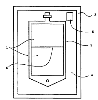
according to the invention is as shown in an attached
drawing (Fig. 1). The package comprises a gas-permeable
plastic container 2 having two chambers partitioned by an
intercommunicable sealing member 6, and a gas-barrier
packaging member 3 enclosing the container, with a carbon
dioxide atmosphere established in the space 4 between the
container and the packaging member where a pH-indicating
device 5 is disposed. Each chamber of the above container
2 holds liquid solution 1, which attains the composition
of the artificial cerebrospinal fluid of the invention
when admixed.
Having the above structure, the packaged product
of the invention can assure the following advantages: it
is not easily breakable, adaptable for increased capacity,
and has a reduced weight due to the use of a plastic
container; problems such as the occurrence of
precipitation and coloration are reliably avoided due to
the container having two chambers; dissipation of carbon
dioxide gas released from the artificial cerebrospinal
fluid (a bicarbonate ion-containing solution) is prevented
due to the employment of the gas-barrier packaging member
and establishment of a carbon dioxide atmosphere within;
and consequent maintenance of the solution pH at a
constant range is attained. Further, the packaged
container holding the artificial cerebrospinal fluid of
the invention in which the pH-indicating device described
earlier is disposed offers easy judgment with the naked
eye of the pH change and the degradation of the fluid
caused by problems such as prolonged storage or formation
. = CA 02604454 2007-10-10
0
-19-
of a pinhole in the packaging member. Furthermore, the
packaged product of the invention has the advantage of
being easily fabricated by conventional manufacturing
techniques in any of the above embodiments.
Moreover, the artificial cerebrospinal fluid of
the invention may be stored in a medical solution
container made of a material to which a gas-barrier
property is imparted. Such a medical solution container
made of a gas-barrier material may be a container formed
of a multi-layered plastic film including a gas-barrier
layer.
Examples of medical solution containers having
such a structure are those disclosed in Japanese
Unexamined Patent Publications Nos. 2002-234102 and 1993-
8318, etc. A specific example of a usable container is
formed of a multi-layered film wherein an inner layer and
an outer layer respectively made of gas-permeable
polyethylene film and polypropylene film, with ethylene
vinylalcohol copolymer (EVOH), a gas-barrier film,
interposed therebetween. The gas-barrier film preferably
used is a transparent film so as to enable the visual
assessment of the medical solution. The medical solution
container to which the gas-barrier properties are imparted
may be a container having at least two intercommunicable
chambers.
Another embodiment of the gas-barrier medical
solution container is a container having a structure in
which both surfaces of the gas-permeable plastic film
constituting the container are covered by gas-barrier
films. In
this embodiment, when the medical solution
container made of a gas-permeable plastic film consists of
two chambers, for example, only the chamber where a
bicarbonate ion-containing solution (Solution A) is
contained may be covered with a gas-barrier film. The
barrier film is preferably a transparent film so that the
medical solution can be visually observed. Examples of
CA 02604454 2007-10-10
-20-
such medical solution containers are described in Japanese
Unexamined Patent Publications Nos. 1999-276547 and 2003-
267451, Japanese Registered Utility Model No. 3112358, etc.
When the artificial cerebrospinal fluid of the
invention is stored in a gas-barrier medical solution
container, an indication area for carbon dioxide gas
concentration detection may be provided at an opening
portion of the container body using the aforementioned ink
composition for carbon dioxide gas detection containing a
pH-indicating agent, binder (thickener), and solvent. For
example, the container can have a structure wherein the
opening portion of the container body is sealed using a
gas-permeable sealer, whose outside is detachably sealed
by a gas-barrier covering member, and a gas detector is
disposed between the covering member and the gas-permeable
sealer. Owing to this structure, in the case that the
sealing performance of the medical solution container is
impaired, it is easily detected from outside. Specific
examples of methods for disposing the gas detector include
a method wherein the above ink composition is applied to
the inner surface of a gas-barrier covering member formed
on the outside the gas-permeable sealer, a method wherein
the ink composition is thus applied, followed by adhesion
of a protective film thereon, etc. The medical solution
container provided with an indication area for carbon
dioxide gas concentration detection at the opening portion
thereof is as described in, e.g., Japanese Unexamined
Patent Publication No. 2005-349182.
(3) Application of the Artificial Cerebrospinal Fluid of
the Invention
The artificial cerebrospinal fluid of the
present invention is used in accordance with known
procedures. For example, the artificial cerebrospinal
fluid of the invention contained in the gas-permeable
plastic container having at least two intercommunicable
. = CA 02604454 2007-10-10
-21-
chambers can be practically used after the above two
chambers are brought into intercommunication to admix (or
dilute) the internal solutions of both chambers.
The artificial cerebrospinal fluid of the
invention can be used as an irrigation fluid or perfusion
fluid for the intracranium or the cerebrospinal cavity.
Specific aspects of this use are as follows.
1. Use in brain neurosurgery (craterization and
craniotomy) as an irrigation fluid for the purpose of
irrigating an operation site, eliminating the air from an
operation site, cooling of tissues to dissipate the heat
generated when operation instruments are used, and not
affecting hemostasis,.
2. Use in neuroendoscopic surgery as a perfusion fluid for
assuring clear visibility, not affecting hemostasis, and
having a texture that does not hinder surgical procedures.
3. Use as a perfusion fluid for eliminating the hematoma
in cisternae perfusion therapy after subarachnoid
hemorrhage.
The amount of the artificial cerebrospinal fluid
of the invention to be used is not limited, and can be
suitably selected in accordance with the purpose of use in
the surgeries mentioned above. It is typically used at a
maximum amount of about 4000 mL as a guide, and the amount
can be increased based on the on-site judgment of a doctor
during an actual operation. Usages of the fluid may be
varied depending on the form of operation. Examples of
suitable applications include the following: the fluid is
directly dropped onto an operation site using a dropper or
syringe; the fluid is sprayed (insufflated) to necessary
areas such as an operation site; the fluid is perfused at
a constant rate to necessary areas such as an operation
site using a suitable tube or the like; a gauze,
impregnated with the fluid, is placed on the brain surface
to prevent the surface from drying; the fluid is dropped
. ' =
CA 02604454 2007-10-10
-22-
from an instrument such as coagulator, drill, when used;
etc.
Further, the artificial cerebrospinal fluid of
the invention can be used as a fluid replenisher for
cerebrospinal fluid in case it is lost during brain
surgery, etc.
As described above, the artificial cerebrospinal
fluid of the invention can significantly reduce the risk
of post-operative cerebral edema incidence when it is used
as an irrigation fluid or perfusion fluid for the
intracranium or the cerebrospinal cavity, or as a fluid
replenisher for lost cerebrospinal fluid during brain
neurosurgery, and is hence very useful in the neurosurgery
field. This is demonstrated in Test Example 1 to be
described later. The artificial cerebrospinal fluid of
the invention when used as an intracerebral irrigation
fluid during neurosurgery is indeed capable of notably
reducing the risk of post-operative cerebral edema
incidence in comparison with normal saline solution and
lactated Ringer's solution.
The known factors of cerebral edema are post-
operative cerebrovascular hyperpermeability and ion
exchange disorders in the brain cell membrane. The
artificial cerebrospinal fluid of the invention has both
an inhibitory action on post-operative cerebrovascular
hyperpermeability and an inhibitory action on brain cell
disorders, and is hence able to remarkably reduce the risk
of cerebral edema incidence. Further, the artificial
cerebrospinal fluid of the invention is also effective in
suppressing, in addition to the ion exchange disorders in
brain cells which is possible risk factors for cerebral
edema, other brain cell disorders, which are likely to
adversely affect other brain functions and the living body,
for example, disorders of glial cells such as neurons,
astrocytes, etc.
= CA 02604454 2007-10-10
-23-
Consequently, the artificial cerebrospinal fluid
of the invention is useful as an agent for the reduction
of the incidence of post-operative cerebral edema, and is
further useful as an inhibitor of brain cell disorders.
Furthermore, the artificial cerebrospinal fluid
of the invention can also be used, for example, as an
entoptic perfusion fluid.
The present inventors performed safety tests on
the artificial cerebrospinal fluid of the invention, and
assured that the fluid has higher safety due to the
specific composition thereof than that of normal saline
solution and lactated Ringer's solution. Based also on
this finding, the artificial cerebrospinal fluid of the
invention has properties for mitigating a risk of a
patient during a neurosurgery operation, and is hence
advantageous in the neurosurgery field.
BRIEF DESCRIPTION OF THE DRAWINGS
Figure 1 is a schematic view showing a preferred
embodiment of the packaged container for holding the
artificial cerebrospinal fluid of the present invention.
Figure 2 is a graph showing the specific
gravities of brain tissues at an incision site, calculated
in accordance with TEST EXAMPLE 1 using each irrigation
fluid.
Figure 3 is a graph showing the measurement
results of Evan's Blue concentrations in brain tissues at
an incision site of each rat group, obtained by testing in
accordance with TEST EXAMPLE 2.
Figure 4 is a graph showing absorbance per mg of
protein at an incision site of each rat group, obtained by
testing in accordance with TEST EXAMPLE 3.
EXPLANATION OF REFERENCE NUMBERS
1 Artificial cerebrospinal fluid of the invention
CA 02604454 2007-10-10
-24-
2 Gas-permeable plastic multiple-chamber container
3 Gas-barrier packaging member
4 Space between the gas-permeable plastic multiple-
chamber container 2 and gas-barrier packaging member
3
5 pH-indicating device
6 Intercommunicable seal portion
BEST MODE FOR CARRYING OUT THE INVENTION
The present invention is described below in
reference to TEST EXAMPLES and EXAMPLES, but is not
limited thereto.
EXAMPLE 1
Each component, shown under the titles Upper
Chamber Solution and Lower Chamber Solution in Table 1
below, was weighed, and mixed and dissolved in distilled
water for injection respectively, thereby preparing 150 mL
of the upper chamber solution and 350 mL of the lower
chamber solution.
A lower chamber (a chamber equipped with a
solution outlet connecting to a port portion, illustrated
in the upper part of Fig. 1) of a container 2 (made of
polyethylene) of the packaged container for holding the
artificial cerebrospinal fluid as shown in Fig.1 was
filled with the thus obtained lower chamber solution from
the solution outlet, and the outlet was tightly sealed.
Similarly, the upper chamber (a chamber separated from the
above lower chamber by a partitioning wall, the chamber
adjoining to a suspension portion, illustrated in the
lower part of the figure) before being tightly sealed was
filled with the obtained upper chamber solution, and then
hermetically sealed.
ak 02604454 2012-07-16
-25-
Table 1
Components Amount (g)
Upper Chamber Solution (per 150 mL)
Calcium Chloride = dihydrate 0.085
Magnesium Chloride = hexahydrate 0.110
Glucose 0.305
Lower Chamber Solution (per 350 mL)
Sodium Bicarbonate 0.970
Potassium Dihydrogenphosphate 0.075
Sodium Chloride 3.575
Potassium Chloride 0.065
The obtained container was subjected to high-
pressure steam sterilization, dehydrated, and packaged in
a gas-barrier film (Bovlon Film, product of The Nippon
Synthetic Chemical Industry Co., Ltd., a biaxially
oriented polyvinyl alcohol film, thickness: 14 m),
together with a pH-indicating device (the device disclosed
_n Production Example 5 of Japanese Unexamined Patent
Publication No. 1999-197215). When
packaging was
performed, about 400 ml of a mixed gas of 18 % carbon
dioxide-air was filled in the space between the container
and the packaging member. A packaged container holding
the artificial cerebrospinal fluid of the invention was
thus obtained.
The packaged container holding the artificial
cerebrospinal fluid of the invention produced above was
allowed to stand indoors for a week, and opened. The
partitioning wall in the container was opened to admix the
20 internal container solutions. The thus obtained mixed
solution was measured for concentrations of electrolytic
ions as well as phosphoric acid and glucose in accordance
with the Liquid Chromatography; THE JAPANESE PHARMACOPOEIA
THIRTEENTH EDITION, General Tests; page 33 (Japanese version);
Published by THE SOCIETY OF JAPANESE PHARMACOPOEIA; April 1,
1996. The pH value of the solution was also determined in
accordance with the pH Determination; THE JAPANESE
PHARMACOPOEIA THIRTHEETH EDITION, General Tests; page 115
(Japanese version); Published by THE SOCIETY OF JAPANESE
PHARMACOPOEIA; April 1, 1996.
ak 02604454 2012-07-16
-26-
Table 2 shows the results.
Table 2
Components Amount
Na+ 145.4 mEq/L
K+ 2.8 mEq/L
C2+
a 2.3 mEq/L
mg2+
2.2 mEq/L
Cl- 128.5 mEq/L
HCO3- 23.1 mEq/L
Phosphoric Acid 1.1 mmol/L
Glucose 0.61 g/L
Solution pH 7.3
EXAMPLE 2
Another packaged container holding the
artificial cerebrospinal fluid of the invention was
produced in the same manner as in EXAMPLE 1, except that
glucose was not included in the upper chamber solution
described in EXAMPLE 1, and the same measurements were
performed. The results obtained are shown in Table 3
below.
Table 3
Components Amount
Na+ 145.4 mEq/L
K+ 2.8 mEq/L
C2+
a 2.3 mEq/L
2.2 mEq/L
Cl- 128.5 mEq/L
HCO3- 23.1 mEq/L
Phosphoric Acid 1.1 mmol/L
Glucose 0.61 g/L
Solution pH 7.3
EXAMPLE 3
Another packaged container holding the
artificial cerebrospinal fluid of the invention was
produced in the same manner as in EXAMPLE 1, except that
. = '
CA 02604454 2007-10-10
-27-
potassium dihydrogenphosphate was not included in the
lower chamber solution described in EXAMPLE 1, and the
same measurements were performed. The results obtained
are shown in Table 4 below.
Table 4
Components Amount
Na+ 145.4 mEq/L
K+ 1.7 mEq/L
Ca2+ 2.3 mEq/L
mg2+
2.2 mEq/L
Cl- 128.5 mEq/L
HCO3- 23.1 mEq/L
Glucose 0.61 g/L
Solution pH 7.3
EXAMPLE 4
Each component shown in Table 5 below was
weighed, and mixed and dissolved in distilled water for
injection to prepare 500 ml of an aqueous solution.
A single-chamber polyethylene container having a
solution outlet was filled with the obtained aqueous
solution via the solution outlet, which was then tightly
sealed.
Table 5
Components Amount (g)
Glucose 0.305
Sodium Bicarbonate 0.970
Potassium Dihydrogenphosphate 0.075
Sodium Chloride 3.575
Potassium Chloride 0.065
Subsequently, the obtained container was
subjected to high-pressure steam sterilization and
dehydrated in the same manner as in EXAMPLE 1, and
packaged in a gas-barrier film together with a pH-
indicating device. About 400 ml of a mixed gas of 18 %
Mk 02604454 2012-07-16
-28-
carbon dioxide-air was filled in the space between the
container and the packaging member. Thus, a packaged
container holding the artificial cerebrospinal fluid of
the invention was obtained.
The packaged container holding the artificial
cerebrospinal fluid of the invention obtained above was
allowed to stand indoors for a week, and opened. The
internal container solution was measured for
concentrations of electrolytic ions as well as phosphoric
acid and glucose in accordance with the Liquid Chromatography;
THE JAPANESE PHARMACOPOEIA THIRTEENTH EDITION, General Tests;
page 33 (Japanese version); Published by THE SOCIETY OF
JAPANESE PHARMACOPOEIA; April 1, 1996. The pH value of the
internal solution was also determined in accordance with the pH
Determination guidance under General Tests in the Japanese
Pharmacopoeia. Table 6 below shows the results.
Table 6
Components Amount
Na+ 145.4 mEq/L
K+ 2.8 mEq/L
Cl- 126.3 mEq/L
HCO3 23.1 mEq/L
Phosphoric Acid 1.1 mmol/L
Glucose 0.61 g/L
Solution pH 7.3
EXAMPLE 5
A packaged container holding the artificial
cerebrospinal fluid of the invention was prepared in the
same manner as in EXAMPLE 4, except that glucose and
potassium dihydrogenphosphate were excluded from the
components used in EXAMPLE 4.
The obtained packaged container holding the
artificial cerebrospinal fluid was allowed to stand
indoors for a week, and the internal fluid was measured
for concentrations of each component in the same manner as
in EXAMPLE 4. The results are shown in Table 7 below.
CA 02604454 2007-10-10
. =
-29-
Table 7
Components Amount
Na + 145.4 mEq/L
K+ 2.8 mEq/L
Cl- 126.3 mEq/L
HCO3- 23.1 mEq/L
Solution pH 7.3
TEST EXAMPLE 1
This test is to study effectiveness of the
artificial cerebrospinal fluid of the invention when used
as a brain irrigation fluid of a rat model for
experimental post-operative cerebral edema, and was
carried out as follows.
(1) Materials
The irrigation fluid sample used was the
artificial cerebrospinal fluid according to the invention
prepared in the same manner as in EXAMPLE 1 and having the
composition shown in Table 8 below (test group). The
artificial cerebrospinal fluid of the invention used
herein can be used by breaking a partitioning wall
separating these two chambers from each other and mixing a
bicarbonate-ion-containing solution held in a chamber
(lower chamber) of the plastic bag having two
intercommunicable chambers as shown in Fig. 1 with a
solution containing each electrolyte and glucose held in
the other chamber (upper chamber). Table 9 shows the
concentration of each component after mixed, and the pH of
the mixed solution.
CA 02604454 2007-10-10
-30-
Table 8
Components Amount (g)
Upper Chamber Solution (per 150 mL)
Sodium Chloride 1.200
Calcium Chloride = dihydrate 0.085
Magnesium Chloride. hexahydrate 0.110
Glucose 0.305
Lower Chamber Solution (per 350 mL)
Sodium Bicarbonate 0.970
Potassium Dihydrogenphosphate 0.075
Sodium Chloride 2.375
Potassium Chloride 0.065
Table 9
Components Amount
Nat 145.4 mEq/L
Kt 2.8 mEq/L
Ca2+ 2.3 mEq/L
mg2+ 2.2 mEq/L
Cl- 128.5 mEq/L
HCO3- 23.1 mEq/L
Phosphoric Acid 1.1 mmol/L
Glucose 0.61 g/L
Solution pH 7.3
For comparison, normal saline solution and
lactated Ringer's solution (Normal Saline Group and
Lactated Ringer Group) were used. The normal saline
solution used was "Otsuka Normal Saline" (Na+154 mEq/L and
C1-154 mEq/L), product of Otsuka Pharmaceutical Factory,
Inc. The
lactated Ringer's solution used was "Lactec
injection (registered trademark)" (Nat 130 mEq/L, K+ 4
mEq/L, Ca2t 3 mEq/L, Cl- 109 mEq/L, lactate- 28 mEq/L),
product of Otsuka Pharmaceutical Factory, Inc.
(2) Test Procedures
Twenty-four SD male rats of 7 to 9 weeks of age
were used for the test. The rats were allowed free access
to drinking water and food until the test was initiated.
. = ' CA 02604454 2007-10-10
-31-
The rats were randomly divided into three groups, each
consisting of 8 rats.
The rats in each group were anesthetized by
intraperitoneal administration of 7 ml/kg (1.4 g/kg) of a
20 w/v% urethane solution, and the entire calvaria was
shaved using a hair clipper. The rats were set in a brain
stereotaxic apparatus (SR-6N, a product of Narishige
Scientific Instrument Lab), the calvariae were disinfected
with 70 % alcohol, and an incision was made on the skin
(initiation of surgery) to expose the cranial bone. A
barrier, having a 1-mm height, made of a silicone tube
(internal diameter 6 mm, external diameter 8 mm, a product
of AS ONE Corporation) was bonded on the left parietal
bone. Within the barrier, a bone window having about a 4-
mm diameter was drilled (drill: MINITOR-7C-307M, product
of Kanto Kiki Co., Ltd.) so that the center of the bone
window was located 2.5 mm to the left from the midline and
4 mm posterior to the bregma.
As test samples, the artificial cerebrospinal
fluid of the invention (present invention group, 8 rats),
normal saline solution (normal saline group, 8 rats), and
lactated Ringer's solution (Lactated Ringer group, 8 rats)
were respectively infused into the bone window using a JMS
syringe pump SP-100S (JMS Inc.) at 150 ml/hr, upon which
irrigation was initiated. Subsequently, while irrigating,
two incisions were made in a crisscross shape to a depth
reaching the left cerebrocortex from the dura mater
through the arachnoid membrane (depth 1.5 mm, length 3.5
mm) within the bone window using the edge of a blade
(Feather Replacement Blade, stainless steel No. 25,
Feather Safety Razor Co., Ltd.) attached to an electrode
holder of a stereotaxic micromanipulator (SM-15, a product
of Narishige Scientific Instrument Laboratory) to detach
the dura mater and arachnoid membrane at the incision site
(incision making). Clots formed within the bone window
CA 02604454 2007-10-10
-32-
within 10 minutes of the incisions having been made were
removed using a pair of tweezers. The
irrigation was
completed 4 hours after the incisions had been made. The
body temperature (rectal temperature) of the model rats
were maintained at about 37 C using a feedback-controlled
lamp and a warming pad (ATB-1100, a product of NIHON
KOHDEN CORPORATION) until completion of the irrigation.
The rats of each group were then sacrificed by
bleeding from the abdominal aorta, and the brain was
quickly extirpated. The brain was temporarily isolated in
ice-cold kerosene to prevent it from drying. Cerebral
cortex tissue samples were collected from the intersection,
along with the lines of the incisions made above, in a
cubical shape having a length of about 0.5 mm and a depth
of about 0.5 mm (tissues at the incision site).
According to the procedure of Marmarou et al. (J.
Neurosurg., 1978 Oct; 49(4), pp. 530-537), specific
gravity columns were made using bromobenzene and kerosene
whose specific gravities are already known. Into the
column were immersed the samples collected above, and the
depths they had reached after 2 minutes were measured from
which specific gravities (water contents) of the samples
were determined based on calibration curves created
beforehand. The specific gravity columns were calibrated
using standard potassium sulfate solutions having a
specific gravity of 1.020, 1.029, 1.038, 1.047, and 1.056,
respectively, before use.
The specific gravity method, by which water
content in a tissue is determined from the density of
moist tissue using specific gravity columns as used in
this test, is widely used in the study of cerebral edema.
In this method, the lower a specific gravity is, the
greater the water content is, thereby indicating more
serious cerebal edema.
(3) Statistical Analysis
CA 02604454 2007-10-10
-33-
The test results are reported using the average
standard deviation of the results obtained from 8 rats
of each group. Further, an intergroup comparison based on
the results on evaluation of the tissues at the incision
sites obtained from each group was conducted using Tukey's
method at a 5 % level of significance. In the Figures,
"##" and "***" indicate p<0.01 and p<0.001, respectively.
(4) Results
The specific gravities determined for the brain
tissues from the incision sites irrigated with each
irrigation fluid sample are shown in FIG. 2 (vertical
scale: specific gravity) for each irrigation fluid.
(5) Discussion
The results shown in FIG. 2 reveal that specific
gravity at the incision site is the lowest in the Normal
Saline Group, the second lowest in the Lactated Ringer
Group, and the highest in the Present Invention Group.
Based on this finding, the artificial cerebrospinal fluid
of the invention evidently reduces the degree of
experimental post-operative cerebral edema in comparison
with normal saline solution and lactated Ringer's solution.
TEST EXAMPLE 2
Cerebral edema is classified into two types
depending on the factors involved. One is vasogenic edema.
In this type of edema, cerebrovascular hyperpermeability
is caused by damage to the blood-brain barrier which
consequently allows water to accumulate in the extra-
cellular spaces of the brain while serum protein such as
albumin leaks. The other is cytotoxic edema. In this
type of edema, the ion exhange in the brain cell membrane
is dysfunctional and causes intracelullar water retention.
Both types of edema are often seen together in actual
cerebral edema cases.
Among these factors for cerebal edema, this test
= CA 02604454 2007-10-10
-34-
example is to examine the effects of the artificial
cerebrospinal fluid of the invention on the permeability
of cerebral blood vessels.
Serum proteins do not influx into the brain
tissue under the noraml conditions; however, when the
brain is damaged, serum proteins pass through the damaged
blood-brain barrier and enter into the extra-cellular
spaces of the brain tissue (vascular hyperpermeability).
Serum proteins, such as albumin, that have entered into
the brain tissue trigger water inflow to the brain tissue
due to osmotic pressure. Such an extravascular serum
protein leakage associated with vascular hyperpermeability
is thought to result in the formation of cerebral edema.
Evan's blue is known to bind to albumin in vivo,
and, due to this property, is widely used for protein
leakage assay as an index of vascular permeability. In
this Test Example, the extravascular leakage of Evan's
blue was assayed as an index of cerebrovascular
permeability.
The test was carried out as follows.
(1) Materials
The artificial cerebrospinal fluid of the
invention prepared in EXAMPLE 1 was used as an irrigation
fluid sample (Test Group). For comparison, normal saline
solution and lactated Ringer's solution (Normal Saline
Group and Lactated Ringer Group) were also used. The
compositions of these solutions are as shown in TEST
EXAMPLE 1 above.
(2) Test Procedures
(2-1) Brain Incision, Incision Site Irrigation and Evan's
Blue Administration
Twenty-four SD male rats of 7 to 9 weeks of age
were used for the test. The rats were allowed free access
to drinking water and food until the test was initiated.
The rats were randomly divided into three groups, each
= CA 02604454 2007-10-10
-35-
consisting of 8 rats.
The rats in each group were anesthetized under
intraperitoneal administration of 7 ml/kg (1.4 g/kg) of a
20 w/v% urethane solution, and the entire calvaria was
shaved using a hair clipper. The rats were set in a brain
stereotaxic apparatus (SR-6N, product of Narishige
Scientific Instrument Lab), the calvariae were disinfected
with 70 % alcohol, and an incision was made on the skin
(initiation of surgery) to expose the cranial bone. A
barrier, having a 1-mm height, made of a silicone tube
(internal diameter 6 mm, external diameter 8 mm, product
of AS ONE Corporation) was bonded on the left parietal
bone. Within the barrier, a bone window having about a 4-
mm diameter was drilled (drill: MINITOR-7C-307M, product
of Kanto Kiki Co., Ltd.) so that the center of the bone
window was located 2.5 mm to the left from the midline and
4 mm posterior to the bregma.
As test samples, the artificial cerebrospinal
fluid (Present Invention Group, 8 rats), normal saline
(Normal Saline Group, 8 rats), and lactated Ringer's
solution (Lactated Ringer Group, 8 rats) were respectively
infused into the bone window using a JMS syringe pump SP-
100S (JMS Inc.) at 150 ml/hr, upon which irrigation was
initiated. Subsequently, while irrigating, two incisions
were made in a crisscross shape to a depth reaching the
left cerebrocortex from the dura mater through the
arachnoid membrane (depth 1.5 mm, length 3.5 mm) within
the bone window with the edge of a blade (Feather
Replacement Blade, stainless steel No. 25, Feather Safety
Razor Co., Ltd.) attached to an electrode holder of a
stereotaxic micromanipulator (SM-15, product of Narishige
Scientific Instrument Laboratory) to detach the dura mater
and arachnoid membrane at the incision site (incision
making). The body temperatures (rectal temperature) of
1
35 the model rats were maintained at about 37 C using a
, = = CA 02604454 2007-10-10
-36-
feedback-controlled lamp and a warming pad (ATB-1100,
product of NIHON KOHDEN CORPORATION) until completion of
the irrigation.
Clots formed within the bone window within 10
minutes of the incisions having been made were removed
using a pair of tweezers. Three hours after the incisions
had been made, 5 ml/kg of a 2 w/v % Evans Blue-containing
normal saline solution (which was prepared by dissolving
0.2 g of Evan's blue, (product of Wako Pure Chemical
Industries, Ltd.) in normal saline solution (Otsuka Normal
Saline, product of Otsuka Pharmaceutical Factory, Inc.) to
give 10 ml) was administered via the tail vain. The
irrigation of the incision site with the test samples was
continued until 4 hours after the incision had been made.
(2-2) Preparation of Fluorescence Measurement Samples
Immediately after completion of irrigation with
the samples, thoracotomy was performed on the rats of each
group, 262 to 266 mL of a normal saline solution (Otsuka
Normal Saline, a product of Otsuka Pharmaceutical Factory,
Inc.) was infused into the left ventricle from a container
set so that the bottom end thereof was located about 100
cm above the rat, and was perfused so as to be discharged
with blood from the right ventricle, followed by taking
out the brain.
Further, the left and right cerebral cortexes
were separated, spread on a plate (biological sample
trimming plate, product of Nisshin EM Corporation), and
brain tissue samples at the incision site of the left
cerebral cortex were collected using a cork borer (No. 1,
a product of Nonaka Rikaki Seisakusho, Co., Ltd.).
After measurement of each tissue sample, 0.01 M
phosphate buffer (which was prepared by diluting 0.1 M
phosphate buffer ("hereinafter abbreviated as "PB") 10
times with water before use, the 0.1 M phosphate buffer
being prepare by mixing 667 ml of water and Phosphate
= CA 02604454 2007-10-10
-37-
Buffer 5 (pH 7.4, product of Latron Ltd.)) in an amount 10
times the tissue sample weight (calculated taking the
specific gravities of the cerebrum and 0.01 M phosphate
buffer as 1) was added to each tissue sample, homogenized
using a homogenizer (potter type, a product of AS ONE
Corporation), and 50 w/v % TCA (which was prepared by
diluting 100 w/v % trichloroacetate solution (Wako Pure
Chemical Industries, Ltd.) with water to give 50 w/v %) in
an amount 10 times the tissue sample weight was further
added thereto, and then mixed for 2 minutes in a touch
mixer. The mixture was moved to a microtube, allowed to
stand at room temperature for 30 minutes, and centrifuged
at 13000 rpm for 40 minutes at room temperature using a
centrifuge (MX-300, rotor: TMA-300, rack: AR015-24,
products of TONY SEIKO Co., Ltd.). The supernatant was
collected, and diluted with ethanol in an amount 3 times
that of the supernatant, thereby obtaining a diluted
solution for use as a fluorescence measurement sample.
(2-3) Fluorescence Measuremernt
The fluorescence measurement of each sample was
performed using a spectrofluorometer (FP-750, product of
JASCO Corporation) under the following conditions.
Measurement Menu: Fixed Wavelength Measurement
Excitation Wavelength (band width): 620 nm (10 nm)
Measurement Wavelength (band width): 680 nm (10 nm)
Response Time: 1 second
Sensitivity: Medium
Zero adjustment (autozero) was calibrated using
Diluent A (which was prepared by adding 0.01 M PB to an
equivalent amount of 50 w/v % TCA and mixing, and further
adding ethanol to the mixture in an amount 3 times the
mixture before use), and standard solutions for the
calibration curves and fluorescence measurement samples
were filtered using DISMIC-13JP (0.2 m, nonaqueous, a
product of Advantec Toyo Kaisha, Ltd.). The fluorescence
=. CA 02604454 2007-10-10
-38-
measurement was first performed on the filtrate of the
standard solutions for the calibration curve, and then on
the filtrate of the fluorescence measurement samples.
(2-4) Determination of Evan's Blue Concentration in Brain
Tissues
The determination was carried out by the
absolute calibration curve method with the fluorescence
intensity at a measurement wavelength of 680 nm, using
calculation software (Microsoft Excel (registered
trademark), Microsoft Corporation). By
measuring the
calibration curve standard solution, calibration curve
formula of the fluorescence intensity at the measurement
wavelength was determined by the least square method. The
correlation coefficient (r) of the calibration curve was
0.99 or higher. Based on the calibration curve formula,
the Evan's blue concentration in the fluorescence
measurement sample was determined. The Evan's blue
concentration in the brain tissue was determined by
multiplying the Evan's blue concentration in a
fluorescence measurement sample by the dilution rate (84-
fold, calculated taking the specific gravities of the
brain and Diluent A as 1) with which fluorescence
measurement samples were prepared from the brain tissue
samples.
(3) Statistical Analysis
The test results were expressed using averages
standard deviation of the results obtained from 8 rats of
each group. Further, an intergroup comparison based on
the results on evaluation of the tissues at the incision
sites obtained from each group were conducted using
Tukey's method at a 5 % level of significance. In the
Figures, ** indicates <0.01.
(4) Results
Evan's Blue Concentrations in the Brain Tissues via Evan's
Blue Stain Test
= ' CA 02604454 2007-10-10
-39-
FIG. 3 shows the measurement results, expressed
as averages standard deviation, of the Evan's blue
concentrations ( g/g of brain tissue) in the brain tissues
at the incision sites for the rats of each group. The
results shown in FIG. 3 reveal the following.
Specifically, the Evan's blue concentrations at
the above incision sites (i.e., cerebrovascular
hyperpermeability) in Present Invention Group were found
to be significantly lower (p<0.01) than those of the
Lactated Ringer Group, and tended to be lower than those
of Normal Saline Group.
(5) Discussion
As a result of the Evan's blue assay in the
brain tissue, the extravascular leakage of the Evan's blue
in the Present Invention Group was significantly lower
than that of the Lactated Ringer Group, and has a lower
value when compared with the Normal Saline Group. These
results reveal that the artificial cerebrospinal fluid of
the present invention is able to effectively reduce the
degree of cerebrovascular hyperpermeability, a primary
factor of cerebral edema, in an experimental neurosurgery
operation using rat models.
As described above,
cerebrovascular
hyperpermeability causes water to flow into the brain
tissue from the inside of vessels and causes cerebral
edema; however, the artificial cerebrospinal fluid of the
invention hardly affects the cerebrovascular permeability,
and is hence evidently not likely to cause post-operative
cerebral edema. Considering that the brain-blood barrier
has a control function on the transfer of various
substances between the blood and brain tissues, the effect,
which scarcely affects the above cerebrovascular
permeability, attained by the use of the artificial
cerebrospinal fluid of the invention further means that
less damage is inflicted upon such a function. Based on
CA 02604454 2007-10-10
-40-
these findings, the artificial cerebrospinal fluid of the
present invention is thought to be useful as an almost
ideal irrigation fluid to maintain the physiological
condition of an intracerebral environment.
TEST EXAMPLE 3
TEST EXAMPLE 3 was to study the influence of the
artificial cerebrospinal fluid of the present invention on
brain cell disorders, one of the risk factors for cerebral
edema.
2,3,5-Triphenyltetrazolium chloride (TTC) is
converted to a red dye (formazan) by mitochondria enzymes
present in viral cells. The
degree of any brain cell
disorders can be evaluated by dyeing the brain tissues
with TTC and measuring the absorbance of the solvent in
which formazan is extracted. More specifically, the lower
the TTC stainability, the higher the degree of brain cell
disorder is.
In this TEST EXAMPLE, incisions made in the
brains of the rats, mimicking surgical brain incisions
made during neurosurgery, were irrigated with each
irrigation fluid, and the degrees of cell disorder were
evaluated using the stainability of TTC in brain tissue as
an index. The test procedures are as follows.
(1) Irrigation Fluid Samples
The irrigation fluid samples used were the
aritificial cerebrospinal fluid of the invention prepared
in EXAMPLE 1 (Present Invention Group), and, for
comparison, normal saline solution and lactated Ringer's
solution (Normal Saline Group and Lactated Ringer Group,
respectively). The compositions of these fluids were the
same as those in TEST EXAMPLE 1 above.
(2) Test Procedures
(2-1) Brain Incisions and Irrigation Thereof
Thirty-two SD male rats of 7 to 9 weeks of age
CA 02604454 2007-10-10
-*
-41-
were used for the test. The rats were allowed free access
to drinking water and food until the test was initiated.
The rats were randomly divided into four groups,
each consisting of 8 rats. Among these groups, the rats
of three groups were categorized as the Present Invention
Group (8 rats), the Normal Saline Group (8 rats), and the
Lactated Ringer Group (8 rats), and incisions were made
and the incision sites were irrigated with the samples in
the same manner as in TEST EXAMPLE 2 above (provided that
a 2 w/v % Evans Blue-containing normal saline solution was
not administered).
The rats of the remaining one group (Control
Group) were anesthetized upon
intraperitoneal
administration of 7 ml/kg of a 20 w/v% urethane solution,
and the body temperature (rectal temperature) was
maintained at about 37 C using a feedback-controlled lamp
and a warming pad (ATB-1100, a product of NIHON KOHDEN
CORPORATION) for 4 hours.
(2-2) TTC Staining and Extraction
After completion of irrigating the incisions,
the rats of the Present Invention Group, Normal Saline
Group, and Lactated Ringer Group were quickly decapitated,
and the heads were cooled with ice for about 30 seconds.
The left and right cerebral cortexes were separated from
the extirpated brain, spread on an ice-cooled plate
(biological sample trimming plate, product of Nisshin EM
Corporation), and brain tissue samples at the incision
site of the left cerebral cortex and at the corresponding
site (non-incision site) of the right cerebral cortex were
collected using a cork borer having an internal diameter
of about 4 mm (No. 1, product of Nonaka Rikaki Seisakusho,
Co., Ltd.).
The equivalent tissue samples of the left and
right cerebral cortexes were collected from the rats of
Control Group.
,* CA 02604454 2007-10-10
-42-
Each brain tissue sample, divided into two parts
in the middle, was placed in a vial, and measured for the
weight at the time of the collection, 5 ml of a 2 % TTC
solution was then added thereto, and incubated in the
shade at 37 C for 90 minutes in a constant temperature
bath equipped with a shaker (Personal-11SD, a product of
TAITEC Corporation). The 2 % TTC solution was removed,
the samples were washed twice respectively with 5 ml of
normal saline solution, and about 5 g of a solvent for
formazan extraction (a mixture of an equivalent amount of
ethanol and dimethylsulphoxide) was added to each vial,
which was then hermetically sealed and allowed to stand in
a dark place. After 24 hours, the formazan extraction
solvent was recovered from the vial, and used as a sample
for absorbance measurement.
(3) Measurement of TTC Stainability of Brain Tissue
The absorbance of the solvent used to extract
formazan from the TTC-stained brain was measured at 485 nm,
using an ultraviolet visible spectrophotometer (v-550,
product of JASCO Corporation). The solvent for formazan
extraction was taken as blank. The measured absorbances
were corrected using the protein concentration of each
brain tissue sample. Lowry's method was employed to
determine the protein concentration in each brain tissue
sample.
The TTC stainabilities of the brain tissues are
indicated by the absorbance per mg of protein.
(4) Statistical Analysis
Mean and standard deviation (SD) values of the
TTC stainability of the brain tissues were determined.
Significance testing was performed using the following
method.
More specifically, as to TTC stainabilities of
the brain tissues at the incision sites of the rats, TTC
stainabilities of the Present Invention Group, Lactated
CA 02604454 2007-10-10
-43-
Ringer Group and Normal Saline Group were compared with
that of Control Group, which were taken as the standard,
using Dunnett's multiple comparison test. In case a
significant difference was found in any group, the TTC
stainabilities of Present Invention Group, taken as the
standard, were compared with those of the Lactated Ringer
Group and Normal Saline Group by Donnett's multiple
comparison test. The non-incision sites were
complementarily tested in the same manner. The
significance level was 5 %.
Data compilation was performed using calculation
software (Microsoft Excel (registered trademark), product
of Microsoft Corporation), and the statistical analysis
software used was EXSAS Ver. 7.14 (product of Arm Systex
Co., Ltd.).
(5) Results
FIG. 4 shows the TTC stainabilities (absorbance
per mg of protein) of the brain tissues at the incision
sites and non-incision sites of each group. The brain
tissue TTC stainabilies at the incision sites
(corresponding site on the left cerebral cortex in the
Control Group) were 0.247 0.017 in the Control Group,
0.220 0.023 in the Present Invention Group, 0.189
0.023 in the Lactated Ringer Group, and 0.168 0.030 in
the Normal Saline Group. The Lactated Ringer Group and
Normal Saline Group exhibited statistically significantly
lower values (p<0.001 in both groups) than the Control
Group. The Lactated Ringer Group and Normal Saline Group
also exhibited significantly lower values (p<0.05 and
p<0.01, respectively) than the Present Invention Group.
The brain tissue TTC stainabilities of the non-
incision sites were 0.244 0.014 in the Control Group,
0.254 0.020 in the Present Invention Group, 0.237
0.016 in the Lactated Ringer Group, and 0.232 0.018 in
the Normal Saline Group, and no significant difference was
CA 02604454 2007-10-10
-44-
found between the Control Group and the other groups.
(6) Discussion
As described above, the absorbance measurement
of the solvent, which was used to extract formazan from
the TTC stained brain tissues, can be used as an index of
experimental brain damage. In this TEST EXAMPLE, the
method of Preston et al. (Preston E, Webster J.
Spectrophotometric Measurement of Experimental Brain
Injury, J. Neurosci Methods. 2000; 94; 187-92) was
partially modified, in that the TTC stainability per unit
weight of the brain tissue sample was determined by
correcting the absorbance of the solvent that was used to
extract formazan with the protein content in the brain
tissue sample, whereby the degrees of cell disorder were
compared. According to this test method, it is shown that
the lower the TTC stainability is, the higher the degree
of cell disorder in the brain tissue is.
The TTC stainabilities at the incision sites in
the Present Invention Group had slightly lower values than
those of the Control Group; however, there was no
significant difference between these groups. The TTC
stainabilities at the incision sites in the Lactated
Ringer Group and the Normal Saline Group were
significantly lower than those of the Control Group, and
cell disorders were demonstrated. Further, since the rats
of the Lactated Ringer Group and the Normal Saline Group
had significantly lower values than Present Invention
Group, they are shown to have a higher degree of cell
disorders than the rats in Present Invention Group.
Considering the above, the evaluation of cell
disorder degree during irrigation of the incision site
made on the cerebrum of the rats, using the TTC
stainability as an index, demonstrated that cell disorders
are milder when the incision site is irrigated with the
artificial cerebrospinal fluid of the invention in
CA 02604454 2007-10-10
-45-
comparison with the cases in which lactated Ringer's
solution and normal saline solution were used for
irrigation.
In TEST EXAMPLE 3, cell disorder degree was
evaluated using mitochondria enzyme activity in brain
cells as an index. It is presumed that when the function
of the mitochondria enzymes decreases, the production of
adenosine triphosphate (ATP) required to sustain cell
membrane functions is reduced, whereby the ion exchange of
the cell membrane is adversely affected. Given this, the
results of TEST EXAMPLE 3 suggest that suppression of ion
exchange disorder in cells can be achieved by irrigating
an incision site with the artificial cerebrospinal fluid
of the present invention.