Note: Descriptions are shown in the official language in which they were submitted.
CA 02604537 2007-09-27
FIELD OF THE INVENTION
[0001] The present invention relates to devices and methods for tissue
augmentation
or regeneration. More specifically, the present invention provides for a
composite of
biocompatible scaffold and autologous tissue, suitable for implantation in a
hollow
organ or skin.
BACKGROUND
[0002] Regenerative medicine strives to treat disease and restore human
tissues by
prompting the body to autonomously regenerate damaged tissue. Tissue
engineered implants
may prompt such regeneration by providing structure and media for cell growth,
and may
enable direct transplantation of healthy tissues into a damaged-tissue
environment.
[0003] Many of these new therapies require implantable biocompatible and
biodegradable scaffolds for use both in vitro and in vivo. These scaffolds may
augment
healing through tissue infiltration or by providing suitable means of cell
attachment and
proliferation. Also, these scaffolds may be seeded with cells and manufactured
in such a way
that chemical, mechanical, and cellular stimuli are optimized. Despite
advances made in this
field in recent years, there remains a need for improved approaches to tissue
scaffolding,
particularly in the area involving the skin, or hollow organs such as the
bladder, urethra,
jejunum, esophagus, or trachea.
1
CA 02604537 2007-09-27
SUMMARY OF THE 1NVENTION
[0004] An aspect of the present invention provides for an improved implant for
tissue
augmentation and regeneration in hollow organs, comprising a biocompatible,
biodegradable
scaffold that sandwiches autologous cells or tissue.
[0005] In one embodiment of the present invention, two or more layers of
biocompatible scaffolding and autologous tissue or cells provide a means to
promote growth
of tissues. In an aspect of the invention, the tissue contains more than one
type of cell.
[0006] In another embodiment of the invention, the device may hold the
cellular
component in place and also include a means of fixation, such as a suture.
[0007] A further object of the present invention provides for a method of
using an
implant to augment tissue regeneration in a hollow organ.
[0008] In another embodiment of the invention, these cellularized implants may
be
used to patch holes, repair areas of damaged tissue, or increase the surface
area of tissues in a
hollow organ.
BRIEF DESCRIPTION OF THE DRAWINGS
[0009] Some features and advantages of the invention are described with
reference to
the drawings of certain preferred embodiments, which are intended to
illustrate and not to
limit the invention.
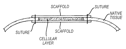
[00010] Figure 1 depicts an embodiment of the present invention in which
autologous cellular tissue is sandwiched between layers of biocompatible,
biodegradable scaffolding.
2
CA 02604537 2007-09-27
[0011] Figure 2 depicts an embodiment of the present invention in which
isolated
cells are sandwiched between layers of biocompatible, biodegradable
scaffolding.
[0012] Figure 3 depicts an embodiment of the present invention in which
dissected
tissue is removed from a hollow organ, placed between layers of biocompatible,
biodegradable scaffolding, and implanted into a hollow organ to provide a
tissue patch that
replaces or increases the surface area of the hollow organ.
[0013] Figure 4 depicts an embodiment of the present invention in which a
composite
comprising autologous cellular tissue and biocompatible, biodegradable
scaffolding is sutured
into native tissue.
DETAILED DESCRIPTION OF THE INVENTION
[0014] It should be understood that this invention is not limited to the
particular
methodology, protocols, etc., described herein and, as such, may vary. The
terminology used
herein is for the purpose of describing particular embodiments only, and is
not intended to
limit the scope of the present invention, which is defined solely by the
claims.
[0015] As used herein and in the claims, the singular forms "a," "an," and
"the"
include the plural reference unless the context clearly indicates otherwise.
Thus, for example,
a reference to a cell may be a reference to one or more such cells, including
equivalents
thereof known to those skilled in the art unless the context of the reference
clearly dictates
otherwise. Other than in the operating examples, or where otherwise indicated,
all numbers
expressing quantities of ingredients or reaction conditions used herein should
be understood
3
CA 02604537 2007-09-27
as modified in all instances by the term "about." The term "about" when used
in connection
with percentages may mean f 1%.
[0016] All patents and other publications identified are identified for the
purpose of
describing and disclosing, for example, the methodologies described in such
publications that
might be used in connection with the present invention. These publications are
provided
solely for their disclosure prior to the filing date of the present
application. Nothing in this
regard should be construed as an admission that the inventors are not entitled
to antedate such
disclosure by virtue of prior invention or for any other reason. All
statements as to the date or
representation as to the contents of these documents is based on the
information available to
the applicants and does not constitute any admission as to the correctness of
the dates or
contents of these documents.
[0017] Unless defined otherwise, all technical terms used herein have the same
meaning as those commonly understood to one of ordinary skill in the art to
which this
invention pertains. Although any known methods, devices, and materials may be
used in the
practice or testing of the invention, the preferred methods, devices, and
materials in this
regard are described here.
[0018] The present invention provides for a layered composite device and
method to
use the device such that the invention assists with tissue augmentation and
regeneration.
More specifically, multiple layers of biocompatible scaffolding and autologous
tissue may
provide the means to promote growth of tissues containing more than one type
of cell. When
utilized, these cellularized patches will be able to fill holes, repair areas
of damage, or
4
CA 02604537 2007-09-27
increase the surface area of tissues in hollow organs. The device may not only
hold the
autologous cellular and tissue in place, but may also contain the means for
fixing the implant.
[0019] Recent publications have discussed various scaffolding approaches for
reconstruction of skeletal or tract tissues. For example, U.S. Patent
Application Pub. No.
20050154458 refers to an "active bio layer" such as hyaluronic acid, capable
of interacting
with stem cells from bone marrow, sandwiched between opposed surfaces of
biomaterials or
synthetic polymer materials, for skeletomuscular applications. The present
invention does not
require an "active bio layer" and is not limited to such skeletomuscular
applications. U.S.
Patent Application Pub. No. 20040225247 refers to a tissue patch having a
protective layer
for repairing an alimentary tract lesion. The present invention has no such
"protective liner,"
nor is it limited to such lesions. U.S. Patent Application Pub. No.
20050272153 refers to a
metal coated scaffold (and thus non-degradable) with biocompatible material
(but not cells or
tissue) for implantation into or in place of bone. Finally, U.S. Patent No.
6,143,293 refers to a
stack of cell-seeded hydroxyapatite scaffolds for use as a 3-dimensional void
filler for use in,
for example, bone. The present invention is not directed to such void filler
uses.
[0020] In one embodiment of the present invention, each composite contains at
least
two scaffolding layers and at least one cellular layer. See, for example,
Figure 1. These layers
may be placed upon each other in various fashions to form an implant system
with a living
component. The scaffolding layers may be porous or non-porous, and may be made
from
natural polymers, synthetic polymers, bioactive glass, hydrogel, or any
biocompatible
material that may be manufactured in a flat sheet. One or more sheets may have
agents or
bioactive agents incorporated within or disposed upon their matrix.
CA 02604537 2007-09-27
[0021] The scaffold layers of the present invention are bioabsorbable, meaning
that
they are biocompatible and biodegradable. Biocompatible refers to materials
which do not
have toxic or injurious effects on biological functions. Biodegradable refers
to material that
can be absorbed or degraded in a patient's body. Representative materials for
forming the
biocompatible structure include natural or synthetic polymers, such as, for
example, collagen,
poly(alpha esters) such as poly(lactic acid), poly(glycolic acid),
polyorthoesters and
polyanhydrides and their copolymers, which degrade by hydrolysis at a
controlled rate and
are reabsorbed. These materials provide the maximum control of degradability,
manageability, size and configuration. Other biodegradable polymer materials
include
polyglycolic acid and polyglactin. See, e.g., U.S. Patent No. 5,514,181.
[0022] Other biodegradable materials include cellulose ether, cellulose,
cellulosic
ester, fluorinated polyethylene, phenolic, poly-4-methylpentene,
polyacrylonitrile, polyamide,
polyamideimide, polyacrylate, polybenzoxazole, polycarbonate,
polycyanoarylether,
polyester, polyestercarbonate, polyether, polyetheretherketone,
polyetherimide,
polyetherketone, polyethersulfone, polyethylene, polyfluoroolefin, polyimide,
polyolefin,
polyoxadiazole, polyphenylene oxide, polyphenylene sulfide, polypropylene,
polystyrene,
polysulfide, polysulfone, polytetrafluoroethylene, polythioether,
polytriazole, polyurethane,
polyvinyl, polyvinylidene fluoride, regenerated cellulose, silicone, urea-
formaldehyde, or
copolymers or physical blends of these materials.
[0023] An example of a biocompatible, biodegradable polymer suitable for the
instant
invention is polyglactin, manufactured as Vicryl (Novartis-Ethicon). Vicryl
(polyglactin
910) is a 90:10 copolymer of glycolide and lactide, derived respectively from
glycolic and
lactic acids.
6
CA 02604537 2007-09-27
[0024] Regarding the use of ceramics as material for scaffold formation, a
bioactive
glass such as the commercially available BioGlass (NovaBone Products, LLC,
Alachua
FL), can be modified with a poly(lactic co-glycolic acid) polymer matrix. See,
e.g., U.S.
Patent No. 6,328,990. "Bioactive" means that the material has the ability to
interact or bind to
living tissue.
[0025] Alternatively, hydrogels such as alginate-RGD may be used. Alginates
are
seaweed-derived copolymers for which the rigidity of the hydrogel may be
controlled by
crosslinking its glucuronate residues with, e.g., calcium or adipic
dihydrazide. Alginate may
further be modified with cell-adhesive peptides such as Arg-Gly-Asp (RDG)
peptide to
promote cellular attachment to the scaffold layer. See, e.g., Wong et al., 570
Science 119-33
(2004); Das & Hollister, Tissue Engineering Scaffolds in Encyclopedia of Mats:
Sci & Tech.
1-7 (Elsevier Sci., Ltd. 2003).
[0026] Further regarding the scaffold layer, an optional pharmaceutical or
bioactive
agent may be incorporated into the scaffolding. The variety of different
pharmaceuticals that
can be used in conjunction with the scaffolds of the present invention is
vast. Such
pharmaceuticals or agents will in general be selected according to the tissue
or organ being
reconstructed or augmented, to ensure that appropriate new tissue is formed in
the engrafted
organ or tissue (for examples of such additives for use in promoting bone
healing, see, e.g.,
Kirker-Head, 24(5) C. A. Vet. Surg. 408-19 (1995)). Common pharmaceuticals and
bioactive
agents which may be administered via the pharmaceutical compositions of the
invention
include, without limitation: anti-infectives such as antibiotics and antiviral
agents;
chemotherapeutic agents; anti-rejection agents; analgesics and analgesic
combinations; anti-
7
CA 02604537 2007-09-27
inflammatory agents; hormones such as steroids; growth factors; and other
naturally derived
or genetically engineered proteins, polysaccharides, glycoproteins, or
lipoproteins.
[0027] Scaffolds containing these materials may be formulated by mixing one or
more agents with the material used to make the scaffold. Alternatively, an
agent could be
coated onto the scaffold, preferably with a pharmaceutically acceptable
carrier. Any
pharmaceutical carrier can be used that does not dissolve or react with the
scaffold. The
pharmaceutical agents may be present as a liquid, a finely divided solid, or
any other
appropriate physical form. Typically, but optionally, they will include one or
more additives,
such as diluents, carriers, excipients, stabilizers or the like. Additionally,
such optional agent
or bioactive agent may be added separately (i.e., not manufactured into the
scaffold matrix.
For example, a bioactive agent such as fibrin may be added to the tissue or
cell layer, and
may serve as a bioactive glue between the cell or tissue layer and the
scaffold layer(s).
[0028] An aspect of the present invention provides for a composite comprising
distinct scaffold layers having different physiochemical properties. For
example, it is known
that chemical, topographical, and mechanical cues affect cellular responses at
the cell-
biomaterial interface. See Wong et al., (2004). Moreover, considerations of
mechanical
strength in maintaining rigidity relating to a particular organ's structure
and placement may
suggest using a particular scaffold layer in that context. Hence, for example,
a composite
according to the present invention may comprise a rigid scaffold layer that
provides
mechanical strength in one layer, and second scaffold layer that promotes
cellular ingrowth.
Alternatively, different layers might be used to promote the growth of
distinct cell types
found in complex tissues. For example, one scaffold layer might promote the
growth of
cartilage, while a second scaffold layer promotes the growth of bone. Or, for
example, one
8
CA 02604537 2007-09-27
scaffold layer might have porosity that will foster muscle-cell infiltration
while another layer
might have porosity that would exclude larger cells and allow only smaller
cell infiltration.
Alternatively, the different scaffold layers may comprise polymers that
degrade at different
rates such that one layer degrades before the other. For example, gamma-
irradiated PLGA
degrades faster and might be used in one layer (e.g., on the inner surface of
a hollow organ),
while polyethylene oxide degrades more slowly and might be used in another
layer (e.g., on
the exterior surface of a hollow organ).
[0029] The living component of the present invention may be autologous cells
or
autologous tissue, obtained by any number of techniques well-known in the art.
For example,
during surgery a tissue sample may be obtained and simply placed on a
scaffolding layer.
Alternatively, tissues containing more than one cell type may be separated,
for example with
a scalpel, into substantially distinct tissue samples. One or more of the
separated tissue
samples may then be used with the scaffolding, or the separated tissues may be
cellularized
before placement on the scaffold. Such cellularization techniques, such as
mincing or treating
with appropriate cellularizing agents, are known in the art.
[0030] Alternatively, the tissue or cellularized (cell) sample may be treated
in vitro
before being placed on the scaffold layer. For example, cells (such as
autologous cells) can be
cultured in vitro to increase the number of cells available for seeding on the
scaffold(s). The
use of allogenic cells, and more preferably autologous cells, is preferred to
prevent tissue
rejection. In certain embodiments, chimeric cells, or cells from a transgenic
animal, can be
seeded onto the polymeric matrix. Cells can also be transfected prior to
seeding with genetic
material. Useful genetic material may be, for example, genetic sequences which
are capable
of reducing or eliminating an immune response in the host. For example, the
expression of
9
CA 02604537 2007-09-27
cell surface antigens such as class I and class II histocompatibility antigens
may be
suppressed. This may allow the transplanted cells to have reduced chance of
rejection by the
host. In addition, transfection could also be used for gene delivery.
Urothelial and muscle
cells could be transfected with specific genes prior to polymer seeding. The
cell-polymer
construct could carry genetic information required for the long term survival
of the host or
the tissue engineered neo-organ.
[0031] The composite of the present invention may be useful in treating
organs. In
particular, hollow organs, such as bladder, urethra, jejunum, esophagus,
trachea, colon, and
stomach may benefit from placement of the present composite as a "patch" in an
area
requiring tissue augmentation or regeneration. For example, regarding the
bladder, if an area
of the bladder is missing due to congenital defect or has been lost due to
disease, injury or
surgery (e.g., partial cystectomy), the patient may benefit from having the
bladder area
increased or restored to the original size as the particulars of the case
allows.
[0032] In an aspect of the present invention, sheets of scaffold materials are
provided
in a sterile form such that the physician, or other member of the surgical
team, may cut the
size of the particular scaffold to a size as required by the instance at hand.
Multiple types of
scaffold with desired physiochemical properties (as discussed above) may be
provided in the
same or in different packages.
[0033] An embodiment of the present invention allows for placement of the
composite in a hollow organ such that one exterior scaffold layer may be
seated upon the
outside surface of the organ and the opposite exterior scaffold layer may be
seated upon the
inside of the organ. In such arrangement, the interior composite layers,
comprising at least
CA 02604537 2007-09-27
one tissue or cell layer (and optionally additional tissue or cell layer(s)
that may or may not
be further separated by additional scaffold layer(s)), to be aligned with and
adjacent to the
hollow organ tissue layer. So, for example, regarding the bladder, one
exterior scaffold layer
would rest on the serosal layer (tunica seros), and the opposite exterior
scaffold layer would
rest on the urotheliuem. Tissue layers might include, for example, detrusor
(tunica
muscularis) and lamina propria, each tissue layer positioned within the
composite such that
they may be aligned with the native organ tissue upon implantation. The
composite is then
fixed in place with, for example, suture. Such placement facilitates
vascularization and cell
organization as the composite integrates into the organ.
[0034] While reference is made herein to augmentation of bladder according to
the
invention, it will be understood that the methods and materials of the
invention are useful for
tissue reconstruction or augmentation of a variety of tissues and organs in a
subject. Thus, for
example, organs or tissues such as bladder, ureter, urethra, renal pelvis, and
the like, can be
augmented or repaired with polymeric scaffolds seeded with cells. The
materials and methods
of the invention further can be applied to the reconstruction or augmentation
of vascular
tissue (see, e.g., Zdrahala, 10(4) J Biomater. Appl. 309-29 (1996)),
intestinal tissues, stomach
(see, e.g., Laurencin et al., 30(2) J Biomed Mater. Res. 133-38 (1996)), and
the like. The
patient to be treated may be of any species of mammals such as a dog, cat,
pig, horse, cow, or
human, in need of reconstruction, repair, or augmentation of a tissue.
[0035] In one embodiment of the invention, living cells are sandwiched between
the
scaffolding layers and substantially cover the surface area of the scaffold
material. See, for
example, Figure 2. These cells may be isolated through chemical digestion of
their tissue
matrix, or may be retained in their matrix and minced. Biocompatible filler
material can be
11
CA 02604537 2007-09-27
introduced to the cells to increase the relative surface area covered. In that
instance, although
initial cell density may have decreased, cell proliferation may eventually
produce tissue
covering the entire surface area. Different cell types can be isolated and
layered on top of
each other to ease the formation of complex tissues. These layers may,
optionally, have
scaffolding placed between them.
[0036] In yet another embodiment of the invention, the dissected tissue is
left in its
natural state and fixed between scaffold layers. See, for example, Figure 3.
Between the
resected tissue and the unmodified tissue, a volume of filler such as fibrin
may be introduced
to keep the composite structure in place.
[0037] Another embodiment of the invention provides for a method of treating
hollow
organ tissue using the patch of the present invention. For example, as shown
in Figure 3 and
Figure 4, during patch fixation native tissue will be placed adjacent to the
autologous cell
layer and between the top and bottom scaffolds. In one aspect of the
invention, the scaffold
not covered with cells or tissue provides a place for suturing and attachment
of the patch to
the hollow organ.
EXAMPLES
Example 1. 90/10 PGA/PLA & Small Intestine Submucosa (SIS) seeded with cells.
[0038] Urothelium cells and smooth muscle cells (SMC) were isolated from
porcine
bladder and cultured in a humidified incubator at 37 C, 5% carbon dioxide and
95% air for
one week until the cells reached 85% confluency. Porcine bladder smooth muscle
cells were
statically seeded at a density of 2 x 10⁶ cells/scaffold onto 90/10 PGA/PLA 11
x 7 mm
scaffold discs. The SIS scaffolds were similarly prepared and seeded with
porcine bladder
12
CA 02604537 2007-09-27
urothelium cells at a density of 2 x 10⁶ cells/scaffold. The scaffolds were
incubated in a
humidified incubator at 37 C for 2 hours, after which the 90/10 PGA/PLA and
SIS scaffolds
were sutured together with 4-0 VICRYL coated suture (ETHICON) with both the
cell-seeded
surfaces placed internally and in contact with each other. The cell-seeded
scaffolds were then
cultured with 50% Keratinocyte-SFM medium (Invitrogen Co) and 50% SMC medium
(Cambrex). After 2 weeks, the scaffolds were evaluated by histology (H&E,
alpha smooth
muscle actin stain, Cytokeratin-7). Both the urothelium and smooth muscle
cells were
retained in their respective scaffolds and retained their phenotypes as
evidenced by
immunostaining.
Example 2. Coated and Uncoated 90/10 PGA/PLA scaffolds seeded with cells.
[0039] Coated 90/10 PGA/PLA scaffolds were prepared by dipping the scaffolds
in a
5% solution of 50/50 PGA/PLA to increase the stiffness of the scaffolds.
Urothelium cells
and smooth muscle cells were isolated from porcine bladder and cultured as
described in
example 1. Urothelium cells were loaded onto the coated 90/10 PGA/PLA
scaffolds with a
cell density of 2 x 10⁶ cells/scaffold. Uncoated 90/10 PGA/PLA nonwoven
scaffolds were
seeded with porcine bladder smooth muscle cells. The cell-seeded scaffolds
were incubated
as in example 1, and after 2 hours incubation the cell seeded scaffolds were
sutured together
and cultured as described in example 1. After two weeks the scaffolds were
evaluated by
histology (H&E, alpha smooth muscle actin stain, Cytokeratin-7). Both the
urothelium and
smooth muscle cells were retained in their respective scaffolds and retained
their phenotypes
as evidenced by immunostaining.
13
CA 02604537 2007-09-27
Example 3. Minced Tissue
[0040] A sma112cm-by-2cm piece of tissue is excised from a normal healthy
bladder.
The smooth muscle cell layer is then removed from the urothelial cell layer
using a scalpel.
This creates two distinct tissue samples for mincing. Each sample is then
processed under
sterile conditions to create a suspension having at least one minced, or
finely divided, tissue
particle.
[0041] The particle size and shape of each tissue fragment may vary. For
example,
the tissue size can range from about 0.1mm3 and 3mm3, or in the range of
0.5mm3 and lmm3,
or in the range of 2mm3 and 3mm3, or less than about 1mm3. The shape of the
tissue
fragments can include, for example, slivers, strips, flakes, or cubes.
[0042] Each sample of tissue is subsequently spread on a separate 3cm-by-3cm
square Polyglactin 910 scaffold (300mg/cc, 1mm thick), leaving bare 1/2 cm
around the
scaffold perimeter. This results in two scaffolds with two different tissue
types. Fibrin glue is
spread on a single scaffold and the two scaffolds are sandwiched into a five-
layer composite
(scaffold, minced tissue type A, fibrin, minced tissue type B, scaffold).
[0043] Larger cuts are made in the patient's bladder in a shape that eases the
placement of the implant. Each scaffold is fixed by passing sutures through a
layer of
scaffold, the native tissue layer, and then next layer of scaffold. In this
way, the scaffold is
situated to sandwich the native tissue and also minced tissue. The interfaces
of the tissue wil.l
also match up so that vascularization and cell organization is facilitated.
14
CA 02604537 2007-09-27
Example 4. Processed Cells
[0044] A sma112cm-by-2cm piece of tissue is excised from a normal healthy
bladder.
The smooth muscle cell layer is then removed from the Urothelial cell layer
using a scalpel.
This creates two distinct tissue samples having different cell types. Each
sample is put
through a digestion process to isolate individual cells. Once isolated, the
cells are suspended
in a collagen gel.
[0045] Each cell-collagen suspension is subsequently spread on a separate 3cm-
by-
3cm square Polyglactin 910 scaffold (300mg/cc, 1mm thick), leaving bare 1/2 cm
around the
scaffold perimeter. This results in two scaffolds with two different tissue
types. The two
scaffolds are sandwiched together to form a four-layer composite (scaffold,
cell-collagen
suspension A, cell-collagen suspension B, scaffold).
[0046] Larger cuts are made in the patient's bladder in a shape that eases the
placement of the implant. Each scaffold is fixed by passing sutures through a
layer of
scaffold, the native tissue layer, and then next layer of scaffold. In this
way, the scaffold is
situated to sandwich the native tissue and also minced tissue. The interfaces
of the tissue will
also match up so that vascularization and cell organization is facilitated.
Example 5. Tissue biopsy
[0047] A sma112cm-by-2cm piece of tissue is excised from a normal healthy
bladder.
Small cuts are made in the tissue at various intervals, and the tissue then
stretched into a 3cm-
by-3cm sample, the stretching creating voids where the cuts have been made.
Biocompatible
filler such as fibrin glue, may be placed inside the void to maintain sample
shape and
facilitate healing.
CA 02604537 2007-09-27
[0048] The processed sample is then sandwiched between a 4cm-by-4cm square
Polyglactin 910 scaffold (300mg/cc, 1 mm thick), leaving bare 1/2 cm around
the scaffold
perimeter. The construction creates a 3-layered composite (scaffold, processed
tissue,
scaffold).
[0049] Larger cuts are made in the patient's bladder in a shape that eases the
placement of the implant. Each scaffold is fixed by passing sutures through a
layer of
scaffold, the native tissue layer, and then next layer of scaffold. In this
way, the scaffold is
situated to sandwich the native tissue and also minced tissue. The interfaces
of the tissue will
also match up so that vascularization and cell organization is facilitated.
[0050] Other embodiments of the invention will be apparent to those skilled in
the art
from consideration of the specification and practice of the invention
disclosed herein. It is
intended that the specification and examples be considered as exemplary only,
with a true
scope and spirit of the invention being indicated by the following claims.
16