Note: Descriptions are shown in the official language in which they were submitted.
CA 02604664 2007-09-28
METHOD FOR DETECTING SMALL OLIGONUCLEOTIDES
Related Applications
[0001) The present application claim priority from U.S. Provisional Patent
Application
Serial No. 60/848,236 filed September 29, 2006, which is herein incorporated
by reference in
its entirety.
Government Interest
[0002] This invention was made with government support under NIH Grant
Nos. GM31528 and A128799 awarded by the National Institute of Health (NIH).
The United
States go-ti:nment may have certain rights in the invention.
Technical Field
[0003] The present invention generally relates to methods for detecting
nucleic acids, and
more particularly to methods for detecting small oligonucleotides, such as
small regulatory
RNAs, microRNA, and small interfering RNA.
Backiaround of the Invention
[0004] Small regulatory RNAs, such as microRNAs (miRNAs), are a family of 21-
25
nucleotide non-coding RNAs that regulate gene expression at the post-
transcriptional level.
Interaction between the miRNA and its mRNA target often results in inhibition
of protein
synthesis. To date, more than 1,000 miRNAs have been identified in animals and
plants
according to the miRNA registry. Growing evidence suggests that miRNAs are
important
regulators of cell division and differentiation as well as human cancer genes.
Recently, the
CA 02604664 2007-09-28
-2-
discovery of regulatory effects on gene expression has led to numerous studies
on the
characterization of miRNA function in molecular processes and as possible
tools in drug
discovery.
[0005] Interest in small regulatory RNAs, such as miRNAs, has created demand
for novel
tools to study expression. Presently, Northern blot is the standard technique
for small RNA
expression analysis. The main advantage of Northern blotting is that it does
not require an
amplification step that may artificially generate false positives. However, a
major drawback
of Northern blots is poor sensitivity, especially when monitoring expression
of short
nucleotide sequences such as miRNAs. In addition, a large amount of total RNA
is often
required for Northern blots. Despite improvements in detection such as using
locked nucleic
acid probes, the procedures for Northern blot assay remain labor intensive and
time-
consuming.
Summary of the Invention
[0006] The present invention relates to a method for detecting a plurality of
different
small oligonucleotides. The method includes providing a biological isolate
comprising at
least one small oligonucleotide. The biological isolate may be contacted with
at least one
detection oligonucleotide having a label moiety and at least one bridge
oligonucleotide under
conditions such that the at least one small oligonucleotide and the at least
one detection
oligonucleotide are preferentially added to the bridge oligonucleotide to
produce at least one
labeled small oligonucleotide. At least one ligating reagent may be added to
preferentially
join the at least one small oligonucleotide and the at least one detection
oligonucleotide. The
at least one labeled small oligonucleotide may then be detected.
[0007] In another aspect of the present invention, a kit is provided for
detecting a
plurality of small oligonucleotides in a biological isolate. The kit includes
a detection
oligonucleotide comprising about 5 to about 500 oligonucleotides (e.g. about 5
to about 50
nucleotides) and having a 5' end and a 3' end. The detection oligonucleotide
preferentially
ligates to the small oligonucleotide. The kit may also include a label moiety
for preferentially
labeling the detection oligonucleotide. The label moiety may be linked to the
detection
oligonucleotide. Further, the kit may include reagents for ligating the small
oligonucleotide
to the detection oligonucleotide.
CA 02604664 2007-09-28
-3-
Brief Description of the Drawings
[0008] The foregoing and other features of the present invention will become
apparent to
those skilled in the art to which the present invention relates upon reading
the following
description with reference to the accompanying drawings, in which:
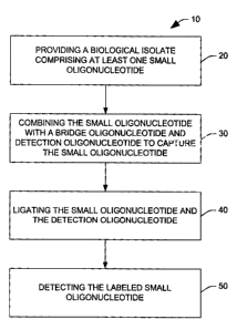
[0009] Fig. 1 is a flowchart showing a process for detecting a plurality of
small
oligonucleotides according to one aspect of the present invention;
[0010] Figs. 2A-C show the quantitative expression of miR-21 using the present
invention (Fig. 2A, upper) and Northern blot (Fig. 2A, lower). The assays
represented in Fig.
2A were performed in parallel using HeLa cell total RNA with the indicated
amounts.
Images were quantif~: :I using phosphorimager analysis (Fig. 2B). Fig. 2C is a
table showing
the experimental parameters and the resultant sensitivities of each assay;
[0011] Figs. 3A-B show miRNA expression profiles in muscle tissue (Fig. 3A,
upper),
brain tissue (Fig. 3A, middle), and HeLa cells (Fig. 3A, lower) using the
present invention.
Fig. 3B is a table comparing experimental parameters between the present
invention and
Northern blot assay; and
[0012] Figs. 4A-C illustrate a comparative example of the present invention
and a
solution hybridization/ribonuclease protection assay. Figs. 4A-B show
detection of miR-
124a and miR-133a-I by the present invention (Fig. 4A) and solution
hybridization/ribonuclease protection assay (Fig. 4B). Both assays were
performed in
parallel using total RNA with the indicated amounts. The image was developed
after 2 hr X-
ray film exposure at -80 C. Fig. 4C is a table comparing the experimental
parameters and
resultant sensitivities of the present invention and the solution
hybridization/ribonuclease
protection assay.
Detailed Description
[0013] The present invention relates generally to methods for detecting
nucleic acids, and
more particularly to methods for detecting small oligonucleotides, such as
small regulatory
RNAs (e.g., microRNA (miRNA) and small interfering RNA (siRNA)). It was
discovered
that a labeled detection oligonucleotide in combination with a bridge
oligonucleotide can be
used to detect small oligonucleotides, such as small regulatory RNAs (e.g.,
miRNAs and
siRNAs). Based on this discovery, the present invention provides a method for
detecting
multiple small oligonucleotides in, for example, about 50 ng or less of total
RNA without the
CA 02604664 2007-09-28
-4-
need for an amplification step. Advantageously, the method of the present
invention is fast
(e.g., capturing and labeling small oligonucleotides in just over 2 hours) and
has a linear
detection range of about 0.1 to about 10 femtomoles.
,[0014] The present invention also allow for the simultaneous detection of
several small
oligonucieotides. Because several small oligonucleotides can be simultaneously
detected, the
diversity of small oligonucleotides present in a cell or organism can be
readily evaluated in a
research or clinical setting using the present invention.
[0015] As used herein, the term "small oligonucleotide" is intended to mean a
nucleic
acid having a length of about 5 to about 500 nucleotides, (e.g., about 5 about
to about 50
nucleotides, about 5 to about 30 nucleotides, or about 10 to about 30
nucleotides), and
terminating in a 5' phosphate and/or a 3' hydroxyl. A 5' phosphate is
understood to be a
(P04)2- (PO4H)- or (POaHz) moiety covalently attached to the 5' carbon of
ribose via one of
the oxygens. A 3' hydroxyl is understood to be an OH or 0- moiety covalently
attached to
the 3' carbon of ribose via the oxygen. Those skilled in the art will
recognize that the
presence or absence of hydrogen in the phosphate and hydroxyl moieties as
listed above is a
function of their pKa values and the pH of their environment.
[0016] Small oligonucleotides can be identified according to their function in
a cell
including, for example, having a non-coding sequence (i.e., not being
translated into protein)
(e.g., non-coding RNA) and being capable of regulating expression of at least
one gene
(e.g., small regulatory RNA). Small oligonucleotides can also be identified
according to their
biosynthesis. For example, one type of small oligonucleotide, siRNA, is
typically
synthesized from endogenous or exogenous double-stranded RNA (dsRNA) molecules
having hairpin structures and processed such that numerous siRNA molecules are
produced
from both strands of the hairpin. In contrast, miRNA molecules are typically
produced from
endogenous dsRNA molecules having one or more hairpin structure such that a
single
miRNA molecule is produced from each hairpin structure. The terms "siRNA" and
"miRNA" are intended to be consistent with their use in the art as described,
for example, in
Ambros et al., RNA 9:277-279 (2003).
[0017] Where a small oligonucleotide comprises small regulatory RNA, such as
siRNA
or miRNA, the small oligonucleotide can be distinguished from mRNA based on
the presence
of a 5' cap structure in mRNA and absence of the cap structure in the small
oligonucleotide.
The 5' cap structure typically found in eukaryotic mRNA is a 7-methylguanylate
having a 5'
CA 02604664 2007-09-28
-5-
to 5' triphosphate linkage to the terminal nucleotide. Small interfering RNA
molecules or
miRNAs can also be distinguished from mRNA based on the presence of a terminal
polyadenylate sequence at the 3' end of mRNA which is absent in siRNAs and
miRNAs.
[0018] As used herein, the term "biological isolate" is intended to mean one
or more
substances removed from at least one co-occurring molecule of an organism. An
isolated
nucleic acid can be, for example, essentially free of other nucleic acids such
that it is
increased to a significantly higher fraction of the total nucleic acid present
in the biological
isolate than in the cells from which it was taken. For example, an isolated
nucleic acid can be
enriched at least 2, 5, 10, 50, 100, 1000 fold or higher in the biological
isolate compared to in
the cell from which it was taken. A biological isolate can be obtained from an
intact
organism, tissue or cell. Examples of eukaryotes from which biological
isolates can be
derived in a method of the invention include, without limitation, a mammal,
such as a rodent,
mouse, rat, rabbit, guinea pig, ungulate, horse, sheep, pig, goat, cow, cat,
dog, primate,
human or non-human primate; a plant such as Arabidopsis thaliana, corn (Zea
mays),
sorghum, oat (Oryza sativa), wheat, rice, canola, or soybean; an algae, such
as
Chlamydomonas reinhardtii; a nematode such as Caenorhabditis elegans; an
insect, such as
Drosophila melanogaster, mosquito, fruit fly, honey bee or spider; a fish such
as zebrafish
(Danio rerio); a reptile; an amphibian such as a frog or Xenopus laevis; a
dictyostelium
discoideum; a fungi, such as Pneumocystis carinii, Takifugu rubripes, yeast,
Saccharamoyces
cerevisiae or Schizosaccharomyces pombe; or Plasmodium falciparum. In addition
to animal
and plant systems, the present invention can be used with a prokaryote system
including, for
example, a bacterium such as Escherichia coli, Staphylococci or Mycoplasma
pneumoniae;
an archae; a virus such as Hepatitis C virus or human immunodeficiency virus;
or a viroid.
Endogenous small oligonucleotides can be isolated from a biological system
from which they
were synthesized. Exogenous small oligonucleotides can be isolated from a
biological
system from which they were transmitted, for example, by viral infection or
treatment with a
small oligonucleotide precursor. Exemplary small oligonucleotide precursors
include
dsRNAs, such as those described in further detail below.
[0019] As used herein, the term "detection oligonucleotide" is intended to
mean a
nucleotide sequence comprising about 5 to about 500 nucleotides (e.g., about.5
to about 200
nucleotides, about 5 to about 100 nucleotides, or about 5 to about 50
nucleotides), and
terminating in a 5' end and a 3' end. In one aspect of the present invention,
the detection
CA 02604664 2007-09-28
-6-
oligonucleotide may include a 5' phosphate and a modified 3' end. A 5'
phosphate is
understood to be a(PO4)2- (PO4H)- or (P04H2) moiety covalently attached to the
5' carbon of
ribose via one of the oxygens. In another aspect of the present invention, the
detection
oligonucleotide may include a 3' hydroxyl and a modified 5' end. A 3' hydroxyl
is
understood to be an OH or 0- moiety covalently attached to the 3' carbon of
ribose via the
oxygen. Those skilled in the art will recognize that the presence or absence
of hydrogen in
the phosphate and hydroxyl moieties as listed above is a function of their pKa
values and the
pH of their environment.
[0020] A modified 5' or 3' end may include any chemical or structural
modification of at
least one nucleotide that may prevent, inhibit and/or reduce phosphodiester
bond formation
between two nucleotides. Examples of such modifications include, without
limitation, C-3
spacers, amino modifiers, inverted dTs, and dideoxy-Cs. By including a
modified 5' or 3'
end, formation of unwanted side ligation products may be reduced or prevented.
[0021] The detection oligonucleotide may be comprised of naturally occurring
or
synthetic RNA, DNA, or other oligonucleotides such as an analog of a naturally
occurring
nucleic acid. At least one locked nucleic acid (LNA) molecule may also be
included in the
detection oligonucleotide. A LNA can include a modified RNA nucleotide, for
example, in
which the ribose moiety of the LNA is modified with an extra bridge connecting
the 2' and 4'
carbons. The bridge may lock the ribose in a 3'-endo structural conformation,
thereby
enhancing base stability and backbone pre-organization.
[0022] A nucleic acid analog can have an alternate backbone including, without
limitation, phosphoramide (see, for example, Beaucage et al., Tetrahedron
49:1925 (1993);
Letsinger, J. Org. Chem. 35:3800 (1970); Sprinzl et al., Eur. J. Biochem.
81:579 (1977);
Letsinger et al., Nucl. Acids Res. 14:3487 (1986); Sawai et al, Chem. Lett.
805 (1984),
Letsinger et al., J. Am. Chem. Soc. 110:4470 (1988); and Pauwels et al.,
Chemica Scripta
26:141 (1986)), phosphorothioate (see, for example, Mag et al., Nucleic Acids
Res. 19:1437
(1991); and U.S. Pat. No. 5,644,048), phosphorodithioate (see, for example,
Briu et al., J.
Am. Chem. Soc. 111:2321 (1989)), O-methylphophoroamidite linkages (see, for
example,
Eckstein, Oligonucleotides and Analogues: A Practical Approach, Oxford
University Press),
and peptide nucleic acid backbones and linkages (see, for example, Egholm, J.
Am. Chem.
Soc. 114:1895 (1992); Meier et al., Chem. Int. Ed. Engl. 31:1008 (1992);
Nielsen, Nature,
365:566 (1993); Carlsson et al., Nature 380:207 (1996)). Other analog
structures include
CA 02604664 2007-09-28
-7-
those with positive backbones (see, for example, Dempcy et a1., Proc. Natl.
Acad. Sci.
USA 92:6097 (1995); non-ionic backbones (see, for example, U.S. Pat. Nos.
5,386,023;
5,637,684; 5,602,240; 5,216,141 and 4,469,863; Kiedrowshi et al., Angew. Chem.
Intl. Ed.
English 30:423 (1991); Letsinger et al., J. Am. Chem. Soc. 110:4470 (1988);
Letsinger et al.,
Nucleoside & Nucleotide 13:1597 (1994); Chapters 2 and 3, ASC Symposium Series
580,
"Carbohydrate Modifications in Antisense Research", Ed. Y. S. Sanghui and P.
Dan Cook;
Mesmaeker et al., Bioorganic & Medicinal Chem. Left. 4:395 (1994); Jeffs et
al., J.
Biomolecular NMR 34:17 (1994); Tetrahedron Lett. 37:743 (1996) and non-ribose
backbones, including, for example, those described in U.S. Pat. Nos. 5,235,033
and
5,034,506, and Chapters 6 and 7, ASC Symposium Series 580, "Carbohydrate
Modifications
in Antisense Research", Ed. Y. S. Sanghui and P. Dan Cook. Analog structures
containing
one or more carbocyclic sugars are also useful in the methods and are
described, for example,
in Jenkins et al., Chem. Soc. Rev. (1995) pp169-176. Several other analog
structures that are
useful in the invention are described in Rawls, C & E News Jun. 2, 1997 page
35. Similar
analogs can be used in a probe or other nucleic acid of the invention.
[0023] A nucleic acid or nucleic acid analog used in the present invention can
include
native or non-native bases or both. Native deoxyribonucleic acid bases include
adenine,
thymine, cytosine or guanine and native ribonucleic acid bases include uracil,
adenine,
cytosine or guanine. Exemplary non-native bases that can be used in the
invention include,
without limitation, inosine, xathanine, hypoxathanine, isocytosine,
isoguanine, 5-
methylcytosine, 5-hydroxymethyl cytosine, 2-aminoadenine, 6-methyl adenine, 6-
methyl
guanine, 2-propyl guanine, 2-propyl adenine, 2-thioLiracil, 2-thiothymine, 2-
thiocytosine, 15-
halouracil, 15-halocytosine, 5-propynyl uracil, 5-propynyl cytosine, 6-azo
uracil, 6-azo
cytosine, 6-azo -thymine, 5-uracil, 4-thiouracil, 8-halo adenine or guanine, 8-
amino adenine or
guanine, 8-thiol adenine or guanine, 8-thioalkyl adenine or guanine, 8-
hydroxyl adenine or
guanine, 5-halo substituted uracil or cytosine, 7-methylguanine, 7-
methyladenine,
8-azaguanine,8-azaadenine,7-deazaguanine,7-deazaadenine,3-deazaguanine,
3-deazaadenine or the like.
[0024] As used herein, the term "bridge oligonucleotide" is intended to mean a
nucleotide
sequence comprising about 10 tol00 nucleotides or longer, and terminating in a
5' end and a
3' end. The bridge oligonucleotide can include naturally occurring or
synthetic RNA, DNA,
or other oligonucleotide, such as an analog of a naturally occurring nucleic
acid (described in
CA 02604664 2007-09-28
-g-
detail above). Both the 5' and 3' ends of the bridge oligonucleotide may
include a
modification, such a C-3 spacer, inverted dT, amino modifier, dideoxy-C, or
another agent to
reduce or prevent the formation of unwanted side ligation products.
Alternatively, the 5' and
3' ends may respectively include a 5' phosphate and a 3' hydroxyl; however,
such end groups
are not preferred as there may be a greater chance of generating side ligation
products. The
bridge oligonucleotide can be complementary to both the detection
oligonucleotide and a
small oligonucleotide at the 5' and 3' ends, respectively. Alternatively, the
bridge
oligonucleotide can be complementary to both the detection oligonucleotide and
a small
oligonucleotide at the 3' and 5' ends, respectively. Further, a portion of the
bridge
oligonucleotide between the 5' and 3' ends may be complementary to thP small
oligonucleotide, while the 5' and 3' ends may be complementary to a plurality
of detection
oligonucleotides.
[0025] As used herein, the term "label moiety" is intended to mean one or more
atom(s)
that can be specifically detected to identify a substance, such as a nucleic
acid, to which the
one or more atom(s) is/are attached. A label moiety can be a primary label
that is directly
detectable or secondary label that can be indirectly detected, for example,
via interaction with
a primary label. Exemplary primary labels include, without limitation, an
isotopic label, such
as a naturally non-abundant heavy isotope or radioactive isotope examples of
which .include
14C ]23h 1241, ]25h 1311, 32P, 33p, 35S or 3H; optically detectable moieties,
such as a
chromophore, luminophore, fluorophore, quantum dot or nanoparticle light
scattering label;
electromagnetic spin label; calorimetric agent; magnetic substance; electron-
rich material,
such as a metal; electrochemiluminescent label, such as Ru(bpy)32+; moiety
that can be
detected based on a nuclear magnetic, paramagnetic, electrical, charge to
mass, or thermal
characteristic; or light scattering or plasmon resonant materials such as gold
or silver
particles. Fluorophores that can be used in the invention include, for
example, fluorescent
lanthanide complexes, including those of Europium and Terbium, fluorescein,
fluorescein
isothiocyanate, dichlorotriazinylamine fluorescein, rhodamine,
tetramethylrhodamine,
umbelliferone, eosin, erythrosin, coumarin, methyl-coumarins, pyrene, Malacite
green, Cy3,
Cy5, stilbene, Lucifer Yellow, CASCADE BLUE, Texas Red, alexa dyes, dansyl
chloride,
phycoerythin, green fluorescent protein and its wavelength shifted variants,
bodipy, and
others known in the art, such as those described in Haugland, Molecular Probes
Handbook,
CA 02604664 2007-09-28
-9-
(Eugene, Oreg.) 6th Edition; The Synthegen catalog (Houston, Tex.), Lakowicz,
Principles of
Fluorescence Spectioscopy, 2nd Ed., Plenum Press New York (1999), or WO
98/59066.
[0026] Examples of secondary labels are binding moieties, such as a receptor,
ligand or
other member of a pair of molecules having binding specificity for each other.
Exemplary
binding moieties having specificity for each other include, without
limitation,
streptavidin/biotin, avidin/biotin or an antigen/antibody complex such as
rabbit IgG and anti-
rabbit IgG. Specific affinity between two binding partners is understood to
mean preferential
binding of one partner to another compared to binding of the partner to other
components or
contaminants in the system. Binding partners that are specifically bound
typically remain
bound under the detection or separation conditions described herein, including
wash steps to
remove non-specific binding. Depending upon the particular binding conditions
used, the
dissociation constants of the pair can be, for example, less than about 10-4,
10-5, 10-6 , 10 -7,
10l, 10l, 10-10, 10-11, or 10-12 M-1. Secondary labels also include enzymes
that produce a
detectable product such as horseradish peroxidase, alkaline phosphatase, 0-
galactosidase, or
acetylcholinesterase.
[0027] As used herein, the term "ligand" is intended to mean a molecule that
is capable of
selectively binding to another molecule.
[0028] Fig. 1 is a flow diagram illustrating a method 10 for detecting small
oligonucleotides in accordance with an aspect of the invention. In the method
10, at 20, a
biological isolate comprising at least one small oligonucleotide is provided.
The biological
isolate can be from any of a variety of organisms including, without
limitation, those set forth
above. In many cases, biological isolates can be available from commercial
sourees or from
banks and depositories administered by public or private institutions such as
the American
Type Culture Collection (ATCC). For many applications, it is desirable that
isolation
protocols used by commercial sources are not biased against retention of small
oligonucleotides of interest for a particular application of the present
invention. A biological
isolate can be from one or more cells, bodily fluids or tissues. Known methods
can be used
to obtain a bodily fluid such as blood, sweat, tears, lymph, urine, saliva,
semen, cerebrospinal
fluid, feces or amniotic fluid. Similarly known biopsy methods can be used to
obtain cells or
tissues, such as buccal swab, mouthwash, surgical removal, biopsy aspiration
or the like. A
biological isolate can also be obtained from one or more cell or tissue in
primary culture, in a
propagated cell line, a fixed archival sample, forensic sample or
archeological sample.
CA 02604664 2007-09-28
-10-
[0029] Exemplary cell types from which a nucleic acid-containing isolate can
be obtained
in the method 10 of the present invention can include, without limitation, a
blood cell such as
a B lymphocyte, T lymphocyte, leukocyte, erythrocyte, macrophage, or
neutrophil; a muscle
cell such as a skeletal cell, smooth muscle cell or cardiac muscle cell; germ
cell such as a
sperm or egg; epithelial cell; connective tissue cell such as an adipocyte,
fibroblast or
osteoblast; neuron; astrocyte; stromal cell; kidney cell; pancreatic cell;
liver cell; or
keratinocyte. A cell from which an isolate is obtained can be at a particular
developmental
level including, for example, a hematopoietic stem cell or a cell that arises
from a
hematopoietic stem cell such as a red blood cell, B lymphocyte, T lymphocyte,
natural killer
cell, neutrophil, basophil, eosinophil, monocyte, macrophage, or platelet.
Other cells include
a bone marrow stromal cell (mesenchymal stem cell) or a cell that develops
therefrom such as
a bone cell (osteocyte), cartilage cells (chondrocyte), fat cell (adipocyte),
or other kinds of
connective tissue cells such as one found in tendons; neural stem cell or a
cell it gives rise to
including, for example, a nerve cell (neuron), astrocyte or oligodendrocyte;
epithelial stem
cell or a cell that arises from an epithelial stem cell such as an absorptive
cell, goblet cell,
Paneth cell, or enteroendocrine cell; skin stem cell; epidermal stem cell; or
follicular stem
cell. Generally, any type of stem cell can be used including, without
limitation; an embryonic
stem cell, adult stem cell, or pluripotent stem cell.
[0030] A cell from which a biological isolate is obtained for use in the
present invention
can be a normal cell or a cell displaying one or more symptom of a particular
disease or
condition. Thus, a biological isolate used in a method of the present
invention'can be
obtained from a cancer cell, neoplastic cell, necrotic cell or cell
experiencing a disease or
condition set forth below. Those skilled in the art will know or be able to
readily determine
methods for isolating samples from a cell, fluid or tissue using methods known
in the art such
as those described in Sambrook et al., Molecular Cloning: A Laboratory Manual,
3rd edition,
Cold Spring Harbor Laboratory, New York (2001) or in Ausubel et al., Current
Protocols in
Molecular Biology, John Wiley and Sons, Baltimore, Md. (1998).
[0031] Another aspect of the present invention can further include steps of
isolating a
particular type of cell or tissue. Exemplary methods that can be used to
isolate a particular
cell from other cells in a population include, but are not limited to,
Fluorescent Activated Cell
Sorting (FACS) as described, for example, in Shapiro, Practical Flow
Cytometry, 3rd edition
Wiley-Liss; (1995), density gradient centrifugation, or manual separation
using
CA 02604664 2007-09-28
-11-
micromanipulation methods with microscope assistance. Exemplary cell
separation devices
that are useful in the invention include, without limitation, a Beckman JE-6
centrifugal
elutriation system, Beckman Coulter EPICS ALTRA computer-controlled Flow
Cytometer-
cell sorter, Modular Flow Cytometer from Cytomation, Inc., Coulter counter and
channelyzer
system, density gradient apparatus, cytocentrifuge, Beckman J-6 centrifuge,
EPICS V dual
laser cell sorter, or EPICS PROFILE flow cytometer. A tissue or population of
cells can also
be removed by surgical techniques. For example, a tumor or cells from a tumor
can be
removed from a tissue by surgical methods, or conversely non-cancerous cells
can be
removed from the vicinity of a tumor.
[0032] A biological isolate can be prepared for use in the method 10 of the
present
invention by lysing a cell that contains one or more desired nucleic acids.
Typically, a cell is
lysed under conditions that substantially preserve the integrity of the
desired nucleic acid.
For example, cells can be lysed or subfractions obtained under conditions that
stabilize RNA
integrity. Such conditions include, for example, cell lysis in strong
denaturants, including
chaotropic salts such as guanidine thiocyanate, ionic detergents such as
sodium dodecyl
sulfate, organic solvents such as phenol, high lithium chloride concentrations
or other
conditions known in the art to be effective in limiting the activity of
endogenous RNases
during RNA purification as described, for example, in Sainbrook et al., supra
(2001) or in
Ausubel et al., supra (1998). Additionally, relatively undamaged nucleic acids
such as RNA
can be obtained from a cell lysed by an enzyme that degrades the cell wall.
Cells lacking a
cell wall either naturally or due to enzymatic removal can also be lysed by
exposure to
osmotic stress. Other conditions that can be used to lyse a cell include
exposure to
detergents, mechanical disruption, sonication, heat, pressure differential
such as in a French
press device, or Dounce homogenization.
[0033] According to another aspect of the present invention, total RNA may be
prepared
using guanidine isothiocyanate, phenol:chloroform, and an inert carrier such
as glycogen or
linear polyacrylamide. For instance, about 20 ng of glycogen per 1 ml
(reaction volume) may
be added during alcohol precipitation to increase recovery of total RNA.
Samples may then
be prepared by commercially available column-based purification methods, such
as the
miRACLE miRNA Purification Kit from STRATAGENE (La Jolla, CA). Isolated RNA
may
be diluted in TE buffer or RNase-free water, for example, and stored
appropriately.
CA 02604664 2007-09-28
-12-
[0034] Agents that stabilize nucleic acids can be included in a cell lysate or
other
biological isolate including, for example, nuclease inhibitors such as
ribonuclease inhibitors
or deoxyribonuclease inhibitors, chelating agents, salts buffers and the like.
Methods for
lysing a cell to obtain nucleic acids can be carried out under conditions
known in the art as
described, for example, in Sambrook et al., supra (2001) or in Ausubel et al.,
supra, (1998).
[0035] In another aspect of the present invention, a biological isolate can be
a crude cell
lysate obtained without further isolation of nucleic acids. Alternatively, a
nucleic acid of
interest can be further isolated from other cellular components according to
the present
invention. The method 10 of the present invention can be carried out on
purified or partially
purified RNA. RNA can be isolated using known separation methods including,
for example,
liquid phase extraction, precipitation or solid phase extraction. Such methods
are described,
for example, in Sambrook et al., supra, (2001) or in Ausubel et al., supra,
(1998) or available
from various commercial vendors including, for example, Qiagen (Valencia,
Calif.) or
Promega (Madison, Wis.).
[0036] If desired, nucleic acids can be separated based on properties such as
mass, charge
to mass, or the presence of a particular sequence. Methods for separating
nucleic acids
include, but are not limited to, electrophoresis using agarose or
polyacrylamide gels, capillary
electrophoresis, conventional chromatography methods such as size exclusion
chromatography, reverse phase chromatography or ion exchange chromatography or
affinity
methods such as affinity chromatography or precipitation using solid-phase
poly dT
oligonucleotides. Those skilled in the art will know or be able to determine
an appropriate
separation method or combination of separation methods to obtain a biological
isolate of a
desired nucleic acid composition and purity. Proteins and large genomic DNA
can be
removed from RNA, for example, using precipitation and centrifugation methods
that exploit
the larger size of the genomic DNA and proteins. Messenger RNA can be removed
from
other RNA species, for example, using precipitation with poly dT
oligonucleotide beads or
size exclusion chromatography. Such methods can be used in combination with
selective
modification of the 5' phosphate of small oligonucleotides to distinguish
small
oligonucleotides from other cellular components.
[0037] At 30, the biological isolate is contacted with a labeled detection
oligonucleotide
and a bridge oligonucleotide to capture a small oligonucleotide from the
biological isolate.
CA 02604664 2007-09-28
-13-
By "capture" it is meant that a nucleic acid, such as a small oligonucleotide,
hybridizes to a
complementary nucleic acid, such as the bridge oligonucleotide.
[0038] The detection oligonucleotide may be prepared under conditions such
that the
5' phosphate and/or the 3' hydroxyl of the detection oligonucleotide is/are
preferentially
modified to include a label moiety. Optionally or additionally, the label
moiety may be
internally incorporated into the detection oligonucleotide. According to
another aspect of the
present invention, a phosphate reactive reagent comprising a label moiety or
label precursor
moiety can be used so that 5' phosphate modification produces a detection
oligonucleotide
containing the label moiety. The detection oligonucleotide may also be
prepared such that
the 5' end or the 3' end includes a modification (e.g., inverted dTs) that
prevents or reduces
the formation of side ligation products.
[0039] By way of example, the detection oligonucleotide may be generated by
5' end-labeling a DNA oligonucleotide with [y-32P]-ATP, including an inverted
dT at the 3'
end, and then removing any unincorporated isotope. Briefly, reagents (i.e.,
the detection
oligonucleotide, RNase-free water, l OX OPTIKINASE Reaction Buffer, [r 32P]-
ATP, and
OPTIKINASE) may be thawed on ice, mixed thoroughly, briefly spun, and then
combined at
room temperature. The mixture may then be incubated for about 30 to 60
minutes. After
incubation, aliquots of the mixture may be treated to remove unincorporated [y-
32P]-ATP.
Thereafter, a population of labeled detection oligonucleotides may be
generated and used as
described below.
[0040] It should be understood that a variety of phosphate reactive reagents
may be used
with the present invention. Generally, the phosphate reactive reagent can
preferentially add a
moiety to a phosphate in a reaction mixture. For example, a phosphate reactive
agent can be
added to a population of detection oligonucleotides so that it preferentially
reacts with the
5' phosphate of the detection oligonucleotide. Phosphate reactive reagents can
include those
that are unreactive to the 3' end of nucleic acids.
[0041] A phosphate reactive reagent can be a single molecule or a combination
of
molecules. For example, a single molecule can contain a reactive moiety linked
to a label
moiety such that reaction between the reactive moiety and the 5' phosphate of
the detection
oligonucleotide produces a detection oligonucleotide linked to the label
moiety.
[0042] Alternatively, a combination of molecules can be used as a phosphate
reactive
reagent. For example, a first label molecule can be contacted with a detection
CA 02604664 2007-09-28
-14-
oligonucleotide and a second molecule that activates the 5' phosphate or the
first label
molecule, thereby producing a detection oligonucleotide with an attached
label. The
phosphate reactive reagent can additionally or alternatively include a label
moiety having a
linked amino group and a second molecule being a carbodiimide molecule that
activates the
5' phosphate to react with the amino group to produce a detection
oligonucleotide having a
phosphoramidite linkage to the label moiety. Other exemplary phosphate
reactive reagents
include, without limitation, c-(6-(biotinoyl)amino)hexanoyl-L-lysine,
hydrazide; DSB-X
biotin hydrazide; DSB-X desthiobiocytin (-desthiobiotinoyl-L-lysine); DSB-X
biotin
ethylenediamine (desthiobiotin-X ethylenediamine, hydrochloride); Biotin-X
cadaverine;
Alexa Fluor cadaverine; 5-(aminoacetamido)fluorescein(fluoresceinyl glycine
amide); 4'-
(aminomethyl)fluorescein, hydrochloride; 5-(((2-(carbohydrazino)methyl)thio)
acetyl)aminofluorescein; fluorescein-5-thiosemicarbazide; N-methyl-4-hydrazino-
7-
nitrobenzofurazan; Oregon Green 488 cadaverine; 5-((5-
aminopentyl)thioureidyl)eosin,
hydrochloride; Texas Red cadaverine; Texas Red hydrazide; bimane amine;
poly(ethylene
glycol) methyl ether, amine-terminated; and Lissamine rhodamine B
ethylenediamine. Those
skilled in the art will recognize that any of a variety of label moieties can
be replaced for
those listed in these reagents. For example, fluorescein can be replaced with
other
fluorophores described herein.
[0043] The bridge oligonucleotide may be a DNA oligonucleotide complementary
to both
the detection oligonucleotide and a specific small oligonucleotide at the 5'
and 3' ends of the
bridge oligonucleotide, respectively. Alternatively, the bridge
oligonucleotide can be
complementary to both the detection oligonucleotide and a small
oligonucleotide at the 3' and
5' ends, respectively. It should be that where, for example, the label moiety
is attached to the
phosphate group at the 5' end of the detection oligonucleotide, and the bridge
oligonucleotide
is designed to join the detection oligonucleotide to the 3' hydroxyl group of
the small
oligonucleotide, that the phosphate-labeled moiety should not have properties
that prevent a
ligation reaction.
[0044] By way of example, a small oligonucleotide can be captured by combining
a
biological isolate with a labeled detection oligonucleotide and a bridge
oligonucleotide. A
positive control containing a small oligonucleotide of interest and negative
control (i.e., not
containing RNA) may also be prepared. The reaction mixture may then be mixed,
briefly
spun, and incubated for a desired period of time at an appropriate
temperature. A small
CA 02604664 2007-09-28
-15-
oligonucleotide may be captured by the bridge oligonucleotide by hybridizing
to the 3' end of
the bridge oligonucleotide. The labeled detection oligonucleotide may also
hybridize to the
5' end of the bridge oligonucleotide either before, simultaneous with, or
after the small
oligonucleotide has been captured. Alternatively, a small oligonucleotide may
be captured by
the bridge oligonucleotide by hybridizing to the 5' end of the bridge
oligonucleotide. In this
case, the labeled detection oligonucleotide may hybridize to the 3' end of the
bridge
oligonucleotide either before, simultaneous with, or after the small
oligonucleotide has been
captured.
[0045] After incubating the reaction mixture, at least one ligating reagent
may be added
to the reaction mixture at 40. A "ligating reagent" as used herein refers to a
molecule,
typically an enzyme, capable of facilitating phosphodiester bond formation
between two
nucleotides. Examples of ligating reagents include DNA ligases, such as T4 DNA
ligase,
which may form covalent phosphodiester bonds between the 3' hydroxyl end of
one
nucleotide and the 5' phosphate end of another nucleotide. Other examples of
ligating
reagents include RNA ligase, DNA ligases I-IV, as well as chemical cross-
linking agents
known in the art.
[0046] Following capture of the small oligonucleotide, at 40, the captured
small
oligonucleotide and the labeled detection oligonucleotide are ligated to form
a labeled small
oligonucleotide. The captured small oligonucleotide and the labeled detection
oligonucleotide can be ligated by combining the captured small oligonucleotide
and the
labeled detection oligonucleotide with at least one ligating reagent. The at
least ligating
reagent can include, for example, a composition comprising T4 DNA ligase. The
ligating
reagent may be added to the reaction mixture, and the resultant reaction
mixture briefly spun
and incubated for a desired period of time at an appropriate temperature. By
adding the
ligating reagent to the reaction mixture, the 5' phosphate of the detection
oligonucleotide and
the 3' hydroxyl of the small oligonucleotide form a covalent phosphodiester
bond so that the
5' and 3' ends of the detection oligonucleotide and the small oligonucleotide
are respectively
ligated. Alternatively, where the detection oligonucleotide and the small
oligonucleotide are
respectively hybridized to the 3' and 5' ends of the bridge oligonucleotide,
the 3' hydroxyl of
the detection oligonucleotide and the 5' phosphate of the small
oligonucleotide may form a
covalent phosphodiester bond so that the 3' and 5' ends of the detection
oligonucleotide and
the small oligonucleotide are respectively ligated.
CA 02604664 2007-09-28
-16-
[0047] In one example, the size of the ligated small oligonucleotide and
detection
oligonucleotide can be about 35 to about 39 nucleotides (e.g., about 21 to
about
25 nucleotides of the small oligonucleotide plus about 14 nucleotides of the
labeled detection
oligonucleotide). It will be appreciated that the size of the ligated small
oligonucleotide and
the detection oligonucleotide can be smaller or larger depending on the size
of the small
oligonucleotide and the detection oligonucleotide.
[0048] Following ligation of the captured small oligonucleotide and the
labeled detection
oligonucleotide, excess detection oligonucleotides and/or labeled moieties
from any unligated
detection oligonucleotides of the reaction mixture can be removed. The excess
labeled
moieties can be removed using, for example, a clean-up reagent comprising a
phosphatase.
The phosphatase can remove phosphate groups from the 5' ends of small
oligonucleotides
containing phosphate-] abeled moieties. Removal of labeled phosphate groups
can prevent
interference with small oligonucleotide detection and analysis. It should be
appreciated that
clean-up reagents may not be required depending upon the properties of the
label moiety
and/or where the label moiety is incorporated. Upon removing any unligated
detection
oligonucleotides from the reaction mixture, a plurality of labeled small
oligonucleotides is
generated that can be subsequently detected.
[0049] Following ligation, at 50, the labeled small oligonucleotides can be
detected. The
labeled small oligonucleotide can be detected and distinguished from other
molecules that are
devoid of a label using methodsknown in the art. Exemplary properties upon
which
detection can be based include, but are not limited to, mass, electrical
conductivity or optical
signals, such as a fluorescent signal, absorption signal, luminescent signal,
chemiluminescent
signal or the like. Detection can also be based on absence or reduced level of
one or more
signal, for example, due to the presence of a signal quenching moiety or
degradation of a
label moiety.
[0050] Various detection methods, such as solution phase and solid phase
assays, may be
used to detect labeled small oligonucleotides. Solution phase detection
methods can be based
on charge, mass, charge-to-mass ratio or other distinguishing properties. Such
distinguishing
properties can be detected, for example, in a chromatography system such as
capillary
electrophoresis, acrylamide gel, agarose gel, or the like, or in a
spectroscopic system such as
mass spectroscopy. For example, detection of fluorescence can be carried out
by irradiating a
labeled small oligonucleotide with an excitatory wavelength of radiation and
detecting
CA 02604664 2007-09-28
-17-
radiation emitted from a fluorophore therein by methods known in the art and
described in
Lakowicz, Principles of Fluorescence Spectroscopy, 2nd Ed., Plenum Press New
York
(1999). A fluorophore can be detected based on any of a variety of
fluorescence phenomena
including, for example, emission wavelength, excitation wavelength,
fluorescence resonance
energy transfer (FRET) intensity, quenching, anisotropy or lifetime. FRET can
be used to
identify hybridization between a first oligonucleotide attached to a donor
fluorophore and a
second oligonucleotide attached to an acceptor fluorophore due to transfer of
energy from the
excited donor to the acceptor. Thus, hybridization can be detected as a shift
in wavelength
caused by reduction of donor emission and appearance of acceptor emission for
the hybrid.
[0051] In another aspect of the present invention, the labeled small
oligonucleotide can
be detected using gel electrophoresis. Briefly, a UREA-polyacrylamide gel of
about 12% to
15% may be prepared and then loaded with samples of a reaction mixture
containing labeled.
small oligonucleotides. The gel may be run using appropriate amperage until
each sample
has reached an appropriate end-point on the gel. The resultant gel may be
transferred onto a
sheet of non-diffusible support material, such as processed film, and then
developed by
exposure to X-ray or a phosphorimager screen for an appropriate amount of
time. After
developing the gel, the presence of darkened bands at known positions on the
gel may
indicate the presence of small oligonucleotides in the biological isolate.
[0052] The present invention provides a fast and efficient method for
detecting small
oligonucleotides over the standard technique for small oligonucleotide
expression analysis,
i.e., Northern blotting. Northern blotting exhibits poor sensitivity, requires
large amounts of
total RNA, and is both labor intensive and time-consuming. As illustrated in
Figs. 2A-C, the
present invention provides a 50-fold better detection sensitivity than
Northern blotting.
Additionally, the present invention significantly reduces both the total
reaction time and the
amount of total RNA needed to perform the assay. Referring to Figs. 3A-B, for
example, the
amount of total RNA needed with the present invention is only 18 g, whereas
180 pg of
total RNA is needed for Northern blotting. Similarly, the reaction time needed
with the
present invention is only
2-4 hours, whereas the reaction time needed for Northern blotting is 2-3 days.
[0053] As noted, other detection methods, such as solid phase assays, may be
used to
detect small oligonucleotides. For example, different types of arrays may be
used. An
"array" refers to a population of different probe molecules that are attached
to one or more
CA 02604664 2007-09-28
-Ig-
substrates such that the different probe molecules can be differentiated from
each other
according to relative location. An array can include different probe molecules
that are each
located at a different addressable location on a substrate. Alternatively, an
array can include
separate substrates each bearing a different probe molecule. Probes attached
to separate
substrates can be identified according to the locations of the substrates on a
surface to which
the substrates are associated or according to the locations of the substrates
in a liquid.
Exemplary arrays in which separate substrates are located on a surface
include, without
limitation, those including beads in wells.
[0054] Another aspect of the present invention may include a kit for detecting
a plurality
of different small oligonucleotides. The kit may include a detection
aligonucleotide, a label
moiety for preferentially labeling the detection oligonucleotide, and reagents
for ligating the
3' end of the small oligonucleotide to the 5' end of the detection
oligonucleotide.
[0055] In another aspect of the present invention, the kit may include
additional
components, including, for example, reagents for removing unbound label
moieties, reagents
for removing non-ligated detection oligonucleotides, reagents for detecting
labeled small
oligonucleotides, a positive control, and a negative control. The positive
control may be used
to assess kit components and procedure, while the negative control may be used
to assess
background signal(s) in tested samples.
[0056] Commercially available kits for detecting small oligonucleotides
include solution
hybridization/ribonuclease protection assays such as the mirVana miRNA
Isolation Kit
available from Ambion (Austin, TX). The present invention provides several
advantages
over such commercial assays. When compared to the mirVana miRNA Isolation Kit,
for
example, the present invention provides greater miRNA detection sensitivity in
a shorter
reaction period. As shown in Figs. 4A-C, the mirVana miRNA Isolation Kit not
only exhibits
poorer sensitivity in a longer reaction period, but also requires the separate
purchase of a
labeling kit and optimization of the amount of labeled probe, hybridization
conditions, and
the amount of ribonuclease.
[0057] Throughout this application various publications, patents and patent
applications
have been referenced. The disclosures of these publications in their
entireties are hereby
incorporated by reference in this application in order to more fully describe
the state of the art
to which this invention pertains.
CA 02604664 2007-09-28
-19-
[0058] The following examples are for the purpose of illustration only and are
not
intended to limit the scope of the claims, which are appended hereto.
Example 1
Detection Oligo Preparation and Clean-Up
[0059] The first step is to 5' end-label the Detection Oligo with [7_32P]-ATP
and remove
unincorporated isotope.
1. Thaw frozen reagents on ice, mix thoroughly followed by a brief spin, and
then place
on ice.
2. Piepare [32P]-latieled Detection Oiigo by combining the following
components at
room temperature:
Components Detection Oligo Component Cap Color
Detection Oligo 2 l Blue
RNase Free Water 12 l White
l OX OPTIKINASE Reaction Buffer 2 l Blue
[y-32P]-ATP (6000 Ci/mMol, 150mCi/ml) 2 l Not Supplied
OPTIKINASE 2 l Blue
Total volume 20 gl
3. Mix thoroughly followed by a brief spin in a microcentrifuge. Incubate for
30-60 mm
at 37 C.
4. While the reactions are incubating, prepare the Clean-Up Column as follows:
a. Centrifuge the Clean-Up Column for 30 sec at 750 x g to collect the dry
resin
at the bottom of the column.
b. Hydrate the resin by adding 600 l RNase-Free Water and vortex. Remove air
bubbles by vonexing or tapping the column. Incubate at least 30 mm at room
temperature.
c. Re-suspend the settled resin by inverting the column several times. Ensure
that no air bubbles are visible. Remove the bottom plug and place in a 2.0 ml
collection tube.
CA 02604664 2007-09-28
-20-
d. Centrifuge for 2 mm at 750 x g to remove the remaining water. Discard the
flow-through.
5. After 30 mm incubation, dilute the labeling reactions to 100 l by adding
80 l of
RNase-Free Water.
6. Place the column from Step 4d in a clean 1.5 ml microcentrifuge tube.
Without
disturbing the gel bed, carefully apply the diluted sample directly onto the
top of the
gel bed.
7. After loading the sample, centrifuge the column for 4-6 mm at 750 x g.
Discard the
used column in a radioactive waste container.
8. The radiolabeled Detection Oligo and Marker are now ready to use.
Example 2
miRNA Capture, Ligation and Clean-Up
[0060] Once the Detection Oligo is radiolabeled, proceed to the miRNA
detection assay.
[00611 Design and Preparation on page 12. The Bridge Oligonucleotide should be
diluted to 0.1 pmol/ l with the provided lOX Capture Buffer.
1. Thaw frozen reagents on ice, mix thoroughly followed by a brief spin, and
then place
on ice.
2. Assemble the capture reaction on ice according to the table below:
Positive No RNA Component Cap
Components Control Control Sample Color
Total RNA Sample 0 gl 041 up to 8 Not Supplied
~
Positive Control I l 0 l 0 l Red
Adjust to 8 gl with RNASE-Free Water White
0.1 pmol/ l Bridge Oligo in lOX I gl 1 l 1 l Not Supplied
Capture Butter
Radio-labeled Detection Oligo I l 1 l 1 1 N/A
Total Volume 10 l l0 l 10 l
CA 02604664 2007-09-28
-21-
= Make a Bridge Oligonucleotide-Detection Oligo Master Mix for assay setup.
Per
sample combine: I g] Bridge Oligonucleotide in l OX Capture Buffer + I
l radiolabeled Detection Oligo.
= Dilute all test samples to 8 l with RNase-Free Water then add 2 l of the
Bridge
Oligonucleotide-Detection Oligo Master Mix.
3. Mix thoroughly followed by a brief spin in a microcentrifuge. Incubate the
mixture at
94 C for I min, 65 C for 2 min and 37 C for 10 min. For convenience, a
thermalcycler may be used.
4. Add 5 ] of 3X Ligate-ITTM Premix (red cap) to each reaction.
5. Mix thoroughly followed by a brief spin. Incubate for 1 hr at 30 C.
Optional: If not
proceeding to the next step immediately, inactivate the reaction by
incubat:o_z for 10
min at 75 C and store at -20 C for later use.
.6. Add I 1 of Clean-Up Mix (red cap) to each reaction.
7. Mix thoroughly followed by a brief spin. Incubate for 15 mm at 37 C.
Optional: If
not proceeding to the next step immediately, inactivate the reaction by
incubation for
min at 75 C and store at -20 C for later use.
8. The ligated miRNA is now ready for gel electrophoresis.
Example 3
Electrophoretic Analysis -
1. Prepare a 12% or 15% UREA-polyacrylamide gel with ]x running buffer. (See
Supplementary Information on Preparation of UREA-polyacrylamide gel)
Optional: Pre-run and warm the gel for 30 minutes.
2. Transfer a 3-15 pl aliquot of the reaction to a new tube. Add an equal
volume of Gel
Loading Dye.
3. Transfer an aliquot of the [32P]-labeled Low Molecular Weight Marker to a
new tube.
Add an equal volume of Gel Loading Dye.
4. Mix thoroughly followed by a brief spin in a microcentrifuge. Incubate for
3 min at
95 C. Immediately cool on ice.
5. Flush wells of the gel to remove acrylamide debris, urea and air bubbles.
6. Load and separate the denatured reactions. Include the [32P)-labeled Low
Molecular
Weight Marker on each gel.
CA 02604664 2007-09-28
-22-
= The expected size of ligated miRNA is 35-39 nucleotides (21-25 nucleotides
of mature miRNA sequence plus 14 nucleotides of the labeled Detection
Oligo).
= In 12% and 15% gels, the approximate band positions of bromophenol blue
and xylene cyanol (loading dyes) are 10 and 30 nucleotides, respectively.
= For 13 cm x 15 cm gel, run at 20-30 mA and stop when the bromophenol blue
dye front has migrated to the bottom of the gel. For 36 cm x 43 cm gel, run at
60 mA and stop when the bromophenol blue dye front has migrated to the
middle of the gel.
7. At the end of the separation, transfer the gel onto a sheet of non-
diffusible support
material such as processed film. Wrap with saran wrap and expose to X-ray film
or a
phosphorimager screen.
= It is unnecessary to dry the gel if using [32P]-isotope. Expose the gel to X-
ray
film with an intensifying screen. Store for 2 hr to overnight at -80 C. The
gel
can be re-exposed several times if required.
= It is recommended to dry the gel if using a phosphorimager screen to prevent
screen damage. Process the phosphorimager screen according to the
manufacturer's instructions.
[0062] From the above description of the invention, those skilled in the art
will perceive
improvements, changes and modifications. Such improvements, changes and
modifications
within the skill of the art are intended to be covered by the appended claims.
All references,
patents, and publications cited herein are incorporated by reference in their
entirety.