Note: Descriptions are shown in the official language in which they were submitted.
CA 02604953 2007-10-15
WO 2006/113562 PCT/US2006/014301
1
INSTRUMENTS, IMPLANTS AND METHODS FOR POSITIONING IMPLANTS
INTO A SPINAL DISC SPACE
BACKGROUND
Normal intervertebral discs between endplates of adjacent vertebrae distribute
forces between the vertebrae and cushion vertebral bodies. The spinal discs
may be
displaced or damaged due to trauma, disease or aging. A herniated or ruptured
annulus
fibrosis may result in nerve damage, pain, numbness, muscle wealcness, and
even
paralysis. Furthermore, as a result of the normal aging processes, discs
dehydrate and
harden, thereby reducing the disc space height and producing instability of
the spine and
decreased mobility. Most surgical corrections of a disc space include a
discectomy, which
can be followed by restoration of normal disc space height and bony fusion of
the adjacent
vertebrae to maintain the disc space height.
Access to a damaged disc space may be accomplished from several approaches to
the spine. One approach is to gain access to the anterior portion of the spine
tlirough a
patient's abdomen. However, extensive vessel retraction is often required and
many
vertebral levels are not readily accessible from this approach. A posterior
approach may
also be utilized. This approach typically requires that both sides of the disc
space on either
side of the spinal cord be surgically exposed, which may require a substantial
incision or
multiple access locations, as well as extensive retraction of the spinal cord.
To alleviate
problems associated with both anterior and posterior approaches to the spine,
a postero-
lateral approach to the disc space may be utilized.
There remains a need for improved instruments, implants and techniques for use
in
a postero-lateral approach to a spinal disc space that facilitate disc space
preparation and
implant insertion to provide bilateral stability to the subject disc space.
SUMMARY
There are provided instruments, implants and methods useful for implant
insertion
from a postero-lateral approach to the spinal disc space, although application
with other
approaches are also contemplated.
CA 02604953 2007-10-15
WO 2006/113562 PCT/US2006/014301
2
BRIEF DESCRIPTION OF THE DRAWINGS
FIG. 1 is a plan view of an inserter instrument and an implant in an initial
position
in a spinal disc space.
FIG. 2 is an enlarged perspective view of the implant of Fig. 1.
FIG. 3 is an enlarged plan view showing the implant engaged with the distal
end of
the inserter instrument in the initial position of Fig. 1.
FIG. 4 is a plan view of the inserter instiument and the implant in a final
position
in the spinal disc space.
FIG. 5 is an enlarged plan view showing the implant engaged with the distal
end of
the inserter instrument in the final position of Fig. 4.
FIG. 6 is a plan view of the distal end of the inserter instrument with the
implant
removed and with the inserter in the initial position.
FIG. 7 is a perspective view of the distal end of the inserter instrument in
an
engaged position with the implant and the implant oriented in the final
position.
FIG. 8 is a perspective view of the distal end of the inserter instrument in a
release
position with the implant and the implant oriented in the final position.
FIG. 9 is a top plan view of the distal end of the inserter instrument in a
release
position immediately after placing the implant in the fmal position.
FIG. 10 is a top plan view of the distal end of the inserter instrument in the
release
position and the inserter withdrawn proximally from the implant oriented in
the fmal
position.
FIG. 11 is a perspective view of the distal end of the inserter instrument
with the
posterior arm removed and the anterior arm in the initial position.
FIG. 12 is a perspective view of the distal end of the inserter instrument
with the
posterior gripping arm removed and the anterior arm in the final position.
FIG. 13 is a top plan view of a portion of the shaft assembly and an interior
portion
of a handle assembly of the inserter instrument.
FIG. 14 is a perspective view of the interior portion of the handle assembly
shown
in Fig. 13 including a frame of the handle assembly.
FIG. 15 is the view of Fig. 13 including the frame of Fig. 14 and also an
articulating driver of the handle assembly.
CA 02604953 2007-10-15
WO 2006/113562 PCT/US2006/014301
3
FIG. 16 is a perspective view showing a portion of the shaft assembly with a
hub
removed and a proximal portion of the interior of the handle assembly.
FIG. 17 is the view of Fig. 16 with a lock driver engaged about a lock screw
of the
handle assembly.
FIG. 18 is a perspective view of the distal portion of the inserter instrument
in the
release position with the implant removed.
FIG. 19 is a perspective view of the distal portion of the inserter instrument
in the
engaged position with the implant removed.
FIG. 20 is a perspective view of another embodiment inserter instrument and
implant.
FIG. 21 is a perspective view of a distal portion of the inserter instrument
and the
implant of,Fig. 20 with the implant partially engaged to the inserter
instrument.
FIG. 22 is the perspective view of Fig. 21 in horizontal section through the
distal
portion of the inserter instrument and implant.
FIG. 23 is an enlarged perspective view in horizontal section showing
engagement
of the inserter instrument with the implant.
FIG. 24 is a perspective view showing the implant and inserter instrument of
Fig.
20 positioned through a retractor sleeve.
DETAILED DESCRIPTION OF THE ILLUSTRATED EMBODIMENTS
For the purposes of promoting an understanding of the principles of the
present
invention, reference will now be made to the embodiments illustrated in the
drawings, and
specific language will be used to describe the same. It will nevertheless be
understood that
no limitation of the scope of the invention is intended thereby. Any
alterations and further
modification in the described processes, systems, or devices, and any further
applications
of the principles of the invention as described herein are contemplated as
would normally
occur to one skilled in the art to which the invention relates.
Instruments, implants and techniques provide and facilitate implant insertion
into a
spinal disc space through a single opening and positioning of the implant so
that it
provides balanced, bi-lateral support of the adjacent vertebrae. The
instruments and
implants can be employed in postero-lateral approaches to the disc space to
obtain proper
positioning of the implant in the portion of the disc space most distal from
the postero-
lateral opening. The instruments and implants facilitate moving the implant
across the
CA 02604953 2007-10-15
WO 2006/113562 PCT/US2006/014301
4
disc space to the distal portion of the disc space so that the implant extends
between distal
and proximal portions of the disc space to provide bi-lateral support of the
adjacent
vertebrae. The inserter instruments provide a low profile engagement with the
implant to
minimize the footprint of the assembly and minimize exposure and retraction of
tissue and
neural elements to accommodate implant insertion.
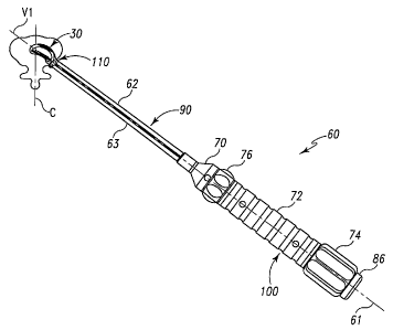
In Figs. 1 and 3 there is shown one embodiment inserter instrument 60 engaged
to
a trailing end of implant 30 at the distal end of inserter instrument 60. A
vertebral body
V1 is shown witli the implant 30 positioned in a disc space adjacent thereto
in an initial
position. In the initial position, implant 30 is inserted into the disc space
while inserter
instrument 60 maintains implant 30 in general align,ment along longitudinal
axis 61 of
inserter instrument 60. In postero-lateral procedures, implant 30 extends
obliquely to
sagittal plane C of the patient when in the initial position.
In Figs. 4 and 5, inserter instrument 60 has been manipulated to reposition
implant
30 to a fmal position for implantation in the disc space. In the orientation
of the final
position, implant 30 is substantially obliquely oriented to longitudinal axis
61 of inserter
instrument 60. Furthermore, implant 30 includes an axis of symmetry Cl. Axis
Cl is
oriented so that it is aligned along or generally parallel to sagittal plane C
of the patient.
In the final position, implant 30 extends across sagittal plane C and contacts
the adjacent
vertebral endplates to provide balanced bi-lateral support of the adjacent
vertebrae.
Inserter instrument 60 can then be disengaged from implant 30 and withdrawn
from the
patient.
The disc space can be accessed and prepared from the postero-lateral approach
using spreaders, cutters, chisels, reamers, and other instruments to prepare
the disc space
and adjacent vertebral endplates to receive implant 30. Examples of such
instruments and
techniques are discussed in U.S. Patent Application Publication No.
2002/0165550,
published November 7, 2002, which is incorporated herein by reference in its
entirety.
One embodiment of implant 30 is shown in further detail in Fig. 2, it being
understood that any suitable implant can be engaged to inserter instrument 60.
Implant 30
includes a body formed by a wal132 extending about a central cavity 42. Cavity
42
extends between and opens at an upper bearing surface 52 and a lower bearing
surface 54.
Upper and lower bearing surfaces 52, 54 contact the adjacent vertebral
endplates to
support the adjacent vertebrae when implant 30 is implanted in the spinal disc
space.
Surfaces 52, 54 may include grooves 50 formed therein to facilitate engagement
with the
CA 02604953 2007-10-15
WO 2006/113562 PCT/US2006/014301
vertebral endplates and resist the implant from migrating in the disc space.
Other surface
features are also contemplated, including teeth, spikes, lcnurlings, peeks and
valleys, and
other projections and/or recesses.
Implant 30 includes convexly curved anterior wall portion 34 and an opposite
concavely curved posterior wall portion 36. Wall portions 34, 36 are connected
by a
convexly curved leading end wall portion 38 and a convexly curved trailing end
wall
portion 40. The overall shape of wa1132 provides a banana, leidney or
boomerang type
shape that facilitates placement of implant 30 along a non-linear insertion
path in the disc
space from the proximal postero-lateral opening to a distal portion of the
disc space
opposite the postero-lateral opening. In the implanted position, posterior
wall portion 36
is oriented toward the spinal foramen. The anterior wall portion 34 extends
anteriorly to
provide anterior support of the vertebrae. The elongated shape of implant 30
facilitates
placement through the postero-lateral opening while minimizing the retraction
of tissue
and neural elements needed to accommodate placement of the implant through the
postero-lateral approach. It should be understood the leading end wall portion
38 can be a
trailing end wall portion, and trailing end wall portion 40 can be a leading
end wall
portion, in situations where wall portion 38 is engaged with an inserter
instrument and
wall portion 40 is first inserted into the disc space through the postero-
lateral opening.
A central opening 49 in anterior wall portion 34 and a central opening 51 in
posterior wall portion 36 provide avenues for bone growth into cavity 42.
Implant 30
further includes a recessed area 44 that extends around the trailing end wall
portion 40 and
along at least a portion of the length of anterior wall portion 34. A
receptacle 46 is formed
in posterior wall portion 36. As discussed further below, the recessed area 44
and
receptacle 46 are configured for engagement by respective portions of a
grasper assembly
110 of inserter instrument 60. Lateral pin holes 48 in recessed areas 44 of
anterior wall
portion 34 can provide additional areas for engagement by the inserter insti-
ument.
As shown in Fig. 1, inserter instrument 60 includes a shaft assembly 90 and a
proximal handle assembly 100 extending along longitudinal axis 61. Implant 30
is
engaged to inserter instrument 60 with grasper assembly 110 at the distal end
of shaft
assembly 90. Handle assembly 100 is operably coupled with grasper assembly 110
through shaft assembly 90 to remotely manipulate grasper assembly 110 to grasp
and
release implant 30 from inserter instrument 60. Grasper assembly 110 is also
remotely
CA 02604953 2007-10-15
WO 2006/113562 PCT/US2006/014301
6
operable to reposition implant 30 relative to longitudinal axis 61 from an
initial position,
shown in Figs. 1 and 3, to a final implanted position, shown in Figs. 4-5.
Shaft assembly 90 of inserter instrument 60 includes a first shaft 62 and a
second
shaft 63. Second shaft 63 extends along and parallel to first shaft 62, and
includes a C-
shaped side oriented toward a C-shaped side of first shaft 62. The C-shaped
sides together
form a passage that receives a locking shaft 68 (Fig 6.) therein. Shaft
assembly 90 further
includes a hub 70 at a proximal end thereof adjacent handle assembly 100.
Second shaft
63 is engaged to hub 70. First shaft 62 and locking shaft 68 extend through
hub 70 and
into handle assembly 100.
Handle assembly 100 includes an outer cylindrical handle member 72 have grip-
enhancing external surface features. A rotatable articulator driver 74 is
between a
proximal end of handle member 72 and a proximal end member 86. Handle assembly
100
further includes a rotatable lock driver 76 at a distal end of handle member
72 between
handle member 72 and hub 70. Drivers 74, 76 each include a series of radial
protuberances and valleys between protuberances to enhance the ability to grip
and apply
the necessary force to rotate drivers 74, 76.
Further details of handle assembly 100 are shown in Figs. 13-17. In Fig. 13
handle
member 72, drivers 74, 76, and second shaft 63 are removed. Locking shaft 68
extends
through hub 70 to a lock screw 66 at a proximal end of locking shaft 68. First
shaft 62
extends through hub 70 and also through lock screw 66 to an articulator screw
64 at a
proximal end of first shaft 62. Screws 64, 66 are linearly movable to linearly
move the
respective shafts 62, 68 in response to rotation of the respective drivers 74,
76 thereabout.
In Fig. 14, a frame 78 is positioned about shafts 62, 68 and screws 64, 66.
Frame
78 includes a distal slot 80 that receives lock screw 66, a proximal slot 82
that receives
articulator screw 64, and an intermediate slot 84 therebetween. Slots 80, 82
are elongated
sufficiently to allow proximal and distal translation of screws 64, 66 to
remotely
manipulate grasper assembly 110. End member 86 at the proximal end of frame 78
can
receive and transmit impaction forces to facilitate insertion of the implant
into the disc
space.
In Fig. 15 there is shown articulator driver 74 rotatably positioned about and
threadingly engaged to articulator screw 64. Rotation of articulator driver 74
about screw
641inearly advances first shaft 62 in a proximal or distal direction,
depending on the
direction of rotation. The linear movement of first shaft 62 in turn
articulates grasper
CA 02604953 2007-10-15
WO 2006/113562 PCT/US2006/014301
7
assembly 110 between the initial position and the final position, as discussed
above and as
discussed further below.
In Fig. 16 there is shown lock screw 66 and shafts 62, 63 are removed. In Fig.
17
lock driver 76 is threadingly engaged to and rotatably positioned about lock
screw 66.
Rotation of lock driver 76 linearly advances locking shaft 68 in a proximal or
distal
direction, depending on the direction of rotation. The linear movement of
locking shaft 68
in turn manipulates grasper assembly 110 between a release position and an
engaged
position relative to the implant positioned therein. In the release position,
grasper
assembly 110 is opened to receive or release the implant, as shown in Figs. 8
and 18, for
example. In the engaged position, the implant positioned in grasper assembly
110 is
engaged by the grasper assembly 110 and to couple the implant to inserter
instrument 60,
as shown in Figs. 7 and 19, for example.
As shown in Figs. 6-12 and 18-19, grasper assembly 110 includes a first arm
112
and a second ann 114. First arm 112 includes a concavely curved inner surface
113, and
second ai7n 114 includes a second concavely curved inner surface 115. Surfaces
113, 115
are oriented toward one another, and are shaped to conform to the outer wall
surfaces of
implant 30 about trailing end wall portion 40 and in recessed area 44 and
receptacle 46,
respectively. First arm 112 may include a pin 102 that is positionable into a
pin hole 48 in
recessed area 44 to further engage implant 30 to grasper assembly 110 and to
maintain the
implant in engagement therewith. Other embodiments contemplated that pin 102
is not
provided, such as shown in Figs. 18 and 19.
First arm 112 includes a proximal lever portion 116 having a first end 118
pivotally coupled to a distal end of first shaft 62 with a pin 108, and a
second end 120
pivotally coupled to a distal end of second shaft 63 with a pin 104. Lever
portion 116
includes a forked arrangement for positioning along the outer surfaces of
shafts 62, 63 to
accommodate placement of a heel portion 122 and toe portion 106 of second arm
114
therebetween.
Second arm 114 includes proximal heel portion 122 having a bulbous shape
positioned in contact with a distal foot 69 of locking shaft 68 (Figs. 6, 18-
19). Heel 122
includes a slotted hole 124 extending between a distal end 126 and a proximal
end 128.
Pin 104 pivotally engages second end 120 of first grasping arm 112 to the
distal end of
second shaft 63. Pin 104 also extends through slotted hole 124 to couple
second aim 114
to the distal end of second shaft 63 while also allowing limited radial
translation of second
CA 02604953 2007-10-15
WO 2006/113562 PCT/US2006/014301
8
arm 114 relative to first arm 112. Slotted hole 124 is configured between its
distal end
126 and proximal end 128 to allow second arm 114 to move toward and away from
first
arm 112 to selectively grip and release the implant therebetween. Second arm
114 further
includes a toe portion 106 opposite heel portion 122. Toe portion 106 is
pivotally coupled
with first shaft 62 and first end 118 of first grasping arm 112 with pin 108.
Linear distal movement of first shaft 62 by rotating articulator driver 74
causes
distal displacement of first end 118 relative to second end 120, which in turn
pivots first
arm 112 and second arm 114 about pin 104 and the fixed second shaft 63. This
movement
in turn moves grasper assembly 110 from its initial position, as shown in
Figs. 1 and 3, to
its final position, as shown in Figs. 4-5. In one embodiment, axis Cl of
implant 30 foims
an angle A1 (Fig. 3) with longitudinal axis 61 in the initial position, and an
angle A2 (Fig.
5) in the final position. In one specific embodiment, angle Al is about 80
degrees to
generally orient implant 30 along axis 61. Angle A2 is about 55 degrees to
orient implant
30 in a substantially oblique orientation to axis 61. Other embodiments
contemplate other
angular orientations, ranging from 70 degrees to 110 degrees for angle Al and
ranging
from 35 degrees to 75 degrees for angle A2. Still other embodiments
contemplate other
angular ranges for angles A1 and A2.
Arms 112, 114 are further moveable to grip and release implant 30 from
therebetween. In the release position, shown in Figs. 8-10 and 18, pin 104 is
adjacent
distal end 126 of slotted hole 124 and foot 69 of locking shaft 68 is moved
distally to a
location spaced a distance 105 from an end wa1165 of a slot in second shaft
63. This
allows second arm 114 to rotate away from first ann 112. To move arms 112, 114
to the
engaged position, foot 69 is advanced distally with distal movement of locking
shaft 68 by
rotation of locking driver 76. As shown in Fig. 7, distal movement of foot 69
displaces it a
second greater distance 105' from end wal165 of second shaft 63, and locking
shaft 68
articulates second arm 114 toward first arm 112. This movement positions pin
104
adjacent the proximal end 128 of slotted hole 124. The articulation of second
arm 114 in
the clockwise direction can be continued to firmly grasp implant 30 between
first and
second arms 112, 114 as shown in Figs. 5 and 7, for example.
Heel portion 122 includes a circular outer perimeter 123 that contacts foot 69
in the
engaged position. While in the engaged position, grasper assembly 110 can be
moved
from the initial position to the implanted position. During this movement, the
circular
CA 02604953 2007-10-15
WO 2006/113562 PCT/US2006/014301
9
perimeter 123 allows foot 69 to maintain contact with heel portion 122 and
maintain arms
112, 114 in the engaged position with implant 30.
In use, arms 112, 114 of inserter instrument 60 are placed in the release
position to
receive implant 30 therebetween. Lock driver 76 is rotated to move arms 112,
114 to the
engaging position to firmly grip implant 30 with grasping assembly 110 in the
initial
position. Implant 300 is delivered to the postero-lateral opening in the disc
space and the
leading end of the implant is positioned through the opening while being
maintained in the
initial position. The implant is advanced in the initial position along axis
61 in a direction
substantially obliquely oriented to sagittal plane C until the trailing end of
implant 30 is
positioned in the disc space. Impaction forces can be delivered to the
proximal end of the
inserter instrument if necessary.
When implant 30 is in the appropriate position in the disc space, articulator
driver
74 can be rotated to manipulate first shaft 62 and grasper assembly 110 to
move implant
30 from the initial position to the final position in the disc space. In the
final position, axis
Cl of implant 30 is oriented along or generally parallel to sagittal plane C.
Lock driver 76
can then be rotated to move locking shaft 68 proximally to allow arms 112, 114
to the
release position for withdrawal of inserter instrument from the disc space.
Figs. 20-23 show another embodiment implant and implant inserter. Implant
inserter 160 includes an elongated shaft assembly 190, a grasper assembly 210
at a distal
end of shaft assembly 190, and a handle assembly 200 at a proximal end of
shaft assembly
190. Implant 130 is releasably engageable at the distal end of shaft asseinbly
190 witli
grasper assembly 210. Handle assembly 200 is operable to manipulate grasper
assembly
210 to grasp and release the implant 130, and to deliver implant 130 to the
spinal disc
space. While specific applications in postero-lateral approaches to the disc
space are
contemplated as discussed above, other approaches to the disc space are also
contemplated.
As shown in further detail in Figs. 21-22, implant 130 includes an overall
size and
shape similar to that discussed above for implant 30. Implant 130 includes an
outer wall
132 extending about a central cavity 142. Cavity 142 extends between and opens
at an
upper bearing surface 152 and a lower bearing surface 154. Upper and lower
bearing
surfaces 152, 154 contact the adjacent vertebral endplates to support the
adjacent vertebrae
when implanted. Surfaces 152, 154 may include pyramidally shaped teeth 150
formed
thereon to facilitate engagement with the vertebral endplates and resist the
implant from
CA 02604953 2007-10-15
WO 2006/113562 PCT/US2006/014301
migrating in the disc space. Other surface features are also contemplated,
including
grooves, spikes, knurlings, peeks and valleys, and other projections and/or
recesses.
Implant 130 includes convexly curved anterior wall portion 134 and an opposite
concavely curved posterior wall portion 136. Wall portions 134, 136 are
connected by a
convexly curved leading end wall portion 138 and a convexly curved trailing
end wall
portion 140. The overall shape of wall 132 provides a banana, kidney or
boomerang type
shape that facilitates placement along a non-linear insertion path in the disc
space. The
elongated shape facilitates placement through the postero-lateral opening
while
minimizing the retraction of tissue and neural elements needed to accommodate
insertion
of the implant through the postero-lateral approach. It should be understood
the leading
end wall portion 138 can be a trailing end wall portion, and trailing end wall
portion 140
can be a leading end wall portion, in situations where wall portion 138 is
engaged with an
inserter instrument and wall portion 140 is first inserted into the disc
space.
A number of openings 149 in posterior wall portion 136 and elongate slots 152
in
anterior wall portion 134 provide avenues for bone growth into cavity 142.
Implant 130
further includes a recessed area 146 that extends into trailing.end wall
portion 140
adjacent posterior wall portion 136, and a receptacle 148 in trailing end wall
portion 140
adjacent anterior wall portion 134. As discussed further below, the recessed
area and
receptacle 146, 148 are configured to receive grasper assembly 210 of inserter
instrument
160. Leading end wall portion 138 can be similarly provided with recessed area
and a
receptacle so that implant 130 can be engaged with an inserter 160 for
insertion from
either direction into the spinal disc space.
Inserter instrument 160 includes shaft assembly 190 extending along
longitudinal
axis 161. Handle assembly 200 is at a proximal end of shaft assembly 190, and
includes a
handle member 172 extending transversely to longitudinal axis 161. A hub
member 170
extends proximally from shaft assembly 190 along longitudinal axis 161. Hub
170
includes a slotted portion 174 formed in and opening along one side thereof. A
lock driver
176 is rotatably positioned therein. Hub 170 further provides a proximally
oriented
platfonn for delivery of impaction forces to facilitate insertion of the
implant engaged to
grasper assembly 210.
As shown in Figs. 22-23, shaft assembly 190 includes a first or outer shaft
162
having a central passage 164 formed therethrough. Locking shaft 168 is
received in and
linearly movable in passage 164 relative to outer shaft 162. Lock driver 176
is threading
CA 02604953 2007-10-15
WO 2006/113562 PCT/US2006/014301
11
engaged about a lock screw (not shown) at the proximal end of a locking shaft
168.
Rotation of lock driver 1761inearly translates loclcing shaft 168 distally and
proximally in
passage 164.
Grasper assembly 210 includes a first arm 166 formed at a distal end of outer
shaft
162. Outer shaft 162 includes an enlarged portion 172 to offset first arm
1661aterally
from passage 164. First arm 166 includes a spherically shaped distal end
portion that is
rotatably received in recessed area 146. In the illustrated embodiment,
recessed area 146
include a complementary spherical shape to interface with first arm 166 and
allow rotation
of implant 130 about first arm 166. Implant 130 is rotatable to position a
distal end wall
180 of outer shaft 162 in abutting contact therewith at trailing end wall
portion 140.
Locking shaft 168 includes a second arm 163 formed at a distal end thereof.
Locking shaft
168 and second arm 163 are distally linearly movable with lock driver 176 to
advance
second arm 163 into receptacle 148. The distal end of second arm 163 can be
beveled to
facilitate insertion into receptacle 148.
In the locking position shown in Fig. 23, locking shaft 168 prevents implant
130
from rotating about first arm 166 and holds implant 130 firmly on inserter
instrument 160.
The distal end of outer shaft 162 includes a recessed area 182 adjacent first
arm 166, and
implant 130 includes a toe 156 between recessed area 146 and receptacle 148.
When
second arm 163 is positioned in receptacle 148, the toe 156 is received in
recessed area
182 as shown in Fig. 23. This provides a dovetail locking arrangement between
implant
130 and grasper assembly 210 that implant 130 from being axially pulled or
rotated
relative to inserter instrument 160.
When implant 130 is positioned in the disc space, inserter instrument 160 can
be
disengaged therefrom by rotating lock driver 176 to proximally withdraw
locking shaft
168 distally and remove second arm 163 from the receptacle 148. The inserter
instrument
160 can then be withdrawn proximally from the disc space. Intrusion into
tissue and
neural elements in the approach to the disc space is minimized since inserter
instrument
160 has the same footprint transversely to longitudinal axis 161 when engaged
to implant
130 and when disengaged to implant 130. The footprint of the implant and
inserter
instrument assembly is also minimized during insertion since arms 163, 166
extend into
implant 130 at or adjacent trailing end wall portion 140, and do not occupy
space
anteriorly or posteriorly of implant 130.
CA 02604953 2007-10-15
WO 2006/113562 PCT/US2006/014301
12
The above-described instruments and methods have been disclosed with reference
to use in substantially open surgical procedures. However, it is contemplated
that the
implants, instruments and methods may be utilized through guide sleeves or
tubes, such as
retractor sleeve 200 shown in Fig. 24. Such instruments can provide greater
protection to
adjacent tissues, to reduce the size of access incisions, to provide direct
visualization of
the surgical site, and/or to provide greater control of the method. The
implants,
instruments and methods may further be used in combination with disc space
preparation
and implant insertion through microscopic or endoscopic instruments that
provide direct
visualization of the surgical site.
The instruments discussed herein are suited for inserting an implant through a
postero-lateral opening in a spinal disc space. The inserter instruments
provide the
surgeon the ability to control insertion of an implant into the spinal disc
space from a
postero-lateral approach. The inserter instruments facilitate positioning of
the implant in
the disc space such that the implant extends across the disc space to provide
bilateral
support of the adjacent vertebrae, and also facilitate positioning of the
implant in the disc
space along a non-linear insertion path. The inserter instruments can also be
used to
position multiple implants at various locations in the disc space, and also
for insertion of
one or more implants from other approaches to the disc space.
Implants 30, 130 can be interbody fusion devices or cages that can be packed
with
bone growth material or other known substance and inserted into a spinal disc
space to
promote bony fusion between vertebrae. Furthermore, the structural features of
implant
30, 130 can have application for a disc prosthesis or a disc nucleus
prosthesis that is to be
inserted into the disc space. The illustrated implants 30, 130 have a
boomerang or banana
shape that is suited for insertion to provide bilateral support in the disc
space through a
unilateral, postero-lateral approach. It is also contemplated that the disc
space can be
accessed and prepared for implant insertion using any other lcnown techniques
and
instruments and other approaches to the disc space, such as posterior,
lateral, anterior or
antero-lateral approaches.
Implants 30, 130 can include other shapes and also include interior bars,
struts and
walls. The upper and lower bearing surfaces can include double convexity to
provide an
intimate fit in the disc space and a profile that matches the concavity of the
endplates,
providing implant stability and promoting fusion. The sidewall openings and
hollow
interior cavity can maximize the volume available to receive bone growth
material and
CA 02604953 2007-10-15
WO 2006/113562 PCT/US2006/014301
13
also the contact surface area between the bone growth material and the
adjacent bony
structure. Furthermore, differences in heights between the upper and lower
bearing
surfaces at the anterior and posterior walls can be provided to establish
lordosis when
implants 30, 130 are inserted in the disc space.
The implants described herein can be made from any biocompatible material,
including synthetic or natural autograft, allograft or xenograft tissues, and
can be
resorbable or non-resorbable nature. Examples of tissue materials include hard
tissues,
connective tissues, demineralized bone matrix and combinations thereof.
Furtlier
examples of resorbable materials are polylactide, polyglycolide, tyrosine-
derived
polycarbonate, polyanhydride, polyorthoester, polyphosphazene, calcium
phosphate,
hydroxyapatite, bioactive glass, and combinations thereof. Further examples of
non-
resorbable materials are non-reinforced polymers, carbon-reinforced polymer
composites,
PEEK and PEEK composites; shape-memory alloys; titanium and titanium alloys;
cobalt
chrome alloys; stainless steel; ceramics; and combinations thereof.
Instruments described
herein can be made from any suitable surgical grade material, including
stainless steel,
aluminum, plastics, and combinations of materials.
Any suitable osteogenetic material or composition is contemplated for
placement
within the cavities defined by the implants described herein. Such osteogenic
material
includes, for example, autograft, allograft, xenograft, demineralized bone,
synthetic and
natural bone graft substitutes, such as bioceramics and polymers, and
osteoinductive
factors. Where bony material is placed within the cavities of the implant, the
material can
be pre-packed into the hollow cavities before the device is implanted, or can
be pushed
through the wall openings after the device is in position in the spinal
column. A separate
carrier to hold the materials within the device can also be used. These
carriers can include
collagen-based carriers, bioceramic materials, such as EIOGLASS
hydroxyapatite and
calcium phosphate compositions. The carrier material can be provided in the
form of a
sponge, a block, folded sheet, putty, paste, graft material or other suitable
form.
Moreover, the osteogenetic compositions contained within the implant can
comprise an
effective amount of a bone morphogenetic protein, transforming growth factor
.beta.1,
insulin-like growth factor 1, platelet-derived growth factor, fibroblast
growth factor, LIM
mineralization protein (LMP), and combinations thereof or other therapeutic or
infection
resistant agent, held within a suitable carrier material.
CA 02604953 2007-10-15
WO 2006/113562 PCT/US2006/014301
14
While the invention has been illustrated and described in detail in the
drawings and
the foregoing description, the same is considered to be illustrative and not
restrictive in
character. All changes and modifications that come within the spirit of the
invention are
desired to be protected.