Note: Descriptions are shown in the official language in which they were submitted.
CA 02605016 2007-10-15
WO 2006/113654 PCT/US2006/014455
SYSTEM AND RELATED METHOD FOR DETERMINING
A MEASUREMENT BETWEEN LOCATIONS ON A BODY
BACKGROUND OF THE INVENTION
Field of the Invention
The invention relates generally to the field of systems that are used for
determining a measurement between locations on a body. More specifically, the
invention
relates to a system and related method for measuring the distance and/or the
orientation between
locations on a body, and characterizing an effect of a rehabilitation therapy
on the body based on
the measurement.
Description of the Related Art
Stroke is a leading cause of permanent impairment and disability. For example,
approximately 70 percent of all stroke survivors have a paralyzed limb, e.g.,
an arm or a hand.
Stroke victims that receive rehabilitative therapy soon after the stroke,
typically within the first
three months after a stroke, may recover some of the original mobility of
their impaired limb(s).
Several techniques have been developed to help make the rehabilitation process
for stroke
victims more efficient, and to aid in the assessment of the patient's
progress. Some of these
techniques include manual rehabilitation performed by a therapist, using
simple rehabilitation
tools. The therapist can assess the patient's progress during the
rehabilitation process using a
variety of methods, including, for example, the Stroke Rehabilitation
Assessment of Movement
("STREAM") test, which associates scores on the Box and Block Test, the
Balance Scale, and
the Barthel Index, which are known to those having ordinary skill in the art.
A patient performs the Box and Block test using a box that includes a
partition,
which divides the box into two equal compartments. A number of small wooden
blocks are
placed one of the box's compartments. During the test, the patient is required
to use the affected
limb, e.g., the arm and hand that are impaired due to the stroke, to move as
many blocks as
possible from one of the box's compartments to the other comparhnent in 60
seconds. The
patient can move the blocks only by grasping one block at a time, transporting
the block over the
partition, and releasing the block into the other compartment. Once the test
is complete, the
number of blocks transported from one compartment to the other compartment is
counted. Some
other devices that are used for the assessment of spasticity are, for example,
the BIODEX
-1-
CA 02605016 2007-10-15
WO 2006/113654 PCT/US2006/014455
MULTI-JOINT SYSTEM II isokinetic dynamometer, which is available from Biodex
Medical
System of Sllirley, New York; and the RIGIDITY ANALYZER by Prochazka of
Edmonton,
Canada.
An alternative to having a therapist manually perform rehabilitation therapy
on a
stroke victim is to use a robotic rehabilitation device. Robotic
rehabilitation devices can
combine both training and assessment capabilities in the same device. For
example, the robot
can cause the patient to move his or her impaired limb according to a
preferred trajectory, or to
access the patient's progress in voluntarily tracking a cursor on a screen
with the impaired linlb.
Some of the robots that are available on the market are offered by Interactive
Motion
Technologies, Inc. ("IMT") of Cambridge, Massachusetts; and Rehab Robotics
Limited,
Staffordshire University of Staffordshire, United Kingdom.
An example of a device that recently has been used to measure the mobility of
a
patient's impaired limb is an angle measurement device called a goniometer.
Several types of
goniometers are known in the art. Example goniometers can determine angle
measurements
based on changes in the resistance of a fluid in a tube as the tube is bent,
changes in the optical
properties of an optical fiber as the optical fiber is bent, the rotation of
wheels, and/or the
extension of cables. However, these goniometers typically require a physical
interconnection,
for example, via a tube, a fiber, wires, andlor cables, between the points on
the patient's body
that are to be compared during the angle measurement. An example goniometer is
the MLTS700
JOINT ANGLE SENSOR by PowerLab of New South Wales, Australia. Additional
examples of
goniometers are discussed in U.S. Patent Application Publication Nuinber
2003/0083596 to
Kramer et al. and U.S. Patent Number 6,651,352 to McGorry et al.
Recently, virtual reality applications have boosted various types of 3-D
tracking
and positioning devices for wrists and fingers, for example the CYBERGLOVE by
Immersion
Corporation of San Jose, California. The CYBERGLOVE is available in an
eighteen sensor
model, which features two bend sensors on each finger, four abduction sensors,
and sensors for
measuring thumb crossover, palm arch, wrist flexion, and wrist abduction. The
CYBERGLOVE
also is available in a twenty-two sensor model, which includes additional
sensors that are used to
measure the flexion and wrist abduction
The devices discussed above and the currently available tools that are used to
assess the mobility of a patient's limb and the patient's progress during
rehabilitation therapy are
-2-
CA 02605016 2007-10-15
WO 2006/113654 PCT/US2006/014455
considered to be poor proxies for the everyday use of an impaired limb. Also,
many of the
currently available tools require a therapist to play an active role during
the assessment
procedure. Accordingly, there is a need for a system that is configured to
assess the mobility of
a stroke patient's impaired limb(s) during rehabilitation therapy, which can
include the patient's
everyday use of the limb(s) and physical therapy. The present invention
satisfies this need, as
well as other needs as discussed below.
SUMMARY OF THE INVENTION
The invention resides in a system and a related method for assessing the
mobility
of a stroke patient's impaired limb(s) during rehabilitation therapy,
including everyday use. An
exemplary embodiment of the present invention is a system that is configured
to characterize an
effect of a rehabilitation therapy on a body. The system includes a first
device, which is
configured to be coupled to a body at a first location, and a second device,
which is configured to
be coupled to the body at a second location that is separated from the first
location by a first
distance. The first device is configured to generate a first wireless signal.
The second device is
configured to detect the first wireless signal and to generate data based on
the detected first
wireless signal that is configured to be used to calculate the first distance.
The first distance is
used to characterize the effect of the rehabilitation therapy on the body.
In other, more detailed features of the invention, the system further includes
a
third device, which is configured to be coupled to the body at a third
location that is separated
from the second location by a second distance. The first device is configured
to generate the first
wireless signal at a first frequency. The third device is configured to
generate a second wireless
signal at a second frequency. The second device is configured to detect the
second wireless
signal and to generate additional data based on the detected second wireless
signal. The
additional data is configured to be used to calculate the second distance,
which is used to
characterize the effect of the rehabilitation therapy on the body. Also, the
third device can be
configured to generate the second wireless signal at the same time that the
first device is
configured to generate the first wireless signal. In addition, the second
device can be configured
to detect in a selectable manner the first wireless signal or the second
wireless signal.
In other, more detailed features of the invention, the apparatus further
includes an
external device, which is configured to communicate with the second device.
The second device
is configured to communicate the data to the external device. The external
device is configured
-3-
CA 02605016 2007-10-15
WO 2006/113654 PCT/US2006/014455
to calculate the tirst distance based on the data. Also, the external device
can be configured to
calculate an angle of orientation between the second device and the first
device based on the
data. In addition, the external device can be configured to communicate with
the second device
via a wireless communication path that is a radio frequency path, an
electrical current path
through the body, a path configured for the communication of modulated sonic
waves, a path
configured for the communication of modulated ultrasonic waves, and/or an
optical
communication path.
In other, more detailed features of the invention, the external device is
configured
to calculate one or more of the following values: an average of the first
distance over a period of
time, a standard deviation of the first distance over a period of time, a
number of times that the
second device is moved relative to the first device over a period of time
based on the first
distance, a velocity of the second device relative to the first device based
on the first distance, an
average velocity of the second device relative to the first device over a
period of time based on
the first distance, an acceleration of the second device relative to the first
device based on the
first distance, and an average acceleration of the second device relative to
the first device over a
period of time based on the first distance.
In other, more detailed features of the invention, the first device and/or the
second
device is configured to be implanted into the body, or attached to the body
using an adhesive, a
piece of clothing, a strap, a belt, a clip, and/or a watch. Also, the first
device can be coupled to
the torso of the body, and the second device can be coupled to a hand or an
arm of the body. In
addition, the wireless signal can be a magnetic field, a low-frequency
magnetic field, a sonic
wave, or an ultrasonic wave.
In other, more detailed features of the invention, the first device and/or the
second
device includes a component that is a battery, a coil, orthogonal coils, a
generator, a voltage
measurement circuit, a transducer, a processing circuit, a transmitter, a
receiver, and/or a
transceiver. Also, the first device and/or the second device can be a
miniature stimulator. In
addition, the first device and/or the second device can include a transmitter
and a receiver.
Another exemplary embodiment of the present invention is a system that is
configured to characterize an effect of a rehabilitation therapy on a body.
The system includes a
transmitter, a plurality of receivers, and an external device. The transmitter
is configured to be
coupled to a body at a first location, and each of the plurality of receivers
is configured to be
-4-
CA 02605016 2007-10-15
WO 2006/113654 PCT/US2006/014455
coupled to the body at a different location that is separated from the
transmitter by one of a
plurality of distances. The external device is configured to communicate with
the plurality of
receivers. The transmitter is configured to transmit a wireless signal. Each
of the plurality of
receivers is configured to detect the wireless signal, to generate data based
on the detected
wireless signal, and to communicate the data to the external device. The
external device is
configured to calculate the plurality of distances between the plurality of
receivers and the
transmitter based on the data. The plurality of distances is used to
characterize the effect of the
rehabilitation therapy on the body.
In other, more detailed features of the invention, the wireless signal is an
ultrasonic wave, and the external device is configured to calculate the
plurality of distances
based on an amplitude of the ultrasonic wave detected by each of the plurality
of receivers, a
phase of the ultrasonic wave detected by each of the plurality of receivers,
and/or a time of
propagation of the ultrasonic wave to each of the plurality of receivers.
In other, more detailed features of the invention, the external device is
configured
to calculate a plurality of angles of orientation between the plurality of
receivers and the
transmitter based on the data. Also, the system can further include an
additional device that is
coupled to the external device and configured to aid in the calculation of the
plurality of
distances and the plurality of angles of orientation. The additional device
can be a distance
sensor, an angle sensor, an acceleration sensor, a vibration sensor, and/or a
video camera. In
addition, the external device can be configured to calculate, based on the
data, a velocity of each
of the plurality of receivers relative to the transmitter, and/or an
acceleration of each of the
plurality of receivers relative to the transmitter.
In other, more detailed features of the invention, the body includes a healthy
limb
and a corresponding impaired limb. One of the plurality of receivers is
configured to be coupled
to the healthy limb, and another of the plurality of receivers is configured
to be coupled to the
impaired limb. The external device is configured to compare the distance
between the one of the
plurality of receivers and the transmitter to the distance between the another
of the plurality of
receivers and the transmitter.
An exemplary method according to the invention is a method for characterizing
an effect of a rehabilitation therapy on a body. The method includes providing
a first device that
is configured to be coupled to the body at a first location and configured to
transmit a wireless
-5-
CA 02605016 2007-10-15
WO 2006/113654 PCT/US2006/014455
signal, providing a second device that is configured to be coupled to the body
at a second
location and configured to detect the wireless signal, using the first device
to transmit the
wireless signal, using the second device to detect the wireless signal,
calculating a distance
between the first device and the second device based on the wireless signal
that is detected by the
second device, and using the distance to characterize the effect of the
rehabilitation therapy on
the body.
Other features of the invention should become apparent from the following
description of the preferred embodiments taken in conjunction with the
accompanying drawings,
which illustrate, by way of example, the principles of the invention.
BRIEF DESCRIPTION OF THE DRAWINGS
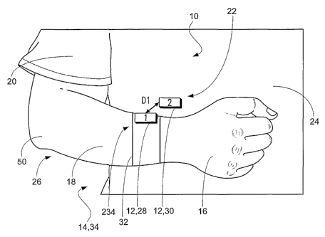
Figs. 1A and 1B are illustrations of a system according to an embodiment of
the
present invention that is configured to monitor the range of movement of a
patient's arm and
hand relative to his or her torso.
Figs. 2A and 2B are illustrations of another system according to an embodiment
of the present invention that is configured to monitor the range of movement
of a patient's arm
and hand relative to his or her torso.
Fig. 3 is a perspective illustration of a battery powered miniature
stimulator.
Fig. 4 is a block diagram of a system according to an embodiment of the
present
invention that includes battery powered miniature stimulators.
Fig. 5 is a block diagram of another system according to an embodiment of the
present invention that includes battery powered miniature stimulators.
Fig. 6 is an illustration of a system according to an embodiment of the
present
invention that is configured to measure the distance and the orientation
between a transmitter and
a receiver.
Fig. 7 is an illustration of a system according to an embodiment of the
present
invention that is configured to measure the distance between a transmitter and
a receiver.
-6-
CA 02605016 2007-10-15
WO 2006/113654 PCT/US2006/014455
Fig. 8 is an illustration of a system according to an embodiment of the
present
invention that is configured to measure the distance between an ultrasonic
transmitter and an
ultrasonic receiver.
Figs. 9A, 9B, and 9C are illustrations of signal characteristics including
signal
amplitude, phase difference, and time of arrival difference, respectively, for
an ultrasonic signal.
Fig. 10 is a schematic illustration of a data acquisition system according to
an
embodiment of the present invention.
Fig. 11 is a flow diagram of an exemplary algorithm according to the present
invention.
Fig. 12 is a graph of a daily average distance between a patient's healthy
hand and
his or her torso as a function of time, and a daily average distance between
the patient's stroke-
affected hand and his or her torso as function of time.
DETAILED DESCRIPTION OF THE PREFERRED EMBODIMENTS
Embodiments of the present invention provide for a relatively inexpensive and
portable approach for assessing the mobility of a patient's limb(s) during
rehabilitation therapy,
including routine daily activity. Referring to Figs. lA-B, embodiments of the
present invention
are systems 10 that include miniature devices 12, which are configured to
communicate with one
another wirelessly, and do not require any physical interconnection between
the devices.
Embodiments of the present invention utilize these miniature devices to
measure static and
motion parameters for parts of a patient's body 14.
In specific embodiments, the devices 12 are used to measure the following: the
distance and/or angle between the patient's hand 16, forearm 18, and/or upper
arm 20 and a
predetermined location 22 on the patient's body 14, e.g., the patient's torso
24; the angle
between the patient's hand and forearm; and the speed and/or the acceleration
of the patient's
hand and arm relative to the predetermined location. By measuring the
distance, angle, and/or
the motion parameters between two or more locations on the patient's body, the
mobility and
rehabilitation status of parts of the body, e.g., the hand and the arm, can be
assessed and tracked.
By tracking the distance between locations on the patient's body, the maximal
and
typical values of displacement of a part 16-20 of the patient's body 14 can be
calculated during
-7-
CA 02605016 2007-10-15
WO 2006/113654 PCT/US2006/014455
daily activity. These displacement values can be used to evaluate the
effectiveness of
rehabilitation tllerapy, including everyday use, on the patient's body. In
extreme disability cases,
an iinpaired limb, e.g., the patient's arm 26, will be kept in close proximity
to the patient's torso
24, and with a limited amount and range of movement. As a result of the
rehabilitation process,
it is expected that the amount and range of the movement of the impaired limb
will increase over
time.
In Figs. 1 A-B, the miniature devices 12, i.e., a first device 28 and a second
device
30, are coupled to a patient's forearm 18 and torso 24, respectively. One or
both of the devices
can be coupled to the patient's body 14 by attaching the device(s) to the
patient using adhesive
and/or straps 32, or iinplanting the device(s) into the patient's forearm
and/or torso. Fig. lA
shows the patient 34 holding his or her arm 26 in close proximity to his or
her torso, in such a
manner that the first device and the second device are near one another. Thus,
in Fig. lA, the
distance Dl between the first device and the second device is relatively
short. In the
configuration shown in Fig. 1B, the patient's ai-m is positioned away from his
or her torso, and
the distance D2 between first device and second device is greater than D 1.
By measuring the distance between the first device 28 and the second device 30
at
different times, the range of arm movement by the patient 34 can be measured.
The distance
measurement data can be stored in a memory (not shown) in the devices, and
later transmitted to
an external device (discussed below) for analysis of the data. In the
embodiment of Figs. 1 A-B,
the first device can be a transmitter of a wireless signal and second device
can be a receiver that
is configured to detect the wireless signal, or vice versa. A wireless signal
is a detectable
physical quantity, for example, a field, e.g., an electric field or a magnetic
field, or a wave, e.g., a
sonic wave or an ultrasonic wave, that is propagated between two points in
space without the use
of electrical wires. By measuring the distance and/or the orientation of the
first device relative to
the second device, it is possible to calculate the position and orientation of
the patient's arm 26
relative to his or her torso 24.
More than a single pair of miniature devices 12 can be attached to and/or
implanted in the patient 34. An example embodiment of a system 36 that
includes more than a
single pair of devices is illustrated in Figs 2A-B, where the patient has a
plurality of devices,
specifically four devices 38-44, coupled to, e.g., attached to and/or
implanted in, his or her body
14. The term "plurality," as used in this document, can mean two or more.
-8-
CA 02605016 2007-10-15
WO 2006/113654 PCT/US2006/014455
In Figs. 2A-B the first device 38 is coupled to the patient's forearm 18; the
second
device 40 is coupled to the patient's upper arm 20; the third device 42 is
coupled to the patient's
hand 16; and the fourth device 44 is coupled to the patient's torso 24. In the
embodiment shown
in Figs. 2A-B, the fourth device can be a transmitter and the first, second,
and third devices can
be receivers. By measuring the distance and orientation between each of the
receivers relative to
the transmitter, it is possible to use the system 36 to calculate the
position, orientation, and
movement of the patient's hand, forearm, and upper arm relative to the
patient's torso.
In another embodiment, the first device 38, the second device 40, and the
third
device 42 are transmitters and the fourth device 44 is a receiver. In this
embodiment, each of the
first device, the second device, and the third device transmits a wireless
signal at a unique
frequency. Thus, the wireless signal output from the first device has a
frequency that is different
from the frequencies of the wireless signals output from the second device and
the third device.
Two or more of the first, second, and third devices can transmit
simultaneously their respective
wireless signals, or the wireless signals can be transmitted at different
times. The fourth device
is configured to receive the wireless signals output from the first, second,
and third devices in a
selectable manner. Thus, the fourth device can be tuned to receive just one of
the three wireless
signals. By tuning to the frequency of the wireless signal output from one of
the first, second,
and third devices, the fourth device can receive the wireless signal from that
device, and can use
the received wireless signal to generate data that is used to calculate the
position, orientation,
and/or movement of that device relative to the fourth device.
In the embodiments of Figs. 2A-B, even though the system 36 includes four
devices 12 that are configured as three receivers and one transmitter, or
three transmitter and one
receiver, it should be understood that, in additional embodiments, the system
can include both a
plurality of transmitters and a plurality of receivers. In other embodiments,
the system can
include a device having both a transmitter and a receiver, and thus, the
device can function in
either capacity.
In Figs. 2A-B, a plurality of distances D 11-D23 is shown, three distances D
11-
D13 in Fig. 2A and three distances D21-D23 in Fig. 2B. Fig. 2A is similar to
Fig. lA in that it
shows the patient's arm 26 positioned close to his or her torso 24. In Fig.
2A, the distance D11
between the first device 38 and the fourth device 44; the distance D12 between
the second device
and the fourth device; and the distance D13 between the third device 42 and
the fourth device
are relatively short in comparison to the respective distances, D21, D22, and
D23, in Fig. 2B
-9-
CA 02605016 2007-10-15
WO 2006/113654 PCT/US2006/014455
where the patient's arm is extended away from his or her torso. In addition to
using the devices
12 to measure the distance of movement for a limb, in other embodiments,
measurements of the
angles between the devices can be performed. Thus, the system 36 shown in
Figs. 2A-B also
allows for orientation and movement measurements for the patient's wrist 46,
elbow 48, and
shoulder 50.
Referring additionally to Fig. 3, each of the miniature devices 12 can be a
miniature, implantable, battery powered stimulator, for example, a Battery
Powered BION
("BPB") 52, which includes a miniature, rechargeable battery 54 and is made by
Advanced
Bionics Inc. of Santa Clarita, California. Each BPB is generally cylindrical
in shape and has a
diameter d of approximately 3 mm and a height h of approximately 25 mm. As
previously
discussed, these devices can be implanted in the patient's body 14 or attached
externally to the
patient's skin 56 using straps 32 or an adhesive, e.g., an adhesive band (see
Figs. 1A and 2A).
Also, the devices can be embedded in consumer devices 58 that are positioned
on or near,the
patient's skin using a holder or coupler 60, e.g., a watch 62, a belt 64, or a
clip 65 (see Figs. 2A-
B). Also, the devices can be attached to a piece of the patient's clothing 66,
if the piece of
clothing is tight enough to the body to follow the movement of parts 16, 18,
and 20 of the
patient's body.
Each of the BPB 52 can be programmed to operate as a transinitter or a
receiver.
In particular, each BPB can include the ability to do the following: to
deliver electrical
stimulation, to generate ultrasonic signals, to measure biopotentials, to
transmit and receive low-
frequency magnetic field, and to transmit and receive bi-directional radio
frequency ("RF")
telemetry to/from an external device (discussed below). An example embodiment
of a BPB is
discussed in Schulman J., et al., "Battery Powered BION FES Network," 2005 -
Electronics,
IEEE-EMBS, Transaction of 26th IEEE EMBC Meeting, p. 418, September, 2004,
which is
incorporated by reference herein.
In the embodiment of Figs. 1A-B, it is likely that the second device 30 is a
transmitter and the first device 28 is a receiver; while in Figs. 2A-B, it is
likely that the fourth
device 44 is a transmitter and the first, second, and third devices 38, 40,
and 42, respectively, are
receivers, because, typically, a transmitter is located on or in the torso 24
rather than on or in the
hand 16 or arm 26. The reason being, is that the transmitter usually is larger
than the receiver
because it includes larger components and larger batteries 54, which can be
used to generate the
low-frequency magnetic fields (discussed in greater detail below).
-10-
CA 02605016 2007-10-15
WO 2006/113654 PCT/US2006/014455
Fig. 4 is a block diagram of a system 67 according to an embodiment of the
present invention that includes a plurality of BPBs 52, e.g., BPB1 68, BPB2
70, BPB3 72, and
BPB4 74. The BPBs are all coupled to, e.g., attached to and/or implanted in, a
patient's body 14
at different locations. An external device 76, e.g., a master control unit
("MCU"), is configured
to maintain wireless communication, e.g., radio frequency ("RF") communication
78, with each
of the implanted BPBs. Various RF bands can be used for communication between
the MCU
and the BPBs, including the UHF band.
During use, the MCU 76 is configured to send commands and data to the BPBs
52, for example, to start or stop stimulation and/or to change the stimulation
parameters. The
BPBs are configured to send data, e.g., status information and measurement
data, back to the
MCU. In one embodiment, as shown in Fig. 4, BPB1 68 is configured to generate
a wireless
signal, e.g., a low-frequency magnetic field, which is detected and measured
by BPB2 70. After
processing the signal, BPB2 communicates the results of its measurements to
the MCU, which is
configured to calculate the distance between BPB2 and BPB1 based on the data
communicated
from BPB2.
Referring additionally to Fig. 5, in additional example embodiment systems,
the
BPBs 52 are configured to communicate with the MCU 76 using a communication
path 81 other
than an RF communication path. For example, the tissue of the body that
surrounds a BPB can
be used as a communication path. In this example embodiment, the BPB that is
operating as a
transmitter can transinit modulated, low-amplitude, electrical current into
the body instead of
transmitting RF telemetry, or in addition to transmitting RF telemetry. In
this example, the
MCU is configured to detect and demodulate the electrical current that has
been transmitted
through the patient's body. The MCU can be coupled to the body to facilitate
the receipt of the
transmitted electrical signal. In additional example embodiments, the BPBs are
configured to
communicate with the MCU using a path 81 that is configured for the
communication of
modulated sonic waves, modulated ultrasonic waves, and/or optical signals,
e.g., infrared signals.
As was the case in the embodiments of Figs. lA-B and 2A-B, by calculating the
distance between the devices 12 and 52, the amount and range of movement
between the devices
can be determined. In the systems 67 and 80 shown in Figs. 4 and 5, the
calculated distances
between the devices can be stored in the MCU 76 for later analysis.
-11-
CA 02605016 2007-10-15
WO 2006/113654 PCT/US2006/014455
Distance and Orientation Determined from Low-Frequency Magnetic Field
Measurements:
In embodiments of the present invention, distance and/or orientation
measurements are determined based on a magnetic field that is generated by one
of the devices
12, e.g., a transmitter, and detected by another device, e.g., a receiver. The
magnetic field can
be, for example, a low-frequency magnetic field, i.e., a magnetic field having
a frequency from
less than approximately 10 KHz to several hundred KHz. If orthogonal low-
frequency magnetic
fields are utilized, the distance and orientation of the receiver relative to
the transmitter can be
calculated. This approach usually requires the use of three miniature
orthogonal coils in both the
transmitter and the receiver.
Examples of systems that include three orthogonal coils in the transmitter and
the
receiver are the MEDICAL POSITIONING SYSTEMS by Medical Guidance Systems
("Mediguide") of Israel, which are used for intra-body navigation of catheters
(see U.S. Patent
Number 6,233,476 to Strommer and Eichler). In the MEDICAL POSITIONING SYSTEMS,
transmitting coils are located in a bed on which the patient 34 rests, and
miniature receiving coils
are embedded in the tip of a catheter that is to be inserted into the patient.
During insertion of
the catheter, the receiving coils are used to detect the position and
orientation of the catheter
relative to the transmitting coils in the bed.
Fig. 6 is an illustration of a system 82 that includes two devices 83, i.e., a
transmitter 84 and a receiver 86, according to an embodiment of the present
invention. Fig. 6
will be referenced in the following discussion of the principles of operation
for embodiments of
the present invention when using a low-frequency magnetic field to determined
distance and
angle of orientation. While the devices that are used in the embodiments of
the present invention
can include three orthogonal coils, for the sake of simplicity, Fig. 6 is
limited to 2-D space, and
thus, only shows two 88 and 90 of the three orthogonal coils for the
transmitter and two 92 and
94 of the three orthogonal coils for the receiver.
The transmitter 84 includes a transmitting coil Ltl 88, which is coupled to
and
driven by a generator Gl 96. Ltl generates a magnetic field MI 98, which is
proportional to
Gl's output. Lines of magnetic field for Ml are shown as curved dashed lines
100 in Fig. 6.
The value of the magnetic field output by Ltl and detected by the receiver 86
depends on the
distance D between Ltl and the receiver, and the angle 0 between a
perpendicular 102 to Ltl's
-12-
CA 02605016 2007-10-15
WO 2006/113654 PCT/US2006/014455
axis 104 and the receiver's location 106. Thus, the value of M1 detected by
the receiver is a
function of D and 0.
The receiver 86, which is configured to detect the magnetic field 98, includes
a
first receiving coil Lrl 92. The magnetic field induces a voltage Vrl 108 in
Lrl. The value of
Vrl depends on the following: Lrl's coil geometry, e.g., the length of the
coil, the diameter of
the coil, and the number of turns of the coil; the intensity of Ml; and the
angle ep between Ltl's
axis 104 and Lrl's axis 110. The following is a mathematical expression for
Vrl as a function of
Gl, which is denoted Vr11:
Vrl 1= fl 1(G1, D, 0, cp), where D, 0, and cp are unknown.
The unknown values can be calculated by inserting additional coils 90 and 94.
For example, the transmitter 84 can include another transmitting coil Lt2 90,
which is orthogonal
to Ltl 88. Also, the receiver 86 can include another receiving coil Lr2 94,
which is orthogonal to
Lrl 92. Assuming that the pairs of orthogonal coils, Ltl and Lt2, and Lrl and
Lr2, are small and
positioned close to one another, it can be assumed that the same distance D
and the same angle 0
can be used in all of the calculations.
During use, the transmitting coils Ltl 88 and Lt2 90 can be operated in turn,
or
operated at different frequencies, to distinguish between the voltages induced
in Lrl 92 and Lr2
94. The following are corresponding equations for the voltage Vr12, which is
induced in
receiving coil Lrl as a function of the magnetic field (not shown) generated
by G2 112, the
voltage V21, which is induced in receiving coil Lr2 as a function of the
magnetic field 98
generated by G1 96, and the voltage V22, which is induced in receiving coil
Lr2 as a function of
the magnetic field generated by G2:
Vrl2 = fl2 (G2, D, 0, cp),
Vr21 = f21 (Gl, D, 0, cp), and
Vr22 = f22 (G2, D, 0, (p).
The three unknown values D, 0, cp can be calculated using the above equations
for
Vl 1, V12, V21, and V22, resulting in the relative distance and angle of
orientation of Lrl and
Lr2 relative to Ltl and Lt2. Similar calculations can be applied to systems
that include a
-13-
CA 02605016 2007-10-15
WO 2006/113654 PCT/US2006/014455
transmitter 84 and a plurality of receivers 86, thus, resulting in a plurality
of distances D and a
plurality of angles of orientation cp.
One having ordinary skill in the art should understand that in a 3-D scenario,
the
transmitter 84 includes a third transmitting coil Lt3 (not shown) and
generator G3 (not shown),
and the receiver 86 includes a third receiving coil Lr3 (not sliown) and
induced voltage Vr3. The
distance and orientation angle of all of coils 88-94 in the 3-D scenario are
determined in an
analogous manner to that previously described for the 2-D scenario.
Distance Determined from Low-Frequency Magnetic Field Measurements:
When tracking the mobility of a part 16-20 and 26 of the patient's body 14,
e.g.,
the patient's hand 16 or arm 26, there may be a need to only measure the
distance of movement,
and not the orientation. When this is the case, referring to Fig. 7, it is
possible to measure the
distance that the part of the patient's body moves by measuring the distance
between two devices
114, i.e., a transmitter 116 having a transmitting coil 118 and a receiver 120
having a receiving
coil 122. This can be done by measuring a voltage 124 that is induced in the
receiving coil by a
magnetic field 126, for example, a low-frequency magnetic field that is
generated by the
transmitting coil.
Fig. 7 illustrates a system 128 for measuring distance using low-frequency
magnetic field 126. The system includes the transmitter 116 and a plurality of
receivers 130,
which includes a first receiver 120 and a second receiver 132. The first
receiver and the second
receiver can be coupled to different locations on the patient's body 14. The
transmitter includes
a low-frequency generator G 134, which supplies current to a transmitting coil
Lt 118. Lt
generates a magnetic field, which spreads out into three-dimensional space.
Lines of magnetic
field are shown as curved dashed lines 136 in Fig. 7. The magnitude of the
magnetic field
usually decreases according to the cubic power of distance from Lt.
The first receiver 120 includes a first receiving coil L1 122 that is
configured to
detect the magnetic field 126 generated by the transmitter 116, which induces
a voltage V 1 124
in L1. V1 is dependent upon the magnitude of the magnetic field at L1's
location 138, and L1's
physical parameters, e.g., the length of L1, the diameter of Ll, and the
number of turns of L1.
Similarly, the second receiver 132 includes a second receiving coil L2 140,
which is configured
to detect the magnetic field generated by the transmitter, and the detected
magnetic field at L2's
location 142 will induce a voltage V2 144 in L2.
-14-
CA 02605016 2007-10-15
WO 2006/113654 PCT/US2006/014455
Keeping all of L1's and L2's physical parameters the same, the values of Vl
124
and V2 144 will be dependent on the distance Dl between Ll 122 and Lt 118, and
the distance
D2 between L2 140 and Lt, respectively. It is possible to correlate V1 to D1
and V2 to D2, and
the resulting correlations can be formalized into a calibration table (not
shown). The correlations
between V1 and D1, and V2 and D2 are almost totally independent of the angle
0, which is the
angle between a receiving coil's position, e.g., L1's or L2's position 138 or
142, respectively,
relative to a perpendicular 146 to Lt's axis 148.
Therefore, the plurality of distances, D1 and D2, between Lt 118 and L1 122,
and
Lt and L2 140, respectively, can be calculated by measuring V 1 124 and V2
144, respectively. It
should be noted that the assuinption about the independence of the measured
voltage, e.g., Vl
and V2, from the angle 0 is not valid for the narrow range of angles 150 that
is identified as the
notch in Fig. 7. While the notch is shown only at one end 152 of Lt in Fig. 7,
one having
ordinary skill in the art should understand that a mirror image of the notch
also exists at the
opposite end 154 of Lt. Experimentally, it has been demonstrated that by using
this technique
distances of up to 20 cm can be measured using a 127 KHz magnetic field.
Greater distances can
be measured by increasing the transmission power of G 134.
Distance Determined from Sonic or Ultrasonic Measurements:
In other embodiments, the distance between devices 12 can be measured based on
the amplitude, phase, and/or time of propagation of a sonic wave(s), i. e., a
wave(s) having a
frequency from approximately 20 Hz to approximately 20 KHz, or ultrasonic
wave(s), i.e., a
wave(s) having a frequency from approximately 20 KHz to approximately 10 MHz.
Referring
again to Fig. 1 A, sonic and ultrasonic wave(s) are types of wireless signals
that can be
transmitted from a transmitter 30 to a receiver 28. Ultrasonic distance
measurement devices are
commercially available from Senix Corporation of St. Bristol, Vermont.
Fig. 8 is a block diagram that illustrates an embodiment system 156 where a
plurality of distances, D 1 and D2, between a transmitter 158 and a plurality
of receivers 160,
respectively, is calculated based on ultrasonic, or ultrasound, waves 162. The
transmitter is
configured to generate the ultrasonic waves, and the plurality of receivers is
configured to detect
the ultrasonic waves. The transmitter includes a generator G 164 and an
ultrasonic transducer T
166, which is coupled to G. G drives T, which generates the ultrasonic waves.
Wavefronts of
the ultrasonic waves are shown as curved dashed lines 168 in Fig. 8.
-15-
CA 02605016 2007-10-15
WO 2006/113654 PCT/US2006/014455
In the example embodiment of Fig. 8, a first receiver 170 is located at
distance D1
from T 166. The first receiver includes an ultrasonic transducer Rl 172, front
end amplifiers
(not shown), buffers (not shown), voltage measurement circuits 174, and
processing circuits 176,
which are coupled to R1. The measured voltage Vl that results from the
ultrasonic waves that
are sensed at the first receiver can be used to calculate D 1. An additional
receiver, for example,
a second receiver 178, which includes a second ultrasonic transducer R2 180,
amplifiers (not
shown), buffers (not shown), voltage measurement circuits 182, and processing
circuits 184, can
be positioned at a different location 186 from the location 188 of the first
receiver. As shown in
Fig. 8, R2 is located at distance D2 away from T.
Distance calculations based on the transmission and receipt of ultrasonic
waves
162 can be determined from measurements of the amplitude, phase, and/or the
delay of the
ultrasonic wave detected by the ultrasonic transducer 172 and 180. These
different distance
measurement possibilities using ultrasonic, or ultrasound, ("US") signals are
shown in Figs. 9A-
C. Fig. 9A shows the decrease in a US signal's amplitude 190 as a function of
the distance 192
between a US transmitter 158 and a US receiver 160. The US signal's amplitude
is inversely
proportional to the distance between the transmitter and the receiver. By
measuring the
amplitude of the US signal, and comparing the measured amplitude to values in
a calibration
curve (not shown), it is possible to calculate distance between the US
transmitter and the US
receiver.
Fig. 9B shows the phase difference AO that can exist between a US transmitter
158 and a US receiver 160. By knowing AO and the wavelength of the US signal
162 it is
possible to calculate the distance between the US transmitter and the US
receiver. In
embodiments of the present invention, the initial phase information, e.g., a
synchronization
signal 194, can be transmitted by the US transmitter via a radio frequency
("RF") channel 78 to
the US receiver. Since RF signals usually propagate 106 times faster than
ultrasonic signals, it
can be assumed that the RF signal arrives at the US receiver with no delay,
and thus, can provide
synchronization between the US transmitter and the US receiver.
Fig. 9C shows the difference in the time between a US signal 196 output from a
US transmitter 158 and the same US signal 198 received by a US receiver 160.
In Fig. 9C, OT is
the time difference between the time of transmission of the US signal by the
US transmitter and
the time of arrival of the US signal at the US receiver. By knowing AT and the
propagation
velocity of the US signal, it is possible to calculate the distance between
the US transmitter and
-16-
CA 02605016 2007-10-15
WO 2006/113654 PCT/US2006/014455
the US receiver. Synchronization between the US transmitter and the US
receiver can be
performed in a manner similar to the previously discussed synchronization
method for the phase
measurement technique.
Data Acquisition:
Fig. 10 is a schematic illustration of a data acquisition system 200 according
to an
embodiment of the present invention. In Fig. 10, a battery powered distance
transmitter TX 202
generates a signal, e.g., a low-frequency magnetic field. The magnetic field
generated by TX is
detected and processed into data by a plurality of battery powered distance
receivers RX1 204
and RX2 206, which are coupled to data transmitters TXl 208 and TX2 210,
respectively, and
positioned at different locations 212 and 214, respectively.
The data at RX1 204 and RX2 206 is transmitted through TX1 208 and TX2 210,
respectively, via wireless RF links, or paths, 216 and 218, respectively, to
an external device
220. The external device includes a data receiver Data RX 222, which is
coupled to a computer
224. TXl, TX2, and Data RX can be off-the-shelf transceivers, e.g., the
nRF2401A (a 2.4 GHz
ultra low-power transceiver) or the nRF905 (a multi-band transceiver -
ope~ational at 433 MHz,
868 MHz, or 915 MHz), both of which are offered by Nordic Semiconductor of
Norway.
After the data is received at Data RX 222, the data is communicated to the
computer 224 where additional processing and/or calibration is performed on
the data. Also, the
computer is configured to calculate the distance D l between RXl 204 and TX
202, and the
distance D2 between RX2 206 and TX, based on the data. In addition, the
computer is
configured to display the resulting data, to control the data processing
and/or calibration, and/or
to control the other components, e.g., TX, RXl, RX2, TX1 208, TX2 210, and
Data RX, of the
system 200.
Algorithm
An exemplary algorithm 226 that represents the steps taken by embodiment
systems 10, 36, 67, 80, 82, 128, 156, and 200 is illustrated in Fig. 11. After
the start 228 of the
algorithm, in the next step 230, a first device 30 is provided, which is
configured to be coupled to
a body 14 at a first location 22, and configured to transmit a wireless signal
98, 126, and 162 (see
Figs. 6-8). Next, in step 232, a second device 28 is provided, which is
configured to be coupled
to the body at a second location 234 (see Fig. lA), and configured to detect
the wireless signal.
-17-
CA 02605016 2007-10-15
WO 2006/113654 PCT/US2006/014455
At step 236, the first device is used to transmit the wireless signal. Next,
at step 238, the second
device is used to detect the wireless signal. At step 240, the distance Dl
between the first and
second devices is calculated based on the wireless signal that is detected by
the second device.
At step 241, the distance between the first and second devices is used to
characterize an effect of
a rehabilitation therapy on the body.
Next, at step 242, an external device 76 and 220 (see Figs. 4 and 10) is
provided,
which is configured to communicate with the second device 28. At step 244, the
second device
is used to generate data based on the detected wireless signal 98, 126, and
162. Next, at step
246, the second device is used to communicate the data to the external device.
At step 248, the
external device is used to calculate the distance Dl based on the data. Next,
at step 250, the
external device is used to calculate an angle of orientation cp (see Fig. 6)
between the first device
30 and the second device based on the data. At step 252, the external device
is used to calculate
the velocity and/or acceleration of the second device relative to the first
device based on the data.
The algorithm ends at step 254.
Data Processing:
Distance and/or orientation data that is accumulated during a period of
patient
activity can be analyzed in real time or off-line by the external device 76
and 220, e.g., the
computer 224. Different algorithms for processing data are available. For
example, the average
distance between a patient's hand 16 and body 14, i.e., torso 24, can be
calculated and presented
as shown in Fig. 12. Fig. 12 illustrates the change in the average movement
distance 256 of a
patient's hands relative to the torso over a period of time 258 after the
patient 34 experiences a
stroke. In particular, Fig. 12 includes a first trace 260 of the average
distance of movement for
the patient's healthy hand, and second trace 262 of the average distance of
movement for the
patient's impaired hand. Referring additionally to Figs. 7, 8, and 10, the
first and second traces
can be calculated by the external device based on data from a first receiver
120, 170, and 204
that is coupled to the healthy hand and a second receiver 132, 178, and 206
that is coupled to the
impaired hand. Referring additionally to Figs. 1 A-B and 2A-B, while the
embodiments of Figs.
1A-B and 2A-B only show devices 12 coupled to one of the patient's arms 26 and
his or her
torso, those having ordinary skill in the art should understand that the
embodiments of the
present invention can include devices coupled to both of the patient's arms as
well as the
patient's torso.
-18-
CA 02605016 2007-10-15
WO 2006/113654 PCT/US2006/014455
In Fig. 12, the first trace 260 includes the following three regions: Dih,
which is
an average initial post-stroke distance 256 of the healthy hand 16 from the
patient's torso 24; Dh,
which is an average daily distance of the patient's healthy hand from his or
her torso during the
rehabilitation process; and Drh, which is an average distance of the patient's
healthy hand from
his or her torso at the end of the rehabilitation process. Similarly, the
second trace 262 includes
the following three regions: Dis, which is an average initial post-stroke
distance of the patient's
stroke-affected hand from his or her torso; Ds, which is an average distance
of the patients
stroke-affected hand from his or her torso during the rehabilitation process;
and Drs, which is the
distance between the patient's stroke-affected hand and torso at the end of
the rehabilitation
process.
Accordingly, Fig. 12 shows the post-stroke recovery for the stroke-affected
hand
16 in comparison to the healthy hand. Initially, in the Dih region of the
first trace 260, the
healthy hand is shown to compensate for the disability of the impaired hand,
i.e., the healthy
hand has a higher average distance 256 than during, or at the completion of,
rehabilitation
therapy. During rehabilitation therapy, the activity (average distance) of the
patient's healthy
hand from his or her torso 24 decreases, as shown in the Dh and Drh regions of
the first trace.
Daily activity can affect the average distance of both hands from the
patient's torso. For
example, walking or physical work can increase the average distance of the
hands from the torso,
while lower daily activity decreases the average distance of both hands from
the torso.
In Fig. 12, the ratio Ds/Dis can be used as a rehabilitation indicator, which
indicates improvement in mobility, e.g., the average distance 256, of the
stroke-affected hand 16,
as compared to the initial post-stroke condition. Another criterion that can
be used is the ratio
Ds/Dh, which is the ratio of the mobility for the stroke-affected hand and the
healthy hand during
rehabilitation tllerapy. It can be expected that the more complete the
rehabilitation, the higher
and more close to one Ds/Dh will become. Most likely, Ds/Dh will never be one
because there
is a difference between the left and rights hands, even in health subjects 34.
Also, the ratio
Dh/Dih can be considered as a rehabilitation indicator because it can indicate
the effect of the
rehabilitation on the patient's healthy hand. Finally, a combination of the
above-mentioned
ratios can be used as a rehabilitation indicator.
The calculated rehabilitation indicators may require normalization to
compensate
for the patient's daily activities, which may affect the position of the hand
16, but are not related
to the rehabilitation, e.g., walking and performing physical work. This
compensation can be
-19-
CA 02605016 2007-10-15
WO 2006/113654 PCT/US2006/014455
performed by measuring the patient's general body activity, for example, by
attaching
accelerometers or pedometers to the patient's body 14. The calculated body
activity is then used
to change the resulting rehabilitation indicator values.
The following are additional examples of parameters that can be used to
characterize limb mobility, for example, hand mobility in following
discussion: an average of
the distance 256 between the hand 16 and the torso 24 can be calculated for
any time period, not
necessarily for a 24 hour cycle; the standard deviation of the average
distance between the hand
and the torso, which is indicative of the actual hand movements and
compensates for any static
hand displacement; the number of movements of the hand away from the torso
that exceed a pre-
defined threshold of distance or angle; the number of hand movements per
minute, hour, or day;
speed parameters related to the movement of the hand, including, for example,
average speed
parameters and the standard deviation of speed parameters; acceleration
parameters related to the
movement of the hand, including, for example, average acceleration parameters
and the standard
deviation of acceleration parameters; and other kinetic and static parameters.
Referring again to Fig. 4, it should be noted that various additional devices
264
can be used to acquire parameters related to the movement of the limb 16-20
and 26. Example
devices include the following: distance sensors, e.g., magnetic sensors and
ultrasonic sensors;
angle sensors, e.g., goniometers; acceleration and/or vibration sensors, e.g.,
micro-electro-
mechanical systems ("MEMS") acceleration sensor ADXL 103 by Analog Devices of
Norwood,
Massachusetts, or MEMS gyroscope ADIS16100 also by Analog Devices; and
calculation of the
limb and other body locations using a video camera and subsequent image
processing, e.g.,
attaching a special marker to the limb, or special colored clothes in order to
ease the computer
recognition algorithms. A rehabilitation indicator can be based on some of the
above-mentioned
parameters, or their combination, and on additional kinetic/static parameters.
The foregoing detailed description of the present invention is provided for
purposes of illustration, and it is not intended to be exhaustive or to limit
the invention to the
particular embodiments disclosed. The embodiments can provide different
capabilities and
benefits, depending on the configuration used to implement the key features of
the invention. In
particular, various types of distance, angle, position, and acceleration
measurement devices, data
channels, and data processing can be used in embodiments of the present
invention. Also,
referring again to Figs. 1A, 2A, 4-8, and 10 the devices 12, e.g., the
transmitters 30, 44, 68, 84,
116, 158, and 202 and receivers 28, 38-42, 70-74, 86, 120, 132, 170, 178, 204,
and 206, that are
-20-
CA 02605016 2007-10-15
WO 2006/113654 PCT/US2006/014455
used in the embodiments can be attached to, and/or implanted in, different
parts of the body
other than the hand 16, forearm 18, upper arm 20, and torso 24. Thus, the
scope of the present
invention is not limited to arm/hand rehabilitation assessment, and can be
expanded to other
parts of the body and to other applications beyond rehabilitation
applications. In addition, while
the previous discussion has focused on the use of the present invention to
measure distance and
orientation of various human body parts, the present invention can be used to
measure the
distance and orientation of parts of non-human bodies, e.g., animals other
than humans.
Accordingly, the scope of the invention is defined only by the following
claims.
-21-