Note: Descriptions are shown in the official language in which they were submitted.
CA 02605062 2007-08-08
FILLER AND SUPPLY DEVICE FOR FORMING A SUPPORT STRUCTURE IN
A BONE CAVITY
The present invention relates to a filler means for the formation of a support
structure in a bone cavity. The in-
vention furthermore relates to a feeding device for the filling of a filler
means
into a bone cavity. Furthermore
the invention relates to a method for the filling of a bone cavity with a
support
structure.
The invention relates to the treatment of bones of humans or animals.
The increasing aging of society leads to an above average increase in diseases
of the locomotion and support apparatus, in particular of the bones. Bones can
be damaged or weakened in the most diverse manners, for example by trauma,
infection, wear, tumor growth or degenerative illnesses such as osteoporosis.
In older humans, osteoporosis, that is to say the depletion of the spongiosa,
represents a problem because in this way the load carrying ability of the bone
is weakened, which has the consequence that bone fractures increasingly oc-
cur, above all in the spinal column, at the neck of the femur and at the
wrist. A
treatment of such bone fractures is difficult, in particular when degenerative
changes in an advanced state are present. For the fixation of such bone frac-
tures external or internal splints (plates, screws, implants) are normally
used
which hold the bone together until it has healed. Such splints cannot,
however,
be used for all bones. Thus, for example, for the spine the so-called vertebro-
plastie is used for the treatment of a degenerated or partly fractured
vertebral
body and the damaged vertebral body is filled with bone cement. The use of
bone cement in vertebral bodies however has various disadvantages, in par-
ticular that the bone cement can emerge in uncontrolled manner via veins or
CA 02605062 2007-08-08
2
small bone defects in the vertebral body and can thereby cause damage in ad-
jacent anatomic structures, such as for example on escaping into the spinal
passage. Further disadvantages of bone cement are that it warms up strongly
during hardening which can damage surrounding tissue or indeed nerves, that
the bone cement must be very quickly processed, that no bone can grow in the
bone cement and that the bone cement becomes brittle in the course of time.
The document US 2004/0052829 discloses a method for treatment of porous
vertebral bodies, in particular of vertebral bodies with an osteoporotic
fracture.
For this purpose, a biocompatible liquid carrier substance such as water is
used which is enriched with biocompatible support bodies. The disadvantage of
this method is the fact that a balloon-like container is required which must
first be introduced into the vertebral body and which is thereafter filled
with
the carrier substance containing the support bodies. The balloon-like
container
is required in order to ensure that both the liquid carrier substance and also
the support bodies contained therein remain within the vertebral body. If no
balloon-like container were used, then the known danger of outflow would also
exist in this method in which the liquid carrier substance and also the
support
bodies contained therein, which have a size in the micrometer range, can es-
cape out of the damaged vertebral body and can damage adjacent tissue in an
uncontrolled manner or propagate in the human body. The known method thus
has the disadvantage that it is complicated and expensive to introduce the bal-
loon-like container into the vertebral body, that this can be damaged during
the introduction or by the support bodies, so that the carrier substance can
flow out and that the vertebral body can not be ideally filled because the bal-
loon-like container "inflates" during the feeding of the liquid carrier
substance
and thus takes up more space than is necessary per se for the support bodies.
CA 02605062 2007-08-08
3
The document US 2004/0097930 discloses a further method for the treatment
of vertebral bodies. This method discloses spherical bodies which are intro-
duced into the internal space of the vertebral body. After the operation has
been completed, the spinal column is loaded in the axial direction, that is to
say in the direction of extent of the spinal column. This loading has the
conse-
quence that the ball-like bodies are urged radially to the axis of the spinal
col-
umn, with the vertebral body bodies sinking in and being deformed. In an un-
favorable case the ball-like bodies penetrate the jacket of the vertebral body
and can thus be distributed in an uncontrolled manner in the human body.
The document moreover discloses four-cornered bodies which are to be intro-
duced into the inner space of the vertebral body. These four-cornered bodies
mutually block one another during the supply such that these bodies cannot be
introduced into the interior space of vertebral body. The disclosed method
thus
has the disadvantages that the bodies introduced into the vertebral body want
to escape under load and/or that the bodies to be introduced cannot be
supplied
to the vertebral body.
It is thus the object of the present invention to propose a more advantageous,
implantable and in particular injectable filler means which permits defective
bones, in particular vertebral bodies, to be ideally supplied in such a way
that
the bones are able to carry the physiological loads that are present from the
time of the implantation onwards.
This object is satisfied with a filler means capable of dry flow having the
fea-
tures of claim 1. The subordinate claims 2 to 24 relate to further advanta-
geously designed filler means. The object is further satisfied with a feeding
device for the introduction of the filler means having the features of claim
25.
The subordinate claims 26 and 30 relate to further advantageous embodi-
ments. The object is further satisfied with a method for the filling of a bone
CA 02605062 2007-08-08
4
cavity with support bodies having the features of claim 31. The subordinate
claims 32 to 40 relate to further advantageous method steps. The object is fur-
ther satisfied with a system having the features of claim 41.
The object is in particular satisfied with a filler means capable of dry flow
for
the formation of a support structure in a bone cavity, with the filler means
in-
cluding a plurality of biocompatible support bodies, which are resistant to de-
formation or fracture under the physiological loads which normally occur in
the bone cavity, with the support body having a size between 2 mm and 10 mm
and an axis as well as two oppositely disposed abutment locations which are
spaced apart in the direction of the axis. The abutment locations are
preferably
designed as abutment surfaces.
The expression "capable of dry flow" signifies that the filler means can be in-
jected, but without using any sort of fluid carrier substance which could
endow
the support bodies with flow characteristics. The support bodies are selected
to
be sufficiently large in that they can all be displaced in a cannula, while
lying
behind one another within the cannula, in that a pressure is exerted on the
rearmost support body and this force is transmitted to all of the support
bodies
located in the cannula so that the support bodies are moved in the cannula to-
wards the tip of the cannula. The support bodies are aligned within the can-
nula in such a way that the abutment locations of two support bodies following
one another each contact one another. These support bodies have dry flowing
characteristics in that they have a type of flow characteristics within the
can-
nula without a carrier substance, in that the support bodies, in similar
manner
to an injection, can be fed to a bone cavity via the cannula.
The expression "support body" designates a body which is able to carry the
forces which occur in a vertebral body without being substantially deformed or
CA 02605062 2007-08-08
indeed destroyed. A plurality of biocompatible materials are known from which
such a support body can be manufactured. For example the support body can
be manufactured from
- ceramic materials, in particular calcium phosphate/hydroxylapatite, alumi-
num oxide, zirconium oxide, ATZ ceramic (aluminum zirconium oxide), bioac-
tive glasses, glass ceramic materials, porcelain or a combination thereof or
- metallic materials in particular titanium, tantalum, stainless steel, steel
al-
loys such as cobalt chrome alloy, titanium alloys such as titanium nickel
alloy
or titanium aluminum niobium/vanadium alloy or a combination thereof, or
- polymers, in particular polymethyl methacrylate (PMMA), polyetherether
ketone (PEEK), polyethylene (PE), polyethylene terephthalate (PET) or a com-
bination thereof, or
- biodegradable polymers such as polylactate.
The filler means in accordance with the invention including a plurality of bio-
compatible support bodies has the advantages that no balloon or other bound-
ing means is required in the bone cavity because, on the one hand, the filler
means has no carrier substance such as a liquid which could run out because
the filler means is capable of dry flow within the cannula and, on the other
hand, because the filler means consists of individual relatively large support
bodies which, as a result of their size, can also hardly escape in
uncontrolled
manner from a severely damaged vertebral body, and which, as a result of
their size, cannot become distributed in an uncontrolled manner in the body.
The filler means in accordance with the invention is capable of dry flow and
can thus be injected into a bone cavity with the aid of a cannula. The filler
means in accordance with the invention is thus also suitable in particular for
the medical care of bones which are difficult to access such as vertebral
bodies.
CA 02605062 2007-08-08
6
In a particularly advantageous embodiment the support bodies are designed
such that they can mutually wedge one another so that the support bodies
which are located in the bone cavity are mutually wedged and thereby form a
cohesive support structure. The support bodies are preferably wedged such
that they form a self-stable support structure. This is in particular
important
for loaded spinal columns.
The filler means of the invention permits bone defects, such as fractured
bones, in particular weakened, fractured or partly fractured vertebral bodies
to
be filled with a support structure consisting of individual support bodies.
Moreover, vertebral bodies which have been partly fractured can be aligned
again. The support structure endows the bone with a stability, transmits
forces
which occur and can moreover promote bone healing and/or bone formation.
The support bodies can, moreover, be filled with a bone healing and/or bone
forming substance and/or be coated or have a bone healing and/or bone form-
ing surface structure. The filler means in accordance with the inventions can
be introduced into the bone by means of a supply device comprising a small
cannula, which enables a caring access to the bone. In an advantageous em-
bodiment the feeding device includes a force measuring device in order to
measure the force which acts the support element on occur during introduction
in the feed direction. In this way an excessive force is avoided during the in-
troduction. Hereby a breaking through of the outer wall of the vertebral body
is avoided. Moreover, the doctor has available an indicator via the
introduction
force which is able to say something about the state of filling of the
vertebral
body. In a further advantageous embodiment the feeding device includes a
drive device which actuates a plunger provided in the cannula. The drive de-
vice exerts a mechanical force on the plunger and on the support bodies ar-
ranged in front of it in the feed direction, for example a constant force, a
tap or
a vibration. In a further embodiment the feeding device has a plunger which
CA 02605062 2007-08-08
7
extends up to and into the bone cavity with the tip of the plunger serving as
a
manipulation instrument in order to change the position of the support bodies
located within the bone cavity, for example to align them.
The invention will be explained in detail in the following with reference to
Figures. There are shown:
Fig. 1 a plan view of a vertebral body with an inserted cannula;
Fig. 2 a section through a vertebral body into which the filler means
has been introduced;
Fig. 3 a section through the vertebral body and also the cannula in
accordance with Fig. 2;
Fig. 4 a section through the vertebral body with a fully introduced
filler means;
Figs. 5a to 5e schematic representations of differently shaped support struc-
tures;
Figs. 6a to 6d support bodies with cornered outer contours;
Figs. 7a to 7d support bodies with rounded outer contours;
Figs. 8a to 8d support bodies with polyhedral outer contours;
Fig. 9 a further embodiment of a bone cavity partly filled with filler
means;
CA 02605062 2007-08-08
8
Fig. 10 the fastening of a cannula in a bone;
Fig. 11 a cannula;
Fig. 12 a cannula tip;
Fig. 13 a pressing apparatus;
Fig. 14 a broken bone having a bone cavity;
Fig. 15 a cross-section through a cannula;
Fig. 16 a further cross-section through a cannula;
Fig. 17 a longitudinal section through a further vertebral body with a
feed apparatus and support bodies which are to be introduced;
Figs. 18a-18e differently designed plunger tips;
Fig. 19 a cross-section through a further vertebral body with intro-
duced support bodies and also a plan view of a feeding device;
Figs. 20a-20c longitudinal sections of differently shaped support bodies;
Fig. 21 a peripheral view of an abutment location;
Figs. 22a-b three individual support bodies arranged within one another;
CA 02605062 2007-08-08
9
Fig. 23 a further embodiment of a support body.
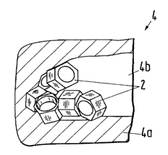
Fig. 1 shows in a plan view a bone 4, specifically a vertebral body having an
internal bone space 4a. The term internal bone space 4a will be understood to
mean the entire volume taken up by the bone 4. In the internal bone space 4a
there is a bone cavity 4b into which a cannula 6 opens. The bone cavity 4b can
for example have arisen because of degenerative processes, in particular osteo-
porosis. The bone cavity 4b can also be artificially produced or enlarged with
a
suitable instrument.
Fig. 2 shows a vertebral body 4 having an internal bone space 4a and also a
supply device 5 comprising a cannula 6, a pressing device 9 with an actuating
handle 9b and a plunger 9c. The cannula 6 opens into the internal bone space
4a which in this embodiment has no bone cavity 4b. Biocompatible support
bodies 2 are arranged behind one another in the inner space of the cannula 6
and of the pressing device 9 and are arranged substantially mutually touching
one another so that the plunger 9c acting on the rearmost support body 2
drives all support bodies 2 in the direction of the internal bone space 4a.
This
makes it possible, if necessary, to transfer the force applied to the
actuating
handle 9b via the support bodies 2 up to the support bodies 2 located at the
tip
of the cannula 6, so that these penetrate into the internal bone space 4a with
a
corresponding force and thereby form a bone cavity 4b during the penetration.
The inner diameter of the cannula 6 and also of the pressing device 9 are de-
signed and matched with respect to the outer diameter of the support bodies 2
in such a way that the support bodies 2 are arranged behind one another in
the flow direction, i.e. towards the outlet opening of the cannula 6, which
has
the consequence that the totality of the support bodies 2 form a filler means
1
capable of dry flow which has flow characteristics without any form of lubri-
cant, in that support bodies 2 can be supplied with a syringe-like device
shown
CA 02605062 2007-08-08
in Fig. 2 to the internal bone space 4a. The cannula 6 can also extend in
curved
manner or consist of a flexible or solid material.
The vertebral body 4 shown in Fig. 3 in section has a bone cavity 4b which was
produced prior to the insertion of the support bodies 2 with a special instru-
ment. Thereafter the tip 6a of the cannula 6 is driven forward up to the bone
cavity 4b and thereafter the support bodies shown in a side view are intro-
duced individually following one another and in a defined mutual position so
they become distributed in random manner in the bone cavity 4b. The cannula
6 has a round internal cross-section so that each support body 2, which are
all
identically designed, has an elongate spherically extending outer contour. The
support body 2 has a maximum dimension between 2 mm and 10 mm. The
support bodies 2 are designed as solid bodies. All support bodies 2 are identi-
cally designed with respect to dimension and shape.
Fig. 4 shows the vertebral body shown in Fig. 3 with a bone cavity 4b com-
pletely filled with support bodies 2, with the support bodies 2 contacting one
another at different points and thereby being entrapped in the bone cavity 4b
so that the totality of these support bodies 4 form a load bearing support
struc-
ture. The access passage to the bone cavity 4b is closed with a plug 4c after
the
introduction of the support bodies 2.
An internal bone space 4a or a bone cavity 4b can be filled with the most di-
versely shaped support bodies 2. Fig. 5a shows schematically a bounded bone
cavity 4b with an inlet opening 4c through which the ball-like support bodies
are introduced. Fig. 5b shows an ellipsoidally designed support body 2. An ad-
vantage of the support bodies shown in Figs. 5a and 5b with a spherical outer
contour is the fact that they can be mutually displaced with a low expenditure
force so that the support bodies 2 can very readily fill the volume of the
bone
CA 02605062 2007-08-08
11
cavity 4b. The Figs. 5c and 5d show support bodies 2 with an edge-like outer
contour, with the side surfaces of the support bodies 2, which converge at an
angle, being able to exert a wedge action so that the support bodies 2 located
in
the bounded bone cavity 4b can become mutually wedged in an advantageous
embodiment such as is shown in Fig. 5c, with larger intermediate spaces re-
sulting between the support bodies 2 into which the bone can grow. The total-
ity of the support bodies 2 in turn forms a mechanically loadable support
struc-
ture. In contrast to the support bodies 2 shown in Figs. 5a to 5d, which are
re-
spectively identical having regard to their shape and size, the support bodies
2
could also have different shapes and/or sizes within the bone cavity 4b as
shown in Fig. 5e.
The Figs. 6a-6d and 7a-7d show support bodies 2 which have an axis A, with
each support body 2 having two oppositely disposed abutment locations 2g
which are spaced apart in the direction of the axis A. The abutment locations
2g of Figs. 6a-6d and also of Fig. 7d are designed as abutment surfaces 2g,
whereas the abutment locations 2g of Figs. 7a-7c are designed as rings with a
small mutual contact surface. In the embodiments of Figs. 6a-6e the abutment
locations 2g extend perpendicular to the axis A. In the embodiments in accor-
dance with Figs. 7a-7d the abutment locations 2g have a circular or spherical
or curved extent. The abutment locations 2g of the embodiments of Figs. 7a-7c
can also be of ring-like shape extending perpendicular to the axis A. The sup-
port bodies 2 have, as shown in Figs. 6a-6c, 7a-7c, a recess disposed perpen-
dicular to the abutment point 2g. This cut-out 2c can be designed as a recess
or
also as a through-going, open, internal cavity 3 which extends between two
oppositely disposed abutment locations 2g and opens into the oppositely dis-
posed recesses 2c. The cut-out 2c can be eccentric or, as shown in Figs. 6a-
6c,
7a-7c, extend concentrically to the axis A. The support bodies 2 can also
consist
CA 02605062 2007-08-08
12
of two or also more partial bodies, for example of four partial bodies which
can
move independently of one another in the bone cavity.
The Figs. 6a-6b and 6c and also 7a, 7b and 7c show support bodies 2 having an
open inner cavity 3. An open inner cavity 3 will be understood to be a hollow
cavity in the support body 2 which is open towards the outside, in contrast to
a
closed cavity which is arranged completely within the interior of the support
body 2 without having an opening towards the outside. These support bodies 2
also have a maximum size in the range between 2 mm and 10 mm. Each sup-
port body 2 includes a total volume which corresponds to the volume of the ma-
terial of the support body 2 and also its inner cavity 3. The volume of the
inner
cavity is larger than 30 % of the total volume, preferably larger than 50 %
and
can amount to up to 90 %. The size of the maximum possible volume of the in-
ternal cavity 3 is dependent on the maximum pressure forces acting on the
support body 2. These pressure forces are dependent on the specific internal
bone space 4a and/or the bone 4 in which the support structure is formed. The
load carrying ability of the support body 2 is naturally dependent on the mate-
rial used. If the support body 2 is for example made of metal the internal cav-
ity 3 can be made relatively large and the support bodies 2 are nevertheless
resistant to the pressure forces that are acting. If the support body 2 is
made of
a material such as bio-glass or a resorbable substance, the internal cavity 3
must be made smaller percentage-wise in accordance with the material char-
acteristics in order to provide the support body 2 with a sufficiently large
load
carrying force. This internal cavity 3, in particular a relatively large
internal
cavity 3 percentage-wise, has the advantage that it can be filled in the
course
of time by growing bone material. In a particularly advantageous design the
internal cavity 3 of the support body 2 is at least partly filled prior to its
intro-
duction with an osteo-inductive and/or osteo-conductive substance, in particu-
lar a bone growth promoting protein or calcium sulphate or a combination of
CA 02605062 2007-08-08
13
these or further substances. Thanks to such filled support bodies 2 it is
possi-
ble to feed both a support structure and also osteo-inductive and/or osteo-
conductive substances to the internal bone space 4a by means of the filler
means 1 capable of dry flow and consisting of a plurality of support bodies 2.
This design has the advantages that the filler means 1 can be supplied to the
internal bone space 4a dry so that no danger exists of anything running out
through cracks, gaps and openings which may possibly be present in the bone.
Moreover, the osteo-inductive substance brings about bone growth so that the
hollow spaces 3 and the intermediate spaces which result through the support
bodies 2 are advantageously increasingly filled with growing bone. Since the
support bodies 2 in the internal bone space 4a are arranged aligned at random,
that is to say the hollow spaces 3 extend in random directions and also the in-
termediate spaces extend in random directions and have a size determined by
chance, the support bodies 2 actually form a support structure similar in the
broader sense to spongiosa, in particular when the volume of the internal cav-
ity 3 amounts to more than 50 % of the total volume. When, in the course of
increasing healing, the hollow cavities 3 and the intermediate spaces are
filled
with growing spongiosa, then a support structure forms in the internal bone
space 4a which is partly comparable with a healthy bone, with randomly
aligned support bodies 2 the hollow cavities and intermediate spaces of which
have spongiosa growing through them.
The inner cavity 3 of the support bodies 2 in accordance with Figs. 6a, 6b,
6c,
7a, 7b, 7c is made cohesive and non-porous so that the internal contour 2b
bounds a relatively large internal cavity 3 laterally. The internal cavity 3
in
accordance with the Figs. 6a, 6b, 7a, 7b is of cylindrical shape, extends
concen-
tric to the axis A and has a circular opening 2c. The angle between the
internal
wall of the internal cavity 3 and the abutment location 2g thus amounts to 90
degrees at the circular opening 2c. The circular opening 2c can, as shown in
CA 02605062 2007-08-08
14
Fig. 6b, have blocking points 2h, for example a plurality of notches arranged
in
the peripheral direction. The opening 2c can have the most diverse shapes and
can for example also be formed as a triangle, tetragon or as a frequency poly-
gon. The internal cavity 3 could have the same shape as given by the opening
2c, with the internal cavity 3 extending in the axial direction A over the
entire
length of the support body 3. The support bodies 2 in accordance with Figs.
7a,
7b are made essentially circular. The support bodies 2 in accordance with the
Figs. 6a, 6b have a polyhedral outer contour in the peripheral direction with
edges 2d extending parallel to the axis A and six surfaces 2f which extend in
a
mutually wedge-shaped manner which respectively meet at an edge 2d or cor-
ner 2e. The support body 2 has a preferably 3-cornered to 10-cornered outer
contour in the peripheral direction, in particular a 4-cornered, 5-cornered or
6-
cornered outer contour. The edges 2d can have blocking points 2h, as shown in
Fig. 6b, such as notches.
The embodiment in accordance with Fig. 7a has a spherical, in particular ball-
shaped or episoidal outer contour. The support bodies 2 in accordance with the
Figs. 6b and 7b are essentially of hollow cylindrical shape. The support
bodies
2 in accordance with Figs. 6c and 7c have essentially a cuboid or spherically
extending outer contour. The support bodies 2 in accordance with Figs. 6d and
7d have, in distinction to the embodiment of Figs. 6a, 7a, no hollow cavity 3.
The Figs. 8a to 8d show further embodiments of support bodies 2 with poly-
hedrally extending outer contours, with the illustrated support bodies 2 not
having any hollow space 3. The support bodies 2 could however also have a
cavity 3. Fig. 8a shows a tetrahedron, Fig. 8b an octahedron, Fig. 8c an ikosa-
hedron and Fig. 8d a small star-shaped dodecahedron. The two oppositely dis-
posed abutment locations 2g spaced apart in the direction of the axis A, which
CA 02605062 2007-08-08
are necessary in order to forward this embodiment in a cannula in the direc-
tion of the axis A, are not shown in this embodiment.
In an advantageous design, as shown in Fig. 6a, the support body 2 has an
outer diameter D and a height H extending orthogonal to it, i.e. in the direc-
tion of the axis A, with the outer diameter D preferably amounting to 1.5-
times
the height H. The support bodies 2 have a surface formed such that the surface
of a filling body 2 filled into the bone cavity can project into the cut-out
of an
adjacently disposed support body 2 as shown in Fig. 9. In an advantageous
embodiment the support bodies 2 have surfaces and also recesses designed
such that mutual wedging or jamming occurs between the surfaces which pro-
ject into the cut-outs and the cut-out, in order to form within the internal
bone
space 4a a preferably cohesive and in particular self-stable support
structure.
The surface of the support body 2 which is radial to the axis A thus
preferably
has shapes from the group: corner, edge, tip, recess, aperture or a
combination
thereof in order to bring about a mutual wedging and jamming of the support
bodies 2. In an advantageous embodiment the support body 2 has, as shown in
Fig. 6a, a form similar to a threaded nut with a multi-cornered outer contour
extending in the direction of extent of the axis A with surfaces 2f which mutu-
ally extend in wedge-like manner and pronounced edges 2d and corners 2e as
well as an inner cavity 3 with an opening 2c of relatively large diameter.
Fig. 9
shows a first bone cavity 4b partly filled with such threaded nut-like support
bodies 2. It is evident from this arrangement of the support bodies 2 how the
support bodies 2 are mutually wedged in that the outer contour partly pene-
trates via the opening 2c into the internal space 3, so that the support
bodies 2
are mutually wedged and in this way form a mechanically at least cohesive
support structure. As soon as the entire bone cavity 4b is completely filled
with
the support bodies 2 the support structure formed in this way can carry the
load acting on the bone 4 from the outside essentially as a composite
assembly.
CA 02605062 2007-08-08
16
The filler means 1 introduced into the internal space 4a of a vertebral body,
comprising a plurality of, for example, 20 to 50 support bodies 2, preferably
forms a self-stable support structure in that the individual support bodies 2
are mutually wedged in such a way that the support structure which is formed
is held together by a force acting in the direction of extent of the spinal
col-
umn. If the internal space 4a were filled with spherical support bodies, then
these would attempt, with a force acting in the direction of extent of the
spinal
column, to escape in a direction perpendicular to the force, which produces an
excessive force on the outer jacket of the vertebral body or can indeed lead
to
damage to the outer jacket and to the escape of the support bodies. Through
the mutual wedging of the support bodies this excessive force acting on the
outer jacket is considerably reduced. The arrangement of the support bodies 2
shown in Fig. 9, or a very similar arrangement, also results when these are
pressed into the vertebral body as shown in Fig. 17.
Fig. 14 shows a broken bone 4, with a bone part missing at the position of the
fracture. This part is covered over with a bone plate 10 so that a bone cavity
4b
forms. A bone cavity 4b of this kind can also be filled with the above-
described
filler means 1 comprising support bodies 2.
Fig. 10 shows rigid cannula 6 with a cannula tip 6a formed as a cutting thread
6c. A Kirschner wire 8 extends within the cannula 6. The Kirschner wire 8 is
first pushed into the bone 4 an order to determine the direction of extent of
the
cannula 6. Thereafter the tip 6a of the cannula 6 is screwed with a rotating
movement into the bone 4 using the handle 7 so that the tip 6a and thus the
cannula 6 is firmly anchored in the bone 4. Thereafter the handle 7 is
removed.
Fig. 11 shows the cannula 6 with the removed handle 7. The cannula 6 in-
cludes a connection part 6b for the handle 7 and for a pressing device 9
respec-
tively. Fig. 12 shows an embodiment of a tip 6a of the cannula 6 in detail.
The
CA 02605062 2007-08-08
17
tip 6a has projecting cutting elements 6e and an outer thread 6d. Fig. 13
shows
a pressing device 9 which is connected via the connection part 6b to the can-
nula 6. Prior to the connection the cannula 6 was filled with support bodies
2.
The pressing device 9 includes a handle 9a and also an actuating grip 9b which
acts on a plunger 9c in such a way that it moves a bit further into the
cannula
6 at each actuation, so that the plunger 9c acting on the support bodies 2
presses the support bodies 2 which are arranged at the foremost position in
the
cannula 6 into the bone 4. The support bodies 2 thus flow into the bone 4 and
in this way also form a bone cavity 4b therein as a result of the pressure and
fill it with support bodies 2 so that a support structure is formed within the
bone 4. The position of the plunger 9c can be read off with the aid of
markings
6f applied to the cannula 6 from which it can be determined how many support
bodies have been pressed into the bone 4. The support bodies 2 could also be
arranged in an additional cannula which is introduced into the cannula 6 be-
fore the pressing device is connected to the cannula 6. Thus a second cannula
which contains the support bodies 2 would be arranged within the cannula 6.
In this way the cannula 6 could be loaded very simply with support bodies 2.
The cannula containing the support bodies 2 must have an internal diameter
matched to the size of the support bodies in such a way that the support
bodies
2 can only be arranged following one another and are displaceable therein in
order to supply the support bodies 2 one after the other to the internal bone
space 4a. After the filling of the internal bone space 4a the pressing device
9
and the cannula 6 are removed and the hole in the bone is closed with a plug
4c as shown in Fig. 4.
The cannula can not only have a round internal cross-section as shown in Figs.
to 13 but also other cross-sectional shapes, for example a hexagonal inter-
nal cross-section as shown in Fig. 15, or a rectangular internal cross-section
as
shown in Fig. 16, which are for example suitable for the introduction of the
CA 02605062 2007-08-08
18
support body 2 shown in Fig. 6a. The cannula 6 can also have further cross-
sectional shapes matched to the respective contour of the support body 2 that
is used.
The tip 6a of the cannula 6 can also be designed without a fastening means 6c.
In particular, if a bone cavity 4b has already previously been formed, the sup-
port bodies 2 could also be fed, as shown in Fig. 2, to the bone cavity 4b
with a
device 9 similar to a syringe. In an advantageous method a group of support
bodies, for example 5 or 10 support bodies following one another, is in each
case supplied to the bone cavity 4b, thereafter the cannula 6 is removed and
the support bodies 2, which are located in the bone cavity 4b, are
additionally
pressed in and compressed with the aid of a stuffing tool in order to
thereafter
replace the cannula at the bone 4 and to introduce a further group of support
bodies 2 into the bone cavity 4b and to compress these again with the stuffing
tool.
In a possible method step an osteo-inductive and/or osteo-conductive substance
can be supplied after the support bodies 2 have been introduced into the inter-
nal bone space 4a, for example as a liquid or as a fluid, in order to fill the
still
present hollow cavities in the bone cavity 4b with this substance. The filler
means 1 could also be supplied to the internal bone space 4a together with a
fluid in that, for example, the support bodies 2 are made available together
with the fluid, in particular mixed, in the pressing device 9 for introduction
into the internal bone space 4a.
Fig. 17 shows a longitudinal section through a vertebral body 4 with a cannula
6 introduced into its internal bone space 4a. The support bodies 2 are
designed
as shown in Fig. 6a and have abutment locations 2g spaced apart in the direc-
tion of the axis A which lie against and mutually contact one another in the
CA 02605062 2007-08-08
19
direction of the axis A. The support bodies 2 are pushed or pressed into the
internal bone space 4a by means of a plunger 9c moved in the direction B on
which a force F acts, with the support bodies 2 becoming randomly arranged in
the internal bone space 4a and moreover forming a bone cavity 4b in the illus-
trated embodiment. The support bodies 2 located in the cannula 6 are ar-
ranged following one another and are otherwise identically arranged with re-
spect to their position, if a cannula 6 in accordance with Fig. 15 is used. If
the
cannula 6 with a circular cross-section is used, then the support bodies 2 are
also arranged following one another, but, with respect to their mutual
position,
could also be arranged rotated around the axis A. Since the support bodies 2
are arranged in defined manner inside the cannula 6 in the direction of extent
of the axis A, i.e. in the direction of movement B, the support bodies 2 can
be
reliably displaced to the internal bone space 4a and with small resistance.
The
support bodies 2 can be structured radially to the axis A in the most diverse
manner and for example have edges 2d, notches 2h or also projecting tips or
cut-outs without these structures hindering the introduction of the support
bodies through the cannula 6. The forces are reliably transmitted between the
abutment locations 2g of the support bodies 2 and no mutual wedging of the
support bodies takes place within the cannula 6 so that the support bodies can
be reliably displaced into the internal bone space 4a. Within the internal
bone
space 4a the support bodies 2 order themselves arbitrarily, so that they mutu-
ally hinder one another with respect to their movement and mutually wedge
and block one another.
Fig. 19 shows, in a cross-section of a vertebral body 4 a plurality of approxi-
mately 20 introduced support bodies 2 which are randomly distributed in a
cloud-like manner within the internal bone space 4a. In order that the
internal
bone space 4a is uniformly filled, the pressing device 9 is mounted after the
CA 02605062 2007-08-08
first insertion of the support bodies 2, preferably the a point designated by
4c,
in order to likewise supply support bodies 2 at this point.
The plunger 9c can have markings 9e in order to monitor the depth of penetra-
tion of the plunger 9c. The length of the plunger 9c can be selected such that
its front part can be introduced up to and into the internal bone space 4a,
for
example by up to a centimeter. The tip of the plunger 9c can, as shown in Fig.
18a and 18b, be made flat or rounded. Such plungers are also termed com-
pressing plungers. The supply device 5 preferably has a plurality of
differently
designed plungers 9c. For example the plunger 9c could have an obliquely ex-
tending tip as shown in Fig. 18b. This can serve to align or to shift support
bodies 2 already arranged within the vertebral body 4. A plunger 9c of this
kind can also be termed a positioning plunger. The position of the support bod-
ies 2 in the vertebral body 4 can be made visible with the aid of an X-ray
image
so that a doctor can change or correct the support bodies 2 with correspond-
ingly designed tools such as plungers, hooks or tongs. Fig. 18d shows a
plunger
9c with a controllably arranged tip. Fig. 18e shows a plunger 9c with a
project-
ing guide part which can for example engage into the hollow space 3 of a sup-
port body 2.
The pressing device 9 shown in Fig. 19 includes a force measuring device 9f
with which the force produced in the direction of movement B is measured and
is preferably also directly indicated. The force measuring device 9f can, for
ex-
ample, include a spring and also a display so that the doctor can directly
measure the force exerted onto the support bodies 2. The force measuring de-
vice 9f can, for example, also be designed as an electronic apparatus
including,
for example, a piezo-crystal and also a display or an acoustic output, with
the
measured signal also being supplied to a monitoring device 11. The maximum
force exerted on the support bodies 2 is preferably restricted. The force meas-
CA 02605062 2007-08-08
21
urement provides an indication of the state of filling, for example the
wedging
of the support bodies. The pressing device 9 preferably includes a handle 9h.
The pressing device 9 could also include a drive 9g in order to bring about a
force, blows or vibrations on the support bodies 2. In an advantageous method
step a plurality of support bodies 2 is supplied to the internal bone space 4a
and thereafter a vibrating force is exerted on the support bodies 2 in order
to
compress the support bodies 2 located in the internal bone space 4a and in or-
der to thus obtain a high packing density and in order to thereby feed a large
number of support bodies 2 to the internal bone space 4a. The frequency of vi-
bration preferably lies in the range between 1 and 15000 Hz, in particular be-
tween 5 and50 Hz.
In an advantageous embodiment the drive device 9g produces an elastic shock
wave which is transferred into the plunger 9a which has the consequence that
the length of the plunger 9a is varied, for example by +/-2 mm at a frequency
of
preferably between 1 and 50 Hz.
The drive device 9g could also be connected to a monitoring device 11 and the
maximum force and/or the frequency and/or the stroke could be monitored.
The Figs. 20a to 20c show differently designed support bodies 2 with rounded
or, for example, hexagonal outer contour in a longitudinal section along the
axis A. In Fig. 20a the abutment locations 2g are designed as abutment sur-
faces, with the abutment locations 2g arranged at the left having a reduced
support surface in comparison to the abutment locations 2g arranged at the
right. The support bodies 2 illustrated in Fig. 20b have at the left-hand side
a
circular abutment 2g which extends fully in the peripheral direction. A plural-
ity of individual projecting elements, for example hemispherically projecting
CA 02605062 2007-08-08
22
elements, could also be arranged distributed in the peripheral direction, with
each element forming an abutment location 2g. The support bodies 2 shown in
Fig. 20c have abutment locations 2g which are designed as truncated cone-like
surfaces. An advantage of this design is that the support bodies are mutually
centered on displacement in the direction of the axis A. The support bodies 2
could also have different outer diameters as shown in Fig. 20c.
Fig. 21 shows in an outside view a development of the abutment locations 2g
between the two support bodies 2. These areally designed abutment locations
2g mutually engage within one another which prevent a mutual rotation of
adjacent support bodies 2 as they are pushed in. The abutment locations 2g
could also be of form-fitted design.
Fig. 22a shows in a side view three support bodies 2, two U-shaped support
bodies 2, which surround a cylindrical support body 2. These three support
bodies 2 are arranged in the cannula 6 in such a way that they can be supplied
to the internal bone space 4a via the abutment locations in the direction of
the
axis A as shown in a side view in Fig. 22b, with the three support bodies 2 be-
coming randomly arranged within the internal bone space 4a since these are
not mutually connected together.
The surface of the support body 2 located within the two U-shaped support
bodies 2 can be of any desired shape and can for example also have edges or
points, also at its end surfaces. The support bodies 2 shown in Figs. 20a-20c
and also 22a and 22b can, for example, be of circular or multi-cornered design
radial to the axis A; or can also have tips, edges, cut-outs or apertures.
The end face of the plunger 9c can be designed in a plurality of shapes and
can
for example also have the end faces shown in Figs. 20a to 20c.
CA 02605062 2007-08-08
23
Fig. 23 shows a further support body 2 with abutment surfaces 2g. The arms
could be designed in a plurality of different forms with it having to be
ensured
that one abutment surface 2g or an abutment location 2g is present on the
front side and also on the reverse side in each case.
The support bodies 2 shown in Figs. 8a to 8d could have abutment locations 2g
spaced apart in the direction of an axis A. For example, the support body 2
shown in Fig. 8b can have a self centering abutment location 2g at the upper-
most and lowermost tip as shown in Fig. 20c, so that a plurality of said
support
bodies 2, which are arranged behind one another in a cannula 6, which mutu-
ally contact each other at the abutment locations 2g, are displaceable in the
direction of the axis A and can thus be fed to the internal bone space 4a.