Note: Descriptions are shown in the official language in which they were submitted.
DEMANDE OU BREVET VOLUMINEUX
LA PRESENTE PARTIE DE CETTE DEMANDE OU CE BREVET COMPREND
PLUS D'UN TOME.
CECI EST LE TOME 1 DE 2
CONTENANT LES PAGES 1 A 252
NOTE : Pour les tomes additionels, veuillez contacter le Bureau canadien des
brevets
JUMBO APPLICATIONS/PATENTS
THIS SECTION OF THE APPLICATION/PATENT CONTAINS MORE THAN ONE
VOLUME
THIS IS VOLUME 1 OF 2
CONTAINING PAGES 1 TO 252
NOTE: For additional volumes, please contact the Canadian Patent Office
NOM DU FICHIER / FILE NAME:
NOTE POUR LE TOME / VOLUME NOTE:
CA 02605143 2007-10-15
WO 2006/113529 PCT/US2006/014241
DIAGNOSIS OF SEPSIS
CROSS-REFERENCE TO RELATED APPLICATIONS
[0001] This application claims benefit, under 35 U.S.C. 119(e), of U.S.
Provisional Patent Application No. 60/671,620, filed on April 15, 2005, which
hereby is
incorporated herein, by reference, in its entirety. This application also
claims benefit, under
35 U.S.C. 119(e), of U.S. Provisional Patent Application No. 60/674,046,
filed on April
22, 2005, which is hereby incorporated herein, by reference, in its entirety.
1. FIELD OF THE INVENTION
[0002] The present invention relates to methods and compositions for
diagnosing or
predicting sepsis and/or its stages of progression in a subject. The present
invention also
relates to methods and compositions for diagnosing systemic inflammatory
response
syndrome in a subject.
2. BACKGROUND OF THE INVENTION
[0003] Early detection of a disease condition typically allows for a more
effective
therapeutic treatment with a correspondingly more favorable clinical outcome.
In many
cases, however, early detection of disease symptoms is problematic due to the
complexity of
the disease; hence, a disease may become relatively advanced before diagnosis
is possible.
Systemic inflammatory conditions represent one such class of diseases. These
conditions,
particularly sepsis, typically, but not always, result from an interaction
between a
patlzogenic microorganism and the host's defense system that triggers an
excessive and
dysregulated inflammatory response in the host. The complexity of the host's
response
during the systemic inflammatory response has complicated efforts towards
understanding
disease pathogenesis (reviewed in Healy, 2002, Annul. Pharmacother. 36:648-
54). An
incomplete understanding of the disease pathogenesis, in turn, contributes to
the difficulty
in finding useful diagnostic biomarkers. Early and reliable diagnosis is
imperative,
however, because of the remarkably rapid progression of sepsis into a life-
threatening
condition.
[0004] The development of sepsis in a subject follows a well-described course,
progressing from systemic inflammatory response syndrome ("SIRS")-negative, to
SIRS-
positive, and then to sepsis, which may then progress to severe sepsis, septic
shock, multiple
organ dysfunction ("MOD"), and ultimately death. Sepsis may also arise in an
infected
subject when the subject subsequently develops SIRS. "Sepsis" is commonly
defined as the
-1-
CA 02605143 2007-10-15
WO 2006/113529 PCT/US2006/014241
systemic host response to infection with SIRS plus a documented infection.
"Severe sepsis"
is associated with MOD, hypotension, disseminated intravascular coagulation
("DIC") or
hypoperfusion abnormalities, including lactic acidosis, oliguria, and changes
in mental
status. "Septic shock" is commonly defined as sepsis-induced hypotension that
is resistant
to fluid resuscitation with the additional presence of hypoperfusion
abnormalities.
[0005] Documenting the presence of the pathogenic microorganisms that are
clinically significant to sepsis has proven difficult. Causative
microorganisms typically are
detected by culturing a subject's blood, sputum, urine, wound secretion, in-
dwelling line
catheter surfaces, etc. Causative microorganisms, however, may reside only in
certain body
microenvironments such that the particular material that is cultured may not
contain the
contaminating microorganisms. Detection may be complicated fiuther by low
numbers of
microorganisms at the site of infection. Low numbers of pathogens in blood
present a
particular problem for diagnosing sepsis by culturing blood. In one study, for
example,
positive culture results were obtained in only 17% of subjects presenting
clinical
manifestations of sepsis (Rangel-Frausto et al., 1995, JAMA 273:117-123).
Diagnosis can
be further complicated by contamination of samples by non-pathogenic
microorganisms.
For example, only 12.4% of detected microorganisms were clinically significant
in a study
of 707 subjects with septicemia (Weinstein et al., 1997, Clinical Infectious
Diseases 24:584-
602).
[0006] The difficulty in early diagnosis of sepsis is reflected by the high
morbidity
and mortality associated with the disease. Sepsis currently is the tenth
leading cause of
death in the United States and is especially prevalent among hospitalized
patients in non-
coronary intensive care units (ICUs), where it is the most common cause of
death. The
overall rate of mortality is as high as 35%, with an estimated 750,000 cases
per year
occurring in the United States alone. The annual cost to treat sepsis in the
United States
alone is on the order of billions of dollars.
[0007] A need, therefore, exists for a method of diagnosing sepsis, using
techniques
that have satisfactory specificity and sensitivity performance, sufficiently
early to allow
effective intervention and prevention.
3. SUMMARY OF THE INVENTION
[0008] The present invention relates to methods and compositions for
diagnosing
sepsis, including the onset of sepsis, in a test subject. The present
invention also relates to
methods and compositions for predicting sepsis in a test subject.
-2-
CA 02605143 2007-10-15
WO 2006/113529 PCT/US2006/014241
[0009] The present invention further relates to methods and compositions for
diagnosing or predicting stages of sepsis progression in a test subject. The
present invention
still further relates to methods and compositions for diagnosing systemic
inflammatory
response syndrome (SIRS) in a test subject.
[0010] In one aspect, the present invention provides a method of predicting
the
development of sepsis in a test subject at risk for developing sepsis. This
method comprises
evaluating whether a plurality of features in a biomarker profile of the test
subject satisfies a
value set, wherein satisfying the value set means that the test subject will
develop sepsis
with a likelihood that is determined by the accuracy of the decision rule to
which the
plurality of features are applied in order to determine whether they satisfy
the value set. In
some embodiments, the accuracy of the decision rule is at least 60%.
Therefore,
correspondingly, the likelihood that the test subject will develop sepsis when
the plurality of
features satisfies the value set is at least 60%.
[0011] Yet another aspect of the invention comprises a method of diagnosing
sepsis
in a test subject. These methods coinprise evaluating whether a plurality of
features in a
biomarker profile of the test subject satisfies a value set, wherein
satisfying the value set
predicts that the test subject has sepsis with a likelihood that is determined
by the accuracy
of the decision rule to whicll the plurality of features are applied in order
to determine
whether they satisfy the value set. In some embodiments, the accuracy of the
decision rule
is at least 60%. Therefore, correspondingly, the likelihood that the test
subject has sepsis
when the plurality of features satisfies the value set is at least 60%.
[0012] In a particular embodiment, the biomarker profile comprises at least
two
features, each feature representing a feature of a corresponding biomarker
listed in column
four or five of Table 30. In one embodiment, the biomarker profile comprises
at least two
different biomarkers listed in colunm four or five of Table 30. In such ari
embodiment, the
biomarker profile can comprise a respective corresponding feature for the at
least two
biomarkers. Generally, the at least two biomarkers are derived from at least
two different
genes. In the case where a biomarker in the at least two different biomarkers
is listed in
colunm four of Table 30, the biomarker can be, for example, a transcript made
by the listed
gene, a complement thereof, or a discriminating fragment or complement
thereof, or a
cDNA thereof, or a discriminating fragment of the cDNA, or a discriminating
amplified
nucleic acid molecule corresponding to all or a portion of the transcript or
its complement,
or a protein encoded by the gene, or a discriminating fragment of the protein,
or an
indication of any of the above. Further still, the biomarker can be, for
example, a protein
-3-
CA 02605143 2007-10-15
WO 2006/113529 PCT/US2006/014241
listed in column five of Table 30, or a discriminating fragment of the
protein, or an
indication of any of the above. Here, a discriminating molecule or fragment is
a molecule
or fragment that, when detected, indicates presence or abundance of the above-
identified
transcript, cDNA, amplified nucleic acid, or protein. In accordance with this
embodiment,
the biomarker profiles of the present invention can be obtained using any
standard assay
known to those skilled in the art, or in an assay described herein, to detect
a biomarker.
Such assays are capable, for example, of detecting the products of expression
(e.g., nucleic
acids and/or proteins) of a particular gene or allele of a gene of interest
(e.g., a gene
disclosed in Table 30). In one embodiment, such an assay utilizes a nucleic
acid
microarray. In some embodiments, the biomarker profile comprises at least two
different
biomarkers from column four or five of Table 32. In some embodiments, the
biomarker
profile comprises at least 2, 3, 4, 5, 6, 7, 8, 9, 10, 11, 12, 13, 14, 15, 16,
17, 18, 19, 20, 21,
22, 23, 24, 25, 30, 35, 40, 45, or 50 different biomarkers from Table 30.
[0013] In a particular embodiment, the biomarker profile comprises at least
two
different biomarkers that each contain one of the probesets listed in column 2
of Table 30,
biomarkers that contain the complement of one of the probesets of Table 30, or
biomarkers
that contain an amino acid sequence encoded by a gene that either contains one
of the
probesets of Table 30 or the complement of one of the probesets of Table 30.
Such
biomarkers can be, for example, mRNA transcripts, cDNA or some other nucleic
acid, for
example amplified nucleic acid, or proteins. The biomarker profile further
comprises a
respective corresponding feature for the at least two biomarkers. Generally,
the at least two
biomarkers are derived from at least two different genes. In the case where a
biomarker is
based upon a gene that includes the sequence of a probeset listed in Table 30,
the biomarker
can be, for example, a transcript made by the gene, a complement thereof, or a
discriminating fragment or complement thereof, or a cDNA thereof, or a
discriminating
fragment of the cDNA, or a discriminating amplified nucleic acid molecule
corresponding
to all or a portion of the transcript or its complement, or a protein encoded
by the gene, or a
discriminating fragment of the protein, or an indication of any of the above.
Further still,
the biomarker can be, for example, a protein encoded by a gene that includes a
probeset
sequence described in Table 30, or a discriminating fragment of the protein,
or an indication
of any of the above. Here, a discriminating molecule or fragment is a molecule
or fragment
that, when detected, indicates presence or abundance of the above-identified
transcript,
cDNA, amplified nucleic acid, or protein. In some embodiments, the biomarker
profile
-4-
CA 02605143 2007-10-15
WO 2006/113529 PCT/US2006/014241
comprises at least 2, 3, 4, 5, 6, 7, 8, 9, or 10 different biomarkers from any
one of Table 31,
32, 33, 34, or 36.
[0014] In a particular embodiment, the biomarker profile comprises at least
two
different biomarkers listed in column three of Table 31. The biomarker profile
further
comprises a respective corresponding feature for the at least two biomarkers.
Generally, the
at least two biomarkers are derived from at least two different genes. The
biomarker can be,
for example, a transcript made by gene listed in Table 31, a complement
thereof, or a
discriminating fragment or complement thereof, or a cDNA thereof, or a
discriminating
fragment of the cDNA, or a discriminating amplified nucleic acid molecule
corresponding
to all or a portion of the transcript or its complement, or a protein encoded
by the gene, or a
discriminating fragment of the protein, or an indication of any of the above.
Further still,
the biomarker can be, for example, a protein encoded by a gened listed in
column three of
Table 31, or a discriminating fragment of the protein, or an indication of any
of the above.
Here, a discriminating molecule or fragment is a molecule or fragment that,
when detected,
indicates presence or abundance of the above-identified transcript, cDNA,
amplified nucleic
acid, or protein. In accordance with this embodiment, the biomarker profiles
of the present
invention can be obtained using any standard assay known to those skilled in
the art, or in
an assay described herein, to detect a biomarker. Such assays are capable, for
exainple, of
detecting the products of expression (e.g., nucleic acids and/or proteins) of
a particular gene
or allele of a gene of interest (e.g., a gene disclosed in Table 31). In one
embodiment, such
an assay utilizes a nucleic acid microarray.
[0015] In a particular embodiment, the biomarker profile comprises at least
two
different biomarkers that each contain one of the probesets listed in column 2
of Table 31,
biomarkers that contain the complement of one of the probesets of Table 31, or
biomarkers
that contain an amino acid sequence encoded by a gene that either contains one
of the
probesets of Table 31 or the complement of one of the probesets of Table 31.
Such
biomarkers can be, for example, mRNA transcripts, cDNA or some other nucleic
acid, for
example amplified nucleic acid, or proteins. The biomarker profile further
comprises a
respective corresponding feature for the at least two biomarkers. Generally,
the at least two
biomarkers are derived from at least two different genes. In the case where a
biomarker is
based upon a gene that includes the sequence of a probeset listed in Table 31,
the biomarker
can be, for example, a transcript made by the gene, a complement thereof, or a
discriminating fragment or complement thereof, or a cDNA thereof, or a
discriminating
fragment of the cDNA, or a discriminating amplified nucleic acid molecule
corresponding
-5-
CA 02605143 2007-10-15
WO 2006/113529 PCT/US2006/014241
to all or a portion of the transcript or its complement, or a protein encoded
by the gene, or a
discriminating fragment of the protein, or an indication of any of the above.
Further still,
the biomarker can be, for example, a protein encoded by a gene that includes a
probeset
sequence described in Table 31, or a discriminating fragment of the protein,
or an indication
of any of the above. Here, a discriminating molecule or fragment is a molecule
or fragment
that, when detected, indicates presence or abundance of the above-identified
transcript,
cDNA, amplified nucleic acid, or protein. In some embodiments, the biomarker
profile
comprises at least 2, 3, 4, 5, 6, 7, 8, 9, 10, 11, 12, 13, 14, 15, 16, 17, 18,
19, 20, 21, 22, 23,
24, 25, 30, 35, 40, 45, or 50 different biomarkers from Table 31.
[0016] In a particular embodiment, the biomarker profile comprises at least
three
features, each feature representing a feature of a corresponding biomarker
listed in colunm 3
or four of Table I. In one embodiment, the biomarker profile comprises at
least three
different biomarkers listed in column three or four of Table I. In such an
embodiment, the
biomarker profile can comprise a respective corresponding feature for the at
least three
biomarkers. Generally, the at least three biomarkers are derived from at least
three different
genes listed in Table I. In the case where a biomarker in the at least three
different
biomarkers is listed in colunm three of Table I, the biomarker can be, for
example, a
transcript made by the listed gene, a complement thereof, a splice variant
thereof, a
complement of a splice variant thereof, or a discriminating fragment or
complement of any
of the foregoing, a cDNA of any of the forgoing, a discriminating fragment of
the cDNA, or
a discriminating amplified nucleic acid molecule corresponding to all or a
portion of the
transcript or its complement, or a protein encoded by the gene, or a
discriminatiuig fragment
of the protein, or an indication of any of the above. Further still, the
biomarker can be, for
example, a protein listed in column four of Table I, or a discriminating
fragment of the
protein, or an indication of any of the above. Here, a discriminating molecule
or fragment is
a molecule or fragment that, when detected, indicates presence or abundance of
the above-
identified transcript, cDNA, amplified nucleic acid, splice-variant thereof or
protein. In
accordance with this embodiment, the biomarker profiles of the present
invention can be
obtained using any standard assay known to those skilled in the art, or in an
assay described
herein, to detect a biomarker. Such assays are capable, for example, of
detecting the
products of expression (e.g., nucleic acids and/or proteins) of a particular
gene or allele of a
gene of interest (e.g., a gene disclosed in Table I). In one embodiment, such
an assay
utilizes a nucleic acid microarray. In some embodiments, the biomarker profile
comprises
-6-
CA 02605143 2007-10-15
WO 2006/113529 PCT/US2006/014241
at least 2, 3, 4, 5, 6, 7, 8, 9, 10, 11, 12, 13, 14, 15, 16, 17, 18, 19, 20,
21, 22, 23, 24, 25, 30,
35, 40, 45, or 50 different biomarkers from Table I.
[0017] In a particular embodiment, the biomarker profile coinprises at least
three
features, each feature representing a feature of a corresponding biomarker
listed in column 3
or four of Table J. In one embodiment, the biomarker profile comprises at
least three
different biomarkers listed in colunm three or four of Table J. In such an
embodiment, the
biomarker profile can comprise a respective corresponding feature for the at
least three
biomarkers. Generally, the at least three biomarkers are derived from at least
three different
genes. In the case where a biomarker in the at least three different
biomarkers is listed in
column three of Table J, the biomarker can be, for example, a transcript made
by the listed
gene, a complement thereof, a splice variant thereof, a complement of a splice
variant
thereof, or a discriminating fragment or complement of any of the foregoing, a
cDNA of
any of the forgoing, a discriminating fragment of the cDNA, or a
discriminating amplified
nucleic acid molecule corresponding to all or a portion of the transcript or
its complement,
or a protein encoded by the gene, or a discriminating fragment of the protein,
or an
indication of any of the above. Further still, the biomarker can be, for
example, a protein
listed in column four of Table J, or a discriminating fragment of the protein,
or an indication
of any of the above. Here, a discriminating molecule or fragment is a molecule
or fraginent
that, when detected, indicates presence or abundance of the above-identified
transcript,
cDNA, amplified nucleic acid, splice-variant thereof or protein. In accordance
with this
embodiment, the biomarker profiles of the present invention can be obtained
using any
standard assay known to those skilled in the art, or in an assay described
herein, to detect a
biomarker. Such assays are capable, for example, of detecting the products of
expression
(e.g., nucleic acids and/or proteins) of a particular gene or allele of a gene
of interest (e.g., a
gene disclosed in Table J). In one embodiment, such an assay utilizes a
nucleic acid
microarray. In some embodiments, the biomarker profile comprises at least 2,
3, 4, 5, 6, 7,
8, 9, 10, 11, 12, 13, 14, 15, 16, 17, 18, 19, 20, 21, 22, 23, 24, 25, 30, 35,
40 different
biomarkers from Table J.
[0018] In a particular embodiment, the biomarker profile comprises at least
three
features, each feature representing a feature of a corresponding biomarker
listed in colunm 3
or four of Table K. In one embodiment, the biomarker profile comprises at
least three
different biomarkers listed in column three or four of Table K. In such an
embodiment, the
biomarker profile can comprise a respective corresponding feature for the at
least three
biomarkers. Generally, the at least two or three biomarkers are derived from
at least two or
-7-
CA 02605143 2007-10-15
WO 2006/113529 PCT/US2006/014241
three different genes, respectively. In the case where a biomarker in the at
least two or three
different biomarkers is listed in column three of Table K, the biomarker can
be, for
example, a transcript made by the listed gene, a complement thereof, a splice
variant
thereof, a complement of a splice variant thereof, or a discriminating
fragment or
complement of any of the foregoing, a cDNA of any of the forgoing, a
discriminating
fragment of the cDNA, or a discriminating amplified nucleic acid molecule
corresponding
to all or a portion of the transcript or its complement, or a protein encoded
by the gene, or a
discriminating fragment of the protein, or an indication of any of the above.
Further still,
the biomarker can be, for example, a protein listed in column four of Table K,
or a
discriminating fragment of the protein, or an indication of any of the above.
Here, a
discriminating molecule or fragment is a molecule or fragment that, when
detected,
indicates presence or abundance of the above-identified transcript, cDNA,
amplified nucleic
acid, splice-variant thereof or protein. In accordance with this embodiment,
the biomarker
profiles of the present invention can be obtained using any standard assay
known to those
skilled in the art, or in an assay described herein, to detect a biomarker.
Such assays are
capable, for example, of detecting the products of expression (e.g., nucleic
acids and/or
proteins) of a particular gene or allele of a gene of interest (e.g., a gene
disclosed in Table
K). In one embodiment, such an assay utilizes a nucleic acid microarray. In
some
embodiments, the biomarker profile comprises at least 2, 3, 4, 5, 6, 7, 8, 9,
10 different
biomarkers from Table K.
[0019] Although the methods of the present invention are particularly useful
for
detecting or predicting the onset of sepsis in SIRS subjects, one of skill in
the art will
understand that the present methods may be used for any subject: including,
but not limited
to, subjects suspected of having SIRS or of being at any stage of sepsis. For
example, a
biological sample can be taken from a subject, and a profile of biomarkers in
the sample can
be evaluated in light of biomarker profiles obtained from several different
types of training
populations. Representative training populations variously include, for
example,
populations that include subjects who are SIRS-negative, populations that
include subjects
who are SIRS-positive, and/or populations that include subjects at a
particular stage of
sepsis. Evaluation of the biomarker profile in light of each of these
different training
populations can be used to determine whether the test subject is SIRS-
negative, SIRS-
positive, is likely to become septic, or has a particular stage of sepsis.
Based on the
diagnosis resulting from the methods of the present invention, an appropriate
treatment
regimen can then be initiated.
-8-
CA 02605143 2007-10-15
WO 2006/113529 PCT/US2006/014241
[0020] In particular embodiments, the invention also provides kits that are
useful in
diagnosing or predicting the development of sepsis or SIRS in a subject (see
Section 5.3,
infi a). The kits of the present invention comprise at least 2, 3, 4, 5, 6, 7,
8, 9, 10, 11, 12, 13,
14, 15, 16, 17, 18, 19, 20, 25, 30, 35, 40, 45, 50, 55, 60, 65, 70, 75, 80,
85, 90, 95, 96, 100,
105, 110, 115, 120, 125, 130, 135, 140, 145, 150, 155, 160, 165, 170, 175,
180, 185, 190,
195 or 200 or more biomarkers and/or reagents used to detect the presence or
abtuidance of
such biomarkers. In some embodiments, each of these biomarkers is from Table
30. In
some embodiments, each of these biomarkers is from Table 31. In some
embodiments, each
of these biomarkers is from Table 32. In some embodiments, each of these
biomarkers is
from Table 33. In some embodiments, each of these biomarkers is from Table 36.
In some
embodiments, each of these biomarkers is from Figure 39, Figure 43, Figure 52,
Figure 53,
or Figure 56. In another embodiment, the kits of the present invention
comprise at least
two, but as many as several hundred or more biomarkers and/or reagents used to
detect the
presence or abundance of such biomarkers.
[0021] In a specific embodiment, the kits of the present invention comprise at
least
2, 3, 4, 5, 6, 7, 8, 9, 10, 11, 12, 13, 14, 15, 16, 17, 18, 19, 20, 25, 30,
35, 40, 45, 50, 55, 60,
65, 70, 75, 80, 85, 90, 95, 96, 100, 105, 110, 115, 120, 125, 130, 135, 140,
145, 150, 155,
160, 165, 170, 175, 180, 185, 190, 195 or 200 or more reagents that
specifically bind the
biomarkers of the present invention. For example, such kits can comprise
nucleic acid
molecules and/or antibody molecules that specifically bind to biomarkers of
the present
invention.
[0022] Specific exemplary biomarkers that are useful in the present invention
are set
forth in Section 5.6, Section 5.11, as well as Tables 30, 31, 32, 34 and 36 of
Section 6. The
biomarkers of the kit can be used to generate biomarker profiles according to
the present
invention. Examples of types of biomarkers and/or reagents within such kits
include, but
are not limited to, proteins and fragments thereof, peptides, polypeptides,
antibodies,
proteoglycans, glycoproteins, lipoproteins, carbohydrates, lipids, nucleic
acids (mRNA,
DNA, cDNA), organic and inorganic chemicals, and natural and synthetic
polymers or a
discriminating molecule or fragment thereof.
[0023] In particular embodiments, the invention also provides still other kits
that are
useful in diagnosing or predicting the development of sepsis or SIRS in a
subject (see
Section 5.3, infta). The kits of the present invention comprise at least 2, 3,
4, 5, 6, 7, 8, 9,
10, 11, 12, 13, 14, 15, 16, 17, 18, 19, 20, 25, 30, 35, 40, 45, 50 or more
biomarkers. In
some embodiments, each of these biomarkers is from Table I. In some
embodiments, each
-9-
CA 02605143 2007-10-15
WO 2006/113529 PCT/US2006/014241
of these biomarkers is from Table J. In some embodiments, each of these
biomarkers is
from Table K. In some embodiments, each of these biomarkers is found in Table
I or Table
30. In some embodiments, each of these biomarkers is found in Table I or Table
31. In
some embodiments, each of these biomarkers is from Figure 39, Figure 43,
Figure 52,
Figure 53, or Figure 56. In another embodiment, the kits of the present
invention comprise
at least two, but as many as 50 or more biomarkers. In a specific embodiment,
the kits of
the present invention comprise at least 2, 3, 4, 5, 6, 7, 8, 9, 10, 11, 12,
13, 14, 15, 16, 17, 18,
19, 20, 25, 30, 35, 40, 45, 50, 55, 60, 65, 70, 75, 80, 85, 90, 95, 96, 100,
105, 110, 115, 120,
125, 130, 135, 140, 145, 150, 155, 160, 165, 170, 175, 180, 185, 190, 195 or
200 or more
reagents that specifically bind the biomarkers of the present invention.
Specific biomarkers
that are useful in the present invention are set forth in Section 5.6, Section
5.11, as well as
Tables I, J, K, L, M, N, and O. The biomarkers of the kits can be used to
generate
biomarker profiles according to the present invention. Examples of classes of
compounds
of the kits include, but are not limited to, proteins and fragments thereof,
peptides,
polypeptides, proteoglycans, glycoproteins, lipoproteins, carbohydrates,
lipids, nucleic acids
(mRNA, DNA, cDNA), organic and inorganic chemicals, and natural and synthetic
polymers or a discriminating molecule or fragment thereof.
[0024] Still another aspect of the present invention comprises computers and
computer readable media for evaluating whether a test subject is likely to
develop sepsis or
SIRS. For instance, one embodiment of the present invention provides a
computer program
product for use in conjunction with a computer system. The computer program
product
comprises a computer readable storage medium and a computer program mechanism
embedded therein. The computer program mechanism comprises instructions for
evaluating
wlzether a plurality of features in a biomarker profile of a test subject at
risk for developing
sepsis satisfies a first value set. Satisfaction of the first value set
predicts that the test
subject is likely to develop sepsis. The features are measurable aspects of a
plurality of
biomarkers comprising at least three biomarkers listed in Table I. In some
embodiments,
the computer program product further comprises instructions for evaluating
whether the
plurality of features in the biomarker profile of the test subject satisfies a
second value set.
Satisfaction of the second value set predicts that the test subject is not
likely to develop
sepsis. In some embodiments, the biomarker profile has between 3 and 50
biomarkers listed
in Table I, between 3 and 40 biomarkers listed in Table I, at least four
biomarkers listed in
Table I, or at least six biomarkers listed in Table I.
-10-
CA 02605143 2007-10-15
WO 2006/113529 PCT/US2006/014241
[0025] Another computer embodiment of the present invention comprises a
central
processing unit and a memory coupled to the central processing unit. - The
memory stores
instructions for evaluating whether a plurality of features in a biomarker
profile of a test
subject at risk for developing sepsis satisfies a first value set.
Satisfaction of the first value
set predicts that the test subject is likely to develop sepsis. The features
are measurable
aspects of a plurality of biomarkers. This plurality of biomarkers comprises
at least three
biomarkers from Table I. In some embodiments, the memory further stores
instructions for
evaluating whether the plurality of features in the biomarker profile of the
test subject
satisfies a second value set, wherein satisfying the second value set predicts
that the test
subject is not likely to develop sepsis. In some embodiments, the biomarker
profile consists
of between 3 and 50 biomarkers listed in Table I, between 3 and 40 biomarkers
listed in
Table I, at least four biomarkers listed in Table I., or at least eight
biomarkers listed in Table
I.
[0026] Another computer embodiment in accordance with the present invention
comprises a computer system for determining whether a subject is likely to
develop sepsis.
The computer system comprises a central processing unit and a memory, coupled
to the
central processing unit. The memory stores instructions for obtaining a
biomarker profile of
a test subject. The biomarker profile comprises a plurality of features. The
plurality of
biomarkers comprises at least three biomarkers listed in Table I. The memory
further
comprises instructions for transmitting the biomarker profile to a remote
computer. The
remote computer includes instructions for evaluating whetlier the plurality of
features in the
biomarker profile of the test subject satisfies a first value set.
Satisfaction of the first value
set predicts that the test subject is likely to develop sepsis. The memory
further comprises
instructions for receiving a detennination, from the remote computer, as to
whether the
plurality of features in the biomarker profile of the test subject satisfies
the first value set.
The memory also comprises instructions for reporting whether the plurality of
features in
the biomarker profile of the test subject satisfies the first value set. In
some embodiments,
the remote computer further comprises instructions for evaluating whether the
plurality of
features in the biomarker profile of the test subject satisfies a second value
set. Satisfaction
of the second value set predicts that the test subject is not likely to
develop sepsis. In such
embodiments, the memory further comprises instructions for receiving a
determination,
from the remote computer, as to whether the plurality of features in the
biomarker profile of
the test subject satisfies the second set as well as instructions for
reporting whether the
plurality of features in the biomarker profile of the test subject satisfies
the second value set.
-11-
CA 02605143 2007-10-15
WO 2006/113529 PCT/US2006/014241
In some embodiments, the plurality of biomarkers comprises at least four
biomarkers listed
in Table I. In some embodiments, the plurality of biomarkers comprises at
least six
biomarkers listed in Table I.
[0027] Still another embodiment of the present invention comprises a digital
signal
embodied on a carrier wave comprising a respective value for each of a
plurality of features
in a biomarker profile. The features are measurable aspects of a plurality of
biomarkers.
The plurality of biomarkers comprises at least three biomarkers listed in
Table I. In some
embodiments, the plurality of biomarkers comprises at least four biomarkers
listed in Table
1. In some embodiments, the plurality of biomarkers comprises at least eight
biomarkers
listed in Table I.
[0028] Still another aspect of the present invention provides a digital signal
embodied on a carrier wave comprising a determination as to whether a
plurality of features
in a biomarker profile of a test subject satisfies a value set. The features
are measurable
aspects of a plurality of biomarkers. This plurality of biomarkers comprises
at least three
biomarkers listed in Table I. Satisfying the value set predicts that the test
subject is likely to
develop sepsis. In some embodiments, the plurality of biomarkers comprises at
least four
biomarkers listed in Table I. In some embodiments, the plurality of biomarkers
comprises
at least eight biomarkers listed in Table I.
[0029] Still another embodiment provides a digital signal embodied on a
carrier
wave comprising a determination as to wllether a plurality of features in a
biomarker profile
of a test subject satisfies a value set. The features are measurable aspects
of a plurality of
biomarkers. The plurality of biomarkers comprises at least three biomarkers
listed in Table
1. Satisfaction of the value set predicts that the test subject is not likely
to develop sepsis.
In some embodiments, the plurality of biomarkers comprises at least four
biomarkers listed
in Table I. In some embodiments, the plurality of biomarkers comprises at
least eight
biomarkers listed in Table I.
[0030] Still another embodiment of the present invention provides a graphical
user
interface for determining whether a subject is likely to develop sepsis. The
graphical user
interface comprises a display field for a displaying a result encoded in a
digital signal
embodied on a carrier wave received from a remote computer. The features are
measurable
aspects of a plurality of biomarkers. The plurality of biomarkers comprises at
least three
biomarkers listed in Table I. The result has a first value when a plurality of
features in a
biomarker profile of a test subject satisfies a first value set. The result
has a second value
when a plurality of features in a biomarker profile of a test subject
satisfies a second value
-12-
CA 02605143 2007-10-15
WO 2006/113529 PCT/US2006/014241
set. In some embodiments, the plurality of biomarkers comprises at least four
biomarkers
listed in Table I. In some embodiments, the plurality of biomarkers comprises
at least eight
biomarkers listed in Table I.
[0031] Yet another aspect of the present invention provides a computer system
for
determining whether a subject is likely to develop sepsis. The computer system
comprises a
central processing unit and a memory, coupled to the central processing unit.
The memory
stores instructions for obtaining a biomarker profile of a test subject. The
biomarker profile
comprises a plurality of features. The features are measurable aspects of a
plurality of
biomarkers. The plurality of biomarkers comprise at least three biomarkers
listed in Table
1. The memory further stores instructions for evaluating whether the plurality
of features in
the biomarker profile of the test subject satisfies a first value set.
Satisfying the first value
set predicts that the test subject is likely to develop sepsis. The memory
also stores
instructions for reporting whether the plurality of features in the biomarker
profile of the
test subject satisfies the first value set. In some embodiments, the plurality
of biomarkers
comprises at least four biomarkers listed in Table I. In some embodiments, the
plurality of
biomarkers comprises at least eight biomarkers listed in Table I.
4. BRIEF DESCRIPTION OF THE FIGURES
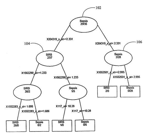
[0032] FIG. 1 illustrates a classification and regression tree for
discriminating
between a SIRS phenotypic state characterized by the onset of sepsis and a
SIRS phenotypic
state characterized by the absence of sepsis using T_36 static data obtained
from a training
population in accordance with an embodiment of the present invention.
[0033] FIG. 2 shows the distribution of feature values for five biomarkers
used in
the decision tree of FIG. 1 across T_36 static data obtained from a training
population in
accordance witli an embodiment of the present invention. The biomarkers are
referenced by
their corresponding Affymetrix U133 plus 2.0 probeset names given in Table 30.
[0034] FIG. 3 illustrates the overall accuracy, sensitivity, and specificity
of 500 trees
used to train a decision tree using the Random Forests method based upon T_36
static data
obtained from a training population in accordance with an embodiment of the
present
invention.
-13-
CA 02605143 2007-10-15
WO 2006/113529 PCT/US2006/014241
[0035] FIG. 4 illustrates the biomarker importance in the decision rule
trained using
the trees of FIG. 3.
[0036] FIG. 5 illustrates the overall accuracy, with 95% confidence interval
bars,
specificity, and sensitivity of a decision rule developed with predictive
analysis of
microarrays (PAM) using the biomarkers of the present invention across T_36
static data
obtained from a training population.
[0037] FIG. 6 is a list of biomarkers, rank-ordered by their respective
degrees of
discriminatory power, identified by PAM using T_36 static data obtained from a
training
population. The biomarkers are referenced by their corresponding Affyinetrix
U133 plus
2.0 probeset names given in Table 30.
[0038] FIG. 7 illustrates CART, PAM, and random forests classification
algorithm
performance data, and associated 95% confidence intervals, for T_36 static
data obtained
from a training population.
[0039] FIG. 8 illustrates the number of times that common biomarkers were
found
to be important across the decision rules developed using (i) CART, (ii) PAM,
(iii) random
forests, and (iv) the Wilcoxon (adjusted) test, for T_36 static data obtained
from a training
population.
[0040] FIG. 9 illustrates an overall ranking of biomarkers for T_36 static
data
obtained from a training population. The biomarkers are referenced by their
corresponding
Affymetrix U133 plus 2.0 probeset names given in Table 30.
[0041] FIG. 10 illustrates a classification and regression tree for
discriminating
between a SIRS phenotypic state characterized by the onset of sepsis and a
SIRS phenotypic
state characterized by the absence of sepsis using data using T_12 static data
obtained from a
training population in accordance witll an embodiment of the present
invention.
[0042] FIG. 11 shows the distribution of feature values for four biomarkers
used in,
the decision tree of FIG. 10 using T.12 static data obtained from a training
population in
-14-
CA 02605143 2007-10-15
WO 2006/113529 PCT/US2006/014241
accordance with an embodiment of the present invention. The biomarkers are
referenced by
their corresponding Affymetrix U133 plus 2.0 probeset names given in Table 30.
[0043] FIG. 12 illustrates the overall accuracy, sensitivity, and specificity
of 500
trees used to train a decision tree using the Random Forests method based upon
T.12 static
data obtained from a training population in accordance with an embodiment of
the present
invention.
[0044] FIG. 13 illustrates the biomarker importance in the decision rule
trained
using the trees of FIG. 12. The biomarkers are referenced by their
corresponding
Affymetrix U133 plus 2.0 probeset names given in Table 30.
[0045] FIG. 14 illustrates a calculation of biomarker importance, summing to
100%,
determined by a multiple additive regression tree (MART) approach using T_12
static data
obtained from a training population. The biomarkers are referenced by their
corresponding
Affymetrix U133 plus 2.0 probeset names given in Table 30.
[0046] FIG. 15 illustrates the distribution of feature values of the
biomarkers
selected by the MART approach illustrated in FIG. 14 between the Sepsis and
SIRS groups
using T_12 static data obtained from a training population. The biomarkers are
referenced by
their corresponding Affymetrix U133 plus 2.0 probeset names given in Table 30.
[0047] FIG. 16 illustrates the overall accuracy, with 95% confidence interval
bars,
specificity, and sensitivity of a decision rule developed with predictive
analysis of
microarrays (PAM) using the biomarkers of the present invention using T_12
static data
obtained from a training population.
[0048) FIG. 17 is a list of biomarkers, rank-ordered by their respective
degrees of
discriminatory power, identified by PAM using T_12 static data obtained from a
training
population. The biomarkers are referenced by their corresponding Affymetrix
U133 plus
2.0 probeset names given in Table 30.
-15-
CA 02605143 2007-10-15
WO 2006/113529 PCT/US2006/014241
[0049] FIG. 18 provides a summary of the CART, MART, PAM, and random
forests (RF) classification algorithm (decision rule) performance and
associated 95%
confidence intervals using T_12 static data obtained from a training
population.
[0050] FIG. 19 illustrates the number of times that common biomarkers were
found
to be important across the decision rules developed using (i) CART, (ii) MART,
(iii) PAM,
(iv) random forests, and (v) the Wilcoxon (adjusted) test using T_12 static
data obtained from
a training population. The biomarkers are referenced by their corresponding
Affymetrix
U133 plus 2.0 probeset names given in Table 30.
[0051] FIG. 20 illustrates an overall ranking of biomarkers using T_12 static
data
obtained from a traiiiing population.
[0052] FIG. 21 illustrates a classification and regression tree for
discriminating
between a SIRS phenotypic state characterized by the onset of sepsis and a
SIRS phenotypic
state characterized by the absence of sepsis using T_12 baseline data obtained
from a training
population in accordance with an embodiment of the present invention.
[0053] FIG. 22 shows the distribution of the feature values of five biomarkers
used
in the decision tree of FIG. 21 using T_12 baseline data obtained from a
training population
in accordance with an embodiment of the present invention. The biomarkers are
referenced
by their corresponding Affymetrix U133 plus 2.0 probeset names given in Table
30.
[0054] FIG. 23 illustrates the overall accuracy, sensitivity, and specificity
of 500
trees used to train a decision tree using the Random Forests method using T_12
baseline data
obtained from a training population in accordance with an embodiment of the
present
invention.
[0055] FIG. 24 illustrates the biomarker importance in the decision rule
trained
using the trees of FIG. 23. The biomarkers are referenced by their
corresponding
Affymetrix U133 plus 2.0 probeset names given in Table 30.
[0056] FIG. 25 illustrates the overall accuracy, with 95% confidence interval
bars,
specificity, and sensitivity of a decision rule developed with predictive
analysis of
-16-
CA 02605143 2007-10-15
WO 2006/113529 PCT/US2006/014241
microarrays (PAM) using select biomarkers of the present invention and T42
baseline data
obtained from a training population.
[0057] FIG. 26 is a list of biomarlcers, rank-ordered by their respective
degrees of
discriminatory power, identified by PAM using T_12 baseline data obtained from
a training
population. The biomarkers are referenced by their corresponding Affymetrix
U133 plus
2.0 probeset names given in Table 30.
[0058] FIG. 27 illustrates CART, PAM, and random forests classification
algorithm
(decision rule) performance data, and associated 95% confidence intervals,
using T_12
baseline data obtained from a training population in accordance with an
embodiment of the
present invention.
[0059] FIG. 28 illustrates the number of times that common biomarkers were
found
to be important across the decision rules developed using (i) CART, (ii) PAM,
(iii) random
forests, and (iv) the Wilcoxon (adjusted) test using T_12 baseline data
obtained from a
training population.
[0060] FIG. 29 illustrates an overall ranking of biomarkers for data obtained
using
T_12 baseline data obtained from a training population. The biomarkers are
referenced by
their corresponding Affymetrix U133 plus 2.0 probeset names given in Table 30.
[0061] FIG. 30 illustrates the filters applied to identify biomarkers that
discriminate
between subjects that will get sepsis during a defined time period and
subjects that will not
get sepsis during the defined time period using data obtained from a training
population, in
accordance with an embodiment of the present invention. Other combinations of
biomarkers are disclosed herein including, for example, in Section 5.3 and in
Section 6.
[0062] FIG. 31 shows the correlation between IL18R1 expression, as determined
by
RT-PCR, and the intensity of the X206618_at probeset, as determined using
Affymetrix
U133 plus 2.0 microarray measurements, across a training population.
[0063] FIG. 32 shows the correlation between FCGRIA expression, as determined
by RT-PCR, and the intensity of the X214511 x at, X216950_s_at and X216951_at
-17-
CA 02605143 2007-10-15
WO 2006/113529 PCT/US2006/014241
probesets, as determined using Affymetrix U133 plus 2.0 microarray
measurements, across
a training population.
[0064] FIG. 33 shows the correlation between MMP9 expression, as determined by
RT-PCR, and the intensity of the X203936_s_at probeset, as determined using
Affymetrix
U133 plus 2.0 microarray measurements, across a training population.
[0065] FIG. 34 shows the correlation between CD86 expression, as determined by
RT-PCR, and the intensity of the X205685_at, X205686_s_at, and X210895_s at
probesets,
as determined using Affymetrix U133 plus 2.0 microarray measurements, across a
training
population.
[0066] FIG. 35 shows a computer system in accordance with the present
invention.
[00671 FIG. 36 illustrates a classification and regression tree for
discriminating
between a SIRS phenotypic state characterized by the onset of sepsis and a
SIRS phenotypic
state characterized by the absence of sepsis using T_12 static data obtained
from an RT-PCR
discovery training population in accordance with an embodiment of the present
invention.
[0068] FIG. 37 shows the distribution of feature values for seven biomarkers
used in
the decision tree of FIG. 36 across T_12 static data obtained from an RT-PCR
discovery
training population in accordance with an embodiment of the present invention.
[0069] FIG. 38 illustrates the overall accuracy, sensitivity, and specificity
of 462
trees used to train a decision tree using the Random Forests method based upon
T_12 static
data obtained from an RT-PCR discovery training population in accordance with
an
embodiment of the present invention.
[0070] FIG. 39 illustrates the biomarker importance in the decision rule
trained
using the trees of FIG. 38.
[0071] FIG. 40 illustrates a calculation of biomarker importance, summing to
100%,
determined by a multiple additive regression tree (MART) approach using T_12
static data
obtained from an RT-PCR discovery training population.
-18-
CA 02605143 2007-10-15
WO 2006/113529 PCT/US2006/014241
[0072] FIG. 41 illustrates the distribution of feature values of the
biomarkers
selected by the MART approach illustrated in FIG. 40 between the Sepsis and
SIRS groups
using T_12 static data obtained from an RT-PCR discovery training population.
[0073] FIG. 42 illustrates the overall accuracy, with 95% confidence interval
bars,
specificity, and sensitivity of a decision rule developed with predictive
analysis of
microarrays (PAM) using the biomarkers of the present invention using T_12
static data
obtained from an RT-PCR discovery training population.
[0074] FIG. 43 is a list of biomarkers, rank-ordered by their respective
degrees of
discriminatory power, identified by PAM using T_12 static data obtained from
an RT-PCR
discovery training population.
[0075] FIG. 44 provides a summary of the CART, MART, PAM, and random
forests (RF) classification algorithm (decision rule) performance and
associated 95%
confidence intervals using T_12 static data obtained from an RT-PCR discovery
training
population.
[0076] FIG. 45 identified fifty selected biomarkers selected based on the
decision
rule performance summarized in FIG. 44.
[0077] FIG. 46 provides a summary of the CART, MART, PAM, and random
forests (RF) classification algorithm (decision rule) performance and
associated 95%
confidence intervals using T_12 static data obtained from an Affymetrix gene
chip discovery
training population.
[0078] FIG. 47 provides a summary of the CART, MART, PAM, and random
forests (RF) classification algorithm (decision rule) performance and
associated 95%
confidence intervals using T_12 static data obtained from an RT-PCR
confimatory training
population.
[0079] FIG. 48 provides a summary of the CART, MART, PAM, and random
forests (RF) classification algorithm (decision rule) performance and
associated 95%
-19-
CA 02605143 2007-10-15
WO 2006/113529 PCT/US2006/014241
confidence intervals using T_12 static data obtained from a combined pool of a
Affymetrix
gene chip confirmatory training population and an RT-PCR confirmatory training
population.
[0080] FIG. 49 illustrates a classification and regression tree for
discriminating
between a SIRS phenotypic state characterized by the onset of sepsis and a
SIRS phenotypic
state characterized by the absence of sepsis using T_12 static data obtained
from a bead-
based protein discovery training population in accordance with an embodiment
of the
present invention.
[0081] FIG. 50 shows the distribution of feature values for ten biomarkers
used in
the decision tree of FIG. 49 across T_12 static data obtained from a bead-
based protein
discovery training population in accordance with an embodiment of the present
invention.
[0082] FIG. 51 illustrates the overall accuracy, sensitivity, and specificity
of 64 trees
used to train a decision tree using the Random Forests method based upon T_12
static data
obtained from a bead-based protein discovery training population in accordance
with an
embodiment of the present invention.
[0083] FIG. 52 illustrates the biomarker importance in the decision rule
trained
using the trees of FIG. 51.
[0084] FIG. 53 illustrates a calculation of biomarker importance, summing to
100%,
detennined by a inultiple additive regression tree (MART) approach using T_12
static data
obtained from a bead-based protein discovery training population in accordance
with an
embodiment of the present invention.
[0085] FIG. 54 illustrates the distribution of feature values of the
biomarkers
selected by the MART approach illustrated in FIG. 53 between the Sepsis and
SIRS groups
using T_12 static data obtained from a bead-based protein discovery training
population in
accordance with an embodiment of the present invention.
[0086] FIG. 55 illustrates the overall accuracy, with 95% confidence interval
bars,
specificity, and sensitivity of a decision rule developed with predictive
analysis of
-20-
CA 02605143 2007-10-15
WO 2006/113529 PCT/US2006/014241
microarrays (PAM) using the biomarkers of the present invention using T_12
static data
obtained from a bead-based protein discovery training population in accordance
with an
embodiment of the present invention.
[0087] FIG. 56 is a list of biomarkers, rank-ordered by their respective
degrees of
discriminatory power, identified by PAM using T_12 static data obtained from a
bead-based
protein discovery training population in accordance with an embodiment of the
present
invention.
[0088] FIG. 57 provides a summary of the CART, MART, PAM, and random
forests (RF) classification algorithm (decision rule) performance and
associated 95%
confidence intervals using T_12 static data obtained from a bead-based protein
discovery
training population in accordance with an embodiment of the present invention.
[0089] FIG. 58 illustrates the number of times that common biomarkers were
found
to be important across the decision rules developed using (i) CART, (ii) MART,
(iii) PAM,
(iv) random forests, and (v) the Wilcoxon (adjusted) test using T_12 static
data obtained from
a bead-based protein discovery training population in accordance with an
embodiment of
the present invention.
[0090] FIG. 59 provides a summary of the CART, MART, PAM, and random
forests (RF) classification algorithm (decision rule) performance and
associated 95%
confidence intervals using T_12 static data obtained from a bead-based protein
confirmation
training population in accordance with an embodiment of the present
invention.'
[0091] Figure 60 plots the sepsis predicting accuracy of each of 24 families
of
subcombinations from Table J, using T_12 nucleic acid data, in a bar graph
fashion, in
accordance with an embodiment of the present invention.
[0092] Figure 61 plots the sepsis predicting performance (accuracy) of each
individual subcombination in each of 24 families of subcombinations, for a
total of 4800
subcombinations from Table J, using T_12 nucleic acid data, in accordance with
an
embodiment of the present invention.
-21-
CA 02605143 2007-10-15
WO 2006/113529 PCT/US2006/014241
[0093] Figure 62 plots the sepsis predicting accuracy of each of 8 families of
subcombinations from Table K, using T_12 protein data, in a bar graph fashion,
in
accordance with an embodiment of the present invention.
[0094] Figure 63 plots the sepsis predicting performance (accuracy) of each
individual subcoinbination in each of 8 fainilies of subcombinations, for a
total of 1600
subcombinations from Table K, using T_12 protein data, in accordance with an
embodiment
of the present invention.
[0095] Figure 64 plots the sepsis predicting accuracy of each of 8 families of
subcombinations from Table K, using T_36 protein data, in a bar graph fashion,
in
accordance with an embodiment of the present invention.
[0096] Figure 65 plots the sepsis predicting performance (accuracy) of each
individual subcombination in eacli of 8 families of subcombinations, for a
total of 1600
subcombinations from Table K, using T_36 protein data, in accordance with an
embodiment
of the present invention.
[0097] Figure 66 plots the sepsis predicting accuracy of each of 23 families
of
subcombinations from Table J, using T_36 nucleic acid data, in a bar graph
fashion, in
accordance with an embodiment of the present invention.
[0098] Figure 67 plots the sepsis predicting performance (accuracy) of each
individual subcombination in each of 23 families of subcombinations, for a
total of 4600
subcombinations from Table J, using T_36 nucleic acid data, in accordance with
an
embodiment of the present invention.
[0099] Figure 68 plots the sepsis predicting accuracy of each of 23 families
of
subcombinations from Table I, using T_12 combined protein and nucleic acid
data, in a bar
graph fashion, in accordance with an embodiment of the present invention.
[00100] Figure 69 plots the sepsis predicting performance (accuracy) of each
individual subcombination in each of 23 families of subcombinations, for a
total of 4600
-22-
CA 02605143 2007-10-15
WO 2006/113529 PCT/US2006/014241
subcombinations from Table I, using T_12 combined protein and nucleic acid
data, in
accordance with an embodiment of the present invention.
[00101] Figure 70 plots the sepsis predicting accuracy of each of 23 families
of
subcombinations from Table I, using T_36 combined protein and nucleic acid
data, in a bar
graph fashion, in accordance with an embodiment of the present invention.
[00102] Figure 71 plots the sepsis predicting performance (accuracy) of each
individual subcombination in each of 23 families of subcombinations, for a
total of 4600
subcombinations from Table I, using T_36 combined protein and nucleic acid
data, in
accordance with an embodiment of the present invention.
5. DETAILED DESCRIPTION OF THE PREFERRED EMBODIMENTS
[00103] The present invention allows for the rapid and accurate diagnosis or
prediction of sepsis by evaluating biomarker features in biomarker profiles.
These
biomarker profiles can be constructed from one or more biological samples of
subjects at a
single time point ("snapshot"), or multiple such time points, during the
course of time the
subject is at risk for developing sepsis. Advantageously, sepsis can be
diagnosed or
predicted prior to the onset of conventional clinical sepsis symptoms, thereby
allowing for
more effective therapeutic intervention.
5.1 DEFINITIONS
[00104] "Systemic inflammatory response syndrome," or "SIRS," refers to a
clinical
response to a variety of severe clinical insults, as manifested by two or more
of the
following conditions within a 24-hour period:
. body temperature greater than 38 C (100.4 F) or less than 36 C (96.8 F);
. heart rate (HR) greater than 90 beats/minute;
. respiratory rate (RR) greater than 20 breaths/minute, or PCO2 less than 32
mmHg, or requiring mechanical ventilation; and
. white blood cell count (WBC) either greater than 12.0 x 10⁹/L or less than
4.0 x 10⁹/L.
[00105] These symptoms of SIRS represent a consensus definition of SIRS that
can
be modified or supplanted by other definitions in the future. The present
definition is used
to clarify current clinical practice and does not represent a critical aspect
of the invention
- 23 -
CA 02605143 2007-10-15
WO 2006/113529 PCT/US2006/014241
(see, e.g., American College of Chest Physicians/Society of Critical Care
Medicine
Consensus Conference: Definitions for Sepsis and Organ Failure and Guidelines
for the Use
of Innovative Therapies in Sepsis, 1992, Crit. Care. Med. 20, 864-874, the
entire contents
of which are herein incorporated by reference).
[00106] A subject with SIRS has a clinical presentation that is classified as
SIRS, as
defined above, but is not clinically deemed to be septic. Methods for
determining which
subjects are at risk of developing sepsis are well known to those in the art.
Such subjects
include, for example, those in an ICU and those who have otherwise suffered
from a
physiological trauma, such as a burn, surgery or other insult. A hallmark of
SIRS is the
creation of a proinflammatory state that can be marked by tachycardia,
tachypnea or
hyperpnea, hypotension, hypoperfusion, oliguria, leukocytosis or leukopenia,
pyrexia or
hypothermia and the need for volume infusion. SIRS characteristically does not
include a
documented source of infection (e.g., bacteremia).
[00107] "Sepsis" refers to a systemic host response to infection with SIRS
plus a
documented infection (e.g., a subsequent laboratory confirmation of a
clinically significant
infection such as a positive culture for an organism). Thus, sepsis refers to
the systemic
inflammatory response to a documented infection (see, e.g., American College
of Chest
Physicians Society of Critical Care Medicine, Chest, 1997, 101:1644-1655, the
entire
contents of which are herein incorporated by reference). As used herein,
"sepsis" includes
all stages of sepsis including, but not limited to, the onset of sepsis,
severe sepsis, septic
shock and multiple organ dysfunction ("MOD") associated with the end stages of
sepsis.
[00108] The "onset of sepsis" refers to an early stage of sepsis, e.g., prior
to a stage
when conventional clinical manifestations are sufficient to support a clinical
suspicion of
sepsis. Because the methods of the present invention are used to detect sepsis
prior to a
time that sepsis would be suspected using conventional techniques, the
subject's disease
status at early sepsis can only be confirmed retrospectively, wllen the
manifestation of
sepsis is more clinically obvious. The exact mechanism by which a subject
becomes septic
is not a critical aspect of the invention. The methods of the present
invention can detect the
onset of sepsis independent of the origin of the infectious process.
[00109] "Severe sepsis" refers to sepsis associated with organ dysfunction,
hypoperfusion abnormalities, or sepsis-induced hypotension. Hypoperfusion
abnormalities
include, but are not liinited to, lactic acidosis, oliguria, or an acute
alteration in mental
status.
-24-
CA 02605143 2007-10-15
WO 2006/113529 PCT/US2006/014241
[001101 "Septic shock" refers to sepsis-induced hypotension that is not
responsive to
adequate intravenous fluid challenge and with manifestations of peripheral
hypoperfusion.
[00111] A "converter" or "converter subject" refers to a SIRS-positive subject
who
progresses to clinical suspicion of sepsis during the period the subject is
monitored,
typically during an ICU stay.
[00112] A "non-converter" or "non-converter subject" refers to a SIRS-positive
subject who does not progress to clinical suspicion of sepsis during the
period the subject is
monitored, typically during an ICU stay.
[00113] A "biomarker" is virtually any detectable compound, such as a protein,
a
peptide, a proteoglycan, a glycoprotein, a lipoprotein, a carbohydrate, a
lipid, a nucleic acid
(e.g., DNA, such as cDNA or amplified DNA, or RNA, such as mRNA), an organic
or
inorganic chemical, a natural or synthetic polymer, a small molecule (e.g., a
metabolite), or
a discriminating molecule or discriminating fragmerit of any of the foregoing,
that is present
in or derived from a biological sample. "Derived from" as used in this context
refers to a
compound that, when detected, is indicative of a particular molecule being
present in the
biological sample. For example, detection of a particular cDNA can be
indicative of the
presence of a particular RNA transcript in the biological sample. As another
example,
detection of or binding to a particular antibody can be indicative of the
presence of a
particular antigen (e.g., protein) in the biological sample. Here, a
discriminating molecule
or fragment is a molecule or fragment that, when detected, indicates presence
or abundance
of an above-identified compound.
[00114] A biomarker can, for example, be isolated from the biological sample,
directly measured in the biological sample, or detected in or detemlined to be
in the
biological sample. A biomarker can, for example, be functional, partially
functional, or
non-functional. In one embodiment of the present invention, a biomarker is
isolated and
used, for example, to raise a specifically-binding antibody that can
facilitate biomarker
detection in a variety of diagnostic assays. Any immunoassay may use any
antibodies,
antibody fragment or derivative thereof capable of binding the biomarker
molecules (e.g.,
Fab, F(ab')2, Fv, or scFv fragments). Such immunoassays are well-known in the
art. In
addition, if the biomarker is a protein or fragment thereof, it can be
sequenced and its
encoding gene can be cloned using well-established techniques.
[00115] As used herein, the term "a species of a biomarker" refers to any
discriminating portion or discriminating fragment of a biomarker described
herein, such as a
splice variant of a particular gene described herein (e.g., a gene listed in
Table 30, or Table
- 25 -
CA 02605143 2007-10-15
WO 2006/113529 PCT/US2006/014241
I, or Table J, or Table K, infi=a). Here, a discriminating portion or
discriminating fragment
is a portion or fragment of a molecule that, when detected, indicates presence
or abundance
of the above-identified transcript, cDNA, amplified nucleic acid, or protein.
[00116] As used herein, the terms "protein", "peptide", and "polypeptide" are,
unless
otherwise indicated, interchangeable.
[00117] A "biomarker profile" comprises a plurality of one or more types of
biomarkers (e.g., an mRNA molecule, a cDNA molecule, a protein and/or a
carbohydrate,
etc.), or an indication thereof, together with a feature, such as a measurable
aspect (e.g.,
abundance) of the biomarkers. A biomarker profile comprises at least two such
biomarkers
or indications thereof, where the biomarkers can be in the same or different
classes, such as,
for example, a nucleic acid and a carbohydrate. A biomarker profile may also
comprise at
least 3, 4, 5, 6, 7, 8, 9, 10, 11, 12, 13, 14, 15, 16, 17, 18, 19, 20, 21, 22,
23, 24, 25, 30, 35,
40, 45, 50, 55, 60, 65, 70, 75, 80, 85, 90, 95, or 100 or more biomarkers or
indications
thereof. In one embodiment, a biomarker profile comprises hundreds, or even
thousands, of
biomarkers or indications thereof. A biomarker profile can further comprise
one or more
controls or internal standards. In one embodiment, the biomarker profile
comprises at least
one biomarker, or indication thereof, that serves as an internal standard. In
another
embodiment, a biomarker profile comprises an indication of one or more types
of
biomarkers. The term "indication" as used herein in this context merely refers
to a situation
where the biomarker profile contains symbols, data, abbreviations or other
similar indicia
for a biomarker, rather than the biomarker molecular entity itself. For
instance, consider an
exemplary biomarker profile of the present invention that comprises the
Affymetrix (Santa
Clara, California) U133 plus 2.0 205013_s at and 209369_at probesets. Another
exemplary
biomarker profile of the present invention comprises the name of genes used to
derive the
Affymetrix (Santa Clara, California) U133 plus 2.0 205013_s_at and 209369_at
probesets.
In still another exemplary biomarker profile of the present invention, the
biomarker profile
comprises a physical quantity of a transcript of a gene from which the
205013_s_at probeset
was derived, and a physical quantity of a transcript of a gene from which the
209369_at
probeset was derived. In another embodiment, the biomarker profile comprises a
nominal
indication of the quantity of a transcript of a gene from which the
205013_s_at probeset was
derived and a nominal indication of the quantity of transcript of a gene from
which the
209369_at probeset was derived. Still another exemplary biomarker profile of
the present
invention comprises a microarray to which a physical quantity of a gene
transcript from
which the 205013_s_at probeset was derived is bound at a first probe spot on
the microarray
-26-
CA 02605143 2007-10-15
WO 2006/113529 PCT/US2006/014241
and an abundance of a gene transcript from which the 209369_at probeset was
derived is
bound to a second probe spot on the microarray. In this last exemplary
biomarker profile, at
least twenty percent, forty percent, or more than forty percent of the probes
spots are based
on sequences in the probesets given in Table 30. In another exemplary
biomarker profile, at
least twenty percent, forty percent, or more than forty percent of the probes
spots are based
on sequences in the probesets given in Table 31.
[00118] Each biomarker in a biomarker profile includes a corresponding
"feature." A
"feature", as used herein, refers to a measurable aspect of a biomarker. A
feature can
include, for example, the presence or absence of biomarkers in the biological
sample from
the subject as illustrated in exemplary biomarker profile 1:
Exemplary biomarker profile 1.
Biomarker Feature
Presence in sample
transcript of gene A Present
transcript of gene B Absent
[00119] In exemplary biomarker profile 1, the feature value for the transcript
of gene
A is "presence" and the feature value for the transcript of gene B is
"absence."
[00120] A feature can include, for example, the abundance of a biomarker in
the
biological sample from a subject as illustrated in exemplary biomarker profile
2:
Exemplary biomarker profile 2.
Biomarker Feature
Abundance in sample in relative
units
transcript of gene A 300
transcript of gene B 400
[00121] In exemplary biomarker profile 2, the feature value for the transcript
of gene
A is 300 units and the feature value for the transcript of gene B is 400
units.
[00122] A feature can also be a ratio of two or more measurable aspects of a
biomarker as illustrated in exemplary biomarker profile 3:
-27-
CA 02605143 2007-10-15
WO 2006/113529 PCT/US2006/014241
Exemplary biomarker profile 3.
Biomarker Feature
Ratio of abundance of transcript of
gene A/ transcript of gene B
transcript of gene A
transcript of gene B 300/400
[00123] In exemplary biomarker profile 3, the feature value for the transcript
of gene
A and the feature value for the transcript of gene B is 0.75 (300/400).
[00124] A feature may also be the difference between a measurable aspect of
the
corresponding biomarker that is taken from two samples, where the two samples
are
collected from a subject at two different time points. For example, consider
the case where
the biomarker is a transcript of a gene A and the "measurable aspect" is
abundance of the
transcript, in samples obtained from a test subject as determined by, e.g., RT-
PCR or
microarray analysis. In this example, the abundance of the transcript of gene
A is measured
in a first sample as well as a second sample. The first sample is taken from
the test subject a
number of hours before the second sample. To compute the feature for gene A,
the
abundance of the transcript of gene A in one sample is subtracted from the
abundance of the
transcript of gene A in the second sample. A feature can also be an indication
as to whether
an abundance of a biomarker is increasing in biological samples obtained from
a subject
over time and/or an indication as to whether an abundance of a biomarker is
decreasing in
biological samples obtained from a subject over time.
[00125] In some embodiments, there is a one-to-one correspondence between
features and biomarkers in a biomarker profile as illustrated in exemplary
biomarker profile
1, above. In some embodiments, the relationship between features and
biomarkers in a
biomarker profile of the present invention is more complex, as illustrated in
Exemplary
biomarker profile 3, above.
[00126] Those of skill in the art will appreciate that other methods of
computation of
a feature can be devised and all such methods are within the scope of the
present invention.
For example, a feature can represent the average of an abundance of a
biomarker across
biological samples collected from a subject at two or more time points.
Furthermore, a
feature can be the difference or ratio of the abundance of two or more
biomarkers from a
biological sainple obtained from a subject in a single time point. A biomarker
profile may
also comprise at least three, four, five, 10, 20, 30 or more features. In one
embodiment, a
biomarker profile comprises hundreds, or even thousands, of features.
-28-
CA 02605143 2007-10-15
WO 2006/113529 PCT/US2006/014241
[00127] In some embodiments, features of biomarkers are measured using
microarrays. The construction of microarrays and the techniques used to
process
microarrays in order to obtain abundance data is well known, and is described,
for example,
by Draghici, 2003, Data Analysis Tools for DNA Microarrays, Chapman &
Hall/CRC, and
international publication number WO 03/061564, each of which is hereby
incorporated by
reference in its entirety. A microarray comprises a plurality of probes. In
some instances,
each probe recognizes, e.g., binds to, a different biomarker. In some
instances, two or more
different probes on a microarray recognize, e.g., bind to, the same biomarker.
Thus,
typically, the relationship between probe spots on the microarray and a
subject biomarker is
a two to one correspondence, a three to one correspondence, or some other form
of
correspondence. However, it can be the case that there is a unique one-to-one
correspondence between probes on a microarray and biomarkers.
[001281 A "phenotypic change" is a detectable change in a parameter associated
with
a given state of the subject. For instance, a phenotypic change can include an
increase or
decrease of a biomarker in a bodily fluid, where the change is associated with
SIRS, sepsis,
the onset of sepsis or with a particular stage in the progression of sepsis. A
phenotypic
change can further include a change in a detectable aspect of a given state of
the subject that
is not a change in a measurable aspect of a biomarker. For example, a change
in phenotype
can include a detectable change in body temperature, respiration rate, pulse,
blood pressure,
or other physiological parameter. Such changes can be determined via clinical
observation
and measurement using conventional techniques that are well-known to the
skilled artisan.
[00129] As used herein, the term "complementary," in the context of a nucleic
acid
sequence (e.g., a nucleotide sequence encoding a gene described herein),
refers to the
chemical affinity between specific nitrogenous bases as a result of their
hydrogen bonding
properties. For example, guanine (G) forms a hydrogen bond with only cytosine
(C), while
adenine forms a hydrogen bond only with thymine (T) in the case of DNA, and
uracil (U) in
the case of RNA. These reactions are described as base pairing, and the paired
bases (G
with C, or A with T/U) are said to be complementary. Thus, two nucleic acid
sequences
may be complementary if their nitrogenous bases are able to form hydrogen
bonds. Such
sequences are referred to as "complements" of each other. Such complement
sequences can
be naturally occurring, or, they can be chemically synthesized by any method
known to
those skilled in the art, as for example, in the case of antisense nucleic
acid molecules which
are complementary to the sense strand of a DNA molecule or an RNA molecule
(e.g., an
-29-
CA 02605143 2007-10-15
WO 2006/113529 PCT/US2006/014241
mRNA transcript). See, e.g., Lewin, 2002, Genes VII. Oxford University Press
Inc., New
York, NY, which is hereby incorporated by reference.
[00130] As used herein, "conventional techniques" in the context of diagnosing
or
predicting sepsis or SIRS are those techniques that classify a subject based
on phenotypic
changes without obtaining a biomarker profile according to the present
invention.
[00131] A "decision rule" is a method used to evaluate biomarker profiles.
Such
decision rules can take on one or more forms that are known in the art, as
exemplified in
Hastie et al., 2001, The Elements of Statistical Learning, Springer-Verlag,
New York,
which is hereby incorporated by reference in its entirety. A decision rule may
be used to act
on a data set of features to, inter alia, predict the onset of sepsis, to
determine the
progression of sepsis, or to diagnose sepsis. Exemplary decision rules that
can be used in
some embodiments of the present invention are described in further detail in
Section 5.5,
below.
[00132] "Predicting the development of sepsis" is the determination as to
whether a
subject will develop sepsis. Any such prediction is limited by the accuracy of
the means
used to make this determination. The present invention provides a method,
e.g., by utilizing
a decision rule(s), for making this determination with an accuracy that is 60%
or greater.
As used herein, the terms "predicting the development of sepsis" and
"predicting sepsis" are
interchangeable. In some embodiments, the act of predicting the development of
sepsis
(predicting sepsis) is accomplished by evaluating one or more biomarker
profiles from a
subject using a decision rule that is indicative of the development of sepsis
and, as a result
of this evaluation, receiving a result from the decision rule that indicates
that the subject
will become septic. Such an evaluation of one or more biomarker profiles from
a test
subject using a decision rule uses some or all the features in the one or more
biomarker
profiles to obtain such a result.
[00133] The terms "obtain" and "obtaining," as used herein, mean "to come into
possession of," or "coming into possession of," respectively. This can be
done, for
example, by retrieving data from a data store in a computer system. This can
also be done,
for exainple, by direct measurement.
[00134] As used herein, the term "specifically," and analogous terms, in the
context
of an antibody, refers to peptides, polypeptides, and antibodies or fragments
thereof that
specifically bind to an antigen or a fragment and do not specifically bind to
other antigens
or other fragments. A peptide or polypeptide that specifically binds to an
antigen may bind
to other peptides or polypeptides with lower affinity, as determined by
standard
-30-
CA 02605143 2007-10-15
WO 2006/113529 PCT/US2006/014241
experimental techniques, for example, by any immunoassay well-known to those
skilled in
the art. Such immunoassays include, but are not limited to, radioimmunoassays
(RIAs) and
enzyme-linked immunosorbent assays (ELISAs). Antibodies or fragments that
specifically
bind to an antigen may be cross-reactive with related antigens. Preferably,
antibodies or
fragments thereof that specifically bind to an antigen do not cross-react with
other antigens.
See, e.g., Paul, ed., 2003, Fundamental Immunology, 5th ed., Raven Press, New
York at
pages 69-105, which is incorporated by reference herein, for a discussion
regarding antigen-
antibody interactions, specificity and cross-reactivity, and methods for
determining all of
the above.
[00135] As used herein, a "subject" is an animal, preferably a mammal, more
preferably a non-human primate, and most preferably a human. The terms
"subject"
"individual" and "patient" are used interchangeably herein.
[00136] As used herein, a "test subject," typically, is any subject that is
not in a
training population used to construct a decision rule. A test subject can
optionally be
suspected of either having sepsis at risk of developing sepsis.
[00137] As used herein, a "tissue type," is a type of tissue. A tissue is an
association
of cells of a multicellular organism, with a common embryoloical origin or
pathway and
similar structure and function. Often, cells of a tissue are contiguous at
cell membranes but
occasionally the tissue may be fluid (e.g., blood). Cells of a tissue may be
all of one type (a
simple tissue, e.g., squamous epithelium, plant parentchyma) or of more than
one type (a
mixed tissue, e.g., connective tissue).
[00138] As used herein, a "training population" is a set of samples from a
population '
of subjects used to construct a decision rule, using a data analysis
algorithm, for evaluation
of the biomarker profiles of subjects at risk for developing sepsis. In a
preferred
embodiment, a training population includes samples from subjects that are
converters and
subjects that are nonconverters.
[00139] As used herein, a "data analysis algorithm" is an algorithm used to
construct
a decision rule using biomarker profiles of subjects in a training population.
Representative
data analysis algorithms are described in Section 5.5. A "decision rule" is
the final product
of a data analysis algoritlun, and is characterized by one or more value sets,
where each of
these value sets is indicative of an aspect of SIRS, the onset of sepsis,
sepsis, or a prediction
that a subject will acquire sepsis. In one specific example, a value set
represents a
prediction that a subject will develop sepsis. In another example, a value set
represents a
prediction that a subject will not develop sepsis.
-31-
CA 02605143 2007-10-15
WO 2006/113529 PCT/US2006/014241
[00140] As used herein, a "validation population" is a set of samples from a
population of subjects used to determine the accuracy of a decision rule. In a
preferred
embodiment, a validation population includes samples from subjects that are
converters and
subjects that are nonconverters. In a preferred embodiment, a validation
population does
not include subjects that are part of the training population used to train
the decision rule for
which an accuracy measurement is sought.
[00141] As used herein, a "value set" is a combination of values, or ranges of
values
for features in a biomarker profile. The nature of this value set and the
values tlierein is
dependent upon the type of features present in the biomarker profile and the
data analysis
algorithm used to construct the decision rule that dictates the value set. To
illustrate,
reconsider exemplary biomarker profile 2:
Exemplary biomarker profile 2.
Biomarker Feature
Abundance in sample in relative
units
transcript of gene A 300
transcript of gene B 400
[00142] In this example, the biomarker profile of each member of a training
population is obtained. Each such biomarker profile includes a measured
feature, here
abundance, for the transcript of gene A, and a measured feature, here
abundance, for the
transcript of gene B. These feature values, here abundance values, are used by
a data
analysis algorithm to construct a decision rule. In this example, the data
analysis algorithm
is a decision tree, described in Section 5.5.1 and the final product of this
data analysis
algorithm, the decision rule, is a decision tree. An exemplary decision tree
is illustrated in
Figure. 1. The decision rule defines value sets. One such value set is
predictive of the onset
of sepsis. A subject whose biomarker feature values satisfy this value set is
likely to
become septic. An exemplary value set of this class is exemplary value set 1:
Exemplary value set 1.
Biomarker Value set component
(Abundance in sample in relative
units)
transcript of gene A < 400
transcript of gene B < 600
-32-
CA 02605143 2007-10-15
WO 2006/113529 PCT/US2006/014241
[00143] Another such value set is predictive of a septic-free state. A subject
whose
biomarker feature values satisfy this value set is not likely to become
septic. An exemplary
value set of this class is exemplary value set 2:
Exemplary value set 2.
Biomarker Value set component
(Abundance in sample in relative
Luiits)
transcript of gene A > 400
transcript of gene B > 600
[00144] In the case where the data analysis algorithm is a neural network
analysis and
the final product of this neural network analysis is an appropriately weighted
neural
network, one value set is those ranges of biomarker profile feature values
that will cause the
weighted neural network to indicate that onset of sepsis is likely. Another
value set is those
ranges of biomarker profile feature values that will cause the weighted neural
network to
indicate that onset of sepsis is not likely.
[00145] As used herein, the term "probe spot" in the context of a microarray
refers to
a single stranded DNA molecule (e.g., a single stranded cDNA molecule or
synthetic DNA
oligomer), referred to herein as a "probe," that is used to determine the
abundance of a
particular nucleic acid in a sample. For example, a probe spot can be used to
determine the
level of mRNA in a biological sample (e.g., a collection of cells) from a test
subject. In a
specific embodiment, a typical microarray comprises multiple probe spots that
are placed
onto a glass slide (or other substrate) in known locations on a grid. The
nucleic acid for
each probe spot is a single stranded contiguous portion of the sequence of a
gene or gene of
interest (e.g., a 10-mer, 11-mer, 12-mer, 13-mer, 14-mer, 15-mer, 16-mer, 17-
mer, 18-mer,
19-mer, 20-mer, 21-mer, 22-mer, 23-mer, 24-mer, 25-mer or larger) and is a
probe for the
mRNA encoded by the particular gene or gene of interest. Each probe spot is
characterized
by a single nucleic acid sequence, and is hybridized under conditions that
cause it to
hybridize only to its complementary DNA strand or mRNA molecule. As such,
there can be
many probe spots on a substrate, and each can represent a unique gene or
sequence of
interest. In addition, two or more probe spots can represent the same gene
sequence. In
some embodiments, a labeled nucleic sample is hybridized to a probe spot, and
the amount
of labeled nucleic acid specifically hybridized to a probe spot can be
quantified to determine
- 33 -
CA 02605143 2007-10-15
WO 2006/113529 PCT/US2006/014241
the levels of that specific nucleic acid (e.g., mRNA transcript of a
particular gene) in a
particular biological sample. Probes, probe spots, and microarrays, generally,
are described
in Draghici, 2003, Data Analysis Tools for DNA Microarrays, Chapman &
Hall/CRC,
Chapter, 2, which is hereby incorporated by reference in its entirety.
[00146] As used herein, the term "annotation data" refers to any type of data
that
describes a property of a biomarker. Annotation data includes, but is not
limited to,
biological pathway membership, enzymatic class (e.g., phosphodiesterase,
kinase,
metalloproteinase, etc.), protein domain information, enzymatic substrate
information,
enzymatic reaction information, protein interaction data, disease association,
cellular
localization, tissue type localization, and cell type localization.
[00147] As used herein, the term "T_12" refers to the last time blood was
obtained
from a subject before the subject is clinically diagnosed with sepsis. Since,
in the present
invention, blood is collected from subjects each 24 hour period, T_12
references the average
time period prior to the onset of sepsis for a pool of patients, with some
patients turning
septic prior to the 12 hour mark and some patients turning septic after the 12
hour mark.
However, across a pool of subjects, the average time period for this last
blood sample is the
12 hour mark, hence the name "T_12."
5.2 METHODS FOR SCREENING SUBJECTS
[00148] The present invention allows for accurate, rapid prediction and/or
diagnosis
of sepsis through detection of two or more features of a biomarker profile of
a test
individual suspected of or at risk for developing sepsis in each of one or
more biological
samples from a test subject. In one embodiment, only a single biological
sample taken at a
single point in time from the test subject is needed to construct a biomarker
profile that is
used to make this prediction or diagnosis of sepsis. In another embodiment,
multiple
biological samples taken at different points in time from the test subject are
used to
construct a biomarker profile that is used to make this prediction or
diagnosis of sepsis.
[00149] In specific embodiments of the invention, subjects at risk for
developing
sepsis or SIRS are screened using the methods of the present invention. In
accordance with
these embodiments, the methods of the present invention can be employed to
screen, for
example, subjects admitted to an ICU and/or those who have experienced some
sort of
trauma (such as, e.g., surgery, vehicular accident, gusishot wound, etc.).
[00150] In specific embodiments, a biological sample such as, for example,
blood, is
taken upon admission. In some embodiments, a biological sample is blood,
plasma, serum,
-34-
CA 02605143 2007-10-15
WO 2006/113529 PCT/US2006/014241
saliva, sputum, urine, cerebral spinal fluid, cells, a cellular extract, a
tissue specimen, a
tissue biopsy, or a stool specimen. In some embodiments a biological sample is
whole
blood and this whole blood is used to obtain measurements for a biomarker
profile. In some
embodiments a biological sample is some component of whole blood. For example,
in
some embodiments some portion of the mixture of proteins, nucleic acid, and/or
other
molecules (e.g., metabolites) within a cellular fraction or within a liquid
(e.g., plasma or
serum fraction) of the blood is resolved as a biomarker profile. This can be
accomplished
by measuring features of the biomarkers in the biomarker profile. In some
embodiments,
the biological sample is whole blood but the biomarker profile is resolved
from biomarkers
in a specific cell type that is isolated from the whole blood. In some
embodiments, the
biological sample is whole blood but the biomarker profile is resolved from
biomarkers
expressed or otherwise found in monocytes that are isolated from the whole
blood. In some
embodiments, the biological sample is whole blood but the biomarker profile is
resolved
from biomarkers expressed or otherwise found in red blood cells that are
isolated from the
whole blood. In some embodiments, the biological sample is whole blood but the
biomarker profile is resolved from biomarkers expressed or otherwise found in
platelets that
are isolated from the whole blood. In some embodiments, the biological sample
is wliole
blood but the biomarker profile is resolved from biomarkers expressed or
otherwise found
in neutriphils that are isolated from the whole blood. In some embodiments,
the biological
sample is whole blood but the biomarker profile is resolved from biomarkers
expressed or
otherwise found in eosinophils that are isolated from the whole blood. In some
embodiments, the biological sample is whole blood but the biomarker profile is
resolved
from biomarkers expressed or otherwise found in basophils that are isolated
from the whole
blood. In some embodiments, the biological sample is whole blood but the
biomarker
profile is resolved from biomarkers expressed or otherwise found in
lymphocytes that are
isolated from the whole blood. In some embodiments, the biological sample is
whole blood
but the biomarker profile is resolved from biomarkers expressed or otherwise
found in
monocytes that are isolated from the whole blood. In some embodiments, the
biological
sample is wliole blood but the biomarker profile is resolved from one, two,
three, four, five,
six, or seven cell types from the group of cells types consisting of red blood
cells, platelets,
neutrophils, eosinophils, basophils, lymphocytes, and monocytes.
[00151] A biomarker profile comprises a plurality of one or more types of
biomarkers
(e.g., an mRNA molecule, a eDNA molecule, a protein and/or a carbohydrate,
etc.), or an
indication thereof, together with features, such as a measurable aspect (e.g.,
abundance) of
-35-
CA 02605143 2007-10-15
WO 2006/113529 PCT/US2006/014241
the biomarkers. A biomarker profile can comprise at least two such biomarkers
or
indications thereof, where the biomarkers can be in the same or different
classes, such as,
for example, a nucleic acid and a carbohydrate. In some embodiments, a
biomarker profile
comprises at least 2, 3, 4, 5, 6, 7, 8, 9, 10, 11, 12, 13, 14, 15, 16, 17, 18,
19, 20, 25, 30, 35,
40, 45, 50, 55, 60, 65, 70, 75, 80, 85, 90, 95, 96, 100, 105, 110, 115, 120,
125, 130, 135,
140, 145, 150, 155, 160, 165, 170, 175, 180, 185, 190, 195 or 200 or more
biomarkers or
indications thereof. In one embodiment, a biomarker profile comprises
hundreds, or even
thousands, of biomarkers or indications thereof. In some embodiments, a
biomarker profile
comprises at least 2, 3, 4, 5, 6, 7, 8, 9, 10, 11, 12, 13, 14, 15, 16, 17, 18,
19, 20, 25, 30, 35,
40, 45, 50, or more biomarkers or indications thereof. In one example, in some
embodiments, a biomarker profile comprises at least 2, 3, 4, 5, 6, 7, 8, 9,
10, 11, 12, 13, 14,
15, 16, 17, 18, 19, 20, 25, 30, 35, 40, 45, 50, or more biomarkers selected
from Table I of
Section 5.11, or indications thereof. In another example, in some embodiments,
a
biomarker profile comprises at least 2, 3, 4, 5, 6, 7, 8, 9, 10, 11, 12, 13,
14, 15, 16, 17, 18,
19, 20, 25, 30, 35, 40 or more biomarkers selected from Table J of Section
5.11, or
indications thereof. In another example, in some embodiments, a biomarker
profile
comprises any 2, 3, 4, 5, 6, 7, 8, 9, or all ten biomarkers in Table K of
Section 5.11, or
indications thereof.
[00152] In typical einbodiments, each biomarker in the biomarker profile is
represented by a feature. In other words, there is a correspondence between
biomarkers and
features. In some embodiments, the correspondence between biomarkers and
features is
1:1, meaning that for each biomarker there is a feature. In some embodiments,
there is more
than one feature for each biomarker. In some embodiments the number of
features
corresponding to one biomarker in the biomarker profile is different than then
number of
features corresponding to another biomarker in the biomarker profile. As such,
in some
embodiments, a biomarker profile can include at least 2, 3, 4, 5, 6, 7, 8, 9,
10, 11, 12, 13, 14,
15, 16, 17, 18, 19, 20, 25, 30, 35, 40, 45, 50, 55, 60, 65, 70, 75, 80, 85,
90, 95, 96, 100, 105,
110, 115, 120, 125, 130, 135, 140, 145, 150, 155, 160, 165, 170, 175, 180,
185, 190, 195 or
200 or more features, provided that there are at least 2, 3, 4, 5, 6, or 7 or
more biomarkers in
the biomarker profile. In some embodiments, a biomarker profile can include at
least 2, 3,
4, 5, 6, 7, 8, 9, 10, 11, 12, 13, 14, 15, 16, 17, 18, 19, 20, 25, 30, 35, 40,
45, 50, or more
features. Regardless of embodiment, these features can be determined through
the use of
any reproducible measurement technique or combination of measurement
techniques. Such
techniques include those that are well known in the art including any
technique described
-36-
CA 02605143 2007-10-15
WO 2006/113529 PCT/US2006/014241
herein or, for example, any technique disclosed in Section 5.4, infta.
Typically, such
techniques are used to measure feature values using a biological sample taken
from a
subject at a single point in time or multiple samples taken at multiple points
in time. In one
embodiment, an exemplary technique to obtain a biomarker profile from a sample
taken
from a subject is a cDNA microarray (see, e.g., Section 5.4.1.2 and Section 6,
infi a). In
another embodiment, an exemplary technique to obtain a biomarker profile from
a sample
taken from a subject is a protein-based assay or other form of protein-based
technique such
as described in the BD Cytometric Bead Array (CBA) Human Inflammation Kit
Instruction
Manual (BD Biosciences) or the bead assay described in U.S. Pat. No.
5,981,180, each of
which is incorporated herein by reference in their entirety, and in particular
for their
teachings of various metliods of assay protein concentrations in biological
samples. In still
another embodiment, the biomarker profile is mixed, meaning that it comprises
some
biomarkers that are nucleic acids, or indications thereof, and some biomarkers
that are
proteins, or indications thereof. In such embodiments, both protein based and
nucleic acid
based techniques are used to obtain a biomarker profile from one or more
samples taken
from a subject. In other words, the feature values for the features associated
with the
biomarkers in the biomarker profile that are nucleic acids are obtained by
nucleic acid based
measurement techniques (e.g., a nucleic acid microarray) and the feature
values for the
features associated with the biomarkers in the biomarker profile that are
proteins are
obtained by protein based measurement techniques. In some embodiments
biomarker
profiles can be obtained using a kit, such as a kit described in Section 5.3
below.
[00153] In specific einbodiments, a subject is screened using the methods and
compositions of the invention as frequently as necessary (e.g., during their
stay in the ICU)
to diagnose or predict sepsis or SIRS in a subject. In a preferred embodiment,
the subject is
screened soon after they arrive in the ICU. In some embodiments, the subject
is screened
daily after they arrive in the ICU. In some embodiments, the subject is
screened every 1 to
4 hours, 1 to 8 hours, 8 to 12 hours, 12 to 16 hours, or 16 to 24 liours after
they arrive in the
ICU.
5.3 KITS
[00154] The invention also provides kits that are useful in diagnosing or
predicting
the development of sepsis or diagnosing SIRS in a subject. In some
embodiments, the kits
of the present invention comprise at least 2, 3, 4, 5, 6, 7, 8, 9, 10, 11, 12,
13, 14, 15, 16, 17,
18, 19, 20, 25, 30, 35, 40, 45, 50, 55, 60, 65, 70, 75, 80, 85, 90, 95, 96,
100, 105, 110, 115,
120, 125, 130, 135, 140, 145, 150, 155, 160, 165, 170, 175, 180, 185, 190, 195
or 200 or
-37-
CA 02605143 2007-10-15
WO 2006/113529 PCT/US2006/014241
more biomarkers and/or reagents to detect the presence or abundance of such
biomarkers.
In other embodiments, the kits of the present invention comprise at least 2,
but as many as
several hundred or more biomarkers. In some embodiments, the kits of the
present
invention comprise at least 2, 3, 4, 5, 6, 7, 8, 9, 10, 11, 12, 13, 14, 15,
16, 17, 18, 19, 20, 25,
30, 35, 40, 45, 50 or more biomarkers selected from Table I of Section 5.11.
In some
embodiments, the kits of the present invention comprise at least 2, 3, 4, 5,
6, 7, 8, 9, 10, 11,
12, 13, 14, 15, 16, 17, 18, 19, 20, 25, 30, 35, 40 or more biomarkers selected
from Table J
of Section 5.11. In some embodiments, the kits of the present invention
conlprise at least 2,
3, 4, 5, 6, 7, 8, 9, or all 10 of the biomarkers in Table K of Section 5.11.
In accordance with
the definition of biomarkers given in Section 5.1, in some instances, a
biomarker is in fact a
discriminating molecule of, for example, a gene, mRNA, or protein rather than
the gene,
mRNA, or protein itself. Thus, a biomarker could be a molecule that indicates
the presence,
or abundance of a particular gene or protein, or fragment thereof, identified
in any one of
Tables I, J, or K of Section 5.11 rather than the actual gene or protein
itself. Such
discriminating molecules are sometimes referred to in the art as "reagents."
In some
embodiments, the kits of the present invention comprise at least 2, but as
many as several
hundred or more biomarkers.
[00155] The biomarkers of the kits of the present invention can be used to
generate
biomarker profiles according to the present invention. Examples of classes of
compounds
of the kit include, but are not limited to, proteins and fragments thereof,
peptides,
proteoglycans, glycoproteins, lipoproteins, carbohydrates, lipids, nucleic
acids (e.g., DNA,
such as cDNA or amplified DNA, or RNA, such as mRNA), organic or inorganic
chemicals, natural or synthetic polymers, small molecules (e.g., metabolites),
or
discriminating molecules or discriminating fragments of any of the foregoing.
In a specific
embodiment, a biomarker is of a particular size, (e.g., at least 10, 15, 20,
25, 30, 35, 40, 45,
50, 55, 60, 65, 70, 75, 80, 85, 90, 95, 100, 105, 110, 115, 120, 125, 130,
135, 140, 145, 150,
155, 160, 165, 170, 175, 180, 185, 190, 195, 200, 1000, 2000, 3000, 5000, 10k,
20k, 100k
Daltons or greater). The biomarker(s) may be part of an array, or the
biomarker(s) may be
packaged separately and/or individually. The kit may also comprise at least
one internal
standard to be used in generating the biomarker profiles of the present
invention. Likewise,
the internal standard or standards can be any of the classes of compounds
described above.
[00156] In one embodiment, the invention provides kits comprising probes
and/or
primers that may or may not be immobilized at an addressable position on a
substrate, such
-38-
CA 02605143 2007-10-15
WO 2006/113529 PCT/US2006/014241
as found, for example, in a microarray. In a particular embodiment, the
invention provides
such a microarray.
[00157] In a specific embodiment, the invention provides a kit for predicting
the
development of sepsis in a test subject that comprises a plurality of
antibodies that
specifically bind the protein-based biomarkers listed in any one of Tables 30,
31, 32, 33, 34,
36, I, J, or K. In such embodiments, the antibodies themselves are biomarkers
within the
scope of the present invention. In accordance with this embodiment, the kit
may comprise a
set of antibodies or functional fragments or derivatives thereof (e.g., Fab,
F(ab')2, Fv, or
scFv fragments) that specifically bind at least 2, 3, 4, 5, 6, 7, 8, 9, 10,
11, 12, 13, 14, 15, 16,
17, 18, 19, 20, 25, 30, 35, 40, 45, 50, 55, 60, 65, 70, 75, 80, 85, 90, 95,
100, 105, 110, 115,
120, 125, 130, 135, 140, 145, 150, 155, 160, 165, 170, 175, 180, 185, 190, 195
or 200 or
more of the protein-based biomarkers set forth in any one of Tables 30, 31,
32, 33, 34, 36, I,
J, or K. In accordance with this einbodiment, the kit may include antibodies,
fragments or
derivatives thereof (e.g., Fab, F(ab')2, Fv, or scFv fragments) that are
specific for the
biomarkers of the present invention. In one embodiment, the antibodies may be
detectably
labeled.
[00158] In a specific embodiment, the invention provides a kit for predicting
the
development of sepsis in a test subject comprises a plurality of antibodies
that specifically
bind a plurality of the protein-based biomarkers listed in Table I of Section
5.11. In
accordance with this embodiment, the kit may comprise a set of antibodies or
functional
fragments or derivatives thereof (e.g., Fab, F(ab')2, Fv, or scFv fragments)
that specifically
bind at least 2, 3, 4, 5, 6, 7, 8, 9, 10, 11, 12, 13, 14, 15, 16, 17, 18, 19,
20, 25, 30, 35, 40, 45,
50 or more of the biomarkers set forth in Table I. In accordance with this
embodiment, the
kit may include antibodies, fragments or derivatives thereof (e.g., Fab,
F(ab')2, Fv, or scFv
fragments) that are specific for the biomarkers of the present invention. In
one
embodiment, the antibodies may be detectably labeled.
[00159] In other embodiments of the invention, a kit may comprise a specific
biomarker binding component, such as an aptamer. If the biomarkers comprise a
nucleic
acid, the kit may provide an oligonucleotide probe that is capable of forming
a duplex with
the biomarker or with a complementary strand of a biomarker. The
oligonucleotide probe
may be detectably labeled. In such embodiments, the probes are themselves
biomarkers that
fall within the scope of the present invention.
[00160] The kits of the present invention may also include additional
compositions,
such as buffers, that can be used in constructing the biomarker profile.
Prevention of the
-39-
CA 02605143 2007-10-15
WO 2006/113529 PCT/US2006/014241
action of microorganisms can be ensured by the inclusion of various
antibacterial and
antifungal agents, for example, paraben, chlorobutanol, phenol sorbic acid,
and the like. It
may also be desirable to include isotonic agents such as sugars, sodium
chloride, and the
like.
[00161] Some kits of the present invention comprise a microarray. In one
embodiment this microarray comprises a plurality of probe spots, wherein at
least twenty
percent of the probe spots in the plurality of probe spots correspond to
biomarkers in any
one of Tables 30, 31, 32, 33, 34, 36, I, J, or K. In some embodiments, at
least twenty-five
percent, at least thirty percent, at least thirty-five percent, at least forty
percent, or at least
sixty percent, or at least eiglity percent of the probe spots in the plurality
of probe spots
correspond to biomarkers in any one of Tables 30, 31, 32, 33, 34, 36, I, J, or
K. Such probe
spots are biomarkers within the scope of the present invention. In some
embodiments, the
microarray consists of between about three and about one hundred probe spots
on a
substrate. In some embodiments, the microarray consists of between about three
and about
one hundred probe spots on a substrate. As used in this context, the term
"about" means
within five percent of the stated value, within ten percent of the stated
value, or within
twenty-five percent of the stated value. In some embodiments, such microarrays
contain
one or more probe spots for inter-microarray calibration or for calibration
with other
microarrays such as reference microarrays using techniques that are known to
those of skill
in the art. In some embodiments such microarrays are nucleic acid microarrays.
In some
embodiments, such microarrays are protein microarrays.
[00162] Some kits of the-invention may further comprise a computer program
product for use in conjunction with a computer system, wherein the computer
program
product comprises a computer readable storage medium and a computer program
mechanism embedded therein. In such kits, the computer program mechanism
comprises
instructions for evaluating whether a plurality of features in a biomarker
profile of a test
subject at risk for developing sepsis satisfies a first value set. Satisfying
the first value set
predicts that the test subject is likely to develop sepsis. In one embodiment,
the plurality of
features corresponds to biomarkers listed in any one of Tables 30, 31, 32, 33,
34, 36, I, J, or
K. In some kits, the computer program product further comprises instructions
for
evaluating whether the plurality of features in the biomarker profile of the
test subject
satisfies a second value set. Satisfying the second value set predicts that
the test subject is
not likely to develop sepsis.
-40-
CA 02605143 2007-10-15
WO 2006/113529 PCT/US2006/014241
[00163] Some kits of the present invention comprise a computer having a
central
processing unit and a memory coupled to the central processing unit. The
memory stores
instructions for evaluating whether a plurality of features in a biomarker
profile of a test
subject at risk for developing sepsis satisfies a first value set. Satisfying
the first value set
predicts that the test subject is likely to develop sepsis. In one embodiment,
the plurality of
features corresponds to biomarkers listed in any one of Tables 30, 31, 32, 33,
34, 36, I, J, or
K.
[00164] Fig. 35 details an exemplary system that supports the functionality
described
above. The system is preferably a computer system 10 having:
= a central processing unit 22;
= a main non-volatile storage unit 14, for example, a hard disk drive, for
storing
software and data, the storage unit 14 controlled by storage controller 12;
= a system memory 36, preferably high speed random-access memory (RAM), for
storing system control programs, data, and application programs, comprising
programs and data loaded from non-volatile storage unit 14; system memory 36
may
also include read-only memory (ROM);
= a user interface 32, comprising one or more input devices (e.g., keyboard
28) and a
display 26 or other output device;
= a network interface card 20 for connecting to any wired or wireless
communication
network 34 (e.g., a wide area network such as the Internet);
= an internal bus 30 for interconnecting the aforementioned elements of the
system;
and
= a power source 24 to power the aforementioned elements.
[00165] Operation of coinputer 10 is controlled primarily by operating system
40,
which is executed by central processing unit 22. Operating system 40 can be
stored in
system memory 36. In addition to operating system 40, in a typical
implementation, system
memory 36 includes:
= file system 42 for controlling access to the various files and data
structures used by
the present invention;
= a training data set 44 for use in construction one or more decision rules in
accordance with the present invention;
= a data analysis algorithm module 54 for processing training data and
constructing
decision rules;
= one or more decision rules 56;
-41-
CA 02605143 2007-10-15
WO 2006/113529 PCT/US2006/014241
= a biomarker profile evaluation module 60 for determining whether a plurality
of
features in a biomarker profile of a test subject satisfies a first value set
or a second
value set;
= a test subject biomarker profile 62 comprising biomarkers 64 and, for each
such
biomarkers, features 66; and
= a database 68 of select biomarkers of the present invention (e.g., Table 30
and/or
Table I and/or Table J and/or Table K, and/or Table L and/or Table M and/or
Table
N and/or Table 0 etc.) and/or one or features for each of these select
biomarkers.
[00166] Training data set 46 comprises data for a plurality of subjects 46.
For each
subject 46 there is a subject identifier 48 and a plurality of biomarkers 50.
For each
biomarker 50, there is at least one feature 52. Although not shown in Figure
35, for each
feature 52, there is a feature value. For each decision rule 56 constructed
using data
analysis algorithms, there is at least one decision rule value set 58.
[00167] As illustrated in Figure 35, computer 10 comprises software program
modules and data structures. The data structures stored in computer 10 include
training data
set 44, decision rules 56, test subject biomarker profile 62, and biomarker
database 68.
Each of these data structures can comprise any form of data storage system
including, but
not limited to, a flat ASCII or binary file, an Excel spreadsheet, a
relational database (SQL),
or an on-line analytical processing (OLAP) database (MDX and/or variants
thereof). In
some specific embodiments, such data structures are each in the form of one or
more
databases that include hierarchical structure (e.g., a star schema). In some
embodiments,
such data structures are each in the form of databases that do not have
explicit hierarchy
(e.g., dimension tables that are not hierarchically arranged).
[00168] In some embodiments, each of the data structures stored or accessible
to
system 10 are single data structures. In other embodiments, such data
structures in fact
comprise a plurality of data structures (e.g., databases, files, archives)
that may or may not
all be hosted by the same computer 10. For example, in some embodiments,
training data
set 44 comprises a plurality of Excel spreadsheets that are stored either on
computer 10
and/or on computers that are addressable by computer 10 across wide area
network 34. In
another example, training data set 44 comprises a database that is either
stored on computer
or is distributed across one or more computers that are addressable by
computer 10
across wide area network 34.
-42-
CA 02605143 2007-10-15
WO 2006/113529 PCT/US2006/014241
[00169] It will be appreciated that many of the modules and data structures
illustrated
in Figure 35 can be located on one or more remote computers. For example, some
einbodiments of the present application are web service-type implementations.
In such
embodiments, biomarker profile evaluation module 60 and/or other modules can
reside on a
client computer that is in communication with computer 10 via network 34. In
some
embodiments, for example, biomarker profile evaluation module 60 can be an
interactive
web page.
[00170] In some embodiments, training data set 44, decision rules 56, and/or
biomarker database 68 illustrated in Figure 35 are on a single computer
(computer 10) and
in other embodiments one or more of such data structures and modules are
hosted by one or
more remote computers (not shown). Any arrangement of the data structures and
software
modules illustrated in Figure 35 on one or more computers is within the scope
of the present
invention so long as these data structures and software modules are
addressable with respect
to each other across network 34 or by other electronic means. Thus, the
present invention
fully encompasses a broad array of computer systems.
[00171] Still another kit of the present invention comprises computers and
computer
readable media for evaluating whether a test subject is likely to develop
sepsis or SIRS. For
instance, one einbodiment of the present invention provides a computer program
product for
use in conjunction with a computer system. The computer program product
comprises a
computer readable storage medium and a computer program mechanism embedded
therein.
The computer program mechanism comprises instructions for evaluating whether a
plurality
of features in a biomarker profile of a test subject at risk for developing
sepsis satisfies a
first value set. Satisfaction of the first value set predicts that the test
subject is likely to
develop sepsis. The plurality of features are measurable aspects of a
plurality of
biomarkers, the plurality of biomarkers comprising at least three biomarkers
listed in Table
1. In certain embodiments, the plurality of biomarkers comprises at least six
biomarkers
listed in Table I, wherein the plurality of biomarkers comprises both IL-6 and
IL-8. In some
embodiments, the computer program product further comprises instructions for
evaluating
whether the plurality of features in the biomarker profile of the test subject
satisfies a
second value set. Satisfaction of the second value set predicts that the test
subject is not
likely to develop sepsis. In some embodiments, the biomarker profile has
between 3 and 50
biomarkers listed in Table I, between 3 and 40 biomarkers listed in Table I,
at least four
biomarkers listed in Table I, or at least eight biomarkers listed in Table I.
- 43 -
CA 02605143 2007-10-15
WO 2006/113529 PCT/US2006/014241
[00172] Another kit of the present invention comprises a central processing
unit and a
memory coupled to the central processing unit. The memory stores instructions
for
evaluating whether a plurality of features in a biomarker profile of a test
subject at risk for
developing sepsis satisfies a first value set. Satisfaction of the first value
set predicts that
the test subject is likely to develop sepsis. The plurality of features are
measurable aspects
of a plurality of biomarkers. This plurality of biomarkers comprises at least
three
biomarkers from Table I. In some embodiments, the plurality of biomarkers
comprises at
least six biomarkers listed in Table I when the plurality of biomarkers
comprises both IL-6
and IL-8. In some embodiments, the memory further stores instructions for
evaluating
whether the plurality of features in the biomarker profile of the test subject
satisfies a
second value set, wherein satisfying the second value set predicts that the
test subject is not
likely to develop sepsis. In some embodiments, the biomarker profile consists
of between 3
and 50 biomarkers listed in Table I, between 3 and 40 biomarkers listed in
Table I, at least
four biomarkers listed in Table I., or at least eight biomarkers listed in
Table I.
[00173] Another kit in accordance with the present invention comprises a
computer
system for determining whetller a subject is likely to develop sepsis. The
computer system
comprises a central processing unit and a memory, coupled to the central
processing unit.
The memory stores instructions for obtaining a biomarker profile of a test
subject. The
biomarker profile comprises a plurality of features. Each feature in the
plurality of features
is a measurable aspect of a corresponding biomarker in a plurality of
biomarkers. The
plurality of biomarkers comprises at least three biomarkers listed in Table I.
The memory
further comprises instructions for transmitting the biomarker profile to a
remote computer.
The remote computer includes instructions for evaluating whether the plurality
of features
in the biomarker profile of the test subject satisfies a first value set.
Satisfaction of the first
value set predicts that the test subject is likely to develop sepsis. The
memory further
comprises instructions for receiving a determination, from the remote
computer, as to
whether the plurality of features in the biomarker profile of the test subject
satisfies the first
value set. The memory also comprises instructions for reporting whether the
plurality of
features in the biomarker profile of the test subject satisfies the first
value set. In some
einbodiments, the plurality of biomarkers comprises at least six biomarkers
listed in Table I
when the plurality of biomarkers comprises both IL-6 and IL-8. In some
embodiments, the
remote computer further comprises instructions for evaluating whether the
plurality of '
features in the biomarker profile of the test subject satisfies a second value
set. Satisfaction
of the second value set predicts that the test subject is not likely to
develop sepsis. In such
-44-
CA 02605143 2007-10-15
WO 2006/113529 PCT/US2006/014241
embodiments, the memory further comprises instructions for receiving a
determination,
from the remote computer, as to whether the plurality of features in the
biomarker profile of
the test subject satisfies the second set as well as instructions for
reporting whether the
plurality of features in the biomarker profile of the test subject satisfies
the second value set.
In some embodiments, the plurality of biomarkers comprises at least four
biomarkers listed
in Table I. In some embodiments, the plurality of biomarkers comprises at
least six
biomarkers listed in Table I.
[00174] Still another aspect of the present invention comprises a digital
signal
embodied on a carrier wave comprising a respective value for each of a
plurality of features
in a biomarker profile. The plurality of features are measurable aspects of a
plurality of
biomarkers. The plurality of biomarkers coinprises at least tllree biomarkers
listed in Table
1. In some embodiments, the plurality of biomarkers comprises at least six
biomarkers
listed in Table I when the plurality of biomarkers comprises both IL-6 and IL-
8. In some
embodiments, the plurality of biomarkers comprises at least four biomarkers
listed in Table
1. In some embodiments, the plurality of biomarkers comprises at least eight
biomarkers
listed in Table I.
[00175] Still another aspect of the present invention provides a digital
signal
einbodied on a carrier wave comprising a determination as to whether a
plurality of features
in a biomarker profile of a test subject satisfies a value set. The plurality
of features are
measurable aspects of a plurality of biomarkers. This plurality of biomarkers
comprises at
least three biomarkers listed in Table I. Satisfying the value set predicts
that the test subject
is likely to develop sepsis. In some embodiments, the plurality of biomarkers
comprises at
least six biomarkers listed in Table I when the plurality of biomarkers
comprises both IL-6
and IL-8. In some embodiments, the plurality of biomarkers comprises at least
four
biomarkers listed in Table I. In some embodiments, the plurality of biomarkers
comprises
at least eight biomarkers listed in Table I.
[00176] Still another embodiment provides a digital signal embodied on a
carrier
wave comprising a determination as to whether a plurality of features in a
biomarker profile
of a test subject satisfies a value set. The plurality of features are
measurable aspects of a
plurality of biomarkers. The plurality of biomarkers comprise at least three
biomarkers
listed in Table I. Satisfaction of the value set predicts that the test
subject is not likely to
develop sepsis. In some embodiments, the plurality of biomarkers comprises at
least six
biomarkers listed in Table I when the plurality of biomarkers comprises both
IL-6 and IL-8.
In some embodiments, the plurality of biomarkers comprises at least four
biomarkers listed
- 45 -
CA 02605143 2007-10-15
WO 2006/113529 PCT/US2006/014241
in Table I. In some embodiments, the plurality of biomarkers comprises at
least eight
biomarkers listed in Table I.
[00177] Still another embodiment of the present invention provides a graphical
user
interface for determining whether a subject is likely to develop sepsis. The
graphical user
interface comprises a display field for a displaying a result encoded in a
digital signal
embodied on a carrier wave received from a remote computer. The plurality of
features are
measurable aspects of a plurality of biomarkers. The plurality of biomarkers
comprise at
least three biomarkers listed in Table I. The result has a first value when a
plurality of
features in a biomarker profile of a test subject satisfies a first value set.
The result has a
second value when a plurality of features in a biomarker profile of a test
subject satisfies a
second value set. In some embodiments, the plurality of biomarkers comprises
at least six
biomarkers listed in Table I when the plurality of biomarkers comprises IL-6
and IL-8. In
some embodiments, the plurality of biomarkers comprises at least four
biomarkers listed in
Table I. In some embodiments, the plurality of biomarkers comprises at least
eight
biomarkers listed in Table I.
[00178] Yet another kit of the present invention provides a computer system
for
determining whether a subject is likely to develop sepsis. The computer system
comprises a
central processing unit and a memory, coupled to the central processing unit.
The memory
stores instructions for obtaining a biomarker profile of a test subject. The
biomarker profile
comprises a plurality of features. The plurality of features are measurable
aspects of a
plurality of biomarkers. The plurality of biomarkers comprise at least three
biomarkers
listed in Table I. The memory further stores instructions for evaluating
whetlier the
plurality of features in the biomarker profile of the test subject satisfies a
first value set.
Satisfying the first value set predicts that the test subject is likely to
develop sepsis. The
memory also stores instructions for reporting whether the plurality of
features in the
biomarker profile of the test subject satisfies the first value set. In some
embodiments, the
plurality of biomarkers comprises at least six biomarkers listed in Table I
when the plurality
of biomarkers comprises both IL-6 and IL-8. In some embodiments, the plurality
of
biomarkers comprises at least four biomarkers listed in Table I. In some
embodiments, the
plurality of biomarkers comprises at least eight biomarkers listed in Table I.
5.4 GENERATION OF BIOMARKER PROFILES
[00179] According to one embodiment, the methods of the present invention
comprise generating a biomarker profile from a biological sample taken from a
subject. The
biological sample may be, for example, whole blood, plasma, serum, red blood
cells,
-46-
CA 02605143 2007-10-15
WO 2006/113529 PCT/US2006/014241
platelets, neutrophils, eosinophils, basophils, lymphocytes, monocytes,
saliva, sputum,
urine, cerebral spinal fluid, cells, a cellular extract, a tissue sample, a
tissue biopsy, a stool
sample or any sample that may be obtained from a subject using techniques well
known to
those of skill in the art. In a specific embodiment, a biomarker profile is
determined using
samples collected from a subject at one or more separate time points. In
another specific
embodiment, a biomarker profile is generated using samples obtained from a
subject at
separate time points. In one example, these samples are obtained from the
subject either
once or, alternatively, on a daily basis, or more frequently, e.g., every 4,
6, 8 or 12 hours. In
a specific embodiment, a biomarker profile is determined using samples
collected from a
single tissue type. In another specific enibodiment, a biomarker profile is
determined using
samples collected from at least two different tissue types.
5.4.1 Methods of detecting nucleic acid biomarkers
[00180] In specific embodiments of the invention, biomarkers in a biomarker
profile
are nucleic acids. Such biomarkers and corresponding features of the biomarker
profile
may be generated, for example, by detecting the expression product (e.g., a
polynucleotide
or polypeptide) of one or more genes described herein (e.g., a gene listed in
Table 30, Table
I, Table J, or Table K.). In a specific embodiment, the biomarkers and
corresponding
features in a biomarker profile are obtained by detecting and/or analyzing one
or more
nucleic acids expressed from a gene disclosed herein (e.g., a gene listed in
Table 30, Table
I, Table J, or Table K) using any method well known to those skilled in the
art including,
but by no means limited to, hybridization, microarray analysis, RT-PCR,
nuclease
protection assays and Northern blot analysis.
[00181] In certain embodiments, nucleic acids detected and/or analyzed by the
methods and compositions of the invention include RNA molecules such as, for
example,
expressed RNA molecules which include messenger RNA (mRNA) molecules, mRNA
spliced variants as well as regulatory RNA, cRNA molecules (e.g., RNA
molecules
prepared from cDNA molecules that are transcribed in vitro) and discriminating
fragments
thereof. Nucleic acids detected and/or analyzed by the methods and
compositions of the
present invention can also include, for example, DNA molecules such as genomic
DNA
molecules, cDNA molecules, and discriminating fragments thereof (e.g.,
oligonucleotides,
ESTs, STSs, etc.).
[00182] The nucleic acid molecules detected and/or analyzed by the methods and
compositions of the invention may be naturally occurring nucleic acid
molecules such as
genomic or extragenomic DNA molecules isolated from a sample, or RNA
molecules, such
-47-
CA 02605143 2007-10-15
WO 2006/113529 PCT/US2006/014241
as mRNA molecules, present in, isolated from or derived from a biological
sample. The
sample of nucleic acids detected and/or analyzed by the methods and
compositions of the
invention comprise, e.g., molecules of DNA, RNA, or copolymers of DNA and RNA.
Generally, these nucleic acids correspond to particular genes or alleles of
genes, or to
particular gene transcripts (e.g., to particular mRNA sequences expressed in
specific cell
types or to particular cDNA sequences derived from such mRNA sequences). The
nucleic
acids detected and/or analyzed by the methods and compositions of the
invention may
correspond to different exons of the same gene, e.g., so that different splice
variants of that
gene may be detected and/or analyzed.
[00183] In specific embodiments, the nucleic acids are prepared in vitro from
nucleic
acids present in, or isolated or partially isolated from biological a sample.
For example, in
one embodiment, RNA is extracted from a sample (e.g., total cellular RNA,
poly(A)+
messenger RNA, fraction thereof) and messenger RNA is purified from the total
extracted
RNA. Methods for preparing total and poly(A)+ RNA are well known in the art,
and are
described generally, e.g., in Sambrook et al., 2001, Molecular Cloning: A
Laboratory
Manual. 3'd ed. Cold Spring Harbor Laboratory Press (Cold Spring Harbor, New
York),
which is incorporated by reference herein in its entirety. In one embodiment,
RNA is
extracted from a sample using guanidinium thiocyanate lysis followed by CsCI
centrifugation and an oligo dT purification (Chirgwin et al., 1979,
Biochemistry 18:5294-
5299). In another embodiment, RNA is extracted from a sample using guanidinium
thiocyanate lysis followed by purification on RNeasy columns (Qiagen,
Valencia,
California). eDNA is then synthesized from the purified mRNA using, e.g.,
oligo-dT or
random primers. In specific embodiments, the target nucleic acids are cRNA
prepared from
purified messenger RNA extracted from a sample. As used herein, cRNA is
defined here as
RNA complementary to the source RNA. The extracted RNAs are amplified using a
process in which doubled-stranded cDNAs are synthesized from the RNAs using a
primer
linked to an RNA polymerase promoter in a direction capable of directing
transcription of
anti-sense RNA. Anti-sense RNAs or cRNAs are then transcribed from the second
strand of
the double-stranded cDNAs using an RNA polymerase (see, e.g., U.S. Patent Nos.
5,891,636, 5,716,785; 5,545,522 and 6,132,997, which are hereby incorporated
by
reference). Both oligo-dT primers (U.S. Patent Nos. 5,545,522 and 6,132,997,
hereby
incorporated by reference herein) or random primers that contain an RNA
polymerase
promoter or complement thereof can be used. In some embodiments the target
nucleic acids
-48-
CA 02605143 2007-10-15
WO 2006/113529 PCT/US2006/014241
are short and/or fragmented nucleic acid molecules which are representative of
the original
nucleic acid population of the sample.
[00184] In one embodiment, nucleic acids of the invention can be detectably
labeled.
For example, cDNA can be labeled directly, e.g., with nucleotide analogs, or
indirectly, e.g.,
by making a second, labeled cDNA strand using the first strand as a template.
Alternatively, the double-stranded cDNA can be transcribed into cRNA and
labeled.
[00185] In some embodiments the detectable label is a fluorescent label, e.g.,
by
incorporation of nucleotide analogs. Other labels suitable for use in the
present invention
include, but are not limited to, biotin, imminobiotin, antigens, cofactors,
dinitrophenol,
lipoic acid, olefinic compounds, detectable polypeptides, electron rich
molecules, enzymes
capable of generating a detectable signal by action upon a substrate, and
radioactive
isotopes. Suitable radioactive isotopes include 32 P, 35 S, 14 C, 15N and
125I. Fluorescent
molecules suitable for the present invention include, but are not limited to,
fluorescein and
its derivatives, rhodamine and its derivatives, Texas red, 5'carboxy-
fluorescein ("FMA"), 6-
carboxy-4',5'-dichloro-2',7'-dimethoxyfluorescein, succinimidyl ester ("JOE"),
6-
carboxytetramethylrhodamine ("TAMRA"), 6Ncarboxy-X-rhodainine ("ROX"), HEX,
TET, IRD40, and IRD41. Fluorescent molecules that are suitable for the
invention further
include, but are not limited to: cyamine dyes, including by not limited to
Cy3, Cy3.5 and
Cy5; BODIPY dyes including but not limited to BODIPY-FL, BODIPY-TR,
BODIPY-TMR, BODIPY-630/650, BODIPY-650/670; and ALEXA dyes, including but not
limited to ALEXA-488, ALEXA-532, ALEXA-546, ALEXA-568, and ALEXA-594; as
well as other fluorescent dyes which will be known to those who are skilled in
the art.
Electron-rich indicator molecules suitable for the present invention include,
but are not
limited to, ferritin, hemocyanin, and colloidal gold. Alternatively, in some
embodiments the
target nucleic acids may be labeled by specifically complexing a first group
to the nucleic
acid. A second group, covalently linked to an indicator molecules and which
has an affinity
for the first group, can be used to indirectly detect the target nucleic acid.
In such an
embodiment, compounds suitable for use as a first group include, but are not
limited to,
biotin and iminobiotin. Compounds suitable for use as a second group include,
but are not
limited to, avidin and streptavidin.
5.4.1.1 Nucleic acid arrays
[00186] In certain embodiments of the invention, nucleic acid arrays are
employed to
generate features of biomarkers in a biomarker profile by detecting the
expression of any
-49-
CA 02605143 2007-10-15
WO 2006/113529 PCT/US2006/014241
one or more of the genes described herein (e.g., a gene listed in Table 30,
Table I, Table J or
Table K). In one embodiment of the invention, a microarray, such as a cDNA
microarray,
is used to determine feature values of biomarkers in a biomarker profile. The
diagnostic use
of eDNA arrays is well known in the art. (See, e.g., Zou et. al., 2002,
Oncogene
21:4855-4862; as well as Draghici, 2003, Data Analysis Tools for DNA
Microarrays,
Chapman & Hall/CRC, each of which is hereby incorporated by reference herein
in its
entirety). Exemplary methods for cDNA microarray analysis are described below,
and in
the examples in Section 6, infra.
[00187] In certain embodiments, the feature values for biomarkers in a
biomarker
profile are obtained by hybridizing to the array detectably labeled nucleic
acids representing
or corresponding to the nucleic acid sequences in mRNA transcripts present in
a biological
sample (e.g., fluorescently labeled cDNA synthesized from the sample) to a
microarray
comprising one or more probe spots.
[00188] Nucleic acid arrays, for example, microarrays, can be made in a number
of
ways, of which several are described herein below. Preferably, the arrays are
reproducible,
allowing multiple copies of a given array to be produced and results from said
microarrays
compared with each other. Preferably, the arrays are made from materials that
are stable
under binding (e.g., nucleic acid hybridization) conditions. Those skilled in
the art will
know of suitable supports, substrates or carriers for hybridizing test probes
to probe spots
on an array, or will be able to ascertain the same by use of routine
experimentation.
[00189] Arrays, for example, microarrays, used can include one or more test
probes.
In some embodiments each such test probe coinprises a nucleic acid sequence
that is
complementary to a subsequence of RNA or DNA to be detected. Each probe
typically has
a different nucleic acid sequence, and the position of each probe on the solid
surface of the
array is usually known or can be determined. Arrays useful in accordance with
the
invention can include, for example, oligonucleotide microarrays, cDNA based
arrays, SNP
arrays, spliced variant arrays and any other array able to provide a
qualitative, quantitative
or semi-quantitative measurement of expression of a gene described herein
(e.g., a gene
listed in Table 30, Table I, Table J or Table K). Some types of microarrays
are addressable
arrays. More specifically, some microarrays are positionally addressable
arrays. In some
embodiments, each probe of the array is located at a known, predetermined
position on the
solid support so that the identity (e.g., the sequence) of each probe can be
determined from
its position on the array (e.g., on the support or surface). In some
embodiments, the arrays
are ordered arrays. Microarrays are generally described in Draghici, 2003,
Data Analysis
-50-
CA 02605143 2007-10-15
WO 2006/113529 PCT/US2006/014241
Tools for DNA Microarrays, Chapman & Hall/CRC, which is hereby incorporated
herein by
reference in its entirety.
[00190] In some embodiments of the present invention, an expressed transcript
(e.g.,
a transcript of a gene described herein) is represented in the nucleic acid
arrays. In such
embodiments, a set of binding sites can include probes with different nucleic
acids that are
complementary to different sequence segments of the expressed transcript.
Exemplary
nucleic acids that fall within this class can be of length of 15 to 200 bases,
20 to 100 bases,
25 to 50 bases, 40 to 60 bases or some other range of bases. Each probe
sequence can also
comprise one or more linker sequences in addition to the sequence that is
complementary to
its target sequence. As used herein, a linker sequence is a sequence between
the sequence
that is complementary to its target sequence and the surface of support. For
example, the
nucleic acid arrays of the invention can comprise one probe specific to each
target gene or
exon. However, if desired, the nucleic acid arrays can contain at least 2, 5,
10, 100, or 1000
or more probes specific to some expressed transcript (e.g., a transcript of a
gene described
herein, e.g., in Table 30, Table I, Table J, or Table K). For example, the
array may contain
probes tiled across the sequence of the longest mRNA isoform of a gene.
[00191] It will be appreciated that when cDNA complementary to the RNA of a
cell,
for example, a cell in a biological sample, is made and hybridized to a
microarray under
suitable hybridization conditions, the level of hybridization to the site in
the array
corresponding to a gene described herein (e.g., a gene listed in Table 30,
Table I, Table J, or
Table K) will reflect the prevalence in the cell of mRNA or mRNAs transcribed
from that
gene. Alternatively, in instances where multiple isoforms or alternate splice
variants
produced by particular genes are to be distinguished, detectably labeled
(e.g., with a
fluorophore) cDNA complementary to the total cellular mRNA can be hybridized
to a
microarray, and the site on the array corresponding to an exon of the gene
that is not
transcribed or is removed during RNA splicing in the cell will have little or
no signal (e.g.,
fluorescent signal), and a site corresponding to an exon of a gene for which
the encoded
mRNA expressing the exon is prevalent will have a relatively strong signal.
The relative
abundance of different mRNAs produced from the same gene by alternative
splicing is then
determined by the signal strength pattern across the whole set of exons
monitored for the
gene.
[00192] In one embodiment, hybridization levels at different hybridization
times are
measured separately on different, identical microarrays. For each such
measurement, at
hybridization time when hybridization level is measured, the microarray is
washed briefly,
-51-
CA 02605143 2007-10-15
WO 2006/113529 PCT/US2006/014241
preferably in room temperature in an aqueous solution of high to moderate salt
concentration (e.g., 0.5 to 3 M salt concentration) under conditions which
retain all bound
or hybridized nucleic acids while removing all unbound nucleic acids. The
detectable label
on the remaining, hybridized nucleic acid molecules on each probe is then
measured by a
method which is appropriate to the particular labeling method used. The
resulting
hybridization levels are then combined to form a hybridization curve. In
another
embodiment, hybridization levels are measured in real time using a single
microarray. In
this embodiment, the microarray is allowed to hybridize to the sample without
interruption
and the microarray is interrogated at each hybridization time in a non-
invasive manner. In
still another embodiment, one can use one array, hybridize for a short time,
wash and
measure the hybridization level, put back to the same sample, hybridize for
another period
of time, wash and measure again to get the liybridization time curve.
[00193] In some embodiments, nucleic acid hybridization and wash conditions
are
chosen so that the nucleic acid biomarkers to be analyzed specifically bind or
specifically
hybridize to the complementary nucleic acid sequences of the array, typically
to a specific
array site, where its complementary DNA is located.
[00194] Arrays containing double-stranded probe DNA situated thereon can be
subjected to denaturing conditions to render the DNA single-stranded prior to
contacting
with the target nucleic acid molecules. Arrays containing single-stranded
probe DNA (e.g.,
synthetic oligodeoxyribonucleic acids) may need to be denatured prior to
contacting with
the target nucleic acid molecules, e.g., to remove hairpins or dimers which
form due to self
complementary sequences.
[00195] Optimal hybridization conditions will depend on the length (e.g.,
oligomer
versus polynucleotide greater than 200 bases) and type (e.g., RNA, or DNA) of
probe and
target nucleic acids. General parameters for specific (i. e., stringent)
hybridization
conditions for nucleic acids are described in Sambrook et al., (supra), and in
Ausubel et al.,
1988, Current Protocols in Molecular Biology, Greene Publishing and Wiley-
Interscience,
New York. When the eDNA microarrays of Shena et al. are used, typical
hybridization
conditions are hybridization in 5 X SSC plus 0.2% SDS at 65 C for four hours,
followed by
washes at 25 C in low stringency wash buffer (1 X SSC plus 0.2% SDS), followed
by 10
minutes at 25 C in higher stringency wash buffer (0.1 X SSC plus 0.2% SDS)
(Shena et al.,
1996, Proc. Natl. Acad. Sci. U.S.A. 93:10614). Useful hybridization conditions
are also
provided in, e.g., Tijessen, 1993, Hybridization With Nucleic Acid Probes,
Elsevier Science
Publishers B.V.; Kricka,1992, Nonisotopic DNA Probe Techniques, Academic
Press, San
-52-
CA 02605143 2007-10-15
WO 2006/113529 PCT/US2006/014241
Diego, CA; and Zou et. al., 2002, Oncogene 21:4855-4862; and Draghici, Data
Analysis
Tools for DNA Microanalysis, 2003, CRC Press LLC, Boca Raton, Florida, pp. 342-
343,
-which are hereby incorporated by reference herein in their entirety.
[00196] In a specific embodiment, a microarray can be used to sort out RT-PCR
products that have been generated by the methods described, for example, below
in Section
5.4.1.2.
5.4.1.2 RT-PCR
[00197] In certain embodiments, to determine the feature values of biomarkers
in a
biomarker profile of the invention, the level of expression of one or more of
the genes
described herein (e.g., a gene listed in Table 30, Table I, Table J, or Table
K) is measured
by amplifying RNA from a sample using reverse transcription (RT) in
combination with the
polymerase chain reaction (PCR). In accordance with this embodiment, the
reverse
transcription may be quantitative or semi-quantitative. The RT-PCR methods
taught herein
may be used in conjunction with the microarray methods described above, for
example, in
Section 5.4.1.1. For example, a bulk PCR reaction may be performed, the PCR
products
may be resolved and used as probe spots on a microarray. See also Section
6.10, infi a.
[00198] Total RNA, or mRNA from a sample is used as a template and a primer
specific to the transcribed portion of the gene(s) is used to initiate reverse
transcription.
Methods of reverse transcribing RNA into cDNA are well known and described in
Sambrook et al., 2001, supra. Primer design can be accomplished based on known
nucleotide sequences that have been published or available from any publicly
available
sequence database such as GenBank. For example, primers may be designed for
any of the
genes described herein (see, e.g., in Table 30, Table I, Table J, or Table K).
Further, primer
design may be accomplished by utilizing commercially available software (e.g.,
Primer
Designer 1.0, Scientific Software etc.). The product of the reverse
transcription is
subsequently used as a template for PCR.
[00199] PCR provides a method for rapidly amplifying a particular nucleic acid
sequence by using multiple cycles of DNA replication catalyzed by a
thermostable,
DNA-dependent DNA polymerase to amplify the target sequence of interest. PCR
requires
the presence of a nucleic acid to be amplified, two single-stranded
oligonucleotide primers
flanking the sequence to be amplified, a DNA polymerase, deoxyribonucleoside
triphosphates, a buffer and salts. The method of PCR is well known in the art.
PCR, is
performed, for example, as described in Mullis and Faloona, 1987, Methods
Enzymol.
155:335, which is hereby incorporated herein by reference in its entirety.
-53-
CA 02605143 2007-10-15
WO 2006/113529 PCT/US2006/014241
[00200] PCR can be performed using template DNA or eDNA (at least lfg; more
usefully, 1=1000 ng) and at least 25 pmol of oligonucleotide primers. A
typical reaction
mixture includes: 2 l of DNA, 25 pmol of oligonucleotide primer, 2.5 l of 10
M PCR
buffer 1 (Perkin-Elmer, Foster City, CA), 0.4 l of 1.25 M dNTP, 0.15 1(or
2.5 units) of
Taq DNA polymerase (Perkin Elmer, Foster City, CA) and deionized water to a
total
volume of 25 l. Mineral oil is overlaid and the PCR is performed using a
programmable
thermal cycler.
[00201] The length and temperature of each step of a PCR cycle, as well as the
number of cycles, are adjusted according to the stringency requirements in
effect.
Annealing temperature and timing are determined both by the efficiency with
which a
primer is expected to anneal to a template and the degree of mismatch that is
to be tolerated.
The ability to optimize the stringency of primer annealing conditions is well
within the
knowledge of one of moderate skill in the art. An annealing temperature of
between 30 C
and 72 C is used. Initial denaturation of the template molecules normally
occurs at between
92 C and 99 C for 4 minutes, followed by 20-40 cycles consisting of
denaturation (94-99 C
for 15 seconds to 1 minute), annealing (temperature determined as discussed
above; 1-2
minutes), and extension (72 C for 1 minute). The final extension step is
generally carried
out for 4 minutes at 72 C, and may be followed by an indefinite (0-24 hour)
step at 4 C.
[00202] Quantitative RT-PCR ("QRT-PCR"), which is quantitative in nature, can
also be performed to provide a quantitative measure of gene expression levels.
In QRT-
PCR reverse transcription and PCR can be performed in two steps, or reverse
transcription
combined with PCR can be performed concurrently. One of these techniques, for
which
there are commercially available kits such as Taqman (Perkin Elmer, Foster
City,
California) or as provided by Applied Biosystems (Foster City, California) is
performed
with a transcript-specific antisense probe. This probe is specific for the PCR
product (e.g. a
nucleic acid fragment derived from a gene) and is prepared with a quencher and
fluorescent
reporter probe complexed to the 5' end of the oligonucleotide. Different
fluorescent
markers are attached to different reporters, allowing for measurement of two
products in
one reaction. When Taq DNA polymerase is activated, it cleaves off the
fluorescent
reporters of the probe bound to the template by virtue of its 5'-to-3'
exonuclease activity. In
the absence of the quenchers, the reporters now fluoresce. The color change in
the reporters
is proportional to the amount of each specific product and is measured by a
fluorometer;
therefore, the amount of each color is measured and the PCR product is
quantified. The
PCR reactions are performed in 96-well plates so that samples derived from
many
-54-
CA 02605143 2007-10-15
WO 2006/113529 PCT/US2006/014241
individuals are processed and measured simultaneously. The Taqman system has
the
additional advantage of not requiring gel electrophoresis and allows for
quantification when
used with a standard curve.
[00203] A second technique useful for detecting PCR products quantitatively is
to
use an intercolating dye such as the commercially available QuantiTect SYBR
Green PCR
(Qiagen, Valencia California). RT-PCR is performed using SYBR green as a
fluorescent
label which is incorporated into the PCR product during the PCR stage and
produces a
flourescense proportional to the amount of PCR product.
[00204] Both Taqman and QuantiTect SYBR systems can be used subsequent to
reverse transcription of RNA. Reverse transcription can either be performed in
the same
reaction mixture as the PCR step (one-step protocol) or reverse transcription
can be
performed first prior to amplification utilizing PCR (two-step protocol).
[00205] Additionally, other systems to quantitatively measure mRNA expression
products are known including Molecular Beacons which uses a probe having a
fluorescent
molecule and a quencher molecule, the probe capable of forming a hairpin
structure such
that when in the hairpin form, the fluorescence molecule is quenched, and when
hybridized
the fluorescence increases giving a quantitative measurement of gene
expression.
[00206] Additional techniques to quantitatively measure RNA expression
include,
but are not limited to, polymerase chain reaction, ligase chain reaction,
Qbeta replicase (see,
e.g., International Application No. PCT/US87/00880, which is hereby
incorporated by
reference), isothermal amplification method (see, e.g., Walker et al.,1992,
PNAS 89:382-
396, which is hereby incorporated herein by reference), strand displacement
amplification
(SDA), repair chain reaction, Asymmetric Quantitative PCR (see, e.g., U.S.
Publication No.
US 2003/30134307A1, herein incorporated by reference) and the multiplex
microsphere
bead assay described in Fuja et al., 2004, Journal of Biotechnology 108:193-
205, herein
incorporated by reference.
[00207] The level of expression of one or more of the genes described herein
(e.g.,
the genes listed in Table 30, Table I, Table J, or Table K) can, for example,
be measured by
amplifying RNA from a sample using amplification (NASBA). See, e.g., Kwoh et
a1.,1989,
PNAS USA 86:1173; International Publication No. WO 88/10315; and U.S. Patent
No.
6,329,179, each of which is hereby incorporated by reference. In NASBA, the
nucleic acids
may be prepared for amplification using conventional methods, e.g.,
phenol/chloroform
extraction, heat denaturation, treatment with lysis buffer and minispin
columns for isolation
of DNA and RNA or guanidinium chloride extraction of RNA. These amplification
-55-
CA 02605143 2007-10-15
WO 2006/113529 PCT/US2006/014241
techniques involve annealing a primer that has target specific sequences.
Following
polymerization, DNA/RNA hybrids are digested with RNase H while double
stranded DNA
molecules are heat denatured again. In either case the single stranded DNA is
made fully
double stranded by addition of second target specific primer, followed by
polymerization.
The double-stranded DNA molecules are then multiply transcribed by a
polymerase such as
T7 or SP6. In an isotliermal cyclic reaction, the RNA's are reverse
transcribed into double
stranded DNA, and transcribed once with a polymerase such as T7 or SP6. The
resulting
products, whether truncated or complete, indicate target specific sequences.
[00208] Several techniques may be used to separate amplification products. For
example, amplification products may be separated by agarose, agarose-
acrylamide or
polyacrylamide gel electrophoresis using conventional methods. See Sambrook et
al., 2001.
Several techniques for detecting PCR products quantitatively without
electrophoresis may
also be used according to the invention (see, e.g., PCR Protocols, A Guide to
Methods and
Applications, Innis et al., 1990, Academic Press, Inc. N.Y., which is hereby
incorporated by
reference). For example, chromatographic techniques may be employed to effect
separation. There are many kinds of chromatography which may be used in the
present
invention: adsorption, partition, ion-exchange and molecular sieve, HPLC, and
many
specialized techniques for using them including column, paper, thin-layer and
gas
chromatography (Freifelder, Physical Biochemistry Applications to Biochemistry
and
Molecular Biology, 2nd ed., Wm. Freeman and Co., New York, N.Y., 1982, which
is
hereby incorporated by reference).
[00209] Another example of a separation methodology is to covalently label the
oligonucleotide primers used in a PCR reaction with various types of small
molecule
ligands. In one such separation, a different ligand is present on each
oligonucleotide. A
molecule, perhaps an antibody or avidin if the ligand is biotin, that
specifically binds to one
of the ligands is used to coat the surface of a plate such as a 96 well ELISA
plate. Upon
application of the PCR reactions to the surface of such a prepared plate, the
PCR products
are bound with specificity to the surface. After washing the plate to remove
unbound
reagents, a solution containing a second molecule that binds to the first
ligand is added. This
second molecule is linked to some kind of reporter system. The second molecule
only binds
to the plate if a PCR product has been produced whereby both oligonucleotide
primers are
incorporated into the final PCR products. The amount of the PCR product is
then detected
and quantified in a commercial plate reader much as ELISA reactions are
detected and
-56-
CA 02605143 2007-10-15
WO 2006/113529 PCT/US2006/014241
quantified. An ELISA-like system such as the one described here has been
developed by
Raggio Italgene (under the C-Track tradename.
[00210] Amplification products should be visualized in order to confinn
amplification of the nucleic acid sequences of interest, i.e., nucleic acid
sequences of one or
more of the genes described herein (e.g., a gene listed in Table 30, Table I,
Table J, or Table
K). One typical visualization method involves staining of a gel with ethidium
bromide and
visualization under UV light. Alternatively, if the amplification products are
integrally
labeled with radio- or fluorometrically-labeled nucleotides, the amplification
products may
then be exposed to x-ray film or visualized under the appropriate stimulating
spectra,
following separation.
[00211] In one embodiment, visualization is achieved indirectly. Following
separation of amplification products, a labeled, nucleic acid probe is brought
into contact
with the amplified nucleic acid sequence of interest, i.e., nucleic acid
sequences of one or
more of the genes described herein (e.g., a gene listed in Table 30, Table I,
Table J, or Table
K). The probe preferably is conjugated to a chromophore but may be
radiolabeled. In
another embodiment, the probe is conjugated to a binding partner, such as an
antibody or
biotin, where the other member of the binding pair carries a detectable
moiety.
[00212] In another embodiment, detection is by Southern blotting and
hybridization
with a labeled probe. The techniques involved in Southern blotting are well
known to those
of skill in the art and may be found in many standard books on molecular
protocols. See
Sambrook et al., 2001. Briefly, ainplification products are separated by gel
electrophoresis.
The gel is then contacted with a membrane, such as nitrocellulose, permitting
transfer of the
nucleic acid and non-covalent binding. Subsequently, the membrane is incubated
with a
chromophore-conjugated probe that is capable of hybridizing witli a target
amplification
product. Detection is by exposure of the membrane to x-ray film or ion-
emitting detection
devices. One example of the foregoing is described in U.S. Pat. No. 5,279,721,
incorporated by reference herein, which discloses an apparatus and method for
the
automated electrophoresis and transfer of nucleic acids. The apparatus permits
electrophoresis and blotting without external manipulation of the gel and is
ideally suited to
carrying out methods according to the present invention.
5.4.1.3 Nuclease protection assays
[00213] In particular einbodiments, feature values for biomarkers in a
biomarker
profile can be obtained by performing nuclease protection assays (including
both
-57-
CA 02605143 2007-10-15
WO 2006/113529 PCT/US2006/014241
ribonuclease protection assays and S 1 nuclease assays) to detect and quantify
specific
mRNAs (e.g., mRNAs of a gene described in Table 30, Table I, Table J, or Table
K). Such
assays are described in, for example, Sambrook et al., 2001, supra. In
nuclease protection
assays, an antisense probe (labeled with, e.g., radiolabeled or nonisotopic)
hybridizes in
solution to an RNA sample. Following hybridization, single-stranded,
unhybridized probe
and RNA are degraded by nucleases. An acrylamide gel is used to separate the
remaining
protected fragments. Typically, solution hybridization is more efficient than
membrane-
based hybridization,- and it can accommodate up to 100 g of sample RNA,
compared with
the 20-30 g maximum of blot hybridizations.
[00214] The ribonuclease protection assay, which is the most common type of
nuclease protection assay, requires the use of RNA probes. Oligonucleotides
and other
single-stranded DNA probes can only be used in assays containing S 1 nuclease.
The single-
stranded, antisense probe must typically be completely homologous to target
RNA to
prevent cleavage of the probe:target hybrid by nuclease.
5.4.1.4 Northern blot assays
[00215] Any hybridization technique known to those of skill in the art can be
used to
generate feature values for biomarkers in a biomarker profile. In other
particular
embodiments, feature values for biomarkers in a biomarker profile can be
obtained by
Northern blot analysis (to detect and quantify specific RNA molecules (e.g.,
RNAs of a
gene described in Table 30, Table I, Table J, or Table K). A standard Northern
blot assay
can be used to ascertain an RNA transcript size, identify alternatively
spliced RNA
transcripts, and the relative ainounts of one or more genes described herein
(in particular,
mRNA) in a sample, in accordance with conventional Northern hybridization
techniques
known to those persons of ordinary skill in the art. In Northern blots, RNA
samples are first
separated by size via electrophoresis in an agarose gel under denaturing
conditions. The
RNA is then transferred to a membrane, crosslinked and hybridized with a
labeled probe.
Nonisotopic or high specific activity radiolabeled probes can be used
including random-
primed, nick-translated, or PCR-generated DNA probes, in vitro transcribed RNA
probes,
and oligonucleotides. Additionally, sequences with only partial homology
(e.g., cDNA from
a different species or genomic DNA fragments that might contain an exon) may
be used as
probes. The labeled probe, e.g., a radiolabelled cDNA, either containing the
full-length,
single stranded DNA or a fragment of that DNA sequence may be at least 20, at
least 30, at
least 50, or at least 100 consecutive nucleotides in length. The probe can be
labeled by any
-58-
CA 02605143 2007-10-15
WO 2006/113529 PCT/US2006/014241
of the many different methods known to those skilled in this art. The labels
most
commonly employed for these studies are radioactive elements, enzymes,
chemicals that
fluoresce when exposed to ultraviolet light, and others. A number of
fluorescent materials
are known and can be utilized as labels. These include, but are not limited
to, fluorescein,
rhodamine, auramine, Texas Red, AMCA blue and Lucifer Yellow. The radioactive
label
can be detected by any of the currently available counting procedures. Non-
limiting
examples of isotopes include 3H, 14C, 32P, 35S, 36CI, 51Cr, 57Co, 58Co, 59Fe,
90y, 125I1131I, and
is6Re. Enzyme labels are likewise useful, and can be detected by any of the
presently
utilized colorimetric, spectrophotometric, fluorospectrophotometric,
amperometric or
gasometric techniques. The enzyme is conjugated to the selected particle by
reaction with
bridging molecules such as carbodiimides, diisocyanates, glutaraldehyde and
the like. Any
enzymes known to one of skill in the art can be utilized. Examples of such
enzymes
include, but are not limited to, peroxidase, beta-D-galactosidase, urease,
glucose oxidase
plus peroxidase and alkaline phosphatase. U.S. Patent Nos. 3,654,090,
3,850,752, and
4,016,043 are referred to by way of example for their disclosure of alternate
labeling
material and methods.
5.4.2 Methods of detecting proteins
[00216] In specific embodiments of the invention, feature values of biomarkers
in a
biomarker profile can be obtained by detecting proteins, for example, by
detecting the
expression product (e.g., a nucleic acid or protein) of one or more genes
described herein
(e.g., a gene listed in Table 30, Table I, Table J, or Table K), or post-
translationally
modified, or otherwise modified, or processed forms of such proteins. In a
specific
embodiment, a biomarker profile is generated by detecting and/or analyzing one
or more
proteins and/or discriminating fragments thereof expressed from a gene
disclosed herein
(e.g., a gene listed in Table 30, Table I, Table J, or Table K) using any
method known to
those skilled in the art for detecting proteins including, but not limited to
protein microarray
analysis, immunohistochemistry and mass spectrometry.
[00217] Standard techniques may be utilized for detennining the amount of the
protein or proteins of interest (e.g., proteins expressed from genes listed in
Table 30, Table
I, Table J, or Table K) present in a sample. For example, standard techniques
can be
employed using, e.g., immunoassays such as, for example Western blot,
immunoprecipitation followed by sodium dodecyl sulfate polyacrylamide gel
electrophoresis, (SDS-PAGE), immunocytochemistry, and the like to determine
the amount
of protein or proteins of interest present in a sample. One exemplary agent
for detecting a
-59-
CA 02605143 2007-10-15
WO 2006/113529 PCT/US2006/014241
protein of interest is an antibody capable of specifically binding to a
protein of interest,
preferably an antibody detectably labeled, either directly or indirectly.
[00218] For such detection methods, if desired a protein from the sample to be
analyzed can easily be isolated using techniques which are well known to those
of skill in
the art. Protein isolation methods can, for example, be such as those
described in Harlow
and Lane, 1988, Antibodies: A Laboratory Manual, Cold Spring Harbor Laboratory
Press
(Cold Spring Harbor, New York), which is incorporated by reference herein in
its entirety.
[00219] In certain embodiments, methods of detection of the protein or
proteins of
interest involve their detection via interaction with a protein-specific
antibody. For
example, antibodies directed to a protein of interest (e.g., a protein
expressed from a gene
described herein, e.g., a protein listed in Table 30, Table I, Table J, or
Table K). Antibodies
can be generated utilizing standard techniques well known to those of skill in
the art. In
specific embodiments, antibodies can be polyclonal, or more preferably,
monoclonal. An
intact antibody, or an antibody fragment (e.g., scFv, Fab or F(ab')2) can, for
example, be
used.
[00220] For example, antibodies, or fragments of antibodies, specific for a
protein of
interest can be used to quantitatively or qualitatively detect the presence of
a protein. This
can be accomplished, for example, by immunofluorescence techniques. Antibodies
(or
fragments thereof) can, additionally, be employed histologically, as in
immunofluorescence
or immunoelectron microscopy, for in situ detection of a protein of interest.
In situ
detection can be accomplished by removing a biological sample (e.g., a biopsy
specimen)
from a patient, and applying thereto a labeled antibody that is directed to a
protein of
interest (e.g., a protein expressed from a gene in Table 30, Table I, Table J,
or Table K).
The antibody (or fragment) is preferably applied by overlaying the antibody
(or fragment )
onto a biological sample. Through the use of such a procedure, it is possible
to determine
not only the presence of the protein of interest, but also its distribution,
in a particular
sample. A wide variety of well-known histological methods (such as staining
procedures)
can be utilized to achieve such in situ detection.
[00221] Immunoassays for a protein of interest typically comprise incubating a
biological sample of a detectably labeled antibody capable of identifying a
protein of
interest, and detecting the bound antibody by any of a number of techniques
well-known in
the art. As discussed in more detail, below, the term "labeled" can refer to
direct labeling of
the antibody via, e.g., coupling (i.e., physically linking) a detectable
substance to the
antibody, and can also refer to indirect labeling of the antibody by
reactivity with another
-60-
CA 02605143 2007-10-15
WO 2006/113529 PCT/US2006/014241
reagent that is directly labeled. Examples of indirect labeling include
detection of a primary
antibody using a fluorescently labeled secondary antibody.
[00222] The biological sample can be brought in contact with and immobilized
onto a
solid phase support or carrier such as nitrocellulose, or other solid support
which is capable
of immobilizing cells, cell particles or soluble proteins. The support can
then be washed
with suitable buffers followed by treatment with the detectably labeled
fingerprint gene-
specific antibody. The solid phase support can then be washed with the buffer
a second
time to remove unbound antibody. The amount of bound label on solid support
can then be
detected by conventional methods.
[00223] By "solid phase support or carrier" is intended any support capable of
binding an antigen or an antibody. Well-known supports or carriers include
glass,
polystyrene, polypropylene, polyethylene, dextran, nylon, amylases, natural
and modified
celluloses, polyacrylamides and magnetite. The nature of the carrier can be
either soluble to
some extent or insoluble for the purposes of the present invention. The
support material can
have virtually any possible structural configuration so long as the coupled
molecule is
capable of binding to an antigen or antibody. Thus, the support configuration
can be
spherical, as in a bead, or cylindrical, as in the inside surface of a test
tube, or the external
surface of a rod. Alternatively, the surface can be flat such as a sheet, test
strip, etc.
Preferred supports include polystyrene beads. Those skilled in the art will
know many other
suitable carriers for binding antibody or antigen, or will be able to
ascertain the same by use
of routine experimentation.
[00224] One of the ways in which an antibody specific for a protein of
interest can be
detectably labeled is by linking the same to an enzyme and use in an enzyme
immunoassay
(EIA) (Voller, 1978, "The Enzyme Linked Immunosorbent Assay (ELISA)",
Diagnostic
Horizons 2:1-7, Microbiological Associates Quarterly Publication,
Walkersville, MD;
Voller et al., 1978, J. Clin. Pathol. 31:507-520; Butler, J.E., 1981, Meth.
Enzymol.
73:482-523; Maggio (ed.), 1980, Enzyme Immunoassay, CRC Press, Boca Raton, FL;
Ishikawa et al., (eds.), 1981, Enzyme Immunoassay, Kgaku Shoin, Tokyo, each of
which is
hereby incorporated by reference in its entirety). The enzyme which is bound
to the
antibody will react with an appropriate substrate, preferably a chromogenic
substrate, in
such a manner as to produce a chemical moiety which can be detected, for
example, by
spectrophotometric, fluorimetric or by visual means. Enzymes which can be used
to
detectably label the antibody include, but are not limited to, malate
dehydrogenase,
staphylococcal nuclease, delta-5-steroid isomerase, yeast alcohol
dehydrogenase,
-61-
CA 02605143 2007-10-15
WO 2006/113529 PCT/US2006/014241
alpha-glycerophosphate, dehydrogenase, triose phosphate isomerase, horseradish
peroxidase, alkaline phosphatase, asparaginase, glucose oxidase, beta-
galactosidase,
ribonuclease, urease, catalase, glucose-6-phosphate dehydrogenase,
glucoamylase and
acetylcholinesterase. The detection can be accomplished by colorimetric
methods which
employ a chromogenic substrate for the enzyme. Detection can also be
accomplished by
visual comparison of the extent of enzymatic reaction of a substrate in
comparison with
similarly prepared standards.
[00225] Detection can also be accomplished using any of a variety of other
immunoassays. For example, by radioactively labeling the antibodies or
antibody
fragments, it is possible to detect a protein of interest through the use of a
radioimmunoassay (RIA) (see, for example, Weintraub, 1986, Principles of
Radioimmunoassays, Seventh Training Course on Radioligand Assay Techniques,
The
Endocrine Society, which is hereby incorporated by reference herein). The
radioactive
isotope (e.g., 125I, 131I33sS or 3H) can be detected by such means as the use
of a gamma
counter or a scintillation counter or by autoradiography.
[00226] It is also possible to label the antibody with a fluorescent compound.
When
the fluorescently labeled antibody is exposed to light of the proper
wavelength, its presence
can then be detected due to fluorescence. Among the most commonly used
fluorescent
labeling compounds are fluorescein isothiocyanate, rhodamine, pliycoerythrin,
phycocyanin,
allophycocyanin, o-phthaldehyde and fluorescamine.
[00227] The antibody can also be detectably labeled using fluorescence
emitting
metals such as 152Eu, or others of the lanthanide series. These metals can be
attached to the
antibody using such metal chelating groups as diethylenetriaminepentacetic
acid (DTPA) or
ethylenediaminetetraacetic acid (EDTA).
[00228] The antibody also can be detectably labeled by coupling it to a
chemiluminescent compound. The presence of the chemiluminescent-tagged
antibody is
then determined by detecting the presence of luminescence that arises during
the course of a
chemical reaction. Examples of particularly useful chemiluminescent labeling
compounds
are luminol, isoluminol, theromatic acridinium ester, imidazole, acridinium
salt and oxalate
ester.
[00229] Likewise, a bioluminescent compound can be used to label the antibody
of
the present invention. Bioluminescence is a type of chemiluminescence found in
biological
systems in, which a catalytic protein increases the efficiency of the
chemiluminescent
reaction. The presence of a bioluminescent protein is determined by detecting
the presence
-62-
CA 02605143 2007-10-15
WO 2006/113529 PCT/US2006/014241
of luminescence. Important bioluminescent compounds for purposes of labeling
are
luciferin, luciferase and aequorin.
[00230] In another embodiment, specific binding molecules other than
antibodies,
such as aptamers, may be used to bind the biomarkers. In yet another
embodiment, the
biomarker profile may comprise a measurable aspect of an infectious agent
(e.g.,
lipopolysaccharides or viral proteins) or a component thereof.
[00231] In some embodiments, a protein chip assay (e.g., The ProteinChip
Biomarker System, Ciphergen, Fremont, California) is used to measure feature
values for
the biomarkers in the biomarker profile. See also, for example, Lin, 2004,
Modem
Pathology, 1-9; Li, 2004, Journal of Urology 171, 1782-1787; Wadsworth, 2004,
Clinical
Cancer Research, 10, 1625-1632; Prieto, 2003, Journal of Liquid Chromatography
&
Related Technologies 26, 2315-2328; Coombes, 2003, Clinical Chemistry 49, 1615-
1623;
Mian, 2003, Proteomics 3, 1725-1737; Lehre et al., 2003, BJU International 92,
223-225;
and Diamond, 2003, Journal of the American Society for Mass Spectrometry 14,
760-765,
each of which is hereby incorporated by reference in its entirety.
[00232] In some embodiments, a bead assay is used to measure feature values
for the
biomarkers in the biomarker profile. One such bead assay is the Becton
Dickinson
Cytometric Bead Array (CBA). CBA employs a series of particles with discrete
fluorescence intensities to simultaneously detect multiple soluble analytes.
CBA is
combined with flow cytometry to create a multiplexed assay. The Becton
Dickinson CBA
system, as embodied for example in the Becton Dickinson Human Inflammation
Kit, uses
the sensitivity of amplified fluorescence detection by flow cytometry to
measure soluble
analytes in a particle-based immunoassay. Each bead in a CBA provides a
capture surface
for a specific protein and is analogous to an individually coated well in an
ELISA plate.
The BD CBA capture bead mixture is in suspension to allow for the detection of
multiple
analytes in a small volume sample.
In some embodiments the multiplex analysis method described in U.S. Pat.
No. 5,981,180 ("the '180 patent"), herein incorporated by reference in its
entirety, and in
particular for its teachings of the general methodology, bead technology,
system hardware
and antibody detection, is used to measure feature values for the biomarkers
in a biomarker
profile. For this analysis, a matrix of microparticles is synthesized, where
the matrix
consists of different sets of microparticles. Each set of microparticles can
have thousands of
molecules of a distinct antibody capture reagent immobilized on the
microparticle surface
and can be color-coded by incorporation of varying amounts of two fluorescent
dyes. The
-63-
CA 02605143 2007-10-15
WO 2006/113529 PCT/US2006/014241
ratio of the two fluorescent dyes provides a distinct emission spectrmn for
each set of
microparticles, allowing the identification of a microparticle a set following
the pooling of
the various sets of microparticles. U.S. Pat. Nos. 6,268,222 and 6,599,331
also are
incorporated herein by reference in their entirety, and in particular for
their teachings of
various methods of labeling microparticles for multiplex analysis.
5.4.3 Use of other methods of detection
[00233] In some embodiments, a separation method may be used determine feature
values for biomarkers in a biomarker profile, such that only a subset of
biomarkers within
the sample is analyzed. For example, the biomarkers that are analyzed in a
sample may be
mRNA species from a cellular extract which has been fractionated to obtain
only the nucleic
acid biomarkers within the sample, or the biomarlcers may be from a fraction
of the total
complement of proteins within the sample, which have been fractionated by
chromatographic techniques.
[00234] Feature values for biomarkers in a biomarker profile can also, for
example,
be generated by the use of one or more of the following methods described
below. For
example, methods may include nuclear magnetic resonance (NMR) spectroscopy, a
mass
spectrometry method, such as electrospray ionization mass spectrometry (ESI-
MS),
ESI-MS/MS, ESI-MS/(MS)" (n is an integer greater than zero), matrix-assisted
laser
desorption ionization time-of-flight mass spectrometry (MALDI-TOF-MS), surface-
enhanced laser desorption/ionization time-of-flight mass spectrometry (SELDI-
TOF-MS),
desorption/ionization on silicon (DIOS), secondary ion mass spectrometry
(SIMS),
quadrupole time-of-flight (Q-TOF), atmospheric pressure chemical ionization
mass
spectrometry (APCI-MS), APCI-MS/MS, APCI-(MS) , atmospheric pressure
photoionization mass spectrometry (APPI-MS), APPI-MS/MS, and APPI-(MS) . Other
mass spectrometry methods may include, inter alia, quadrupole, Fourier
transform mass
spectrometry (FTMS) and ion trap. Other suitable methods may include chemical
extraction partitioning, column chromatography, ion exchange chromatography,
hydrophobic (reverse phase) liquid chromatography, isoelectric focusing, one-
dimensional
polyacrylamide gel electrophoresis (PAGE), two-dimensional polyacrylamide gel
electrophoresis (2D-PAGE) or other chromatography, such as thin-layer, gas or
liquid
chromatography, or any combination thereof. In one embodiment, the biological
sample
may be fractionated prior to application of the separation method.
-64-
CA 02605143 2007-10-15
WO 2006/113529 PCT/US2006/014241
[00235] In one embodiment, laser desorption/ionization time-of-flight mass
spectrometry is used to create determine feature values in a biomarker profile
where the
biomarkers are proteins or protein fragments that have been ionized and
vaporized off an
immobilizing support by incident laser radiation and the feature values are
the presence or
absence of peaks representing these fragments in the mass spectra profile. A
variety of laser
desorption/ionization techniques are known in the art (see, e.g., Guttman et
al., 2001, Anal.
Chem. 73:1252-62 and Wei et al., 1999, Nature 399:243-246, each of which is
hereby
incorporated by herein be reference in its entirety).
[00236] Laser desorption/ionization time-of-flight mass spectrometry allows
the
generation of large amounts of information in a relatively short period of
time. A biological
sample is applied to one of several varieties of a support that binds all of
the biomarkers, or
a subset thereof, in the sample. Cell lysates or samples are directly applied
to these surfaces
in volumes as small as 0.5 L, with or without prior purification or
fractionation. The
lysates or sample can be concentrated or diluted prior to application onto the
support
surface. Laser desorptionlionization is then used to generate mass spectra of
the sample, or
samples, in as little as three hours.
5.5 DATA ANALYSIS ALGORITHMS
[00237] Biomarkers whose corresponding feature values are capable of
discriminating between converters and nonconverters are identified in the
present invention.
The identity of these biomarkers and their corresponding features (e.g.,
expression levels)
can be used to develop a decision rule, or plurality of decision rules, that
discriminate
between converters and nonconverters. Section 6 below illustrates how data
analysis
algorithms can be used to construct a nuinber of such decision rules. Each of
the data
analysis algorithms described in Section 6 use features (e.g., expression
values) of a subset
of the biomarkers identified in the present invention across a training
population that
includes converters and nonconverters. Typically, a SIRS subject is considered
a
nonconverter when the subject does not develop sepsis in a defined time period
(e.g.,
observation period). This defined time period can be, for example, twelve
hours, twenty
four hours, forty-eight hours, a day, a week, a month, or longer. Specific
data analysis
algorithms for building a decision rule, or plurality of decision rules, that
discriminate
between subjects that develop sepsis and subjects that do not develop sepsis
during a
defined period will be described in the subsections below. Once a decision
rule has been
built using these exemplary data analysis algorithms or other techniques known
in the art,
the decision rule can be used to classify a test subject into one of the two
or more
-65-
CA 02605143 2007-10-15
WO 2006/113529 PCT/US2006/014241
phenotypic classes (e.g., a converter or a nonconverter). This is accomplished
by applying
the decision rule to a biomarker profile obtained from the test subject. Such
decision rules,
therefore, have enormous value as diagnostic indicators.
[00238] The present invention provides, in one aspect, for the evaluation of a
biomarker profile from a test subject to biomarker profiles obtained from a
training
population. In some einbodiments, each biomarker profile obtained from
subjects in the
training population, as well as the test subject, comprises a feature for each
of a plurality of
different biomarkers. In some embodiments, this comparison is accomplished by
(i)
developing a decision rule using the biomarker profiles from the training
population and (ii)
applying the decision rule to the biomarker profile from the test subject. As
such, the
decision rules applied in some embodiments of the present invention are used
to determine
whether a test subject having SIRS will or will not likely acquire sepsis.
[00239] In some embodiments of the present invention, when the results of the
application of a decision rule indicate that the subject will likely acquire
sepsis, the subject
is diagnosed as a "sepsis" subject. If the results of an application of a
decision rule indicate
that the subject will not acquire sepsis, the subject is diagnosed as a "SIRS"
subject. Thus,
in some embodiments, the result in the above-described binary decision
situation has four
possible outcomes:
[00240] (i) truly septic, where the decision rule indicates that the subject
will acquire
sepsis and the subject does in fact acquire sepsis during the definite time
period (true
positive, TP);
[00241] (ii) falsely septic, where the decision rule indicates that the
subject will
acquire sepsis and the subject, in fact, does not acquire sepsis during the
definite time period
(false positive, FP);
[00242] (iii) truly SIRS, where the decision rule indicates that the subject
will not
acquire sepsis and the subject, in fact, does not acquire sepsis during the
definite time period
(true negative, TN); or
[00243] (iv) falsely SIRS, where the decision rule indicates that the subject
will not
acquire sepsis and the subject, in fact, does acquire sepsis during the
definite time period
(false negative, FN). '
[00244] It will be appreciated that other defmitions for TP, FP, TN, FN can be
made.
For example, TP could have been defined as instances where the decision rule
indicates that
the subject will not acquire sepsis and the subject, in fact, does not acquire
sepsis during the
definite time period. While all such alternative definitions are within the
scope of the
-66-
CA 02605143 2007-10-15
WO 2006/113529 PCT/US2006/014241
present invention, for ease of understanding the present invention, the
definitions for TP,
FP, TN, and FN given by definitions (i) through (iv) above will be used
herein, unless
otherwise stated.
[00245] As will be appreciated by those of skill in the art, a nunzber of
quantitative
criteria can be used to communicate the performance of the comparisons made
between a
test biomarker profile and reference biomarker profiles (e.g., the application
of a decision
rule to the biomarker profile from a test subject). These include positive
predicted value
(PPV), negative predicted value (NPV), specificity, sensitivity, accuracy, and
certainty. In
addition, other constructs such a receiver operator curves (ROC) can be used
to evaluate
decision rule performance. As used herein:
[00246] PPV = TP
TP + FP
[00247] NPV = TN
TN+FN
[00248] specificity = TN
TN+FP
[00249] sensitivity = TP
TP+FN
[00250] accuracy = certainty = TP + TN
N
[00251] Here, N is the number of samples compared (e.g., the number of test
samples
for which a determination of sepsis or SIRS is sought). For example, consider
the case in
which there are ten subjects for which SIRS/sepsis classification is sought.
Biomarker
profiles are constructed for each of the ten test subjects. Then, each of the
biomarker
profiles is evaluated by applying a decision rule, where the decision rule was
developed
based upon biomarker profiles obtained from a training population. In this
example, N,
from the above equations, is equal to 10. Typically, N is a number of samples,
where each
sample was collected from a different member of a population. This population
can, in fact,
be of two different types. In one type, the population comprises subjects
whose samples
and phenotypic data (e.g., feature values of biomarkers and an indication of
whether or not
the subject acquired sepsis) was used to construct or refine a decision rule.
Such a
population is referred to herein as a training population. In the other type,
the population
comprises subjects that were not used to construct the decision rule. Such a
population is
referred to herein as a validation population. Unless otherwise stated, the
population
-67-
CA 02605143 2007-10-15
WO 2006/113529 PCT/US2006/014241
represented by N is either exclusively a training population or exclusively a
validation
population, as opposed to a mixture of the two population types. It will be
appreciated that
scores such as accuracy will be higher (closer to unity) when they are based
on a training
population as opposed to a validation population. Nevertheless, unless
otherwise explicitly
stated herein, all criteria used to assess the performance of a decision rule
(or other forms of
evaluation of a biomarker profile from a test subject) including certainty
(accuracy) refer to
criteria that were measured by applying the decision rule corresponding to the
criteria to
either a training population or a validation population. Furthermore, the
definitions for
PPV, NPV, specificity, sensitivity, and accuracy defined above can also be
found in
Draghici, Data Analysis Tools for DNA Microanalysis, 2003, CRC Press LLC, Boca
Raton,
Florida, pp. 342-343, which is hereby incorporated herein by reference.
[00252] In some embodiments the training population comprises nonconverters
and
converters. In some embodiments, biomarker profiles are constructed from this
population
using biological samples collected from the training population at some time
period prior to
the onset of sepsis by the converters of the population. As such, for the
converters of the
training population, a biological sample can be collected two week before, one
week before,
four days before, three days before, one day before, or any other time period
before the
converters became septic. In practice, such collections are obtained by
collecting a
biological sample at regular time intervals after admittance into the hospital
with a SIRS
diagnosis. For example, in one approach, subjects who have been diagnosed with
SIRS in a
hospital are used as a training population. Once admitted to the hospital with
SIRS, the
biological samples are collected from the subjects at selected times (e.g.,
hourly, every eight
hours, every twelve hours, daily, etc.). A portion of the subjects acquire
sepsis and a
portion of the subjects do not acquire sepsis. For the subjects that acquire
sepsis, the
biological sample taken from the subjects just prior to the onset of sepsis
are termed the T_12
biological samples. All other biological samples from the subjects are
retroactively indexed
relative to these biological samples. For instance, when a biological sample
has been taken
from a subject on a daily basis, the biological sample taken the day before
the T_12 sample is
referred to as the T_36 biological sample. Time points for biological samples
for a
nonconverter in the training population are identified by "time-matching" the
nonconverter
subject with a converter subject. To illustrate, consider the case in which a
subject in the
training population became clinically-defined as septic on his sixth day of
enrollment. For
the sake of illustration, for this subject, T_36 is day four of the study, and
the T_36 biological
sample is the biological sample that was obtained on day four of the study.
Likewise, T_36
-68-
CA 02605143 2007-10-15
WO 2006/113529 PCT/US2006/014241
for the matched nonconverter subject is deemed to be day four of the study on
this paired
nonconverter subject.
[00253] In some embodiments, N is more than one, more than five, more than
ten,
more than twenty, between ten and 100, more than 100, or less than 1000
subjects. A
decision rule (or other forms of comparison) can have at least about 99%
certainty, or even
more, in some embodiments, against a training population or a validation
population. In
other embodiments, the certainty is at least about 97%, at least about 95%, at
least about
90%, at least about 85%, at least about 80%, at least about 75%, at least
about 70%, at least
about 65%, or at least about 60% against a training population or a validation
population
(and therefore against a single subject that is not part of a training
population such as a
clinical patient). The useful degree of certainty may vary, depending on the
particular
method of the present invention. As used herein, "certainty" means "accuracy."
In one
embodiment, the sensitivity and/or specificity is at is at least about 97%, at
least about 95%,
at least about 90%, at least about 85%, at least about 80%, at least about
75%, or at least
about 70% against a training population or a validation population. In some
embodiments,
such decision rules are used to predict the development of sepsis with the
stated accuracy.
In some embodiments, such decision rules are used to diagnoses sepsis with the
stated
accuracy. In some embodiments, such decision rules are used to determine a
stage of sepsis
with the stated accuracy.
[00254] The number of features that may be used by a decision rule to classify
a test
subject with adequate certainty is two or more. In some embodiments, it is
three or more,
four or more, ten or more, or between 10 and 200. Depending on the degree of
certainty
sought, however, the number of features used in a decision rule can be more or
less, but in
all cases is at least two. In one embodiment, the number of features that may
be used by a
decision rule to classify a test subject is optimized to allow a
classification of a test subject
with high certainty.
[00255] In some of the examples in Section 6 below, microarray data abundance
data
was collected for a plurality of biomarkers in each subject. That is, for each
biomarker in a
biomarker profile, a feature, microarray abundance data for the biomarker, was
measured.
Decision rules are developed from such biomarker profiles from a training
population using
data analysis algorithms in order to predict sample phenotypes based on
observed gene
expression patterns. While new and microarray specific classification tools
are constantly
being developed, the existing body of pattern recognition and prediction
algorithms provide
effective data analysis algorithms for constructing decision rules. See, for
example,
-69-
CA 02605143 2007-10-15
WO 2006/113529 PCT/US2006/014241
National Research Council; Panel on Discriminant Analysis Classification and
Clustering,
Discriminant Analysis and Clustering, Washington, D.C.: National Academy
Press, which
is hereby incorporated by reference. Furthermore, the techniques described in
Dudoit et al.,
2002, "Comparison of discrimination methods for the classification of tumors
using gene
expression data." JASA 97; 77-87, hereby incorporated by reference herein in
its entirety,
can be used to develop such decision rules.
[00256] Relevant data analysis algorithms for developing a decision rule
include, but
are not limited to, discriminant analysis including linear, logistic, and more
flexible
discrimination techniques (see, e.g., Gnanadesikan, 1977, Methods for
Statistical Data
Analysis of MultivaNiate Observations, New York: Wiley 1977, which is hereby
incorporated by reference herein in its entirety); tree-based algorithms such
as classification
and regression trees (CART) and variants (see, e.g., Breiman, 1984,
Classification and
Regression Trees, Belmont, California: Wadsworth International Group, which is
hereby
incorporated by reference herein in its entirety, as well as Section 5.1.3,
below); generalized
additive models (see, e.g., Tibshirani , 1990, Generalized Additive Models,
London:
Chapman and Hall, which is hereby incorporated by reference herein in its
entirety); and
neural networks (see, e.g., Neal, 1996, Bayesian Learning fof Neural
Networks, New York:
Springer-Verlag; and Insua, 1998, Feedforward neural networks for
nonparametric
regression In: Practical Nonparametric and Semiparametric Bayesian Statistics,
pp. 181-
194, New York: Springer, which is hereby incorporated by reference herein in
its entirety,
as well as Section 5.5.6, below).
[00257] In one embodiment, comparison of a test subject's biomarker profile to
a
biomarker profiles obtained from a training population is performed, and
comprises
applying a decision rule. The decision rule is constructed using a data
analysis algorithm,
such as a computer pattern recognition algorithm. Other suitable data analysis
algorithms
for constructing decision rules include, but are not limited to, logistic
regression (see
Section 5.5.10, below) or a nonparametric algorithm that detects differences
in the
distribution of feature values (e.g., a Wilcoxon Signed Rank Test (unadjusted
and
adjusted)). The decision rule can be based upon two, three, four, five, 10, 20
or more
features, corresponding to measured observables from one, two, three, four,
five, 10, 20 or
more biomarkers. In one embodiment, the decision rule is based on hundreds of
features or
more. Decision rules may also be built using a classification tree algorithm.
For example,
each biomarker profile from a training population can comprise at least three
features,
where the features are predictors in a classification tree algorithm (see
Section 5.5.1,
-70-
CA 02605143 2007-10-15
WO 2006/113529 PCT/US2006/014241
below). The decision rule predicts membership within a population (or class)
with an
accuracy of at least about at least about 70%, of at least about 75%, of at
least about 80%, of
at least about 85%, of at least about 90%, of at least about 95%, of at least
about 97%, of at
least about 98%, of at least about 99%, or about 100%.
[00258] Suitable data analysis algorithms are known in the art, some of which
are
reviewed in Hastie et al., supra. In a specific embodiment, a data analysis
algorithm of the
invention comprises Classification and Regression Tree (CART; Section 5.5.1,
below),
Multiple Additive Regression Tree (MART; Section 5.5.4, below), Prediction
Analysis for
Microarrays (PAM; Section 5.5.2, below) or Random Forest analysis (Section
5.5.1, below).
Such algorithms classify complex spectra from biological materials, such as a
blood sample,
to distinguish subjects as normal or as possessing biomarker expression levels
characteristic
of a particular disease state. In other embodiments, a data analysis algorithm
of the
invention comprises ANOVA and nonparametric equivalents, linear discriminant
analysis
(Section 5.5.10, below), logistic regression analysis (Section 5.5.10, below),
nearest
neighbor classifier analysis (Section 5.5.9, below), neural networks (Section
5.5.6, below),
principal component analysis (Section 5.5.8, below), quadratic discriminant
analysis
(Section 5.5.11, below), regression classifiers (Section 5.5.5, below) and
support vector
machines (Section 5.5.12, below). While such algorithms may be used to
construct a
decision rule and/or increase the speed and efficiency of the application of
the decision rule
and to avoid investigator bias, one of ordinary skill in the art will realize
that computer-
based algorithms are not required to carry out the methods of the present
invention.
[00259] Decision rules can be used to evaluate biomarker profiles, regardless
of the
method that was used to generate the biomarker profile. For example, suitable
decision
rules that can be used to evaluate biomarker profiles generated using gas
cliromatography,
as discussed in Harper, "Pyrolysis and GC in Polymer Analysis," Dekker, New
York
(1985). Further, Wagner et al., 2002, Anal. Chem. 74:1824-1835 disclose a
decision rule
that improves the ability to classify subjects based on spectra obtained by
static time-of-
flight secondary ion mass spectrometry (TOF-SIMS). Additionally, Bright et
al., 2002, J.
Microbiol. Methods 48:127-38, hereby incorporated by reference herein in its
entirety,
disclose a method of distinguishing between bacterial strains with high
certainty (79-89%
correct classification rates) by analysis of MALDI-TOF-MS spectra. Dalluge,
2000,
Fresenius J. Anal. Chem. 366:701-711, hereby incorporated by reference herein
in its
entirety, discusses the use of MALDI-TOF-MS and liquid chromatography-
electrospray
-71-
CA 02605143 2007-10-15
WO 2006/113529 PCT/US2006/014241
ionization mass spectrometry (LC/ESI-MS) to classify profiles of biomarkers in
complex
biological sainples.
5.5.1 Decision Trees
[00260] One type of decision rule that can be constructed using the feature
values of
the biomarkers identified in the present invention is a decision tree. Here,
the "data analysis
algorithm" is any technique that can build the decision tree, whereas the
final "decision
tree" is the decision rule. A decision tree is constructed using a training
population and
specific data analysis algorithms. Decision trees are described generally by
Duda, 2001,
Pattern Classification, John Wiley & Sons, Inc., New York. pp. 395-396, which
is hereby
incorporated by reference. Tree-based methods partition the feature space into
a set of
rectangles, and then fit a model (like a constant) in each one.
[00261] The training population data includes the features (e.g., expression
values, or
some other observable) for the biomarkers of the present invention across a
training set
population. One specific algorithm that can be used to construct a decision
tree is a
classification and regression tree (CART). Other specific decision tree
algorithms include,
but are not limited to, ID3, C4.5, MART, and Random Forests. CART, ID3, and
C4.5 are
described in Duda, 2001, Pattern Classification, John Wiley & Sons, Inc., New
York. pp.
396-408 and pp. 411-412, which is hereby incorporated by reference. CART,
MART, and
C4.5 are described in Hastie et al., 2001, The Elements of Statistical
Learning, Springer-
Verlag, New York, Chapter 9, which is hereby incorporated by reference in its
entirety.
Random Forests are described in Breiman, 1999, "Random Forests - Random
Features,"
Technical Report 567, Statistics Department, U.C.Berkeley, September 1999,
which is
hereby incorporated by reference in its entirety.
[00262] In some embodiments of the present invention, decision trees are used
to
classify subjects using features for combinations of biomarkers of the present
invention.
Decision tree algorithms belong to the class of supervised learning
algorithms. The aim of a
decision tree is to induce a classifier (a tree) from real-world example data.
This tree can be
used to classify unseen examples that have not been used to derive the
decision tree. As
such, a decision tree is derived from training data. Exemplary training data
contains data
for a plurality of subjects (the training population). For each respective
subject there is a
plurality of features the class of the respective subject (e.g., sepsis /
SIRS). In one
embodiment of the present invention, the training data is expression data for
a combination
of biomarkers across the training population.
-72-
CA 02605143 2007-10-15
WO 2006/113529 PCT/US2006/014241
[00263] The following algorithm describes an exemplary decision tree
derivation:
Tree(Examples,Class,Features)
Create a root node
If all Examples have the same Class value, give the root this label
Else if Features is empty label the root according to the most common value
Else begin
Calculate the information gain for each Feature
Select the Feature A with highest information gain and make this the root
Feature
For each possible value, v, of this Feature
Add a new branch below the root, corresponding to A = v
Let Examples(v) be those examples with A = v
If Examples(v) is empty, make the new branch a leaf node labeled
with the most common value among Examples
Else let the new branch be the tree created by
Tree(Examples(v),Class,Features - {A})
end
[00264] A more detailed description of the calculation of information gain is
shown
in the following. If the possible classes vi of the examples have
probabilities P(vi) then the
information content I of the actual answer is given by:
T(P(V1),,..,,P(111x)) ! P(Va ) 10~9 2 P(VPO
The I- value shows how much information we need in order to be able to
describe the
outcome of a classification for the specific dataset used. Supposing that the
dataset contains
p positive (e.g. will develop sepsis) and n negative (e.g. will not develop
sepsis) examples
(e.g. subjects), the information contained in a correct answer is:
p p p 11 17
1092 - 5092
p+~: P+11 p +n P+11 P+17 p+n
where log2 is the logarithm using base two. By testing single features the
amount of
information needed to make a correct classification can be reduced. The
remainder for a
specific feature A (e.g. representing a specific biomarker) shows how much the
information
that is needed can be reduced.
-73-
CA 02605143 2007-10-15
WO 2006/113529 PCT/US2006/014241
' 'r + 17.i Pr nla.
Re Tnai~~~~~er(4)
r=~ p + n. p; + n~ p, = raa
"v" is the number of unique attribute values for feature A in a certain
dataset, "i" is a certain
attribute value, "pi" is the nuinber of examples for feature A where the
classification is
positive (e.g. will develop sepsis), "ni" is the number of examples for
feature A where the
classification is negative (e.g. will not develop sepsis).
The information gain of a specific feature A is calculated as the difference
between
the information content for the classes and the remainder of feature A:
Go in(~) =I( p n
-Re nza indea~-A)
~+n p +17
The information gain is used to evaluate how important the different features
are for the
classification (how well they split up the examples), and the feature with the
highest
infonnation.
[00265] In general there are a number of different decision tree algorithms,
many of
which are described in Duda, Pattern Classification, Second Edition, 2001,
John Wiley &
Sons, Inc. Decision tree algorithms often require consideration of feature
processing,
impurity measure, stopping criterion, and pruning. Specific decision tree
algorithms
include, but are not limited to classification and regression trees (CART),
multivariate
decision trees, ID3, and C4.5.
[00266] In one approach, when a decision tree is used, the gene expression
data for a
select combination of genes described in the present invention across a
training population
is standardized to have mean zero and unit variance. The members of the
training
population are randomly divided into a training set and a test set. For
example, in one
einbodiment, two thirds of the members of the training population are placed
in the training
set and one third of the members of the training population are placed in the
test set. The
expression values for a select combination of biomarkers described in the
present invention
is used to construct the decision tree. Then, the ability for the decision
tree to correctly
classify members in the test set is determined. In some embodiments, this
computation is
performed several times for a given combination of biomarkers. In each
computational
iteration, the members of the training population are randomly assigned to the
training set
and the test set. Then, the quality of the combination of biomarkers is taken
as the average
of each such iteration of the decision tree computation.
[00267] In addition to univariate decision trees in which each split is based
on a
feature value for a corresponding biomarker, among the set of biomarkers of
the present
-74-
CA 02605143 2007-10-15
WO 2006/113529 PCT/US2006/014241
invention, or the relative feature values of two such biomarkers, multivariate
decision trees
can be implemented as a decision rule. In such multivariate decision trees,
some or all of
the decisions actually comprise a linear combination of feature values for a
plurality of
biomarkers of the present invention. Such a linear combination can be trained
using known
techniques such as gradient descent on a classification or by the use of a sum-
squared-error
criterion. To illustrate such a decision tree, consider the expression:
0.04x1+0.16x2<500
[00268] Here, xl and x2 refer to two different features for two different
biomarkers
from among the biomarkers of the present invention. To poll the decision rule,
the values of
features xl and x2 are obtained from the measurements obtained from the
unclassified
subject. These values are then inserted into the equation. If a value of less
than 500 is
computed, then a first branch in the decision tree is taken. Otherwise, a
second branch in
the decision tree is taken. Multivariate decision trees are described in Duda,
2001, Pattern
Classification, John Wiley & Sons, Inc., New York, pp. 408-409, which is
hereby
incorporated by reference.
[00269] Another approach that can be used in the present invention is
multivariate
adaptive regression splines (MARS). MARS is an adaptive procedure for
regression, and is
well suited for the high-dimensional problems addressed by the present
invention. MARS
can be viewed as a generalization of stepwise linear regression or a
modification of the
CART method to improve the performance of CART in the regression setting. MARS
is
described in Hastie et al., 2001, The Elements of Statistical Learning,
Springer-Verlag, New
York, pp. 283-295, which is hereby incorporated by reference in its entirety.
5.5.2 Predictive analysis of microarrays (PAM)
[00270] One approach to developing a decision rule using feature values of
biomarkers of the present invention is the nearest centroid classifier. Such a
technique
computes, for each class (sepsis and SIRS), a centroid given by the average
feature levels of
the biomarkers in the class, and then assigns new samples to the class whose
centroid is
nearest. This approach is similar to k-means clustering except clusters are
replaced by
known classes. This algorithm can be sensitive to noise when a large number of
biomarkers
are used. One enhancement to the technique uses shrinkage: for each biomarker,
differences between class centroids are set to zero if they are deemed likely
to be due to
chance. This approach is implemented in the Prediction Analysis of Microarray,
or PAM.
-75-
CA 02605143 2007-10-15
WO 2006/113529 PCT/US2006/014241
See, for example, Tibshirani et al., 2002, Proceedings of the National Academy
of Science
USA 99; 6567-6572, which is hereby incorporated by reference in its entirety.
Shrinkage is
controlled by a threshold below which differences are considered noise.
Biomarkers that
show no difference above the noise level are removed. A threshold can be
chosen by cross-
validation. As the threshold is decreased, more biomarkers are included and
estimated
classification errors decrease, until they reach a bottom and start climbing
again as a result
of noise biomarkers - a phenomenon known as overfitting.
5.5.3 Bagging, boosting, and the random subspace method
[00271] Bagging, boosting, the random subspace method, and additive trees are
data
analysis algorithms known as combining techniques that can be used to improve
weak
decision rules. These techniques are designed for, and usually applied to,
decision trees,
such as the decision trees described in Section 5.5.1, above. In addition,
such techniques
can also be useful in decision rules developed using other types of data
analysis algorithms
such as linear discriminant analysis.
[00272] In bagging, one samples the training set, generating random
independent
bootstrap replicates, constructs the decision rule on each of these, and
aggregates them by a
simple majority vote in the final decision rule. See, for example, Breiman,
1996, Machine
Learning 24, 123-140; and Efron & Tibshirani, An Introduction to Boostrap,
Chapman &
Hall, New York, 1993, which is hereby incorporated by reference in its
entirety.
[00273] In boosting, decision rules are constructed on weighted versions of
the
training set, which are dependent on previous classification results.
Initially, all features
under consideration have equal weights, and the first decision rule is
constructed on this
data set. Then, weights are changed according to the performance of the
decision rule.
Erroneously classified features get larger weights, and the next decision rule
is boosted on
the reweighted training set. In this way, a sequence of training sets and
decision rules is
obtained, which is then combined by simple majority voting or by weighted
majority voting
in the final decision rule. See, for example, Freund & Schapire, "Experiments
with a new
boosting algorithm," Proceedings 13th International Conference on Machine
Learning,
1996, 148-156, which is hereby incorporated by reference in its entirety.
[00274] To illustrate boosting, consider the case where there are two
phenotypes
exhibited by the population under study, phenotype 1 (e.g., acquiring sepsis
during a
defined time periond), and phenotype 2 (e.g., SIRS only, meaning that the
subject does
acquire sepsis within a defined time period). Given a vector of predictor
biomarkers (e.g., a
vector of features that represent such biomarkers) from the training set data,
a decision rule
-76-
CA 02605143 2007-10-15
WO 2006/113529 PCT/US2006/014241
G(X) produces a prediction taking one of the type values in the two value set:
{ phenotype 1,
phenotype 2}. The error rate on the training sample is
_ N
err = 1 E I(yi # G(x; ))
N ,_1
[00275] where N is the number of subjects in the training set (the sum total
of the
subjects that have either phenotype 1 or phenotype 2). For example, if there
are 49
organisms that acquire sepsis and 72 organisms that remain in the SIRS state,
N is 121. A
weak decision rule is one whose error rate is only slightly better than random
guessing. In
the boosting algorithm, the weak decision rule is repeatedly applied to
modified versions of
the data, thereby producing a sequence of weak decision rules G,,,(x), m, = 1,
2, ..., M. The
predictions from all of the decision rules in this sequence are then combined
through a
weighted majority vote to produce the final decision rule:
G(x) = sign an, G. (x)
[00276] Here al, a2, ..., aM are computed by the boosting algorithm and their
purpose
is to weigh the contribution of each respective decision rule Gm(x). Their
effect is to give
higher influence to the more accurate decision rules in the sequence.
[00277] The data modifications at each boosting step consist of applying
weights wl,
w2, ..., wõ to each of the training observations (xi, y;), i = 1, 2, ..., N.
Initially all the weights
are set to w; = 1/N, so that the first step simply trains the decision rule on
the data in the
usual manner. For each successive iteration m = 2, 3, ..., M the observation
weights are
individually modified and the decision rule is reapplied to the weighted
observations. At
step m, those observations that were misclassified by the decision rule G,,;
1(x) induced at
the previous step have their weights increased, whereas the weights are
decreased for those
that were classified correctly. Thus as iterations proceed, observations that
are difficult to
correctly classify receive ever-increasing influence. Each successive decision
rule is
thereby forced to concentrate on those training observations that are missed
by previous
ones in the sequence.
[00278] The exemplary boosting algorithm is summarized as follows:
[00279] 1. Initialize the observation weights w; = 1/N, i= 1, 2, ..., N.
[00280] 2. For m = 1 to M:
[00281] (a) Fit a decision rule Gm(x) to the training set using weights w;.
[00282] (b) Compute
-77-
CA 02605143 2007-10-15
WO 2006/113529 PCT/US2006/014241
wi I(.yi # G. (xi ))
errm =
~ N
m wi
[00283] (c) Compute am log((1-err,,)/err,,,).
[00284] (d) Setw; Fw; = exp[an, = I(y; # Gm(x;))],i =1,2,...,N.
[00285] 3. Output G(x) = sign t, 1 amG(x) J
[00286] In one embodiment in accordance with this algorithm, each object is,
in fact,
a factor. Furthermore, in the algorithm, the current decision rule Gm(x) is
induced on the
weighted observations at line 2a. The resulting weighted error rate is
computed at line 2b.
Line 2c calculates the weight a,,, given to Gm(x) in producing the fmal
classifier G(x) (line
3). The individual weights of each of the observations are updated for the
next iteration at
line 2d. Observations misclassified by G,,,(x) have their weights scaled by a
factor exp(am),
increasing their relative influence for inducing the next classifier G,,,+1(x)
in the sequence.
In some embodiments, modifications of the Freund and Schapire, 1997, Journal
of
Computer and System Sciences 55, pp. 119-139, boosting methods are used. See,
for
example, Hasti et al., The Elements of Statistical Learning, 2001, Springer,
New York,
Chapter 10, which is hereby incorporated by reference in its entirety. For
example, in some
embodiments, feature preselection is performed using a technique such as the
nonparametric
scoring methods of Park et al., 2002, Pac. Symp. Biocomput. 6, 52-63, which is
hereby
incorporated by reference in its entirety. Feature preselection is a form of
dimensionality
reduction in which the genes that discriminate between classifications the
best are selected
for use in the classifier. Then, the LogitBoost procedure introduced by
Friedman et al.,
2000, Ann Stat 28, 337-407 is used rather than the boosting procedure of
Freund and
Schapire. In some embodiments, the boosting and other classification methods
of Ben-Dor
et al., 2000, Journal of Computational Biology 7, 559-583, hereby incorporated
by reference
in its entirety, are used in the present invention. In some embodiments, the
boosting and
other classification methods of Freund and Schapire, 1997, Journal of Computer
and System
Sciences 55, 119-139, hereby incorporated by reference in its entirety, are
used.
[00287] In the random subspace method, decision rules are constructed in
random
subspaces of the data feature space. These decision rules are usually combined
by simple
majority voting in the final decision rule. See, for example, Ho, "The Random
subspace
method for constructing decision forests," IEEE Trans Pattern Analysis and
Machine
Intelligence, 1998; 20(8): 832-844, which is hereby incorporated by reference
in its
entirety.
-78-
CA 02605143 2007-10-15
WO 2006/113529 PCT/US2006/014241
5.5.4 Multiple additive regression trees
[00288] Multiple additive regression trees (MART) represents another way to
construct a decision rule that can be used in the present invention. A generic
algorithm for
MART is:
N
1. Initialize f0(x) = arg miny ~i=1 L(yi' Y)
2.Forin=ltoM:
(a) For I= 1,2, ..., N compute
_ _ 8L(yi, .f (xi ))
af(xi)
lf=A-l
(b) Fit a regression tree to the targets rim giving terminal regions
Rjm, j = 1,2, ..., Jm.
(c) For j= 1, 2, ..., Jm compute
y, =argmin I L(.Yi, fm-I (xi)+Y)=
Y xiERim
~_lY~ml(x E R~n~)
(d) Update fm(x) = fin-1(x) +
3. Ouput f(x) = fM (x).
[00289] Specific algorithms are obtained by inserting different loss criteria
L(y,f(x)).
The first line of the algorithm initializes to the optimal constant model,
which is just a single
terminal node tree. The components of the negative gradient computed in line
2(a) are
referred to as generalized pseudo residuals, r. Gradients for commonly used
loss functions
are summarized in Table 10.2, of Hastie et al., 2001, The Elements of
Statistical Learning,
Springer-Verlag, New York, p. 321, which is hereby incorporated by reference.
The
algorithm for classification is similar and is described in Hastie et al.,
Chapter 10, which is
hereby incorporated by reference in its entirety. Tuning parameters associated
with the
MART procedure are the number of iterations M and the sizes of each of the
constituent
trees J,,,, m= 1, 2, ...,1V1.
5.5.5 Decision rules derived by regression
[00290] In some embodiments, a decision rule used to classify subjects is
built using
regression. In such embodiments, the decision rule can be characterized as a
regression
classifier, preferably a logistic regression classifier. Such a regression
classifier includes a
coefficient for each of the biomarkers (e.g., a feature for each such
biomarker) used to
construct the classifier. In such embodiments, the coefficients for the
regression classifier
are computed using, for example, a maximum likelihood approach. In such a
computation,
the features for the biomarkers (e.g., RT-PCR, microarray data) is used. In
particular
-79-
CA 02605143 2007-10-15
WO 2006/113529 PCT/US2006/014241
embodiments, molecular marker data from only two trait subgroups is used
(e.g., trait
subgroup a: will acquire sepsis in a defined time period and trait subgroup b:
will not
acquire sepsis in a defmed time period) and the dependent variable is absence
or presence of
a particular trait in the subjects for which biomarker data is available.
[00291] In another specific embodiment, the training population comprises a
plurality
of trait subgroups (e.g., three or more trait subgroups, four or more specific
trait subgroups,
etc.). These multiple trait subgroups can correspond to discrete stages in the
phenotypic
progression from healthy, to SIRS, to sepsis, to more advanced stages of
sepsis in a training
population. In this specific embodiment, a generalization of the logistic
regression model
that handles multicategory responses can be used to develop a decision that
discriminates
between the various trait subgroups found in the training population. For
example,
measured data for selected molecular markers can be applied to any of the
multi-category
logit models described in Agresti, An Introduction to Categorical Data
Analysis, 1996, John
Wiley & Sons, Inc., New York, Chapter 8, hereby incorporated by reference in
its entirety,
in order to develop a classifier capable of discriminating between any of a
plurality of trait
subgroups represented in a training population.
5.5.6 Neural networks
[00292] In some embodiments, the feature data measured for select biomarkers
of the
present invention (e.g., RT-PCR data, mass spectrometry data, microarray data)
can be used
to train a neural network. A neural network is a two-stage regression or
classification
decision rule. A neural network has a layered structure that includes a layer
of input units
(and the bias) connected by a layer of weights to a layer of output units. For
regression, the
layer of output units typically includes just one output unit. However, neural
networks can
handle multiple quantitative responses in a seamless fashion.
[00293] In multilayer neural networks, there are input units (input layer),
hidden units
(hidden layer), and output units (output layer). There is, furthermore, a
single bias unit that
is connected to each unit other than the input units. Neural networks are
described in Duda
et al., 2001, Pattern Classification, Second Edition, John Wiley & Sons, Inc.,
New York;
and Hastie et al., 2001, The Elements of Statistical Learning, Springer-
Verlag, New York,
each of which is hereby incorporated by reference in its entirety. Neural
networks are also
described in Draghici, 2003, Data Analysis Tools for DNA Microarrays, Chapman
&
Hall/CRC; and Mount, 2001, Bioinformatics: sequence and genome analysis, Cold
Spring
Harbor Laboratory Press, Cold Spring Harbor, New York, each of which is hereby
- 80 -
CA 02605143 2007-10-15
WO 2006/113529 PCT/US2006/014241
incorporated by reference in its entirety. What is disclosed below is some
exemplary forms
of neural networks.
[00294] The basic approach to the use of neural networks is to start with an
untrained
network, present a training pattern to the input layer, and to pass signals
through the net and
determine the output at the output layer. These outputs are then compared to
the target
values; any difference corresponds to an error. This error or criterion
function is some
scalar function of the weights and is minimized when the network outputs match
the desired
outputs. Thus, the weights are adjusted to reduce this measure of error. For
regression, this
error can be sum-of-squared errors. For classification, this error can be
either squared error
or cross-entropy (deviation). See, e.g., Hastie et al., 2001, The Elements of
Statistical
Learning, Springer-Verlag, New York, which is hereby incorporated by reference
in its
entirety.
[00295] Three commonly used training protocols are stochastic, batch, and on-
line.
In stochastic training, patterns are chosen randomly from the training set and
the network
weights are updated for each pattern presentation. Multilayer nonlinear
networks trained by
gradient descent methods such as stochastic back-propagation perform a maximum-
likelihood estimation of the weiglit values in the classifier defined by the
network topology.
In batch training, all patterns are presented to the network before learning
takes place.
Typically, in batch training, several passes are made through the training
data. In online
training, each pattern is presented once and only once to the net.
[00296] In some embodiments, consideration is given to starting values for
weights.
If the weights are near zero, then the operative part of the sigmoid commonly
used in the
hidden layer of a neural network (see, e.g., Hastie et al., 2001, The Elements
of Statistical
Learning, Springer-Verlag, New York, hereby incorporated by reference) is
roughly linear,
and hence the neural network collapses into an approximately linear
classifier. In some
embodiments, starting values for weights are chosen to be random values near
zero. Hence
the classifier starts out nearly linear, and becomes nonlinear as the weiglits
increase.
Individual units localize to directions and introduce nonlinearities where
needed. Use of
exact zero weights leads to zero derivatives and perfect symmetry, and the
algorithm never
moves. Alternatively, starting with large weights often leads to poor
solutions.
[00297] Since the scaling of inputs determines the effective scaling of
weights in the
bottom layer, it can have a large effect on the quality of the final solution.
Thus, in some
embodiments, at the outset all expression values are standardized to have mean
zero and a
standard deviation of one. This ensures all inputs are treated equally in the
regularization
-81 -
CA 02605143 2007-10-15
WO 2006/113529 PCT/US2006/014241
process, and allows one to choose a meaningful range for the random starting
weights.
With standardization inputs, it is typical to take random uniform weights over
the range [-
0.7, +0.7].
[00298] A recurrent problem in the use of three-layer networks is the optimal
number
of hidden units to use in the network. The number of inputs and outputs of a
three-layer
network are determined by the problem to be solved. In the present invention,
the number
of inputs for a given neural network will equal the number of biomarkers
selected from the
training population. The number of output for the neural network will
typically be just one.
However, in some embodiments more than one output is used so that more than
just two
states can be defined by the network. For example, a multi-output neural
network can be
used to discriminate between, healthy phenotypes, various stages of SIRS,
and/or various
stages of sepsis. If too many hidden units are used in a neural network, the
network will
have too many degrees of freedom and is trained too long, there is a danger
that the network
will overfit the data. If there are too few hidden units, the training set
cannot be learned.
Generally speaking, however, it is better to have too many hidden units than
too few. With
too few hidden units, the classifier might not have enough flexibility to
capture the
nonlinearities in the date; with too many hidden units, the extra weight can
be shrunk
towards zero if appropriate regularization or pruning, as described below, is
used. In typical
embodiments, the number of hidden units is somewhere in the range of 5 to 100,
with the
number increasing with the number of inputs and number of training cases.
[00299] One general approach to determining the number of hidden units to use
is to
apply a regularization approach. In the regularization approach, a new
criterion function is
constructed that depends not only on the classical training error, but also on
classifier
complexity. Specifically, the new criterion function penalizes highly complex
classifiers;
searching for the minimum in this criterion is to balance error on the
training set with error
on the training set plus a regularization term, which expresses constraints or
desirable
properties of solutions:
J = JVat + 2JYeg.
The parameter A is adjusted to impose the regularization more or less
strongly. In other
words, larger values for 2 will tend to shrink weights towards zero: typically
cross-
validation with a validation set is used to estimate A. This validation set
can be obtained by
setting aside a random subset of the training population. Other forms of
penalty have been
proposed, for example the weight elimination penalty (see, e.g., Hastie et
al., 2001, The
-82-
CA 02605143 2007-10-15
WO 2006/113529 PCT/US2006/014241
Elernents of Statistical Learning, Springer-Verlag, New Yorlc, hereby
incorporated by
reference).
[00300] Another approach to determine the number of hidden units to use is to
eliminate - prune - weights that are least needed. In one approach, the
weights with the
smallest magnitude are eliminated (set to zero). Such magnitude-based pruning
can work,
but is nonoptimal; sometimes weights with small magnitudes are important for
learning and
training data. In some embodiments, rather than using a magnitude-based
pruning
approach, Wald statistics are computed. The fundanlental idea in Wald
Statistics is that
they can be used to estimate the importance of a hidden unit (weight) in a
classifier. Then,
hidden units having the least importance are eliminated (by setting their
input and output
weights to zero). Two algorithms in this regard are the Optimal Brain Damage
(OBD) and
the Optimal Brain Surgeon (OBS) algorithms that use second-order approximation
to
predict how the training error depends upon a weight, and eliminate the weight
that leads to
the smallest increase in training error.
[00301] Optimal Brain Damage and Optimal Brain Surgeon share the same basic
approach of training a network to local minimum error at weight w, and then
pruning a
weight that leads to the smallest increase in the training error. The
predicted functional
increase in the error for a change in full weight vector Sw is:
1t 2
~ I CSW -{ ~ C5W t = ~ 2 = (SW "- O(ICSW 3 ) a2j where 2 is the Hessian
matrix. The first term vanishes at a local minimum in error; third
and higher order terms are ignored. The general solution for minimizing this
function given
the constraint of deleting one weight is:
2
w w9
8rv g H"1 u9 and L9 = 1-
LH J99 2 LH 199
Here, uq is the unit vector along the qth direction in weight space and Lq is
approximation to
the saliency of the weight q - the increase in training error if weight q is
pruned and the
other weights updated Sw. These equations require the inverse of H. One method
to
calculate this inverse matrix is to start with a small value, Ho' = a"lI,
where a is a small
parameter - effectively a weight constant. Next the matrix is updated with
each pattern
according to
-83-
CA 02605143 2007-10-15
WO 2006/113529 PCT/US2006/014241
H-m1 H-' _Hm1Xm+1Xm+1 m Eqn. = qn. 1
n
-+Xn+IHmXm+1
am
where the subscripts correspond to the pattern being presented and a,n
decreases with m.
After the full training set has been presented, the inverse Hessian matrix is
given by H-1=
Hõ'. In algorithmic form, the Optimal Brain Surgeon method is:
begin initialize nH, w, 0
train a reasonably large network to minimum error
do compute H"1 by Eqn. 1
q* f- arg min w9 1(2[H-' ]99 )(saliency Lq)
9
W.
w F- w- q H-'e 9.(saliency Lq)
[H 1 J..
9 4
until i(w) > 0
return w
end
[00302] The Optimal Brain Damage method is computationally simpler because the
calculation of the inverse Hessian matrix in line 3 is particularly simple for
a diagonal
matrix. The above algorithm terminates when the error is greater than a
criterion initialized
to be 0. Another approach is to change line 6 to terminate when the change in
J(w) due to
elimination of a weight is greater than some criterion value. In some
embodiments, the
back-propagation neural network See, for example Abdi, 1994, "A neural network
primer," J. Biol System. 2, 247-283, hereby incorporated by reference in its
entirety.
5.5.7 Clustering
[00303] In some embodiments, features for select biomarkers of the present
invention
are used to cluster a training set. For example, consider the case in which
ten features
(corresponding to ten biomarkers) described in the present invention is used.
Each member
m of the training population will have feature values (e.g. expression values)
for each of the
ten biomarkers. Such values from a member m in the training population define
the vector:
Xlm X2m X3m X4m X5m X6m X7m XSm X9m XIOm
where Xi,,, is the expression level of the ith biomarker in organism m. If
there are m
organisms in the training set, selection of i biomarkers will define m
vectors. Note that the
- 84 -
CA 02605143 2007-10-15
WO 2006/113529 PCT/US2006/014241
methods of the present invention do not require that each the expression value
of every
single biomarker used in the vectors be represented in every single vector m.
In other
words, data from a subject in which one of the ith biomarkers is not found can
still be used
for clustering. In such instances, the missing expression value is assigned
either a "zero" or
some other normalized value. In some embodiments, prior to clustering, the
feature values
are normalized to have a mean value of zero and unit variance.
Those members of the training population that exhibit similar expression
patterns
across the training group will tend to cluster together. A particular
combination of genes of
the present invention is considered to be a good classifier in this aspect of
the invention
when the vectors cluster into the trait groups found in the training
population. For instance,
if the training population includes class a: subjects that do not develop
sepsis, and class b:
subjects that develop sepsis, an ideal clustering classifier will cluster the
population into two
groups, with one cluster group uniquely representing class a and the other
cluster group
uniquely representing class b.
[00304] Clustering is described on pages 211-256 of Duda and Hart, Pattern
Classifzcation and Scene Analysis, 1973, John Wiley & Sons, Inc., New York,
(hereinafter
"Duda 1973") which is hereby incorporated by reference in its entirety. As
described in
Section 6.7 of Duda 1973, the clustering problem is described as one of
finding natural
groupings in a dataset. To identify natural groupings, two issues are
addressed. First, a way
to measure similarity (or dissimilarity) between two samples is determined.
This metric
(similarity measure) is used to ensure that the samples in one cluster are
more like one
another than they are to samples in other clusters. Second, a mechanism for
partitioning the
data into clusters using the similarity measure is determined.
[00305] Similarity measures are discussed in Section 6.7 of Duda 1973, where
it is
stated that one way to begin a clustering investigation is to define a
distance function and to
compute the matrix of distances between all pairs of samples in a dataset. If
distance is a
good measure of similarity, then the distance between samples in the same
cluster will be
significantly less than the distance between samples in different clusters.
However, as
stated on page 215 of Duda 1973, clustering does not require the use of a
distance metric.
For example, a nonmetric similarity function s(x, x') can be used to compare
two vectors x
and x'. Conventionally, s(x, x') is a symmetric function whose value is large
when x and x'
are somehow "similar". An example of a nonmetric similarity function s(x, x')
is provided
on page 216 of Duda 1973.
-85-
CA 02605143 2007-10-15
WO 2006/113529 PCT/US2006/014241
[00306] Once a method for measuring "similarity" or "dissimilarity" between
points
in a dataset has been selected, clustering requires a criterion function that
measures the
clustering quality of any partition of the data. Partitions of the data set
that extremize the
criterion function are used to cluster the data. See page 217 of Duda 1973.
Criterion
functions are discussed in Section 6.8 of Duda 1973.
[00307] More recently, Duda et al., Pattern Classification, 2"d edition, John
Wiley &
Sons, Inc. New York, has been published. Pages 537-563 describe clustering in
detail.
More information on clustering techniques can be found in Kaufman and
Rousseeuw, 1990,
Finding Groups in Data: An Introduction to Cluster Analysis, Wiley, New York,
NY;
Everitt, 1993, Cluster analysis (3d ed.), Wiley, New York, NY; and Backer,
1995,
Conzputer-Assisted Reasoning in Cluster Analysis, Prentice Hall, Upper Saddle
River, New
Jersey. Particular exemplary clustering techniques that can be used in the
present invention
include, but are not limited to, hierarchical clustering (agglomerative
clustering using
nearest-neighbor algorithm, farthest-neighbor algorithm, the average linkage
algorithm, the
centroid algorithm, or the sum-of-squares algorithm), k-means clustering,
fuzzy k-means
clustering algorithm, and Jarvis-Patrick clustering.
5.5.8 Principle component analysis
[00308] Principal component analysis (PCA) has been proposed to analyze gene
expression data. More generally, PCA can be used to analyze feature value data
of
biomarkers of the present invention in order to construct a decision rule that
discriminates
converters from nonconverters. Principal component analysis is a classical
technique to
reduce the dimensionality of a data set by transforming the data to a new set
of variable
(principal coinponents) that summarize the features of the data. See, for
example, Jolliffe,
1986, Principal Component Analysis, Springer, New York, which is hereby
incorporated by
reference. Principal component analysis is also described in Draghici, 2003,
Data Analysis
Tools for DNA Microarrays, Chapman & Hall/CRC, which is hereby incorporated by
reference. What follows is non-limiting examples of principal components
analysis.
[00309] Principal components (PCs) are uncorrelated and are ordered such that
the ktn
PC has the kth largest variance among PCs. The kth PC can be interpreted as
the direction
that maximizes the variation of the projections of the data points such that
it is ortliogonal to
the first k - 1 PCs. The first few PCs capture most of the variation in the
data set. In
contrast, the last few PCs are often assumed to capture only the residual
'noise' in the data.
[00310] PCA can also be used to create a classifier in accordance with.the
present
invention. In such an approach, vectors for the select biomarkers of the
present invention
-86-
CA 02605143 2007-10-15
WO 2006/113529 PCT/US2006/014241
can be constructed in the same manner described for clustering above. In fact,
the set of
vectors, where each vector represents the feature values (e.g., abundance
values) for the
select genes from a particular member of the training population, can be
viewed as a matrix.
In some embodiments, this matrix is represented in a Free-Wilson method of
qualitative
binary description of monomers (Kubinyi, 1990, 3D QSAR in drug design theory
methods
and applications, Pergamon Press, Oxford, pp 589-63 8), and distributed in a
maximally
compressed space using PCA so that the first principal component (PC) captures
the largest
amount of variance information possible, the second principal component (PC)
captures the
second largest amount of all variance information, and so forth until all
variance
information in the matrix has been considered.
[00311] Then, each of the vectors (where each vector represents a member of
the
training population) is plotted. Many different types of plots are possible.
In some
embodiments, a one-dimensional plot is made. In this one-dimensional plot, the
value for
the first principal component from each of the members of the training
population is plotted.
In this form of plot, the expectation is that members of a first subgroup
(e.g. those subjects
that do not develop sepsis in a determined time period) will cluster in one
range of first
principal component values and members of a second subgroup (e.g., those
subjects that
develop sepsis in a determined time period) will cluster in a second range of
first principal
component values.
[00312] In one ideal example, the training population comprises two subgroups:
"sepsis" and "SIRS." The first principal component is computed using the
molecular
marker expression values for the select biomarkers of the present invention
across the entire
training population data set. Then, each member of the training set is plotted
as a function
of the value for the first principal component. In this ideal example, those
members of the
training population in which the first principal component is positive are the
"responders"
and those members of the training population in which the first principal
component is
negative are "subjects with sepsis."
[00313] In some embodiments, the members of the training population are
plotted
against more than one principal component. For example, in some embodiments,
the
members of the training population are plotted on a two-dimensional plot in
which the first
dimension isthe first principal component and the second dimension is the
second principal
component. In such a two-dimensional plot, the expectation is that members of
each
subgroup represented in the training population will cluster into discrete
groups. For
example, a first cluster of members in the two-dimensional plot will represent
subjects that
-87-
CA 02605143 2007-10-15
WO 2006/113529 PCT/US2006/014241
develop sepsis in a given time period and a second cluster of members in the
two-
dimensional plot will represent subjects that do not develop sepsis in a given
time period.
5.5.9 Nearest neighbor analysis
[00314] Nearest neighbor classifiers are memory-based and require no
classifier to be
fit. Given a query point xo, the k training points x(Y), r, ..., k closest in
distance to xo are
identified and then the point xo is classified using the k nearest neighbors.
Ties can be
broken at random. In some embodiments, Euclidean distance in feature space is
used to
determine distance as:
dc=> - IIxO) - x. 11
Typically, when the nearest neighbor algorithm is used, the expression data
used to compute
the linear discriminant is standardized to have mean zero and variance 1. In
the present
invention, the members of the training population are randomly divided into a
training set
and a test set. For example, in one embodiment, two thirds of the members of
the training
population are placed in the training set and one third of the meinbers of the
training
population are placed in the test set. A select combination of biomarkers of
the present
invention represents the feature space into which members of the test set are
plotted. Next,
the ability of the training set to correctly characterize the members of the
test set is
computed. In some embodiments, nearest neighbor computation is performed
several times
for a given combination of biomarkers of the present invention. In each
iteration of the
computation, the members of the training population are randomly assigned to
the training
set and the test set. Then, the quality of the combination of biomarkers is
taken as the
average of each such iteration of the nearest neighbor computation.
[00315] The nearest neighbor rule can be refined to deal with issues of
unequal class
priors, differential misclassification costs, and feature selection. Many of
these refinements
involve some form of weighted voting for the neighbors. For more information
on nearest
neighbor analysis, see Duda, Pattern Classification, Second Edition, 2001,
John Wiley &
Sons, Inc; and Hastie, 2001, The Elements of Statistical Learning, Springer,
New York,
each of which is hereby incorporated by reference in its entirety.
5.5.10 Linear discriminant analysis
[00316] Linear discriminant analysis (LDA) attempts to classify a subject into
one of
two categories based on certain object properties. In other words, LDA tests
whether object
attributes measured in an experiment predict categorization of the objects.
LDA typically
requires continuous independent variables and a dichotomous categorical
dependent
-88-
CA 02605143 2007-10-15
WO 2006/113529 PCT/US2006/014241
variable. In the present invention, the feature values for the select
combinations of
biomarkers of the present invention across a subset of the training population
serve as the
requisite continuous independent variables. The trait subgroup classification
of each of the
members of the training population serves as the dichotomous categorical
dependent
variable.
[00317] LDA seeks the linear combination of variables that maximizes the ratio
of
between-group variance and within-group variance by using the grouping
information.
Implicitly, the linear weights used by LDA depend on how the feature values of
a molecular
marker across the training set separates in the two groups (e.g., a group a
that develops
sepsis during a defined time period and a group b that does not develop sepsis
during a
defined time period) and 17ow these feature values correlate with the feature
values of other
biomarkers. In some embodiments, LDA is applied to the data matrix of the N
members in
the training sample by K biomarkers in a combination of biomarkers described
in the
present invention. Then, the linear discriminant of each member of the
training population
is plotted. Ideally, those members of the training population representing a
first subgroup
(e.g. those subjects that develop sepsis in a defined time period) will
cluster into one range
of linear discriminant values (e.g., negative) and those member of the
training population
representing a second subgroup (e.g. those subjects that will not develop
sepsis in a defmed
time period) will cluster into a second range of linear discriminant values
(e.g., positive).
The LDA is considered more successful when the separation between the clusters
of
discriminant values is larger. For more information on linear discriminant
analysis, see
Duda, Pattern Classification, Second Edition, 2001, John Wiley & Sons, Inc;
and Hastie,
2001, The Elements of Statistical Learning, Springer, New York; and Venables &
Ripley,
1997, Modern Applied Statistics with s-plus, Springer, New York, each of which
is hereby
incorporated by reference in its entirety.
5.5.11 Quadratic discriminant analysis
[00318] Quadratic discriminant analysis (QDA) takes the same input parameters
and
returns the same results as LDA. QDA uses quadratic equations, rather than
linear
equations, to produce results. LDA and QDA are interchangeable, and which to
use is a
matter of preference and/or availability of software to support the analysis.
Logistic
regression takes the same input parameters and returns the same results as LDA
and QDA.
-89-
CA 02605143 2007-10-15
WO 2006/113529 PCT/US2006/014241
5.5.12 Support vector machines
[00319] In some embodiments of the present invention, support vector machines
(SVMs) are used to classify subjects using feature values of the genes
described in the
present invention. SVMs are a relatively new type of learning algorithm. See,
for example,
Cristianini and Shawe-Taylor, 2000, An Introduction to Support Vector
Machines,
Cambridge University Press, Cambridge; Boser et al., 1992, "A training
algorithm for
optimal margin classifiers," in Proceedings of the Sth Annual ACM Workshop on
Computational Learning T/zeory, ACM Press, Pittsburgh, PA, pp. 142-152;
Vapnik, 1998,
Statistical Learning Theory, Wiley, New York; Mount, 2001, Bioinformatics:
sequence and
genome analysis, Cold Spring Harbor Laboratory Press, Cold Spring Harbor, New
York,
Duda, Pattern Classification, Second Edition, 2001, John Wiley & Sons, Inc.;
and Hastie,
2001, The Elements of Statistical Learning, Springer, New York; and Furey et
al., 2000,
Bioinformatics 16, 906-914, each of which is hereby incorporated by reference
in its
entirety. When used for classification, SVMs separate a given set of binary
labeled data
training data with a hyper-plane that is maximally distance from them. For
cases in which
no linear separation is possible, SVMs can work in combination with the
technique of
'kernels', which automatically realizes a non-linear mapping to a feature
space. The hyper-
plane found by the SVM in feature space corresponds to a non-linear decision
boundary in
the input space.
[00320] In one approach, when a SVM is used, the feature data is standardized
to
have mean zero and unit variance and the members of a training population are
randomly
divided into a training set and a test set. For example, in one embodiment,
two thirds of the
members of the training population are placed in the training set and one
third of the
members of the training population are placed in the test set. The expression
values for a
combination of genes described in the present invention is used to train the
SVM. Then the
ability for the trained SVM to correctly classify members in the test set is
determined. In
some embodiments, this computation is performed several times for a given
combination of
molecular markers. In each iteration of the computation, the members of the
training
population are randomly assigned to the training set and the test set. Then,
the quality of the
combination of biomarkers is talcen as the average of each such iteration of
the SVM
computation.
5.5.13 Evolutionary methods
[00321] Inspired by the process of biological evolution, evolutionary methods
of
decision rule design employ a stochastic search for an decision rule. In broad
overview,
-90-
CA 02605143 2007-10-15
WO 2006/113529 PCT/US2006/014241
such methods create several decision rules - a population - from a combination
of
biomarkers described in the present invention. Each decision rule varies
somewhat from the
other. Next, the decision rules are scored on feature data across the training
population. In
keeping with the analogy with biological evolution, the resulting (scalar)
score is sometimes
called the fitness. The decision rules are ranked according to their score and
the best
decision rules are retained (some portion of the total population of decision
rules). Again,
in keeping with biological terminology, this is called survival of the
fittest. The decision
rules are stochastically altered in the next generation - the children or
offspring. Some
offspring decision rules will have higher scores than their parent in the
previous generation,
some will have lower scores. The overall process is then repeated for the
subsequent
generation: the decision rules are scored and the best ones are retained,
randomly altered to
give yet another generation, and so on. In part, because of the ranking, each
generation has,
on average, a slightly higher score than the previous one. The process is
halted when the
single best decision rule in a generation has a score that exceeds a desired
criterion value.
More information on evolutionary methods is found in, for example, Duda,
Pattern
Classification, Second Edition, 2001, John Wiley & Sons, Inc.
5.5.14 Other data analysis algorithms
[00322] The data analysis algorithms described above are merely examples of
the
types of methods that can be used to construct a decision rule for
discriminating converters
from nonconverters. Moreover, combinations of the techniques described above
can be
used. Some combinations, such as the use of the combination of decision trees
and
boosting, have been described. However, many other combinations are possible.
In
addition, in other techniques in the art such as Projection Pursuit and
Weighted Voting can
be used to construct decision rules.
5.6 BIOMARKERS
[00323] In specific embodiments, the present invention provides biomarkers
that are
useful in diagnosing or predicting sepsis and/or its stages of progression in
a subject. While
the methods of the present invention may use an unbiased approach to
identifying predictive
biomarkers, it will be clear to the artisan that specific groups of biomarkers
associated with
physiological responses or with various signaling pathways may be the subject
of particular
attention. This is particularly the case where biomarkers from a biological
sample are
contacted with an array that can be used to measure the amount of various
biomarkers
through direct and specific interaction with the biomarkers (e.g., an antibody
array or a
-91-
CA 02605143 2007-10-15
WO 2006/113529 PCT/US2006/014241
nucleic acid array). In this case, the choice of the components of the array
may be based on
a suggestion that a particular pathway is relevant to the determina.tion of
the status of sepsis
or SIRS in a subject. The indication that a particular biomarker has a feature
that is
predictive or diagnostic of sepsis or SIRS may give rise to an expectation
that other
biomarkers that are physiologically regulated in a concerted fashion likewise
may provide a
predictive or diagnostic feature. The artisan will appreciate, however, that
such an
expectation may not be realized because of the complexity of biological
systems. For
example, if the amount of a specific mRNA biomarker were a predictive feature,
a
concerted change in mRNA expression of another biomarker might not be
measurable, if
the expression of the other biomarker was regulated at a post-translational
level. Further,
the mRNA expression level of a biomarker may be affected by multiple
converging
pathways that may or may not be involved in a physiological response to
sepsis.
[00324] Biomarkers can be obtained from any biological sample, which can be,
by
way of example and not of limitation, whole blood, plasma, saliva, serum, red
blood cells,
platelets, neutrophils, eosinophils, basophils, lymphocytes, monocytes, urine,
cerebral spinal
fluid, sputum, stool, cells and cellular extracts, or other biological fluid
sample, tissue
sample or tissue biopsy from a host or subject. The precise biological sample
that is taken
from the subject may vary, but the sampling preferably is minimally invasive
and is easily
performed by conventional techniques.
[00325] Measurement of a phenotypic change may be carried out by any
conventional technique. Measurement of body temperature, respiration rate,
pulse, blood
pressure, or other physiological parameters can be achieved via clinical
observation and
measurement. Measurements of biomarker molecules may include, for example,
measurements that indicate the presence, concentration, expression level, or
any other value
associated with a biomarker molecule. The form of detection of biomarker
molecules
typically depends on the method used to form a profile of these biomarkers
from a
biological sample. See Section 5.4, above, and Tables 30, I, J, K, L, and M
below.
[00326] In a particular embodiment, the biomarker profile comprises at least
two
different biomarkers listed in column four or five of Table 30. The biomarker
profile
further comprises a respective corresponding feature for the at least two
biomarkers. Such
biomarkers can be, for example, mRNA transcripts, cDNA or some other nucleic
acid, for
example amplified nucleic acid, or proteins. Generally, the at least two
biomarkers are
derived from at least two different genes. In the case where a biomarker in
the at least two
different biomarkers is listed in column four of Table 30, the biomarker can
be, for
-92-
CA 02605143 2007-10-15
WO 2006/113529 PCT/US2006/014241
example, a transcript made by the listed gene, a complement thereof, or a
discriminating
fragment or complement thereof, or a cDNA thereof, or a discriminating
fragment of the
cDNA, or a discriminating amplified nucleic acid molecule corresponding to all
or a portion
of the transcript or its complement, or a protein encoded by the gene, or a
discriminating
fragment of the protein, or an indication of any of the above. Further still,
the biomarker
can be, for example, a protein listed in column five of Table 30, or a
discriminating
fragment of the protein, or an indication of any of the above. Here, a
discriminating
molecule or fragment is a molecule or fragment that, when detected, indicates
presence or
abundance of the above-identified transcript, cDNA, amplified nucleic acid, or
protein. In
accordance with this embodiment, the biomarker profiles of the present
invention can be
obtained using any standard assay known to those skilled in the art, or in an
assay described
herein, to detect a biomarker. Such assays are capable, for example, of
detecting the
products of expression (e.g., nucleic acids and/or proteins) of a particular
gene or allele of a
gene of interest (e.g., a gene disclosed in Table 30). In one embodiment, such
an assay
utilizes a nucleic acid microarray.
[00327] In a particular embodiment, the biomarker profile comprises at least
two
different biomarkers that each contain one of the probesets listed in column 2
of Table 30,
biomarkers that contain the complement of one of the probesets of Table 30, or
biomarkers
that contain an amino acid sequence encoded by a gene that either contains one
of the
probesets of Table 30 or the complement of one of the probesets of Table 30.
Such
biomarkers can be, for example, inRNA transcripts, cDNA or some other nucleic
acid, for
example amplified nucleic acid, or proteins. The biomarker profile further
comprises a
respective corresponding feature for the at least two biomarkers. Generally,
the at least two
biomarkers are derived from at least two different genes. In the case where a
biomarker is
based upon a gene that includes the sequence of a probeset listed in Table 30,
the biomarker
can be, for example, a transcript made by the gene, a coinplement thereof, or
a
discriminating fragment or complement thereof, or a cDNA thereof, or a
discriminating
fragment of the cDNA, or a discriminating amplified nucleic acid molecule
corresponding
to all or a portion of the transcript or its complement, or a protein encoded
by the gene, or a
discriminating fragment of the protein, or an indication of any of the above.
Further still,
the biomarker can be, for example, a protein encoded by a gene that includes a
probeset
sequence described in Table 30, or a discriminating fragment of the protein,
or an indication
of any of the above. Here, a discriminating molecule or fragment is a molecule
or fragment
- 93 -
CA 02605143 2007-10-15
WO 2006/113529 PCT/US2006/014241
that, when detected, indicates presence or abundance of the above-identified
transcript,
cDNA, amplified nucleic acid, or protein.
[00328] In some embodiments the biomarker profile has between 2 and 626
biomarkers listed in Table 30. In some embodiments, the biomarker profile has
between 3
and 50 biomarkers listed in Table 30. In some embodiments, the biomarker
profile has
between 4 and 25 biomarkers listed in Table 30. In some embodiments, the
biomarker
profile has at least 3 biomarkers listed in Table 30. In some embodiments, the
biomarker
profile has at least 4 biomarkers listed in Table 30. In some embodiments, the
biomarker
profile has at least 2, 3, 4, 5, 6, 7, 8, 9, 10, 11, 12, 13, 14, 15, 16, 17,
18, 19, 20, 25, 30, 35,
40, 45, 50, 55, 60, 65, 70, 75, 80, 85, 90, 95, 96, or 100 biomarkers listed
in Table 30. In
some embodiments, each such biomarker is a nucleic acid. In some embodiments,
each
such biomarker is a protein.
[00329] In some embodiments, some of the biomarkers in the biomarker profile
are
nucleic acids and some of the biomarkers in the biomarker profile are
proteins. In some
embodiments the biomarker profile has between 2 and 130 biomarkers listed in
Table 31.
In some embodiments, the biomarker profile has between 3 and 50 biomarkers
listed in
Table 31. In some embodiments, the biomarker profile has between 4 and 25
biomarkers
listed in Table 31. In some embodiments, the biomarker profile has at least 3
biomarkers
listed in Table 31. In some embodiments, the biomarker profile has at least 4
biomarkers
listed in Table 30. In some embodiments, the biomarlcer profile has at least
6, 10, 15, 20,
25, 30, 35, 40, 45, 50, 55, 60, 65, 70, 75, 80, 85, 90, 95, 96, or 100
biomarkers listed in
Table 31.
[00330] In some embodiments the biomarker profile has between 2 and 10
biomarkers listed in Table 33. In some embodiments, the biomarker profile has
between 3
and 10 biomarkers listed in Table 32. In some embodiments, the biomarker
profile has
between 4 and 10 biomarkers listed in Table 32. In some embodiments, the
biomarker
profile has at least 3 biomarkers listed in Table 32. In some embodiments, the
biomarker
profile has at least 4 biomarkers listed in Table 32. In some embodiments, the
biomarker
profile has at least 6, 7, 8, 9, or 10 biomarkers listed in Table 32. In some
embodiments,
each such biomarker is a nucleic acid. In some embodiments, each such
biomarker is a
protein. In some embodiments, some of the biomarkers in the biomarker profile
are nucleic
acids and some of the biomarlcers in the biomarker profile are proteins.
[00331] In some embodiments the biomarker profile has between 2 and 10
biomarkers listed in Table 33. In some embodiments, the biomarker profile has
between 3
-94-
CA 02605143 2007-10-15
WO 2006/113529 PCT/US2006/014241
and 10 biomarkers listed in Table 33. In some embodiments, the biomarker
profile has
between 4 and 10 biomarkers listed in Table 33. In some embodiments, the
biomarker
profile has at least 3 biomarlcers listed in Table 33. In some embodiments,
the biomarker
profile has at least 4 biomarkers listed in Table 33. In some embodiments, the
biomarker
profile has at least 6, 7, 8, 9, or 10 biomarkers listed in Table 33. In some
embodiments,
each such biomarker is a nucleic acid. In some embodiments, each such
biomarker is a
protein. In some embodiments, some of the biomarkers in the biomarker profile
are nucleic
acids and some of the biomarkers in the biomarker profile are proteins.
[00332] In some embodiments the biomarker profile has between 2 and 130
biomarkers listed in Table 34. In some embodiments, the biomarker profile has
between 3
and 40 biomarkers listed in Table 34. In some embodiments, the biomarker
profile has
between 4 and 25 biomarkers listed in Table 34. In some embodiments, the
biomarker
profile has at least 3, 4, 5, 6, 7, 8, 9, 10, 11, 12, 13, 14, 15, 16, 17, 18,
19, 20, 25, 30, 35, or
40 biomarkers listed in Table 34. In some embodiments, each such biomarker is
a nucleic
acid. In some embodiments, each such biomarlcer is a protein. In some
embodiments, some
of the biomarkers in the biomarker profile are nucleic acids and some of the
biomarkers in
the biomarker profile are proteins.
[00333] In some embodiments the biomarker profile has between 2 and 7
biomarlcers
listed in Table 36. In some embodiments, the biomarker profile has between 3
and 6
biomarkers listed in Table 36. In some embodiments, the biomarker profile has
between 4
and 7 biomarkers listed in Table 36. In some embodiments, the biomarker
profile has at
least 3 biomarkers listed in Table 36. In some embodiments, the biomarker
profile has at
least 4 biomarkers listed in Table 36. In some embodiments, the biomarker
profile has at
least 6, 7, 8, 9, or 10 biomarkers listed in Table 36. In some embodiments,
each such
biomarker is a nucleic acid. In some embodiments, each such biomarker is a
protein. In
some embodiments, some of the biomarkers in the biomarker profile are nucleic
acids and
some of the biomarkers in the biomarker profile are proteins.
[00334] In some embodiments the biomarker profile has between 2 and 53
biomarkers listed in Table I. In some embodiments, the biomarker profile has
between 3
and 50 biomarkers listed in Table I. In some embodiments, the biomarker
profile has
between 4 and 25 biomarkers listed in Table I. In some embodiments, the
biomarker profile
has at least 3 biomarkers listed in Table I. In some embodiments, the
biomarker profile has
at least 4 biomarkers listed in Table I. In some embodiments, the biomarker
profile has at
least 5, 6, 7, 8, 9, 10, 11, 12, 13, 14, 15, 15, 17, 18, 19, 20, 21, 22, 23,
24, 25, 26, 27, 28, 29,
-95-
CA 02605143 2007-10-15
WO 2006/113529 PCT/US2006/014241
30, 31, 32, 33, 34, 35, 36, 37, 38, 39, 40, 41, 42, 43, 44, 45, 46, 47, 48,
49, 50, 51, 52, or 53
biomarkers listed in Table I. In some embodiments, each of the biomarkers in
the
biomarker profile is a nucleic acid in Table I. In some embodiments, each of
the
biomarkers in the biomarker profile is a protein in Table I. In some
embodiments, some of
the biomarkers in a biomarker profile are proteins in Table I and some of the
biomarkers in
the same biomarker profile are nucleic acids in Table I.
[00335] In some embodiments the biomarker profile has between 2 and 44
biomarkers listed in Table J. In some embodiments, the biomarker profile has
between 3
and 44 biomarkers listed in Table J. In some embodiments, the biomarker
profile has
between 4 and 25 biomarkers listed in Table J. In some embodiments, the
biomarker profile
has at least 3 biomarkers listed in Table J. In some embodiments, the
biomarker profile has
at least 4 biomarkers listed in Table J. In some embodiments, the biomarker
profile has at
least 5, 6, 7, 8, 9, 10, 11, 12, 13, 14, 15, 15, 17, 18, 19, 20, 21, 22, 23,
24, 25, 26, 27, 28, 29,
30, 31, 32, 33, 34, 35, 36, 37, 38, 39, 40, 41, 42, or 43 biomarkers listed in
Table J. In some
embodiments, each of the biomarkers in the biomarker profile is a nucleic acid
in Table J.
In some embodiments, each of the biomarkers in the biomarker profile is a
protein in Table
J. In some embodiments, some of the biomarkers in a biomarker profile are
proteins in
Table J and some of the biomarkers in the same biomarker profile are nucleic
acids in Table
J.
[00336] In some embodiments the biomarker profile has between 2 and 10
biomarkers listed in Table K. In some embodiments, the biomarker profile has
between 3
and 10 biomarkers listed in Table K. In some embodiments, the biomarker
profile has
between 4 and 10 biomarkers listed in Table K. In some embodiments, the
biomarker
profile has at least 3 biomarkers listed in Table K. In some embodiments, the
biomarker
profile has at least 4 biomarkers listed in Table K. In some embodiments, the
biomarker
profile has at least 5, 6, 7, 8, or 9 biomarkers listed in Table K. In some
embodiments, each
of the biomarkers in the biomarker profile is a nuoleic acid in Table K. In
some
embodiments, each of the biomarkers in the biomarker profile is a protein in
Table K. In
some embodiments, some of the biomarkers in a biomarker profile are proteins
in Table K
and some of the biomarkers in the same biomarker profile are nucleic acids in
Table K.
5.6.1 Isolation of Useful Biomarkers
[00337] The biomarkers of the present invention may, for example, be used to
raise
antibodies that bind the biomarker if it is a protein (using methods described
in Section
5.4.2, supra, or any method well known to those of skill in the art), or they
may be used to
-96-
CA 02605143 2007-10-15
WO 2006/113529 PCT/US2006/014241
develop a specific oligonucleotide probe, if it is a nucleic acid, for
example, using a method
described in Section 5.4.1, supra, or any method well known to those of skill
in the art. The
skilled artisan will readily appreciate that useful features can be further
charadterized to
determine the molecular structure of the biomarker. Methods for characterizing
biomarkers
in this fashion are well-known in the art and include X-ray crystallography,
high-resolution
mass spectrometry, infi ared spectrometry, ultraviolet spectrometry and
nuclear magnetic
resonance. Methods for determining the nucleotide sequence of nucleic acid
biomarkers,
the amino acid sequence of polypeptide bioinarkers, and the composition and
sequence of
carbohydrate biomarkers also are well-known in the art.
5.7 APPLICATION OF THE PRESENT INVENTION TO SIRS SUBJECTS
[00338] In one embodiment, the presently described methods are used to screen
SIRS
subjects who are at risk for developing sepsis. A biological sample is taken
from a SIRS-
positive subject and used to construct a biomarker profile. The biomarker
profile is then
evaluated to determine whether the feature values of the biomarker profile
satisfy a first
value set associated with a particular decision rule. This evaluation
classifies the subject as
a converter or a nonconverter. A treatment regimen may then be initiated to
forestall or
prevent the progression of sepsis when the subject is classified as a
converter.
5.8 APPLICATION OF THE PRESENT INVENTION TO STAGES OF SEPSIS
[00339] In one embodiment, the presently described methods are used to screen
subjects who are particularly at risk for developing a certain stage of
sepsis. A biological
sample is taken from a subject and used to construct a biomarker profile. The
biomarker
profile is then evaluated to determine whether the feature values of the
biomarker profile
satisfy a first value set associated with a particular decision rule. This
evaluation classifies
the subject as having or not having a particular stage of sepsis. A treatment
regimen may
then be initiated to treat the specific stage of sepsis. In some embodiments,
the stage of
sepsis is for example, onset of sepsis, severe sepsis, septic shock, or
multiple organ
dysfunction.
5.9 EXEMPLARY EMBODIMENTS
[00340] In some embodiments of the present invention, a biomarker profile is
obtained using a biological sample from a test subject, particularly a subject
at risk of
developing sepsis, having sepsis, or suspected of having sepsis. The biomarker
profile in
such embodiments is evaluated. This evaluation can be made, for example, by
applying a
-97-
CA 02605143 2007-10-15
WO 2006/113529 PCT/US2006/014241
decision rule to the test subject. The decision rule can, for example, be or
have been
constructed based upon the biomarker profiles obtained from subjects in the
training
population. The training population, in one embodiment, includes (a) subjects
that had
SIRS and were then diagnosed as septic during an observation time period as
well as (b)
subjects that had SIRS and were not diagnosed as septic during an observation
time period.
If the biomarker profile from the test subject contains appropriately
characteristic features,
then the test subject is diagnosed as having a more likely chance of becoming
septic, as
being afflicted with sepsis or as being at the particular stage in the
progression of sepsis.
Various populations of subjects including those who are suffering from SIRS
(e.g., SIRS-
positive subjects) or those who are suffering from an infection but who are
not suffering
from SIRS (e.g., SIRS-negative subjects) can serve as training populations.
Accordingly,
the present invention allows the clinician to distinguish, inter alia, between
those subjects
who do not have SIRS, those who have SIRS but are not likely to develop sepsis
within a
given time frame, those who have SIRS and who are at risk of eventually
becoming septic,
and those who are suffering from a particular stage in the progression of
sepsis. For more
details on suitable training populations and suitable data collected from such
populations,
see Section 5.5, above.
5.10 USE OF ANNOTATION DATA TO IDENTIFY DISCRIMINATING
BIOMARKERS
[00341] In some embodiments, data analysis algorithms identify a large set of
biomarkers whose features discriminate between converters and nonconverters.
For
example, in some embodiments, application of a data analysis algorithm to a
training
population results in the selection of more than 500 biomarkers, more than
1000
biomarkers, or more than 10,000 biomarkers. In some embodiments, further
reduction in
the number of biomarkers that are deemed to be discriminating is desired.
Accordingly, in
some embodiments, filtering rules that are complementary to data analysis
algorithms (e.g.,
the data analysis algorithms of Section 5.5) are used to further reduce the
list of
discriminating biomarkers identified by the data analysis algorithms.
Specifically, the list
of biomarkers identified by application of one or more data analysis
algorithms to the
biomarker profile data measured in a training population is further refined by
application of
annotation data based filtering rules to the list. In such embodiments, those
biomarkers in
the set of biomarlcers identified by the one or more data analysis algorithms
that satisfy the
one or more applied annotation data based filtering rules remain in the set of
discriminating
biomarkers. In some instances, those biomarkers in the set of biomarkers
identified by the
-98-
CA 02605143 2007-10-15
WO 2006/113529 PCT/US2006/014241
one or more data analysis algorithms that do not satisfy the one or more
applied annotation
data based filtering rules are removed from the set. In other instances, those
biomarkers in
the set of biomarkers identified by the one or more data analysis algorithms
that do not
satisfy the one or more applied annotation data based filtering rules stay in
the set and those
that satisfy the one or more applied annotation data based filtering rules are
removed from
the set. In this way, annotation data can be used to reduce the number of
biomarkers in the
set of discriminating biomarkers identified by the data analysis algorithms.
[00342] Annotation data based filtering rules are rules based upon annotation
data.
Annotation data refers to any type of data that describes a property of a
biomarker. An
example of annotation data is the identification of biological pathways to
which a given
biomarker belongs. Another example of annotation data is enzymatic class
(e.g.,
phosphodiesterases, kinases, metalloproteinases, etc.). Still other examples
of annotation
data include, but are not limited to, protein domain information, enzymatic
substrate
information, enzymatic reaction information, and protein interaction data. Yet
another
example of annotation data is disease association, in other words, which
disease process a
given biomarker has been linked to or otherwise affects. Another form of
annotation data is
any type of data that associates biomarker expression, other forms of
biomarker abundance,
and/or biomarker activity, with cellular localization, tissue type
localization, and/or cell type
localization.
[00343] As the name implies, annotation data is used to construct an
annotation data
based filtering rule. An example of an annotation data based filtering rule
is:
Annotation rule 1:
remove all transcription factors from the training set.
Application of this filtering rule to a set of biomarkers will remove all
transcription factors
from the set.
[00344] Another type of annotation data based filtering rule is:
Annotation rule 2:
keep all biomarkers that are enriched for annotation X in a
biomarker list.
-99-
CA 02605143 2007-10-15
WO 2006/113529 PCT/US2006/014241
Application of this filtering rule will only keep those biomarkers in a given
list that are
enriched (overrepresented) for annotation X in the list. To more fully
appreciate this
filtering rule, consider an exemplary biomarker set that has been identified
by application of
a data analysis algorithm (Section 5.5) to biomarker profiles measured using
training
population data measured in accordance with a technique disclosed in Section
5.4. This
exemplary biomarker set has 500 biomarkers. Assume, for in this illustrative
example, that
the full set of biomarkers in a human consists of 25,000 biomarkers. Here, the
25,000
biomarkers is a population and the 500 biomarker set is the sample. As used
here, the term
"population" consists of all possible observable biomarkers. The term "sample"
is the data
that is actually considered. Now, for this example, let X= kinases. Suppose
there are 800
known human kinases and further suppose that the set of 500 biomarkers was
randomly
selected with respect to kinases. Under these circumstances, the list of 500
biomarkers
identified by the data analysis algorithms should select about (500 / 25,000)
* 800 = 16
kinases. Since there are, in fact, 50 kinases in the sample, a conclusion can
be reached that
kinases are indeed enriched in the sample relative to the population.
[00345] More formally, in this example, a determination can be made as to
whether
kinases are enriched in the set of biomarkers identified by the data analysis
algorithm (the
sample) relative to the population by analysis of the two-way contingency
table that
describes the observed sample and population:
Kinase
Group Yes No Total
Population 800 24,200 25,000
Sample 50 450 500
[00346] Following Agresti, 1996, An Introduction to Categorical Data Analysis,
John
Wiley & Sons, New York, which is hereby incorporated by reference in its
entirety, this
two-way contingency table can be analyzed by treating each row as an
independent
bionomial variable. In such instances, the true difference in proportions,
termed 7c1- n2,
compares the probabilities in the two rows. This difference falls between -1
and +1. It
equals zero when 7e1= 7c2; that is, when the selection of kinases in the
sample from the
population is independent of the kinase annotation. Of the N1= 25,000
biomarkers in the
population, 800 are kinases, a proportion ofp1= 800 / 25,000 = 0.032. Of the
N2 = 500
biomarkers in the sample identified using a data analysis algorithm, 50 are
kinases, a
- 100 -
CA 02605143 2007-10-15
WO 2006/113529 PCT/US2006/014241
proportionp2 of 50/500 = 0.10. The sample difference of proportions is 0.032 -
0.10 =-
0.068. In accordance with Agresti, when the counts in the two rows are
independent
binomial samples, the estimated standard error of pl -P2 is:
&(pi - p2) pi(1-pi) + p2(1-P2)
=
N, Nz
where Nl and N2 are the samples sizes for the population and the sample
selected by data
analysis algorithm, respectively. The standard error decreases, and hence the
estimate of 7c1
-7c2 improves, as the sample sizes increase. A large-sample (100(1- a))%
confidence
interval for 7E1- n2 is
(pi + p2 ) -!" Za = Z0.025 = 1.96
2
Thus, for this example, the estimated error is
0.032(1- 0.032) + 0.10(1- 0.10) _ 0.013
25,000 500
and a 95% confidence interval for the true difference 7c1- 7c2 is -0.068
1.96(0.013), or -
0.068 0.025. Since the 95% confidence interval contains only negative
values, the
conclusion can be reached that kinases are enriched in the sample (tlie
biomarker set
produced by the data analysis algorithm) relative to the population of 25,000
biomarkers.
[00347] The two-way contingency table in the example above can be analysed
using
methods known in the art other than the one disclosed above. For example, the
chi-square
test for independence and/or Fisher's exact test can be used to test the null
hypothesis that
the row and column classification variables of the two-way contingency table
are
independent.
[00348] The term "X" in annotation rule 2 can be any form of annotation data.
In one
embodiment, "X" is any biological pathway. As such the annotation data based
filtering
rule has the following form.
Annotation rule 3:
Select all biomarkers that are in any biological pathway that
is enriched in the biomarker list.
To determine whether a particular biological pathway is enriched, the number
of biomarkers
in a particular biological pathway in the sample is compared with the number
of biomarkers
that are in the particular biological pathway in the population using, for
example, the two-
- 101 -
CA 02605143 2007-10-15
WO 2006/113529 PCT/US2006/014241
way contingency table analysis described above, or other techniques known in
the art. If the
biological pathway is enriched in the sample, then all biomarkers in the
sample that are also
in the biological pathway are retained for fizrther analysis, in accordance
with the annotation
data based filtering rule.
[00349] An example of enrichment, in which it was shown that the proportion of
kinases in the sample was greater than the proportion of kinases in the
population across its
entire 95% confidence interval has been given. In one embodiment, biomarkers
having a
given annotation are considered enriched in the sample relative to the
population when the
proportion of biomarkers having the annotation in the sample is greater than
the proportion
of biomarkers having the annotation in the population across its entire 95%
degree
confidence interval as determined by two-way contingency table analysis. In
another
embodiment, biomarkers having a given annotation are considered enriched in
the sample
relative to the population if a p value as determined by the Fisher exact
test, Chi-square test,
or relative algorithms is 0.05 or less, 0.005 or less or 0.0005 or less.
[00350] Another form of annotation data based filtering rule has the following
form:
Annotation rule 4:
Select all biomarkers that are in biological pathway X.
[00351] In an embodiment, a set of biomarkers is determined using a data
analysis
algorithm. Exemplary data analysis algorithms are disclosed in Section 5.5. In
addition,
Section 6 describes certain tests that can also serve as data analysis
algorithms. These tests
include, but are not limited to a Wilcoxon test and the like with a
statistically significant p
value (e.g., 0.05 or less, 0.04, or less, etc.), and/or a requirement that a
biomarker exhibit a
mean differential abundance between biological samples obtained from
converters and
biological samples obtained from nonconverters in a training population. Upon
application
of the data analysis algorithm, a set of biomarkers that discriminates between
converters and
nonconverters is determined. Next, an annotation rule, for example annotation
rule 4, is
applied to the set of discriminating biomarkers in order to further reduce the
set of
biomarkers. Those of skill in the art will appreciate that the order in which
these rules are
applied is generally not important. For example, annotation rule 4 can be
applied first and
then certain data analysis algorithms can be applied, or vice versa. In some
embodiments,
biomarkers ultimately deemed as discriminating between converters and
nonconverters
satisfy each of the following criteria: (i) ap value of 0.05 or less (p <
0.05) as determined
-102-
CA 02605143 2007-10-15
WO 2006/113529 PCT/US2006/014241
from a Wilcoxon adjusted test using static (single time point) data; (ii) a
mean-fold change
of 1.2 or greater between converters and nonconverters across the training set
using static
(single time point data), and (iii) present in a specific biological pathway.
See also, Section
6.7, infra, for a detailed example. In this example, there is no requirement
that members of
the pathway are enriched in the set of biomarkers identified by the data
analysis algorithms.
Furthermore, it is noted that criteria (i) and (ii) are forms of data analysis
algorithms and
criterion (iii) is a annotation data based filtering rule.
[00352] In another embodiment, once a list of discriminating biomarkers is
identified,
the biomarkers can then be used to determine the identity of the particular
biological
pathways from which the discriminating biomarkers are implicated. In certain
embodiments, annotation data-based filtering rules are applied to the list of
discriminating
biomarkers identified by the methods of the present invention (e.g., the
methods described
in Sections 5.4, 5.5 and 6). Such annotation data-based filtering rules
identify the particular
biological pathway or pathways that are enriched in the discriminating list of
biomarkers
identified by the data analysis algorithms. In an exemplary embodiment of the
invention,
DAVID 2.0 software, available at appsl.niaid.nih.gov/david/, is used to
identify and apply
such annotation data-based filtering rules to the set of biomarkers identified
by the data
analysis algorithms in order to identify pathways that are enriched in the
set. In some
embodiments, those biomarkers that are in an enriched biological pathway are
selected for
use as discriminating biomarkers in the kits of the present invention.
[00353] In some embodiments of the present invention, biomarkers that are in
biological pathways that are enriched in the biomarker set determined by
application of a
data analysis algorithm to a training population that includes converters and
nonconverters
can be used as filtering step to reduce the number of biomarkers in the set.
In one such
approach, biological samples from subjects in a training population are
obtained using, e.g.,
any of one or more of the methods described in Section 5.4, supra, and- in
Section 6, infi a.
In accordance with this embodiment, a nucleic acid array, such as a cDNA
microarray, may
be employed to generate features of biomarkers in a biomarker profile by
detecting the
expression of any one or more of the genes known to be or suspected to be
involved in the
selected biological pathways. Data derived from the cDNA microarray analysis
may then
be analyzed using any one or more of the analysis algoritluns described in
Section 5.5,
supra, to identify biomarkers whose features discriminate between converters
and
nonconverters. Biomarkers whose corresponding feature values are capable of
discriminating, for example, between converters (i.e., SIRS patients who
subsequently
-103-
CA 02605143 2007-10-15
WO 2006/113529 PCT/US2006/014241
develop sepsis) and non-converters (i.e., SIRS patients who do not
subsequently develop
sepsis) can thus be identified and classified as discriminating biomarkers.
Biomarkers that
are in enriched biological pathways can be selected from this set by applying
Annotation
rule 3, from above. Representative biological pathways that could be found
include, for
example, genes involved in the Thl/Th2 cell differentiation pathway). In one
embodiments,
biomarkers ultimately deemed as discriminating between converters and
nonconverters
satisfy each of the following criteria: (i) ap value of 0.05 or less (p <
0.05) as determined
from a Wilcoxon adjusted test; (ii) a mean-fold change of 1.2 or greater
between converters
and nonconverters across the training set, and (iii) present in a biological
pathway that is
enriched in the set of biomarkers derived by application of criteria (i) and
(ii).
[00354] In some embodiments of the present invention, annotation data based
filtering rules are used to identify biological patllways that are enriched in
a given
biomarker set. This biomarker set can be, for example, a set of biomarkers
that is identified
by application of a data analysis algorithm to training data comprising
converters and
nonconverters. Then, biomarkers in these enriched biological pathways are
analyzed using
any of the data analysis algorithms disclosed herein in order to identify
biomarkers that
discriminate between converters and nonconverters. In some instances, some of
the
biomarkers analyzed in the enriched biological pathways were not among the
biomarkers in
the original given biomarker set. In some instances, some of the biomarkers in
the enriched
biological pathways are among the biomarkers in the original given biomarker
set. In some
embodiments, a secondary assay is used to collect feature data for biomarkers
that are in
enriched pathways and it is this data that is used to determine whether the
biomarkers in the
enriched biological pathways discriminate between converters and
nonconverters.
[00355] In some embodiments, biomarkers in biological pathways of interest are
identified. In one example, genes involved in the Thl/Th2 cell differentiation
pathway are
identified. Then, these biomarkers are evaluated using the data analysis
algorithms
disclosed herein to determine whether they discriminate between converters and
nonconverters.
5.11 REPRESENTATIVE EMBODIMENT IN ACCORDANCE WITH THE
PRESENT INVENTION
[00356] Sections 6.11 through 6.13 identify a number of biomarkers that are of
interest in one embodiment in accordance with the present invention.
Specifically, one
embodiment of the present invention comprises the 10 biomarkers identified in
Table 48 of
Section 6.11.1, the 34 biomarkers listed in Table 59 of Section 6.11.2, and
the 10
- 104 -
CA 02605143 2007-10-15
WO 2006/113529 PCT/US2006/014241
biomarkers listed in Table 93 of Section 6.13.1, below. Table 48 and Table 93
each identify
MMP9 as a discriminating biomarker. Thus, the total number of biomarkers in
Table I is
one less than the sum of the biomarkers identified in Tables 48, Table 59, and
Table 93, (34
+ 10 + 10 -1) or 53. These biomarkers are reproduced in Table I, below.
Section 5.11.1
provides details on each of the individual biomarkers. Section 5.11.2, below,
provides more
details on select combinations of the biomarkers listed in Tables I, J, and K.
Each of the
biomarkers listed in Table I were selected based on the experimental results
summarized in
Sections 6.11 through 6.13. In some experiments, the identified biomarkers
were proteins
or fragments thereof. Such protein biomarkers, which discriminate between
sepsis and
SIRS, are listed in Table I with a "P" designation in column 5. In some
experiments, the
identified biomarkers were nucleic acids or fragment thereof. Such nucleic
acid
biomarkers, which discriminate between sepsis and SIRS, are listed in Table I
with an "N"
designation in column 5. As indicated above, one biomarker MMP9, was
identified both as
a protein and as a nucleic acid biomarker. Table J below lists the biomarkers
in accordance
with one embodiment of the present invention in which the biomarkers were
discovered
using nucleic acid based assays described in Section 6, such as RT-PCR. Table
K below
lists the biomarkers in accordance with one embodiment of the present
invention in which
the biomarkers were discovered using protein based assays, described in
Section 6, such as
bead assays. One embodiment of the invention comprises at least 3, 4, 5, 6, 7,
8, 9, or 10
biomarkers from any one of Tables 48, 59, or 93.
[00357] Unless indicated in specific embodiments below, the biomarkers of
Tables I,
J and K are not limited by their physical form in the experiments summarized
in Sections
6.11 through 6.13. For example, although the discriminatory nature of a
biomarker may
have been discovered by the abundance of the biomarker, in nucleic acid form,
in a nucleic
acid assay such as RT-PCR and accordingly listed in Table I on this basis with
an "N"
designation in column 5 of Table I, the physical manifestation of the
biomarker in the
methods, kits, and biomarker profiles of the present invention is not limited
to nucleic acids.
Rather, any physical manifestation of the biomarker as defined for the term
"biomarker" in
Section 5.1 is encompassed in the present invention. Column 6 of Table I
indicates, based
on the data summarized in Section 6 below, whether the biomarker is up-
regulated or
down-regulated in the subjects that will convert to sepsis (the' converters)
relative to the
subjects that will not convert (the SIRS subjects). Thus, if a biomarker has
the designation
UP, in column 6, that means that the biomarker, in the form indicated in
colunm 5, was, on
average, more abundant in subjects that will convert to sepsis (sepsis
subjects) relative to
-105-
CA 02605143 2007-10-15
WO 2006/113529 PCT/US2006/014241
subjects that will not convert to sepsis (SIRS subjects). Furthermore, if a
biomarker has the
designation DOWN, in column 6, that means that the biomarker, in the form
indicated in
column 5, was, on average, less abundant in subjects that will convert to
sepsis (sepsis
subjects), relative to subjects that will not convert to sepsis (SIRS
subjects).
Table I: Biomarkers in accordance with an embodiment of the present invention.
Gene Gene Name Gene Protein Source Regulation
Symbol Accession Accession in SEPSIS
Number Number
1 2 3 4 5 6
AFP ALPHA-FETOPROTEIN NM 001134 CAA79592 P UP
ANKRD22 ANKYRIN REPEAT NM144590 NP_653191 N UP
DOMAIN 22
ANXA3 ANNEXIN A3 NM 005139 NP 005130 N UP
APOC3 APOLIPOPROTEIN CIII NM 000040 CAA25648 P DOWN
ARG2 ARGINASE TYPE II NM 001172 CAG38787 N UP
B2M BETA-2 NM 004048 AAA51811 P UP
MICROGLOBULIN
BCL2A1 BCL2-RELATED NM004049 NP004040 N UP
PROTEIN Al
CCL5 CHEMOKINE (C-C NM_002985 NP_002976 N DOWN
MOTIF) LIGAND 5
CD86 CD86 ANTIGEN (CD28 NM_006889 NP_008820 N DOWN
ANTIGEN LIGAND 2, NM_175862 NP_787058
B7-2 ANTIGEN)
CEACAM1 CARCINOEMBRYONI NM001712 NP_001703 N UP
C ANTIGEN-RELATED
CELL ADHESION
MOLECULE 1
CRP C REACTIVE PROTEIN NM 000567 CAA39671 P UP
CRTAP CARTILAGE- NM_006371 NP_006362 N DOWN
ASSOCIATED
PROTEIN
CSF1R COLONY NM_005211 NP_005202 N DOWN
STIMULATING
FACTOR 1 RECEPTOR,
FORMERLY
MCDONOUGH FELINE
SARCOMA VIRAL (V-
FMS)ONCOGENE
HOMOLOG
FAD104 FIBRONECTIN TYPE NM_022763 NP_073600 N UP
III DOMAIN
CONTAINING 3B
(FNDC3B)
FCGRIA FC FRAGMENT OF NM 000566 NP 000557 N UP
- 106 -
CA 02605143 2007-10-15
WO 2006/113529 PCT/US2006/014241
Gene Gene Name Gene Protein Source Regulation
Symbol Accession Accession in SEPSIS
Number Number
1 2 3 4 5 6
IGG, HIGH AFFINITY
IA
GADD45A GROWTH ARREST NM_001924 NP001915 N UP
AND DNA-DAMAGE-
INDUCIBLE, ALPHA
GADD45B GROWTH ARREST- NM_015675 NP_056490 N UP
AND DNA DAMAGE-
INDUCIBLE GENE
GADD45
BLA-DRA MAJOR NM_002123 NP_002114 N DOWN
HISTOCOMPATIBILIT
Y COMPLEX, CLASS
II, DR ALPHA
IFNGR1 INTERFERON GAMMA NM_000416 NP_000407 N UP
RECEPTORI
IL1RN INTERLEUKIN=1 NM_000577, AAN87150 N UP
RECEPTOR NM173841,
ANTAGONIST GENE NM173842,
NM 173 843
IL-6 INTERLEUKIN 6 NM 000600 NP 000591 P UP
IL-8 INTERLEUKIN 8 M28130 AAA59158 P UP
IL-10 INTERLEUKIN 10 NM 000572 CAH73907 P UP
IL10RA INTERLEUKIN 10 NM_001558 NP_001549 N DOWN
RECEPTOR, ALPHA
IL18R1 INTERLEUKIN 18 NM_003855 NP_003846 N UP
RECEPTOR 1
INSL3 INSULIN-LIKE 3 NM_005543 NP_005534 N UP
(LEYDIG CELL)
IRAK2 INTERLEUKIN-1 NM_001570 NP_001561 N UP
RECEPTOR-
ASSOCIATED KINASE
2
IRAK4 INTERLEUKIN-1 NM 016123 NP_057207 N UP
RECEPTOR-
ASSOCIATED KINASE
4
ITGAM INTEGRIN, ALPHA M NM_000632 NP_000623 N UP
(COMPLEMENT
COMPONENT
RECEPTOR 3, ALPHA;
ALSO KNOWN AS
CD11B (P170),
MACROPHAGE
ANTIGEN ALPHA
POLYPEPTIDE)
- 107 -
CA 02605143 2007-10-15
WO 2006/113529 PCT/US2006/014241
Gene Gene Name Gene Protein Source Regulation
Symbol Accession Accession in SEPSIS
Number Number
1 2 3 4 5 6
JAK2 JANUS IUNASE 2 (A NM_004972 NP_004963 N UP
PROTEIN TYROSINE
KINASE)
LDLR LOW DENSITY NM_000527 NP000518 N UP
LIPOPROTEIN
RECEPTOR
LY96 LYMPHOCYTE NM_015364 NP_056179 N UP
ANTIGEN 96
MAP2K6 MITOGEN- NM_002758 NP002749 N UP
ACTIVATED PROTEIN NM_031988 NP_114365
KINASE KINASE 6
MAPK14 MAPK14 MITOGEN- NM_001315 NP_001306 N UP
ACTIVATED PROTEIN NM_139012 NP620581
KINASE 14 NM139013 NP620582
NM 139014 NP 620583
MCP1 MONOCYTE AF493698, AAQ75526 P UP
CHEMOATTRACTANT AF493 697
PROTEIN 1
MKNK1 MAP KINASE NM003684 NP_003675 N UP
INTERACTING NM_198973 NP_945324
SERINE/THREONINE
KINASE 1
MMP9 MATRIX NM_004994 NP004985 N/ P UP (both
METALLOPROTEINAS protein and
E 9 (GELATINASE B, nucleic
92KDA GELATINASE, acid)
92KDA TYPE IV
COLLAGENASE)
NCR1 NATURAL NM004829 NP_004820 N UP
CYTOTOXICITY
TRIGGERING
RECEPTOR 1
OSM ONCOSTATIN M NM 020530 NP_065391 N UP
PFKFB3 6-PHOSPHOFRUCTO- NM_004566 NP_004557 N UP
2-KINASE/FRUCTOSE-
2,6-BISPHOSPHATASE
3
PRV1 NEUTROPHIL- NM_020406 NP_065139 N UP
SPECIFIC ANTIGEN 1
(POLYCYTHEMIA
RUBRA VERA 1)
PSTPIP2 PROLINE/SERINE/THR NM_024430 NP077748 N UP
EONINE
PHOSPHATASE-
INTERACTING
PROTEIN 1 (PROLINE-
-108-
CA 02605143 2007-10-15
WO 2006/113529 PCT/US2006/014241
Gene Gene Name Gene Protein Source Regulation
Symbol Accession Accession in SEPSIS
Number Number
1 2 3 4 5 6
SERINE-THREONINE
PHOSPHATASE
INTERACTING
PROTEIN 2)
SOCS3 SUPPRESSOR OF NM_003955 NP_003946 N UP
CYTOKINE
SIGNALING 3
SOD2 SUPEROXIDE NM000636 NP000627 N UP
DISMUTASE 2,
MITOCHONDRIAL
TDRD9 TUDOR DOMAIN NM_153046 NP_694591 N UP
CONTAINING 9
TGFBI TRANSFORMING NM000358 NP_000349 N DOWN
GROWTH FACTOR,
BETA-1
(TRANSFORMING
GROWTH FACTOR,
BETA-INDUCED,
68KDA)
TIFA TRAF-INTERACTING NM_052864 NP_443096 N UP
PROTEIN WITH A
FORKHEAD-
ASSOCIATED
DOMAIN
TIMP1 TISSUE INHIBITOR OF NM003254 AAA75558 P UP
METALLOPROTEINAS
El
TLR4 TOLL-LIKE AH009665 AAF05316 N UP
RECEPTOR 4
TNFRSF6 TUMORNECROSIS NM_152877 NP_000034 N UP
FACTOR RECEPTOR
SUPERFAMILY,
MEMBER 6
TNFSF10 TUMORNECROSIS NM_003810 NP003801 N UP
FACTOR (LIGAND)
SUPERFAMILY,
MEMBER 10
TNFSF13B TUMORNECROSIS NM006573 NP_006564 N UP
FACTOR (LIGAND)
SUPERFAMILY,
MEMBER 13B
VNN1 VANIN 1 NM 004666 NP 004657 N UP
[00358] Each of the sequences, genes, proteins, and probesets identified in
Table I is
hereby incorporated by reference.
- 109 -
CA 02605143 2007-10-15
WO 2006/113529 PCT/US2006/014241
Table J: Biomarkers identified based on the ability of the nucleic acid form
of the
biomarker to discriminate between SIRS and sepsis
Gene Gene Name Gene Protein
Symbol Accession Accession
Number Number
1 2 3 4
FCGRIA FC FRAGMENT OF NM 000566 NP 000557
IGG, HIGH AFFINITY - -
IA
MMP9 MATRIX NM_004994 NP_004985
METALLOPROTEINAS
E9
IL18R1 INTERLEUKIN 18 NM003855 NP003846
RECEPTORI
ARG2 ARGINASE TYPE II NIVI_001172 CAG38787
IL1RN INTERLEUKIN-1 NM 000577, AAN87150
RECEPTOR NNI173 841,
ANTAGONIST GENE NM_173 842,
NM_173843
TNFSF13B TUMORNECROSIS NM 006573 NP006564
FACTOR
SUPERFAMILY,
MEMBER 13B
ITGAM INTEGRIN, ALPHA M NM_000632 NP_000623
TGFB1 TRANSFORMING NM 000358 NP000349
GROWTH FACTOR,
BETA-1
CD86 CD86 ANTIGEN NM_006889 NP_008820
NM_175682 NP 787058
TLR4 TOLL-LIKE AH009665 AAF05316
RECEPTOR 4
BCL2-RELATED NM 004049 NP004040
PROTEIN Al
~
CCL5 CHEMOKINE (C-C NM002985 NP002976
MOTIF) LIGAND 5
CSF1R COLONY NM005211 NP005202
STIMULATING
FACTOR 1 RECEPTOR,
FORMERLY
MCDONOUGH FELINE
SARCOMA VIRAL (V-
FMS) ONCOGENE
HOMOLOG
GADD45A GROWTH ARREST NM 001924 NP001915
AND DNA-DAMAGE-
INDUCIBLE, ALPHA
GADD45B GROWTH ARREST- NM 015675 NP056490
AND DNA DAMAGE- -
INDUCIBLE GENE
GADD45
IFNGRl INTERFERON NM_000416 NP000407
GAMMA RECEPTOR 1
- 110 -
CA 02605143 2007-10-15
WO 2006/113529 PCT/US2006/014241
Gene Gene Name Gene Protein
Symbol Accession Accession
Number Number
1 2 3 4
IL10RA INTERLEUKIN 10 NM_001558 NP-001549
RECEPTOR, ALPHA
IRAK2 INTERLEUKIN-1 NM_0015 70 NP_0015 61
RECEPTOR-
ASSOCIATED KINASE
2
IRAK4 INTERLEUKIN-1 NM_016123 NP_057207
RECEPTOR-
ASSOCIATED KINASE
4
JAK2 JANUS KINASE 2 (A NM_004972 NP_004963
PROTEIN TYROSINE
KINASE)
LY96 LYMPHOCYTE NM_015364 NP_056179
ANTIGEN 96
MAP2K6 MITOGEN- NM002758 NP002749
ACTIVATED PROTEIN NM 031988 NP 114365
KINASE 6 - -
MAPK14 MAPK14 MITOGEN- NM001315 NP001306
ACTIVATED PROTEIN NM139012 NP620581
KINASE 14 NM_139013 NP_620582
NM_139014 NP_620583
MKNK1 MAP KINASE NM003684 NP003675
INTERACTING NM 198973 NP 945324
SERINE/THREONINE - -
KINASE 1
OSM ONCOSTATIN M NM020530 NP_065391
SOCS3 SUPPRESS R OF NM_003955 NP_003946
CYTOKINE
SIGNALING 3
TDRD9 TUDOR DOMAIN NM153046 NP694591
CONTAINING 9
TNFRSF6 TUMOR NECROSIS NM152877 NP_000034
FACTOR RECEPTOR
SUPERFAMILY,
MEMBER 6
TNFSF10 TUMORNECR IS NM003810 NP_003801
FACTOR (LIGAND)
SUPERFAMILY,
MEMBER 10
ANKRD22 ANKYRIN REPEAT NM_144590 NP_653191
DOMAIN 22
ANXA3 ANNEXIN A3 NM_005139 NP_005130
CEACAMI CARCINOEMBRYONI NM_001712 NP_001703
C ANTIGEN-RELATED
CELL ADHESION
MOLECULE 1
LDLR LOW DENSITY NM_000527 NP_000518
LIPOPROTEIN
RECEPTOR
-111-
CA 02605143 2007-10-15
WO 2006/113529 PCT/US2006/014241
Gene Gene Name Gene Protein
Symbol Accession Accession
Number Number
1 2 3 4
PFKFB3 6-PHOSPHOFRUCTO- NM 004566 NP_004557
2-KINASE/FRUCTOSE- -
2,6-BISPHOSPHATASE
3
PRV1 NEUTROPHIL- NM 020406 NP_065139
SPECIFIC ANTIGEN 1
(POLYCYTHEMIA
RUBRA VERA 1)
PSTPIP2 PROLINE SERINE THR NM_024430 NP_077748
EONINE
PHOSPHATASE-
INTERACTING
PROTEIN 1 (PROLINE-
SERINE-THREONINE
PHOSPHATASE
INTERACTING
PROTEIN 2)
TIFA TRAF-INTERACTING NM_052864 NP_443096
PROTEIN WITH A
FORKHEAD-
ASSOCIATED
DOMAIN
VNN1 VANIN 1 NM_004666 NP004657
NCR1 NATURAL NM_004829 NP_004820
CYTOTOXICITY
TRIGGERING
RECEPTOR 1
FAD104 FIBRONECTIN TYPE NM_022763 NP_073600
III DOMAIN
CONTAINING 3B
(FNDC3B)
INSL3 INSULIN-LIKE 3 NM_005543 NP005534
(LEYDIG CELL)
CRTAP CARTILAGE- NM_006371 NP_006362
ASSOCIATED
PROTEIN
HLA-DRA MAJOR NM_002123 NP_002114
HISTOCOMPATIBILIT
Y COMPLEX, CLASS
II, DR ALPHA
SOD2 SUPEROXIDE NM_000636 NP_000627
DISMUTASE 2,
MITOCHONDRIAL
Table K: Biomarkers identified based on the ability of the protein form of the
biomarker to discriminate between SIRS and sepsis
Gene Symbol Gene Name Gene Protein
Accession Accession
Number Number
1 2 3 4
IL-6 INTERLEUKIN 6 NM 000600 NP_000591
- 112 -
CA 02605143 2007-10-15
WO 2006/113529 PCT/US2006/014241
Gene Symbol Gene Name Gene Protein
Accession Accession
Number Number
1 2 3 4
IL-8 INTERLEUKIN 8 M28130 AAA59158
CRP C Reactive protein CAA39671 NM 000567
IL-10 INTERLEUKIN 10 NM 000572 CAH73907
APOC3 APOLIPOPROTEIN CIII NM 000040 CAA25648
MMP9 MATRIX NM_004994 NP_004985
METALLOPROTEINASE 9
(GELATINASE B, 92KDA
GELATINASE, 92IC-DA TYPE
IV COLLAGENASE)
TIMP1 TISSUE INHIBITOR OF NM_003254 AAA75558
METALLOPROTEINASE 1
MCP1 MONOCYTE AF493698, AAQ75526
CHEMOATTRACTANT AF493697
PROTEIN 1
AFP ALPHA-FETOPROTEIN NM 001134 CAA79592
B2M BETA-2 MICROGLOBULIN NM 004048 AAA51811
5.11.1 Biomarker descriptions
[00359] The references for the biomarkers in this section merely provide
exemplary
sequences for the biomarkers set forth in the present application.
[00360] The nucleotide sequence of AFP (identified by accession no. NM 001134)
is
disclosed in, e.g., Beattie et al., 1982, "Structure and evolution of human
alpha-fetoprotein
deduced from partial sequence of cloned cDNA" Gene 20 (3): 415-422, Harper,
M.E. et al.,
1983, "Linkage of the evolutionarily-related serum albumin and alpha-
fetoprotein genes
within ql 1-22 of huinan chromosome 4," Am. J. Hum. Genet. 35 (4):565-572,
Morinaga, T.
et al., 1983, "Primary structures of human alpha-fetoprotein and its mRNA,"
Proc. Natl.
Acad. Sci. U.S.A. 80 (15):4604-4608, and the a.inino acid sequence of AFP
(identified by
accession no. CAA79592) is disclosed in, e.g., McVey, 1993, Direct Submission,
Clinical
Research Centre, Haemostasis Research Group, Watford Road, Harrow, UK, HAl
3UJ,
McVey et al., 1993, "A G-->A substitution in an HNF I binding site in the
human alpha-
fetoprotein gene is associated with hereditary persistence of alpha-
fetoprotein (HPAFP),"
Hum. Mol. Genet. 2 (4): 379-384, each of which is incorporated by reference
herein in its
entirety.
[00361] The nucleotide sequence of ANKRD22 (identified by accession no.
NM 144590) is disclosed in, e.g., Strausberg, 2002, "Homo sapiens ankyrin
repeat domain
22, mRNA (cDNA clone MGC:22805 IMAGE:3682099)," unpublished, and the amino
acid
-113-
CA 02605143 2007-10-15
WO 2006/113529 PCT/US2006/014241
sequence of ANKRD22 (identified by accession no. NP_653191) is disclosed in,
e.g.,
Strausberg, 2002, "Homo sapiens ankyrin repeat domain 22, mRNA (cDNA clone
MGC:22805 IMAGE:3682099)," unpublished, each of which is incorporated by
reference
herein in its entirety.
[00362] The nucleotide sequence of ANXA3 (identified by accession no.
NM 005139) is disclosed in, e.g., Pepinsky, R.B. et al., 1988," Five distinct
calcium and
phospholipid binding proteins share homology with lipocortin I," J. Biol.
Chem. 263 (22):
10799-10811, Tait, J.F. et al., 1988, "Placental anticoagulant proteins:
isolation and
comparative characterization four members of the lipocortin family,"
Biochemistry 27
(17):6268-6276, Ross, T.S. et al., 1990, "Identity of inositol 1,2-cyclic
phosphate 2-
phosphollydrolase with lipocortin III," Science 248 (4955):605-607, and the
amino acid
sequence of ANXA3 (identified by accession no. NP_005130) is disclosed in,
e.g.,
Pepinsky, R.B et al., 1988," Five distinct calcium and phospholipid binding
proteins share
homology with lipocortin I," J. Biol. Chem. 263 (22): 10799-10811, Tait, J.F.
et al., 1988,
"Placental anticoagulant proteins: isolation and comparative characterization
four members
of the lipocortin family," Biochemistry 27 (17):6268-6276, Ross, T.S. et al.,
1990, "Identity
of inositol 1,2-cyclic phosphate 2-phosphohydrolase with lipocortin III,"
Science 248
(4955):605-607, each of which is incorporated by reference herein in its
entirety.
[00363] The nucleotide sequence of Apolipoprotein CIII (APOC3) (identified by
accession no. NM 000040) is disclosed in, e.g., Ruiz-Narvaez. et al., 2005
"APOC3/A5
haplotypes, lipid levels, and risk of myocardial infarction in the Central
Valley of Costa
Rica," J. Lipid Res. 46 (12), 2605-2613; Garenc et al., 2005, "Effect of the
APOC3 Sst I
SNP on fasting triglyceride levels in men heterozygous for the LPL P207L
deficiency," Eur.
J. Hum. Genet. 13, 1159-1165; Baum. et al., 2005, "Effect of hepatic lipase -
514C->T
polymorphism and its interactions with apolipoprotein C3 -482C->T and
apolipoprotein E
exon 4 polymorpliisms on the risk of nepliropathy in chinese type 2 diabetic
patients,"
Diabetes Care 28, 1704-1709, and the amino acid sequence of APOC3 (identified
by
accession no. CAA25648) is disclosed in, e.g., Protter et al., 1984,
"Isolation and sequence
analysis of the human apolipoprotein CIII gene and the intergenic region
between the apo
AI and apo CIII," DNA 3, 449-456, each of which is incorporated by reference
herein in its
entirety.
[00364] The nucleotide sequence of ARG2 (identified by accession no. N1VI
001172)
is disclosed in, e.g., Gotoh et al.,1996 "Molecular cloning of cDNA for
nonhepatic
mitochondrial arginase(arginase II) and comparison of its induction with
nitric oxide
- 114 -
CA 02605143 2007-10-15
WO 2006/113529 PCT/US2006/014241
synthase in a murine macrophage-lilce cell line," FEBS Lett. 395 (2-3):119-
122, Vockley et
al., 1996, "Cloning and characterization of the human type II arginase gene,"
Genomics 38
(2):118-123, Gotoh et al., 1997, "Chromosomal localization of the human
arginase II gene
and tissue distribution of its mRNA," Biochem. Biophys. Res. Commun. 233
(2):487-491,
and the amino acid sequence of ARG2 (identified by accession no. CAG38787) is
disclosed
in, e.g., Halleck et al., 2004, Direct Submission, RZPD Deutsches
Ressourcenzentrum fuer
Genomforschung GmbH, Im Neuenheimer Feld 580, D-69120 Heidelberg, Germany,
Halleck et al., unpublished, "Cloning of human full open reading frames in
Gateway(TM)
system entry vector (pDONR201)," each of which is incorporated by reference
herein in its
entirety.
[00365] The nucleotide sequence of B2M (identified by accession no. NM 004048)
is disclosed in, e.g., Krangel, M.S. et al., 1979, "Asseinbly and maturation
of HLA-A and
HLA-B antigens in vivo," Cell 18 (4):979-991, Suggs, S.V. et al., 1981, "Use
of synthetic
oligonucleotides as hybridization probes: isolation of cloned cDNA sequences
for human
beta 2-microglobulin," Proc. Natl. Acad. Sci. U.S.A. 78 (11):6613-6617, Rosa,
F. et al.,
1983, "The beta2-microglobulin mRNA in human Daudi cells has a mutated
initiation
codon but is still inducible by interferon," EMBO J. 2 (2):239-243, and the
amino acid
sequence of B2M (identified by accession no. AAA51811) is disclosed in, e.g.,
Gussow, D.
et al., 1987, "The human beta 2-microglobulin gene. Primary structure and
definition of the
transcriptional unit," J. Immunol. 139 (9): 3132-3138, each of which is
incorporated by
reference herein in its entirety.
[00366] The nucleotide sequence of BCL2A1 (identified by accession no.
NM 004049) is disclosed in, e.g., Lin, E.Y. et al.,1993, "Characterization of
Al, a novel
hemopoietic-specific early-response gene with sequence similarity to bcl-2,"
J. Immunol.
151 (4):1979-1988, Savitsky, K. et al., "The complete sequence of the coding
region of the
ATM gene reveals similarity to cell cycle regulators in different species,"
Hum. Mol. Genet.
4(11):2025-2032, Choi, S.S. et al., 1995, "A novel Bcl-2 related gene, Bfl-1,
is
overexpressed in stomach cancer and preferentially expressed in bone marrow,"
Oncogene
11 (9):1693-1698, and the amino acid sequence of BCL2A1 (identified by
accession no.
NP_004040) is disclosed in, e.g., Lin, E.Y. et al., 1993, "Characterization of
Al, a novel
hemopoietic-specific early-response gene with sequence similarity to bcl-2,"
J. Immunol.
151 (4):1979-1988, Savitsky, K. et al., "The complete sequence of the coding
region of the
ATM gene reveals similarity to cell cycle regulators in different species,"
Hum. Mol. Genet.
4(11):2025-2032, Choi, S.S. et al., 1995, "A novel Bcl-2 related gene, Bfl-1,
is
- 115 -
CA 02605143 2007-10-15
WO 2006/113529 PCT/US2006/014241
overexpressed in stomach cancer and preferentially expressed in bone marrow,"
Oncogene
11 (9):1693-1698, each of which is incorporated by reference herein in its
entirety.
[00367] The nucleotide sequence of CCL5 (identified by accession no. NM
002985)
is disclosed in, e.g., Schall, T.J. et al., 1988, "A human T cell-specific
molecule is a
member of a new gene family," J. Immunol. 141 (3):1018-1025, Donlon, T.A. et
al., 1990,
"Localization of a human T-cell-specific gene, RANTES (D17S136E), to
chromosome
17q11.2-q12," Genomics 6 (3):548-553, Kameyoshi, Y. et al., 1992, "Cytokine
RANTES
released by thrombin-stimulated platelets is a potent attractant for human
eosinophils," J.
Exp. Med. 176 (2):587-592, and the amino acid sequence of CCL5 (identified by
accession
no. NP002976) is disclosed in, e.g., Schall, T.J. et al., 1988, "A human T
cell-specific
molecule is a member of a new gene family," J. Immunol. 141 (3):1018-1025,
Donlon, T.A.
et al., 1990, "Localization of a huinan T-cell-specific gene, RANTES
(D17S136E), to
chromosome 17q11.2-q12," Genomics 6 (3):548-553, Kameyoshi, Y. et al., 1992,
"Cytokine RANTES released by thrombin-stimulated platelets is a potent
attractant for
human eosinophils," J. Exp. Med. 176 (2):587-592, each of which is
incorporated by
reference herein in its entirety.
[00368] The nucleotide sequence of CD86 (identified by accession nos.
NM_006889,
NM 175862) is disclosed in, e.g., Azuma, M. et al., 1993, "B70 antigen is a
second ligand
for CTLA-4 and CD28," Nature 366 (6450):76-79, Freeman, G.J. et al., 1993,
"Cloning of
B7-2: a CTLA-4 counter-receptor that costimulates human T cell proliferation,"
Science
262 (5135):909-911, Chen, C. et al., 1994, "Molecular cloning and expression
of early T
cell costimulatory molecule-1 and its characterization as B7-2 molecule," J.
Immunol. 152
(10):4929-4936, and the amino acid sequence of CD86 (identified by accession
nos.
NP_008820, NP_787058) is disclosed in, e.g., Azuma, M. et al., 1993, "B70
antigen is a
second ligand for CTLA-4 and CD28," Nature 366 (6450):76-79, Azuma, M. et al.,
1993,
"B70 antigen is a second ligand for CTLA-4 and CD28," Nature 366 (6450):76-79,
Freeman, G.J. et al., 1993, "Cloning of B7-2: a CTLA-4 counter-receptor that
costimulates
human T cell proliferation," Science 262 (5135):909-911, Chen, C. et al.,
1994, "Molecular
cloning and expression of early T cell costimulatory molecule-1 and its
characterization as
B7-2 molecule," J. Immunol. 152 (10):4929-4936, each of wliich is incorporated
by
reference herein in its entirety.
[00369] The nucleotide sequence of CEACAMI (identified by accession no.
NM 001712) is disclosed in, e.g., Svenberg, T. et al., 1979,
"Immunofluorescence studies
on the occurrence and localization of the CEA-related biliary glycoprotein I
(BGP I) in
- 116 -
CA 02605143 2007-10-15
WO 2006/113529 PCT/US2006/014241
normal human gastrointestinal tissues," Clin. Exp. Immunol. 36 (3) :436-441,
Hinoda, Y. et
al., 1988, "Molecular cloning of a cDNA coding biliary glycoprotein I: primary
structure of
a glycoprotein immunologically crossreactive with carcinoembryonic antigen,"
Proc. Natl.
Acad. Sci. U.S.A. 85 (18):6959-6963, Barnett, T.R. et al., 1989,
"Carcinoembryonic
antigens: alternative splicing accounts for the multiple mRNAs that code for
novel members
of the carcinoembryonic antigen family," J. Cell Biol. 108 (2):267-276, and
the amino acid
sequence of CEACAM1 (identified by accession no. NP_001703) is disclosed in,
e.g.,
Svenberg, T. et al., 1979, "Immunofluorescence studies on the occurrence and
localization
of the CEA-related biliary glycoprotein I (BGP I) in normal human
gastrointestinal tissues,"
Clin. Exp. Immunol. 36 (3) :436-441, Hinoda, Y. et al., 1988, "Molecular
cloning of a
cDNA coding biliary glycoprotein I: primary structure of a glycoprotein
immunologically
crossreactive with carcinoeinbryonic antigen," Proc. Natl. Acad. Sci. U.S.A.
85 (18):6959-
6963, Barnett, T.R. et al., 1989, "Carcinoembryonic antigens: alternative
splicing accounts
for the multiple mRNAs that code for novel members of the carcinoembryonic
antigen
family," J. Cell Biol. 108 (2):267-276, each of wliich is incorporated by
reference herein in
its entirety.
[00370] The nucleotide sequence of C Reactive Protein (CRP) (identified by
accession no. NM 000567) is disclosed in, e.g., Song et al., 2006, "C-reactive
protein
contributes to the hypercoagulable state in coronary artery disease," J.
Thromb. Haemost. 4
(1), 98-106; Wakugawa et al., 2006, "C-reactive protein and risk of first-ever
ischemic and
hemorrhagic stroke in a general Japanese population: the Hisayama Study,"
Stroke 37, 27-
32; Tong et al., 2005, "Association of testosterone, insulin-like growth
factor-I, and
C-reactive protein with metabolic syndrome in Chinese middle-aged men with a
family
history of type 2 diabetes," J. Clin. Endocrinol. Metab. 90, 6418-6423, and
the amino acid
sequence of CRP (identified by accession no. CAA39671 is described in a direct
submissiong by Tenchini et al., 1990, Tenchini M.L., Dipartimento di Biologia
e Genetica
per le Scienze mediche, via Viotti 3, 20133 Milano, Italy, each of which is
incorporated by
reference herein in its entirety.
[00371] The nucleotide sequence of CRTAP (identified by accession no.
NM_006371) is disclosed in, e.g., Castagnola, P. et al., 1997, "Cartilage
associated protein
(CASP) is a novel developmentally regulated chick embryo protein," J. Cell.
Sci. 110 (PT
12):1351-1359; Tonachini, L. et al., 1999, "cDNA cloning, characterization and
chromosome mapping of the gene encoding human cartilage associated protein
(CRTAP),"
Cytogenet. Cell Genet. 87:(3-4); Morello, R. et al., 1999, "cDNA cloning,
characterization
- 117 -
CA 02605143 2007-10-15
WO 2006/113529 PCT/US2006/014241
and chromosome mapping of Crtap encoding the mouse cartilage associated
protein,"
Matrix Biol. 18 (3):319-324, and the amino acid sequence of CRTAP (identified
by
accession no. NP_006362) is disclosed in, e.g., Castagnola, P. et al., 1997,
"Cartilage
associated protein (CASP) is a novel developmentally regulated chick embryo
protein," J.
Cell. Sci. 110 (PT 12):1351-1359, Tonachini, L. et al., 1999, "cDNA cloning,
characterization and chromosome mapping of the gene encoding human cartilage
associated
protein (CRTAP)," Cytogenet. Cell Genet. 87:(3-4), Morello, R. et al., 1999,
"cDNA
cloning, characterization and chromosome mapping of Crtap encoding the mouse
cartilage
associated protein," Matrix Biol. 18 (3):319-324, each of which is
incorporated by reference
herein in its entirety.
[00372] The nucleotide sequence of CSF1R (identified by accession no.
NM 005211) is disclosed in, e.g., Verbeek, J.S. et al., 1985, "Human c-fms
proto-
oncogene: comparative analysis with an abnormal allele," Mol. Cell. Biol. 5
(2):422-426;
Xu, D.Q. et al., 1985, "Restriction fragment length polymorphism of the human
c-fins
gene," Proc. Natl. Acad. Sci. U.S.A. 82 (9):2862-2865; Sherr, C.J. et al.,
1985, "The c-fms
proto-oncogene product is related to the receptor for the mononuclear
phagocyte growth
factor, CSF-1," Cell 41 (3):665-676, and the amino acid sequence of CSF1R
(identified by
accession no. NP 005202) is disclosed in, e.g., Verbeek, J.S. et al., 1985,
"Human c-fins
proto-oncogene: comparative analysis with an abnormal allele," Mol. Cell.
Biol. 5 (2):422-
426, Xu, D.Q. et al., 1985, "Restriction fragment length polymorphism of the
human c-fms
gene," Proc. Natl. Acad. Sci. U.S.A. 82 (9):2862-2865, Sherr, C.J. et al.,
1985, "The c-fins
proto-oncogene product is related to the receptor for the mononuclear
phagocyte growth
factor, CSF-1," Cell 41 (3):665-676, each of which is incorporated by
reference herein in its
entirety.
[00373] The nucleotide sequence of FAD 104 (identified by accession no.
NM 022763) is disclosed in, e.g., Clark, H.F. et al., 2003, "The secreted
protein discovery
initiative (SPDI), a large-scale effort to identify novel human secreted and
transmembrane
proteins:a bioinformatics assessment," Genome Res. 13 (10):2265-2270,
Tominaga, K. et
al., 2004, "The novel gene fad104, containing a fibronectin type III domain,
has a
significant role in adipogenesis," FEBS Lett. 577 (1-2):49-54, and the amino
acid sequence
of FAD104 (identified by accession no. NP_073600) is disclosed in, e.g.,
Clark, H.F. et
al.,2003, "The secreted protein discovery initiative (SPDI), a large-scale
effort to identify
novel human secreted and transmembrane proteins:a bioinformatics assessment,"
Genome
Res. 13 (10):2265-2270, Tominaga, K. et al., 2004, "The novel gene fad104,
containing a
- 118 -
CA 02605143 2007-10-15
WO 2006/113529 PCT/US2006/014241
fibronectin type III domain, has a significant role in adipogenesis," FEBS
Lett. 577 (1-
2):49-54, each of which is incorporated by reference herein in its entirety.
[00374] The nucleotide sequence of FCGRIA (identified by accession no.
NM 000566) is disclosed in, e.g., Eizuru, Y. et al., 1988, "Induction of Fc
(IgG) receptor(s)
by simian cytomegaloviruses in human embryonic lung fibroblasts,"
Intervirology 29
(6):339-345, Allen, J.M. et al., 1988, "Nucleotide sequence of three cDNAs for
the human
high affinity Fc receptor (FcRI)," Nucleic Acids Res. 16 (24):11824, van de
Winkel, J.G. et
al., 1991, "Gene organization of the human high affinity receptor for IgG, Fc
gamma RI
(CD64). Characterization and evidence for a second gene," J. Biol. Chem. 266
(20):13449-
1345, and the ainino acid sequence of FCGRIA (identified by accession no.
NP_000557) is
disclosed in, e.g., Eizuru, Y. et al., 1988, "Induction of Fc (IgG)
receptor(s) by simian
cytomegaloviruses in human embryonic lung fibroblasts," Intervirology 29
(6):339-345,
Allen, J.M. et al., 1988, "Nucleotide sequence of three cDNAs for the human
high affinity
Fc receptor (FcRI)," Nucleic Acids Res. 16 (24):11824, van de Winkel, J.G. et
al., 1991,
"Gene organization of the human high affinity receptor for IgG, Fc gamma RI
(CD64).
Characterization. and evidence for a second gene," J. Biol. Chem. 266
(20):13449-1345,
each of which is incorporated by reference herein in its entirety.
[00375] The nucleotide sequence of GADD45A (identified by accession no.
NM 001924) is disclosed in, e.g., Papathanasiou, M.A. et al., 1991, "Induction
by ionizing
radiation of the gadd45 gene in cultured human cells: lack of mediation by
protein kinase
C," Mol. Cell. Biol. 11 (2):1009-1016, Hollander, M.C. et al., 1993, "Analysis
of the
mammalian gadd45 gene and its response to DNA damage," J. Biol. Chem. 268
(32):24385-
24393, Smith, M.L. et al., 1994, "Interaction of the p53-regulated protein
Gadd45 with
proliferating cell nuclear antigen," Science 266 (5189):1376-1380, and the
amino acid
sequence of GADD45A (identified by accession no. NP_001915) is disclosed in,
e.g.,
Papathanasiou, M.A. et al., 1991, "Induction by ionizing radiation of the
gadd45 gene in
cultured human cells: lack of mediation by protein kinase C," Mol. Cell. Biol.
11 (2):1009-
1016, Hollander, M.C. et al., 1993, "Analysis of the mammalian gadd45 gene and
its
response to DNA damage," J. Biol. Chem. 268 (32):24385-24393, Smith, M.L. et
al., 1994,
"Interaction of the p53-regulated protein Gadd45 with proliferating cell
nuclear antigen,"
Science 266 (5189):1376-1380, each of which is incorporated by reference
herein in its
entirety.
[00376] The nucleotide sequence of GADD45B (identified by accession no.
NM 015675) is disclosed in, e.g., Abdollahi, A. et al., 1991, "Sequence and
expression of a
- 119 -
CA 02605143 2007-10-15
WO 2006/113529 PCT/US2006/014241
cDNA encoding MyD 118: a novel myeloid differentiation primary response gene
induced
by multiple cytokines," Oncogene 6 (1): 165-167, Vairapandi, M. et al., 1996,
"The
differentiation primary response gene MyD 118, related to GADD45, encodes for
a nuclear
protein which interacts with PCNA and p21WAFl/CIP1," Oncogene 12 (12):2579-
2594,
Koonin, E.V., 1997, "Cell cycle and apoptosis: possible roles of Gadd45 and
MyD118
proteins inferred from their homology to ribosomal proteins," J. Mol. Med. 75
(4):236-238,
and the amino acid sequence of GADD45B (identified by accession no. NP_056490)
is
disclosed in, e.g., Abdollahi, A. et al., 1991, "Sequence and expression of a
cDNA encoding
MyD118: a novel myeloid differentiation primary response gene induced by
multiple
cytokines," Oncogene 6 (1):165-167,, Vairapandi, M. et al., 1996, "The
differentiation
primary response gene MyD 118, related to GADD45, encodes for a nuclear
protein which
interacts with PCNA and p21WAFl/CIP1," Oncogene 12 (12):2579-2594, Koonin,
E.V.,
1997, "Cell cycle and apoptosis: possible roles of Gadd45 and MyD118 proteins
inferred
from their homology to ribosomal proteins," J. Mol. Med. 75 (4):236-238, each
of which is
incorporated by reference herein in its entirety.
[00377] The nucleotide sequence of HLA-DRA (identified by accession no.
NM 002123) is disclosed in, e.g., Larhammar, D. et al., 1981, Evolutionary
relationship
between HLA-DR antigen beta-chains, HLA-A, B, C antigen subunits and
immunoglobulin
chains," Scand. J. Irnmunol. 14 (6):617-622, Wiman, K. et al., 1982,
"Isolation and
identification of a cDNA clone corresponding to an HLA-DR antigen beta chain,"
Proc.
Natl. Acad. Sci. U.S.A. 79 (6):1703-1707, Larhammar, D. et al., 1982,
"Complete amino
acid sequence of an HLA-DR antigen-like beta chain as predicted from the
nucleotide
sequence: similarities with immunoglobulins and HLA-A, -B, and -C antigens,"
Proc. Natl.
Acad. Sci. U.S.A. 79 (12):3687-3691, and the amino acid sequence of HLA-DRA
(identified by accession no. NP_002114) is disclosed in, e.g., Larhammar, D.
et al., 1981,
Evolutionary relationship between HLA-DR antigen beta-chains, HLA-A, B, C
antigen
subunits and immunoglobulin chains," Scand. J. Immunol. 14 (6):617-622, Wiman,
K. et
al., 1982, "Isolation and identification of a cDNA clone corresponding to an
HLA-DR
antigen beta chain," Proc. Natl. Acad. Sci. U.S.A. 79 (6):1703-1707,
Larhammar, D. et al.,
1982, "Complete amino acid sequence of an HLA-DR antigen-like beta chain as
predicted
from the nucleotide sequence: similarities with immunoglobulins and HLA-A, -B,
and -C
antigens," Proc. Natl. Acad. Sci. U.S.A. 79 (12):3687-3691, each of which is
incorporated
by reference herein in its entirety.
- 120 -
CA 02605143 2007-10-15
WO 2006/113529 PCT/US2006/014241
[00378] The nucleotide sequence of IFNGR1 (identified by accession no.
NM 000416) is disclosed in, e.g., Novick, D. et al., 1987, "The human
interferon-gamma
receptor. Purification, characterization, and preparation of antibodies, each
of which is
incorporated by reference herein in its entirety," J. Biol. Chem. 262 (18):
8483-8487, Aguet,
M. et al., 1988, "Molecular cloning and expression of the human interferon-
gamma
receptor," Cell 55 (2): 273-280, Le Coniat, M. et al., 1989, "Human interferon
gamma
receptor 1 (IFNGR1) gene maps to chromosome region 6q23-6q24," Hum. Genet. 84
(1):92-94, and the amino acid sequence of IFNGRI (identified by accession no.
NP_000407) is disclosed in, e.g., Novick, D. et al., 1987, "The human
interferon-gamma
receptor. Purification, characterization, and preparation of antibodies," J.
Biol. Chem. 262
(18):8483-8487, Aguet, M. et al., 1988, "Molecular cloning and expression of
the human
interferon-gamma receptor," Cell 55 (2): 273-280, Le Coniat, M. et al., 1989,
"Human
interferon gamma receptor 1(IFNGR1) gene maps to chromosome region 6q23-6q24,"
Hum. Genet. 84 (1):92-94, each of which is incorporated by reference herein in
its entirety.
[00379] The nucleotide sequence of IL1RN (identified by accession nos.
NM 000577, NM 173841, NM 173842, NM 173843) is disclosed in, e.g., Eisenberg,
S.P.
et al., 1990, "Primary structure and functional expression from complementary
DNA of a
human interleukin-1 receptor antagonist," Nature 343 (6256):341-346, Carter,
D.B. et al.,
1990, "Purification, cloning, expression and biological characterization of an
interleukin-1
receptor antagonist protein," Nature 344 (6267):633-638, Seckinger, P. et al.,
1990,
"Natural and recombinant human IL-1 receptor antagonists block the effects of
IL-1 on
bone resorption and prostaglandin production," J. Immunol. 145 (12):4181-4184,
and the
amino acid sequence of IL1RN (identified by accession no. AAN87150) is
disclosed in,
e.g., Rieder, M.J. et al., 2002, Direct Submission, Genome Sciences,
University of
Washington,1705 NE Pacific, Seattle, WA 98195, USA, each of which is
incorporated by
reference herein in its entirety.
[00380] The nucleotide sequence of IL-6 (identified by accession no. NM
000600) is
disclosed in, e.g., Haegeman, G. et al., 1986, "Structural analysis of the
sequence coding for
an inducible 26-kDa protein in human fibroblasts," Eur. J. Biochem. 159
(3):625-632,
Zilberstein, A. et al., 1986, "Structure and expression of cDNA and genes for
human
interferon-beta-2, a distinct species inducible by growth-stimulatory
cytokines," EMBO J. 5
(10):2529-2537, Hirano, T. et al., 1986, "Complementary DNA for a novel human
interleukin (BSF-2) that induces B lymphocytes to produce iinmunoglobulin,"
Nature 324
(6092):73-76, and the amino acid sequence of IL-6 (identified by accession no.
NP 000591)
-121-
CA 02605143 2007-10-15
WO 2006/113529 PCT/US2006/014241
is disclosed in, e.g., Haegeman, G. et al., 1986, "Structural analysis of the
sequence coding
for an inducible 26-10a protein in human fibroblasts," Eur. J. Biochem. 159
(3):625-632,
Zilberstein, A. et al., 1986, "Structure and expression of cDNA and genes for
human
interferon-beta-2, a distinct species inducible by growth-stimulatory
cytokines," EMBO J. 5
(10):2529-2537, Hirano, T. et al., 1986, "Complementary DNA for a novel human
interleukin (BSF-2) that induces B lymphocytes to produce immunoglobulin,"
Nature 324
(6092):73-76, each of which is incorporated by reference herein in its
entirety.
[00381] The nucleotide sequence of IL-8 (identified by accession no. M28130)
and
the amino acid sequence of IL-8 (identified by accession no.AAA59158) are each
disclosed
in, e.g., Mukaida et al., 1989, "Genomic structure of the human monocyte-
derived
neutrophil chemotactic factor IL-8," J. Immunol. 143, 1366-1371 which is
incorporated by
reference herein in its entirety.
[00382] The nucleotide sequence of IL- 10 (identified by accession no. NM
000572)
is disclosed in, e.g., Ghosh, S. et al., 1975, "Anaerobic acidogenesis of
wastewater sludge,"
Breast Cancer Res. Treat. 47 (1):30-45, Hsu, D.H. et al., 1990, "Expression of
interleukin-
activity by Epstein-Barr virus protein BCRF1," Science 250 (4982):830-832,
Vieira, P.
et al., 1991, "Isolation and expression of human cytokine synthesis inhibitory
factor cDNA
clones: homology to Epstein-Barr virus open reading frame BCRFI," Proc. Natl.
Acad. Sci.
U.S.A. 88 (4):1172-1176, and the amino acid sequence of IL-10 (identified by
accession no.
CAH73907) is disclosed in, e.g., Tracey, A., 2005, Direct Submission, Wellcome
Trust
Sanger Institute, Hinxton, Cambridgeshire, CB 10 1 SA, each of which is
incorporated by
reference herein in its entirety.
[00383] The nucleotide sequence of IL10RA (identified by accession no.
NM 001558) is disclosed in, e.g., Tan, J.C. et al., 1993, "Characterization of
interleukin-10
receptors on human and mouse cells," J. Biol. Chem. 268 (28):21053-21059, Ho,
A.S. et al.,
1993, "A receptor for interleukin 10 is related to interferon receptors,"
Proc. Natl. Acad.
Sci. U.S.A. 90 (23):11267-11271, Liu, Y. et al., 1994, "Expression cloning and
characterization of a human IL-10 receptor," J. Immunol. 152 (4):1821-1829,
and the amino
acid sequence of IL10RA (identified by accession no. NP_001549) is disclosed
in, e.g., Tan,
J.C. et al., 1993, "Characterization of interleukin- 10 receptors on human and
mouse cells,"
J. Biol. Chem. 268 (28):21053-21059, Ho, A.S. et al., 1993, "A receptor for
interleukin 10
is related to interferon receptors," Proc. Natl. Acad. Sci. U.S.A. 90
(23):11267-11271, Liu,
Y. et al., 1994, "Expression cloning and characterization of a.human IL-10
receptor," J.
- 122 -
CA 02605143 2007-10-15
WO 2006/113529 PCT/US2006/014241
Immunol. 152 (4):1821-1829, each of which is incorporated by reference herein
in its
entirety.
[00384] The nucleotide sequence of IL18R1 (identified by accession no.
NM 003855) is disclosed in, e.g., Pamet, P. et al., 1996, "IL-1Rrp is a novel
receptor-like
molecule similar to the type I interleukin-1 receptor and its homologues
T1/ST2 and IL-1R
AcP," J. Biol. Chem. 271 (8):3967-3970, Lovenberg, T.W. et al., 1996, "Cloning
of a
cDNA encoding a novel interleukin- 1 receptor related protein (IL 1 R-rp2),"
J.
Neuroimmunol. 70 (2):113-122, Torigoe, K. et al., 1997, "Purification and
characterization
of the human interleukin-18 receptor," J. Biol. Chem. 272 (41):25737-25742,
and the amino
acid sequence of IL18R1 (identified by accession no. NP_003846) is disclosed
in, e.g.,
Pamet, P. et al., 1996, "IL-1Rrp is a novel receptor-like molecule similar to
the type I
interleukin-1 receptor and its homologues T1/ST2 and IL-1R AcP," J. Biol.
Chem. 271
(8):3967-3970, Lovenberg, T.W. et al., 1996, "Cloning of a cDNA encoding a
novel
interleukin-1 receptor related protein (IL 1R-rp2)," J. Neuroimmunol. 70
(2):113-122,
Torigoe, K. et al., 1997, "Purification and characterization of the human
interleukin- 18
receptor," J. Biol. Chem. 272 (41):25737-25742, each of which is incorporated
by reference
herein in its entirety.
[00385] The nucleotide sequence of INSL3 (identified by accession no. NM
005543)
is disclosed in, e.g., Adham, I.M. et al., 1993, "Cloning of a cDNA for a
novel insulin-like
peptide of the testicular Leydig cells," J. Biol. Chem. 268 (35):26668-26672,
Burkhardt, E.
et al., 1994, "Structural organization of the porcine and human genes coding
for a Leydig
cell-specific insulin-like peptide (LEY I-L) and chromosomal localization of
the human
gene (INSL3)," Genomics 20 (1):13-19, Burkhardt, E. et al., 1994, "A human
cDNA coding
for the Leydig insulin-like peptide (Ley I-L)," Hum. Genet. 94 (1):91-94, and
the amino
acid sequence of INSL3 (identified by accession no. NP_005534) is disclosed
in, e.g.,
Adham, I.M. et al., 1993, "Cloning of a cDNA for a novel insulin-like peptide
of the
testicular Leydig cells," J. Biol. Chem. 268 (35):26668-26672, Burkhardt, E.
et al., 1994,
"Structural organization of the porcine and human genes coding for a Leydig
cell-specific
insulin-like peptide (LEY I-L) and chromosomal localization of the human gene
(INSL3),"
Genomics 20 (1):13-19, Burkhardt, E. et al., 1994, "A human cDNA coding for
the Leydig
insulin-like peptide (Ley I-L)," Hum. Genet. 94 (1):91-94, each of which is
incorporated by
reference herein in its entirety.
[00386] The nucleotide sequence of IRAK2 (identified by accession no.
N1V1 001570) is disclosed in, e.g., Muzio, M. et al., 1997, "IRAK (Pelle)
family member
-123-
CA 02605143 2007-10-15
WO 2006/113529 PCT/US2006/014241
IRAK-2 and MyD88 as proximal mediators of IL-1 signaling," Science 278
(5343):1612-
1615, Auron, P.E., 1998, "The interleukin 1 receptor: ligand interactions and
signal
transduction," Cytokine Growth Factor Rev. 9 (3-4):221-237, Maschera, B. et
al., 1999,
"Overexpression of an enzymically inactive interleukin-l-receptor-associated
kinase
activates nuclear factor-kappaB," Biochem. J. 339 (PT 2):227-23 1, and the
amino acid
sequence of IRAK2 (identified by accession no. NP_001561) is disclosed in,
e.g., Muzio,
M. et al., 1997, "IRAK (Pelle) family member IRAK-2 and MyD88 as proximal
mediators
of IL-1 signaling," Science 278 (5343):1612-1615, Auron, P.E., 1998, "The
interleukin 1
receptor: ligand interactions and signal transduction," Cytokine Growth Factor
Rev. 9 (3-
4):221-237, Maschera, B. et al., 1999, "Overexpression of an enzymically
inactive
interleukin-l-receptor-associated kinase activates nuclear factor-kappaB,"
Biochem. J. 339
(PT 2):227-231, each of which is incorporated by reference herein in its
entirety.
[00387] The nucleotide sequence of IRAK4 (identified by accession no.
NM 016123) is disclosed in, e.g., Siu, G. et al., 1986, "Analysis of a human V
beta gene
subfamily," J. Exp. Med. 164 (5):1600-1614, Scanlan, M.J. et al., 1999,
"Antigens
recognized by autologous antibody in patients with renal-cell carcinoma," Int.
J. Cancer 83
(4):456-464, Li, S. et al., 2002, "IRAK-4: a novel member of the IRAK family
with the
properties of an IRAK-kinase," Proc. Natl. Acad. Sci. U.S.A. 99 (8):5567-5572,
and the
amino acid sequence of IRAK4 (identified by accession no. NP_057207) is
disclosed in,
e.g., Siu, G. et al., 1986, "Analysis of a human V beta gene subfamily," J.
Exp. Med. 164
(5):1600-1614, Scanlan, M.J. et al., 1999, "Antigens recognized by autologous
antibody in
patients with renal-cell carcinoma," Int. J. Cancer 83 (4):456-464, Li, S. et
al., 2002,
"IRAK-4: a novel member of the IRAK family with the properties of an IRAK-
kinase,"
Proc. Natl. Acad. Sci. U.S.A. 99 (8):5567-5572, each of which is incorporated
by reference
herein in its entirety.
[00388] The nucleotide sequence of ITGAM (identified by accession no.
NM 000632) is disclosed in, e.g., Micklem, K.J. et al., 1985, "Isolation of
complement-
fragment-iC3b-binding proteins by affinity chromatography. The identification
of p150,95
as an iC3b-binding protein," Biochem. J. 231 (1):233-236, Pierce, M.W. et al.,
1986, "N-
terminal sequence of human leukocyte glycoprotein Mol :conservation across
species and
homology to platelet IIb/IIIa," Biochim. Biophys. Acta 874 (3):368-371,
Arnaout, M.A. et
al., 1988, " Molecular cloning of the alpha subunit of human and guinea pig
leukocyte
adhesion glycoprotein Mol: chromosomal localization and homology to the alpha
subunits
of integrins," Proc. Natl. Acad. Sci. U.S.A. 85 (8):2776-2780, and the amino
acid sequence
-124-
CA 02605143 2007-10-15
WO 2006/113529 PCT/US2006/014241
of ITGAM (identified by accession no. NP_000623) is disclosed in, e.g.,
Micklem, K.J. et
al., 1985, "Isolation of complement-fragment-iC3b-binding proteins by affinity
chromatography. The identification of p150,95 as an iC3b-binding protein,"
Biochem. J.
231 (1):233-236, Pierce, M.W. et al., 1986, "N-terminal sequence of human
leukocyte
glycoprotein Mol:conservation across species and homology to platelet
lIb/Illa," Biochim.
Biophys. Acta 874 (3):368-371, Arnaout, M.A. et al., 1988, "Molecular cloning
of the alpha
subunit of human and guinea pig leukocyte adhesion glycoprotein Mol :
chromosomal
localization and homology to the alpha subunits of integrins," Proc. Natl.
Acad. Sci. U.S.A.
85 (8):2776-2780, each of wllich is incorporated by reference herein in its
entirety.
[00389] The nucleotide sequence of JAK2 (identified by accession no. NM004972)
is disclosed in, e.g., Wilks, A.F. et al., 1991, "Two novel protein-tyrosine
kinases, each with
a second pliosphotransferase-related catalytic domain, define a new class of
protein kinase,"
Mol. Cell. Biol. 11 (4):2057-2065, Pritchard, M.A. et al., 1992, "Two members
of the JAK
family of protein tyrosine kinases map to chromosomes lp3l.3 and 9p24," Mamm.
Genome
3(1):36-38, Witthuhn, B.A. et al., 1993, "JAK2 associates with the
erythropoietin receptor
and is tyrosine phosphorylated and activated following stimulation with
erythropoietin,"
Cell 74 (2):227-236, and the amino acid sequence of JAK2 (identified by
accession no.
NP_004963) is disclosed in, e.g., Wilks, A.F. et al., 1991, "Two novel protein-
tyrosine
kinases, each witli a second phosphotransferase-related catalytic domain,
define a new class
of protein kinase," Mol. Cell. Biol. 11 (4):2057-2065, Pritchard, M.A. et al.,
1992, "Two
members of the JAK family of protein tyrosine kinases map to chromosomes
1p31.3 and
9p24," Mamm. Genome 3(1):36-38, Witthuhn, B.A. et al., 1993, "JAK2 associates
with the
erythropoietin receptor and is tyrosine phosphorylated and activated following
stiinulation
with erythropoietin," Ce1174 (2):227-236, each of which is incorporated by
reference herein
in its entirety.
[00390] The nucleotide sequence of LDLR (identified by accession no. NM
000527)
is disclosed in, e.g., Brown, M.S. et al., 1979, "Receptor-mediated
endocytosis: insights
from the lipoprotein receptor system," Proc. Natl. Acad. Sci. U.S.A. 76
(7):3330-3337,
Goldstein, J.L. et al., 1982, "Receptor-mediated endocytosis and the cellular
uptake of low
density lipoprotein," Ciba Found. Symp. 92, 77-95, Tolleshaug H. et al., 1983,
"The LDL
receptor locus in familial hypercholesterolemia: multiple mutations disrupt
transport and
processing of a membrane receptor," Cel132 (3):941-951, and the amino acid
sequence of
LDLR (identified by accession no. NP_000518) is disclosed in, e.g., Brown,
M.S. et al.,
1979, "Receptor-mediated endocytosis: insights from the lipoprotein receptor
system,"
-125-
CA 02605143 2007-10-15
WO 2006/113529 PCT/US2006/014241
Proc. Natl. Acad. Sci. U.S.A. 76 (7):3330-3337, Goldstein, J.L. et al., 1982,
"Receptor-
mediated endocytosis and the cellular uptake of low density lipoprotein," Ciba
Found.
Symp. 92, 77-95, Tolleshaug, H. et al., 1983, "The LDL receptor locus in
familial
hypercholesterolemia: multiple mutations disrupt transport and processing of a
membrane
receptor," Ce1132 (3):941-951, each of which is incorporated by reference
herein in its
entirety.
[00391] The nucleotide sequence of LY96 (identified by accession no. NM
015364)
is disclosed in, e.g., Shimazu, R. et al., 1999, "MD-2, a molecule that
confers
lipopolysaccharide responsiveness on Toll-like receptor 4," J. Exp. Med. 189
(11):1777-
1782, Kato, K. et al., 2000, "ESOP-1, a secreted protein expressed in the
hematopoietic,
nervous, and reproductive systems of embryonic and adult mice," Blood 96
(1):362-364,
Dziarski, R. et al., 2001, "MD-2 enables Toll-like receptor 2 (TLR2)-mediated
responses to
lipopolysaccharide and enhances TLR2-mediated responses to Gram-positive and
Gram-
negative bacteria and their cell wall components," J. Immunol. 166 (3):1938-
1944, and the
amino acid sequence of LY96 (identified by accession no. NP_056179) is
disclosed in, e.g.,
Shimazu, R. et al., 1999, "MD-2, a molecule that confers lipopolysaccharide
responsiveness
on Toll-like receptor 4," J. Exp. Med. 189 (11):1777-1782, Kato, K. et al.,
2000, "ESOP-1,
a secreted protein expressed in the heinatopoietic, nervous, and reproductive
systems of
embryonic and adult mice," Blood 96 (1):362-364, Dziarski, R. et al., 2001,
"MD-2 enables
Toll-like receptor 2 (TLR2)-mediated responses to lipopolysaccharide and
enhances
TLR2-mediated responses to Gram-positive and Gram-negative bacteria and their
cell wall
components," J. Immunol. 166 (3):1938-1944, each of which is incorporated by
reference
herein in its entirety.
[00392] The nucleotide sequence of MAP2K6 (identified by accession nos.
NM 002758, NM 031988) is disclosed in, e.g., Han, J. et al., 1996,
"Characterization of
the structure and function of a novel MAP kinase kinase (MKK6), J. Biol. Chem.
271
(6):2886-2891, Raingeaud, J.,et al., 1996, "MKK3- and MKK6-regulated gene
expression is
mediated by the p38 mitogen-activated protein kinase signal transduction
pathway," Mol.
Cell. Biol. 16 (3), 1247-1255, Stein, B. et al., 1996, "Cloning and
characterization of
MEK6, a novel member of the mitogen-activated protein kinase kinase cascade,"
J. Biol.
Chem. 271 (19): 11427-11433, and the amino acid sequence of MAP2K6 (identified
by
accession nos. NP 002749, NP_114365) is disclosed in, e.g., Han, J. et al.,
1996,
"Characterization of the structure and function of a novel MAP kinase kinase
(MKK6), J.
Biol. Chem. 271 (6):2886-2891, Raingeaud, J. et al., 1996, "MKK3- and MKK6-
regulated
- 126 -
CA 02605143 2007-10-15
WO 2006/113529 PCT/US2006/014241
gene expression is mediated by the p38 mitogen-activated protein kinase signal
transduction
pathway," Mol. Cell. Biol. 16 (3), 1247-1255, Stein, B. et al., 1996, "Cloning
and
characterization of MEK6, a novel member of the mitogen-activated protein
kinase kinase
cascade," J. Biol. Chem. 271 (19): 11427-11433, each of which is incorporated
by reference
herein in its entirety.
[00393] The nucleotide sequence of MAPK14 (identified by accession nos.
NM 001315, N1VI 139012, N1V1 139013, N1V1 139014) is disclosed in, e.g.,
Zhukov-
Verezhnikov, N.N. et al., 1976, "Study of the heterogenetic antigens in
vaccinal
preparations of V. cholerae," Biochem. Biophys. Res. Commun. 82 (8):961-962,
Schultz,
S.J. et al., 1993, Identification of 21 novel human protein kinases, including
3 members of a
family related to the cell cycle regulator nimA of Aspergillus nidulans," Cell
Growth Differ.
4 (10):821-830, Lee, J.C. et al., 1994, "A protein kinase involved in the
regulation of
inflammatory cytokine biosynthesis," Nature 372 (6508):739-746, and the amino
acid
sequence of MAPK14 (identified by accession nos. NP_001306, NP 620581,
NP_620582,
NP_620583) is disclosed in, e.g., Zhukov-Verezhnikov, N.N. et al., 1976,
"Study of the
heterogenetic antigens in vaccinal preparations of V. cholerae," Biochem.
Biophys. Res.
Commun. 82 (8):961-962, Schultz, S.J. et al., 1993, Identification of 21 novel
human
protein kinases, including 3 members of a family related to the cell cycle
regulator nimA of
Aspergillus nidulans," Cell Growth Differ. 4 (10):821-830, Lee, J.C. et al.,
1994, "A protein
kinase involved in the regulation of inflammatory cytokine biosynthesis,"
Nature 372
(6508):739-746, each of which is incorporated by reference herein in its
entirety.
[00394] The nucleotide sequence of Monocyte Chemoattractant Protein 1(MCP1)
(identified by accession nos. AF493698 and AF493697) is disclosed in, e.g.,
Shanmugasundaram et al., 2002, Virology II, National Institute of Immunology,
Aruna
Asag Ali Marg, J.N.U. Campus, New Delhi 110 067, India, and the amino acid
sequence of
MCP1 (identified by accession no. AAQ75526) is disclosed in, e.g., Nyquist et
al., 2003,
direct submission, Medicine, Inova Fairfax, 3300 Gallows Road, Falls Church,
Virginia
22402-3300, each of which is incorporated by reference herein in its entirety.
[00395] The nucleotide sequence of MKNK1 (identified by accession nos.
NM 003684, NM_198973) is disclosed in, e.g., Fukunaga et al., 1997, "MNK1, a
new
MAP kinase-activated protein kinase, isolated by a novel expression screening
method for
identifying protein kinase substrates, EMBO J. 16: 1921-1933; Pyronnet et al.,
1999,
"Human eukaryotic translation initiation factor 4G (eIF4G) recruits mnkl to
phosphorylate
eIF4E," EMBO J. 18: 270-279; Cuesta et al., 2000, "Chaperone hsp27 inhibits
translation
- 127 -
CA 02605143 2007-10-15
WO 2006/113529 PCT/US2006/014241
during heat shock by binding eIF4G and facilitating dissociation of cap-
initiation
complexes," Genes Dev. 14: 1460-1470, and the amino acid sequence of MKNK1
(identified by accession nos. NP_003675, NP_945324) is disclosed in, e.g.,
Fukunaga et al.,
1997, "MNK1, a new MAP lcinase-activated protein kinase, isolated by a novel
expression
screening method for identifying protein kinase substrates," EMBO J. 16:1921-
1933,
Pyronnet et al., 1999, "Human eukaryotic translation initiation factor 4G
(eIF4G) recruits
mnkl to phosphorylate eIF4E," EMBO J. 18: 270-279, Cuesta et al., 2000,
"Chaperone
hsp27 inhibits translation during heat shock by binding eIF4G and facilitating
dissociation
of cap-initiation complexes," Genes Dev. 14: 1460-1470, each of which is
incorporated by
reference herein in its entirety.
[00396] The nucleotide sequence of MMP9 (identified by accession no. NM
004994)
is disclosed in, e.g., Wilhelm et al., 1989, "SV40-transformed human lung
fibroblasts
secrete a 92-kDa type IV collagenase which is identical to that secreted by
normal human
macrophages," J. Biol. Chem. 264: 17213-17221, Huhtala et al., 1990,
"Completion of the
primary structure of the human type IV collagenase preproenzyme and assignment
of the
gene (CLG4) to the q21 region of chromosome 16," Genomics 6: 554-559, Collier
et al.,
1991, "On the structure and chromosome location of the 72- and 92-kDa human
type IV
collagenase genes," Genomics 9: 429-434, and the amino acid sequence of MMP9
(identified by accession no. NP_004985) is disclosed in, e.g., Wilhelm et al.,
1989,
"SV40-transformed human lung fibroblasts secrete a 92-kDa type IV collagenase
which is
identical to that secreted by normal human macrophages," J. Biol. Chem. 264:
17213-17221, Huhtala et al., 1990, "Coinpletion of the primary structure of
the human type
IV collagenase preproenzyme and assignment of the gene (CLG4) to the q21
region of
chromosome 16," Genomics 6: 554-559, Collier et al., 1991, "On the structure
and
chromosome location of the 72- and 92-kDa human type IV collagenase genes,"
Genomics
9: 429-434, each of which is incorporated by reference herein in its entirety.
[00397] The nucleotide sequence of NCRl (identified by accession no. NM
004829)
is disclosed in, e.g., Sivori et al., 1997, "p46, a novel natural killer cell-
specific surface
molecule that mediates cell activation," J. Exp. Med. 186:1129-1136, Vitale,M.
et al.,
NKp44, 1998, "NKp44, a novel triggering surface molecule specifically
expressed by
activated natural killer cells, is involved in non-major histocompatibility
complex-restricted
tumor cell lysis," J. Exp. Med. 187: 2065-2072, Pessino et al., 1998,
"Molecular cloning of
NKp46: a novel member of the immunoglobulin superfamily involved in triggering
of
natural cytotoxicity," J. Exp. Med. 188: 953-960, and the amino acid sequence
of NCRl
- 128 -
CA 02605143 2007-10-15
WO 2006/113529 PCT/US2006/014241
(identified by accession no. NP004820) is disclosed in, e.g., Sivori et al.,
1997, "p46, a
novel natural killer cell-specific surface molecule that mediates cell
activation," J. Exp.
Med. 186:1129-1136, Vitale et al., NKp44, 1998, "NKp44, a novel triggering
surface
molecule specifically expressed by activated natural killer cells, is involved
in non-major
histocompatibility complex-restricted tumor cell lysis," J. Exp. Med. 187:
2065-2072,
Pessino et al., 1998, "Molecular cloning of NKp46: a novel member of the
immunoglobulin
superfamily involved in triggering of natural cytotoxicity," J. Exp. Med. 188:
953-96, each
of which is incorporated by reference herein in its entirety.
[00398] The nucleotide sequence of OSM (identified by accession no. NM 020530)
is disclosed in, e.g., Zarling et al., 1986, "Oncostatin M: a growtli
regulator produced by
differentiated histiocytic lymphoma cells," Proc. Natl. Acad. Sci. U.S.A. 83
(24):9739-9743, Malik et al., 1989, "Molecular cloning, sequence analysis, and
functional
expression of a novel growth regulator, oncostatin M," Mol. Cell. Biol. 9
(7):2847-2853,
Linsley, P.S. et al., 1990, "Cleavage of a hydrophilic C-terminal domain
increases growth-
inhibitory activity of oncostatin M," Mol. Cell. Biol. 10 (5):1882-1890, and
the amino acid
sequence of OSM (identified by accession no. NP_065391) is disclosed in, e.g.,
Zarling,
J.M. et al., 1986, "Oncostatin M: a growth regulator produced by
differentiated histiocytic
lymphoma cells," Proc. Natl. Acad. Sci. U.S.A. 83 (24):9739-9743, Malik, N. et
a1.,1989,
"Molecular cloning, sequence analysis, and functional expression of a novel
growth
regulator, oncostatin M," Mol. Cell. Biol. 9 (7):2847-2853, Linsley, P.S. et
al., 1990,
"Cleavage of a liydrophilic C-terminal domain increases growth-inhibitory
activity of
oncostatin M," Mol. Cell. Biol. 10 (5):1882-1890, each of which is
incorporated by
reference herein in its entirety.
[00399] The nucleotide sequence of PFKFB3 (identified by accession no.
NM 004566) is disclosed in, e.g., Sakai, A. et al., 1996, "Cloning of cDNA
encoding for a
novel isozyme of fructose 6-phosphate, 2-kinase/fructose 2,6-bisphosphatase
from human
placenta," J. Biochem. 119 (3):506-511, Hamilton, J.A. et aL, 1997,
"Identification of
PRG1, a novel progestin-responsive gene with sequence homology to 6-
phosphofructo-2-
kinase/fructose-2,6-bisphosphatase,"
[00400] Mol. Endocrinol. 11 (4):490-502, Nicholl, J. et al., "The third human
isoform
of 6-phosphofructo-2-kinase/fructose-2,6-bisphosphatase (PFKFB3) map position
lOpl4-
p15, Chromosome Res. 5(2):150, and the amino acid sequence of PFKFB3
(identified by
accession no. NP_004557) is disclosed in, e.g., Sakai, A. et al., 1996,
"Cloning of cDNA
encoding for a novel isozyme of fructose 6-phosphate, 2-kinase/fructose 2,6-
bisphosphatase
-129-
CA 02605143 2007-10-15
WO 2006/113529 PCT/US2006/014241
from human placenta," J. Biochem. 119 (3):506-511, Hamilton, J.A. et al.,
1997,
"Identification of PRG1, a novel progestiri-responsive gene with sequence
homology to 6-
phosphofructo-2-kinase/fructose-2,6-bisphosphatase," Mol. Endocrinol. 11
(4):490-502,
Nicholl, J. et al., "The third human isoform of 6-phosphofructo-2-
kinase/fructose-2,6-
bisphosphatase (PFKFB3) map position 1Op14-p15, Chromosome Res. 5 (2):150,
each of
which is incorporated by reference herein in its entirety.
[00401] The nucleotide sequence of PRV 1(identified by accession no. NM
020406)
is disclosed in, e.g., Lalezari, P. et al., 1971, "NB 1, a new neutrophil-
specific antigen
involved in the pathogenesis of neonatal neutropenia," J. Clin. Invest. 50
(5):1108-1115,
Goldschmeding, R. et al., 1992, "Further characterization of the NB 1 antigen
as a variably
expressed 56-62 kD GPI-linked glycoprotein of plasma membranes and specific
granules of
neutrophils," Br. J. Haematol. 81 (3):336-345, Stroncek, D.F. et al.,
"Neutrophil-specific
antigen NB 1 inhibits neutrophil-endothelial cell interactions," J. Lab. Clin.
Med. 123
(2):247-255, and the amino acid sequence of PRV 1(identified by accession no.
NP_065139) is disclosed in, e.g., Lalezari, P. et al., 1971, "NB1, a new
neutrophil-specific
antigen involved in the pathogenesis of neonatal neutropenia," J. Clin.
Invest. 50 (5):1108-
1115, Goldschmeding, R. et al., 1992, "Further characterization of the NB 1
antigen as a
variably expressed 56-62 kD GPI-linked glycoprotein of plasma membranes and
specific
granules of neutrophils," Br. J. Haematol. 81 (3):336-345, Stroncek, D.F. et
al.,
"Neutrophil-specific antigen NB 1 inhibits neutrophil-endothelial cell
interactions," J. Lab.
Clin. Med. 123 (2):247-255, each of which is incorporated by reference herein
in its
entirety.
[00402] The nucleotide sequence of PSTPIP2 (identified by accession no.
NM 024430) is disclosed in, e.g., Hillier, L.D. et al., 1996, "Generation and
analysis of
280,000 human expressed sequence tags," Genome Res. 6 (9):807-828, Wu,Y. et
al., 1998,
"PSTPIP 2, a second tyrosine phosphorylated, cytoskeletal-associated protein
that binds a
PEST-type protein-tyrosine phosphatase," J. Biol. Chem. 273 (46):30487-30496,
Yeung,
Y.G. et al., 1998, "A novel macrophage actin-associated protein (MAYP) is
tyrosine-
phosphorylated following colony stimulating factor-1 stimulation," J. Biol.
Chem. 273 (46):
30638-30642, and the amino acid sequence of PSTPIP2 (identified by accession
no.
NP_077748) is disclosed in, e.g., Hillier, L.D. et al., 1996, "Generation and
analysis of
280,000 human expressed sequence tags," Genome Res. 6 (9):807-828, Wu,Y. et
al., 1998,
"PSTPIP 2, a second tyrosine phosphorylated, cytoskeletal-associated protein
that binds a
PEST-type protein-tyrosine phosphatase," J. Biol. Chem. 273 (46):30487-30496,
Yeung,
- 130 -
CA 02605143 2007-10-15
WO 2006/113529 PCT/US2006/014241
Y.G. et al., 1998, "A novel macrophage actin-associated protein (MAYP) is
tyrosine-
phosphorylated following colony stimulating factor-1 stimulation," J. Biol.
Chem. 273 (46):
30638-30642, each of which is incorporated by reference herein in its
entirety.
[00403] The nucleotide sequence of SOCS3 (identified by accession no.
NM 003955) is disclosed in, e.g., Minamoto, S. et al., 1997, "Cloning and
functional
analysis of new meinbers of STAT induced STAT inhibitor (SSI) family: SSI-2
and SSI-3,"
Biochem. Biophys. Res. Commun. 237 (1):79-83, Masuhara, M. et al., 1997,
"Cloning and
characterization of novel CIS family genes," Biochem. Biophys. Res. Commun.
239
(2):439-446, Zhang, J.G. et al., 1999, "The conserved SOCS box motif in
suppressors of
cytokine signaling binds to elongins B and C and may couple bound proteins to
proteasomal
degradation," Proc. Natl. Acad. Sci. U.S.A. 96 (5):2071-2076, and the amino
acid sequence
of SOCS3 (identified by accession no. NP_003946) is disclosed in, e.g.,
Minamoto, S. et
al., 1997, "Cloning and functional analysis of new members of STAT induced
STAT
inhibitor (SSI) family: SSI-2 and SSI-3," Biochem. Biophys. Res. Commun. 237
(1):79-83,
Masuhara, M. et al., 1997, "Cloning and characterization of novel CIS family
genes,"
Biochem. Biophys. Res. Commun. 239 (2):439-446, Zhang, J.G. et al., 1999, "The
conserved SOCS box motif in suppressors of cytokine signaling binds to
elongins B and C
and may couple bound proteins to proteasomal degradation," Proc. Natl. Acad.
Sci. U.S.A.
96 (5):2071-2076, each of which is incorporated by reference herein in its
entirety.
[00404] The nucleotide sequence of SOD2 (identified by accession no. NM
000636)
is disclosed in, e.g., Smith, M. et al., 1978, "Regional localization of HLA,
MES, and
SODM on chromosome 6," Cytogenet. Cell Genet. 22 (1-6):428-433, Beck, Y. et
al., 1987,
"Human Mn superoxide dismutase cDNA sequence," Nucleic Acids Res. 15
(21):9076, Ho,
Y.S. et al., 1988, "Isolation and characterization of complementary DNAs
encoding human
manganese-containing superoxide dismutase," FEBS Lett. 229 (2):256-260, and
the amino
acid sequence of SOD2 (identified by accession no. NP_000627) is disclosed in,
e.g.,
Smith, M. et al., 1978, "Regional localization of HLA, MES, and SODM on
chromosome
6," Cytogenet. Cell Genet. 22 (1-6):428-433, Beck, Y. et al., 1987, "Human Mn
superoxide
dismutase cDNA sequence," Nucleic Acids Res. 15 (21):9076, Ho, Y.S. et al.,
1988,
"Isolation and characterization of complementary DNAs encoding human manganese-
containing superoxide dismutase," FEBS Lett. 229 (2):256-260, each of which is
incorporated by reference herein in its entirety.
[00405] The nucleotide sequence of TDRD9 (identified by accession no.
N1VI 153046) is disclosed in, e.g., Isogai et al., 2003, "Homo sapiens cDNA
FLJ43990 fis,
- 131 -
CA 02605143 2007-10-15
WO 2006/113529 PCT/US2006/014241
clone TESTI4019566, weakly similar to Dosage compensation regulator,"
unpublished, and
the amino acid sequence of TDRD9 (identified by accession no. NP_694591) is
disclosed
in, e.g., Isogai et al., 2003, "Homo sapiens cDNA FLJ43990 fis, clone
TESTI4019566,
weakly similar to Dosage compensation regulator," unpublished, each of which
is
incorporated by reference herein in its entirety.
[00406] The nucleotide sequence of TGFBI (identified by accession no.
NM 000358) is disclosed in, e.g., Skonier et al., 1992, "cDNA cloning and
sequence
analysis of beta ig-h3, a novel gene induced in a human adenocarcinoma cell
line after
treatment with transforming growth factor-beta," DNA Cell Biol. 11 (7):511-
522, Stone et
al., 1994, "Three autosomal dominant corneal dystrophies map to chromosome
5q," Nat.
Genet. 6(1):47-51, Skonier et al., 1994, "beta ig-h3: a transforming growth
factor-beta-
responsive gene encoding a secreted protein that inhibits cell attachment in
vitro and
suppresses the growth of CHO cells in nude mice," DNA Cell Biol. 13 (6):571-
584, and the
amino acid sequence of TGFBI (identified by accession no. NP_000349) is
disclosed in,
e.g., Skonier et al., 1992, "cDNA cloning and sequence analysis of beta ig-h3,
a novel gene
induced in a human adenocarcinoma cell line after treatment with transforming
growth
factor-beta," DNA Cell Biol. 11 (7):511-522; Stone et al., 1994, "Three
autosomal
dominant corneal dystrophies map to chromosome 5q," Nat. Genet. 6(1):47-51;
Skonier et
al., 1994, "beta ig-h3: a transforming growth factor-beta-responsive gene
encoding a
secreted protein that inhibits cell attachnient in vitro and suppresses the
growth of CHO
cells in nude mice," DNA Cell Biol. 13: 571-584, each of which is incorporated
by
reference herein in its entirety.
[00407] The nucleotide sequence of TIFA (identified by accession no. NM
052864)
is disclosed in, e.g., Kanamori, M. et al., 2002, "T2BP, a novel TRAF2 binding
protein, can
activate NF-kappaB and AP-1 without TNF stimulation," Biochem. Biophys. Res.
Comniun. 290 (3):1108-1113, Takatsuna, H. et al., 2003, "Identification of
TIFA as an
adapter protein that links tumor necrosis factor receptor-associated factor 6
(TRAF6) to
interleukin- 1 (IL- 1) receptor-associated kinase-1 (IRAK- 1) in IL-1 receptor
signaling," J.
Biol. Chem. 278 (14):12144-12150, Matsuda et a1.,2003, "Large-scale
identification and
characterization of human genes that activate NF-kappaB and MAPK signaling
pathways,"
Oncogene 22 (21):3307-3318, and the amino acid sequence of TIFA (identified by
accession no. NP 443096) is disclosed in, e.g., Kanamori et al., 2002, "T2BP,
a novel
TRAF2 binding protein, can activate NF-kappaB and AP-1 without TNF
stimulation,"
Biochem. Biophys. Res. Commun. 290:1108-1113, Takatsuna et al., 2003,
"Identification
- 132 -
CA 02605143 2007-10-15
WO 2006/113529 PCT/US2006/014241
of TIFA as an adapter protein that links tumor necrosis factor receptor-
associated factor 6
(TRAF6) to interleukin- 1 (IL-1) receptor-associated kinase- 1 (IRAK- 1) in IL-
1 receptor
signaling," J. Biol. Chem. 278 (14):12144-12150, Matsuda et al.,2003, "Large-
scale
identification and characterization of human genes that activate NF-kappaB and
MAPK
signaling pathways," Oncogene 22 (21):3307-3318, each of which is incorporated
by
reference herein in its entirety.
[00408] The nucleotide sequence of Tissue Inhibitor of Metalloproteinase 1
(TIMP1)
(identified by accession no. NM 003254) is disclosed in, e.g., Domeij et al.,
2005, "ell
expression of MMP-1 and TIMP-1 in co-cultures of human gingival fibroblasts
and
monocytes: the involvement of ICAM-1," Biochem. Biophys. Res. Commun. 338,
1825-1833; Zureik et al., "Serum tissue inhibitors of metalloproteinases
1(TIMP-1) and
carotid atherosclerosis and aortic arterial stiffness", J. Hypertens. 23, 2263-
2268; Crombez,
2005, "High level production of secreted proteins: example of the human tissue
inhibitor of
metalloproteinases 1", Biochem. Biophys. Res. Commun. 337, 908-915 and the
amino acid
sequence of TIMPl (identified by accession no. AAA75558) is disclosed in,
e.g., Hardcastle
et al., 1997, "Genomic organization of the human TIMP-1 gene. Investigation of
a
causative role in the pathogenesis of X-linked retinitis pigmentosa," Invest.
Ophthalmol.
Vis. Sci. 38, 1893-1896, which is incorporated by reference herein in its
entirety.
[00409] The nucleotide sequence of TLR4 (identified by accession no. AH009665)
is
disclosed in, e.g., Arbour, N.C. et al., 1999, Direct Submission, Medicine,
University of
Iowa, 2182 Med Labs, Iowa City, IA 52242, USA, Arbour, N.C. et al., A Genetic
Basis for
a Blunted Response to Endotoxin in Humans, Arbour, N.C. et al., unpublished,
"A Genetic
Basis for a Blunted Response to Endotoxin in Humans", and the amino acid
sequence of
TLR4 (identified by accession no. AAF05316) is disclosed in, e.g., Beutler,
1999, Direct
Submission, Department of Internal Medicine, University of Texas Southwestern
Medical
Center and the Howard Hughes Medical Institute, 5323 Harry Hines Boulevard,
Dallas, TX
75235-9050, USA, Smirnova, I. et al., 2000, "Phylogenetic variation and
polymorphism at
the toll-like receptor 4 locus (TLR4)," Genome Biol. 1, res. 002.1-002.10,
each of which is
incorporated by reference herein in its entirety.
[00410] The nucleotide sequence of TNFRSF6 (identified by accession no.
NM 152877) is disclosed in, e.g., Oehm, A. et al., 1992, "Purification and
molecular
cloning of the APO-1 cell surface antigen, a member of the tumor necrosis
factor/nerve
growth factor receptor superfamily. Sequence identity with the Fas antigen,"
J. Biol. Chem.
267 (15):10709-10715, Inazawa, J. et al., 1992, "Assignment of the human Fas
antigen gene
-133-
CA 02605143 2007-10-15
WO 2006/113529 PCT/US2006/014241
(Fas) to 10q24.1," Genomics 14 (3):821-822, Cheng, J. et al., 1994,
"Protection from Fas-
mediated apoptosis by a soluble form of the Fas molecule," Science 263
(5154):1759-1762,
and the amino acid sequence of TNFRSF6 (identified by accession no. NP000034)
is
disclosed in, e.g., Oehm, A. et al., 1992, "Purification and molecular cloning
of the APO-1
cell surface antigen, a member of the tumor necrosis factor/nerve growth
factor receptor
superfamily. Sequence identity with the Fas antigen," J. Biol. Chem. 267
(15):10709-10715,
Inazawa, J. et al., 1992, "Assignment of the human Fas antigen gene (Fas) to
10q24.1,"
Genomics 14 (3):821-822, Cheng, J. et al., 1994, "Protection from Fas-mediated
apoptosis
by a soluble form of the Fas molecule," Science 263 (5154):1759-1762, each of
which is
incorporated by reference herein in its entirety.
[00411] The nucleotide sequence of TNFSF10 (identified by accession no.
NM 003810) is disclosed in, e.g., Wiley, S.R. et al., 1995, "Identification
and
characterization of a new member of the TNF family that induces apoptosis,"
Immunity 3
(6):673-682, Pitti, R.M. et al., 1996, "Induction of apoptosis by Apo-2
ligand, a new
member of the tumor necrosis factor cytokine family," J. Biol. Chem. 271
(22):12687-
12690, Pan, G. et al., 1997, "The receptor for the cytotoxic ligand TRAIL,"
Science 276
(5309):111-113, and the amino acid sequence of TNFSF 10 (identified by
accession no.
NP_003801) is disclosed in, e.g., Wiley, S.R. et al., 1995, "Identification
and
characterization of a new member of the TNF family that induces apoptosis,"
Immunity 3
(6):673-682, Pitti, R.M. et al., 1996, "Induction of apoptosis by Apo-2
ligand, a new
member of the tumor necrosis factor cytokine family," J. Biol. Chem. 271
(22):12687-
12690, Pan, G. et al., 1997, "The receptor for the cytotoxic ligand TRAIL,"
Science 276
(5309):111-113, each of which is incorporated by reference herein in its
entirety.
[00412] The nucleotide sequence of TNFSF13B (identified by accession no.
NM 006573) is disclosed in, e.g., Shu, H.B. et al., 1999, "TALL-1 is a novel
member of the
TNF family that is down-regulated by mitogens," J. Leukoc. Biol. 65 (5): 680-
683,
Mukhopadhyay, A. et al., 1999, "Identification and characterization of a novel
cytokine,
THANK, a TNF homologue that activates apoptosis, nuclear factor-kappaB, and c-
Jun
NH2-terminal kinase," J. Biol. Chem. 274 (23):15978-15981, Schneider, P. et
al., 1999,
"BAFF, a novel ligand of the tumor necrosis factor family, stimulates B cell
growth," J.
Exp. Med. 189 (11):1747-1756, and the amino acid sequence of TNFSF13B
(identified by
accession no. NP_006564) is disclosed in, e.g., Shu, H.B. et al., 1999, "TALL-
1 is a novel
member of the TNF family that is down-regulated by mitogens," J. Leukoc. Biol.
65 (5):
680-683, Mukhopadhyay, A. et al., 1999, "Identification and characterization
of a novel
- 134 -
CA 02605143 2007-10-15
WO 2006/113529 PCT/US2006/014241
cytokine, THANK, a TNF homologue that activates apoptosis, nuclear factor-
kappaB, and
c-Jun NH2-terminal kinase," J. Biol. Chem. 274 (23):15978-15981, Schneider, P.
et al.,
1999, "BAFF, a novel ligand of the tumor necrosis factor family, stimulates B
cell growth,"
J. Exp. Med. 189 (11):1747-1756, each of which is incorporated by reference
herein in its
entirety.
[00413] The nucleotide sequence of VNN1 (identified by accession no.
NM_004666)
is disclosed in, e.g., Aurrand-Lions, M. et al., 1996, "Vanin-1, a novel GPI-
linlced
perivascular molecule involved in thymus homing," Immunity 5 (5):391-405,
Galland, F. et
al., 1998, "Two human genes related to murine vanin-1 are located on the long
arm of
human chromosome 6," Genomics 53 (2):203-213, Maras, B. et al., 1999, "Is
pantetheinase
the actual identity of mouse and human vanin-1 proteins?," FEBS Lett. 461
(3):149-152,
and the amino acid sequence of VNN1 (identified by accession no. NP_004657) is
disclosed
in, e.g., Aurrand-Lions,M. et al., 1996, "Vanin-1, a novel GPI-linked
perivascular molecule
involved in thymus homing," Immunity 5 (5):391-405, Galland, F. et al., 1998,
"Two
liuman genes related to murine vanin-1 are located on the long arm of human
chromosome
6," Genomics 53 (2):203-213, Maras, B. et al., 1999, "Is pantetheinase the
actual identity of
mouse and human vanin-1 proteins?," FEBS Lett. 461 (3):149-152, each of which
is
incorporated by reference herein in its entirety.
5.11.2 Exemplary combinations of biomarkers in accordance with embodiments of
the
invention
[00414] In some embodiments, the methods or kits respectively described or
referenced in Section 5.2 and Section 5.3 use at least 2, 3, 4, 5, 6, 7, 8, 9,
10, 11, 12, 13, 14,
15, 16, 17, 18, 19, 20, 21, 22, 23, 24, 25, 30, 35, 40, 45, 50, or more
biomarkers selected
from Table I regardless of whether each such biomarker has an "N" designation
or a "P"
designation in Table I. In some nonlimiting exemplary embodiments, between 2
and 53,
between 3 and 40, between 4 and 30, or between 5 and 20 such biomarkers are
used.
[00415] Nucleic acid based kits and methods. In some embodiments, the methods
or
kits respectively described or referenced in Section 5.2 and Section 5.3 use
at least 2, 3, 4, 5,
6, 7, 8, 9, 10, 11, 12, 13, 14, 15, 16, 17, 18, 19, 20, 21, 22, 23, 24, 25,
30, 35, 40, or more
biomarkers selected from Table J. Typcially, in these embodiments, each
biomarker is a
nucleic acid (e.g., DNA, such as cDNA or amplified DNA, or RNA, such as mRNA),
or a
discriminating molecule or discriminating fragment of a nucleic acid. In some
nonlimiting
-135-
CA 02605143 2007-10-15
WO 2006/113529 PCT/US2006/014241
exemplary embodiments, between 2 and 44, between 3 and 35, between 4 and 25,
or
between 5 and 20 such biomarkers are used.
[00416] Protein orpeptide based kits and methods. In some embodiments, the
methods or kits respectively described or referenced in Section 5.2 and
Section 5.3 use at
least 2, 3, 4, 5, 6, 7, 8, 9, or 10 of the biomarkers selected from Table K.
Typcially, such
biomarkers are peptide-based (e.g., a peptide, a full length protein, etc.),
or a discriminating
molecule or discriminating fragment of the foregoing. In some embodiments, the
biomarkers in the kit are specific antibodies to two or more of the biomarkers
listed in Table
K. In some nonlimiting exemplary embodiments, between 2 and 10, between 3 and
10,
between 4 and 10, or between 5 and 10 such biomarkers are used.
[00417] Homogenous kits and methods. In some embodiments, each of the
biomarkers in the methods or kits respectively described or referenced in
Section 5.2 and
Section 5.3 use at least two or more biomarkers selected from Table I where
each biomarker
used in such methods or kits is in the same physical form. In one example in
accordance
with such embodiments, each biomarker in a method or kit in accordance Section
5.2 and
Section 5.3, respectively, is a biomarker selected from Table I and is a
nucleic acid or a
discriminating molecule of a nucleic acid in the method or kit. In another
example in
accordance with such embodiments, each biomarker in a method or kit in
accordance
Section 5.2 and Section 5.3, respectively, is a biomarker selected from Table
I and is
peptide-based (e.g., a peptide, a full length protein, etc.) or a
discriminating molecule of the
forgoing. In these embodiments, biomarkers are selected without regard as to
whether they
are designated "P" or "N" in Table I. Thus, a kit in accordance with these
embodiments ca.n
include a biomarker in nucleic acid form, even when the bioinarker is
designated "P" on
Table I. Correspondingly, a lcit in accordance with this embodiment can
include a
biomarker in peptidic form, even when the biomarker is designated "N" on Table
I.
[00418] Heterogeneous kits and methods. In some embodiments, each of the
biomarkers in the methods and kits respectively described or referenced in
Section 5.2 and
Section 5.3 use at least two or more biomarkers selected from Table I where
each such
biomarker is in the same physical form that the biomarker was in wlien
identified in
Sections 6.11 through 6.13 below. In other words, if the biomarker has an "N"
designation
in Table I, a nucleic acid form of the biomarker is used in the methods and
kits respectively
described or referenced in Section 5.2 and 5.3 in accordance with this
embodiment of the
invention. If the biomarker has a "P" designation in Table I, a peptidic form
of the
biomarker is used in the methods and kits respectively described or referenced
in Section
- 136 -
CA 02605143 2007-10-15
WO 2006/113529 PCT/US2006/014241
5.2 and 5.3 in accordance with this embodiment of the invention. Further,
there is at least
one biomarker used in such methods or kits that has an "N" designation in
Table I and at
least one biomarker that has a "P" designation. In such embodiments,
biomarkers having an
N designation in Table I are nucleic acids and biomarkers having a P
designation in Table I
are peptide-based or protein-based.
[00419] A non-limiting exemplary kit in accordance witli such mixed
embodiments
use two biomarkers from among the biomarkers listed in Table J, in nucleic
acid form, and
three biomarkers from among the biomarkers listed in Table K, in peptidic-
based form. In
some embodiments, the non-limiting methods and kits respectively described or
referenced
in Sections 5.2 and 5.3 use at least 2, 3, 4, 5, 6, 7, 8, 9, 10, 11, 12, 13,
14, 15, 16, 17, 18, 19,
20, 21, 22, 23, 24, 25, 30, 35, 40, or more biomarkers from Table J, in
nucleic acid form,
and 1, 2, 3, 4, 5, 6, 7, 8, 9, or 10 biomarkers from Table K in peptide-based
or protein-based
form.
[00420] Additional kits and methods. In some embodiments, each of the
biomarkers
in the methods and kits respectively described or referenced in Section 5.2
and Section 5.3
use at least one biomarkers selected from Table I and at least one different
biomarker from
Table 31. In some embodiments, each of the biomarkers in the methods and kits
respectively described or referenced in Section 5.2 and Section 5.3 use at
least 2, 3, 4, 5, 6,
7, 8, 9, or 10 biomarkers selected from Table I and at least 2, 3, 4, 5, 6, 7,
8, 9, or 10
different biomarkers from Table 31.
[00421] In some embodiments, each of the biomarkers in the methods and kits
respectively described or referenced in Section 5.2 and Section 5.3 use at
least one
biomarker in, nucleic acid form, selected from Table J and at least one
different biomarker
from Table 31. In some embodiments, each of the biomarkers in the methods and
kits
respectively described or referenced in Section 5.2 and Section 5.3 use at
least 2, 3, 4, 5, 6,
7, 8, 9, or 10 biomarkers selected from Table I, each in nucleic acid form,
and at least 2, 3,
4, 5, 6, 7, 8, 9, or 10 different biomarkers from Table 31.
[00422] In some embodiments, each of the biomarkers in the methods and kits
respectively described or referenced in Section 5.2 and Section 5.3 use at
least one
biomarker in, protein form, selected from Table K and at least one different
biomarker from
Table 31. In some embodiments, each of the biomarkers in the methods and kits
respectively described or referenced in Section 5.2 and Section 5.3 use at
least 2, 3, 4, 5, 6,
7, 8, 9, or 10 biomarkers selected from Table I, each in protein form, and at
least 2, 3, 4, 5,
6, 7, 8, 9, or 10 different biomarkers from Table 31.
-137-
CA 02605143 2007-10-15
WO 2006/113529 PCT/US2006/014241
[00423] In some embodiments, each of the biomarkers in the methods and kits
respectively described or referenced in Section 5.2 and Section 5.3 use at
least one
biomarker from among the biomarkers listed in Table J, in nucleic acid form,
and at least
one biomarkers from among the biomarkers listed in Table K, in protein form.
In some
embodiments, the non-limiting methods and kits respectively described or
referenced in
Sections 5.2 and 5.3 use at least 2, 3, 4, 5, 6, 7, 8, 9, 10, 11, 12, 13, 14,
15, 16, 17, 18, 19,
20, 21, 22, 23, 24, 25, 30, 35, 40, or more biomarkers from Table J, in
nucleic acid form,
and 1, 2, 3, 4, 5, 6, 7, 8, 9, or 10 biomarkers from Table K in protein form.
[00424] In some embodiments, any of the above-described combinations of
biomarkers are used in methods or kits in accordance Section 5.2 and Section
5.3 with the
exception that the IL-6, IL-8, MMP9, B2M, HLA-DRA, and MCP1 biomarkers are not
used
in such metliods or kits. For example, in embodiments where certain monocytes
are
isolated from whole blood and tested, such biomarkers are not utilized,
especially when
such biomarkers are nucleic acids. In some embodiments, any of the above-
described
combinations of biomarkers are used in methods or kits in accordance Section
5.2 and
Section 5.3 with the exception that the IL-6, IL-8, IL-10, and CRP protein
biomarkers are
not used in such methods or kits. In some embodiments, any of the above-
described
combinations of bioinarkers are used in methods or kits in accordance Section
5.2 and
Section 5.3 with the exception that the IL-6, IL-8, IL-10, and CRP nucleic
acid biomarkers
are not used in such methods or kits. In some embodiments, any of the above-
described
combinations of biomarkers are used in methods or kits in accordance Section
5.2 and
Section 5.3 with the exception that the IL-6 and MAPK biomarkers are not used
in such
methods or kits. In some embodiments, any of the above-described combinations
of
biomarkers are used in methods or kits in accordance Section 5.2 and Section
5.3 with the
exception that the IL-6, IL-8, and IL-10 biomarkers are not used in such
methods or kits. In
some embodiments, any of the above-described combinations of biomarkers are
used in
methods or kits in accordance Section 5.2 and Section 5.3 with the exception
that the CD86,
IL-6, IL-8, IL-10, and CRP biomarkers are not used in such methods or kits. In
some
embodiments, any of the above-described combinations of biomarkers are used in
methods
or kits in accordance Section 5.2 and Section 5.3 with the exception that the
IL-6 and IL-10
biomarkers are not used in such methods or kits. In some embodiments, any of
the
above-described combinations of biomarkers are used in methods or kits in
accordance
Section 5.2 and Section 5.3 with the exception that the IL-6 and CRP
biomarkers are not
used in such methods or kits. In some embodiments, any of the above-described
-138-
CA 02605143 2007-10-15
WO 2006/113529 PCT/US2006/014241
combinations of biomarkers are used in methods or kits in accordance Section
5.2 and
Section 5.3 with the exception that the CRP biomarlcer is not used in such
methods or kits.
In some embodiments, any of the above-described combinations of biomarkers are
used in
methods or kits in accordance Section 5.2 and Section 5.3 with the exception
that the IL-8
biomarker is not used in such methods or kits. In some embodiments, any of the
above-described combinations of biomarkers are used in methods or kits in
accordance
Section 5.2 and Section 5.3 with the exception that the B2M biomarker is not
used in such
methods or kits.
5.11.3 Exemplary subcombinations of biomarkers in accordance with embodiments
of
the invention
[00425] In soine embodiments, the methods or kits respectively described or
referenced in Section 5.2 and Section 5.3 use any one biomarker set listed in
Table L. The
biomarker sets listed in Table L were identified in the computational
experiments described
in Section 6.14.1, below, in which 4600 random subcombinations of the
biomarkers listed
in Table J were tested. Table L, below, lists some of the biomarker sets that
provided high
accuracy scores against the validation population described in Section 6.14.1.
Each row of
Table L lists a single biomarker set that can be used in the methods and kits
respectively
referenced in Sections 5.2 and 5.3. In other words, each row of Table L
describes a
biomarker set that can be used to discriminate between sepsis and SIRS
subjects (e.g., to
determine whether a subject is likely to acquire sepsis). In some embodiments,
nucleic acid
forms of the biomarkers listed in a biomarker set in Table L are used in the
methods and kits
respectively referenced in Sections 5.2 and 5.3. In some embodiments, protein
forms of the
biomarkers listed in a biomarker set in Table L are used in the methods and
kits respectively
referenced in Sections 5.2 and 5.3. In some hybrid embodiments, some of the
biomarkers in
a biomarker set listed in Table L are in protein form and some of the
biomarkers in the same
biomarker set from Table L are in nucleic acid form in the methods and kits
respectively
referenced in Sections 5.2 and 5.3.
[00426] In some embodiments, a given biomarker set listed in Table L is used
with
the addition of one, two, three, four, five, six, seven, eight, or nine or
more additional
biomarkers listed in Table I that are not within the given set of biomarkers
from Table L. In
some embodiments, a given biomarker set listed in Table L is used with the
addition of one,
two, three, four, five, six, seven, eight, or nine or more additional
biomarkers from any one
of Tables I, 30, 31, 32, 33, 34, or 36 that are not within the given biomarker
set from Table
-139-
CA 02605143 2007-10-15
WO 2006/113529 PCT/US2006/014241
L. In Table L, accuracy, specificity, and senstitivity are described with
reference to T_12
time point data described in Section 6.14.1, below.
Table L: Exemplary sets of biomarkers used in the methods or kits referenced
in
Sections 5.2 and 5.3
BIOMARKER SET ACCURACY SPECIFICITY SENSISTIVITY
INSL3, BCL2A1, CD86 0.82 0.82 0.83
MAP2K6, INSL3, CD86 0.82 0.75 0.87
ARG2, MAP2K6, SOCS3 0.82 0.75 0.87
NCR1, GADD45A, OSM 0.81 0.77 0.85
GADD45B, TNFSF13B, PFKFB3 0.80 0.74 0.87
TLR4, FCGR1A, CSFIR 0.80 0.82 0.78
SOCS3, FCGR1A, PSTPIP2 0.80 0.79 0.81
TGFBI , MAP2K6, PSTPIP2 0.80 0.76 0.83
IFNGR1, JAK2, TNFRSF6, OSM 0.83 0.80 0.87
IRAK2, GADD45A, CD86, JAK2 0.83 0.86 0.81
GADD45A, PRV1, OSM, FCGRIA 0.83 0.80 0.86
IRAK4, CCL5, INSL3, CD86 0.83 0.76 0.90
VNN1, BCL2A1, GADD45B, FAD104 0.82 0.83 0.81
OSM, CD86, PRV1, BCL2A1 0.82 0.78 0.85
VNN1, SOCS3, CSF1R, FCGRIA 0.82 0.78 0.85
VNNI, CCL5, ANKRD22, OSM 0.82 0.77 0.86
LDLR, SOCS3, CD86, ILlOalpha 0.81 0.78 0.85
TLR4, SOCS3, IRAK2, CSFIR 0.81 0.76 0.85
IL1RN, SOCS3, ARG2, LDLR 0.81 0.76 0.84
IL18R1, MAP2K6, TGFBI, OSM 0.80 0.86 0.75
FCGRIA, HLA-DRA, IL18R1, PSTPIP2 0.80 0.79 0.82
OSM, IL1RN, SOD2, SOCS3 0.80 0.78 0.82
NCR1, JAK2, TNFSF13B, FCGRIA 0.80 0.76 0.86
TIFA, VNN1, ANXA3, ITGAM 0.80 0.73 0.88
PFKFB3, IRAK2, CSF1R, CD86, PSTPIP2 0.88 0.83 0.91
PSTPIP2, FAD104, TIFA, CD86, LY96 0.84 0.85 0.84
IL1RN, ILlOalpha, IFNGR1, OSM, MKNKI 0.83 0.78 0.89
IL18R1, CCL5, JAK2, SOCS3, SOD2 0.83 0.81 0.84
- 140 -
CA 02605143 2007-10-15
WO 2006/113529 PCT/US2006/014241
BIOMARKER SET ACCURACY SPECIFICITY SENSISTIVITY
JAK2, MKNKI, TNFSF13B, PRVI, TNFSFIO 0.83 0.81 0.84
MAP2K6, ARG2, OSM, ANKRD22,
Gene MMP9 0.83 0.8 0.85
SOCS3, IL1RN, ARG2, FCGRIA, CCL5 0.83 0.78 0.87
CCL5, INSL3, SOD2, TLR4, ARG2 0.83 0.78 0.87
FCGRIA, ARG2, CD86, MAPK14, TNFRSF6 0.82 0.83 0.82
INSL3, TLR4, SOCS3, CSF1R, FCGRIA 0.82 0.79 0.85
CEACAM1, TNFRSF6, MAPK14, IL10alpha,
CSF 1R 0.82 0.79 0.84
ANKRD22, CD86, CRTAP, OSM, PFKFB3 0.82 0.79 0.84
OSM, IL18R1, LDLR, GADD45B, MKNK1 0.82 0.76 0.86
CRTAP, SOCS3, PSTPIP2, TIFA, FAD1Q4 0.81 0.82 0.81
PSTPIP2, ARG2, ILlOalpha, TLR4, CSF1R 0.81 0.81 0.82
TIFA, PFKFB3, CSF1R, LDLR, Gene MMP9 0.81 0.79 0.83
NCR1, PSTPIP2, GADD45A, LY96, MAPK14 0.81 0.77 0.86
ARG2, BCL2A1, NCR1, PSTPIP2, ILlOalpha 0.81 0.82 0.8
PFKFB3, OSM, CSF1R, CD86, TIFA 0.81 0.81 0.81
IL10alpha, CD86, SOCS3, GADD45A, TGFBI 0.81 0.78 0.84
PSTPIP2, PFKFB3, INSL3, PRVI, IL1RN 0.81 0.78 0.85
ITGAM, PRV1, IL18R1, INSL3, JAK2 0.81 0.77 0.85
PSTPIP2, OSM, IL18Rl, TNFSF13B, ITGAM 0.81 0.72 0.9
CD86, TIFA, CSF1R, FCGRIA, CRTAP 0.81 0.84 0.78
LY96, TGFBI, SOCS3, ANKRD22, MAPK14 0.81 0.83 0.79
IL1RN, SOD2, VNN1, OSM, TNFSFIO 0.81 0.79 0.82
CRTAP, NCRl, OSM, PRV1, ANXA3 0.81 0.76 0.85
TDRD9, LY96, CEACAM1, OSM, NCR1 0.81 0.72 0.88
TGFBI, INSL3, GADD45A, LDLR, PSTPIP2 0.8 0.75 0.85
PFKFB3, IRAK2, CSF1R, CD86, PSTPIP2 0.88 0.83 0.91
MAP2K6, ARG2, CD86, PRV1, FAD104,
MAPK14 0.85 0.86 0.84
ARG2, LY96, INSL3, MAP2K6, TNFSF10,
NCR1 0.85 0.86 0.84
SOCS3, GADD45B, CSF1R, ARG2, PSTPIP2,
OSM 0.85 0.83 0.86
GADD45B, PFKFB3, PSTPIP2, FCGRIA,
HLA-DRA, ARG2 0.85 0.81 0.88
- 141 -
CA 02605143 2007-10-15
WO 2006/113529 PCT/US2006/014241
BIOMARKER SET ACCURACY SPECIFICITY SENSISTIVITY
TIFA, IL18R1, MAPK14, CD86, ARG2,
TNFSF13B 0.84 0.8 0.88
FCGRIA, ARG2, GADD45B, ILlOalpha,
NCR1, LDLR 0.84 0.81 0.86
TGFBI, INSL3, IRAK4, GADD45B, SOCS3,
CSF1R 0.84 0.8 0.87
SOCS3, CSF1R, CEACAM1, ARG2,
ILlOalpha, IFNGR1 0.83 0.82 0.85
TLR4, PFKFB3, ARG2, PRV l, LDLR,
TNFSF13B 0.83 0.81 0.85
PSTPIP2, OSM, TLR4, INSL3, IRAK4,
IL18R1 0.83 0.8 0.85
GADD45A, CCL5, FCGRIA, PSTPIP2,
MAP2K6, IL1RN 0.82 0.83 0.82
OSM, FAD104, JAK2, CRTAP, TDRD9,
TNFSF13B 0.82 0.79 0.85
FAD104, SOCS3, TNFSF13B, GADD45B,
CRTAP, TGFBI 0.82 0.84 0.81
IL18R1, TNFRSF6, INSL3, CD86, ANXA3,
PSTPIP2 0.82 0.79 0.84
HLA-DRA, INSL3, ARG2, CD86, CCL5,
SOCS3 0.82 0.79 0.84
TNFRSF6, IL18R1, CD86, PFKFB3,
IL l0alpha, FAD 104 0.81 0.82 0.81
FAD104, TGFBI, TDRD9, CD86, SOD2,
ARG2 0.81 0.79 0.83
CD86, ARG2, GADD45A, TLR4, BCL2A1,
GADD45B 0.81 0.79 0.83
SOD2, CEACAM1, OSM, GADD45A,
PSTPIP2, ILlOalpha 0.81 0.74 0.88
FCGRIA, CSF1R, NCR1, ANXA3, SOCS3,
Gene MMP9 0.81 0.81 0.8
TNFSF10, IL1RN, OSM, CSF1R, PSTPIP2,
JAK2 0.81 0.78 0.83
CD86, VNN1, LDLR, IL1RN, MAP2K6,
TDRD9 0.81 0.76 0.84
ARG2, OSM, CSF1R, ITGAM, CRTAP,
SOCS3 0.81 0.76 0.85
ANXA3, CSF1R, CEACAM1, Gene_MMP9,
CD86, OSM 0.8 0.8 0.81
LY96, VNN1, SOD2, TGFBI, ARG2, CSF1R 0.8 0.78 0.83
- 142 -
CA 02605143 2007-10-15
WO 2006/113529 PCT/US2006/014241
BIOIVIARKER SET ACCURACY SPECIFICITY SENSISTIVITY
GADD45A, PSTPIP2, BCL2A1, ANKRD22,
BLA-DRA, ANXA3 0.8 0.77 0.83
TGFBI, FCGRIA, ARG2, CD86, PFKFB3,
BCL2A1, TNFRSF6 0.86 0.86 0.85
SOCS3, ITGAM, TDRD9, INSL3, PRV1,
TGFBI, ARG2 0.84 0.81 0.87
MKNK1, GADD45B, IRAK2, TIFA, OSM,
VNN1, PSTPIP2 0.83 0.81 0.85
SOCS3, PSTPIP2, TDRD9, ILlOalpha, ARG2,
CD86, CCL5 0.83 0.82 0.84
CSF1R, PSTPIP2, MAPK14, INSL3, IL18R1,
JAK2, OSM 0.83 0.78 0.87
MKNK1, PSTPIP2, ARG2, LY96, ANKRD22,
SOCS3, IRAK4 0.82 0.85 0.8
PSTPIP2, FAD104, TNFSF13B, ITGAM,
BCL2A1, FCGRIA, ANXA3 0.82 0.83 0.82
SOCS3, IRAK2, IFNGR1, CD86, OSM,
PSTPIP2, GADD45A 0.82 0.81 0.83
INSL3, NCR1, PSTPIP2, PFKFB3,
ANKRD22, HLA-DRA, MKNK1 0.82 0.8 0.84
FCGRIA, HLA-DRA, CSF1R, SOCS3,
IRAK4, TIFA, ARG2 0.82 0.76 0.88
FAD 104, TGFBI, MAP2K6, IRAK4, LY96,
CD86, ANKRD22 0.82 0.81 0.83
LDLR, INSL3, GADD45B, ARG2, PFKFB3,
HLA-DRA, ITGAM 0.82 0.78 0.86
FCGRIA, TIFA, CD86, PFKFB3, TDRD9,
GADD45A, LDLR 0.82 0.79 0.85
SOCS3, CSF1R, SOD2, CD86, MAP2K6,
GADD45B, PSTPIP2 0.82 0.76 0.86
MKNK1, CD86, FAD104, PRV1, SOCS3,
IL10alpha, MAP2K6 0.82 0.76 0.87
TIFA, JAK2, LDLR, IRAK2, VNNI, CD86,
ARG2 0.81 0.81 0.81
ANKRD22, MAPK14, INSL3, BCL2A1,
CRTAP, IRAK2, FCGRIA 0.81 0.79 0.83
FAD 104, INSL3, CD86, TNFRSF6,
GADD45A, IFNGRl, JAK2 0.81 0.79 0.83
CSF1R, INSL3, VNN1, TIFA, IFNGR1,
LDLR,ARG2 0.81 0.79 0.83
0.81 0.79 0.84
PFKFB3, BCL2A1, ANXA3, IL10a1 ha,
-143-
CA 02605143 2007-10-15
WO 2006/113529 PCT/US2006/014241
BIONIARKER SET ACCURACY SPECIFICITY SENSISTIVITY
FAD 104, VNN1, INSL3
CSF1R, CEACAM1, MAP2K6, GADD45B,
TNFSFIO, TNFSF13B, TIFA 0.81 0.82 0.8
FCGRIA, ARG2, IRAK2, GADD45A, CD86,
Gene MMP9, BCL2A1 0.81 0.82 0.79
CRTAP, CEACAM1, FAD104, MKNK1,
INSL3, ITGAM, SOD2 0.81 0.76 0.84
OSM, TDRD9, BCL2A1, IRAK2, GADD45A,
CD86, LDLR 0.8 0.77 0.84
PFKFB3, CCL5, CSF1R, LDLR, TLR4, LY96,
FAD 104 0.8 0.77 0.84
IRAK4, GADD45B, CEACAMI, FAD 104,
CSF1R, IRAK2, MAPK14 0.8 0.75 0.85
IFNGR1, FAD 104, MAP2K6, TNFRSF6,
FCGRIA, IRAK2, ARG2 0.8 0.74 0.86
TGFBI, IRAK2, CRTAP, BCL2A1, ITGAM,
ANXA3, FCGRIA 0.8 0.81 0.79
SOD2, PFKFB3, GADD45B, IRAK2, PRV1,
SOCS3, FCGRIA 0.8 0.77 0.82
TNFRSF6, TLR4, IRAK2, ITGAM, JAK2,
OSM, NCR1 0.8 0.77 0.83
ILlOalpha, ANKRD22, Gene_MMP9, IL1RN,
LY96, FAD 104, PSTPIP2 0.8 0.76 0.84
IRAK4, INSL3, CSF1R, ITGAM, VNN1,
HLA-DRA, IL18R1 0.8 0.74 0.86
IRAK2, TGFBI, MAP2K6, IL18R1, IFNGRI,
CRTAP, PSTPIP2 0.8 0.73 0.87
TDRD9, ITGAM, OSM, NCR1, CD86,
MAP2K6, CCL5 0.8 0.73 0.87
ANXA3, FCGRIA, TNFSF10, VNN1,
TNFSF13B, ARG2, 12, CD86 0.85 0.86 0.84
INSL3, PFKFB3, MAPK14, FCGRIA,
TDRD9, CSF1R, 12, IRAK4 0.85 0.84 0.85
VNN1, CSF1R, ANKRD22, OSM,
GADD45A, LY96, 12, MAP2K6 0.85 0.78 0.9
ITGAM, OSM, LY96, TDRD9, ANKRD22,
TLR4, 12,1VIKNK1 0.84 0.84 0.83
ARG2, ANXA3, MAP2K6, CCL5, CD86,
OSM, 12, LDLR 0.84 0.84 0.83
OSM, IL1RN, FCGRIA, GADD45A, ARG2,
ILlOalpha, 12, ITGAM 0.84 0.82 0.86
- 144 -
CA 02605143 2007-10-15
WO 2006/113529 PCT/US2006/014241
BIOMARKER SET ACCURACY SPECIFICITY SENSISTIVITY
TIFA, ANKRD22, TNFSF13B, CRTAP,
MAP2K6, IRAK4, 12, ARG2 0.84 0.81 0.86
IRAK2, TLR4, ILlOalpha, TGFBI, PRV1,
FAD104, 12, MAP2K6 0.83 0.8 0.86
IL18R1, FAD104, TNFSF13B, MAP2K6,
OSM, SOD2, 12, TNFRSF6 0.83 0.81 0.85
OSM, LDLR, VNN1, LY96, ARG2, MAPK14,
12, IRAK2 0.83 0.8 0.85
PRV1, ITGAM, SOD2, Gene_MMP9, OSM,
JAK2, 12, ARG2 0.83 0.79 0.86
ANXA3, TNFSF10, CEACAM1, FCGRIA,
HLA-DRA, ILlOalpha, 12, SOCS3 0.83 0.78 0.86
ITGAM, CD86, CEACAM1, TDRD9,
GADD45A, PFKFB3, 12, SOCS3 0.83 0.77 0.88
SOCS3, PRV1, ARG2, CEACAM1, LDLR,
GADD45A, 12, ILlOalpha 0.82 0.83 0.82
INSL3, CSF1R, IL1RN, PSTPIP2, MKNK1,
SOCS3, 12, JAK2 0.82 0.8 0.84
TDRD9, LY96, ITGAM, NCR1, PSTPIP2,
ILlOalpha, 12, OSM 0.82 0.75 0.9
PFKFB3, MAP2K6, ARG2, TGFBI, LDLR,
FAD 104, 12, MAPK14 0.82 0.84 0.8
ARG2, IL18R1, NCR1, CD86, FCGRIA,
TGFBI, 12, IL1RN 0.82 0.79 0.84
HLA-DRA, CEACAM1, IFNGR1,IVIKNK1,
LDLR, GADD45B, 12, CSF1R 0.82 0.76 0.87
ILlOalpha, IL1RN, OSM, PSTPIP2, INSL3,
TIFA, 12, TLR4 0.82 0.81 0.82
MKNK1, CSF1R, VNN1, OSM, ARG2,
GADD45B, 12, SOCS3 0.81 0.81 0.82
IL18R1, GADD45B, TNFRSF6, TNFSF10,
TIFA, JAK2, 12, GADD45A 0.81 0.79 0.84
LDLR, ILlOalpha, PRV1, LY96, ANXA3,
TNFRSF6, 12, CCL5 0.81 0.76 0.86
IL18R1, CD86, PFKFB3, ANKRD22, CSF1R,
SOCS3, 12, TIFA 0.81 0.8 0.82
CCL5, TDRD9, PSTPIP2, ARG2, INSL3,
OSM, 12, CSF1R 0.81 0.77 0.85
CEACAM1, IL10alpha, IL18R1, PSTPIP2,
TGFBI, TIFA, 12, VNN1 0.81 0.77 0.85
GADD45B, Gene MMP9, TLR4, PFKFB3, 0.81 0.76 0.85
-145-
CA 02605143 2007-10-15
WO 2006/113529 PCT/US2006/014241
BIOMARKER SET ACCURACY SPECIFICITY SENSISTIVITY
VNN1, JAK2, 12, IL18Rl
FAD104, MAPK14, IFNGRI, IL18Rl,
TNFSF10, CD86, 12, HLA-DRA 0.81 0.76 0.86
PSTPIP2, SOCS3, OSM, CSF1R, PFKFB3,
NCR1, 12, PRV1 0.81 0.75 0.86
TGFBI, SOCS3, ITGAM, TNFSF13B,
IL18R1, PSTPIP2, 12, ANKRD22 0.81 0.8 0.81
CEACAM1, SOCS3, PFKFB3, TNFRSF6,
PSTPIP2, OSM, 12, BCL2A1 0.81 0.79 0.82
TNFRSF6, ITGAM, BCL2A1, INSL3, CD86,
TIFA, 12, PFKFB3 0.81 0.77 0.84
PFKFB3, PSTPIP2, MAP2K6, IRAK4, OSM,
CCL5, 12, TNFSF10 0.81 0.76 0.85
MKNK1, TIFA, IL1RN, ARG2, SOCS3,
IL10alpha, 12, IFNGRl 0.81 0.76 0.85
FAD104, TNFSF13B, OSM, BCL2A1,
TDRD9, LY96, 12, SOD2 0.8 0.8 0.8
CD86, SOCS3, PSTPIP2, CCL5, OSM, TLR4,
12, MAPK14 0.8 0.77 0.83
TGFBI, LDLR, CRTAP, CSF1R, NCR1,
LY96, 12, PSTPIP2 0.8 0.82 0.78
JAK2, TIFA, TNFSF10, IL18R1, CCL5,
INSL3, 12, VNN1 0.8 0.82 0.78
TIFA, IL1RN, MAP2K6, HLA-DRA, OSM,
FAD 104, 12, INSL3 0.8 0.8 0.8
JAK2, IRAK2, PRV1, TNFSF13B, OSM,
HLA-DRA, 12, IFNGR1 0.8 0.8 0.8
ANXA3, CSF1R, TLR4, SOCS3, IRAK4,
PRV1, 12, INSL3 0.8 0.8 0.8
TGFBI, IRAK4, PFKFB3, SOD2, ANXA3,
ITGAM, 12, TDRD9 0.8 0.8 0.8
TIFA, FCGRIA, TNFRSF6, LY96, ILlOalpha,
SOCS3, 12, OSM 0.8 0.78 0.81
PRV1, TLR4, CSF1R, IL18R1, PSTPIP2,
TDRD9, 12, HLA-DRA 0.8 0.78 0.82
LY96, TNFSF13B, OSM, TGFBI, TIFA,
FAD104, 12, NCR1 0.8 0.77 0.82
ITGAM, ARG2, ILlOalpha, SOD2, LY96,
OSM, 12, FCGRIA 0.8 0.76 0.83
ANKRD22, HLA-DRA, PRV1, NCRI,
CSF1R, PSTPIP2, 12, LY96 0.8 0.76 0.84
- 146 -
CA 02605143 2007-10-15
WO 2006/113529 PCT/US2006/014241
BIOMARKER SET ACCURACY SPECIFICITY SENSISTIVITY
Gene_MMP9, PSTPIP2, GADD45B, SOD2,
ANKRD22, TNFSF13B, 12, ITGAM 0.8 0.74 0.86
ANXA3, FCGRIA, TNFSFIO, VNN1,
TNFSF13B, ARG2, 12, CD86 0.85 0.86 0.84
CSF1R, PFKFB3, BCL2A1, SOCS3, NCR1,
TNFSFIO, FCGRIA, ARG2, CD86 0.86 0.87 0.85
PSTPIP2, ITGAM, IRAK2, OSM, NCR1,
CEACAM1, PFKFB3, TLR4, ANXA3 0.84 0.81 0.87
INSL3, ARG2, LDLR, HLA-DRA, NCR1,
TIFA, LY96, ITGAM, SOCS3 0.84 0.78 0.9
CD86, CSF1R, SOD2, OSM, SOCS3,
BCL2A1, GADD45B, ARG2, HLA-DRA 0.83 0.79 0.87
ARG2, CD86, MAP2K6, HLA-DRA,
ILlOalpha, IRAK2, GADD45B, MKNK1,
IL 18R1 0.83 0.82 0.84
IFNGR1, BCL2A1, ARG2, TNFSF13B,
GADD45A, FCGRIA, TNFRSF6, CD86,
MAP2K6 0.83 0.8 0.86
OSM, TNFSFIO, CSF1R, CCL5, IRAK2,
INSL3, ARG2, TNFSF13B, TNFRSF6 0.83 0.82 0.83
LY96, TNFSFIO, GADD45B, CRTAP, ARG2,
ANXA3, CSF1R, CCL5, OSM 0.82 0.87 0.77
NCRl, LY96, FAD104, ANKRD22, BCL2A1,
PSTPIP2, ARG2, PRV1, IL18R1 0.82 0.79 0.84
SOD2, IRAK2, JAK2, CCL5, ILlOalpha,
ARG2, BCL2A1, SOCS3, CSF1R 0.82 0.77 0.86
TNFRSF6, TGFBI, FCGRlA, IRAK4,
GADD45A, LDLR, IFNGRI, CSF1R, TIFA 0.82 0.81 0.83
GADD45B, ITGAM, PRV1, SOD2,
TNFSF13B, HLA-DRA, FAD104, TNFRSF6,
TLR4 0.81 0.79 0.83
IRAK2, SOCS3, GADD45B, MAP2K6, PRV1,
PFKFB3, CD86, IFNGR1, ANKRD22 0.81 0.83 0.8
HLA-DRA, GADD45A, FCGRIA,
ANKRD22, ARG2, NCR1, BCL2A1, IRAK2,
SOCS3 0.81 0.81 0.81
IRAK4, SOCS3, MKNK1, JAK2, OSM,
ANXA3, VNN1, ITGAM, TNFRSF6 0.81 0.79 0.83
SOD2, JAK2, FAD104, CD86, ARG2, CCL5,
MAP2K6, IFNGR1, PFKFB3 0.81 0.79 0.83
IL18R1, CSF1R, IRAK2, HLA-DRA,
PFKFB3, CRTAP, CD86, TIFA, TNFSF10 0.81 0.78 0.83
- 147 -
CA 02605143 2007-10-15
WO 2006/113529 PCT/US2006/014241
BIOIVIARKER SET ACCURACY SPECIFICITY SENSISTIVITY
MAP2K6, FAD 104, TGFBI, IRAK4, CRTAP,
LDLR, IRAK2, FCGRIA, ARG2 0.81 0.77 0.84
CEACAMI, SOD2, GADD45A, VNN1,
IRAK4, OSM, TDRD9, GADD45B, PSTPIP2 0.81 0.73 0.88
PSTPIP2, ANKRD22, TNFSF10, INSL3,
HLA-DRA, NCRI, TNFSF13B, CSF1R,
Gene_MMP9 0.81 0.84 0.78
JAK2, MAP2K6, CSF1R, IRAK2, TNFSFIO,
LDLR, OSM, BCL2A1, ARG2 0.81 0.81 0.8
Gene_MMP9, MAP2K6, IL18R1, VNN1,
INSL3, ANKRD22, CCL5, PFKFB3,
MAPK14 0.81 0.8 0.81
IL18R1, ARG2, FCGRIA, CRTAP,
GADD45B, FAD104, IRAK4, MAPK14,
TDRD9 0.81 0.8 0.82
SOD2, PRV1, MKNK1, FCGRIA, CD86,
GADD45A, IL18R1, TNFSF13B, HLA-DRA 0.81 0.78 0.83
ANXA3, TNFRSF6, MAP2K6, OSM,
ANKRD22, IL18R1, MAPK14, GADD45A,
GADD45B 0.81 0.78 0.83
OSM, IRAK2, ANXA3, TNFSF13B, IL18R1,
ANKRD22, MAP2K6, ILlOalpha, FAD104 0.81 0.78 0.83
ITGAM, SOD2, CSFIR, TGFBI, IFNGR1,
TDRD9, JAK2, ARG2, GADD45A 0.81 0.73 0.87
INSL3, ITGAM, OSM, TIFA, IRAK2,
MKNK1, SOCS3, TNFSF10, ANKRD22 0.8 0.84 0.77
GADD45A, PFKFB3, SOD2, IRAK2,
MAPK14, INSL3, IRAK4, ITGAM, ARG2 0.8 0.79 0.82
NCR1, INSL3, ARG2, IFNGR1, LDLR, OSM,
PRV1, GADD45B, CD86 0.8 0.78 0.83
IRAK2, FAD104, TLR4, CSF1R, PRV1, OSM,
MKNK1, BCL2A1, CD86 0.8 0.77 0.83
NCR1, SOCS3, HLA-DRA, PFKFB3,
FAD104, IRAK4, VNN1, CCL5, MAP2K6 0.8 0.74 0.86
CRTAP, TLR4, PFKFB3, CSF1R, TIFA,
PSTPIP2, PRV1, IFNGR1, CCL5 0.8 0.82 0.78
LY96, SOD2, IL18R1, TNFRSF6, TLR4,
MAP2K6, FAD 104, Gene MMP9, NCR1 0.8 0.81 0.79
ITGAM, SOD2, SOCS3, LDLR, MAP2K6,
FAD104, NCR1, CSF1R, CD86 0.8 0.8 0.8
CRTAP, ARG2, SOD2, TDRD9, TNFRSF6,
TIFA, OSM, Gene MMP9, HLA-DRA 0.8 0.8 0.8
-148-
CA 02605143 2007-10-15
WO 2006/113529 PCT/US2006/014241
BIOMA.RKER SET ACCURACY SPECIFICITY SENSISTIVITY
OSM, LY96, CEACAM1, IRAK4, INSL3,
PSTPIP2, PRV1, IRAK2, JAK2 0.8 0.78 0.82
CD86, IL1RN, IFNGR1, ANXA3, CSF1R,
ITGAM, NCR1, TDRD9, MAP2K6 0.8 0.78 0.82
TNFSF13B, JAK2, IRAK4, TDRD9, HLA-
DRA, SOCS3, PSTPIP2, FAD104, SOD2 0.8 0.78 0.82
Gene_MMP9, SOD2, JAK2, CD86, HLA-
DRA, IRAK2, CEACAM1, MAPK14, ANXA3 0.8 0.74 0.85
GADD45B, ITGAM, TLR4, NCR1, CD86,
TNFSF13B, HLA-DRA, FCGRIA, OSM 0.8 0.71 0.88
OSM, GADD45B, CSF1R, CCL5, ANXA3,
CEACAMI, CD86, TNFSF10, ARG2, LY96,
TDRD9 0.85 0.85 0.84
NCR1, HLA-DRA, BCL2A1, ARG2, SOCS3,
IL18R1, PSTPIP2, VNN1, CD86, GADD45A,
CCL5 0.84 0.84 0.84
PFKFB3, SOCS3, TNFRSF6, GADD45A,
OSM, TDRD9, IL18R1, NCR1, CSF1R,
ANXA3, PSTPIP2 0.84 0.8 0.87
ARG2, IFNGRl, MAPK14, Gene_MMP9,
IRAK4, CEACAM1, ITGAM, ANKRD22,
GADD45B, VNN1, OSM 0.84 0.8 0.88
BCL2A1, LY96, GADD45B, IL10alpha,
CRTAP, OSM, IFNGR1, IL1RN, TIFA,
IRAK4, GADD45A 0.84 0.82 0.86
TGFBI, SOCS3, MAP2K6, ANXA3, TLR4,
IL1RN, VNN1, HLA-DRA, TIFA, JAK2,
TDRD9 0.83 0.77 0.89
TNFSF13B, GADD45A, ANXA3, IL18R1,
FCGRIA, JAK2, CD86, SOCS3, INSL3,
CRTAP, NCR1 0.83 0.83 0.83
LY96, INSL3, TNFSF10, MAP2K6, OSM,
ITGAM, JAK2, CD86, FCGRIA, ILlOalpha,
CCL5 0.83 0.78 0.88
ARG2, OSM, TLR4, NCR1, CCL5, BCL2A1,
IL1RN, GADD45A, MAPK14, SOCS3,
TDRD9 0.83 0.76 0.88
INSL3, IL18R1, IFNGR1, ARG2, ILlOalpha,
LY96, CRTAP, LDLR, JAK2, CSF1R, VNN1 0.83 0.82 0.83
ANXA3, IFNGRI, GADD45A, TNFRSF6,
CCL5, JAK2, FAD 104, IL 1 RN, ARG2,
ILlOalpha, INSL3 0.83 0.79 0.86
CRTAP, TNFRSF6, LDLR, VNN1, BLA- 0.82 0.86 0.79
DRA, SOCS3, TGFBI, TNFSF10, IFNGR1,
-149-
CA 02605143 2007-10-15
WO 2006/113529 PCT/US2006/014241
BIOMARKER SET ACCURACY SPECIFICITY SENSISTIVITY
ARG2, FCGRIA
GADD45A, VNN1,IVIKNK1, CCL5,
ILlOalpha, PSTPIP2, IRAK2, TNFRSF6,
CEACAM1, FAD104, TGFBI 0.82 0.83 0.82
HLA-DRA, BCL2A1, PSTPIP2, PFKFB3,
JAK2, TNFSF10, ARG2, CEACAMI, IL18R1,
MAPK14, CSF1R 0.82 0.82 0.83
GADD45B, TNFSF10, TNFSF13B, OSM,
VNN1, PRV1, MKNK1, Gene_MMP9,
ANXA3, TGFBI, HLA-DRA 0.82 0.82 0.83
GADD45A, IFNGR1, IRAK4, TGFBI, NCR1,
FAD104, INSL3, ILlOalpha, OSM, TIFA,
CSF1R 0.82 0.75 0.88
Gene_MMP9, IRAK2, JAK2, TGFBI,
BCL2A1, PSTPIP2, GADD45A, ARG2, OSM,
CEACAM1, IFNGRl 0.82 0.83 0.81
MAP2K6, FCGRIA, TNFSF13B, SOD2,
NCR1, ANXA3, TLR4, CD86, ITGAM,
IRAK2, INSL3 0.82 0.79 0.85
FAD104, ARG2, NCR1, ANKRD22, OSM,
CSF1R, BCL2A1, CRTAP, LY96, SOD2,
TNFRSF6 0.82 0.78 0.85
LY96, TDRD9, CD86, GADD45A, ARG2,
VNN1, ILlOalpha, SOD2, CRTAP, TIFA,
FCGRIA 0.82 0.82 0.81
BCL2A1, VNN1, LDLR, TLR4, OSM,
IRAK4, IRAK2, CRTAP, IFNGR1, TGFBI,
CD86 0.82 0.82 0.81
CCL5, IFNGRI, TIFA, SOCS3, INSL3, TLR4,
IRAK4, ANXA3, TGFBI, TDRD9, CSF1R 0.82 0.81 0.82
VNNI, SOD2, CCL5, BCL2A1, HLA-DRA,
ANKRD22, CD86, TDRD9, TLR4, FCGRIA,
TNFSF10 0.82 0.79 0.85
CEACAM1, OSM, IRAK4, MAP2K6,
PSTPIP2, GADD45A, IRAK2, PRV1, IL1RN,
TNFSF10, PFKFB3 0.82 0.77 0.85
TNFSFIO, IL1RN, IFNGR1, TIFA, FCGRIA,
PSTPIP2, OSM, ANXA3, TGFBI, INSL3,
CRTAP 0.81 0.82 0.81
LDLR, VNN1, GADD45B, IL18R1,
GADD45A, Gene_1VIIVIP9, FAD104, IL1RN,
IRAK4, JAK2, TGFBI 0.81 0.8 0.83
FCGRIA, OSM, GADD45A, IL18Rl, 0.81 0.79 0.83
GADD45B, TLR4, MAP2K6, CRTAP, TIFA,
-150-
CA 02605143 2007-10-15
WO 2006/113529 PCT/US2006/014241
BIOMARKER SET ACCURACY SPECIFICITY SENSISTIVITY
CCL5, BCL2A1
CSF1R, ITGAM, HLA-DRA, MAP2K6,
JAK2, FCGRIA, OSM, LDLR, SOCS3,
TNFRSF6, IL18R1 0.81 0.78 0.85
ILlOalpha, IRAK2, OSM, TIFA, TNFSF10,
FAD104, GADD45B, ITGAM, CD86, VNN1,
SOD2 0.81 0.75 0.87
ARG2, GADD45A, LDLR, TNFRSF6,
CEACAMI, ANKRD22, MAPK14, IRAK4,
SOD2, INSL3, PSTPIP2 0.81 0.84 0.78
TGFBI, TNFRSF6, IRAK4, IRAK2, OSM,
TNFSF13B, TIFA, FAD104, ANKRD22,
MAPK14, CD86 0.81 0.82 0.79
VNN1, INSL3, TNFSFIO, TGFBI, JAK2,
CRTAP, IRAK2, TNFRSF6, TNFSF13B,
LY96, OSM 0.81 0.82 0.8
GADD45B, OSM, SOD2, FCGRIA, VNN1,
CEACAMI, TIFA, PSTPIP2, ILIRN, TDRD9,
LY96 0.81 0.77 0.85
PFKFB3, LDLR, IL10alpha, IRAK4, ANXA3,
NCR1, IL18R1, VNN1, TDRD9, TNFSF13B,
CSF1R 0.81 0.77 0.85
CD86, TNFRSF6, PFKFB3, MKNKl, OSM,
JAK2, FAD104, IL10alpha, BCL2A1, SOCS3,
IRAK4 0.81 0.76 0.85
OSM, GADD45A, TNFSF10, IFNGR1,
CRTAP, JAK2, ANKRD22, HLA-DRA,
TNFSF13B, SOCS3, FCGRIA 0.81 0.75 0.87
CCL5, CD86, BLA-DRA, SOCS3, TGFBI,
PSTPIP2, ANXA3, GADD45A, CSF1R,
IRAK4, FAD104, MAPK14 0.84 0.85 0.82
IRAK2, CD86, IL1RN, TLR4, ANKRD22,
ANXA3, IL10alpha, GADD45B, BCL2A1,
CSF1R, INSL3, FCGRIA 0.84 0.82 0.85
CD86, TNFRSF6, TIFA, GADD45B,
CEACAMI, TNFSF13B, OSM, IL18R1,
CCL5, ITGAM, TGFBI, FAD104 0.84 0.8 0.87
NCR1, CCL5, BCL2A1, IL18R1, ARG2,
MKNK1, FCGRIA, CD86, GADD45B,
INSL3, IRAK4, ANXA3 0.83 0.82 0.85
TNFSF13B, IFNGR1, Gene_MMP9, SOD2,
LDLR, NCR1, CD86, INSL3, SOCS3, VNN1,
PSTPIP2, CEACAM1 0.83 0.8 0.86
SOD2, INSL3, TDRD9, OSM, TNFSF13B, 0.83 0.82 0.84
BCL2A1, JAK2, CSFIR, ANXA3, TNFSFIO,
-151-
CA 02605143 2007-10-15
WO 2006/113529 PCT/US2006/014241
BIOMARKER SET ACCURACY SPECIFICITY SENSISTIVITY
GADD45A, CRTAP
ILlOalpha, MKNK1, GADD45A, TGFBI,
MAPK14, IRAK4, TDRD9, IL1RN,
TNFRSF6, FCGRIA, ITGAM, CD86 0.83 0.79 0.86
TNFRSF6, ILlOalpha, PSTPIP2, HLA-DRA,
CRTAP, ARG2, MKNK1, NCR1, OSM,
INSL3, VNN1, FAD 104 0.83 0.78 0.87
ANXA3, PRV1, LDLR, TNFSF13B, PFKFB3,
TNFRSF6, VNN1, ARG2, ANKRD22, INSL3,
NCR1, OSM 0.82 0.8 0.84
FCGRIA, HLA-DRA, IFNGR1, CD86, LY96,
ANXA3, MAP2K6, TDRD9, IL18R1, PRV1,
SOCS3, TIFA 0.82 0.76 0.89
GADD45B, OSM, ITGAM, CSF1R, CD86,
CEACAMI, IFNGR1, SOCS3, MAP2K6,
IL1RN, FAD104, CCL5 0.82 0.84 0.8
TGFBI, PRV1, JAK2, FCGRIA, ANKRD22,
TNFSF10, VNN1, SOCS3, PSTPIP2, IRAK2,
INSL3, FAD 104 0.82 0.83 0.81
FCGRIA, GADD45A, SOD2, OSM, ARG2,
PFKFB3, ANKRD22, ILlOalpha, CCL5,
SOCS3, CD86, ITGAM 0.82 0.8 0.83
LDLR, MAP2K6, INSL3, TDRD9, NCRl,
IL1RN, HLA-DRA, ARG2, MKNK1,
MAPK14, OSM, PFKFB3 0.82 0.84 0.79
IL1RN, PFKFB3, TIFA, OSM, IRAK2,
TGFBI, INSL3, TNFSF13B, TNFRSF6,
MAP2K6, PSTPIP2, CEACAM1 0.82 0.83 0.81
LY96, TNFSF13B, HLA-DRA, IRAK2,
FCGRIA, ANXA3, CEACAMI, FAD104,
TDRD9, IL1RN, ARG2, LDLR 0.82 0.82 0.82
IL1RN, ARG2, IRAK2, IRAK4, SOCS3,
ILlOalpha, CCL5, Gene_MMP9, MAPK14,
FAD 104, LY96, TGFBI 0.82 0.81 0.82
BCL2A1, LY96, ITGAM, OSM, TNFSFIO,
INSL3, CD86, IRAK2, MAP2K6, IFNGR1,
PRV 1, TNFRSF6 0.81 0.81 0.82
BCL2A1, ANXA3, LY96, TNFSF10, NCR1,
OSM, MAPK14, MKNK1, IFNGR1,
GADD45A, INSL3, ANKRD22, TNFSF13B 0.83 0.83 0.83
LY96, GADD45B, MAPK14, OSM,IVIKNK1,
BCL2A1, ARG2, IL1RN, INSL3, PFKFB3,
LDLR, CRTAP, TIFA 0.83 0.82 0.84
OSM, CD86, GADD45B, IRAK4, MAPK14, 0,83 0.82 0.84
SOCS3, VNN1, ARG2, TNFSF13B, TDRD9,
- 152 -
CA 02605143 2007-10-15
WO 2006/113529 PCT/US2006/014241
BIOMARKER SET ACCURACY SPECIFICITY SENSISTIVITY
PRVl, IL1RN, IL18R1
OSM, NCR1, BLA-DRA, TNFSFIO, PSTPIP2,
IL1RN, SOCS3, INSL3, TNFRSF6, MAPK14,
Gene MMP9, CEACAM1, IL18R1 0.83 0.82 0.84
CCL5, ARG2, ILlOalpha, MAPK14, CSF1R,
GADD45B, LDLR, SOD2, Gene_MMP9,
IFNGR1, IL18R1, CEACAM1, CD86 0.83 0.8 0.85
TDRD9, SOCS3, Gene_MMP9, IL18R1,
CRTAP, ANXA3, PRV1, ARG2, CD86,
ITGAM, OSM, NCR1, VNN1 0.83 0.85 0.81
SOD2, JAK2, PSTPIP2, MAPK14, MAP2K6,
FCGRIA, CCL5, ITGAM, CD86, GADD45B,
IL1RN, HLA-DRA, VNN1 0.83 0.83 0.83
IRAK4, JAK2, SOD2, Gene_MMP9, PSTPIP2,
PFKFB3, HLA-DRA, TNFRSF6, FAD 104,
ARG2, IFNGR1, IRAK2, MAP2K6 0.83 0.81 0.84
PSTPIP2, MAPK14, CCL5, Gene_MMP9,
TNFRSF6, ILlOalpha, LY96, IL1RN, ARG2,
SOCS3, TLR4, OSM, HLA-DRA 0.83 0.79 0.86
CRTAP, CEACAM l, ARG2, JAK2,
TNFSF10, VNN1, PSTPIP2, IRAK2,
TNFRSF6, ITGAM, SOCS3, OSM, IL18R1 0.83 0.79 0.87
VNN1, PSTPIP2, GADD45B, ITGAM,
IL1RN, FAD104, NCR1, TIFA, OSM,
TDRD9, SOD2, ARG2, TGFBI 0.83 0.77 0.88
TGFBI, IL1RN, INSL3, PSTPIP2, NCR1,
FAD104, HLA-DRA, CD86, IRAK4,
ILlOalpha, ARG2, CSF1R, MAP2K6 0.82 0.84 0.81
FAD104, IRAK2, TIFA, TGFBI, IL18R1,
MAPK14, SOCS3, PSTPIP2, CD86, PRV1,
NCR1, FCGRIA, ANXA3 0.82 0.82 0.83
GADD45A, HLA-DRA, INSL3, ANKRD22,
ANXA3, CD86, IRAK4, GADD45B, PFKFB3,
ITGAM, VNN1, NCR1, JAK2 0.82 0.79 0.85
MKNK1, CCL5, PSTPIP2, ANXA3, VNN1,
LY96, IRAK2, IFNGR1, CRTAP, PFKFB3,
IL18R1, LDLR, FAD104 0.82 0.86 0.78
TNFSF10, OSM, FCGRIA, IRAK4, TLR4,
SOCS3, IL18R1, CRTAP, GADD45B, IL1RN,
ILlOalpha, PRV1, JAK2 0.82 0.81 0.83
FAD104, ITGAM, ARG2, PSTPIP2, TLR4,
NCR1, IL1RN,IVIAP2K6, FCGRIA, PFKFB3,
LDLR, IFNGR1, BCL2A1 0.82 0.79 0.84
LDLR, ARG2, NCR1,IVIKNK1, GADD45B, 0,82 0.79 0.84
GADD45A, CEACAM1, PSTPIP2,
-153-
CA 02605143 2007-10-15
WO 2006/113529 PCT/US2006/014241
BIOMARKER SET ACCURACY SPECIFICITY SENSISTIVITY
Gene MMP9, CCL5, BCL2A1, TIFA, TDRD9
IRAK2, Gene_MMP9, INSL3, ARG2, OSM,
ITGAM, PSTPIP2, TNFSF13B, FCGRIA,
BCL2Al, CRTAP, PRV1, MAP2K6 0.81 0.81 0.82
CEACAM1, PSTPIP2, TLR4, IFNGR1,
GADD45B, CSF1R, CD86, VNNI, IL18R1,
ANKRD22, MAPK14, OSM, CCL5, IRAK4 0.85 0.89 0.83
LY96, ANKRD22, Gene_MMP9, ARG2,
GADD45A, MKNK1, CD86, PSTPIP2, OSM,
FAD104, FCGRIA, IL18R1, TIFA, ITGAM 0.85 0.84 0.86
ARG2, ANKRD22, VNN1, TLR4, OSM,
TIFA, TGFBI, TDRD9, ANXA3, CCL5,
TNFRSF6, GADD45B, FAD104, CD86 0.84 0.82 0.86
IFNGR1, TLR4, CRTAP, ANKRD22,
Gene_MMP9, JAK2, INSL3, ITGAM, IRAK4,
HLA-DRA, BCL2A1, OSM, TNFSF10, NCR1 0.84 0.79 0.87
TNFSFIO, VNNI, TDRD9, CSF1R, OSM,
IFNGR1, TLR4, PSTPIP2, TIFA, ARG2,
FCGRIA, CD86, MAPK14, MAP2K6 0.83 0.83 0.83
LDLR, IL18R1, BCL2A1, IL1RN, ARG2,
IRAK2, JAK2, GADD45A, ANKRD22,
MAP2K6, OSM, CD86, IRAK4, SOD2 0.83 0.83 0.84
IL1RN, IRAK4, VNN1, CRTAP, TNFSFIO,
IFNGRI, FAD104, ARG2, OSM, NCR1,
JAK2, ANXA3, CEACAM1, TDRD9 0.83 0.8 0.86
CD86, FCGRIA,IVIKNK1, TNFRSF6,
GADD45B, LY96, NCR1, PSTPIP2, HLA-
DRA, VNN1, ANXA3, IRAK4, ARG2, TGFBI 0.83 0.78 0.88
IRAK2, ANKRD22, JAK2, CD86, INSL3,
TNFSFIO, OSM, PSTPIP2, ILlOalpha, CCL5,
TDRD9, GADD45B, Gene MMP9, LY96 0.83 0.77 0.88
LY96, FCGRIA, CCL5, IL18R1, VNN1,
TNFSF10, MAP2K6, PRV1, IRAK4, IL1RN,
TLR4, PSTPIP2, PFKFB3, TGFBI 0.83 0.83 0.83
SOD2, IL1RN, JAK2, PRV1, IRAK2, CD86,
TGFBI, CCL5, MAPK14, TLR4, INSL3,
PFKFB3, GADD45B, LY96 0.83 0.83 0.83
TDRD9, FCGRIA, NCR1, IFNGR1, ARG2,
SOD2, TNFRSF6, CD86, PFKFB3, LDLR,
JAK2, CCL5, ANKRD22, FAD 104 0.83 0.81 0.85
MAPK14, INSL3, MAP2K6, CCL5, CSF1R,
CD86, GADD45A, SOCS3, GADD45B,
ANXA3, TGFBI, TNFRSF6, IFNGR1,
CRTAP 0.83 0.82 0.83
-154-
CA 02605143 2007-10-15
WO 2006/113529 PCT/US2006/014241
BIOMARKER SET ACCURACY SPECIFICITY SENSISTIVITY
GADD45B, MAPK14, GADD45A, IL1RN,
CEACAM1, CRTAP, MKNK1, IL18R1,
NCR1, FCGRIA, TIFA, MAP2K6, CD86,
TLR4 0.83 0.81 0.84
ARG2, ANKRD22, OSM, LDLR, CCL5,
IL1RN, FCGRIA, PFKFB3, CSF1R, ANXA3,
HLA-DRA, INSL3, NCRI, TIFA 0.82 0.83 0.82
TNFSF10, ANXA3, OSM, JAK2, VNN1,
ANKRD22, INSL3, IFNGR1, CD86,
MAPK14, GADD45B, TNFRSF6, MAP2K6,
LY96 0.82 0.81 0.83
TGFBI, IL18R1, IFNGR1, TDRD9, ANXA3,
TNFSFIO, ANKRD22, CD86, TNFRSF6,
BCL2A1, FAD104, Gene_MMP9, TNFSF13B,
CRTAP 0.82 0.8 0.84
OSM, ANXA3, SOCS3, INSL3, ITGAM,
SOD2, NCR1, TNFSF10, BCL2A1, PSTPIP2,
TLR4, IRAK2, Gene MMP9, IL18Rl 0.82 0.87 0.79
SOD2, IRAK4, TNFRSF6, PRV1, FCGRlA,
LDLR, MAP2K6, TIFA, CEACAM1, IL18R1,
SOCS3, OSM, IL10alpha, MKNK1 0.82 0.84 0.8
TLR4, MKNK1, SOD2, SOCS3, FAD104,
HLA-DRA, PSTPIP2, ANKRD22, TIFA,
TNFRSF6, JAK2, TNFSF10, ARG2, CSF1R,
TLR4 0.85 0.83 0.86
CCL5, MAP2K6, SOCS3, IFNGR1, TGFBI,
ANXA3, OSM, GADD45A, TNFSFIO,
Gene_MMP9, TNFRSF6, TIFA, ARG2,
INSL3, SOCS3 0.84 0.85 0.83
ANXA3, IL18R1, VNN 1, NCRI, TIFA,
INSL3, TGFBI, MAPK14, CEACAMI,
CRTAP, CSF1R, TDRD9, ILlOalpha, CCL5,
MAPK14 0.84 0.84 0.83
TLR4, MKNK1, SOD2, SOCS3, FAD104,
HLA-DRA, PSTPIP2, ANKRD22, TIFA,
TNFRSF6, JAK2, TNFSF10, ARG2, CSF1R,
IRAK4 0.85 0.83 0.86
CCL5, MAP2K6, SOCS3, IFNGR1, TGFBI,
ANXA3, OSM, GADD45A, TNFSF10,
Gene_MMP9, TNFRSF6, TIFA, ARG2,
INSL3, TLR4 0.84 0.85 0.83
ANXA3, IL18R1, VNN1, NCR1, TIFA,
INSL3, TGFBI, MAPK14, CEACAM1,
CRTAP, CSF1R, TDRD9, ILlOalpha, CCL5,
SOCS3 0.84 0.84 0.83
IL18R1, MAP2K6, INSL3, IRAK4, CCL5, 0.84 0.79 0.88
- 155 -
CA 02605143 2007-10-15
WO 2006/113529 PCT/US2006/014241
BIOMARKER SET ACCURACY SPECIFICITY SENSISTIVITY
PFKFB3, CSF1R, LDLR, ITGAM,
GADD45A, ARG2, PSTPIP2, TLR4, CD86,
MAPK14
SOD2, IFNGR1, CEACAM1, OSM, FAD104,
HLA-DRA, CRTAP, ILlOalpha, TGFBI,
GADD45A, ITGAM, IL18Rl, CCL5, TLR4,
FCGRIA 0.83 0.84 0.83
SOCS3, OSM, TIFA, TNFRSF6, INSL3,
LDLR, IL18R1, PFKFB3, TGFBI, IL10alpha,
GADD45B, ARG2, TNFSF10, VNN1,
ANXA3 0.83 0.83 0.84
PRV1, PFKFB3, CEACAM1, FCGRIA, TIFA,
MKNK1, ARG2, GADD45B, IL18R1, CD86,
ITGAM, VNN1, IFNGR1, OSM, JAK2 0.83 0.82 0.85
NCR1, INSL3, HLA-DRA, TNFSFIO,
TNFRSF6, FCGRIA, OSM, GADD45B,
MKNK1, TNFSF13B, CSF1R, LY96,
MAPK14, PRV 1, CCL5 0.83 0.82 0.84
FCGRIA, CD86, CEACAM1, ANXA3,
FAD104, CRTAP, JAK2,IVIKNK1, MAPK14,
IFNGR1, GADD45A, PFKFB3, ANKRD22,
IL18R1, LY96 0.83 0.79 0.87
IRAK2, ILlOalpha, INSL3, FAD104, TIFA,
SOD2, IFNGR1, IL1RN, HLA-DRA, LY96,
IL18R1, CCL5, CD86, TDRD9, TNFSFIO 0.83 0.78 0.87
LY96, BCL2A1, Gene_MMP9, OSM, ARG2,
MAP2K6, INSL3, ITGAM, MAPK14, TIFA,
IRAK2, PSTPIP2, FCGRIA, CEACAMI,
IFNGR1 0.83 0.8 0.85
IL18Rl, BCL2A1, PFKFB3, Gene_MMP9,
ILIRN, ILlOalpha, SOCS3, PSTPIP2, CRTAP,
OSM, CD86, FCGRIA, FAD104, JAK2,
SOD2 0.83 0.76 0.88
MKNK1, CRTAP, PRVl, IL1RN, GADD45A,
TNFRSF6, FAD104, HLA-DRA, CEACAMI,
PSTPIP2, OSM, JAK2, IL18Rl, LDLR,
IRAK4 0.82 0.82 0.82
FCGRIA, BCL2A1, IFNGR1, CRTAP, VNN1,
TIFA, CCL5, NCRI, OSM, HLA-DRA,
IRAK4, INSL3, MAP2K6, TNFSF13B, ARG2 0.82 0.8 0.84
FAD104, BCL2A1, PRV1, MKNKl, CRTAP,
IRAK4, PFKFB3, SOD2, CD86, ARG2,
FCGRIA, ANKRD22, INSL3, IFNGR1,
LDLR 0.82 0.77 0.87
SOCS3, CD86, FCGRIA, MAP2K6, TGFBI, 0.82 0.84 0.8
IRAK2, PSTPIP2, CCL5, ILIRN, GADD45B,
- 156 -
CA 02605143 2007-10-15
WO 2006/113529 PCT/US2006/014241
BIOMARKER SET ACCURACY SPECIFICITY SENSISTIVITY
TDRD9, OSM, ILlOalpha, PFKFB3, FAD104
TIFA, SOD2, LDLR, FCGRIA, BCL2A1,
TNFSF13B, ARG2, PSTPIP2, MAPK14,
LY96, Gene_MMP9, IFNGR1, GADD45B,
ANXA3, PRV1, CD86 0.86 0.84 0.87
HLA-DRA, IRAK2, FCGRIA, ANXA3,
ITGAM, LY96, TDRD9, SOCS3, IL1RN,
PFKFB3, GADD45B, TNFSF13B, TLR4,
ARG2, CSF1R, FAD104 0.84 0.83 0.84
OSM, CRTAP, CEACAM1, NCR1, IRAK4,
TLR4, FAD 104, MKNKl, TDRD9, PSTPIP2,
IL1RN, CSFIR, MAP2K6, ITGAM, ARG2,
IFNGR1 0.83 0.81 0.86
TIFA, IL10alpha, VNN1, OSM, MAP2K6,
GADD45B, PSTPIP2, TDRD9, TNFRSF6,
INSL3, IL1RN, FAD104, TNFSF10, TGFBI,
IL18R1, TLR4 0.83 0.77 0.9
GADD45A, CSFIR,INSL3, BCL2A1,
TDRD9, LDLR, HLA-DRA, MAP2K6,
PSTPIP2, CCL5, ANXA3, PRV1, TNFRSF6,
TLR4, CD86, JAK2 0.83 0.82 0.84
TDRD9, PFKFB3, MAPK14, IL1RN,
IFNGRl, FCGRIA, MAP2K6, TNFRSF6,
ARG2, VNN1, CRTAP, LDLR, CEACAM1,
FAD104, NCR1, TNFSFIO 0.83 0.85 0.8
ARG2, ILlOalpha, TLR4, PRV1, INSL3,
OSM, CD86, TGFBI, SOCS3, GADD45B,
TIFA, LDLR, IRAK2, GADD45A, SOD2,
TNFSF13B 0.83 0.82 0.83
TNFSF10, PRV1, SOCS3, FAD104,
TNFRSF6, ARG2, Gene MMP9, FCGRIA,
TGFBI, NCR1, CRTAP, MAP2K6, ANXA3,
CSF1R, HLA-DRA, JAK2 0.83 0.8 0.85
TNFRSF6, BCL2A1, VNN1, ANXA3, SOCS3,
GADD45A, CRTAP, CCL5, FAD104,
ANKRD22,1VIKNKI, FCGRIA, SOD2,
IRAK2, MAPK14, Gene_MMP9 0.83 0.8 0.85
FAD104, OSM, LDLR, TNFSF10, GADD45B,
HLA-DRA, TNFRSF6, GADD45A, CD86,
TDRD9, ITGAM, ANXA3, IFNGR1,
MAPK14, CSF1R, TGFBI 0.82 0.78 0.87
CSF1R, PRV1, ANXA3, SOD2, PSTPIP2,
CEACAMI, IFNGR1, IRAK4, LY96,
MAPK14, IL10alpha, MKNK1, TNFRSF6,
OSM, TGFBI, INSL3 0.82 0.77 0.87
0.82 0.76 0.89
PSTPIP2, ARG2, MAP2K6, INSL3, SOCS3,
- 157 -
CA 02605143 2007-10-15
WO 2006/113529 PCT/US2006/014241
BIOMARKER SET ACCURACY SPECIFICITY SENSISTIVITY
JAK2, FAD 104, ANKRD22, HLA-DRA,
ITGAM, GADD45B, LY96, IltAK2, PFKFB3,
TNFRSF6, IFNGRI
CRTAP, MKNK1, BCL2Al, PRV1, CD86,
TNFRSF6, PSTPIP2, MAPK14, TNFSF13B,
ARG2, PFKFB3, CEACAMI, FAD104,
Gene MMP9, OSM, SOD2 0.82 0.88 0.77
1VIKNKI, SOCS3, CRTAP, FCGRIA, CD86,
ILlOalpha, GADD45A, IL18R1, IRAK2,
CCL5, JAK2, ANKRD22, TIFA, TGFBI,
CSF1R, BCL2A1 0.82 0.82 0.82
GADD45B, CEACAM1, ANKRD22, IRAK4,
LDLR, CRTAP, MKNK1, OSM, MAPK14,
MAP2K6, INSL3, GADD45A, PFKFB3,
TNFSF10, CSF1R, TIFA 0.82 0.81 0.82
CSF1R, INSL3, TNFRSF6, BCL2A1, CD86,
CEACAM1, ILlOalpha, IL18R1, TLR4,
ITGAM, TNFSFIO, OSM, ARG2, SOD2,
FCGRlA, PSTPIP2 0.82 0.79 0.85
NCRl, LDLR, MKNK1, INSL3, BCL2A1,
JAK2, FCGRIA, IL1RN, TNFRSF6, PRV1,
GADD45B, ARG2,IVIAP2K6, OSM, VNN1,
TDRD9 0.82 0.78 0.85
TLR4, CD86, MAPK14, TNFSF13B, INSL3,
CRTAP, NCR1, ARG2, GADD45A, CSF1R,
TNFRSF6, MAP2K6, JAK2,1V1KNK1,
ANKRD22, OSM 0.82 0.85 0.79
CSF1R, CCL5, ARG2, BCL2A1, FCGRIA,
MKNK1, TDRD9, IFNGRI, PFKFB3,
ITGAM, JAK2, OSM, GADD45B, FAD 104,
NCR1, HLA-DRA 0.82 0.85 0.79
IFNGRI, FCGRIA, TLR4, OSM, PSTPIP2,
IL18Rl, NCR1, SOCS3, PFKFB3, INSL3,
LDLR, TNFRSF6, SOD2, GADD45B,
ILlOalpha, CCL5, IL1RN 0.86 0.82 0.9
CEACAMI, IL18R1, SOCS3, CRTAP, LDLR,
HLA-DRA, LY96, IL1RN, IL10alpha,
BCL2A1, GADD45A, TIFA, FAD104,
ANKRD22, OSM, CCL5, IFNGR1 0.86 0.84 0.88
FAD104, GADD45B, HLA-DRA, VNN1,
ILlOalpha, CD86, JAK2, INSL3, TDRD9,
TLR4, IRAK4, SOD2, LDLR, CCL5,
MKNK1, ARG2, IL18R1 0.85 0.79 0.91
IL18R1, PRV1, IL1RN, TNFSF10, FAD104,
ITGAM, FCGRIA, INSL3, MAP2K6, LDLR,
TNFSF13B, IRAK2, OSM, PFKFB3, TGFBI,
IL10alpha, LY96 0.85 0.81 0.89
-158-
CA 02605143 2007-10-15
WO 2006/113529 PCT/US2006/014241
BIOMARKER SET ACCURACY SPECIFICITY SENSISTIVITY
IRAK2, HLA-DRA, IFNGR1, MAP2K6,
TLR4, ITGAM, SOCS3, CD86, ARG2, VNN1,
IL18R1, ANXA3, FCGRIA, IL1RN,
Gene MMP9, TGFBI, ILlOalpha 0.85 0.81 0.89
MAP2K6, IL18R1, IL1RN, CSFIR,
TNFRSF6, FCGRIA, NCR1, TDRD9,
TNFSFIO, SOCS3, CCL5, IFNGR1, TIFA,
CRTAP, GADD45B, ILlOalpha, TGFBI 0.84 0.81 0.87
CD86, CCL5, IRAK4, GADD45A, ANXA3,
OSM, JAK2, INSL3, SOCS3, BCL2A1,
FAD104, MAPK14, TIFA, TLR4, NCRI,
PRV1, TDRD9 0.84 0.8 0.88
BCL2A1, IL18R1, TLR4, OSM, CD86,
FAD104, PRV1, JAK2, MAPK14, TNFRSF6,
CEACAM1, IL1RN, ILlOalpha, SOD2,
Gene MMP9, CSF1R, PFKFB3 0.84 0.8 0.89
LY96, TIFA, ILlOalpha, ANXA3, LDLR,
JAK2, IFNGR1, IRAK2, MAP2K6, TGFBI,
MAPK14, TDRD9, FCGRIA, ITGAM,
TNFSF10, GADD45B, SOCS3 0.84 0.81 0.87
TNFSF13B, FAD104, SOD2, SOCS3,
CEACAM1, TDRD9, ARG2, CD86, IRAK2,
PFKFB3, FCGRIA, NCR1, MAPK14,
CRTAP, LDLR, GADD45A, TNFRSF6 0.84 0.85 0.83
IRAK2,IVIKNK1, PSTPIP2, ANXA3, HLA-
DRA, TNFSF10, IFNGR1, PFKFB3, OSM,
PRV1, IL1RN, ILlOalpha, FAD104, CD86,
TIFA, BCL2Al, TNFSF13B 0.84 0.83 0.84
VNN1, IFNGR1, LY96, SOD2, IL18R1,
SOCS3, FCGRIA, ARG2, CSF1R,
Gene_MMP9, IRAK4, MAP2K6, TIFA,
FAD104, HLA-DRA, GADD45B, ILIRN 0.84 0.82 0.86
CD86, Gene_MMP9, IL18R1, TNFSF13B,
FCGRIA, TNFRSF6, INSL3,1L1RN,
PFKFB3, PSTPIP2, NCR1, GADD45B,
VNN1, CRTAP, IRAK4, MAP2K6, OSM 0.83 0.82 0.84
TNFSF13B, FAD104, PRV1, TIFA, SOD2,
TDRD9, TLR4, TNFRSF6, MKNK1, OSM,
MAP2K6, CCL5, ARG2, LDLR, HLA-DRA,
PSTPIP2, IL18Rl 0.83 0.79 0.88
IRAK4, MAP2K6, JAK2, LY96, ITGAM,
CCL5, CSF1R, ARG2, FCGRIA, FAD104,
CD86, TNFSF10, IL18R1, CRTAP,
GADD45A, TLR4, Gene MMP9 0.83 0.82 0.84
IRAK2, OSM, MAP2K6, TNFSF13B,
ANKRD22, HLA-DRA, SOD2, TNFSF10, 0.83 0.8 0.86
VNN1, ARG2, IRAK4, LY96, IFNGR1, JAK2,
- 159 -
CA 02605143 2007-10-15
WO 2006/113529 PCT/US2006/014241
BIOMARKER SET ACCURACY SPECIFICITY SENSISTIVITY
BCL2A1, FCGRIA, CSF1R
ILlOalpha, FCGRIA, TGFBI, ANKRD22,
IRAK4, CD86, TNFSF13B, TNFRSF6,
IL18R1, JAK2, IL1RN, PSTPIP2, OSM,
MAP2K6, GADD45A, Gene_MMP9,
MAPK14 0.83 0.78 0.87
PRV1, IRAK4, MKNKI, JAK2, OSM,
MAP2K6, BCL2A1, GADD45B,
Gene_MMP9, II., l 0alpha, FAD 104, ARG2,
PSTPIP2, SOD2, TNFRSF6, TNFSF10,
IL1RN 0.82 0.83 0.82
IL1RN, OSM, FAD104, CRTAP, IRAK4,
ILlOalpha, LDLR, INSL3, TNFSF10, CCL5,
IL18R1, ANXA3, PRV1, ARG2,
Gene MMP9, CEACAM1, SOCS3 0.82 0.82 0.83
TNFRSF6, MAP2K6, FCGRIA, MAPK14,
ARG2, INSL3, TNFSFIO, NCR1, PRV1,
CEACAMl, ANXA3, IL18R1, TIFA,
IFNGRI, IRAK4, CCL5, VNNl 0.82 0.81 0.83
IRAK2, ANKRD22, MAPK14, TIFA,
GADD45B, OSM, ILlOalpha, SOD2, CCL5,
GADD45A, CD86, IRAK4, SOCS3, TDRD9,
MAP2K6, FAD 104, PRV1, ANXA3 0.85 0.83 0.87
TLR4, LDLR, OSM, MAP2K6, GADD45A,
TIFA, NCRl, IL18R1, IFNGR1, INSL3,
ANXA3, ILlOalpha, IL1RN, CSF1R,
GADD45B, PFKFB3, TGFBI, CRTAP 0.85 0.84 0.86
HLA-DRA, GADD45A, ANXA3, ARG2,
FAD104, PFKFB3, ITGAM, JAK2, MAPK14,
OSM, CD86, LDLR, TIFA, CCL5, NCRl,
IRAK2, SOD2, PRV 1 0.84 0.85 0.83
GADD45A, INSL3, IRAK2, TNFSF10,
TGFBI, IRAK4, NCR1, HLA-DRA,
CEACAMI, GADD45B, MAPK14, CD86,
IL18Rl, CRTAP, ANKRD22, PSTPIP2, LY96,
PFKFB3 0.83 0.82 0.85
MAP2K6, IL1RN, TIFA, TLR4, OSM, TGFBI,
ANXA3, NCR1, IL18R1, ANKRD22,
GADD45A, TNFSF10, PRV1, IRAK2,
TDRD9, JAK2, Gene MMP9, CSF1R 0.83 0.85 0.81
CD86, GADD45A, GADD45B, TNFSF13B,
CRTAP, TNFRSF6, NCR1, ILlOalpha,
CSF1R, OSM, MKNK1, CEACAM1, TLR4,
IFNGR1, IRAK2, SOCS3, TGFBI,
Gene MMP9 0.83 0.83 0.83
BCL2A1, ANKRD22, OSM, CD86, ITGAM, 0.83 0.82 0.84
ANXA3, FCGRIA, CCL5, TIFA, IRAK4,
-160-
CA 02605143 2007-10-15
WO 2006/113529 PCT/US2006/014241
BIOIVIARKER SET ACCURACY SPECIFICITY SENSISTIVITY
HLA-DRA, NCR1, CRTAP, TLR4,
CEACAM1, FAD104, ARG2, MAP2K6
GADD45A, IFNGR1, MAP2K6, CRTAP,
MAPK14, TNFSF10, LDLR, TIFA, OSM,
SOCS3, CD86, ARG2, PSTPIP2, IL1RN,
LY96, GADD45B, ANKRD22, TGFBI 0.83 0.82 0.84
INSL3, TLR4, BCL2A1, ANKRD22, FAD104,
MAP2K6, GADD45B, ARG2, NCR1,
1VII{.NK1, ITGAM, CSF1R, IL1RN, HLA-
DRA, LDLR, CRTAP, PRV 1, LY96 0.83 0.81 0.85
CRTAP, HLA-DRA, ARG2, PSTPIP2,
1VIKNKI, INSL3, TIFA, CEACAM1, JAK2,
Gene_MMP9, TLR4, IRAK4, CD86, FAD104,
CCL5, TNFSFIO, LDLR, IFNGRI 0.83 0.81 0.86
IL18R1, TNFRSF6, PFKFB3, FAD104,
GADD45A, OSM, JAK2, VNN1, MKNK1,
BCL2A1, SOCS3, NCR1, TLR4, FCGRIA,
CSF1R, ITGAM, IRAK4, CRTAP 0.83 0.8 0.86
FAD104, TNFRSF6, OSM, TIFA, PSTPIP2,
ANXA3, TLR4, CD86, IRAK4, TNFSF13B,
IL1RN, IFNGR1, ITGAM, BCL2A1,
CEACAM1,IVIKNK1, TGFBI, ARG2 0.83 0.78 0.87
TNFSF10, BCL2A1, TGFBI, LY96, PRV1,
MKNKI, SOD2, ARG2, SOCS3, CD86,
IL10alpha, TNFSF13B, ITGAM, OSM,
MAPK14, PSTPIP2, ANXA3, CCL5 0.83 0.85 0.81
SOCS3, OSM, CCL5, JAK2, MAP2K6,
IL18R1, NCR1, CEACAM1, IRAK2, ARG2,
LY96, PRV1, ITGAM, TNFSF13B, TNFSFIO,
TGFBI, ILlOalpha, LDLR 0.83 0.85 0.81
ARG2, IRAK2, Gene_MMP9, GADD45B,
MKNK1, PFKFB3, MAPK14, IRAK4, CSF1R,
FCGRIA, GADD45A, TDRD9, TIFA, CD86,
IL18R1, BCL2A1, CRTAP, TNFRSF6 0.83 0.83 0.82
FAD104, IL1RN, TGFBI, TLR4, BCL2A1,
IFNGRl, IRAK4, PRV1, ANKRD22, CRTAP,
TNFRSF6, CSF1R, ARG2, OSM, GADD45A,
VNN1, INSL3, CEACAM1 0.83 0.82 0.83
CSF1R, SOCS3, FAD104, TLR4, INSL3,
ANXA3, NCRl, CRTAP, IFNGR1, TIFA,
OSM, MAPK14, TDRD9, IL1RN, ANKRD22,
TNFRSF6, IRAK2, BCL2A1 0.83 0.81 0.84
MAP2K6, IFNGR1, CD86, FCGRIA, IRAK2,
MKNK1, CRTAP, FAD104, ILlOalpha,
VNN1, ANXA3, NCR1, IL18R1, CEACAM1,
CCL5, ARG2, MAPK14, SOCS3 0.83 0.81 0.84
-161-
CA 02605143 2007-10-15
WO 2006/113529 PCT/US2006/014241
BIOMARKER SET ACCURACY SPECIFICITY SENSISTNITY
IL1RN, IRAK4, JAK2, CD86, BCL2A1,
TGFBI, Gene_MMP9, NCR1, IFNGRI,
VNN1, SOCS3, CCL5, TNFSF13B, TDRD9,
MAPK14, PRV1, OSM, TLR4 0.83 0.79 0.86
MAP2K6, CSF1R, HLA-DRA, ANKRD22,
MKNK1, SOCS3, TNFSF10, LDLR, FAD104,
CEACAM1, TNFSF13B, TDRD9, IRAK4,
VNNI, IL18R1, OSM, PSTPIP2, Gene MMP9 0.82 0.8 0.85
CCL5, SOD2, JAK2, IRAK4, IRAK2,
Gene_MMP9, IFNGR1, TLR4, GADD45A,
TNFSF10, CSF1R, IL18Rl, PRVI,
TNFSF13B, HLA-DRA, LDLR, CD86,
SOCS3, FAD 104 0.84 0.79 0.89
MAP2K6, TNFSF13B, SOD2, GADD45B,
HLA-DRA, CSF1R, CCL5, TIFA, NCR1,
IFNGRI, OSM, CD86, SOCS3, ARG2,
ILlOalpha, BCL2A1, TDRD9, LDLR,
GADD45A 0.84 0.84 0.84
IFNGR1, OSM, MAPK14, CEACAM1,
PFKFB3, TLR4, CSF1R, JAK2, IL18R1,
TGFBI, CD86, IL10alpha, INSL3, BCL2A1,
FCGRIA, GADD45B, LDLR, PSTPIP2,
FAD104 0.84 0.83 0.85
ARG2, PRV 1, IRAK4, TNFRSF6, MAP2K6,
SOCS3, IL18R1, HLA-DRA, IFNGR1,
ANXA3, TNFSF10, JAK2, FCGRIA,
GADD45A, INSL3, IL1RN, TNFSF13B,
ITGAM, CSF1R 0.84 0.8 0.86
LDLR, INSL3, JAK2, TNFRSF6, PRV1,
IFNGR1, OSM, ITGAM, FCGRIA, ILlOalpha,
NCR1, TDRD9, MAP2K6, TNFSF13B, TIFA,
HLA-DRA, ANKRD22, GADD45B, IL1RN 0.83 0.82 0.84
MAPK14, SOD2, CSF1R, ITGAM, MAP2K6,
TLR4, ANXA3, BCL2A1, CRTAP, ILlOalpha,
IRAK4, CCL5, SOCS3, TNFSF13B, ARG2,
FCGRIA, CEACAM1, OSM, IL1RN 0.83 0.82 0.84
LY96, IL10alpha, GADD45A, GADD45B,
IL1RN, IL18R1, PSTPIP2, ARG2, IRAK2,
CEACAM1, MKNK1, PFKFB3, TNFSF10,
ANKRD22, ANXA3, SOD2, MAP2K6,
IRAK4, SOCS3 0.83 0.78 0.87
IL18R1, MAP2K6, ARG2, CD86, TNFSF13B,
MAPK14, TNFSF10, CRTAP, GADD45A,
NCR1, GADD45B, JAK2, MKNK1,
TNFRSF6, VNN1, FAD104, LY96,
CEACAMl, PRVI 0.82 0.82 0.83
HLA-DRA, CD86, SOCS3, TIFA, TNFSF13B, 0.82 0.79 0.85
FCGRIA, JAK2, PFKFB3, MAP2K6, OSM,
-162-
CA 02605143 2007-10-15
WO 2006/113529 PCT/US2006/014241
BIOMARKER SET ACCURACY SPECIFICITY SENSISTIVITY
TGFBI, ANKRD22, CEACAM1, IRAK4,
ARG2, IL18R1, SOD2, MKNK1, GADD45B
TDRD9, IRAK2, PFKFB3, CSF1R, TGFBI,
SOCS3, ILlOalpha, IFNGR1, TNFRSF6,
VNN1, FCGRIA, PRV1, TNFSF13B,
MAPK14, BCL2A1, CD86, SOD2, INSL3,
ARG2 0.82 0.79 0.85
LDLR, ITGAM, IL18R1, ANXA3,
GADD45A, VNN1, TDRD9, LY96, BCL2A1,
CD86, IRAK2, FAD104, Gene_MMP9, TLR4,
TIFA, OSM, ARG2, CRTAP, PSTPIP2 0.82 0.79 0.86
CCL5, TGFBI, BCL2A1, VNN1, TDRD9,
SOCS3, CRTAP, CD86, TNFRSF6, LDLR,
CSF1R, PRV1, IL18Rl, INSL3, GADD45B,
TNFSF13B, PFKFB3, JAK2, SOD2 0.82 0.8 0.84
SOD2, ARG2, HLA-DRA, LY96,
Gene_MMP9, VNN1, CD86, ILlOalpha,
CSFIR, PSTPIP2, JAK2, TNFSF13B, IRAK2,
CCL5, ANKRD22, TLR4, IL1RN, OSM,
GADD45B 0.82 0.8 0.84
SOCS3, TGFBI, FCGRIA, TDRD9,
GADD45A, TIFA, IFNGR1, VNN1, ITGAM,
MAPK14, OSM, ANXA3, TNFSF13B,
IL1RN, HLA-DRA, ARG2, MAP2K6, TLR4,
PSTPIP2 0.82 0.78 0.86
CD86, INSL3, MAPK14, TIFA, MAP2K6,
Gene_MMP9, CRTAP, CSF1R, MKNK1,
IL10alpha, FAD104, PRV1, BCL2A1, NCRl,
LDLR, IRAK4, HLA-DRA, IFNGR1, TDRD9 0.82 0.76 0.87
NCR1, LDLR, IRAK2, TNFRSF6, CD86,
SOD2, TNFSF13B, VNN1, GADD45A,
Gene_MMP9, PFKFB3, ANKRD22, PSTPIP2,
PRV1, FCGRIA, IL18R1, TIFA, INSL3,
CRTAP 0.82 0.74 0.89
IL1RN, TLR4, PSTPIP2, IL18R1, GADD45A,
ILlOalpha, BCL2A1, MKNKI, IRAK2, HLA-
DRA, ANKRD22, NCR1, CEACAM1,
IRAK4, OSM, TIFA, SOD2, TGFBI,
Gene MMP9 0.82 0.82 0.82
GADD45A, LY96, ITGAM, CCL5, TNFSF10,
TNFSF13B, HLA-DRA, CSFIR, TIFA,
SOCS3, MKNKl, ARG2, IFNGRl, ILIRN,
BCL2A1, OSM, PFKFB3, PSTPIP2, IRAK2 0.82 0.78 0.85
Gene_MMP9, GADD45A, PSTPIP2, INSL3,
IRAK4, BLA-DRA, CCL5, TGFBI, OSM,
LY96, TDRD9, NCR1, PFKFB3, IFNGRl,
IRAK2, VNN1, CRTAP, TIFA, CD86 0.82 0.78 0.85
-163-
CA 02605143 2007-10-15
WO 2006/113529 PCT/US2006/014241
BIOMARKER SET ACCURACY SPECIFICITY SENSISTIVITY
LDLR, ARG2, MAP2K6, MAPK14, IL18R1,
CCL5, PSTPIP2, ANKRD22, OSM, TDRD9,
HLA-DRA, SOCS3, ANXA3, TNFRSF6,
TIFA, CD86, FAD104, MKNK1, BCL2A1,
IRAK2 0.85 0.81 0.9
FCGRIA, FAD104, Gene_MMP9, LDLR,
ANKRD22, VNN1, SOCS3, TNFSF13B,
TLR4, TDRD9, CEACAMI, PSTPIP2,
MAPK14, ARG2, IRAK4, OSM, PRV1,
TNFRSF6, IL10alpha, PFKFB3 0.85 0.8 0.88
TNFSF10, IRAK2, TDRD9, TGFBI, PFKFB3,
CD86, OSM, IFNGRl, FAD104, ANXA3,
CCL5, IRAK4, PSTPIP2, GADD45A, SOCS3,
CSF1R, NCRI, CRTAP, IL1RN, BCL2A1 0.84 0.8 0.88
IFNGR1, TIFA, ARG2, IRAK2, CCL5, LDLR,
OSM, SOCS3, SOD2, IL1RN, PSTPIP2,
BCL2A1, FAD104, IL18R1, ILlOalpha, CD86,
FCGRIA, ITGAM, JAK2, Gene MMP9 0.84 0.83 0.85
PSTPIP2, SOCS3, OSM, FCGRIA, IL1RN,
IRAK4, ITGAM, ARG2, TGFBI,
Gene_MMP9, CSF1R, TLR4, GADD45A,
GADD45B, PRV 1, IFNGR1, IL18R1, VNN 1,
FAD104, PFKFB3 0.84 0.82 0.85
TNFRSF6, TIFA, PFKFB3, PRV1, OSM,
JAK2, TGFBI, ILlOalpha, CEACAMI, INSL3,
IRAK2, LY96, ARG2, CD86, FAD104,
MAP2K6, TLR4, SOCS3, IL18R1, ITGAM 0.84 0.8 0.87
FCGRIA, HLA-DRA, ARG2, CRTAP,
CEACAMI, TNFSF13B, OSM, ANXA3,
IL1RN, Gene_MMP9, TNFRSF6, FAD104,
JAK2, IFNGR1, MKNK1, LDLR, ILlOalpha,
TGFBI, SOD2, CCL5 0.84 0.83 0.84
GADD45A, MAPK14, ARG2, TDRD9, NCR1,
IL18Rl, SOD2, ITGAM, FCGRIA, SOCS3,
HLA-DRA, IRAK4, TNFRSF6, PRV 1, CD86,
TGFBI, TNFSF13B, TIFA, VNN1, FAD104 0.84 0.83 0.85
HLA-DRA, ARG2, IL1RN, SOCS3, PSTPIP2,
CCL5, IFNGR1, CD86, TLR4, TGFBI, LY96,
TNFRSF6, OSM, MAP2K6, VNN1, ITGAM,
TNFSF10, NCR1, IRAK4, MAPK14 0.84 0.79 0.88
BCL2A1, ITGAM, ANKRD22, ARG2,
FAD104, OSM, GADD45A, CCL5, TGFBI,
CD86, PSTPIP2, PFKFB3, IFNGRl, IL18R1,
CEACAM1, Gene_MMP9, IRAK2, IL1RN,
NCR1, LY96 0.84 0.79 0.87
JAK2, VNNI, CSF1R, TLR4, OSM, SOCS3,
ANXA3, LY96, MKNK1, TDRD9, ITGAM, 0.84 0.79 0.88
Gene MMP9, TGFBI, CEACAMI, CD86,
-164-
CA 02605143 2007-10-15
WO 2006/113529 PCT/US2006/014241
BIOMARKER SET ACCURACY SPECIFICITY SENSISTIVITY
0
MAP2K6, CCL5, TNFSFIO, IL1RN, IL18R1
FAD104, PSTPIP2, CEACAMI, MAP2K6,
TIFA, ANKRD22, INSL3, TLR4, CRTAP,
LY96, SOCS3, MAPK14, JAK2, ARG2,
MKNK1, IL18R1, CSF1R, CD86, PRV1, OSM 0.83 0.84 0.83
CEACAM1, SOCS3, FCGRIA, ARG2,
INSL3, FAD104, IRAK4, GADD45A,
ITGAM, PRVI, TNFSF13B, NCR1,
Gene_MMP9, IL18R1, SOD2, MAPK14,
TIFA, IRAK2, ANKRD22, IL1RN 0.83 0.8 0.87
GADD45B, SOD2, CRTAP, OSM,
TNFSF13B, CCL5, CD86, INSL3, HLA-DRA,
TNFRSF6, TGFBI, GADD45A, FCGRIA,
FAD104, JAK2, IL1RN, PFKFB3, MAP2K6,
CEACAM1, TDRD9 0.83 0.78 0.88
FCGRIA, GADD45A, ANKRD22, IL1RN,
PFKFB3, CCL5, TIFA, ILlOalpha, CRTAP,
MKNK1, PSTPIP2, PRV1, CSF1R, ANXA3,
NCRI, JAK2, VNN1, IRAK4, CD86,
MAP2K6 0.83 0.78 0.88
TLR4, GADD45A, JAK2, OSM, CD86,
SOCS3, CEACAM1, IL18R1, MAP2K6,
PRV1, FAD104, BCL2A1, VNN1, INSL3,
PSTPIP2, ANKRD22, TNFSF10, IFNGR1,
CRTAP, IiLA-DRA 0.83 0.78 0.88
FAD104, IL18R1, TIFA, TNFRSF6,
Gene_MMP9, ARG2, OSM, TNFSF13B,
FCGRIA, CD86, CEACAM1, LY96, NCRl,
TNFSFIO, PFKFB3, PRV1, GADD45A,
SOCS3, HLA-DRA, IRAK2 0.83 0.77 0.88
TDRD9, MKNK1, PFKFB3, IRAK2, INSL3,
ITGAM, MAPK14, JAK2, HLA-DRA,
CSF1R, CRTAP, NCR1, SOD2, TIFA, IRAK4,
CD86, OSM, BCL2A1, LY96, ANKRD22 0.83 0.82 0.84
ANKRD22, CRTAP, NCRl, OSM, INSL3,
CD86, CCL5, JAK2, CSF1R, GADD45B,
ANXA3, SOCS3, PSTPIP2, FCGRIA, HLA-
DRA, IRAK2, IL1RN, IL18R1, PFKFB3,
Gene MMP9 0.83 0.82 0.84
IL1RN, LY96, ARG2, PRV1, GADD45A,
TNFSFIO, FCGRIA, ILlOalpha, LDLR,
PFKFB3, CRTAP, SOD2, CEACAM1,
IL18R1, CCL5, PSTPIP2, TLR4, VNN1,
HLA-DRA, JAK2, ANKRD22 0.85 0.84 0.85
CD86, LDLR, CRTAP, OSM, TGFBI,
FCGRlA, NCR1, MAPK14, GADD45A,
ARG2, TLR4, GADD45B, INSL3, TNFSF10, 0.84 0.88 0.82
ANXA3, MKNK1, PSTPIP2, CSF1R, SOD2,
-165-
CA 02605143 2007-10-15
WO 2006/113529 PCT/US2006/014241
BIOMARKER SET ACCURACY SPECIFICITY SENSISTIVITY
MAP2K6, BCL2A1
IRAK4, GADD45A, MAP2K6, ANKRD22,
Gene_MMP9, TDRD9, PSTPIP2, VNN1,
IL18R1, ARG2, IL1RN, PFKFB3, FCGRIA,
TNFRSF6, JAK2, NCR1, TLR4, FAD 104,
SOCS3, IFNGR1, SOD2 0.84 0.82 0.85
SOCS3, ITGAM, Gene_MMP9, MKNK1,
ARG2, CRTAP, BCL2A1, PRV1, NCR1,
HLA-DRA, MAP2K6, FCGRIA, CD86,
FAD104, CCL5, TGFBI, TDRD9, OSM,
GADD45B, IRAK4, LY96 0.84 0.82 0.86
INSL3, BCL2A1, PSTPIP2, OSM, MAP2K6,
CCL5, MKNK1, FAD104, ITGAM, MAPK14,
IL1RN, VNN1, IRAK2, FCGRIA, CD86,
PFKFB3, TDRD9, HLA-DRA, ARG2, TLR4,
CEACAM1 0.83 0.84 0.83
TIFA, MKNK1, TNFSF13B, CSF1R, HLA-
DRA, IL18R1, MAPK14, INSL3, PFKFB3,
ANKRD22, LDLR, ARG2, CCL5, LY96,
PSTPIP2, GADD45A, CEACAM1, JAK2,
TGFBI, VNN1, IL1RN 0.83 0.82 0.84
CRTAP, FAD104, TIFA, BCL2A1, IRAK2,
PSTPIP2, PFKFB3, MKNK1, ANKRD22,
IL18R1, GADD45B, TDRD9, TLR4, INSL3,
CEACAM1, MAP2K6, ARG2, CD86, NCR1,
TNFSF13B, PRV1 0.83 0.81 0.85
JAK2, SOCS3, IFNGR1, IL1RN, OSM,
BCL2A1, SOD2, ITGAM, FAD104, IL18R1,
PSTPIP2, ARG2, PRV1, TNFSF13B,
FCGRIA, IRAK2, ILlOalpha, PFKFB3,
MAPK14, INSL3, TGFBI 0.83 0.81 0.85
GADD45A, CCL5, LDLR, ARG2, IRAK2,
SOCS3, SOD2, PRV 1, MAP2K6, INSL3,
TNFSF10, IL18R1, IL1RN, MAPK14,
FAD104, IFNGR1, HLA-DRA, PSTPIP2,
ITGAM, CSF1R, IL10alpha 0.83 0.86 0.8
CD86, TGFBI, ITGAM, ILlOalpha, JAK2,
TIFA, FAD104, CRTAP, IL1RN, BCL2A1,
CCL5, GADD45B, HLA-DRA, SOD2, OSM,
NCR1, VNN1, IL18R1, ANXA3,
Gene MMP9, PSTPIP2 0.83 0.79 0.86
ILlOalpha, TNFSF13B, GADD45B, MAP2K6,
CCL5, IRAK2, MKNK1, LDLR, VNN1,
GADD45A, ARG2, OSM, IFNGRI, IL18Rl,
ANKRD22, JAK2, TLR4, TGFBI, TNFRSF6,
FAD 104, PFKFB3 0.83 0.76 0.89
MAPK14, SOD2, PRV1, GADD45B, 0.83 0.83 0.82
MKNK1, IL18R1, INSL3, NCR1, LY96,
-166-
CA 02605143 2007-10-15
WO 2006/113529 PCT/US2006/014241
BIOMARKER SET ACCURACY SPECIFICITY SENSISTIVITY
IRAK2, CSF1R, TNFRSF6, BLA-DRA,
VNN1, IRAK4, FAD104, CEACAM1,
IFNGR1, FCGRIA, TIFA, CD86
IL1RN, PFKFB3, IL18R1, PRV1, CRTAP,
ITGAM, TNFRSF6, ILlOalpha, SOCS3,
VNN1, BCL2A1, MAPK14, GADD45A,
IRAK2, CCL5, ARG2, TLR4, CD86,
ANKRD22, TNFSF10, TGFBI 0.83 0.81 0.84
HLA-DRA, PRV1, GADD45A, IL1RN,
IL18R1, TNFRSF6, LDLR, IRAK4, BCL2A1,
TIFA, PSTPIP2, SOCS3, ILlOalpha, FAD104,
MKNK1, TNFSF13B, JAK2, TDRD9,
TNFSF10, FCGRIA, CD86 0.83 0.81 0.84
INSL3, GADD45A, TGFBI, JAK2, IRAK2,
OSM, TIFA, TNFSF13B, HLA-DRA,
FCGRIA, BCL2A1, PRVI, CEACAMI,
SOCS3, MAPK14, IRAK4, ANXA3,
TNFRSF6, FAD 104, IFNGRI, Gene MMP9 0.82 0.82 0.82
BCL2A1, ANKRD22, IL10alpha, HLA-DRA,
VNN1, GADD45B, TNFRSF6, CSF1R,
IRAK4, ITGAM, ]I,1RN, IRAK2, LY96,
MAPK14, JAK2, Gene_MMP9, TLR4, ARG2,
CCL5, SOCS3, MAP2K6 0.82 0.8 0.84
TDRD9, VNN1, GADD45A, ANKRD22,
PFKFB3, TNFSF13B, SOCS3, IL18R1,
IL1RN, ARG2, CSF1R, HLA-DRA, PRV1,
CEACAMI, CD86, IFNGR1, CCL5,
MAP2K6, TGFBI, IL10alpha, Gene MMP9 0.82 0.79 0.85
CRTAP, IL1RN, TIFA, IRAK4, ANXA3,
SOCS3, CD86, CSF1R, FCGRIA, FAD104,
ANKRD22, TNFSF13B, PSTPIP2, TDRD9,
ARG2, TGFBI, Gene_MMP9, CCL5,
ILlOalpha, GADD45B, TNFRSF6 0.82 0.78 0.86
ANXA3, TNFRSF6, TDRD9, IRAK2,
MAP2K6, INSL3, FCGRIA, GADD45A,
NCR1, ARG2, VNN1, PRV1, MAPK14,
IRAK4, SOCS3, ITGAM, HLA-DRA, CD86,
CEACAMI, LY96, GADD45B 0.82 0.78 0.87
VNN1, CCL5, IFNGRI, LY96, ILlOalpha,
ITGAM, FCGRIA, FAD104, NCR1,
TNFRSF6, TNFSF13B, SOCS3, TIFA,
TNFSF10, PSTPIP2, ARG2, IL18R1, CSF1R,
OSM, PFKFB3, LDLR, IRAK2 0.87 0.84 0.89
IL18R1, GADD45A, BCL2A1, HLA-DRA,
PSTPIP2, ANKRD22, CRTAP, FAD 104,
CD86, TNFRSF6, Gene_MMP9, IRAK2,
SOD2, ILlOalpha, IFNGRI, FCGRIA, TIFA,
OSM, CCL5, GADD45B, TGFBI, TLR4 0.84 0.79 0.88
- 167 -
CA 02605143 2007-10-15
WO 2006/113529 PCT/US2006/014241
BIOMARKER SET ACCURACY SPECIFICITY SENSISTIVITY
TNFSF13B, LDLR, GADD45B, MAPK14,
PFKFB3, CRTAP, MAP2K6, NCR1, CCL5,
ARG2, SOD2, BCL2A1, MKNK1, TIFA,
ANKRD22, Gene_MMP9, TGFBI, IL1RN,
HLA-DRA, IL18Rl, VNN1, CSF1R 0.84 0.79 0.89
TNFRSF6, PSTPII.'2, CD86, VNN1, CCL5,
MAPK14, TLR4, BCL2A1, ANKRD22,
ARG2, ITGAM, IL10alpha, IRAK4, SOCS3,
LY96, CRTAP, JAK2, IL1RN, FCGRIA,
MAP2K6, TNFSF10, GADD45A 0.83 0.85 0.82
TDRD9, CRTAP, ANKRD22, TNFSF13B,
ANXA3, CCL5, FCGRIA, TNFSFIO,
TNFRSF6, PRV1, IRAK2, CEACAM1,
SOCS3, CSF1R, FAD104, PSTPIP2, VNN1,
ARG2, IL1RN, HLA-DRA, BCL2A1, INSL3 0.83 0.82 0.85
TNFSF10, TLR4, MAP2K6, PFKFB3,
FCGRIA, INSL3, MAPK14, PSTPIP2,
IFNGR1, CD86, PRV1, IL10alpha, OSM,
FAD 104, ITGAM, ANXA3, TIFA,
CEACAM1, IL18R1, TNFRSF6, NCR1,
GADD45A 0.83 0.79 0.86
GADD45B, HLA-DRA, NCR1, TGFBI, OSM,
MKNK1, TLR4, ARG2, CCL5, LDLR,
IFNGR1, SOCS3, INSL3, TIFA, TNFSF10,
CD86, ILlOalpha, GADD45A, CSF1R,
TDRD9, BCL2A1, ANXA3 0.83 0.8 0.86
TLR4, ANXA3, IL10alpha, NCR1, JAK2,
TNFSF13B, GADD45A, OSM, SOCS3,
CEACAM1, BCL2A1, MKNK1, ARG2,
CRTAP, TNFRSF6, Gene_MMP9, PSTPIP2,
SOD2, CD86, IL1RN, FCGRIA, CSF1R 0.83 0.76 0.88
LY96, TIFA, TLR4, PSTPIP2, Gene_MMP9,
PRV 1, HLA-DRA, CEACAMI, FCGRIA,
ARG2, IRAK4, IL1RN, OSM, IFNGRI,
TNFSF13B, CSF1R, TDRD9, GADD45B,
ANXA3, SOCS3, GADD45A, LDLR 0.83 0.82 0.83
INSL3, PSTPIP2, MKNK1, FCGRIA,
PFKFB3, OSM, TGFBI, MAPK14, IRAK2,
GADD45A, ANKRD22, CCL5, HLA-DRA,
ILlOalpha, SOCS3, CD86, IFNGR1, ARG2,
Gene_MMP9, GADD45B, VNN1, IL1RN 0.83 0.82 0.83
IL1RN, IFNGR1, CCL5, GADD45B, VNN1,
CSF1R, TNFSF10, LDLR, TNFRSF6, INSL3,
CD86, OSM, FCGRIA, BCL2A1, CRTAP,
TLR4, NCR1, PSTPIP2, SOCS3, MAP2K6,
TNFSF13B, Gene MMP9 0.83 0.78 0.87
ARG2, GADD45B, TNFSF10, IRAK2, 0.82 0.83 0.82
MAPK14, IL1RN, MKNK1, CRTAP,
-168-
CA 02605143 2007-10-15
WO 2006/113529 PCT/US2006/014241
BIOMARKER SET ACCURACY SPECIFICITY SENSISTIVITY
TNFSF13B, PRV1, SOD2, VNN1, IL18Rl,
HLA-DRA, MAP2K6, INSL3, CEACAMI,
ILlOalpha, LY96, SOCS3, FCGRIA,
ANKRD22
IFNGRI, LDLR, ITGAM, VNN1, IL18Rl,
TGFBI, SOCS3, ANKRD22, HLA-DRA,
TIFA, OSM, TLR4, IRAK4, INSL3, SOD2,
TNFSF13B, LY96, IRAK2, BCL2A1,
MAPK14, CCL5, MKNK1 0.82 0.83 0.82
Gene_MMP9, BCL2A1,1DRD9, OSM,
MAPK14, IRAK2, CRTAP, MAP2K6, TGFBI,
IL18R1, TNFSF10, ANXA3, IFNGRl,
GADD45A, TIFA, PSTPIP2, SOCS3, ITGAM,
ARG2, HLA-DRA, FAD 104, IRAK4 0.82 0.81 0.83
IRAK2, IL1RN, ITGAM, LY96, IFNGR1,
TGFBI, TIFA, PFKFB3, Gene_MMP9,
FAD104, TNFSF13B, VNN1, LDLR, INSL3,
HLA-DRA, NCR1, TDRD9, TNFRSF6,
ANXA3, CSF1R, SOCS3, IL18Rl 0.82 0.81 0.84
TNFRSF6, INSL3, LDLR, CD86, TGFBI,
NCRl, Gene_MMP9, CRTAP, HLA-DRA,
BCL2A1, MKNK1, IL18R1, TLR4,
CEACAMI, PRV1, CCL5, OSM, TDRD9,
PFKFB3, IFNGR1, IRAK2, PSTPIP2 0.82 0.8 0.84
PFKFB3, ITGAM, ANKRD22, MAPK14,
TGFBI, PSTPIP2, BCL2A1, IFNGRl,
MKNK1, NCR1, ARG2, BLA-DRA, INSL3,
CRTAP, FCGRIA, LDLR, CCL5, JAK2,
IRAK4, TLR4, LY96, ILlOalpha 0.82 0.8 0.85
TIFA, IFNGR1, HLA-DRA, Gene_MMP9,
PRV1, FAD104, ILlOalpha, GADD45B,
IRAK4, IL1RN, TDRD9, IL18R1, BCL2A1,
CD86, GADD45A, CCL5, ANXA3, OSM,
SOCS3, PFKFB3, LDLR, CSF1R 0.82 0.8 0.84
FAD104, NCRI, BCL2A1, IRAK2, TLR4,
IL18R1, SOD2, MAPK14, GADD45B, CD86,
FCGRIA, CSF1R, OSM, MAP2K6, PFKFB3,
LY96, TIFA, MKNK1, PSTPIP2, CRTAP,
TGFBI, GADD45A 0.82 0.8 0.84
GADD45A, CSF1R, IL18R1, TGFBI,
TNFSF13B, ANXA3, OSM, SOCS3, LY96,
TDRD9, ITGAM, FCGRIA, IFNGR1,
FAD104, HLA-DRA, PSTPIP2, MKNK1,
CRTAP, GADD45B, Gene_MMP9, LDLR,
TLR4, VNN1 0.85 0.81 0.89
MAP2K6, TGFBI, HLA-DRA, ILlOalpha,
VNN1, GADD45B, CEACAMI, PRV1, OSM, 0.85 0.82 0.87
IRAK4, IRAK2, ITGAM, CSF1R, TDRD9,
- 169 -
CA 02605143 2007-10-15
WO 2006/113529 PCT/US2006/014241
BIOMARKER SET ACCURACY SPECIFICITY SENSISTIVITY
NCR1, TNFSF13B, CRTAP, BCL2A1, TIFA,
IFNGR1, GADD45A, IL18R1, SOD2
GADD45B, MAPK14, TDRD9, CCL5, OSM,
TNFSF13B, ANXA3, TIFA, ANKRD22,
TNFRSF6, TNFSF10, PSTPIP2, TLR4, VNN1,
FCGRIA, IL18Rl, NCR1, GADD45A, LY96,
INSL3, ITGAM, BCL2A1, IRAK2 0.84 0.83 0.85
HLA-DRA, PFKFB3, IRAK4, MKNK1,
TGFBI, CRTAP, ANXA3, CEACAMI, CCL5,
JAK2, TNFSF10, IL1RN, CSF1R, IFNGR1,
ARG2, LY96, Gene_MMP9, PRV1, CD86,
IRAK2, ITGAM, IL10alpha, OSM 0.84 0.8 0.88
FAD104, LY96, NCR1, TLR4, TNFSF13B,
MAPK14, MAP2K6, HLA-DRA, FCGRIA,
CD86, ANKRD22, LDLR, IL1RN, IFNGR1,
TDRD9, TGFBI, GADD45A, PRV1, PFKFB3,
ITGAM, JAK2, PSTPIP2, CRTAP 0.84 0.8 0.89
BCL2A1, FCGRIA, CRTAP, Gene_MMP9,
TDRD9, CEACAM1, SOCS3, SOD2, LDLR,
GADD45B, LY96, CSF1R, ARG2, TNFRSF6,
PSTPIP2, PFKFB3, IL1RN, ILlOalpha, VNN1,
GADD45A, INSL3, JAK2, IFNGR1 0.84 0.82 0.85
Gene_MMP9, LDLR, CEACAM1, MAPK14,
TLR4, ANXA3, IRAK4, FCGRIA,
GADD45B, GADD45A, TGFBI, BCL2A1,
CSF1R, PRV1, TNFRSF6, IFNGR1, TDRD9,
LY96, MAP2K6, OSM, CRTAP, CD86,
FAD 104 0.84 0.82 0.86
FCGRIA, ANXA3,IVIAPK14, TNFRSF6,
PSTPIP2, INSL3, ANKRD22, CD86, CRTAP,
FAD104, GADD45B, IL18R1, TLR4, IRAK2,
ITGAM, JAK2, GADD45A, BCL2A1,
IFNGR1, CSF1R, TIFA, NCR1, IRAK4 0.83 0.82 0.84
CRTAP, OSM, TNFRSF6, IRAK2, VNN1,
IRAK4, ANXA3, SOD2, ANKRD22, ITGAM,
TLR4, MKNK1, IL18R1, CEACAM1, TGFBI,
PRV1, Gene_M1VIP9, TNFSF13B, BCL2A1,
HLA-DRA., INSL3, NCRI, CSF1R 0.83 0.8 0.85
FAD104, CEACAM1, CCL5, PSTPIP2,
TNFSF10, VNN1, CRTAP, IRAK2, FCGRIA,
TNFSF13B, CD86, ILlOalpha, ARG2,
BCL2A1, IFNGRl, PRV1, IL18R1, TNFRSF6,
TIFA, TLR4, JAK2, MAPK14, MAP2K6 0.83 0.79 0.87
ARG2, MAPK14, IRAK4, LDLR, ILlOalpha,
Gene_MMP9, NCR1, OSM, CEACAM1,
SOD2, CSF1R, CCL5, GADD45A, ITGAM,
BCL2A1, HLA-DRA, PFKFB3, TNFSF13B,
TNFSF10, IRAK2, VNN1, JAK2, PRV1 0.83 0.84 0.81
-170-
CA 02605143 2007-10-15
WO 2006/113529 PCT/US2006/014241
BIOMARKER SET ACCURACY SPECIFICITY SENSISTIVITY
FAD104, IFNGR1, INSL3, PFKFB3,
MAP2K6, LDLR, CD86, ARG2, PRV1,
IL1RN, OSM, ITGAM, VNN1, MKNK1,
ANXA3, JAK2, GADD45B, CSF1R,
TNFSF13B, PSTPIP2, FCGRIA, CRTAP,
TGFBI 0.83 0.83 0.83
VNN1, SOCS3, ANKRD22, FAD104, IL18R1,
OSM, ITGAM, CCL5, TGFBI, MAPK14,
MKNK1, HI,A-DRA, LDLR, PSTPIP2,
ARG2, CSF1R, IL10alpha, MAP2K6, LY96,
FCGRIA, TNFSF10, JAK2, TLR4 0.83 0.77 0.89
CEACAM1, MAP2K6, IL18R1, TIFA, HLA-
DRA, FAD 104, TGFBI, LDLR, ANKRD22,
IL1RN, SOCS3, TNFSF13B, NCR1, CD86,
BCL2A1, ILlOalpha, TLR4, CRTAP,
MKNK1, ITGAM, JAK2, OSM, ARG2 0.82 0.82 0.83
VNN1, FCGRIA, SOD2, CRTAP, TGFBI,
LDLR, FAD104, NCR1, TNFRSF6, ARG2,
GADD45A, OSM, ANXA3, ITGAM,
BCL2A1, CSF1R, IFNGRI, TIFA,
CEACAM1, CCL5, SOCS3, ANKRD22,
Gene M1VIP9 0.82 0.81 0.84
CCL5, IL1RN, TIFA, PRV1, TNFSF13B,
INSL3, IRAK2, MKNK1, MAPK14,
FCGRIA, SOCS3, JAK2, FAD104, IFNGRI,
CRTAP, IL18R1, GADD45B, SOD2,
TNFSFIO, HLA-DRA, TNFRSF6, ANKRD22,
LDLR 0.82 0.81 0.84
PRV1, BCL2A1, SOD2, VNN1, FAD104,
TIFA, ILlOalpha, SOCS3, ITGAM, IL18R1,
CEACAM1, MAP2K6, TNFSF13B, JAK2,
IRAK4, TNFRSF6, OSM, CRTAP, PSTPIP2,
TLR4, CSF1R, IL1RN, FCGRIA 0.82 0.74 0.9
TNFRSF6, TNFSFIO, CD86, ILlOalpha,
ARG2, TLR4, JAK2, MAP2K6, GADD45B,
LDLR, TIFA, IRAK2, BCL2A1, SOD2, LY96,
PFKFB3, HLA-DRA, CSF1R, FAD104,
CRTAP, FCGRIA, ANXA3, SOCS3 0.82 0.81 0.83
TNFSF13B, IRAK4, CD86, LDLR, OSM,
CCL5, ANXA3, IL1RN, GADD45B, SOCS3,
TGFBI, BCL2A1, FAD104, IRAK2,
ILlOalpha, NCR1, MAP2K6, INSL3, TIFA,
CEACAM1,IVIKNKI, MAPK14, JAK2 0.82 0.8 0.83
LY96, ANXA3, TIFA, CSF1R, GADD45B,
PFKFB3, IL1RN, IL18R1, LDLR, TNFRSF6,
OSM, INSL3, CRTAP, MAP2K6, IRAK2,
ARG2, IL10alpha, NCR1, FAD 104, IRAK4,
MKNK1, VNN1, IFNGR1, SOD2 0.85 0.8 0.89
-171-
CA 02605143 2007-10-15
WO 2006/113529 PCT/US2006/014241
BIOMARKER SET ACCURACY SPECIFICITY SENSISTIVITY
NCR1, IL 1RN, PRV 1, IL 18R1, HLA-DRA,
BCL2A1, GADD45A, FAD104, TLR4, OSM,
FCGRIA, TNFSF10, CRTAP, INSL3,
GADD45B, LY96, IRAK2, CD86, VNN1,
CCL5, JAK2, IL10a1pha,1VIKNK1, IRAK4 0.84 0.82 0.86
GADD45A, MKNK1, ANXA3, TLR4,
MAP2K6, TIFA, FCGRIA, IRAK2, TDRD9,
VNN1, CSF1R, GADD45B, LDLR, IL1RN,
ANKRD22, JAK2, HLA-DRA, IL10alpha,
PSTPIP2, Gene_MMP9, CRTAP, IL18R1,
MAPK14, ARG2 0.84 0.83 0.85
FAD104, IL18Rl, IRAK2, TIFA, ILlOalpha,
ITGAM, SOCS3, TDRD9, PSTPIP2, ARG2,
INSL3, IL1RN, TLR4, IFNGR1, VNN1,
MAPK14, TNFRSF6, SOD2, ANKRD22,
NCRI, ANXA3, FCGRIA, CD86, OSM 0.83 0.83 0.84
TLR4, TGFBI, CEACAM1, OSM, CRTAP,
IL1RN, TNFRSF6, PRV1, SOD2, MKNK1,
VNN1, CSF1R, IL18R1, ANKRD22,
MAPK14, ANXA3, TNFSFIO, TDRD9,
BCL2A1, IRAK4, FCGRIA, CCL5,
TNFSF13B, GADD45B 0.83 0.83 0.83
ARG2, JAK2, CSF1R, NCR1, LY96, HLA-
DRA, ANXA3, PSTPIP2, IRAK4, BCL2A1,
IL1RN, IFNGR1, FCGRIA, VNN1, TNFSFIO,
MAPK14, TGFBI, GADD45B, INSL3,
IRAK2, OSM, CD86, CRTAP, TNFSF13B 0.83 0.82 0.84
HLA-DRA, INSL3, PRV1, MAP2K6, TIFA,
NCR1, CSF1R, TDRD9, IL18R1, MKNKl,
TNFRSF6, TNFSF10, LDLR, IRAK4,
FAD104, ITGAM, PSTPIP2, MAPK14,
TNFSF13B, GADD45B, CEACAM1, IL1RN,
ANXA3, PFKFB3 0.83 0.81 0.84
INSL3, TDRD9, GADD45A, BCL2A1,
PFKFB3, TNFRSF6, MAP2K6, GADD45B,
TGFBI, IRAK2, CEACAM1, ITGAM,
ILlOalpha, ANXA3, JAK2, IL1.RN, CRTAP,
PRV1, SOCS3, TIFA, CCL5, LY96,
TNFSFIO, OSM 0.83 0.78 0.87
VNN1, LDLR, FAD104, HLA-DRA, ARG2,
IFNGR1, IRAK4, TNFRSF6, TIFA, MAP2K6,
NCR1, OSM, PRV1, CSF1R, INSL3,
TNFSF13B, JAK2, MAPK14, BCL2A1,
IRAK2, TLR4, PSTPIP2, TDRD9, ANXA3 0.82 0.85 0.8
ANXA3, TNFSF10, TGFBI, MKNKl,
PSTPIP2, GADD45A, CRTAP, LDLR, INSL3,
MAPK14, IFNGR1, BCL2A1, TNFSF13B,
GADD45B, Gene_MMP9, IRAK2, 0.82 0.83 0.82
CEACAM1, PRV1, SOD2, FAD104, JAK2,
- 172 -
CA 02605143 2007-10-15
WO 2006/113529 PCT/US2006/014241
BIOMARKER SET ACCURACY SPECIFICITY SENSISTIVITY
NCR1, ARG2, ILIRN
TDRD9, LY96, PFKFB3, IRAK2, FAD104,
NCR1, Gene_MMP9, MAPK14, CCL5,
LDLR, PSTPIP2, OSM, VNN1, IRAK4,
BCL2A1, TIFA, GADD45A, TGFBI,
ANKRD22, FCGRIA, IFNGR1, ARG2, CD86,
IL18Rl 0.82 0.8 0.85
CD86, TNFSF13B, PSTPIP2, IL10alpha,
HLA-DRA, MAP2K6, FCGRIA,
Gene_MMP9, JAK2, SOCS3, CSF1R,
TDRD9, ARG2, NCR1, OSM, FAD104,
BCL2A1, TNFRSF6, INSL3, VNNI, ITGAM,
PRV1, TLR4, CEACAM1 0.82 0.8 0.84
IL18R1, ARG2, VNN1, TNFRSF6, TIFA,
MKNK1, ILlOalpha, CD86, NCR1, OSM,
ANKRD22, TDRD9, PSTPIP2, ITGAM,
IFNGRl, MAP2K6, BCL2A1, IRAK2, TLR4,
LY96, SOCS3, GADD45B, IRAK4, PRV1 0.82 0.78 0.86
TNFSFIO, ITGAM, MAP2K6, TIFA, CSF1R,
TDRD9, FAD104, TLR4, GADD45B, HLA-
DRA, IRAK2, IRAK4, OSM, FCGRIA,
CCL5, SOD2, VNN1, MKNK1, ARG2,
Gene_MMP9, TGFBI, TNFSF13B, MAPK14,
PFKFB3 0.82 0.78 0.86
TNFSF10, CEACAM1, IFNGR1, TIFA,
MKNK1, ANXA3, IL1RN,, IL10alpha,
IL18R1, HLA-DRA, SOCS3, Gene_MMP9,
MAPK14, TGFBI, JAK2, IRAK2, TLR4,
CSF1R, BCL2A1, PSTPIP2, MAP2K6, CD86,
ITGAM, SOD2 0.82 0.75 0.88
SOD2, PFKFB3, MAP2K6, HLA-DRA,
ANKRD22, IL18R1, Gene_MMP9, LDLR,
ARG2, GADD45A, JAK2, MKNK1, PRV1,
FCGRIA, ITGAM, OSM, NCR1, VNN1,
LY96, IFNGR1, TIFA, PSTPIP2, IL1RN,
TLR4 0.82 0.85 0.79
CSF1R, FCGRIA, IL18R1, ANKRD22,
MKNK1, NCR1, IRAK2, TDRD9, GADD45A,
CRTAP, GADD45B, JAK2, PRV 1, SOCS3,
CD86, MAPK14, MAP2K6, IFNGR1, LY96,
FAD 104, OSM, SOD2, TLR4, ILlOalpha 0.82 0.81 0.83
ARG2, TGFBI, TIFA, IL18R1, TNFRSF6,
CSF1R, CCL5, SOCS3, LY96, MKNK1,
BCL2A1, SOD2, FCGRIA, PSTPIP2,
GADD45B, IFNGR1, NCR1, TNFSF10,
LDLR, PRV1, ILIRN, TDRD9, ANKRD22,
TLR4 0.82 0.8 0.83
ILIRN, IL10al ha, IFNGR1, TDRD9, 0.82 0.8 0.84
-173-
CA 02605143 2007-10-15
WO 2006/113529 PCT/US2006/014241
BIOMARKER SET ACCURACY SPECIFICITY SENSISTIVITY
PFKFB3, GADD45B, TNFSF10, PSTPIP2,
SOCS3, TIFA, MAPK14, CSF1R, TNFSF13B,
CRTAP, TNFRSF6, ARG2, IL18R1, LY96,
TGFBI, CD86, TLR4, GADD45A, OSM,
Gene MMP9
SOD2, IRAK4, SOCS3, VNN1, IL1RN,
ITGAM, TNFSF10, GADD45A, CCL5,
CEACAMI, ANKRD22, NCR1, IL18R1,
OSM, ARG2, INSL3, MAPK14, MAP2K6,
TGFBI, TNFSF13B, PFKFB3, MKNK1,
LY96, FCGRIA, CSF1R 0.86 0.88 0.85
LDLR, VNN1, GADD45A, SOCS3, TLR4,
SOD2, BCL2A1, IL18R1, IRAK2, HLA-DRA,
TIFA, CEACAM1, OSM, INSL3, TNFSF13B,
TNFRSF6, Gene_MMP9, CRTAP, ARG2,
LY96, GADD45B, CSF1R, FCGRIA, IL1RN,
PFKFB3 0.85 0.85 0.85
ARG2, PRV1, TNFSF10, FAD104, SOD2,
ANXA3, IL18R1, JAK2, LDLR, OSM,
IFNGR1, PSTPIP2, TNFRSF6, IRAK4,
IL1RN, VNN1, FCGRIA, ITGAM, ILlOalpha,
IRAK2, INSL3, CD86, TDRD9, TIFA,
1VIKNKI 0.85 0.84 0.86
GADD45B, IRAK2, MAPK14, Gene_MMP9,
CD86, CEACAMI, SOD2, SOCS3, ARG2,
ANXA3, LDLR, JAK2, VNN1, IFNGRl,
FAD104, NCR1, PRV1, OSM, TDRD9,
MKNKl, ITGAM, INSL3, IL1RN,
ANKRD22, CCL5 0.85 0.81 0.89
ILlOalpha, IRAK2, HLA-DRA, Gene_MMP9,
TGFBI, LDLR, TIFA, GADD45A, ARG2,
CSF1R, MAP2K6, CEACAM1, PRV1, OSM,
CD86, TNFRSF6, LY96, FAD104, PSTPIP2,
ANXA3, IFNGR1, NCR1, CCL5, GADD45B,
PFKFB3 0.84 0.85 0.84
GADD45A, SOCS3, SOD2, TGFBI, HLA-
DRA, VNN1, CD86, CCL5, BCL2A1,
CRTAP, MAP2K6, PRV1, IL18R1, CSFIR,
OSM, IRAK2, PSTPIP2, TLR4, FCGRIA,
ANKRD22, CEACAM1, JAK2, INSL3,
TDRD9, TNFSF10 0.84 0.84 0.85
FCGRIA, TLR4, ANKRD22, CEACAM1,
IRAK4, LY96, TDRD9, ARG2, CRTAP,
ANXA3, LDLR, MAPK14, CD86,
Gene_MMP9, INSL3, GADD45B, TNFSF10,
VNN1, IRAK2, PSTPIP2, TIFA, TNFRSF6,
TGFBI, IL18R1, IL1RN 0.84 0.78 0.9
SOCS3, VNN1, FCGRIA, SOD2, OSM, 0.84 0.83 0.84
TNFSF10, LY96, Gene MMP9, GADD45B,
-174-
CA 02605143 2007-10-15
WO 2006/113529 PCT/US2006/014241
BIOMARKER SET ACCURACY SPECIFICITY SENSISTIVITY
CRTAP, PRV1, HLA-DRA, GADD45A,
TLR4, ARG2, IRAK2, FAD104, INSL3,
PSTPIP2, TIFA, TGFBI, IL18R1, MAP2K6,
LDLR, ANXA3
MAP2K6, LDLR, TIFA, TNFSF13B, IL18R1,
ITGAM, SOCS3, OSM, ANXA3, GADD45A,
Gene_MMP9, CD86, IL1RN, IFNGR1, PRVI,
FCGRIA, MAPK14, CCL5, VNN1, ARG2,
PSTPIP2, IRAK2, NCR1, TDRD9, TNFRSF6 0.84 0.83 0.84
TIFA, ANKRD22, TNFSF13B, SOCS3,
NCR1, IRAK4, JAK2, GADD45A, CCL5,
LDLR, MAPK14, IL18R1, SOD2, TGFBI,
CSF1R, IFNGR1, MAP2K6, TNFSFIO,
IRAK2, LY96, IL1RN, TNFRSF6, VNN1,
INSL3, PFKFB3 0.84 0.82 0.86
MAP2K6, FAD 104, CCL5, IL18R1, NCR1,
VNN1, ILlOalpha, ANKRD22, IFNGR1,
MAPK14, CD86, MKNK1, TLR4, LY96,
TIFA, PSTPIP2, TNFRSF6, LDLR, CSF1R,
ARG2, TGFBI, JAK2, PFKFB3, OSM,
TDRD9 0.84 0.81 0.86
JAK2, OSM, IRAK4, VNN1, SOCS3,
GADD45B, IL1RN, FCGRIA, TNFRSF6,
Gene_MMP9, ANKRD22, ARG2, IL10alpha,
CCL5, IL18R1, ANXA3, LY96, PSTPIP2,
TIFA, TNFSF10, FAD104, MAP2K6,
MKNK1, PFKFB3, CRTAP 0.83 0.84 0.83
TIFA, IRAK2, ANKRD22, CCL5, ILlOalpha,
INSL3, CEACAM1, TLR4, FCGRIA, NCR1,
CD86, BCL2A1, GADD45A, ITGAM,
MAP2K6, CRTAP, VNN1, TDRD9, SOCS3,
ANXA3, TNFSF10, LY96, MKNK1, JAK2,
ARG2 0.83 0.85 0.82
PSTPIP2, CEACAM1, FAD104, TIFA,
ANKRD22, OSM, TNFSF13B, IRAK4,
INSL3, GADD45A, IL10alpha, CSF1R, HLA-
DRA, SOCS3, GADD45B, CCL5,
Gene_IVIlVIP9, LY96, TLR4, IFNGRI, TGFBI,
BCL2A1, MAP2K6, CD86, PFKFB3 0.83 0.82 0.84
IL1RN, JAK2, PFKFB3, OSM, CD86, IL18R1,
SOD2, GADD45B, ITGAM, TNFRSF6,
MAP2K6, LDLR, TLR4, TIFA, INSL3,
SOCS3, IFNGR1, ANKRD22, GADD45A,
IRAK4, CRTAP, CSF1R, TNFSF13B, PRV1,
PSTPIP2 0.83 0.82 0.84
IFNGR1, VNN1, ANKRD22, FCGRIA, JAK2,
MAP2K6, SOD2, TNFSF13B, IRAK4,
CEACAM1, LY96, MAPK14, INSL3, NCR1, 0.83 0.78 0.87
Gene MMP9, CCL5, HLA-DRA, LDLR,
-175-
CA 02605143 2007-10-15
WO 2006/113529 PCT/US2006/014241
BIOMARKER SET ACCURACY SPECIFICITY SENSISTIVITY
TNFRSF6, PFKFB3, ANXA3, SOCS3, ARG2,
ITGAM, CSF1R
LDLR, GADD45A, IFNGRI, ARG2,
MAPK14, HLA-DRA, CRTAP, OSM,
TDRD9, CSF1R, FCGRIA, Gene_MMP9,
NCR1, PRV1, IRAK4, TGFBI, TLR4, LY96,
IL1RN, FAD104, SOD2, CCL5, TNFRSF6,
MAP2K6, TNFSF13B 0.83 0.83 0.82
IL18R1, ILIRN, IRAK4, CEACAM1,
ITGAM, LY96, ANKRD22, ARG2, TDRD9,
LDLR, NCR1, ILlOalpha, ANXA3, CD86,
MAPK14, TNFRSF6, SOD2, MKNK1,
GADD45B, CRTAP, PFKFB3, CSF1R,
INSL3, PSTPIP2, CCL5 0.83 0.83 0.83
PSTPIP2, NCR1, MKNK1, SOCS3, IL1RN,
IFNGRI, IL18Rl, CSF1R, ITGAM, LDLR,
TIFA, CRTAP, OSM, TLR4, CEACAM1,
Gene_MMP9, INSL3, MAP2K6, CCL5,
FAD104, HLA-DRA, PRV1, VNN1, PFKFB3,
JAK2 0.83 0.81 0.84
[00427] In some embodiments, the methods or kits respectively described or
referenced in Section 5.2 and Section 5.3 use any one of the biomarker sets
listed in Table
M. The biomarker sets listed in Table M were identified in the computational
experiments
described in Section 6.14.2, below, in which 1600 random subcombinations of
the
biomarkers listed in Table K were tested. Table M lists some of the biomarker
sets that
provided high accuracy scores against the validation population described in
Section 6.14.2.
Each row of Table M lists a single biomarker set that can be used in the
methods and kits
respectively referenced in Sections 5.2 and 5.3. In other words, each row of
Table M
describes a biomarker set that can be used to discriminate between sepsis and
SIRS subjects
(e.g., to determine whether a subject is likely to acquire SEPSIS). In some
embodiments,
nucleic acid forms of the biomarkers listed in Table M are used in the methods
and kits
respectively referenced in Sections 5.2 and 5.3. In some embodiments, protein
forms of the
biomarkers listed in Table M are used. In some hybrid embodiments, some of the
biomarkers in a biomarker set from Table M are in protein form and some of the
biomarkers
in the same biomarker set from Table M are in nucleic acid form in the methods
and kits
respectively referenced in Sections 5.2 and 5.3.
[00428] In some embodiments, a given biomarker set listed in Table M is used
with
the addition of one, two, three, four, five, six, seven, eight, or nine or
more additional
- 176 -
CA 02605143 2007-10-15
WO 2006/113529 PCT/US2006/014241
biomarkers from Table I that are not within the given set of biomarkers from
Table M. In
some embodiments, a given set of biomarkers from Table M is used with the
addition of
one, two, three, four, five, six, seven, eight, or nine or more additional
biomarkers from any
one of Table I, 30, 31, 32, 33, 34, or 36 that are not within the given
biomarker set from
Table M. In Table M, accuracy, specificity, and senstitivity are described
with reference to
T_12 time point data described in Section 6.14.2, below.
Table M: Exemplary sets of biomarkers used in the methods or kits referenced
in
Sections 5.2 and 5.3
BIOMARKER SET ACCURACY SPECIFICITY SENSISTIVITY
ALPHAFETOPROTEIN, IL6, IL8 0.78 0.76 0.8
CREACTIVEPROTEIN, TIMP1, IL6 0.78 0.75 0.8
PROTEIN MMP9, IL8, IL6 0.77 0.8 0.74
IL8, IL6, IL10 0.77 0.72 0.81
CREACTIVEPROTEIN, IL6,
PROTEIN MMP9 0.77 0.77 0.76
APOLIPOPROTEINCIII, IL8, IL6 0.76 0.74 0.78
IL6, IL8, CREACTIVEPROTEIN 0.76 0.74 0.79
ALPHAFETOPROTEIN, MCP1, IL10, IL6 0.8 0.8 0.8
ALPHAFETOPROTEIN, IL10, IL6,
PROTEIN_MMP9 0.79 0.7 0.86
ALPHAFETOPROTEIN,
PROTEIN MMP9, IL6,
APOLIPOPROTEINCIII 0.78 0.74 0.81
APOLIPOPROTEINCIII, IL6,
BETA2MICROGLOBULIN, TIMP1 0.78 0.73 0.81
IL6, BETA2MICROGLOBULIN, IL10,
APOLIPOPROTEINCIII 0.77 0.73 0.81
IL6, PROTEIN_MMP9, IL 10, MCP 1 0.77 0.81 0.73
APOLIPOPROTEINCIII,
ALPHAFETOPROTEIN,
PROTEIN_MMP9, IL6 0.77 0.78 0.75
IL10, TIMP1, IL6, ALPHAFETOPROTEIN 0.77 0.71 0.83
TIMPl, IL6, CREACTIVEPROTEIN,
BETA2MTCROGLOBULIN 0.76 0.8 0.73
PROTEIN_MMP9, CREACTIVEPROTEIN,
MCP1, IL10, IL6 0.8 0.78 0.81
APOLIPOPROTEINCIII, 0.79 0.81 0.78
CREACTIVEPROTEIN, IL 10,
- 177 -
CA 02605143 2007-10-15
WO 2006/113529 PCT/US2006/014241
ALPHAFETOPROTEIN, IL6
CREACTIVEPROTEIN,
ALPHAFETOPROTEIN, IL6,
PROTEIN MMP9, IL8 0.79 0.77 0.81
IL6, TIMP1, MCP1,
APOLIPOPROTEINCIII,
CREACTIVEPROTEIN 0.78 0.75 0.82
CREACTIVEPROTEIN,
APOLIPOPROTEINCIII, TIMP1, IL8,
PROTEIN MMP9 0.78 0.79 0.76
CREACTIVEPROTEIN, IL10, MCP1, IL6,
TIMP 1 0.77 0.78 0.77
IL10, IL8, APOLIPOPROTEINCIII, IL6,
TIMP 1 0.77 0.73 0.8
IL10, CREACTIVEPROTEIN, MCP1, IL6,
APOLIPOPROTEINCIII 0.77 0.72 0.82
IL6, ALPHAFETOPROTEIN, IL8,
CREACTIVEPROTEIN, TIMP1 0.77 0.75 0.78
TIMP1, MCP1, PROTEIN_MMP9, IL6,
APOLIPOPROTEINCIII,
CREACTIVEPROTEIN 0.8 0.81 0.79
TIMP1, IL6, IL10, CREACTIVEPROTEIN,
APOLIPOPROTEINCIII, PROTEIN_MM.P9 0.79 0.77 0.8
MCP 1, PROTEIN_MMP9,
APOLIPOPROTEINCIII, IL6, TIMP1, IL10 0.79 0.75 0.82
IL10, CREACTIVEPROTEIN, IL6,
ALPHAFETOPROTEIN, TIMP1,
PROTEIN NIlvIP9 0.78 0.78 0.79
PROTEIN_MMP9, CREACTIVEPROTEIN,
ALPHAFETOPROTEIN, IL 10, IL6, MCP 1 0.78 0.77 0.79
IL6, MCP1, IL10, TIlVIP1,
APOLIPOPROTEINCIII, IL8 0.78 0.76 0.79
TIMPI, IL6, IL10,
BETA2MICROGLOBULIN,
PROTEIN MIVVIP9, APOLIPOPROTEINCIII 0.77 0.72 0.83
IL 10, MCP 1, ALPHAFETOPROTEIN,
APOLIPOPROTEINCIII, IL6,
PROTEIN MMP9 0.77 0.76 0.78
BETA2MICROGLOBULIN, IL6, TTMP1,
ALPHAFETOPROTEIN,
CREACTIVEPROTEIN, PROTEIN_MMP9 0.77 0.74 0.79
MCP1, IL10, IL8, IL6, TIMPI,
PROTEIN MMP9, CREACTIVEPROTEIN 0.79 0.77 0.81
PROTEIN_MMP9, 0.79 0.77 0.8
BETA2MICROGLOBULIN,
-178-
CA 02605143 2007-10-15
WO 2006/113529 PCT/US2006/014241
APOLIPOPROTEINCIII, IL8, IL6,
ALPHAFETOPROTEIN,
CREACTIVEPROTEIN
IL8, MCP 1, CREACTIVEPROTEIN,
APOLIPOPROTEINCIII,
ALPHAFETOPROTEIN,
PROTEIN MMP9, II.,6 0.79 0.71 0.85
TIMP1, IL6, CREACTIVEPROTEIN,
APOLIPOPROTEINCIII,
PROTEIN_MMP9, IL8,
ALPHAFETOPROTEIN 0.78 0.76 0.8
IL10, IL6, BETA2MICROGLOBULIN,
CREACTIVEPROTEIN,
APOLIPOPROTEINCIII, MCP1, IL8 0.78 0.7 0.85
APOLIPOPROTEINCIII,
PROTEIN_MMP9, MCP 1, IL6,
ALPHAFETOPROTEIN, IL 10, TIMP 1 0.78 0.8 0.76
IL 10, CREACTIVEPROTEIN,
ALPHAFETOPROTEIN,
BETA2MICROGLOBULIN, IL8,
PROTEIN MMP9, APOLIPOPROTEINCIII 0.78 0.74 0.82
TIMP1, IL 10, CREACTIVEPROTEIN,
APOLIPOPROTEINCIII, IL6, IL8, MCP1 0.78 0.81 0.74
IL8, TIMP1, CREACTIVEPROTEIN, IL6,
IL10, BETA2MICROGLOBULIN,
APOLIPOPROTEINCIII 0.78 0.8 0.76
APOLIPOPROTEINCIII,
CREACTIVEPROTEIN, IL8, IL10,
PROTEIN_MMP9, IL6,
BETA2MICROGLOBULIN 0.78 0.78 0.77
TIMPI, MCP1, IL10,
BETA2MICROGLOBULIN,
PROTEIN_MMP9, IL6,
ALPHAFETOPROTEIN,
APOLIPOPROTEINCIII 0.8 0.74 0.86
TIMP1, PROTEIN_MMP9, IL6,
ALPHAFETOPROTEIN, IL 10,
APOLIPOPROTEINCIII, MCPI, IL8 0.79 0.77 0.82
IL10, IL6, MCP1, CREACTIVEPROTEIN,
APOLIPOPROTEINCIII,
PROTEIN_MMP9,
BETA2MICROGLOBULIN,
ALPHAFETOPROTEIN 0.79 0.79 0.79
TIMP1, MCP1, IL10,
CREACTIVEPROTEIN,
ALPHAFETOPROTEIN, IL6,
PROTEIN MMP9, IL8 0.79 0.76 0.81
-179-
CA 02605143 2007-10-15
WO 2006/113529 PCT/US2006/014241
APOLIPOPROTEINCIII,
ALPHAFETOPROTEIN, TIMP 1,
BETA2MICROGLOBULIN, MCP1, IL10,
IL6, IL8 0.79 0.73 0.83
CREACTIVEPROTEIN, TIMP1,
APOLIPOPROTEINCIII, MCP 1, IL6,
ALPHAFETOPROTEIN,
BETA2MICROGLOBULIN,
PROTEIN MMP9 0.78 0.78 0.79
BETA2MICROGLOBULIN, IL10, IL8,
APOLIPOPROTEINCIII,
PROTEIN_MMP9, IL6, TIMP 1,
CREACTIVEPROTEIN 0.78 0.73 0.83
APOLIPOPROTEINCIII, IL8,
ALPHAFETOPROTEIN, IL6,
PROTEIN MMP9, IL 10, TIMP 1, MCP 1 0.78 0.78 0.77
APOLIPOPROTEINCIII, IL6, IL8,
PROTEIN_MMP9, TIMP 1,
BETA2MICROGLOBULIN, IL 10,
CREACTIVEPROTEIN 0.78 0.71 0.83
APOLIPOPROTEINCIII, MCP1, IL10,
PROTEIN_MMP9, TIMP 1,
ALPHAFETOPROTEIN,
CREACTIVEPROTEIN, IL6 0.77 0.76 0.78
PROTEIN_MMP9, CREACTIVEPROTEIN,
IL6, TIMP1, BETA2MICROGLOBULIN,
IL10, APOLIPOPROTEINCIII, MCP1,
ALPHAFETOPROTEIN 0.79 0.78 0.81
APOLIPOPROTEINCIII,
PROTEIN_MMP9,
ALPHAFETOPROTEIN,
CREACTIVEPROTEIN, IL6, IL10, IL8,
TIMP1, BETA2MICROGLOBULIN 0.79 0.77 0.81
ALPHAFETOPROTEIN, TIMP1,
PROTEIN_MMP9, MCP 1, IL6,
APOLIPOPROTEINCIII,
BETA2MICROGLOBULIN, IL10,
CREACTIVEPROTEIN 0.79 0.79 0.79
APOLIPOPROTEINCIII,
PROTEIN_IvIMP9, MCP1,
BETA2MICROGLOBULIN, IL8, IL6, IL10,
CREACTIVEPROTEIN, TIMP 1 0.79 0.78 0.79
TIMP1, APOLIPOPROTEINCIII, IL6,
CREACTIVEPROTEIN, MCP 1,
PROTEIN_1VIIVIP9, IL8,
BETA2MICROGLOBULIN,
ALPHAFETOPROTEIN 0.79 0.72 0.84
BETA2NIICROGLOBULIN, IL8, 0.78 0.77 0.79
CREACTIVEPROTEIN, TIMP1, E1,6,
- 180 -
CA 02605143 2007-10-15
WO 2006/113529 PCT/US2006/014241
ALPHAFETOPROTEIN,
APOLIPOPROTEINCIII,
PROTEIN_MMP9, IL10
IL6, IL8, TIMP1, PROTEIN-MMP9, IL10,
BETA2MICROGLOBULIN,
APOLIPOPROTEINCIII,
CREACTIVEPROTEIN,
ALPHAFETOPROTEIN 0.78 0.73 0.83
PROTEIN_MMP9, TL10, MCP1,
CREACTIVEPROTEIN,
ALPHAFETOPROTEIN, IL6, TIMP1,
APOLIPOPROTEINCIII,
BETA2MICROGLOBULIN 0.78 0.8 0.75
IL10, IL8, ALPHAFETOPROTEIN, IL6,
TIMP 1, PROTEIN-MMP9, MCP 1,
BETA2MICROGLOBULIN,
CREACTIVEPROTEIN 0.78 0.79 0.76
ALPHAFETOPROTEIN, MCP 1, IL6,
BETA2MICROGLOBULIN,
PROTEIN_MMP9, CREACTIVEPROTEIN,
TIMP1, APOLIPOPROTEINCIII, IL10 0.78 0.78 0.78
TIMP1, IL6, CREACTIVEPROTEIN,
ALPHAFETOPROTEIN, IL 10,
BETA2MICROGLOBULIN, MCP1,
APOLIPOPROTEINCIII, IL8,
PROTEIN MMP9 0.79 0.78 0.81
IL8, CREACTIVEPROTEIN, TIMP1, IL10,
MCP1, IL6, ALPHAFETOPROTEIN,
PROTEIN_MMP9,
APOLIPOPROTEINCIII,
BETA2MICROGLOBULIN 0.79 0.78 0.8
MCP1, TIMP1, APOLIPOPROTEINCIII,
ALPHAFETOPROTEIN,
PROTEIN-MMP9, IL10,
CREACTNEPROTEIN,
BETA2MICROGLOBULIN, IL8, IL6 0.78 0.8 0.77
BETA2MICROGLOBULIN, MCPI, IL6,
CREACTIVEPROTEIN, IL10, IL8,
ALPHAFETOPROTEIN,
APOLIPOPROTEINCIII, TIMP 1,
PROTEIN MMP9 0.78 0.78 0.79
CREACTIVEPROTEIN, TIMP1,1L10, IL6,
ALPHAFETOPROTEIN,
APOLIPOPROTEINCIII, IL8,
BETA2MICROGLOBULIN, MCP 1,
PROTEIN_MMP9 0.78 0.76 0.8
MCP1, TIMP1, IL6,
ALPHAFETOPROTEIN,
PROTEIN-MMP9, 0.78 0.78 0.78
BETA2MICROGLOBULIN,
-181-
CA 02605143 2007-10-15
WO 2006/113529 PCT/US2006/014241
APOLIPOPROTEINCIII,
CREACTIVEPROTEIN, IL8, IL10
ALPHAFETOPROTEIN,
APOLIPOPROTEINCIII,
PROTEIN_MMP9,
BETA2MICROGLOBULIN, IL10, TIMP1,
MCP1, IL6, IL8, CREACTIVEPROTEIN 0.78 0.8 0.75
TIMP 1, IL 10, BETA2MICROGLOBULIN,
IL8, APOLIPOPROTEINCIII, IL6, MCP1,
CREACTIVEPROTEIN,
ALPHAFETOPROTEIN, PROTEIN_MMP9 0.78 0.76 0.8
BETA2MICROGLOBULIN,
ALPHAFETOPROTEIN, MCP 1, IL 10,
APOLIPOPROTEINCIII, TIMP1,
CREACTIVEPROTEIN, IL8,
PROTEIN MMP9, E1,6 0.77 0.74 0.8
IL8, MCP1, BETA2MICROGLOBULIN,
PROTEIN_MMP9, IL10, TIMP1, IL6,
CREACTIVEPROTEIN,
ALPHAFETOPROTEIN,
APOLIPOPROTEINCIII 0.77 0.79 0.75
[00429] In some embodiments, the methods or kits respectively described or
referenced in Section 5.2 and Section 5.3 use any one of the subsets of
biomarkers listed in
Table N. The subsets of biomarkers listed in Table N were identified in the
computational
experiments described in Section 6.14.5, below, in which 4600 random
subcombinations of
the biomarkers listed in Table I were tested. Table N lists some of the
biomarker sets that
provided high accuracy scores against the validation population described in
Section 6.14.5.
Each row of Table N lists a single set of biomarkers that can be used in the
methods and kits
respectively referenced in Sections 5.2 and 5.3. In other words, each row of
Table N
describes a set of biomarkers that can be used to discriminate between sepsis
and SIRS
subjects. In some embodiments, nucleic acid forms of the biomarkers listed in
Table N are
used in the methods and kits respectively referenced in Sections 5.2 and 5.3.
In some
embodiments, protein forms of the biomarkers listed in Table N are used. In
some
embodiments, some of the biomarlcers in a biomarlcer set from Table N are in
protein form
and some of the biomarkers in the same biomarker set from Table N are in
nucleic acid
form in the methods and kits respectively referenced in Sections 5.2 and 5.3.
[00430] In some embodiments, a given set of biomarkers from Table N is used
with
the addition of one, two, three, four, five, six, seven, eight, or nine or
more additional
biomarkers from from any one of Table 30, 31, 32, 33, 34, or 36 that are not
within the
- 182 -
CA 02605143 2007-10-15
WO 2006/113529 PCT/US2006/014241
given set of biomarkers from Table N. In Table N, accuracy, specificity, and
senstitivity are
described with reference to T_12 time point data described in Section 6.14.5,
below.
Start here
Table N: Exemplary sets of biomarkers used in the methods or kits referenced
in
Sections 5.2 and 5.3
BIOMARKER SET ACCURACY SPECIFICITY SENSISTIVITY
TLR4, ARG2, OSM 0.85 0.83 0.88
IRAK4, OSM, TNFSF10 0.83 0.79 0.87
PSTPIP2, SOCS3, TIMP1 0.82 0.81 0.83
FCGR1A, IL6, MAP2K6 0.81 0.84 0.79
SOCS3, TNFSF10, NCR1 0.81 0.73 0.87
IL8, IL18R1, Beta2Microglobulin 0.81 0.79 0.82
OSM, NCRI, IL8 0.81 0.77 0.83
PFKFB3, MKNK1, FCGRIA 0.8 0.79 0.81
TI1VIl'1, IL18R1, ARG2 0.8 0.78 0.83
FCGRIA, MAP2K6, IRAK4 0.8 0.75 0.86
Gene MMP9, IL8, GADD45B 0.8 0.75 0.84
INSL3, ANKRD22, MAP2K6, LDLR 0.87 0.83 0.9
PSTPIP2, ARG2, CRTAP, GADD45A 0.83 0.81 0.85
CEACAM1, GADD45B, GADD45A, OSM 0.83 0.75 0.91
OSM, CSF1R, IL10, ANKRD22 0.83 0.88 0.78
TIMP1, ARG2, GADD45B, VNN1 0.83 0.83 0.82
HLA-DRA, PSTPIP2, INSL3, MKNK1 0.83 0.79 0.86
CD86, TGFBI, ANKRD22, SOCS3 0.82 0.82 0.83
GADD45A, PSTPIP2, GADD45B, IL18R1 0.82 0.76 0.86
ANKRD22, MAP2K6, Protein_MMP9,
FAD 104 0.81 0.8 0.82
IFNGR1, FAD104, CSF1R, IL8 0.81 0.78 0.84
OSM, TDRD9, ARG2, HLA-DRA 0.81 0.77 0.85
ANKRD22, CReactiveProtein, OSM, IL10 0.81 0.76 0.85
TDRD9, TNF SF 13 B, CReactiveProtein,
MAP2K6 0.81 0.76 0.85
TNFSF10, Gene MMP9, IL8, FCGRIA 0.8 0.79 0.81
IL10, NCRl, IL6, INSL3 0.8 0.79 0.81
CD86, FCGR1A, BCL2A1, LY96 0.8 0.79 0.81
IL8, VNN1, IL6, GADD45B 0.8 0.79 0.82
HLA-DRA, 'I'NFSF10, OSM, MKNK1 0.8 0.76 0.84
PFKFB3, INSL3, ILlOalpha, FCGRIA 0.8 0.76 0.84
- 183 -
CA 02605143 2007-10-15
WO 2006/113529 PCT/US2006/014241
BIOMARKER SET ACCURACY SPECIFICITY SENSISTIVITY
TNFSF10, IRAK4, OSM, ARG2, MAPK14 0.85 0.84 0.86
CD86, CEACAMI, IL18R1, GADD45B,
CCL5 0.83 0.85 0.81
MCP1, CSF1R, GADD45B, Proteui_MMP9,
Beta2Microglobulin 0.83 0.83 0.82
IL8, CD86, IRAK2, IL1RN, TIFA 0.82 0.84 0.81
IRAK4, OSM, INSL3, CEACAM1,
TNFSF13B 0.82 0.82 0.82
CReactiveProtein, SOCS3, HLA-DRA,
GADD45B, OSM 0.82 0.76 0.88
CD86, NCR1, PRV1, IL1RN, GADD45B 0.82 0.77 0.86
TNFRSF6, ITGAM, PSTPIP2, ARG2,
BCL2A1 0.82 0.77 0.87
IRAK4, LDLR, OSM, PSTPIP2, GADD45A 0.81 0.81 0.82
Gene_MMP9, SOD2, PFKFB3, ARG2,
CD86 0.81 0.78 0.84
CReactiveProtein, IL18R1, NCRI, CD86,
GADD45A 0.81 0.78 0.84
IL8, IL18R1, LDLR, SOD2, PSTPIP2 0.81 0.77 0.84
Gene_MMP9, CSF1R, TGFBI, MAP2K6,
ANKRD22 0.81 0.8 0.81
CReactiveProtein, LDLR, IRAK2, OSM,
PSTPIP2 0.81 0.8 0.82
ITGAM, SOCS3, IL8, ARG2, JAK2 0.81 0.79 0.83
TNFSF10, LY96, ILlOalpha, IL10, OSM 0.8 0.83 0.78
GADD45B, IL6, INSL3, ANKRD22, IL8 0.8 0.81 0.8
CSF1R, IL6, IL1RN, TLR4, JAK2 0.8 0.79 0.81
TDRD9, OSM, ITGAM, ANKRD22,
Gene MMP9 0.8 0.73 0.87
IL8, TNFRSF6, CReactiveProtein, IRAK4,
PRV1 0.8 0.79 0.81
OSM, IL1RN, JAK2, GADD45B, CSF1R 0.8 0.78 0.82
CD86, Beta2Microglobulin, PFKFB3,
TNFSF13B, TNFRSF6 0.8 0.78 0.82
MAPK14, TGFBI, GADD45A, ANKRD22,
CReactiveProtein 0.8 0.75 0.85
MKNK1, CD86, OSM, TIFA, HLA-DRA,
SOCS3 0.85 0.79 0.89
CD86, CEACAM1, LDLR, NCR1,
A1phaFetoprotein, IRAK2 0.83 0.81 0.84
INSL3, PRV1, LY96, Protein_MMP9, IL8,
OSM 0.82 0.82 0.82
- 184 -
CA 02605143 2007-10-15
WO 2006/113529 PCT/US2006/014241
BIOMARKER SET ACCURACY SPECIFICITY SENSISTIVITY
FAD104, ARG2, FCGRIA, SOCS3, HLA-
DRA, ANXA3 0.82 0.8 0.84
CCL5, TIMP1, ARG2, IL6, IFNGR1, SOD2 0.82 0.77 0.87
CRTAP, OSM, GADD45B, TNFSF10,
IvIKNK1, TGFBI 0.82 0.75 0.88
LDLR, OSM, IL6, JAK2, INSL3, FCGRIA 0.82 0.82 0.81
Beta2Microglobulin, FAD 104, TGFBI,
NCRI, ARG2, GADD45B 0.82 0.82 0.82
CSF1R, VNN1, MAP2K6, LY96, OSM,
Beta2Microglobulin 0.82 0.81 0.82
ApolipoproteinClII, HLA-DRA, GADD45A,
ITGAM, TNFRSF6, MAP2K6 0.82 0.8 0.83
PRVI, TGFBI, VNN1, GADD45B, IL1RN,
CSF 1R 0.81 0.8 0.82
IRAK4, TIMP1, ANKRD22, GADD45B,
OSM, TLR4 0.81 0.78 0.83
SOD2,IVIKNK1, MCP1, OSM, TIFA,
SOCS3 0.81 0.77 0.84
FAD104, TGFBI, ANXA3, IL18Rl, PRV1,
IL10alpha 0.81 0.77 0.85
FCGRIA, IL8, Beta2Microglobulin,
GADD45B, ANKRD22, TNFSF10 0.81 0.83 0.79
TIFA, Beta2Microglobulin, IL 1 8R1,
CRTAP, IL6, TGFBI 0.81 0.8 0.81
CD86, IL10, MCPI, TIMP1, OSM, ANXA3 0.81 0.79 0.82
INSL3, FAD104, TGFBI, CEACAM1,
CSF1R, PFKFB3 0.81 0.77 0.85
PRVl, IL8, TNFSF10, FCGRIA, IFNGRl,
CReactiveProtein 0.81 0.77 0.84
ANKRD22, BCL2A1, CRTAP, NCR1,
SOCS3, IL18R1 0.81 0.72 0.88
INSL3, IRAK2, CD86, JAK2, IL10,
FAD 104 0.8 0.84 0.77
MCP1, PSTPIP2, A1phaFetoprotein,
CReactiveProtein, IL6, ApolipoproteinCIII 0.8 0.81 0.8
CSF1R, OSM, IFNGR1, TDRD9,
Gene MMP9, FCGRIA 0.8 0.8 0.8
TIMP1, IFNGRI, TNFSFIO, GADD45A,
BCL2A1, SOD2 0.8 0.8 0.81
FCGRIA, MKNK1, CRTAP, LDLR,
Gene MMP9, Beta2Microglobulin 0.8 0.79 0.81
ITGAM, AlphaFetoprotein, FCGRIA,
MCP1, MKNK1, GADD45A 0.8 0.78 0.82
-185-
CA 02605143 2007-10-15
WO 2006/113529 PCT/US2006/014241
BIOMARKER SET ACCURACY SPECIFICITY SENSISTIVITY
MCP1, FCGRIA, OSM, PFKFB3, FAD104,
TDRD9 0.8 0.77 0.82
TNFSF10, Gene MMP9, FCGRIA,
AlphaFetoprotein, INSL3, CSF1R, IL8 0.86 0.85 0.86
OSM, Beta2Microglobulin, ANKRD22,
CSF1R, GADD45B, TNFRSF6,
ApolipoproteinCIIl 0.84 0.85 0.83
BCL2A1, TDRD9, OSM, PRV1, IRAK2,
TLR4, MAPK14 0.84 0.83 0.85
LDLR, OSM, ApolipoproteinCllI, IL6,
TIMP1, ARG2, TNFRSF6 0.83 0.83 0.83
IL1RN, TNFSF13B, A1phaFetoprotein,
MCP1, ANKRD22, ARG2, OSM 0.83 0.8 0.85
NCR1, ARG2, PSTPIP2, GADD45A, LY96,
OSM, BCL2A1 0.83 0.81 0.84
FCGRIA, TNFSF13B, INSL3, TIFA,
ApolipoproteinClII, ITGAM, CD86 0.83 0.79 0.87
LY96, CReactiveProtein, FCGRIA,
Beta2Microglobulin, IL8, OSM, VNN1 0.82 0.85 0.8
PSTPIP2, ARG2, IRAK2, TNFSF13B,
GADD45A, II.,8, CRTAP 0.82 0.85 0.8
MCP1, SOCS3, BLA-DRA,
ApolipoproteinClII, IL 10alpha, GADD45A,
MAP2K6 0.82 0.84 0.81
IL18R1, MAPK14, Gene_MMP9, TIFA,
FCGRIA, SOCS3, MKNK1 0.82 0.75 0.89
Beta2Microglobulin, CRTAP, ARG2,
ANKRD22, TNFRSF6, IRAK4, OSM 0.82 0.82 0.82
PFKFB3, IRAK2, IRAK4, OSM, JAK2,
Beta2Microglobulin, CEACAM1 0.82 0.82 0.82
TIFA, CRTAP, PFKFB3, JAK2, IL6,
TGFBI, CD86 0.82 0.82 0.82
GADD45B, Gene_MMP9, TNFSF13B,
IRAK2, VNN1, TIFA, SOCS3 0.82 0.76 0.88
INSL3, IL6, CD86, ILlOalpha,
CReactiveProtein, TGFBI, ITGAM 0.82 0.86 0.78
GADD45B, MCP 1, INSL3,
CReactiveProtein, ARG2, CCL5, SOCS3 0.82 0.81 0.82
TNFSF10, IL8, ApolipoproteinCIII, TGFBI,
CSF1R, OSM, SOD2 0.82 0.8 0.83
PRV1, PSTPIP2, ARG2, TIMPl,
Protein MMP9, IL6, SOD2 0.82 0.79 0.84
CD86, LY96, MAP2K6, IL6, IL10, IRAK2,
TNFSF 10 0.81 0.78 0.84
- 186 -
CA 02605143 2007-10-15
WO 2006/113529 PCT/US2006/014241
BIOMARKER SET ACCURACY SPECIFICITY SENSISTIVITY
BCL2A1, MCP1, ARG2, SOCS3, NCR1,
IL10, LY96 0.81 0.8 0.81
SOCS3, ApolipoproteinCIII, NCR1,
CEACAM1, ANKRD22, FCGRIA, IL6 0.81 0.8 0.82
INSL3, TNFSF10, SOD2, FCGRIA,
PSTPIP2, IL10, IL8 0.81 0.77 0.85
FCGRIA, OSM, Protein_MMP9,
GADD45A, PSTPIP2, ARG2, Gene MMP9 0.81 0.76 0.84
TIMP1, SOCS3, LY96, CSF1R,
CReactiveProtein, CCL5, TNFSF13B 0.81 0.84 0.77
ANKRD22, CEACAMl, TLR4,
ApolipoproteinCIII, SOCS3, ITGAM, IL10 0.81 0.82 0.8
INSL3, IRAK2, FCGRIA, MAP2K6,
CRTAP, ITGAM, CSF 1 R 0.81 0.81 0.8
VNN1, SOCS3, Beta2Microglobulin,
MAP2K6, IL6, ANKRD22, IL 10 0.81 0.81 0.81
TNFSF10, TGFBI, CReactiveProtein,
Beta2Microglobulin, TNFRSF6, ARG2,
PRV 1 0.81 0.8 0.82
IL18R1, IL6, CRTAP, IItAK4, GADD45A,
Protein MMP9, TNFSF13B 0.8 0.81 0.79
A1phaFetoprotein, ARG2, NCR1, PSTPIP2,
ApolipoproteinClII, CD86, GADD45B 0.8 0.8 0.81
ANKRD22, TIFA, JAK2, IL10, IL6, CCL5,
CSF1R 0.8 0.79 0.82
PRV1, TNFSF13B, TLR4, OSM, ARG2,
AlphaFetoprotein, HLA-DRA 0.8 0.78 0.82
CSF1R, TLR4, SOD2, FCGRIA, CRTAP,
TNFSF13B, GADD45A 0.8 0.78 0.83
JAK2, IRAK2, ITGAM, IL6, MKNK1,
Gene MMP9, FCGRIA 0.8 0.77 0.83
GADD45B, PRV1, CSF1R, NCR1, CD86,
MKNKl, JAK2 0.8 0.76 0.83
Beta2Microglobulin, TNFSF10, IL18Rl,
GADD45B, Protein_MMP9, FAD104,
PSTPIP2 0.8 0.75 0.85
MAPK14, TIFA, ITGAM, MKNK1, CSF1R,
IRAK4, Protein MMP9 0.8 0.73 0.86
TIFA, TNFSF13B, LY96, GADD45B, IL6,
INSL3, OSM 0.8 0.8 0.8
IL6, GADD45B, CEACAM1, IRAK4,
TGFBI, INSL3, TNFSF13B 0.8 0.8 0.8
GADD45B, ARG2, IL18Rl, ANKRD22,
AlphaFetoprotein, IL10, PSTPIP2 0.8 0.8 0.8
- 187 -
CA 02605143 2007-10-15
WO 2006/113529 PCT/US2006/014241
BIOMARKER SET ACCURACY SPECIFICITY SENSISTIVITY
TNFSF13B, IFNGR1, OSM, FAD104,
CSF1R, PSTPIP2, TIFA 0.8 0.8 0.8
TDRD9, ITGAM, TNFSF10, ANXA3,
ApolipoproteinCIII, MCP1, INSL3 0.8 0.8 0.8
SOCS3, Protein_MMP9, SOD2, LY96,
ARG2, IRAK2, OSM 0.8 0.78 0.82
CEACAM1, IL10, TNFRSF6, IL18R1,
ARG2, FCGRIA, CReactiveProtein 0.8 0.78 0.82
CCL5, FCGRIA, CReactiveProtein,
ApolipoproteinClII, IL 18R1,
Protein MMP9, ITGAM 0.8 0.74 0.86
NCR1, SOD2, IRAK2, IL8, OSM, HLA-
DRA, ARG2, GADD45A 0.86 0.89 0.84
PFKFB3, PSTPIP2, GADD45B, INSL3,
FAD104, TNFRSF6, ARG2, ILlOalpha 0.85 0.86 0.84
CSF1R, CEACAM1, GADD45B, OSM,
LDLR, MCP1, ARG2, AlphaFetoprotein 0.84 0.86 0.82
TIFA, NCR1, BCL2A1, OSM, CCL5, TLR4,
CD86, CEACAM1 0.82 0.82 0.82
FAD104, LDLR, INSL3, IRAK4, LY96,
TLR4, GADD45B, TIMP1 0.82 0.81 0.83
FAD104, JAK2, TNFSF13B, ARG2,
CReactiveProtein, IL10alpha, TLR4, PRV1 0.82 0.79 0.85
CRTAP, LY96, TDRD9, Gene MMP9,
HLA-DRA, SOCS3, IL8, Protein MMP9 0.82 0.79 0.85
CRTAP, GADD45B, TIFA,
ApolipoproteinCIII, LY96, IL8, GADD45A,
MKNK1 0.81 0.83 0.8
IL8, CSF1R, ARG2, TGFBI, PRV1,
TNFRSF6, CEACAMI, JAK2 0.81 0.82 0.81
ARG2, Beta2Microglobulin, GADD45A,
IL6, INSL3, IL8, JAK2, TIMP1 0.81 0.79 0.83
SOD2, IL10, IL8, ARG2, PSTPIP2, INSL3,
CSF1R, ANXA3 0.81 0.77 0.86
CD86, IL6, BCL2A1, CCL5, GADD45B,
IRAK4, LDLR, ARG2 0.81 0.86 0.76
ANXA3, MAP2K6, VNN1, GADD45A,
CSF1R, FAD104, IL6, IRAK2 0.81 0.81 0.81
IL8, GADD45A, TDRD9,
Beta2Microglobulin, ANKRD22,
GADD45B, PRV1, CSF1R 0.81 0.8 0.82
IRAK4, PRV1, GADD45A, IL8,
TNFSF13B, CD86, FCGRIA, TIMP1 0.81 0.79 0.82
IRAK4, MAPK14, GADD45B, HLA-DRA,
JAK2, PRV1, SOD2, IL6 0.81 0.77 0.84
-188-
CA 02605143 2007-10-15
WO 2006/113529 PCT/US2006/014241
BIOMARKER SET ACCURACY SPECIFICITY SENSISTIVITY
IL18R1, IL6, GADD45B, MAPK14, IL10,
JAK2, IL8, ANKRD22 0.81 0.76 0.86
TLR4, IRAK2, TNFRSF6, TGFBI, IL8,
ARG2, GADD45B, GADD45A 0.81 0.88 0.74
ANXA3, IFNGRI, SOCS3, VNN1, TIFA,
CReactiveProtein, IRAK4, A1phaFetoprotein 0.81 0.8 0.81
ANKRD22, CCL5, TGFBI, CEACAM1,
CD86, Gene MMP9, IFNGR1, GADD45B 0.81 0.8 0.82
NCR1, ARG2, TIMP1, Beta2Microglobulin,
ANXA3, TIFA, BCL2A1, MAP2K6 0.81 0.77 0.84
OSM, TIMP1, IL1RN, IL8, BCL2A1,
IFNGR1, CSF1R, CD86 0.81 0.76 0.84
IL10, NCR1, IRAK2, IL18R1, ARG2,
PSTPIP2, Gene MMP9, LY96 0.81 0.76 0.84
IL18R1, TNFSF10, SOCS3,
ApolipoproteinCIII, HLA-DRA, GADD45A,
Beta2Microglobulin, ARG2 0.81 0.74 0.88
ILlOalpha, IL10,1VIKNK1, LY96, OSM,
JAK2, IFNGR1, CEACAMI 0.8 0.8 0.81
HLA-DRA, TIMP1, OSM, CD86, NCR1,
IL1RN, TNFSF10, FAD104 0.8 0.78 0.82
CSF1R, TNFSF13B, ANKRD22, IFNGR1,
Protein MMP9, PFKFB3, NCR1, TGFBI 0.8 0.77 0.82
ANXA3, ILlOalpha, PSTPIP2, CSF1R,
IL1RN, FAD104, CD86, CReactiveProtein 0.8 0.77 0.83
CSF1R, ANKRD22, TGFBI, IRAK4,
Protein_MMP9, TIMP1, HLA-DRA,
PFKFB3 0.8 0.77 0.83
TNFSF13B, JAK2, ARG2, CCL5, IL18R1,
GADD45B, CD86, GADD45A 0.8 0.76 0.85
GADD45B, BCL2A1, IL1RN, FCGRIA,
MAPK14, SOCS3, ITGAM, PRV1 0.8 0.76 0.84
IL6, JAK2, CReactiveProtein, MCP 1,
TIMP1, BCL2A1, GADD45B, LY96 0.8 0.85 0.76
AlphaFetoprotein, ApolipoproteinCIII,
CEACAMI, CRTAP, IL18R1, NCR1,
TIMP 1, TGFBI 0.8 0.84 0.77
ANXA3, FAD104, MKNK1,
CReactiveProtein, A1phaFetoprotein, CSF1R,
IRAK4, IL6 0.8 0.82 0.78
TIMPl, VNN1, TIFA, CCL5, TDRD9,
FCGRIA, ApolipoproteinCIII, IL1RN 0.8 0.82 0.78
TNFSF10, TLR4, JAK2, OSM,
Beta2Microglobulin, ITGAM, IL1RN, HLA-
DRA 0.8 0.8 0.81
- 189 -
CA 02605143 2007-10-15
WO 2006/113529 PCT/US2006/014241
BIOMARKER SET ACCURACY SPECIFICITY SENSISTIVITY
ApolipoproteinCllI, MAPK14, IRAK2,
TNFSF13B, GADD45B, SOCS3,
CEACAM1, TNFSF10 0.8 0.79 0.81
IL1RN, ANKRD22, FCGRIA, GADD45A,
TGFBI, CSF1R, MCP1, MAPK14 0.8 0.79 0.81
SOCS3, HLA-DRA, IRAK2,
Protein MMP9, MAP2K6, INSL3,
CReactiveProtein, Gene MMP9 0.8 0.79 0.81
INSL3, BCL2A1, ARG2, GADD45B,
MAPK14, ITGAM, IRAK2, LDLR 0.8 0.78 0.82
GADD45B, Beta2Microglobulin,
Protein_MMP9, IFNGR1, IRAK2, PSTPIP2,
IL8, FCGRIA 0.8 0.77 0.82
CSF1R, HLA-DRA, IRAK4, FAD104,
CRTAP, MCP1, GADD45B, CCL5, IL6 0.84 0.88 0.82
HLA-DRA, Gene_MMP9, FAD 104, IRAK2,
TNFRSF6, LY96, CReactiveProtein,
FCGRIA, CD86 0.84 0.85 0.83
GADD45A, GADD45B, OSM, ARG2,
FAD104, MAPK14, IRAK2, ITGAM,
MKNK1 0.84 0.81 0.86
IL6, IL18R1, IL1RN, HLA-DRA, CD86,
IRAK2, NCRl, TNFSFIO, CCL5 0.83 0.88 0.8
CD86, IRAK2, ARG2, PFKFB3, MAPK14,
PRV1, VNN1, HLA-DRA, FAD104 0.83 0.85 0.82
TDRD9, FCGRIA, ARG2,
AlphaFetoprotein, JAK2,
ApolipoproteinCIII, TIlVIPI, MAP2K6,
CCL5 0.83 0.8 0.86
PFKFB3, CReactiveProtein, TDRD9, OSM,
IFNGR1, CCL5, TIMP1, ARG2, ITGAM 0.83 0.85 0.81
NCR1, CSF1R, MAP2K6, INSL3, IFNGR1,
FAD104, IL6, ARG2, IL18R1 0.83 0.85 0.81
PSTPIP2, OSM, LDLR, Protein_MMP9,
LY96, TNFSF13B, ANXA3, IL1RN,
Beta2Microglobulin 0.83 0.82 0.84
FAD 104, NCRl, VNN1, IRAK2,
ApolipoproteinCIIl, IL I Oalpha, LDLR,
FCGRIA, IRAK4 0.83 0.81 0.84
ILlOalpha, HLA-DRA, TGFBI, FCGRIA,
CSF1R, IRAK2, GADD45A, PFKFB3,
SOCS3 0.82 0.84 0.81
IL8, IRAK4, CSF1R, IL18R1,
A1phaFetoprotein, IL1RN, BCL2A1,
TNFSF13B, INSL3 0.82 0.83 0.81
SOCS3, MAP2K6, PSTPIP2, OSM, 0.82 0.82 0.82
-190-
CA 02605143 2007-10-15
WO 2006/113529 PCT/US2006/014241
BIOMARKER SET ACCLTRACY SPECIFICITY SENSISTIVITY
MAPK14, MKNKl, ApolipoproteinClII,
IL18R1, TLR4
TIMP1, IL8, PFKFB3, CD86, SOCS3,
JAK2, IRAK2, IL10alpha, Protein MMP9 0.82 0.81 0.83
TIMP1, INSL3, TNFRSF6, PFKFB3, CD86,
JAK2, IL8, CRTAP, Protein MMP9 0.82 0.8 0.85
IRAK4, MAPK14, ApolipoproteinCIII, IL6,
MAP2K6, MCP1, TIFA, ARG2, CD86 0.82 0.88 0.76
TLR4, IL10alpha, IL8, GADD45A, IRAK2,
MAP2K6, MCP1, HLA-DRA, MAPK14 0.82 0.82 0.81
LY96, MCP1, CD86, VNN1, OSM, ARG2,
TDRD9, CCL5, INSL3 0.81 0.83 0.8
CCL5, CRTAP, ApolipoproteinCIII,
Gene_MMP9, IFNGR1, TNFSF13B,
ANKRD22, GADD45A, OSM 0.81 0.81 0.81
MCP1, IL6, FCGRIA, PSTPIP2, VNN1,
TNFSF10, TIMP1, Protein_MMP9,
CReactiveProtein 0.81 0.79 0.83
PFKFB3, IL6, NCR1, MAP2K6, FAD104,
CD86, TLR4, TDRD9, OSM 0.81 0.79 0.84
ApolipoproteinCIII, CReactiveProtein,
TGFBI, MKNK1, PRV1, FAD104, HLA-
DRA, ARG2, TIMP 1 0.81 0.77 0.86
ILlOalpha, IL10, ANXA3, IL6, CSF1R,
TGFBI, PSTPIP2, IL8, INSL3 0.81 0.82 0.8
IL6, IRAK2, CReactiveProtein, CCL5,
ANKRD22, MCP1, GADD45A, PFKFB3,
ILlOalpha 0.81 0.8 0.82
TIFA, IL1RN, IL6, ITGAM,
CReactiveProtein, CCL5, TGFBI, IL 10,
NCR1 0.81 0.79 0.82
CEACAM1, IFNGR1, TNFSF10, INSL3,
BCL2A1, Beta2Microglobulin, IL10, ARG2,
SOCS3 0.81 0.78 0.84
SOCS3, LDLR, SOD2, FAD104, MAP2K6,
PSTPIP2, GADD45B, IRAK4, GADD45A 0.81 0.75 0.86
MKNK1, IL8, TNFSF13B, FAD104,
ITGAM, GADD45B, NCRI, IL18R1,
ApolipoproteinCIII 0.81 0.83 0.78
IL8, Gene_MMP9, TNFSF10, MKNKI,
MCP1, IL6, CCL5, ApolipoproteinCIII,
SOD2 0.81 0.82 0.79
NCR1, PFKFB3, ApolipoproteinCIII,
INSL3, OSM, VNN1, A1phaFetoprotein,
TNFSF10, CRTAP 0.81 0.78 0.83
FCGRlA, CReactiveProtein, PRV1, NCR1, 0.81 0.75 0.86
- 191 -
CA 02605143 2007-10-15
WO 2006/113529 PCT/US2006/014241
BIOMARKER SET ACCURACY SPECIFICITY SENSISTIVITY
ARG2, INSL3, IL10, TGFBI, MAPK14
IL8, IRAK2, PFKFB3, CEACAM1, TIFA,
Protein MMP9, IRAK4, CRTAP, TDRD9 0.8 0.8 0.81
ARG2, INSL3, CSF1R, TNFSF13B,
Beta2Microglobulin, PRV1, FCGRIA,
GADD45B, CRTAP 0.8 0.79 0.82
GADD45A, IL8, TI1VIl'1, CReactiveProtein,
MAP2K6, TGFBI, CRTAP, TNFRSF6,
BCL2A1 0.8 0.77 0.84
HLA-DRA, ApolipoproteinCIII, INSL3,
FAD104, TIMP1, IRAK4, FCGRIA, IL6,
GADD45A 0.8 0.77 0.84
ARG2, JAK2, IL1RN, VNN1, IRAK4,
CSF1R, ANKRD22, BCL2A1, TDRD9 0.8 0.84 0.77
CReactiveProtein, PFKFB3, CD86, IL1RN,
TLR4, Beta2Microglobulin, IRAK2,
TNFSF10, TNFRSF6 0.8 0.8 0.8
GADD45B, MAP2K6, JAK2, MAPK14,
TIMP1, IRAK4, CReactiveProtein, TLR4,
TGFBI 0.8 0.79 0.81
JAK2, TLR4, CCL5, IL6, CReactiveProtein,
IFNGR1, ApolipoproteinClII, GADD45B,
NCR1 0.8 0.73 0.87
CSF1R, TNFRSF6, INSL3, IVIKNKI, IL8,
MAP2K6, FAD104, NCR1, IL1RN, MCP1 0.86 0.85 0.86
IL8, PRV1, SOCS3, IRAK2, ARG2,
ILlOalpha, NCR1, CCL5, CReactiveProtein,
MKNK1 0.85 0.86 0.83
TNFSF13B, TLR4, ARG2, IL6, SOCS3,
Beta2Microglobulin, FAD 104, MCP1, HLA-
DRA, PSTPIP2 0.84 0.8 0.88
IL6, MCP1, Beta2Microglobulin, IL1RN,
TDRD9, IFNGR1, ApolipoproteinCIII,
FCGRIA, OSM, IL8 0.84 0.82 0.86
FCGRIA, IL6, LY96, LDLR, IL18R1,
CSF1R, CCL5, NCR1, TNFRSF6, IRAK4 0.84 0.81 0.87
IL6, TGFBI, IL18Rl, ANXA3, IL1RN,
GADD45B, ANKRD22, LDLR, TLR4,
CEACAMI 0.83 0.84 0.82
MAPK14, IL6, CSF1R, IL1RN, ITGAM,
Beta2Microglobulin, MAP2K6, IL10,
PSTPIP2, FAD104 0.83 0.86 0.79
CReactiveProtein, FCGRIA, CCL5,
ApolipoproteinCIII, OSM, IRAK2,
GADD45A, CRTAP, PFKFB3, ITGAM 0.83 0.86 0.8
TNFSF10, Al haFeto rotein, CCL5, IL8, 0.82 0.79 0.85
- 192 -
CA 02605143 2007-10-15
WO 2006/113529 PCT/US2006/014241
BIOMARKER SET ACCURACY SPECIFICITY SENSISTIVITY
IRAK4, OSM, IL10alpha, ARG2,
CReactiveProtein, TIFA
TDRD9, TNFSF10, GADD45B,
CReactiveProtein, IL8, ARG2, ANXA3,
TGFBI, IL1RN, CCL5 0.82 0.85 0.78
ILlOalpha, ANXA3, TNFSF10, IL1RN,
TGFBI, FAD 104, INSL3, MAP2K6,
MAPK14, ApolipoproteinCIII 0.82 0.85 0.78
TIMP1, Beta2Microglobulin, ITGAM,
LDLR, MCP1, IL8, FCGRIA, TIFA,
IL10alpha, MAP2K6 0.82 0.82 0.81
TNFRSF6, TGFBI, JAK2, SOD2, ANXA3,
VNN1, CCL5, INSL3, CSF1R, IL10 0.82 0.82 0.81
TDRD9, ILlOalpha, MAPK14, NCR1,
LY96, GADD45B, IRAK2,
CReactiveProtein, INSL3, ITGAM 0.82 0.8 0.83
LDLR, JAK2, IFNGR1, IRAK2, SOCS3,
ITGAM, Protein_MMP9, INSL3,
ApolipoproteinCIII, CEACAMI 0.82 0.79 0.84
CSF1R, Beta2Microglobulin, IRAK4,
MKNK1, PRV1, TNFRSF6, PSTPIP2,
IL18Rl, HLA-DRA, CCL5 0.82 0.78 0.85
BCL2A1, TLR4, IL8, TIMP1, SOD2,
CReactiveProtein, CRTAP,
ApolipoproteinCIII, GADD45B, FAD 104 0.82 0.77 0.86
ARG2, OSM, TNFSF13B, CReactiveProtein,
AlphaFetoprotein, IL6, CRTAP,
Beta2Microglobulin, MCP1, TDRD9 0.81 0.86 0.77
FAD104, TNFSF13B, IL1RN, GADD45B,
IFNGR1, IL18R1, TNFRSF6, MCP1, PRV1,
IL8 0.81 0.82 0.8
IL8, ITGAM, CSF1R, TNFRSF6, INSL3,
ILlOalpha, IFNGR1, IL10, ILIRN, SOD2 0.81 0.82 0.81
MCP1, IFNGRl, TNFRSF6, MAPK14,
FAD104, IL18R1, IRAK4, INSL3,
ILlOalpha, Beta2Microglobulin 0.81 0.78 0.84
NCR1, PRV1, Protein_MMP9, TIMP1,
ANKRD22, INSL3, CD86, CCL5, MKNK1,
Gene MMP9 0.81 0.74 0.88
NCR1, INSL3, CEACAMI, FAD104,
ILlOalpha, TIFA, TNFSF13B, IL6, CCL5,
CReactiveProtein 0.81 0.86 0.76
CRTAP, IL1RN, IL18R1, FAD104, NCRl,
HLA-DRA, TGFBI; LY96, IL6, IRAK4 0.81 0.8 0.82
OSM, NCR1, IL8, GADD45B,
Protein_MMP9, TNFRSF6, TNFSF13B,
Beta2Microglobulin, IL1RN, IRAK2 0.81 0.8 0.82
-193-
CA 02605143 2007-10-15
WO 2006/113529 PCT/US2006/014241
BIOMARKER SET ACCURACY SPECIFICITY SENSISTIVITY
CD86, IL10alpha, CSF1R, IRAK2,
ANKRD22, OSM, A1phaFetoprotein,
Gene MMP9, IL10, IRAK4 0.81 0.79 0.83
ARG2, IRAK4, GADD45A, VNN1, IL18R1,
JAK2, ANXA3, CSF1R, HLA-DRA,
PFKFB3 0.81 0.79 0.83
LY96, TDRD9, NCR1, TNFRSF6, CSF1R,
PRV1, ILl8R1, ARG2, Beta2Microglobulin,
ILlOalpha 0.81 0.76 0.86
INSL3, TDRD9, CRTAP, TNFRSF6,
IRAK4, SOD2, LDLR, ANKRD22, OSM,
CSF1R 0.81 0.75 0.88
FAD104, PFKFB3, IL18R1, IL10, MAPK14,
ARG2, CD86, IL1RN, CCL5, GADD45A 0.81 0.83 0.79
TNFSF10, CSF1R, TNFSF13B, MKNK1,
ITGAM, PFKFB3, A1phaFetoprotein,
SOCS3, TNFRSF6, FAD104 0.81 0.81 0.8
CSF1R, PFKFB3, ApolipoproteinCllI,
TLR4, ARG2, PRV1, ANKRD22, ITGAM,
TIFA, TNFRSF6 0.81 0.81 0.81
TGFBI, IL10, TDRD9, PFKFB3, INSL3,
CSF1R, PSTPIP2, MKNK1, NCR1, HLA-
DRA 0.81 0.8 0.81
ANKRD22, TIMP1, CRTAP, HLA-DRA,
ApolipoproteinCIII, CD86, TNFRSF6,
Gene MMP9, VNN1, IL10 0.81 0.78 0.83
ARG2, NCR1, IRAK4, FCGRIA, FAD104,
TNFRSF6, PFKFB3, MAP2K6, TGFBI,
MKNK 1 0.81 0.77 0.84
TLR4, ANKRD22, ILlOalpha, VNN1,
Protein_MMP9, TNFRSF6, ARG2,
TNFSF10, OSM, FCGRIA 0.81 0.76 0.84
FAD104, PRV1, Protein_MMP9, IL10alpha,
ARG2, TNFSF13B, FCGRIA, CEACAMl,
CCL5, IL1RN 0.81 0.75 0.86
TNFRSF6, IL6, TGFBI, PSTPIP2, ANXA3,
ANKRD22, ApolipoproteinCIII, OSM,
SOCS3, MAPK14 0.8 0.83 0.78
IL8, OSM, IRAK4, TDRD9, LDLR,
TNFSF13B, IL10, IFNGR1, ARG2, SOD2 0.8 0.81 0.8
PSTPIP2, BCL2A1, CD86, ANXA3,
ILlOalpha, SOD2, OSM, INSL3,
TNFSF13B, GADD45B 0.8 0.84 0.77
IL6, ANXA3, SOCS3, MAP2K6, TGFBI,
ANKRD22, CRTAP, BCL2A1, CCL5, TLR4 0.8 0.83 0.77
HLA-DRA, CSF1R, TGFBI, MAP2K6,
BCL2A1, CD86, TLR4, IL1RN, IL6, 0.8 0.82 0.78
- 194 -
CA 02605143 2007-10-15
WO 2006/113529 PCT/US2006/014241
BIOMARKER SET ACCURACY SPECIFICITY SENSISTIVITY
ApolipoproteinCIII
ApolipoproteinCIII, CCL5, SOCS3, TIMP1,
Gene_MMP9, A1phaFetoprotein, ITGAM,
INSL3, CEACAM l, LDLR 0.8 0.81 0.79
IL8, TNFRSF6, IL6, IL1RN, PSTPIP2,
ApolipoproteinClII, CD86, JAK2, TLR4,
Protein MMP9 0.8 0.79 0.81
ILlOalpha, JAK2, MCP1, CEACAM1,
ApolipoproteinCIIl, BCL2A1, PRV1,
Protein MMP9, MAP2K6, IFNGR1 0.8 0.78 0.82
FCGRIA, LY96, JAK2, GADD45B, LDLR,
IL6, VNN1, MCP1, Gene MMP9, SOD2 0.8 0.77 0.82
CSF1R, TNFRSF6, INSL3, MKNK1, IL8,
MAP2K6, FAD104, NCR1, IL1RN, MCP1 0.86 0.85 0.86
IL8, PRV1, SOCS3, IRAK2, ARG2,
IL10alpha, NCR1, CCL5, CReactiveProtein,
MKNK1 0.85 0.86 0.83
LDLR, CD86, NCR1, IRAK4, IL18R1,
Protein_MMP9, PRV 1, GADD45B, ARG2,
LY96, AlphaFetoprotein 0.85 0.84 0.87
MAP2K6, CD86, INSL3,
ApolipoproteinClII, IL8, OSM, TNFSF13B,
IL1RN, BCL2A1, FAD104, GADD45A 0.85 0.81 0.88
NCR1, GADD45B, TNFSFIO, ILlOalpha,
FAD104, LY96, IL6, IL10, ARG2,
CReactiveProtein, TGFBI 0.84 0.87 0.82
CD86, CEACAM1, INSL3, PFKFB3,
ILlOalpha, FAD104, SOD2, Gene_MMP9,
SOCS3, ApolipoproteinCIII, FCGRIA 0.83 0.86 0.81
SOCS3, ARG2, ApolipoproteinCllI, IRAK4,
PFKFB3, IFNGRI, NCR1, IL8,
CReactiveProtein, VNN1, TDRD9 0.83 0.84 0.82
ARG2, OSM, CReactiveProtein, SOD2,
CEACAM1, FCGRIA, TIMP1, IL10,
IL18Rl, ANKRD22, IRAK2 0.83 0.85 0.81
TGFBI, SOD2, IL10, CD86, CEACAM1,
TDRD9, IRAK4, ANXA3, LDLR, OSM,
ARG2 0.83 0.83 0.83
CReactiveProtein, IL10alpha, TI1VIl'1, LY96,
IL8, SOD2, MAP2K6, MAPK14, TLR4,
PSTPIP2, INSL3 0.83 0.83 0.83
ARG2, PSTPIP2, SOD2, INSL3, FAD 104,
JAK2, TIFA, PFKFB3, IRAK2, IL6,
ANXA3 0.83 0.8 0.86
PSTPIP2, CEACAM1, GADD45A,
ApolipoproteinCIII, ITGAM, PRV1, TLR4,
ILlOalpha, ARG2, SOCS3, NCR1 0.83 0.84 0.82
-195-
CA 02605143 2007-10-15
WO 2006/113529 PCT/US2006/014241
BIOMARKER SET ACCURACY SPECIFICITY SENSISTIVITY
OSM, SOCS3, CSF1R, IRAK2, VNN1, IL6,
SOD2, LDLR, BCL2A1, ANKRD22, CD86 0.83 0.82 0.83
CRTAP, LDLR, TGFBI, INSL3, TIFA,
FAD 104, A1phaFetoprotein, IL8, JAK2,
IRAK4, BCL2A1 0.83 0.82 0.83
CD86, ITGAM, PSTPIP2, IL18R1, IL6,
IFNGRI, GADD45B, IL10,
Beta2Microglobulin, FCGRIA, FAD 104 0.83 0.77 0.87
PRV1, Beta2Microglobulin, IL1RN, NCR1,
CSF1R, IFNGR1, TIMP1, SOCS3, LDLR,
TIFA, ARG2 0.82 0.84 0.81
ILlOalpha, GADD45A, LDLR, SOCS3,
MAP2K6, LY96, CSF1R, Protein_MMP9,
MCP1, TDRD9, IL8 0.82 0.83 0.81
CSF1R, TDRD9, TIMP1, SOD2, FCGRIA,
IFNGR1, INSL3, CD86, TNFRSF6, HLA-
DRA, MAP2K6 0.82 0.83 0.82
IL8, IL18R1, BCL2Al, MKNK1,
CReactiveProtein, CCL5, IL6, SOCS3,
FCGRIA, PSTPIP2, ApolipoproteinCIII 0.82 0.82 0.83
ANXA3, IL6, CD86, SOD2, CEACAM1,
FCGRIA, ANKRD22, NCRI, PSTPIP2, IL8,
MAPK14 0.82 0.8 0.85
Protein_MMP9, TNFRSF6, ITGAM,
CSF1R, INSL3, TIFA, BCL2A1, ILIRN,
TGFBI, FCGRIA, ApolipoproteinCIII 0.82 0.79 0.85
ANKRD22, ILlOalpha, SOCS3, IRAK4,
OSM, INSL3, TGFBI, MCP1, IL8,
TNFSF13B, PRV1 0.82 0.76 0.88
ANKRD22, LDLR, VNN1, TIMP1, IRAK2,
ILlOalpha, GADD45B, ARG2, MAPK14,
CSFIR, TNFRSF6 0.82 0.85 0.8
TIFA, ARG2, TNFSF10, INSL3, CD86, IL8,
IRAK2, OSM, CSF1R, HLA-DRA, ITGAM 0.82 0.82 0.82
ANKRD22, TIFA, PSTPIP2, CCL5,
Gene_MMP9, Beta2Microglobulin, NCR1,
FCGRIA, INSL3, SOCS3, ILlOalpha 0.82 0.79 0.84
ApolipoproteinCIII, AlphaFetoprotein,
NCR1, CCL5, GADD45A, IL18R1, JAK2,
TDRD9, OSM, TLR4, Gena MMP9 0.82 0.82 0.81
CReactiveProtein, IL 18R1, TGFBI,
TNFSF 10, MAP2K6, LDLR, FAD 104,
ARG2, HLA-DRA, GADD45B, ANXA3 0.82 0.8 0.83
IL18R1, IRAK4, LY96, INSL3, TNFRSF6,
CReactiveProtein, CD86, GADD45B,
CRTAP, IL8, MAPK14 0.82 0.8 0.83
- 196 -
CA 02605143 2007-10-15
WO 2006/113529 PCT/US2006/014241
BIOMARKER SET ACCURACY SPECIFICITY SENSISTIVITY
IL8, FCGRIA, CSF1R, VNN1, ILlOalpha,
PSTPIP2, IL6, IL1RN, TLR4, GADD45B,
LY96 0.81 0.85 0.78
IILA-DRA, IL6, FAD 104, GADD45A,
INSL3, ITGAM, CSF1R, IFNGR1,
Protein MMP9, SOCS3, NCRI 0.81 0.79 0.84
IL10, LDLR, A1phaFetoprotein, IL1RN,
INSL3, ApolipoproteinCIII, PSTPIP2,
CCL5, SOD2, TGFBI, VNN1 0.81 0.79 0.84
Protein_MMP9, IL10, TGFBI, INSL3,
IRAK2, TNFRSF6, IL8, PSTPIP2, OSM,
AlphaFetoprotein, NCR1 0.81 0.84 0.78
IL8, TLR4, MCP1, ApolipoproteinClII,
Beta2Microglobulin, IL6, IL10, VNN1,
CD86, PSTPIP2, ITGAM 0.81 0.83 0.79
FAD104, GADD45A, SOCS3, PSTPIP2,
IL6, TGFBI, TIMP1, HLA-DRA, TNFSF10,
IL10alpha, MKNK1 0.81 0.81 0.81
PRV1, IL8, FCGRIA, GADD45A, IR.AK2,
VNN1, CD86, IL18Rl, Protein_MMP9,
MAP2K6, ITGAM 0.81 0.81 0.81
CRTAP, JAK2, IRAK2, CEACAMI, PRV1,
CCL5, SOD2, BCL2A1, SOCS3, IL1RN,
ApolipoproteinCIII 0.81 0.79 0.82
MCP1, CCL5, HLA-DRA, IRAK4, OSM,
LDLR, PFKFB3, CReactiveProtein,
MKNK1, GADD45A, LY96 0.81 0.76 0.85
ARG2, INSL3, IL6, ITGAM, TGFBI, LDLR,
IL10, CD86, IL8, TNFSF13B, ILlOalpha 0.81 0.82 0.79
Protein_MMP9, CD86, GADD45B, LY96,
SOD2, FCGRIA, IL8, AlphaFetoprotein,
CSF1R, FAD104, CRTAP 0.81 0.82 0.8
TNFSF13B, ApolipoproteinClII, LDLR,
TDRD9, CEACAM1, A1phaFetoprotein,
IRAK4, INSL3, GADD45A, CRTAP,
IFNGR1 0.81 0.77 0.84
MKNK1, PSTPIP2, Beta2Microglobulin,
ANKRD22, TIFA, ILlOalpha, TGFBI,
A1phaFetoprotein, NCRl, PRV1, SOCS3 0.8 0.82 0.79
TIFA, MKNK1, IL6, ANXA3, FAD 104,
PSTPIP2, TNFSF13B, LDLR, INSL3,
SOD2, JAK2 0.8 0.82 0.79
TNFSF13B, IFNGR1, IL18R1, CD86,
Beta2Microglobulin, TGFBI, CSF1R,
CReactiveProtein, CRTAP, MCP 1, JAK2 0.8 0.81 0.8
TNFSF 13B, Beta2Microglobulin, CSF 1R,
JAK2, CRTAP, IL1RN, IL10, SOCS3, 0.8 0.79 0.82
- 197 -
CA 02605143 2007-10-15
WO 2006/113529 PCT/US2006/014241
BIOMARKER SET ACCURACY SPECIFICITY SENSISTIVITY
ANKRD22, PFKFB3, LDLR
TNFRSF6, OSM, PRV1, INSL3, TLR4,
1VIKNKI, IRAK4, HLA-DRA, VNN1,
ILlOalpha, FCGRIA 0.8 0.77 0.83
IL8, ApolipoproteinCllI, GADD45B,
SOCS3, ARG2, TNFSF13B, IL1RN, CCL5,
ANXA3, CReactiveProtein, TIFA 0.8 0.77 0.83
TNFSF13B, SOCS3, Protein_1VIMP9, SOD2,
TNFRSF6, NCR1, FAD 104, IL6, OSM,
CCL5, TDRD9 0.8 0.76 0.86
CD86, INSL3, ANXA3, GADD45B, VNN1,
IFNGR1, IL6, PFKFB3, PSTPIP2,
Beta2Microglobulin, IRAK2 0.8 0.84 0.77
IL6, Gene_MMP9, FAD 104, TIFA, TGFBI,
Beta2Microglobulin, IL10alpha, ANXA3,
IL18R1, NCR1, INSL3 0.8 0.82 0.78
CEACAM1, JAK2, MCPI, OSM, IL18R1,
MKNK1, ANKRD22, TLR4, CSF1R,
PSTPIP2, IL1RN 0.8 0.82 0.78
CSF1R, AlphaFetoprotein, HLA-DRA,
TDRD9, ITGAM, SOCS3, FCGRIA,
IRAK2, TIFA, TNFSF 10, Protein MMP9 0.8 0.8 0.8
CSF1R, ILlOalpha, TNFRSF6, TNFSF13B,
LDLR,INSL3, AlphaFetoprotein, IL10,
TIFA, VNN1, HLA-DRA 0.8 0.79 0.81
IL18R1, MCP1, ANKRD22, TGFBI, ARG2,
ANXA3, GADD45A, IL1RN, TNFRSF6,
PSTPIP2, IRAK2 0.8 0.77 0.83
ILlOalpha, IFNGRI, MAPK14, FCGRIA,
Gene_MMP9, GADD45A, VNN1,
ANKRD22, TNFSF13B, CCL5, IRAK2 0.8 0.76 0.83
IL8, CReactiveProtein, CSF1R, TLR4,
TNFRSF6, Gene_MMP9, TDRD9, OSM,
PFKFB3, IFNGR1, ApolipoproteinCIII,
PSTPIP2 0.85 0.85 0.85
JAK2, OSM, GADD45B, MCP1, IL1RN,
ANKRD22, IL18R1, Gene_MMP9, ITGAM,
NCR1, ApolipoproteinClII, PFKFB3 0.85 0.81 0.88
TNFSF10, MKNK1, PFKFB3, ANXA3,
CRTAP, CD86, MAPK14, IL8, OSM,
GADD45B, HLA-DRA, INSL3 0.84 0.84 0.84
IL1RN, A1phaFetoprotein, ARG2, MAP2K6,
CEACAM1, GADD45B, CRTAP, ANXA3,
INSL3, ApolipoproteinCIII, NCR1, FAD 104 0.83 0.86 0.82
IL6, LDLR, TDRD9, TNFRSF6, NCRl,
ITGAM, A1phaFetoprotein, FCGRIA,
ARG2, TNFSFIO, OSM, BCL2A1 0.83 0.84 0.83
-198-
CA 02605143 2007-10-15
WO 2006/113529 PCT/US2006/014241
BIOMARKER SET ACCURACY SPECIFICITY SENSISTIVITY
IL10, SOD2, GADD45A, TNFSF13B,
IRAK4, LY96, HLA-DRA, PSTPIP2, IL6,
IFNGRI, ARG2, LDLR 0.83 0.86 0.81
CCL5, CSF1R, LDLR, GADD45A, INSL3,
JAK2, A1phaFetoprotein, OSM,
Beta2Microglobulin, PRV l, HLA-DRA,
MKNK1 0.83 0.8 0.86
ANKRD22, TNFSF13B, TIMP1, VNN1,
IRAK4, FCGRIA, CEACAMI, IRAK2,
ARG2, ANXA3, CD86, IL1RN 0.83 0.83 0.83
JAK2, AlphaFetoprotein, IL1RN, SOCS3,
ANKRD22, ILlOalpha, IL8, TGFBI, CD86,
IL10, CSF1R, CReactiveProtein 0.82 0.83 0.82
VNN1, GADD45B, MAP2K6, TNFSF13B,
IRAK2, TLR4, CReactiveProtein, PSTPIP2,
MCP1, CSF1R, IL8, TDRD9 0.82 0.79 0.85
SOD2, IL10, CReactiveProtein,
ApolipoproteinCIII, Beta2Microglobulin,
IFNGR1, OSM, TNFSF13B, VNN1,
GADD45B, CD86, PFKFB3 0.82 0.77 0.86
LDLR, CRTAP, PSTPIP2, GADD45B, IL8,
TNFRSF6, MAP2K6, IL10, ARG2, LY96,
MAPK14, IL18R1 0.82 0.82 0.82
IFNGRI, NCR1, ApolipoproteinCIII,
ANXA3, CSF1R, CCL5, FCGRIA, TIFA,
TLR4, INSL3, IL8, ARG2 0.82 0.82 0.82
LY96, Beta2Microglobulin, CCL5, LDLR,
IRAK4, TIlvIP1, MKNKl,
ApolipoproteinCIII, IL8, SOCS3,
ANKRD22, PRV 1 0.82 0.78 0.86
Protein_MMP9, MAPK14, IL1RN, SOCS3,
MKNK1, ApolipoproteinCIII, IL10, OSM,
MAP2K6, TNFSF13B, NCRl, IL18R1 0.82 0.77 0.87
TNFSF13B, FAD104, OSM, TNFRSF6,
TDRD9, TIFA, IL10alpha, INSL3,
Protein MMP9, HLA-DRA,
Beta2Microglobulin, ApolipoproteinCIII 0.82 0.81 0.83
TLR4, Protein_MMP9, VNN1, IFNGR1,
ITGAM, MCP1, LY96, IRAK2, OSM,
TDRD9, IL8, ApolipoproteinCIII 0.82 0.78 0.85
MAP2K6, OSM, GADD45B, IL1RN,
MAPK14, ARG2, LY96, VNNI, TNFRSF6,
TGFBI, CD86, Beta2Microglobulin 0.81 0.81 0.81
MKNKI, ARG2, CEACAM1, GADD45A,
AlphaFetoprotein, GADD45B, HLA-DRA,
CReactiveProtein, SOD2, TLR4, LDLR,
TNFRSF6 0.81 0.81 0.81
- 199 -
CA 02605143 2007-10-15
WO 2006/113529 PCT/US2006/014241
BIOMARKER SET ACCURACY SPECIFICITY SENSISTIVITY
ProteinMMP9, TGFBI, PRV 1,
Beta2Microglobulin, TNF SF 13 B, TLR4,
INSL3, Gene_MMP9, ARG2,
ApolipoproteinCIII, MKNK1, ILlOalpha 0.81 0.81 0.82
TLR4, TGFBI, FCGRIA, NCR1, LY96,
IL10, CCL5, IRAK2, INSL3, TDRD9, OSM,
BCL2A1 0.81 0.81 0.82
Gene_MMP9, FCGRIA, PSTPIP2, TIFA,
CSF1R, SOD2, ITGAM, PFKFB3, JAK2,
IL8, LY96, OSM 0.81 0.81 0.82
CRTAP,IVIKNKI, TDRD9, LY96, TLR4,
TNFSF10, SOD2, JAK2,
Beta2Microglobulin, CD86, PSTPIP2,
MAP2K6 0.81 0.8 0.83
TGFBI, TDRD9, ARG2, OSM, TNFSF13B,
CEACAM1, CCL5, CReactiveProtein,
TLR4, ILlOalpha, LY96, SOCS3 0.81 0.78 0.85
IRAK2, LDLR, ARG2, SOD2, IL10alpha,
ANKRD22, FCGRIA, Beta2Microglobulin,
FAD104, ITGAM, PRV1, OSM 0.81 0.78 0.84
FAD104, ILlOalpha, INSL3, IL18R1,
ILIRN, MKNK1, MAP2K6, Gene_MMP9,
IRAK2, PSTPIP2, CEACAM1, IL6 0.81 0.83 0.79
VNN1, ApolipoproteinClII, IL10, JAK2,
Protein_MMP9, INSL3, Beta2Microglobulin,
OSM, IRAK4, MAP2K6, IL1RN,
AlphaFetoprotein 0.81 0.82 0.8
BCL2A1, GADD45A, JAK2, FCGRIA,
FAD104, Gene_MMP9, CRTAP, TDRD9,
MAP2K6, CSF1R, PRV1, Protein MMP9 0.81 0.81 0.81
INSL3, TIFA, LY96, FAD104, PSTPIP2,
PFKFB3, Beta2Microglobulin, TIMP1,
IL18R1, GADD45A, IL6, A1phaFetoprotein 0.81 0.8 0.82
TDRD9, PFKFB3, CSFIR, ITGAM, MCP1,
ARG2, TNFSF13B, PSTPIP2, MAP2K6,
ANXA3, OSM, TGFBI 0.81 0.79 0.83
TNFSFIO, TNFRSF6, Beta2Microglobulin,
PRV1, SOCS3, E1,8, VNN1, TDRD9,
CReactiveProtein, GADD45B, TNFSF 13B,
CD86 0.81 0.77 0.84
MAP2K6, IFNGR1, LY96,
Beta2Microglobulin, GADD45B,
ANKRD22, SOCS3, ANXA3, INSL3,
Protein MMP9, CD86, HLA-DRA 0.81 0.76 0.85
SOD2, TIMP1, ApolipoproteinCIII,
Protein_MMP9, FAD 104, ANXA3, TLR4,
CCL5, ITGAM, IRAK4, SOCS3, HLA-DRA 0.81 0.76 0.85
- 200 -
CA 02605143 2007-10-15
WO 2006/113529 PCT/US2006/014241
BIOMARKER SET ACCURACY SPECIFICITY SENSISTIVITY
ITGAM, INSL3, FCGRIA, ARG2, IRAK2,
FAD104, IRAK4, MAPK14, LY96, TIMP1,
PRV 1, TLR4, CD86 0.85 0.82 0.88
PRV1, MKNK1, IL8, FAD104, VNN1,
SOCS3, ARG2, MAP2K6, ILl.RN, SOD2,
IL18R1, NCRl, BCL2A1 0.85 0.84 0.86
NCR1, CEACAMI, IRAK4, ARG2,
TNFSF13B, PFKFB3, OSM, TNFRSF6,
SOCS3, HLA-DRA, TNFSFIO, JAK2,
SOD2 0.85 0.79 0.89
MCP1, Protein_1NIMP9, ILlOalpha, FAD104,
FCGRIA, ITGAM, TGFBI,
ApolipoproteinCIII, ARG2, PRV1, CRTAP,
TIFA, LDLR 0.84 0.81 0.87
ARG2, ANKRD22, GADD45B, IRAK2,
OSM, MKNK1, ANXA3, IL18R1,
TNFRSF6, MAP2K6, A1phaFetoprotein,
MCP1, ApolipoproteinCIII 0.84 0.83 0.85
HLA-DRA, NCR1, CEACAM1,
Beta2Microglobulin, VNN1,
A1phaFetoprotein, MCP1, IL6, FCGRIA,
OSM, CSF1R, IRAK2, CRTAP 0.83 0.82 0.85
CReactiveProtein, SOD2, GADD45A,
ARG2, IRAK4, FCGRIA, IL18R1, TLR4,
JAK2, BCL2A1, IL10alpha, TGFBI,
AlphaFetoprotein 0.83 0.86 0.81
ApolipoproteinClII, HLA-DRA, TNFSF10,
TLR4, IL10, GADD45B, BCL2A1, IL6,
CCL5, INSL3, MAP2K6, LDLR, IFNGR1 0.83 0.83 0.83
TIFA, JAK2, HLA-DRA, SOCS3, ARG2,
OSM, AlphaFetoprotein, MAPK14, IRAK2,
IFNGR1, FCGRlA, MAP2K6, PRV1 0.83 0.79 0.87
Beta2Microglobulin, IRAK2, MKNK1,
ANKRD22, CD86, OSM, CSFIR, TNFSF10,
IFNGR1, TLR4, MCP1, FAD104, TGFBI 0.83 0.83 0.83
VNN1, FCGRIA, ANKRD22, CRTAP,
ANXA3, IL8, PFKFB3, NCR1, TLR4,
A1phaFetoprotein, TIFA, IRAK4, CD86 0.83 0.82 0.84
Gene_MMP9, INSL3, FCGRIA, LDLR,
OSM, PFKFB3, ANKRD22, IL1RN, IL8,
IFNGR1, TDRD9, BCL2A1, TNFSF13B 0.83 0.81 0.84
PFKFB3, A1phaFetoprotein, IRAK4, NCR1,
TNFSF10, TDRD9, JAK2, FAD104,
IL10alpha, PRV1, CReactiveProtein, TGFBI,
Protein MMP9 0.82 0.81 0.84
Gene_MMP9, MAP2K6, MAPK14,
CReactiveProtein, PFKFB3, CCL5, CSF1R,
INSL3, MKNKl, ARG2, FAD104, SOD2, 0.82 0.76 0.88
-201-
CA 02605143 2007-10-15
WO 2006/113529 PCT/US2006/014241
BIOMARKER SET ACCURACY SPECIFICITY SENSISTIVITY
Protein MMP9
IL8, TNFSF13B, ARG2, TIFA, CRTAP,
OSM, IL18R1, MCP1, IRAK4, LY96,
A1phaFetoprotein, TDRD9,
CReactiveProtein 0.82 0.85 0.79
PSTPIP2, CEACAMI, GADD45B,
MAPK14, ARG2, FCGRIA, ITGAM,
TGFBI, ILlOalpha, OSM, PRV1, IL8, TLR4 0.82 0.83 0.81
OSM, CReactiveProtein, CD86, LY96,
IL10alpha, FAD104, TDRD9, IL6,
ApolipoproteinClIl, LDLR, CSF1R, IL18R1,
MCP 1 0.82 0.82 0.82
JAK2, IL8, ARG2, OSM, BCL2A1, TIFA,
IL6, Gene_MMP9, PRV1, TLR4, IL1RN,
LY96, IRAK2 0.82 0.83 0.8
IL6, INSL3, BCL2A1, TLR4, HLA-DRA,
IL10alpha,lVIKNK1, TDRD9, GADD45A,
OSM, SOCS3, CCL5, MAPK14 0.82 0.81 0.82
LDLR, FCGRIA, SOD2, LY96, MKNK1,
PRV1, MAP2K6, NCR1, Protein_MMP9,
SOCS3, AlphaFetoprotein, IFNGR1, INSL3 0.82 0.77 0.86
TNFRSF6, ARG2, INSL3, ANXA3, IL10,
TIFA, ITGAM, VNN1, SOD2, TIMP1,
CSF1R, Protein MMP9, SOCS3 0.82 0.77 0.87
ILlOalpha, TNFRSF6, ARG2, TIMP1, IL8,
CSF1R, MAP2K6, IRAK4, PFKFB3,
FCGRIA, A1phaFetoprotein, OSM, HLA-
DRA 0.81 0.85 0.78
Protein_MMP9, CD86, IFNGR1, TIMP1,
IL1RN, FCGRIA, ARG2, TIFA, IL8,
CRTAP, CSF1R, IL6, ITGAM 0.81 0.81 0.81
CEACAMI, ANKRD22, CCL5, TLR4,
IRAK4, Beta2Microglobulin, MAP2K6,
PRV l, TGFBI, FAD 104, SOD2, JAK2,
MCP1 0.81 0.81 0.82
CD86, VNN1, PSTPIP2, PFKFB3,
CReactiveProtein, IL6, TLR4, CCL5,
FCGRIA, TDRD9, TNFRSF6, CSF1R,
CRTAP 0.81 0.86 0.77
LDLR, OSM, MCP1, CD86, IL1RN,
Protein_MMP9, MAP2K6, FCGRIA, IL8,
CEACAM1, PFKFB3, IRAK4, LY96 0.81 0.83 0.78
CReactiveProtein, TNFSF 13B,
ApolipoproteinCI1I, IRAK2, VNN1,
FCGRIA, PFKFB3, HLA-DRA, ANKRD22,
SOD2, CD86, TGFBI, Beta2Microglobulin 0.81 0.79 0.82
LY96, TNFSF10, PRV1, PSTPIP2, SOCS3,
TIMP1, IFNGR1, ARG2, CEACAM1, 0.81 0.79 0.83
- 202 -
CA 02605143 2007-10-15
WO 2006/113529 PCT/US2006/014241
BIOMARKER SET ACCURACY SPECIFICITY SENSISTIVITY
CCL5, TNFSF13B, LDLR,
ApolipoproteinCIII
PRV1, JAK2, FCGRIA, VNN1, SOCS3,
TIFA, CRTAP, INSL3, IFNGR1, TDRD9,
CEACAM1, Protein MMP9, IL8 0.81 0.79 0.83
GADD45A, SOCS3, OSM, CD86, ITGAM,
ApolipoproteinClII, FAD 104, INSL3,
PSTPIP2, IL18Rl, A1phaFetoprotein,
TDRD9, MAP2K6 0.81 0.77 0.84
PSTPIP2, VNN1,1L1RN, CSF1R, CD86,
TLR4, IRAK4, IFNGR1, CRTAP, TNFSFIO,
SOD2, TIFA, TDRD9 0.81 0.77 0.85
TNFRSF6, IFNGRI, TNFSF13B, MAP2K6,
MKNK1, ANXA3, TGFBI, OSM, ARG2,
Beta2Microglobulin, CReactiveProtein,
LY96, ApolipoproteinClII, TIFA 0.85 0.88 0.83
CSF1R, TLR4, IL6, TNFSF13B,
Beta2Microglobulin, IRAK4, FCGRIA,
CCL5, ITGAM, VNN1, TIFA, CRTAP,
PFKFB3, TDRD9 0.84 0.85 0.83
CReactiveProtein, IL6, MAP2K6, OSM,
ARG2, ANKRD22, JAK2, HLA-DRA,
ApolipoproteinClII, MAPK14, TLR4,
TNFSF13B, IFNGRl, IL10alpha 0.84 0.82 0.85
VNN1, GADD45B, IRAK2, TGFBI, NCR1,
IL6, CEACAM1, CRTAP, Gene_MMP9,
TNFRSF6, CD86, TDRD9,
CReactiveProtein, IL10 0.84 0.77 0.89
CRTAP, IL18Rl, Beta2Microglobulin,
ANXA3, TDRD9, MKNK1, Protein_MMP9,
IL6, TNFSF10, OSM, MCP1, PFKFB3,
ApolipoproteinCIII, VNN1 0.83 0.81 0.85
PSTPIP2, IL8, IL18R1, CEACAM1, BLA-
DRA, OSM, NCR1, MCP1, FCGRIA,
TNFRSF6, TLR4, IRAK2, Protein_MMP9,
CReactiveProtein 0.83 0.79 0.86
PRV1, IRAK4, FAD104, TGFBI,
Protein_MMP9, INSL3, A1phaFetoprotein,
CD86, VNN1, CSFIR, Beta2Microglobulin,
GADD45B, BCL2A1, IL10 0.83 0.83 0.82
CD86, MAP2K6, PSTPIP2, TNFSF10,
OSM, GADD45B, TLR4, HLA-DRA, LY96,
TNFSF13B, ARG2, SOD2, PRVl,
Beta2Microglobulin 0.83 0.81 0.84
TIFA, CSF1R, ILlOalpha, IFNGR1,
CEACAM1, CRTAP, ANKRD22, FCGRlA,
MAP2K6, FAD104, PSTPIP2, MAPK14,
ARG2, IRAK2 0.83 0.77 0.87
- 203 -
CA 02605143 2007-10-15
WO 2006/113529 PCT/US2006/014241
BIOMARKER SET ACCURACY SPECIFICITY SENSISTIVITY
CReactiveProtein, TDRD9, IL8, ITGAM,
ILlOalpha, TNFRSF6, SOD2, MCP1,
SOCS3, MKNK1, FAD104, MAP2K6,
IFNGR1, A1phaFetoprotein 0.82 0.86 0.78
TLR4, ANKRD22, IL10alpha,
CReactiveProtein, ApolipoproteinCIII,
BCL2A1, FCGRIA, SOD2, OSM, IFNGRI,
TGFBI, TIFA, VNN1, CEACAMI 0.82 0.86 0.78
FCGRIA, IRAK4, MAP2K6, ANXA3,
MAPK14, INSL3, A1phaFetoprotein, IL8,
1VIKNKI, ARG2, VNN1, TIMP1, CSF1R,
GADD45A 0.82 0.86 0.79
ANKRD22, HLA-DRA, IFNGR1,
GADD45A, TNFSF13B, FAD104, LDLR,
ILlOalpha, IL6, MAPK14,
ApolipoproteinCI1I, PRV 1,
CReactiveProtein, TIMP 1 0.82 0.81 0.84
IL 10, PSTPIP2, INSL3, LY96, NCR1,
MAPK14, VNN1, MCP1, PRV1,
ApolipoproteinCIII, TIMP l, Protein_MMP9,
TDRD9, PFKFB3 0.82 0.76 0.87
TNFRSF6, TGFBI, LY96, TDRD9, CRTAP,
A1phaFetoprotein, TNFSF10, CCL5, JAK2,
IL6, IRAK2, HLA-DRA, OSM,
ApolipoproteinCIII 0.82 0.85 0.79
TIFA, Gene_MMP9, IL18Rl, TDRD9,
SOCS3, TIMP1, IL6, CCL5, ARG2, CSF1R,
OSM, ILlOalpha, IL8, TNFSF13B 0.82 0.84 0.8
PSTPIP2, PRV1, MAPK14, OSM, CRTAP,
IFNGR1, IL6, FAD104, IL18R1, JAK2,
GADD45B, LY96, BCL2A1, TLR4 0.82 0.82 0.82
GADD45A, IL6, TGFBI, BCL2A1, CRTAP,
CCL5, TIFA, TLR4, CD86, PRV1, FAD104,
TDRD9, TNFSFIO, SOCS3 0.82 0.8 0.84
Beta2Microglobulin, JAK2, TDRD9,
PSTPIP2, HLA-DRA, IL1RN, TGFBI,
INSL3, ARG2, LDLR, AlphaFetoprotein,
IRAK2, SOD2, MAPK14 0.82 0.79 0.85
CD86, FAD104, AlphaFetoprotein,
Gene_MMP9, MCP1, HLA-DRA, INSL3,
PSTPIP2, IL1RN, ITGAM, TIMPl,
Protein MMP9, IL6, IRAK4 0.82 0.77 0.86
ANKRD22, MAPK14, GADD45A, TDRD9,
IL10alpha, Protein_MMP9, ARG2, CD86,
TIMP1, IRAK2, TIFA, VNN1, OSM,
ITGAM 0.82 0.88 0.75
SOCS3, IL1RN, CEACAM1, FCGRIA,
LDLR, CCL5, CReactiveProtein, 0.82 0.85 0.79
- 204 -
CA 02605143 2007-10-15
WO 2006/113529 PCT/US2006/014241
BIOMARKER SET ACCURACY SPECIFICITY SENSISTIVITY
AlphaFetoprotein, ARG2, IL6, CD86,
MCP1, INSL3, IL18R1
ANKRD22, FAD 104, ApolipoproteinCIII,
IRAK2, TNFSF13B, TGFBI, TLR4,
CRTAP, MCP l, LDLR, JAK2, SOD2,
PSTPIP2, Protein MMP9 0.82 0.84 0.79
BCL2A1, IL1RN, FCGRIA, GADD45A,
JAK2, NCR1, TDRD9, TIFA, TNFSF10,
Protein_MMP9, CRTAP, CSF1R, IL6,
INSL3 0.82 0.84 0.79
SOD2, ITGAM, ApolipoproteinCIII,
ANXA3, FAD104, IL6, ARG2, CD86,
TGFBI, SOCS3, OSM, TDRD9, IL18R1,
LY96 0.82 0.8 0.83
VNN1, IRAK2, ApolipoproteinCIII, IL10,
TDRD9, FCGRIA, IL8, TIMPI, MCPI,
JAK2, TIFA, TGFBI, OSM, MAPK14 0.82 0.79 0.84
ApolipoproteinCIII, IL10, TDRD9, ARG2,
IRAK4, ANXA3, TNFRSF6,
CReactiveProtein, INSL3, JAK2, IL1RN,
IL6, NCRl, Gene MMP9 0.82 0.78 0.85
CD86, CSF1R, TNFSF13B, FCGRlA,
MCP1, GADD45A, LDLR, IRAK2, CCL5,
Beta2Microglobulin, SOCS3, MAP2K6,
LY96, INSL3 0.81 0.83 0.79
MCP1, NCR1, TGFBI, TDRD9, MAP2K6,
ApolipoproteinCIII, INSL3, LY96, IFNGR1,
JAK2, Protein_MMP9, GADD45B, IRAK4,
CCL5 0.81 0.83 0.8
Beta2Microglobulin, FCGRIA, TNFSF13B,
OSM, IRAK4, IRAK2, IL8, MAPK14,
PSTPIP2, TIFA, TIMP1, ApolipoproteinCI1I,
MAP2K6, TLR4 0.81 0.82 0.8
TNFSF13B, LY96, OSM,IVIAP2K6, IRAK2,
CRTAP, JAK2, PFKFB3, BCL2A1,
CReactiveProtein, INSL3, GADD45A,
TIFA, IL10alpha 0.81 0.82 0.81
OSM, JAK2, GADD45A, CEACAM1,
ARG2, NCR1, TLR4, PRV1, PFKFB3, ILB,
Beta2Microglobulin, GADD45B, HLA-
DRA, INSL3 0.81 0.82 0.81
LY96, TIFA, CSFIR, IL10, SOCS3, ARG2,
IRAK4, CD86, IL10alpha, Protein_MMP9,
TNFSF10, ITGAM, Gene MMP9, LDLR 0.81 0.79 0.83
AlphaFetoprotein, ApolipoproteinCIII,
SOD2, PSTPIP2, CSF1R,
Beta2Microglobulin, NCRl, GADD45B,
FCGRIA, CReactiveProtein, CEACAMI, 0.81 0.78 0.84
- 205 -
CA 02605143 2007-10-15
WO 2006/113529 PCT/US2006/014241
BIOMARKER SET ACCURACY SPECIFICITY SENSISTIVITY
CD86, Protein MMP9, HLA-DRA
MAPK14, ARG2, TNFSF10, TNFSF13B,
FAD104, ANKRD22, GADD45A, ANXA3,
CReactiveProtein, NCR1, IFNGR1, OSM,
Protein MMP9, IL18R1 0.81 0.74 0.88
VNN1, NCRI, ILlOalpha, ARG2, IL6,
LY96, CReactiveProtein, JAK2, TGFBI,
SOCS3, CRTAP, ITGAM, IRAK4, PRV1 0.81 0.84 0.79
TNFSF 13B, CReactiveProtein, INSL3,
CEACAM1, Beta2Microglobulin, CD86,
IL6, JAK2, ApolipoproteinCIII, IL18R1,
ANXA3, PSTPIP2, SOD2, ILIRN 0.81 0.78 0.84
IRAK2, FCGR1A, Gene_MMP9, BCL2A1,
TGFBI, PSTPIP2, CEACAM1, GADD45A,
CCL5, TNFSF13B, ARG2, IL8, TIFA,
IL18R1 0.81 0.78 0.84
[00431] In some embodiments, the methods or kits respectively described or
referenced in Section 5.2 and Section 5.3 use any one of the subsets of
biomarkers listed in
Table O. The subsets of biomarkers listed in Table 0 were identified in the
computational
experiments described in Section 6.14.5, below, in which 4600 random
subcombinations of
the biomarkers listed in Table I were tested. Table 0 lists some of the
biomarker sets that
provided high accuracy scores against the validation population described in
Section 6.14.5.
Each row of Table 0 lists a single set of biomarkers that can be used in the
methods and kits
respectively referenced in Sections 5.2 and 5.3. In other words, each row of
Table 0
describes a set of biomarkers that can be used to discriminate between sepsis
and SIRS
subjects. In some embodiments, nucleic acid forms of the biomarkers listed in
Table 0 are
used in the methods and kits respectively referenced in Sections 5.2 and 5.3.
In some
embodiments, protein forms of the biomarkers listed in Table 0 are used. In
some
embodiments, some of the biomarkers in a biomarker set from Table 0 are in
protein form
and some of the biomarkers in the same biomarker set from Table 0 are in
nucleic acid
form in the methods and kits respectively referenced in Sections 5.2 and 5.3.
[00432] In some embodiments, a given set of biomarkers from Table 0 is used
with
the addition of one, two, three, four, five, six, seven, eight, or nine or
more additional
biomarkers from from any one of Table 30, 31, 32, 33, 34, or 36 that are not
within the
given set of biomarkers from Table O. In Table 0, accuracy, specificity, and
senstitivity are
described with reference to T_36 time point data described in Section 6.14.6,
below.
- 206 -
CA 02605143 2007-10-15
WO 2006/113529 PCT/US2006/014241
Table 0: Exemplary sets of biomarkers used in the methods or kits referenced
in
Sections 5.2 and 5.3
BIOMARKER SET ACCURACY SPECIFICITY SENSISTIVITY
SOCS3, ApolipoproteinCI1I, NCR1 0.81 0.75 0.85
IL8, PRV1, CEACAM1 0.8 0.79 0.8
PSTPIP2, TLR4, GADD45B 0.8 0.72 0.87
ARG2, PRV1, MKNK1 0.79 0.71 0.85
CD86, SOCS3, TLR4 0.79 0.74 0.82
PRVI, GADD45B, TNFSF13B, ITGAM 0.83 0.73 0.91
PRV1, ApolipoproteinCIII, FCGR1A,
LDLR 0.81 0.78 0.84
TNFRSF6, MAP2K6, PRV1, ANKRD22 0.81 0.77 0.85
PRV1, ARG2, CD86, CEACAM1 0.81 0.8 0.82
GADD45B, CReactiveProtein, PRV1, CD86 0.81 0.73 0.88
GADD45B, TNFSF13B, FAD104, PFKFB3 0.81 0.73 0.86
PRV1, FAD104, IL18R1, MCP1 0.8 0.69 0.88
PRV1, IRAK2, PSTPIP2, ANXA3 0.8 0.68 0.87
FCGR1A, JAK2, MKNK1, PRV1 0.8 0.65 0.91
IL10, TNFSF13B, GADD45B, CEACAMI 0.79 0.73 0.85
Beta2Microglobulin, GADD45B, ARG2,
TNFSF13B, OSM 0.81 0.73 0.88
CD86, BCL2A1, PSTPIP2, PRV1, JAK2 0.8 0.71 0.89
GADD45A, GADD45B, CSF1R, MAP2K6,
PSTPIP2 0.8 0.69 0.88
A1phaFetoprotein, CReactiveProtein,
GADD45B, MAPK14, ANXA3 0.8 0.76 0.82
PRV 1, FCGR1A, NCR1, CReactiveProtein,
TNFRSF6 0.8 0.74 0.84
MAPK14, CSF1R, OSM, IL1RN, TLR4 0.8 0.74 0.84
IRAK4, MAPK14, GADD45B, TNFSF13B,
CSF1R 0.8 0.71 0.86
ITGAM, ANXA3, Beta2Microglobulin,
PRV 1, IRAK2 0.79 0.76 0.82
NCR1, MCP1, PRV1, CD86, FCGRIA 0.79 0.72 0.86
CRTAP, Beta2Microglobulin, TDRD9,
GADD45A, PRV1 0.79 0.65 0.91
PRV1, PFKFB3, FCGRIA, TIFA,
ANKRD22 0.79 0.73 0.84
PRV1, ApolipoproteinCllI, FCGR1A,
Protein_MMP9, TIlVIP1 0.79 0.72 0.85
FCGR1A, IRAK2, TNFSF13B, OSM, 0.84 0.79 0.89
- 207 -
CA 02605143 2007-10-15
WO 2006/113529 PCT/US2006/014241
BIOMARKER SET ACCURACY SPECIFICITY SENSISTIVITY
CRTAP, PFKFB3
ANXA3, CEACAM1, PRV1, OSM, MCP1,
CCL5 0.81 0.77 0.84
IRAK4, TNFSF10, MCP1, PRV1, MKNKl,
SOCS3 0.81 0.75 0.84
TGFBI, CEACAM1, CD86, MAPK14,
LDLR, PRV 1 0.8 0.76 0.83
MCP1, GADD45B, CEACAM1, TIMP1,
MAP2K6, IFNGR1 0.8 0.76 0.83
LY96, PRV1, MCP1, IRAK2, CD86,
TNFSF 10 0.8 0.76 0.83
BCL2A1, PRV1, LDLR, TNFSF10,
IL18R1, SOCS3 0.8 0.73 0.85
SOCS3, ApolipoproteinClII, FCGRIA,
TNFSF13B, IFNGR1, Beta2Microglobulin 0.79 0.7 0.87
ARG2, PSTPIP2, TNFRSF6, GADD45B,
MAPK14, TIMP1 0.79 0.82 0.77
NCR1, IL8, FCGRIA, ILIRN,
ApolipoproteinClII, IFNGR1 0.79 0.73 0.84
LDLR, MAP2K6, PRV1, TIMP1, HLA-
DRA, CCL5 0.79 0.72 0.85
TIFA, GADD45B, HLA-DRA, CEACAM1,
OSM, ARG2 0.79 0.74 0.83
TIIVIPI, GADD45A, MKNK1, SOCS3,
LDLR, TNFSF10 0.79 0.73 0.83
SOD2, LY96, PRV1, FAD104, BCL2A1,
GADD45A 0.79 0.72 0.83
CEACAMI, BCL2A1, IRAK4, LDLR,
TIFA, IL10alpha 0.79 0.69 0.85
TNFSF10, TIFA, GADD45B, ANXA3,
BCL2A1, TNFRSF6 0.78 0.65 0.88
Beta2Microglobulin, TIMP1, GADD45A,
CRTAP, FAD 104, GADD45B 0.78 0.79 0.77
ApolipoproteinCIII, IL18R1, CSF1R,
LDLR, FCGRIA, MCP1 0.78 0.72 0.83
IVIKNK1, GADD45B, IL1RN, NCR1, IL10,
LDLR 0.78 0.71 0.83
CD86, IL10, IFNGR1, SOCS3, TDRD9,
MCPl 0.78 0.7 0.85
PRV1, SOD2, INSL3, TIFA, IRAK2, MCP1 0.78 0.7 0.84
AlphaFetoprotein, Protein_MMP9,
ANKRD22, BLA-DRA, MAP2K6,
GADD45B, CEACAM1 0.83 0.79 0.87
TNFSF13B, OSM, PRV1, CSF1R, IFNGRl, 0.83 0.79 0.85
- 208 -
CA 02605143 2007-10-15
WO 2006/113529 PCT/US2006/014241
BIOMARKER SET ACCURACY SPECIFICITY SENSISTIVITY
TNFRSF6, FCGRIA
FCGRIA, CCL5, TNFSF13B,
Gene MMP9, IL6, MAP2K6, OSM 0.81 0.83 0.8
GADD45B, IL1RN, Beta2Microglobulin,
VNNI, PRV1, CD86, IL10 0.81 0.68 0.91
IL8, TIFA, IL18R1, SOD2, CSF1R,
FAD 104, PRV 1 0.8 0.81 0.8
MAP2K6, SOD2, IL18R1, LDLR, ANXA3,
CD86, GADD45B 0.8 0.78 0.82
ANKRD22, PRV1, TIMPI, NCR1,
GADD45A, FCGRlA, TNFSF13B 0.8 0.75 0.84
IL l Oalpha, CRTAP, IL 10, TIMP l, TIFA,
PRV 1, ARG2 0.8 0.73 0.87
TNFRSF6, TLR4, LY96, CSF1R,
GADD45B, CCL5, INSL3 0.8 0.7 0.88
TDRD9, ANXA3, TNFSF10, TNFRSF6,
PRV1, CCL5, IFNGR1 0.8 0.68 0.9
CD86, GADD45B, CReactiveProtein,
LDLR, CCL5, FAD 104, IL8 0.8 0.82 0.78
IRAK4, TGFBI, PRV1, CEACAMI,
IFNGR1, PSTPIP2, TLR4 0.8 0.75 0.83
OSM, Gene_MMP9, TLR4, TDRD9, CCL5,
CRTAP, HLA-DRA 0.8 0.72 0.84
CRTAP, CEACAM1, FAD104, GADD45A,
PRV1, MAP2K6, TNFSF 10 0.79 0.72 0.84
TNFRSF6, MKNK1, SOD2, TGFBI, MCP1,
GADD45B, ANKRD22 0.79 0.72 0.86
TIIvD.'1, BCL2A1, TNFSF10, PRV1, HLA-
DRA, CRTAP, PFKFB3 0.79 0.7 0.86
INSL3, ANXA3, Beta2Microglobulin,
GADD45B, TNFRSF6, ANKRD22, LDLR 0.79 0.7 0.86
TIFA, GADD45B, HLA-DRA, CD86, IL10,
ILlOalpha, MCP1 0.79 0.72 0.83
FCGRIA, CReactiveProtein, BCL2A1,
GADD45B, PRV1, PFKFB3, MAP2K6 0.79 0.71 0.83
IL8, INSL3, ANKRD22, TNFSF10, HLA-
DRA, PFKFB3, CSF1R 0.79 0.7 0.85
ILlOalpha, MCP1, SOD2, TNFSF13B,
CRTAP, MAP2K6, PRV 1 0.78 0.75 0.81
FAD104, SOD2, LY96, IL8, IRAK4, PRV1,
Protein MMP9 0.78 0.73 0.83
MAPK14, OSM, PRV1, CRTAP,
ILlOalpha, MKNKI, IFNGR1 0.78 0.7 0.85
OSM, Al haFeto rotein, IFNGRl, SOD2, 0=78 0.73 0.82
- 209 -
CA 02605143 2007-10-15
WO 2006/113529 PCT/US2006/014241
BIOMARKER SET ACCURACY SPECIFICITY SENSISTIVITY
GADD45A, CEACAMI,IVIKNKI
IL18R1, TDRD9, INSL3, JAK2,
Protein MMP9, TNFRSF6, NCR1 0.78 0.7 0.85
IFNGR1, CEACAM1, JAK2, SOD2, HLA-
DRA,IVIAPK14, PRV1, VNNl 0.83 0.82 0.84
NCR1, IRAK2, MAP2K6,
CReactiveProtein, FCGRIA, ARG2, CD86,
SOCS3 0.83 0.83 0.82
GADD45B, ARG2, GADD45A, ILlOalpha,
TDRD9, PFKFB3, CReactiveProtein, OSM 0.81 0.75 0.84
PRV1, ITGAM, IL1RN, MAPK14,
TNFSF10, SOD2, ARG2, PFKFB3 0.81 0.74 0.85
TNFRSF6, Beta2Microglobulin, PSTPIP2,
IL8, SOCS3, GADD45B, CRTAP, IFNGR1 0.81 0.73 0.87
CReactiveProtein, LY96, MAP2K6,
IL18R1, INSL3, OSM, CSF1R, IL6 0.8 0.78 0.82
ITGAM, PRV 1, MAP2K6, IL8, OSM,
SOD2, IRAK4, CCL5 0.8 0.74 0.86
CReactiveProtein, OSM, PSTPIP2,
TNFSF10, ANKRD22, TDRD9, INSL3,
CD86 0.8 0.73 0.85
ANKRD22, CD86, PRV1, ANXA3, IL10,
TNFSF13B, TIFA, AlphaFetoprotein 0.79 0.81 0.78
ApolipoproteinCIII, MKNK1, FCGRIA,
PSTPIP2, VNN1, TNFRSF6,
A1phaFetoprotein, OSM 0.79 0.75 0.82
PRV1, CCL5, PFKFB3, TNFSF13B,
TIMP1, LDLR, ANKRD22, MAP2K6 0.79 0.74 0.83
ARG2, VNN1, ANKRD22, IFNGR1,
IL1RN, CD86, FAD104, GADD45B 0.79 0.74 0.85
IL10, PFKFB3, NCR1, TNFSF13B, MCP1,
MAPK14, PRV1, TIMP1 0.79 0.7 0.86
ApolipoproteinCIII, INSL3, IL10alpha,
FCGRIA, IL1RN, IL6, TNFRSF6, IL8 0.79 0.74 0.83
IL10, FAD104, CCL5, SOCS3, CD86,
HLA-DRA, LDLR, GADD45A 0.79 0.79 0.78
PFKFB3, CReactiveProtein, MAPK14,
TNFSF10, BCL2A1, ITGAM, ILlOalpha,
TDRD9 0.79 0.74 0.81
Beta2Microglobulin, TNF SF 13 B,
ANKRD22, MCP1, TDRD9, IRAK4,
TIMP1, OSM 0.78 0.73 0.82
PSTPIP2, MAP2K6, A1phaFetoprotein,
TDRD9, PFKFB3, IL8, ANXA3, PRV1 0.78 0.69 0.84
TIFA, Al haFeto rotein, PRV1, IL18R1, 0.78 0.68 0.87
-210-
CA 02605143 2007-10-15
WO 2006/113529 PCT/US2006/014241
BIOMARKER SET ACCURACY SPECIFICITY SENSISTIVITY
Gene MMP9, VNN1, TDRD9, TNFRSF6
IRAK2, FAD 104, PRV1, GADD45A, TIFA,
MCP1, TIMP1, SOD2 0.78 0.72 0.83
IL6, CSF1R, MAP2K6, ANXA3, MCP1,
PRV1, ITGAM, A1phaFetoprotein 0.78 0.72 0.82
CCL5, ILlOalpha, GADD45B, LDLR,
PSTPIP2, CD86, HLA-DRA, TLR4 0.78 0.69 0.84
LDLR, CRTAP, NCR1, TNFRSF6,
ApolipoproteinCIII, MAPK14, FCGRIA,
IRAK2 0.78 0.69 0.84
TGFBI, ANXA3, IL18R1, MAP2K6,
FCGRIA, IL10, OSM, PRV1 0.78 0.67 0.87
NCR1, JAK2, ANKRD22, IL1RN, ANXA3,
LDLR, CD86, IFNGR1, OSM 0.82 0.78 0.86
CSF1R, TDRD9, FAD104, TNFSF10,
OSM, LDLR, MAPK14, TIFA, BCL2A1 0.82 0.79 0.84
TNFSF10, IFNGRI, TNFRSF6, GADD45B,
CCL5, TNFSF13B, ANXA3, JAK2, PRV1 0.82 0.75 0.87
TNFSF13B, CD86, TIFA, SOCS3,
GADD45B, ARG2, TNFSF10, IRAK4,
IL10 0.81 0.68 0.91
FCGRIA, PSTPIP2, CEACAM1, IL1RN,
FAD104, IL6, INSL3, CSF1R, PRV1 0.81 0.79 0.83
IL1RN, SOD2, TGFBI, ApolipoproteinCIII,
JAK2, CEACAM1, IRAK2, IFNGR1, OSM 0.8 0.83 0.78
TDRD9, CD86, Protein_MMP9, TNFRSF6,
SOCS3, MCPI, A1phaFetoprotein, TIFA,
INSL3 0.8 0.78 0.82
BCL2A1, TGFBI, TLR4, IL8, LDLR,
ANKRD22, TNFSF13B, IL10, GADD45B 0.8 0.77 0.83
TNFSF13B, AlphaFetoprotein, TDRD9,
MAPK14, SOCS3, ANXA3, IL1RN,
CRTAP, TNFRSF6 0.8 0.73 0.86
IL6, TNFRSF6, MCPl, JAK2, GADD45A,
TIFA, ARG2, FCGRIA, ANKRD22 0.8 0.75 0.84
PSTPIP2, ANXA3, MCP1, FAD 104, PRV1,
ANKRD22, NCR1, HLA-DRA, FCGRIA 0.8 0.73 0.86
IL8, PRV 1, TDRD9, Beta2Microglobulin,
ILlOalpha, VNN1, INSL3, TIFA, CSF1R 0.8 0.73 0.86
GADD45B, TNFRSF6, OSM, IRAK4,
A1phaFetoprotein, IL1RN, TNFSF13B,
MCP1, FAD104 0.8 0.71 0.88
ANKRD22, OSM, INSL3, IFNGR1,
MKNK1, GADD45B, TDRD9, MAP2K6,
IRAK4 0.8 0.69 0.89
-211-
CA 02605143 2007-10-15
WO 2006/113529 PCT/US2006/014241
BIOMARKER SET ACCURACY SPECIFICITY SENSISTIVITY
NCR1, JAK2, ANKRD22, IL1RN, ANXA3,
LDLR, CD86, IFNGR1, OSM 0.82 0.78 0.86
ApolipoproteinClII, ANXA3, IL 18R1,
PRV1, CD86, LDLR, TDRD9,
CReactiveProtein, MAP2K6, CSF1R,
CRTAP 0.84 0.73 0.93
CCL5, Protein_MMP9, NCRI, PRV 1,
TNFRSF6, TGFBI, HLA-DRA, FCGRIA,
IFNGR1, CSF1R, MCP1 0.82 0.74 0.88
GADD45B, CSF1R, IL1RN, PSTPIP2,
PRV1, ApolipoproteinCIII, ARG2, SOCS3,
FAD 104, ITGAM, TIMP 1 0.81 0.81 0.81
JAK2, MKNK1, CRTAP, GADD45B,
OSM, INSL3, TIMP1, TIFA, TNFRSF6,
AlphaFetoprotein, CD86 0.81 0.79 0.83
ApolipoproteinCIII, CD86, FCGRIA,
ARG2, GADD45B, IL8, CRTAP, IFNGR1,
TIMP1, ANXA3, HLA-DRA 0.81 0.73 0.88
MCP1, IL8, TNFSF13B, A1phaFetoprotein,
LDLR, Protein_MMP9, JAK2, FAD 104,
IRAK2, TNFRSF6, GADD45B 0.81 0.78 0.83
TLR4, NCRI, CCL5, IL6,
CReactiveProtein, IRAK4,
AlphaFetoprotein, FCGRIA,
ApolipoproteinCIII, GADD45B, PRV1 0.81 0.76 0.85
ANKRD22, OSM, VNN1, LDLR,
ApolipoproteinCIII, IL1RN, SOCS3,
MAPK14, GADD45B, JAK2, ITGAM 0.8 0.81 0.8
NCR1, ARG2, GADD45B, GADD45A,
CD86, TNFSF10, CCL5, PSTPIP2,
Beta2Microglobulin, CRTAP, LDLR 0.8 0.76 0.84
SOCS3, JAK2, IL1RN, IFNGR1, CRTAP,
TIMP1, Protein_MMP9, VNN1, TNFRSF6,
CD86, ANKRD22 0.8 0.76 0.84
OSM, PSTPIP2, IL1RN, A1phaFetoprotein,
PRV1, IL6, LY96, IL18Rl, CSF1R,
TNFSF13B, LDLR 0.8 0.86 0.75
IL10alpha, CReactiveProtein, TIFA, NCR1,
CRTAP, TGFBI, PFKFB3, LDLR, IRAK4,
GADD45B, TDRD9 0.8 0.75 0.84
ApolipoproteinCIII, ANXA3, IL 18R1,
PRV1, CD86, LDLR, TDRD9,
CReactiveProtein, MAP2K6, CSF1R,
CRTAP 0.84 0.73 0.93
-212-
CA 02605143 2007-10-15
WO 2006/113529 PCT/US2006/014241
[00433] In some embodiments, the methods or kits respectively described or
referenced in Section 5.2 and Section 5.3 use at least two different
biomarkers that each
contain a probeset listed in any one of Figures 6, 14, 17, or 26. In a
particular embodiment,
a biomarker profile comprises at least two different biomarkers that each
contain one of the
probesets listed in any one of Figures 6, 14, 17, or 26, biomarkers that each
contain the
complement of one of the probesets of any one of Figures 6, 14, 17, or 26, or
biomarlcers
that each contain an amino acid sequence encoded by a gene that either
contains one of the
probesets of any one of Figures 6, 14, 17, or 26, or the complement of one of
the probesets
of any one of Figures 6, 14, 17, or 26. Such biomarkers can be, for example,
mRNA
transcripts, cDNA or some other nucleic acid, for example, amplified nucleic
acid, or
proteins. The biomarker profile further comprises a respective corresponding
feature for the
at least two biomarkers. Generally, the at least two biomarkers are derived
from at least two
different genes. In the case where a biomarker is based upon a gene that
includes the
sequence of a probeset listed in any one of Figures 6, 14, 17, or 26, the
bioinarker can be,
for example, a transcript made by the gene, a complement thereof, or a
discriminating
fragment or complement thereof, or a cDNA thereof, or a discriminating
fragment of the
cDNA, or a discriminating amplified nucleic acid molecule corresponding to all
or a portion
of the transcript or its complement, or a protein encoded by the gene, or a
discriminating
fragment of the protein, or an indication of any of the above. Further still,
the biomarker
can be, for example, a protein encoded by a gene that includes a probeset
sequence
described in any one of Figures 6, 14, 17, or 26, or a discriminating fragment
of the protein,
or an indication of any of the above. Here, a discriminating molecule or
fragment is a
molecule or fragment that, when detected, indicates presence or abundance of
the
above-identified transcript, cDNA, amplified nucleic acid, or protein. In some
embodiments, the biomarker profile comprises at least 2, 3, 4, 5, 6, 7, 8, 9,
or 10 different
biomarkers that each contains a probeset listed in any one of Figures 6, 14,
17, or 26.
[00434] In some embodiments, the methods or kits respectively described or
referenced in Section 5.2 and Section 5.3 use at least two different
biomarkers listed in any
one of Figures 39, 43, 52, 53, or 56. In a particular embodiment, the
biomarker profile
comprises at least two different biomarkers listed in any one of Figures 39,
43, 52, 53, or
56. The biomarker profile further comprises a respective corresponding feature
for the at
least two biomarkers. Generally, the at least two biomarkers are derived from
at least two
different genes. In the case where a biomarker in the at least two different
biomarkers is
listed in any one of Figures 39, 43, 52, 53, or 56, the biomarker can be, for
example, a
- 213 -
CA 02605143 2007-10-15
WO 2006/113529 PCT/US2006/014241
transcript made by the listed gene, a complement thereof, or a discriminating
fragment or
complement thereof, or a cDNA thereof, or a discriminating fragment of the
cDNA, or a
discriminating amplified nucleic acid molecule corresponding to all or a
portion of the
transcript or its complement, or a protein encoded by the gene, or a
discriminating fragment
of the protein, or an indication of any of the above. Further still, the
biomarker can be, for
example, a protein encoded by a gene listed in any one of Figures 39, 43, 52,
53, or 56, or a
discriminating fragment of the protein, or an indication of any of the above.
Here, a
discriminating molecule or fragment is a molecule or fragment that, when
detected,
indicates presence or abundance of the above-identified transcript, cDNA,
amplified nucleic
acid, or protein. In accordance with this embodiment, the biomarker profiles
of the present
invention can be obtained using any standard assay known to those skilled in
the art, or in
an assay described herein, to detect a biomarker. Such assays are capable, for
example, of
detecting the products of expression (e.g., nucleic acids and/or proteins) of
a particular gene
or allele of a gene of interest (e.g., a gene disclosed in Table 30). In one
embodiment, such
an assay utilizes a nucleic acid microarray. In some embodiments, the
biomarker profile
comprises at least two different biomarkers from any one of Figures 39, 43,
52, 53, or 56.
In some embodiments, the biomarker profile comprises at least 2, 3, 4, 5, 6,
7, 8, 9, 10, 11,
12, 13, 14, 15, 16, 17, 18, 19, or 20 different biomarkers from any one of
Figures 39, 43, 52,
53, or 56.
[00435] In some embodiments, the metllods or kits respectively described or
referenced in Section 5.2 and Section 5.3 use specific biomarkers containing
probes from
any one of the probeset collections listed in Table P. In a particular
embodiment, a
biomarker profile comprises at least two different biomarkers that each
contain one of the
probesets listed in any one of the probeset collections of Table P, biomarkers
that each
contain the complement of one of the probesets from any one of the probeset
collections of
Table P. or biomarkers that each contain an amino acid sequence encoded by a
gene that
either contains one of the probesets from any one of the probeset collections
of Table P, or
the complement of one of the probesets of any one of the probeset collections
of Table P.
Such biomarkers can be, for example, mRNA transcripts, cDNA or some other
nucleic acid,
for example, amplified nucleic acid, or proteins. The biomarker profile
further comprises a
respective corresponding feature for the at least two biomarkers. Generally,
the at least two
biomarkers are derived from at least two different genes. In the case where a
biomarker is
based upon a gene that includes the sequence of a probeset listed in any one
of the probeset
collections of Table P, the biomarker can be, for example, a transcript made
by the gene, a
-214-
CA 02605143 2007-10-15
WO 2006/113529 PCT/US2006/014241
complement thereof, or a discriminating fragment or complement thereof, or a
cDNA
thereof, or a discriminating fragment of the eDNA, or a discriminating
amplified nucleic
acid molecule corresponding to all or a portion of the transcript or its
complement, or a
protein encoded by the gene, or a discriminating fragment of the protein, or
an indication of
any of the above. Further still, the biomarker can be, for example, a protein
encoded by a
gene that includes a probeset sequence from any one of the probeset
collections listed in
Table P. or a discriminating fragment of the protein, or an indication of any
of the above.
Here, a discriminating molecule or fragment is a molecule or fragment tliat,
when detected,
indicates presence or abundance of the above-identified transcript, cDNA,
amplified nucleic
acid, or protein. In some embodiments, the biomarker profile comprises at
least 2, 3, 4, 5, 6,
7, 8, 9, or 10 different biomarkers that each contains a probeset from any one
of probeset
collections listed in Table P.
Table P: Exem la probesets
PROBESET IDENTITY OF PROBE IN PROBESET COLLECTION
COLLECTION
1 X206513_at, X214681_at, X235359_at, X221850_x_at, X213524_s_at,
X225656_a, X200881_s_at, X229743_at, X215178_x_at, X215178_x_at,
X216841_s_at, X216841_at, X244158_at, X238858_at, X205287_s_at,
X233651_s_at, X229572at, X214765_s_at.
2 X206513_at, X213524_s_at, X200881_s_at, X218992_at, X238858_at,
X221123 x_at, X228402at, X230585_at, X209304 x_at, X2146817 at.
3 X204102_s_at, X236013_at, X213668_s_at, X1556639_at, X218220_at,
X207860at, X232422at, X218578at, X205875_s_at, X226043at,
X225879_at, X224618_at, X216316_x_at, X243159_x_at,
X202200_s_at, X201936_s_at, X242492at, X216609_at, X214328_s_at,
X228648at, X223797_at, X225622_at, X205988_at, X201978_s_at,
X200874_s_at, X210105_s_at, X203913_s_at, X204225_at, X227587_at,
X220865_s_at, X206682_at, X222664_at, X212264_s_at, X219669_at,
X221971_x_at, X1554464_a_at, X242590at, X227925at,
X221926_s_at, X202101_s_at, X211078_s_at, X44563_at, X206513_at,
X215178_x_at, X235359_at, X225656_at, X244158_at, X214765_s_at,
X229743_at, X214681.
[00436] In some embodiments, the methods or kits respectively described or
referenced in Section 5.2 and Section 5.3 use at least two different
biomarkers listed in any
one of the biomarker sets in Table Q. In a particular embodiment, the
biomarker profile
comprises at least two different biomarkers listed in any one of the biomarker
sets in Table
Q. The biomarker profile further comprises a respective corresponding feature
for the at
- 215 -
CA 02605143 2007-10-15
WO 2006/113529 PCT/US2006/014241
least two biomarkers listed in any of the biomarker sets in Table Q.
Generally, the at least
two biomarkers are derived from at least two different genes. In the case
where a biomarker
in the at least two different biomarkers is listed in any one of biomarker
sets of Table Q, the
biomarker can be, for example, a transcript made by the listed gene, a
complement thereof,
or a discriminating fragment or complement thereof, or a cDNA thereof, or a
discriminating
fragment of the cDNA, or a discriminating amplified nucleic acid molecule
corresponding
to all or a portion of the transcript or its complement, or a protein encoded
by the gene, or a
discriminating fragment of the protein, or an indication of any of the above.
Further still,
the biomarker can be, for example, a protein encoded by a gene listed in any
one of the
biomarker sets in Table Q, or a discriminating fragment of the protein, or an
indication of
any of the above. Here, a discriminating molecule or fragment is a molecule or
fragment
that, when detected, indicates presence or abundance of the above-identified
transcript,
cDNA, amplified nucleic acid, or protein. In accordance with this embodiment,
the
biomarker profiles of the present invention can be obtained using any standard
assay known
to those skilled in the art, or in an assay described herein, to detect a
biomarker. Such
assays are capable, for example, of detecting the products of expression
(e.g., nucleic acids
and/or proteins) of a particular gene or allele of a gene of interest (e.g., a
gene disclosed in
any on of the biomarker sets of Table Q). In one embodiment, such an assay
utilizes a
nucleic acid microarray. In some embodiments, a biomarker profile comprising
at least 2 or
3 different biomarkers from any one of the biomarker sets of Table Q is used.
Table Q: Exem la biomarker sets
BIOMARKER IDENTITY OF BIOMARKERS
SET NUMBER
1 IL18R1, ARG2, FCGRIA
2 ITGAM, TGFB1, TLR4, TNFSF, FCGRlA, IL18R1, ARG2
3 ARG2, TGFB1, MMP9, TLR4, ITGAM, IL18R1, TNFSF, IL1RN,
FCGRIA
4 OSM, GADD45B, ARG2, IL18R1, TDRD9, PFKFB3, MAPK14,
PRV1, MAP2K6, TNFRSF6, FCGRIA, INSL3, LY96, PSTPIP2,
ANKRD22, TNFSF10, HLA-DRA, FNDC3B, TIFA, GADD45A,
VNN1, ITGAM, BCL2A1, TLR4, TNFSF13B, SOCS3, IL1RN,
CEACAM1, SOD2
ARG2, GADD45B, OSM, LY96, INSL3, ANKRD22, MAP2K6,
PSTPIP2, TGFB1, GADD45B, TDRD9, MAP2K6, OSM, TNFSFIO,
ANKRD22
-216-
CA 02605143 2007-10-15
WO 2006/113529 PCT/US2006/014241
6. EXAMPLES
[00437] The following examples are representative of the embodiments
encompassed
by the present invention and in no way limit the subject embraced by the
present invention.
In the following examples, data was collected at twenty-four hour time
intervals from each
subject in a population of subjects. The population included two subject
types. The first
subject type was those that initially had SIRS and developed sepsis at a
terminal time point
in the analysis. The second subject type was those that initially had SIRS and
did not
develop sepsis at the terminal time point in the analysis. For subjects that
initially had SIRS
and developed sepsis, a T_12 time point was defined as the time frame
immediate prior to the
onset of clinically-diagnosed sepsis. In practice, the T_12 time point for
each respective
sepsis subject was the day the last blood sample was collected from the
respective subject
prior to being diagnosed witli sepsis.
[00438] For each time point, two types of analyses were performed, a static
and a
baseline analysis. In the static analysis, only data from a single time point
was considered.
In particular, univariate and/or multivariate techniques were used to identify
biomarkers
whose abundance on corresponding microarray probesets on the U133 plus 2.0
(Affymetrix,
Santa Clara, California) discriminate between those subjects that develop
sepsis from those
subjects that do not develop sepsis during the study. To illustrate, consider
the case in
which there are two subjects in the population, subject A, who develops sepsis
shortly after
time period T_12, and subject B, who does not develop sepsis in any of the
observed time
points. In the static analysis, microarray biomarker abundance data from the
two subjects
that was collected at a particular single time point is evaluated in order to
identify those
biomarkers that have different abundance levels in the two subjects, as
determined by a
U133 plus 2.0 microarray experiment. In fact, in the present examples, a whole
population
of subjects of type A and type B are evaluated and parametric and/or
nonparametric
statistical techniques are used to identify those biomarkers whose abundance
levels
discriminate between subjects that develop sepsis at some point during the
observation
period and subjects that do not develop sepsis during the observation period.
Here, an
observation period refers to a time period that was a matter of hours, days,
or weeks.
[00439] In addition to static analyses, baseline analyses were performed in
the
examples below. In a baseline analysis, rather than identifying biomarkers
whose
corresponding features (e.g. abundance value) at a single time point
discriminate between
sepsis subjects (subjects that develop sepsis at some point during the
observation time
period) and subjects that do not develop sepsis during the observed time
frame, biomarkers
-217-
CA 02605143 2007-10-15
WO 2006/113529 PCT/US2006/014241
whose change in abundance value across two or more time points discriminates
between the
two populations types were identified. For example, again consider subject A,
who
develops sepsis shortly after time period T_12, and subject B, who does not
develop sepsis in
any of the observed time points. In the basesline analysis, what were needed
are biomarker
abundance values for each subject from two different time points (e.g., time
point 1 and
time point 2). For each respective biomarker considered, the difference in the
abundance of
the biomarker at the two different time points was computed. These
differential abundances
from each of the subjects is then used to determine which corresponding
biomarkers,
expressed as a differential between two different time points, discriminate
between subjects
that develop sepsis during the observation period and subjects that do not
develop sepsis
during the observation time period.
6.1 DATA COLLECTION
[00440] SIRS positive subjects admitted to an ICU were recruited for the
study.
Subjects were eighteen years of age or older and gave informed consent to
comply with the
study protocol. Subjects were excluded from the study if they were (i)
pregnant, (ii) taking
antibiotics to treat a suspected infection, (iii) were taking systemic
corticosteroids (total
dosage greater than 100 mg hydrocortisone or equivalent in the past 48 hours
prior to study
entry), (iv) had a spinal cord injury or other illness requiring high-dose
corticosteroid
therapy, (v) pharmacologically immunosuppressed (e.g., azathioprine,
methotrexate,
cyclosporin, tacrolimus, cyclophosphamide, etanercept, anakinra, infliximab,
leuflonamide,
mycophenolic acid, OKT3, pentoxyphylin, etc.), (vi) were an organ transplant
recipient,
(vii) had active or metastatic cancer, (viii) had received chemotherapy or
radiation therapy
within 8 weeks prior to enrollment, and/or (ix) had taken investigational use
drugs within
thirty days prior to enrollment.
[00441] In the study SIRS criteria were evaluated daily. APACHE II and SOFA
scoring was performed following ICU admission. APACHE II is a system for
rating the
severity of medical illness. APACHE stands for "Acute Physiology And Chronic
Health
Evaluation," and is most frequently used to predict in-hospital death for
patients in an
intensive care unit. See, for example, Gupta et al., 2004, Indian Journal of
Medical
Research 119, 273-282, which is hereby incorporated herein by reference in its
entirety.
SOFA is a test to measure the severity of sepsis. See, for example, Vincent et
al., 1996,
Intensive Care Med. 22, 707-710, which is hereby incorporated herein by
reference in its
entirety. Patients were monitored daily for up to two weeks for clinical
suspicion of sepsis
including, but not limited to, any of the following signs and symptoms:
-218-
CA 02605143 2007-10-15
WO 2006/113529 PCT/US2006/014241
[00442] = pneumonia: temperature > 38.3 C or <36 C + white blood cell count
(WBC) > 12,000/mm3 or < 4,000/mm3 + new-onset of purulent sputum + new or
progressive infiltrate on chest radiograph (3 out of 4 findings);
[00443] = wound infection: temperature > 38.3 C or < 36 C + pain + erythema +
purulent discharge (3 out of 4 findings);
[00444] = urinary tract infection: temperature > 38.3 C or WBC > 12,000/mm3 or
<
4,000/mm3 + bacteruria and pyuria (>10 WBC/hpf or positive leukocyte esterase)
(all
findings);
[00445] = line sepsis: temperature > 38.3 C or < 36 C + erythema / pain
/purulence at
catheter exit site (3 out of 4 findings, including fever);
[00446] = intra-abdominal abscess: temperature > 38.3 C or < 36 C + WBC >
12,000/mm3 or <4,000/mm3 + radiographic evidence of fluid collection (2 out of
3 criteria);
[00447] = CNS Infection: temperature > 38.3 C or < 36 C + WBC > 12,000/mm3 or
<4,000/mm3 + CSF pleocytosis via LP or Ventricular drainage.
[00448] Blood was drawn daily for a minimum of four consecutive days beginning
within 24 hours following study entry. Patients were followed and blood
samples were
drawn daily for a maximum of fourteen consecutive days unless clinical
suspicion of
infection occurred. The maximum volume of blood drawn from any one subject did
not
exceed 210 mL over the course of a 14 day study maximum. Blood draws for the
study
were discontinued if the loss of blood posed risk to the patient as defined by
physician's
judgment. Each patient had two Paxgene (RNA) tubes drawn on each day. One tube
was
used for the microarray analysis described in Section 6.2. The other tube was
used for the
RT-PCR analysis described in Section 6.10.
6.2 MICROARRAY ANALYSIS
[00449] RNA was extracted from each blood sample described in Section 6.1,
labeled, reversed transcribed to generate cDNA which was labeled, and the
labeled cDNA
was hybridized to Affymetrix (Santa Clara, California) U133 plus 2.0 human
genome chips
containing 54,675 probesets. To enhance detection sensitivity of the
microarray, globin
mRNA molecules were removed from the total RNA extracted from the blood
samples
using the metliods described in, for example, U.S. Patent Publication
20050221310, filed
August 9, 2004, and 10/948,635, filed September 24, 2004, both entitled
"Methods of
Enhancing Gene Expression Analysis," each of which is incorporated by
reference herein in
its entirety. The U133 plus 2.0 has 62 probesets designed for special
functions, such as
-219-
CA 02605143 2007-10-15
WO 2006/113529 PCT/US2006/014241
measuring supplementally added transcripts. This leaves 54,613 probesets
designed
specifically for the detection of human genes. The Affymetrix human genome
U133 (HG-
U133) set, consisting of two microarrays, contains almost 45,000 probesets
representing
more than 39,000 transcripts derived from approximately 33,000 human genes.
This set
design uses sequences selected from GenBank, dbEST, and RefSeq. As used
herein, the
abundance value measured for each of the biomarkers that bind to these
probesets is
referred to as a feature. The examples below discuss abundance values of
biomarkers that
bind to particular probesets in the U133 plus 2.0 human genome chip.
6.3 STATIC T_36 DATA ANALYSIS
[00450] In one experiment, a T.36 static analysis was performed. In the T.36
static
analysis, biomarkers features are determined using a specific blood sample,
designated the
T_36 blood sample, from each subject in a training population. The identity of
this specific
blood sample from each respective subject in the training population is
dependent upon
whether the subject was a SIRS subject (did not develop sepsis during the
observation
period) or was a sepsis subject (did develop sepsis during the observation
period). In the
case of a sepsis subject, the T_36 sample is defined as the second to last
blood sample taken
from the subject before the subject acquired sepsis. Identification of T_36
samples in the
SIRS subjects in the training population was more discretionary than for the
sepsis
counterpart subjects because there was no significant event in which the SIRS
subjects
became septic. Because of this, the identity of the T_36 samples for the
sepsis subjects in the
training population was used to identify the T_36 samples in the SIRS subjects
in the training
population. Specifically, T_36 time points (blood samples) for SIRS subjects
in the training
population were identified by "time-matching" a septic subject and a SIRS
subject. For
example, consider the case in which a subject that entered the study became
clinically-
defined as septic on their sixth day of enrollment. For this subject, T_36 is
day four of the
study, and the T_36 blood sample is the blood sample that was obtained on day
four of the
study. Likewise, T_36 for the SIRS subject that was matched to this sepsis
subject is deemed
to be day four of the study on this paired SIRS subject.
[00451] Although SIRS subjects did not progress on to develop sepsis, they did
have
changes in their expressed genes (and proteins, etc.) over time. Thus, a one-
to-one time
matching of sepsis subjects to SIRS subjects for the purpose of obtaining a
relevant set of
T.36 blood samples from the SIRS subjects was sought in the manner described
above. Just
as subjects who progressed to become septic did so at varying rates, this time
matching was
done to mimic feature variability in SIRS subjects. While time matching
between arbitrary
- 220 -
CA 02605143 2007-10-15
WO 2006/113529 PCT/US2006/014241
pairs of SIRS and sepsis subjects was done to identify T_36 blood samples for
as many of the
SIRS subjects in the training population as possible, in some instances, T_36
samples from
SIRS subjects had to be selected from time points based on sample
availability.
[00452] For the T_36 static analysis there were 54,613 biomarkers measured on
84
samples for a total of 84 corresponding microarray experiments from 84
different subjects.
Each sample was collected from a different subject in the population of 84
subject. Of the
54,613 probesets measured in each microarray experiment, 30,464 were
transformed by log
transformations. The log transformation is described in Draghici, 2003, Data
Analysis
Tools foN DNA Microarrays, Chapman & Hall/CRC, Boca Raton, pp. 309-311, which
is
hereby incorporated by reference in its entirety. Further, of the 54,613
probesets in each
microarray experiment, 2317 were transformed by a square root transformation.
The square
root transformation is described in Ramdas, 2001, Genome Biology 2, 47.1 -
47.7, which is
hereby incorporated by reference in its entirety. The remaining 21,832
probesets in each
microarray experiment were not transforined.
[00453] The 84 member population was initially split into a training set (n =
64) and
a validation set (n = 20). The training set was used to estimate the
appropriate classification
algorithm parameters while the trained algorithin was applied to the
validation set to
independently assess performance. Of the 64 training samples, 35 were Sepsis,
meaning
that the subjects developed sepsis at some point during the observation time
period, and 29
were SIRS, meaning that they did not develop sepsis during the observation
time period.
Table 1 provides distributions of the race, gender and age for these samples.
[00454]
Table 1: Distributions of the race, gender, and age for the training data
Group Gender Black Caucasian Other
Sepsis Male 10 13 1
Female 0 10 1
SIRS Male 5 17 0
Female 0 7 0
Group Minimum Mean Median Maximum
Sepsis 18 42 41 80
SIRS 18 43 40 90
[00455] For the 20 validation samples, 9 were Sepsis and 11 were SIRS. Table 2
provides distributions of the race, gender and age for these samples.
-221-
CA 02605143 2007-10-15
WO 2006/113529 PCT/US2006/014241
Table 2: Distributions of the race, gender, and age for the validation data
Group Gender Black Caucasian Other
Sepsis Male 1 7 0
Female 0 3 0
SIRS Male 0 6 0
Female 0 3 0
Group Minimum Mean Median Maximum
Sepsis 18 41.8 43 81
SIRS 19 47.7 51 77
[00456] Each sample in the training data was randomly assigned to one of ten
groups
used for cross-validation. The number of training samples in these groups
ranged from 6 to
7. The samples were assigned in way that attempted to balance the number of
sepsis and
SIRS samples across folds. As described in more detail below, several
different methods
were used to judge whether select biomarkers, which bind to particular
probesets in the
microarray, discriminate between the Sepsis and SIRS groups.
[00457] Wilcoxon and Q-value tests. The first method used to identify
discriminating
biomarkers was a Wilcoxon test (unadjusted). The Wilcoxon test is a
distribution-free test
is resistant to extreme values. The Wilcoxon test is described in Agresti,
1996, An
Introduction to Categorical Data Analysis, John Wiley & Sons, Inc, New York,
Chapter 2,
which is hereby incorporated by reference in its entirety. The Wilcoxon test
produces ap
value. The abundance value for a given biomarker from all samples in the
training data is
subjected to the Wilcoxon test. The Wilcoxon test considers both group
classification
(sepsis versus SIRS) and abundance value in order to compute ap value for the
given
biomarker. The p value provides an indication of how well the abundance value
for the
given biomarker across the samples collected in the training set discriminates
between the
sepsis and SIRS state. When the p value is less than a specific confidence
level, such as
0.05, an inference is made that the biomarker discriminates between Sepsis and
SIRS.
There were 9520 significant biomarkers using this method (see Table 3).
[00458] The second method used to identify discriminating biomarkers was the
Wilcoxon Test (adjusted). Due to the large number of biomarkers, 54613, and
the relatively
small number of samples, 84, there was a high risk of finding falsely
significant biomarkers.
An adjusted p-value was used to counter this risk. In particular, the method
of Benjamini
and Hochberg, 1995, J.R. Statist. Soc. B 57, pp 289-300, which is hereby
incorporated by
reference in its entirety, was used to control the false discovery rate. Here,
the false
discovery rate is defined as the number of biomarkers truly significant
divided by the
- 222 -
CA 02605143 2007-10-15
WO 2006/113529 PCT/US2006/014241
number of biomarkers declared significant. For example, if the adjusted p-
value is less than
0.05, there is a 5% chance that the biomarker is a false discovery. Results
using this test are
reported in Table 3. There were 1618 significant biomarkers using this method
(see Table
3). As used, herein, a biomarker is considered significant if the feature
values
corresponding to the biomarker have a p-value of less than 0.05 as determined
by the
Wilcoxon test (adjusted).
[00459] The third method used to identify discriminating biomarkers was the
use of
Q values. Q-values are described in Storey, 2002, J.R. Statist. Soc. B 64,
Part 3, pp. 479-
498, which is hereby incorporated by reference in its entirety. The biomarkers
are ordered
by their q-values and if a biomarker has a q-value of X, then this biomarker
and all others
more significant have a combined false discovery rate of X. However, the false
discovery
rate for any one biomarker may be much larger. There were 2431 significant
markers using
this method (see Table 3).
[00460] Table 3: Cumulative number of significant calls for the three methods.
Note that all 84 samples (training and validation) were used to compare
converters
and nonconverters. Missing biomarker values were not included in the analyses.
1e-04 _0.001 <_0.01 <_0.025 <_0.05 <_0.1 1
p-value 0 1362 4210 6637 9520 13945 54613
(unadjusted)
p-value 0 0 0 584 1618 3315 54613
(adjusted)
q-value 0 0 0 1055 2431 4785 54613
[00461] CART. In addition to analyzing the microarray data using Wilcoxon test
and
Q-value tests in order to identify biomarkers that discriminate between the
sepsis and SIRS
subpopulations in the training set, classification and regression tree (CART)
analysis was
used. CART is described in Section 5.5.1, above. Specifically, the data
summarized above
was used to predict the disease state by iteratively partitioning the data
based on the best
single-variable (feature of biomarker across training set) split of the data.
In other words, at
each stage of the tree building process, the biomarker whose abundance value
across the
training population best discriminates between the sepsis and SIRS population
was invoked
as a decision branch. Cross-validation was carried out, with the optimal
number of splits
estimated independently in each of the 10 iterations. The final tree is
depicted in Figure 1.
In Figure 1, decision 102 makes a decision based on the abundance of the
biomarker that
- 223 -
CA 02605143 2007-10-15
WO 2006/113529 PCT/US2006/014241
binds to X204319 s at. If the biomarker that binds to X204319_s_at has an
abundance that
is greater than 2.331 units in a biological sample from a subject to be
diagnosed (test
biological sample), then control passes to decision 104. If, on the other
hand, the biomarker
that binds to probeset X204319_s at has abundance that is less than 2.331
units in the test
biological sample, decision control passes to decision 106. Decisions are made
in this
manner until a terminal leaf of the decision tree is reached, at which point
diagnoses of
sepsis or SIRS is made. The decision tree in Figure 1 makes use of the
biomarkers that bind
to the following five probesets: X204319_s_at, X1562290 at, X1552501_a at,
X 15 5 22 8 3_s_at, and Xl 17_at.
[00462] Figure 2 shows the distribution of the biomarkers that bind to the
five
probesets used in the decision tree between the sepsis and SIRS groups in the
training data
set. In Figure 2, the top of each box denotes the 75th percentile of the data
across the
training set and the bottom of each box denotes the 25th percentile, and the
median value for
each biomarker across the training set is drawn as a line within each box. The
confusion
matrix for the training data where the predicted classifications were made
from the cross-
validated model is given in Table 4. From this confusion matrix, the overall
accuracy was
estimated to be 70.3% with a 95% confidence interval of 57.6% to 81.1%. The
estimated
sensitivity was 60% and the estimated specificity was 82.8%.
Table 4: Confusion matrix for training samples using the cross-validated CART
algorithm of Figure 1.
True Diagnosis
Predicted Sepsis SIRS
Sepsis 21 5
SIRS 14 24
[00463] For the 20 validation samples held back from training data set, the
overall
accuracy was estimated to be 70% with a 95% confidence interval of 45.7% to
88.1%,
sensitivity 88.9% and specificity 54.5%. Table 5 shows the confusion matrix
for the
validation samples.
Table 5: Confusion matrix for validation samples using the cross-validated
CART
algorithm of Figure 1.
True Diagnosis
Predicted Sepsis SIRS
Sepsis 8 5
- 224 -
CA 02605143 2007-10-15
WO 2006/113529 PCT/US2006/014241
SIRS 1 6
[00464] Random Forests. Another decision rule that can be developed using
biomarkers of the present invention is a Random Forests decision tree. Random
Forests is a
tree based method that uses bootstrapping instead of cross-validation. For
each iteration, a
random sample (with replacement) is drawn and the largest tree possible is
grown. Each
tree receives a vote in the final class prediction. To fit a random forest,
the number of trees
(e.g. bootstrap iterations) is specified. No more than 500 were used in this
example, but at
least 50 are needed for a burn-in period. The number of trees was chosen based
on the
accuracy of the training data. For this data, 500 trees were used to train the
algorithm (see
Figure 3). In Figure 3, curve 302 is a smootlzed estimate of overall accuracy
as a function
of tree number. Curve 304 is a smoothed curve of tree sensitivity as a
function of tree
number. Curve 306 is a smoothed curve of tree specificity as a function of
tree number.
Using this algorithm, 901 biomarkers had non-zero importance and were used in
the model.
The random forest algorithm gauges biomarker importance by the average
reduction in the
training accuracy. The biomarkers were ranked by this method and are shown in
Figure 4.
In Figure 4, the biomarkers are labeled by the name of the U133 plus 2.0
probeset to which
they bind. The figure only reflects the 50 most important biomarkers found by
using
Random Forest analysis. However, 901 biomarkers were actually found to have
discriminating significance. The random forest method uses a number of
different decision
trees. A biomarker is considered to have discriminating significance if it
served as a
decision branch of a decision tree from a significant random forest analysis.
As used herein,
a significant random forest analysis is one where the lower 95% confidence
interval on
accuracy by cross validation on a training data set is greater than 50% and
the point estimate
for accuracy on a validation set is greater than 65%.
[00465] The predicted confusion matrix for the training dataset using the
decision
tree developed using the Random Forest method is given in Table 6. From this
confusion
matrix, the overall accuracy was estimated to be 68.8% (confidence intervals
cannot be
computed when using the bootstrap accuracy estimate). The estimated
sensitivity was
74.3% and the estimated specificity was 62.1%.
Table 6: Confusion matrix for training samples against the decision tree
developed
using the Random Forest method.
True Diagnosis
Predicted Sepsis SIRS
- 225 -
CA 02605143 2007-10-15
WO 2006/113529 PCT/US2006/014241
Sepsis 18 9
SIRS 11 26
[00466] For the 20 validation samples held back from training, the overall
accuracy
was estimated to be 65% with a 95% confidence interval of 40.8% to 84.6%,
sensitivity
66.7% and specificity 63.6%. Table 7 shows the confusion matrix for the
validation
samples.
Table 7: Confusion matrix for the 20 validation samples against the decision
tree
developed using the Random Forest method.
True Diagnosis
Predicted Sepsis SIRS
Sepsis 6 4
SIRS 3 7
[00467] PAM. Yet another decision rule developed using the biomarkers of the
present invention is predictive analysis of microarrays (PAM), which is
described in Section
5.5.2, above. In this method, a shrinkage parameter that determines the number
of
biomarkers used to classify samples is specified. This parameter was chosen
via cross-
validation. There were no biomarkers with missing values. Based on cross-
validation, the
optimal threshold value was 2.07, corresponding to 258 biomarkers. Figure 5
shows the
accuracy across different thresholds. In Figure 5, curve 502 is the overall
accuracy (with
95% confidence interval bars). Curve 504 shows decision rule sensitivity as a
function of
threshold value. Curve 506 shows decision rule specificity as a function of
threshold value.
Using the threshold of 2.07, the overall accuracy for the training samples was
estimated to
be 73.4% with 95% a confidence interval of 61.4% to 82.8%. The estimated
sensitivity was
79.3% and the estimated specificity was 68.6%.
Table 8: Confusion matrix for training samples using cross-validated PAM
algorithm
True Diagnosis
Predicted Sepsis SIRS
Sepsis 23 11
SIRS 6 24
[00468] For the twenty validation samples held back from training, the overall
accuracy was estimated to be 70% with a 95% confidence interval of 45.7% to
88.1%,
-226-
CA 02605143 2007-10-15
WO 2006/113529 PCT/US2006/014241
sensitivity 66.7% and specificity 72.7%. Table 9 shows the confusion matrix
for the
validation samples.
Table 9: Confusion matrix for training samples using cross-validated PAM
algorithm
True Diagnosis
Predicted Sepsis SIRS
Sepsis 6 3
SIRS 3 8
[00469] Figure 6 shows the selected biomarkers, ranked by their relative
discriminatory power, and their relative importance in the model. Figure 6
only shows the
fifty most important biomarkers found using the PAM analysis. However, 258
important
biomarkers were found. The biomarkers in Figure 6 are labeled based upon the
U133 plus
2.0 probeset to which they bind.
[00470] Figure 7 provides a summary of the CART, PAM, and random forests
classification algorithm (decision rule) performance and associated 95%
confidence
intervals. Fifty distinct biomarkers were selected from across all the
algorithms illustrated
in Figure 7. Figure 8 illustrates the number of times that common biomarkers
were selected
across the techniques of Wilcoxon (adjusted), CART, PAM, and RF. Figure 9
illustrates an
overall ranking of biomarkers for the T-36 data set. For the selected
biomarkers, the x-axis
depicts the percentage of times that it was selected. Within the percentage of
times that
biomarkers were selected, the biomarkers are ranked. The biomarkers in Figure
7 are
labeled based upon the probeset (oligonucleotide identity) to which they bind.
6.4 STATIC T_12 DATA ANALYSIS
[00471] In another experiment, a T_12 static analysis was performed. In the
T_12 static
analysis, biomarkers features were measured using a specific blood sample,
designated the
T_12 blood sample, obtained from each subject in the training population. The
identity of
this specific blood sample from a given subject in the training population was
dependent
upon whether the subject was a SIRS subject (did not develop sepsis during the
observation
period) or a sepsis subject (did develop sepsis during the observation
period). In the case of
a sepsis subject, the T_12 sample was defined as the last blood sample taken
from the subject
before the subject acquired sepsis. Identification of T_12 samples in the SIRS
subjects in the
training population was more discretionary than for the sepsis counterpart
subjects because
there was no significant event in which the SIRS subjects became septic.
Because of this,
the identity of the T_12 samples for the sepsis subjects in the training
population was used to
- 227 -
CA 02605143 2007-10-15
WO 2006/113529 PCT/US2006/014241
identify the T_12 samples in the SIRS subjects in the training population.
Specifically, T_12
time points (blood samples) for SIRS subjects in the training population were
identified by
"time-matching" a septic subject and a SIRS subject. For example, consider the
case in
which a subject that entered the study became clinically-defined as septic on
their sixth day
of enrollment. For this subject, T_12 was day five of the study (1-24 hours
prior to sepsis),
and the T_12 blood sample was the blood sample that was obtained on day five
of the study.
Likewise, T_12 for the SIRS subject that was matched to this sepsis subject
was deemed to be
day five of study on this paired SIRS subject. While time matching between
arbitrary pairs
of SIRS and sepsis subjects was done to identify T_12 blood samples for as
many of the
SIRS subjects in the training population as possible, in some instances, T_12
samples from
SIRS subjects had to be selected from the time points based on sample
availability.
[00472] For the T_12 static analysis, there were 54,613 biomarkers measured on
90
samples for a total of 90 corresponding microarray experiments from 90
different subjects.
Each sample was collected from a different member the population. Of the
54,613
probesets in each microarray experiment, 31,047 were transformed by log
transformations.
Further, of the 54,613 probesets in each microarray experiment, 2518 were
transformed by a
square root transformation. The remaining 21,048 probesets in each microarray
experiment
were not transformed.
[00473] The 90 member population was initially split into a training set (n =
69) and
a validation set (n = 21). The training set was used to estimate the
appropriate classification
algorithm parameters while the trained algorithm was applied to the validation
set to
independently assess performance. Of the 69 trainiuig samples, 34 were labeled
Sepsis,
meaning that the subjects developed sepsis at some point during the
observation time
period, and 35 were SIRS, meaning that they did not develop sepsis during the
observation
time period. Table 10 provides distributions of the race, gender and age for
these samples.
Table 10: Distributions of the race, gender, and age for the training data
Group Gender Black Caucasian Other
Sepsis Male 9 13 1
Female 0 10 1
SIRS Male 5 20 0
Female 0 10 0
Group Minimum Mean Median Maximum
Sepsis 18 42.1 39 80
SIRS 18 44.1 40 90
-228-
CA 02605143 2007-10-15
WO 2006/113529 PCT/US2006/014241
[00474] For the 21 validation samples, 11 were labeled Sepsis and 10 were
labeled
SIRS. Table 11 provides distributions of the race, gender and age for these
samples.
Table 11: Distributions of the race, gender, and age for the validation data
Group Gender Black Caucasian Other
Sepsis Male 0 7 0
Female 0 3 0
SIRS Male 2 6 0
Female 0 3 0
Group Minimum Mean Median Maximum
Sepsis 18 43.3 40 81
SIRS 19 53 52 85
[00475] Each sample in the training data was randomly assigned to one of ten
groups
used for cross-validation. The number of training samples in these groups
ranged from 6 to
8. The samples were assigned in way that attempted to balance the number of
sepsis and
SIRS samples across folds. As described in more detail below, several
different methods
were used to judge whether select biomarkers discriminate between the Sepsis
and SIRS
groups.
[00476] Wilcoxon and Q-value tests. The first method used to identify
discriminating
biomarkers was a Wilcoxon test (unadjusted). The abundance value for a given
biomarker
across the samples in the training data was subjected to the Wilcoxon test.
The Wilcoxon
test considers both group classification (sepsis versus SIRS) and abundance
value in order
to compute ap value for the given biomarker. Thep value provides an indication
of how
well the abundance value for the given biomarker across the samples collected
in the
training set discriminates between the sepsis and SIRS state. The lower the p
value, the
better the discrimination. When the p value is less than a specific confidence
level, such as
0.05, an inference is made that the biomarker discriminates between the sepsis
and SIRS
phenotype. There were 19,791 significant biomarkers using this method (see
Table 12).
[00477] The second method used to identify discriminating biomarkers was the
Wilcoxon Test (adjusted). Due to the large number of biomarkers, 54613, and
the relatively
small number of samples, 90, there was a high risk of finding falsely
significant biomarkers.
An adjusted p-value was used to counter this risk. In particular, the method
of Benjamini
- 229 -
CA 02605143 2007-10-15
WO 2006/113529 PCT/US2006/014241
and Hochberg, 1995, J.R. Statist. Soc. B 57, pp 289-300, which is hereby
incorporated by
reference in its entirety, was used to control the false discovery rate. Here,
the false
discovery rate is defined as the number of biomarkers truly significant
divided by the
number of biomarkers declared significant. For example, if the adjusted p-
value is less than
0.05, there is a 5% chance that the biomarker is a false discovery. Results
using this test are
reported in Table 12. There were 11851 significant biomarkers using this
method (see
Table 12). As used, herein, a biomarker is considered significant if it has a
p-value of less
than 0.05 as determined by the Wilcoxon test (adjusted).
[00478] The third method used to identify discriminating biomarkers was the
use of
Q values. In such an approach, the biomarkers are ordered by their q-values
and if a
respective biomarker has a q-value of X, then respective biomarker and all
others more
significant have a combined false discovery rate of X. However, the false
discovery rate for
any one biomarker may be much larger. There were 11851 significant biomarkers
using
this method (see Table 12).
Table 12: Cumulative number of significant calls for the three methods.
Note that all 90 samples (training and validation) were used to compare Sepsis
and
SIRS groups. Missing biomarker feature values were not included in the
analyses.
1e-04 :!0.001 <-0.01 <-0.025 _0.05 <_0.1 <_1
p-value 0 5417 11537 15769 19791 24809 54613
(unadjusted)
p-value 0 0 5043 8374 11851 16973 54613
(adjusted)
q-value 0 0 7734 12478 17820 24890 54613
[00479] CART. In addition to analyzing the microarray data using Wilcoxon test
and
Q-value tests in order to identify biomarkers that discriminate between the
sepsis and SIRS
subpopulations in the training set, classification and regression tree (CART)
analysis was
used. CART is described in Section 5.5.1, above. Specifically, the data
summarized above
was used to predict the disease state by iteratively partitioning the data
based on the best
single-variable split of the data. In other words, at each stage of the tree
building process,
the biomarker whose expression values across the training population best
discriminate
between the sepsis and SIRS population was invoked as a decision branch. Cross-
validation
was carried out, with the optimal number of splits estimated independently in
each of the 10
iterations. The final tree is depicted in Figure 10. In Figure 10, decision
1002 makes a
-230-
CA 02605143 2007-10-15
WO 2006/113529 PCT/US2006/014241
decision based on the abundance of the biomarker that binds to probeset
X214681_at. If
biomarker X214681_at has an abundance that is greater than 7.862 units in a
biological
sample from a subject to be diagnosed (test biological sample), than control
passes to
decision 1004. If, on the other hand, if the biomarker that binds to probeset
(U133 plus 2.0
oligonucleotide) X214681_at has an abundance that is less than 7.862 units in
the test
biological sample, decision control passes to decision 1006. Decisions are
made in this
manner until a terminal leaf of the decision tree is reached, at which point
diagnoses of
sepsis or SIRS is made. The decision tree in Figure 10 makes use of the
biomarkers that
bind to the following four probesets: X214681_at, X1560432_at, X230281 at, and
X 1007_s_at.
[00480] Figure 11 shows the distribution of the four biomarkers used in the
decision
tree between the sepsis and SIRS groups in the training data set. In Figure
11, the top of
each box denotes the 75t" percentile of the data across the training set and
the bottom of
each box denotes the 25th percentile, and the median value for each biomarker
across the
training set is drawn as a line within each box. The biomarkers are labeled in
Figure 11
based on the identity of the U133 plus 2.0 probes to which they bind). The
confusion
matrix for the training data where the predicted classifications were made
from the cross-
validated model is given in Table 13. From this confusion matrix, the overall
accuracy was
estimated to be 65.2% with a 95% confidence interval of 52.8% to 76.3%. The
estimated
sensitivity was 61.8% and the estimated specificity was 68.6%.
Table 13: Confusion matrix for training samples using the cross-validated CART
algorithm of Figure 10
True Diagnosis
Predicted Sepsis SIRS
Sepsis 21 11
SIRS 13 24
[00481] For the 21 validation samples held back from training data set, the
overall
accuracy was estimated to be 71.4% with a 95% confidence interval of 47.8% to
88.7%,
sensitivity 90.9% and specificity 50%. Table 14 shows the confusion matrix for
the
validation samples.
Table 14: Confusion matrix for validation samples using the cross-validated
CART
algorithm of Figure 10
-231-
CA 02605143 2007-10-15
WO 2006/113529 PCT/US2006/014241
True Diagnosis
Predicted Sepsis SIRS
Sepsis 10 5
SIRS 1 5
[00482] Random Forests. Another decision rule that can be developed using
biomarkers of the present invention is a Random Forests decision tree. Random
Forests is a
tree based method that uses bootstrapping instead of cross-validation. For
each iteration, a
random sample (with replacement) is drawn and the largest tree possible is
grown. Each
tree receives a vote in the final class prediction. To fit a random forest,
the number of trees
(e.g. bootstrap iterations) is specified. No more than 500 were used in this
example, but at
least 50 are needed for a bum-in period. The number of trees was chosen based
on the
accuracy of the training data. For this data, 439 trees were used to train the
algoritlun (see
Figure 12). In Figure 12, curve 1202 is a smoothed estimate of overall
accuracy as a
function of tree number. Curve 1204 is a smoothed curve of tree sensitivity as
a function of
tree number. Curve 1206 is a smoothed curve of tree specificity as a function
of tree
number. Using this algorithm, 845 biomarkers had non-zero importance and were
used in
the model. The random forest algorithm gauges biomarker importance by the
average
reduction in the training accuracy. The biomarkers were ranked by this method
and are
shown in Figure 13. The figure only reflects the 50 most important biomarkers
found by
using Random Forest analysis. However, 845 biomarkers were actually found to
have
discriminating significance. The random forest method uses a number of
different decision
trees. A biomarker is considered to have discriminating significance if it
served as a
decision branch of a decision tree from a significant random forest analysis.
As used herein,
a significant random forest analysis is one where the lower 95% confidence
interval on
accuracy by cross validation on a training data set is greater than 50% and
the point estimate
for accuracy on a validation set is greater than 65%.
[00483] The predicted confusion matrix for the training dataset using the
decision
tree developed using the Random Forest method is given in Table 15. From this
confusion
matrix, the overall accuracy was estimated to be 75.4% (confidence intervals
cannot be
computed when using the bootstrap accuracy estimate). The estimated
sensitivity was
73.5% and the estimated specificity was 77.1%.
Table 15: Confusion matrix for training samples against the decision tree
developed
using the Random Forest method.
- 232 -
CA 02605143 2007-10-15
WO 2006/113529 PCT/US2006/014241
True Diagnosis
Predicted Sepsis SIRS
Sepsis 27 9
SIRS 8 25
[00484] For the 21 validation samples held back from training, the overall
accuracy
was estimated to be 95.2% with a 95% confidence interval of 76.2% to 99.9%,
sensitivity
100% and specificity 90%. Table 16 shows the confusion matrix for the
validation samples.
Table 16: Confusion matrix for the 20 validation samples against the decision
tree
developed using the Random Forest method.
True Diagnosis
Predicted Sepsis SIRS
Sepsis 11 1
SIRS 0 9
[00485] MART. Multiple Additive Regression Trees (MART), also known as
"gradient boosting machines," was used to simultaneously assess the importance
of
biomarkers and classify the subject samples. Several fitting parameters are
specified in this
approach including (i) number of trees, (ii) step size (commonly referred to
as "shrinkage"),
and (iii) degree of interaction (related to the number of splits for each
tree). More
information on MART is described in Section 5.5.4 above. The degree of
interaction was
set to 1 to enforce an additive model (e.g. each tree has one split and only
uses one
biomarker).
[00486] Estimating interactions may require more data to function well. The
step
size was set to 0.05 so that the model complexity was dictated by the number
of trees. The
optimal number of trees was estimated by leaving out a random subset of cases
at each
fitting iteration, then assessing quality of prediction on that subset. After
fitting more trees
than were warranted, the point at which prediction performance stopped
improving was
estimated as the optimal point.
[00487] The estimated model used 28 trees and 17 biomarkers across all trees.
The
MART algorithm also provides a calculation of biomarker importance (summing to
100%),
which are given in Figure 14. Biomarkers with zero importance were excluded.
In Figure
14, biomarkers are labeled by the U133 plus 2.0 oligonucleotide to which they
bind. Figure
15 shows the distribution of the selected biomarkers between the Sepsis and
SIRS groups.
In Figure 15, biomarkers are labeled by the U133 plus 2.0 oligonucleotide to
which they
bind.
- 233 -
CA 02605143 2007-10-15
WO 2006/113529 PCT/US2006/014241
[00488] Cross-validation was carried out, with the optimal number of trees
estimated
independently in each of the 10 iterations. The confusion matrix for the
training data where
the predicted classifications were made from the cross-validated model is
given in Table
17. From this confusion matrix, the overall accuracy was estimated to be 76.8%
with a 95%
confidence interval of 65.1% to 86.1%. The estimated sensitivity was 76.5% and
the
estimated specificity was 77.1 %.
Table 17: Confusion matrix for the training samples using the cross-validated
MART
algorithm.
True Diagnosis
Predicted Sepsis SIRS
Sepsis 26 8
SIRS 8 27
[00489] For the 21 validation samples held back from training, the overall
accuracy
was estimated to be 85.7% with a 95% confidence interval of 63.7% to 97%,
sensitivity
80% and specificity 90.9%. Table 18 shows the confusion matrix for the
validation
sainples.
Table 18: Confusion matrix for the validation samples using the MART
algorithm.
True Diagnosis
Predicted Sepsis SIRS
Sepsis 8 1
SIRS 2 10
[00490] PAM. Yet anotlier decision rule developed using biomarkers of the
present
invention is predictive analysis of microarrays (PAM), which is described in
Section 5.5.2,
above. In this method, a shrinkage parameter that determines the number of
biomarkers
used to classify samples is specified. This parameter was chosen via cross-
validation.
There were no biomarkers with missing values. Based on cross-validation, the
optimal
threshold value was 2.1, corresponding to 820 biomarkers. Figure 16 shows the
accuracy
across different thresholds. In Figure 16, curve 1602 is the overall accuracy
(with 95%
confidence interval bars). Curve 1604 shows decision rule sensitivity as a
function of
threshold value. Curve 1606 shows decision rule specificity as a function of
threshold
value. Using the threshold of 2.1, the overall accuracy for the training
samples was
estimated to be 80.9% with a 95% confidence interval of 73.4% to 86.7%. The
estimated
- 234 -
CA 02605143 2007-10-15
WO 2006/113529 PCT/US2006/014241
sensitivity was 85.7% and the estimated specificity was 76.5%. Table 19 shows
the
confusion matrix for the training data where the predicted classifications
were made from
the cross-validated models.
- 235 -
CA 02605143 2007-10-15
WO 2006/113529 PCT/US2006/014241
The two time points for each respective subject in a training population were
(i) the T_12
time point and (ii) the first measurement, Tfrst, taken of the respective
subject. It will be
appreciated that Tf,rst could differ across the training population. For
example, in some
subjects, Tfrstwas two days before T_12, in some subjects Tf,rstwas three days
before T_12, and
so forth. To illustrate the computation of a feature value in accordance with
the T_12
baseline analysis, consider the case in which biomarker A was evaluated. To
compute a
feature value for biomarker A for the purposes of the baseline T_12 analysis,
the abundance
of biomarker A in the T_12 blood sample for a respective subject in the
training population
[A]T_12, was obtained. Further, the abundance of biomarker A from the first
blood sample
taken for the respective subject, [A]frst, was obtained. The feature value for
A for this
respective subject was then computed as AA =[A]T_l2 -[A]f,rst. This
calculation was
repeated for each subject in the training population and for each biomarker
under
consideration.
[00495] For the baseline T_12 analysis, there were 54,613 probesets measured
on 89
samples for a total of 89 corresponding microarray experiments from 89
different subjects.
Each sample was collected from a different member of the population. Of the
54,613
probesets in each microarray experiment, 31,047 were transformed by log
transformations.
Further, of the 54,613 probesets in each microarray experiment, 2518 were
transformed by a
square root transformation. The remaining 21,048 probesets in each microarray
experiment
were not transformed.
[00496] The 89 member population was initially split into a training set (n =
68) and
a validation set (n = 21). The training set was used to estimate the
appropriate classification
algorithm parameters while the trained algorithm was applied to the validation
set to
independently assess performance. Of the 68 training samples, 33 were Sepsis,
meaning
that the subjects developed sepsis at some point during the observation time
period, and 35
were SIRS, meaning that they did not develop sepsis during the observation
time period.
Table 21 provides distributions of the race, gender and age for these samples.
Table 21: Distributions of the race, gender, and age for the training data
Group Gender Black Caucasian Other
Sepsis Male 9 12 1
Female 0 10 1
SIRS Male 5 20 0
Female 0 10 0
-237-
CA 02605143 2007-10-15
WO 2006/113529 PCT/US2006/014241
Table 19: Confusion matrix for training samples using cross-validated PAM
algorithm
True Diagnosis
Predicted Sepsis SIRS
Sepsis 30 8
SIRS 5 26
[00491] For the 21 validation samples held back from training, the overall
accuracy
was estimated to be 95.2% with a 95% confidence interval of 76.2% to 99.9%,
sensitivity
100% and specificity 90%. Table 20 shows the confusion matrix for the
validation samples.
Table 20: Confusion matrix for validation samples using cross-validated PAM
algorithm
True Diagnosis
Predicted Sepsis SIRS
Sepsis 11 1
SIRS 0 9
[00492] Figure 17 shows the selected biomarkers, ranked by their relative
discriminatory power, and their relative importance in the model. Fig. 17 only
shows the
fifty most important biomarkers found using the PAM analysis. However, 820
important
biomarkers were found. In Figure 17, biomarkers are labeled by the U133 plus
2.0
oligonucleotide to which they bind.
[00493] Figure 18 provides a summary of the CART, MART, PAM, and random
forests (RF) classification algorithm (decision rule) performance and
associated 95%
confidence intervals. Fifty distinct biomarkers were selected from across all
the algorithms
illustrated in Figure 18. The identity of these fifty selected features is
shown in Figure 20.
Figure 19 illustrates the number of times that common biomarkers were selected
across the
techniques of CART, MART, PAM, RF, and Wilcoxon (adjusted). Figure 20
illustrates an
overall ranking of biomarkers for the T_12 data set. For the selected
biomarkers, the x-axis
depicts the percentage of times that it was selected. Within the percentage of
times that
biomarkers were selected, the biomarkers are ranked. In Figure 20, biomarkers
are labeled
by the U133 plus 2.0 oligonucleotide to which they bind.
6.5 BASELINE T_12 DATA ANALYSIS
[00494] In another example, a baseline T_12 analysis was performed. Feature
values
for biomarkers in this example were computed as the differential between two
time points.
-236-
CA 02605143 2007-10-15
WO 2006/113529 PCT/US2006/014241
Group Minimum Mean Median Maximum
Sepsis 18 42.7 39 80
SIRS 18 44.1 40 90
[00497] For the 21 validation samples, 11 were Sepsis and 10 were SIRS. Table
22
provides distributions of the race, gender and age for these samples.
Table 22: Distributions of the race, gender, and age for the validation data
Group Gender Black Caucasian Other
Sepsis Male 0 7 0
Female 0 3 0
SIRS Male 2 6 0
Female 0 3 0
Group Minimum Mean Median Maximum
Sepsis 18 43.3 40 81
SIRS 19 53 52 85
[004981 Each sa.inple in the training data was randomly assigned to one of ten
groups
used for cross-validation. The number of training samples in these groups
ranged from 6 to
8. The samples were assigned in way that attempted to balance the number of
sepsis and
SIRS samples across folds. As described in more detail below, several
different methods
were used to judge whether select biomarkers discriminate between the Sepsis
and SIRS
groups.
[00499] Wilcoxon and Q-value tests. The first method used to identify
discriminating
biomarkers was a Wilcoxon test (unadjusted). The abundance value for a given
biomarker
from all samples in the training data was subjected to the Wilcoxon test. The
Wilcoxon test
considers both group classification (sepsis versus SIRS) and abundance value
in order to
compute ap value for the given biomarker. Thep value provides an indication of
how well
the abundance value for the given biomarker across the samples collected in
the training set
discriminates between the sepsis and SIRS state. The lower the p value, the
better the
-238-
CA 02605143 2007-10-15
WO 2006/113529 PCT/US2006/014241
discrimination. When the p value is less than a specific confidence level,
such as 0.05, an
inference is made that the biomarker discriminates between the sepsis and SIRS
phenotype.
There were 6427 significant biomarkers using this method (see Table 23).
[00500] The second method used to identify discriminating biomarkers was the
Wilcoxon Test (adjusted). Due to the large number of biomarkers, 54613, and
the relatively
small number of samples, 89, there was a higli risk of finding falsely
significant biomarkers.
An adjusted p-value was used to counter this risk. In particular, the method
of Benjamini
and Hochberg, 1995, J.R. Statist. Soc. B 57, pp 289-300, which is hereby
incorporated by
reference in its entirety, was used to control the false discovery rate. Here,
the false
discovery rate is defined as the number of biomarkers truly significant
divided by the
number of biomarkers declared significant. For example, if the adjusted p-
value is less than
0.05, there is a 5% chance that the biomarker is a false discovery. Results
using this test are
reported in Table 12. There were 482 significant biomarkers using this method
(see Table
23). As used, herein, a biomarker is considered significant if it has a p-
value of less than
0.05 as detennined by the Wilcoxon test (adjusted).
[00501] The third method used to identify discriminating biomarkers was the
use of
Q values. The biomarkers are ordered by their q-values and if a biomarker has
a q-value of
X, then this biomarker and all otliers more biomarkers have a combined false
discovery rate
of X. However, the false discovery rate for any one biomarker may be much
larger. There
were 482 significant biomarkers using this method (see Table 23).
Table 23: Cumulative number of significant calls for the three methods. Note
that all
89 samples (training and validation) were used to compare Sepsis and SIRS
groups.
Missing biomarker values were not included in the analyses.
1e-04 <-0.001 <-0.01 <-0.025 <-0.05 0.1 1
p-value 0 808 2486 4230 6427 10051 54613
(unadjusted)
p-value 0 0 0 0 482 1035 54613
(adjusted)
q-value 0 0 0 0 606 1283 54613
[00502] CART. In addition to analyzing the microarray data using Wilcoxon test
and
Q-value tests in order to identify biomarkers that discriminate between the
sepsis and SIRS
subpopulations in the training set, classification and regression tree (CART)
analysis was
used. CART is described in Section 5.5.1, above. Specifically, the data
summarized above
-239-
CA 02605143 2007-10-15
WO 2006/113529 PCT/US2006/014241
was used to predict the disease state by iteratively partitioning the data
based on the best
single-variable (biomarker) split of the data. In other words, at each stage
of the tree
building process, the biomarker whose abundance value across the training
population best
discriminates between the sepsis and SIRS population was invoked as a decision
branch.
Cross-validation was carried out, with the optimal number of splits estimated
independently
in each of the 10 iterations. The final tree is depicted in Figure 21. In
Figure 21, decision
2102 makes a decision based on the abundance of the biomarker that bind to
U133 plus 2.0
probe X210119_at. If this biomarker that binds to X210119_at has an abundance
that is
less than -0.03669 units in a biological sample from a subject to be diagnosed
(test
biological sample), then control passes to decision 2104. If, on the other
hand, the
biomarker that binds to probeset X210119_at has an abundance that is greater
than -0.03669
units in the test biological sample, decision control passes to decision 2106.
Decisions are
made in this manner until a terminal leaf of the decision tree is reached, at
which point
diagnoses of sepsis or SIRS is made. The decision tree in Figure 21 makes use
of the
biomarkers that bind to the following five U133 plus 2.0 oligonucleotides:
X210119_at,
X1552554_a at, X1554390_s_at, X1552301_a at, and X1555868_at.
[00503] Figure 22 shows the distribution of the five biomarkers used in the
decision
tree between the sepsis and SIRS groups in the training data set. In Figure
22, the top of
each box denotes the 75th percentile of the data across the training set and
the bottom of
each box denotes the 25th percentile, and the median value for each biomarker
across the
training set is drawn as a line within each box. In Figure 22, biomarkers are
labeled by the
U133 plus 2.0 oligonucleotides to which they bind. The confusion matrix for
the training
data wliere the predicted classifications were made from the cross-validated
model is given
in Table 24. From this confusion matrix, the overall accuracy was estimated to
be 80.9%
with a 95% confidence interval of 69.5% to 89.4%. The estimated sensitivity
was 93.9%
and the estimated specificity was 68.6%.
Table 24: Confusion matrix for training samples using the cross-validated CART
algorithm of Figure 21.
True Diagnosis
Predicted Sepsis SIRS
Sepsis 31 11
SIRS 2 24
[00504] For the 21 validation samples held back from training data set, the
overall
accuracy was estimated to be 71.4% with a 95% confidence interval of 47.8% to
88.7%,
- 240 -
CA 02605143 2007-10-15
WO 2006/113529 PCT/US2006/014241
sensitivity 72.7% and specificity 70%. Table 25 shows the confusion matrix for
the
validation samples.
Table 25: Confusion matrix for validation samples using the cross-validated
CART
algorithm of Figure 21.
True Diagnosis
Predicted Sepsis SIRS
Sepsis 10 5
SIRS 1 5
[00505] Random Forests. Another decision rule that can be developed using
biomarkers is a Random Forests decision tree. To fit a random forest, the
number of trees
(e.g. bootstrap iterations) is specified. No more than 500 were used in this
example, but at
least 50 are needed for a burn-in period. The number of trees was chosen based
on the
accuracy of the training data. For this data, 482 trees were used to train the
algorithm (see
Figure 23). In Figure 23, curve 2302 is a smoothed estimate of overall
accuracy as a
function of tree number. Curve 2304 is a smoothed curve of tree sensitivity as
a function of
tree number. Curve 2306 is a smoothed curve of tree specificity as a function
of tree
number. Using this algorithm, 482 biomarkers had non-zero importance and were
used in
the model. The random forest algorithm gauges biomarker importance by the
average
reduction in the training accuracy. The biomarkers were ranked by this method
and are
shown in Figure 24. The figure only reflects the 50 most important biomarkers
found by
using Random Forest analysis. However, 893 biomarkers were actually found to
have
discriminating significance. The random forest method uses a number of
different decision
trees. A biomarker is considered to have discriminating significance if it
served as a
decision branch of a decision tree from a significant random forest analysis.
As used herein,
a significant random forest analysis is one where the lower 95% confidence
interval on
accuracy by cross validation on a training data set is greater than 50% and
the point estimate
for accuracy on a validation set is greater than 65%.
[00506] The predicted confusion matrix for the training dataset using the
decision
tree developed using the Random Forest method is given in Table 26. From this
confusion
matrix, the overall accuracy was estimated to be 61.8% (confidence intervals
cannot be
computed when using the bootstrap accuracy estimate). The estimated
sensitivity was
57.6% and the estimated specificity was 65.7%.
-241-
CA 02605143 2007-10-15
WO 2006/113529 PCT/US2006/014241
Table 26: Confusion matrix for training samples against the decision tree
developed
using the Random Forest method.
True Diagnosis
Predicted Sepsis SIRS
Sepsis 23 14
SIRS 12 19
[00507] For the 21 validation samples held back from training, the overall
accuracy
was estimated to be 72.6% with a 95% confidence interval of 52.8% to 91.8%,
sensitivity
63.9% and specificity 90%. Table 27 shows the confusion matrix for the
validation
samples.
Table 27: Confusion matrix for the 20 validation samples against the decision
tree
developed using the Random Forest method.
True Diagnosis
Predicted Sepsis SIRS
Sepsis 7 1
SIRS 4 9
[00508] PAM. Yet another decision rule developed using biomarkers is
predictive
analysis of microarrays (PAM), which is described in Section 5.5.2, above. In
this method,
a shrinkage parameter that determines the number of biomarkers used to
classify samples is
specified. This parameter was chosen via cross-validation. There were no
biomarkers with
missing values. Based on cross-validation, the optimal threshold value was
1.62,
corresponding to 269 biomarkers. Figure 25 shows the accuracy across different
thresholds.
In Figure 25, curve 2502 is the overall accuracy (with 95% confidence interval
bars). Curve
2504 shows decision rule sensitivity as a function of threshold value. Curve
2506 shows
decision rule specificity as a function of threshold value. Using the
threshold of 1.62, the
overall accuracy for the training samples was estimated to be 67.7% with a 95%
confidence
interval of 55.9% to 77.6%. The estimated sensitivity was 68.6% and the
estimated
specificity was 66.7%. Table 28 shows the confusion matrix for the training
data where the
predicted classifications were made from the cross-validated models.
Table 28: Confusion matrix for training samples using cross-validated PAM
algorithm
True Diagnosis
Predicted Sepsis SIRS
- 242 -
CA 02605143 2007-10-15
WO 2006/113529 PCT/US2006/014241
Sepsis 24 11
SIRS 11 22
[00509] For the 21 validation samples held back from training, the overall
accuracy
was estimated to be 81 % with a 95% confidence interval of 58.1 % to 94.6%,
sensitivity
72.7% and specificity 100%. Table 26 shows the confusion matrix for the
validation
samples.
Table 29: Confusion matrix for validation samples using cross-validated PAM
algorithm
True Diagnosis
Predicted Sepsis SIRS
Sepsis 8 1
SIRS 3 9
[00510] Figure 26 shows the selected biomarkers, ranked by their relative
discriminatory power, and their relative importance in the model. Fig. 26 only
shows the
fifty most important biomarkers found using the PAM analysis. However, 269
biomarker
were found. In Figure 26, biomarkers are labeled by the U133 plus 2.0
oligonucleotides to
which they bind.
[00511] Figure 27 provides a summary of the CART, PAM and random forests
classification algorithm (decision rule) performance and associated 95%
confidence
intervals. Fifty distinct biomarkers were selected from across all the
algorithms illustrated
in Figure 27. Figure 28 illustrates the number of times that common biomarkers
were
selected across the techniques of CART, PAM, RF, and Wilcoxon (adjusted). In
Figure 28,
biomarkers are labeled by the U133 plus 2.0 oligonucleotide to which they
bind. Figure 29
illustrates an overall ranking of biomarkers for the To base data set. In
Figure 29,
biomarkers are labeled by the U133 plus 2.0 oligonucleotide to which they
bind. For the
selected biomarkers, the x-axis depicts the percentage of times that it was
selected. Within
the percentage of times that biomarkers were selected, the biomarkers are
ranked.
6.6 SELECT BIOMARKERS
[00512] Sections 6.3 through 6.5 describe experiments in which blood samples
from
SIRS positive subjects have been tested using Affymetrix U133 plus 2.0 human
genome
chips containing 54,613 probesets. This section describes the criteria applied
to the data
described in Sections 6.3 through 6.5 in order to identify a list of
biomarkers that
discriminate between subjects likely to develop sepsis in a defined time
period (sepsis
- 243 -
CA 02605143 2007-10-15
WO 2006/113529 PCT/US2006/014241
subjects) and subjects not likely to develop sepsis in a defined time period
(SIRS subjects).
Figure 30 illustrates the filters applied to identify this list of biomarkers.
[00513] A first criterion that was imposed was a requirement that a biomarker
discriminate between SIRS and sepsis with a p value of 0.05 or less, as
determined by the
Wilcoxon test after correction for multiple comparisons, at any time point
measured or the
biomarker was used in a multivariate analysis with significant classification
performance
where significant classification performance is defined by having a lower 95
th percentile for
accuracy on a training data set that is greater than 50% and a point estimate
for accuracy on
the validation set greater than 65% at any time point measured. At T_36
(Section 6.3), 1,618
biomarkers met this criterion. At T_12 (Section 6.4), 12,728 biomarkers met
this criterion.
Some biomarkers met this criterion at both T_12 and T_36 time points. In
total, there were
14,346 biomarkers (including duplicates from T_12 and T_36 time points) that
discriminated
between the sepsis and SIRS states. Thus, the first filter criterion reduced
the number of
eligible biomarkers from 54,613 to 14,346.
[00514] The second criterion that was imposed was a requirement that each
respective biomarker under consideration exhibit at least a 1.2x fold change
between the
median value for the respective biomarker among the subjects that acquired
sepsis during a
defined time period (sepsis subjects) and the median value for the respective
biomarker
among subjects that do not acquire sepsis during the defmed time period (SIRS
subjects) at
the T_36 time or the T_IZ time point period. Furthermore, to satisfy the
second criterion, the
biomarker must have been used in at least one multivariate analysis with
significant
classification performance where significant classification performance is
defined by having
a lower 95t' percentile for accuracy on a training data set that is greater
than 50% and a
point estimate for accuracy on the validation set that is greater then 65% at
any time point
measured. As noted in Figure 30, application of the third filter criterion
reduced the number
of eligible biomarkers from 14,346 to 626.
[00515] In column one of Table 30, each biomarker is listed by a gene name,
such as,
for example, a Human Gene Nomenclature Database (HUGO) symbol set forth by the
Gene
Nomenclature Committee, Department of Biology, University College London. As
is
known in the art, some human genome genes are represented by more than one
probeset in
the U133 plus 2.0 array. Furthermore, some of the oligonucleotides in the U133
plus 2.0
array represent expressed sequence tags (ESTs) that do not correspond to a
known gene (see
column two of Table 30). Where known, the names of the different human genes
are listed
in column three of Table 30.
- 244 -
CA 02605143 2007-10-15
WO 2006/113529 PCT/US2006/014241
[00516] In the case where a biomarker is based upon a gene that includes the
sequence of a probeset listed in Table 30 or a complement thereof, the
biomarker can be, for
example, a transcript made by the gene, a complement thereof, or a
discriminating fragment
or complement thereof, or a cDNA thereof, or a discriminating fragment of the
cDNA, or a
discriminating amplified nucleic acid molecule corresponding to all or a
portion of the
transcript or its complement, or a protein encoded by the gene, or a
discriminating fragment
of the protein, or an indication of any of the above. Further still, the
biomarker can be, for
example, a protein encoded by a gene that includes a probeset sequence
described in Table
30 or a discriminating fragment of the protein, or an indication of the above.
Here, a
discriminating molecule or fragment is a molecule or fragment that, when
detected,
indicates presence or abundance of the above-identified transcript, cDNA,
amplified nucleic
acid, or protein. In one embodiment, a biomarker profile of the present
invention comprises
a plurality of biomarkers that contain at least five, at least ten at least
fifteen, at least twenty,
at least thirty, between 2 and 5, between 3 and 7, or less than 15 of the
sequences of the
probesets of Table 30, or complements thereof, or genes including one of at
least five of the
sequences or complements thereof, or a discriminating fragment thereof, or an
amino acid
sequence encoded by any of the foregoing nucleic acid sequences, or any
discriminating
fragment of such an amino acid sequence. Such biomarkers can be mRNA
transcripts,
cDNA or some other form of amplified nucleic acid or proteins. In some
einbodiments a
biomarker is any gene that includes the sequence in an Affymetrix probeset
given in Table
30, or any gene that includes a complement of the sequence in an Affymetrix
probeset given
in Table 30, or any mRNA, cDNA or other form of amplified nucleic acid of the
foregoing,
for any discriminating fragment of the foregoing, or any amino acid sequence
coded by the
foregoing, or any discriminating fragment of such a protein.
Table 30: Exemplary biomarkers that discriminate between responders and
nonresponders
Gene Symbol Affymetrix Gene Name Gene Protein
Probeset name Accession Accession
Number Number
Column Column Column Column Column
1 2 3 4 5
FLJ20445 218582at HYPOTHETICAL PROTEIN NM 017824
FLJ20445
3'HEXO 231852_at HISTONE MRNA 3' END NM153332 NP699163
EXORIBONUCLEASE
- 245 -
CA 02605143 2007-10-15
WO 2006/113529 PCT/US2006/014241
Gene Symbol Affymetrix Gene Name Gene Protein
Probeset name Accession Accession
Number Number
Column Column Column Column Column
1 2 3 4 5
3'HEXO 226416at HISTONE MRNA 3' END NM_153332 NP699163
EXORIBONUCLEASE
ABCA2 212772_s_at ATP-BINDING CASSETTE, NM 001606 NP001597
SUB-FAMILY A (ABC1), NM_212533 NP__997698
MEMBER2
ABHD2 228490_at ABHYDROLASE DOMAIN NM_007011 NP008942
CONTAINING 2 NIVI 152924 NP 690888
ACN9 218981_at ACN9 HOMOLOG (S. NM020186 NP_064571
CEREVISIAE)
ACSL1 201963_at ACYL-COA SYNTHETASE NM001995 NP001986
LONG-CHAIN FAMILY
MEMBER 1
ACSL3 201660_at ACYL-COA SYNTHETASE NM_004457N NP004448
LONG-CHAIN FAMILY M_203372 NP 976251
MEMBER 3
ACSL4 202422_s at ACYL-COA SYNTHETASE NM004458 NP_004449
~ LONG-CHAIN FAMILY NM022977 NP 075266
MEMBER 4
ACTR3 213101 s_at ARP3 ACTIN-RELATED NM005721 NP005712
~ PROTEIN 3 HOMOLOG
(YEAST)
ADM 202912_at ADRENOMEDULLIN NM 001124 NP 001115
ADORA2A 205013_s_at ADENOSINE A2 NM_000675 NP000666
RECEPTOR
AIM2 206513 at ABSENT IN MELANOMA 2 NM_004833 NP 004824
ALOX5AP 204174_at ARACHIDONATE 5- NM001629 NP001620
LIPOXYGENASE-
ACTIVATING PROTEIN
AMPD2 212360_at ADENOSINE NM_004037 NP_004028
MONOPHOSPHATE NiVi_139156 NP631895
DEAMINASE 2 (ISOFORM NM 203404 NP 981949
L)
ANKRD22 238439_at ANKYRIN REPEAT NM144590 NP_653191
DOMAIN 22
ANKRD22 239196at ANKYRIN REPEAT NM144590 NP_653191
DOMAIN 22
ANXA3 209369_at ANNEXIN A3 NM 005139 NP 005130
APG3L 220237at APG3 AUTOPHAGY 3-LIKE NM_022488 NP071933
(S. CEREVISIAE)
ARHGAP8 47069_at RHO GTPASE NM015366 NP_056181
ACTIVATING PROTEIN 8 NM_017701 NP 060171
NM_181333 NP_851850
NM_181334 NP 851851
NM 181335 NP 851852
- 246 -
CA 02605143 2007-10-15
WO 2006/113529 PCT/US2006/014241
Gene Symbol Affymetrix Gene Name Gene Protein
Probeset name Accession Accession
Number Number
Column Column Column Column Column
1 2 3 4 5
ARID5B 212614at AT RICH INTERACTIVE XM_084482 XP084482
DOMAIN 5B (MRF1-LIKE)
ASAHL 214765_s_at N-ACYLSPHINGOSINE NM014435 NP055250
AMIDOHYDROLASE-LIKE
PROTEIN
ASAHL 232072_at N-ACYLSPHINGOSINE NM_014435 NP_055250
AMIDOHYDROLASE-LIKE
PROTEIN
ASAHL 227135at N-ACYLSPHINGOSINE NM014435 NP055250
AMIDOHYDROLASE-LIKE
PROTEIN
ASPH 242037_at ASPARTATE BETA- NM004318 NP_004309
HYDROXYLASE NM 020164 NP_064549
NM 032466 NP_115855
NM 032467 NP_115856
NM 032468 NP 115857
ATP11B 1554557_at ATPASE, CLASS VI, TYPE XM087254 XP_087254
11B
ATP11B 1564064_a_at ATPASE, CLASS VI, TYPE XM_087254 XP_087254
11B
ATP11B 1554556aat ATPASE, CLASS VI, TYPE XM_087254 XP_087254
11B
ATP11B 212536_at ATPASE, CLASS VI, TYPE XM_087254 XP_087254
11B
ATP11B 1564063_a_at ATPASE, CLASS VI, TYPE XM_087254 XP_087254
11B
ATP6V1C1 202872_at ATPASE, H+ NM_00100725 NP_001007
TRANSPORTING, 4NM_001695 255NP_001
LYSOSOMAL, 42-KD, V1 686
SUBUNIT C, ISOFORM 1
ATP6V1C1 202874_s_at ATPASE, H+ NM_00100725 NP_001007
TRANSPORTING, 4NM_001695 255NP_001
LYSOSOMAL, 42-KD, V1 686
SUBUNIT C, ISOFORM 1
ATP6V1C1 226463_at ATPASE, H+ NM 00100725 NP001007
TRANSPORTING, 4NM 001695 255NP001
LYSOSOMAL, 42-KD, V 1 686
SUBUNIT C, ISOFORM 1
- 247 -
CA 02605143 2007-10-15
WO 2006/113529 PCT/US2006/014241
Gene Symbol Affymetrix Gene Name Gene Protein
Probeset name Accession Accession
Number Number
Column Column Column Column Column
1 2 3 4 5
ATP9A 212062_at ATPASE, CLASS II, TYPE XM_030577 XP_030577
9A
B4GALT5 221485_at BETA-1,4- NM_004776 NP_004767
GALACTOSYLTRANSFER
ASE
BASP1 202391_at BRAIN-ABUNDANT NM006317 NP006308
SIGNAL PROTEIN
BAT5 224756_s_at HLA-B ASSOCIATED NM021160 NP066983
TRANSCRIPT 5
BATF 205965_at BASIC LEUCINE ZIPPER NM006399 NP006390
TRANSCRIPTION FACTOR,
ATF-LIKE
BAZ1A 217986_s_at BROMODOMAIN NM_013448 NP_038476
ADJACENT TO ZINC NM 182648 NP 872589
FINGER DOMAIN, 1A
BAZIA 217985_s_at BROMODOMAIN NM_013448 NP 038476
ADJACENT TO ZINC NM 182648 NP-872589
FINGER DOMAIN, 1A
BCL2Al 205681at BCL2-RELATED PROTEIN NM004049 NP004040
Al
BCL3 204908 s at B-CELL CLL/LYMPHOMA NM 005178 NP 005169
-- 3 - -
BCL3 204907 s at B-CELL CLL/LYMPHOMA NM 005178 NP 005169
-- 3
BCL6 203140 at B-CELL LYMPHOMA 6 NM 001706 NP 001697
NM 138931 NP_620309
BCL6 215990 s at B-CELL LYMPHOMA 6 NM 001706 NP 001697
NM 138931 NP_620309
BIK 205780 at BCL2-INTERACTING NM001197 NP_001188
~ KILLER (APOPTOSIS-
INDUCING)
BMX 206464_at BONE MARROW KINASE, NM_001721 NP_001712
X-LINKED N1Vi 203281 NP 975010
C13orfl2 217769_s_at CHROMOSOME 13 OPEN NM015932 NP_057016
READING FRAME 12
C14orfl01 225675_at CHROMOSOME 14 OPEN NM017799 NP060269
READING FRAME 101
C14orfl01 219757 s_at CHROMOSOME 14 OPEN NM017799 NP060269
~ READING FRAME 101
C14orfl47 213508_at CHROMOSOME 14 OPEN NM138288 NP_612145
READING FRAME 147
C16orf30 219315_s_at CHROMOSOME 16 OPEN NM024600 NP078876
READING FRAME 30
Cl6orf7 205781at CHROMOSOME 16 OPEN- NM 004913 NP 004904
READING FRAME 7
- 248 -
CA 02605143 2007-10-15
WO 2006/113529 PCT/US2006/014241
Gene Symbol Affymetrix Gene Name Gene Protein
Probeset name Accession Accession
Number Number
Column Column Column Column Column
1 2 3 4 5
CIGALT1 219439_at CORE 1 SYNTHASE, NM_020156 NP_064541
GLYCOPROTEIN-N-
ACETYLGALACTOSAMIN
E 3-BETA-
GALACTOSYLTRANSFER
ASE
CIGALTICI 219283_at CIGALT1-SPECIFIC NM_00101155 NP_001011
CHAPERONE 1 1 551
NM_152692 NP_689905
CIGALTICI 238989_at C1GALT1-SPECIFIC NM_00101155 NP_001011
CHAPERONE 1 1 551
NM 152692 NP_689905
ClorfB 200620_at CHROMOSOME 1 OPEN NM004872 NP004863
READING FRAME 8
C1RL 218983at COMPLEMENT NM_016546 NP_057630
COMPONENT 1, R
SUBCOMPONENT-LIKE
C20orf24 217835x at CHROMOSOME 20 OPEN NM018840 NP_061328
~ READING FRAME 24 NM 199483 NP 955777
NM_199484 NP_955778
NM 199485 NP_955779
C20orf24 223880x_at CHROMOSOME 20 OPEN NM_018840 NP061328
READING FRAME 24 NM_199483 NP 955777
NM 199484 NP 955778
NM_199485 NP 955779
C20orf32 1554786_at CHROMOSOME 20 OPEN- NM_020356 NP065089
READING FRAME 32
C21orf91 220941_s_at CHROMOSOME 21 OPEN NM017447 NP_059143
READING FRAME 91
C2orf25 217883_at CHROMOSOME 2 OPEN NM_015702 NP056517
READING FRAME 25
C2orf33 223354_x_at CHROMOSOME 2 OPEN NM020194 NP064579
READING FRAME 33
C6orf83 225850_at CHROMOSOME 6 OPEN NM145169 NP_660152
READING FRAME 83
C9orfl9 225604_s_at CHROMOSOME 9 OPEN NM022343 NP 071738
READING FRAME 19 ~
C9orf46 218992_at CHROMOSOME 9 OPEN NM_018465 NP_060935
READING FRAME 46
C9orf84 1553920at CHROMOSOME 9 OPEN NM173521 NP_775792
READING FRAME 84
CA4 206208at CARBONIC ANHYDRASE NM 000717 NP_000708
IV
CA4 206209sat CARBONIC ANHYDRASE NM000717 NP000708
IV
-249-
CA 02605143 2007-10-15
WO 2006/113529 PCT/US2006/014241
Gene Symbol Affymetrix Gene Name Gene Protein
Probeset name Accession Accession
Number Number
Column Column Column Column Column
1 2 3 4 5
CAB39 217873_at CALCIUM BINDING NM016289 NP_057373
PROTEIN 39
CACNAIE 236013_at CALCIUM CHANNEL, NM_000721 NP_000712
VOLTAGE-DEPENDENT,
ALPHA lE SUBUNIT
CACNA2D3 219714_s_at CALCIUM CHANNEL, NM_018398 NP_060868
VOLTAGE-DEPENDENT,
ALPHA 2/DELTA 3
SUBUNIT
CAPZA2 1569450_at CAPPING PROTEIN (ACTIN NM006136 NP006127
FILAMENT) MUSCLE Z-
LINE, ALPHA 2
CARD12 1552553_a_at CASPASE RECRUITMENT NM_021209 NP_067032
DOMAIN FAMILY,
MEMBER 12
CASP4 209310_s_at CASPASE 4, APOPTOSIS- NM_001225 NP_001216
RELATED CYSTEINE I=rM_033306 NP150649
PROTEASE NM 033307 NP 150650
CCL5 1555759_a_at CHEMOKINE (C-C MOTIF) NM_002985 NP_002976
LIGAND 5
CCPG1 221511_x_at CELL CYCLE NM004748 NP_004739
PROGRESSION 1 NM 020739 NP 065790
CD4 203547 at CD4 ANTIGEN (P55) NM 000616 NP 000607
CD48 237759_at CD48 ANTIGEN (B-CELL NM_001778 NP_001769
MEMBRANE PROTEIN)
CD58 211744_s_at CD58 ANTIGEN, NM_001779 NP_001770
(LYMPHOCYTE
FUNCTION-ASSOCIATED
ANTIGEN 3)
CD58 205173_x_at CD58 ANTIGEN, NM 001779 NP_001770
(LYMPHOCYTE
FUNCTION-ASSOCIATED
ANTIGEN 3)
CD58 216942_s_at CD58 ANTIGEN, NM_001779 NP_001770
(LYMPHOCYTE
FUNCTION-ASSOCIATED
ANTIGEN 3)
CD59 228748at CD59 ANTIGEN P18-20 NM000611 NP_000602
(ANTIGEN IDENTIFIED BY NM203329 NP976074
MONOCLONAL NM 203330 NP 976075
ANTIBODIES 16.3A5, EJ16, - -
EJ30, EL32 AND G344) NM 203331 NP_976076
CD59 200985_s_at CD59 ANTIGEN P 18-20 NM000611 NP000602
(ANTIGEN IDENTIFIED BY NM203329 NP976074
MONOCLONAL NM_203330 NP 976075
ANTIBODIES 16.3A5, EJ16, -
EJ30, EL32 AND G344) NM 203331 NP_976076
-250-
CA 02605143 2007-10-15
WO 2006/113529 PCT/US2006/014241
Gene Symbol Affymetrix Gene Name Gene Protein
Probeset name Accession Accession
Number Number
Column Column Column Column Column
1 2 3 4 5
CD59 200984_s_at CD59 ANTIGEN P18-20 NM_000611 NP000602
(ANTIGEN IDENTIFIED BY NM 203329 NP_976074
MONOCLONAL ~ _ -203330 NP 976075
ANTIBODIES 16.3A5, EJ16, -
EJ30, EL32 AND G344) NM 203331 NP 976076
CD59 212463_at CD59 ANTIGEN P18-20 NM000611 NP000602
(ANTIGEN IDENTIFIED BY NM203329 NP976074
MONOCLONAL NM_203330 NP 976075
ANTIBODIES 16.3A5, EJ16, -
EJ30, EL32 AND G344) NM_203331 NP_976076
CD74 209619_at CD74 ANTIGEN NM_004355 NP_004346
(INVARIANT
POLYPEPTIDE OF MAJOR
HISTOCOMPATIBILITY
COMPLEX, CLASS II
ANTIGEN-ASSOCIATED)
CD74 1567628_at CD74 ANTIGEN NM_004355 NP_004346
(INVARIANT
POLYPEPTIDE OF MAJOR
HISTOCOMPATIBILITY
COMPLEX, CLASS II
ANTIGEN-ASSOCIATED)
CD86 210895_s_at CD86 ANTIGEN (CD28 NM_006889 NP_008820
ANTIGEN LIGAND 2, B7-2 NM175862 NP_787058
ANTIGEN)
CDKN3 209714_s_at CYCLIN-DEPENDENT NM_005192 NP_005183
KINASE INHIBITOR 3
(CDK2-ASSOCIATED
DUAL SPECIFICITY
PHOSPHATASE)
CEACAMI 209498_at CARCINOEMBRYONIC NM_001712 NP_001703
ANTIGEN-RELATED CELL
ADHESION MOLECULE 1
CEACAM1 206576_s_at CARCINOEMBRYONIC NM_001712 NP001703
ANTIGEN-RELATED CELL
ADHESION MOLECULE 1
CEACAM1 211889_x_at CARCINOEMBRYONIC NM001712 NP_001703
ANTIGEN-RELATED CELL
ADHESION MOLECULE 1
CEACAM1 211883_x_at CARCINOEMBRYONIC NM001712 NP_001703
ANTIGEN-RELATED CELL
ADHESION MOLECULE 1
CEACAM3 208052_x_at CARCINOEMBRYONIC NM_001815 NP001806
ANTIGEN-RELATED CELL
ADHESION MOLECULE 3
CECR1 219505_at CAT EYE SYNDROME NM_017424 NP_059120
CHROMOSOME REGION, NM 177405 NP 803124
CANDIDATE 1
- 251 -
CA 02605143 2007-10-15
WO 2006/113529 PCT/US2006/014241
Gene Symbol Affymetrix Gene Name Gene Protein
Probeset name Accession Accession
Number Number
Column Column Column Column Column
1 2 3 4 5
CHCHD7 222701_s_at COILED-COIL-HELIX NM_00101166 NP_001011
DOMAIN-CONTAINING 7 667
PROTEIN 7 NM_00101166 NP_001011
8 668
NM_00101166 NP_001011
9 669
NM_00101167 NP_001011
0 670
NM_00101167 NP_001011
1 671
NM_024300 NP 077276
CHSY1 203044_at CARBOHYDRATE NM_014918 NP_055733
SYNTHASE 1
CIR 209571_at CBFIINTERACTING NM_004882 NP004873
COREPRESSOR NM 199075 NP 951057
CKLF 223451_s at CHEMOKINE-LIKE NM_016326 NP057410
~ FACTOR NM 016951 NP 058647
NM_181640 NP_857591
NM 181641 NP 857592
CKLF 219161_s_at CHEMOKINE-LIKE NM016326 NP_057410
FACTOR NM 016951 NP 058647
NM 181640 NP_857591
NM 181641 NP 857592
CKLF 221058s_at CHEMOKINE-LIKE NM_016326 NP_057410
FACTOR NM 016951 NP 058647
NM 181640 NP 857591
NM 181641 NP_857592
-252-
DEMANDE OU BREVET VOLUMINEUX
LA PRESENTE PARTIE DE CETTE DEMANDE OU CE BREVET COMPREND
PLUS D'UN TOME.
CECI EST LE TOME 1 DE 2
CONTENANT LES PAGES 1 A 252
NOTE : Pour les tomes additionels, veuillez contacter le Bureau canadien des
brevets
JUMBO APPLICATIONS/PATENTS
THIS SECTION OF THE APPLICATION/PATENT CONTAINS MORE THAN ONE
VOLUME
THIS IS VOLUME 1 OF 2
CONTAINING PAGES 1 TO 252
NOTE: For additional volumes, please contact the Canadian Patent Office
NOM DU FICHIER / FILE NAME:
NOTE POUR LE TOME / VOLUME NOTE: