Note: Descriptions are shown in the official language in which they were submitted.
CA 02605330 2007-10-16
WO 2006/115832 PCT/US2006/014086
1
MULTIPLE SENSORS FOR SLEEP APNEA WITH PROBABILITY INDICATION
FOR SLEEP DIAGNOSIS AND MEANS FOR AUTOMATIC ACTIVATION OF
ALERT OR THERAPY
FIELD OF THE INVENTION
The present invention relates generally to medical devices and in particular
to a
device and method for detecting and treating sleep apnea.
BACKGROUND OF THE INVENTION
Central or obstructive forms of sleep apnea syndrome are prevalent in both
normal
and heart failure populations. Respiratory disturbances are associated with a
number of
pathological conditions. Cheyne-Stokes respiration is the waxing and waning of
respiration associated with congestive heart failure. Kussmaul breathing is
rapid deep
breathing associated with diabetic ketoacidosis. Detection of respiratory
disturbances,
such as sleep apnea, Cheyne-Stokes respiration, Kussmaul breathing, or other
disordered
breathing, may be useful in monitoring a patient's disease status, selecting
treatment and
monitoring its effectiveness.
A standard diagnostic approach for sleep apnea includes polysomnography, which
requires the patient to stay overnight in a hospital for observation, in
addition to medical
history and screening questionnaires. Polysomnography involves monitoring of
multiple
parameters including electroencephalography, electromyography,
electrocardiography,
oximetry, airflow, respiratory effort, snoring, body position and blood
pressure.
Polysomnography measures a patient's respiratory patterns during a single
sleeping period
and is expensive and inconvenient for the patient. A single evaluation of the
patient's
sleep patterns may not be adequate to detect and diagnose a problem.
Furthermore, a
physician must actively prescribe the sleep study and therefore must already
suspect a
sleep-related breathing disorder.
Respiratory disturbances in the form of sleep-related disordered breathing
often go
undetected in patients suffering from heart failure or sleep apnea. Nocturnal
Cheyne-
Stokes respiration, a form of central sleep apnea, occurs frequently in
patients with chronic
heart failure. The presence of sleep apnea significantly worsens the prognosis
for a heart
failure patient. Therefore, recognizing and monitoring the presence of
disordered
CA 02605330 2007-10-16
WO 2006/115832 PCT/US2006/014086
2
breathing in heart failure patients could provide useful diagnostic and
prognostic
information and may initiate and steer therapies for breathing disorders.
Monitoring of respiratory disturbances is also desirable in diabetic patients.
Diabetic ketoacidosis may be the first symptom to appear in a person with Type
I diabetes.
Diabetic ketoacidosis develops when blood is more acidic than body tissues due
to the
accumulation of ketones in the blood when body fat is metabolized for energy
in place of
glucose reserves when insulin is not available. Persons having Type II
diabetes usually
develop ketoacidosis only under conditions of severe stress. Recurrent
episodes of
ketoacidosis in diabetic persons are generally the result of poor compliance
with dietary
restrictions or self-administered treatments. Kussmaul breathing is a common
symptom of
ketoacidosis. Therefore early detection and monitoring of Kussmaul breathing
in diabetic
patients may be valuable in the effective control of diabetes. Respiratory
monitoring may
be a preferred method for monitoring diabetic status in combination with or in
place of
periodically measuring blood glucose, which requires the use of hypodermic
needles with
associated risks of infection or contamination.
Respiration may be measured directly using, for example, external breathing
masks
equipped with airflow sensors or other types of sensors for sensing
respiration. Breathing
masks, however, are generally not well tolerated by patients for extended
periods of time.
It is desirable to provide a system and method that is easily tolerated by the
patient for
detecting and monitoring episodes of respiratory disturbances, which
disturbances may be
associated with a particular pathological condition. Monitoring of respiratory
disturbances
may be valuable in the diagnosis, prognosis, and therapy management of a
patient.
BRIEF SUMMARY OF THE INVENTION
The invention provides an apparatus and method for detecting episodes of a
respiratory disturbance based on multiple physiological parameters. The method
includes
sensing one or more physiological signals, deriving from the sensed signals
multiple
physiological parameters that change during a respiratory disturbance,
determining the
probability that the respiratory disturbance is present using the multiple
physiological
parameters, and detecting the respiratory disturbance if the probability
exceeds a
predetermined threshold. In some embodiments, the method further includes
generating
CA 02605330 2007-10-16
WO 2006/115832 PCT/US2006/014086
3
an alert signal or other report in response to a respiratory disturbance
detection. In other
embodiments, the method further includes triggering the delivery of a therapy
in response
to a respiratory disturbance detection.
The apparatus for respiratory disturbance detection may be an implantable or
external medical device system. The apparatus includes one or more
physiological sensors
coupled to signal processing circuitry for deriving multiple physiological
parameters. The
apparatus further includes processing circuitry for receiving the
physiological parameters,
computing a respiratory disturbance probability using the physiological
parameters, and
generating a respiratory disturbance detection signal if the probability
exceeds a
predetermined threshold. The apparatus may further include alert circuitry for
generating a
patient or physician alert, which may include the transfer of data via a
coinmunication link
or network, in response to a respiratory detection signal. In other
embodiments, the
apparatus may further include therapy control and delivery circuitry for
delivering a
therapy in response to a respiratory disturbance detection signal.
Another aspect of the invention is a set of instructions stored on a computer-
readable medium which, when implemented by a medical device 'causes the device
to
derive multiple physiological parameters from one or more physiological signal
sources,
compute a respiratory disturbance probability form the physiological
parameters, compare
the respiratory disturbance probability to a detection threshold, and generate
a response to
a respiratory disturbance detection.
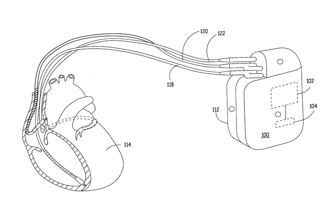
BRIEF DESCRIPTION OF THE DRAWINGS
Figure 1 is an illustration of one type of a medical device in which the
invention
may be implemented.
Figure 2 is a block diagram summarizing the data acquisition and processing
functions included in the medical device shown in Figure 1.
Figure 3 is a flow chart summarizing one method for detecting sleep apnea
using
multiple physiological signals.
Figure 4 is a flow chart summarizing steps included in a method for responding
to
a sleep apnea detection made according to the method of Figure 3.
CA 02605330 2007-10-16
WO 2006/115832 PCT/US2006/014086
4
DETAILED DESCRIPTION
The invention provides a method and apparatus for detecting a respiratory
disturbance and providing a response thereto. The invention may be implemented
in
implantable medical devices (IMDs) that include sensing capabilities for
monitoring a
physiological condition and may include therapy delivery capabilities. An IMD
in which
the invention is implemented may be primarily intended for monitoring
respiratory
disturbances for diagnostic or prognostic purposes. In one embodiment, an IMD
may be
primarily intended for monitoring for sleep apnea. The IMD may alternatively
be
intended primarily for detecting and treating sleep apnea. IMDs used for
treating sleep
apnea may deliver a sleep apnea therapy in the form of cardiac overdrive
pacing or
neuromuscular stimulation such as pectoral stimulation, phrenic nerve
stimulation, or
stimulation of excitable tissue in the neck or throat. An IMD may, via
telemetry, trigger
an external system to generate a patient alert or deliver a therapy or for
transmitting an
alert signal to a clinician or medical facility via wireless or wired
communications
network.
The invention may alternatively be implemented in IMDs that are used primarily
for other monitoring and/or therapy delivery purposes. Appropriate IMDs in
which the
invention may be incorporated include, but are not limited to, cardiac
pacemakers,
implantable cardioverter defibrillators (ICDs), cardiac monitoring devices,
neuromuscular
stimulators and drug pumps. The inclusion of respiratory disturbance detection
in such
devices can improve the therapeutic, diagnostic and/or prognostic usefulness
of the device
when the respiratory disturbance is associated with the primary condition
being monitored
or treated by the IMD, such as heart failure or diabetes.
The invention may also be implemented in external medical devices. External
medical devices may be used for bedside monitoring of a patient for diagnosing
and/or
treating sleep apnea or another medical condition that can be associated with
respiratory
disturbances. For example, external continuous positive airway pressure (CPAP)
devices
are used for detecting sleep apnea and providing positive pressure to open the
airways in
patients having obstructive sleep apnea. External devices used for monitoring
heart failure
patients may incorporate respiratory disturbance detection methods provided by
the
present invention for use as a prognostic indicator.
CA 02605330 2007-10-16
WO 2006/115832 PCT/US2006/014086
In the description that follows, various embodiments of the invention are
described
relating to the detection of sleep apnea. The methods and apparatus provided
by the
present invention, however, are not limited to the detection of sleep apnea
but may be used
for the detection of other types of respiratory disturbances, such as Cheyne-
Stokes
5 breathing or Kussmaul breathing.
Figure 1 is an illustration of one type of a medical device in which the
invention
may be implemented. IMD 100 is shown as an implantable cardiac stimulation
device
coupled to a set of cardiac leads used for positioning electrodes and other
physiological
sensors relative to a patient's heart 114 or in the blood volume. IMD 100 may
be
configured to integrate both monitoring and therapy features, as will be
described below.
IMD 100 collects and processes data from one or more sensors for deriving
parameters
used in computing a probability of a respiratory disturbance, such as sleep
apnea. IlVID
100 may further provide therapy or other response to the patient as
appropriate, and as
described more fully below.
IMD 100 is provided with a hermetically-sealed housing 112 that encloses a
processor 102, a digital memory 104, and other components as appropriate to
produce the
desired functionalities of the device. In various embodiments, IlVID 100 is
implemented as
any implanted medical device capable of measuring physiological signals for
use in
detecting sleep apnea or other respiratory disturbances, including, but not
limited to a
pacemaker, defibrillator, electrocardiogram monitor, blood pressure monitor,
drug pump,
insulin monitor, or neurostimulator.
Processor 102 may be implemented with any type of microprocessor, digital
signal
processor, application specific integrated circuit (ASIC), field programmable
gate array
(FPGA) or other integrated or discrete logic circuitry programmed or otherwise
configured
to provide functionality as described herein. Processor 102 executes
instructions stored in
digital memory 104 to provide functionality as described below. Instructions
provided to
processor 102 may be executed in any manner, using any data structures,
architecture,
programming language and/or other techniques. Digital memory 104 is any
storage
medium capable of maintaining digital data and instructions provided to
processor 102
such as a static or dynamic random access memory (RAM), or any other
electronic,
magnetic, optical or other storage medium.
CA 02605330 2007-10-16
WO 2006/115832 PCT/US2006/014086
6
As further shown in Figure 1, IMD 100 may receive one or more cardiac leads
for
connection to circuitry enclosed within the housing 112. In one embodiment,
IMD 100
collects cardiac electrogram (EGM) signals for use in deriving one or more
heart rate
related parameters and/or one or more Q-T interval related parameters for use
in
computing a probability of sleep apnea. In the example of Figure 1, IMD 100
receives a
right ventricular endocardial lead 118, a left ventricular coronary sinus lead
122, and a
right atrial endocardial lead 120, although the particular cardiac leads used
can vary from
embodiment to embodiment. Other lead systems can be substituted for the lead
system
shown in Figure 1 and may include auxiliary leads that measure breathing or
minute
ventilation through impedance changes. In addition, the housing 112 of IMD 100
may
function as an electrode and be used for sensing EGM signals. In alternate
embodiments,
cardiac sensing electrodes may be provided on subcutaneous electrodes located
on housing
112 or on subcutaneous leads extending from IMD 100 for sensing ECG signals.
Ventricular leads 118 and 122 may include, for example, pacing electrodes and
defibrillation coil electrodes (not shown) in the event IMD 100 is configured
to provide
pacing, cardioversion and/or defibrillation. In addition, ventricular leads
118 and 122 may
deliver pacing stiinuli in a coordinated fashion to provide biventricular
pacing, cardiac
resynchronization, extra systolic stimulation therapy or other benefits.
Atrial lead 120
may include pacing electrodes for providing atrial pacing pulses. In one
embodiment of
the invention, atrial lead 120 is used to provide atrial overdrive pacing in
response to sleep
apnea detection.
Electrodes carried on leads 118, 120 and 122 or the housing 112 or other
auxiliary
leads extending from IMD 100 may also be used for measuring impedance signals.
Impedance signals are used in deriving respiration-related paraineters for use
in computing
a sleep apnea or other respiratory disturbance probability. The use of
impedance signals
for monitoring respiration rate and minute ventilation is known in the art,
for example in
rate responsive cardiac pacemakers.
IMD 100 may obtain other physiological signals used in detecting sleep apnea
or
other respiratory disturbances. IMD 100 may obtain blood pressure signals,
blood oxygen
saturation signals, acoustical signals, or other physiological signals for
deriving multiple
parameters used in computing sleep apnea probability. In one embodiment, IMD
100
CA 02605330 2007-10-16
WO 2006/115832 PCT/US2006/014086
7
receives physiological signals for deriving a heart rate variability, a Q-T
interval
variability, respiration rate, respiration depth, and blood oxygen saturation.
IMD 100 may
receive physiological signals from sensors deployed on any of leads 118, 120
and 122 or
other auxiliary cardiac or subcutaneous leads or included on or in IMD housing
112.
In operation, IMD 100 obtains data via electrodes and/or sensors deployed on
leads
118, 120, 122, and/or other sources. This data is provided to processor 102,
which suitably
analyzes the data, stores appropriate data in memory 104, and/or provides a
response or
report as appropriate. Any identified respiratory disturbance episodes can be
responded to
by intervention of a physician or in an automated manner. In various
embodiments, IMD
100 activates an alert upon detection of a respiratory disturbance.
Alternatively or in
addition to alert activation. INID 100 selects or adjusts a therapy and
coordinates the
delivery of the therapy by IMD 100 or another appropriate device, which could
be another
IMD or an external device adapted to communicate with INID 100 and respond to
a sleep
apnea signal from IMD 100. Communication between IMD 100 and another device
can
occur via telemetry, such as a long-distance telemetry system. Optional
therapies that may
be applied in response to sleep apnea detection in various embodiments may
include
overdrive pacing, neuromuscular stimulation, and continuous positive airway
pressure.
Figure 2 is a block diagram summarizing the data acquisition and processing
functions included in IIVID 100. IMD 100 includes a data collection module
206, a data
processing module 202, a response module 218 and/or a reporting module 220.
Each of
the various modules may be implemented with computer-executable instructions
stored in
memory 104 and executing on processor 102 (shown in Figure 1), or in any other
manner.
The exemplary modules and blocks shown in Figure 2 are intended to illustrate
one logical
model for implementing an IMD 100 for monitoring respiratory disturbances
using
multiple physiological signals, and should not be construed as limiting.
Indeed, the various
practical embodiments may have widely varying software modules, data
structures,
applications, processes and the like. As such, the various functions of each
module may in
practice be combined, distributed or otherwise organized in any fashion in or
across a
medical device system that includes physiological signal sources.
Data collection module 206 is interfaced with one or more data sources 207 to
obtain data about the patient. Data sources 207 are generally embodied as
sensors that can
CA 02605330 2007-10-16
WO 2006/115832 PCT/US2006/014086
8
monitor electrical, mechanical, chemical, or optical information that contains
pliysiological data of the patient. Data sources 207 include any source of
physiological
signals used for monitoring for a respiratory disturbance or any other
physiological event
or condition. Data sources 207 include an ECG or EGM source 208 that provides
cardiac
electrical signals such as P-waves, R-waves or T-waves used to monitor the
patient's heart
rhythm or conduction times. Data sources 207 further include a respiration
signal source
210 for determining respiration rate and depth that can be used for minute
ventilation
computations. Respiration signal source 210 may be provided as an impedance
signal
obtained from cardiac electrodes or auxiliary electrodes, for example in the
manner used
for determining minute ventilation in rate responsive pacemakers. Respiration
signal
source 210 may alternatively be provided as any physiological signal that
varies in
response to the respiration cycle.
Data sources 207 further includes a blood oxygen saturation source 212 for
monitoring decreases in oxygen saturation that may be indicative of sleep
apnea. An
activity sensor 214 may be provided which generates a signal responsive to
patient activity
level and can be used in detecting a rest or sleep state.
Data sources 207 may include other physiological signal sources 216 for
acquiring
physiological signals useful in monitoring a patient. Other sources 216 may
include, for
example, an accelerometer or heart wall motion sensor, a blood pressure
sensor, a position
sensor or a pH sensor. Physiological parameters used for detecting sleep apnea
or another
respiratory disturbance may be determined from these alternative signal
sources. For
example, heart rate may be determined from an EGM/ECG signal 208 but may
alternatively be determined from a blood pressure signal, a wall motion signal
or other
heart signal if EGM/ECG source 208 is not available. The various data sources
207 may
be provided alone or in combination with each other, and may vary from
embodiment to
embodiment.
Data collection module 206 receives data from each of the data sources 207 by
polling each of the sources 207, by responding to interrupts or other signals
generated by
the sources 207, by receiving data at regular time intervals, or according to
any other
temporal scheme. Data may be received at data collection module 206 in digital
or analog
format according to any protocol. If any of the data sources generate analog
data, data
CA 02605330 2007-10-16
WO 2006/115832 PCT/US2006/014086
9
collection module 206 translates the analog signals to digital equivalents
using an analog-
to-digital conversion scheme. Data collection module 206 may also convert data
from
protocols used by data sources 207 to data formats acceptable to data
processing module
202, as appropriate.
Data processing module 202 is any circuit, programming routine, application or
other hardware/software module that is capable of processing data received
from data
collection module 206. In various embodiments, data processing module 202 is a
software
application executing on processor 102 (Figure 1) to implement the processes
described
below for detecting sleep apnea. Accordingly, data processing module 202
processes data
received from sources 207 for computing a probability of sleep apnea, as
described more
fully below, or another respiratory disturbance.
In an exemplary embodiment, processing module 202 receives data from
respiration source 210, EGM/ECG source 208, and oxygen saturation source 212
from
data collection module 206 and interprets the data using digital signal
processing
techniques to derive certain information from these sources for computing a
probability of
sleep apnea. The sleep apnea probability and/or intermediate computational
results may
be stored in memory 204, which may correspond to hardware memory 104 shown in
Figure 1, or may be implemented with any other available digital storage
device. Data
storage allows a clinician to access information from the various separate
data sources
over time and from any combination of these sources over time. This data can
be valuable
to a clinician, even when sleep apnea is not detected based on the computed
sleep apnea
probability, since the data can provide insight on the progression of a
respiratory
disturbance, even when the respiratory disturbance is not yet symptomatic.
When the computed sleep apnea probability exceeds a predetermined threshold,
processing module 202 may trigger an appropriate response. Responses may be
activated
by sending a digital message in the form of a signal, passed parameter or the
like to
response module 218. Response module 218 is any circuit, software application
or other
component that interacts with any type of therapy-delivery system 224 and/or
reporting
module 220. In some embodiments, therapy delivery system 224 is provided as a
pulse
generating device integrated with IMD 100 to deliver overdrive cardiac pacing
or other
neuromuscular stimulation in response to sleep apnea detection. Any therapy
provided
CA 02605330 2007-10-16
WO 2006/115832 PCT/US2006/014086
may be controlled or adjusted in response to a sleep apnea detection made
using
physiological signals acquired by data sources 207.
Reporting module 220 is any circuit or routine capable of producing
appropriate
feedback from the medical device to the patient or to a clinician or other
caregiver. In
5 various embodiments, suitable reports might include storing data in memory
204;
generating an alert 228; or producing a communication for transmission from a
telemetry
circuit or other communication module 230. Communication module 230 may be
provided as a hardwired or wireless communication network interface that can
be used to
transfer an alert or report to a designated recipient via a network, which may
be telephone
10 network, local area network, or the like. Reports may include information
about sleep
apnea episode detections such as the time, date and duration and the severity
of the
episode, the physiological data collected, and any other appropriate data.
An alert generated by the IlVID or an external device responsive to a
telemetry
signal received from the IlVID can be directed to the patient, e.g. as an
audible sound,
vibration, perceivable muscle stimulation or other sensory alert. An alert may
alternatively be directed to a clinician in form of a visual display and/or
audible signal.
An external device receiving an alert signal from IMD 100 may display
recommended
actions to be taken by the patient or a caregiver. The external device may
include
processing circuitry for interpreting data received from the implanted device
or transfer
data to an expert patient management system containing knowledge that is
captured from
general therapy protocol of physicians dealing with these respiration
disturbances.
An alert signal may result in the telemetry uplink of data obtained from the
various
sensors to a networked external device (such as a home monitor, personal
computer, or
cell phone). As such, coinmunication module 230 may include telemetry
circuitry for
transmitting data from an INff) to an external device adapted for
bidirectional telemetric
communication with the INID. The external device receiving the wireless
message may be
a programmer/monitor device that advises the patient, a physician or other
attendant of the
sleep apnea detection or related data. Information stored in memory 204 may be
provided
to an external device to aid in diagnosis or treatment of the patient.
Alternatively, the
external device may be an interface to a communications network such that the
IMD is
able to transfer sleep apnea data to an expert patient management center. The
external
CA 02605330 2007-10-16
WO 2006/115832 PCT/US2006/014086
11
device may transmit data to an expert data management center programmed to
process the
data and retrieve relevant information for distribution to a clinician,
medical center, and/or
back to the patient.
The various components and processing modules shown in Figure 2 may be
housed in a common housing such as that shown in Figure 1. Alternatively,
portions of
the components and processing modules may be housed separately. For example,
portions
of the tlierapy delivery system 224 could be integrated with IMD 100 or
provided in a
separate housing or as an external device. In this case, response module 218
may interact
with therapy delivery system 224 via an electrical cable or wireless link.
Figure 3 is a flow chart summarizing one method 300 for detecting sleep apnea
using multiple physiological signals. Sleep apnea monitoring according to
method 300
may be performed continuously, or on a scheduled or triggered basis. For
example, '
method 300 may be programmed to operate during nighttime hours, when a patient
is
expected to be asleep, and/or when a position sensor indicates a supine
position. Method
300 may additionally or alternatively be enabled to be performed upon a
triggering
condition. A triggering condition may be a sleep indicator based on an
activity signal,
posture signal, time of day, or other physiological signal or any combination
thereof.
Methods for determining or detecting a sleep state are known in the art.
Reference is
made, for example, to U.S. Pat. No. 6,731,984, issued to Yong, et al. A
triggering
condition may alternatively be a threshold crossing of any of the
physiological signals
used in detecting sleep apnea or any combination of those signals, such as a
heart rate, a
respiration rate or depth, minute ventilation, or blood oxygen saturation
level.
Sleep apnea monitoring begins by sensing an EGM/ECG signal at step 302, a
respiration signal at step 304, and a blood oxygen saturation signal at step
306. Each of
these signals are sensed simultaneously to allow multiple, concurrent
physiological
parameter values to be determined for use in sleep apnea detection. In some
embodiments, the medical device may not be capable of simultaneous sensing and
processing of all signals in which case sequential sensing and processing may
be
performed but may be less sensitive or have a slower response time for sleep
apnea
detection.
CA 02605330 2007-10-16
WO 2006/115832 PCT/US2006/014086
12
The physiological signals are used for computing a number of parameters that
will
be used to calculate a sleep apnea probability. At step 308, the EGMIECG
signal is used
to measure heart rate. The measured heart rate (HR) is used to compute
parameters related
to HR such as the HR variability at step 320. HR variability may be computed
according
to methods known in the art. It is recognized that heart rate and heart rate
variability
parameters can be determined from alternative cardiac-related signals, such as
blood
pressure. HR variability or other HR related parameters may become abnormal or
otherwise change in a characteristic way at the onset, during, or just after a
respiratory
disturbance.
At step 310, the EGM/ECG signal is used to measure Q-T intervals. The Q-T
interval variability, QT rate dependency, the absolute length of the QT
interval or any
other QT related parameter can be computed at step 322 using the measured Q-T
intervals.
The Q-T interval and/or its relation to HR may change in a characteristic
manner at the
onset, during or just after a sleep apnea episode and therefore be useful in
sleep apnea
detection or confirmation.
The respiration signal sensed at step 304, which may be an impedance signal,
is
used to measure the respiration rate at step 312 and the respiration depth at
step 314.
Respiration rate and depth may be measured on a cycle-by-cycle basis or as
mean or
median value determined from a predetermined number of successive respiration
cycles.
The respiration rate and depth are used at step 324 for computing minute
ventilation (MV).
A low respiration rate and/or low respiration depth, and/or low minute
ventilation occurs
during sleep apnea.
The oxygen saturation signal sensed at step 306 is used to measure the oxygen
saturation level at step 316. The oxygen saturation signal may be averaged
over a
predeterinined interval of time for determining the oxygen saturation level at
step 316. A
decrease in oxygen saturation can be a result of sleep apnea.
At step 330, method 300 may perform threshold comparisons of one or more of
the
measured parameters. Threshold values that would be indicative of a sleep
apnea episode
may be predefined for any of the measured parameters.
At step 340, the parameter values and/or threshold comparison results are used
in
computing a sleep apnea probability. The measured or computed parameter value
may be
CA 02605330 2007-10-16
WO 2006/115832 PCT/US2006/014086
13
used in computing the probability at step 340. Alternatively, the result of a
threshold
comparison for any given parameter value may be used. For example, if the
oxygen
saturation level goes below a threshold value, the oxygen saturation parameter
may be
assigned a logical value of 1, indicating the oxygen saturation paraineter is
positive for
sleep apnea detection. If the oxygen saturation level remains or returns to a
value above
the threshold value, the oxygen saturation parameter may be assigned a logical
value of 0,
indicating the oxygen saturation parameter is negative for sleep apnea
detection. Each of
the monitored parameters may be assigned a weighting coefficient used in
computing the
sleep apnea probability at step 340. A positive indication for sleep apnea may
therefore be
derived from a change in one or more parameter values and/or from a threshold
crossing
of one or more parameter values.
A sleep indicator determined at step 336 may also be used in computing the
sleep
apnea probability at step 340. A sleep indicator may be based on an activity
sensor signal
332 and/or the time of day 334. If the activity level is below a threshold
level and the time
of day is nighttime, the sleep indicator is positive. Other methods known in
the art for
detecting a sleep state may be used.
In one embodiment, the sleep apnea probability (SAP) computed at step 340 is
computed according to the following equation:
SAP = a(HRV) +b(QTV) + c(RR) + d(RD) + f(MV) + g(O2sat) + h(SI)
wherein HRV is the measured heart rate variability or the logical result of a
threshold comparison of the HR variability to a predetermined threshold. QTV
is the
measured Q-T interval variability or the logical result of a threshold
comparison of Q-T
interval variability to a predetermined threshold. RR is the respiration rate,
RD is the
respiration depth, and MV is minute ventilation. O2sat is the oxygen
saturation level, and
SI is the sleep indicator. The values used for each of these parameters may be
a measured
or computed value or a logical value based on the results of a threshold
comparison
performed at step 330. The constants a, b, c, d, f, g, and h are weighting
coefficients that
may be any predefined value including 0. The appropriate values for the
weighting
coefficients may be determined through optimization techniques applied to
individual
patients to maximize the sensitivity and specificity of sleep apnea detection.
CA 02605330 2007-10-16
WO 2006/115832 PCT/US2006/014086
14
The weighting coefficient values may alternatively be based on historical
clinical
experience. For example, the coefficient values may be derived from the long
term storage
of individual sensor data. The clinician can review the sensor data for a
given patient and
determine correlations between monitored parameter values and periods of sleep
apnea.
Automatic learning algorithms may be implemented for automatically adjusting
the
coefficients, for example, based on the composite result of all the sensor
signals.
Typically, an automatic learning algorithm will require one or more sleep
apnea episodes
to be confirmed by the patient or a caregiver. Manual conformation can be
entered into
the system using an external patient device or programmer and communicated to
the IMD
through telemetry. The coefficients can then be preset to values that would
result in a
positive sleep apnea detection during the confirmed sleep apnea episode.
At step 350, method 300 determines if the sleep apnea probability exceeds a
predeterinined sleep apnea detection threshold. If the detection threshold is
crossed, a
sleep apnea response is provided at step 354. The sleep apnea response may
include a
therapy delivery and/or reporting operations as described above. If sleep
apnea is not
detected according to a probability less than the detection threshold, sleep
apnea
monitoring may continue at step 352 according to the scheduled, triggered or
continuous
basis for which it is enabled.
Figure 4 is a flow chart summarizing steps included in a method for responding
to
a sleep apnea detection made according to the method 300 of Figure 3. As
described
above, monitored sleep apnea parameters 405 are provided as input for
computing a sleep
apnea probability at step 410. A sleep state indicator 435 is determined using
an activity
sensor signal 425, the time of day 430, and/or one or more of the monitored
sleep apnea
parameters 405. Heart rate and minute ventilation are known to be low during
sleep. The
Q-T interval is known to be long during sleep. As such, any of these
parameters may be
used in detecting a sleep state. Other physiological signals may be used in
detecting a
sleep state, such as a posture signal. The sleep state indicator may be
provided as input for
computing the sleep apnea probability at step 410.
The sleep apnea probability is compared to a sleep apnea detection threshold
at
decision step 412. If the sleep apnea probability is greater than a detection
threshold, sleep
CA 02605330 2007-10-16
WO 2006/115832 PCT/US2006/014086
apnea is declared at step 420. If the sleep apnea probability is not greater
than the
detection threshold, sleep apnea monitoring continues at step 415.
After declaring a sleep apnea detection at step 420, one or more response
conditions may be required prior to generating a sleep apnea response. In one
5 embodiment, the condition of verifying a sleep state at decision step 440
may be required
before generating a sleep apnea response. The sleep state may be verified
according to
sleep indicator 435. If the sleep state is not verified, sleep apnea
monitoring continues at
step 415 without delivering a sleep apnea response.
Another condition that may be required for delivering a sleep apnea response
is a
10 sleep apnea probability that exceeds a predetermined response threshold.
The response
threshold may be defined as a required magnitude of the sleep apnea
probability. The
response threshold may additionally include a minimal time duration over which
the sleep
apnea probability must continuously exceed the required magnitude. A unique
response
threshold may be set for different types of reporting or therapy delivery
responses. A
15 response threshold magnitude may be equal to or greater than the sleep
apnea detection
threshold. The response threshold may be relatively low for triggering storage
of sleep
apnea episode data and relatively higher for generating an alert or delivering
a therapy.
If the sleep apnea probability exceeds a response threshold, the corresponding
response is provided. In the example of Figure 4, if the probability exceeds a
response
threshold for therapy delivery, the therapy is delivered at step 450. If the
probability
exceeds a response threshold for generating an alert, the alert is generated
at step 455. If
the response threshold requirement is not met for any of the enabled
responses, sleep
apnea monitoring continues at step 415.
A clinician may program the desired responses to be enabled or disabled in
response to a sleep apnea detection and may program corresponding response
thresholds
for each of the enabled responses. Various responses that can be enabled by a
clinician
may include, but are not limited to, a patient alert transmitted from an IMD
to an external
home monitor or patient activator, a patient alert provided as a perceptible
muscle
stimulation or vibration, a patient alert provided as an audible sound (for
example, to
arouse the patient), a clinician alert provided via a communication network,
e.g. through
CA 02605330 2007-10-16
WO 2006/115832 PCT/US2006/014086
16
remote patient management system, or a sleep apnea therapy such as atrial
overdrive
pacing, or other neuromuscular stimulation.
Thus a medical device system and method have been described for detecting
respiratory disturbances such as sleep apnea. It is recognized that one having
skill in the
art and the benefit of the teachings provided herein may conceive of numerous
variations
to the embodiments presented herein. The systems and methods described are
intended to
be illustrative embodiments of the invention and should not be construed as
limiting with
regard to the following claims.