Note: Descriptions are shown in the official language in which they were submitted.
CA 02605335 2007-10-16
WO 2006/115901 PCT/US2006/014542
APPARATUS AND METHODS FOR FACILITATING ACCESS THROUGH A
PUNCTURE INCLUDING SEALING COMPOUND THEREIN
FIELD OF INVENTION
The present invention relates generally to apparatus and methods for sealing
punctures in a body, to apparatus and methods for facilitating access through
a puncture
extending through tissue, and, more particularly, to apparatus and methods for
delivering a
flexible sleeve or other lining into a puncture extending through tissue that
includes a
sealing compound therein, e.g., to facilitate accessing a vessel or otller
body lumen via the
puncture during a procedure.
BACKGROUND
Apparatus and methods are known for accessing a patient's vasculature
percutaneously for performing a procedure within the vasculature. For example,
a hollow
needle may be inserted through a patient's skin and overlying tissue into a
blood vessel. A
guidewire is then passed through the needle into the blood vessel, whereupon
the needle is
removed. An introducer sheath is then advanced over the guidewire into the
vessel, e.g., in
conjunction with or subsequent to one or more dilators. A catheter or other
device may be
advanced through the introducer sheath and over the guidewire into a position
for
performing a medical procedure within the patient's body. In this manner, the
introducer
sheath facilitates introducing various instruments into the vessel, while
minimizing trauma
to the vessel wall and blood loss.
Upon completing the procedure, the instrument(s) and introducer sheath are
removed, leaving a puncture extending between the skin and the vessel. To seal
the
puncture, external pressure may be applied to the overlying tissue, e.g.,
manually and/or
using sandbags, until hemostasis occurs. This procedure, however, can be time
consuming
and expensive, requiring as much as an hour of a medical professional's time.
It is also
uncomfortable for the patient, and may require the patient to remain
immobilized in an
operating room, catheter lab, or holding area. In addition, a risk of
heinatoma exists from
bleeding before hemostasis occurs.
CA 02605335 2007-10-16
WO 2006/115901 PCT/US2006/014542
-2-
Various apparatus and methods have been suggested for sealing a percutaneous
puncture instead of or in addition to using external pressure. For example,
U.S. Patent No.
5,108,421 to Fowler discloses using a collagen plug that is delivered into a
puncture
through tissue. After completing the procedure, the introducer sheath and/or
guidewire
used to access the patient's vasculature via the puncture are removed. In one
embodiment,
a catheter is inserted through the puncture into the blood vessel. A balloon
on the catheter
is expanded and then retracted until the balloon is disposed adjacent the
puncture at the
wall of the vessel. A plug is then advanced into the puncture until the plug
contacts the
balloon, thereby preventing the plug from entering the vessel. Once the plug
is positioned
within the puncture, the balloon is deflated and withdrawn, leaving the plug
to expand and
seal the puncture and/or promote hemostasis.
By way of another example, U.S. Patent Nos. 5,192,302 and 5,222,974 issued to
Kensey et al. describe using a collagen plug that may be delivered through an
introducer
sheath into a puncture site.
Such sealing methods generally involve introducing plugs or other materials
into
the puncture after completing the procedure and removing the introducer
sheath. With the
introducer sheath removed, there is substantial risk of hematoma within the
tissue
surrounding the puncture as blood from the vessel leaks into the puncture,
which may be
uncomfortable and/or harmful to the patient. Further, temporary hemostasis
devices for
isolating the vessel from the puncture may be difficult to use effectively
and/or may be
expensive. Despite attempts to isolate the vessel from the puncture while
delivering a plug
or other sealing material, the sealing material may still leak and/or become
exposed in the
vessel, where the sealing material may risk causing an einbolism in the
vessel.
SUMMARY OF THE INVENTION
The present invention is directed to apparatus, systems, and methods for
facilitating
access through a puncture in a body, e.g., extending from a patient's skin to
a blood vessel
or other body lumen, and/or for sealing such punctures. More particularly, the
present
invention includes apparatus and methods for delivering a sleeve into a
puncture extending
through tissue to a blood vessel or other body lumen and/or for facilitating
access through
a sealing compound disposed within the puncture.
CA 02605335 2007-10-16
WO 2006/115901 PCT/US2006/014542
-3-
In accordance with one embodiment, a systenz is provided that includes a
tubular
sheatli including a proximal end, a distal end sized for insertion into the
puncture, and a
lumen extending between the proximal end and an opening in the distal end; and
a sleeve
including first and second ends, a hub on the first end disposed adjacent the
distal end of
the sheath. The second end of the sleeve may extend into the opening and lumen
of the
sheath, the hub being slidable along an exterior of the sheath for drawing the
sleeve out of
the opening and along the exterior of the sheath.
In addition, the system may include a guidewire, and an assembly for
delivering a
sealing compound into the puncture around the guidewire. In one embodiment,
the
assembly may include a delivery sheath and a source of sealing coinpound for
delivering
sealing compound through the delivery sheath.
In accordance with another embodiment, a method is provided for lining a
puncture
extending from a patient's skin to a body lumen using a tubular sheath and a
thin-walled
sleeve disposed within a lumen of the tubular sheath. The sleeve may include a
hub on a
first end thereof adjacent a distal end of the sheath and a second end
disposed within the
lumen of the sheath.
A guide wire may be placed through the puncture from the patient's skin into
the
body lumen, and a sealing compound may be introduced into the puncture, e.g.,
around at
least a portion of the guide wire. The liub of the sleeve may be placed
adjacent the
patient's skin, and the sheath may be advanced into the puncture over the
guidewire while
maintaining the hub adjacent the patient's skin. This causes the sleeve to be
deployed
from the lumen of the tubular sheath and cover an exterior of the sheath to
line the
puncture as the sheath is advanced into the puncture.
In accordance with yet another embodiment, a system is provided that includes
an
elongate occlusion member including a proximal end, a distal end having a size
and shape
for insertion into the puncture, and an expandable occlusion element on the
distal end. A
thin-walled sleeve may extend along an exterior of the occlusion member from
the
proximal end towards the occlusion element, the sleeve being separable from
the exterior
of the occlusion meinber. The system may also include a delivery sheath
advanceable over
the occlusion member and sleeve for delivering a sealing compound into the
puncture
around at least a portion of the sleeve.
CA 02605335 2007-10-16
WO 2006/115901 PCT/US2006/014542
-4-
In addition or alternatively, the system may also include an introducer or
procedure
sheath including a proximal end, a distal end sized for insertion into the
puncture, and a
lumen extending between the proximal and distal ends for delivering one or
more
instruments into the body lumen. The distal of the introducer sheath may be
advanceable
through the sleeve after removing the occlusion member from the puncture or
between the
sleeve and the occlusion member.
In accordance with still another embodiment, a method is provided for
delivering a
sealing compound into a puncture extending from a patient's skin to a body
lumen. An
elongate member may be introduced from the patient's skin through the puncture
into the
body lumen, the elongate member including a flexible thin-walled sleeve
extending along
an exterior of the elongate member. A sealing compound may be delivered into
the
puncture, the sealing compound at least partially surrounding the elongate
member and
sleeve.
The body lumen may then be accessed through the sleeve For example, an
introducer or procedure sheath may be advanced through the sleeve into the
puncture. The
introducer sheath may be advanced between the sleeve and elongate member into
the
puncture, or the elongate member may be removed from the puncture while the
sleeve
remains within the puncture, whereupon the introducer sheath may be advanced
through
the sleeve.
Other aspects and features of the invention will become apparent from
consideration of the following description taken in conjunction with the
accompanying
drawings.
BRIEF DESCRIPTION OF THE DRAWINGS
The drawings illustrate exemplary embodiments of the invention, in which:
FIG. 1 is a side view of a system for sealing a puncture, including an
introducer
sheath carrying an everting sleeve, an occlusion member, a delivery sheath,
and a syringe
assembly for delivering sealing compound via the delivery sheath.
FIG. 1A is a side view of the occlusion member of FIG. 1, with an occlusion
eleinent thereon in an expanded condition.
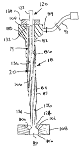
FIG. 2 is a cross-sectional view of the introducer sheath of FIG. 1, taken
along line
2-2.
CA 02605335 2007-10-16
WO 2006/115901 PCT/US2006/014542
-5-
FIGS. 3A and 3B are cross-sectional side views, showing a method for loading
an
everting sleeve into a tubular member.
FIGS. 4A-4C are cross-sectional views of a patient's body, illustrating a
method
for delivering a sealing compound a puncture extending between the patient's
skin and a
blood vessel.
FIG. 5A-5C are cross-sectional views of a patient's body, showing a method for
delivering a sleeve into the puncture of FIGS. 4A-4E after delivering a
sealing compound
therein.
FIG. 6 is a side view of another system for delivering a sleeve into a
puncture
extending through tissue.
FIGS. 7A-7D are cross-sectional views of a patient's body, showing a method
for
delivering a sleeve into the puncture using the system of FIG. 6.
DETAILED DESCRIPTION OF THE ILLUSTR.ATED EMBODIMENTS
Turning to the drawings, FIG. 1 shows an exemplary embodiment of a system 10
for delivering sealing compound into a puncture through tissue, e.g., a
percutaneous
puncture for accessing a blood vessel or other body lumen (not shown), and/or
for
accessing the body lumen via the puncture. Generally, the systein 10 includes
a delivery or
injection sheath 12, a source of sealing compound 14, an occlusion member 16,
and an
introducer or procedure sheath 18 carrying a dilator 19 and an everting sleeve
20.
Optionally, the system 10 may include other components, e.g., one or more of a
needle for
creating the puncture, a guidewire, and/or one or more sections of tubing (not
shown). In
addition or alternatively, the system 10 may include other or further
components for
creating the puncture, introducing the delivery sheath 12 and/or guidewire
into a body
lumen, and/or accessing the vessel, e.g., for introducing instruments into the
vessel via the
puncture.
Generally, the delivery sheath 12 is an elongate tubular member, including a
proximal end 22, a distal end 24, and a primary or guidewire lumen 26
extending between
the proximal and distal ends 22, 24. In addition, the delivery sheath 12 may
include one or
more secondary or injection lumens 30 that extend from the proximal end 22 to
one or
more outlets 31 (e.g., two, as shown) in the wall of the delivery sheath 12.
CA 02605335 2007-10-16
WO 2006/115901 PCT/US2006/014542
-6-
As shown, a single secondary lumen 30 is disposed concentrically around the
primary lumen 26. Alternatively, one or more secondary lumens (not shown) may
be
formed or otlierwise provided in the wall of the delivery sheath 12, e.g., in
a side-by-side
arrangeinent. In a further alternative, a delivery sheath including a single
lumen (not
shown) may be provided. The primary lu.inen 26 may be of sufficient size to
accommodate
sliding a guidewire therethrougll, e.g., between about 0.014 and 0.018 inch
(0.35-0.45
mm), while the secondary lumen 30 may be of sufficient size to accommodate
delivering
sealing compound therethrough.
The secondary lumen 30 extends from a housing 28 on the proximal end 22 of the
delivery sheath 12 to an intermediate portion 25 between the proximal and
distal ends 22,
24. As shown, the intermediate portion 25 tapers where the secondary lumen 30
terminates, with the delivery sheath 12 having a smaller diameter from the
intermediate
portion 25 to the distal end 24 (e.g., since only the primary lumen 26 extends
along this
portion of the delivery sheath 12). The smaller diameter distal portion may
have a desired
length, e.g., at least about five millimeters (5 mm). The outlet(s) 31 may be
provided on
the intermediate portion 25, e.g., where the delivery sheath 12 tapers, which
may facilitate
directing the sealing compound delivered through the secondary lumen 30
radially
outwardly away from the delivery sheath 12.
The housing 28 may be attached to or otherwise provided on the proximal end 22
of the delivery sheath 12. The housing 28 may include one or more side ports
32 (one
shown) that communicate with an interior of the housing 28 and the secondary
lumen 30
of the delivery sheath 12. The housing 28 may include one or more seals 29 to
seal the
interior of the housing 28 such that sealing compound delivered from the side
port 32 may
be directed through the secondary lumen 30. Optionally, the housing 28 may
also include
one or more seals (not shown), e.g., a hemostatic seal, for sealing the
primary lumen 26
while accommodating inserting a guidewire or other instrument (not shown) into
the
lumen 26, e.g., preventing body fluids, such as blood, from escaping
proximally through
the delivery sheath 12, as is known in the art.
A section of flexible tubing 36 may be connected to or otherwise extend from
the
side port 32 to a luer lock adapter 38, a manual shut-off valve (not shown),
and/or other
connector (also not shown), e.g., to facilitate connecting tubing and the like
(also not
CA 02605335 2007-10-16
WO 2006/115901 PCT/US2006/014542
-7-
shown) to the side port 32. A source of sealing compound, such as the dual-
syringe
assembly 40 described below, may be connected to the luer lock adapter 38.
In alternative embodiments, the delivery sheath may be a tubular member
including
a single lumen (not shown), which may include a hub, side port, and/or other
components
similar to the einbodiment described above. Additional infonnation on such
delivery
sheaths and methods for using them may be found in co-pending application
Serial Nos.
10/454,362, filed June 4, 2003 and 10/745,946, filed December 24, 2003.
Turning to FIGS. 1 and 1A, the occlusion member 16 includes a guidewire or
other
elongate member 60 carrying a tamp or other occlusion element 70. Optionally,
the
occlusion member 16 may also include a retaining sheath or other constraint 50
slidable
over the guidewire 60, e.g., for maintaining the tamp 70 in a contracted
condition. For
example, the retaining sheath 50 may be an elongate tubular member including
proximal
and distal ends 52, 54, and a lumen 56 extending therebetween. A hub 58 may be
located
on the proximal end 52, e.g., to facilitate manipulating the retaining sheath
50. The
retaining sheath 50 may have a diameter or other size to allow the distal end
54 to be
inserted into and/or through the primary lumen 26 of the delivery sheath 12,
while the hub
58 may be larger than the size of the primary lumen 26, e.g., to provide a
stop limiting
distal advancement of the retaining sheath 50 into the delivery sheath 12. The
retaining
sheath 50 may be sufficiently flexible to confonn to the surrounding anatomy,
e.g., when
the retaining sheath 50 is inserted into or removed from a puncture, e.g.,
along with other
components, such as the guidewire 60.
As best seen in FIG. 1A, the guidewire 60 may be an elongate member including
a
proximal end 62 and a distal end 64, e.g., including a "J" tip 66. The
guidewire 60 may be
formed from a solid wire, one or more coiled wires, and/or a solid-walled
tube.
Optionally, one or more coatings (not shown) may be provided on an interior or
exterior
surface of the guidewire 60, e.g., to seal the wall of the guidewire and/or to
provide a
lubricious exterior surface. The guidewire 60 may be formed from a variety of
known
materials, e.g., metals, such as stainless steel or Nitinol, plastics, and/or
composite
materials. Thus, the guidewire 60 may be sufficiently flexible to navigate
tortuous
anatomy, but may have sufficient column strength to be pushable from the
proximal end
62.
CA 02605335 2007-10-16
WO 2006/115901 PCT/US2006/014542
-8-
The tamp 70 may an expandable structure adjacent the distal tip 66 that may be
biased towards an enlarged condition (e.g., as shown in FIG. 1A), but may be
resiliently
compressible towards a contracted condition (e.g., as shown in phantom in FIG.
1). In the
embodiment shown, the tamp 70 includes a braided mesh of wires or other fibers
72 that
assume a generally spherical or elliptical disk shape in the enlarged
condition. The fibers
72 may be formed from a shape memory material, e.g., Nitinol, stainless steel,
plastic, and
the like, that has the enlarged condition programmed into the fibers 72, e.g.,
by heat
treatment. Thus, the fibers 72 may be elastically (or super-elastically)
deformed, e.g.,
compressed into the contracted condition using the retaining sheath 50, yet
resiliently
expandable towards the enlarged condition once released, as explained further
below. The
tamp 470 may shorten as it expands from the contracted condition towards the
enlarged
condition, and may lengthen again as it is compressed back towards the
contracted
condition.
The fibers 72 may include a coating, cover, or other skin (not shown) that
covers
all or a portion of the tamp 70. For example, at least the proximal portion
70a of the tamp
70 may include a coating or other skin that extends across the spaces between
the fibers 72
such that the proximal portion 70a is substantially nonporous. Alternatively,
all of the
tamp 70 may include a coating or other skin.
Iil another alternative, the tamp may include a plurality of struts (not
shown) that
are expandable between enlarged and contracted conditions. The struts may
extend
substantially axially in the contracted condition and may buckle at an
intermediate location
thereon as they expand radially outwardly towards the enlarged condition. The
struts may
be biased towards the enlarged condition (similar to the mesh above), or may
be
selectively expanded and/or compressed, e.g., using an internal pull wire or
other actuator
(not shown). In another alternative, the occlusion member 16 may include an
elongate
tubular member carrying a balloon (not shown), such as those described below
or
disclosed in co-pending application Serial No. 10/454,362, filed June 4, 2003,
and Serial
No. 10/806,927, filed March 22, 2004.
Returning to FIG. 1, the source of sealing compound 14 may include a dual
syringe
assembly 40 or other delivery device, e.g., that includes two components of a
sealing
compound. As shown, the syringe assembly 40 includes a pair of syringe barrels
42,
including outlets 43 and a plunger assembly 44 slidable into the barrels 42 to
cause the
CA 02605335 2007-10-16
WO 2006/115901 PCT/US2006/014542
-9-
components therein to be delivered through the outlets 43. In the embodiment
shown, the
plunger assembly 44 includes a pair of plungers 45 coupled to one another that
are
received in respective barrels 42. In this manner, both plungers 45 may be
manually
depressed substantially simultaneously to deliver the components together from
the syringe
barrels 42. Alternatively, a system for automatically advancing the plungers
45 and/or
otherwise delivering the components in the barrels 42 may be used, such as
those disclosed
in co-pending application Serial No. 10/806,934, filed March 22, 2004.
Optionally, the deliveiy device 14 may include a "Y" fitting 46, a static
mixer 48,
and/or tubing 49, e.g., for connecting the "Y" fitting 48 to outlets 43 of the
barrels 42, the
mixer 48 to the "Y" fitting 46 and/or to the side port 32 of the delivery
sheath 12, such that
the sealing components ejected out of the barrels 42 may mix before being
directed into
the side port 32 of the delivery sheath 12. The outlets 43, "Y" fitting 46,
mixer 48, and/or
tubing 49 may include cooperating connectors, e.g., luer lock connectors and
the like (not
shown), for connecting them together.
Respective sealing components may be provided in each syringe barrel 42 of the
syringe assembly 40 that, when mixed together, are activated to fonn a
hydrogel or other
sealing compound. Additional information on such hydrogels and systems for
delivering
them are disclosed in U.S. Patent Nos. 6,152,943, 6,165,201, 6,179,862,
6,514,534, and
6,379,373, and in co-pending published applications US 2002/0106409 published
August
8, 2002, US 2003/0012734, published January 16, 2003, US 2002/0114775
published
August 22, 2002, and US 2004/0249342 published December 9, 2004.
With continued reference to FIG. 1, the introducer sheath 18 is an elongate
tubular
member including a proximal end 82, a distal end 84, and a lumen 86 extending
between
the proximal and distal ends 82, 84. The introducer sheath 18 may terminate in
a tapered
distal tip 85 for facilitating advancing the introducer sheath 18
substantially atraumatically
through tissue into a puncture. Exemplary materials for the introducer sheath
18 may
include one or more plastics, such as polyvinyl chloride (PVC), FEP,
polyimide,
polyamide, PEEK, nylon, PET, PEBAX, and polyethylene, metals, such as
stainless steel,
and nickel titanium, and/or composite materials. The introducer sheath 18 may
be
substantially rigid, semi-rigid, or substantially flexible, e.g., to
facilitate insertion through a
puncture into a blood vessel or other body lumen. The introducer sheath 18 may
have an
CA 02605335 2007-10-16
WO 2006/115901 PCT/US2006/014542
-10-
outer diameter or other cross-section between about 0.050-0.20 inch (1.25-5.0
mm) and/or
a wall thiclcness between about 0.005-0.015 inch (0.125-0.375 mm).
A housing 88 may be attached to or otherwise provided on the proximal end 82
of
the introducer sheath 18. The housing 88 may include a side port 89 that
communicates
with an interior of the housing 88 and the lumen 86 of the introducer sheath
18. A section
of flexible tubing 91 may be connected to or otherwise extend from the side
port 89,
tenninating in a manual shut-off valve and/or a luer lock or other connector
(not shown),
e.g., to facilitate connecting tubing and the like (not shown) to the side
port 89. The
housing 88 may also include one or more seals (not shown), e.g., a hemostatic
seal, for
substantially sealing the lumen 86 of the delivery sheath 18, yet
accommodating inserting
one or more instruments (not shown) into the lumen 86.
With reference to FIG. 1 and 2, the sleeve 20 may be a relatively thin-walled
substantially flexible tubular member including a first end 102, a second end
104, and a
lumen 106 extending between the first and second ends 102, 104. In one
embodiment, the
sleeve 20 may be a substantially closed-walled tube, e.g., with one or more
longitudinal
seams (not shown) extending between the first and second ends 102, 104. The
longitudinal seam(s) may be substantially permanently fixed or may be weakened
or
otherwise separable, as described further below.
In an exemplary embodiment, the sleeve 20 may be formed from a substantially
inelastic sheet of material whose longitudinal edges are bonded, melted,
welded, or
otherwise attached to one another, e.g., butted together or lapped one over
the other, to
form a tubular structure. In one embodiment, the sleeve 20 may be formed from
expanded
polytetraflouroethylene, e.g., having a wall thickness of between about 0.0001-
0.1 inch
(0.0025-2.5 mm), e.g., less than about 0.004 inch, and/or less than about
0.001 inch,
similar to the membranes described in U.S. Patent Nos. 5,531,717, 5,676,688,
and
6,240,968.
Alternatively, the sleeve 20 may be extruded or otherwise formed from a
continuous length of flexible tubing. In further alternatives, the sleeve 20
may be formed
from other materials, such as Dacron or silk fabrics, fiber meshes, and the
like, e.g., with
or without coatings to provide a substantially nonporous wall. Such materials
may be
woven, knitted, or otherwise formed into a tubular shape. In yet another
alternative, the
sleeve 20 may be formed from a sheet whose longitudinal edges (not shown) are
simply
CA 02605335 2007-10-16
WO 2006/115901 PCT/US2006/014542
-11-
lapped over one another between the first and second ends 102, 104 and/or
butted against
one another. The edges may be bonded together, adhered using an adhesive,
connected
with threads or other fasteners, and the like.
Optionally, one or both of the inner and outer surfaces of the sleeve 20 may
include
a lubricious coating, e.g., to reduce friction as the sleeve 20 everts around
itself and/or
around the distal end 84 of the introducer sheath 82 (or other components), as
described
further below. As shown, the second end 104 of the sleeve 20 may be open.
Alternatively,
the second end 104 may be substantially closed, e.g., including a weakened or
penetrable
end wall (not shown) allowing the second end to be opened by advancing a
guidewire or
other instrument until it penetrates through the end wall.
An annular hub 108 may be attached to or otherwise provided on the first end
102
of the sleeve 20. The hub 108 may have a generally planar or curved
configuration, e.g., to
facilitate placing the hub 108 in contact with a patient's skin or other
anatomy such that
the hub 108 confomis substantially to the anatomy. The hub 108 may include a
passage
110 theretlirough, e.g., including a frustoconical proximal portion 110a and a
cylindrical
distal portion 110b. The distal portion 110b may have a diameter or other
cross-section
larger than the outer diameter of the introducer sheath 18. Thus, the distal
end 84 of the
introducer sheath 18 may be directed into the passage 110, e.g., guided by the
ramped
surfaces of the proximal portion 110a into and through the distal portion
110b, as
described further below.
As shown in FIGS. 1 and 2, the sleeve 20 may be carried within a tubular
member
120 sized to be slidably received within the lumen 86 of the introducer sheath
18. The
tubular member 120 may include a proximal end 122, a distal 124, and a lumen
126
extending therebetween, within which the sleeve 20 may be received. The
tubular member
120 may be formed from substantially rigid, semi-rigid, or substantially
flexible material,
similar to the introducer sheath 18.
The distal end 124 of the tubular member 120 may include a blunt, tapered,
and/or
rounded edge, e.g., to facilitate the sleeve 20 sliding around the distal end
124 of the
tubular member 120 during deployment, as described further below. The tubular
member
120 may have sufficient length that the proximal end 122 of the tubular member
120 may
extend proximally beyond the hub 88 of the introducer sheath 18, while the
distal end 124
CA 02605335 2007-10-16
WO 2006/115901 PCT/US2006/014542
-12-
of the tubular member 120 is disposed adjacent to or beyond the distal end 84
of the
introducer sheath 18.
The tubular member 120 may facilitate loading the sleeve 20 into the
introducer
sheath 18, e.g., during original manufacturing or immediately before a medical
procedure.
For example, turning to FIGS. 3A and 3B, the sleeve 20 may be provided
initially with the
second end 104 disposed distally relative to the first end 102. With the hub
108 disposed
adjacent the distal end 124 of the tubular member 120, a tool 112, e.g.,
including an
elongated hook 114, forceps, grabber, or other mechanism (not shown), may be
inserted
into the proximal end 122 of the tubular member, through the lumen 126, and
into the first
end 102 and lumen 104 of the sleeve 20. The hook 114 (or other mechanism) may
engage
the second end 104, whereupon the too1112 may be withdrawn proximally back
through
the tubular member 120, thereby everting the second end 104 of the sleeve 20
within itself
as the second end 104 is pulled into the lumen.126 of the tubular member 120.
Once the second end 104 is disposed within the lumen 126 of the tubular member
120, e.g., adjacent the proximal end 122 of the tubular member, the hook 114
may be
disengaged from the second end 104. The hook 114 may then be withdrawn
proximally
from the tubular member 120. Optionally, the sleeve 20 may be twisted about
its
longitudinal axis or otherwise compressed before being loaded into the tubular
member
120, e.g., to reduce its initial profile and/or facilitate loading.
The tubular member 120 may then be inserted through the introducer sheath 18,
e.g., before the introducer sheath 18 is packaged during manufacturing.
Alternatively, the
tubular member and introducer sheath 18 may be packaged separately or side-by-
side in a
single package. The proximal end of the tubular member 120 may be inserted
into the
lumen 86 from the distal end 84 of the introducer sheath 18 until the hub 108
of the sleeve
20 is disposed adjacent to or distal to the distal end 84 of the introducer
sheath 18, as
shown in FIGS. 1 and 2.
Optionally, as shown in FIGS. 1 and 2, a dilator 19 may also be provided,
e.g.,
within the lumen 86 of the introducer sheath 18. The dilator 19 may include a
proximal
end 132, a distal end 134 sized for insertion through the lumen 86 of the
introducer sheath
18, a lumen 136 extending between the proximal end distal ends 132, 134, and a
hub or
other handle 138 on the proximal end 132. The distal end 134 may include a
tapered or
multiple ramped shape, similar to known dilators. The dilator 19 may be
forined from
CA 02605335 2007-10-16
WO 2006/115901 PCT/US2006/014542
-13-
substantially rigid, semi-rigid, or substantially flexible materials, similar
to the introducer
sheath 18.
Similar to the tubular member 120, the dilator 19 may be loaded into the
introducer
sheath 18 during manufacturing or immediately before a procedure. In addition,
the dilator
19 may be loaded into the introducer sheath 18 before or after the tubular
member 120,
e.g., by inserting the distal end 134 of the dilator 19 into the hub 88 and
lumen 86 of the
introducer sheath 18 (around the tubular member 120 if already loaded) until
the hub 138
abuts or is locked at the hub 88. Once inserted into the introducer sheath 18,
the distal end
134 of the dilator 19 may extend beyond the distal end 84 of the introducer
sheath 18, e.g.,
to provide a gradually tapering transition for the assembly. Thus, before a
procedure, the
sleeve 20, tubular member 120, dilator 19, and introducer sheath 18 may be
disposed
concentrically around one another in an assembly, as shown in FIGS. 1 and 2.
Optionally,
one or both of the tubular member 120 and dilator 19 may be eliminated, if
desired, and
the sleeve 20 may be everted and disposed directly within the lumen 86 of the
introducer
sheath 18 or the lumen 136 of the dilator 19.
Turning to FIGS. 4A-4E and 5A-5C, a method is shown for delivering an
introducer sheath and/or sleeve, such as the introducer sheath 18 and sleeve
20 described
above, into a passage extending through tissue 96. In the illustrated
embodiment, the
passage is a percutaneous puncture 90 extending from a patient's skin 92 to a
blood vessel
or other body lumen 94. For example, the vessel 94 may be a peripheral artery,
e.g., a
femoral artery, a carotid artery, and the like. It will be appreciated that
systems and
methods constructed and undertaken in accordance with various embodiments of
the
invention may be used to seal other passages through tissue within a patient's
body.
Initially, as shown in FIGS. 4A-4C, the puncture 90 may be created and sealing
compound 99 may be delivered into the puncture 90. Turning to FIG. 4A, to
create the
puncture 90, a hollow needle 15 may be inserted through the patient's skin 92
and
intervening tissue 96 into the vessel 94. The occlusion member 16, e.g., the
guidewire 60
and retaining sheath 50, may be inserted into the puncture 90, e.g., through
the needle 15
until the distal tip 66 is disposed within the vessel 94. As shown, the
retaining sheath 50
covers the tamp 70 on the guidewire 460 as the guidewire 460 is advanced
through the
needle 416, thereby maintaining the tamp 70 in the contracted condition.
CA 02605335 2007-10-16
WO 2006/115901 PCT/US2006/014542
-14-
Turning to FIG. 4B, once the tamp 70 is within the vessel 94, the needle may
be
removed, and the tamp 70 may be expanded within the vessel 94. For example,
the
retaining sheath 50 may be retracted completely (or only partially, not shown)
out of the
puncture 90 to expose the tamp 70, whereupon the tamp 70 may self expand
within the
vessel 94. Alternatively, the tamp 70 may be selectively expandable, e.g.,
using an internal
pull wire or other actuator (not shown). Thus, once the tamp 60 is exposed
within the
vesse194, the tamp 70 may be expanded, e.g., by pulling the pull wire until
the tamp 70
attains a desired enlarged size and/or configuration. In another alternative,
the tamp 70
may be a balloon or other expandable member (not shown), such as those
described in co-
pending applications Serial Nos. 10/454,362 and 10/806,927, e.g., that may be
inflated
using inflation media.
Turning to FIG. 4C, the delivery sheath 12 may be advanced over the guidewire
60
into the puncture 90, e.g., before or after the tamp 70 is expanded. As shown,
the delivery
sheath 12 may be advanced over the guidewire 60 until the distal end 24 enters
the vessel
94. For exaniple, the proximal end 62 of the guidewire 460 may be backloaded
through
the primary lumen 26 of the delivery sheath 12, and the delivery sheath 12 may
be
advanced into the puncture 90, the guidewire 60 sliding through the primary
lumen 26.
After the tamp 70 is expanded, the guidewire 60 may be partially retracted
from the
vesse194, e.g., by pulling the proximal end 62 of the guidewire, until the
proximal portion
40a of the tamp 470 contacts the distal end 24 of the delivery sheath 12
(providing a first
tactile feedback). The guidewire 60 may then be pulled fizrther until the tamp
70 contacts
the wall of the vesse194 (providing a second tactile feedback), thereby
partially in
retracting the delivery sheath 12 back into the puncture 90, e.g., until the
distal end 24 is
disposed adjacent the vesse194.
Alternatively, the guidewire 60 may be retracted until the tamp 70 contacts
the wall
of the vessel 94 before the delivery sheath 12 is introduced. The delivery
sheath 12 may
then be advanced into the puncture 90 until the distal end 24 contacts the
wall of the vessel
94 with the tamp 70 underneath, thereby providing tactile feedback that the
outlets 25 are
disposed within the puncture 90 proximal to the vesse194 when the distal end
24 contacts
the tamp 70.
A source of sealing compound 14, e.g., the dual syringe assembly 40 described
above, may be prepared and connected to the side port 32 of the delivery
sheath 12, e.g.,
CA 02605335 2007-10-16
WO 2006/115901 PCT/US2006/014542
-15-
via tubing 49, either before or after the delivery sheath 12 is advanced into
the puncture
90. The sealing compound 99 may then be delivered through the secondary lumen
30 and
the outlets 25 and into the puncture 90. The sealing compound 99 may flow
radially
outwardly to permeate at least partially into the tissue surrounding the
puncture 90.
Optionally, the delivery sheath 12 may be retracted as the sealing compound 99
is
delivered, e.g., to fill the puncture 90 along its length substantially
filling the tissue tract
with sealing compound.
Once a desired amount of the sealing compound 99 is delivered into the
puncture
90, the guidewire 60 may be maintained such that the tamp 70 continues to seal
the
puncture 90 from the vessel 94, e.g., for sufficient time for the sealing
compound 99 to at
least partially or coinpletely cure. Thereafter (or iinmediately after filling
the puncture 90),
the delivery sheath 12 may be renzoved entirely from the puncture 90.
Additional
apparatus and methods for delivering the sealing compound 99 into the puncture
90 are
disclosed in co-pending application Serial Nos. 10/454,362 and 10/745,946, or
in co-
pending application Serial, No. 10/975,205, filed October 27, 2004.
Turning now to FIGS. 5A-5C, the introducer sheath 18 and sleeve 20 may then be
delivered into the puncture 90 and/or through the sealing compound 99. As
shown in FIG.
5A, the guidewire 60 may remain within the puncture 90 and vesse194 after
delivering the
sealing compound 99. Optionally, the tamp 70 (not shown) may remain exposed
and/or
expanded, or the retaining sheath 50 (also not shown) may be advanced over the
guidewire
60 to cover and/or collapse the tamp 70. Alternatively, the guidewire 60 may
be removed
from the puncture and exchanged for a separate guidewire (not shown), e.g.,
without a
tamp, may be advanced through the puncture 90 into the vessel 94.
Turning to FIG. 513, the introducer sheath 18, dilator 19, and sleeve 20 may
then be
introduced into the puncture 90, e.g., over the guidewire 60. Although not
shown in FIG.
5B, the tubular member 120 (shown in FIG. 2) may be inserted along with the
introducer
sheath 18. For example, the guidewire 60 may be backloaded into the introducer
sheath
18, e.g., by inserting the proximal end 62 of the guidewire 60 through the
passage 110 (not
shown, see FIG. 2) of the hub 108 into the lumen 106 (also not shown, see FIG.
2) of the
sleeve 20 and directed proximally through the lumen 136 of the tubular member
120 (also
not shown, see FIG. 2) (or through the lumen 136 of the dilator 19 or the
lumen 86 of the
CA 02605335 2007-10-16
WO 2006/115901 PCT/US2006/014542
-16-
introducer sheath 18, depending upon whether the tubular member 120 and/or
dilator 19
are included or eliminated).
As shown in FIG. 5B, the hub 108 of the sleeve 20 may be placed against or
immediately above the patient's skin 92 overlying the puncture 90. The
introducer sheath
18 (and any of the dilator 19 and/or tubular member 120 carried therein) may
then be
advanced into the puncture 90 through the passage 110 in the hub 108. Because
the sleeve
20 is disposed inside the introducer sheath 18, as the distal end 124 (not
shown, see FIG.
2) of the tubular member 120 (or the distal end 134 of the dilator 19 or the
distal end 84 of
the introducer sheath 18) enters the passage 110, it contacts the sleeve 20
adjacent the first
end 102. Further advancement of the introducer sheath 18 causes the sleeve to
slide
around the distal end 124 of the tubular member 120, pulling the sleeve 20 out
of the
lumen 106 and unfurling or everting the sleeve 20 over the distal end 124 of
the tubular
member 120.
As shown in FIG. 4B, as the distal end 134 of the dilator 19 and the distal
end 84 of
the introducer sheath 18 enter the passage 110 of the hub 108, they pass
through the sleeve
that has everted before them. With respect to the tissue surrounding the
puncture 90,
the sleeve 20 unfurls or everts from the tubular member 120 as the introducer
sheath 18 is
advanced into the puncture 90, thereby lining the puncture 90 from the
patient's skin 92
toward the vessel 94. In one embodiment, the sleeve 20 has sufficient length
that the
20 sleeve 20 substantially lines the puncture 90 through the sealing compound
99 and the
second end 104 terminates adjacent the wall of the vesse194.
Because the sleeve 20 unfurls from within the tubular member 120 as the
introducer sheath 18 is advanced into the puncture 90, shear stress on the
surrounding
tissue, and/or on the sealing compound 99 are substantially reduced, e.g., as
compared
with advancing the introducer sheath 18 and/or dilator 19 through the puncture
90 without
the sleeve 20. Because the introducer sheath 18 is not pushed directly along
the tissue
surrounding the puncture, this may substantially reduce damage to the
surrounding tissue
and/or to the sealing compound 99. Thus, risk of pieces of the sealing
compound 99 being
broken off and conveyed into the vessel 94, where they may travel downstream
and cause
an embolism or other damage may be substantially reduced.
Turning to FIG. 5C, the introducer sheath 18 may be advanced through the
puncture 90 until the distal end 84 is disposed within the vessel 94. As the
introducer
CA 02605335 2007-10-16
WO 2006/115901 PCT/US2006/014542
-17-
sheath 18 is advanced, the second end 104 of the sleeve 20 may be completely
unfurled
from the tubular member 120 and exposed, e.g., within the vesse194. In the
embodiment
shown, the length of the sleeve 20 is shorter than the introducer sheath 18
such that the
second end 104 of the sleeve 20 is disposed proximal to the distal end 84 of
the introducer
sheath 18.
The dilator 19 and/or tubular member 120 may be withdrawn through the
introducer sheath 18 from the puncture 90, e.g., together or successively,
leaving the
introducer sheath 18 and sleeve within the puncture 90. The guidewire 60 may
also be
removed along with, before, or after the dilator 19 and/or tubular member 120,
e.g., after
collapsing the tamp 70 (not shown). Alternatively, if the tamp 70 is still
expanded, the
guidewire 60 may be removed, causing the tamp 70 to compress to the contracted
condition as it is directed into the lumen 86 of the introducer sheath 18.
Once a distal end 84 of the introducer sheath 18 is disposed within the
vesse194,
one or more instruments (not shown) may be advanced through the luinen 86 into
the
vesse194, e.g., to perform one or more diagnostic and/or interventional
procedures witllin
the patient's body, as is known to those skilled in the art. The sleeve 20
generally does not
interfere with the introduction of such instruments, since it is located only
around the
introducer sheath 18. Optionally, if the sleeve 20 includes any weakened
seams, the sleeve
may be removed from around the introducer sheath 18 to provide a conventional
20 introducer sheath arrangement for the subsequent procedure. For example,
the hub 108
may separate into two or more pieces (not shown), causing the sleeve 20 to
tear or
separate, e.g., along one or more predetermined seams. Thus, conventional
procedures
may be used without need for extra attention to the sleeve 20.
Upon completing any such procedures, the instrument(s) may be removed from the
vessel 94 through the introducer sheath 18. The introducer sheath 18 and
sleeve 20 (if
remaining around the introducer sheath 18) may then be removed from the
vesse194 and
puncture 90, e.g., simultaneously or successively. The sealing compound 99
and/or tissue
may recoil sufficiently to substantially fill the puncture 90, thereby
allowing and/or
encouraging hemostasis to occur between the vessel 94 and puncture 90.
Optionally,
external pressure may be applied to the patient's skin 92 during removal of
the introducer
sheath 18, e.g., to further enhance sealing of the puncture 90 until
heinostasis occurs.
CA 02605335 2007-10-16
WO 2006/115901 PCT/US2006/014542
-18-
Turning to FIG. 6, another embodiment of a system 210 is shown for delivering
a
sleeve 220 into a puncture extending through tissue. Generally, the system 210
includes a
delivery sheath 12, a source of sealing compound 14, an occlusion member 216,
and an
introducer or procedure sheath, such as the introducer sheath 18 described
above (without
the tubular member 120 and sleeve 20). Unlike the previous embodiments, the
sleeve 220
is initially carried on an outer surface of the occlusion member 216, rather
than within the
introducer sheath 218, as described further below. Optionally, the system 210
may include
other components, e.g., one or more needles, guidewires, dilators, and/or
sections of tubing
(not shown), similar to the previous embodiments.
Generally, the delivery sheath 12 is an elongate tubular member, similar to
the
previous embodiments, e.g., including a proximal end 22, a distal end 24, and
a primary or
guidewire lumen 26 extending between the proximal and distal ends 22, 24. The
delivery
sheath 12 may include one or more secondary or injection lumens 30 that extend
from the
proximal end 22 to one or more outlets 31 (e.g., two, as shown) in the wall of
the delivery
sheath 12.
The delivery sheath 12 may include a housing 28 on the proximal end 22, a side
port 32 that communicates with an interior of the housing 28 and the secondary
lumen 30
of the delivery sheath 12, and a section of tubing 36 extending from the side
port 32. The
housing 28 may include one or more seals 29 to seal the interior pf the
housing 28 such
that sealing compound delivered into the side port 32 may be directed through
the
secondary lumen 30. Optionally, the housing 28 may also include one or more
seals (not
shown), e.g., a hemostatic seal, for sealing the primary lumen 26 while
accommodating
inserting one or more instruments (not shown) into the lumen 26.
The source of sealing compound 14 may include a dual syringe assembly 40
including a pair of syringe barrels 42 with outlets 43, and a plunger assembly
44 slidable
into the barrels 42 to cause components therein to be delivered through the
outlets 43,
similar to the previous embodiments. Optionally, the delivery device 14 may
include a
"Y" fitting 46, a static mixer 48, and/or tubing 49, e.g., for connecting the
outlets 43, the
"Y" fitting 48, the mixer 48, and/or the side port 32 of the delivery sheath
12, such that the
sealing components ejected out of the barrels 42 may mix before being directed
into the
side port 32 of the delivery sheath 12, also similar to the embodiments
described above.
CA 02605335 2007-10-16
WO 2006/115901 PCT/US2006/014542
-19-
The occlusion member 216 is an elongate tubular member 260 including a
proximal end 262, a distal end 264, and a lumen (not shown) extending between
the
proximal and distal ends 262, 264. The tubular member 260 may be flexible,
semi-rigid,
or rigid, e.g., having a uniform or variable flexibility along its length. For
example, a
proximal portion of the tubular member 260 may be substantially rigid, e.g., a
section of
hypotube (not shown), to facilitate advancing the occlusion member 216 into a
puncture
through tissue, while a distal portion of the tubular member 260 may be
substantially
flexible to facilitate insertion through a puncture into a blood vessel or
other body lumen.
A balloon 270 is carried on the distal end 264 of the tubular member 260 that
includes an interior communicating with the lumen of the tubular member 260.
The
balloon 270 is expandable from a contracted condition (not shown) to an
enlarged
condition, such as that shown in FIG. 6, e.g., when fluid or other inflation
media is
delivered through the tubular member 260 into the interior of the balloon 270.
The
balloon 270 may be formed from a flexible, substantially inelastic material,
e.g., a
nonelastomeric material, such as PET, nylon, polyethylene, polyurethane,
PEBAX, and the
like, that may provide a substantially noncompliant or semi-compliant balloon
270 that
may expand to a predetermined size when a minimum pressure is introduced into
the
interior 82. Alternatively, the balloon 270 may be formed from an elastic
material, such
that the size of the balloon 270 in the expanded state is dependent upon the
pressure or
volume of fluid delivered within the interior.
In the contracted condition, the balloon 270 may conform substantially to the
diameter of the tubular member 260. In one embodiment, the balloon 270 may at
least
partially evert in the enlarged condition, i.e., the length of the balloon 270
may be
substantially smaller than the diameter. In alternative embodiments, other
expandable
members, e.g., a mechanically expandable or self-expanding member, such as
those
described above, may be provided instead of the balloon 270.
A hub 250 may be coupled to or otherwise provided on the proximal end 262 of
the
tubular member 260. In one einbodiment, the hub 250 may be removable from the
tubular
member 260, e.g., using mating threads or other connectors (not shown) on the
hub 250
and/or the proximal end 262 of the tubular member 260. The hub 250 may include
a side
port 252 that communicates with the lumen in the tubular member 260, such that
a source
CA 02605335 2007-10-16
WO 2006/115901 PCT/US2006/014542
-20-
of inflation media, e.g., a syringe containing saline (not shown), may be
coupled to the side
port 252 for delivering inflation media into the interior of the balloon 270.
Optionally, the occlusion member 216 may include an elongate inner member (not
shown) slidable within the tubular member 260, and the hub 250 may include a
piston or
other mechanism (not shown) for biasing the inner member relative to the
tubular member
260. For example, the inner member may be coupled to a distal end 274 of the
balloon
270 and may be biased to move distally relative to the tubular member 260,
e.g., to
facilitate collapsing the balloon 80 when it is deflated. The piston within
the hub 250 may
be directed proximally when inflation media is delivered into the side port
252, thereby
pulling the inner member to shorten the balloon 270 as it expands. Additional
information
on occlusion members that may be provided may be found in co-pending
application Serial
No. 10/454,362.
Returning to FIG. 6, the sleeve 220 may be a relatively thin-walled
substantially
flexible tubular member including a first or proximal end 282, a second or
distal end 284,
and a lumen 286 extending therebetween, similar to the sleeves described
above. In one
embodiment, the sleeve 220 may be a substantially closed-walled tube that may
be
collapsed around the tubular member 260 of the occlusion member 216. For
example, the
sleeve 220 may be folded, crimped, and/or twisted about the tubular member 260
to define
a collapsed state.
Optionally, the sleeve 220 may be bonded to the outer surface of the tubular
member 260 and/or to itself (e.g., if folded over itself, e.g., using an
adhesive that may
separate when sufficient force is applied. For example, the sleeve 220 may
separate from
the tubular member 260 and expand towards an expanded state when an
instrument, e.g.,
the introducer sheath 18, is advanced between the sleeve 220 and the tubular
member 260,
or when a fluid is injected between the sleeve 220 and the tubular member 260,
as
described further below.
Thus, the sleeve 220 may have a diameter or other cross-section in the
expanded
state that is substantially larger than the tubular member 260. In one
embodiment, the
sleeve 220 may be formed from a substantially inelastic material such that the
sleeve 220
may assume a fixed diameter or other cross-section in the expanded state,
e.g., larger than
the introducer sheath 18. Alternatively, the sleeve 220 may be formed from an
elastic
CA 02605335 2007-10-16
WO 2006/115901 PCT/US2006/014542
-21-
material such that the sleeve 220 may resiliently expand to accommodate
different size
instruments therein.
Optionally, one or both of the inner and outer surfaces of the sleeve 220
and/or the
outer surface of the tubular member 260 may include a lubricious coating. For
exaniple, if
the sleeve 220 is crimped, twisted, or otherwise compressed around the tubular
meinber
260 without being attached thereto, a lubricious coating may be provided on
the inner
surface of the sleeve 220 and/or on the outer surface of the tubular member
260. Such a
lubricious coating may reduce friction or otherwise facilitate separation of
the sleeve 220
from the tubular menlber 260 when the sleeve 220 is expanded, as described
below.
An annular hub 288 may be attached to or otherwise provided on the proximal
end
282 of the sleeve 220. The hub 288 generally includes a passage 289
therethrough that
conununicates with the lumen 286. The hub 288 may be expandable, e.g., such
that the
passage 289 may have a diameter or other cross-section larger than the outer
diameter of
the introducer sheath 18 when the hub 288 is expanded. The hub 288 may have an
outer
diameter or other cross-section that is smaller than the primary lumen 26 of
the delivery
sheath 12 such that the hub 288 may pass through the primary lumen 26, e.g.,
when the
delivery sheath 12 is advanced over the occlusion member 216, as described
below.
Alternatively, other structures may be provided on the proximal end 282 of the
sleeve 220,
e.g., one or more tabs (not shown) that may lie initially against the outer
surface of the
occlusion member 216, instead of the hub 288. The tab(s) may be pulled
transversely
away from the occlusion member 216 to open and/or expand the proximal end 282
of the
sleeve 220 to allow insertion of the introducer sheatli 18 therein, as
described further
below.
Optionally, the distal end 84 of the introducer sheath 18 may be directed into
the
passage 289, e.g., guided by ramped surfaces or other guides (not shown) on
the hub 288,
and thereby into the lumen 286 of the sleeve 220, as described further below.
In addition
or alternatively, the hub 288 may be separable into two or more pieces (not
shown) to open
the proximal end 282 of the sleeve. Optionally, the sleeve 220 may include one
or more
seams (also not shown) such that the hub 288 and/or sleeve 220 may be
separated into two
or more pieces to remove the sleeve 220, similar to the embodiments described
above.
Turning to FIGS. 7A-7D, a method is shown for delivering an introducer sheath
and/or sleeve, such as the introducer sheath 18 and sleeve 220 described
above, into a
CA 02605335 2007-10-16
WO 2006/115901 PCT/US2006/014542
-22-
puncture 90 extending through tissue 96, e.g., from a patient's skin 92 to a
blood vessel 94.
Initially, the puncture 90 may be created, e.g., using a needle (not shown),
and a guidewire
(also not shown) may be advanced through the needle into the vessel 94,
similar to the
embodiments described above. In one embodiment, the needle may be removed, and
the
delivery sheath 12 may be advanced over the guidewire, e.g., alone or in
conjunction with
one or more dilators (not shown).
As shown in FIG. 7A, once the distal end 24 of the delivery sheath 12 is
disposed
within the vessel 94, the guidewire and any dilators (not shown) may be
removed, and the
occlusion member 216 may be inserted into the puncture 90. For example, with
the
balloon 270 collapsed, the distal end 264 of the occlusion member 216 may be
advanced
through the primary lumen 26 of the delivery sheath 12 until the balloon 270
is disposed
within the vessel 94. Because the sleeve 220 is collapsed around the tubular
member 260,
the sleeve 220 may remain unobtrusively around the tubular member 260 as the
occlusion
member 216 is advanced into the delivery sheath 12.
Alternatively, the occlusion member 216 may be used as the guidewire directed
through the needle, and the delivery sheath 12 may be advanced over the
occlusion
member 216 into the puncture 90. In this alternative, the hub 250 may be
separated from
the tubular member 260 to allow the proximal end 262 of the tubular member 260
to be
directed into the primary lumen 26 of the delivery sheath 12 before the
delivery sheath 12
is advanced into the puncture 90. Optionally, the sleeve 220 may include a
lubricious
coating on its outer surface, e.g., to facilitate advancing the occlusion
member 216 through
the delivery sheath 12 and/or puncture 90 without damaging or otherwise
disrupting the
sleeve 220 carried on the occlusion member 216.
Turning to FIG. 7B, once the balloon 270 is within the vesse194, the balloon
270
may be expanded and used to substantially seal the vessel 94 from the puncture
90. For
example, a syringe or other source of inflation media (not shown) may be
coupled to the
side port 252 of the hub 250, and saline or other inflation media may be
delivered through.
the tubular member 260 into the interior of the balloon 270, causing the
balloon 270 to
expand to the expanded state. If the hub 250 is previously separated from the
tubular
meinber 260, the hub 250 may be attached to the proximal end 262 of the
tubular member
260 to allow the inflation media to be delivered via the side port 252 into
the tubular
member 260 and the interior of the balloon 270. Alternatively, otlier
selectively
CA 02605335 2007-10-16
WO 2006/115901 PCT/US2006/014542
- 23 -
expandable members may be provided on the distal end 264 of the tubular member
260
instead of the balloon 270, such as the expanding mesh or expandable frame
(not shown)
described above, and the expandable member may be expanded within the vessel
94.
With the balloon 270 (or other expandable member) expanded, the occlusion
member 216 may be partially retracted from the vessel 94, e.g., by pulling the
hub 250,
until the balloon 270 contacts the distal end 24 of the delivery sheath 12
(providing a first
tactile feedbaclc). The occlusion member 216 may then be pulled further until
the balloon
270 contacts the wall of the vesse194 (providing a second tactile feedback),
thereby
partially retracting the delivery sheath 12 back into or above the puncture
90.
Alternatively, the balloon 270 may be directed against the wall of the
vesse194 before the
delivery sheath 12 is advanced fully into the puncture 90, e.g., until the
distal end 24 of the
delivery sheath 12 contacts the vesse194 with balloon 270 underneath.
A source of sealing compound 14, e.g., the dual syringe assembly 40 described
above, may be connected to the side port 32 of the delivery sheath 12, and
sealing
compound 99 may be delivered through the delivery sheath 12 into the puncture
90.
Optionally, the delivery sheath 12 may be retracted as the sealing compound 99
is
delivered, e.g., similar to the einbodiments described above. The occlusion
member 216
may be maintained such that the balloon 270 continues to seal the puncture 90
from the
vesse194, e.g., for sufficient time for the sealing compound 99 to at least
partially or
completely cure.
Thereafter, as shown in FIG. 7C, the delivery sheath 12 may be removed
entirely
from the puncture 90, leaving the tubular member 260 and sleeve 220 within the
puncture
90. For example, the balloon 270 may be collapsed, e.g., by evacuating the
inflation media
from the balloon 270 and/or by removing the hub 250 from the proximal end 262
of the
tubular member 260. The hub 250 may be removed from the tubular member 260 (if
not
already), and the delivery sheath 12 may then be withdrawn from the puncture
90 around
the tubular member 260 and sleeve 220.
Turning to FIG. 7D, the introducer sheath 18 may then be delivered into the
puncture 90 and/or through the sealing compound 99 via the sleeve 220. As
shown, the
distal end 84 of the introducer sheath 18 may then be advanced over the
proximal end 262
of the tubular member 260, and inserted into the proximal end 282 of the
sleeve 220, i.e.,
such that the introducer sheath 18 is disposed between the sleeve 220 and the
tubular
CA 02605335 2007-10-16
WO 2006/115901 PCT/US2006/014542
-24-
member 260. Optionally, the hub 288 may be expanded or tabs or others
structures (not
shown) on the proximal end 282 of the sleeve 220 may be manipulated to
separate and/or
expand the proximal end 282 of the sleeve 220 to accommodate the introducer
sheath 18.
As the introducer sheat1118 is advanced distally, the sleeve 220 may separate
and
expand away from the tubular member 260, thereby pushing the surrounding
tissue and/or
sealing compound radially away from the introducer sheath 18. The distal end
84 of the
introducer sheath 18 may exit the distal end 284 of the sleeve 220 and enter
the vessel 94,
thereby providing an arrangement similar to that shown in FIG. 5C.
Alternatively, the occlusion member 216 may be removed from the puncture 90,
leaving the sleeve 220 behind before the introducer sheath 18 is advanced into
the
puncture 90. For example, the occlusion member 216 may be twisted about its
longitudinal axis to cause the sleeve 220 to separate from the tubular member
260.
Alternatively, fluid may be delivered into the lumen 286 of the sleeve 220 to
cause the
sleeve 220 to expand, and thereby separate from the tubular member 260. For
example,
the hub 288 may include a side port (not shown) that may be coupled to a
syringe or other
source of fluid (not shown) such that fluid from the syringe may be directed
between the
sleeve 220 and the tubular member 260. The introducer sheath 18 (optionally
with one or
more dilators 19) may then be advanced through the sleeve 220 and into the
puncture 90
until the distal end 84 enters the vesse194.
Because the introducer sheath 18 slides along the inner surface of the sleeve
220,
the surrounding tissue and/or sealing compound may be substantially protected
from shear
stresses that may otherwise damage the tissue or break off pieces of the
sealing compourid.
In the embodiment shown, the length of the sleeve 220 is shorter than the
introducer sheath
18 such that the second end 284 of the sleeve 220 is disposed proximal to the
distal end 84
of the introducer sheath 18.
The dilator (if provided) may be withdrawn through the introducer sheath 18
from
the puncture 90, leaving the introducer sheath 18 and sleeve within the
puncture 90. The
occlusion member 260 may also be removed if not already removed from the
introducer
sheath 18. One or more instruments (not shown) may be advanced through the
lumen 86
into the vesse194, e.g., to perform one or more diagnostic and/or
interventional procedures
within the patient's body, as is known to those skilled in the art. The sleeve
220 generally
CA 02605335 2007-10-16
WO 2006/115901 PCT/US2006/014542
- 25 -
does not interfere with the introduction of such instruments, since it is
located only around
the introducer sheath 18.
Optionally, if the sleeve 220 includes any wealcened seams, the sleeve 220 may
be
removed from around the introducer sheath 18 to provide a conventional
introducer sheath
arrangement for the subsequent procedure. For example, the hub 288 may
separate into
two or more pieces (not shown), causing the sleeve 220 to tear or separate,
e.g., along one
or more predetermined seams. Thus, conventional procedures may be used without
need
for extra attention to the sleeve 20.
Upon coinpleting any such procedures, the instrument(s) may be renioved from
the
vessel 94 through the introducer sheath 18. The introducer sheath 18 and
sleeve 220 (if
remaining around the introducer sheath 18) may then be removed from the vessel
94 and
puncture 90, e.g., simultaneously or successively. As described above, the
sleeve 220 may
include a lubricious coating on its outer surface, e.g., to minimize the risk
of the sealing
compound adhering to the sleeve 220 and/or to facilitate removing the sleeve
220 from the
puncture 90.
The sealing compound 99 and/or tissue may recoil sufficiently to substantially
fill
the puncture 90, thereby allowing and/or encouraging hemostasis to occur
between the
vesse194 and puncture 90. Optionally, external pressure may be applied to the
patient's
skin 92 during removal of the introducer sheath 18, e.g., to furtlier enhance
sealing of the
puncture 90 until hemostasis occurs.
While the invention is susceptible to various modifications, and alternative
forms,
specific examples thereof have been shown in the drawings and are herein
described in
detail. It should be understood, however, that the invention is not to be
limited to the
particular embodiments or methods disclosed, but to the contrary, the
invention is to cover
all modifications, equivalents and alternatives falling within the scope of
the appended
claims.