Note: Descriptions are shown in the official language in which they were submitted.
CA 02605474 2007-10-19
WO 2006/113771 PCT/US2006/014668
COMPOSITE STRUCTURE FOR BIOMEDICAL IMPLANTS
BACKGROUND
The present disclosure relates generally tQ composite structures for use in
prosthetic devices and systems. In particular, the composite structures
provide both
flexibility and resistance to prosthetic devices and systems.
Spinal discs that extend between the endplates of adjacent vertebrae in a
spinal
column of the human body provide critical support between the adjacent
vertebrae. These
discs can rupture, degenerate and/or protrude by injury, degradation, disease
or the like to
such a degree that the intervertebral space between adjacent vertebrae
collapses as the disc
loses at least a part of its support function, which can cause impingement of
the nerve
roots and severe pain. In some cases, surgical correction may be required.
Typically, the surgical correction includes the removal of the spinal disc
from
between the adjacent vertebrae, and, in order to preseive the intervertebral
disc space for
proper spinal-column function, a prosthetic device is sometimes inserted
between the
adjacent vertebrae. In this context, prosthetic devices may be referred to as
intervertebral
prosthetic joints, prosthetic implants, disc prostheses or artificial discs,
among other labels.
While preserving the intervertebral disc space for proper spinal-column
function,
most prosthetic devices permit at least one of the adjacent vertebrae to
undergo different
types of motion relative to the other, including bending and rotation. Bending
may occur
in several directions: flexion or forward bending, extension or baclcward
bending, left-side
bending (bending towards the human's left side), right-side bending (bending
towards the
human's right side), or any combination thereof. Rotation may occur in
different
directions: left rotation, that is, rotating towards the human's left side
with the spinal
column serving generally as an imaginary axis of rotation; and right rotation,
that is,
rotating towards the human's right side with the spinal column again serving
generally as
an imaginary axis of rotation.
In addition to the aforementioned motion types, some prosthetic devices
further
permit relative translation between the adjacent vertebrae in the anterior-
posterior (front-
to-back), posterior-anterior (back-to-front), medial-lateral right (middle-to-
right side), or
medial-lateral left (middle-to-left side) directions, or any combination
thereof. Also, some
prosthetic devices may permit combinations of the aforementioned types of
motion.
CA 02605474 2007-10-19
WO 2006/113771 PCT/US2006/014668
2
SUMMARY
The present disclosure relates generally to composite structures for use in
prosthetic devices and systems. In particular, the composite structures
provide both
flexibility and resistance to prosthetic devices and systems.
According to one example, a device comprises a surgical implant. The surgical
implant includes two opposing shells, a central body, and a sheath surrounding
the shells
and the central body. Each shell has an outer surface and an inner surface
that is smoother
than the outer surface. The outer surface is adapted to engage the surfaces of
the bones of
a joint in such a way that movement of the shell relative to the bone surface
is resisted by
friction between the outer surface and the surface of the bone.
The central body is disposed between the inner surfaces of the shells, and has
an
outer surface, at least a portion of which has a shape that complements and
articulates with
the shape of the inner surface of one or both of the shells.
The sheath extends between edges of the opposing shells, and comprises a
flexible
material and a resistant material. The sheath has an inner surface that,
together with the
inner surfaces of the shells, defines a cavity containing the central body.
According to another example, a system is provided that includes an implant
adapted for
insertion between adjacent vertebrae. The implant comprises two opposing
shells, a
central body, and means for encapsulating the central body between the
opposing shells,
which means also resists at least one of flexion, extension, rotation and
translation, of the
vertebrae adjacent to the implant.
According to another example, a method is provided,that includes inserting an
implant between adjacent vertebrae, and limiting nzoveinent at the site of
implantation to a
constrained range, which limiting of motion is caused at least in part by a
component of
the implant that comprises a composite structure as described herein.
According to one
such method, the implant comprises two opposing shells, a central body, and a
sheath,
which sheath comprises a composite structure. Each shell has an outer surface,
an inner
surface that is smoother than the outer surface, and an edge between the outer
surface and
the inner surface. The central body is disposed between the inner surfaces of
the shells,
and comprises an outer surface, at least a portion of which has a shape that
complements
and articulates with the shape of the inner surface of one or both opposing
shells. The
CA 02605474 2007-10-19
WO 2006/113771 PCT/US2006/014668
3
sheath extends between edges of the opposing shells, and comprises a composite
structure
as described herein.
BRIEF DESCRIPTION OF DRAWINGS
The disclosure can be more clearly understood by reference to the following
drawings, which illustrate exemplary embodiments thereof, and which are not
intended to
limit the scope of the appended claims.
FIG. 1 is a perspective view of an exemplary composite structure according to
the
present disclosure.
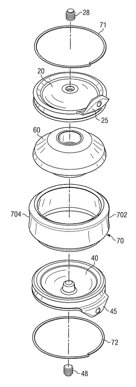
.10 FIG. 2 is an exploded perspective view of an exemplary embodiment of an
intervertebral endoprosthesis.
FIG. 3 is a sectional view of the inteivertebral endoprosthesis shown in FIG.
2.
FIG. 4 is a perspective drawing of the intervertebral endoprosthesis shown in
FIG.
2, assembled as a unitary structure.
FIG. 5 is an elevational view of the intervertebral endoprosthesis shown in
FIG. 2.
FIG. 6 is a plan view of an implant plug and plug installation tool used to
insert a
plug into an intervertebral endoprosthesis.
FIG. 7 is a sectional view of the intervertebral endoprosthesis shown in FIG.
2, as
implanted between two vertebrae.
The disclosure can be more clearly understood by reference to some of its
specific
embodiments, described in detail below, which description is not intended to
limit the
scope of the claims in any way.
DETAILED DESCRIPTION OF SPECIFIC EMBODIMENTS
Composite structures as described herein can be made for use in prosthetic
devices
such as implants. The composite structures described herein provide
flexibility and
resistance. In an implant formed at least in part with a composite structure
as described
herein and inserted at a joint, the flexible aspect of the composite structure
provides for a
range of motion at the site of the implant's insertion. The resistant aspect
of the composite
structure provides for restriction of such motion to a desired range, as well
as increased
durability of that part of the implant formed with the composite structure.
Referring now to FIG. 1, an example of a composite structure 1 as described
herein
is illustrated. Composite structure 1 is illustrated in FIG. 1 as a tubular-
shaped structure
CA 02605474 2007-10-19
WO 2006/113771 PCT/US2006/014668
4
merely for convenience with respect to an exemplary embodiment, which is
illustrated in
FIG. 2, of an implant incorporating a composite structure as described herein.
Those of
ordinary skill in the art will recognize that a composite structure as
described herein can be
formed as a sheet, or in any other shape. It is understood that shapes other
than tubular
can be suitable for use in the manufacture of an implant, and that structure I
can be
extruded or formed in other such suitable shapes.
Composite structure 1 includes a inner flexible layer 1000, a mesh layer 1002,
and
a outer flexible layer 1003. Inner flexible layer 1000 comprises a flexible
material.
According to one example, the flexible material comprises a biocompatible
elastomeric polymeric material, such as segmented polyurethane or
polyethylene. Other
examples of suitable flexible materials include polyurethanes, such as poly
carbonates and
polyethers, polyurethane-containing elastomeric copolymers, such as
polycarbonate-
polyurethane elastomeric copolymers and polyether-polyurethane elastomeric
copolymers.
In certain examples, polyurethanes generally having a durometer hardness
ranging
from about 80A to about 65D (based upon raw, unmolded resin) are used. In
still other
examples, suitable flexible materials include materials commercially lcnown as
BIOSPAN-
S (aromatic polyetherurethaneurea with surface modified end groups, Polymer
Technology Group), CHRONOFLEX AR/LT (aromatic polycarbonate polyurethane with
low-tack properties, CardioTech International), CHRONOTHANE B (aromatic
polyether
polyurethane, CardioTech International), CARBOTHANE PC (aliphatic
polycarbonate
polyurethane, Thermedics). In still other examples, the flexible material
comprises
silicone.
Inner flexible layer 1000 can be manufactured according to known methods.
According to some examples, inner flexible layer 1000 can be extruded through
a suigle
screw extruder, twin screw extruder, cross-head extruder, or other extrusion
and die
assembly. According to other examples, inner flexible layer 1000 can be molded
by
dipping a mold or a mandrel into a curable solution of the flexible material.
The inner
flexible layer 1000 cures in the shape of the mold. Extruding, dipping and
molding
procedures are known to those of ordinary skill in the art.
In the exemplary embodiment illustrated in FIG. 1, a mesh layer 1002 is
attached
to an exterior surface of the iuuier flexible layer 1000. With a tubular
shaped-inner flexible
layer such as inner flexible layer 1000, the inner flexible layer 1000 can be
inserted into a
tubular shaped mesh layer such as mesh layer 1002 illustrated in FIG. 1.
According to
CA 02605474 2007-10-19
WO 2006/113771 PCT/US2006/014668
some such examples, the inner flexible layer 1000 can be extruded into the
mesh layer
1002. According to other examples, the mesh layer can be a sheet that is
wrapped around
a tubular-shaped inner flexible layer or a sheet of inner flexible layer.
Other methods
lcnown to those of ordinary slcill in the art for attaching a mesh layer 1002
to an exterior
5 surface of a inner flexible layer 1000 are suitable. According to still
other examples, a
mesh layer 1002 is attached to an interior surface of the inner flexible layer
1000, or to
both an interior surface and an exterior surface of the inner flexible layer
1000.
Mesh layer 1002 comprises a resistant material. The resistant material
selected for
use in the mesh layer 1002 will be a tear-resistant material, and the mesh
layer 1002 will
be more resistant to flexion, extension, rotation and translation than the
flexible material
comprising the inner flexible layer 1000 and outer flexible layer 1003.
According to one
example, the resistant material comprises polytetrafluorethylene (PTFE)
fibers. According
to one such example, the mesh layer 1002 is formed from PTFE fibers
commercially
available from W.L. Gore & Associates under the tradename GORTEXTM. According
to
other examples, the resistant material comprises polyester fibers. In one such
example, the
polyester fibers are made from a condensation polymer obtained from ethylene
glycol and
terephthalic acid, and commercially available from INVISTA, a subsidiary of
DuPont,
under the tradename DACRONTM. According to still other examples, a mesh layer
1002 is
prepared from polyamide fibers or polyethylene fibers. Other materials having
resistant
properties as described herein are also suitable.
Mesh layer 1002 can be prepared in a tubular shape, a sheet, or any of a
variety of
shapes and sizes, according to methods known to those of ordinaiy skill in the
art.
Exemplaty methods for preparing a mesh layer 1002 include weaving and
knitting.
Suitable weaving methods include but are not linlited to those utilizing a
shuttle
loom, Jacquard loom or Gripper loom, each of which are known to those of
ordinary skill
in the art. A suitable weave for the mesh layer 1002 can be any of a variety
of weaves,
including but not linlited to a plain weave, a twill weave, a satin weave, or
a leno weave.
Suitable knitting methods include but are not limited to weft knitting and
warp
knitting, each of which is known to those of ordinary skill in the art. Still
other suitable
methods for preparing a mesh layer 1002 include a combination of any weaving
method
with any lrnitting method.
Referring still to the exemplary embodiment illustrated in FIG. 1, a outer
flexible
layer 1003 is deposited onto or extruded onto the mesh-covered inner flexible
layer. Outer
CA 02605474 2007-10-19
WO 2006/113771 PCT/US2006/014668
6
flexible layer 1003 coniprises a flexible material such as that described
above with respect
to inner flexible layer 1000, The flexible material used to form outer
flexible layer 1003
can be the same as the flexible material used to form inner flexible layer
1000, or it can be
a different flexible material. According to one example, the outer flexible
layer 1003 can
be extruded onto the mesh-covered inner flexible layer. Alternatively, the
outer flexible
layer 1003 is deposited on the mesh-covered inner flexible layer by dipping
the mesh-
covered inner flexible layer into a solution of the flexible material and
allowing the
resulting composite structure 1 to cure.
The mesh layer 1002 embedded between the inner flexible layer 1000 and the
outer
flexible layer 1003 comprise a composite structure 1 that can be used as made,
or can be
cut or otherwise sized for a variety of uses, including forming an implant as
described
herein with respect to FIG. 2. The implants descr-ibed herein include a
component made
from a composite structure such as that described in FIG. 1. The composite
structure
provides that component of the implant with the ability to be flexible, but
also to be
resistant. The flexibility provided by such component allows for a range of
niotion at the
site of implantation. The resistant property provided by such component acts
to restrict
such range of inotion to a desired amount. By incorporating a resistant
material into an
otherwise flexible component of the implant, such component becomes a
functional part of
the implant that restricts a range of allowed motion.
Implants as described herein can be used as a prosthetic implant in a wide
variety
of joints, including hips, knees, shoulders, etc. The description below
focuses on an
exemplary embodiment wherein the implant is a spinal disc endoprosthesis, but
sirnilar
principles apply to adapt the implant for use in otherjoints. Those of skill
in the art will
readily appreciate that the particulars of the internal geometry will likely
require
modification from the description below to prepare an implant for use in other
joints.
However, the concept of using a composite structure to form a functional part
of
the implant in order to provide control of motion at the implantation site is
applicable to
use in any joint implant.
In broad aspect, the size and shape of the implant are substantially variable,
and
this variation will depend upon the joint geometry. Moreover, implants of a
particular
shape can be produced in a range of sizes, so that a surgeon can select the
appropriate size
prior to or during surgery, depending upon his assessment of the joint
geometry of the
CA 02605474 2007-10-19
WO 2006/113771 PCT/US2006/014668
7
patient, typically made by assessing the joint using CT, MRI, fluoroscopy, or
other
iniaging techniques.
Referring now to FIGs, 2 and 3, an exemplary embodiment of an implant that
includes a component made from a composite structure such as that described in
FIG. 1 is
illustrated. According to the exemplary embodiment illustrated in FIGs. 2 and
3, an
implant comprises a first shell 20, a second she]140, a central body 60, and a
sheath 70.
As will be discussed further herein, sheath 70 is made from a composite
structure
comprising a flexible material and a resistant material.
Shells 20, 40 include outer convex surfaces 23, 43, and inner concave surfaces
21,
41. Outer convex surfaces 23, 43 are rough, in order to restrict motion of the
shells
relative to the bone surfaces that are in contact with the shells.
According to certain examples, the outer surfaces 23, 43 are coated with a
biocompatible porous coating 22, 42. In certain examples, coating 22, 42
comprises a
nonspherical sintered bead coating, while in other examples, coating 22, 42
comprises any
coating that will promote bony ingrowth. A coating formed from nonspherical
sintered
beads provides for high friction between the outer surface of the shell and
the bone, as
well as providing an interaction with the cancellous bone of the joint,
increasing the
chances of bony ingrowth. One example of a suitable nonspherical sintered bead
coating
is that made of pure titanium, such as ASTM F-67. The coating can be formed by
vacuum
sintering.
At least a portion of the inner surface of each shell is smooth, and of a
shape that
complements and articulates with the shape of at least a portion of the
central body. The
inner surfaces of the shells are adapted to slide easily with low friction
across a portion of
the outer surface of the central body disposed between the shells. Desirably,
the inner
surfaces have an average roughness of about 1 to about 8 microinches, more
particularly
less than about 3 microinches. The central body has a shape that cooperates
with the shape
of the inner surface of the shell so as to provide motion similar to that
provided by a
healthy joint.
In certain examples, the shells, 20, 40 further include a number of geometric
features that, as described in further detail below, cooperate with other
components of the
implant. Specifically, these features include a central retaining post 27, 47,
an outer
circumferential groove 82, 84, and radial stop 86, 88. The central retaining
post 27, 47
extends axially from inner surfaces 21, 41. In addition, each she1120, 40
includes an edge
CA 02605474 2007-10-19
WO 2006/113771 PCT/US2006/014668
8
73, 74, respectively. The outer circumferential grooves 82, 84 extend into the
edges 73,
74 of the shells 20, 40. The radial stops 86, 88 extend from the edge 73, 74
in a direction
generally perpendicular to the general plane of the shells 20, 40.
Radial stops 86, 88 and retaining posts 27, 47 help prevent the central body
from
being expelled from between the opposing shells when the shells are at maximum
range of
motion in flexion/extension. The hole receiving the post can have a diameter
sufficiently
large that relative motion between the shells and central body is
unconstrained within the
allowable range of motion, but that will nevertheless cause the post to arrest
the central
body before it is expelled from the implant under extreme compression.
Alternatively, the
diameter of the post may be such that it limits the translational movement of
the central
body during normal motion of the spine by contacting the surface of the hole
in the central
body at the limit of the allowable range of motion for the device.
Each shell may also be provided with tabs 25, 45. Tabs 25, 45 are optional
features, but if present, extend from a portion of the edge 73, 74 in a
direction generally
perpendicular to the general plane of the shells 20, 40, and generally
opposite the radial
stops 86, 88. If present, tabs 25, 45 help to prevent long-term migration
within the disc
space, as well as catastrophic posterior expulsion, and the resulting damage
to the spinal
cord, other nerves, or vascular structures. Tabs 25, 45 may contain openings
26, 46 that
can releasably engage an insertion tool (not shown).
The shells 20, 40, may be identical, or may be of different design (shape,
size,
and/or materials) to achieve different mechanical results. For example,
differing plate or
shell sizes may be used to more closely tailor the implant to a patient's
anatomy, or to shift
the center of rotation in the cephalad or caudal direction.
The shells can be made from any suitable biocompatible material. According to
certain examples, the shells are made from a titanium alloy. In some such
examples, the
titanium alloy is ASTM F-136. In certain other examples, the shells are made
of a
biocompatible metal, such as stainless steel, cobalt chrome, or ceramics, such
as those
including A1203 or Zr203.
Central body 60 comprises a convex upper contact surface 94, a convex lower
contact surface 96, and a central axial opening 98. In certain examples,
central body
member 60 includes an upper shoulder 92 and a lower shoulder 90. Each shoulder
90, 92
consists of an indentation in the surface of the central body member which
defines a ledge
that extends around the circumference of the central body 60. Shoulders 90, 92
can be
CA 02605474 2007-10-19
WO 2006/113771 PCT/US2006/014668
9
used to constrain motion of the central body, and to provide a buffer that
prevents contact
between the shells. Preventing contact between the shells prevents friction
and wear
between the shells, thereby avoiding the production of particulates, which
could cause
increased wear on the internal surfaces of the implant.
The central body 60 is both deformable and resilient, and is composed of a
material
that has surface regions that are harder than the interior region. This allows
the central
body to be sufficiently deformable and resilient such that the implant
functions effectively
to provide resistance to compression and to provide dampening, while still
providing
adequate surface durability and wear resistance. In addition, the material of
the central
body has surfaces that are lubricious, in order to decrease friction between
the central body
aiid the opposing shells.
The material used to make the central body 60 is typically a slightly
elastomeric
biocompatible polymeric material. Examples of suitable polymeric materials
include
polyurethanes, such as poly carbonates and polyethers, polyurethane-containing
elastomeric copolymers, such as polycarbonate-polyurethane elastomeric
copolymers and
polyether-polyurethane elastomeric copolymers. In certain examples,
polyurethanes
generally having a durometer hardness ranging from about 80A to about 65D
(based upon
raw, unmolded resin) are used.
In other examples, suitable polyurethanes include polycarbonates and
polyethers,
such as Chronothane P 75A or P 55D (P-eth-PU aromatic, CT Biomaterials);
Chronoflex
C 55D, C 65D, C 80A, or C 93A (PC-PU aromatic, CT Biomaterials); Elast-Eon II
80A
(Si-PU aromatic, Elastomedic); Bionate 55D/S or 80A-80A/S (PC-PU aromatic with
S-
SME, PTG); CarboSil-10 90A (PC-Si-PU aromatic, PTG); Tecothane TT-1055D or TT-
1065D (P-eth-PU aromatic, Thermedics); Tecoflex EG-93A (P-eth-PU aliphatic,
Thermedics); and Carbothane PC 3585A or PC 3555D (PC-PU aliphatic,
Thermedics).
The material used to make the central body may be coated or impregnated to
increase surface hardness, or lubricity, or both. Coating of the material used
to form the
central body may be done by any suitable technique, such as dip coating, and
the coating
solution may include one or more polymers, including those described above for
the
central body. The coating polymer may be the same as or different from the
polymer used
to form the central body, and may have a different durometer hardness from
that used in
the central body. Typical coating thickness is greater than about 1 mil, more
particularly
from about 2 mil to about 5 mil.
CA 02605474 2007-10-19
WO 2006/113771 PCT/US2006/014668
The central body 60 may also vary somewhat in shape, size, composition, and
physical properties, depending upon the particular joint for which the implant
is intended.
The shape of the central body should complement that of the inner surface of
the shell to
allow for a range of translational, flexural, extensional, and rotational
motion, and lateral
5 bending appropriate to the particularjoint being replaced.
Sheath 70 is made from a composite structure comprising a flexible material
and a
resistant material as described above with respect to FIG. 1. In certain
examples, a
tubular-sliaped composite structure 1 as illustrated in FIG. 1 is prepared,
and one more
sheaths 70 are cut from the composite structure. The sheath can be cut so as
to be of an
10 approximately even height on the anterior and posterior sides 702, 704, or
can be cut so as
to have a trapezoidal configuration, where one side, for example the anterior
side of the
sheath 702, is greater height than the posterior side 704.
In certain examples, the thickness of the sheath is in the range of from about
5 to
about 30 mils, and in other examples, about 10-11 mils. The inner flexible
layer, mesh
layer, and outer flexible layer can have the same thiclaress, or different
thicknesses. In
certain examples, the mesh layer will be thinner than the inner flexible layer
and the outer
flexible layer.
The resistant material in the composite structure forming the sheath is more
resistant to flexion, extension, rotation and translation than the flexible
material in the
composite structure forming the sheath. Thus, using a composite structure as
described
herein to form the sheath 70 provides the sheath 70 with the ability to allow
motion
between the central body 60 and the shells 20, 40, and thereby allow motion at
the implant
site, but also to limit the range of motion allowed. Limiting the range of
motion can
include resisting at least one predetermined type of relative directional
motion, for
example, at least one of flexion, extension, rotation or translation in at
least one of the left,
right, anterior or posterior direction,
Attachment of the sheath 70 to the shells 20, 40 can be accomplished in a
variety
of ways. According to one example, attachment of the sheath 70 to the shells
20, 40
comprises providing the edge of each shell with a circumferential groove (the
term
"circumferential" in this context does not imply any particular geometry).
The sheath 70 can be disposed so that the edges of the sheath 70 overlap the
outer
circumferential grooves 82, 84 of the shells 20, 40. Retaining rings 71, 72
are then placed
over the edges of the sheath 70 and into the circumferential grooves 82, 84,
thereby
CA 02605474 2007-10-19
WO 2006/113771 PCT/US2006/014668
11
holding the flexible sheath in place and attaching it to the shells. The
retaining ring can be
formed by wrapping a wire around the groove over the overlapping portion of
the sheath,
cutting the wire to the appropriate size, and welding the ends of the wire to
form a ring.
While any suitable biocompatible material can be used for the retaining rings,
stainless steel, titanium or titanium alloys are particularly suitable. The
retaining rings are
desirably fixed in place by, e.g., welding the areas of overlap between the
ends of the
retaining rings. Because of the high temperatures needed to weld titanium and
titanium
alloys, and because of the proximity of the weld area to both the sheath 70
and the central
body 60, laser welding is typically used.
Other components of the implant, for example the central body 60, and shells
20,
40, can provide features that contribute to the limitation of motion. As
discussed above,
radial stops on the shells and shoulders on the central body can be used to
constrain
motion. For example, contact of the walls or extensions 86, 88 of the shells
with shoulders
90, 92 of the central body may also contribute to limiting the range of motion
to that
desired. The central retaining posts 27, 47 may also contribute to limiting
the range of
motion by contact with the central axial opening of the central body.
In some examples, limitation of motion provided by the shells and/or the
central
body can be in addition to the limitation of motion provided by the sheath. In
other
examples, such function of the shells and/or the central body can be a
replacement for the
limitation of motion provided by the sheath, for example, when the sheath is
at a
maximum range of motion that it can resist, features of the shells and/or
central body can
take over at such range. In still other examples, such function of the shells
and/or central
body can provide for limitation of motion in a direction other than that
provided by the
sheath.
Thus, in certain examples, the kinematics of the motion provided by the
implant
are defined primarily by the sheath, the central body 60, and the shells 20,
40. Although
the central body is encapsulated within the sheath and the shells, it is not
attached to these
components. Accordingly, the central body 60 freely moves within the enclosed
structure
provided by the sheath 70 and shells 20, 40, but is constrained by limitations
imposed by
the sheath 70, and, if used, geometric limitations imposed by interaction
between the
shells and the central body.
An example of a geometry of the sheath, shells and central body that limits
the
motion of the central body is illustrated in FIG. 3. In certain examples, when
the sheath
CA 02605474 2007-10-19
WO 2006/113771 PCT/US2006/014668
12
has reached the maximum range of motion it can constrain, other features of
the implant,
such as the shells and the central body, can provide further or additional
restraint.
For example, extensions 86, 88 on shells 20, 40 can contact shoulders 90, 92
on the
central body 60. Specifically, the inner portion of the extension forms a
circumferential
ridge that limits the range of motion of the shells 20, 40 relative to the
central body 60 by
contacting central body shoulders 90, 92. This limitation of motion can occur
during or
subsequent to the limitation of motion provided by the sheath.
As explained above, in one embodiment, the shells are concavo-convex, and
their
inner surfaces mated and articulated with a convex outer surface of the
central body. The
sheath is secured to the rims of the shells with retaining rings, and which,
together with the
inner surfaces of the shells, forms an implant cavity. In a particular aspect
of this
embodiment, using a coordinate system wherein the geometrical center of the
implant is
located at the origin, and assigning the x-axis to the anterior (positive) and
poster-ior
(negative) aspect of the implant, the y-axis to the right (positive) and left
(negative) aspect
of the implant, and the z-axis to the cephalad (positive) and caudal
(negative) aspects of
the implant, the convex portion of the outer surface and the concave portion
of the inner
surface of the shells can be described as quadric surfaces, such that x2/a''
+Y2/b2 + z2/c2 =
1, where (+/-a,0,0), (0,+/-b,0), and (0,0,+/-c) represent the x, y, and z
intercepts of the
surfaces, respectively. Typical magnitudes for a, b, and c are about 11 nlni,
30 mm, and 10
mm, respectively.
The implant is symmetrical about the x-y plane, and is intended to be
implanted in
the right-left center of the disc space, but may or may not be centered in the
anterior-
posterior direction. In any event, the implant is not allowed to protrude in
the posterior
direction past the posterior margin of the vertebral body.
In the coordinate system described above, the central axis of retaining post
27, 47
is typically coincident with the z-axis, but may move slightly to accommodate
various
clinical scenarios. The shape of the post may be any quadric surface. However,
a truncated
tapered elliptical cone is a particularly suitable geometry. Similarly, the
geometry of the
central axial opening of the central body will correspond to the geometry of
the retaining
post, and will have a similar geometry.
The central body contains surfaces that are described by an equation similar
to that
for the inner surfaces of the shells, and which articulates with those inner
surfaces. The
central body will have a plane of symmetry if identical opposing shells are
used.
CA 02605474 2007-10-19
WO 2006/113771 PCT/US2006/014668
13
The complete assembly of the exemplary implant illustrated in FIG. 2 is
illustrated
in FIGS. 4 and 5, wherein the central body 60 is bracketed between shells 20,
40. The
flexible sheath 70 extends between the two opposing shells 20, 40, and
encapsulates the
central body 60 such that the implant is a unitary structure. FIG. 7
illustrates the implant
inserted as a unitary structure between two vertebrae.
According to certain embodiments, means for accessing the interior of the
implant
after it has been assembled into a unitary structure are provided. This means
consists of a
central axial opening included in the shells 20, 40. Typically, this opening
will be
provided through central retaining posts 27, 47. By providing access to the
interior of the
implant, sterilization can be done just prior to implantation. Sterilization
is preferably
accomplished by introducing an ethylene oxide surface sterilant. Caution
should be
exercised in using irradiation sterilization, as this can result in
degradation of the
polymeric materials in the sheath or central body, particularly if these
include
polyurethanes.
After sterilization, the central openings can be sealed using plugs 28, 48.
Preferably, only one plug is inserted first. The plug is inserted using
insertion tool 100,
shown in FIG. 5, and which contains handle 101 and detachable integral plug
28, 48. The
tool is designed so that plug 28, 48 detaches from the tool when a
predetermined torque
has been reached during insertion of the plug. The tool can then be discarded.
After one plug has been inserted to one of the shells, a lubricant 80 is
preferably
introduced into the interior of the device prior to inserting the second plug.
To do this a
syringe is used to introduce the lubricant into the remaining central opening,
and the
implant is slightly compressed to remove some of the excess air. Another
insertion tool
100 is then used to insert a plug into that central opening, and thereby
completely seal the
interior of the device from its exterior environment. In certain examples, the
lubricant 80
is saline. In other examples, other lubricants may be used, for example,
hyaluronic acid,
mineral oil, and the lilce.
Where the implant is used as an endoprosthesis inserted between two adjacent
vertebral bodies, the iinplant may be introduced using a posterior or anterior
approach.
For cervical implantation, an anterior approach is preferred. The implanting
procedure is
caxried out after discectomy, as an alternative to spinal fusion. The
appropriate size of the
implant for a particular patient, determination of the appropriate location of
the implant in
the intervertebral space, and implantation are all desirably accomplished
using precision
CA 02605474 2007-10-19
WO 2006/113771 PCT/US2006/014668
14
stereotactic teclmiques, apparatus, and procedures, such as the techniques and
procedures
known to those of ordinary skill in the art. Non-stereotactic techniques can
also be used.
In either case, discectomy is used to remove degenerated, diseased disc
material and to
provide access to the intervertebral space sufficient to prepare the surfaces
of the vertebral
bodies for insertion of the implant. To prepare the vertebral bodies, a
cutting or milling
device is used to shape the endplates of the vertebral bodies to complement
the outer
surfaces of the implant and to expose cancellous bone.
For example, after gaining access to the intervertebral space, a portion of
the
vertebral body can be removed using a burr or other appropriate instruments,
in order to
provide access to the intervertebral space for a transverse milling device.
Transverse
milling devices, and use and acquisition thereof, are lcnown to those of
ordinary skill in the
art. The milling device is used to mill the surfaces of the superior and
inferior vertebral
bodies that partially defme the intervertebral space to create an insertion
cavity having
surfaces that (a) complenlent the outer surfaces of the implant and (b)
contain exposed
cancellous bone.
This provides for an appropriate fit of the inlplant with limited motion
during the
acute phase of implantation, thereby limiting the opportunity for fibrous
tissue formation,
and increases the likelihood for bony ingrowth, thereby increasing long-term
stability.
The relative thicknesses of the inner flexible layer, mesh, and outer flexible
layers are
shown only for the purpose of example, it being understood that these
thicknesses can be
varied within the scope of the invention. In addition, more or less layers
than those
illustrated herein can be used to make a composite structure according to the
present
disclosure.
Spatial references, such as "under", "over", "between", "outer", "inner" and
"surrounding" are for the purpose of illustration only and do not limit the
specific
orientation or location of the layers described above.
The invention has been described above with respect to certain specific
embodiments thereof. Those of skill in the art will understand that variations
from these
specific embodiments that ate within the spirit of the invention will fall
within the scope of
the appended claims and equivalents thereto.