Note: Descriptions are shown in the official language in which they were submitted.
CA 02605531 2007-11-07
WO 2006/119572 PCT/AU2006/000620
Title of the Irlvention
Ultrasound Diagnosis and Treatment Apparatus
Field of the Invention
This invention relates to apparatus for the generation of sound waves, in
particular to apparatus for transcranial Doppler sound devices for medical
treatment of patients suffering from blood vessel occfusiorts or restriction
in
the brain Gharacteristic of a strdke.
Background
Blood circulation through the body is essential for maintenance and growth of
celfs and tissues. Any conditiorf that restricts blood flow can have mild tv
disastrc-us consequences. For example, when blood flow in the brain is
impeded, stroke Can result. Stroke is a medicai affliction that nas severe
consequences for rnost people who suffer it, Stroke is classified into four
types, two of which are caused by bloc-d clots (ischa.emic stroke) and two of
which ara caused by haemorrhage (haemorrhagic stroke). Cerebral
thrombosis and cerebral embolism account for up to 80 per cent of af1 strokes.
Treatment options for strQke are limited. For example, only tissuo
ptasminogen activator (tPA) has been appfoved by the United States Food
and Drug Administration as a pharmaceutical therapeutic treatment for
ischaemic stroke.
It has been shown that the use of uftrasound waves and the Doppier
frequency shift can be used to monitor the flow of b1Qod through vessels (eg.
't"egeler and Ratanakorn, 1999). Apparatus have been devefoped to expioit
the potential of ultrasound to locate the interface between tissue types in
the
body, in particular, in the head, using transcranial Doppler uitrasound
technology (TCD). US patent no. 4,817,621 described apparatus to locate
reilab4y blood vessels in the brain and to determine blood flow through
vessels
in the head using TCD. The apparatus relied on the combination of two
parallelogram-like iinkage systems to support and locate an ultrasound
CA 02605531 2007-11-07
WO 2006/119572 PCT/AU2006/000620
2
transducer near the head of a patient to locate occluded blood vessels in the
brain using TGD. More recently, it has been shown that monitoring of patients
with TCQ, in addition to treatment with tPA may increase the effectiveness of
tPA in the treatment of ischaemic stroke (Alexandrov et al. 2004), using '
commonly avaiia.ble TCD devices and operators skilled in using the devices to
locate occlusions.
Transcranial Doppler technoiagy has been shown to be useful in the
identification and treatment of small vessei knock (WO2004/1 a$y 84)
assoaiated with small vessei occlusion lead'irig to stroke. The treatment
taught in W02004/1 aa184 requires significant effort by an operator to find
and
diagnose the occlucied blood vessels characteristic of stroke. While it has
been shown that currently avaiiable ultrasound transducers and systems may
be used for monitoring occluded blood vessels in stroke might also be a
beneficial treatment method alleviating the symptoms of stroke, the ability to
use TCD as a therapeutic treatment is currently constrained by the ease of
use of said currently available systems. Clinicians who have used currently
available TCD systems have noted that vascular tests that require the use of
said TCD systems are among the most difficult to perform (Alexandrov et al.,
2004). The ability of clinicians to diagnose and treat stroke with the
promising
TCD ultrasound technology may be limited by the apparatus with which to
diagnose and treat the condition. For example, the current method of
identifying the presence of occlusions in brain blood vessels is a rnanual
grading system, known as the thrornbolysis in brain ischaemia (TIBI) flow
grading system. One of the prcblems with a head cap or band m unted or
any other body or head mounted automatic diagnostic or treatment uitrasound
device is that stability of movement of the sensor with patient movement or
simply device attachment slippage can affect measures and data integrity.
What is needed is an apparatus and method to more efficientfy locate
occluded blood vesseis or vessels having restriction in the brain
characterisiic
of stroke and to treat the occlusions or restrictions to alleviate the stroke
symptoms.
CA 02605531 2007-11-07
WO 2006/119572 PCT/AU2006/000620
3
}n this document the words "inciuding" and "Gomprising" are used
interchangeably and with the same meaning, which is intended to indicate
non-limiting incorporation of elements.
Brief Description of the FigUres
Figure 1 shows an embodiment of an ultrasound transducer useful in the
invention.
Figure 2a shows a table of data providing near-field lengths and far-field
divergence of ultrasound waves generated by typicsl ultrascaund transducers.
Figure 2b shows a table of data showing the variation in veiocity of
ultrasound
waves through selected materials found in iiving organisms.
Figure 3a shows an embodiment of multiple ultrasound transducers focussed
electronically.
Figure 3b shows an embodiment of a concave lens being used to focus a
linear array of ultrasound transducers.
Figure 4 shows embodiments of arrays of tra.nsducers, 'inoiuding curved and
iinear arrays.
Figure 5 shows embodiments of phased arrays of transducers which can be
steered and focussed efectrunicafly.
Figure 6 shows an embodiment of the invention a.s an adjusta.bfe servo-array.
of ultrasound transducers.
Figure 7 shows an embodiment of invention in a sequence of operation of a
phased array of ultrasound transducers.
Figure 8a shows an exampie of eiectric focussing of an ultrasound transducer.
Figure 8b shows an example of a phased array of uitrasound transducers.
Figure 9a shows an example of the pulse rate of an ultrasound transducer.
Figure 9b shows and example of depth resoiution of an ultrasound transducer,
Sumrnary of the Inrrention
in one aspect the invention provides apparatus for imaging or treatment-of
restrictions or occlusions in vessels using sound waves, comprising at least
one sound transducer member including at ieast one sound-emitting element
for producing at ieast one sound-wave beam; means to adjust the parameters
of said at least one sound-wave beam; means to spatially locate said at leasx
CA 02605531 2007-11-07
WO 2006/119572 PCT/AU2006/000620
4
one sound-emitting element; means to automatically or semi-automatically
focus sound waves generated by said at least one sound-emitting element
into a k,eam; and means to accept sound signals from sound-emitting element
or elements. Preferably the apparatus includes means to move said at least
one transducer member and means to control movament of said at least one
transducer member automatically or semi-autcmatically. Preferably the
sound-emitting eiement and the means to accept sound signals are the same.
Preferably the at least one sound-wave beam is pulsed. Preferably the at
least one sound-wave beam is fQdussed electronically, The invention may
include two or more sound-emitting elements forming an array and the array
may be curved. The at least one sound-wave=beam may incorporate a
plurality of frequencies of sound waves in combinations of concurrent
frequencies generated by the sound-emitting elements in the array or in a
series of frequencies over time. The array may be comprised of sound-
emitting elements in any of fixed position, adjustable position, or scanning
position. The apparatus may include fiducial registration means and
communication mean for communicating the position of the at least one
sound-emitting element and maintaining the at lea$t one sound-emitting
element in optimal positioning during sonication.
In another aspect, the invention provides apparatus for imaging or treatment
of restrictions or occlusions in vessels using sound waves, comprising at
leasfi
one sound-wave transducer member including at least one sound-emitting
element for producing at least one sound-wave beam; means to adjust the
2S parameters of said at least one sound-wave beam; means to orient said at
least one transducer member; means to focus sound waves generated by
said sound-emitting element into a beam;=and means to accept sound signa4s
from sound-emitting aiement or elements. The sound-emitting elements may
be moveable singly or in a coordinated manner, including simultaneously.
30 The sound-emitting elements may be servo-controlled, including feedback
control. The servo-control means may be self-tracking and include means for
determining out-of-range positioning of said at least on sound-emitting
element. The feedback control may Incorporate a signal characteristic of an
occlusion in a fluid flow. The apparatus may include a plurality of sound-
CA 02605531 2007-11-07
WO 2006/119572 PCT/AU2006/000620
emitting elements in at least two layers of at least tw4 arrays. Preferably
each
transducer member is operable to enable a continuously etdjustable focus
point comprising of two or more sound beams emitted by at least two sound-
emitting elements. Preferably the apparatus includes means to transform
5 sound signals from analogue to digital forms or digital to analogue forms.
Preferably the apparatus includes means to store transformed digitai data.
preferabty the apparatus includes means to display analogue or digital data.
Preferably the apparatus irrcludes video display rneans for displaying data.
The apparatus may include voice coil control means. Preferably the
apparatus is operable in real-time or near real time. Preferably the apparatus
includes fiducial regi$tration means for maintaining targeted sonlcation.
Preferably the apparatus is used for detecting and= sonicating vessels in the
brain of a being. Preferably the sound waves are uftrasound waves,
1_5 In another aspect, the inventirrn provides a method for locating art
occiWsion or
restriction in a=vessel, including the steps of identifying regions of a body
in
which an ocolusion or restriction in a vessel might be fou.nd; selecting a
region
of interest for sanication; sonicating the region of interest with at least
one
sound-wave beam by moving said at least one sound-wave beam across the
region of interest; receiving reflected sound signals from the region of
interest;
arid calculating the poppler effect parameters of flow and turbulence from
safd
reflected sound signals.
In another aspect, the invention provides a method for distinguishing
anatomioal features of an organism inoluding the steps of sonicating a region
of interest in a subjeut with at least one sound-wave beam, whereby the
frequency of said at least one sound-wave beam is suitable for determining a
particular tissue type, receiving reflected sound signals from said region of
interest, ca.lculating the Doppler effect parameters of said reflected sound
signals and characterising said Doppler effect parameters according to
parameters associated with known tissue types.
The method of the invention may include the step of modifying the
characteristics of the at least one sound-wave beam to target a region of
CA 02605531 2007-11-07
WO 2006/119572 PCT/AU2006/000620
6
interest wherein the Doppler effect parameters are indicative of reduced flow
attributable to an occlusion or a restriction. The method may inciude the stop
of sonicating the region of interest thereby causing agitation or dissipation
of
the occlusion by prolonged sonication or recanalisation of a restriction. The
method may include the step of automatically or semi-autorriatically
evaiuating
and optimising the effect of sonication on an occlusion for feedback
modification of said at least one sound-wave beam, said evaluating and
optimising including tissue safety guidelines, The method may include that
the step of automatically or serni-a.utomatically evaluating and optimising
the
.10 affect of sonication includes maintaining a fiducial registration
'befinreen sound
waves beams and a registration signal. The method may include the iatep of
calculating and displaying any one or a combination .of an index, measure, or.
rrmarker or suitabie representation indioating the progress of dissipation of
an
occlusion in a vessel or recanalisation of a vessel having an occlusion or
restriction. The method may be carried out automatically or semi-
automaticaliy substantialiy without human contrcal. The method may be
carried out wherein the region of the body is the head. Preferably the region
of the head is the circle of Willis. The method may include the two or more
sound-wave beams moving across said region of interest in either a
simultanaous or sequentiai manner. The method may irticiude having the at
ieast one sound-wave beam pulsed. The method may include the parameters
calculated from saici reflected sound signals being any one or a combination
of power, spectral, amplitude, phase coupling or frequency characteristics
characteristic of a spatial representation of anatomical features in said
region
of interest or occlusive material. Preferably the power or ampiitude spectral
ana.lysis are carried out using Fast Fourier Transform techniques. Preferably
the at least one sound-wave beam is continuous. The method may include
the at least one sound-wave beam being initially transmitted with a first
frequency or amplitude and subsequently with periodic changes resulting a
second and further frequency or frequencies or amplitude(s) reiative to the
first beam frequency so that the mark-to-space ratio of the apparent changing
and pulse formation is continuous. Preferably the sound-wave beam is
comprised of ultrasound waves. The method may include the stop of
conducting a spatial voxela.ted analysis of rer~ived signals,
CA 02605531 2007-11-07
WO 2006/119572 PCT/AU2006/000620
7
Detaited Description of the Drawings and Preferred Embodiments
!t is an object of the present invention to provide an apparatus that reduces
the need for significant operator interaction with a sound-wave generating
device used in fih$ identification and treatment of embolism or stenosis. It
is a
further object of the invention to provide a method of semi-automatic or
automatic location of embolism or stenosis using sound waves. It is a fUrther
object of the invention to provide an apparatus for the automatic or semi-
automatiG location of blood vessal emboli or stenosis in the brain. It is a
further object of the invention to provide a means to serni-automatica{iy or
automatically locate an occlusio-n or restriction in a blood vessel. It is a
furrther.:
object of the invention to provide a means to serni-automaticafly or
automatically treat said emboli or occlusion or restriction with sound waves.
!t
is a further object of the invention to provide an apparatus to utilise the
energy
of transcranial Doppler ultrasonography in thromobolysis and recarEalisation
of
blood vessels in the brain.
The following description refers to the preferred embodiment of the invention
using ultrasound waves, It wiit be.=understood that saund waves of other-
frequencies than ultrasound are'sUitable for other embodiments within the
scope of the invention. For example, in another embodiment, the invention
includes apparatus that utiilises low-frequency pulsatile sound waves that may
be focussed to achieve similar results as the preferred ultrasaund waves. In
this document, the word, "ocolusion", includes any one of, or a combination
of,
an embolism, thrombus, or other biological matter, non-biological matter,
including gases, from whatever source. In this document, the word,
"stenosis", includes any restriction in a fluid-containing vessel.
An exarnple transducer member 24 comprising a single ultrasound-emitting
element is il{ustrated in Figure 1, the ultrasound-emitting eiernent includes
a
piezoelectric element 21, backing material 22, and electrodes 23. The
Ultrasound-emitting element converts electric voltage applisd across the
ultrasound-emitting element into ultrasonic sound-wave energy. When a
CA 02605531 2007-11-07
WO 2006/119572 PCT/AU2006/000620
8
beam of ultrasonic sound-wave energy is diracted toward heterogeneous
biological material of interest, the ultrasonic sound-wave energy is reflected
at
the interfaces of biological structures within the biological material. The
reflected enargy causes an ultrasound-receiving element to vibrate and to
produce a voltage signal which can be. processed to decipher the reflective
properties of the biological material. it will be understood by persons
skilled in
the art that a single ultrasound element may function either as an ultrasound-
emitting element, an ultrasound receiving element, or both an ultrasound-
emitting and an ultrasound-receiving element. An ultrasound beam produced
by an ultrasound-emitting element may be pulsed or unpulsed in duration. A
,=puls.ed beam is pulsed at a rate required for the biological material of
interest
as illustrated in Figure 9a. Figure 9b shows the time sequence of an
ultrasound beam resolving two surfaces, It will be understood that axial or
depth resolution is the ability to.de#ermine the axial resolution of two
objects
located tandem to the ultrasonic beam. The axial resolution is determined by
:the spatial pulse length.
The transducer diameter can be selected to suit various depth ranges
required for different treatment applications_ Figure 2a shows the properties
=of a range of depths of commercially availabie ultrasound transducers and
Figure 2b shows the. variability of velocity of- ultrasound beams in different
biological materials, which is exploited in the invention. The velocity of the
ultrasound signal depends on the constitution of the material through which
the signal travels, the velocity being directly proportional to the density
through which the ultrasound is transmitted. The transmission through tissue
is 1540 m sec' or, alternatively, a 1 cm transmission depth requires 13 psec
to be traversed by an ultrasound wave.
The ultrasonic beams generated by a transducer can be focussed with
focussing means. Preferably the lateral and depth positioning of the
ultrasonic beam foGus point can be adjusted by way of electronic focussing,
illustrated in Figure 8a. An embodiment of a focussed transducer member 25
with an ultrasound-emitting element, shown in Figure Sa, can provide
improved lateral resolution at depth. Focussing types can include curved
CA 02605531 2007-11-07
WO 2006/119572 PCT/AU2006/000620
9
mirrors, acoustic crystals, acoustic lens, or phased array (employing
electronic
focussing). The operation of uitrasound-emitting elements in a phased array
is shown in Figure 8b.
The ultrasound-emitting elements may be positioned in an array within a
transducer merriber 25 and may take alterna.tive fQrms. Figure 4 shows that
such forms may include curved. arrays 26 and linear arrays 27. The
application of voltage to an array of ultrasoundremitxing elements of a
transducer member may be pulsed out-of-phase to achieve steering and
IO focussing of an ultrasound beam as illustfated in Figure 5. The invention
inciucies that each of said at least one transducer members 25 may be
comprised of any combination of ultrasound-emitting and receiving elements.
Iilustratec3 in Figure 5 is an embodinlent of=the invention, It wiil be
understood
that a particular embodiment will incorporate a seiection of features to
achieve
the objects of the invention and that- the embodiment in Figure 6 is
illustrative
only. A sensor enabling said ultrasonic-beam transmission and/or handling
may be camprised of piezo or PVp material, or other suitable material or a
suitabie sensor capabie of generating and/or receiving ultrasonic beam
signscls, Said at ieast one transducer momber may include a combination of
one or more memaers in fixed-position array, adjustable-position array 1,
scan n ing-position singie-member 2 or multiple-element transducer members,
fixed-position single-member or muitiple-eiement transducer memers,
The focussing point 5 of an ultrasonic beam can be achieved through any
combination of servd-driven control c7f an adjustablerposition array-
transducer
member 3, servo-driven control of the scanning-position single-mernber 2 or
multiple-element transducer member 4, the switching combination of fixed-
array beams 6 or any combination thereof. Preferably the lvcation of
ultrasound transducers is achieved through servo-rr+ovement.
Said ultrasonic beams can be accurately positioned via ultrason9c grid arrays
or reference markers located on a device case or housing 7 or at any point
within said beams as a measure of final beam-pcsitioning feeclback for high
CA 02605531 2007-11-07
WO 2006/119572 PCT/AU2006/000620
resolution and focus accuracy for said converging ultrasonic beams. The
beam-positioning feedbacK enables the servo-control position circuitry 8, in
turn, to reflect the positional requirement for said ultrasonic beams, either
in
accordance with an operator's remote selectian, by way of communication
5 means, or locally selected by-way.of direct communication, input into said
servo-oontrol position circuitry through=manipulation of.u1trasonic beams I
and
focal point 5. Transducer members 4 may be. adjusted so.that the direction of
the ultrasonic beams 1 oan be oriented either two-dimensionally or three--
dimensionally according to the positioning of -at least one =servo-adjust
spigot
10 3. A secondary ultrasound member comprising a single transducer member,
or alternatively at ieast two transducer eiernents, may comprise of either a
singte ultrasonic beam 2 or rnu{ti-array.ultrasorsic beams 4. - Furthermore,
multi-layer, multi-array ultrasor?ic beams may be used where a singular or
group of three-dimensianal (in space) positioning ultrasonic beam focus
capability by way of aperture adjustment 11=,=such as piezo-aperture control.
As stated, the scope of the invention includes alternative embodiments to that
herein il4ustrated and may inciude embadiments wherein the sound
transducers are-arranged in arrays and in multiple layers crf arrays.
20. Embodiments include those where the sound transducers. are arranged in a
single array, the.sound beams generated by a pluratity of transducers in an
array focussed at a single target. The transiJucers in an array may be in
fixed
position or moveable.
The apparatus may include a fiducial registration means (not shown) for
maintaining the optimal positioning of sound-emitting elements for continued
sonication when the subject moves, for example, Preferably said fiduciai
registration means are additional sensors attached to ultrasonic transducers
and transceiver devices attached to a subject. Gommunication between
320 fiducial registration means way be through wire Gannectors or wireless
communication means, Said fiducial registration means includes means for
attachment to the subject and means for communicating between the subject
and the servo-control means S. The control means includes means to
measure the correct registration of signals from the fiducial registration
CA 02605531 2007-11-07
WO 2006/119572 PCT/AU2006/000620
11
means. The fiducial registration means is placed in fixed position at the
commencement of sonication with a suitable adhesive material such as self-
adhesive locators attached to a subject. When the communication between
the fiducial registration rrreans and the control means indicate a- departure
from the initial optimal signal registration; the control systemmay prompt the
operator by way of a prompting means that the sound-emitting means is out of
alignrnent. Said prompting may occur automatically. Such a method and
device enhances the u(trasonic beam targeting method,.where.for example, a
subject uftrastrnic tranducer attachment device,, such as one iocated on
glasses or a headcap, moves -during automatic or remote-cQntrolled
recanalisation or sonioation of the subject.
The invention includes that the va.ria.bility of velocity of ultrasound waves
in
different tissues may be exploited to charactr;rise the-tissues in a region of
interest in a subject where it is suspeGted that there might be an occlusion
or
a restriction in fluid flow, for exarnple, blood flow. 5onica.ting a region of
interest with mu{tipfe frequencies of sound-wave beams, each seiected
frequency being associated with a tissue type as indicated in Figure 2b,
wherein the multiple frequencies are generated by any of at least one sound-
emitting element at spaced intervals or two or more sound-emitting elements
concurrently or in a pre-determined series of frequencies al}QVtis the
characterisation of an occlusion or restriction in fluid flow as being
attributabie
to a cause such as a gas bubble or t7ubbles, solid material, blood, tissue,
vessel, skin, organs or other material.
The invention includes an apparatus having an arrangement of ultrasound
transducers that enables the automatic mapping or v'isuaiisation of the
progress of the dissofution of the target emboius.
The invention includes means capable of serva-fpedback to ultrasonic
turbulence (such as fast Fcurier transformation) representation of the
turbulence associated with blood vessel occlusion. Prefsrably the servo-
feedback is optimised for the most effective vessel recanatisation.
CA 02605531 2007-11-07
WO 2006/119572 PCT/AU2006/000620
12
The invention advantagenusly utilises sound waves produced by an
ultrasound transducer to locate blood vessels that might show emboli$m. One
embodiment of the invention includes a method for identifyirig an embolism or
stenosis. in a first step of said method, regions of the body in which emboli
might be found are identified. Preferabiy the region of the body is the head.
More preferably the region of the body is the circie of Willis in the head. In
a
second step, a particular region of interest is, selected. In a third step
pars:m-eters of flow and turbulence ara calculated=for subsequent automatic
.=ultrasound beam localisation in a fourth step, said parameters including the
= 10 spectral power or amplitude or phase.Goupfing or.frequQncy segrnented
characteristics of flow and turbulence of flow blood.
in said first step of identifying regions of-the body 9n wh.ich emboli might
be
found, at least one ultrasound transducer generates an ultrasound beam
15, which is moved across the.surface.of,the regio.n of the body in a scanning
motion. Sald ultrasound transducer or transducers may be fixed in an array in
space relatively to one another or moveabie in space reiative to one another.
Alternatively, said transducers may be fixed in arrays in iayors. Said beams
from sa.id ultrasound transd+ucers.ir- said scanning motion =may be operated
in
20 said scanning motion either simultarieously or sequentially across said
body
regions.
In the second step the Doppler effect on echo beams received by the
transducers is calculated. The analysis characterises fhe flow characteristics
25 associated with the variation in frequency detected from the original
ultrasonic
transmisslon beam frequency. The anaiysis incorporates referencing and
compensating for beam signais associated with normal echo beams such as
flows associated with heart pumping or respiration and distinguishes such
echo beams from beam Signals of interest. The analysis further incorporates
30 compensating for beam signals such as those associated with flow artefacts
associated with ghost echoes, and those attributable to partial flows around
occlusions and/or locally enhanced flows near ooclusions.
CA 02605531 2007-11-07
WO 2006/119572 PCT/AU2006/000620
13
Said ultrasound transducers generating the puised or unpuised ultrasound
beams may 61so receive the transmitted return echoes of transmitted beams.
Where ultrasound beams are transmitted from a transducer in a continuous,
i.e. unpulsed wave, a beam is initiaily transmitted with a first frequency or
a.mplitude and subsequently with periodic changes resulting a second and
further frequency or frequencies or amplitude(s) relative to the first beam
frequency so that the mark-to-space ratio of the changing (apparent) pulse
formation (but continuous) enables the computation of distanoe by decoding
and determining the received (apparent): pulse from the. last or a specific
transmission puise (known from the changes=amplitude, frequency; phase or
any combination thereof characteristics in the second and. later
characteristics
of the continuous beam, known receive time and known speed of beam
enables distance calculation related to reflecteci beam and Doppler shifted
return pulse, for exampte),
J5
The changes in.frequency based upon the principles-Qf Doppler frequency
rriodification provide a composited signal comprised of various blood flow
characteristics associated with said scanning k7eam. Contained within said
ce-mposited signal is a range of data which may~ be extracted by means of
frequency' power and frequency segment characterisation.
Preferably the sound waves generated by an ultrasound-emitting elernent are
within the ultrasour-d frequency range. It will be understood that the
invention
is not restricted to an apparatus or method comprising ultrasound waves, but
that an apparatus or method according to the invention can accommodate
frequency bands other than those within the ultrasound frequency band,
Frequency power or amplitude spectral analysis can be conducted using
same or similar means to Fast Fourier Transform, whereupon various
components of the flow and flow turbulence signals associated with said
ultrasound beam are represented in terms, of power or amplitude of each
respective frequency or range of frequencies of said beams, The frequencies
or range of frequencies in turn represent the various changes or modifications
through the Doppler principle of the original transmission ultrasonic beam. In
CA 02605531 2007-11-07
WO 2006/119572 PCT/AU2006/000620
14
turn the combinatiori or characteristic "fingerprint" of the combinations of
frequency power and absolute frequencies presdnt provide an indication of
Suspicious Regicns of Interest (SRal) wherein an embolism might be louated.
For the desirable target scanning and detection of SROI certain properties
will
be detected in a sequence of more and more sensitive scans, conducted in.=a
spatial voxelated (3D spatial biological substance segmented into triangular
voxeis each associated to a mathematical matrix to enabfe recall with spatial:
=
loca{isation of x, y, z coordinates) visualisation (means to represent said
!U voxels into an image or image view or travel path through said bioi4gical
subject) until the most sensitive scan sequence is conducted and the
subsequent SRaI also marked.
The properties of the characteristics or "finger-prints" and the sequence of
progressively more sensitive scans will determine the sensitivity and
specificity of the present device and method for detection SROI in relation in
pe.rticu{ar to vessel occlusians.
The invention includes that the unique combination of=.blood flow or absolute
and specific frequency of the blood and the spectral power enables =a
determination of the location of a specific occiusion and a determination of
the
nature of the material causing the occlusion.
The invention includes a method that utilises such charauteristics and
assQciated determinations to firstly, detect the spatial Ioeation of such
occiusions and, secondly, determine the s~pecific lacation of the occluding
material in order to determine where to direct the ultrasonic beam to assist
with the agitation or dissipation of such an occiusion to advantageously be
most effective in eiiminating or reducing the occlusions. Similarly, the beams
can be directed in such a manner that the paths or trajectory of the beams
provide minimal power and energy transfer to healthy cells but the focus or
combined beams enhance the ability to diffuse of break-up such blockage
materiat, in the fastest but safe manner.
CA 02605531 2007-11-07
WO 2006/119572 PCT/AU2006/000620
The invention includes apparatus and methods for focussing a plurality of
beams of ultrasound waves-with the concentration of said beams causing the
agitation or dissipation of an occlusion in a blood vessel.
5 The invention irrcludes apparatus that generates ultrasound waves and
measures the Doppler effect on reflected waves in a stabl& manner. In
operation the-apparatus scans the target areas of the body for known spatial
f{ow Ghara+uteristics. of relatively strong and distinct blood vessels, The
locational map of the spatial characteristics or a simplified syntactic
l(l representation- of the blood vessel spatial characteristics are stored in
memory, in particular, specific coordinates that respond to certain known.
vessel locati on -p roperti es.
By utilising a biologicai referencing system said apparatus or method can
15 periodically checK the movement of the apparatus against said biological
reference point and appropriately adjust the display or data coordinates in
accordancc to.the compensation of such detected movement. This enables
an operator to continue to read and view reiatively stabte readings, data or
image di,splay. Also the servo mechanisms of the apparatus can compensate
for the movement of the apparatus during operation and continue to treat or
diagnose the selected areas or regions of interest.
The method includes the use of Doppier signal data to calcula.te parameters
assQciated with fluid flow, including the speed, volume, and intensity of
flvw.
This includes the ataility to determine the rate of change in any parameter
over
time. By caicuiating these parameters at spaoed intervals and calcutating the
differences in the parameters over the intervals the progress of dissipation
of
an occlusion or reGanalisation of a restriction may be convenientfy measured,
The changes in the parameters can be ce7nveniently used to determine the
effectiveness of the sonication procedure, in particular, that the procedure
has
effectively dissipated an occlusion or recanalised a restriction so that the
cause of the reduced ituid flow has been rernoved substantially from the
vessel. tn particular, the rate of change of any parameter may be included in
CA 02605531 2007-11-07
WO 2006/119572 PCT/AU2006/000620
16
the calcutation of any index, marker, measure, or representation of the
progress of the sonication.
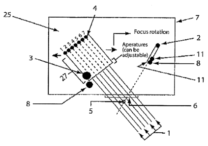
Figure 7 shows a typical sequence of operation of an embodiment of the
invention in apparatus and method=f.o.r identifying and sanication a
thrombosis
In the following sequence of steps, wherein the number of the step below is
indicated at its corresponding place in= Figure 7.
1. Start, ultra-sound o+cclusion-related detection and target.treatment;,
11) 2. UitrasQnic Bean1 Focus Control.
3. Scan for Suspicious Region Of interest (SROI). The poppler stroke-
treatrnent ultrasaund phased-array transducers can be steered across
larger regions to enable a means to scan for SRC31_
4. Once the SROI candidate(s) are detected the finer resolution focus
mode can be applied to determine the Region Of Interest (RO1). The
POl as with SRO1 -can be subjected to further FFT or acoustic footprint
analysis, characterisation and comparison (with deviation
consideration) and final operator verification.
5. Scan for ROl cor-tprising finer bes.m focus and ultrasonic speGtrai
(FFT), phase, amplitude or any combinational analysis thereof. FFT
spectral "foot-print" or acoustic characteristic footprint, associated with
occlusion blooci flow can be detected firstly on the course scan and
detect mode (SR(.71j Acquire ultrasonic data for archive, anaiysis,
retrieval, remote view, remote cantroi purposes.
6. Transform ultrasonic return echo data into frequency spectral (FFT),
phase and/or amplitude characteristics. RQI can be detected by way of
computing the acoustic or FFT foot print across a region using sweep
mode and then utilisation representing characteristic of blood
turbulence or flow characteristics asscciated with vascular partial or
total occlusion.
7. Compare acquisifioned ultrasonic acoustic echo data charaCteristics
with those of refarence fingerprints for likely acoustic characteristic
representing stroke-related occlusion acoustic flow related turbulence.
CA 02605531 2007-11-07
WO 2006/119572 PCT/AU2006/000620
17
S. Reference data base presenting FFT characteristics and ultrasonic
characteristics or footprint associated with SRO! or ROI dejection
determination.
9. Devia.tion Characteristics- being the allowable detection tolerances
against the data base of acoustic "occlusia.n" "fingerprint" data base
compe.rison events.
10, Using any combination of- neural network, arti-ficial intelligence, or
other
analysis methods compare reference data base of typical "finger-fsririts"
of stroke-related. occlusions-, along with acceptable devia#ion
characteristics, in=orderto compute-valid detection of occlusion and co-
ordinates for optimal treatment focus and targets.
11. Go-ordinate data associateci with= RC7i and assonating target data
avaiiab4e for phase array or other type of ultrasonia transduoer
focussing 'control,
i 5 12, Ultrasonically enhanced Thrombosis Treatment C]pera.tor validation and
Focus Treatment Focus Targeting,
1S. Once operator verificatiorr is. acknowledged user can select to
automatically lock in ultrasonic-enhanced thrombosis mode, where
upon phase array multiple beam focus. treatment can be applied.
14. User display and user intertace allowing- manual, automatic or
computer assisted ultrasound stroke-related occlusion detection and/or
treatment.
15. Knowledge decision base option for artificial intelligent ocnlusion
detection and uftras4nic control analysis option. Artificial Intelligence
reference to support neuraf network processing decision matrix. Can
be updated by system expert users or remote inteiligence base.
Current system can be designated as expert user to enable artificial
intelligence supporting increased accuracy in detection and control of
stroke-related occlusion (emboli).
16. Spatial cycling or "massaging" modes possibie with variation of
frequency or sequences of treatmertt frequencies as well as variation of
spatial positioning to enable optimal working at the edges and/or solid
occluding materia.l in order to control optimal diffusion and safe
CA 02605531 2007-11-07
WO 2006/119572 PCT/AU2006/000620
18
agitation or dissipation of dissolving of material (minimise further
occlusion with risk of large break-away material)..
17.Treatment temporal analysis and progress tracking in order to enable
adjustment for intensity or and power of ultrasonic treatrnent from most
gentle and safest treatment to most aggressive and rapid emergency
treatment modes,
18. Screen shows treatment target display. marking of. SROI and also
audible or other local or remote system alert. - -
19. Screen target display marking of ROI and also audible or other local or
remote system aiert, - . . . .
20.SGreen target display marking of.ROl= -and SROI and also audible or
other local or remote system-alert:
The invention includes apparatus having remote video capabilities for
observing the output of ultrasound bearns' generated f.rom an ultrasound
transducer at a location remote to- thesubject.on which the beams are
focussed.
The apparatus include the possibility of.re:mote manual adjustment to the
aontrol on ultrasound beam parameters: .
The apparatus may include a three-dimensional laiv-optical means wherein
the ultrasound signal received by an ultrasound transducer 9s-transformed into
computer gra.phics for easy viewing and interpretation by an operator.
The apparatus may include a method for transforming TOD output signals into
graphical representations for a computer or other viewing soreen. It will be
understood that the screen may be any screen oapable of displaying a digita.l
or analogue signal,
The invention includes an array of ultrasound-emitting elements wherein each
element of said array is capable of focussing ultrasound waves at a focal
point. The invention includes ultrasound elements that are piezo-eiectric
crystals in an ultrasound transducer.
CA 02605531 2007-11-07
WO 2006/119572 PCT/AU2006/000620
19
The invention may include apparatus operabie with voice coil technology for
the positioning of ultrasound transducers on a patient's head.
S The invention includes an apparatus for TCp ultrasound.that automatically or
semi-automatically scans and maps the location of blood vessel onclusions,
using the methods d+asuribed her..ein. Said automatic or semi-automatic
mapping is effected by the use of means, such as=a computer database and
program or programs in which normalitive sound-wave data is-stored for
automatic corriparison with -sound-wave data acquired from the sonication of
an SRUi or RC71 according to the procedure illustrated.in.Figure 7. Said
database may contain data representative of a. two-dimensionai or three-
dimensional map for the SROI or Rdl in a subjoct or a representative
rtormalisad subjeot. Said means may further include computer program or
programs for displaying representations of the acquired. data in comparison
with normafitive data, such as on a video display unit...Said computer program
or programs may suitable processing techniques for reflected ultrasound
waves such as fast Fourier transformation techniques,arid determining the
rnost iikely regions for occlusions by comparing fast.Fourier outputs for free-
flowing or occluded vessels. Said video display. unit may show a
representation of the region being sonicated, preferably in real-time
according
to the information stored in the database. Said computer program may further
display acquired sonication data on said video display unit to indicate the
position of sonication in two-dimenslonal or three-dimensional space in
relation to known anatomical characteristics of a subject.
The method of the invention includes a diagnostic capability to detect an
occlusion either by way of the measurement of fluid flow in a RC7! or
turbulence in the vioinity of an occlusion. Said diagnostic capability may
include a comparison of sound-wave data indicating the presence of fluld
spurts within a vessel or fluid flow rates associated with recanalisation of
stenosis in such a way as to enable the distinction between the presence of
an occlusion or a stenosis. Said diagnostic capabiilty includes the
calcuiation
of fluid flow and turbulence in the region of the occfusion or stenosis by way
of
CA 02605531 2007-11-07
WO 2006/119572 PCT/AU2006/000620
using sound-wave data for measurement of any of fluid flow rate, fluid flow
quantity, fluid turbulence, or intensity.
The apparatus or method further includes displaying an index, measure,
5 -.marlcer, or series of markers or other suitable representation
characteristic of
the progress of recanallsatian of a vessel during any or both of diagnosis or
tre-atment on said video. display unit. The apparatus or =method further may
include the incorporation of said index, measure, or series of markers into a
means, such as a computer program, for optimising the at least one=sound- =
i0 wave beam inctrrporating at least one frequency in order to provide better
control over the rate of recanallsation=.and sonicatiori power.
The invention includes many embodirnents. Far exampie, the invention
includes apparatus wherein a first ultrasound transducer is fixed in relation
to
15 a target occlusion characteristic of embolism in at blood vessei.: A second
ultrasraund transducer is positioned in relation to said 'first transducer
such that
-=both transducers focus the emitted beams of ultrasound waves- onto an
: occiusion in said blood vessel. The second transducer may be positioned
using a servo device. It will be apparent to those skilled in the art that
more
20 than two ultrasound transducers rriay be included in the invention and that
.each transduCEr may be positioned rela.tive to the others so that the target
occlusion is located at a common focal point for the beams of uitrasound
waves. Preferably the invention includes an array of transducers. Preferabiy
the array is a structured array. The advantageous effect of the multiple
ultrasound transducers with a common focal point will be to focus the
maximum ultrasound energy on the occlusion and result in the most effective
embolism dispersing treatment.
The invention provides a method to optimise the focus Qf the ultrasound beam
or beams using servo-movement. One or more servo mechanisms and/or
ultrasonic phased array transducer control systems can be depioyeci to enable
a continuously variable positioning of focus point in order to enable optimal
energy focu$ of one or more beams, The said energy focus is able to be
divided at one or more precise locations with high spatial resolution in order
to
CA 02605531 2007-11-07
WO 2006/119572 PCT/AU2006/000620
21
focus energy away from healthy tissue (is desirabie and as desirable), where
at the sarne time focussing energy of a set of beams at the location of
vascular occluding material, in order to disintegrata or diffuse or disperse
said
occtuding material in an optimal and safe treatment manner,
Furthermore, the at least one treatment beam of ultrasonic frequency can
comprise one.or more frequencies optimized for functions enabling any
sequence or simultaneous combination of optimal a) gaseous partial or total
=.= . . . . . =
occiusiva material detection; b) solid material.,partlal or total ocafusive=
material
detection; or c) gaseous partial or total thromoboly$is and recanalisation of
partial or fully occluded blood vessels,
The invention provides an apparatus and method to facilitate the safe
dispersion or dissolving of an occlusion (thrvmbolysis or recsnal'isation of
blood vessel). One of the rlsks associated With uitrasonically enhanced
thrombolysis is the risk that dislodged occluding material can "break away" or
disintegrate in large and unsafe particle sizes, which in turit can cause,
further
occlusion or risks of occlusion or partial vessef blockages. In particular,
vessel blockages such as within the legs oi= loin'rer body may be recanalised
and caus6 particles to travel higher in the circulatory system such as in'the
brain. In these regions the vessols can be snialler and lead to further
blockages "and more serious consequences such as ischaemic stroke.
The invention provides an apparatus and method for simultaneous diagnosis
and treatment to regulate towards safe thromobolysis and recanalisation of
blood vassels. The present invention includes the diagnosis or identificatian
of a particle or total occlusion of a vessel while at the same time or
separately
providing uitrasonicalty enhanced thrombolysis or vessel recanalisation,
The focus adjustment and targeting of the at least one ultrasonic beam, along
with accurate spatiai resolution and power control (of targeted ultrasaund
treatment) enables a controlled dispersion of occluding material in a vessel
by
allcwirtg different patterns or mas~3aging (movement of beam focus around,
over and near occluding material) and different ultrasonic frequencies or
combination or sequences of frequencies (dififerent frequencies effect
different
CA 02605531 2007-11-07
WO 2006/119572 PCT/AU2006/000620
22
material types and also effect the dispersion rate and size of dispersed
occluding material particles) or any combination thereof to be applled to the
region of vesse4 occlusion or partial occlusion. The treatment of an occluded
vessel can thus be controfled, targeted and regulated in order to minimize the
particle dispersion size and risk for further occlusion.
The-present invention enables one or more ultrasonic.frequencies either
sequentially or simultaneously to be generated as a means to both enhance
diagnosis and imaging and also enhance ultrasonic vessel recanaflsation
treatment.
The present invention includes a three-dimnsional mapping capability and
tracking of the maximum power generation by the at least one ultrasound
transducer. This enables a register or matrix representing the computation of
ultrasonic power generation at any point in time and,any spatial location
under
scan. Said "ma.trix or register" computes the probable power dissipation of
ultrasound scan beams based on the beam dispersion characteristics.
Furthermare, the intersection of beams along with the focus characteristics
are. computed and provide a resulting reference data set to enable or to
ensure thaf maximal safe power thresholds are achieved. at all locations and
that the additional ultrasonic power required to rapidly diffuse vessel
occlusion
is only directed specificaliy where required and where safe, i.e., targeted at
surrounding healthy tissues.
The present invention provides the capability to enable ultrasonically-
enhanced thrombolysis and vessei recanalisation ta be moderated in
treatment intensity (power) in accordance with or in harmony with "clot-
krusting
drug" characteristics of action. This consideration can minimize risk of side
effects of each said treatrnent, such as haemorrhage risk with "olot-busting
drug" treatment, or excessive ultrasound pewer and cell harm with ultra.sound
treatment.
The present invention provides that data such as the drug administration rate,
drug cornpQsition or type, and patient risk category to haemorrhage, such as
CA 02605531 2007-11-07
WO 2006/119572 PCT/AU2006/000620
23
haemophiliacs, can Ue entered into a clot drug-profusion device and also
ultrasound power and focus control, for example.
The present invention most advantageously enables the intravenous or
manual administration of clot-busting drugs to be regulated or monitored in
such a manner as to regulate the balance between the strength of the clot
balanced or administration optimized to minimize the risk of side effects the
occluding material and minimally targeted clot-busting drug administration,
with risks of haemorrhage side effects, with the aggressive or high powered
t0 application of ultrasonically enhanced thrombosis treatment, with the risk
of
healthy celf harm and also dispersion of further clot-causing material.
The present invention enables the ultrasonic control and the "clot-busting
drug" administratlon to be servo-controlled in such a manner that optimal
15= speed of thrombosis and recanalisation of the vessel(s) and also optimal
safety or=mitigation to patient risks are possible.
References. = .
20. Aiexandrov, AV. 2002. European J Ultrasound 16: 131-140. Ultrasound-
enhanced thrombolysis for stroke: clinical significance.
Alexandrov, AV. Malina, CA., Grotta, JC., Garami, Z., Ford, SR., Alvarez-
25 Sabin, J., Montaner, J., Saqqur, M., Dernchuk, AM., Moye, LA., Hill, MC?.,
and
Wojner, AW. 2004. New England J Medicine. 351: 2170- 2178. Ultrasound-
enhanced systemic thrombolysis for acute ischemic stroke.
Demchuk, AM, Burgin, WS, Cristou, l., Felberg, RA, Barber, PA, Hill MD,
30 Atexandrov AV,. 2001. Stroke 32: 80-93. Thrombolysis in brain ischemia_
Tegeler, CH, and Ratanakorn, D. 1999. "Physics and Principles". in
Transcranial Dopplar U/trascnography, Bibikian, VL, Wechsler, LR, Toole, JF.
Eds. Butterworth Heinerrtann, Melbourne, pp 3-11.