Note: Descriptions are shown in the official language in which they were submitted.
CA 02605912 2010-03-08
APPARATUS AND METHOD FOR SHAPED MAGNETIC FIELD CONTROL
FOR CATHETER, GUIDANCE, CONTROL, AND IMAGING
Background
Field of the Invention
[0001] The present invention relates to magnetic guiding, steering, and
advancing invasive medical devices such as catheters and catheter-type
devices.
Description of the Related Art
[00021 Catheterization is typically performed by inserting an invasive
device into an incision or a body orifice. These procedures rely on manually
advancing
the distal end of the invasive device by pushing, rotating, or otherwise
manipulating the
proximal end that remains outside of the body. Real-time X-ray imaging is a
common
method for determining the position of the distal end of the invasive device
during the
procedure. The manipulation continues until the distal end reaches the
destination area
where the diagnostic or therapeutic procedure is to be performed. This
technique requires
great skills on the part of the surgeon/operator. Such skill can only be
achieved after a
protracted training period and extended practice. A relatively high degree of
manual
dexterity is also required.
[0003] Recently, magnetic systems have been proposed, wherein magnetic
fields produced by one or more electromagnets are used to guide and advance a
magnetically-tipped catheter. The electromagnets in such systems produce large
magnetic
-1-
CA 02605912 2007-10-19
WO 2006/128160 PCT/US2006/020895
fields that are potentially dangerous to medical personnel and that can be
disruptive to
other equipment.
[0004] Therefore, there is a great and still unsatisfied need for an apparatus
and method for guiding, steering, and advancing invasive devices and for
accurately
controlling their positions for providing positioning of magnetic fields and
field gradient,
for providing a fields configured to push/pull, bend/rotate, and by further
enabling
apparatus to align the distal end of the catheter tip so as to achieve
controlled movement
in 3D space and ability of apparatus to control the magnetic field
characteristics without
the customary power and field intensities seen in the prior art.
Summary
[0005] These and other problems are solved by a magnetic catheter guidance
system that uses moveable electromagnets to configure a magnetic field for
guiding a
catheter or other device through a body.
[0006] In one embodiment, a magnetic circuit is configured to generate a
desired magnetic field in the region of a multi-coil cluster of
electromagnets. In one
embodiment, one or more poles of the cluster are moveable with respect to
other poles in
the cluster to allow shaping of the magnetic field. In one embodiment, one or
more
magnet poles can be extended or retracted to shape the magnetic field. In one
embodiment, the electromagnets can be positioned to generate magnetic fields
that exert a
desired torque on the catheter, but without advancing force on the tip (e.g.,
distal end of
the catheter). This affords bend and rotate movements of the catheter tip
toward a selected
direction. In one embodiment, the multi-coil cluster is configured to generate
a relatively
high gradient field region for exerting a moving force on the tip (e.g., a
push-pull
movement), with little or no torque on the tip.
[0007] In one embodiment, the catheter guidance system includes a closed-
loop servo feedback system. In one embodiment, a radar system is used to
determine the
location of the distal end of the catheter inside the body, thus, minimizing
or eliminating
the use of ionizing radiation such as X-rays. The catheter guidance system can
also be
-2-
CA 02605912 2007-10-19
WO 2006/128160 PCT/US2006/020895
uscu in combination with an X-ray system (or other imaging systems) to provide
additional imagery to the operator. The magnetic system used in the magnetic
catheter
guidance system can also be used to locate the catheter tip to provide
location feedback to
the operator and the control system. In one embodiment, a magnetic field
source is used to
create a magnetic field of sufficient strength and orientation to move a
magnetically-
responsive catheter tip in a desired direction by a desired amount.
[0008] In one embodiment, the multi-coil cluster is configured to generate a
magnetic field gradient for exerting an orthogonal force on the tip (side-ways
movement),
with little or no rotating torque on the tip. This is useful for aligning the
tip at narrow
forks of artery passages and for scraping a particular side of artery or in
treatment of
mitral valve stenosis.
[0009] In one embodiment, the multi-coil cluster is configured to generate a
mixed magnetic field to push/pull and/or bend/rotate the distal end of the
catheter tip, so
as to guide the tip while it is moving in a curved space and in cases where
the stenosis is
severe or artery is totally blocked.
[0010] In one embodiment, the multi-coil cluster is configured to move the
location of the magnetic field in 3D space relative to the patient. This
magnetic shape
control function provides efficient field shaping to produce desired magnetic
fields for
catheter manipulations in the operating region (effective space).
[0011] One embodiment includes a catheter and a guidance and control
apparatus that allows the surgeon/operator to position the catheter tip inside
a patient's
body. The catheter guidance and control apparatus can maintain the catheter
tip in the
correct position. One embodiment includes a catheter and a guidance and
control
apparatus that can steer the distal end of the catheter through arteries and
forcefully
advance it through plaque or other obstructions.
[0012] One embodiment includes a catheter guidance and control apparatus
that displays the catheter tip location with significantly reduced X-ray
exposure to the
patient and staff.
-3-
CA 02605912 2007-10-19
WO 2006/128160 PCT/US2006/020895
[0013] One embodiment includes a catheter guidance and control apparatus
that is more intuitive and simpler to use, that displays the catheter tip
location in three
dimensions, that applies force at the catheter tip to pull, push, turn, or
hold the tip as
desired, and that is configured to producing a vibratory or pulsating motion
of the tip with
adjustable frequency and amplitude to aid in advancing the tip through plaque
or other
obstructions. One embodiment provides tactile feedback at the operator control
to indicate
an obstruction encountered by the tip.
[0014] In one embodiment, the Catheter Guidance Control and Imaging
(CGCI) system allows a surgeon to advance, accurately position a catheter, and
to view
the catheter's position in three dimensions by using a radar system to locate
the distal end
of the catheter. In one embodiment, the radar data can be combined with X-ray
imagery to
produce a composite display that includes radar and X-ray data. In one
embodiment, the
radar system includes a Synthetic Aperture Radar (SAR). In one embodiment, the
radar
system includes a wideband radar. In one embodiment, the radar system includes
an
impulse radar.
[0015] One embodiment includes a user input device called a "virtual tip."
The virtual tip includes a physical assembly, similar to a joystick, which is
manipulated
by the surgeon/operator and delivers tactile feedback to the surgeon in the
appropriate
axis or axes if the actual tip encounters an obstacle. The Virtual tip
includes a joystick
type device that allows the surgeon to guide actual catheter tip through the
patient's body.
When actual catheter tip encounters an obstacle, the virtual tip provides
tactile force
feedback to the surgeon to indicate the presence of the obstacle.
[0016] In one embodiment, the physical catheter tip (the distal end of the
catheter) includes a permanent magnet that responds to the magnetic field
generated
externally to the patient's body. The external magnetic field pulls, pushes,
turns, and
holds the tip in the desired position. One of ordinary skill in art will
recognize that the
permanent magnet can be replaced or augmented by an electromagnet.
-4-
CA 02605912 2007-10-19
WO 2006/128160 PCT/US2006/020895
[0017] In one embodiment, the physical catheter tip (the distal end of the
catheter) includes a permanent magnet and two or more piezoelectric rings, or
semiconductor polymer rings to allow the radar system to detect the second
harmonics of
the resonating signal emanating from the rings.
[0018] In one embodiment, the CGCI apparatus provides synchronization by
using a radar and one or more fiduciary markers to provide a stereotactic
frame of
reference.
[0019] In one embodiment, the electromagnetic circuit of the CGCI apparatus
includes a C-Ann geometry using a ferromagnetic substance (e.g., a furous,
substance,
nickel substance, etc.) so as to increase the efficiency of the magnetic
circuit.
[0020] In one embodiment, the CGCI apparatus uses numerical
transformations to compute currents to be provided to various electromagnets
and
position of one or more of the electromagnet to control the magnetic field
used to
push/pull and rotate the catheter tip in an efficient manner.
[0021] hl one embodiment, the CGCI apparatus includes a UWB impulse
radar for detecting the catheter tip and body organs, and synchronizing their
motions.
[0022] In one embodiment, the CGCI apparatus includes a motorized and/or
hydraulic mechanism to allow the electromagnet poles to be moved to a position
and
orientation that reduces the power requirements desired to push, pull, and
rotate the
catheter tip.
[0023] In one embodiment, the CGCI apparatus is used to perform an
implantation of a pacemaker during an electrophysiological (EP) procedure.
[0024] In one embodiment, the CGCI apparatus uses radar or other sensors to
measure, report and identify the location of a moving organ within the body
(e.g., the
heart, lungs, etc.) with respect to the catheter tip and one or more fiduciary
markers, so as
-5-
CA 02605912 2007-10-19
WO 2006/128160 PCT/US2006/020895
to provide guidance, control, and imaging to compensate for movement of the
organ,
thereby simplifying the surgeon's task of manipulating the catheter through
the body.
[0025] In one embodiment, the operator control provides the position and
orientation command inputs to a servo system that controls the catheter tip
position by
generating and shaping the magnetic fields. A measurement of actual tip
position and
orientation is made via a sensory apparatus that includes a radar system. This
measurement is used to provide feedback to the servo system and the operator
interface.
[0026] In one embodiment, the servo system has a correction input that
compensates for the dynamic position of a body part, or organ, such as the
heart, thereby
offsetting the response such that the actual tip moves substantially in unison
with the
dynamic position (e.g., with the beating heart).
[0027] In one embodiment of the catheter guidance system: i) the operator
adjusts the physical position of the virtual tip, ii) a change in the virtual
tip position is
encoded and provided along with data from a radar system, iii) the control
system
generates servo system commands that are sent to a servo system control
apparatus, iv)
the servo system control apparatus operates the servo mechanisms to adjust the
position of
one or more electromagnet clusters by varying the distance and/or angle of the
electromagnet clusters and energizing the electromagnets to control the
magnetic catheter
tip within the patient's body, v) the new position of actual catheter tip is
then sensed by
the radar, thereby allowing synchronization and superimposing of the catheter
position on
an image produced by fluoroscopy and/or other imaging modality vi) providing
feedback
to the servo system control apparatus and to the operator interface and vii)
updating the
displayed image of the catheter tip position in relation to the patient's
internal body
structures.
[0028] In one embodiment, the operator can make further adjustments to the
virtual catheter tip position and the sequence of steps ii through vii are
repeated. In one
embodiment, the feedback from the servo system control apparatus creates
command
logic when the actual catheter tip encounters an obstacle or resistance in its
path. The
con-nand logic is used to control stepper motors which are physically coupled
to the
-6-
CA 02605912 2007-10-19
WO 2006/128160 PCT/US2006/020895
virtual catheter tip. The stepper motors are engaged as to create resistance
in appropriate
directions that can be felt by the operator, and tactile feedback is thus
provided to the user.
[0029] In one embodiment, the apparatus uses scaling factors to calculate the
magnetic field generated along the effective magnetic space.
[0030] In one embodiment, the apparatus is configured to generate a
maximum force of 35 grams for push/pull of the catheter tip and a 35 gram
force while
the coil cluster is generating dB/dS field gradients between 1.6 T/m to 3.0
T/m.
[0031] In one embodiment, the apparatus generates a maximum torque of
0.013 Newton-meter on the catheter tip, while the coil cluster is generating a
magnetic
field strength between B = 0.04T and 0.15T.
[0032] In one embodiment, the coil current polarity and polarity rotation are
configured to allow the coil cluster to generate torque on the catheter tip.
[0033] In one embodiment, the coil current polarity and rotation are
configured to provide an axial and/or orthogonal force on the catheter.
[0034] In one embodiment, a topological transformation allows control of the
magnetic field in the 2D four coil geometry to form the magnetic field desired
for
navigating and controlling the catheter tip.
[0035] In one embodiment, a second topological transformation allows the
apparatus to operate in 3D space while creating the magnetic field desired to
push/pull
and rotate the catheter tip.
[0036] In one embodiment, a symmetrical transformation is provided allowing
the apparatus to operate with eight coil clusters.
-7-
CA 02605912 2007-10-19
WO 2006/128160 PCTIUS2006/020895
[0037] In one embodiment, the eight coil symmetry is reduced to a six coil
symmetry allowing the CGCI apparatus to generate the desired magnetic field in
an
optimized pattern.
[0038] In one embodiment, the coil cluster is fitted with a parabolic shield
which collects the magnetic flux from the effective space and creates a return
path to
decrease the need to shield the stray magnetic radiation.
[0039] In one embodiment, the magnetic circuit efficacy of the CGCI
apparatus is evaluated as to its topological properties and it is measured
relative to torque
control field variations in the magnetic center.
[0040] In one embodiment, the magnetic circuit efficacy of the CGCI
apparatus is evaluated as to its topological properties and it is measured
relative to force
control gradient variations in the 100mm region around the magnetic center.
[0041] In one embodiment, the rotational transformation and its relationship
to
field strength and field gradient are mathematically established.
[0042] In one embodiment, a mathematical model for topological
transformations of the geometry versus magnetic field generation is
established.
[0043] In one embodiment, the CGCI apparatus is fitted with at least one
hydraulically-actuating extension core, for varying the magnetic pole
configuration to
allow shaping of the magnetic field.
[0044] In one embodiment, the shaped magnetic field is configured as a
variable magnetic pole geometry to control the catheter tip. The shaped field
provides for
operator control of the catheter tip while reducing power and reducing field
strength by
tailoring the field geometry.
-8-
CA 02605912 2007-10-19
WO 2006/128160 PCTIUS2006/020895
[0045] In one embodiment, the CGCI apparatus is fitted with a parabolic
shield for flux return to reduce the emission of the radiating field outside
of the effective
area to less than 20 gauss.
[0046] In one embodiment, the control scheme of the CGCI apparatus includes
a boundary condition controller. The controller computes the fields
surrounding the
catheter based on the fields on the 2D planes enclosing the magnetic chamber.
Equations
for computing the fields with rotated coils on the surface of the sphere are
established in
the magnetic chamber.
[0047] In one embodiment, the coil is controlled from a bi-polar DC power
source. A six channel regulator assisted by a computer using matrix algorithms
controls
the six coil magnetic configuration.
[0048] In one embodiment, user control is provided by an aircraft-type
joystick, wherein movement of the joystick between the torque mode and the
force mode
is provided by a mode switch.
[0049] In one embodiment, the mode switch allows the controller to switch
from torque control to force control as well as mixed torque and force
control.
[0050] In one embodiment, the coil current polarities and magnitudes are
defined and cross-referenced to the desired field directions for torque and
force fields.
[0051] In one embodiment, the coil polarity combinations are expressed as a
set of matrices, wherein the grouping of coils is used such that four coil and
three coil
groups associated with the virtual tip 2D planes are established.
[0052] In one embodiment, the symmetry group is a four coil group with 16
polarity combinations. Control simulated under the four coil XY plane, and
under the
topological transformation allows the state of the CGCI machine torque and
force to be
controlled.
-9-
CA 02605912 2011-10-13
100531 In one embodiment, the coils are configured using symmetry, where
the group is rotated 90 from the symmetry group.
[00541 In one embodiment, the rotational steps are smoothly transferred while
the coil currents is oscillating from -100% to +100% through zero, and where
the control
slope between 0% - to - 100% coil current is subject to a nonlinear inverse
cosine
function.
100551 In one embodiment, the entire CGCI magnetic circuit is modeled using
a low-level logic simulation of the action performed by the joystick prior to
activating the
power amplifiers that provide current to the coils.
100561 In one embodiment, the magnitude control function of the CGCI
controller directing the deployment as well as retraction of the piston
actuated extension
core is used to shape the magnetic field affording a variable magnetic field
for moving the
catheter tip in the desired direction.
100571 In one embodiment, a Hall effect ring measures the boundary plane
field strength as a measure of the joystick movement. This allows the CGCI to
operate on the
boundary planes of the field, rather than the interior of the magnetic
chamber, while
allowing the Hall effect sensor to operate in a range of a few hundred gauss
fields.
[0057a1 In accordance with an aspect of the present invention there is
provided an apparatus for controlling the movement of a catheter-type tool
inside a body of
a patient, comprising: a magnetic field source for generating a magnetic
field, said
magnetic field source comprising: a first semi-spherical symmetry cluster
comprising a first
coil corresponding to a first magnetic pole and a second coil corresponding to
a second
magnetic pole, wherein said first magnetic pole is moveable with respect to
said second
magnetic pole, a third magnetic pole corresponding to a third coil, and a
fourth magnetic
pole corresponding to a fourth coil, a second semi-spherical symmetry cluster
comprising a
fifth coil corresponding to a fifth magnetic core, a sixth coil corresponding
to a sixth
magnetic core, a seventh coil corresponding to a seventh magnetic core, and an
eighth coil
corresponding to an eighth magnetic core; and a system controller for
controlling said
-10-
CA 02605912 2011-10-13
magnetic field source to control extension and retraction of at least said
first magnetic core
to position said first magnetic core of said first semi-spherical symmetry
cluster, said system
controller configured to receive position data regarding said current position
of a distal end
of a catheter, said distal end responsive to said magnetic field, said system
controller further
configured to control currents in said first, second, third, fourth, fifth,
sixth, seventh and
eighth coils, to control a movement of a distal end of a catheter to a desired
position with
torque control fields according to the following equation:
BTq =Bxf.=COS(9)
and with force control fields according to the following equation:
dB dB,YY ,cos(9)
ds ds
xy is the field in an XY plane, and i9 is angle of spherical rotation of the
first, second, third, fourth, fifth, sixth, seventh and eighth coils from an
XY plane.
Brief Description of the Figures
100581 Figure 1 is a perspective view of the magnet structure of the Catheter
Guidance Control and Imaging (CGCI) system.
100591 Figure 1A is a perspective view of the CGCI right section showing the
hydraulically-actuated core extended.
[00601 Figure 1 B is a perspective view of the CGCI right section showing the
hydraulically-actuated core extracted.
- l 0a -
CA 02605912 2007-10-19
WO 2006/128160 PCT/US2006/020895
[0061] Figure 1C is a system block diagram for a surgery system that includes
an operator interface, a catheter guidance system, and surgical equipment.
[0062] Figure 1D is a block diagram of the imaging module for use in a CGCI
surgery procedure that includes the catheter guidance system, a radar system,
Hall Effect
sensors, and a hydraulically actuating core extension mechanism.
[0063] Figure 2 shows a magnet assembly of a CGCI scale model.
[0064] Figure 2A shows parameters of the assembly shown in Figure 2.
[0065] Figure 2B is a first view of the catheter assembly.
[0066] Figure 2C is a second view of the catheter assembly.
[0067] Figure 2D shows a catheter assembly with piezoelectric rings.
[0068] Figure 3 shows a magnet assembly with retracted cores.
[0069] Figure 3A shows the magnet assembly of Figure 3 with an extended
core.
[0070] Figure 4 shows field directions corresponding to currents in the magnet
assembly of Figure 3.
[0071] Figure 5 is a vector field plot of the B fields in a central region of
the
magnet assembly of Figure 3 with a first current configuration where the B-
vector is
parallel to the X-axis.
[0072] Figure 5A is a field intensity plot corresponding to Figure 5.
[0073] Figure 5B is a field contour plot corresponding to Figure 5.
-11-
CA 02605912 2007-10-19
WO 2006/128160 PCT/US2006/020895
[0074] Figure 6 is a vector field plot of the B fields in a central region of
the
magnet assembly of Figure 3 with a second current configuration where the B-
vector is
parallel to the Y-axis.
[0075] Figure 6A is a field intensity plot corresponding to Figure 6.
[0076] Figure 6B is a field contour plot corresponding to Figure 6.
[0077] Figure 7 is a vector field plot of the B fields in a central region of
the
magnet assembly of Figure 3 with a third current configuration where the B-
vector
direction is 135 .
[0078] Figure 7A is a field intensity plot corresponding to Figure 7.
[0079] Figure 7B is a field contour plot corresponding to Figure 7.
[0080] Figure 8 is a vector field plot of the B fields in a central region of
the
magnet assembly of Figure 3 with a fourth current configuration corresponding
to the
force control mode.
[0081] Figure 8A is a field intensity plot corresponding to Figure 8.
[0082] Figure 8B is a field contour plot corresponding to Figure 8.
[0083] Figure 9 is a vector field plot of the B fields in a central region of
the
magnet assembly of Figure 3 with a fifth current configuration in the force
control mode
where the B vector is orthogonal to the magnetic tip axis.
[0084] Figure 9A is a field intensity plot corresponding to Figure 9.
[0085] Figure 9B is a field contour plot corresponding to Figure 9.
-12-
CA 02605912 2007-10-19
WO 2006/128160 PCT/US2006/020895
[0086] Figure 10 is a vector field plot of the B fields in a central region of
the
magnet assembly of Figure 3 with a sixth current configuration with a
hydraulically
extended core.
[0087] Figure 10A is a field intensity plot corresponding to Figure 10.
[0088] Figure lOB is a field contour plot corresponding to Figure 10.
[0089] Figure 11 shows the magnetic fields with a first core extension
configuration.
[0090] Figure 11A shows the magnetic fields with a second core extension
configuration.
[0091] Figure 1lB shows the magnetic fields with a third core extension
configuration.
[0092] Figure 12 is a field map showing fields corresponding to a first coil
and
current configuration using a transformed magnet cluster.
[0093] Figure 12A is a field map showing fields corresponding to a second
coil and current configuration using a transformed magnet cluster.
[0094] Figure 12B is a field map showing fields corresponding to a third coil
and current configuration using a transformed magnet cluster.
[0095] Figure 12C is a field map showing fields corresponding to a fourth coil
and current configuration using a transformed magnet cluster.
[0096] Figure 12D is a field map showing fields corresponding to a fifth coil
and current configuration using a transformed magnet cluster.
-13-
CA 02605912 2007-10-19
WO 2006/128160 PCT/US2006/020895
[0097] Figure 12E is a field map showing fields corresponding to a sixth coil
and current configuration using a transformed magnet cluster.
[0098] Figure 12F is a field map showing fields corresponding to a seventh
coil and current configuration using a transformed magnet cluster.
[0099] Figure 12G is a field map showing fields corresponding to a eighth coil
and current configuration using a transformed magnet cluster.
[0100] Figure 13A is a side view of the apparatus of Figure 1.
[0101] Figure 13B is an underside view of the apparatus of Figure 1.
[0102] Figure 14 is an isometric view showing the apparatus of Figure 1 in an
open mode where the left and right clusters are separated.
[0103] Figure 14A is a side view of the configuration shown in Figure 14.
[0104] Figure 15 is an underside view of the configuration shown in Figure
14.
[0105] Figure 16 is an end view of the configuration shown in Figure 14.
[0106] Figure 17 shows a magnet cluster of a full-scale system.
[0107] Figure 17A is a graph of torque range of the full-scale system.
[0108] Figure 17B is a graph of field gradients for the full-scale system.
[0109] Figure 18 is a front view of a magnet cluster of the CGCI apparatus.
[0110] Figure 18A is a side view of a magnet cluster of the CGCI apparatus.
-14-
CA 02605912 2007-10-19
WO 2006/128160 PCT/US2006/020895
[0111] Figure 18B is a underside view of a magnet cluster of the CGCI
apparatus.
[0112] Figure 19 is an isometric view of a coil assembly.
[0113] Figure 19A shows water connections of the coil assembly.
[0114] Figure 19B is a front view of the cylindrical coil assembly.
[0115] Figure 19C is a side view of the cylindrical coil assembly.
[0116] Figure 19D is a rear view of the cylindrical coil assembly.
[0117] Figure 19E shows the hydraulic actuator for extending the magnetic
core.
[0118] Figure 19F shows the hydraulic actuator of Figure 19E mounted to the
coil cluster.
[0119] Figure 19G is an interior view of the coil and extendable core with the
hydraulic actuator.
[0120] Figure 20A is a side view of the tapered coil assembly.
[0121] Figure 20B is a front view of the tapered coil assembly.
[0122] Figure 20C is a rear view of the tapered coil assembly.
[0123] Figure 21 is an isometric view of the tapered coil assembly.
[0124] Figure 22A shows 4 coil circular symmetry.
[0125] Figure 22B shows 4 coil semi-spherical synunetry.
-15-
CA 02605912 2007-10-19
WO 2006/128160 PCT/US2006/020895
[0126] Figure 22C shows 8 coil spherical symmetry.
[0127] Figure 22D shows 6 coil cluster symmmetry.
[0128] Figure 22E shows 6 coil cluster symmetry in a magnetic shield.
[0129] Figure 23 shows 4 coil circular symmetry and a reference coordinate
system.
[0130] Figure 23A shows B fields of the 4 coil circular symmetry.
[0131] Figure 23B shows field gradients of the 4 coil circular symmetry.
[0132] Figure 24 shows 4 coil semi-spherical symmetry and a reference
coordinate system.
[0133] Figure 24A shows B fields of the 4 coil semi-spherical symmetry.
[0134] Figure 24B shows field gradients of the 4 coil semi-spherical
symmetry.
[0135] Figure 25 shows 8 coil spherical symmetry and a reference coordinate
system.
[0136] Figure 25A shows B fields of the 8 coil spherical symmetry.
[0137] Figure 25B shows field gradients of the 8 coil spherical symmetry.
[0138] Figure 26 shows the 6 coil cluster and a reference coordinate system.
[0139] Figure 26A shows B fields of the 6 coil cluster.
-16-
CA 02605912 2007-10-19
WO 2006/128160 PCT/US2006/020895
[0140] Figure 26B shows B fields of the 6 coil cluster with double ampere-
turns on the upper coils.
[0141] Figure 26C shows field gradients of the 6 coil cluster.
[0142] Figure 26D shows the 6 coil cluster with a shield.
[0143] Figure 26E shows B fields of the 6 coil cluster with the shield.
[0144] Figure 26F shows field gradients of the 6 coil cluster with the shield.
[0145] Figure 26G shows field strength versus topology.
[0146] Figure 26H shows field gradient versus topology.
[0147] Figure 261 relates geometry to figure number.
[0148] Figure 27 shows the torque control field vector diagram on the XZ
plane (B-vector).
[0149] Figure 27A shows simulation of the torque control field diagram in the
YZ plane (B-vector).
[0150] Figure 27B shows the behavior of the B-vector in the XY plane
[0151] Figure 28 shows the B-field gradient in the XZ plane.
[0152] Figure 28A shows the B-field gradient in the YZ plane.
[0153] Figure 28B shows the B-field showing gradient in the XY plane.
-17-
CA 02605912 2007-10-19
WO 2006/128160 PCT/US2006/020895
[0154] Figure 29 is an orthographic representation of the virtual tip user
input
device employed by the servo closed loop control of the CGCI apparatus.
[0155] Figure 30 is a block diagram of the radar used in capturing the
position
of the catheter tip and fiduciary markers.
[0156] Figure 30A is a graphical representation of the methodology used in
capturing the catheter tip while using a piezoelectric ring.
[0157] Figure 30B is a graphic depiction of the radar forming a stereotactic
frame of reference for the use in synchronizing the image such as x-ray and
radar data
combined with the EKG feed.
[0158] Figure 30C shows a display of a catheter inside a patient.
[0159] Figure 30D shows position capture using radar and employing
fiduciary markers.
[0160] Figure 30E shows use of a radar and fiduciary markers while
performing an electrophysiological procedure.
[0161] Figures 31 shows the six cluster system and a coordinate system for use
in connection with Figures 3 1A and 31B.
[0162] Figure 3 1A shows operational for the torque field.
[0163] Figure 31B shows operation for the force field.
[0164] Figures 31 C illustrates the torque matrix used by the CGCI controller.
[0165] Figures 31D illustrates force matrix used by the CGCI controller.
[0166] Figures 32 shows amplifier block diagrams.
-18-
CA 02605912 2007-10-19
WO 2006/128160 PCT/US2006/020895
[0167] Figure 32A provides graphs showing time versus amperes in the coils.
[0168] Figure 32B provides graphs showing time versus voltage across the
coils.
[0169] Figure 32C shows plots of timer versus circuit voltages and fields.
[0170] Figure 33 is a block diagram of one embodiment of the CGCI
apparatus with magnetic sensors.
[0171] Figure 34 is a block diagram of the Hall effect/magnetic sensors used
in the control of the magnetic chamber.
[0172] Figure 35 is a schematic showing the Hall effect sensor array as used
in
measuring the boundary condition of the magnetic chamber.
[0173] Figure 36 is a vector representation of an electro magnetic field
located
in a three dimensional coordinate system.
Detailed Description
[0174] Figures 1, IA and 1B are isometric drawings of a Catheter Guidance
Control and Imaging (CGCI) system 1500, having a left coil cluster 100 and a
right coil
cluster 101 provided to rails 102. The rails 102 act as guide aligiunent
devices. The CGCI
system workstation 1500 includes a structural support assembly 120, a
hydraulic system
140, a propulsion system 150, a cooling system 160, and a coil-driver system
170.
[0175] A central arc 106 supports an upper cylindrical coil 110 and two
shorter arcs 107, 108 support two conical shaped coils 115, 116. The two
shorter arcs 107,
108 are displaced from the central arc 106 by approximately 35 degrees. The
angle of
separation between the two smaller arcs is approximately 70 degrees.
-19-
CA 02605912 2007-10-19
WO 2006/128160 PCT/US2006/020895
[0176] At the end of each arc 106, 107 and 108 is a machined block of 1010
steel with a connection that provides for attachment of the coil assemblies
115, 116, 110.
[0177], Two curved shield plates 105 form a shield to at least partially
contain
and shape the magnetic fields. The shields 105 also provide lateral strength
to the
assembly. A base 117 houses the propulsion system 150 and locking mechanism
118. In
one embodiment, the plates 105 are made from steel, nickel, or other magnetic
material.
[0178] Figures 1A and lB further show various mechanical details which form
the CGCI cluster half section (right electromagnetic cluster 101). A locking
hole 103, a
spur-drive rail 104, cam rollers 118, and the solenoid locking pin 119, are
configured to
allow portions of the CGCI to move along the tracks 102. The cluster 101
includes three
electromagnets forming a magnetic circuit. The left coil 116 and right coil
115 are
mounted as shown and are supported by C-Aims 107 and 108. The coil 110
includes a
hydraulically-actuated core 111, supported by a coil clamping disc 127 made
out of
stainless steel. A coil stress relief disc 113 made out of Teflon. The coil
cylinder 110, is
enclosed by a coil base disc 114 made out of stainless steel. The coil core
111 is actuated
(extended and retracted) by a hydraulic system 109.
[0179] Figure 1B shows the right coil cluster 101 with the hydraulically-
actuated core 111 retracted by the use of the hydraulic system 109 which
allows the CGCI
to shape the magnetic field.
[0180] Figure IC is a system block diagram for a surgery system 800 that
includes an operator interface 500, the CGCI system 1500, surgical equipment
502 (e.g., a
catheter tip 377, etc.), one or more user input devices 900, and a patient
390. The user
input devices 900 can include one or more of a joystick, a mouse, a keyboard,
a virtual tip
905, and other devices to allow the surgeon to provide command inputs to
control the
motion and orientation of the catheter tip 377.
[0181] In one embodiment, the CGCI system 1500 includes a controller 501
and an imaging synchronization module 701. The Figure 1 C shows the overall
relationship between the various functional units and the operator interface
500, auxiliary
-20-
CA 02605912 2010-03-08
equipment 502, and the patient 390. In one embodiment, the CGCI system
controller 501
calculates the Actual Tip (AT) position of the distal end of a catheter as
further described
in the text in connection with Figures 30C and 30D. Using data from the
Virtual Tip (VT)
905 and the imaging and synchronization module 701, the CGCI system controller
501
determines the position error, which is the difference between actual tip
position (AP) and
the desired tip position (DP). In one embodiment, the controller 501 controls
electromagnets to move the catheter tip in a direction selected to minimize
the position
error (PE). In one embodiment, the CGCI system controller 501, provides
tactile feedback
to the operator by providing force-feedback to the VT 905.
[0182] Figure ID is a block diagram of a surgery system 503 that represents
one embodiment of the CGCI system 1500. The system 503 includes the controller
501, a
radar system 1000, a Hall effect sensor array 350 and the hydraulically-
actuated
mechanism 140. In one embodiment, the sensor 350 includes one or more Hall
effect
magnetic sensors as described in connection with Figure 34. The radar system
1000 can
be configured as an ultra-wideband radar, an impulse radar, a Continuous-Wave
(CW)
radar, a Frequency-Modulated CW (FM-CW) radar, a pulse-Doppler radar, etc. In
one
embodiment, the radar system 1000 uses Synthetic Aperture Radar (SAR)
processing to
produce a radar image. In one embodiment, the radar system 1000 includes an
ultra-
wideband radar such as described, for example, in U.S. Patent No. 5,774,091.
In one
embodiment, the radar 1000 is configured as a radar range finder to identify
the location of
the catheter tip 377. The radar 1000 is configured to locate reference markers
(fiduciary
markers) placed on the patient 390. Data regarding location of the reference
markers can
be used, for example, for image capture synchronization 701. The motorized
hydraulically and actuated motion control mechanism 140 allows the
electromagnets
of the cylindrical coils 51AT and 51DT to be moved relative to the patient
390.
[0183] In one embodiment, the use of the radar for identifying the position of
the catheter tip 377 has advantages over the use of Fluoroscopy, Ultrasound,
Magnetostrictive sensors, or SQUID. Radar can provide accurate dynamic
position
information, which provides for real-time, relatively high resolution,
relatively high
fidelity compatibility in the presence of strong magnetic fields. Self-
calibration of the
-21 -
CA 02605912 2007-10-19
WO 2006/128160 PCT/US2006/020895
range measurement can be based on time-of-flight and/or Doppler processing.
Radar
further provides for measurement of catheter position while ignoring "Hard"
surfaces
such as a rib cage, bone structure, etc., as these do not interfere with
measurement or
hamper accuracy of the measurement. In addition, movement and displacement of
organs
(e.g., pulmonary expansion and rib cage displacement as well as cardio output
during
diastole or systole) do not require an adjustment or correction of the radar
signal. Radar
can be used in the presence of movement since radar burst emission above 1 GHz
can be
used with sampling rates of 50Hz or more, while heart movement and catheter
dynamics
occur at 0.1Hz to 2Hz.
[0184] In one embodiment, the use of the radar 1000 reduces the need for
complex image capture techniques normally associated with expensive modalities
such as
fluoroscopy, ultrasound, Magnetostrictive technology, or SQUID which require
computationally-intensive processing in order to translate the pictorial view
and reduce it
to a coordinate data set. Position data synchronization of the catheter tip
377 and the
organ in motion is readily available through the use of the radar 1000. The
radar 1000 can
be used with phased-array or Synthetic Aperture processing to develop detailed
images of
the catheter location in the body and the structures of the body. In one
embodiment, the
radar system includes an Ultra Wide Band (UWB) radar with a relatively high
resolution
swept range gate. In one embodiment, a differential sampling receiver is used
to
effectively reduce ringing and other aberrations included in the receiver by
the near
proximity of the transmit antenna. As with X-ray systems, the radar system can
detect the
presence of obstacles or objects located behind barriers such as bone
structures. The
presence of different substances with different dielectric constants such as
fat tissue,
muscle tissue, water, etc., can be detected and discerned. The outputs from
the radar can
be correlated with similar units such as multiple catheters used in Electro-
Physiology (EP)
studies while detecting spatial location of other catheters present in the
heart lumen. The
radar system 1000 can use a phased array antenna and/or SAR to produce 3D
synthetic
radar images of the body structures, catheter tip and organs.
[0185] In one embodiment, the location of the patient relative to the CGCI
system (including the radar system 1000) can be determined by using the radar
1000 to
locate a plurality of fiduciary markers. In one embodiment, the data from the
radar 1000 is
-22-
CA 02605912 2007-10-19
WO 2006/128160 PCT/US2006/020895
used to locate the body with respect to an imaging system. The catheter
position data from
the radar 1000 can be superimposed (synchronized) with the images produced by
the
imaging system. The ability of the radar and the optional Hall effect sensors
350 to
accurately position the catheter tip 377 relative to the stereotactic frame
allows the pole
pieces to be moved by the actuators 109, 140 to optimize the location of the
magnet poles
with respect to the patient 390 and thus reduce the power needed to manipulate
the
catheter tip.
[0186] Figures 2 and 2A show the construction of a demonstration unit 50
having an effective field region of 80mm.
[0187] The scale model 50 is constructed using four coils 51A, 51B, 51C, and
51D in the XY plane. The 2D configuration is supplemented with a flux return
ring 52.
The coil 51D is provided with an extendable iron core 53. The scale model 50
is
approximately one-eighth the size of the full-scale CGCI apparatus. The full
size
expansion is based on the four-coil XY plane (2D) scale-model 50, and a dual
three plus
three coil cluster XYZ (3D) 1500. The results in terms of geometry
optimization as well
as the topological transformation from 2D to 3D resulting in the six coil CGCI
configuration 1500 is enhanced by the use of the hydraulically operated pole
pieces
111,161. These movable pole pieces 111, 161 aid the magnetic shaping function
by
reducing coil size and power requirements. The optimization of the
electromagnetic
circuit is obtained as a geometrical expansion of the 2D scale model 50
further augmented
by the topological transformation of the 3D model, which results in the CGCI
unit 1500.
[0188] In one embodiment, the system provides a 0.15-0.3 Tesla field density
for torque control and a 1.6-3.0 Teslalm field gradient for force control
within the center
region. Using a 4nin x 10mm size NbFe35 permanent magnet in the catheter tip
377, the
CGCI apparatus is able to achieve a force of 35 grains for catheter movement.
The six coil
cluster can generate a magnetic field in the center region of the cluster to
exert a torque on
the catheter tip 377 in the desired direction, without an advancing force on
the tip. This
torque force is used to bend and rotate the tip toward the selected direction.
The magnetic
field can also be configured to generate a relatively high field gradient in
the center region
-23-
CA 02605912 2007-10-19
WO 2006/128160 PCT/US2006/020895
for exerting a moving force on the tip (e.g., push-pull force), but without
rotating torque
on the tip.
[0189] The magnetic field can also generate a relatively high field gradient
in
the region for exerting a orthogonal force on the tip (sideways movement),
without
rotating torque on the tip. This is useful, for example, to align the tip at
narrow forks of
artery passages and for cleaning the sides of an artery.
[0190] The magnetic field can also generate a mixed relatively high field
strength and field gradient to push/pull and/or bend/rotate the tip
simultaneously. This is
useful, for example, to guide the tip while it is moving in curved arteries.
[0191] In one embodiment, the 80 min scale model 50 shown in Figure 2 is
expanded to a full scale CGCI machine 1500 (600mm diameter) by using the
scaling
equation:
AT(rf) = (2 & ) )
(1)
where
1, DSEEYCC DSCAIP m m
11Dam:c 801x1111
Scaling the demonstration unit 50 pole face diameters (PF) of the scale model
SO to the
CGCI full scale (600mm) follows the pole face diameter scaling multiplier.
III(r)
FF(r) = (2 *: flth(2) (2)
Forces on the catheter tip 377 permanent magnet (NbFe35) shown in Figure 2A
(2mrn
radius and 10mm length) are calculated as the force on a dipole in a magnetic
field.
F11 = V(.B-M)
(3)
Where M is the dipole magnetization vector and B is the field density vector
around the
dipole. Calculating B along axis S of the dipole, using the scalar derivative
404 gives
FS = M=An,' LM' aB
(4)
where A1õ is the magnetic cross section and Lõ . is its length. For
-24-
CA 02605912 2007-10-19
WO 2006/128160 PCT/US2006/020895
aB 1.6 Teslcr
as in
and
M=980,000
""P
in
then
FS = 20.1 Friar
For a maximum gradient of
aB Tesla
as rn
and when deploying the extractable core 53, the force generates is
F =37gr"am
The torque on the same size catheter tip 377 is calculated as the torque on
the permanent
magnet in field B is T"~ =M`B'Aõ,'L,,,-sin(O) , where 0 is angle between the
magnet axis
and B.
[01921 For B = 0.15 Tesla and an operating angle of 0 = 45 , T,,, = 0.013
Newton = m, and the torque on a 10mm arm with a 35 gram force is T35g = 0.0034
Newton
= m.
[01931 In one embodiment, the field strength for this torque is B = 0.04
Tesla.
Using B = 0.15 Tesla yields a bending arias of 38nun.
[01941 Using the scale factors in Equations 1 and 2 along with Equations 3
and 4 allows the design of CGCI apparatus 1500 to accomplish the desired tasks
of
control and navigation of the catheter tip 377.
[01951 Figures 2B and 2C shows one embodiment of a catheter assembly 375
and guidewire assembly 379 to be used with the CGCI apparatus 1500. The
catheter
assembly 375 is a tubular tool that includes a catheter body 376 which extends
into a
flexible section 378 that possesses sufficient flexibility for allowing a
relatively more
rigid responsive tip 377 to be steered through the patient.
-25-
CA 02605912 2007-10-19
WO 2006/128160 PCT/US2006/020895
[0196] In one embodiment, the magnetic catheter assembly 375 in
combination with the CGCI apparatus 1500 reduces or eliminates the need for
the
plethora of shapes normally needed to perform diagnostic and therapeutic
procedures.
During a conventional catheterization procedure, the surgeon often encounters
difficulty
in guiding the conventional catheter to the desired position, since the
process is manual
and relies on manual dexterity to maneuver the catheter through a tortuous
path of, for
example, the cardiovascular system. Thus, a plethora of catheters in varying
sizes and
shapes are to be made available to the surgeon in order to assist him/her in
the task, since
such tasks require different bends in different situations due to natural
anatomical
variations within and between patients.
[0197] By using the CGCI apparatus 1500, only a single catheter is needed for
most, if not all patients. The catheterization procedure is now achieved with
the help of
the CGCI system 1500 that guides the magnetic catheter and guidewire assembly
375 and
379 to the desired position within the patient's body 390 as dictated by the
surgeon's
manipulation of the virtual tip 905. The magnetic catheter and guidewire
assembly 375,
379 (i.e. the magnetic tip 377 can be attracted or repelled by the
electromagnets of the
CGCI apparatus 1500) provides the flexibility needed to overcome tortuous
paths, since
the CGCI apparatus 1500 overcomes most, if not all the physical limitations
faced by the
surgeon while attempting to manually advance the catheter tip 377 through the
patient's
body.
[0198] In one embodiment, the catheter tip 377 includes a guidewire assembly
379, a guidewire body 380 and a tip 381 response to magnetic fields. The Tip
377 steered
around sharp bends so as to navigate a torturous path. The responsive tips 377
and 381 of
both the catheter assembly 375 and the guidewire assembly 379, respectively,
include
magnetic elements such as permanent magnets. The tips 377 and 381 include
permanent
magnets that respond to the external flux generated by the electromagnets 110,
115, 116
and its symmetric counterpart 100.
[0199] In one embodiment, the responsive tip 377 of the catheter assembly
375 is tubular, and the responsive tip 381 of the guidewire assembly 379 is a
solid
-26-
CA 02605912 2007-10-19
WO 2006/128160 PCT/US2006/020895
cylinder. The responsive tip 377 of the catheter assembly 375 is a dipole with
longitudinal
polar orientation created by the two ends of the magnetic element positioned
longitudinally within it. The responsive tip 381 of the guidewire assembly 379
is a dipole
with longitudinal polar orientation created by two ends of the magnetic
element 377
positioned longitudinally within it. These longitudinal dipoles allow the
manipulation of
both responsive tip 377 and 381 with the CGCI apparatus 1500, as the
electromagnet
assemblies 100, 101, and will act on the tips 377 and 381 and "drag" them in
unison to a
desired position as dictated by the operator.
[0200) Figure 2D shows a catheter assembly 310 with two piezoelectric rings
311, and 312, located as shown. An ultrasonic detector in combination with the
apparatus
1500 provides an additional detection modality of the catheter tip wherein an
ultrasonic
signal is used to excite the two piezoelectric rings and provide a measure of
rotation of the
catheter tip relative to the North Pole axis of the magnet 377. With aid of
the computer
324, the CGCI apparatus 1500 is configured to determine an angle of rotation
of the tip
377. The piezoelectric rings 311, 312 can also provide additional position
information to
determine the position, orientation, and rotation of the catheter tip 377
relative to the
stereotactic framing available from the fiduciary markers described in
connection with
Figures 30D and 30E.
[02011 Figures 3, 3A, and 4 are orthographic representations of the scale
model 50 shown in Figures 2 and 2A. In the scale model 50, the coil assemblies
51A,
51B, 51C, and 51D are combined with the coil the direction rule in Equation 9
and the
resultant B field direction in Equation 8 in combination allow the operator
interface
equipment 500 and its user input devices to direct and navigate the catheter
tip 377 to its
desired position (DP).
[02021 Using the scaling rules in Equations 8 and 9, one can expand the 80mm
scale model 50 to its CGCI 1500 scale of 600mm or more. The scale model 50 is
defined
on the XY plane 2D configuration shown in Figures 3 and 3A. Coil assemblies
51A, 51B,
51 C, and 51D are mounted as shown on the XY 2D plane and each includes a coil
with its
associated core made of 1018 iron. The cores are provided to an actuator that
can extend
or retract the cores.
-27-
CA 02605912 2007-10-19
WO 2006/128160 PCT/US2006/020895
[0203] In one embodiment, each pole core is tangential to the 80mm inner
circle identified as the operating region/effective magnetic space 419. The
core extension
53 is deployed by moving the hydraulically-actuated piston toward the
operating table.
Figure 3A shows a deployed core extension.
[0204] Depending on the current directions and magnitudes in the coils, the
center region can be set up for magnetic fields producing just torque, just
force, or mixed
torque and force. In the Torque Mode, four combinations of coil current
directions in A,
B, C, and D magnets produce an approximately uniform B field in the center
region 419.
The main B field vector directions (90 rotations) follow a rotational rule
shown in Figure
4.
[0205] Figures 5, 5A, and 5B illustrate the scalability equations 1-4 as
applied
to the coil current direction and the resultant B field direction.
[0206] The B vector is parallel to the +X axis and within the central region B
is about 0.23 Tesla. The torque at a 45 angle between B and the magnet is
0.03 Newton
meters.
[0207] In Figure 5, the case +X shows application of the coil current
direction.
The B field direction and the resultant position of the catheter tip 377, in
the effective
region 419 are shown. Figure 5B shows the field intensity as a gradation from
black to
white on a scale of 0.02-0.4 Tesla. The electromagnetic circuit formed by
coils 51A, 51B,
51C, and 51D applied in the effective region 419 and manipulated by the coil
current
direction and the B field direction generates the torque as well as force
predicted by
Equations 3 and 4.
[0208] Figures 6, 6A, and 6B show the predicting capability of the scaling
Equations I and 2 as to the behavior of the electromagnetic circuit and the
scale model 50.
Figure 6 further shows a case where the B vector is parallel to the -Y axis
and within the
central region/effective space 419 ( 50mm around the 300mmn mark). B is about
0.23
-28-
CA 02605912 2007-10-19
WO 2006/128160 PCT/US2006/020895
Tesla, the torque is at a 45 angle between B and the magnet 377 is 0.03
Newton meters.
Figures 6, 6A, and 6B further confirm the accuracy of the scaling Equations 1
and 2.
[0209] Figures 7, 7A, and 7B show the scaling Equations I and 2 in a
boundary condition where the B vector is pointing to the coil pole face 51A
within the
central region/effective space 419. B is about 0.195 Tesla. This 135 B vector
direction is
accomplished by setting the scale model 50 such that current in the coil 5 IA
is directed as
CCW, the current direction in the coil 51C is CW and the coil current of coils
51B and
51 C are set at zero.
[0210] Figures 8, 8A, and 8B illustrate the behavior of the scale model 50 in
a
force control mode along the magnet axis with zero torque on the tip 377. In
this case, coil
51D in a CCW current direction, coil 51B has CCW current, and coils 51A and
51C are
set to zero current. The resultant force is 12 grams.
[0211] Figures 9, 9A, and 9B illustrate the force control mode 406, orthogonal
to the magnet axis with a substantially zero torque on the catheter tip 377.
In this case, the
coil 51A is set at CW, and the coil 51B is set at CCW, the coil 51 C at CW
direction, and
the coil 51D direction is CCW. The force is 22 grains.
[0212] Figures 10, 10A and 10B show the scale model 50 as it is set for the
force control mode. This case illustrates the use of the hydraulically
extended piston with
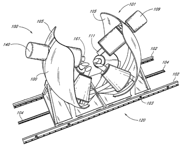
its core extension rod 53. The core extension 53 varies the magnetic field
characteristics
as disclosed below. Figures 10, 10A and 10B illustrate the model 50 in the
force control
mode when the four cores are extended into the effective space 419 and where
the coil
51A is set to CW, the coil 51B set to CCW, the coil 51C to CW and the coil 51D
is set to
CCW. The resultant field geometry produces a force of 37 grains on the
catheter tip 377.
[0213] Figure 11 shows the four-coil formation 51A, 51B, 51C and 51D when
the magnetic core extensions 53 are deployed into the effective region 419.
Figure 11
further shows that by deploying the magnetic core extensions, the magnetic
field is
shaped. Figure 11 also shows the resulting magnetic field is relatively
symmetrical around
the catheter tip 377.
-29-
CA 02605912 2007-10-19
WO 2006/128160 PCT/US2006/020895
[0214] Figure 11A shows the core coil 51A with its core withdrawn, hence
forming a new geometry configured to generate a shaped magnetic field for
better control
of the catheter movements in the effective space 419.
[0215] Figure 11B shows the shaped magnetic field when the core on coil 51D
is retracted.
[0216] Figures 12 and 12A show the CGCI apparatus 1500 and magnetic field
where the magnetic field is generated by actuating and deploying the core
extensions. As
shown in Figure 12, when the current on coil 51C is set at zero, the field has
a similar
geometry to that in Figures 11A and IlB, respectively. In the case of Figures
12 and 12A,
when the current of coils 51B and 51C are set at substantially zero, the
magnetic
extension core 53 and its associated hydraulic actuating piston can vary the
deployment
distance and hence vary the field geometry relative to its respective
position. The shaped
magnetic field using the actuator-deployed variable-length extension cores
allows the
creation of an effective magnetic field geometry for control and navigation of
the CGCI
catheter tip 377.
[0217] Figures 12B, 12C, 12D, 12E, 12F and 12G show the CGCI apparatus
1500 wherein a combination of the cores and current control are used in
shaping the
magnetic field characteristics. The resultant magnetic field geometry allows
the CGCI
apparatus 1500 to shape the magnetic field by varying the magnetic circuit
characteristics
by extending and/or retracting the cores while varying the PWM duty cycle on
the power
supply. The cores are identified as 51AT through 51DT respectively. Figure 12B
shows a
condition wherein the core 51AT is deployed while core 51DT is retracted. The
magnetic
field is measured along the XZ plane.
[0218] Figure 12C shows the cores 51AT and 51DT fully extended. The
magnet current is set at 1%.
[0219] Figure 12D shows the coils 51B and 51C where current control is set at
1% along the YZ plane.
-30-
CA 02605912 2007-10-19
WO 2006/128160 PCT/US2006/020895
[0220] Figure 12E shows a condition wherein core 51AT is retracted. The
forces are shown on the XZ plane.
[0221] Figure 12F shows the coils 51B and 51C at a current of 1% on the XZ
plane where the geometry accommodates the catheter tip control as shown.
[0222] Figures 12G is a graphic representation of coil currents 51A and 51B at
+100%, coils 51C and coil 51D are at -100% and 1% respectively along the XY
plane.
[0223] Figures 13A and 13B show the CGCI apparatus 120. The CGCI is
configured so as to facilitate the use of X-Ray and/or other surgical medical
equipment
502 in and around the patient during operation. The two symmetrical left 100
and right
101 electromagnetic clusters are mounted on the stainless steel guide rails
102, allowing
the two sections 100 and 101 to move away from each other as shown in Figures
14, 14A
and 15. The rails 102 are bolted to a floor or mounting pad. The cluster on
the CGCI
structure 120 rolls inside the rails 102, under relatively tight tolerance to
prevent lateral or
vertical movement during a seismic event. In one embodiment, the rails 102 are
designed
to withstand the forces of a Zone 4 seismic event without allowing the CGCI
structure to
escape containment.
[0224] A stainless steel spur toothed rail 104 is bolted to the floor or
mounting
pad under the CGCI structure 120. A Servo Dynamic model HJ96 C-44 brushless
servomotor 128 (max 27 lb.-in torque) with its associated servomotor amplifier
model
815-BL 129 are provided to move the clusters 101, 100. The motor has a
reduction
gearbox with a ratio of 100:1. A stainless steel spur gear attached to the
reduction gear
shaft meshes with the spur toothed rail 104. The propulsion system 150 is
configured to
exert up to 2700 lbs. of force to move the CGCI sections 100 and 101.
[0225] Two Ledex model 175 solenoids 118 are mounted in the base of the
CGCI structure. The solenoid shafts extend into the c-channel rails. Normally
the
solenoids are de-energized and the shafts are pushed out by an internal spring
119. This
ensures that in case of a power outage or equipment failure, the CGCI does not
roll out of
-31-
CA 02605912 2007-10-19
WO 2006/128160 PCT/US2006/020895
the rails because the solenoid shafts engage into the solenoid locking shaft
holes
automatically. When moving the CGCI sections, the solenoids 118 retract the
shafts from
the holes. The motor then engages and the sections 100, 101 begin to move.
Once the
shafts have moved away from the holes, the solenoids are de-energized and the
shaft tips
(e.g., ball bearing tips) roll against the inner side of the channel. When the
shafts reach the
next locking hole the shafts are pushed into the holes by the springs and the
motor (by
interlocks) is disengaged.
[0226] In one embodiment, the control of the propulsion system 150 is
performed remotely at the CGCI control room.
[0227] Figure 13 further shows the CGCI 120 assembly when the system is set
in "operational mode." The two symmetrical clusters 100 and 101 are engaged as
described above. Figures 13A and 13B show the location of the spur toothed
rail 104 and
the brushless servo motor 128.
[0228] Figures 14, 14A, 15, and 16 are isometric views of the CGCI apparatus
120 when its main two symmetric left 100 and right 101 coil clusters are in a
fully open
mode (non operational) and the magnetic cores are retracted.
[0229] The rear view of the symmetrical one half of the CGCI, shows the
parabolic flux collector shields 105 with the C-Arm upper cylinder coil
support 106.
[0230] In one embodiment, the CGCI apparatus 120 is configured to meet the
structural as well as safety considerations associated with the generation of
a magnetic
field of 2 Tesla.
[0231] Figures 17, 17A, and 17B illustrate the scaling factors and rules of
interpretation of the scale model 50 and its electrical as well as mechanical
characteristics,
as noted in Figure 3A. The scaling factors; AT(r), Eq(1), and PF (r), Eq(2)
allow the
design of coils 51A, 51B, 51C, and 51D and the core sizes for the multi-coil
CGCI
magnetic field generators. Figure 17B further summarizes the magnetic force
equation
(F,,,) as it is applied to a permanent magnet catheter tip 377 (in one
embodiment, the
-32-
CA 02605912 2007-10-19
WO 2006/128160 PCT/US2006/020895
catheter tip is configured as a 4mm diameter x 10mm NdFeB35 magnet) and the
field
needed to push/pull the catheter tip 377 with 20-35 grains of force. The coil
clusters 110,
115, and 116 of the CGCI half section 101 and its counterpart assembly 100
generate
dB/ds field gradients between 1.6T/m to 3.OT/m. Figure 22A shows that
according to the
magnetic torque equation, the desired maximum torque is 0.013 Newton meters.
The 101
coil cluster and its symmetrical counterpart 100 generate a maximum magnetic
field
strength between B = 0.04T and 0.15T.
[0232] Figures 18, 18A, and 18B are CAD-generated machine drawings
showing the dimensional envelope of the CGCI apparatus 120. Figure 1 S shows
the
orientation of coil cluster 101 and its symmetrical counterpart 100 including
angular
orientation of the conical coils 115 and 116 respectively relative to the coil
cylinder 110.
Figure 1 8A is an orthographic side view of the right coil cluster 101
describing the
angular relationship between coil 115, 116, and 110.
[0233] Figure 18B is a top view of the structural assembly and rail system of
the CGCI apparatus 120.
[0234] Figures 19, 19A, 19B, 19C, 19D, 19E, 19F and 19G are orthographic
representations of the coil assemblies 130, identified as item 110.
[0235] In one embodiment, the Coil Assemblies include two different
geometry assemblies that contain the coils that generate the magnetic fields.
The two base
coils 115 and 116 are conical and the top coil 110 is cylindrical. Their
construction is
similar except the top coil assembly includes the hydraulically-activated
piston 109.
[0236] In one embodiment, the coils are constructed with an inner core made
of 1010 low carbon steel. The core is 134mm in diameter and 450 rmn long. Both
ends
are threaded to provide for attachment of the core to the base block and
attaching a 0.5"
thick 440 stainless steel end plate is used to hold and compress the coil.
[0237] In one embodiment, a representative coil is wound using 0.162" x
1.162" hollow copper tube 123 with a 0.090 inner diameter. The tube is wrapped
with 5
-33-
CA 02605912 2007-10-19
WO 2006/128160 PCT/US2006/020895
mil Nomex 124. The bobbin for the coil is made of Kevlar reinforced resin with
a Nomex
inner sheath. A total of 1487 tunes are wound onto the bobbin with a layer of
20 mil
Kevlar cloth placed every 4 layers of tubing. A final layer of 20 mil Kevlar
is wound on
the coil with Kevlar straps wound toroidally. Copper bus bars and hose
fittings 161 are
braised to the ends of the copper tubing. The coil is vacuum-impregnated with
resin and
placed in a prefabricated mold filled with resin.
[0238] In one embodiment, the core is screwed onto the mounting block 122
on are 106. A notched 0.50" thick 440 stainless steel disk is then slid onto
the core, a
0.50" thick Teflon compression disk 113 slides on top of the stainless steel
plate 127. The
Teflon disk helps distribute the forces of the coil onto the stainless steel
plate 114. The
finished coil 110 then slides on top with the Teflon disk placed on top. The
end disk made
of stainless steel 112 is screwed onto the core and tightened to compress the
coil.
[0239] In one embodiment, the coils are water-cooled with a water flow of 0.4
gpm. Water is provided by medium pressure hoses. Three separate water lines
from the
three coils feed into inlet and outlet manifolds 161 located in the base
structure. Cooled
water is fed by an umbilical harness.
[0240] In one embodiment, the coil assemblies are designed to withstand the
stresses caused by the coil's magnetic field. When the coils are energized,
the magnetic
forces attempt to shoot the coil off of the core. The end plates are subject
to a force of up
to 4500 lbs. and are designed to withstand many times this value.
[0241] Figures 19, 19A, 19B, 19C and 19D further show the hydraulic system
140 used in the CGCI assembly. The hydraulic system is used to position the
cores to
reduce the power needed for the coils of the cluster 101 and its left
syrmnetrical
counterpart 100. The core 111 of the upper cylinder coil 110 is hydraulically
moved closer
to the effective magnetic space of the CGCI assembly 120. The core 111 is made
of two
parts, a center core 111, and a hydraulically-actuated piston 142. During
operation, the
piston 142 can be subjected to 2200 lbs. of force, pushing and pulling on the
core 111 and
housing 109. The hydraulic system 140 includes the cylinder 141, a servo valve
143, and
a pump 144. In one embodiment, the pump is an EatonlVickers vane pump model
-34-
CA 02605912 2007-10-19
WO 2006/128160 PCT/US2006/020895
VMQ125. The pump generates an oil pressure of 1000 PSI with flow rate of 12
L/min. An
Eaton Vickers model SM4-10 servo valve electronically regulates the oil flow
to the
cylinder. The cylinder 141 is an Eaton N5J-2 cylinder with a stainless steel
shaft. The use
of stainless steel or other substantially non-magnetic material (e.g.,
aluminum, titanium,
etc.) prevents the conduction of additional magnetic fields in the vicinity of
the assembly.
The cylinder 141 provides a pushing force of 4900 lbs. and a pulling force of
4100 lbs.
The hydraulic system 140 is shown by Figure 19G, where the cylinder 141 is
mounted on
the rear of the central arc 106. The servo valve 143 and vane pump 144 are
located near
the CGCI base support 117.
[0242] Figures 20A, 20B, 20C and 21 show the construction of the coils 51A,
51B, 51C and 51D respectively.
[0243] In one embodiment, the four base coils are conical. Their construction
is similar to the cylinder coils 51AT and 51DT except for the top coil
assembly which has
a hydraulically-activated core.
[0244] In one embodiment, the coils 180 are constructed with an inner core
made of 1010 low carbon steel. The core is 134mm in diameter 450mm long both
ends
are threaded to provide a method of attaching the core to the base block and
attaching a
0.5" thick 440 stainless steel end plate to hold and compress the coil.
[0245] In one embodiment, the coils are constructed of 0.162" x 1.162"
hollow copper tube with a 0.090 inner diameter. The tube is wrapped with 5
roil 440
Nomex. The bobbin of the coil is made of Kevlar reinforced resin with a Nomex
inner
sheath. A total of 1487 turns are wound onto the bobbin with a layer of 20 mil
Kevlar
cloth placed every 4 layers of tubing. A final layer of 20 mil is wound on the
coil with
Kevlar straps wound toroidally. Copper bus bars and hose fittings are braised
to the ends
of the copper tubing. The coil is vacuum-impregnated with resin and heat
curved. The coil
is then placed in a prefabricated resist mold which is filled with pigmented
epoxy and heat
curved.
-35-
CA 02605912 2007-10-19
WO 2006/128160 PCT/US2006/020895
[0246] In one embodiment, the core is screwed onto the mounting block on are
107 and 108. A notched 0.50" thick 440 stainless steel disk 127 is then slid
onto the core.
A 0.50" thick Teflon compression disk 113 slides on top of the stainless steel
plate. The
Teflon disk helps distribute the forces of the coil onto the stainless steel
plate. The
finished coil slides on top with a similar Teflon disk placed on top. The last
piece is the
end disk 133 made of stainless steel that is screwed onto the core and
tightened to
compress the coil.
[0247] In one embodiment, the coils are water cooled with a water flow of 0.4
gpm. Water is provided by medium pressure hoses that run through a hose way
running
along the side of the arc tube. Three separate water lines from the three
coils are fed into
inlet and outlet manifolds located in the base structure. Cooled water is fed
by the
umbilical harness.
[0248] Low resistance 1/0 copper welding cables attach to the coil bus bars.
The cables run from the base of the structure to an isolated connector 166.
[0249] In one embodiment, the two conical coils have extension rods screwed
onto their ends 112. The extensions are made of 1010 steel and their ends are
cut at an
angle.
[0250] In one embodiment, the coil assemblies are configured to withstand the
stresses caused by the magnetic fields. The end plates 127 and 133 are
subjected to a force
of up to 4500 lbs. and are designed to withstand five times this value.
[0251] Figures 22A, 22B, 22C, 22D and 22E are isometric representations of
the use of the scaling Equations(1), (2), (3), (4) and (6) as applied while
expanding the
scale model 2D four-coil geometry from 80mm to the 3D full scale six-coil
geometry.
[0252] The scaling Equations(1) and (2) and the magnetic force equations(5)
and (6) are used in combination with coil current polarity and polarity
rotation
equations(8) and (9) design the magnetic circuit 400 performance.
-36-
CA 02605912 2007-10-19
WO 2006/128160 PCT/US2006/020895
[0253] Figure 22C is an isometric representation of the first order expansion
from the 2D (600mm) scale model 50 showing a four coil cluster 411.
[0254] Figure 22D is an isometric representation of the second order
expansion of Figure 22C to four coils rotated 45 in the +Y direction on a
surface of a
sphere to give a four coil semi-spherical symmetry cluster 412.
[0255] Figure 22E is an isometric representation of the third iteration of
Figure
22C wherein the four coils shown in the cluster 412 are mirror imaged on the
XY plane to
produce an eight coil spherical symmetry cluster 413.
[0256] Figure 22F is an isometric representation of the fourth iteration under
the topological transformation wherein the coil structure is reduced to a six
coil cluster
414.
[0257] Figure 22G is a graphic rendition of the CGCI apparatus 120 wherein
the configuration coil cluster shown in Figure 22F is encased with parabolic
flux return
antennas 105 and is tilted under its transformation encasement of the six coil
cluster into
the YZ symmetrical magnetic return shield 415.
[0258] In one embodiment, the topological iterations from 411 through 415
under the boundary conditions set forth by the scalability Equations(1) and
(2) are
possible because the 2D scale model 50 space is continuous and the
homeomorphism one-
to-one correspondence is preserved and as defined by the Euler-Poincare
characteristics
for such locally equivalent space of the same dimension.
[02591 Figure 22C and its simulation shown in Figures 23 and 23A shows the
resultant torque field generation performance. Figures 23 and 23B show the
gradient field
performance, where the field is shown for torque TM = 0. 12T and where
gradient for force
central is F,,, = 1.20 T/m.
[0260] Figures 22D, 24, 24A and 24B illustrate the topological transformation
from the geometry configuration of the four coil circular symmetry (Figure
22C) to its
-37-
CA 02605912 2007-10-19
WO 2006/128160 PCT/US2006/020895
topologically homeomorphic four coil semi-spherical symmetry cluster 412. (The
transformation is a rotation of the four coils 45 in the +Y direction on the
surface of the
sphere generated from the 600mm circle with the center at X, Y, Z = 0). The
orientation
of the coils 51A, 51B, 51C and 51D are placed on a semi-circular topology. The
transformation allows evaluation of the performance of the 2D model 50
relative to
orientation. The resultant data from the transformation 412 is shown in Figure
22A for
torque control T,,, = 0.085T and in Figure 24B for gradient for force control
(F,,,) = 0.85
Tin.
[0261] Figures 22E, 25, 25A and 25B show the topological transformation
413. (The transformation is an XZ plane mirror duplicate of the four coils).
The four coil
semi-spherical symmetry is duplicated to generate an XZ axial return path for
the
magnetic field. The physical configuration is shown in Figure 22E and the
field for torque
control T,,, = 0.265T is shown in Figure 25A, where the gradient field
performance F,,, _
1.65 Tin. The symmetrical arrangement of the coils does not violate the
predictable
magnetic relationship identified in Figures 2, 2A, 3, 3A and 4.
[0262] Figures 22F, 26, 26A, 26B and 26C show the topology transformation
414 wherein the upper cluster top left 51AT and top right 51DT coils are
rotated 45 back
to the XZ plane and combined into a left and right top coil respectively. The
resultant
geometry is shown in Figure 26A where the performance is measured with coils
of equal
Ampere-turns (AT) and found to be symmetrical where B < 0.15 Tesla. The
geometry is
further investigated where coils 51AT and 51DT are fitted with twice as many
Ampere-
turis. Figure 26B shows symmetrical performance on the centerline and B =
0.162T. The
symmetrical geometry performance shown in Figure 26B is further confirmed by
Figure
26C where the gradients are symmetrical and dB/dS = 1.7 Tin.
[0263] In one embodiment, the cylindrical upper coils 51AT and 51DT are
provided with twice the Ampere turns (AT) of separate lower coils so as to
allow a
symmetrical force and force gradient as shown.
[0264] Figures 26D, 26E and 26F illustrate the CGCI configuration when the
coil clusters 100 and 101 are fitted with the parabolic flux return shields
105. The six-coil
-38-
CA 02605912 2007-10-19
WO 2006/128160 PCT/US2006/020895
configuration and magnetic circuit is further enhanced by the use of such
parabolic shields
to collect the magnetic flux radiated above and beyond the effective
boundaries. As
shown in Figure 26E, in the six coil cluster with shield 105, the magnetic B
405 field is
symmetric and B = 0.173 Tesla. Figure 26F shows that in the six coil cluster
configuration
with shield, the gradient field mode 406 is symmetric and dB/dz = 1.8 Tesla/m.
The
shielding produced by the parabolic antennas 105 is such that with a B field
of 20 gauss to
2 Tesla, the effective perimeter magnetic field is less than 20 gauss 12" away
from the
CGCI apparatus 120. The effective mass of the shield 105 further improves the
overall
magnetic circuit and improves the magnetic circuit.
[0265] Figures 26G, 26H and 261 illustrate the topological transformations as
they alter the maximum field strength and field gradient. The transformation
from one
iteration to the next assumes similar conditions as to power and coil size and
evaluates the
transformations relative to torque control field variations in the magnetic
center. The steps
of the transformation are shown in percentage where the scale model 50 is set
as the base
configuration shown in Figure 3 respectively.
[0266] Figure 22H shows measured field strength and field gradient
parameters. Power optimization is a measure of efficiency. One performance
benchmark
is the power consumption of the CGCI apparatus 120.
[0267) The graph in Figure 26G shows the similarity between performance of
the eight coil chapter (Figure 22E) and the six coil cluster (Figure 22G).
Each topology
includes the use of retractable cores for field control.
[0268] Figure 26H further illustrates the force control gradient variations in
the 100mm region. The effective field around the magnetic center, over the
steps of the
transformation is shown in Figures 221, 3A and rule 409 for coil current
direction and its
resultant B-field direction 408. The figure further shows the performance of
the eight coil
Figure 22E and six coil Figure 26B topologies.
[0269] Figures 27, 27A and 27B show the common topological characteristics
which allow the transformation from the 2D four coil configuration 50 to the
3D eight
-39-
CA 02605912 2007-10-19
WO 2006/128160 PCT/US2006/020895
coil spherical configuration. The homeomorphism characteristics of the 2D 50
to 3D
geometry and the four coil cluster to the eight coil cluster are expressed by
the fact that
various transformations of the CGCI apparatus include the following
relationship:
BTq = B.rr, cos ($) (405.1)
for torque control fields and
dB dB 'cos(9)
ds ds (406.1)
for force control fields, where Bxy is the field in the XY plane, and 19 is
angle of
spherical rotation of the coils from the XY plane.
[0270] In one embodiment, the use of the relationship shown by 405.1 and
406.1 is established by the eight coil spherical configuration. Figure 22E
demonstrates
that the eight coil cluster doubles B and Grad B fields as configured to the
four coil 45
semi-spherical configuration. The relationships shown by expressions 405.1 and
406.1 are
further shown in the six coil cluster configuration. Figure 26B reproduces the
eight coil
performance by doubling the ampere-turns of the top coils 51AT and 51DT in the
XY
plane.
[0271] The field strength for torque control in the magnetic center and the
field gradient for force control have the following relationship for various
configurations
in the topological transformation steps:
1
dsdBS
ds l I BTQC l
where is the scalar absolute value of the gradient along line S and is the
scalar value of the field in the magnetic center.
[0272] Figure 27 shows the torque control field vector diagram on the XZ
plane (B-vector). Figure 27A is a result of the simulation of the torque
control field
diagram on the YZ plane (B-vector). Figure 27B shows the behavior of the B-
vector on
the XY plane.
-40-
CA 02605912 2007-10-19
WO 2006/128160 PCT/US2006/020895
[0273] Figures 28, 28A and 28B illustrate the principles 406.2 by showing B-
field showing gradient in the XZ plane. Figure 28A further shows the B-field
showing
gradient in the YZ plane. Figure 28B demonstrates the B-field showing gradient
in the
XY plane.
[0274] Figure 29 is a perspective view showing one embodiment of the
Virtual Tip user input device 905. The Virtual Tip 905 is a multi-axis
joystick-type device
that allows the surgeon to provide inputs to control the position,
orientation, and rotation
of the catheter tip 377.
[0275] In one embodiment, the Virtual Tip 905 includes an X input 3400, a Y
input 3404, Z Input 3402, and a phi rotation input 3403 for controlling the
position of the
catheter tip. The Virtual Tip 905 further includes a tip rotation 3405 and a
tip elevation
input 3404. As described above, the surgeon manipulates the Virtual Tip 905
and the
Virtual Tip 905 communicates the surgeon's movements to the controller 501.
The
controller 501 then generates currents in the coils to effect motion of actual
catheter tip
377 to cause actual catheter tip 377 to follow the motions of the Virtual Tip
905. In one
embodiment, the Virtual Tip 905 includes various motors and/or actuators
(e.g.,
permanent-magnet motors/actuators, stepper motors, linear motors,
piezoelectric motors,
linear actuators, etc.) to provide force feedback to the operator to provide
tactile
indications that the catheter tip 377 has encountered an obstruction of
obstacle.
[0276] Figure 30 is a block diagram of a radar system 1000. The radar 1000
shown in Figure 30 includes a phased-array radar module 1100 having
transmit/receive
antenna elements and a Radio Frequency (RF) module 1150. The radar system 1000
includes an amplifier 1101, an A/D converter 1102, a Fast Fourier Transform
module
1103, and a microcontroller 1105. The apparatus further includes a memory
module in the
form of RAM 1112, and a look-up table in the form of a ROM 1111. One
embodiment
includes a voice messaging and alarm module 1110, a set of control switches
1109, and a
display 1108. The data generated by the radar system 1000 is provided to the
CGCI
apparatus 501 via communications port 1113.
-41-
CA 02605912 2007-10-19
WO 2006/128160 PCT/US2006/020895
[0277] In one embodiment, the radar system 1000 includes a phased-array and
uses Microwave Imaging via Space-Time (MIST) beam-forming for detecting the
catheter
tip 377. An antenna, or an array of antennas, is brought relatively near the
body of the
patient and an ultra wideband (UWB) signal is transmitted sequentially from
each
antenna. The reflected backscattered signals that are received as radar echoes
are passed
through a space-time beam-former of the radar unit which is designed to image
the energy
of the backscattered signal as a function of location. The beam-fonner
spatially focuses
the backscattered signals so as to discriminate it from the background clutter
and noise
while compensating for frequency-dependent propagation effects. The contrast
between
the dielectric properties of normal tissue and the catheter tip 377 (formed
out of a ferrite
such as samarium-cobalt SinCo5, or neodymium-iron-boron, NdFeB, etc.), in the
regions
of interest produces sufficient backscatter energy levels in the image to
distinguish normal
tissue from the catheter tip 377, affording detection and discern ability. A
data-adaptive
algorithm is used in removing artifacts in the received signal due to
backscatter from the
body tissue interface (e.g., the skin layer). One or more look-up tables
containing the
known dielectric constants of the catheter tip contrasted against the
background dielectric
information relative to the biological tissue can be used to identify features
in the radar
image.
[0278] In one embodiment, the physical basis for microwave detection of the
catheter tip 377 in the biological tissue is based on the contrast in the
dielectric properties
of body tissue versus the signature of the catheter tip 377. The contrast of
the dielectric
values of biological tissue versus that of the catheter tip is amplified,
filtered and
measured.
[0279] A typical summary of dielectric properties in living tissues for
medical
imaging in the range of 10 Hz to 20 GHz and parametric models for the
dielectric
spectrum of tissues are given up by C. Gabriel et al., "The dielectric
properties of
biological tissues: II. Measurements in the frequency range 10Hz to 20 GHz"
Phys. Biol.,
vol. 41, 1996a, p2251-69, which yields an (s'),of 5-60 and electrical
conductivity ((5) of
0.065-1.6 Simens/m (Shn) the relative complex permittivity, s,=, of a material
is:
E,.=,+JEõ
6,=E/0
-42-
CA 02605912 2007-10-19
WO 2006/128160 PCT/US2006/020895
E" = U/ Coco
Where s is the permittivity, Eo is the pennittivity of free space = 8.854e-12
Farads/m, e " is
the relative dielectric loss factor and co is angular frequency. Combining the
above
expression with a look-up table for material dielectric properties yields the
data
distinguishing between the magnetic tip 377 and tissue.
[0280] In one embodiment, the radar 1000 return waveform is provided to a
computer using a software such as MATLAB. A target such as the catheter tip
377 is
sampled with a transmitted pulse of approximately 100 ps in duration
containing
frequencies from 400Hz to 5GHz with a range of approximately 1 meter in air
(the range
of the electromagnetic coil location). The radar emits a pulse every 250 ins
(4MHz). The
return signals are sampled and integrated together to form the return waveform
as
measured on circuit 1000. A specific window of data of the radar interaction
with the
target 377 is obtained and a Fast Fourier Transform (FFT) of the window of
data is taken
to produce the frequency response of the target
,v
X(k) = , x(,l)TT,'
and by taking a Fast Fourier Transform (FFT) 1103 we are able to identify the
differences
between received radar waveform, such as metal 377 or human tissues. This
process uses
the look-up table residing in the ROM 1111 of the system. The synthetic
aperture radar
1117 (SAR) aid in the signal processing, thus, making the antenna seem like it
is bigger
than it really is, hence allowing more data to be collected from the area to
be imaged.
[0281] In one embodiment, synthetic aperture processing is using two
modalities, radar processing method as noted above or time domain focusing
technique,
wherein propagation distance is computed by 959.
d =? ()` +tz)2
[0282] and alternatively a propagation time computed by 960.
t- (x)Z+(/
V
[0283] Hence, target identification and matching is performed by
characterizing the target waveform of the catheter tip 377 into a vector. The
dot product is
-43-
CA 02605912 2007-10-19
WO 2006/128160 PCT/US2006/020895
taken from the identification vector and the data, wherein, perfectly aligned
data and ID
results in a dot product of 1, and data perpendicular to the ID results in a
dot product
equal to zero. The radar controller 1105 converts the results to percent match
(dielectric
value, conductivity measure) of the data of the identification vector.
[0284] The catheter tip 377 has a microwave scattering cross-section that is
different relative to biological tissue of comparable size, relative to their
dielectric
properties, which is indicated by greatly different backscatter energy
registered by the
receiver, and processed so as to afford a pictorial representation on a
monitor 325 with a
significant contrast between the two mediums. The pictorial view of the
catheter tip 377
generated by the radar system 1000 can be superimposed over the X-ray
fluoroscopy
image and its coordinate data set linked to the CGCI controller 501 for use as
a position
coordinate by the servo feedback loop. Hence, microwave imaging via space-time
(MIST)
beam-forming is used for detecting backscattered energy from the catheter tip
377 while
the background is biological tissue.
[0285] In one embodiment, the radar system 1000 detects the presence and
location of various microwave scatters, such as the catheter tip 377, embedded
in
biological tissue. The space-time beam-former assumes that each antenna in an
array
transmits a low-power ultra-wideband (UWB) signal into the biological tissue.
The UWB
signal can be generated physically as a time-domain impulse 960 or
synthetically by using
a swept frequency input. In one embodiment, the radar system 1000 uses a beam-
former
that focuses the backscattered signals of the catheter tip 377 so as to
discriminate against
clutter caused by the heterogeneity of normal tissue and noise while
compensating for
frequency dependent propagation effects. The space-time beam-former achieves
this
spatial focus by first time-shifting the received signals to align the returns
from the
targeted location. One embodiment of the phased-array radar 1000 forms a band
of finite-
impulse response (FIR) filters such as relatively high dielectric doping in
antenna cavity,
forming the reference signal, where the doping is relative to the device of
interest. The
signals from antenna channels are summed to produce the beam-former output. A
technique such as weights in the FIR filters can be used with a "least-squares
fitting"
teclulique, such as Savitzky-Golay Smoothing Filter, (as explained, for
example, in
Numerical Recipes, The Art of Scientific Computing, by W.H. Press, B.P.
Flannery, S.A.
-44-
CA 02605912 2007-10-19
WO 2006/128160 PCT/US2006/020895
Teukolsky and W.T. Vettrling, Cambridge, University Press, 1992, Chapter 14.8)
to
provide enhancement of the received signal and to compute its energy as a
function of the
dielectric properties versus the scattered background noise of body tissue,
thereby
providing a synthetic representation of such a signal. The system can
distinguish
differences in energy reflected by biological tissues and the catheter tip 377
and display
such energy differences as a function of location and co-ordinates relative to
the fiduciary
markers 700Ax through 700Bx, thereby providing an image proportional to the
backscattered signal strength, which is further used by the CGCI controller
501 in
computing the position co-ordinates and orientation of the catheter tip 377
relative to the
stereotactic framing of the fiduciary markers. The details of the formation of
the
coordinate settings of the catheter tip 377 relative to the stereotactic frame
and the
synchronization of such image with the fluoroscopy frame 702 is further
described. In one
embodiment, the radar module 1000 uses an FFT algorithm 1103 which uses a
filtering
technique residing in look-up tables 1111 to allow the radar 1000 sensor to
discern
various of dielectric properties of specific objects, such as a guidewire 379
and/or a
catheter 310 with piezoelectric ring 311 and 312.
[0286] Figure 30A is a graphical representation of the catheter tip 377
embedded with one or two piezoelectric rings 311 and 312 such as Lead-
Zirconate-
Titanate (PZT) and/or molecularly conjugated polymers such as switchable
diodes
(polyacetylene). The second harmonics generated by the rings 311 and 312
provide an
identifiable return signature in the second harmonic due to the non-linearity
of the
material. While the fundamental harmonic (e.g., 5 MHz) is transmitted by the
radar, the
second harmonic (e.g., 10 MHz) is distinguishable by the radar system 1000.
The radar
system 1000 can discern between the catheter tip (which is formed out of
ferrite such as
samarium-cobalt SinCo5, or neodyiniumn-iron-boron, NdFeB, etc.) and the PZT
rings 311
and 312. The ability to distinguish between the signal return from the
catheter tip 377 and
the PZT rings 311 and 312, allows the radar system 1000 to filter out the
background
clutter received from the body tissue and recognize the position and
orientation of the
rings 311 and 312 and the position coordinates of the catheter tip 377. The
technique of
using two different dielectric properties and electrical characteristics of
the tip 377 versus
the PZT rings 311 and 312 provides the catheter tip 377 with a radar signature
that is
unique and readily recognized by the radar system 1000.
-45-
CA 02605912 2007-10-19
WO 2006/128160 PCT/US2006/020895
[0287] Figure 30B further shows how the radar system 1000 with its transmit
and receive antennas is used to detect the position coordinates and
orientation of catheter
tip 377 relative to its two PZT rings 311 and 312. A geometrical manipulation
is
employed by the radar system 1000 and its associated FFT filter 1103 by the
resident
microcontroller 1105. As shown in Figure 2D, a catheter-like device is
provided with a
magnetically-responsive tip 377. In one embodiment, the tip 377 includes a
permanent
magnet. The polarity of the permanent magnet is marked by two PZT rings where
the
north pole is indicated by a PZT ring 312 and the distal end of the ferrite
where the semi-
flexible section 310 of the catheter 376 is marked with additional PZT ring
311, also
marking the south pole of the ferrite.
[0288] In one embodiment, the radar system 1000 transmits a burst of energy
that illuminates the ferrite catheter tip 377. The return signal from the
catheter tip 377 is
received by the radar and its position is registered by observing the time of
flight of the
energy, thereby detennining the location of the catheter tip 377 as position
coordinates in
a three-dimensional space. By employing the two PZT rings 311 and 312, the
radar
detector 1000 is also configured to discerning the location of the tip 377
relative to the
two PZT rings so as to afford a measurement of PZT ring 312 relative to the
second
piezoelectric ring 311 with reference to the position coordinates of the
catheter tip 377.
The radar detector 1000 can discern the return signal from PZT rings 311 and
312 due to
the non-linear characteristic of PZT material that generates a second harmonic
relative to
the incident wave. By comparing the strength of the fundamental frequency and
the
second harmonic, the radar system 1000 is able to discern the position and
orientation of
the two PZT rings relative to the ferrite 377, thereby providing position and
orientation of
the catheter tip 377.
[0289] Figure 30B shows the technique of measuring the position and
orientation of the catheter tip by the use of the radar detector 1000 and
using fiduciary
markers 700AX and 700BX to form a frame of reference for the catheter relative
to the
frame of reference of the markers. As shown in Figures 30B and 30F, the
fiduciary
markers 700AX and 700BC form a manifold 701. The locations of the markers
700AX
and 700BZ are measured by the radar system 1000.
-46-
CA 02605912 2007-10-19
WO 2006/128160 PCT/US2006/020895
[0290] In one embodiment, the markers are electrically passive and can be
made from a polymer or PZT material that allows the radar antenna to receive
an RF
signal return which is discernable by its harmonic structure. Criteria such as
the
conductivity of the body affects how much the radar signal is attenuated for a
given depth
(i.e., the relatively higher the conductivity the relatively higher the loss
for a constant
depth). An average conductivity of 1S/in at 1GHz signal will penetrate the
human body
approximately 1.8cm.
[0291] In one embodiment, the dielectric constant of most targets will be -1.
The relative permittivity of the targets is typically of several orders of
magnitude lower
than that of the surrounding tissue. The conductivity of the metals is
typically several
orders of magnitude greater than that of the surrounding tissue. For example,
the
permittivity of nylon is 2-3 orders of magnitude less than that of the
surrounding tissue
(within a bandwidth of 1MHz-1GHz).
[0292] Hence, the dielectric properties as well as the conductivity measure of
the target catheter tip 377 and/or its directional markers PZT rings 311 and
312 allow the
radar 1000 to discern the target out of the surrounding clutter (e.g., body
tissue 390) and
perform the task of position definition 377 within the referential frame of
fiduciary
markers 700AX and 700BX.
[0293] In one embodiment, the return waveform is recorded for a static
(clutter) environment, and then a target is inserted into the environment and
once the
clutter is subtracted from the return waveform the radar 1000 processes a
target response
(clutter is a general tern referring to anything the radar interact with that
is not a desired
target).
[0294] Figures 30C and 30D show all image captured by monitor 325. The
cineoangiographic image 702 of an arterial tree is shown with a reconstructed
radar
signature of the catheter tip 377. The image 702 contains a numerical grid
defined and
calculated by the radar 1000 and data set of coordinate or vector
representation of catheter
position (actual position AP) is displayed. Data on catheter position is fed
to the controller
-47-
CA 02605912 2007-10-19
WO 2006/128160 PCTIUS2006/020895
501 for the purpose of closing the loop of the servo control systems of the
CGCI
apparatus 1500. An illustration of the catheter tip 377 is shown in Figure
30A, wherein
monitor 325 displays the stereotactic frame formed by the fiduciary markers
700AX and
700BX obtained from the radar signature 1000. The catheter tip 377 is shown as
a cube
formed by the fiduciary markers 700AX and 700BX. The position data relative to
the
coordinates is used to form a dynamic manifold 704. The manifold 704 allows
synchronization of the catheter tip position (AP) relative to the stereotactic
frame 701.
The process of synchronization is gated in the time domain with aid of an EKG
electrocardiogram 502, where an internal clock of the controller 501 is
synchronized with
the EKG QRS complex so as to provide a Wiggers' diagram. The synchronization
allows
the CGCI controller 501 to gate the dimensional data and coordinate set of
fiduciary
markers so as to move in unison with the beating heart.
[0295] Synchronization of the image of the catheter tip 377 or guidewire 379,
captured by the radar system 1000, is superimposed onto the fiduciary markers
which are
represented digitally and are linked dynamically with the image 702. This is
done to
create a combined manifold 701, which is superimposed onto the fluoroscopic
image 702.
The combined manifold moves in unison with area of interest relative to the
anatomy in
question. For example, the beating heart, the pulmonary expansion and
contraction, and/or
spasm of the patient are dynamically captured and linked together so as to
achieve a
substantial motion in unison between the catheter's tip and the body organs.
[0296] Synchronization 701 of the catheter tip 377 with its referential
markers
700AX and 700BX, dynamically calibrate the relative position allows the CGCI
1500 to
capture the data set-manifold 704 on the time domain of the patient 390 EKG
signal. This
allows the CGCI controller 501 to display and control the movement of the
catheter tip
377 in unison with the beating heart or other body movements. Synchronization
to close
the servo loop modality is also used by the controller 501.
[0297] The CGCI controller can perfonn the data synchronization without
active use of x-ray imagery since catheter position (AP) 377 data is provided
independently by the radar signal 1000. In one embodiment, the radar data is
used to
close the servo loop.
-48-
CA 02605912 2007-10-19
WO 2006/128160 PCT/US2006/020895
[0298] Figure 30E shows the use of the apparatus described in Figures 30C
and 30D while performing a pacemaker electrode implantation. Figure 30B
further shows
the implantation of cardiac pacemaker 801 with electrodes as shown, placed in
an area
relative to the S.A. Node 802, A.V. Node 803, and the bundle of HIS 804.
Further
illustrated are the right and left bundle branches 805. This procedure is
performed by the
implantation of a small electrode in the heart cavity wall (ventricle or
atrium). The other
end of the electrode is attached to an electronic device 801 which is
implanted under the
chest skin and which generates stimulation pulses to simulate the heart
rhythm. Similar
devices apply electrical shock when life-threatening heart electrical
disturbances are
detected by the electrodes. These electrodes are typically placed through a
vein by pushing
and manipulating under fluoroscopy. Through the use of the CGCI apparatus
1500, the
guidewire 379 fitted with a magnetic tip 381 is used to carry and place the
electrodes of
the pacemaker 801 in their proper position. With the fiduciary markers 700A
and 700B in
place, the physician navigates the guidewire 379 through the heart lumen while
having a
continuous dynamic referential frame identifying the guidewire tip 381 using
the position
data from radar 1000 as shown in Figure 30C and further illustrated by Figure
30D. Often
the manipulation to place the electrodes in the proper position is difficult
and the results
are sub-optimal due to anatomical variations. The use of the controller 501
simplifies the
operation allowing the physician to place the electrodes of pacemaker 801 in
desired
anatomical positions. The CGCI apparatus allows the procedure to be performed
accurately, with minimal exposure to ionizing radiation.
[0299] Figures 31, 31A and 31B show one embodiment of the CGCI 1500,
where the CGCI six coil magnetic configuration is used to explain the control
algorithm.
The system controller 501 of the six coil magnetic configuration 400 uses a
matrix of coil
combinations, bipolar coil current settings 409, and piston 111 movement
control. Torque
control 406 is used to push and pull the tip 377 around its axis. Force
control is used to
push and pull the tip 377 along a path and to provide a controlled mix of
torque and force
for negotiating curved paths in arteries. Figure 30 show the bottom four coils
51A, 51B,
51C and 51D and the top two coils 51AT and 51DT. The coil current polarity is
defined
by facing the coil pole 402 from the center region with the patient's
perspective with the
patient positioned along the Y axis. Polarity is positive for clockwise (CW)
current flow
-49-
CA 02605912 2007-10-19
WO 2006/128160 PCT/US2006/020895
on the "left side" for coil 51AT, 51B and 51A. Polarity is positive on the
"right side" for
coils 51DT, 51C and 51D in the counter clockwise (CCW) current direction (see
Figure 4
for combination settings).
[0300] Each of the coils 51A, 51B, 51C, 51D, 51AT and 51DT are separately
fed and controlled from a bipolar power source 526. The power source is
controlled from
a central six channel regulator 525 assisted by the computer 527 containing
the matrix
algorithm 528 for the three modes 405 (Torque), 406 (Force), and sloped 417,
418 (L)
noted above.
[0301] In one embodiment, the "man-in-loop" control is a joystick (JS) 900
and its virtual tip 905. In one embodiment, a "fire" thumb button serves as a
selection
between force and torque modes. The movement of the stick forward, left, back
and right
rotates the catheter tip 377 (using torque mode) around its axis in these
directions. When
the push-button is pressed, the catheter tip 377 is moved forward, left, right
and back
(force control). When the JS 900 "fire" push-button is not pressed, the
computer 527 uses
the torque matrix 528 tables (see Figure 4) available to the regulator 527.
[0302] In one embodiment, the example below expresses a simple matrix used
for regulating a full catheter 377 rotation. The matrix locks the possible
current polarity
combinations and sequences for the six coils for torque fields. Selection of
the valid
combination for the location and direction of the tip 377 is set when the
joystick 900 is
moved by the operator. The torque field 405 begins to rotate while the JS 900
is pressed.
When the JS 900 is released, the field is held constant. A "right click" on
the JS 900
button drops the field to zero. Similar matrix selections and coil current
regulation setups
are available for JS 900 force control 406.
[0303] The coil current polarities and magnitudes are set to produce the
desired field directions for the torque and force fields. The torque field
generating
combinations uses an adjacent coil current direction such that the B-vector
flows from
core to core aiding each other. The coils 51A, 51B, etc., are viewed as if
connected in
series linked by a common magnetic field as shown in Figure 30A. The force
field
generating coil combinations uses an adjacent coil circulating their current
such that they
-50-
CA 02605912 2007-10-19
WO 2006/128160 PCT/US2006/020895
work against each other as shown in Figure 31C. There are 64 combinations of
positive
and negative current flow polarities for the six coil design. The 2D baseline
configurations 50 of four coils can have 16 combinations, half of them
generate torque
fields, the other half are force gradient configurations. Once the
coil/polarity
combinations are defined, they can be grouped into a set of matrixes according
to above
rules. Torque and force matrixes are extracted according to four coil and
three coil groups
associated with virtual 2D planes as follows: Coils 15AT, 51DT, 51B and 51D
form a
four-coil group with 16 polarity combinations. This can be considered an
approximation
of the four coil XY plane baseline design from which the six coil
configuration through
topological transformations.
[03041 The coils 51AT, 51DT, 51A, and 51D form another group on a plane
rotated 90 from the group above. Again there are 16 combinations for two/two
sets of
torque/force matrixes. The third group is formed as two triangular "side
plane"
combinations of 8 and 8 combinations for two/two sets of torque/force
matrixes, (shaped
magnetic field 417).
[03051 Selecting the right combination of coils 51X and current polarities
from each of these virtual planes is performed by the computer 527 and
algorithm 528 by
applying the superposition rules. The selection occurs when the JS 900 is
activated. As
shown by previous Figures in deriving the 3D six coil geometry, there is
always a
coil/polarity combination set for the desired direction within the magnetic
boundary. In
case of possible multiple selection for the same mode and direction, the
algorithm 528
selects a single combination based on possible combinations available for
anticipated
movement in the same direction and in accordance with the rules of optimal
power
setting.
[03061 Figure 31C shows a simplified example by using the XY plane to
illustrate the control scheme of the CGCI apparatus 1500. The case illustrated
by Figure
30C is a demonstration of the control requirements of a torque field rotation
of the
catheter tip 377 around the XY plane. The graphic representation shows the
coil current
polarity selection and actual control of the coil currents to achieve a
continuous rotation
without changing the field intensity. The torque matrix is shown in Figure 30C
and when
the "fire" push-button is released and the JS 900 is turned right, the B-
vector torque field
-51-
CA 02605912 2007-10-19
WO 2006/128160 PCT/US2006/020895
rotates clockwise going through the following steps: the catheter tip 377
located in the
XY plane (shown in case 1) facing in the +X direction and if the operator
wishes to rotate
the tip 377 in the negative rotational -Y and continue with the rotation over
360 . To
achieve such condition, the CGCI controller 501 brings the field up in the +X
direction by
increasing the four coil 51X currents to 100% in positive polarity shown in
30C, case 1.
Reducing coil current 51B and 51D to zero causes the field to rotate toward
the 45 line,
and the catheter tip 377 reaches its position as shown in 30C, case 2.
Reversing polarity in
the coils 51B and 51D rotates the field and the catheter tip 377 toward the -
90 line.
When the 51D current is equal - 100%, the field faces in the -Y direction, see
30C, case
3. Reducing the coil current of 51A and 51C, causes the field to rotate to -
135 degree, see
30C, case 4. By increasing the current in coils 51A and 51C in the reverse
direction, the
field rotates to the 180 position, see 30C, case 5. By reducing the coil
current 51B and
51D back to zero, the field rotates to -225 , see 30C, case 6. By increasing
the coil current
51B and 51D to positive 100% the field is set to -275 , see 30C, case 7.
[0307] Reducing current 51A and 51C down to zero causes the field to point
to the 315 line, see 30C, case 8. The rotational circle is completed by
turning to Figure
30C, case 1. All the rotational steps are reached smoothly with continuous
control of the
coil current oscillating from -100% to 100% through zero, the central slope
between the 0
to 100% coil currents is not a linear function of the coil current but
follows an inverse
cosine function.
I O!
0=100s,
2 '100% (420)
where 0 is the B vector rotation angle, I coil is actual coil current, and
1100% is the coil
current of full field strength.
[0308] Figure 31D shows the control scheme used in the force mode as
described in connection with Figure 30C. The force matrix used for setting a
coil/polarity
combination shown above in describing 30C apply and the same regulation
circuits can
control the current magnitudes.
-52-
CA 02605912 2007-10-19
WO 2006/128160 PCT/US2006/020895
[0309] Figures 32, 32A, 32B, and 32C show the CGCI 1500 low level logic
simulation of the magnetic circuit 400. The torque angle control circuit real
time behavior
is simulated using SPICE for the first - 90 rotation, while the coil winding
parameters are
R = 1.752 and L = 1.611 and the inverse cosine function 420 is also used. The
case shown
by circuit analysis in Figure 32 is a demonstration of how the CGCI controller
501 and its
algorithm performs the desired position change of the operator input 500 and
its
mechanical movement by the JS 900 so as to simulate this change on the low
level logic
and the virtual tip 905 prior to initiating the power level of the DC
amplifier 525 and its
coil counterpart 51A etc. In this simulation, V5 is the regulator 8001
controlling the
power supply voltage of coil 51D and coil 51B (B2 8004 and B1 8005
respectively). V5
8001 is the control variable in this simulation, and it is selected to the
coil currents in 51B
and 51D by the torque matrix step Figure 31C, 1, 2, and 3. The currents of the
51B and
51D go up to +100% together with 51A and 51C, but then 51D and 51B go through
zero
(-45 ) and reverse polarity to -100% (-90 ) while 51A and 51C are constant.
The
regulator 8001 output voltage profile shown in Figure 32B forces coil current
51B and
51D to reverse after dropping to zero. V1 8002 is the power supply for coil
51A. Initially
it ramps up to full voltage together with the V5 8001 regulator, and stays at
100% current
for this particular simulation for the rest of the rotational cycle. Because
the rotational
matrix is already set, coil 51A will not vary magnitude or polarity during the
rotation.
[0310] V3 8003 is the power supply of coil 51 C which functions the same way
as V1 8002 coil 51A above. The control matrix causes it to be same polarity
and
magnitude as VI 8002. B2 8004 is the power supply for coil 51D and its voltage
follows
V5 8001 regulator coil, as designed by the scale model 50. B1 8005 is the
power supply
for coil 51B and also follows the regulator 8001 command. B4, 8006 computes
the B-
field strength in percent. It takes the current of coil 51A (equal to 51C) and
defines B in
percent (%) by using the following equation.
B ~=100= sin L
C' [U01 (421)
where IA varies from 0 to 100.
[0311] B3 8007 computes the rotational angle according to the following
equation:
-53-
CA 02605912 2007-10-19
WO 2006/128160 PCT/US2006/020895
0=- - Cos' Ip
? IA (422)
[0312] where IA and ID are coils 51A and 51D currents. The rotational
procedure uses the regulator 8001 which controls the four coils to rise to
full current as
(shown by the parameters R = 1.75 and L = 1.6H), V Reg 8001 rises to 100%,
current
51IA, 51IB, 51IC, and 51ID rise together according to L/R time constant (see
Figure
31C), and lines up to +X at zero degree phase. The regulator controls 511B and
511D
together to zero starting at 6 seconds (Figure 31 C). 511A and 511C remain
constant. The
phase rotates to -45 while the field strength remains constant. The regulator
commands
current of coils 51IB and 51ID to reverse starting at 12 seconds (see Figure
31C). The
phase angle rotates to -90 (see Figure 31A and 31B) while the field strength
remains
constant.
[0313] The system can also proceed a B-field rotation as shown in case 31C
step 4 and 5 to rotate -90 clockwise. The matrix changes the current control
sequence so
that coil 51A and 51C go through the polarity reversal. In summary, Figures
31, 31A, 31B
and 31 C show the control scheme of the controller which provides a low level
simulation
of the magnetic circuit 400 and provides, as an additional safety measure,
verification of
the joystick 900 movement prior to activation of the power level of the coils.
[0314] Figure 33 shows the CGCI 1500 top architecture showing the major
elements comprising the controller 501 of the magnetic circuit. The controller
501
includes a system memory, a torque/force matrix algorithm residing in 528 and
a
CPU/computer 527. The CPU/computer such as PC 527 provides computation and
regulation tasks. Figure 33 further shows the six coil electromagnetic circuit
formed out
of coils 51A, 51B, 51C, 51D, 51AT and 51DT and the magnetic field sensors
(MFS) 351,
352, 353, 354, 355 and 356 such as Hall sensor ring 350 mounted on an assembly
forming
the X, Y, and Z axis controls. A D/A converter 550 and an I/O block 551
provide
communication between the controller 501 and the coils 51A and the
hydraulically-
systems 140. The six channel DC amplifier 525 provides current to the coils.
-54-
CA 02605912 2007-10-19
WO 2006/128160 PCT/US2006/020895
[0315] Figure 33 shows the relationship and command structure between the
joystick 900, the virtual tip 905, and the CPU 701. The CPU 701 displays
control
conveying real time images generated by the X-ray, radar 1000, or other
medical imaging
technologies such as fluoroscopy, MRI, PAT SCAN, CAT SCAN, etc., on a display
730.
A flow diagram of the command structure of the control scheme is shown by the
use of
the 2D virtual plane coil polarity matrixes. By assigning the coil position
and polarity
elements to the directions of torque rotation and force field gradient on each
2D plane of a
six coil cluster 414, a computer program such as MathLab or Math Cad is able
to sift
through the combination matrixes and compute the proper combination for the
six coil
current polarities and amplitudes. In one embodiment, a boundary condition
controller is
used for regulating the field strength 405 and field gradient 406 in the
effective region.
The controller 501 computes the fields in the neighborhood of the catheter tip
377 and as
defined by the fields on the 2D planes in the effective area. Rules for
computing the fields
with rotated coil on the surface of the sphere are given by Equations 405 and
406 and the
topological transformation 407 for the six coil CGCI 1500 configuration.
[0316] In one embodiment, look-up tables are used as a reference library for
use by the controller 501. Lookup tables of the setting of various scenarios
of force as
well as torque position and magnitude allow the controller 501 to use a
learning algorithm
for the control computations. The look-up tables shorten the computational
process for
optimal configuration and setting of the coil currents and pole positions. The
D/A and
AID system 550 allows the connection of voltage and current measuring
instruments as
well as input from the magnetic field sensor (MFS) 350 array, the MFS 351,
352, 353,
354, 355 and 356. The magnetic field sensor measuring the boundary plane field
strength
allows the CGCI to use a low-level logic algoritlun to compute the positions,
settings, coil
currents, etc. The low-level simulation is performed prior to activating the
power section
of the CGCI apparatus 1500, thus, providing a "soft" level check prior to
action
performed by actual machine. The two-level control architecture that starts
with low-level
simulation architecture of low-level simulation allows the surgeon or operator
of the
CGCI apparatus 1500 to test each movement prior to actually performing the
move.
[0317] Figures 34, 35, and 36 illustrate the field regulator loop outlined in
Figure 33 using the Hall effect ring 350. Figure 34 shows the generator
interface joystick
-55-
CA 02605912 2007-10-19
WO 2006/128160 PCT/US2006/020895
900 and its virtual tip 905 where the user commands are initiated. In one
embodiment,
movement of the catheter tip 377 is initiated as a field having a vector with
components
Bx, By, and Bz, for torque control and a vector dBx,y,z for force control are
computated.
The B-field loop with its functional units, include a (I) regulator 901.3.
Hall effect sensors
351X, 352X, 353X, 354X, 355X, 356X, and 351Y-356Y as well as 351Z-356Z measure
the B and dB fields. Computation regulators 527.1-527.6 calculate position,
desired
position (DP) change and the desired field and field gradients. The coil
current 51A, 51B,
51C, and 51D are set and the catheter tip 377 position is changed from actual
position
9040 to desired position 9060.
[0318] In one embodiment, the movement of the catheter tip 377 is seen in
real time by the operator 500 while observing the display 730.
[0319] A visual display of the magnetic fields can be generated using 3-axis
Hall sensors 351-356 placed on the 2D planes.
[0320] The "fire" push-button on the JS 900 selects torque or force modes for
"rotate" or "move" commands. The magnitude and direction of the torque and
force are
determined by user inputs to the JS 900.
[0321] In one embodiment, the system sets the maximum torque and force by
limiting the maximum currents.
[0322] In one embodiment, catheter movement is stopped by releasing the JS
900. The fields are held constant by "freezing" the last coil current values.
The magnetic
tip 377 is held in this position until the JS 900 is advanced again. The
computer 527 also
memorizes the last set of current values. The power can be turned off for
radar
positioning, Hall effect recalibration of the sensor array and the system
returns to the
previous coil current values. The memorized coil matrix sequences along the
catheter
movement creates a computational track-record useful for the computer to
decide matrix
combinations for the next anticipated movements.
-56-
CA 02605912 2007-10-19
WO 2006/128160 PCT/US2006/020895
[03231 In one embodiment, the magnetic field is sensed by 3-axis
instrumentation-quality Hall sensors 351-356 placed in the centers of the 2D
planes (six
sensors all together). Each sensor 35X x, y, z provides the Bx, By, and Bz
components of
the field sufficient to describe the 2D boundary conditions numerically. The
measurements are used to calculate B magnitude and angle for each 2D plane.
From the
fixed physical relationship between the plane centers, the field can be
calculated for the
catheter 377. As shown in Figure 35, coils 51A, 51B, 51C, 51D forms a 2D
virtual plane
with respect to the 3-axis Hall devices wherein Hall sensors 351X identify Bx,
By, and Bz
relative to the X axis, similarly 351Y identify Bx, By, and Bz relative to the
Y axis, and
351Z identify Bx, By, and Bz relative to the Z axis respectively.
[03241 In one embodiment, the Hall sensors 351X, 351Y, and 351Z produces
three analog outputs 907. One for each component, for the A/D converter 550
shown in
Figure 33. This data is used to compute the superimposed fields in the 3D
region of the
catheter 377 (effective region 419).
[03251 Each Hall sensor 351x, y, and z is a multi-axis sensor such as the one
manufactured by F.W. Bell having three individual Hall elements oriented in
mutually
perpendicular planes. This allows the sensors to produce voltages proportional
to the three
orthogonal components (Bx, By, Bz) 907 of a magnetic flux in any direction.
Thus, the
sensors 351 can be permanently mounted or arbitrarily oriented to sense fields
in any
direction. The magnitude of the flux vector, B 907 can be found using the
following
relation:
BT B'+By+B2
The flux direction relationship is formed by using the above relationship of
angle.
ii=cos4Bx/B, b=cos-'By/B, d=cos-'Bz/B
Where a 904.1, b 904.2, d 904.3 are angles between B, Bx, By, Bz respectively.
The Hall sensors operate in an environment of 5 C temperature variation and
0 to 5
1cGauss field strength range.
[03261 The CGCI controller 501 generates a Bx, y, z, field readout error which
is approximately 2.5% for all causes including linearity, matching and various
-57-
CA 02605912 2007-10-19
WO 2006/128160 PCT/US2006/020895
temperature drifts. Some of the temperature drifts can be compensated for and
residual
flux errors can be readily offset. These error correction techniques reduce
the total readout
error to approximately 1 %. This error is inconsequential for the magnetic
field displays of
the manual man-in-the-loop control mode, and can be tolerated for a fully
closed loop
system.
[03271 Another embodiment of the CGCI controller 501 uses close loop
control wherein the biasing of the field is performed without the visual man-
in-the-loop
joystick feedback, but through position control and a digital "road-map" based
on a pre-
operative data using such as the MRI, PET SCAN, etc. The digital road map
allows the
CGCI controller 501 and the radar 1000 with aid of the fiduciary markers
700AX, 700BX
to perform an autonomous movement from the point 9040 (actual position of the
catheter
tip 377) to desired position (DP) 9060 based on closed loop control.
[03281 In one embodiment, the CGCI system has magnetic capability for
torque control up to 1.6 Tesla, and force control up to 1.7 Teslalmeter.
Precision catheter
377 positioning is based on control of the direction and magnitude of both
type fields
within these ranges. The manual control with the man-in-the-loop provides a
relatively
coarse control of these values. The JS 900 visual navigation is based on
imaging and
navigation operator skill. The precision computer-aided catheter guidance
system uses
actual magnetic field regulation for precision catheter positioning.
[03291 Field regulation 740 is based on providing the coil current control
loops used in the manual navigation system within the field regulating loop as
a minor
loop, and to be a correction and/or supervisory authority over machine
operation. Control
of B-field loops is defined by the joystick 900/905 and its associated field
commands
900.1. The closed servo loop uses position data from the radar 1000 to allow
the servo
control loop to be closed and is used as the primary loop control.
[0330] Figure 34 further shows field regulation 740. The field regulator 740
receives a command signal field 900.1 from the radar 1000 position 9040 and
the JS 900
new position 9060 data from the computation unit 528, which generates a Bx, y,
z vector
for torque control, and the dBx, y, z vector gradient for force control. This
position
-58-
CA 02605912 2007-10-19
WO 2006/128160 PCT/US2006/020895
computational value identified in Figure 34 allows the (1) regulator 901.3 to
receive two
sets of field values for comparison.
[0331] The present value (actual value 9040) of Bcath and dBcath at the
catheter tip 377 are calculated from the five 3 axis Hall effect outputs Blx,
y, z 351
through B5x, y, z 356.
[0332] The new field values for the desired position (DP) 9060 Bx, y, z 907
and dBx, y, z 907 to advance the catheter tip 377 are generated in the CGCI
controller
501. The difference is translated to the Matrix block 528 for setting the coil
currents and
polarities.
[0333] In one embodiment, the matrix 528 issues the current reference signals
to the six regulator CREG1 527.1 through CREG 527.6. The regulators 750 drive
the six-
channel power amplifier 525 to obtain the desired coil currents.
[0334] In one embodiment, the precision of field regulation is determined by
the precision of the field measurement. The Hall effect devices 351-356 have
about 1%
error under the operating room environment. The CGCI controller 501 calculates
the
catheter position error (PE) in actual distance for a particular case of a
catheter tip 377.
[0335] In one embodiment, the torque on a permanent magnet in field B 405
is:
Tin = M- B -,4111 - Lira' 5111()
where M is the dipole magnetization vector, and B is the field density vector
around the
dipole. A,,, is the magnet cross section, and L,,, is its length. For B - 0.15
Tesla the
calculated bending arm is Laõ a = 38mm. Assuming B is measured with I% error,
T,,, will
have a 1 % error.
Therefore, the position error due to measuring error of 1 % is
L,,ro,, = 100 = 0.38mrn or 0.015 inch
-59-
CA 02605912 2011-10-13
WO 2006/128160 PCT/US2006/020895
[0336] A position error of less than 0.015 will leave room for other
computational errors, and the regulation scheme provides an expectation of 22
mils. (0.22
inch) error.
[0337] Many other variations are possible within the scope of the present
invention. For example, the modulation of the electromagnets can be controlled
in such a
way as to cause a vibratory or pulsating motion of the tip to aid in crossing
plaque. The
responsive tip(s) can be electromagnetic rather than permanent magnets. The
magnetic
field external to the body can be generated by a permanent magnet or magnets.
The
control of the external magnetic field can be accomplished by manually
administering the
field generating devices. AC induction with its associated magnetic effects
can be used by
causing a coil or coils wound around the tip to respond to an impressed time
variant field.
Materials with curie temperatures within a few degrees of body temperature can
be used
as magnetic flux switches for selective tip control by irrigating them with
fluids having
appropriate temperatures; electrostatic phenomena can enhance magnetic
effects.
Artificial intelligence can replace the operator control for producing command
inputs; an
expert system can replace or augment operator inputs. The apparatus can be
used to
incubate various body cavities and organs other than the heart. The apparatus
can be used
for human and animal procedures such as egg harvesting and embryo
implantation. The
responsive tip can be attached to a coherent fiber optic bundle to provide
viewing of
internal structures with unprecedented maneuverability; internal radioisotope
therapy can
be precisely performed by delivering a palletized source directly to a tumor
using a guided
catheter. Internal tissue samples can be obtained without major surgery; a
fiber optic light
guide equipped with a responsive tip can be accurately positioned to deliver
last light to a
specific internal location without major surgery.
-60-