Note: Descriptions are shown in the official language in which they were submitted.
CA 02606378 2007-10-26
WO 2006/118772 PCT/US2006/014182
FcRN ANTIBODIES AND USES THEREOF
RELATED APPLICATIONS
This application claims priority to U.S. Provisional Application No.
60/676,412, filed April 29, 2005, the specification of which is hereby
incorporated
herein by reference in its entirety.
FUNDING
Work described herein was funded, in whole or in part, by National Institutes
of Health Grant Number NIH DK57597. The United States government has certain
rights in the invention.
BACKGROUND OF THE INVENTION
Antibodies have been known since before the 20th century to play an
important role in immunological protection against infectious organisms. The
immune system cells that produce antibodies are B-lymphocytes. There are four
major classes: iminunoglobulin M(IgM), IgG, IgA, and IgE, but IgG is by far
the
most prevalent class, comprising about 90% of all antibodies in adults. Each
class
of antibody has a specific role in immunity, including primary and secondary
immune responses, antigen inactivation and allergic reactions. IgG is the only
class
of antibody that can pass the placental barrier, thus providing protection
from
pathogens before the newborn's immune systein develops. Antibody molecules
have two ends. One end is the antigen-specific receptor, which is highly
variable
and engenders each antibody with the capacity to bind a specific molecular
shape.
The other end, referred to as Fc, has sequence and structural similarities
within a
class and confers the ability to bind to receptors on immune cells. In a
perfectly
operating immune system, the diverse specificities of the antigen specific
receptor
engenders the host with a diverse repertoire of antibodies with the ability to
bind to a
wide array of foreign infectious microorganisms, the result being destruction
of the
microbe and immunity.
-1-
CA 02606378 2007-10-26
WO 2006/118772 PCT/US2006/014182
Autoinimune diseases occur when the immune system erroneously senses
that normal tissue is foreign and attacks it. One of the most prevalent
immunological participants in autoimmune destruction are auto-antibodies,
which
are nonnal antibody molecules that have gone awry and destroy normal tissue.
This
leads to many types of autoimmune diseases, including systemic lupus
erythematosus (SLE). SLE is a prototypic disease of systemic antibody
dysregulation with the common feature of hypergaminaglobulinemia, anti-DNA
features and anti-nuclear protein antibodies, and immune complexes that
accumulate
at many sites including the kidney glomeruli, vascular system, joints and skin
(Theofilopolos and Dixon, 1985, Adv. Immunol. 37: 296-390; Theofilopolos and
Dixon, 1981, Immunol. Rev., 1981, 55:179-215; Boumpas et al., 1995, Ann Int.
Med. 122:940). The severity can range from mild to very severe, from minimally
debilitating to lethal. There are currently few effective treatments for
autoimmune
diseases.
SUMMARY OF THE INVENTION
Accordingly, it is the goals of the application to develop more effective
compositions and methods for manipulating antibody concentrations as a way to
treat autoimmune diseases.
In certain embodiments, the present invention provides an isolated antibody
or antigen binding portion thereof that binds to an epitope on human FcRn. The
isolated antibody is referred to herein as an FcRn antibody. The FcRn antibody
or
the antigen-binding portion thereof binds epitopes of human FcRn aild
selectively
inhibits the binding of the Fc portion of IgG antibody to human FcRn, but does
not
inhibit the binding of human albumin to human FcRn. In certain specific
embodiments, the FcRn antibody is a monoclonal antibody. In certain
embodiments, the FcRn antibody or antigen binding portion thereof can be
administered to a human. In certain cases, the FcRn antibody or antigen
binding
portion thereof selectively decreases the serum half-life of a human IgG but
does not
substantially decrease the serum half-life of human albumin in vivo. In other
cases,
the FcRn antibody or antigen binding portion thereof ameliorates or inhibits
-2-
CA 02606378 2007-10-26
WO 2006/118772 PCT/US2006/014182
inflammatory lesions induced by a human autoantibody in a person. Examples of
the antibody include, but are not limited to, FcRn antibodies denoted herein
as
DVN21 and DVN24. The subject FcRn antibody includes, but is not limited to, a
recombinant antibody, a humanized antibody, a chimeric antibody, a human
antibody, a bispecific or multispecific antibody, and an isolated antigen-
binding
portion (e.g., an Fab fragment, an F(ab')2 fragment, and an Fv fragment CDR3).
In certain embodiments, the FcRn antibody or antigen-binding portion
thereof is selected for its ability to bind live cells expressing FcRn (e.g.,
a labeled
FcRn protein). In certain cases, the antibody or antigen-binding portion
thereof is
selected in vivo for its ability to decrease the serum half-life of a human
IgG and
inability to substantially decrease the serum half-life of human albumin. In
other
cases, the antibody or antigen-binding portion thereof is selected in a
transgenic
mouse which is deficient in the endogenous FcRn gene but has a transgene
encoding
human FcRn.
In further embodiments, the isolated FeRn antibody or antigen binding
portion thereof is covalently linked to an additional functional moiety, such
as a
label. In specific embodiments, the label is suitable for detection by. a
method
selected from the group consisting of fluorescence detection methods, positron
emission tomography detection methods and nuclear magnetic resonance detection
methods. For example, the label is selected from a fluorescent label, a
radioactive
label, and a label having a distinctive nuclear magnetic resonance signature.
In
certain cases, the additional functional moiety confers increased serum half-
life on
the antibody or antigen binding portion thereof. To illustrate, the additional
functional moiety comprises a polyethylene glycol (PEG) moiety or a biotin
moiety.
In certain embodiments, the present invention provides a hybridoma cell line
that produces an FcRn antibody as described above. In certain embodiments, the
hybridoma cell line produces a monoclonal FcRn antibody that selectively
inhibits
the binding of the Fc portion of IgG antibody to human FcRn, but does not
inhibit
the binding of human albumin to human FcRn. For example, the hybridoma cell
line produces an FcRn antibody such as DVN21 and DVN24.
-3-
CA 02606378 2007-10-26
WO 2006/118772 PCT/US2006/014182
In certain embodiments, the present invention provides a composition
comprising at least one FcRn antibody or antigen-binding portion thereof as
described above and a pharmaceutically acceptable carrier, excipient, or
stabilizer.
The composition can further comprise an immunostimulatory agent, an
immunomodulator, or a combination thereof. For example, the composition
comprises an immunomodulator, such as but not limited to, alpha-interferon,
gannna-interferon, tumor necrosis factor-alpha, or a combination thereof. As
another example, the composition comprises an immunostimulatory agent
including
but not liinited to, interleukin-2, immunostimulatory oligonucleotides, or a
combination thereof.
In certain embodiments, the present invention provides an isolated nucleic
acid molecule encoding a FcRn antibody or antigen-binding portion thereof as
described above.
In certain embodiments, the present invention provides a method for
inhibiting FcRn mediated IgG protection in an individual. Such method
comprises
administering to an individual in need of inllibition of FcRn mediated IgG
protection
an FcRn antibody in sufficient amounts to selectively inhibit binding of human
FcRn
to a human IgG but not to human albumin. For example, such an FcRn antibody
can
be administered to an individual with an autoimmune disease. Examples of
autoimmune diseases include, but are not limited to, SLE, insulin resistant
diabetes,
myasthenia gravis, polyarteritis, autoimmune thrombocytopenic purpura,
cutaneous
vasculitis, bullous pemphigoid, pemphigus vulgaris, pemphigus foliaceus,
Goodpasture's syndrome, rheumatoid arthritis, Kawasaki's disease, and
Sjogren's
syndrome.
In certain embodiments, the present invention provides a method of
preventing or treating an autoiminune disease in a patient. Such method
comprises
administering an FcRn antibody to a patient in sufficient amounts to prevent
or treat
the autoimmune disease. In a specific embodiment, the FcRn antibody
administered
selectively inhibits binding of the Fc portion of IgG antibody to human FcRn,
but
does not inhibit binding of human albumin to human FcRn. Examples of the
autoimmune diseases include, but are not limited to, SLE, insulin resistant
diabetes,
-4-
CA 02606378 2007-10-26
WO 2006/118772 PCT/US2006/014182
myasthenia gravis, polyarteritis, autoimmune throinbocytopenic purpura,
cutaneous
vasculitis, bullous pemphigoid, pemphigus vulgaris, pemphigus foliaceus,
Goodpasture's syndrome, rheumatoid arthritis, Kawasaki's disease, and
Sjogren's
syndrome. The isolated antibody or antigen binding portion thereof can be
administered to an individual systemically or locally. In certain cases, the
method
further comprises administering to a patient an immunomodulator such as alpha-
interferon, gamma-interferon, tumor necrosis factor-alpha, or a combination
thereof.
In certain einbodiments, the present invention provides an in vitro method of
identifying an inhibitor that selectively inhibits binding of human FcRn to a
human
IgG but not to human albuinin (a "selective FcRn inhibitor"). Such method
comprises: (a) contacting a candidate inhibitor with huinan FcRn, a human IgG,
and
human albumin; (b) assaying for binding of human FcRn to the human IgG in the
presence of the candidate inhibitor, as compared to binding of human FcRn to
the
human IgG in the absence of candidate inhibitor; and (c) assaying for binding
of
human FcRn to human albumin in the presence of the candidate inliibitor, as
coinpared to binding of human FeRn to human albumin in the absence of
candidate
inhibitor. The desired selective FcRn inhibitor inhibits binding of human.
FcRn to
the human IgG but not to human albumin. In certain embodiments, the selective
FcRn inhibitor is selected from an antibody, a polypeptide, a synthetic
peptide, a
peptidomimetic, or a small molecule. In certain cases, the selective FcRn
inhibitor
is either a fusion protein comprising an Fe portion of an IgG polypeptide.
Alternatively, the selective FeRn inhibitor is an Fc portion of an IgG
polypeptide.
In further embodiments, the present invention provides alternative in vitro
method of identifying an inhibitor that selectively inhibits binding of human
FcRn to
a human IgG but not to human albumin. Such method comprises: (a) contacting a
candidate inhibitor with huinan FeRn and a liuman IgG under conditions
appropriate
for binding of the human FeRn to the human IgG; (b) assaying for binding of
human
FcRn to the human IgG in the presence of the candidate inhibitor, as compared
to
binding of human FeRn to the human IgG in the absence of candidate inhibitor;
(c)
contacting a candidate inhibitor to human FeRn and human albumin under
conditions appropriate for binding of the human FcRn to human albumin; and (d)
assaying for binding of human FcRn to human albumin in the presence of the
CA 02606378 2007-10-26
WO 2006/118772 PCT/US2006/014182
candidate inhibitor, as compared to binding of human FeRn to human albumin in
the
absence of candidate inhibitor. The desired selective FcRn inhibitor inhibits
binding
of human FcRn to the human IgG but not to human albumin. In certain
embodiments, the selective FcRn inhibitor is selected from an antibody, a
polypeptide, a synthetic peptide, a peptidomimetic, or a small molecule. In
certain
cases, the selective FcRn inhibitor is a fusion protein comprising an Fe
portion of an
IgG polypeptide. Alternatively, the selective FcRn inhibitor is an Fc portion
of an
IgG polypeptide.
In certain embodiinents, the present invention provides an in vivo method of
identifying an agent that selectively reduces the half-life of lluman IgG but
not the
half-life of human albumin. Such method comprises: (a) administering,a
candidate
agent and a tracer human IgG to an FcRn"'-/ huFcRn transgenic mouse; (b)
determining the half-life of the tracer izuman IgG in the mouse in the
presence of the
candidate agent, as compared to the half-life of the tracer human IgG in the
absence
of candidate agent; (c) administering the candidate agent and a tracer human
albumin to the FcRn"'-/ huFcRn+ transgenic mouse; and (d) determining the half-
life
of the tracer human albumin iri the mouse in the presence of the candidate
agent, as
compared to the half-life of the tracer huinan albumin in the absence of
candidate
agent. If the candidate agent reduces the half-life of the tracer human IgG
but not
the half-life of the tracer human albumin, the candidate agent is an agent
that
selectively reduces the half-life of human IgG but not the half-life of human
albumin. The agent can be, for example, selected from an antibody, a
polypeptide, a
synthetic peptide, a peptidomimetic, and a small molecule. In certain cases,
the
agent is a fusion protein comprising an Fe portion of an IgG polypeptide.
Alternatively, the agent is an Fc portion of an IgG polypeptide.
In certain embodiments, the present invention is an in vivo method of
identifying an agent that selectively reduces the half-life of human IgG but
not the
half-life of human albuinin. Such method comprises: (a) administering a
candidate
agent, a tracer human IgG, and a tracer human albumin to an FcRn t/ huFcRn+
transgenic mouse; (b) determining the half-life of the tracer human IgG in the
mouse
in the presence of the candidate agent, as compared to the half-life of the
tracer
human IgG in the absence of candidate agent; and (c) determining the half-life
of the
-6-
CA 02606378 2007-10-26
WO 2006/118772 PCT/US2006/014182
tracer human albumin in the mouse in the presence of the candidate agent, as
compared to the half-life of the tracer human albumin in the absence of
candidate
agent. A desired agent selectively reduces the half-life of the tracer human
IgG but
not the half-life of the tracer human albumin. The agent can be, for example,
selected from an antibody, a polypeptide, a synthetic peptide, a
peptidomimetic, or a
small molecule. In certain cases, the agent is a fusion protein comprising an
Fc
portion of an IgG polypeptide. Alternatively, the agent is an Fc portion of an
IgG
polypeptide.
In certain embodiments, the present invention provides use of an isolated
FeRn antibody or antigen binding portion tliereof to make a pharmaceutical
preparation for treating an autoimmune disease. In certain cases, the FcRn
antibody
selectively inhibits the binding of human FeRn to the Fc portion of IgG
antibody,
but not to human albumin. In a specific embodiment, the FcRn antibody is a
monoclonal antibody.
In certain embodiments, the present invention provides use of an isolated
FcRn antibody or antigen binding portion thereof to promote clearance of
radioactive antibodies or antibody conjugated toxins. In certain embodiments,
these
radioactive antibodies or antibody conjugated toxins are used for imaging or
treatment of cancer. In a specific embodiment, the FcRn antibody is a
monoclonal
antibody.
BRIEF DESCR]PTION OF THE DRAWINGS
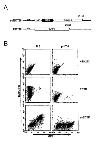
Figures 1 A-1 B show construction aild validation of hFcRn constructs.
Figure 1A: Schematic of hFcRn cDNA constructs. ssECTM (signal
sequence-GFP-ectodomain-transmembrane domain), and ECTM (signal sequence-
ectodomain-transmembrane domain). Cloning sites and FeRn codon positions are
indicated. The STOP codon is denoted by *.
Figure 1B: Flow cytometric analysis of pH-dependent binding of hIgG to
ECTM and ssECTM transfected HEK 293 cells.
Figures 2A-2D show flow cytometry of albumin and hIgG binding to hFcRn.
-7-
CA 02606378 2007-10-26
WO 2006/118772 PCT/US2006/014182
Figure 2A: Binding of HSA or IgG to ssECTM cells. (1 & 6) Ctrl HSA,
biotinylated goat anti-HSA + SA-APC. (2 & 7) Ctrl hIgG, goat anti-hIgG-PE. (3
&
8) HSA binding, HSA + biotinylated goat anti-HSA + SA-APC. (4 & 10) HSA
binding, human serum + biotinylated goat anti-HSA + SA-APC. (5 & 9) hIgG
binding, human serum + goat anti-hIgG-PE.
Figure 2B: Binding of HSA-biotin to ssECTM cells. (1 & 3) Ctrl HSA, SA-
APC. (2 & 4) HSA binding, biotinylated-HSA + SA-APC.
Figures 2C and 2D: Binding of HSA-biotin and hIgG3-AF647 to (C) ECTM
and (D) HEK293 cells. Ctrl hIgG3, no treatment; hIgG3 binding, hIgG3-AF647;
Ctrl
HSAbio, SA-APC; HSAbio binding, HSA-biotin + SA-APC. For an internal
negative control, a population of GFP-hFcRn negative cells was deliberately
maintained with the GFP-hFcRn positive ssECTM and ECTM cells. Relative mean
fluorescence intensity (MFI) is the ratio between MFI of the hFcRn-GFP
positive
population and MFI of hFcRn-GFP negative population. The bar graphs are the
mean + s. e. m. (see M&M) of at least 4 independent experiments.
Figures 3A-3B are graphs showing results of competition between HSA and
hIgG for binding hFcRn. ssECTM cells were incubated with the indicated doses
of
unlabeled hIgG (triangle), HSA (square), or hTF (circle), and then either hIgG-
AF647
or HSA-biotin was added. Assays were performed at pH 6.
Figure 3A: Competition vs. 50 g/ml hIgG-AF647.
Figure 3B: Competition vs. 250 g/ml HSA-biotin. Data are expressed as
MFI of GFP-positive gated cells. Representative data from one of two
experiments
with similar results is shown. HIgG-AF647 binding to ssECTM cells at pH 7.5
without competitor resulted in an MFI of 6. HSA-biotin/SA-PE binding to ssECTM
cells at pH 7.5 without competitor resulted in an MFI of 4.
Figure 4 shows data on the binding activity of anti-hFcRn mAbs at pH 7.5
and 6.0, and the ability of DVN24 to block the binding of hIgG to hFcRn at pH
6Ø
For direct binding data (left and middle scattergrams, 1 g of the indicated
mAbs
were incubated with 106 ssECTM cells for 30 min at 4 C in the indicated pH
buffer.
The ssECTM cells were then washed 2X, and incubated with phycoerythrin
conjugated goat anti-mouse IgG (Southern Biotech, Birmingham, AL), and then
analyzed by flow cytometry. For inhibition of hIgG (right scattergrams), the
mAbs
-8-
CA 02606378 2007-10-26
WO 2006/118772 PCT/US2006/014182
were added in a concentration of 10 g to 106 ssECTM cells for 30 min. at 4 C
in
pH 6.0 buffer, washed 2X, incubated with 1 g AlexiFluor647-conjugated hIgG3,
washed 2X and analyzed by flow cytometry.
Figures 5A-5B are graphs of data that show that certain anti-hFcRn mAbs
selectively block binding of hIgG or HSA to hFcRn at pH 6Ø
Figure 5A: Blockade of hIgG. 106 ssETCM cells were incubated at 4 C
with increasing concentrations of purified anti-hFcRn mAbs, washed 2X, and
then
incubated with 1 g AlexaFluor647-conjugated hIgG for 1 hour at 4 C. The
ssECTM cells were then washed 2X and analyzed by flow cytometry.
Figure 5B: Blockade of HSA. 106 ssETCM cells were incubated at 4 C with
increasing concentrations of purified anti-hFcRn mAbs, washed 2X, and then
incubated with 1 g biotin-conjugated HSA. The ssECTM cells were then washed
2X, incubated with streptavidin-phycoerythrin, washed 2X, and analyzed by flow
cytometry. All incubations were perfonned in pH 6.0 buffer. Data are presented
as
mean fluorescence intensity (MFI) of the GFP-positive gated cells.
Figure 6 is a graph of data that show that administration of DVN24 mAbs
reduces the serum concentration of hIgG. 100 g of tracer hIgG was injected
intraperitoneally into groups of 5 mouse FcRn-/- hFcRn Line 276 transgenic
mice on
day 0. Varying concentrations of DVN24 or 1000 g of an isotype-matched
negative control mAb was injected intraperitoneally on days 2, 3, and 4. Sera
from
serial eye bleeds were then analyzed by ELISA for the concentration of inj
ected
hIgG tracer. Data are presented based on the % of serum tracer hIgG
concentrations
24 hr after tracer injection.
Figures 7A and 7B are graphs of data that show that administration of
DVN24 but not ADM32 mAbs reduces the seram concentration of hIgG but not
HSA.
Figure 7A: Clearance of hIgG.
Figure 7B: Clearance of HSA. 100 g of tracer hIgG and HSA was injected
intraperitoneally into groups of 3 mouse FcRn-/- hFcRn (line 276 transgenic)
mice
on day 0. 1000 gg of DVN24, ADM31, or negative control mAb was injected
intraperitoneally on days 2, 3 and 4. Sera from serial eye bleeds were then
analyzed
by ELISA for the concentration of hIgG tracer. The mean standard error
values
-9-
CA 02606378 2007-10-26
WO 2006/118772 PCT/US2006/014182
are based on the percent of serum tracer hIgG concentrations remaining
relative to
concentrations 24 hr after tracer injection. Comparisons of p < 0.05 are
indicated (*).
Figures 8A and 8B are graphs of data that show that DVN24 reduces arthritic
lesions caused by human rheuinatoid arthritis plasma. Groups of 3 mFcRn-/-
Fcgr2b-/- hFcRn transgenic (line 32) mice were injected intraperitoneally with
0.5,
1, and 1 ml of human RA plasma on days 0, 2 and 7, respectively, and also
injected
intraperitoneally witli 1 mg of purified DVN24 or isotype control IgGa mAbs on
days 1, 3 and 8. ' Ankle width and overall arthritis scores were measured in a
blinded
manner by two independent observers, as described (Akilesh et al., 2004, J
Clin
Invest 113: 1328-33). Data are the mean standard error. Coinparisons of p <
0.05
(*) and (p < 0.005) (**) of the ankle widths are indicated.
DETAILED DESCRIPTION OF THE INVENTION
Certain aspects of the present invention are based, at least in part, on the
finding that the receptor FcRn (FcRp/Fcgrtl) selectively protects antibodies
of the
IgG isotype from normal protein catabolism in a Fc-dependent manner. FcRn is a
novel member of a family of proteins that perform varied immunological
fiulctions.
The FcRn molecule is expressed in the vascular endothelium along with other
tissues of adult animals, including mice and humans. FcRn binds to antibody
molecules, but only those from the IgG class. The crystal structures of the
FcRn/IgG complex have been solved (Bjorkman and Simister, 1992, PNAS 89:638-
42; West and Bjorkinan, 2000, Biochemistry 39:9698-9708), proving that a
receptor/ligand relationship exists between the two molecules. Further, FcRn
heterodimerizes with (32-microglobulin, and the (32-microglobulin complex is
critical
for FcRn to bind to IgG in a pH-dependant manner.
Most serum proteins have a short seruin half-life (about 1-2 days). However,
two types of serum proteins, albumin and antibodies of the IgG class, have
greatly
extended serum half-lives. Their half-lives are extended because they are
naturally
rescued from normal catabolic degradation by the major histocompatibility
complex
family protein, FcRn. Several investigators have indirectly demonstrated a
protective effect by coupling the Fc region of IgG to different polypeptides
to
-10-
CA 02606378 2007-10-26
WO 2006/118772 PCT/US2006/014182
improve stability of the polypeptide (e.g., U.S. Pat. Nos. 6,096,871 and
6,121,022).
PCT Application WO 97/34631 also describes the use of immunoglobulin-like
domains in increasing the stability and longevity of pharmaceutical
compositions for
therapeutic and diagnostic purposes. In addition, Applicants have shown that
the
genetic elimination of FcRn by gene targeting protects K/.BxN mice from
developing
autoimmune arthritis (Akilesh et al., 2004, J Clin Invest 113: 1328-33.
Applicants
have also shown that genetic elimination of FcRn by gene targeting reduces the
severity of systemic lupus erythematosus (SLE) in mice genetically predisposed
to
develop SLE-like disease.. Applicant and others have suggested that the
fiulctional
saturation of the FcRn protection pathway results in an amelioration of
arthritis and
in immune thrombocytopenic purpura mouse models (Akilesh et al., 2004, J Clin
Invest 113: 1328-33; Hanson and Balthasar, 2002, Thromb Haemo 88: 898-899) in
pathogenic serum transfer models. Thus, these experiments suggest that FcRn is
a
promising therapeutic target to treat autoiminune diseases such as those
caused by
autoantibodies. Recent studies by Applicants and their collaborators (e.g.,
Chaudhury et al., 2003, J Exp Med 197: 315-322) have shown that FcRn also
protects albumin from nonzial catabolic elimination. This occurs because FcRn
binds albumin and protects it from normal catabolic elimination in a similar
manner
as found for IgG. A major complication to the strategy of therapeutic blockade
of
FcR.ii protection of IgG is that such therapeutics could also reduce the serum
half-
life of albumin. This may result in deleterious side effects since maintenance
of a
normal serum concentration of albumin is critical for the maintenance of
normal
osmolarity and other biological functions for which albumin plays an essential
role.
To avoid this potentially serious side effect of anti-FcRn therapeutics,
certain
embodiments of the invention provide anti-FcRn therapeutics that are designed
to
selectively decrease the serum half-life of IgG but not the serum half-life of
human
albumin.
I. FcRn Antibodies and Other FcRn Binding Agents
This invention provides, in part, FcRn binding agents that selectively target
portions of the FeRn molecule, such as, for exaniple, FcRn antibodies, antigen
binding portions of FcRn antibodies, and non-inununoglobulin binding agents of
FcRn. The FeRn binding agents described herein may be used to treat a variety
of
-11-
CA 02606378 2007-10-26
WO 2006/118772 PCT/US2006/014182
disorders, particularly FcRn-related autoimmune diseases. The invention
provides
antibodies and antigen binding portions thereof that modulate (inhibit or
enhance)
FcRn mediated functions, suclz as Fc binding or IgG protection activities.
Such
binding agents may be used to modulate FcRn fiuictions in vitro and in vivo,
and, in
particular, for treating FeRn-related autoimmune diseases. In particular
embodiments, the present invention relates to monoclonal antibodies against
FcRn.
In one embodiment, FcRn antibodies (immunoglobulins) are raised against
an isolated and/or recombinant human FcRn or portion thereof (e.g., peptide)
or
against a host cell which expresses recombinant human FcRn. As used herein,
the
term "FcRn," also referred to in the literature as FcRn alpha chain, refers to
an FcRn
polypeptide from a mammal including, for example, a human. In certain aspects,
antibodies of the invention specifically bind to a region of an FcRn protein
(e.g., the
alpha 2 domain helix), which constitutes an Fc binding site (see, e.g., West
and
Bjorkman, 2000, Biochemistry 39:9698-9708). In other cases, antibodies of the
invention specifically bind to a region of an FcRn protein that constitutes a
P2-
microglobulin binding site. Antibodies of the invention inhibit binding of
FcRn to
IgG but do not inhibit binding of FcRii to human albumin.
An "iminunoglobulin" is a tetrameric molecule. In a naturally-occurring
immunoglobulin, each tetramer is coinposed of two identical pairs of
polypeptide
chains, each pair having one "liglit" (about 25 kDa) and one "heavy" chain
(about
50-70 kDa). The alnino-terminal portion of each chain includes a variable
region of
about 100 to 110 or more amino acids primarily responsible for antigen
recognition.
The carboxy-terminal portion of each chain defines a constant region primarily
responsible for effector function. Human light chains are classified as kappa
and
lambda light chains. Heavy chains are classified as mu, delta, gamma, alpha,
or
epsilon, and define the antibody's isotype as IgM, IgD, IgG, IgA, and IgE,
respectively. Within light and heavy chains, the variable and constant regions
are
joined by a "J" region of about 12 or more amino acids, with the heavy.chain
also
including a "D" region of about 10 more amino acids. See generally,
Fundamental
Iminunology Ch. 7 (Paul, W., ed., 2nd ed. Raven Press, N.Y. (1989))
(incorporated
by reference in its entirety for all purposes). The variable regions of each
-12-
CA 02606378 2007-10-26
WO 2006/118772 PCT/US2006/014182
light/heavy chain pair form the antibody binding site such that an intact
immunoglobulin has two binding sites.
Immunoglobulin chains exhibit the same general structure: they include
relatively conserved framework regions (FR) joined by three hypervariable
regions,
also called compleinentarity determining regions or CDRs. The CDRs from the
two
chains of each pair are aligned by the framework regions, enabling binding to
a
specific epitope. From N-terminus to C-terminus, both light and heavy chains
comprise the domains FR1, CDR1, FR2, CDR2, FR3, CDR3 and FR4. The
assignment of amino acids to each domain is in accordance with the definitions
of
Kabat Sequences of Proteins of Immunological Interest (National Institutes of
Health, Bethesda, Md. (1987 and 1991)), or Chothia & Lesk J. Mol. Biol., 1997,
196:901-917; Chothia et al. Nature, 1989,342:878-883 (1989).
As used herein, the term "antibody" refers to an intact iminunoglobulin or to
an antigen-binding portion thereof that competes with the intact antibody for
specific
binding. Antigen-binding portions may be produced by recombinant DNA
techniques or by enzymatic or chemical cleavage of intact antibodies. Antigen-
binding portions include, inter alia, Fab, Fab', F(ab')2, Fv, dAb, and
complementarity determining region (CDR) fragments, single-chain antibodies
(scFv), single domain antibodies, chimeric antibodies, diabodies and
polypeptides
that contain at least a portion of an immunoglobulin that is sufficient to
confer
specific antigen binding to the polypeptide. The terms "anti-FeRn antibody"
and
"FcRn antibody" are used interchangeably herein.
An Fab fragment is a monovalent fragment consisting of the VL, VH, CL
and CH I domains; a F(ab')2 fragment is a bivalent fragment comprising two Fab
fragments linked by a disulfide bridge at the hinge region; an Fd fragment
consists
of the VH and CH1 domains; an Fv fragment consists of the VL and VH domains of
a single arm of an antibody; and a dAb fragment (Ward et al., Nature 341:544-
546,
1989) consists of a VH domain.
A single-chain antibody (scFv) is an antibody in which a VL and VH regions
are paired to form a monovalent molecules via a synthetic linker that enables
them
to be made as a single protein chain (Bird et al., Science 242:423-426, 1988
and
-13-
CA 02606378 2007-10-26
WO 2006/118772 PCT/US2006/014182
Huston et al., Proc. Nat1. Acad. Sci. USA 85:5879-5883, 1988). Diabodies are
bivalent, bispecific antibodies in which VH and VL domains are expressed on a
single polypeptide chain, but using a linker that is too short to allow for
pairing
between the two domains on the same chain, thereby forcing the domains to pair
with complementary domains of another chain and creating two antigen binding
sites (see e.g., Holliger, P., et al., Proc. Natl. Acad. Sci. USA 90:6444-
6448, 1993,
and Poljak, R. J., et al., Structure 2:1121-1123, 1994). One or more CDRs
maybe
incorporated into a molecule either covalently or noncovalently.
An antibody may have one or more binding sites. If there is more than one
binding site, the binding sites may be identical to one another or may be
different.
For instance, a naturally-occurring immunoglobulin has two identical binding
sites,
a single-chain antibody or Fab fragment has one binding site, while a
"bispecific" or
"bifunctional" antibody has two different binding sites.
The term "human antibody" includes all antibodies that have one or more
variable and constant regions derived from human immunoglobulin sequences. In
one embodiment, all of the variable and constant domains are derived from
human
immunoglobulin sequences (a fully human antibody). These antibodies may be
prepared in a variety of ways, as described below.
The tenn "chimeric antibody" refers to an antibody that contains one or more
regions from one antibody and one or more regions from one or more other
different
antibodies. In one embodiment, one or more of the CDRs are derived from a
human
anti-FcRn antibody. In a more preferred embodiment, all of the CDRs are
derived
from a human anti-FcRn antibody. In another preferred embodiment, the CDRs
from more than one human anti-FcRn antibodies are mixed and matched in a
chimeric antibody. For instance, a chimeric antibody may comprise a CDR1 from
the light chain of a first human anti-FcRn antibody combined with CDR2 and
CDR3
from the light chain of a second human anti-FcRn antibody, and the CDRs from
the
heavy chain may be derived from a third anti-FcRn antibody. Further, the
framework regions may be derived from one of the same anti-FeRn antibodies,
from
one or more different antibodies, such as a human antibody, or from a
humanized
antibody.
-14-
CA 02606378 2007-10-26
WO 2006/118772 PCT/US2006/014182
In certain embodiments, the FcRn antibody or antigen binding portion
thereof is linked to an additional functional moiety. Such linkage may be
covalent
or non-covalent. In one embodiment, the functional moiety may be therapeutic,
e.g.,
a drug conjugate or toxin.
In certain further embodiments, the FcRn antibody or antigen binding
portion thereof is labeled to facilitate detection. As used herein, the terms
"label" or
"labeled" refers to incorporation of another molecule in the antibody. In one
embodiment, the label is a detectable marker, e.g., incorporation of a
radiolabeled
amino acid or attachment to a polypeptide of biotinyl moieties that can be
detected
by marked avidin (e.g., streptavidin containing a fluorescent marker or
enzymatic
activity that can be detected by optical or colorimetric methods). Various
methods
of labeling polypeptides and glycoproteins are known in the art and may be
used.
Examples of labels for polypeptides include, but are not limited to, the
following:
radioisotopes or radionuclides (e.g., 3H, 14C, 15N, 35S, 90Y, 99Tc, 111In,
1251,
131I), fluorescent labels (e.g., FITC, rhodamine, lanthanide phosphors),
enzymatic
labels (e.g., horseradish peroxidase, beta-galactosidase, luciferase, alkaline
phosphatase), chemiluminescent markers, biotinyl groups, predetermined
polypeptide epitopes recognized by a secondary reporter (e.g., leucine zipper
pair
sequences, binding sites for secondary antibodies, metal binding domains,
epitope
tags), magnetic agents, such as gadolinium chelates, toxins such as pertussis
toxin,
taxol, cytochalasin B, gramicidin D, ethidium bromide, emetine, nlitomycin,
etoposide, tenoposide, vincristine, vinblastine, colchicin, doxorubicin,
daunorubicin,
dihydroxy anthracin dione, mitoxantrone, mithramycin, actinomycin D, 1-
dehydrotestosterone, glucocorticoids, procaine, tetracaine, lidocaine,
propranolol,
and puromycin and analogs or homologs thereof. In some enZbodiments, labels
are
attached by spacer arms of various lengths to reduce potential steric
hindrance.
As shown in the Examples below, Applicants have generated monoclonal
antibodies against human FcRn, as well as hybridoma ce111ines producing FcRn
monoclonal antibodies. These antibodies were further characterized in many
ways,
for example, their ability to inhibit interaction between human FcRn and its
ligands
(e.g., huinan IgG or human serum albumin), their ability to decrease the serum
half-
life of IgG in vivo, their ability to promote clearaiice of IgG in vivo, and
their ability
-15-
CA 02606378 2007-10-26
WO 2006/118772 PCT/US2006/014182
to aineliorate the inflammatory lesions induced by pathogenic human
antibodies.
The FcRn antibodies that specifically bind to human IgG, but do not bind to
human
serum albumin (HSA) are particularly useful for therapeutic purposes.
In certain embodiments, antibodies of the invention specifically bind to an
extracellular domain (ECD) of an FeRn protein (also referred to herein as a
soluble
FcRn polypeptide). A representative soluble FcRn polypeptide may comprise
amino
acids residues 24-297 of SEQ ID NO: 1 below. As used herein, the FcRn soluble
polypeptides include fragmeiits, functional variants, and modified forms of
FcRu
soluble polypeptide.
mgvprpqpwa lglllfllpg slgaeshlsl lyhltavssp apgtpafwvs
gwlgpqqyls
ynslrgeaep cgawvwenqv swywekettd lrikeklfle afkalggkgp
ytlqgllgce
lgpdntsvpt akfalngeef mnfdlkqgtw ggdwpealai sqrwqqqdka
ankeltfllf
scphrlrehl ergrgnlewk eppsmrlkar psspgfsvlt csafsfyppe
lqlrflrngl
aagtgqgdfg pnsdgsfhas ssltvksgde hhyccivqha glaqplrvel
espakssvlv
vgivigvlll taaavggall wrrmrsglpa pwislrgddt gvllptpgea
qdadlkdvnv
ipata (SEQ ID NO: 1)
In certain embodiments, the present invention provides monoclonal FcRn
antibodies that specifically bind an FcRn or a portion of FcRn. Examples of
the
monoclonal FeRn antibodies include, but are not limited to, DVN21 and DVN24 as
described below in the working examples. In certain embodiments, the
immunoglobulins bind to FcRn with an affinity of at least about 1x 10⁻⁶, 1X 10-
7,
1x 10"$, 1x 10⁻⁹ M or less.
In certain aspects of the invention, anti-FcRn antibodies of the invention
demonstrate both molecule and species selectivity. For example, antibodies
disclosed herein are preferably specific for FcRi1, with minimal binding to
other
FcRn ligand molecules, such as, for example, HSA. In one embodiment, the anti-
FeRn antibody binds to human, cynomologous or rhesus FcRn. In one embodiinent,
the anti-FcRn antibody does not bind to mouse, rat, guinea pig, dog, goat or
rabbit
FcRn. Alternatively, the antibody binds to more than one different FcRn
molecules
from different species, such as human and mouse. Following the teachings of
the
-16-
CA 02606378 2007-10-26
WO 2006/118772 PCT/US2006/014182
specification, one may determine the molecule and species selectivity for the
anti-
FcRn antibody using methods well known in the art , for example,
iminunofluorescence microscopy, Western blot, FACS, ELISA or RIA. In one
embodiment, the anti-FcRn antibody has a tendency to bind to FcRn that is at
least
50 times greater than its tendency to bind to other FcRn ligand molecules, and
preferably 100 or 200 times greater.
In certain einbodiments, antibodies of the present invention bind to one or
more specific domains of FcRn. For example, a subject antibody binds to a
region
in the Fc-binding site of the FcRn heavy chain.
The anti-FeRn antibody may be an IgG, an IgM, an IgE, an IgA or an IgD
molecule. In a preferred embodiment, the antibody is an IgG and is an IgGI,
IgG2,
IgG3 or IgG4 subtype. In an specific embodiment, the anti-FcRn antibody is
subclass IgG2. The class and subclass of FcRn antibodies may be determined by
any method known in the art. In general, the class and subclass of an antibody
may
be determined using antibodies that are specific for a particular class and
subclass of
antibody. Such antibodies are available commercially. The class and subclass
can
be deterinined by ELISA, Western Blot as well as other techniques.
Alternatively,
the class and subclass may be determined by sequencing all or a portion of the
constant domains of the heavy and/or light chains of the antibodies, comparing
their
amino acid sequences to the known amino acid sequences of various class and
subclasses of immunoglobulins, and determining the class and subclass of the
antibodies.
In certain embodiments, single chain antibodies, and chimeric, humanized or
primatized (CDR-grafted) antibodies, as well as chimeric or CDR-grafted single
chain antibodies, comprising portions derived from different species, are also
encoinpassed by the present invention as antigen binding portions of an FcRn
antibody. The various portions of these antibodies can be joined together
chemically
by conventional techniques, or can be prepared as a contiguous protein using
genetic
engineering teclmiques. For example, nucleic acids encoding a chimeric or
humanized chain can be expressed to produce a contiguous protein. See, e.g.,
Cabilly et al., U.S. Pat. No. 4,816,567; Cabilly et al., European Patent No.
-17-
CA 02606378 2007-10-26
WO 2006/118772 PCT/US2006/014182
0,125,023; Boss et al., U.S. Pat. No. 4,816,397; Boss et al., European Patent
No.
0,120,694; Neuberger, M. S. et al., WO 86/01533; Neuberger, M. S. et al.,
European
Patent No. 0,194,276 B1; Winter, U.S. Pat. No. 5,225,539; and Winter, European
Patent No. 0,239,400 B1. See also, Newman, R. et al., BioTechnology, 10: 1455-
1460 (1992), regarding primatized antibody. See, e.g., Ladner et al., U.S.
Pat. No.
4,946,778; and Bird, R. E. et al., Science, 242: 423-426 (1988)), regarding
single
chain antibodies.
In addition, functional fragments of antibodies, including fragments of
chimeric, humanized, primatized or single chain antibodies, can be produced.
Functional fragments of the subject antibodies retain at least one binding
function
and/or modulation function of the full-length antibody from which they are
derived.
Preferred functional fragments retain an antigen binding function of a
corresponding
full-length antibody (e.g., specificity for an FcRn). Certain preferred
functional
fraginents retain the ability to inhibit one or more functions characteristic
of an
FcRn, such as a binding activity or a transport activity. For example, in one
embodiment, a functional fragment of an FcRn antibody can specifically inhibit
the
interaction of FcRn with one of its ligands (e.g., IgG) and/or can inhibit one
or more
FcRn-mediated functions in vivo, such as IgG transport and autoimniune
responses.
In certain embodiments, a.ntibody fragments that bind to an FcRn receptor or
portion thereof, including, but not limited to, Fv, Fab, Fab' and F(ab')2
fragments are
encompassed by the invention. Such fragments can be produced by enzymatic
cleavage or by recombinant techniques. For instance, papain or pepsin cleavage
can
generate Fab or F(ab')Z fragments, respectively. Antibodies can also be
produced in
a variety of tnuicated forms using antibody-encoding genes in which one or
more
stop codons has been introduced upstreain of the natural stop site. For
example, a
chimeric gene encoding a F(ab')2 heavy chain portion can be designed to
include
DNA sequences encoding the CHl domain and hinge region of the heavy chain.
A humanized antibody can be, for example, an antibody that is derived from
a non-liuman species, in which certain amino acids in the framework and
constant
domains of the heavy and light chains have been mutated so as to reduce of
abolish
an immune response in humans. Alternatively, a humanized antibody may be
-18-
CA 02606378 2007-10-26
WO 2006/118772 PCT/US2006/014182
produced by fusing the constant domains from a human antibody to the variable
domains of a non-human species. Examples of how to make humanized antibodies
may be found in U.S. Pat. Nos. 6,054,297, 5,886,152 and 5,877,293. A humanized
antibody may coinprise portions of immunoglobulins of different origin. For
example, at least one portion can be of human origin. Accordingly, the present
invention relates to a humanized immunoglobulin having binding specificity for
an
FcRn (e.g., huinan FcRn), said immunoglobulin comprising an antigen binding
region of nonhuman origin (e.g., rodent) and at least a portion of an
immunoglobulin
of human origin (e.g., a human framework region, a hunlan constant region or
portion thereof). For example, the humanized antibody can comprise portions
derived from an immunoglobulin of nonhuman origin with the requisite
specificity,
such as a mouse, and from immunoglobulin sequences of human origin (e.g., a
chimeric immunoglobulin), joined together chemically by conventional
techniques
(e.g., synthetic) or prepared as a contiguous polypeptide using genetic
engineering
techniques (e.g., DNA encoding the protein portions of the cllimeric antibody
can be
expressed to produce a contiguous polypeptide chain).
Another example of a humanized immunoglobulin of the present invention is
an immunoglobulin containing one or more immunoglobulin chains comprising a
CDR of nonhuman origin (e.g., one or more CDRs derived from an antibody of
nonhuman origin) and a framework region derived from a light and/or heavy
chain
of human origin (e.g., CDR-grafted antibodies with or without framework
changes).
In one embodiment, the humanized immunoglobulin can compete with murine
monoclonal antibody for binding to an FcRn polypeptide. Chimeric or CDR-
grafted
single chain antibodies are also encompassed by the term humanized
immunoglobulin.
In certain embodiments, the present invention provides FcRn antagonist
antibodies. As described herein, the term "antagonist antibody" refers to an
antibody that can inhibit one or more functions of an FcRn, such as a binding
activity (e.g., ligand binding and 02-microglobin binding) or a transport
activity
(e.g., transporting IgG and protecting IgG from lysosomal catabolism).
-19-
CA 02606378 2007-10-26
WO 2006/118772 PCT/US2006/014182
In certain embodiments, anti-idiotypic antibodies are also provided. Anti-
idiotypic antibodies recognize antigenic determinants associated with the
antigen-
binding site of another antibody. Anti-idiotypic antibodies can be prepared
against a
second antibody by immunizing an animal of the same species, and preferably of
the
same strain, as the aniinal used to produce the second antibody. See e.g.,
U.S. Pat.
No. 4,699,880. In one embodiment, antibodies are raised against FeRn or a
portion
tliereof, and these antibodies are used in turn to produce an anti-idiotypic
antibody.
The anti-idiotypic antibodies produced thereby can bind compounds which bind
receptor, such as ligands of receptor function, and can be used in an
immunoassay to
detect or identify or quantitate such compounds. Such an anti-idotypic
antibody can
also be an inhibitor of an FcRn receptor function, although it does not bind
receptor
itself. Such an anti-idotypic antibody can also be called an antagonist
antibody.
In certain aspects, the present invention relates to hybridoma cell lines, as
well as to monoclonal antibodies produced by these hybridoma cell lines. The
cell
lines of the present invention have uses other than for the production of the
monoclonal antibodies. For example, the cell lines of the present invention
can be
fused with other cells (such as suitably drug-marked lzuman myeloma, mouse
myeloma, huinan-mouse heteromyeloma or human lymphoblastoid cells) to produce
additional hybridomas, and thus provide for the transfer of the genes encoding
the
monoclonal antibodies. In addition, the cell lines can be used as a source of
nucleic
acids encoding the anti-FcRn immunoglobulin chains, which can be isolated and
expressed (e.g., upon transfer to other cells using any suitable technique
(see e.g.,
Cabilly et al., U.S. Pat. No. 4,816,567; Winter, U.S. Pat. No. 5,225,539)).
For
instance, clones comprising a rearranged anti-FcRn light or heavy chain can be
isolated (e.g., by PCR) or cDNA libraries can be prepared from mRNA isolated
from the cell lines, and cDNA clones encoding an anti-FcRn immunoglobulin
chain
can be isolated. Thus, nucleic acids encoding the heavy and/or light chains of
the
antibodies or portions thereof can be obtained and used in accordance with
recombinant DNA techniques for the production of the specific immunoglobulin,
inununoglobulin chain, or variants thereof (e.g., humanized immunoglobulins)
in a
variety of host cells or in an in vitro translation system. For example, the
nucleic
acids, including cDNAs, or derivatives thereof encoding variants such as a
-20-
CA 02606378 2007-10-26
WO 2006/118772 PCT/US2006/014182
humanized immunoglobulin or immunoglobulin chain, can be placed into suitable
prokaryotic or eukaryotic vectors (e.g., expression vectors) and introduced
into a
suitable host cell by an appropriate method (e.g., transformation,
transfection,
electroporation, infection), such that the nucleic acid is operably linked to
one or
more expression control elements (e.g., in the vector or integrated into the
host cell
genome). For production, host cells can be maintained under conditions
suitable for
expression (e.g., in the presence of inducer, suitable media supplemented with
appropriate salts, growth factors, antibiotic, nutritional supplements, etc.),
whereby
the encoded polypeptide is produced. If desired, the encoded protein can be
recovered and/or isolated (e.g., from the host cells or medium). It will be
appreciated that the method of production encompasses expression in a host
cell of a
transgenic animal (see e.g., WO 92/03918, GenPharm International, published
Mar.
19, 1992).
II. Metlzods ofAntibody Production
Preparation of immunizing antigen, and polyclonal and monoclonal antibody
production can be performed as described herein, or using other suitable
techniques.
A variety of methods have been described. See e.g., Kohler et al., Nature,
256: 495-
497 (1975) and Eur. J. Immunol. 6: 511-519 (1976); Milstein et al., Nature
266:
550-552 (1977); Koprowski et al., U.S. Pat. No. 4,172,124; Harlow, E. and D.
Lane,
1988, Antibodies: A Laboratory Manual, (Cold Spring Harbor Laboratory: Cold
Spring Harbor, N.Y.); Current Protocols In Molecular Biology, Vol. 2
(Supplement
27, Summ.er'94), Ausubel, F. M. et al., Eds., (John Wiley & Sons: New York,
N.Y.),
Chapter 11, (1991). Generally, a hybridoma can be produced by fusing a
suitable
immortal cell line (e.g., a myeloma cell line such as SP2/0) with antibody
producing
cells. The antibody producing cell, preferably those of the spleen or lymph
nodes,
are obtained from animals immunized with the antigen of interest. The fused
cells
(hybridomas) can be isolated using selective culture conditions, and cloned by
limiting dilution. Cells which produce antibodies with the desired specificity
can be
selected by a suitable assay (e.g., ELISA).
Other suitable methods of producing or isolating antibodies of the requisite
specificity can used, including, for example, methods which select recombinant
-21-
CA 02606378 2007-10-26
WO 2006/118772 PCT/US2006/014182
antibodies from a library, or which rely upon immunization of transgenic
animals
(e.g., mice) capable of producing a full repertoire of human antibodies. See
e.g.,
Jakobovits et a1., Proc. Natl. Acad. Sci. USA, 90: 2551-2555 (1993);
Jakobovits et
al., Nature, 362: 255-258 (1993); Lonberg et al., U.S. Pat. No. 5,545,806;
Surani et
al., U.S. Pat. No. 5,545,807. For example, FcRn antibodies may be isolated
from a
synthetic human combinatorial antibody library (HuCAL). See, e.g., Knappik et
al.,
2000, J Mol boi1296:57-86.
To illustrate, iminunogens derived from an FeRn polypeptide (e.g., an FcRn
polypeptide or an antigenic fragment thereof which is capable of eliciting an
antibody response, or an FcRn fusion protein) can be used to immunize a
mammal,
such as a mouse, a hamster or rabbit. See, for example, Antibodies: A
Laboratory
Manual ed. by Harlow and Lane (Cold Spring Harbor Press: 1988). Techniques for
conferring immunogenicity on a protein or peptide include conjugation to
carriers or
other techniques well known in the art. An immunogenic portion of an FcRn
polypeptide can be administered in the presence of adjuvant. The progress of
immunization can be monitored by detection of antibody titers in plasma or
serua.n.
Standard ELISA or other immunoassays can be used with the immunogen as antigen
to assess the levels of antibodies. In one embodiment, antibodies of the
invention
are specific for the extracellular portion of an FcRii protein or fragments
thereof. In
another embodiment, antibodies of the invention are specific for the
intracellular
portion or the transmembrane portion of the FcRn protein.
Following immunization of a.n animal with an antigenic preparation of an
FcRn polypeptide, antisera can be obtained and, if desired, polyclonal
antibodies can
be isolated from the serum. To produce monoclonal antibodies, antibody-
producing
cells (lymphocytes) can be harvested from an immunized animal and fused by
standard somatic cell fusion procedures with immortalizing cells such as
myeloma
cells to yield hybridoma cells. Such techniques are well known in the art, and
include, for example, the hybridoma technique (originally developed by Kohler
and
Milstein, (1975) Nature, 256: 495-497), the human B cell hybridoma technique
(Kozbar et al., (1983) Immunology Today, 4: 72), and the EBV-hybridoma
technique to produce human monoclonal antibodies (Cole et al., (1985)
Monoclonal
Antibodies and Cancer Therapy, Alan R. Liss, Inc. pp. 77-96). Hybridoma cells
can
-22-
CA 02606378 2007-10-26
WO 2006/118772 PCT/US2006/014182
be screened immunochemically for production of antibodies specifically
reactive
with an FcRn polypeptide and monoclonal antibodies isolated from a culture
comprising such hybridoma cells.
In certain embodiments, antibodies of the present invention can be
fragmented using conventional techniques and the fragments screened for
utility in
the same manner as described above for whole antibodies. For example, F(ab)2
fraginents can be generated by treating antibody with pepsin. The resulting
F(ab)2
fragment can be treated to reduce disulfide bridges to produce Fab fragments.
In certain embodiments, antibodies of the present invention are further
intended to include bispecific, single-chain, and chimeric and humanized
molecules
having affinity for an FcRn polypeptide conferred by at least one CDR region
of the
antibody. Techniques for the production of single chain antibodies (US Patent
No.
4,946,778) can also be adapted to produce single chain antibodies. Also,
transgenic
mice or other organisms including other mammals, may be used to express
humanized antibodies. Methods of generating these antibodies are known in the
art.
See, e.g., Cabilly et al., U.S. Pat. No. 4,816,567; Cabilly et al., European
Patent No.
0,125,023; Queen et al., European Patent No. 0,451,216; Boss et al., U.S. Pat.
No.
4,816,397; Boss et al., European Patent No. 0,120,694; Neuberger, M. S. et
al., WO
86/01533; Neuberger, M. S. et al., European Patent No. 0,194,276; Winter, U.S.
Pat.
No. 5,225,539; winter, European Patent No. 0,239,400; Padlan, E. A. et al.,
European Patent Application No. 0,519,596 Al. See also, Ladner et al., U.S.
Pat.
No. 4,946,778; Huston, U.S. Pat. No. 5,476,786; and Bird, R. E. et al.,
Science, 242:
423-426 (1988)).
Such humanized immunoglobulins can be produced using synthetic and/or
recombinant nucleic acids to prepare genes (e.g., cDNA) encoding the desired
humanized chain. For example, nucleic acid (e.g., DNA) sequences coding for
humanized variable regions can be constructed using PCR mutagenesis methods to
alter DNA sequences encoding a human or humanized chain, such as a DNA
teinplate from a previously humanized variable region (see e.g., Kamman, M.,
et al.,
Nucl. Acids Res., 17: 5404 (1989)); Sato, K., et al., Cancer Research, 53: 851-
856
(1993); Daugherty, B. L. et al., Nucleic Acids Res., 19(9): 2471-2476 (1991);
and
-23-
CA 02606378 2007-10-26
WO 2006/118772 PCT/US2006/014182
administered and/or thereafter. Administration of the antibodies may be made
in a
single dose, or in multiple doses. In some instances, administration of the
antibodies
is commenced at least several days prior to the conventional therapy, while in
other
instances, administration is begun either immediately before or at the time of
the
administration of the conventional therapy.
V. Phaf rnaceutical Compositions and Modes of Administration
In certain embodiments, the subject antibodies of the present invention are
formulated witlz a pharmaceutically acceptable carrier. Such antibodies can be
administered alone or as a component of a pharmaceutical formulation
(composition). The compounds may be formulated for administration in any
convenient way for use in human or veterinary medicine. Wetting agents,
emulsifiers and lubricants, such as sodium lauryl sulfate and magnesium
stearate, as
well as coloring agents, release agents, coating agents, sweetening, flavoring
and
perfuming agents, preservatives and antioxidants can also be present in the
compositions.
Formulations of the subject antibodies include those suitable for oral,
dietary,
topical, parenteral (e.g., intravenous, intraarterial, intramuscular,
subcutaneous
injection), inhalation (e.g., intrabronchial, intranasal or oral inhalation,
intranasal
drops), rectal, and/or intravaginal administration. Other suitable methods of
administration can also include rechargeable or biodegradable devices and slow
release polymeric devices. The pharmaceutical compositions of this invention
can
also be administered as part of a combinatorial tl7erapy with other agents
(either in
the same formulation or in a separate formulation).
The formulations may conveniently be presented in unit dosage form and
may be prepared by any methods well known in the art of pharmacy. The-amount
of
active ingredient which can be coinbined with a carrier material to produce a
single
dosage form will vary depending upon the host being treated, the particular
mode of
administration. The amount of active ingredient which can be combined with a
carrier material to produce a single dosage form will generally be that amount
of the
compound which produces a therapeutic effect.
-30-
CA 02606378 2007-10-26
WO 2006/118772 PCT/US2006/014182
In certain embodiments, methods of preparing these formulations or
compositions include combining another type of immune-modulating agent and a
carrier and, optionally, one or more accessory ingredients. In general, the
formulations can be prepared with a liquid carrier, or a finely divided solid
carrier,
or both, and then, if necessary, shaping the product.
Formulations for oral administration may be in the form of capsules, cachets,
pills, tablets, lozenges (using a flavored basis, usually sucrose and acacia
or
tragacanth), powders, granules, or as a solution or a suspension in an aqueous
or
non-aqueous liquid, or as an oil-in-water or water-in-oil liquid emulsion, or
as an
elixir or syrup, or as pastilles (using an inert base, such as gelatin and
glycerin, or
sucrose and acacia) and/or as mouth washes and the like, each containing a
predetermined amount of one or more subject antibodies as an active
ingredient.
Liquid dosage forms for oral administration include pharmaceutically
acceptable einulsions, microemulsions, solutions, suspensions, syrups, and
elixirs.
In addition to the active ingredient, the liquid dosage forms may contain
inert
diluents commonly used in the art, such as water or other solvents,
solubilizing
agents and emulsifiers, such as ethyl alcohol, isopropyl alcohol, ethyl
carbonate,
ethyl acetate, benzyl alcohol, benzyl benzoate, propylene glycol, 1,3-butylene
glycol, oils (in particular, cottonseed, groundnut, corn, germ, olive, castor,
and
sesame oils), glycerol, tetrahydrofuryl alcohol, polyethylene glycols and
fatty acid
esters of sorbitan, and mixtures thereof. Besides inert diluents, the oral
compositions can also include adjuvants such as wetting agents, emulsifying
and
suspending agents, sweetening, flavoring, coloring, perfiuning, and
preservative
agents.
Suspensions, in addition to the active compounds, may contain suspending
agents such as ethoxylated isostearyl alcohols, polyoxyethylene sorbitol, and
sorbitan esters, microcrystalline cellulose, aluminum metahydroxide,
bentonite,
agar-agar and tragacanth, and mixtures tllereof.
Methods of the invention can be administered topically, for example, to skin.
The topical formulations may further include one or more of the wide variety
of
agents known to be effective as skin or stratum comeum penetration enhancers.
-31-
CA 02606378 2007-10-26
WO 2006/118772 PCT/US2006/014182
Examples of these are 2-pyrrolidone, N-methyl-2-pyrrolidone,
dimethylacetamide,
dimethylformamide, propylene glycol, methyl or isopropyl alcohol, dimethyl
sulfoxide, and azone. Additional agents may fixrther be included to make the
formulation cosmetically acceptable. Examples of these are fats, waxes, oils,
dyes,
fragrances, preservatives, stabilizers, and surface active agents. Keratolytic
agents
such as those known in the art may also be included. Examples are salicylic
acid
and sulfur.
Dosage forms for the topical or transdermal administration include powders,
sprays, ointments, pastes, creams, lotions, gels, solutions, patches, and
inhalants.
The subject antibodies may be mixed under sterile conditions witli a
pharmaceutically acceptable carrier, and with any preservatives, buffers, or
propellants which may be required. The ointments, pastes, creams and gels may
contain, in addition to an antibody, excipients, such as animal and vegetable
fats,
oils, waxes, paraffins, starch, tragacantli, cellulose derivatives,
polyethylene glycols,
silicones, bentonites, silicic acid, talc and zinc oxide, or mixtures thereof.
Pharmaceutical compositions suitable for parenteral administration may
comprise one or more antibodies in combination with one or more
pharmaceutically
acceptable sterile isotonic aqueous or nonaqueous solutions, dispersions,
suspensions or emulsions, or sterile powders which may be reconstituted into
sterile
injectable solutions or dispersions just prior to use, which may contain
antioxidants,
buffers, bacteriostats, solutes which render the forinulation isotonic with
the blood
of the intended recipient or suspending or thickening agents. Examples of
suitable
aqueous and nonaqueous carriers which may be employed in the pharmaceutical
compositions of the invention include water, ethanol, polyols (such as
glycerol,
propylene glycol, polyethylene glycol, and the like), and suitable mixtures
thereof,
vegetable oils, such as olive oil, and injectable organic esters, such as
ethyl oleate.
Proper fluidity can be maintained, for example, by the use of coating
materials, such
as lecithin, by the maintenance of the required particle size in the case of
dispersions, and by the use of surfactants.
These coinpositions may also contain adjuvants, such as preservatives,
wetting agents, emulsifying agents and dispersing agents. Prevention of the
action
-32-
CA 02606378 2007-10-26
WO 2006/118772 PCT/US2006/014182
of microorganisms may be ensured by the inclusion of various antibacterial and
antifungal agents, for example, paraben, chlorobutanol, phenol sorbic acid,
and the
like. It may also be desirable to include isotonic agents, such as sugars,
sodium
chloride, and the like into the compositions. In addition, prolonged
absorption of the
injectable pharmaceutical form may be brought about by the inclusion of agents
which delay absorption, such as aluminuin monostearate and gelatin.
Injectable depot forms are made by fonning microencapsule matrices of one
or more antibodies in biodegradable polymers such as polylactide-
polyglycolide.
Depending on the ratio of drug to polymer, and the nature of the particular
polymer
employed, the rate of drug release can be controlled. Examples of other
biodegradable polymers include poly(orthoesters) and poly(anhydrides). Depot
injectable formulations are also prepared by entrapping the drug in liposomes
or
microemulsions which are compatible with body tissue.
EXEMPLIFICATION
The invention now being generally described, it will be more readily
understood by reference to the following examples, which are included merely
for
purposes of illustration of certain aspects and embodiments of the present
iiivention,
and are not intended to limit the invention.
Applicants' first goal was to produce a cell line in which hIgG and albumin
binding to hFcRn could be measured conveniently using cell surface monitoring
methods, such as flow cytometry. The steady state localization of FcRn is
nonnally
endosomal (Claypool et al., 2002, J Biol Chem 277: 28038-50; Ober et al.,
2004, J
Immunol 172: 2021-9). To facilitate visualization of hFcRn, Applicants
produced a
construct with a green fluorescent protein (GFP)-encoding cDNA fragment cloned
in-frame between the terminal signal sequence codon and the first codon of the
mature hFcRn protein. To divert hFcRn from the endosomes to the plasma
membrane, Applicants then engineered the construct so that the normal
cytoplasmic
endosoinal targeting domain was deleted (Fig. IA). When transfected into human
HEK293 cells, the GFP-modified construct (ssECTM) and a similar construct
-33-
CA 02606378 2007-10-26
WO 2006/118772 PCT/US2006/014182
lacking GFP (ECTM) diverted hFcRn from the normal endosomal pattern to the
plasma membrane.
ssECTM and ECTM constructs stably transfected into HEK293 cells were
then used for flow cytometric analysis to analyze their ability to bind hIgG
in a pH-
dependent manner (Fig. 1B). The ssECTM and ECTM transfectants demonstrated
equivalent hIgG binding at pH 6, but not pH 7.4, indicating that the GFP tag
did not
influence pH-dependent binding of hIgG to hFcRn. These results validated the
use
of the ssECTM transfected HEK293 cell line to monitor for hIgG and HSA
binding.
Flow cytometry was then used to assess the possible interaction of HSA,
along with hIgG, with hFcRn (Fig. 2). Incubation of ssECTM cells at pH 6 with
both purified HSA (Fig. 2A3; p<_ 0.002) and human serum (Fig. 2A4; p<_ 0.002)
resulted in HSA binding detected by GAHbio in conjunction with SA-APC, while
no binding was observed when ssECTM cells were incubated with GAHSA-biotin +
SA-APC (Fig. 2A1) alone. At pH 6.0, it was similarly possible to detect hIgG
binding (Fig. 2A5; p S 0.003) to hFcRn-GFP when ssECTM cells were pre-
incubated with human serum and GAH IgG-PE, while no binding was detected
when cells were incubated only with GAH IgG-PE (Fig. 2A2). At neutral pH (pH
7.4), no binding of IgG or HSA to ssECTM cells expressing hFcRn-GFP was
observed (Fig. 2A6-10).
To corroborate the specificity of HSA binding, Applicants performed similar
experiment by using biotinylated HSA (HSA-bio; Fig. 2B). At an acidic pH,
binding of HSA-bio to ssECTM cells was observed when detected with SA-APC
(Fig. 2B2; p<_ 0.001), as compared to SA-APC alone (Fig. 2B1). No binding was
observed at neutral pH (Fig. 2B3&4). Similar results were obtained by using
ECTM
cells (lacking the GFP tag), confirming that both HSA-bio (p <_ 0.006) and
hIgG3-
AF647 (p <_ 0.001) were able to bind specifically to hFcRn independent of the
GFP
tag (Fig. 2C). Thus, the acidic pH-dependent binding was not an artifact
generated
by the fusion of hFcRn and GFP. In addition, untransfected HEK 293 cells did
not
show appreciable HSAbio or hIgG3-AF binding (Fig. 2D) at an acidic pH. These
results validate the use of ssECTM-transfected cells for evaluating hIgG and
HSA
-34-
CA 02606378 2007-10-26
WO 2006/118772 PCT/US2006/014182
binding and suggested that hIgG and HSA hFcRn bind hFcRn at an acid (pH 6.0)
but not neutral (pH 7.4) pH.
Having shown that both huIgG and HSA specifically bind to hFcRn in a pH
dependent mamler, Applicants then addressed whether there was overlap between
the albumin and IgG binding sites of hFcRn. Applicants first evaluated the
ability of
HSA and hIgG, along with human transferrin (hTF), to inhibit binding of hIgG-
AF
and HSA-bio to ssECTM cells at pH 6. HSA and hTF failed to appreciably inhibit
hIgG binding to hFcRn (Fig. 3A). Conversely, HSA inhibited hIgG-AF647 binding
minimally, only at high concentrations (>16 mg/ml), and no more than hTF (Fig.
3B). These results suggest that HSA and huIgG bind non-competing acid-pH-
dependent sites on hFcRn.
Therapeutic blockade of FcRn is envisioned as a promising approach to treat
autoirnrnune diseases caused by IgG autoantibodies (Christianson et al., 1996,
J.
Immunol. 176: 4933-39; Christianson et al., 1997, J Immunol 159: 4781-92; Liu
et
al., 1997, J Exp Med 186: 777-83; Akilesh et al., 2004, J Clin Invest 113:
1328-33).
hldeed, mice deficient in FcRil are resistant to arthritis caused by
pathogenic IgG
antibodies (Akilesh et al., 2004, J Clin Invest 113: 1328-33). However, owing
to the
fact that hIgG and HSA bind hFcRn at an acid pH 6, a primary concern is that
blockade of FcRn could result in the reduction of the T1/2 and the serum
concentration of albumin. Since albumin is considered to be critical for the
maintenance of normal colloid osmotic pressure, pH buffering and for transport
of
numerous molecules, including bile acids, fatty acids, vitamins and drugs
(reviewed
in Peters 1996, All About Albumin. New York, Academic Press), FcRn blockade
could lead to serious side effects. To avoid such potentially serious side
effects,
anti-FcRn therapeutics would need to selectively inhibit IgG binding but not
albumin.
To determine whether it is possible to selectively block hIgG binding to
hFcRn without impairing FcRn's binding and protection of albumin, Applicants
generated a panel of monoclonal antibodies (mAbs) whose antigen combining site
is
specific for hFcRn. To do so, Applicants first immunized mice deficient in
mouse
FcRn with cells from mice expressing an hFcRn transgeneAs a primary goal was
to
-35-
CA 02606378 2007-10-26
WO 2006/118772 PCT/US2006/014182
identify mice producing antibodies capable of blocking the hIgG/hFcRn
interaction,
the sera were then screened using the ssECTM cell line in a flow cytometric
assay to
measure their ability to block hIgG from binding hFcRn at pH 6. Blocking
activity
was detected in sera of 14 % of the immunized mice. Spleen cells of mice whose
sera showed blocking activity were then immortalized using conventional
llybridoma technology (Cooper and Paterson, 2004, Production of antibodies.
Current Protocols in Immunology. New York, Wiley. 1: 2.4.1-2.5.14). Culture
supematants from growing hybridomas were then screened for pH 7.5 binding to
hFcRn using a cellular ELISA described in the Materials and Methods.
Supernatants from recloned hybridomas were similarly screened. As it was
important for the invention that the mAbs secreted were able to bind hFcRn not
only
under neutral but also under acidic conditions, supernatants from stable
hybridoma
clones were then tested using ssECTM cells for their ability to bind hFcRn at
pH 6.
Purified mAbs ADM11, ADM12, DVN21, DVN23, DVN24, ADM 31 and ADM32
bound hFcRn in vitro at both pHs, while mAbs DVN1 and DVN22 bound hFcRn at
pH 7.5 but not at pH 6Ø Example data for DVN 24, ADM3 1, ADM32 and a non-
hFcRn specific control mAb ADM33 are shown in Figure 4, left and center
scattergrams.
As a goal is the use of anti-FcRn mAbs for therapeutic blockade of hFcRn,
the anti-hFcRn mAbs were then analyzed for their ability to block the binding
of
hFcRn at pH6 in vitro. A modification of the blocking assay used in Figure 3A
was
used for this purpose. Figure 4, far right scattergrams, shows data
demonstrating
that DVN24 effectively blocked the binding of hIgG3 to hFcRn, while none of
the
other anti-FcRn mAbs analyzed in this same experiment blocked binding of hIgG3
to hFcRn. Figure 5A shows a compilation of flow cytometry data in which
varying
concentrations of several of the anti-hFcRn mAbs were used to determine their
ability to block hIgG from binding hFcRn at pH 6. Only two mAbs, DVN21 and
DVN24, showed effective blocking across a range of concentrations, and thus
are
candidates for the therapeutic blockade hFcRn, with DVN24 being most effective
on
a concentration basis.
-36-
CA 02606378 2007-10-26
WO 2006/118772 PCT/US2006/014182
However, for therapeutic application, it was critical that the mAbs capable of
blocking the binding of hIgG did not also block albumin binding. Applicants
therefore determined, using a modification of the blocking assay described in
Figure
3B, whether the panel of anti-hFcRn mAbs were capable of blocking the pH 6.0-
dependent binding of HSA. As shown in Figure 5B, increasing concentrations of
DVN21 and DVN24 failed to appreciably inhibit the binding of albunlin. In
contrast, two anti-hFcRn mAbs, ADM31 and ADM32, effectively blocked the
binding of HSA to hFcRn.
The fact that some anti-hFcRn mAbs effectively blocked the pH 6 dependent
binding of hIgG, while others effectively blocked HSA binding strongly
suggests
that anti-hFcRn therapeutics can be developed which selectively target the IgG
protection pathway while leaving the albumin protection pathway intact. It is
thus
envisioned that anti-hFcRn mAbs, exemplified by DVN21 and DVN24, would be
excellent candidates for selective therapeutic blockade of hFcRn stabilizing
the HSA
in vivo.
A primary goal of the invention is identify anti-FcRn mAbs that decrease the
serum T112 of hIgG in vivo. To do so, Applicants produced mice lacking mouse
FcRn but transgenic for hFcRn (Chaudhury et al., 2003, J Exp Med 197: 315-22;
Roopenian et al., 2003, J Inmuno1170: 3528-33). The extended T112 of hIgG
compared with mice lacking mouse FcRn but not carrying the hFcRn transgene is
a
direct consequence of the hFcRn transgene (Roopenian et al., 2003, J Immunol
170:
3528-33). Applicants then tested whether the infusion of DVN24 was capable of
therapeutically blocking hFcRn from stabilizing hIgG, resulting in a
shortening of
the serum T1/2 of previously administered hIgG tracer antibodies. Figure 6
shows
that increasing concentrations of infused DVN24 did indeed promote the
clearance
and thus decrease the T1/2 of the hIgG tracer in a dose dependent maimer. The
concentration of hIgG was reduced over 3-fold by day 9 compared wit11 similar
dose
of a negative control isotype matched mAb.
Applicants then compared the ability of DVN24 and ADM32 to promote the
clearance of hIgG. DVN24 again promoted an approximately 3-fold reduction in
the
serum concentration of hIgG tracer at d6 (Fig. 7A). However, infusion with
-37-
CA 02606378 2007-10-26
WO 2006/118772 PCT/US2006/014182
ADM32 failed significantly the influence the serum concentration of hIgG
tracer
beyond that accomplished by the negative control mAbs (Fig. 7A). Moreover
DVN24 failed to significantly affect the concentration of HSA tracer (Fig.
8B).
Since the in vitro blocking results (Fig. 6) indicated that ADM31 blocks HSA
binding but not hIgG binding, and since DVN24 blocks hIgG but not HSA binding,
these results indicate that it is possible to produce mAb blocking agents
(exemplified
by DVN24), which selectively increase the in vivo clearance of hIgG while not
promoting the clearance of HSA. Such a block agent would thus be considered to
be
a prime candidate to deplete pathogenic autoantibodies without affecting serum
albumin concentrations.
A key consideration toward the exploitation of anti-hFcRn therapeutics
would be whether such therapeutics protect patients from autoimmune lesions.
Indeed, Applicants have shown previously that a deficiency in mouse FcRn
protects
mice from developing arthritic joint lesions nonnally caused by the transfer
of
arthritogenic mouse IgG (Akilesh et al. 2004, J Clin Invest 113: 1328-33).
However, it remained to be determined whether anti-hFcRn mAb therapeutics
could
be used to block huinan pathogenic autoantibodies. Applicants therefore
developed
a model in which IgG from patients with rheumatoid arthritis causes joint
inflammation when transferred into mice genetically hypersensitized to develop
humoral autoimmune disease because they are deficient in the inhibitory Fc
receptor, Fcgr2b (Bolland and Ravetch, 2000, Immunity 13: 277-85; Akilesh et
al.
2004, J Clin Invest 113: 1328-33). Applicants have found that sera or plasma
from
patients diagnosed with rheumatoid arthritis but not serum or plasma from
undiseased controls causes transient ankle swelling and inflammation when
transferred into Fcgr2b mice. The inflammatory activity was in the IgG
fraction
indicating that it was caused by IgG antibodies. To study whether the blockade
of
hFcRn by DVN24 could lead to ainelioration of the joint lesions, Applicants
produced mFcRn-/- Fcgr2b-/- hFcRn transgenic mice. Because the only version of
FcRn that these mice express is human, hIgG stabilization in such mice should
occur
solely as a consequence of hFcRn. Accordingly, the ability of anti-hFcRn to
ameliorate the inflammatory lesions would be evidence that anti-hFcRn blockade
provides positive therapeutic benefits in treatment the pathogenic human
antibody-
-38-
CA 02606378 2007-10-26
WO 2006/118772 PCT/US2006/014182
induced lesions. Data presented in Figurc 8 shows that DVN24 administration
considerably reduced the arthritic lesions compared with administration with
the
negative control mAb. This exemplary data shows that mAbs directed against a
determinant of hFcRn, which the aforementioned studies indicate does not
interfere
with albumin stabilization, can provide protection against lesions caused by
pathological human antibodies. It is therefore envisioned that agents that
provide
this selective therapeutic blockade of hFcRn's normal protection of hIgG could
be
used to treat human autoimmune diseases.
MATERIALS AND METHODS
Mice. Mice carrying null alleles for FcRn 11Dcr (Roopenian et al., 2003, J
Immunol 170: 3528-33) were backcrossed for a minimum of 10 onto either
C57BL/6J (B6) mice. Mice isogenic for human (h) FcRn transgenic (Tg) line
hFcRn276, carrying a human FcRn cDNA driven by a heterologous
promoter/enhancer were established from independent B6 founder mice, as
described (Chaudhury et al., 2003, J Exp Med 197: 315-22; Roopenian et al.,
2003, J
Immunol 170: 3528-33). Mice deficient for Fcgr2b-/- were obtained from Taconic
Farnls, Germantown NY. FcRn-/- Fcgr2b-/- hFcRn transgenic line 276 mice were
produced by intercrossing FcRn-/- hFcRn transgenic line 276 mice with Fcgr2b-/-
mice.
In vivo monitoring of tracer human serum albumin (HSA) and hlgG. 100 g of HSA
(biotinylated with N-hydroxysuccinimidobiotin at a 10:1 weight ratio;
Sigma-Aldrich, St. Louis, MO) and humanized IgGl (anti-Her-2 IgGi kindly
provided by G. Meng, Genentech, Inc.) tracers were injected intraperitoneally,
as
described (Roopenian et al., 2003, J Immunol 170: 3528-33). Blood was serially
collected from the retroorbital plexus just before the tracer injection and
every 24 hr
for 7 days. Anti-Her-2 hIgGl antibody tracer in mouse serum was detected by a
standard sandwich ELISA, where the capture antigen was Her-2 ligand and the
detection antibody was goat anti-hIgG alkaline phosphatase (Southern
Biotechnology, Birminghan AL). A modified sandwich ELISA protocol was used
to detect HSA-biotin in mouse serum, with the diluent buffer substituted by
albumin
free ELISA wash buffer. Rabbit anti-HSA antibodies (US Biological, 5 g/ml)
was
-39-
CA 02606378 2007-10-26
WO 2006/118772 PCT/US2006/014182
used to capture HAS-biotin and streptavidin alkaline phosphatase (Southern
Biotechnology, 1 g/ml) was used for detection. Clearance was based on the
ainount of tracer retained relative to that present 24 h after injection.
Generation and validation of hFcRn constructs. hFcRn constructs (CDS,
ECTM and ssECTM) were cloned into the pEGFP-C1 vector backbone (BD
Biosciences, Franklin Lakes, NJ). 118bp of 5' non-coding sequence and the
first 23
amino acids encoding the human FcRn signal sequence were PCR-amplified from
human FcRn cDNA (kindly provided by Clark Anderson, Oliio State University)
using the following primers: FcRN.SigSeq-F, CCCCCCCCGCTAGCGAAG
CCCCTCCTCG GCGTCCTGGT (SEQ ID NO: 2) (NIzeI site underlined) and
FcRN.SigSeq-R, CCCCCCCCACCGGTCCGCCCAGGCTCCCAGG
AAGGAGAAA (SEQ ID NO: 3) (AgeI site underlined). Extra bases were included
at the 5' ends of all PCR primers to increase the efficiency of restriction
endonuclease activity. This PCR product was inserted downstream of the CMV IE
promoter, between the Nhel and Agel restriction sites upstream and in frame
with
the GFP coding sequence. This intermediate construct was used to produce N-
terminal GFP-tagged tail-less hFcRn (ssECTM) described below. In order to
produce tail-less FcRn (ssECTM), PCR primers CDS-F and ECTM-R,
CCCCCCCCGAATTCttaCCTCATCCTTCTCCACAACAGAGCT (SEQ ID NO: 4)
(EcoRI site underlined; premature STOP codon in lower case) were used to
amplify
codons 24-325 and a premature STOP codon. This PCR product was also inserted
into the vector backbone containing the FcRu signal sequence and GFP as
described
above. Lastly, to generate the non-GFP tagged tail-less FcRn construct, ECTM,
the
product of PCR primers FcRn.SigSeq-F and ECTM-R was inserted between the
Nhel and EcoRI sites of the pEGFP-Cl vector resulting in the excision of the
GFP
coding fragment. All PCR-amplified inserts were bi-directionally sequence
verified
across the cloning sites.
Cell culture and transfection. Stable HEK293 transfectants were produced
similarly using FCS-supplemented DMEM with 800 g/ml, and then 400 g/ml
G418 (Sigma-Aldrich, St. Louis, MO) for selection. The cell lines were then
-40-
CA 02606378 2007-10-26
WO 2006/118772 PCT/US2006/014182
deliberately maintained with a population of hFcRn positive and negative cells
for
analysis.
Flow cytometry. Confluent adherent HEK293 cells, untransfected or stably
expressing GFP-hFcRn (ssECTM) or hFcRn (ECTM) were gently washed once with
PBS and harvested after a 5 min incubation at 37 C with 0.5% trypsin / 5.3 mM
EDTA in PBS. The activity of trypsin was then blocked by adding DMEM with 5%
FCS. Cells were then washed twice in PBS pH 7.4 to remove serain (and
albumin),
followed by two additional washes with PBS pH 7.4 or pH 6. Human serum
albumin (HSA; Sigma-Aldrich, 6 g/ml), human IgG3 (Calbiochem) Alexafluor647
(Molecular Probes, Eugene OR) conjugate (hIgG3-AF647, 100 g/ml), or 2% human
serum in pH 6.0 or 7.4 PBS were used to determine HSA or hIgG binding. These
reagents were added to 106 cells in a volume of 50 l. HSA binding was
detected
with biotinylated goat anti-HSA antibody (GAHSA-biotin; Antibodies
Incorporated,
Davis, CA, 40 g/ml) or using HSA-biotin. Biotinylated reagents were detected
with
2 g/ml either streptavidin allophycocyanin or streptavidin phycoerythrin (SA-
APC
or SA-PE; Molecular Probes). hIgG was detected with goat anti-liuman IgG
phycoerythrin (GAH IgG-PE; Southenl Biotech, 2 ghnl). Each reagent was
incubated witll cells for 1 hr on ice, and the cells were washed between each
step to
remove unbound reagent. Each incubation step and wash was performed with PBS
of the indicated pH. Cells were acquired after propidium iodide exclusion
using a
FACSCalibur and Ce1lQuest software (Becton-Dickinson, Frankin Lakes, NJ).
For competition experiments, ssECTM cells were washed 2X at pH 7.5, and
serial doses of unlabeled HSA, hIgG (purified from GammaGuard hIgG), or human
transferrin (hTfn, Sigma-Aldricli) were preloaded onto 106 ssECTM cells in a
volume of 50 l for 1 hr. Either hIgG-AF647 (final concentration 50 g/ml) or
HSA-
biotin (final concentration 250 g/ml) was then added and incubated with
ssECTM
cells for 60 min. For HSA competition, the cells were then washed two times
and
stained with SA-PE at 5 g/ml. After 30 minutes, the cells were washed and
flow
data of GFP-positive cells were acquired after propidium iodide exclusion. All
treatments were performed on ice.
-41-
CA 02606378 2007-10-26
WO 2006/118772 PCT/US2006/014182
Production and screening of anti-hFcRn naAbs. To produce anti-hFcRn
mAbs, B6-FcRn-/- mice were immunized with 2x10⁷ spleen cells from B6 mice
transgenic for hFcRn. Sera from the mice were then screened for their ability
to
specifically bind ssECTM cells. Mice whose serum showed appreciable anti-hFcRn
activity were rechallenged with B6-hFcRn transgenic spleen cells and three
days
later spleen cells from these mice were fused with SP2 for hybridoma
production
(Cooper and Paterson 2004, supra). Fused cells were cultured for one day in
20%-
FBS supplemented DMEM (DMEM20) with 300 U/ml IL6 in flasks to remove
adherent fibroblasts, then plated at approximately 2.5x1051100 Uwell into
flat
bottomed 96 well plates containing 100 1/well DMEM20 with 2X
hypoxanthinlaminopterin/thymidine (HAT) and 300 U/ml IL6. Supematants from
individual wells were screened for specific binding to ssECTM cells in a
cellular
ELISA. ssECTM cells were plated at 3x10⁵/ well into 96 well flat bottomed
plates.
Supernatants from the 96 well hybridoma cultures were harvested at day 8 to 10
of
culture and added to the ssECTM cells after the plates had been centrifuged
and
decanted. After a 30 minute incubation on ice, cells were washed 2X 300
l/well
with cell ELISA buffer (PBS with 5% FBS and 0.05% NaN3) by centrifagation and
decanting. Goat anti-mouse IgG-alkaline phosphatase (Southern Biotech,
Birmingham, AL) was diluted 1:1000 in cell ELISA buffer, added at 100 gl/well,
and incubated 30 minutes on ice. Cells were washed 2X 300 l/well with cell
ELISA buffer and anti-hFcRn activity was detected using the substrate p-
nitrophenyl
phosphate (100 l/well at 1 mghnl, Sigma, St. Louis, MO). Plates were read at
an
absorbance of 405 nm on an EL212e Microplate Bio-Kinetics Reader (Bio-Tek
Instruments, Winooski, VT). Hybridoma cells whose supernatants showed
absorbance above an optical density (O.D.) of 0.03 were cloned and recloned,
and
stably growing clones were re-tested in the cellular ELISA. Ascites from
selected
anti-hFcRn hybridoma clones was then produced in C.B-17-scid mice, purified on
HiTrap Protein G columns (Amersham Biosciences, Uppsala, Sweden), and their
specificity was confirmed using ssECTM and ECTM cells. Aliquots of each
antibody were also labeled with Alexafluor647 using a kit (Molecular Probes,
Eugene, OR).
-42-
CA 02606378 2007-10-26
WO 2006/118772 PCT/US2006/014182
Anti-laFcRn inAb blockade of hIgG and HSA binding to hFcRn. To
determine how anti-hFcRn mAbs compete with hIgG and HSA for hFcRn binding,
serial doses of unlabeled anti-hFcRn inAbs were added to ssECTM cells. After
30
minutes, either hIgG-Alexafluor647 (50 gg/ml) or HSA-biotin (250 g/ml) was
added. After 60 minutes, hIgG-Alexafluor647 stained cells were washed and
acquired. HSA-biotin stained cells were washed two times and stained with SA-
PE
at 54g/ml. After 30 minutes, HSA-biotin stained cells were washed flow
cytometric
data were acquired. All incubations were on ice and cell washes used 4 ml PBS,
pH
6.
Statistical analysis. Statistical analysis was performed by using the non-
parametric Rank Sum test or the two tailed Student's T-test. Differences were
considered significant when p< 0.05. All values were expressed as mean
standard
error of the mean (s. e. m.).
INCORPORATION BY REFERENCE
All publications and patents mentioned herein are hereby incorporated by
reference in their entirety as if each individual publication or patent was
specifically
and individually indicated to be incorporated by reference.
While specific einbodiments of the subject invention have been discussed,
the above specification is illustrative and not restrictive. Many variations
of the
invention will become apparent to those skilled in the art upon review of this
specification and the claims below. The full scope of the invention should be
determined by reference to the claims, along with their fiill scope of
equivalents, and
the specification, along with such variations.
- 43 -